THE DIAGNOSIS OF TRANSIENT ISCHEMIC ATTACKS

Welcome message from author
This document is posted to help you gain knowledge. Please leave a comment to let me know what you think about it! Share it to your friends and learn new things together.
Transcript

THE DIAGNOSIS OF TRANSIENT ISCHEMIC ATTACKS


THE DIAGNOSIS OF TRANSIENT ISCHEMIC ATTACKS
De diagnose transient ischemic attack (TIA)
PROEFSCHRIFT
Ter verkrijging van de graad van doctor aan de Erasmus Universiteit Rotterdam
op gezag van de Rector Magnificus Prof. Dr. C.J. Rijnvos
en · volgens besluit van het college van Dekanen. De openbare verdediging zal plaatsvinden op woensdag 29 november 1989 om 13.45 uur
door
PIETER JAN KOUDSTAAL
Geboren te Rotterdam

Promotor: Promotor:
Prof. Dr. J. van Gijn Prof. Dr. A. Staal
Overige leden: Prof. Dr. H.F.M. Busch Prof. Dr. J.R.T.C. Roelandt
From the Department of Neurology University Hospital Dijkzigt Rotterdam
This study was supported by: - Stichting Universiteitsfonds Rotterdam
The Dutch TIA trial is supported by: - Dutch Heart Foundation - ICI-Farma Rotterdam and ICI Pharmaceuticals, Great Britain - ACF -Chemiefarma Maarssen - Brocacef Maarssen - University Hospital Utrecht

''Please listen to the patient, he's trying to tell you what disease he has"
Michael H. Brooke [18]


List of abbreviations
General introduction
Chapter I
Introduction
CONTENTS
The development of the recognition of transient ischemic attacks The pathogenesis of transient ischemic attacks The diagnosis of transient ischemic attacks Main questions addressed in this thesis
Chapter II
Diagnosis of transient ischemic attacks: Improvement of interobserver agreement by a check-list in ordinary language
1
3
3 5 7
11
(Stroke 1986; 17:723-728) 13
Chapter III
Clinical disagreement on the diagnosis of TIA: Is the patient or the doctor to blame? (Stroke 1989; 20:300-301) 24
Chapter IV
The Dutch TIA trial: Background and design (Stroke 1988; 19:512-517)
Chapter V
Cerebral infarction on CT in patients with a TIA, RIND, or partial stroke
26
(To be submitted for publication) 32
Chapter VI
Transient ischemic attacks with and without a relevant cerebral infarct on CT cannot be clinically distinguished (To be submitted for publication) 40
Chapter VII
Headache in acute cerebral ischemia (To be submitted for publication)
Chapter VIII
"Atypical TIAs" may herald cardiac rather than cerebral events
49
(Submitted for publication) 59
General discussion 69 General summary 72 Samenvatting 77 References 82 Acknowledgements 94 Curriculum vitae 95 List of publications 96
Appendices 1. TIA Anamnese Scoringslijst (TAS) 99 2. TIA checklist 111 3. Dutch TIA trial: notification form, committees,
participants 117

LIST OF ABBREVIATIONS.
ASA CI CITS CT(-scan) DF ECG EEG GP Hb Ht MI MRI PET RIND SD TAS TIA
Acetylsalicylic acid Confidence Interval Cerebral Infarction with Transient Signs Computed Tomography (-scan) Degrees of Freedom Electrocardiogram Electroencephalogram General Practitioner Haemoglobulin Haematocrit Myocardial Infarction Magnetic Resonance Imaging Positron Emission Tomography Reversible Ischemic Neurological Deficit Standard Deviation TIA -anamnese-scoringslijst Transient Ischemic Attack


GENERAL INTRODUCTION
Each year 20.000 patients in the Netherlands are estimated to suffer an ischemic stroke. [71] In about 10-20% of these patients the stroke is preceded by a transient ischemic attack (TIA) or reversible ischemic neurological deficit (RIND). Both kinds of events offer the opportunity to prevent the catastrophe of a major stroke and also to protect against ischemic heart disease, which is the most life-threatening danger in these patients. The collective evidence from well over 20 clinical trials has established that aspirin prevents 30% (SD 4%) of all nonfatal vascular events (stroke or myocardial infarction) and 15% (SD 4%) of all fatal events. [4] It is obvious that the generalizability (or external validity) of therapeutic trials depends on the accuracy of the diagnosis of the randomizable event. Most trials in patients with cerebrovascular disease have included a considerable proportion of patients with a RIND or nondisabling stroke. In these patients the diagnosis is usually reliable because persisting symptoms or objective signs are present at the time of randomization. In contrast, the diagnosis of TIA is difficult since it fully depends on the history. Neurologists, even within the same department, often disagree on whether the patient's symptoms represent a TIA. [86] In this thesis I shall attempt to unravel possible causes of disagreement between neurologists, report measures to improve the agreement rate, illustrate some aspects of the definition of TIAs, and compare the outcome of some patients with atypical TIAs with that in patients with definite TIAs.
In Chapter I the historical development of the concept "transient ischemic attacks" will be briefly reviewed, as well as the possible causes of these attacks. This introductory chapter continues with the international diagnostic guidelines for the diagnosis of TIA, [148] and with an outline of the pitfalls in making this diagnosis. The chapter ends with a delineation of the main questions for· this thesis.
The subject of the next two chapters is the interobserver agreement on the diagnosis of TIA. In a previous study a rather disappointing agreement rate was found. [86] One of the main reasons of disagreement may be that diagnostic criteria are phrased in abstract diagnostic terms, for instance "amaurosis fugax". These terms are likely to be interpreted in a different fashion by individual neurologists. We have therefore developed a checklist
1

on which the nature and the time course of the symptoms are recorded in plain language. We investigated whether this method improved the agreement rate (Chapter II). Possible causes of disagreement were further studied by means of two actresses who were thoroughly trained in giving a consistent history under all circumstances, and who were interviewed by different neurologists, unaware of the study design (Chapter III).
The checklist was subsequently incorporated in the notification form for a multicentre clinical trial in TIA patients, the Dutch TIA trial. [147] In this ongoing trial two different therapeutic comparisons are made: 30 mg versus 300 mg of aspirin, and 50 mg atenolol against placebo. The background and design of this study are described in Chapter IV.
The next three chapters present the preliminary results from the analysis of baseline data and CT scan findings of 3150 patients who were entered into the Dutch TIA trial during the three-year randomization period. In Chapter V the frequency of cerebral infarction on CT in patients with a TIA, RIND, or partial stroke is studied. In Chapter VI the analysis is done the other way around, by first grouping patients according to the presence or absence of a relevant infarct on CT, and then comparing the clinical features. The subject of Chapter VII is the occurrence of headache in patients with a TIA, RIND, or partial stroke.
The subject of Chapter VITI is the outcome in a preliminary series of patients with atypical symptoms that could not be classified as unequivocal TIA, according to internationally accepted diagnostic criteria. The events in these patients are compared with those in a similar group of patients with definite TIAs who were entered into the Dutch TIA trial. The findings offer a possible explanation for the marked differences in outcome events between different series of TIA patients, in clinical trials or in follow-up studies.
2
CHAPTER I
INTRODUCTION
Transient ischemic attacks (TIAs) are episodes of temporary and focal cerebral dysfunction of vascular origin. This term, now familiar to most physicians, was coined by Fisher in 1958. [51] During the past thirty years these attacks have been identified as the most important harbingers of a major stroke. The incidence for all age groups is about 0.3/1000 patient years. At ages 55-64 the figure is 0.7 per 1000 per year and at 65-74 it is 2.2. [74] The yearly risk of subsequent stroke varies in different studies between 2 and 62%, but the most commonly used approximation is a risk of stroke or death, in untreated patients, of about 10% per year. [160]
THE DEVELOPMENT OF THE RECOGNITION OF TRANSIENT ISCHEMIC ATTACKS
Although the concept of TIAs is relatively new, transient and brief episodes of neurological symptoms have been recognized since early civilization. In one of his famous aphorisms Hippocrates stated that "Unaccustomed attacks of numbness and anesthesia are signs of impending apoplexy". [103] He also noted that "During the spasms the loss of speech for a long time is unfortunate; if present for a short time it proclaims a paralysis of the tongue, of the arms or parts situated on the right side". Later, several Roman writers described warning attacks of impending apoplexy, which consisted, however, of a mixture of what we now interpret as focal symptoms, such as difficulty in making accustomed motions or disturbances of language, and non-specific symptoms such as ringing in the ears, trembling voice, and painful evacuations. [68] In the seventeenth century Wepfer (1658) noted that he had seen patients who had recovered from hemiplegia in a day or less. [103] The first self-reported case of a transient ischemic attack is provided by Jean Paul Grand jean de Fouchy, who wrote in 1783: [68]
3

"Towards the end of the dinner, I felt a little increase of pain above the left eye, and in that very instant I became unable to pronounce the words that I wanted. I heard what was said, and I thought of what I ought to reply, but I spoke other words than those which would express my thoughts, or if I began them did not complete them, and I substitued other words for them. I had nevertheless all movements as freely as usual.. .. I saw all objects clearly, I heard distinctly what was being said; and the organs of thought were, as it seemed to me, in a natural state. This sort of paroxysm lasted almost a minute."
Hachinski concludes that during the nineteenth century textbooks of medicine still confused symptoms of what we now call transient cerebral ischemia with vague and non-specific symptoms, such as ringing in the ears, dimness of sight, failure of attention, and insomnia. [68] Hughlings Jackson (1875) was among the first to argue that for a diagnosis of softening of the brain a focal cerebral loss of function such as hemiplegia or affection of speech is absolutely mandatory. [68] Recurrent TIAs preceding a stroke were described by Peabody in 1891 [119] and by Russell in 1909. [137] The latter described a patient with approximately a dozen attacks of hemiplegia in eight months. The patient was lost to follow-up. Osler in 1911 reports from Oxford several attacks of aphasia, monoplegia and hemiplegia occurring in his friend Dr. George Ross. [117] By far the most import'ant reports are provided by Fisher, in 1951, who was the first to describe in detail "fleeting attacks of unilateral blindness, aphasia, paresis, paresthesia and dizziness", which preceded infarction in several of his patients with occlusion of the internal carotid artery. [52] His observations were inspired by the following cases: [54]
4
"In 1950, at the Neurology Clinic of Queen Mary Veterans Hospital, Montreal, an unfortunate victim of hemiplegia, as he mournfully related his tale of misery, mentioned how in the weeks before his stroke developed, he had several times become temporarily blind in one eye. While the note in the record was being completed, he remarked, "Isn't it funny that I went blind in the wrong eye? My paralysis is on the left and it was my right eye that went blind." The significance of this history, although readily apparent today, was not recognized at that time. One week later another patient presented with an almost identical history. He stated that prior to his stroke he had several times noted transient blindness in one eye while imbibing at his local tavern. Upon telling his friends of this they said: "Don't worry, everybody has those things. It will be all right in a minute," and it was. The coincidence of the histories of these two patients prompted

some reflection as to the mechanism of the stroke, and the possibility of internal carotid artery disease came to mind. A few days later, direct questioning of another stroke patient revealed a third instance of premonitory monocular blindness."
In 1974, the Study Group on TIA Criteria and Detection has summarized the clinical manifestations of transient ischemic attacks and has provided the first internationally accepted criteria for the diagnosis of TIA [148](see below).
THE PATHOGENESIS OF TRANSIENT ISCHEMIC ATTACKS
The speculations about the cause of transient ischemic attacks are as ancient as the recognition of the clinical syndrome itself. In Hippocrates' days, apoplexy was supposed to be related to heating of the blood vessels of the brain, which attracted phlegm and the flow of black bile to the brain. [103] Arteries were believed to contain air. During the Middle Ages and the Renaissance little was added to the development of the concept of apoplexy and transient ischemic attacks. [103] Wepfer (1658) attributed apoplexy to cerebral hemorrhage, but he also described other causes, including fibrous lesions in the carotid and vertebral arteries which resulted in narrowing and occlusion and could interrupt the nourishment of the brain. [68] He noticed that "To this variety of apoplexy those are most liable who lead an idle life, those who are obese and those whose face and hands are livid and whose pulse constantly unequal". [68] In 1761, Morgagni was the first to demonstrate that the pathological lesion was on the side of the brain opposite to the clinical signs. [103] Cheyne, in 1812, postulated that anemia of the brain rather than vascular congestion might be the cause of apoplexy. [103] The role of the diseases of arteries was further elucidated by Bright in 1831. [103]
From the second half of the nineteenth century the cause of transient ischemic· attacks was more specifically addressed. Raynaud (1862), Peabody (1891) [119], and Russell (1909) [137] all speculated that intermittent cerebral spasms were the most likely cause of transient ischemic attacks. This theory remained the predominant explanation until the first half of the 20th century. Fisher, in his early publications on the subject, in 1952, also favored vasospasm as the most likely cause. [49] He was puzzled however, by the fact that the retinal and cerebral ischemia in his patients with carotid occlusion did not occur simultaneously. [52] Hunt, in 1914, was the first to introduce the hemo-
5
dynamic theory. [78] He stressed the importance of atherosclerotic occlusion of the carotid arteries in the production of transient cerebral ischemic attacks, and suggested that the symptoms were a form of "cerebral intermittent claudication". This concept was supported by Denny-Brown, in 1953, who reported transient neurological symptoms in six patients with severe narrowing of the carotid, basilar, or retinal arteries. [36] He hypothesized that the symptoms resulted from fluctuations in the blood pressure, but was unable to prove this. Several studies employing tilt-table techniques have subsequently shown, however, that lowering of the blood pressure produces focal symptoms in only a minority of patients with carotid and vertebral-basilar disease. [107, 82] Moreover, pressure measurements in the ophthalmic and carotid artery have shown normal values in patients with mild or moderate stenosis of the internal carotid artery. [136] In patients with more severe degrees of narrowing the impairment of cerebral blood flow is compensated by a variety of natural bypasses, such as the circle of Willis, anastomoses between the extracranial and intracranial arteries, and between pial vessels. Finally, in patients with significant dysrhythmias, focal symptoms were reported in only 4 of 290 patients. [128] Today, hemodymamic factors are thought to account for TIAs in only a minority of patients, with severe cardiac arrhythmias, orthostatic hypotension, carotid sinus hypersensitivity, or tight aortic stenosis, on the one hand, and arterial narrowing on the other. [9, 10, 135, 128]
Neurologists nowadays favor recurrent thromboembolism as the most common causal link between atherosclerosis and TIAs. That embolism from the heart or cervical arteries may cause retinal or cerebral ischemia was suggested as early as in 1859 by von Graefe. [158] In a classical article Chiari (1905) emphasized the frequency of atherosclerotic lesions at the carotid bifurcation and suggested that these lesions could cause cerebral symptoms by embolization. [25] Millikan and colleagues described in 1955 seven patients with transient ischemic attacks who were treated with anticoagulants. [110] These authors speculated that "A thrombus begins to form on an area of diseased endothelium. This soft material may reach a size sufficient to produce alteration in blood flow to cause symptoms, break from its source, fragment and be carried away. More likely, however, appears the possibility that the newly formed clot becomes dislodged before symptoms occur, travels to a place where the vessels branch, lodges for a few minutes (symptoms produced) and then fragments and is carried away." [110] In 1959 Fisher observed whitish material within the retinal arterial system of a patient during an attack of monocular blindness. He suggested that the material might be a microembolus but was uncertain about its chief components. [48] In 1960 Denny-Brown suggested that the
6

emboli consisted of blood platelets. [35] Retinal emboli were subsequently reported by Ross Russell in 1961. [134] Moving emboli have been actually seen in extracerebral vessels, during angiography, [10], and in intracranial vessels during craniotomy. [161] Temporary occlusions of intracranial vessels have been demonstrated by CT and angiography. [60] The evidence that many TIAs are caused by thromboembolism from the extracranial arteries fits with all of the existing evidence and explains better than any other theory why the retinal and cerebral symptoms never occur simultaneously.
During the past years many disorders other than extracranial atherosclerosis have been discovered to cause transient ischemic attacks: blood dyscrasias (polycythemia [108], thrombocytosis [91]), heart disease [12] (valvular disease, acute myocardial infarction, cardiac dysrythmias [102, 159] such as atrial fibrillation, open foramen ovale [90], atrial myxoma), myeloproliferative disorders [99], hypercoagulable states [99, 163], vasculitis (lupus erythematosus [89], anticardiolipin antibodies), arterial dissection [70, 112], and many others, especially in young stroke victims. Therefore, TIAs do not have a single underlying cause. This is likely to have important prognostic implications. But even within the group of patients in whom atherothromboembolism is the most likely cause the prognosis may widely differ. For instance, the outcome of patients with evidence of small vessel disease, often secondary to hypertension, [47, 39] may well be different from that of patients with cortical ischemia resulting from the occlusion of a major cerebral vessel. Fisher's statement in 1962 that "So far it has not been possible to predict which patients fare well and which develop a stroke" [54] still holds today.
THE DIAGNOSIS OF TRANSIENT ISCHEMIC ATTACKS
Transient ischemic attacks are commonly defined as episodes of temporary and focal cerebral dysfunction of vascular (occlusive) origin, rapid in onset (no symptoms to maximal symptoms in less than 5 minutes and usually less than 1 minute), which are of variable duration, ordinarily lasting 2 to 15 minutes but rarely as long as a day (24 hours). The resolution or diappearance of each attack is swift (ordinarily a few minutes at most). Each attack leaves no persistent neurological deficit. [109] The manifestations of TIA differ according to the site of the temporary focal ischemia. TIAs have been classified as related to the carotid or the vertebral-basilar arterial system by the Ad Hoc Committee for the classification and outline of cerebrovascular disease: [148]
7

Symptoms that pertain to the carotid territory:
- Motor dysfunction: weakness, paralysis, or clumsiness of one limb or both limbs on the same side;
- Sensory alteration: numbness, loss of sensation, or paresthesias involving one or both limbs on the same side;
- Speech or language disturbance (aphasia): difficulty in speaking or writing, in comprehension of language, in reading, or in performing calculations;
- Loss of vision in one eye or part of one eye when vision in both eyes previously had been intact;
- Homonymous hemianopia; - Combination of any of the above.
When sensory or motor manifestations occur, they usually appear all at one time, that is, whithout a spread or "march" effect.
Symptoms that pertain to the vertebral-basilar territory:
- Motor dysfunction: weakness, clumsiness, or paralysis in one or more limbs in any combination, sometimes changing from one side to the other in different attacks and varying in degree from a slight loss ·of voluntary movement to quadriplegia;
- Sensory alteration: numbness, loss of sensation, or paresthesias in one or more limbs in any combination, and usually involving one or both sides of the face, mouth, or tongue;
- Visual loss: complete or partial loss of vision in both homonymous fields (bilateral homonymous hemianopia);
- Homonymous hemianopia; - Equilibratory gait or postural disturbance: ataxia, imbalance, or
unsteadiness not associated with vertigo; - Diplopia, dysphagia, dysarthria, or vertigo (with or without nausea or
vomiting): none of these symptoms alone should be considered evidence of vertebral-basilar TIA; in combination with each other or with any of the symptoms listed above, they should be regarded as part of the TIA syndrome in the vertebral-basilar system;
- Combinations of the above.
Symptoms not to be considered as manifestations of TIA:
- Altered consciousness or syncope - Dizziness, wooziness, or giddiness
8
- Impaired vision associated with alteration of consciousness ("gray out") - Amnesia alone - Confusion alone - Tonic and/ or clonic motor activity - March of motor and/ or sensory deficits - Vertigo alone, with or without nausea or vomiting - Diplopia alone - Focal symptoms associated with migraine headache - Scintillating scotomata - Dysphagia alone - Dysarthria alone - Bowel or bladder incontinence
The diagnostic criteria outlined above are very helpful, but in practice the diagnosis of TIA is fraught with difficulty for a number of reasons:
1. The diagnosis rests entirely upon the history of the patient's symptoms and on the neurologist's skill in questioning the patient, except in those few cases in which the physician is able to witness the attack. There is no "gold standard" against which the history can be measured. The acquired information may be very inaccurate because the patients as well as their relatives usually are more frightened than observant, and also because the patient's mental acuity and memory may be impaired during the attack.
2. The diagnostic criteria for the diagnosis of TIA are phrased in abstract diagnostic terms, such as dysarthria, homonymous hemianopia, or vertigo. The actual symptoms are expressed by the patient in ordinary language ("I could not get up", "I felt drunk") and therefore must be interpreted by the interviewer. Thus, not only differences in the content of the history, but also differences in the interpretation of the history may explain the considerable interobserver variation found in a previous study. [86]
3. The time limit of 24 hours is now widely accepted but nevertheless arbitrary. Other authors have proposed a period of 30 minutes [142], one hour [1], or 72 hours. [43] The reason for including any time limit at all in the definition of TIAs is the assumption that attacks which have fully cleared within that period have not caused an irreversible brain lesion. However, many patients who by their own account recovered completely are described by their relatives as having cognitive changes
9

or altered judgment. [162] Furthermore, even in the absence of symptoms, the neurologist may find subtle signs such as asymmetrical reflexes or a slight clumsiness. [23, 160] Finally, many reports have been published of patients with attacks lasting less than 24 hours in whom a relevant infarct was found on CT scanning or MRI. [121, 87, 3, 5, 39, 64, 21, 16, 166, 162] Accordingly, it has been suggested that TIAs, RINDs (reversible attacks with symptoms lasting up to 6 weeks) and partial strokes (persisting symptoms or signs) should be regarded as a continuum rather than as different subgroups. [11, 24] On the other hand it has also been postulated that TIA patients with a relevant infarct on CT (so called Cerebral Infarction with Transient Signs, or CITS) represent a clinical subgroup with a different prognosis. [16, 162]
4. It may be very difficult to differentiate a TIA on the basis of the history from the following disorders: [155, 20, 146, 148, 151]
10
- Migraine Focal cerebral symptoms not uncommonly accompany migrainous attacks. These attacks have been referred to as late-life migraine accompaniments if they occur after the age of 45. They are particularly deceptive, since only half are associated with headache. [45] The distinction between TIAs and migraine is complicated even further by the experience that patients with a TIA or non-disabling stroke may report some degree of headache around the time of the event. [50, 67, 105, 111, 41, 122, 95, 66] The frequency and characteristics of this accompanying headache has not been extensively studied, however. The main criteria supporting the diagnosis of late-life migraine accompaniments are: (1) the presence of typical visual prodromes, particularly scintillating scotomata, (2) a gradual "build-up", expansion and migration of the visual aura, (3) a "march" of paresthesias, ( 4) the serial progression from one symtom category to another, for instance, from visual to paresthesias to aphasia. [46]
- Epilepsy Focal epilepsy may cause attacks of weakness or sensory symptoms, which often begin distally and spread up the limb. This "march" is different from migraine in that it tends to be more rapid (seconds rather than minutes). Repeated jerking or a generalized seizure often provide additional evidence of the epileptic cause of the attack, but involuntary movements, described as "limb shaking", have also been reported in patients with extensive atherosclerotic lesions in the carotid arteries, who shortly afterwards suffered a stroke. [9]
- Meniere's syndrome Deafness and tinnitus as accompanying symptoms represent the major clues in the differentiation from a TIA.

- Syncope These attacks of generalized cerebral ischemia result from a sudden reduction in cardiac output, for instance in cardiac dysrhythmias. Most often there are rather typical accompanying symptoms such as light-headedness, sweating, nausea, palpitations, and black spots before the eyes. In one recent study, however, brief and transient attacks of complete blindness were particularly associated with subsequent disabling stroke. [34]
- Neurosis or anxiety - Hyperventilation
This can produce an almost unlimited variety of symptoms, most often lightheadedness, blurred vision, tingling of the fingers and around the mouth, and unsteadiness of gait, with or without symptoms in the chest and abdomen.
- Finally, attacks that are clinically indistinguishable from true TIAs can result from hypoglycemia [123], or intracranial mass lesions [29], particularly subdural hematoma. [114]
Thus, despite the guidelines for the diagnosis of TIA, the neurologist still faces many diagnostic and prognostic uncertainties. For instance, the prognosis of patients with atypical attacks that can not be classified as unequivocal TIAs according to the diagnostic criteria nor as migraine, epilepsy, or neurosis is unknown.
MAIN QUESTIONS ADDRESSED IN THIS THESIS
1. Can the interobserver agreement for the diagnosis of transient ischemic attacks be improved if the observers are guided by a check-list in which the symptoms are recorded in ordinary language, instead of in abstract terms such as dysarthria or hemianopia? (Chapter II)
2. Should disagreements on the diagnosis of TIA be attributed mainly to a differ~nce in the content of the history or to a difference in interpretation? (Chapter III)
3. What are the differences - if any - between patients with a clinical diagnosis of TIA, RIND, or partial stroke with regard to the frequency, type, and location of cerebral infarction on CT? (Chapter V)
4. Conversely, what are the differences between TIA patients with and without cerebral infarction on CT, with regard to the nature of the attacks or presence of vascular risk factors? (Chapter VI)
11
5. How often does headache, which is supposed to be a distinguishing feature of migraine, occur in patients with cerebral ischemia. Can its occurrence be related to the presence of vascular risk factors, the nature and time course of neurological symptoms, or the CT scan findings? (Chapter VII)
6. What is the outcome of patients with atypical transient cerebral or visual symptoms that can not be classified as unequivocal TIAs nor as migraine, epilepsy or neurosis? (Chapter VIII)
12

CHAPTER II
DIAGNOSIS OF TRANSIENT ISCHEMIC ATTACKS: IMPROVEMENT OF INTEROBSERVER AGREEMENT
BY A CHECK-LIST IN ORDINARY LANGUAGE
The diagnosis of TIA is subject to considerable interobserver variation. [143, 150, 86, 20] In a previous study we found an agreement rate between seasoned neurologists of 0.65 (kappa value, adjusted for chance agreement). [86]
One of the reasons of interobserver disagreement may be that symptoms are usually recorded in diagnostic terms such as amaurosis fugax of dysarthria. [37] This tendency to record inference rather than evidence is encouraged because international criteria [148] for the diagnosis of TIA are phrased in the same manner. Therefore, the actual cause for disagreement for the diagnosis of TIA might be the lack of an exact definition of each of these diagnostic terms. Another reason might be that the content of acquired information differs between observers. For instance, repetition of the history tends to uncover new data. [37]
Measures for improving the interobserver agreement follow from the considerations mentioned above: (1) The symptoms should be recorded in ordinary words. If the diagnostic criteria are phrased in exactly the same way, diagnostic terms are redundant and interpretation is simplified. (2) Agreement could be improved if the observers employ a check-list. [132, 130] This is likely to improve uniformity because it discourages the interviewers from omitting parts of the history. (3) After the observers have independently interviewed the patient and recorded their conclusion, they should bG offered the opportunity of mutual consultation. [86, 37]
The main purpose of this study was to explore whether the interobserver agreement for the diagnosis of TIA could be improved by the three measures mentioned above. As a base-line we used the results of our previous study. [86] Other studies could not be used for comparison since most investigated only the agreement for the individual symptoms and since chance agreement (kappa statistics) was not taken into account. In those cases in which our measures had failed, we wanted to investigate the
13

reasons for the remaining disagreement in order to find new possibilities for improving the uniformity of the diagnosis. We therefore tried to answer the following questions: (1) Are disagreements on the diagnosis of TIA mainly explained by a difference in the content of the history or by a difference in interpretation? (2) Is the agreement rate among observers on the diagnosis of TIA dependent on the symptom category involved (e.g. vision, muscle strength, etc.)? (3) To what extent are recommended criteria for the diagnosis of TIA actually used by the observers and would the diagnosis be different if these were strictly applied?
PATIENTS AND METHODS
During the study period, we selected all patients in whom a diagnosis of TIA was considered by the referring physician or by one of the residents on the department of neurology. Some patients had a minor physical deficit, but not so obvious that it could influence the history. Every patient was independently interviewed, within two days, by two physicians. These were either senior neurologists or residents with at least one year of clinical training in neurology. Eight senior neurologists and ten residents participated in the study. Six of the senior neurologists had also participated in our previous study. [86] All belonged to the same University department of neurology. If a diagnosis of TIA was first suggested by one of the participating physicians, two others interviewed the patient. They were paired according to tables for random numbers, [56] into 32 pairs of two senior neurologists and 40 pairs of one senior neurologist and one resident. During the study period (November 1983 to May 1984), 79 patients were found eligible. Seventy-two patients entered the study, 48 men and 24 women, ranging in age between 30 and 84 years, with a mean age of 60.3 years. Seven patients could not be included for the following reasons: more than two days between the interviews (2), absence of one or both observers (3), patient not consenting (1) or too low intelligence of the patient (1).
For the history a check-list was used, in which the observers had to record the symptoms as closely as possible in the words of the patient. For every item such as vision, muscle strength or speech, the check-list contained a number of possible symptoms in ordinary language, which could be ticked by the observers (Table 1). If none of these terms were adequate, the observer could write down the symptom himself, again in plain words. If the patient had experienced different kinds of attacks, these could be separately recorded, up to a maximum of three. Next on the list were
14

multiple-choice questions about the mode of onset of the symptoms (in a split second, within seconds, minutes, hours or days), including the synchronicity of different symptoms, the duration of attack, the mode of disappearance and the number of attacks. The last items of the check-list concerned the diagnosis: TIA specified according to the vascular territory (carotid, either carotid or vertebrobasilar, vertebrobasilar or unknown) or no TIA, including an alternative diagnosis. Finally, the observers could list the symptoms they considered strong arguments pro or contra TIA.
Table 1. Part of the Check-list
1st kind 2nd kind 3rd kind Vision of attack* of attack* of attack*
Normal D D D
Nature of the abnormality blurred, foggy D D D black, blind D D D grey D D D distorted D D D flickering D D D image moving with change of posture D D D
double also with one eye covered D D D horizontal D D D vertical D D D oblique D D D
Distribution of the abnormality
one eye D D D both eyes D D D part of the visual field
left or right half D D D upper or lower half D D D central D D D peripheral D D D
otherwise (nature and distribution):
* When the patient had experienced different kinds of attacks, these could be recorded separately
15
The recommended criteria for a diagnosis of TIA were based on arbitrary, but internationally accepted criteria, [148] identical to those employed in a former study. [86] For the purpose of the study these criteria were "translated" from abstract diagnostic terms into ordinary language. For instance, amaurosis fugax was defined as a complete loss of vision or black vision of one eye or of the upper or lower half of the visual field, with the exclusion of blurred, distorted or grey vision. The mode of onset should be within a few seconds and the duration of the attack at least one minute. The translation was made by the most senior neurologists of the department, who did not participate in the interviews. All observers were advised to use these criteria, which were included in the check-list as a supplement. After both observers had independently taken the history and recorded their conclusions, a short discussion followed between them. They were instructed to exchange the arguments for their diagnosis, including the presumed vascular territory in case of a TIA. After this consultation they again independently recorded the final diagnosis, with an explanation if there was a change of opinion. The degree of agreement between the two observers was measured by kappa statistics. [26] Kappa = (P 0 - P e)/(1- P e), where P 0 is the observed percentage of agreement, and P e is the percentage of agreement that is to be expected by chance when judgments are statistically independent. Kappa = 0.0 when there is just chance agreement, and kappa = 1.0 when there.is perfect agreement. The interobserver agreement for the items "mode of onset", "mode of disappearance", "number of attacks" and "duration of the attack" was assessed by comparing all check-lists two by two. Next, the mean kappa value for all pairs of observers was determined. [139] All data from the check-lists were analyzed by computer. For each recorded symptom we checked whether it had been interpreted according to the recommended criteria. We also reconstructed the diagnosis that would have been reached if these criteria had been strictly applied. This computer diagnosis served as the "gold standard". To explore the reasons for possible idiosyncrasies in interpretation we performed an enquiry among the observers in which they could express their own view on the importance of various symptoms.
16

RESULTS
Before the discussion between the two observers
a. Interobserver agreement for the diagnosis.
In 39 cases, the observers agreed that the diagnosis was TIA, in 25 cases they both concluded "no TIA". Taken together, there was agreement in 64 of the 72 patients, which results in a kappa value of 0.77 (P 0 = 0.89; P e = 0.52). The agreement rate between two senior neurologists was not significantly different from that between a senior neurologist and a resident (kappa value 0.76 and 0.78, respectively). Agreement on the vascular territory, if a division was made between carotid, either carotid or vertebrobasilar attacks on the one hand and vertebrobasilar or unknown on the other, reached a kappa value of 0.65 (P
0 = 0.85;
Pe = 0.57).
b. The agreement rate for the diagnosis of TIA according to the symptom category involved.
Table 2 shows the interobserver agreement on the diagnosis of TIA for each of the symptom categories. It appears that the agreement rate is not better for the identification of TIAs of one kind versus another. With regard to the time scale of the symptoms, which is an important factor in the diagnostic decision, the mean kappa value after two by two analysis was 0.46 for the mode of onset, 0.54 for the mode of disappearance, 0.80 for the duration of the attack and 0.82 for the number of attacks.
c. Application of the diagnostic criteria.
After reconstruction of the diagnosis on the basis of a computer analysis, which applied the recommended criteria as a "gold standard" to the results of each observers, it appeared that in 29 of the 144 interviews the diagnosis should have been different. These deviations involved 16 of the 18 physicians. In only one of these cases the physician diagnosed "no TIA" although -according to the criteria - the recorded symptoms were sufficient for a diagnosis of TIA. In the other 28 cases "soft" symptoms were interpreted as a TIA against the recommended criteria. This is illustrated in figure 1. In the same figure the results of the enquiry among the observers are given. The enquiry (histogram on the right) shows that the observers
17

Table 2. lnterobserver agreement for the diagnosis of TIA involving each of the symptom categories
Agreement on Diagnosis Disagreement ----------------------------
Symptom Category TIA No TIA Total Total
Vision alone 13 8 21 3 24 +strength + sensation 1 1 1 +speech 2 2 2 + sensation + speech 1 1 1 +reading 1 1 2 2 + strength + speech 1 1 1 2
Muscle strength alone 3 1 4 1 5 + sensation 2 3 5 5 + sensation + speech 9 2 11 2 13 +speech 4 1 5 5 +speech + equilibrium 1 1 1 + sensation + equilibrium 1 1 1
Sensation 3 3 1 4
Speech 2 2 2
Reading 1 1 1
Equilibrium 2 2 2
Memory 1 1 1
Total 39 25 64 8 72
held widely different views on the interpretation of various symptoms, but most did not take extreme positions on the interpretation of equivocal symptoms. Comparison with the left side of figure 1 shows that such symptoms were more often interpreted as a TIA, when they came from the patient's mouth than when they had to be judged on a questionnaire. The most striking example is the interpretation of blurred or foggy vision. Eight observers rated this symptom as absolutely incompatible with the diagnosis of TIA, six found it not sufficient for the diagnosis and four were neutral.
18

Yet in practice, six interviews in which the patient actually mentioned blurred or foggy vision led to a diagnosis of TIA. In all six cases the observer found the symptom strong evidence for the diagnosis. No difference was found between the seasoned neurologists and the residents in the kind or number of "misinterpretations".
No. of Absolutely Absolutely No. of Interpretations contra TIA pro TIA
Symptomffime Course Interpretations as TIA* ! ! Vision j no. of observers
4 blurred, foggy 20 6 (6)
grey 15 10 (3)
Muscle strength
tired or heavy feeling (isolated) 4 I (I)
Sensation
numb. stiff (unilateral &
isolated) 7
Mode of onset
in minutes 22 6 (4) 4
not all symptoms at the same
time 24 4
with a march in one limb 17 2
Duration
seconds 38 7 (4)
*Number in parentheses: cases in which the symptom was explicitly marked by the observer in the check-list as strongly supporting TIA.
Figure 1. Interpretation of symptoms with atypical nature or onset. Observers' personal view on the interpretation (histogram) -results of an enquiry among the observers after completion of the study.
During and after the discussion between the two observers
a. Interobserver agreement for the diagnosis.
Mter their discussion the two observers agreed in 43 cases on a diagnosis of TIA and in 29 cases on no TIA, which means maximal agreement (kappa value of 1.0). The consensus for the vascular territory increased to a kappa value of 0.77 (P
0 = 0.90; P e = 0.57).
b. Causes of interobserver disagreement.
Of the eight cases in which the observers reached opposite conclusions before their discussion, a difference in information accounted for the disagreement in only two. This was easily corrected during the
19
discussion. For instance, one patient had told the first observer only irrelevant and vague symptoms in spite of tenacious questioning, whereas the second observer obtained a clear history of a right-sided weakness, with sudden onset, lasting for fifteen minutes. After hearing this, the first observer also diagnosed TIA. The remaining six cases of disagreement before the discussion could be explained by a difference in interpretation, in all cases because one of the two observers had diagnosed TIA against the recommended criteria. This concerned the nature of the main symptom in two cases, the duration of the attack in two cases, the mode of onset in one case and a combination of the main symptom and the mode of onset in one case. In all six cases the "unorthodox" observer changed his interpretation according to the rules.
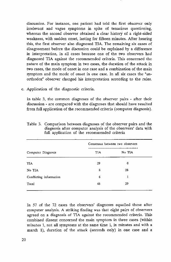
c. Application of the diagnostic criteria.
20
In table 3, the common diagnoses of the observer pairs - after their discussion - are compared with the diagnoses that should have resulted from full application of the recommended criteria (computer diagnosis).
Table 3. Comparison between diagnoses of the observer pairs and the diagnosis after computer analysis of the observers' data with full application of the recommended criteria
Computer Diagnosis
TIA
No TIA
Conflicting information
Total
Consensus between two observers
TIA
29
8
6
43
No TIA
0
28
1
29
In 57 of the 72 cases the observers' diagnoses equalled those after computer analysis. A striking finding was that eight pairs of observers agreed on a diagnosis of TIA against the recommended criteria. This combined dissent concerned the main symptom in three cases (within minutes 1, not all symptoms at the same time 1, in minutes and with a march 1), duration of the attack (seconds only) in one case and a

combination of the main symptom (blurred vision) and mode of onset (within minutes) in one case. In the remaining seven cases the computer analysis resulted in a diagnosis of TIA from the information of one observer and a conclusion "no TIA" from that of the other, which means that one of the pair had not applied the recommended criteria. In six of these seven cases the rules were broken to diagnose TIA, in only one case to diagnose no TIA.
DISCUSSION
In the absence of objective criteria for the diagnosis of TIA, improvement of the accuracy of the diagnostic process is not possible. All our efforts should therefore be directed at improving the precision of the diagnosis. In the present study, the use of (1) plain language in recording and interpreting the history and (2) a checklist in multiple-choice format resulted in an improvement of the interobserver agreement for the diagnosis of TIA in comparison with our previous results (kappa value 0.77 against 0.65 in the earlier study). The improvement could not be attributed only to a better agreement between the six observers who participated in both studies. In contrast with our previous study, neurology residents also participated, but if anything, this would lead to a lower agreement rate in view of the greater number of observers [140, 27] and the inclusion of less experienced physicians. [126, 30] This makes the improvement the more convincing. The agreement for the vascular territory of presumed TIAs also improved (kappa value 0.65 against 0.36 in the previous study). After (3) a discussion between the two observers, the agreement on the diagnosis was maximal (kappa value 1.0).
In achieving maximal interobserver agreement for the diagnosis of TIA we answered the main purpose of this study. On the other hand, analysis of the underlying data showed that some sources of error had remained. First, the observers showed striking differences in the classification and chronological assessment of the individual symptoms, but apparently this hardly effected agreement on the ultimate diagnosis. This does not mean that the differences were always subtle: in 7 of the 72 patients no diagnosis could be made when the check-lists of both observers were analyzed by computer, because of essential differences in recorded data. This finding might be important if the aid of complex computer systems is used to diagnose TIA from the information of the physician. [129] The validity of such a diagnostic procedure clearly depends on the ability of the physician to transmit the history as neutrally as possible to the computer. Studies on the
21

improvement of agreement should concentrate on optimal classification of symptoms by nature and time course. This could be achieved by carefully adapting the check-list and by training the observers.
Second, analysis of the check-lists showed that a considerable number of symptoms were not interpreted according to the recommended criteria. This was at odds with our aim of improving the precision of the diagnosis. Apparently, the presence of such criteria in no way guarantees uniformity in interpretation. In 29 of the 144 interviews the initial diagnosis would have been different if the recommended criteria had been fully applied. It is striking that the rules were far more often broken to diagnose "TIA" (28 cases) than to diagnose "no TIA'' (only one case). From the enquiry it appeared that the observers held widely different views on the interpretation of various symptoms, which partly explains why the recommended criteria were not consistently adhered to. These criteria, however, originated from internationally accepted diagnostic guidelines [148], which had been used for many years in our department and were implicitly endorsed by all participating observers. But these traditional criteria were phrased in abstract diagnostic terms, while the idiosyncrasies were apparently related to the "translation" of these criteria into plain language. For example, the observers have no difficulty in agreeing that amaurosis fugax is consistent with a TIA, but they often disagree on the actual definition of amaurosis. Even when observers did agree in theory that a particular symptom did not qualify for the diagnosis of TIA, they might still make this diagnosis when they had heard the same symptom from a real patient. It must of course be remembered that the recommended criteria are arbitrary guidelines, but the impressive variance of opinion among the observers about "atypical" symptoms makes it clear that more explicit definitions are necessary.
Although the number of interpretations against the recommended criteria is rather disappointing, there is good evidence that this might be further improved. In two previous studies [104, 61] attention has been drawn to the necessity .of prior discussion and agreement on the pieces of evidence required for a diagnosis. In both studies this subsequently improved the interobserver agreement on the ultimate diagnosis. Unfortunately, it is impossible to infer from the published data to what extent this diagnosis involved consultation of previously agreed criteria. Yet in all probability the number of "misinterpretations" in our study would have been smaller if the recommended criteria had not been merely added as an appendix to each form, but instead had been thoroughly discussed with all participating physicians before the start of the study.
22

Finally, it is surpnsmg that the eight pair-wise deviations from the recommended rules were not discovered by the observers during their discussions. Apparently in the absence of precise instructions for checking each other's history and subsequent interpretation, the observers quickly found out that they agreed on the diagnosis and then decided that they had nothing to talk about. This was confirmed by many observers after the study.
In conclusion, the present design has resulted in the maximal interobserver agreement for the diagnosis of TIA. However, as has been noted by others [38], we found that focusing only on the agreement between two observers does not exclude the possibility that both are "wrong" in the sense of not applying common diagnostic guidelines. This implies that the homogeneity of the group of patients classified as TIA can be further improved by a thorough discussion on the exact grounds for the diagnostic decision, and by a mutual check on the adherence to previously agreed rules.
23

CHAPTER III
CLINICAL DISAGREEMENT ON THE DIAGNOSIS OF TIA: IS THE PATIENT OR
THE DOCTOR TO BLAME?
Like many other clinical diagnoses, the diagnosis of TIA is subject to considerable interobserver disagreement. [143, 86, 20, 84] One reason for these variations may be that the clinicians obtain different information from the patient, another that the observers interpret the same history in a different way. Physicians participating in interobserver studies tend to stress the former and overlook the latter. In a previous study, 72 patients with possible TIAs were separetely interviewed by two neurologists, allocated in random pairs from a total group of 18. We found that of the 8 cases in which the observers disagreed on the diagnosis, six could be attributed to differences in interpretations, whereas only two resulted from a difference in acquired information. [84]
The aim of this study was to further elucidate whether the doctor or the patient is the major source of clinical disagreement.
PATIENTS AND METHODS
The population of 72 patients from our previous study was mixed with two simulated patients, without the participating neurologists knowing this. These two "patients" were actresses who were thoroughly trained in giving consistent information under all circumstances. One of them, aged fiftythree, was taught a history of a single attack of clumsiness of one arm and disturbed. articulation, which had come on suddenly and had lasted ten minutes. The other, fifty-eight years old, was supposed to have experienced two kinds of attacks. First she had noticed a rather vague visual disturbance of the left eye, "like looking through a steamy pane" during one minute. Some weeks later she had experienced a tingling sensation in the right arm, spreading in minutes to the face and leg. This attack had lasted 30 minutes. Neither of the two attacks was followed by headache, and the patient was not known to have migraine. Each of these "patients" was interviewed by four different pairs of neurologists. One of the sixteen observers appeared
24
slightly suspicious after having interviewed the patient, the others had noticed nothing unusual. The observers were asked to adhere to recommended rules for the diagnosis of TIA, which were based on internationally accepted criteria [148] and had been used for many years in the department. These criteria were included as a supplement to a checklist, on which the symptoms had to be recorded in detail. [84] According to the recommended criteria, the attack of the first patient qualified for a TIA, whereas the two attacks of the second patient did not.
RESULTS
All eight pairs of neurologists showed a complete uniformity in the description of the nature and time course of the individual symptoms. Yet in the first patient, seven concluded 'TIA', while one observer concluded 'no TIA'. In the second patient, six observers concluded 'no TIAs', whereas two observers from two different pairs concluded 'TIAs'. Altogether, only five of the eight pairs agreed on the diagnosis (agreement corrected for chance: kappa 0.25, against 0.77 in the real patients [84]).
DISCUSSION
The results from this small experiment confirm that differences in interpretation of symptoms are probably more important as a source of disagreement than differences in the content of the history. This implies that the consistency of the diagnosis of TIA could be improved if the diagnostic guidelines are thoroughly discussed and consistently adhered to. The patient is not always to blame.
25
CHAPTER IV
THE DUTCH TIA TRIAL: BACKGROUND AND DESIGN
The discovery that (1) transient ischemic attacks (TIAs) are the major precursors of stroke, (2) most TIAs are caused by thromboembolism, and (3) aspirin is a powerful inhibitor of platelet aggregation [116] has led to the hypothesis that this drug might prevent stroke and other cardiovascular complications in patients with TIAs. [69] This has been confirmed in a number of randomized controlled clinical trials. [22, 17, 153, 44, 144] Most studies have also included patients with prolonged attacks (RINDs) and partial strokes. [22, 17, 153] An overview of all trials has shown that antiplatelet agents - mostly aspirin - decrease the odds of nonfatal stroke and nonfatal myocardial infarction by 30% (SD 4% ), and the odds of fatal vascular complications by 15% (SD 4%). [4] The lowest dose of aspirin that was proved effective was 300 mg. [153]
During the past years new developments have generated new hypotheses. Firstly, further clarification of the biochemical effects of aspirin have led to the expectation that lower doses of aspirin might be equally effective or even better. Aspirin mediates its antiplatelet effects by inactivation of the enzyme cyclooxygenase. [133] This enzyme is present in platelets, where it controls the production of thromboxane A2, a powerful promotor of platelet aggregation. It is also found in endothelial cells, where it regulates the production of prostacyclin, which has precisely the opposite action. [113] Clearly, the latter effect of aspirin - inhibiting an anti-aggregant agent - is undesirable. Recent studies have shown that platelet cyclooxygenase is more susceptible to aspirin than vessel wall cyclooxygenase. [96, 120] The production of thromboxane A2, as measured by the serum level of the stable metabolite thromboxane B2, is suppressed by more than 90% by daily doses between 20 and 50 mg of aspirin. [33, 125, 164, 32, 80, 55, 165, ] Bleeding time is prolonged by doses over 30 mg. [80] The synthesis of prostacyclin, as measured by its urinary metabolite 6-keto-PGFw, shows unchanged excretion with aspirin doses up to 35 mg [32, 80, 118] and is partially suppressed by 50 mg. [131] The extrarenal production of prostacyclin is not spared even with 20 mg aspirin per day. [55] Thus, 20 mg aspirin/ day is the minimum for inhibiting the synthesis of thromboxane, 30
26
mg for prolonging bleeding time, and both these low doses have a slight but transient effect on the production of prostacyclin.
Secondly, the importance of ischemic heart disease as the most lifethreatening complication in patients with cerebrovascular disease has become more widely recognized. [22, 153, 152, 73, 2] Death from heart disease can be estimated at 1.5-5% per year, comparable to patients with angina pectoris. [2] In addition 1-2% of patients suffer a nonfatal myocardial infarct. [22, 17, 153] The discovery that beta-blockers decrease mortality after myocardial infarction by 20% (95% confidence interval15~ 30%) [170] has led to the hypothesis that that this benefit might also apply to patients with cerebrovascular disease.
Aims of the study
Two main hypotheses will be tested in patients with TIAs or a partial stroke:
1. 30 mg aspirin/ day is more effective than 300 mg in preventing death and disability or, more specifically, the occurrence of vascular death, nonfatal stroke, nonfatal myocardial infarction, or retinal infarction;
2. 50 mg atenolol is more effective than placebo in preventing these same events.
A subsidiary aim of the study is to investigate the prognostic importance of several variables, including the nature and time course of the ischemic attack, vascular risk factors, age, sex, blood pressure, ECG, and various types of ischemic lesions on the CT -scan.
Entry criteria
Transient ischemic attacks. Time course: the symptoms should develop within a few seconds, should not progress from one part of the body to another in an orderly march, and should last between 1 minute and 24 hours. Nature: 1) loss of vision (black or grey) in one eye, completely or in the upper or lower half; 2) language disorder; 3) weakness or clumsiness on one side; 4) loss of vision on one side, involving both eyes; 5) bilateral weakness, simultaneously or separately, or symptoms involving the face on
27
one side and the body on the other; 6) combinations of vertigo, diplopia, dysphagia, sensory loss, misdirections of limbs, or drop attacks. TIAs should not include loss of consciousness, convulsions, incontinence, or prominent headache.
Partial strokes. The mode of onset and nature of symptoms should be as specified above for TIAs, and the degree of disability should not be so severe that preventive treatment is not realistic. To be included, patients should be independent in most activities of daily living, corresponding to grade 3 or better on the modified Rankin scale: [157]
Grade 0. no symptoms at all Grade 1. no significant disability despite symptoms: able to carry out all
usual duties and activities Grade 2. slight disability: unable to carry out some previous activities but
able to look after own affairs without assistance Grade 3. moderate disability: requiring some help but able to walk without
assistance Grade 4. moderately severe disability: unable to walk without assistance
or unable to attend to own bodily needs without assistance Grade 5. severe disability: bedridden, incontinent, and requiring constant
nursing care and attention.
Exclusion criteria
I. Last ischemic attack more than 3 months ago. II. Forms of cerebral ischemia which are unlikely to be caused by arterial
thromboembolism from atherosclerosis: A. Precipitation of attack by standing, head turning or warming of the
face. B. Migraine, or attacks exactly resembling an aura of migraine
previously experienced, or scintillating scotomas not preceded by migraine.
28
C. Age under 40 years, unless relevant lesions of the carotid artery have been demonstrated.
D. Changes in heart rhythm, directly related to the attack (clinical diagnosis or ECG)
E. A source of embolism in the heart (atrial fibrillation, valve disorders, transmural myocardial infarcts less than 4 weeks old)
F. Haematological disorders: persistent anaemia (Hb 6.0 mmol/1 or less), polycythaemia rubra vera (Ht 0.60 or over), thrombocytosis (500 x 109/1 or over), thrombocytopenia (100 x 109/1 or less)
G. Vasculitis (SLE, arteriitis temporalis, polyarteriitis nodosa, lues, herpes zoster ophthalmicus)
III. Disorders that mimick cerebral ischaemia:
A. Intracranial haemorrhage, tumor cerebri, subdural haematoma. B. Hypo glycaemia (2 mmolfl or less) during the attack.
IV. Situations likely to confound interpretation of the trial results:
A. Cerebral infarction in the past with disabling residual deficits (Modified Rankin scale grade 4 or worse).
B. Myocardial infarction within the past month. C. Malignant tumor likely to cause death within a few weeks or
months. D. Likelihood of poor patient compliance. E. Patient does not speak the Dutch language fluently (an interpreter
does not solve this problem).
V. Disorders possibly exacerbated by acetylsalicylic acid:
A. Chronic renal failure (creatinine over 150 mmol/1) B. Liver failure C. Peptic ulceration (proved; within the past three years) D. Abnormal bleeding tendency (e.g. haemophilia, thrombopathia) E. Intra-cranial haemorrhage in the past F. Asthma bronchiale G. Patients already taking anti-platelet drugs for other reasons H. Patients already taking acetylsalicylic acid for other reasons
VI. Disorders possibly exacerbated by beta-blockers:
A. Frequency of heartbeat 50/min or less. B. PQ-time on ECG 0.25 seconds or more. C. A V-block of the 2nd or 3rd degree. D. Hypotension (diastolic blood pressure less than 80 mmHg) E. Decompensatio cordis (heart/thorax ratio on chest X-ray of 0.65
or over) F. Asthma bronchiale or chronic bronchitis
29

G. Diabetes mellitus H. Myasthenia gravis I. Raynaud's disease J. Intermittent claudication K. Patient already taking beta-blockers and having to continue
Mandatory investigations
Blood tests: haemoglobin, haematocrit, platelet count, erythocyte sedimentation rate, blood glucose, serum creatinine, hepatic enzymes, syphilis serology.
Chest x-ray. ECG. CT scan of the brain.
Evaluation of treatment
1. All events will be analyzed on an "intention-to~treat" basis. Thus, all randomized patients, including those withdrawn from study medication and those who are non-compliant, will be followed until the end of the study. This includes randomized patients who should have been excluded according to the study protocol (protocol violations). An additional analysis will be undertaken in which these patients as well as those who did not receive the full treatments will have been removed ("explanatory analysis"). In accordance with the intention-to-treat principle, patients should be kept on the study treatment as much as possible.
2. Analysis of events.
30
Primary measures for analysis: - Death and disability (measured by means of the modified Rankin
scale), from all causes.
Secondary outcome events: - Vascular death. - Nonfatal stroke. - Nonfatal myocardial infarction. - Retinal infarction.

3. The above analyses will be performed in each of the subgroups defined by the following major prognostic variables: - sex - age - presence of ischemic heart disease - presence and type of cerebral infarction on CT (cortical, lacunar,
borderzone) - degree of handicap at entry
For participants and committees, see appendix 3.
31
CHAPTER V
CEREBRAL INFARCTION ON CT IN PATIENTS WITH A TIA, RIND, OR PARTIAL STROKE
It is common to classify patients with cerebral ischemia according to the duration of the symptoms (TIA: completely reversible symptoms, lasting less than 24 hours, RIND: also reversible, but with symptoms lasting up to six weeks, stroke: persisting symptoms or signs). But in a proportion of patients with the time course of a TIA, for instance, computed tomography shows cerebral infarction in the corresponding area. [121, 87, 3, 5, 39, 64, 16, 21, 166, 162] For this reason, it has been suggested that TIAs, RINDs, and partial strokes should be regarded as a continuum rather than as strictly separated subgroups. [24, 11]
The proportion of cerebral infarction occurring in patients with transient ischemic attacks is unclear. Some authors did not find any infarcts at all, [83, 28, 14] others have reported numbers up to 50%. [121, 87, 3, 5, 39, 64, 16, 21, 166] It is also unknown whether the type of infarction (lacunar, cortical or watershed) is similarly distributed in patients with transient and those with persistent signs. Such qualitative differences might uncover a different pathogenesis. The only comparative study is that of Calandre et al, who studied 214 patients with transient or non-disabling cerebral ischemia and found that cerebral infarcts were equally common in patients with TIAs and RINDs (25%), and that these occurred only slightly more often in patients with permanent handicap(35%). [21] Disadvantages of this study are, however, that all patients with vertebrobasilar ischemia were included, in whom evidence of infarction is difficult to produce by CTscanning, that focal dilatation of a ventricle or a cistern was interpreted as an ischemic lesion, and that the authors did not specify the type of infarct in the three study groups.
We studied the CT scan findings of 2171 patients with ischemic attacks of one cerebral hemisphere who were entered into a multi-centre clinical trial (Chapter IV). Our aim was to assess the frequency, type, and location of cerebral infarction on CT in patients with a clinical diagnosis of TIA, RIND, or partial stroke.
32

PATIENTS AND METHODS
All patients in this study had been randomized into the Dutch TIA Trial. The background and design of this study is described in Chapter IV.
During the randomization period between March 1, 1986, and March 1, 1989 a total of 3150 patients were randomized. The clinical details of 13 patients were not yet available at the time of analysis, and in 116 patients the CT-scan was not yet available for analysis. Eleven patients were excluded for the present study because the CT-scan, which was sometimes made after randomization, showed an intracerebral hemorrhage ( 4 patients) or a cerebral tumor (7 patients). Also excluded were 270 patients in whom CT-scanning could not be expected to show an appropriate infarct from the outset, either because the interval between the CT-scan and the onset of symptoms was unknown (35 patients), or less than 24 hours (235 patients). Further excluded were 155 patients with monocular visual blindness, 256 patients with attacks of subtentorial ischemia, and 105 patients with attacks of uncertain vascular territory. In 53 patients the duration of the symptoms was not recorded. The clinical data and CT-scan findings in the remaining 2171 patients with ischemic attacks of a single hemisphere were analyzed. Six hundred thirty-seven patients had suffered a TIA (symptoms completely reversible within 24 hours), 367 a RIND (symptoms lasting more than 24 hours, but completely reversed within 6 weeks), and 1167 a partial stroke (persisting symptoms or signs, but with no greater handicap than a partial dependence upon others with regard to activities of daily life).
All CT-scans were independently and blindly reviewed by at least two neurologists or one neurologist and a neuroradiologist. In case of disagreement, a third neurologist or neuroradiologist arbitrated. Only after this the observers were given access to clinical details in order to assess the relevance of the CT-scan abnormalities.
Cerebral infarcts were defined as well-defined radiolucent lesions, and were subdivided into lacunar infarcts (small deep lesions), cortical infarcts (superficial radiolucent areas, involving the cortex), and watershed infarcts (wedge-shaped hypodensities in the borderzone area between two major cerebral arteries, or between deep and superficial branches of the middle cerebral artery). Lacunar infarcts were further subdivided according to their location: anterior limb of the internal capsule, genu, posterior limb, corona radiata, basal ganglia, thalamus, or other. The scans were classified
33
as showing a relevant infarct only, an irrelevant infarct only, or both a relevant and irrelevant infarct, dependent on the clinical symptoms.
The data were analyzed by means of the Statistical Package for the Social Sciences (SPSS) and Epistat statistical software. Yates' corrected chisquare test was used where appropriate.
RESULTS
In Table 1 the interval between the onset of symptoms and the CT scanning in patients with TIAs, RINDs, and partial strokes is shown. Patients with partial strokes were scanned, on average, earlier than TIAs and RINDs (x2 =50.40, df=12; p=0.00002). Table 1 also shows the percentage of patients with infarction for different intervals between onset of symptoms and CT. Altogether, 35% of CT scans showed a relevant infarct. CT scans made between 3 and 7 days after the onset of symptoms showed significantly more, and scans made between 15 and 28 days significantly less relevant infarcts (Goodness of fits test, p < 0.05 and p < 0.01, respectively).
Table 1. Interval between onset of symptoms and CT -scan in TIAs, RINDs, and partial strokes, and the relation between this interval and the prevalence of cerebral infarction.
Interval symptoms-CT
24 - 48 hrs
48 - 72 hrs
3 - 7 days
8 - 10 days
11 - 14 days
15 - 28 days
> 28 days
34
TIA RIND Stroke Relevant ----------------------------- infarct on CT
% of patients %
5 8 9 29
7 7 9 39
23 23 25 40
13 14 15 34
8 10 13 35
20 18 13 26
24 20 16 36

Cerebral infarction and duration of attack
In Figure 1 the occurrence of a relevant cerebral infarct on CT is related to the duration of the symptoms, subdivided in eight instead of three time categories: five sub-groups within the first day, two in the first six weeks, and one group with persisting symptoms. Cerebral infarcts were found in each time category, even in patients with attacks of a minute or less. The longer the duration of the attack, the more often CT showed a relevant infarct (x2 =221.74, df=7; p<O.OOOOOl). This is shown more explicitly in Figure 2, which depicts the percentage of patients with a · relevant infarct instead of absolute numbers. The increased frequency of infarcts with longer attacks was not in any way related to the 'bounderies' at 24 hours or six weeks.
number of patients 700.-----------------------------------------------~
600
500
400
300
200
100 5 15
0~~~~~~~~~~~~~
0-60 sec 1-30 min 31-60 min 1-4 hrs 5-24 hrs 1-7 days 1-6 wks persisting
duration of attack
Ill infarct !Z22l no Infarct
Figure 1. Duration of attack and presence or absence of a relevant infarct on CT.
35

percentage of patients 60 I
"RIND" I 60
I I
+ "TIA" I r-c- I 50 50
40 I I - 40
30 I r- - I - 30
r-1- I I 20 20
10 + I I - ,...__
I I 10 rfl I I 0 0 1-30 min 31-60 min 1-4 hrs 5-24 hrs 1-7 days 1-6 wks persisting
duration of attack The vertical lines represent the 95% confidence Interval
Figure 2. Duration of attack and percentage of patients with a relevant infarct on CT.
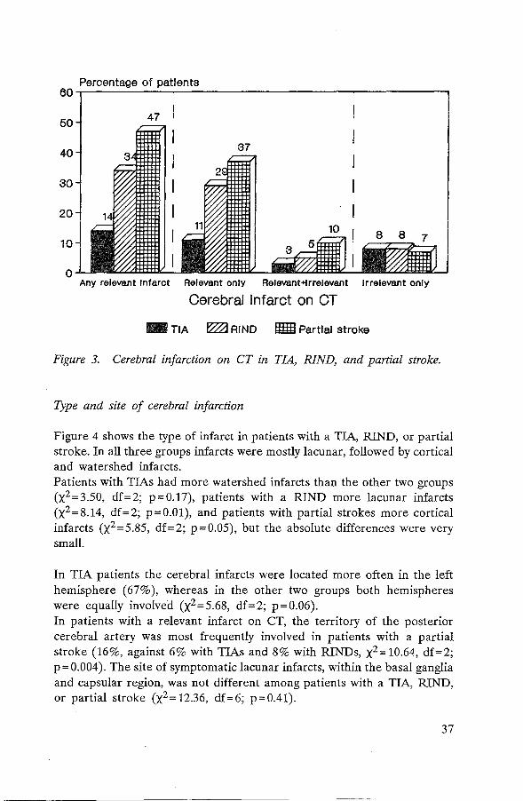
Figure 3 groups the proportion of relevant cerebral infarcts in patients with a TIA, RIND, or partial stroke. The difference between the three groups is highly significant, both for a relevant infarct only (11 %, 29%, and 37%, respectively, x2 =137.09, df=2; p<O.OOOOOl), and for a combination of relevant and irrelevant infarcts (3%, 5%, and 10%, respectively, x2 =37.69, df=2; p<O.OOOOOl). Irrelevant infarcts as the only abnormality on CT were equally common in the three groups (x2 =0.27, df=2; p=0.86).
36
Percentage of patients 60~----~--~~----------------------~--------~
50 47
40
30
20
10
0 Any relevant Infarct Relevant only Relevant+lrrelevant Irrelevant only
Cerebral Infarct on CT
- TIA ~RIND II!m Partial stroke
Figure 3. Cerebral infarction on CT in TIA, RIND, and partial stroke.
Type and site of cerebral infarction
Figure 4 shows the type of infarct in patients with a TIA, RIND, or partial stroke. In all three groups infarcts were mostly lacunar, followed by cortical and watershed infarcts. Patients with TIAs had more watershed infarcts than the other two groups (x2 =3.50, df=2; p=0.17), patients with a RIND more lacunar infarcts (X2 =8.14, df=2; p=0.01), and patients with partial strokes more cortical infarcts (x2 =5.85, df=2; p=0.05), but the absolute differences were very small.
In TIA patients the cerebral infarcts were located more often in the left hemisphere (67% ), whereas in the other two groups both hemispheres were equally involved (x2 =5.68, df=2; p=0.06). In patients with a relevant infarct on CT, the territory of the posterior cerebral artery was most frequently involved in patients with a partial stroke (16%, against 6% with TIAs and 8% with RINDs, x2 = 10.64, df=2; p = 0.004). The site of symptomatic lacunar infarcts, within the basal ganglia and capsular region, was not different among patients with a TIA, RIND, or partial stroke (x2 = 12.36, df=6; p=0.41).
37

24% TIA
79%
RIND
mil lacunar
~cortical
• watershed
66%
27% Stroke
Figure 4. Type of infarct in TIA, RIND, and partial stroke.
DISCUSSION
7%
Our study shows that relevant cerebral infarcts on CT can be found in any cerebral ischemic attack, regardless of its duration, even in attacks lasting less than a minute. We found a relevant cerebral infarct in 14% of TIAs, 34% of RINDs, and 47% of partial strokes. Although the incidence of infarction among patients with TIAs is somewhat lower than reported by others, [5, 64, 21, 166] the mere presence of cerebral infarcts definitely links TIAs to strokes. Infarcts were predominantly of the lacunar type in all three groups. This over-representation of lacunar infarcts (about 70%, against 25% of all infarcts in population studies [8] should be attributed to selection bias, as many patients with cortical infarcts were too severely handicapped to qualify for the clinical trial of secondary prevention from which our comparisons were made. Only minor qualitative differences between the three groups were found. Firstly, patients with TIAs showed a relatively high proportion of watershed infarcts, patients with RINDs of lacunar infarcts, and patients with partial strokes of cortical infarcts. Owing to the large number of patients in our study, the last two differences, although very small, just reached statistical significance. Secondly, in patients with partial strokes the posterior cerebral artery was more frequently involved, but no other differences in location were found. The latter difference probably again
38

reflects the selection criteria for the study, as patients with middle cerebral artery infarcts more often are dependent on others. The overwhelming similarities of both the type and the location of the cerebral infarcts among TIAs, RINDs, and partial strokes and the gradual increase of the proportion of infarcts with the duration of the attack suggests that the differences are quantitative rather than qualitative. These findings support the notion that the three groups should be regarded as a continuum rather than as sharply separated subgroups. [24, 11] That the time limit of 24 hours which separates TIAs from RINDs and partial strokes is very inaccurate is recently also shown by Levy, who showed that the large majority· of TIAs last shorter than one hour, whereas longer attacks rarely clear within the next hour, but often exceed the arbitrary boundery of 24 hours. [92]
However, before discarding terms such as TIA, RIND, and stroke [24], we need more information on the prognosis in each of these groups. Aggregate data from different studies suggest a similar outcome for the three groups [24], but this has never been studied concurrently. The results of a recent study suggest a better outcome in stroke patients in comparison with TIAs, but because of the small numbers both of study patients and outcome events the results can not be regarded conclusive. [42] Therefore, we do not know whether long attacks herald more harm in the future than short ones, or whether reversible attacks are less often followed by disabling stroke than those with persistent symptoms. It is also unknown whether patients with a cerebral infarct on CT suffer an greater risk of major stroke than those with similar attacks but a normal scan. These questions are currently under investigation in the Dutch TIA trial, as an adjunct to the main questions regarding the efficacy of low-dose aspirin and of atenolol.
39
CHAPTER VI
TRANSIENT ISCHEMIC ATTACKS WITH AND WITHOUT A RELEVANT CEREBRAL INFARCT ON CT CANNOT BE CLINICALLY DISTINGUISHED
A transient ischemic attack (TIA) is defined as an acute focal neurological deficit lasting less than 24 hours. [148] This arbitrary time limit has proved inaccurate with regard to the integrity of brain tissue, since a proportion of TIA patients shows a relevant cerebral infarct on CT-scanning, as shown in Chapter IV and previously by others. [121, 87, 39, 21, 15] This condition has been called cerebral infarction with transient signs (CITS) by Waxman and Toole. [162] These authors have speculated that CITS patients have a different lesion in the vascular tree than TIA patients without a demonstrable infarct, and a different prognosis. Bogousslavsky and Regli initially found an association between CITS and occlusion of the carotid artery [16], but in a subsequent study of 57 TIA patients, including 16 with a cerebral infarct on CT, no specific underlying cause was found. With regard to the attacks, however, CITS patients had a significantly longer duration of symptoms and a smaller number of attacks. [15]
In the present study we have prospectively investigated possible differences in risk factors or in clinical manifestations in three groups of patients: TIA patients with and without a relevant cerebral infarction on CT, and patients with a resolving (RIND) or non-disabling stroke and a corresponding infarct on CT.
PATIENTS AND METHODS
All patients in this study had been entered into the Dutch TIA triaL Background and design of this multicentre trial have been described in Chapter IV.
For the history a check-list is used, on which the symptoms are recorded in plain language. [84] The list contains a number of detailed multiple-choice questions about the nature of the symptoms as well as about the time course, including the mode of onset, whether or not the symptoms occurred at the same time, the duration of the attack, the time the symptoms took to disappear, and the number of attacks.
40

During the randomization period, between March 1, 1986, and March 1, 1989, 3150 patients were randomized. From these, we selected patients with TIAs or a partial stroke that could be attributed to ischemia in one cerebral hemisphere, and in whom CT-scanning was performed not earlier than 24 hours after the first attack. We therefore excluded 116 cases in which the CT-scan was not yet available for analysis, 4 patients with an intracerebral hemorrhage, 7 with a cerebral tumor, 270 patients in whom the interval between the neurological symptoms and the CT-scan was unknown (35 patients) or less than 24 hours (235 patients), 155 patients with monocular visual symptoms, 256 patients with symptoms of subtentorial ischemia, 105 patients with attacks of uncertain vascular territory, and 53 patients in whom the duration of the attack was not recorded. Of the remaining 2171 patients, 637 had suffered a TIA (symptoms completely resolving within 24 hours) and 1534 either a reversible ischemic neurological deficit (RIND; symptoms lasting more than 24 hours but completely resolving within six weeks) or non-disabling stroke (symptoms persisting longer than 24 hours, but not leading to loss of independence in daily living at the time of randomization, that is, up to three months after the event but as a rule not more than 4 weeks later).
All CT-scans were independently and blindly reviewed by_ at least two neurologists, or by one neurologist and a neuroradiologist. In case of disagreement, a third neurologist or neuroradiologist arbitrated. Only after this the observers were given access to clinical details in order to assess the relevance of the CT -scan abnormalities.
Cerebral infarcts were defined as well-defined radiolucent lesions, subdivided into lacunar infarcts (small deep lesions), cortical infarcts (superficial radiolucent areas, involving the cortex), and watershed infarcts (wedge-shaped hypodensities in the borderzone area between two major cerebral arteries, or between the deep and superficial branches of the middle cerebral artery. Lacunar infarcts were further subdivided according to their location: anterior limb of the internal capsule, genu, posterior limb, corona radiata, basal ganglia, thalamus, or other. White matter hypodensity with ill-defined borders was interpreted and recorded as periventricular leukoencephalopathy. [156] Dependent on the clinical features, the scans were classified as showing a relevant infarct only, an irrelevant infarct only, or both relevant and irrelevant infarcts.
The data were analyzed by means of the Statistical Package for the Social Sciences (SPSS) and Epistat statistical software. Yates' corrected chi-square test and Fisher's exact probability test were used where appropriate. Comparisons between the groups were expressed as Odds Ratios (TIA with infarct: TIA without infarct, and TIA with
41

42
infarct: stroke with infarct), with the corresponding 95% confidence intervals (CI). [170] A level of p < 0.05 was considered significant.
RESULTS
In 87 of the 637 TIA patients (14% ), and in 669 of the 1534 patients with RIND or persisting stroke ( 44% ), a relevant infarct was found on CT. In TIA patients the infarcts were lacunar in 57, cortical in 21, and in watershed areas in 9 patients, against 458, 170, and 41, respectively, in the patients with longer or persisting symptoms. The interval between neurological symptoms and CT-scanning was not different in TIA patients with or without infarction (x2 =7.59, df=7; p=0.37).
Comparison between TIA patients with and without a relevant cerebral infarct on CT
a. Vascular risk factors and vascular disease Figure 1 shows that TIA patients with cerebral infarction more often were known to have hypertension (p=0.04). No other significant differences between the two groups were found.
Hypertension
Diabetes
Hyperlipidemia
Smoking
Intermittent claud
Angina pectoris
Past Ml
Enlarged heart
r I
I I
I I
_l I
I I
0 0.5 more common
In TIA without Infarct
I I
r I
1 I
1.5 2 more common
In CITS
The horizontal lines represent the 95$ confidence limits
p•0.04
- -
2.5 3 Odds ratio
Figure 1. Vascular risk factors and vascular disease in CLTS and in TIA without cerebral infaction.
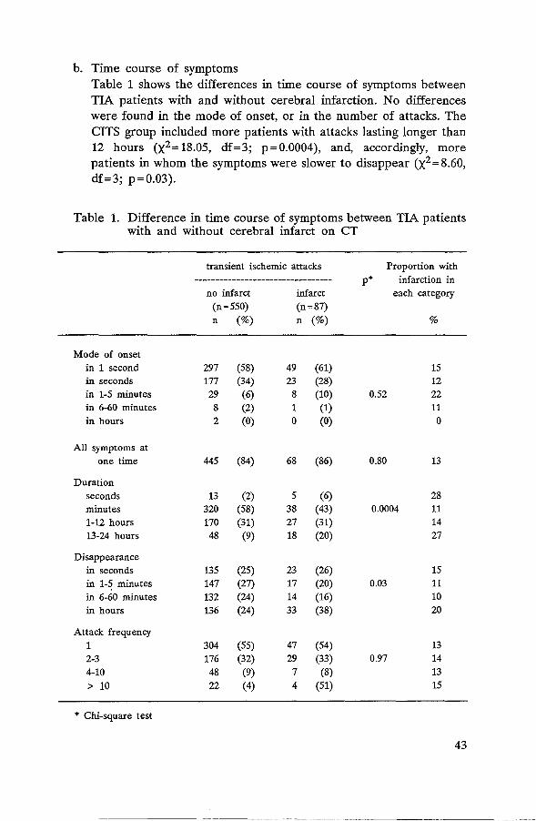
b. Time course of symptoms Table 1 shows the differences in time course of symptoms between TIA patients with and without cerebral infarction. No differences were found in the mode of onset, or in the number of attacks. The CITS group included more patients with attacks lasting longer than 12 hours (x2 =18.05, df=3; p=0.0004), and, accordingly, more patients in whom the symptoms were slower to disappear (x2 = 8.60, df=3; p=0.03).
Table 1. Difference in time course of symptoms between TIA patients with and without cerebral infarct on CT
Mode of onset in 1 second in seconds in 1-5 minutes in 6-60 minutes in hours
All symptoms at one time
Duration seconds minutes 1-12 hours 13-24 hours
Disappearance in seconds in 1-5 minutes in 6-60 minutes in hours
Attack frequency 1 2-3 4-10 > 10
* Chi-square test
transient ischemic attacks
no infarct (n=550) n (%)
297 (58) 177 (34) 29 (6) 8 (2) 2 (0)
445 (84)
13 (2) 320 (58) 170 (31) 48 (9)
135 (25) 147 (27) 132 (24) 136 (24)
304 (55) 176 (32) 48 (9) 22 (4)
infarct (n=87) n (%)
49 (61) 23 (28) 8 (10) 1 (1) 0 (0)
68 (86)
5 (6) 38 (43) 27 (31) 18 (20)
23 (26) 17 (20) 14 (16) 33 (38)
47 (54) 29 (33)
7 (8) 4 (51)
p* Proportion with
infarction in each category
0.52
0.80
0.0004
0.03
0.97
%
15 12 22 11 0
13
28 11 14 27
15 11 10 20
13 14 13 15
43

44
Despite this difference in time course between patients with CITS or TIA, the prediction of infarction on CT on the basis of the time course proved impossible, since in each time course category the majority of patients had a normal CT scan (see Table 1, proportion with infarction in each category).
c. Nature of symptoms In figure 2 the differences in main symptoms between the two groups are shown. CITS patients had significantly more often dysphasia, or combined weakness and dysphasia. Sensory symptoms, in any combination, were less common among CITS patients, but the differences did not reach statistical significance (Odds ratio 0.69, 95% CI 0.42-1.11; p=0.14).
Motor only Motor+sensory+speech I
p•0.001
A
Motor + dysphasia I -Motor + dysarthria
Any motor symptoms I'
Sensorimotor ny sensory symptoms ',
I
Dysphasia I p-0.01 I
Dysarthria I I Dysgraphia - -
Visual field defect I I
Non-specific. onJy• l p•0.03 l
0 0.5 1.5 2 2.5 3 3.5 4 more common more common Odds ratio
In TIA In CITS without Infarct
The horizontal lines represent the 95$ confidence llmlts •tor explanation, see text
Figure 2. Main symptom(s) in CITS and in TIA without cerebral infarction.
In only 1 of the 87 CITS patients, against 47 of the 550 TIA patients without cerebral infarction the history consisted of non-specific symptoms only, such as isolated sensory symptoms (the single CITS patient, and 19 TIA patients), blurred vision, fortification spectra, or

"flashlights" in one hemifield (9 patients), a "heavy" or "tired" sensation in one or more limbs without weakness or clumsiness (15 patients), combined isolated sensory symptoms and atypical visual symptoms (!,patient), or accompanying unconsciousness (4 patients). The Odds ratio of this difference was 0.12, 95% CI 0.02-0.85; p=0.03.
No significant differences were found in accompanying symptoms, such as headache (Odds ratio 0.68, 95% CI 0.33-1.38; p=0.33), or lightheadedness or aspecific dizziness (Odds ratio 1.02, 95% CI 0.52-1.98; p = 0.94).
The distribution of both motor and sensory symptoms (arm, leg, face, or any combination) was not different between the two groups.
Comparison between CITS patients and RIND or stroke patients with cerebral infarction
a. Vascular risk factors and vascular disease Figure 3 shows no differences in vascular risk factors or vascular disease between CITS patients and patients with RIND or stroke who had a demonstrable cerebral infarct.
Hypertension
Diabetes
Hyperlipidemia
Smoking
Intermittent claud
Angina pectoris
Past Ml
Enlarged heart
I I
I I
I I
I I
I I
0 0.6 more common
In stroke
I
I I
1.6 2 more common
In CITS
The horizontal lines represent the 95% confidence limits
2.6 3 Odds ratio
Figure 3. Vascular risk factors and vascular disease in CITS and in stroke with cerebral infarction.
45

46
b. Time course of symptoms The duration of the attack differed by definition between CITS patients and stroke patients. The mode of onset was not significantly different between the two groups, but tended to be slower in stroke patients (p = 0.08). The number of attacks was significantly smaller in stroke patients: a single attack in 54% of CITS, against 80% of stroke patients, 2-3 attacks in 33% and 16%, 4-10 attacks in 8% and 3%, and more than 10 attacks in 5% and 1%, respectively (x2 =34.41, df=3, p<0.000001).
c. Nature of symptoms Figure 4 shows that the differences between the two groups were small with regard to symptoms. CITS patients had more often dysphasia, or a combination of dysphasia and motor symptoms, and less often isolated motor symptoms. No difference was found either in the frequency of non-specific symptoms only, or in the distribution of motor and sensory symptoms. With regard to accompanying symptoms, CITS patients tended to have more often lightheadedness (Odds ratio 1.63, 95% Cl 0.82-3.16; p=0.17), and less often headache (Odds ratio 0.71, 95% Cl 0.34-1.44; p=0.41).
Motor only p-Q.04 I M otor+sensory+speech
Motor + dysphasia I p•0.02
A
Motor + dysarthria Any motor symptoms
Sensorimotor ny sensory symptoms
Dysphasia Dysarthria
Dysgraphia
VIsual field defect
Non-specific only•
I
I I
J
0 0.5 more common
In stroke
I
I
1.5 2 more common
In CITS
2.5
The horizontal linea represent the 95% confldenoe limits •for explanation, see text
-
p•0.03
3 3.5 Odds ratio
Figure 4. Main symptom(s) in CITS and in stroke with cerebral infarction.

DISCUSSION
Our study shows not more than a few slight clinical differences between TIA patients with and without a relevant infarct on CT. The relative preponderance of attacks exceeding 12 hours in TIAs with infarction confirms the findings of Bogousslavsky and Regli. [15] We also found a slower disappearance of symptoms in these patients, but no difference in the number of attacks. In contrast to this previous study, which showed that 84% of patients with attacks lasting more than 45 minutes had a relevant infarct on CT [15], we found that the presence of cerebral infarction could not be predicted from any of the time course variables, since in each time category patients with a normal CT scan formed a large majority. The difference in the nature of symptoms -more often dysphasia or combined dysphasia and weakness, and less often non-specific symptoms in TIA patients with infarction - is not easy to explain. One reason might be that the diagnosis of TIA was incorrect in a proportion of the patients who were entered into the treatment trial. The inclusion of patients with these non-specific symptoms was discouraged, but some such patients were still included because the randomizing neurologist considered other disorders such as migraine, epilepsy, hyperventilation or neurosis an even more unlikely diagnosis. These differences in whether the attacks were really of ischemic origin may result in a lower incidence of relevant CT lesions in patients with less convincing symptoms such as isolated sensory disturbances or a "heavy" or "tired" limb, and a higher incidence in patients with the unequivocal symptom of abnormal speech, with or without weakness. Little is known about the prognostic value of individual symptoms, although the results of a preliminary study suggest a low risk of subsequent cerebral events in TIA patients with "soft" symptoms (Chapter VIII). This issue is currently under investigation in the Dutch TIA trial.
Does CITS represent a subgroup with a specific vascular cause? This seems ·very unlikely. Firstly, our study shows that the clinical manifestations of TIA patients with and without infarction mostly overlap. Secondly, others found no difference in angiographical and cardiac abnormalities between the two groups. [15] Thirdly, in a previous study (see Chapter V), we found no difference in the type of cerebral infarction (lacunar, cortical, watershed) between patients with a TIA, RIND, or partial stroke, but only in the frequency. Finally, newer imaging techniques, such as positron emission tomography and magnetic resonance imaging may be even more sensitive than CT in documenting
47

48
ischemic lesions. [6, 124] Separating TIA patients by the results of CTscanning alone would therefore be just as inaccurate as the artificial time limit of 24 hours which separates TIAs from RINDs and strokes. In addition, our study shows that the clinical differences - other than the duration - between CITS patients and stroke patients with cerebral infarction were equally small, which suggests that CITS as closely resembles strokes with cerebral infarction as TIAs without CT abnormalities.
The next step will be to investigate whether CITS represents a clinical subgroup with regard to prognosis. In the coming years we shall analyze the outcome events in the same cohort of patients to determine possible differences between 4 groups of patients: with symptoms lasting less or more than 24 hours, and with or without a relevant infarct on CTscanning.

CHAPTER VII
HEADACHE IN ACUTE CEREBRAL ISCHEMIA
Previous reports have shown that headache is a common feature in acute cerebrovascular disease, but the frequency widely varies between different studies. [50, 67, 105, 111, 41, 40, 122, 95] The main cause of the variation is that only two surveys have used a propective design in studying the occurrence and nature of headache in acute cerebrovascular events. [40, 95] Moreover, the majority of previous studies have included patients with widely different cerebrovascular disorders such as transient ischemic attacks, cerebral infarction of all types, intraparenchymal and subarachnoid hemorrhage. The reason was that these studies have mainly addressed the question whether certain headache features help to distinguish infarction from bleeding or to locate the lesion.
Some studies suggest that headache is more common in patients with occlusive disease of major extra or intracranial blood vessels than in patients with small vessel disease. [50, 105 , 40, 122, 95, 66] The frequency of headache in lacunar infarction differs between various studies, however, ranging from 4% to 17%. [50, 105, 122, 95, 66] There is also disagreement about the occurrence of headache in patients with transient monocular blindness, [50, 67, 65] and about whether headache occurs relatively more often in women. [67, 40, 122] Little is known about the relation with vascular risk factors, the time course and the duration of the attack, or the number of previous attacks.
We have prospectively studied headache features in 3126 patients with acute cerebral ischemia of variable duration, who were entered into a multicenter treatment trial. We have attempted to relate the occurrence and nature of headache to the presence of vascular risk factors, the probable site of origin of the neurological symptoms (the cerebral cortex, the capsular region or the retina), the time course of the attack, and the CT scan findings.
49

50
PATIENTS AND METHODS
All patients had been entered into the The Dutch TIA Trial, described in Chapter IV. In this trial a check-list is used, by means of which the symptoms are recorded in plain language. The list contains a number of detailed multiple-choice questions about the nature of the symptoms as well as about the time course, including the mode of onset, the duration of the attack, and the number of attacks. One question pertains to the presence and nature (throbbing versus constant) of headache. The investigators were also encouraged to specify the side of the headache, but as this item was not included in the check-list the data were incomplete in this respect and could not be analyzed. The degree of disability was scored by means of the modified Rankin scale (see Chapter IV). To be included, patients should be independent in most activities of daily living, corresponding to grade 3 or better on this scale. [157]
During the randomization period, between March 1, 1986, and March 1, 1989, 3150 patients were randomized. In 13 patients the clinical information was not yet complete at the time of analysis. Eleven more patients were excluded because the CT-scan, which was sometimes made after randomization, showed an intracerebral hemorrhage ( 4 patients) and a cerebral tumor (7).
In 2931 patients a CT scan was available at the time of analysis. These scans were independently and blindly reviewed by at least two neurologists or by one neurologist and a neuroradiologist. In case of disagreement, a third neurologist or neuroradiologist arbitrated. Only then the observers were given access to clinical details in order to assess the clinical relevance of the CT scan abnormalities.
Cerebral infarcts were defined as circumscribed hypodense lesions, subdivided into lacunar (small deep lesions), cortical infarcts (radiolucent lesions in the territory of one or more cortical arteries), and watershed infarcts (wedge-shaped hypodensities in the borderzone area between two major cerebral arteries, or between deep and superficial branches of the middle cerebral artery). Lacunar infarcts were further subdivided according to their location: anterior limb of the internal capsule, genu, posterior limb, corona radiata, basal ganglia, thalamus, or other. Dependent on the clinical details, the scans were classified as showing a relevant infarct only, an irrelevant infarct only, or both relevant and irrelevant infarcts.

The data were analyzed by means of the Statistical Package for the Social Sciences (SPSS) and Epistat Statistical software. Yates' corrected chi-square test was used where appropriate. Comparisons between the groups were expressed as Odds Ratios (with headache:without headache), with the corresponding 95% confidence intervals (CI). [170] A level of p < 0.05 was considered significant.
RESULTS
In 557 of the 3126 patients the cerebral ischemic attack was accompanied by headache (18%, 95% Cl 17-19%). The occurrence of headache was not related to sex (Odds ratio 0.91, 95% CI 0.75-1.11; p=0.38), or Rankin disability score at the time of randomization (X2=2.49, df=3; p=0.47).
Figure 1 shows the relation between headache and vascular risk factors. Patients with headache more frequently had evidence of ischemic cardiac disease, such as angina pectoris (Odds ratio 1.62, 95% CI 1.21-2.17; p=0.0008), past myocardial infarction (Odds ratio 1.33, 95% Cl 0.99-1.78; p=0.06), and past coronary bypass surgery (Odds ratio 1.52, 95% CI 0.82-2.77; p=0.17). This difference could not be explained by a higher frequency of a specific type of infarction, especially cortical, among patients with a history of cardiac disease.
Hypertension -t- p•0.04
Diabetes I
Hyperlipidemia I I
Smoking -t-c-Intermittent claud I
I
Angina pectoris I I
---1----p-0.0008
Past Ml I I
Enlarged heart
Coronary bypass I I
0 0.5 1.5 2 2.5 3 less frequent In more frequent In Odds ratio
patients with patients with headache headache
The horizontal lines represent the 95% confidence limite
Figure 1. Vascular risk factors and headache.
51

Patients with headache less frequently were known to have hypertension (Odds ratio 0.81, 95% CI 0.67-0.98; p=0.04). This finding could be explained by the higher frequency of lacunar infarcts, which were associated with a lower frequency of headache (see below), among hypertensive patients.
Table 1 shows the relation between the time course of symptoms and the occurrence of headache. No statistical difference was found either in onset, duration and disappearance of symptoms, or in the number of attacks between patients with and without headache. The same was true for the number of previous attacks. Conversely, Table 1 also shows that the presence of headache could not be predicted from any of the time course variables, the more so since in each category patients without headache were a large majority (see proportion of patients with headache in Table 1). With regard to the duration of the attack, headache was equally frequent in TIA, RIND, and stroke patients (17%, 17%, and 19%, respectively; x2 = 1.76; p=0.41).
Figure 2 shows the difference in the nature of the main symptoms between patients with and without headache. Patients with headache less often had a lacunar syndrome (Odds ratio 0.70, 95% CI 0.58-0.85; p=0.0002) and more often symptoms favoring a cortical localisation (Odds ratio 1.63, 95% CI 1.28-2.07; p = 0.00005). Of 155 patients with transient monocular blindness 26, or 17%, reported accompanying headache, which is the same as for all other patients (Odds ratio 0.92, 95% CI 0.58-1.45; p=0.80).
Figure 3 shows the relation between the CT scan findings and the presence of headache. Lacunar infarcts were significantly less frequent (Odds ratio 0.60, 95% CI 0.46-0.79, p = 0.0002), and cortical infarcts more frequent in patients in with headache (Odds ratio 1.71, 95% CI 1.25-2.34; p=0.0004). The proportion with headache in patients with lacunar infarcts was 13% (95% CI 11-16%) and in patients with cortical infarcts 26% (95% CI 23-29% ). Cortical infarcts in the partial territory of a major cerebral vessel were less. often accompanied by headache than those involving the complete territory (Odds ratio 0.43, 95% CI 0.18-0.99; p=O.OS).
52

Table 1. Relation between the time course of the cerebral ischemic attack and the presence of accompanying headache.
proportion with no headache headache p* headache in
(n=2569) (n=557) each category n (%) n (%) %
ONSET 1 second 1282 (57) 243 (49) 16 seconds 627 (28) 161 (33) 20 1-5 minutes 154 (7) 40 (8) 0.07 20 6-60 minutes 56 (2) 10 (2) 15 hours 95 (4) 25 (5) 21 days 53 (2) 15 (3) 22
DURATION seconds 34 (1) 9 (2) 21 minutes 508 (20) 97 (17) 16 hours 319 (12) 67 (12) 0.58 17 days 432 (17) 88 (16) 17 persisting 1272 (50) 294 (5) 19
DISAPPEARANCE 1 second 86 (5) 14 (4) 14 seconds 210 (14) 36 (11) 15 1-5 minutes 247 (16) 56 (17) 037 18 6-60 minutes 216 (14) 42 (12) 16 hours 284 (19) 72 (21) 20 days 492 (32) 117 (35) 19
NUMBER OF ATTACKS 1 1768 (69) 377 (67) 18 2-3 592 (23) 134 (24) 18 4-10 158 (6) 33 (6) 0.81 21 > 10 55 (2) 15 (3) 21
* Chi-square test
53
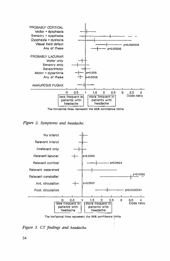
1
I
1 I
PROBABLY CORTICAL Motor + dysphasia
Sensory + dysphasia Dysphasia + dyslexia
VIsual field defect Any of these
---+---- p•0.000003
--1--1- p•0.00006
I+ PROBABLY LACUNAR
Motor only Sensory only Sensorimotor
Motor + dysarthria Any of these
---t- p-0.006
+ p•0.0002
AMAUROSIS FUGAX
0 0.5 less frequent In
patients with headache
I I
1.5 2 2.5 more frequent In
patients with headache
3 3.5 4 Odds ratio
The horizontal linea represent the 96% confidence limite
Figure 2. Symptoms and headache.
No Infarct
Relevant Infarct
Irrelevant only
Relevant lacunar + p-0.0002
Relevant cortical --+---- p-0.0004
Relevant watershed p-0.0002
Relevant cerebellar --------r--Ant. circulation + p•0.0001
Post. circulation ----'f----- p<0.0000001
0 0.5 ess frequent n patients with
headache
1.5 2 2.5 more frequent n
patients with headache
3
The horizontal linea represent the 95% confidence limite I
3.5 4 Odds ratio
Figure 3. CT findings and headache. I 54

Seventy-five patients with a lacunar infarct on CT had accompanying headache. Apart from symptoms which could be attributed to the side and site of the lacune, 24 of these patients (28%) complained of one or more symptoms compatible with aphasia (16), dysgraphia (5), dyslexia (2) or hemianopia (6), against 109 of the 521 (20%) patients with a lacunar infarct without headache (Odds ratio 1.47, 95% CI 0.82-2.63; p=0.20). In these cases cortical structures may have been involved as well, without evidence on CT. The presence of headache was not related to the location of the lacunar infarcts.
Figure 3 also · shows that headache was less frequent in patients with infarcts in the anterior circulation, that is, in the territory of the middle and anterior cerebral artery compared with all other patients (Odds ratio 0.64, 95% CI 0.51-0.80; p=0.0001) and more frequent in patients with infarcts in the posterior circulation, i.e. the territory of the posterior cerebral artery, the basilar artery, or cerebellar arteries (Odds ratio 2.28, 95% CI 1.67-3.12; p < 0.000001). These differences were independent of a smaller number of lacunar infarcts in the posterior circulation (63 lacunes, mostly in the territory of the perforating arteries to the thalamus, against 535 in the anterior circulation), and of the larger number of cortical infarcts (including cerebellar infarcts). Both lacunes and cortical infarcts were more often accompanied by headache if these corresponded to ischemia in the posterior circulation, and less often if they resulted from ischemia in the anterior circulation.
The nature of headache was mostly constant, and not throbbing, in the majority of patients with headache, regardless of the main symptom or the findings on CT (94%, 95% CI 92-96).
DISCUSSION
Our study confirms that headache not uncommonly accompanies a transient ischemic attack or the onset of a non-disabling stroke, although its overall frequency of 18% in our series is somewhat lower than in previous reports. (50, 67, 105, 111, 41, 40, 122, 95, 66] The headache was mostly constant in all types of attacks. Furthermore, our study shows two distinctive subgroups that were particularly associated with headache. Firstly, headache occurred twice as often in patients with a relevant cortical infarct on CT as in patients with symptomatic lacunar infarct (26% versus 13% ).
55

Secondly, headache was much more common among patients with infarcts in the posterior circulation (posterior cerebral artery, basilar artery or cerebellar arteries) than in patients in whom the anterior circulation (anterior or middle cerebral artery) was involved. The latter difference was independent of the difference in number of lacunar and cortical infarcts. Both patterns were recognized in earlier studies, [50, 105, 40, 122, 95, 66] but were never convincingly proved because of the small numbers of patients studied. No relation was found between the presence of headache on the one hand and sex, the mode of onset, mode of disappearance, the duration of the attack, or the number of previous attacks on the other. In particular, headache was equally common among patients with TIAs (17%), RINDs (17%), or partial strokes (19%).
Our finding that headache occurred in 17% of TIA patients implies that the mere presence of headache during an attack is not very helpful in the differentiation between a TIA and late life migraine accompaniments, the more so since a previous study has reported accompanying headache in only 50% of the latter patients. [45] This is illustrated by the following approximation of the predictive value of headache. One study has shown that neurologists rejected the diagnosis of TIA in about 30% of patients in whom that diagnosis was considered by the referring physician. [20] In about 3% of patients the investigators agreed on a diagnosis of migraine. So if 1000 patients are referred to a neurologist with a possible TIA, then the diagnosis will be confirmed in 700 patients, wheras migraine will be considered the most likely diagnosis in 30. Of the 700 TIA patients 119 (17%) may be expected to have had accompanying headache, against 15 of the migraine patients. The predictive value of headache alone for the diagnosis of migraine can then be calculated as 15/(119+15)=0.11 (with an a priori probability of 0.03). In other words, if a patient is referred to a neurologist because of a possible TIA, and the attack was accompanied by headache, then the probability that the most likely diagnosis will be migraine is 11%, against 3% if headache was absent. Thus, the presence of headache alone does hardly influence the decision whether the patient has had a TIA or a migraine accompaniment. Other characteristics of the attack may provide more important clues for the diagnosis of late-life migraine accompaniments, such as the presence of typical visual prodromes, particularly scintillating scotomata, a gradual ''build-up", expansion and migration of the visual aura, a "march" of paresthesias, and the serial progression from one symptom category to another. [46]
56

Headache occurred in 17% of patients with transient monocular blindness, which is surprisingly high in view of earlier reports. Fisher reported headache in none of his 58 patients with monocular blindness [50], and Goodwin in only one patient from a group of 37 patients. [65] Grindal and Toole [67] noted headache in 5 patients with amaurosis fugax, but did not mention the total number of patients investigated. We found no relation between the occurrence of headache and the nature of the visual symptoms.
The lower frequency of headache among patients with lacunar infarction in comparison with cortical infarcts confirms earlier reports, [50, 105, 40, 122, 95, 66] although all previous studies, except one [122] report a much lower frequency than the 13% that we found. Yet the mere presence of headache in patients with lacunar infarcts is a puzzling finding. We found that a high proportion (28%) of these patients with an appropriate lacune and headache also reported symptoms compatible with aphasia, dysgraphia, dyslexia, or visual field defects, suggesting cortical ischemia. Other investigators have demonstrated cortical dysfunction in patients with lacunar infarcts and aphasia by means of positron emission tomography (PET). [13] It is conceivable that some occlusions in the stem of the middle cerebral artery affect perforating arteries as well as cortical branches. Another explanation is that the lacunar infarct in these patients is related to another episode, preceding the one that produced the headache. At any rate, it might well be that the ischemic lesion in patients with a lacunar infarct and headache is indeed more extensive than the results of CT scanning alone suggest.
The cause of headache in patients with cerebral ischemia is unknown. Stress alone is a very unlikely explanation since, for instance, a visual field defect can not be expected to produce more anxiety than a complete hemiplegia from a lacunar infarct. Headache has been commonly related to deformation and stretching of intracranial pain-sensitive structures, such as the intracranial internal carotid artery, the proximal parts of its main branches, and the dura. This mechanism well explains the headache in hemorrhagic infarction, but does not clarify the headache in TIAs and ischemic infarction, in which the size of the cerebral ischemia is rarely sufficient to cause stimulation (mechanical, chemical) of sensitive structures. Willis has speculated, in 1664, that the headache may be produced by dilatation of collateral channels, [168] but this could not be confirmed in a more recent study. [41] Edmeads has postulated that the headache may be caused by the release of vasoactive substances, such as serotonin and
57

prostaglandins by activated platelets. [41, 40] Several authors have reported platelet activation in cerebral ischemia. [53, 141, 75, 88, 145] This interesting hypothesis could explain the lower frequency of headache in patients with lacunar infarction, in whom, according to others, platelet activation could not be demonstrated. [141] Since platelet release substances are also implicated in the production of migraine headache, [63] this might explain the association with prior throbbing headache in some patients with cerebral ischemia. [122] Further studies of headache in cerebral ischemia, specifically addressing its relationship with subsequent events, are under way.
58

CHAPTER VIII
"ATYPICAL TIAs" MAY HERALD CARDIAC RATHER THAN CEREBRAL EVENTS
In the absence of an objective "gold standard", the diagnosis of a transient ischaemic attack (TIA) rests entirely on the neurologist's skill in questioning the patient and in deciding whether the symptoms suggest a risk of future stroke. In 1974 the Study Group on TIA Criteria and Detection has proposed guidelines for the diagnosis of TIA. [146] Nevertheless, the neurologist still faces diagnostic dilemmas as the criteria represent interpretations rather than actual symptoms. [86, 84] Late life migraine accompaniments, for instance, can be particularly deceptive, as only half are associated with headache. [ 45] In some patients with atypical attacks postural hypotension or cardiac dysryhthmias have been found to account for the symptoms, [20] but on the other hand some unusual attacks such as limb shaking or loss of vision in bright light have been associated with extensive atherosclerotic disease of the extracranial carotid arteries. [9, 135] Even patients with transient bilateral blurring of vision were often found to have a carotid atheroma, [62] but this is also common in asymptomatic elderly subjects. [106] One study in the general population has shown a lower overall mortality in patients with nonspecific attacks in comparison with definite TIAs, and also a lower stroke rate, but the number of patients was small and the authors did not specify which symptoms were associated with further cardiac and cerebral events. [72]
To find out whether certain atypical cerebral and visual symptoms carry the same risk of cerebral infarction as "typical" TIAs, we kept track of 64 consecutive patients who were referred and admitted to our department with a provisional diagnosis of TIA, but who, in the end, neither fulfilled the internationally accepted criteria for a TIA, [146] nor satisfactorily fitted one of the differential diagnoses. We compared their outcome with that in a control group of patients with either an unequivocal TIA or a minor stroke.
59
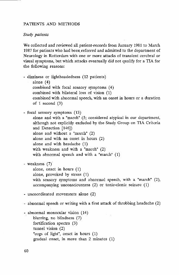
PATIENTS AND METHODS
Study patients
We collected and reviewed all patient-records from January 1981 to March 1987 for patients who had been. referred and admitted to the department of Neurology in Rotterdam with one or more attacks of transient cerebral or visual symptoms, but which attacks eventually did not qualify for a TIA for the following reasons:
- dizziness or lightheadedness (12 patients) alone (4) combined with focal sensory symptoms ( 4) combined with bilateral loss of vision (1) combined with abnormal speech, with an onset in hours or a duration of 1 second (3)
- focal sensory symptoms (13) alone and with a "march" (5; considered atypical in our department, although not explicitly excluded by the Study Group on TIA Criteria and Detection [146]) alone and without a "march" (2) alone and with an onset in hours (2) alone and with headache (1) with weakness and with a "march" (2) with abnormal speech and with a "march" (1)
- weakness (7) alone, onset in hours (1) alone, provoked by stress (1) with sensory symptoms and abnormal speech, with a "march" (2), accompanying unconsciousness (2) or tonic-clonic seizure (1)
- uncoordinated movements alone (2)
- abnormal speech or writing with a first attack of throbbing headache (2)
- abnormal monocular vision (14) blurring, no blindness (7) fortification spectra (3) tunnel vision (2) "cogs of light", onset in hours (1) gradual onset, in more than 2 minutes (1)
60

- bilateral visual symptoms (14) complete blindness for 1 second (2) blurring and scintillations ( 4) blurring in one hemifield (2) double vision alone ( 4) fortification spectra (2)
Thus, 64 patients, 35 men and 29 women, were identified for the study. Forty-nine patients had been referred by their general practitioner, three by an internist, eight by an ophthalmologist, one by a psychiatrist, and three by a neurologist. All patients had been interviewed by one neurology resident and one senior neurologist. In case of disagreement about the diagnosis, at least a third neurologist had interviewed the patient. [86, 84] None of the attacks could be satisfactorily explained by migraine, epilepsy, neurosis, or postural hypotension. The mean age of the patients was 55 years (range 27 to 82 years). Six patients were under 40.
The following investigations were performed in these patients: routine blood tests (64 patients, all normal), chest X-ray (60 patients, heart/thorax ratio more than 0.5 in 9 patients), CT-scan ( 49 patients, with a questionable infarct in two), EEG (16 patients, normal), carotid angiography (7 patients, normal), ECG (52 patients, normal), echocardiography (12 patients, normal), Holter 24 hour ECG monitoring (22 patients, with paroxysmal supraventricular tachycardia in one, bradycardia and short sinus arrests ( < 1.5 seconds) in three, and short ventricular tachycardias in three patients. These dysrhythmias were not considered sufficient evidence of a cardiac cause of the symptoms by the consulting cardiologist.
Seven patients were newly treated with aspirin, in 6 patients anticoagulant treatment was continued, and 9 patients remained on betablockers.
Most patients returned for one or more follow-up visits, but after a mean time of 1.2 years, all were seen only by their general practitioner (GP). For all living patients the follow-up was completed between April and June 1988 by telephone interviews with the patients and their GP. The total number of patient years was 240. The patients were carefully questioned for the recurrence of attacks, and their GP's for the occurrence of major vascular events. In case of such an event, relevant information was obtained from the hospitals involved. All neurological and cardiac events were verified by two neurologists and a cardiologist, respectively.
61

Control patients
From March 1986 until March 1988 one hundred and eighty-five patients were admitted to our department with a definite diagnosis of TIA or partial stroke. In all TIA patients the symptoms completely fulfilled the above-mentioned criteria. [146] These 185 patients were entered into a multi-centre study, the Dutch TIA trial, [147] and were seen every four months. The mean follow-up was 1.26 years, making up a total of 233 patient-years, which is almost equal to that in the study group. All patients had routine laboratory investigations, a chest X-ray, an ECG and a CTscan. Non-fatal ischaemic stroke, non-fatal myocardial infarction and fatal vascular events were recorded and subsequently audited by an endpoint committee. All patients were blindly treated with acetylsalicylic acid, 30 or 300 mg per day; in addition, about half the patients were eligible for treatment with a beta-blocker (50 mg atenolol) and were randomized between active treatment and placebo (Chapter IV).
RESULTS
The presence of vascular risk factors in the study and control group are summarized in Table 1. Cardiovascular risk factors were equally common in both groups. Twenty-four patients (38%) in the study group and 79 patients (42%) in the control group had two or more risk factors.
Vascular events in the study group During follow-up 6 patients died, three of whom had a fatal myocardial infarct, and one a probable sudden death (he was found dead at home, after having been seen alive less than 24 hours before). One patient died from a melanoma, and one from a fatal brainstem haemorrhage. Two patients had a partial ischaemic stroke. In one of these two patients the atypical attacks had consisted of hundreds of attacks of tunnel vision of the left eye, followed six months later by a right-sided hemiparesis. CTscanning showed an appropriate infarct, and angiography a total occlusion of the left internal carotid artery. The other patient had had 20 attacks of left-sided isolated sensory symptoms, developing in hours and accompanied by headache. Doppler studies had shown a stenosis of the right carotid artery. Two years later he also suffered a left-sided non-disabling hemiparesis, with an appropriate infarct on CT-scanning.
62
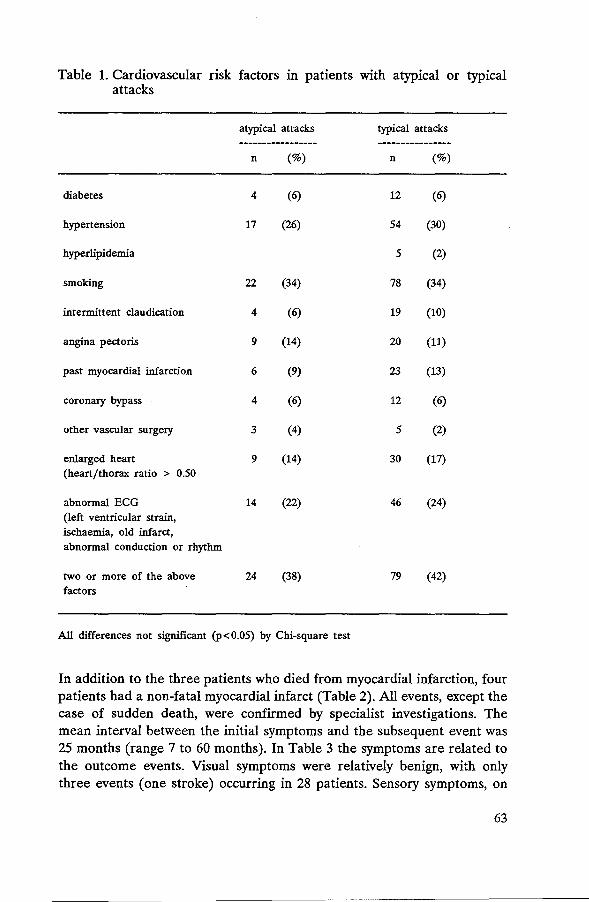
Table 1. Cardiovascular risk factors in patients with atypical or typical attacks
atypical attacks typical attacks
----------------- ----------------n (%) n (%)
diabetes 4 (6) 12 (6)
hypertension 17 (26) 54 (30)
hyperlipidemia 5 (2)
smoking 22 (34) 78 (34)
intermittent claudication 4 (6) 19 (10)
angina pectoris 9 (14) 20 (11)
past myocardial infarction 6 (9) 23 (13)
coronary bypass 4 (6) 12 (6)
other vascular surgery 3 (4) 5 (2)
enlarged heart 9 (14) 30 (17) (heart/thorax ratio > 0.50
abnormal ECG 14 (22) 46 (24) (left ventricular strain, ischaemia, old infarct, abnormal conduction or rhythm
two or more of the above 24 (38) 79 (42) factors
All differences not significant (p<0.05) by Chi-square test
In addition to the three patients who died from myocardial infarction, four patients had a non-fatal myocardial infarct (Table 2). All events, except the case of sudden death, were confirmed by specialist investigations. The mean interval between the initial symptoms and the subsequent event was 25 months (range 7 to 60 months). In Table 3 the symptoms are related to the outcome events. Visual symptoms were relatively benign, with only three events (one stroke) occurring in 28 patients. Sensory symptoms, on
63

the other hand, were strongly associated with further events, as six out of 13 patients suffered an endpoint: four cardiac events, one ischaemic stroke, and one brainstem haemorrhage. All 4 cardiac events occurred in patients with isolated sensory symptoms. Two of the four patients with dizziness alone suffered a cardiac event. Seven of the eight patients with later cardiac complications had two or more cardiovascular risk factors, against 17 of the 56 patients without cardiac complications (p=0.003; Odds ratio = 16.1, 95% CI 1.7 - 117.6). The mean age of the patients with cardiac events was 60 years, against 54 years in the others (difference not statistically significant).
Table 2. Cardiac and cerebral events in patients with typical or atypical attacks
event atypical typical p* Odds (95% CI) attacks attacks ratio
fatal
sudden death 1 1 1.00 (0.06 - 15.61)
myocardial infarct 3 0 NS
ischaemic stroke 0 2
other vascular disease 1 0
non fatal
ischaemic stroke 2 5 NS 0.38 (0.03 - 2.25)
myocardial infarct 4 0 0.06
all cerebral events 2 7 l 0.27 (0.04 - 1.43) O.Q7
all cardiac events 8 1 8.00 (1.01 - 63.1)
* Fisher's exact probability test
64
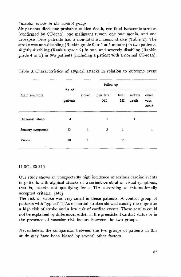
Vascular events in the control group Six patients died: one probable sudden death, two fatal ischaemic strokes (confirmed by CT-scan), one malignant tumor, one pneumonia, and one urosepsis. Five patients had a non-fatal ischaemic stroke (Table 2). The stroke was non-disabling (Rankin grade 0 or 1 at 3 months) in two patients, slightly disabling (Rankin grade 2) in one, and severely disabling (Rankin grade 4 or 5) in two patients (including a patient with a normal CT-scan).
Table 3. Characteristics of atypical attacks in relation to outcome event
follow-up
no of -----------------------------------------------------Main symptom stroke non fatal fatal sudden other
patients MI MI death vase.
death
Dizziness alone 4 1 1
Sensory symptoms 13 1 3 1 1
Vision 28 1 2
DISCUSSION
Our study shows an unexpectedly high incidence of serious cardiac events in patients with atypical attacks of transient cerebral or visual symptoms, that is, attacks not qualifying for a TIA according to internationally accepted criteria. [146] The risk of stroke was very small in these patients. A control group of patients with "typical" TIAs or partial strokes showed exactly the opposite: a high risk of stroke and a low risk of cardiac events. These results could not be explained by differences either in the preexistent cardiac status or in the presence of vascular risk factors between the two groups.
Nevertheless, the comparison between the two groups of patients in this study may have been biased by several other factors.
65

Firstly, the two groups were not studied concurrently but in different periods, though in the same department. Secondly, the methods of treatment were different: patients in the control group were treated with aspirin, against only seven of the study patients. Despite this, however, the incidence of stroke in the study group was less than 1 percent per patient year, which equals that of the general population. [167] Thirdly, the study group contained only patients without residual signs, whereas the control group also contained patients with partial strokes. On the other hand, patients with TIAs - if strictly defined - and patients with partial strokes are often considered to have a similar prognosis. [24] Fourthly, the approximately similar number of follow-up years in the control group was made up of more patients, followed for a shorter period. If the risk of stroke after a TIA is indeed highest in the first year, [101] then relatively more strokes might be expected in the control group on this account alone. But other studies have shown a more or less linear course of stroke incidence over time in patients with TIAs and partial stroke. [153, 17]
Our study confirms the results of Heyden et al, who found a lower stroke rate in patients with non-specific attacks during a ten year follow-up period (17%, against 25% in patients with definite TIAs). [72] Contrary to our results, their study did not show an excess of fatal and non fatal heart disease in patients with uncertain TIAs. This might be explained by the fact that isolated focal sensory symptoms, which were most strongly associated with further cardiac events in our study, were considered as definite TIAs by these authors.
The occurrence of cardiac complications in patients with atypical attacks may reflect the presence of latent cardiac disease at a time when these patients were referred to the neurologist. A cardiac cause of the symptoms was considered - but not established - in only one patient, although 24 patients had an increased risk of cardiac events on the basis of the history, chest X-ray and ECG. This high prevalence of cardiac risk factors does not reflect a particular referral pattern, as 90% of the study patients were referred to the department of neurology directly by the general practitioner or by an ophthalmologist. Patients with later cardiac events more often had cardiovascular risk factors than the other study patients.
It is not quite clear whether and how the atypical symptoms might have been caused by premonitory cardiac dysfunction. Two symptoms in particular, isolated dizziness and isolated sensory symptoms, were associated with subsequent cardiac events.
66

Cardiac dysrhythmia might have directly caused dizziness and bilateral visual disturbances as symptoms of global rather than focal cerebral ischaemia. Ambulatory ECG monitoring shows relevant dysrhythmias in 20 -50% of patients with non focal symptoms or syncopal attacks. [79, 154, 31] How cardiac disease could have caused focal sensory symptoms is less obvious. In patients with significant dysrhythmias, focal symptoms were reported in only 4 of 290 patients. [128] Given the high incidence of dysrhythmias in the general population, [127] however, this explanation might account for some cases. Francis et al found significant dysrhythmias in 25% of 64 patients with transient focal cerebral deficits, compared with 7% in a control group, and after anti-arrhythmic treatment the symptoms stopped or improved. [57] Cardiac dysrhythmia may cause a sudden reduction in blood flow across a stenosed extracranial or intracranial artery and may in this way produce focal cerebral ischemia. [128] Another explanation is that some of the attacks might have started as attacks of angina pectoris, followed by anxiety-induced hyperventilation, with associated sensory symptoms such as tingling limbs.
Although the number of events in our study is limited, our findings may shed new light on available data on the natural history of TIAs. Aggregate data from several clinical trials and follow-up studies of TIA patients show a striking variance in reported stroke rate, ranging from 2.8 to 65.9 nonfatal strokes per 1000 patient years in patients treated with aspirin or anticoagulants, and from 23.9 to 51.7 in untreated patients (Table 4). Although this can be partially explained by differences in race, age and vascular risk factors between the study subjects, the criteria for the diagnosis of TIA are perhaps an even more important factor. Studies with a low stroke rate showed an average or more than average cardiac event rate (UK-TIA study, [153] Muuronen et al [115]), whereas a study with a higher stroke rate showed relatively few cardiac events (AICLA [17]). In the light of our findings one might speculate that the former studies contained relatively more patients with atypical attacks.
The risk of cardiac death in patients with TIAs has been estimated at 3-4% per year. [2] Our results suggest that the risk may be substantially lower, as long as diagnostic criteria for the diagnosis of TIA in the sense of focal cerebral ischaemia are rigorously applied. Although we do not wish to deny that patients with "typical" TIAs may harbour silent coronary atherosclerosis, the possibility of symptomatic heart disease should be considered in patients with atypical attacks, particularly those consisting of dizziness or focal sensory symptoms, in combination with cardiovascular risk factors.
67

Table 4. Occurrence of non fatal stroke, non fatal myocardial infarction, vascular death and cardiac death in TIA, clinical trials and TIA follow-up studies (expressed in number of events/1000 pt years)
non fatal non fatal vascular cardiac approximate study stroke MI death death no of
ASA controls ASA controls ASA controls ASA controls patient years
TIA-trials
ESPS [149] 32.8 50.8 8.4 14.0 31.6 42.4 11.6 15.6 5000
UK-TIA [153] 12.7 28.3 6.5 10.5 26.2 26.4 20.5* 21.8* 10000
AICLA (17] 26.7 45.0 3.3 15.0 20.8 19.6 3.3 4.9 1800
CCSG [22] 65.9 51.7 16.7 0.0 59.2 51.7 40.8 34.4 1200
Follow-up studies
Muuronen et al [115] 2.8 6.5 10.6 2450
Toole et al [152] 24.2 7.3 42.0 1250
Heyman et a1 (73] 23.9 9.1 21.8 17.3 2450
* recorded as "mostly cardiac", but includes a few non-cardiac vascular deaths
68

GENERAL DISCUSSION
During the past three decades it has become established that transient ischemic attacks (TIAs) represent the major precursors of a disabling stroke. The importance of these attacks has become evident after the discovery that a proportion of stroke victims had experienced brief attacks of cerebral or visual symptoms in the period preceding their stroke. Early recognition of TIAs in the general population is therefore very important because it offers the opportunity to prevent the catastrophe of a major stroke. However, the identification of TIAs in patients who have not yet suffered a stroke is difficult for a number of reasons: (1) the diagnosis of TIA rests entirely upon the history of the patient, and the diagnosis lacks an objective "gold standard", (2) diagnostic criteria for the nature of the attacks are phrased in abstract terms, such as amaurosis fugax or dysarthria, which represent interpretations rather than the patient's actual symptoms, (3) confusion has arisen by the introduction of arbitrary time limits (for instance, 24 hours for TIAs), which may not be relevant with regard to the risk of a future stroke, and ( 4) it may be very difficult to differentiate a TIA from other disorders, particularly migraine, epilepsy, neurosis, postural hypotension, or hyperventilation. It is equally important that these disorders are not erroneously interpreted as TIA, since this may entail unnecessary investigations, long-term drug treatment, or even surgical intervention.
There are several strategies to cope with the diagnostic problems mentioned above:
1. Adhere to diagnostic guidelines for the diagnosis. This strategy aims at improvement of the precision of the diagnosis: if one can not improve the accuracy of the diagnosis because objective criteria are lacking, then one can at least try to achieve maximal interobserver agreement for the diagnosis. The diagnostic criteria for the diagnosis of TIA have been outlined in 1974 by the Study Group on TIA Criteria and Detection. In essence, these criteria represent the temporary variants of the clinical deficits that are known to occur in permanent stroke. Despite these guidelines the interobserver agreement for the diagnosis was disappointing in an earlier study. [86] The main reason may be that the criteria are phrased in abstract diagnostic terms instead of in plain language. In this thesis, we found that the use of (1)
69

ordinary language in recording and interpreting the history and (2) a check-list in multiple-choice format resulted in a marked improvement of the interobserver agreement for the diagnosis of TIA (Chapter II). However, a considerable number of participating neurologists did not strictly apply the diagnostic guidelines. This was the major source of disagreement (Chapters II and III). Therefore, the consistency of the diagnosis could be further improved if the diagnostic criteria are thoroughly discussed and then more consistently adhered to.
2. Try and improve the diagnostic criteria by studying differences and similarities between T!As with and without cerebral infarction on CT, and between T!As and disorders resembling TIAs.
70
Probably the most well-known diagnostic criterion for a TIA is its upper time limit of 24 hours. In this thesis we found that relevant cerebral infarcts on CT can be found in any cerebral ischemic attack, regardless of its duration, even in attacks lasting less than a minute. Furthermore, we did not find major differences in the type and location of the infarcts between patients with transient and those with persistent symptoms. Thus, the time limit of 24 hours that separates TIAs from strokes seems highly artificial (Chapter V). We also studied clinical differences between TIAs with and without a relevant infarct on CT, since the attacks in the latter patients definitely resulted from cerebral ischemia. We found that these differences were very small (Chapter VI). The only striking difference was that TIA patients with a relevant infarct on CT less often had atypical symptoms, such as blurring of vision, isolated sensory symptoms, heaviness in one or more limbs, or accompanying unconsciousness. This might suggest that the latter attacks are not of ischemic origin and do indeed not qualify for a TIA.
Since migraine represents the most important differential diagnosis of TIAs, we studied the occurrence of headache in patients with cerebral ischemia of varying duration (Chapter VII). We found that headache not uncommonly accompanies TIAs and non-disabling stroke. We also showed that the predictive value of headache for the diagnosis of migraine in patients who are referred to the neurologist with a possible TIA is very low. The presence of headache alone is therefore not very helpful in distinguishing a TIA from a migrainous attack. The occurrence of headache was partly related to the underlying cause of the ischemic lesion.

3. Investigate the prognostic value of specific features of TIAs. From the patient's perspective it is not so much the question whether the attack conforms to arbitrary diagnostic guidelines, but rather whether particular symptoms imply an increased risk of subsequent major vascular complications. This approach therefore represents the real "gold standard". Chapter VIII shows the results of a preliminary study, which suggest that patients with atypical symptoms, particularly those consisting of dizziness and focal sensory symptoms were associated with a very low risk of stroke but a high risk of subsequent cardiac events. In the coming years we shall prospectively analyze the prognostic value of such specific features in the cohort of 3150 patients who were randomized into the Dutch TIA trial.
71

GENERAL SUMMARY
The diagnosis of transient ischemic attack (TIA) is fraught with difficulty, since the diagnosis rests entirely upon the history of the patient's symptoms and the neurologist's skill in questioning the patient. The aim of this thesis is to investigate various measures to improve the reliability in making this diagnosis.
In the introductory chapter a brief historical review describes the development of the concept "transient ischemic attacks" and the identification of possible causes. Although transient and brief episodes of neurological symptoms have been recognized since antiquity, both the recognition of TIAs and the comprehension of their pathogenesis have largely developed during the past three decades. For a diagnosis of TIA a focal loss of function such as hemiplegia, monocular visual loss or a disorder of language is mandatory. Recurrent thromboembolism is considered the most common cause of TIAs. Yet many other disorders have been discovered to cause transient ischemic attacks in some cases, such as hemodynamic disturbances, blood dyscrasias, heart disease, small vessel disease, and vasculitis. An additional problem - and not the least - is that many attacks that more or less resemble "true TIAs" can result from a variety of disorders other than cerebral ischemia. The chapter continues with the international diagnostic guidelines for the diagnosis of TIA, the problems that remain in making this diagnosis, and the main objectives of this thesis.
In Chapter II the interobserver agreement on the diagnosis of TIA is addressed. To improve the agreement rate, we used a checklist in which the nature and time course of the symptoms were recorded in plain language, instead of in abstract terms such as amaurosis fugax. Criteria for the diagnosis of TlA were similarly phrased and recommended to all observers. Eight senior neurologists and ten neurology residents interviewed 72 patients in random pairs. In 64 cases the observers agreed on the diagnosis (kappa value = 0.77). After a short discussion between the two observers the agreement increased to a maximum (kappa = 1.0). However, in 29 of the 144 interviews the diagnosis would have been different, had the recommended criteria been fully applied. In 28 of these the observer had diagnosed TIA on insufficient evidence. Six "misinterpretations" led to disagreement for the diagnosis and only these were corrected by the observers during their discussion.
72

Main conclusion: The study design had led to a maximal interobserver agreement, but the agreement between the pairs and the usual diagnostic criteria is not yet ideal. The precision of the diagnosis could be improved if details of the recommended criteria are discussed in general as well as for each patient.
In Chapter III we further studied whether the interobserver disagreement is caused mainly by differences in interpretation of the symptoms or by differences in the content of the history. To the previous group of 72 patients with possible TIAs we added with two actresses who were thoroughly trained in giving a consistent history under all circumstances. They were interviewed by eight pairs of neurologists, unaware of the study design. Although the observers showed an optimal agreement on the nature and time course of the symptoms, only 5 pairs agreed on the diagnosis (kappa value 0.25). Main conclusion: Differences in the interpretation of the symptoms and
the application of diagnostic criteria are indeed the leading cause of interobserver disagreement on the diagnosis of TIA, and not inconsistency on the side of the patient.
The checklist was subsequently incorporated in the notification form for a multicentre clinical trial in TIA patients, the Dutch TIA trial. The background and design of this trial are described in Chapter IV. Patients with one or more cerebral ischemic attacks are randomized between 30 mg and 300 mg acetylsalicylic acid, and also between 50 mg atenolol and placebo. Patients with cerebral ischemia from causes other than artery-to-artery embolism, such as polycythemia, thrombocytosis, thrombocytemia, anemia, hypoglycemia, vasculitis, or a cardiac source of embolism are excluded. Patients with partial strokes are included in the study as long as they are still independent in most activities of daily living. The patients are followed up at four-monthly intervals. The analysis of events is based on the intention-to-treat principle. During the randomization period between March 1, 1986 and March 1, 1989, a total of 3150 patients were randomized.
Chapter V reports the results of CT scanning in 2171 randomized patients with symptoms that could be attributed to one cerebral hemisphere, and in whom CT scanning was performed not earlier than 24 hours after the first attack. A relevant cerebral infarct was found in 14% of TIAs (symptoms completely reversible within 24 hours), 34% of RINDs (symptoms lasting
73
more than 24 hours, but completely reversed within 6 weeks) and 47% of partial stroke (persisting symptoms or signs). Even within the 24 hour margin, relevant infarcts occurred more often with longer attacks, but were still found in some patients with attacks lasting less than a minute. No major differences in type or location of the infarcts were found between the three groups. Main conclusion: The differences between TIAs, RINDs, and partial
strokes are quantitative rather than qualitative, and the time limits of 24 hours and 6 weeks which separate TIAs, RINDs and strokes are highly artificial.
In Chapter VI we studied clinical differences between TIA patients with (87) and without (550) a relevant infarct on CT from the same group of 2171 patients entered into the Dutch TIA trial. TIA patients with cerebral infarcts more often had a history of hypertension, the attacks had, on average, a slower rate of disappearance and a longer duration, and the symptoms more often involved speech, or a combination of weakness and speech, and less frequently non-specific symptoms such as isolated sensory symptoms or blurred vision in one hemifield. Despite these small differences, the prediction of infarction on CT on the basis of the nature or the time course of symptoms proved impossible, since in each sub-category the majority of patients had a normal CT scan. In comparison with a different control group, consisting of RIND and stroke patients with cerebral infarction, TIA patients with cerebral infarcts showed only a slight preponderance of abnormal speech, and a larger number of attacks. Main conclusion: The clinical differences between transient ischemic
attacks with and without a cerebral infarct on CT are small, and equally small between TIAs with cerebral infarction and strokes with cerebral infarction - apart from the duration, which was different by definition. These clinical similarities do not exclude a difference in prognosis.
In Chapter VII we studied headache features in 3126 patients entered into the Dutch TIA trial. Headache was reported by 18% of these patients and was mostly non-throbbing in all types of attacks. The occurrence of headache was not related to the mode of onset, mode of disappearance, or the duration of the attack. Patients with headache less often had a history of hypertension and more often were known to have heart disease. Headache was less frequent in patients with lacunar infarcts and in patients with infarcts in the anterior circulation (territory of the middle and
74

anterior cerebral artery) and more frequent in patients with cortical infarcts and in patients with infarcts in the posterior circulation (territory of the posterior cerebral, basilar, and cerebellar arteries). Headache occurred in 17% of patients with monocular visual symptoms. Based on our findings and those of previous studies, [20] we estimated that headache increased the probability for the diagnosis of migraine in patients with possible TIAs from 3% (the a priori probability) to 11%. Main conclusions: (1) Headache is a not uncommon symptom
accompanying acute cerebral and retinal ischemia. Its mere presence is not helpful in distinguishing atherosclerotic cerebrovascular disease from a migrainous attack. (2) The occurrence of headache is partly related to the underlying cause of the ischemic lesion.
The subject of Chapter VIII is the fate of 64 patients with atypical cerebral or visual symptoms that could neither be classified as unequivocal TIAs nor as migraine, epilepsy or neurosis, and who were followed up for a mean of 3.75 years (range 11 months to 9 years, 240 patient years). Only two patients suffered a (non-disabling) stroke, but eight had a major cardiac event: fatal myocardial infarction in three, sudden death in one and nonfatal myocardial infarction in four patients. Seven of these eight patients were known to have cardiac risk factors, against 17 of the other 56 patients. Visual symptoms were relatively benign with regard to cardiac events (2 events in 28 patients), whereas dizziness alone and focal sensory symptoms alone were most strongly associated with subsequent cardiac complications ( 2 out of 4, and 4 out of 10 patients, respectively). In a control group of 185 patients with "typical" TIAs or partial strokes, followed for a total of 233 patient years, seven patients had an ischemic stroke, and only one a cardiac event. Main conclusion: Patients with atypical transient cerebral deficits and
cardiac risk factors carry a low risk of subsequent stroke, but a much higher risk of major cardiac events. For patients with typical TIAs, it is the other way around. The large differences in previous studies with regard to the ratio between cardiac and cerebral events may be caused by selection bias with regard to the nature of the symptoms by which patients qualified for the diagnosis of TIA.
In the general discussion the specific problems in the diagnosis of TIA are summarized. Three strategies that may help to solve these problems are described:
75

1. Adhere to arbitrary guidelines and improve the interobserver agreement for the diagnosis
2. Try and find analogies between TIA patients with or without a relevant infarct on CT, and between patients with TIAs and patients with disorders resembling TIAs
3. Investigate the prognostic value of specific features of TIAs. This approach is most relevant from the patient's perspective. In the coming years we shall analyze the prognostic value of such features in the cohort of 3150 patients who were entered into the Dutch TIA triaL
76

SAMENVATIING
Het stellen van de diagnose transient ischemic attack (TIA) is verre van gemakkelijk aangezien deze in bijna aile gevallen geheel berust op anamnestische gegevens. Het doel van dit proefschrift is het belichten van diverse aspecten van de diagnose TIA en het verbeteren van de overeenstemming ten aanzien van deze diagnose tussen neurologen.
In hoofdstuk I wordt allereerst een kort historisch overzicht gegeven van de ontwikkeling van het begrip TIA. Hoewel korte aanvallen van neurologische verschijnselen al in de oudheid beschreven zijn, heeft het herkennen van zowel het klinisch beeld als de mogelijke oorzaken van TIA's pas in de laatste drie decennia de belangrijkste ontwikkeling doorgemaakt. Voor het stellen van de diagnose TIA is een uitvalverschijnsel van focale aard, zoals een hemiparese, taalstoornis, of blindheid aan een oog absoluut vereist. Trombo-embolieen worden tegenwoordig in het algemeen beschouwd als verreweg de belangrijkste oorzaak van TIA's. Een scala van andere aandoeningen kan echter eveneens met TIA's gepaard gaan, zoals hemodynamische veranderingen, afwijkingen in de samenstelling van het bloed of van het hart, en vasculitiden. Een bijkomend - en belangrijk - probleem is dat min of meer op TIA's gelijkende aanvallen het gevolg kunnen zijn van geheel andere aandoeningen dan ischemie. In hoofdstuk I worden hierna achtereenvolgens de internationaal aanvaarde definitie van een TIA vermeld, en worden de specifieke moeilijkheden bij het stellen van deze diagnose.besproken. Dit leidt tot de vraagstellingen die aan dit proefschrift ten grondslag hebben gelegen.
In hoofdstuk II komt de overeenstemming tussen neurologen bij het stellen van de diagnose TIA aan de orde. Om deze overeenstemming te verbeteren maakten wij gebruik van een door ons zelf ontworpen anamneselijst, waarop de aard en beloop van de klachten van de patient in diens eigen bewoordingen - dus in gewone omgangstaal - door de deelnemende neurologen werden genoteerd in plaats van in abstracte medische termen als amaurosis fugax. De artsen werden bovendien verzocht gebruik te maken van de gangbare richtlijnen voor de diagnose TIA, die eveneens in gewone taal waren verwoord en aan de lijst waren toegevoegd. Acht ervaren neurologen en 10 arts-assistenten in opleiding tot neuroloog werden ingedeeld in steeds wisselende, aselect samengestelde koppels. Deze koppels namen de anamnese af bij in totaal 72 patienten. In 64 gevallen
77

waren beide artsen het eens over de diagnose ( overeenstemming gecorrigeerd voor het toeval: kappa = 0.77). Na kart overleg tussen de twee koppelgenoten werd opnieuw een diagnose gesteld en bleek de overeenstemming maximaal (kappa = 1.0). Echter, in 29 van de 144 anamnesegesprekken zou de diagnose anders zijn geweest als de aanbevolen diagnostische criteria consequent waren gebruikt. In 28 van deze gevallen had de arts ten onrechte - althans op grond van de verkregen gegevens - de diagnose TIA gesteld. Zes van deze "onjuiste" interpretaties leidden tot een verschillende diagnose en werden om die reden door de onderzoekers zelf opgemerkt- en gecorrigeerd- tijdens hun onderling overleg. In de andere gevallen werd de diagnose door een der onderzoekers (7x) of beide onderzoekers (8 paren) niet overeenkomstig de criteria gesteld. Hoofdconclusie: De opzet van het onderzoek heeft geleid tot maximale
overeenstemming over de diagnose TIA, maar de toepassing van diagnostische richtlijnen blijkt nag niet altijd valgens de bedoeling te geschieden. Een verdere verbetering van de nauwkeurigheid van de diagnose is derhalve te bereiken indien de diagnostische criteria met betrokken artsen zowel in het algemeen als bij individuele patienten in detail worden besproken.
In hoofdstuk III wordt verder ingegaan op de vraag waardoor een gebrek aan overeenstemming over de diagnose TIA vooral wordt veroorzaakt: door een verschil in interpretatie van de klachten door de arts of door verschillen in informatie die door de patient aan achtereenvolgende artsen wordt verstrekt. Om dit te onderzoeken werden aan de bovengenoemde groep van 72 patienten twee simulatie-patienten toegevoegd, die speciaal geoefend waren om onder aile omstandigheden de klachten steeds op dezelfde wijze te verwoorden. Deze "patienten" werden ondervraagd door twee maal acht artsen, die niet wisten dat de anamnese gesimuleerd was. Hoewel de artsen optimaal overeenstemden wat betreft de verkregen informatie, was er over de diagnose bij slechts 5 koppels overeenstemming (kappa = 0.25). Hoofdconclusie: Verschillen in interpretatie van de klachten en in de
consequente toepassing van diagnostische criteria zijn de inderdaad belangrijkste oorzaak van gebrek aan overeenstemming tussen artsen over de diagnose TIA.
De anamneselijst werd vervolgens gebruikt in het basisformulier voor een 'multicentre clinical trial', het Nederlands TIA onderzoek. De achtergrond en de opzet van dit onderzoek worden beschreven in hoofdstuk IV. Patien-
78

ten met een of meer aanvallen van cerebrale of oculaire ischemie worden in dit onderzoek door loting ingedeeld voor behandeling met 30 mg of 300 mg acteylsalicylzuur, en tevens voor behandeling 50 mg atenolol of placebo. Andere oorzaken van de ischemie dan arteriele tromboembolieen, zoals polycytemie, trombocytose, anemie, hypoglycemie, vasculitis of een emboliebron in het hart vormen een reden tot uitsluiting voor het onderzoek. Patienten met een kleine beroerte komen in aanmerking indien ze in het dagelijks Ieven nog onafuankelijk zijn. De patienten worden om de 4 maanden gecontroleerd. De analyse van de gegevens vindt plaats volgens het 'intention-to-treat' principe. Gedurende de periode waarin randomisatie plaats vond - van 1 maart 1986 to 1 maart 1989 - zijn 3150 patienten tot het onderzoek toegelaten.
In hoofdstuk V worden de resultaten vermeld van CT scan onderzoek bij 2171 patienten uit het Nederlands TIA-onderzoek bij wie de uitval past bij een lesie in een cerebrale hemisfeer en bij wie de CT scan niet eerder dan tenminste 24 uur na de eerste aanval is gemaakt. Een bijpassend infarct op de CT-scan werd gevonden bij 14% van de patienten met een TIA, 34% van patienten met een RIND (uitvallanger dan 24 uur, maar met volledig herstel binnen zes weken), en 47% van patienten met blijvende uitval. Ook binnen de groep van patienten bij wie de aanval korter duurde dan 24 uur nam de kans op een infarct op de CT -scan toe met de duur van de aanval. Tach werden zelfs infarcten gevonden bij een aantal patienten bij wie de aanval slechts enkele seconden duurde. Er werden geen verschillen gevonden in de aard of de localisatie van de infarcten tussen patienten met TIA's, RIND's of kleine beroertes. Hoofdconclusie: Verschillen tussen TIA's, RIND's en kleine beroertes zijn
eerder van kwantitatieve dan kwalitatieve aard. De bekende grens van 24 uur die TIA's van beroertes scheidt is uiterst kunstmatig.
In hoofdstuk VI worden klinische verschillen onderzocht tussen TIA patienten met en zonder een relevant infarct op de CT scan in dezelfde groep van 2171 patienten die tot het Nederlands TIA onderzoek werden toegelaten. TIA patienten met een infarct op de CT bleken vaker bekend te zijn met hypertensie, de neurologische uitvalverschijnselen verbeterden langzamer en duurden Ianger, en betroffen vaker de spraak of een combinatie van spraak en zwakte .en minder vaak aspecifieke klachten zoals uitsluitend klachten over het gevoel of wazig zien. De verschillen waren echter klein en bovendien bleek het niet mogdijk om op grand van de aard of het beloop van de klachten te voorspellen of de CT een infarct toonde. In vergelijking met patienten met blijvende uitvalverschijnselen en een infarct
79
op de CT, betrof de hoofdklacht bij TIA patienten met een herseninfarct op de CT vaker de spraak en was het aantal aanvallen geringer. Hoofdconclusie: De klinische verschillen tussen TIA's met en zonder
infarct op de CT scan zijn klein. Dit geldt eveneens wat betreft TIA patienten met een infarct op de CT en patienten met een beroerte en een infarct op de CT. Dit sluit echter een verschil in prognose tussen deze drie groepen niet uit.
Hoofdstuk VII behandelt het voorkomen van hoofdpijn bij 3126 patienten met een TIA, RIND of kleine beroerte die tot het Nederlands TIA onderzoek werden toegelaten. Door 18% van de patienten werd over hoofdpijn geklaagd; deze was meestal drukkend van karakter. Het voorkomen van hoofdpijn was niet gebonden aan de snelheid van ontstaan, de duur of de snelheid van verdwijnen van de aanva~ of van het aantal voorafgaande aanvallen. Geen verschillen werden gevonden tussen patienten met TIA's, RIND's of blijvende uitvalverschijnselen. Patienten met hoofdpijn waren minder vaak bekend met hypertensie maar vaker met hartlijden. Hoofdpijn kwam minder vaak voor bij patienten met lacunaire infarcten en bij patienten met een infarct in de voorste hersencirculatie ( stroomgebied arteria cerebri anterior en media) en vaker bij corticale infarcten en in de achterste circulatie ( stroomgebied arteria cerebri posterior, arteria basilaris en cerebellaire arterien). Door 17% van de patienten met aanvallen van unilaterale blindheid werd over hoofdpijn geklaagd. Op basis van onze resultaten en die van anderen [20] berekenden wij dat de aanwezigheid van hoofdpijn tijdens de aanval de waarschijnlijkheid van de diagnose migraine verhoogde van 3% (de a priori waarschijnlijkheid) naar 11%. Hoofdconclusies: (1) Hoofdpijn is een frequente bijkomende klacht bij
patienten met cerebrale of retinale ischemie. Het al dan niet aanwezig zijn van hoofdpijn is van weinig betekenis bij het onderscheid met een migraine aanval. (2) De hoofdpijn lijkt, althans ten dele, bepaald door de onderliggende oorzaak van de ischemie.
In hoofdstuk VIII werd de prognose onderzocht van 64 patienten met atypische voorbijgaande cerebrale of visuele klachten welke niet als een zekere TIA konden worden geduid, maar evenmin als migraine, epilepsie of psychogene klacht. Deze patienten werden gemiddeld 3.75 jaar vervolgd (varierend van 11 maanden tot 9 jaar, in totaal240 patient-jaren). Slechts 2 patienten kregen een (niet-invaliderende) beroerte, terwijl 8 getroffen werden door een ischemische hartziekte: een fataal myocardinfarct bij drie
80

patienten, een plotse dood bij een en niet-fataal hartinfarct bij vier patienten. Zeven van deze 8 patienten hadden risicofactoren m.b.t. het hart, tegenover 17 van de andere 56 patienten. Visuele symptomen waren relatief goedaardig wat betreft cardiale complicaties (2 van de 28 patienten), terwijl duizeligheid als enig verschijnsel en sensibele klachten het hoogste risico vertoonden (respectievelijk 2 van de 4 en 4 van de 13 patienten). In een controlegroep van 185 patienten met "zekere" TIAs of kleine beroertes, die gedurende in totaal 233 patient-jaren vervolgd werden, kregen zeven patienten een beroerte en slechts 1 een hartinfarct. Hoofdconclusie: Patienten met voor TIA's atypische voorbijgaande neuro-
logische symptomen en tevens cardiale risicofactoren worden zelden getroffen door een beroerte, maar hebben een verhoogd risico op een hartinfarct of hartstilstand.
In de general discussion worden de problemen bij het stellen van de diagnose TIA nog eens samengevat. Vervolgens worden drie benaderingen om deze problemen op te lossen besproken:
1. Het strict volgen van algemeen aanvaarde, maar niettemin arbitraire spelregels voor de diagnose, en het verbeteren van de overeenstemming over de diagnose tussen neurologen.
2. Het bestuderen van analogieen tussen TIA's enerzijds en aanvallen die zeker berusten op cerebrale ischemie, b.v. aanvallen met een relevant infarct op de CT-scan anderzijds en tussen TIA's en op TIA's gelijkende aandoeningen, zoals migraine.
3. Het bestuderen van de prognostische waarde van specifieke kenmerken van de aanval, zoals de aard (verlamming, spraakstoornis, blindheid) en het beloop (duur, snelheid van ontstaan, restverschijnselen) van de aanval. Voor de patient is deze benadering veruit het belangrijkst. De komende jaren zullen hiertoe de gegevens van alle 3150 patienten die in het Nederlands TIA-onderzoek werden opgenomen worden bestudeerd.
81
REFERENCES
1. Acheson J, Hutchinson EC. Observations on the natural history of transient cerebral ischaemia. Lancet 1964;ii:871-874.
2. Adams HP, Kassell NF, Mazuz H. The patient with transient ischaemic attacks - Is this the time for a new therapeutic approach? Stroke 1984;15:371-375.
3. Allen GS, Preziosi TJ. Carotid endarterectomy: A prospective study of its efficacy and safety. Medicine 1981;60:298-304.
4. Antiplatelet Trialists Collaboration: Secondary prevention of vascular disease by prolonged antiplatelet therapy. Br Med J 1988;296:320-331.
5. Araki G, Mihara H, Shizuka M, et al. CT and arteriographic comparison of patients with transient ischemic attacks - Correlation with small infarction of basal ganglia. Stroke 1982;14:276-280.
6. Awad I, Modic M, Little JR, Furlan AV, Weinstein M. Focal parenchymal lesions in transient ischemic attacks: correlation of computed tomography and magnetic resonance imaging. Stroke 1986;17:399-402.
7. Baker RN, Carroll Ramseyer J, Schwartz WS. Prognosis in patients with transient cerebral ischemic attacks. Neurology 1968;18:1157-1165.
8. Bamford J, Sandercock PAG, Jones L, Warlow CP. The natural history of lacunar infarction: The Oxfordshire Community Stroke Project. Stroke 1987;18:545-551.
9. Baquis GD, Pessin MS, Scott RM. Limb shaking - a carotid TIA. Stroke 1985;16:444-448.
10. Barnett HJM. The pathophysiology of transient ischemic attacks -therapy with platelet antiaggregants. Med Clin N Am 1979;63(4):649-678.
11. Barnett HJM. Stroke prevention. In: Vascular diseases of the nervous system, Ross Russell RW, ed. Edinburgh: Churchill Livingstone, 1983; Ch. 20, pp. 453-477.
12. Barnett HJM. Heart in ischemic stroke - a changing emphasis. N eur Clin 1983;1:291-315.
13. Basso A, Della Sala S, Farabola M. Aphasia arising from purely deep lesions. Cortex 1987;23:29-44.
82

14. Biller J, Laster DW, Howard G, Toole JF, McHenry LC jr. Cranial computerized tomography in carotid artery transient ischemic attacks. Eur Neural 1982;21:98-101.
15. Bogousslavsky J, Regli F. Cerebral infarct in apparent transient ischemic attack. Neurology 1985;35:1501-1503.
16. Bogousslavsky J, Regli F. Cerebral infarction with transient signs (CITS): Do TIAs correspond to small deep infarcts in internal carotid artery-occlusion? Stroke 1984;15:536-539.
17. Bousser MG, Eschwege E, Haguenau M, et al. "AICLA" controlled trial of aspirin and dipyridamol in the secondary prevention of atherosclerotic cerebral ischemia. Stroke 1983;14:5-14.
18. Brooke MH. A clinician's view of neuromuscular diseases. Baltimore: Williams and Wilkins, 1977.
19. Brust JCM. Transient ischemic attacks: Natural history and anticoagulation. Neurology 1977;701-707.
20. Calanchini PR, Swanson PD, Gotshall RA, et al. Cooperative study of hospital frequency and character of transient ischemic attacks: IV. The reliability of diagnosis. JAMA 1977;238:2029-2033.
21. Calandre L, Gomara S, Bermejo F, Millan JM, Del Prozo G. ClinicalCT correlations in TIA, RIND, and strokes with minimum residuum. Stroke 1984;15:663-666.
22. Canadian Cooperative Study Group: A randomized trial of aspirin and sulphinpyrazone in threatened stroke. N Engl J Med 1978;299:53-59.
23. Caplan LR. TIAs: We need to return to the question, "What is wrong with Mr. Jones?" Neurology 1988;38:791-793.
24. Caplan LR. Are terms such as completed stroke or RIND of continued usefulness? Stroke 1983;14:431-433.
25. Chiari H. Ueber das Verhalten des Teilungswinkels der Carotis Communis bei der Endarteriits chronica deformans. V erh Dtsch Pathol Ges 1905;9:326-330.
26. Cohen J. A coefficient of agreement for nominal scales. Educational and Physiological Measurement 1960;20:37-46.
27. Conn HO, Spencer RP. Observer error in liver scans. Gastroenterology 1972;62:1085-1090.
28. Constant P, Renou AM, Callie JM, Vernhiet J. Follow-up study of cerebral ischemia with computed tomography. Acta Neural Scand 1977;64 (suppl 56):164-165.
29. Daly DD, Svien HJ, Yoss RE. Intermittent cerebral symptoms with meningeomas. Arch Neural 1961;5:287-293.
83

30. Davies LG. Observer variation in reports on electrocardiograms. Br Heart J 1958;20:153-161.
31. De Bono DP, Warlow CP, Hyman NM. Cardiac rhythm abnormalities in patients presenting with transient non-focal neurological symptoms: a diagnostic grey area? Br Med J 1982;284:1437-1439.
32. De Caterina R, Gianessi D, Bernini W et al. Selective inhibition of thromboxane related platelet function by low-dose aspirin in patients after myocardial infarction. Am J Cardiol 1985;55:589-590.
33. De Caterina R, Gianessi D, Boem A et al. Equal antiplatelet effects of aspirin 50 or 324 mg/ day in patients after acute myocardial infarction. Thromb Haemost 1985;54:528-532.
34. Dennis MS, Bamford JM, Sandercock PAG, Warlow CP. Lone bilateral blindness: A transient ischemic attack. Lancet 1989;i:185-188.
35. Denny-Brown D. Recurrent cerebrovascular episodes. Arch Neural 1960;2: 194-210.
36. Denny-Brown D. The treatment of recurrent cerebrovascular symptoms and the question of "vasospasm". Med Clin N Am 1951;35: 1457-1474.
37. Department of Clinical Epidemiology and Biostatistics, McMaster University Hamilton, Ontario (Haynes RB, et al). Clinical disagreement. II. How to avoid it and how to learn from one's mistakes. J C.an Med Assoc 1980;123:613-617.
38. Department of Clinical Epidemiology and Biostatistics, McMaster University, Hamilton, Ontario (Sackett DL, et al). Clinical disagreement. I. How often it occurs and why. J Can Med Assoc 1980;123:499-504.
39. Donnan G, Tress BM, Bladin PF. A prospective study of lacunar infarction using computed tomography. Neurology 1982;32:49-56.
40. Edmeads J. Complicated migraine and headache in cerebrovascular disease. Neural Clin 1983;1:385-397.
41. Edmeads J. The headaches of ischemic cerebrovascular disease. Headache 1979;19:345-349.
42. Falke P, Stavenow L, Young M, Lindgarde F. Differences in mortality and cardiovascular morbidity during a 3-year follow-up of transient ischemic attacks and minor strokes. Stroke 1989;3:340-344.
43. Fields WS, Maslenikov V, Meyer JS, Hass WK., Remington RD, MacDonald M. Joint study of extracranial arterial occlusion. V. Progress report of prognosis following surgery or non-surgical treatment for transient cerebral ischaemic attacks and cervical carotid artery lesions. JAMA 1970;211:1993-2003.
84
44. Fields WS, Lemak NA, Frankowski RF, Hardy RJ. Controlled trial of aspirin in cerebral ischemia. Stroke 1977;8:301-316.
45. Fisher CM. Late-life migraine equivalents as a cause of unexplained transient ischaemic attacks. Can J Neurol Sci 1980;7:9-17.
46. Fisher CM. Late-life migraine accompaniments - further experience. Stroke 1986;17:1033-1042.
47. Fisher CM. Lacunes: Small, deep cerebral infarcts. Neurology (Minneapolis) 1965;15:774-784.
48. Fisher CM. Observations of the fundus oculi in transient monocular blindness. Neurology 1959;9:333-347.
49. Fisher CM. Transient monocular blindness associated with hemiplegia. Arch Ophthal 1952;17:167-203.
50. Fisher CM. Headache in cerebrovascular disease. In: Handbook of Clinical Neurology, vol 5; Vinken PJ, Bruyn HW eds. Amsterdam: North Holland Publishing Co, Amsterdam, 1968;pp. 124-156.
51. Fisher CM. In: Cerebral vascular disease; Wright I, Millikan CH, eds. New York: Grune and Stratton, 1958.
52. Fisher CM. Occlusion of the internal carotid artery. Arch Neurol Psychiat 1951;65:346-377.
53. Fisher CM, Levine PH, Fullerton AL, Forsberg A, Duffy CP, Hoogasian JJ, Drachman DA. Marker proteins of platelet activation in patients with cerebrovascular disease. Arch Neurol 1982;39:692-695.
54. Fisher CM. Concerning recurrent transient cerebral ischemic attacks. Can Med Assoc J 1962;86:1091-1099.
55. FitzGerald GA, Oates JA, Hawiger J et al. Endogenous synthesis of prostacyclin and thromboxane and platelet function during chronic administration of aspirin in man. J Clin Invest 1983;71:676-688.
56. Fleiss JL. Statistical methods for rates and proportions, New York, Wiley ed. 1981.
57. Francis DA, Heron JR, Clarke M. Ambulatory electrocardiographic monitoring in patients with transient focal cerebral ischaemia. J Neurol Neurosurg Psychiat 1984;47:256-259.
58. Fransesco V, Cesare A, Luigi I, Stefano F, Andrea G, Fransesco B. Malondialdehyde-like material and beta-thromboglobuline plasma levels in patients suffering from transient ischemic attacks. Stroke 1985;16:14-16.
59. Friedman GD, WilsonS, Mosier JM, Colandrea MA, Nichaman MZ. Transient ischemic attacks in a community. JAMA 1969;210:1428-1434.
85

60. Gacs G, Fox AJ, Barnett HJM, Vinuela F. CT visualisation of intracranial arterial thromboembolism. Stroke 1983;14:756-762.
61. Garraway AM, Akhtar AJ, Gore SM, Prescott RJ, Smith RG. Observer variation in the clinical assessment of stroke. Age and Ageing 1976;5:233-240.
62. Gaul JJ, Marks SJ, Weinberger J. Visual disturbances and carotid artery disease - 500 symptomatic patients studied by non-invasive carotid artery testing including B-mode ultrasonography. Stroke 1986; 17:393-398.
63. Gawel M, Burkitt M, Clifford Rose F. The platelet release reaction during migraine attacks. Headache 1979;19:323-327.
64. Goldenberg G, Reisner T. Angiographic findings in relation to clinical course and results of computed tomography in cerebrovascular disease. Bur Neurol 1983;22:124-130.
65. Goodwin JA, Gorelick PB, Helgason CM. Symptoms of amaurosis fugax in atherosclerotic carotid artery disease. Neurology 1987;37:829-832.
66. Gorelick PB, Hier DB, Caplan LR, Langenberg P. Headache in acute cerebrovascular disease. Neurology 1986;36:1445-1450.
67. Grindal AB, Toole JF. Headache and transient ischemic attacks. Stroke 1974;5:603-606.
68. Hachinski V. Transient cerebral ischemia: A historical sketch. In: Historical aspects of the neurosciences; A festschrift for Macdonald Critchley. Clifford Rose F, Bynum WF, eds. New York: Raven Press, 1982; pp.185-193.
69. Harrison MJG, Marshall J, Meadows JC, Russell RRW, Lefaucconnier JM, Thibult N, Touboul D, Touboul PJ. Effect of aspirin in amaurosis fugax. Lancet 1971;ii:743-744.
70. Hart RG, Easton JD. Dissections of cervical and cerebral arteries. Neur Clin 1983;1:155-182.
71. Herman B, Lyten ACM, van Lyuk JH, Frenken CWGM, Op de Coull AA W, Schulte BPM. Epidemiology of stroke in Tilburg, the Netherlands. The population-based stroke incidence registre. II. Incidence, initial clinical picture and medical care, and 3-week case fatality. Stroke 1982;13:629-634.
72. Heyden S, Heiss G, Heyman A, Tyroler AH, Hames CG, Patzschke U, Manegold C. Cardiovascular mortality in transient ischemic attacks. Stroke 1980;11:252-255.
73. Heyman A, Wilkinson WE, Hurwitz BJ, et al. Risk of ischaemic heart disease in patients with TIA. Neurology 1984;34:626-630.
86
74. Heyman A, Leviton A, Millikan CH, et al. Transient focal cerebral ischemia: Epidemiological and clinical aspects. Stroke 1974;5:277-287.
75. Hoogendijk EM, Jenkins CS, van Kijk EM, Vas J, ten Cate JW. Spontaneous platelet aggregation in cerebrovascular disease. II. Further classification of platelet defect. Thromb Haemostas 1979;46:512-522.
76. Rossmann KA, Kleihues P. Reversibility of ischemic brain damage. Arch Neural 1973;29:375-384.
77. Houston Merritt H. A textbook of neurology, Philadelphia: Lea & Febiger 1979; pp. 186-193.
78. Hunt JR. The role of the carotid arteries, in the causation of vascular lesions of the brain, with remarks on certain special features of the symptomatology. Am J Med Sci 1914;147:704-713.
79. Jonas S, Klein I, Dimant J. Importance of halter monitoring in patients with periodic cerebral symptoms. Ann Neural 1977;1:470-474.
80. Kalmann R, Nieuwenhuis HK, de Groot PG, van Gijn J, Sixma JJ. Effects of low doses of aspirin, 10 mg and 30 mg daily, on bleeding time, thromboxane production and 6-keto-PGF-1 excretion in healthy subjects. Thromb Res 1987;45:355-361.
81. Karp HR, Heyman A, Heyden S, et al. Transient cerebral ischemia. Prevalence and prognosis in a biracial rural community. JAMA 1973;225:125-128.
82. Kendell RE, Marshall J. Role of hypotension in the genesis of transient ischemic attacks. Br Med J 1963;2:344-348.
83. Kinkel WR, Jacobs L. Computerized axial transverse tomography in cerebrovascular disease. Neurology 1976;26:924-930.
84. Koudstaal PJ, Van Gijn J, Staal A, Duivenvoorden HJ, Gerritsma JGM, Kraaijeveld CL. Diagnosis of transient ischemic attacks: Improvement of interobserver agreement by a check-list in ordinary language. Stroke 1984;17:723-728.
85. Kou9staal PJ, Van Gijn J, Naarding P, De Kluyver E, Pop GAM, Staal A. "Atypical TIAs" may herald cardiac rather than cerebral events. Submitted for publication.
86. Kraaijeveld CL, van Gijn J, Schouten HJA, Staal A. Interobserver agreement for the diagnosis of transient ischemic attacks. Stroke 1984; 15:723-725.
87. Ladurner G, Sager WD, Iliff LD, Lechner H. A correlation of clinical findings and CT in ischemic cerebrovascular disease. Eur Neural 1979; 18:281-288.
87

---------------------
88. Lane DA, Wolff S, Ireland H, Gawel M, Foadi M. Activation of coagulation and fibrinolytic systems following stroke. Br J Haematol 1983;53:655-658.
89. Lee P, Urowitz MB, Bookman AM et al. Systemic lupus erythematosus. Am J Med 1977;46:1-32.
90. Lechat P, Mas JL, Lascault G, et al. Prevalence of patent foramen ovale in patients with stroke. N Engl J Med 1988;318:1148-1152.
91. Levine J, Swanson PD. Idiopathic thrombocytosis: A treatable cause of transient ischemic attacks. Neurology 1968;8:711-713.
92. Levy DE. How transient are transient ischemic attacks? Neurology 1988;38:674-677.
93. Loeb C. Clinical evaluation of patient with transient ischemic attacks. Adv Neural 1979;25:141-148.
94. Loeb C. Protracted transient ischemic attacks. Eur Neurol1980;19:1-11.
95. Loeb C, Gandolfo C, Dall'Agata D. Headache in transient ischemic attacks (TIA). Cephalalgia 1985; suppl. 2:17-19.
96. Marcus AJ. Recent progress in the role of platelets in occlusive vascular disease. Stroke 1983;14:475-479.
97. Marshall J. The natural history of transient ischaemic cerebro-vascular attacks. Quart J Med 1964;131:309-324.
98. Marshall J. A survey of occlusive disease of the vertebrobasilar arterial system. In: Handbook of Clinical Neurology, Vinken PJ, Bruyn GW, eds. Amsterdam: North Holland Publishing Company 1972; Ch. 1:pp. 1-12.
99. Marshall RJ, Malone RGS. Cryoglobulinaemia with cerebral purpura. Br Med J 1954;3:279-280.
100. Marshall J, Wilkinson IMS. The prognosis of carotid transient ischaemic attacks in patients with normal angiograms. Brain 1971;94:395-402.
101. Matsumoto N, Whisnant JP, Kurland LT, et al. Natural history of stroke in Rochester, Minnesota, 1955 through 1969: an extension of a previous study, 1945 through 1954. Stroke 1973;4:20-29.
102. McAllen PM, Marshall J. Cardiac dysrhythmia and transient cerebral ischaemic attacks. Lancet 1973;ii:1212-1214.
103. McHenry LC. Garrison's History of Neurology. Springfield: Thomas CC publisher, 1969.
104. McLachlan MSF, Thomson JG, Taylor DW, Kelly ME, Sackett DL.
88
Observer variation in the interpretation of lower limb venograms. Am J Roentgenol 1977;132:227-229.

105. Medina J, Diamond S, Rubino F. Headaches in patients with transient ischemic attacks. Headache 1975;15:194-197.
106. Merode van T, Hick P, Hoeks PG, Reneman RS. Serum HDL/total cholesterol ratio and blood pressure in asymptomatic atherosclerotic lesions of the cervical carotid arteries in men. Stroke 1985;16:34-38.
107. Meyer JS, Leiderman H, Denny-Brown D. Electroencephalographic study of insufficiency of the basilar and carotid arteries. Neurology 1956;6:455-477.
108. Millikan CH, Siekert RG, Whisnant JP. Intermittent carotid and vertebral basilar insufficiency associated with polycythemia. Neurology 1960;10:188-196.
109. Millikan CH. The transient ischemic attack. Adv Neurol1979;25:135-140.
110. Millikan CH, Siekert RG, Shick RM. Studies in cerebro-vascular disease. V. The use of anticoagulant drugs in the treatment of intermittent insufficiency of the internal carotid arterial system. Proc Mayo Clin 1955;30:578-586.
111. Mohr JP, Caplan LR, Melski JW, Goldstein RJ, Duncan GW, Kistler JP, Pessin MS, Bleich HL. The Harvard Cooperative Stroke Registry: A prospective registry. Neurology 1978;28:754-762.
112. Mokri B, Sundt TM, Houser OW, Piepgras G. Spontaneous dissection of the cervical internal carotid artery. Ann Neurol 1986;19:126-138.
113. Moncada S. Vane JR. Arachidonic acid metabolites and the interactions between platelets and blood-vessel walls. N Engl J Med 1979;300:1142-1147.
114. Moster ML, Johnston DE, Reinmuth OM. Chronic subdural hematoma with transient neurological deficits: A review of 15 cases. Ann Neurol 1983;14:539-542.
115. Muuronen A, Kaste M. Outcome of 314 patients with transient ischaemic attacks. Stroke 1982; 13:24-31.
116. O'Brien JR. Effects of salicylates on human platelets. Lancet 1968;i:779-783.
117. Osler W. Transient attacks of aphasia and paralysis in states of high blood pressure and arterio-sclerosis. Can Med Assoc J 1911;1:919-926.
118. Patrignani P, Filabozzi P, Patrono C. Selective cumulative inhibition of platelet thromboxane production by low-dose aspirin in healthy subjects. J Clin Invest 1982;69: 1366-1372.
119. Peabody GL. Relations between arterial disease and visceral changes. Trans Assoc Am Physicians 1891;6:154-178.
89

120. Pedersen AK, FitzGerald GA. Dose-related kinetics of aspirin - Presystemic acetylation of platelet cyclo-oxygenase. N Engl J Med 1984;311: 1206-1211.
121. Perrone P, Candelise L, Scotti G, De Grandi D, Scialfa G. CT evaluation in patients with transient ischemic attack. Correlation between clinical and angiographic findings. Bur Neural 1979;18:217-221.
122. Portenoy RK, Bissi CJ, Lipton RB, Berger AR, Mebler MF, Baglivo J, Solomon S. Headache in cerebrovascular disease. Stroke 1984;15: 1009-1012.
123. Portnoy HD. Transient "ischemic" attacks produced by carotid stenosis and hypoglycemia. Neurology 1965;15:830-832.
124. Powers WJ, Raichle ME. Positron emission tomography and its application to the study of cerebrovascular diseases in man. Stroke 1985;16:361-376.
125. Preston FE, Greaves M, Jackson CA, Stoddard CJ: Low dose aspirin inhibitis platelet and venous cyclo-oxygenase in man. Thromb Res 1982;27 :4 77-484.
126. Raftery EB, Holland WW. Examination of the heart: an investigation into variation. Am J Epidemiol 1967;85:438-444.
127. Rai GS. Cardiac arrhythmias in the elderly. Age Ageing 1982;11:113-115.
128. Reed RL, Siekert RG, Merideth J. Rarity of transient focal cerebral ischemia in cardiac dysrhythmia. JAMA 1973;223:893-895.
129. Reggia JA, Tabb DR, Prices TR, Banko M, Hebel R. Computer-aided assessment of transient ischemic attacks. Arch Neurol 1984;41:1248~ 1254.
130. Richardson FM. Peer review of medical care. Med Care 1972;10:29-39.
131. Roberts MS, Joyce RM, McLeod LJ et al. Slow-release aspirin and prostaglandin inhibition. Lancet 1986;i: 1153-1154.
132. Rose GA. Chest pain questionnaire. Milbank Mem Fund Q 1965;43(2):32-39.
133. Roth GJ, Majerus PW. The mechanism of the effect of aspirin on human platelets. I. Acetylation of a particulate fraction protein. J Clin Invest 1975;56:624-632.
134. Ross Russell WR. Observations on the retinal blood vessels in monocular blindness. Lancet 1961;ii:1422-1428.
135. Ross Russell WR, Page NGR. Critical perfusion of brain and retina. Brain 1983;296:320-321.
90

136. Ross Russell WR, Cranston WI. Ophthalmodynamometry in carotid artery disease. J Neurol Neurosurg Psychiatry 1961;24:281-286.
137. Russell W. Intermittent closing of cerebral arteries: Its relation to temporary and permanent paralysis. Br Med J 1909;2:1109-1110.
138. Sandok BA, Furlan AJ, Whisnant JP, Sundt TM. Guidelines for the management of transient ischemic attacks. Mayo Clin Proc 1978;53:665-674.
139. Schouten HJA. Measuring pairwise agreement among many observers. II. Some improvements and additions. Biometric J 1982;24:431-435.
140. Segal HN. The electrocardiogram and its interpretation. A study of report by 20 physicians on a set of 100 electrocardiograms. J Can Med Assoc 1960;81:2-6.
141. Shah AB, Beamer N, Coull BM. Enhanced in vivo platelet activation in subtypes of ischemic stroke. Stroke 1985;16:643-647.
142. Siekert RG, Whisnant JP, Millikan CH. Surgical and anticoagulant therapy of occlusive cerebrovascular disease. Ann Int Med 1963;58:637 -641.
143. Sisk C, Ziegler DK, Zileli T. Discrepancies in recorded results from duplicate neurological history and examination in patients studied for prognosis in cerebrovascular disease. Stroke 1970;1:14-18.
144. Sorensen PS, Pedersen H, Marquardsen Jet al. Acetylsalicylic acid in the prevention of stroke in patients with reversible cerebral ischemic attacks. A Danish cooperative study. Stroke 1983;14:15-22.
145. Stewart ME, Douglas JT, Lowe GDO, Prentice CRM, Forbes CD. Prognostic value of beta-thromboglobulin in patients with transient cerebral ischemia. Lancet 1983:ii:479-482.
146. Study Group on TIA Criteria and Detection. Transient focal cerebral ischemia: epidemiological and clinical aspects. Stroke 1974;5:277-287.
147. The Dutch TIA Study Group. The Dutch TIA Trial: protective effects of low-dose aspirin and atenolol in patients with transient ischemic attacks or non-disabling stroke. Stroke 1988;19:512-517.
148. The Ad Hoc Committee on the classification and outline of cerebrovascular disease. II. (Chairman Millikan CH.). Stroke 1975;6:566-616.
149. The European Stroke Prevention Study Group. The European stroke prevention study (ESPS). Lancet 1987;ii:1351-1354.
150. Tomasello F; Mariani F, Fieschi C, et al. Assessment of interobserver differences in the Italian Multicenter Study on reversible cerebral ischemia. Stroke 1982;13:32-35.
151. Toole JF. Cerebrovascular disorders. New York: McGraw-Hill Book Company 1974; Ch. 15:pp. 187-197.
91

--------------
152. Toole JF, Yuson CP, Janeway R, et al. Transient ischemic attacks: A prospective study of 225 patients. Neurology 1978;28:746-753.
153. UK-TIA Study Group: United Kingdom transient ischaemic attack (UK-TIA) aspirin trial: Interim results. Br Med J 1988;296:316-320.
154. Van Durme JP. Tacchyarrhythmias and transient cerebral ischaemic attacks. Am Heart J 1975;89:538-540.
155. Van Gijn J, Staal A, Van Urk H. Transient ischaemic attacks en het voorkomen van herseninfarcten. Ned Tijdschr Geneesk 1982;126:1405-1413.
156. Van Swieten JC, Hijdra A, Koudstaal PJ, Van Gijn J. Periventricular leukoencephalopathy on CT -scans; a simple scale. Submitted for publication. ·
157. Van Swieten JC, Koudstaal PJ, Visser MC, Schouten HJA, Van Gijn J. Interobserver agreement for the assessment of handicap in stroke patients. Stroke 1988;19:604-607.
158. Von Graefe A. Ueber Embolie der Arteria centralis retinae als Ursache plotzlicher Erblindung. Arch Ophthalmol 1859;5:136-157.
159. Walter PF, Reid SD, Kass Wenger N. Transient cerebral ischemia due to arrhythmia. Ann Int Medicine 1970;72:471-474.
160. Warlow CP. Transient ischaemic attacks. In: Recent advances m clinical neurology, Matthews WB, Glaser GH, eds. 1982; val 3, pp.191-214. .
161. Watts C. External carotid artery embolus from the internal carotid artery "stump" during angiography- case report. Stroke 1982;13:515-517.
162. Waxman SG, Toole JF. Temporal profile resembling TIA in the setting of cerebral infarction. Stroke 1983;14:433-437.
163. Weber MD. The neurological complications of consumption coagulopathies. Neurology 1968;18:185-188.
164. Weksler BB, Kent JL, Rudolph D, Scherer PB, Levy DE. Effects of low dose aspirin on platelet function in patients with recent cerebral ischemia. Stroke 1985;16:5-9.
165. Weksler BB, Tack-Goldman K, Subramanian VA, Gay WA. Cumulative inhibitory effect of low-dose aspirin on vascular prostacyclin and platelet thromboxane production in patients with atherosclerosis. Circulation 1985;71:332-340.
166. Werdelin L, Juhler M. The course of transient ischemic attacks. Neurology 1988;38:677-680.
167. Whisnant JP, Matsumoto N, Elveback LR. Cerebral ischaemic attacks in a community: Rochester, Minnesota, 1955 through 1969. Mayo Clin proc 1973;48: 194-198.
92

168. Willis T. The anatomy of brain and nerves. Translated by Samuel Prodage, 1681. Facsimile; Feindel W, ed. Birmingham, UK: The classics of Neurology and Neurosurgery Library, Division of Gryphon Editions, 1983:90.
169. Yates PO. The pathogenesis of transient ischemic attacks. Stroke 1976;7: 178-187.
170. Yusuf S, Peto R, Lewis J, Sleight P. Beta blockade during and after myocardial infarction: An overview of the randomized trials. Prog Cardiovasc Dis 1985;27:335-371.
171. Ziegler DK, Stephenson Hassanein R. Prognosis in patients with transient ischemic attacks. Stroke 1973;4:666-673.
93

ACKNOWLEDGEMENTS
I am much indebted to the following persons who have contributed a great deal throughout this study. Prof. Dr. J. van Gijn introduced me to clinical research, and proposed the subject of this thesis. He is the principal investigator of the Dutch TIA trial, which formed the basis for the major part of this thesis. His many valuable comments and continuous support have been of major importance. Prof. Dr. A. Staal deserves my gratitude for his great interest in the subject of this thesis, his stimulating remarks, and helpful comments. The unfailing support of Dr. Rien Vermeulen has kept up my pleasure in completing this thesis. I wish to thank Dr. Hugo Duivenvoorden for his expert help and patient guidance during the interobserver study. The accurate comments and helpful advice of Dr. Jaap Gerritsma are also gratefully acknowledged. Special gratitude is due to Harrie Frericks, Francine van Vliet, Marieke Herber, and Anita te Nijenhuis (trial office Dutch TIA trial) for their major contribution to the success of the Dutch TIA trial and all their efforts to supply me with data, over and over again. I wish to thank Mrs. Blaauw and Mrs. Lubberhuizen for acting as the two simulated patients, and doing it so well. Mrs. Betty Mast provided excellent secretarial help, and more than that, without ever losing good temper. The enthusiastic help of the students Eveline de Kluyver and Paul Naarding in the follow-up of patients with atypical TIAs is specifically acknowledged. Djo Hasan deserves my gratitude for his help in drawing the figures in Chapters V, VI, and VII, and his skillful assistance whenever the computer failed me. I wish to thank all my colleagues at the department of neurology who participated in the two interobserver studies, and did not lose their enthusiasm even when they found out that they had been cheated occasionally. I am also much indebted to all the neurologists participating in the Dutch TIA trial, who have randomized the incredible total of 3150 patients. Their names are listed in Appendix 3. Finally, I would like to thank the most stimulating and inspiring person of all, my wife Adrie.
94
CURRICULUM VITAE
De schrijver van dit proefschrift werd op 19 januari 1954 in Rotterdam geboren. Hij bezocht het Marnix Gymnasium te Rotterdam, waar hij in 1972 het gymnasium-beta diploma behaalde. Na het vervullen van de militaire dienstplicht began hij in 1974 de studie geneeskunde aan de Erasmus Universiteit te Rotterdam. Tijdens zijn studie vervulde hij gedurende 2 maanden een assistentschap op de afdeling neurologie van het Dijkzigt Ziekenhuis Rotterdam. In 1981 werd hij tot arts bevorderd. Op 1 januari 1982 began hij zijn specialisatie tot neuroloog met een stage op de afdeling neurochirurgie van het Academisch Ziekenhuis van de Vrije Universiteit te Amsterdam (Prof. Dr. H.A.M. van Alphen). Vanaf 1 juli 1982 verbleef hij op de afdeling neurologie van het Academisch Ziekenhuis Dijkzigt te Rotterdam ( opleider: Prof. Dr. A. Staal). Op 1 januari 1986 werd hij in het specialistenregister als neuroloog ingeschreven. Momenteel is hij als staflid aan de afdeling neurologie van het Dijkzigt Ziekenhuis verbonden.
95
LIST OF PUBLICATIONS
1. Koudstaal PJ, Van Gijn J, Klootwijk APJ, Van der Meche FGA, Kappelle U. Holter monitoring in patients with transient and focal ischemic attacks of the brain. Stroke 1986;17:192-195.
2. Koudstaal PJ, Van Gijn J, Staal A, Duivenvoorden HJ, Gerritsma JGM, Kraaijeveld CL. Diagnosis of transient ischemic attacks: Improvement of interobserver agreement by a check-list in ordinary language. Stroke 1986; 17:723-728.
3. Koudstaal PJ, Van Donselaar CA, Vermeulen M. Cerebral borderzone infarcts after phlebotomy. Clin Neurol Neurosurg 1986;88:279-282.
4. Koudstaal PJ, Vermeulen M, Wokke JHJ. Argyll Robertson pupils in lymphocytic meningoradiculitis (Bannwarth's syndrome). J Neurol Neurosurg Psychiatry 1987;50:363-365.
5. Koudstaal PJ. Is medicamenteuze behandeling in het acute stadium van een herseninfarct mogelijk? (The possibilities of treatment for acute cerebral infarction). Tijdschr Gerontol Geriatr 1987;18:65-68.
6. Van Donselaar CA, Stefanko SZ, Van der Kwast TH, Arts WFM, Koudstaal PJ. Basilar artery giant fusiform aneurysms caused by congenital defect of the internal elastic lamina and media. Clin Neuropathol 1988;7:68-72.
7. Van Gijn J, Frericks H, Koudstaal PJ, Vermeulen M, Van Creve! H, Hijdra A for the Dutch TIA Study Group. The Dutch TIA trial: protective effects of low-dose aspirin and atenolol in patients with transient ischemic attacks or nondisabling stroke. Stroke 1988;19:512-517.
8. Van Swieten JC, Koudstaal PJ, Visser MC, Schouten HJA, Van Gijn J. Interobserver agreement for the assessment of handicap in stroke patients. Stroke 1988; 19:604-607.
9. Kappelle U, Koudstaal PJ, Van Gijn J, Ramos LMP, Keunen JEE. Carotid angiography in patients with lacunar infarction. Stroke 1988;19:1093-1096.
10. Bulens C, Meerwaldt JD, Koudstaal PJ, Van der Wildt GJ. Spatial contrast sensitivity in benign intracranial hypertension. J Neurol Neurosurg Psychiatry 1988;51: 1323-1329.
96

11. Koudstaal PJ, Stibbe J, Vermeulen M. Fatal ischaemic brain oedema after early thrombolysis with tissue plasminogen activator in acute stroke. Br Med J 1988;297:1571-1574.
12. Koudstaal PJ, Gerritsma JGM, Van Gijn J. Clinical disagreement on the diagnosis of transient ischemic attacks: Is the patient or the doctor to blame? Stroke 1989;20:300-301.
13. Bonke B, Koudstaal PJ, Dijkstra G, Van Hillegersberg R, Van Knippenberg FCE, Duivenvoorden HJ, Kappelle U. Detection of lacunar infarction in brain CT -scans: No evidence of bias by accompanying patient information. Neuroradiology 1989;31:170-173.
14. Koudstaal PJ. De preventie van een herseninfarct bij patienten met niet-reumatisch atriumfibrilleren. Ned Tijdschr Geneeskd 1989;133: 1485-1486.
15. Koudstaal PJ. Stroke prevention in non-valvular atrial fibrillation (NV AF). Proc Int Symp Clinical Trial Methodology in Stroke; Corsendonk March 1989, in press.
16. Pop GAM, Sutherland GR, Koudstaal PJ, Sit TW, de Jong G, Roelandt JRTC. Does transesophageal echocardiography increase the yield of intracardiac embolic source detection in patients with transient ischemic attack? Stroke 1989; in press.
17. Koudstaal PJ, van Gijn J, Naarding P, de Kluyver E, Pop GAM, Staal A. "Atypical TIA's" may herald cardiac rather than cerebral events. Submitted for publication.
18. Van Swieten JC, Hijdra A, Koudstaal PJ, van Gijn J. Grading periventricular leukoencephalopathy on CT -scans; a simple scale. Submitted for publication.
19. Koudstaal PJ, van Gijn J, Hijdra A, Lodder J, Frenken CWGM, Vermeulen M, Bulens C, Franke CL for the Dutch TIA Study Group. Cerebral infarction on CT in patients with a TIA, RIND, or partial stroke. In preparation.
20. Koudstaal PJ, van Gijn J, Lodder J, Vermeulen M, Frenken CWGM, Franke CL, Hijdra A, Bulens C for the Dutch TIA Study Group. Transient ischemic attacks with or without a relevant infarct on CT cannot be clinically distinguished. In preparation.
21. Koudstaal PJ, van Gijn J, Kappelle U, Vermeulen M, Hijdra A, Lodder J, Franke CL, Frenken CWGM, Bulens C for the Dutch TIA Study Group. Headache in acute cerebral ischemia. In preparation.
97


APPENDIX 1
TIA-ANAMNESE-SCO RINGSLIJST (TAS)
Instructie
- De lijst is gebaseerd op door de patient geuite klachten in diens eigen bewoordingen.
- Elke door de patient genoemde klacht geeft men aan door een X in het betreffende vakje te plaatsen. Staat de klacht niet in de lijst vermeld, dan kan men deze onder "anders" op de stippellijn zelf vermelden, ook weer in de bewoordingen van de patient zelf.
- Soms zal men eerst even moeten doorvragen om de ware klacht te achterhalen. Voorbeeld: de patient klaagt aanvankelijk over wazig zien. Na doorvragen blijkt hij hiermee dubbelzien te bedoelen met de beelden naast elkaar. Deze laatste klacht wordt dan in de scoringslijst genoteerd.
- Bij de items 1, 2 en 3 (zien, kracht en gevoel) kan men nader preciseren door in plaats van een X een L (=links), R ( =rechts), A ( =alternerend links en rechts, 0 (=onder) of B (=hoven) te plaatsen.
- Er zijn 3 kolommen beschikbaar om de klachten aan te kruisen. In de lijst staat hoven deze kolommen genoteerd le type aanval, 2e type aanval etc. Als de patient dus meerdere aanvallen heeft gehad en als daarbij de aanvallen verschillend verliepen, dan kan men dus voor elk type aanval een aparte kolom invullen.
- Het is de bedoeling dat alle items van de lijst doorgenomen worden. Als er wat betreft een bepaald item geen klachten waren, kan men dit aangeven met een X bij "normaal".
- Indien men over een bepaald item geen oordeel kan geven, b.v. als de patient tijdens de aanval niet gesproken of gelezen heeft, dan kan men dit aangeven door bij "anders" in te vullen: niet getest.
- Het is de bedoeling dat gebruik wordt gemaakt van de "vaste criteria voor diagnose TIA" (zie bijlage).
Naam patient
Geboortedatum
Datum anamnese tijd: ------------------------
Naam onderzoeker:
99
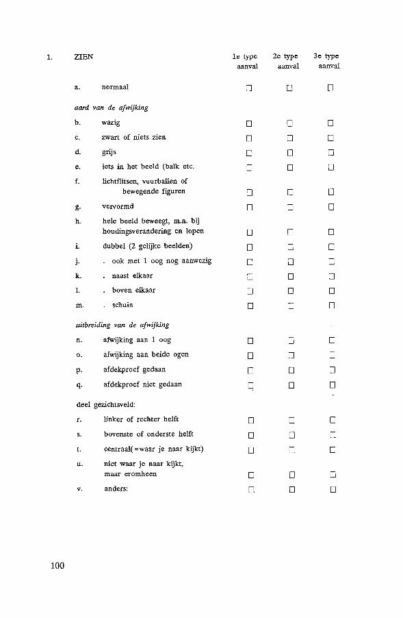
1. ZIEN 1e type 2e type 3e type a an val aanval aanval
a. normaal D D D
aard van de afwijking
b. wazig D D D
c. zwart of niets zien D D D
d. grijs D D D
e. iets in het beeld (balk etc. D D D
f. lichtflitsen, vuurballen of bewegende figuren D D D
g. vervormd D D D
h. hele beeld beweegt, m.n. bij houdingsverandering en lopen D D D
i. dubbel (2 gelijke beelden) D D D
j. ook met 1 oog nog aanwezig D D D
k. naast elkaar D D D
L boven elkaar D D D
m. schuin D D D
uitbreiding van de afwijking
n. afwijking aan 1 oog D D D
0. afwijking aan beide ogen D D D
p. afdekproef gedaan D D D
q. afdekproef niet gedaan D D D
deel gezichtsveld:
r. linker of rechter helft D D D
s. bovenste of onderste helft D D D
t. centraal( =waar je naar kijkt) D D D
u. niet waar je naar kijkt, maar eromheen D D D
v. anders: D D D
100

2. KRACHT 1e type 2e type 3e type aanval aanval aanval
a. normaal D D D
afwijking betreft
b. slap afllangenfgeheel verlamd D D D c. onhandigheid D D D
arm d. voorwerpen laten D D D vallen of minder kracht
e. moe of zwaar gevoel D D D f. stuurloosheid D D D
g. slepen met been/minder kracht D D D h. door 1 been zakkenfgeheel verlamd D D D
been i. door beide benen zakken p D D j. moe of zwaar gevoel D D D k. stuurloosheid D D D
gelaat/ 1. scheef gezicht (in de hoofd spiegel of door iemand D D D (1 kan.t) anders waargenomen)
m. tong scheef D D D
n. "opkruipen" van de stoornis (bijv. vingers - hand - arm, of
gelaat - arm - been) seconden D D D minuten D D D
0. anders D D D
101

3. GEVOEL le type 2e type 3e type aanval aanval aanval
a. normaal 0 0 0
afwijking betreft
b. doof, verdoofd gevoel 0 0 0
c. tintelingen 0 0 0
arm d. of de arm er niet bij hoort 0 0 0 e. stijf 0 0 0
f. slap end 0 0 0
g. doof, verdoofd 0 0 0
h. tintelingen 0 0 0
been i. of het been er niet bij hoort 0 0 0
j. stijf 0 0 0
k. slap end 0 0 0
gelaatj 1. doof, verdoofd 0 0 0
hoofd m. tintelingen 0 0 0
1 kant n. stijf 0 0 0 0. slap end 0 0 0
p. "opkruipen" van de stoornis (bijv. vingers - hand - arm, of gelaat - arm - been)
second en 0 0 0 minuten 0 0 0
q. anders 0 0 0
102

4. SPRAAK le type 2e type 3e type aanval a an val aanval
a. normaal 0 0 0 b. niet op de goede woorden komen 0 0 0 c. gesproken taal niet begrijpen 0 0 0 d. woord wei weten, maar niet
kunnen uitspreken 0 0 0 e. onduidelijk uitspreken van de
woorden ( dronken) 0 0 0 f. vreemde woorden zeggen 0 0 0 g. anders 0 0 0
5. LEZEN a. normaal 0 0 0 b. woorden afzonderlijk wei te
lezen maar niet te begrijpen 0 0 0 c. woorden niet te lezen 0 0 0
d. halve woorden zien of stukken uit de tekst verdwenen 0 0 0
e. begin nieuwe regel moeilijk te vinden 0 0 0
f. anders 0 0 0
6. SCHRIJVEN a. normaal 0 0 0 b. onleesbaar gekrabbel 0 0 0 c. verkeerde woorden schrijven
of woorden verkeerd op- 0 0 0 schrijven (spelling)
d. anders 0 0 0
103

7. DUIZELIGHEID
a. normaal 0 0 0 b. licht in het hoofd 0 0 0
c. zwaar gevoel in het hoofd 0 0 0 d. valneiging (van koers af) 0 0 0
e. zweverig 0 0 0 f. gevoel als in een draaimolen 0 0 0 g. de omgeving draait 0 0 0 h. de omgeving beweegt bij
houdingsverandering en lopen 0 0 0 i. anders 0 0 0
8. SLIKKEN
a. normaal 0 0 0
b. passagestoornis (mond-maag) 0 0 0 c. voortdurend gevoel van brok 0 0 0
in de keel
d. verslikken 0 0 0 e. anders 0 0 0
9. VERWARDHEID (ook heteroanamnese)
a. normaal 0 0 0 b. omgeving wordt niet herkend 0 0 []
c. verkeerd kleden 0 0 0 d. zich niets herinneren van
gebeurtenissen echter wel van 0 0 0 dingen uit het verleden;
e. vreemde woorden zeggen 0 0 0 f. anders 0 0 0
104
10. ANDERE KLACHTEN (ook hetero-anamnese)
a. geen 0 0 0
b. hoofdpijn kloppend 0 0 0 drukkend 0 0 0 voor andere verschijnselen 0 0 0 na andere verschijnselen 0 0 0
c. braken, misselijkheid 0 0 0 d. bewusteloosheid 0 0 0 e. trekkingen 0 0 0 f. tintelingen in de handen en/
of rond de mond 0 0 0 g. hik 0 0 0 h. pijn rond, naast etc. oog 0 0 0 i. anders 0 0 0
11. ALGEMENE ANAMNESE
Voorgeschiedenis F amilie-anamnese
a. migraine 0 f. migraine 0 b. diabetes 0 g. diabetes 0 c. epilepsie 0 h. epilepsie 0 d. hart-fvaatziekte 0 i. geen van deze o_
105

12. ONTSTAAN 1e type 2e type 3e type aanval aanval a an val
a. binnen een tel D D D
b. geleidelijk in seconden
minuten
uren
dagen
c. alle klachten tegelijk ontstaan D D D
d. alle klachten niet tegelijk ontstaan D D D
e. direct in aansluiting aan of tijdens D D D
....... . . . . . . . (invullen)
13. VERDWIJNEN
a. binnen een tel D D D
b. geleidelijk in seconden minuten uren dagen
c. restloos D D D
d. niet restloos volgens patient D D D want D D D
...............
14. DUUR AANVAL
a. second en
b. minuten
c. uren
d. dagen
106
15. AANTAL
a. 1e type aanval ... maal
b. 2e type aanval ... maal
c. 3e type aanval ... maal
16. CONCLUSIE 1e type 2e type 3e type
aanval aanval aanval TIA car otis 0 0 0 vertebrobasilaris 0 0 0 stroomgebied onzeker 0 0 0 hyperventilatie 0 0 0 migraine 0 0 0 epilepsie 0 0 0 orthostatische hypotensie 0 0 0 hypoglycaemie 0 0 0 anders 0 0 0
17. Welke zijn volgens u harde TIA-klachten (bijv. 2b, 2c etc.):
1e type aanval
2e type aanval
3e type aanval
18. Wat vindt u tegen een TIA pleiten: (bijv. 1b, 2n, etc.) (bijv. patient maakt geen rei;\le indruk)
1e type aanval
2e type aanval
3e type aanval
107

CRITERIA VOOR DIAGNOSE TIA
Absolute voorwaarden voor de diagnose TIA zijn:
De verschijnselen moeten binnen een tel of in enkele seconden zijn ontstaan. De verschijnselen moeten aile tegelijk zijn ontstaan (geen "opkruipen") De duur van de aanval is minimaal 1 minuut, in ieder geval enkele seconden is te kort. (De duur mag langer zijn dan 24 uur. Het gaat bij het huidige onderzoek om de anamnese, dus ook patienten met een partial stroke doen mee voor zover er geen waameembare handicap is die de anamnese bei:nvloedt).
BIJ TIA-BOOM BIJ TIA-ANAMNESE SCORlNGSLIJST
- Amaurosis fugax Zwart of niet zien met 1 oog, mag ook aileen bovenste of onderste gezichtsveld zijn. De afdekproef stelt in staat om onderscheid te maken met:
- Hemianopsie "plus" Zwart of niets zien met beide ogen ( d.w.z. bij uit- in linker of rechter gezichtsveld. sluitend hemian-
CAR OTIS opsie is onder-scheid carotis of vertebrobasilaire stroomgebied niet mogelijk)
- Hemiparese Geheel of gedeeltelijke verlarnming of vaardigheidstoornis van 1 lichaamshelft of van 1 mondhoek of· 1 arm. (Wat dit laatste betreft dienen andere oorzaken te worden uitgesloten, bijv. de patient heeft tijdens een dutje op de arm gelegen.)
- Afasie Niet op goede woorden komen; gesproken taal niet begrijpen of woord wel weten, maar niet kunnen uitspreken of vreemde woorden zeggen.
- ...... V erkeerde woorden schrijven; woorden verkeerd schrijven, aileen indien patient schrijfproef bij zich heeft
108

CRITERIA VOOR DIAGNOSE TIA
BIJ TIA-BOOM BIJ TIA-ANAMNESE SCORINGSLIJST
Bilaterale, bascule- Gevoelsstoornissen en/ of rende of altemerende krachtsverlies, vaardigheids-lange baan verschijn- stoornis in beide lichaams-selen helften of afwisselend links
en rechts
Hemianopsie zie bij CAROTIS
Transient global Zich niets herinneren van ge-amnesia beurtenissen die zojuist
hebben plaatsgevonden met goed geheugen voor gebeurtenissen uit verleden en voortdurend
VERTE- herhalen van dezelfde vragen. BRO-BASI- Combinaties van o.a. Combinaties van o.a. LAIR
- diplopie - dubbelzien (2 gelijke beelden) naast of boven elkaar of schuin verdwijnend bij kijken met 1 oog
- dysartrie - onduidelijk uitspreken van de woorden ( dronken)
- draaiduizelingen - gevoel als in een draai-molen, of omgeving draait
- drop attacks - plotseling door beide benen zakken
- ataxie - stuurloosheid of zwalken (!open als een dronkeman)
- dysfagie - passagestoornis bij slikken; verslikken
109
Bij de diagnose TIA past NIET:
- bewustzijnsverlies - trekkingen - lichtflitsen etc. in beeld, m.n. buitenste ring gezichtsveld - bewegen van het beeld m.n. bij lopen - licht of zweverig gevoel in het hoofd - voortdurend gevoel van brok in de keel - uitsluitend sensibele stoornissen in 1 lichaamshelft - uitsluitend wazig zien - pijn rond, naast etc. het oog
U wordt verzocht de volgende vragen te beantwoorden nadat u met uw koppelgenoot van gedachten gewisseld hebt.
1. Is uw uiteindelijke conclusie gewijzigd?
ja 0
nee 0
2. Zo ja, wat is uw conclusie nu?
le type 2e type 3e type. aanval aanval aanval
- TIA carotis 0 0 0 vertebrobasilaris 0 0 0 stroomgebied onzeker 0 0 0
- hyperventilatie 0 0 0 - migraine 0 0 0 - epilepsie 0 0 0 - orthostatische hypotensie 0 0 0 - hypoglycaemie 0 0 0 - anders 0 0 0
3. Waarom bent u van mening veranderd?
110
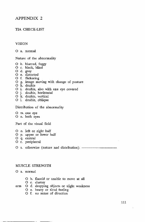
APPENDIX 2
TIA CHECK-LIST
VISION
0 a. normal
Nature of the abnormality
0 b. blurred, foggy 0 c. black, blind 0 d. grey 0 e. distorted 0 f. flickering 0 g. image moving with change of posture 0 h. double 0 i. double, also with one eye covered 0 j. double, horizontal 0 k. double, vertical 0 1. double, oblique
Distribution of the abnormality
0 m. one eye 0 n. both eyes
Part of the visual field
0 o. left or right half 0 p. upper or lower half 0 q. central 0 r. peripheral
0 s. otherwise (nature and distribution): -------------------------------
MUSCLE STRENGTH
0 a. normal
0 b. flaccid or unable to move at all 0 c. clumsy
arm 0 d. dropping objects or slight weakness 0 e. heavy or tired feeling 0 f. no sense of direction
111

0 g. dragging one leg 0 h. weakness of one leg
leg 0 i. weakness of both legs 0 j. heavy or tired feeling 0 k. no sense of direction
0 1. face droops to one side (seen by the patient himself in the mirror or seen by another person)
0 m. march of the symptoms (e.g. fingers to hand to arm, or to arm to leg)
0 n. otherwise: -----------------------------------------------------------------
SENSATION
0 a. normal
0 b. numb 0 c. pins and needles
arm 0 d. as if the arm does not belong to oneself 0 e. stiff
0 f. numb 0 g. pins and needles
leg 0 h. as if the leg does not belong to oneself 0 1. stiff
Oj. numb face 0 k. pins and needles
01. stiff
0 m. "march" of the symptoms (e.g. fingers to hand to arm, or face to arm to leg)
0 n. otherwise: ---------------------------------------------------------------
112
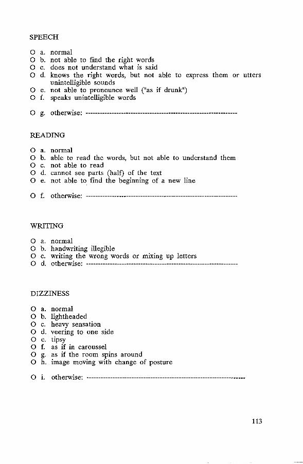
SPEECH
0 a. normal 0 b. not able to find the right words 0 c. does not understand what is said 0 d. knows the right words, but not able to express them or utters
unintelligible sounds 0 e. not able to pronounce well ("as if drunk") 0 f. speaks unintelligible words
0 g. otherwise: ----------------------------------------------------------------
READING
0 a. normal 0 b. able to read the words, but not able to understand them 0 c. not able to read 0 d. cannot see parts (half) of the text 0 e. not able to find the beginning of a new line
0 f. otherwise: ----------------------------------------------------------------
WRITING
0 a. normal 0 b. handwriting illegible 0 c. writing the wrong words or mixing up letters 0 d. otherwise: ----------------------------------------------------------------
DIZZINESS
0 a. normal 0 b. lightheaded 0 c. heavy sensation 0 d. veering to one side 0 e. tipsy 0 f. as if in caroussel 0 g. as if the room spins around 0 h. image moving with change of posture
0 i. otherwise: -------------------------------------------------------------------
113

SWALLOWING
0 a. normal 0 b. unable to swallow food or fluids 0 c. constant feeling of a lump in the throat
0 d. otherwise: ----------------------------------------------------------------
CONFUSION (if necessary supplementary information may be obtained from relatives or nurses)
0 a. normal 0 b. the environment cannot be recognized 0 c. unable to dress properly 0 d. does not recall recent events, but is able to remember
events from the past; permanently repeats the same questions
0 e. otherwise: ----------------------------------------------------------------
OTHER SYMPTOMS
0 a. none 0 b. headache
(if necessary supplementary information may be obtained from relatives or nurses)
0 c. headache, throbbing 0 d. headache, constant 0 e. headache, before the main symptoms 0 f. headache, after the main symptoms 0 g. vomiting, nausea 0 h. unconsciousness 0 i. jerking of limbs 0 j. tingling hands or mouth 0 k. hiccup 0 1. pain around, under the eye
0 m. otherwise: ----------------------------------------------------------------
114

GENERAL INFORMATION
Past history
0 a. migraine 0 b. diabetes 0 c. epilepsy 0 d. cardiovascular disease 0 e. none of these
Family history
0 f. migraine 0 g. diabetes 0 h. epilepsy 0 i. none of these
MODE OF ONSET
0 a. within one second 0 b. within seconds 0 c. within minutes 0 d. within hours 0 e. within days
0 f. all symptoms at the same time 0 g. not all symptoms at the same time
0 h. symptoms occurred during or after ----------------------------------------
DISAPPEARANCE OF SYMPTOMS
0 a. within one second 0 b. within seconds 0 c. within minutes 0 d. within hours 0 e. within days
0 f. without residual symptoms 0 g. with residual symptoms: ---------------------------------------------------
DURATION OF THE ATTACK
0 a. seconds 0 b. minutes 0 c. hours 0 d. days
115

NUMBER OF ATTACKS
Number of attacks: ----
CONCLUSION
OTIA
0 carotid 0 vertebrobasilar 0 vascular territory uncertain
0 Hyperventilation 0 Migraine 0 Epilepsy 0 Orthostatic hypotension 0 Hypoglycemia . 0 Otherwise: -------------------------------------------------------------------
116
APPENDIX 3
THE DUTCH TIA-TRIAL
Participating Centres
Medisch Centrum Alkmaar (J.W.W.H. Dammers, J.A. van Leusden, H.J.S. Straatman, R. ten Houten); Streekziekenhuis Almelo (J.W.M. ter Berg); St. Elizabeth Ziekenhuis Amersfoort (R. Bijlsma, M.B.M. Vermeulen); Ziekenhuis "De Lichtenberg" Amersfoort (C. Boutkan, B.A.A.M. Kools, D.J. van der Voort, J.L. van der Zwan); Academisch Medisch Centrum Amsterdam (D. Herderschee, A. Hijdra); St. Lucas Ziekenhuis Amsterdam (J. Vas, J.A.L. Vanneste); Ziekenhuis Malberg Arnhem (G. Schouwink, L. van der Graaff); Hervormd Diaconessenhuis Arnhem (H. Becker, F.A. Jongbloed); Ziekenhuis Lievensberg, Bergen op Zoom (P.J.I.M. Berntsen, T.B. Gebbink, F.M. Sleegers); St. GeertruidenjSt.Jozefziekenhuis Deventer (W.J. Feikema, J.A. van Beeck, A.N. Veltema, .l.H.M. van Gasteren, N.A.C. Roelvink); Merwedeziekenhuis Dordrecht (P.A.T. Carbaat, L.I. Hertzberger, C.S.D. Hoekstein); Refajaziekenhuis Dordrecht (C. Oppelaar, J.J. Groen); Ziekenhuis "Nij Smellinghe" Drachten (H.L. van der Wiel, A.J. Stegink); Catharina Ziekenhuis Eindhoven (J.A.M. Frederiks, J.N. Berendes, G.J. Lammers); Diakonessenhuis Eindhoven (W. Groeneveld, H.J. Troelstra, A.J. Vermeij); St. Joseph Ziekenhuis Eindhoven (B.J. van Kasteren, L.H.T.S. Kortbeek, P.M.G.A.W. Mulkens); De Stadsmaten Enschede (H. Solleveld, J.A.G. Geelen, G. Wilts); Ziekenhuis "Ziekenzorg" Enschede (J. Troost, E.N.H. Jansen, G. Hageman); Ziekenhuis St. Jansdal Harderwijk (M.J.P. Muller Kobold, - R.T. Schoenmaker); St. Anna Ziekenhuis Geldrop (P.F.M. Houben, C.G. Staas, T.J.M. Breuer); Stichting Oosterscheldeziekenhuizen, Lokatie Bergzicht Goes (A.M. Boon, W.H.G. Lieuwens); Streekziekenhuis Gorkum "Beatrix" (P.M. Stuurman, M.H. Dijkman); St. Jozefziekenhuis Gouda (A. Aanen); Westeinde Ziekenhuis 's Gravenhage (J. Nihom, J.T.J. Tans); Ziekenhuis Bethlehem's Gravenhage (C.H. Terpstra, A. Verkijk); St. Elisabeth's Gasthuis Haarlem (L. de Vries, J.A.M. Kuster); Streekziekenhuis "Oranjeoord" Harlingen (G.S.D. van Leersum); De Wever Ziekenhuis Heerlen (C.L. Franke, C.J. Hagen, J.F. Mirandolle); Streekziekenhuis Midden Twente Hengelo (M.M. Klaver); Stichting Streekziekenhuis Hilversum (F.A.M. Zandhuis); Streekziekenhuis "West-Friesland" Hoorn (G.J. Lambrechts, H.B. Grolman-Kovarova); Medisch Centrum "Bonifatius Ziekenhuis
117
Leeuwarden (J. Dijkstra, B.O.M. Bomers); Medisch Centrum Diakonessenhuis Leeuwarden (J.G. de Bruijn, J.G. Koster); Academisch Ziekenhuis Leiden (A.R. Wintzen, G.W. Bruyn, O.J.S. Buruma, G.W.A.M. Padberg, A.C.B. Peters, R.A.C. Roos); Diaconessenhuis Leiden (P.E. Briet, J. van Rossum); St. Elizabethziekenhuis Leiderdorp (A.J.P. Boesten, M.M. Veering, H.G.S. Snijder); Antoniushove Leidschendam (C.H. Terpstra, A. van der Marel, A. Verkijk); Zuiderzeeziekenhuis Lelystad (J.P. Geervliet, E. Siebenga); Academisch Ziekenhuis Maastricht (J. Ladder, P.J.M. van der Lugt); Canisius-Wilhelmina Ziekenhuis Nijmegen (C.W.G.M. Frenken, E.F.J. Poels, M.J.J. Prick, W.I.M. Verhagen, E.V. van Zuilen, M.C. Verhulsdonck, E.S. Louwerse); St. Franciscus Ziekenhuis Roosendaal (H.N.A.M. Wouters, L.J.M.A. van Hoof£, A.C.G.A. van Spreeken, G.A.M. Verheul); Academisch Ziekenhuis Dijkzigt Rotterdam (P.J. Koudstaal, A. Staal, M. Vermeulen, G. de Jong, W. Hoppenbrouwers); Bergwegziekenhuis Rotterdam (R. Gelsema, T.C.M. van Woerkom); St. Clara Ziekenhuis Rotterdam (J.G. van Woerkom, J.A.G. Strijbosch, H.A.W. Sinnige, H.J. van den Brand); van Dam/Bethesda Ziekenhuis Rotterdam (A. van der Zwart); Eudokia Ziekenhuis Rotterdam (A.T. Tjiam, A.H.C. Geerlings); Ikazia Ziekenhuis (L.J.M.M. Mulder); St. Franciscus Gasthuis Rotterdam (C. Bulens, L.H. Penning de Vries-Bos, P.R. Beneder); Havenziekenhuis Rotterdam (H.J. Vroon, J.Z. van den Berg); Schieland Ziekenhuis Schiedam (W.C. Ba~rd, R. Agema); Academisch Ziekenhuis Utrecht (L.J. Kappelle, J. van Gijn, J.J. Jansen, W. Weststrate); Diaconessenhuis Utrecht (D.N.J. Danker, E. Ossentjuk); St. Maartens Gasthuis Venlo (T.G. Segeren, G.D.M. van Hellemondt, B.J. Meems); Holy Ziekenhuis Vlaardingen (W.F. van Oudenaarden, H.G.J. Krouwer, J.J.M. Driesen, G.L. Liem); Diaconessenhuis Voorburg (J.L. van Doorn, G.J. de Jong); Hofpoort Ziekenhuis Woerden (E.J. Wieringa, R. Wielaard); Juliana Ziekenhuis Ede (M.G. Smits, A.J.M. Vos); Van Weel/Bethesda Ziekenhuis Dirksland (U.W. Huisman); St. Antonius Ziekenhuis Sneek (P.J.H.W. Jansen, J.D.M. van der Meulen); Diaconessenhuis Heemstede (R.H. Groen, J.O. Mispelblom Beyer); St. Elisabeth Ziekenhuis Tilburg (A.A.W. Op de Coul, A.C.M. Leyten, C.C. Tijssen, R.L.A.A. Schellens); Maria Ziekenhuis Tilburg (H.J.M. van der Leeuw, J.G. Eerenberg, C.C. Tijssen).
Steering Committee
J. van Gijn, University of Utrecht (neurologist, chairman); A. Staal, Erasmus University, Rotterdam (neurologist); M. Vermeulen, University Hospital, Rotterdam (neurologist); P.J. Koudstaal, University Hospital,
118

Rotterdam (neurologist); H. van Creve!, University of Amsterdam (neurologist); A. Hijdra, Academic Medical Centre, Amsterdam (neurologist); A.F.A.M. Schobben, University Hospital, Utrecht (pharmacist).
Trial Office
University Department of clinical neurology, Nicolaas Beetsstraat 24, 3511 HG Utrecht, The Netherlands, H. Frericks (trial coordinator); Mrs. F. van Vliet (data manager); Mrs. A. te Nijenhuis (secretary).
Executive Committee
J. van Gijn, University of Utrecht (neurologist, chairman); P.J. Koudstaal, University Hospital, Rotterdam (neurologist); A. van Latum, University of Utrecht (fellow of the Dutch Heart Fund); A.F.A.M. Schobben, University Hospital, Utrecht (pharmacist); W.F.A. Verheugt, Free University of Amsterdam (cardiologist).
Advisory Board
P.G. Hugenholtz, Erasmus University, Rotterdam (cardiologist); G.T. Meester, University of Utrecht (cardiologist); J. Lubsen, Erasmus University, Rotterdam (epidemiologist); J.J. Sixma, University of Utrecht (hematologist); R.W. Brower, Thorax Center, University Hospital, Rotterdam (data management); J.H. Glerum, University Hospital, Utrecht (pharmacist); G .S. Baarsma, Eye Infirmary, Rotterdam (ophthalmologist).
Monitoring Committee
C.P. Warlow, University of Edinburgh (neurologist); R. Peto, University of Oxford (statistician).
Computed Tomography Committee
P.J. Koudstaal, University Hospital, Rotterdam (neurologist, chairman); J. van Gijn, University of Utrecht (neurologist); L. Ramos, University Hospital, Utrecht (radiologist); L.J. Kappelle, University Hospital, Utrecht (neurologist).
119

Electrocardiography Committee
P.J. Koudstaal, University Hospital, Rotterdam (neurologist, chairman); G.A.M. Pop, University Hospital, Rotterdam (cardiologist); F.W.A. Verheugt, Free University of Amsterdam (cardiologist); G. de Jong, University Hospital, Rotterdam (resident in neurology).
End Point Committee
J. van Gijn, University of Utrecht (neurologist, chairman); P.J. Koudstaal, University Hospital, Rotterdam (neurologist, co-chairman); A. Staal, Erasmus University Rotterdam (neurologist); M. Vermeulen, University Hospital, Rotterdam (neurologist); H. van Crevel, University of Amsterdam (neurologist); A. Hijdra, Academic Medical Centre, Amsterdam (neurologist); F.W.A. Verheugt, Free University, Amsterdam (cardiologist); G.A.M. Pop, University Hospital Rotterdam (cardiologist); G.S. Baarsma, Eye Infirmary, Rotterdam (ophthalmologist).
Funding Agencies
Dutch Heart Fund; ICI-Farma, Rotterdam and ICI Pharmaceuticals, Great Britain (production of Tenormin and financial support); ACF-Chemiefarma, Maarssen (production of Ascal); Brocacef, Maarssen (distribution of drugs to participating centers); and University Hospital, Utrecht (office facilities).
120

NEDERLANDS TIA-ONDERZOEK: FORMULIER BIJ OPNEMING IN HET ONDERZOEK
Vul dit formulier direkt in bij iedere patient bij wie men een TIA of partial stroke overweegt
1. ALGEMEEN
RUJMTE VOOR PATI~NTENPLAAT .JE:
1.1 Zlekenhuis ______________________________________________________________________ __
1.2 Arts __________________________________________________________________________ ___
1.3 Datum van invullen _________ _ ______ _ D M J
2. PATI~NTGEGEVENS
2.1 Naam -----------------------------------------------------------------------2.2 Geboortedatum _________ ________ __ ______ _
D M J
2.3 Geslacht __________________ _
2.4 Naam huisarts __________________________________ Tel.nr. __________________________ _
3. ANAMNESE
* Bij aile onderdelen van de anamnese is het de bedoeling dat de klachten van de patient in diens eigen bewoordingen worden genoteerd.
* Dit kan gebeuren door de voorgedrukte klachten aan te kruisen of door de klachten zelf bij "anders" in omgangstaal te noteren.
* Neem aile onderdelen een voor een door. Als de patient over een onderdeel geen klachten had, kan men dit aangeven door een X bij "normaal".
* Als de patient meerdere, verschillend verlopende aanvallen heeft gehad dan de meest recente TIA of partial stroke noteren.
* Als men over een bepaald onderdeel geen oordeel kan geven, b.v. omdat patient tijdens de aanval niet gesproken heeft, dan kan men dit aangeven door bij "anders" te vermelden: niet getest.
_.., * S.v.p. nader preciseren door behalve X te noteren: L =links; R = rechts; A= alternerend links en rechts; 0 =onder of B = boven. Bijvoorbeeld: L ®
3.1 Zien
0 a. normaa\ aard van de afwijking
0 b. wazig 0 c. niets zien (zwart of grijs) 0 d. iets in het beeld (balk, lijn, walk, slip, etc.) 0 e. Jichtflitsen, vuurballen of bewegende figuren 0 f. vervormd 0 g. hele bee\d beweegt, m.n. bij houdingsverandering en \open 0 h. dubbel (2 gelijke beelden) 0 i. dubbel oak met 1 cog neg aanwezig 0 j. dubbel naast elkaar 0 k. dubbel boven elkaar 0 I. dubbel schuin
uitbreiding van de afwijking 0 m. afwijking aan 1 cog 0 n. afwijking aan beide ogen 0 o. afdekproef gedaan (door patient tijdens de klacht) 0 p. afdekproef niet gedaan (door patient tijdens de klacht)
dee\ gezichtsveld 0 q. linker of rechter helft 0 r. bovenste of onderste helft 0 s. centraal (= waar je naar kijkt) 0 t. niet waar je naar kijkt, maar eromheen 0 u. andeffi, _________________________________________________________ _

3.2. Kracht
0 a. normaal
afwijklng betreft
arm { been { gelaat
0 b. slap afhangen 0 c. onhandigheid 0 d. voorwerpen Iaten vallen of minder kracht 0 e. moe of zwaar gevoel 0 f. stuurloosheid
0 g. slepen met been 0 h. door 1 been zakken 0 i. door beide benen zakken 0 j. moe of zwaar gevoel 0 k. stuurloosheid
0 1. scheef gezicht (in de spiegel of door iemand anders waargenomen).
0 n. "opkruipen" van de stoornis (b.v. vingers---> hand---> arm, of gelaat---> arm---> been)
0 o. andeffi ______________________________________________________________________ __
3.3. Gevoel
0 a. normaal
afwijking betreft
arm { been { gelaat {
0 b. doof, verdoofd gevoel 0 c. tintelingen 0 d. of de arm er niet bijhoort 0 e. stijf 0 f. slapend
0 g. doof 0 h. tintelingen 0 i. of het been er niet bijhoort o j. stijf 0 k. slapend
0 I. doof, verdoofd 0 m. tintelingen 0 n. stijf 0 o. slapend
0 p. "opkruipen" van de stoornis (b.v. vingers---> hand---> arm, of gelaat---> arm---> been)
0 q. andeffi ______________________________________________________________________ __
3.4 Spraak
0 a. normaal 0 b. niet op de goede woorden komen 0 c. gesproken taal niet begrijpen 0 d. woord wei weten, maar niet kunnen uitspreken 0 e. onduidelijk uitspreken van de woorden (dronken) 0 f. vreemde woorden zeggen 0 g. andeffi __________________________________________________________________ __
3.5. Lezen
0 a. normaal 0 b. woorden afzonderlijk wei te lezen, maar niet te begrijpen 0 c. woorden niet te lezen 0 d. halve woorden zien of stukken uit de tekst verdwenen 0 e. begin nieuwe regel moeilijk te vinden
0 f. anders

3.6. Schrijven
0 a. normaal 0 b. onleesbaar gekrabbel 0 c. verkeerde woorden schrijven of woorden verkeerd opschrijven (spelling) 0 d. andern __________________________________________________________________ __
3.7. Duizeligheid
0 a. normaal 0 b. Iicht in het hoofd 0 c. zwaar gevoel in het hoofd 0 d. valneiging (van koers af) 0 e. gevoel als in een draaimolen 0 f. zweverig 0 g. de omgeving draa it 0 h. de omgeving beweegt bij houdingsverandering en lopen 0 i. anders ______________________________________________________________________ __
3.8. Slikken
0 a. normaal 0 b. passagestoornis (mond--> maag) 0 c. voordurend gevoel van brok in de keel 0 d. andern __________________________________________________________________ __
3.9. Verwardheid (zonodig ook hetero-anamnese)
0 a. normaal 0 b. omgeving wordt niet herkend 0 c. verkeerd kleden 0 d. zich niets herinneren van recente gebeurtenissen, echter wei van dingen uit het verleden;
steeds herhalen van dezelfde vragen 0 e. andern ______________________________________________________________________ __
3.10 Andere klachten (zonodig ook hetero-anamnese)
0 a. geen 0 b. hoofdpijn 0 c. hoofdpijn kloppend 0 d. hoofdpijn drukkend 0 e. hoofdpijn voor andere verschijnselen 0 f. hoofdpijn na andere verschijnselen 0 g. braken, misselijkheid 0 h. bewusteloosheid 0 i. trekkingen 0 j. tintelingen in de handen en/of rond de mond 0 k. hik 0 I. pijn rond, naast etc. het oog 0 m. anders __________________________________________________________________ __
3.11 Ontstaan
0 a. binnen een tel 0 b. in sekonden 0 c. in 1-5 minuten 0 d. in 5-60 minuten 0 e. in uren 0 f. in dagen
0 g. aile klachten tegelijk ontstaan 0 h. aile klachten niet tegelijk ontstaan 0 i. direct in aansluiting aan of tijdens -----------------------------------------------------
3.12 Duurvan deaanval (zo mogelijk getal invullen)
0 a. sekonderr
0 b.
0 c.
0 d.
_________ minuten
---------- uren _________ dagen
0 e. nog bestaand

5.
3.13 Verdwijnen
0 a. binnen een tel 0 b. in sekonden 0 c. in 1-5 minuten 0 d. in 5-60 minuten 0 e. in uren 0 f. in dagen 0 g. nag bestaand
0 h. restloos 0 i. niet restloos volgens patient, want---------------------------
3.14 Aantal
a. hoeveel aanvallen heeft patient in het laatste jaar gehad?
b. als er meerdere aanvallen waren, was de aard dan steeds hetzelfde?
1 2-3 4-10 >10
ja neen
0 0 0 0
0 0
c. Zo neen, gaarne een korte omschrijving van de andere type(n) aanval(len) ------------
3.15 Datum laatste aanval ____ _ D M J
Raadpleeg bij twijfel over de diagnose de criteria (zie laatste biz.) lndien men besluit tot TIA » doorgaan naar punt 5.
Neem de patient indien enigszins mogelijk op in de kliniek. Verricht het volgende aanvullend onderzoek:
* Bleed: BSE (als hager dan 30 mm: ANF) Hb., hematocriet, trombocyten, bloedsuiker, fibrinogeen nierfuncties, leverfuncties, luesreacties
* X-thorax (vraagstelling: hart/thorax ratio?) * E.C.G. (vraagstelling: PO-tijd? Hartfrequentie?) * C.T.-scan (niet bij amaurosis fugax) * Holter 24-uurs E.C.G. } * Angiografie facultatief, per centrum af te spreken

6. UITSLUITINGSCRITERIA
Neem aile punten een voor een door
6.1 Algemeen
0 laatste ischaemische aanvallanger dan 3 maanden geleden 0 herseninfarct in verleden met invaliderende restverschijnselen 0 leeftijd < 40 jaar, tenzij atherosclerose aannemelijk is 0 patient spreekt geen vloeiend Nederlands 0 terminaal stadium maligniteit 0 te verwachten therapie-ontrouw
6.2 Bloed
0 anaemie (Hb < 6.0 mmol/1) 0 polycythaemie (Ht;;. 0.60)
") 0 !rombocytose (;;> 500 X 1 09/1) ") 0 trombocytopenie(,;; 100 X 1 09/l) *) 0 afwijkingen passend bij vasculitis (bijv.: pos. ANF, luesreakties)
0 hypoglycemia(,;; 2 mmol/1) tijdens aanval
6.3 Cardiaal
0 myocardinfarct in voorafgaande maand 0 ritmestoornissen die in verband staan met de aanvallen (klinisch of E.C.G.) 0 emboliebron (atriumfibrillatie, kleplijden, transmuraal infarct< 4 weken)
6.4 C.T.-scan *)
0 intracraniele bleeding 0 tumor cerebri 0 subduraal hematoom 0 andero ____________________________________________________________________ ___
6.5 0 Geen
*) V66r dit bekend is, kan de patient eventueel alvast gerandomiseerd worden. Gaarne de nagekomen uitslagen alsnog invullen op dit formulier
lndien een of meer criteria zijn aangekruist komt de patient niet voor de studie in aanmerking. Stuur in dat geval het originele formu/ier op naar het secretariaat TIA-onderzoek, kliniek voor neurologie, Nic. Beetsstraat 24, 3511 HG Utrecht en bewaar zelf de copieen. lndien geen van de criteria aanwezig is--> door naar punt 6.5.
6.5 Specifieke criteria m.b.t. acel:osal
Patient is bekend met:
0 leverinsufficientie 0 ulcus pepticum (laatste 3 jaar) 0 abnormale bloedingsneiging (b.v. hemofilie, thrombopathieen) 0 intracraniele bloeding in de voorgeschiedenis (relatief) 0 asthma bronchiale 0 anticoagulantia 0 allergie 0 dagelijks acetosal-gebruik tijdens studie noodzakelijk op andere indicatie, bijv. rheumato'ide arthritis
0 anders,
Bij aanwezigheid van een of meerdere van deze criteria, geen randomisatie voor acetosal. Ga verder naar punt 6.6.

6.6 Specifieke criteria m.b.t. beta·blokkers
0 hartfrequentie .; 50/min. 0 op ECG: PO·tijd ;;> 0.25 sec. 0 2' of 3' graads AV-block 0 diastolische bloeddruk < 80 mm Hg 0 op X-thorax CT ratio> 0.65
Patient is bekend met:
0 manifeste decompensatio cordis 0 asthma bronchiale of chronische bronchitis 0 insuline afhankelijke diabetes mellitus 0 nierinsufficientie 0 myasthenia gravis 0 ziekte van Raynaud 0 claudicatio intermittens 0 patient gebruikt reeds beta-blokker op andere indicatie en meet daarmee doorgaan. 0 anders, ________________________________________________________________________ ___
Bij aanwezigheid van een of meerdere van deze criteria, geen randomisatie voor beta-blokkers.
Vraag nu toestemming aan de patient voor acetosal en! of beta-blokker, eventueel na het verstrekken van schriftelijke informatie.
lndien de patient voor geen van beide medicamenten gerandomiseerd kan of wit worden ~ originele formu/ier naar secretariaat TIA-onderzoek, kliniek voor neurologie, Nic. Beetsstraat 24, 3511 HG Utrecht sturen en copieen zelf bewaren. lndien randomisatie voor een of beide moge/ijk ~door naar punt 7.
7. RANDOMISATIE
7.1 Waarvoor kan de patient gerandomiseerd worden? aspirine 0 beta-blokker 0 beide 0
Bel nu voor randomisatie het studiecentrum: tel. 030-372520. Vul nu patientnummer (Pat.nr.) in.
7.2 Pat.nr. -------------------
7.3 Datum van randomisatie ________ /_________ _ _____ ___ D M J
De patient is nu in de TRIAL en blijft a/tijd in de follow-up. ledere relevante gebeurtenis moet worden gemeld, ook als de medikatie niet gegeven of ingenomen wordt (intention-to-treat analyse)
Stuur de patient nu met het speciale medica tie formulier naar de apotheek.
8. OVERIGE ANAMNESTISCHE GEGEVENS
8.1 Heeft de patient in het verleden een of meerdere keren een myocardinfarct gehad? ja nee
8.2 Zo ja, wanneer (a.u.b. jaar invullen) -----------------------------------------
0 0

8.3. Welke medikatie gebruikt de patient en waarom? (a.u.b. invullen).
Soort Red en a. __________________________________ _
b. ______________________________ _
c. __________________________________ _
d. ______________________________ _
e, __________________________________ _
8.4 lndien de patient reeds voor de randomisatie alvast acetosal gebruikte, hoe lang tevoren was dit voorgeschreven?
8.5
8.6
8.7
1-3 dagen 4-7 dagen 1-4weken >4weken
0 0 0 0
Is de patient bekend met een van de volgende aandoeningen?
Rookt patient?
Onderging patient in het verleden een van deze ingrepen?
Hypertensie 0 zo ja, ooit behandeld? 0 angina pectoris 0 claudicatio intermittens 0 hyperlipidaemie 0 diabetes 0
neen 0 <> 20 sigaretten/ dg 0 > 20 sigaretten/dg 0
hartklepoperatie 0 bypassoperatie 0 carotis desobstructie 0 aorta- of vaatprothese 0
8.8 Noteer naam en ad res van een familielid of kennis van de patient
LICHAMELIJK ONDERZOEK
9.1 Bloeddruk(a.u.b. invullen)
9.2
9.3
9.4
systolisch -----------------------------------------------------------------------
diastolisch _______________________________________________________ (ionen niet meer .hoorbaar)
(gemeten in liggende houding in aansluiting op het neurologisch onderzoek)
Gewicht (a.u.b. invullen) ___________________ kg.
Zijn er neurologische restverschijnselen van laatste aanval? ja 0-+
nee 0-+
Zijn er (niet-neurologische) aandoeningen die de validiteit be'invloeden?
door naar punt 9.5
door naar punt 10
0 ja 0 nee
9.5 Rankin invaliditeitsscore
0 0. Geen klachten. 0 1. Niet-invaliderende klachten, die geen beperking inhouden op de alledaagse activiteiten. 0 2. Geringe invaliditeit. Kan niet aile vroegere activiteiten uitvoeren, maar is in staat zichzelf te verzorgen. 0 3. Matige invaliditeit. De patient word! duidelijk belemmerd in het dagelijks Ieven of is niet meer volledig onaf
hankelijk. 0 4. Matig tot ernstige invaliditeit. Kan aileen met hulp lopen of heeft verpleging nodig, echter niet ieder uur van
de dag. 0 5. Ernstige invaliditeit. Bedlegerig of rolstoelpatient. Meestal incontinent. Is voortdurend verpleegbehoeftig, ie
der uur van de dag. 0 6. Dood.

10. AANVULLEND ONDERZOEK
10.1 Bloed: Ht(l/1) _____________________________ _
glucose (nuchter, mmol/1) --------------------------
kreatinine (mmol/1) -----------------------------
fibrinogeen (g/l) _____________________________ _
luesreacties _________________________________ _
1 0.2 X -thorax: Cor /thorax ratio > 1 /2 ( r6ntgenverslag) 0 Cor /thorax ratio :i 1 /2 0
10.3 Holter 24-uurs E. C. G.: wordt verrichVniet verricht"
10.4 Angiografie: wordt verrichVniet verricht"
•: doorhalen wat niet van toepassing is.
10.5
10.6
Bijlage1
Stuuroriginee/ of copie van E. C. G. en C. T.-scan naar secretariaat TIA-onderzoek, kliniek voor neurologie, Nic. Beetsstraat 24, 3511 HG Utrecht. Originele exemplaren zullen worden geretourneerd.
Stuur nu het origine/e formulier naar hetzelfde adres en bewaar zelf de copieen in specia/e ordner. U ontvangt zo spoedig mogelijk een bevestiging van de eerste controle-afspraak.
CRITERIA VOOR DE DIAGNOSE TIA
Voorwaarden voor de diagnose TIA 1. Ontstaan binnen een tel of in sekonden. 2. Geen "opkruipen" van de uitvalverschijnselen. 3. Duur: tenminste 1 minuut. 4. Aard:
Aanvallen in het carotisgebied * Niels zien (zwart of grijs) met 1 oog of in bovenste of onderste gezichtshelft van 1 oog. * taalstoornis (spreken, Jezen of schrijven) * halfzijdige verlamming of onhandigheid (een dysarthria daarbij mag).
Aanvallen in het vertebrobasilaire gebied * halfzijdige uitval (zwart of grijs) van het gezichtsveld. * verlamming die dubbelzijdig is, wisselt van de ene naar de andere zijde,
of het gelaat aan een zijde en de ledematen aan de andere zijde betreft. * combinatie van: draaiduizelingen, dubbelzien, slikstoornissen, spraakstoornissen,
stuurloosheid en plots door de benen zakken.
In strijd met de diagnose TIA: * bewustzijnsverlies, * trekkingen, * incontinentie, * hoofdpijn. * De klachten zijn te provoceren door snel opstaan van de patient, warmte, fellicht, of persen.


Related Documents


![Aortic Arch Plaque and Ischemic Stroke Detection Modalities and … · transient ischemic attacks published in the AHA/ASA guidelines have received widespread recognition [8]. According](https://static.cupdf.com/doc/110x72/5f115e3ee4b66540f13150e9/aortic-arch-plaque-and-ischemic-stroke-detection-modalities-and-transient-ischemic.jpg)


