COMMUNITY EYE HEALTH JOURNAL SOUTH ASIA | VOLUME 28 ISSUE 92 | 2015 61 Community Eye Health JOURNAL VOLUME 28 | ISSUE 92 | 2015 The world is facing an unprecedented epidemic of diabetes. The International Diabetes Federation (IDF) estimates that there are 415 million adults living with diabetes worldwide. No country is immune from this epidemic: 70% of people with diabetes live in low- and middle-income countries. Type 2 diabetes accounts for over 90% of all cases. The increase in type 2 diabetes is associated with modern-day lifestyles, characterised by unhealthy eating (foods high in sugar, salt and fat), physical inactivity and increasing obesity. Diabetes causes high levels of glucose (a form of sugar) in the bloodstream. Over time, this damages blood vessels, with devastating effects in many different parts of the body, leading to heart attacks, stroke, foot amputations, kidney failure and blindness. It is these complications of diabetes that lead to premature death in so many. IDF estimates that, in 2015, five million people died from causes associated with diabetes. That is more than all the deaths from malaria, tuberculosis and HIV combined. The compli- cations of diabetes also account for the staggering cost of treating diabetes, estimated at over US $670 billion dollars a year. Diabetic retinopathy (DR) is caused by damage to retinal blood vessels. It is estimated that one third of people with diabetes have DR, and that up to a third of them have impaired vision. Although advanced DR can lead to blindness, the early stages are entirely asymptomatic. It is therefore essential that everyone with diabetes has their eyes examined for DR, ideally every year. Nearly half of all people with diabetes don’t know they have the condition, so the damage to their eyes progresses to an advanced stage before there is an oppor- tunity to prevent vision loss. This is tragic, as the risk of developing DR and vision loss can be reduced by keeping blood glucose, cholesterol and blood pressure as near normal levels as possible. We need to build health systems that will help people to achieve good control of their diabetes through lifestyle changes and medication where required, and that will provide regular retinal screening to detect DR in its early stages, as well as laser or anti-VEGF treatment to prevent blindness. As most cases of type 2 diabetes can be prevented, there is also an urgent need to promote policies that support healthy lifestyles. Unfortunately, we are far from achieving this ideal. Too many people already have advanced retinopathy at the time they are diagnosed with diabetes. Too many people with diabetes are not aware of the risk of blindness, and too many of those who see a doctor for their diabetes do not have their eyes examined. Understanding the challenges: the DR Barometer project The DR Barometer was a global project undertaken by IDF, the International Association for the Prevention of Blindness (IAPB), the International Federation on Ageing (IFA) and the New York Academy of Medicine. It collated the experiences of 3,590 people with diabetes and 1,451 health professionals from 41 countries across the globe. The preliminary findings were released at the EURETINA meeting in Nice in September 2015 and provided revealing insights. The diabetes epidemic and its implications for eye health Continues overleaf ➤ David Cavan Director of Policy & Programmes: International Diabetes Federation, Brussels, Belgium. [email protected] GV Murthy An eye care worker screens a patient in Colombo. SRI LANKA ‘Nearly half of all people with diabetes don’t know they have the condition’

Welcome message from author
This document is posted to help you gain knowledge. Please leave a comment to let me know what you think about it! Share it to your friends and learn new things together.
Transcript
COMMUNITY EYE HEALTH JOURNAL SOUTH ASIA | VOLUME 28 ISSUE 92 | 2015 61© The author/s and Community Eye Health Journal 2015. This is an Open Access article distributed under the Creative Commons Attribution Non-Commercial License.
Community Eye Health
JOURNALVOLUME 28 | ISSUE 92 | 2015
The world is facing an unprecedented epidemic of diabetes. The International Diabetes Federation (IDF) estimates that there are 415 million adults living with diabetes worldwide. No country is immune from this epidemic: 70% of people with diabetes live in low- and middle-income countries.
Type 2 diabetes accounts for over 90% of all cases. The increase in type 2 diabetes is associated with modern-day lifestyles, characterised by unhealthy eating (foods high in sugar, salt and fat), physical inactivity and increasing obesity.
Diabetes causes high levels of glucose (a form of sugar) in the bloodstream. Over time, this damages blood vessels, with devastating effects in many different parts of the body, leading to heart attacks, stroke, foot amputations, kidney failure
and blindness. It is these complications of diabetes that lead to premature death in so many. IDF estimates that, in 2015, five million people died from causes associated with diabetes. That is more than all the deaths from malaria, tuberculosis and HIV combined. The compli-cations of diabetes also account for the staggering cost of treating diabetes, estimated at over US $670 billion dollars a year.
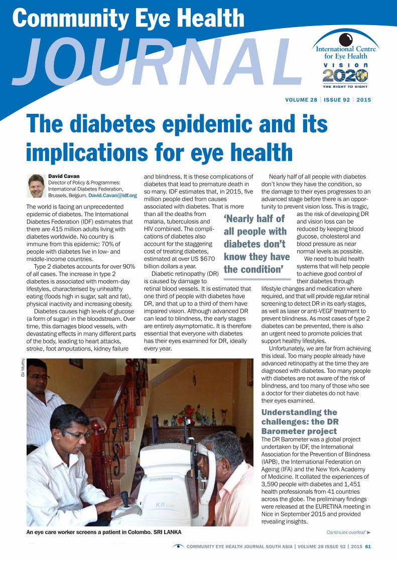
Diabetic retinopathy (DR) is caused by damage to retinal blood vessels. It is estimated that one third of people with diabetes have DR, and that up to a third of them have impaired vision. Although advanced DR can lead to blindness, the early stages are entirely asymptomatic. It is therefore essential that everyone with diabetes has their eyes examined for DR, ideally every year.
Nearly half of all people with diabetes don’t know they have the condition, so the damage to their eyes progresses to an advanced stage before there is an oppor-tunity to prevent vision loss. This is tragic,
as the risk of developing DR and vision loss can be reduced by keeping blood glucose, cholesterol and blood pressure as near normal levels as possible.
We need to build health systems that will help people to achieve good control of their diabetes through
lifestyle changes and medication where required, and that will provide regular retinal screening to detect DR in its early stages, as well as laser or anti-VEGF treatment to prevent blindness. As most cases of type 2 diabetes can be prevented, there is also an urgent need to promote policies that support healthy lifestyles.
Unfortunately, we are far from achieving this ideal. Too many people already have advanced retinopathy at the time they are diagnosed with diabetes. Too many people with diabetes are not aware of the risk of blindness, and too many of those who see a doctor for their diabetes do not have their eyes examined.
Understanding the challenges: the DR Barometer projectThe DR Barometer was a global project undertaken by IDF, the International Association for the Prevention of Blindness (IAPB), the International Federation on Ageing (IFA) and the New York Academy of Medicine. It collated the experiences of 3,590 people with diabetes and 1,451 health professionals from 41 countries across the globe. The preliminary findings were released at the EURETINA meeting in Nice in September 2015 and provided revealing insights.
The diabetes epidemic and its implications for eye health
Continues overleaf ➤
David CavanDirector of Policy & Programmes: International Diabetes Federation, Brussels, Belgium. [email protected]
GV
Mur
thy
An eye care worker screens a patient in Colombo. SRI LANKA
‘Nearly half of all people with diabetes don’t know they have the condition’
62 COMMUNITY EYE HEALTH JOURNAL SOUTH ASIA | VOLUME 28 ISSUE 92 | 2015
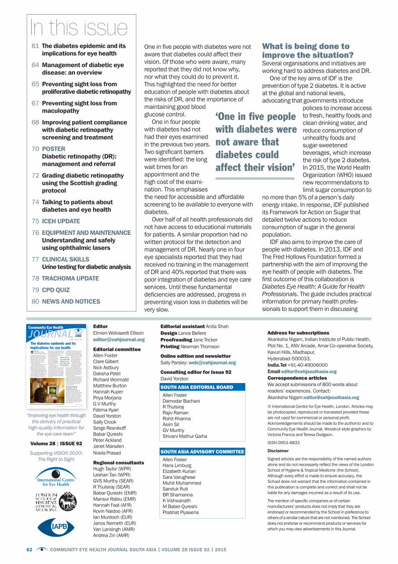
61 The diabetes epidemic and its implications for eye health
64 Management of diabetic eye disease: an overview
65 Preventing sight loss from proliferative diabetic retinopathy
67 Preventing sight loss from maculopathy
68 Improving patient compliance with diabetic retinopathy screening and treatment
70 POSTER Diabetic retinopathy (DR): management and referral
72 Grading diabetic retinopathy using the Scottish grading protocol
74 Talking to patients about diabetes and eye health
75 ICEH UPDATE
76 EQUIPMENT AND MAINTENANCE Understanding and safely using ophthalmic lasers
77 CLINICAL SKILLS Urine testing for diabetic analysis
78 TRACHOMA UPDATE
79 CPD QUIZ
80 NEWS AND NOTICES
“Improving eye health through the delivery of practical
high-quality information for the eye care team”
Volume 28 | ISSUE 92
Supporting VISION 2020: The Right to Sight
COMMUNITY EYE HEALTH JOURNAL | VOLUME 28 ISSUE 92 | 2015 61
Community Eye Health
JOURNALVOLUME 28 | ISSUE 92 | 2015
The world is facing an unprecedented epidemic of diabetes. The International Diabetes Federation (IDF) estimates that there are 415 million adults living with diabetes worldwide. No country is immune from this epidemic: 70% of people with diabetes live in low- and middle-income countries.
Type 2 diabetes accounts for over 90% of all cases. The increase in type 2 diabetes is associated with modern-day lifestyles, characterised by unhealthy eating (foods high in sugar, salt and fat), physical inactivity and increasing obesity.
Diabetes causes high levels of glucose (a form of sugar) in the bloodstream. Over time, this damages blood vessels, with devastating effects in many different parts of the body, leading to heart attacks, stroke, foot amputations, kidney failure
and blindness. It is these complications of diabetes that lead to premature death in so many. IDF estimates that, in 2015, five million people died from causes associated with diabetes. That is more than all the deaths from malaria, tuberculosis and HIV combined. The compli-cations of diabetes also account for the staggering cost of treating diabetes, estimated at over US $670 billion dollars a year.
Diabetic retinopathy (DR) is caused by damage to retinal blood vessels. It is estimated that one third of people with diabetes have DR, and that up to a third of them have impaired vision. Although advanced DR can lead to blindness, the early stages are entirely asymptomatic. It is therefore essential that everyone with diabetes has their eyes examined for DR, ideally every year.
Nearly half of all people with diabetes don’t know they have the condition, so the damage to their eyes progresses to an advanced stage before there is an oppor-tunity to prevent vision loss. This is tragic,
as the risk of developing DR and vision loss can be reduced by keeping blood glucose, cholesterol and blood pressure as near normal levels as possible.
We need to build health systems that will help people to achieve good control of their diabetes through
lifestyle changes and medication where required, and that will provide regular retinal screening to detect DR in its early stages, as well as laser or anti-VEGF treatment to prevent blindness. As most cases of type 2 diabetes can be prevented, there is also an urgent need to promote policies that support healthy lifestyles.
Unfortunately, we are far from achieving this ideal. Too many people already have advanced retinopathy at the time they are diagnosed with diabetes. Too many people with diabetes are not aware of the risk of blindness, and too many of those who see a doctor for their diabetes do not have their eyes examined.
Understanding the challenges: the DR Barometer projectThe DR Barometer was a global project undertaken by IDF, the International Association for the Prevention of Blindness (IAPB), the International Federation on Ageing (IFA) and the New York Academy of Medicine. It collated the experiences of 3,590 people with diabetes and 1,451 health professionals from 41 countries across the globe. The preliminary findings were released at the EURETINA meeting in Nice in September 2015 and provided revealing insights.
The diabetes epidemic and its implications for eye health
Continues overleaf ➤
David CavanDirector of Policy & Programmes: International Diabetes Federation, Brussels, Belgium. [email protected]
Pete
r Blo
ws
Nurse Baele Fidzani grades an image at Donga Diabetes Centre, Francistown. BOTSWANA
‘Nearly half of all people with diabetes don’t know they have the condition’
EditorElmien Wolvaardt Ellison [email protected]
Editorial committeeAllen FosterClare GilbertNick AstburyDaksha PatelRichard WormaldMatthew BurtonHannah KuperPriya MorjariaG V MurthyFatima KyariDavid YorstonSally CrookSerge ResnikoffBabar QureshiPeter AcklandJanet MarsdenNoela Prasad
Regional consultantsHugh Taylor (WPR) Leshan Tan (WPR)GVS Murthy (SEAR)R Thulsiraj (SEAR)Babar Qureshi (EMR)Mansur Rabiu (EMR)Hannah Faal (AFR)Kovin Naidoo (AFR) Ian Murdoch (EUR)Janos Nemeth (EUR)Van Lansingh (AMR)Andrea Zin (AMR)
One in five people with diabetes were not aware that diabetes could affect their vision. Of those who were aware, many reported that they did not know why, nor what they could do to prevent it. This highlighted the need for better education of people with diabetes about the risks of DR, and the importance of maintaining good blood glucose control.
One in four people with diabetes had not had their eyes examined in the previous two years. Two significant barriers were identified: the long wait times for an appointment and the high cost of the exami-nation. This emphasises the need for accessible and affordable screening to be available to everyone with diabetes.
Over half of all health professionals did not have access to educational materials for patients. A similar proportion had no written protocol for the detection and management of DR. Nearly one in four eye specialists reported that they had received no training in the management of DR and 40% reported that there was poor integration of diabetes and eye care services. Until these fundamental deficiencies are addressed, progress in preventing vision loss in diabetes will be very slow.
What is being done to improve the situation?Several organisations and initiatives are working hard to address diabetes and DR.
One of the key aims of IDF is the prevention of type 2 diabetes. It is active at the global and national levels, advocating that governments introduce
policies to increase access to fresh, healthy foods and clean drinking water, and reduce consumption of unhealthy foods and sugar-sweetened beverages, which increase the risk of type 2 diabetes. In 2015, the World Health Organization (WHO) issued new recommendations to limit sugar consumption to
no more than 5% of a person’s daily energy intake. In response, IDF published its Framework for Action on Sugar that detailed twelve actions to reduce consumption of sugar in the general population.
IDF also aims to improve the care of people with diabetes. In 2013, IDF and The Fred Hollows Foundation formed a partnership with the aim of improving the eye health of people with diabetes. The first outcome of this collaboration is Diabetes Eye Health: A Guide for Health Professionals. The guide includes practical information for primary health profes-sionals to support them in discussing
‘One in five people with diabetes were not aware that diabetes could affect their vision’
Editorial assistant Anita ShahDesign Lance BellersProofreading Jane TrickerPrinting Newman Thomson
Online edition and newsletter Sally Parsley: [email protected]
Consulting editor for Issue 92David Yorston
SOUTH ASIA EDITORIAL BOARD
Allen Foster Damodar Bachani R Thulsiraj Rajiv Raman Rohit Khanna Asim Sil GV Murthy Shivani Mathur Gaiha
SOUTH ASIA ADVISORY COMMITTEE
Allen Foster Hans Limburg Elizabeth Kurian Sara Varughese Muhit Muhammed Sanduk Ruit BR Shamanna K Vishwanath M Baber Qureshi Prabhat Piyasena
Address for subscriptionsAkanksha Nigam, Indian Institute of Public Health, Plot No. 1, ANV Arcade, Amar Co-operative Society, Kavuri Hills, Madhapur, Hyderabad-500033,India.Tel +91-40-49006000Email [email protected] Correspondence articlesWe accept submissions of 800 words about readers’ experiences. Contact: Akanksha Nigam:[email protected]
© International Centre for Eye Health, London. Articles may be photocopied, reproduced or translated provided these are not used for commercial or personal profit. Acknowledgements should be made to the author(s) and to Community Eye Health Journal. Woodcut-style graphics by Victoria Francis and Teresa Dodgson.
ISSN 0953-6833
Disclaimer
Signed articles are the responsibility of the named authors alone and do not necessarily reflect the views of the London School of Hygiene & Tropical Medicine (the School). Although every effort is made to ensure accuracy, the School does not warrant that the information contained in this publication is complete and correct and shall not be liable for any damages incurred as a result of its use.
The mention of specific companies or of certain manufacturers’ products does not imply that they are endorsed or recommended by the School in preference to others of a similar nature that are not mentioned. The School does not endorse or recommend products or services for which you may view advertisements in this Journal.
COMMUNITY EYE HEALTH JOURNAL SOUTH ASIA | VOLUME 28 ISSUE 92 | 2015 63© The author/s and Community Eye Health Journal 2015. This is an Open Access article distributed under the Creative Commons Attribution Non-Commercial License.
diabetes management for good eye health and detecting DR in people with diabetes. The guide will be published in the six UN languages (English, French, Spanish, Arabic, Chinese and Russian) and is available for download free of charge via the IDF website. It is hoped that this guide will help fill the gaps identified in the Barometer study and provide a framework to guide individual health professionals and local health systems in structuring screening and treatment services for DR. Further details of the Guide are presented in the panel below.
While some aspects of training in DR require hands-on instruction, much can be learnt online, by e-learning, and IDF is planning to develop interactive modules to make available the latest information on screening and treatment for DR. This will help health professionals to provide accurate information to people with diabetes from the moment they are diagnosed. IDF has recently launched the
first of a two-part introductory module for non-specialist health professionals, aimed at equipping everyone involved in the care of people with diabetes with the knowledge they need to provide basic lifestyle advice (see Useful Resources below).
The Queen Elizabeth Diamond Jubilee Trust (QEDJT), through the Commonwealth Eye Health Consortium, has enabled the formation of the Diabetic Retinopathy Network (DR-NET) in 10 Commonwealth countries, whereby existing VISION 2020 LINKS between UK and overseas eye departments share learning on DR screening and treatment. In addition, DR-NET facilitates improved coverage of fundus cameras and screening databases and also works with Ministries of Health to implement national frameworks for diabetic retinopathy.
Further initiatives are planned in 2016 to promote better screening for, and treatment of, DR. There are excellent examples, such as the UK Retinal Screening
programme, which demonstrate that effective population-based screening can be achieved. The challenge is to see this replicated elsewhere, in different national health systems and with different levels of resources. In order to build the evidence base for a structured approach to managing DR in low-resource settings, The Fred Hollows Foundation have partnered with the QEDJT and others to implement trials of models of care that integrate eye health into diabetes care in Pakistan, Bangladesh and the Pacific Islands.
What can eye health professionals do to improve the situation?Unless we act now to develop prevention, screening and treatment services for DR, we face the prospect of nearly 40 million people experiencing vision loss from diabetes, all of whom will require multiple review and treatment visits. This will be a significant burden on top of ophthalmolo-gists’ existing work load – even more so in low- and middle-income countries where there are very few of these specialists.
DR is preventable and blindness from DR is avoidable, but only if there is close collaboration between diabetes and eye health professionals at local, national and global level. In order to promote this, IDF and The Fred Hollows Foundation are exploring the creation of a global ‘DR Alliance’ to raise awareness of DR and to take the lead in recommending solutions to help address it.
In the meantime, we encourage all eye health professionals to improve the situation in their area in the following ways:
• Set an example by adopting a healthy lifestyle.
• Provide basic lifestyle advice to all patients, whether or not they have diabetes. This will help to prevent new cases of type 2 diabetes and help prevent DR in those with diabetes.
• Ensure patients with diabetes are being appropriately monitored by a primary care or specialist diabetes physician. This is especially important for those with DR.
• Build links with local diabetes professionals to develop reliable care pathways for patients with DR and set up a screening (fundus) camera in the diabetes clinic, so it is easily accessed by the target population.
Useful resources
IDF Diabetes Atlas. Available for download from http://www.diabetesatlas.org
IDF module: An introduction to diabetes. Register (free of charge) at http://d-net.idf.org/en/
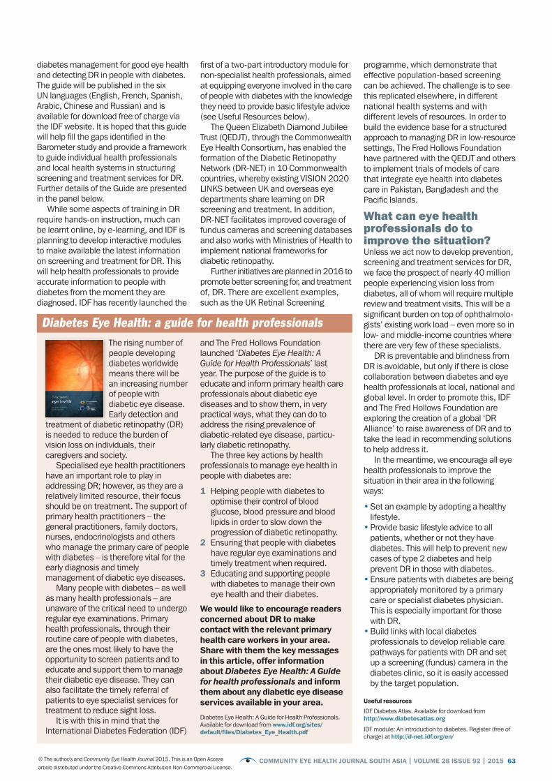
Diabetes Eye Health: a guide for health professionalsThe rising number of people developing diabetes worldwide means there will be an increasing number of people with diabetic eye disease. Early detection and
treatment of diabetic retinopathy (DR) is needed to reduce the burden of vision loss on individuals, their caregivers and society.
Specialised eye health practitioners have an important role to play in addressing DR; however, as they are a relatively limited resource, their focus should be on treatment. The support of primary health practitioners – the general practitioners, family doctors, nurses, endocrinologists and others who manage the primary care of people with diabetes – is therefore vital for the early diagnosis and timely management of diabetic eye diseases.
Many people with diabetes – as well as many health professionals – are unaware of the critical need to undergo regular eye examinations. Primary health professionals, through their routine care of people with diabetes, are the ones most likely to have the opportunity to screen patients and to educate and support them to manage their diabetic eye disease. They can also facilitate the timely referral of patients to eye specialist services for treatment to reduce sight loss.
It is with this in mind that the International Diabetes Federation (IDF)
and The Fred Hollows Foundation launched ‘Diabetes Eye Health: A Guide for Health Professionals’ last year. The purpose of the guide is to educate and inform primary health care professionals about diabetic eye diseases and to show them, in very practical ways, what they can do to address the rising prevalence of diabetic-related eye disease, particu-larly diabetic retinopathy.
The three key actions by health professionals to manage eye health in people with diabetes are:
1 Helping people with diabetes to optimise their control of blood glucose, blood pressure and blood lipids in order to slow down the progression of diabetic retinopathy.
2 Ensuring that people with diabetes have regular eye examinations and timely treatment when required.
3 Educating and supporting people with diabetes to manage their own eye health and their diabetes.
We would like to encourage readers concerned about DR to make contact with the relevant primary health care workers in your area. Share with them the key messages in this article, offer information about Diabetes Eye Health: A Guide for health professionals and inform them about any diabetic eye disease services available in your area.
Diabetes Eye Health: A Guide for Health Professionals. Available for download from www.idf.org/sites/default/files/Diabetes_Eye_Health.pdf
64 COMMUNITY EYE HEALTH JOURNAL SOUTH ASIA | VOLUME 28 ISSUE 92 | 2015
Systemic risk factorsIn order to reduce the risk of diabetic eye disease (both retinopathy and maculopathy) progressing and causing visual loss, it is important for all people with diabetes to maintain good overall health and good control over their diabetes. This is especially important for patients who already have diabetic retinopathy (DR) which is already affecting their vision, or is likely to damage it soon.
The two most important risk factors are high blood glucose (sugar) and high blood pressure.
High cholesterol and lipids also seem to be related to DR getting worse. Treatment of high cholesterol and lipids with statin medications, if available, reduces the risk of DR progressing. Maintenance of a healthy lifestyle overall will be beneficial for DR. This includes not smoking (or giving up) and getting regular exercise. People with diabetes should follow a healthy diet and avoid sugar and refined carbohydrates as much as possible.
Eye health professionals have a role in identifying patients at risk of sight loss from DR, and reinforcing messages about diabetes control and healthy living. Screening for DR and laser treatment for DR are both good opportunities for eye health professionals to get these messages across to patients.
Laser for DR and maculopathyLaser is the mainstay of treatment for both DR and maculopathy. Table 1 summarises the indications, desired response indicators, insufficient response indicators and side effects of both, based on the articles on preventing sight loss from DR and preventing sight loss from maculopathy, on pages 65 and 67 respectively. There is a glossary of terms on page 65 and the poster in the centre of this issue (pages 70–71) provides helpful background about the terms used.
MANAGEMENT
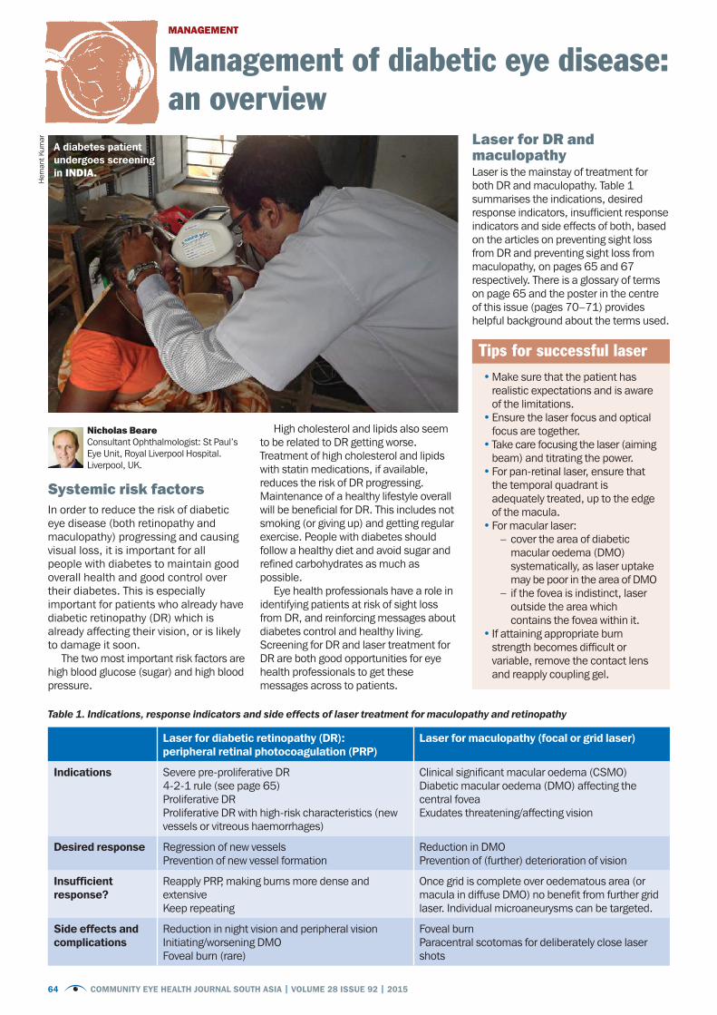
Management of diabetic eye disease: an overview
Nicholas BeareConsultant Ophthalmologist: St Paul’s Eye Unit, Royal Liverpool Hospital. Liverpool, UK.
Laser for diabetic retinopathy (DR): peripheral retinal photocoagulation (PRP)
Laser for maculopathy (focal or grid laser)
Indications Severe pre-proliferative DR4-2-1 rule (see page 65)Proliferative DR Proliferative DR with high-risk characteristics (new vessels or vitreous haemorrhages)
Clinical significant macular oedema (CSMO)Diabetic macular oedema (DMO) affecting the central foveaExudates threatening/affecting vision
Desired response Regression of new vesselsPrevention of new vessel formation
Reduction in DMOPrevention of (further) deterioration of vision
Insufficient response?
Reapply PRP, making burns more dense and extensiveKeep repeating
Once grid is complete over oedematous area (or macula in diffuse DMO) no benefit from further grid laser. Individual microaneurysms can be targeted.
Side effects and complications
Reduction in night vision and peripheral visionInitiating/worsening DMOFoveal burn (rare)
Foveal burnParacentral scotomas for deliberately close laser shots
Table 1. Indications, response indicators and side effects of laser treatment for maculopathy and retinopathy
• Make sure that the patient has realistic expectations and is aware of the limitations.
• Ensure the laser focus and optical focus are together.
• Take care focusing the laser (aiming beam) and titrating the power.
• For pan-retinal laser, ensure that the temporal quadrant is adequately treated, up to the edge of the macula.
• For macular laser: – cover the area of diabetic macular oedema (DMO) systematically, as laser uptake may be poor in the area of DMO
– if the fovea is indistinct, laser outside the area which contains the fovea within it.
• If attaining appropriate burn strength becomes difficult or variable, remove the contact lens and reapply coupling gel.
Tips for successful laser
Hem
ant K
umar
A diabetes patient undergoes screening in INDIA.
COMMUNITY EYE HEALTH JOURNAL SOUTH ASIA | VOLUME 28 ISSUE 92 | 2015 65© The author/s and Community Eye Health Journal 2015. This is an Open Access article distributed under the Creative Commons Attribution Non-Commercial License.
GlossaryClinically significant macular oedema (CSMO)CSMO is when leakage from small retinal blood vessels causes macular oedema (retinal swelling) and exudates (fat deposits from the blood) which are sufficiently close to the fovea (central macula) to affect or threaten the vision.
Diabetic maculopathyDiabetic maculopathy is part of diabetic retinopathy. Maculopathy is damage to the macula, the part of the eye respon-sible for central vision.
Diabetic macular oedema (DMO)Diabetic macular oedema occurs when blood vessels near to the macula leak fluid or protein onto the macula.
Diabetic retinopathy (DR)Diabetic retinopathy occurs when changes in blood glucose levels cause changes in retinal blood vessels. In some cases the vessels leak fluid into the macula part of the retina which swells up (DMO). In other cases, abnormal blood vessels will grow on the surface of the retina.
Peripheral retinal photocoagulation (PRP)Cauterisation of the peripheral retina using laser, with a minimum of 2,000 effective burns.
Proliferative diabetic retinopathyThis is the advanced stage of diabetic retinopathy. New blood vessels grow along the inside surface of the retina and into the vitreous gel, the fluid that fills the eye. These vessels are fragile and more likely to leak and bleed. Scar tissue is formed and can contract and cause retinal detachment (the pulling away of the retina from underlying tissue) – which results in blindness.
The Early Treatment in Diabetic Retinopathy Study (ETDRS)The Early Treatment in Diabetic Retinopathy study (ETDRS) has produced over 20 publications. They are listed at https://clinicaltrials.gov/ct2/show/study/NCT00000151
ETDRS and the preceding Diabetic Retinopathy Study are summarised in Chapters 1 and 2 of Clinical Trials in Ophthalmology, Eds PJ Kertes and MD Conway. Lippincott Williams and Wilkins 1998.
The keys to preventing sight loss from proliferative DR are as follows.
1 Identify the right patients to treat with peripheral retinal photocoagulation (PRP). PRP is destruction of the peripheral retina using laser, with a minimum of 2,000 effective burns.
2 In those with established new vessels, treat them with enough PRP and, if the response to PRP is insufficient, keep going with more and more.
When to do laser In patients with obvious new vessels at the disc (NVD) or elsewhere in the fundus (NVE), or if there is some vitreous haemorrhage associated with new vessels, it is a straightforward decision: treat them with PRP. (These are called ‘high-risk
characteristics’ because there is a high risk of visual loss in the ensuing years.)
There are also benefits to treating patients with less advanced DR. The Early Treatment in Diabetic Retinopathy Study (ETDRS) showed that PRP treatment can prevent these patients from progressing to the high-risk state. In a resource-poor setting where follow-up of patients may be haphazard, or patients are unable to attend for regular appointments, treating patients with less severe disease will prevent them getting worse. The threshold which ETDRS has established for laser has become known as the 4-2-1 rule (see panel below) and equates to severe pre-proliferative DR. Patients with this level of DR and above should be treated.
Preventing sight loss from proliferative diabetic retinopathy
Continues overleaf ➤
The 4-2-1 rule is:
• 4 quadrants of the fundus with dense retinal haemorrhages and microaneurysms; or • 2 quadrants with venous beading; or • 1 quadrant with intra-retinal microvascular abnormalities (IRMA).
These are all signs of retinal ischaemia, which is the stimulus for eventual new vessels – leading to tractional retinal detachment, vitreous haemorrhage and visual loss.
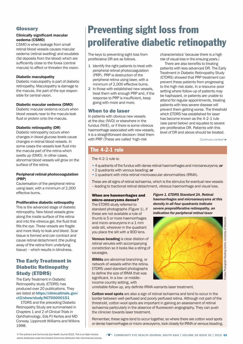
When are haemorrhages and micro-aneurysms dense? The ETDRS study referred to standard photographs (Figure 1), if these are not available a rule of thumb is 5 or more haemorrhages and micro-aneurysms in a 1 mm wide slit, wherever in the quadrant you place the slit with a 90D lens.
Venous beading is clear dilation of retinal venules with accompanying constriction so it looks like a string of sausages.
IRMAs are abnormal branching, or network of vessels within the retina. ETDRS used standard photographs to define the size of IRMA that was significant. In a low- or middle-income country setting, with unreliable follow up, any definite IRMA warrants laser treatment.
Cotton wool spots are also a sign of retinal ischaemia and tend to occur in the border between well-perfused and poorly perfused retina. Although not part of the threshold, cotton wool spots are important in gaining an assessment of retinal ischaemia particularly in the absence of fluorescein angiography. They can push the clinician towards laser treatment.
Remember, these signs tend to occur together, so where there are cotton wool spots or dense haemorrhages or micro-aneurysms, look closely for IRMA or venous beading.
The 4-2-1 rule
Figure 1. ETDRS Standard 2A. Retinal haemorrhages and microaneurysms at this density in all four quadrants indicate severe preproliferative retinopathy, an indication for peripheral retinal laser
Fund
us P
hoto
grap
h Re
adin
g Ce
ntre
, Uni
vers
ity o
f Wis
cons
in
66 COMMUNITY EYE HEALTH JOURNAL SOUTH ASIA | VOLUME 28 ISSUE 92 | 2015
Tips for successful laserIt is important that, before you start, the patient knows what to expect and what the aims of treatment and potential side effects are. In particular, you should stress that the laser treatment is to prevent visual loss in the future and is not intended to improve vision. At the start of the treatment, titrate the strength of the laser burn and adjust the laser power to achieve a visible burn which is not too harsh or bright white.
Remember: doubling the duration or power doubles the fluence (laser energy delivered per square millimetre), whereas halving the diameter increases the fluence by a factor of 4.
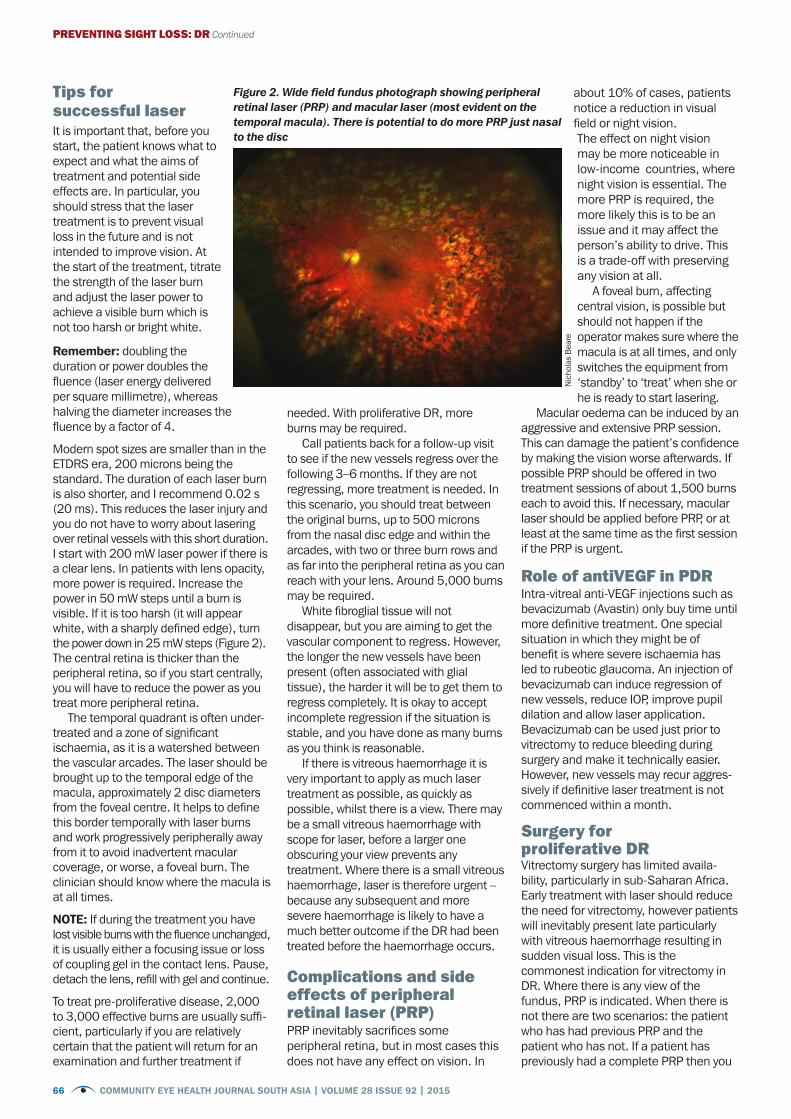
Modern spot sizes are smaller than in the ETDRS era, 200 microns being the standard. The duration of each laser burn is also shorter, and I recommend 0.02 s (20 ms). This reduces the laser injury and you do not have to worry about lasering over retinal vessels with this short duration. I start with 200 mW laser power if there is a clear lens. In patients with lens opacity, more power is required. Increase the power in 50 mW steps until a burn is visible. If it is too harsh (it will appear white, with a sharply defined edge), turn the power down in 25 mW steps (Figure 2). The central retina is thicker than the peripheral retina, so if you start centrally, you will have to reduce the power as you treat more peripheral retina.
The temporal quadrant is often under-treated and a zone of significant ischaemia, as it is a watershed between the vascular arcades. The laser should be brought up to the temporal edge of the macula, approximately 2 disc diameters from the foveal centre. It helps to define this border temporally with laser burns and work progressively peripherally away from it to avoid inadvertent macular coverage, or worse, a foveal burn. The clinician should know where the macula is at all times.
NOTE: If during the treatment you have lost visible burns with the fluence unchanged, it is usually either a focusing issue or loss of coupling gel in the contact lens. Pause, detach the lens, refill with gel and continue.
To treat pre-proliferative disease, 2,000 to 3,000 effective burns are usually suffi-cient, particularly if you are relatively certain that the patient will return for an examination and further treatment if
needed. With proliferative DR, more burns may be required.
Call patients back for a follow-up visit to see if the new vessels regress over the following 3 –6 months. If they are not regressing, more treatment is needed. In this scenario, you should treat between the original burns, up to 500 microns from the nasal disc edge and within the arcades, with two or three burn rows and as far into the peripheral retina as you can reach with your lens. Around 5,000 burns may be required.
White fibroglial tissue will not disappear, but you are aiming to get the vascular component to regress. However, the longer the new vessels have been present (often associated with glial tissue), the harder it will be to get them to regress completely. It is okay to accept incomplete regression if the situation is stable, and you have done as many burns as you think is reasonable.
If there is vitreous haemorrhage it is very important to apply as much laser treatment as possible, as quickly as possible, whilst there is a view. There may be a small vitreous haemorrhage with scope for laser, before a larger one obscuring your view prevents any treatment. Where there is a small vitreous haemorrhage, laser is therefore urgent – because any subsequent and more severe haemorrhage is likely to have a much better outcome if the DR had been treated before the haemorrhage occurs.
Complications and side effects of peripheral retinal laser (PRP)PRP inevitably sacrifices some peripheral retina, but in most cases this does not have any effect on vision. In
about 10% of cases, patients notice a reduction in visual field or night vision. The effect on night vision may be more noticeable in low-income countries, where night vision is essential. The more PRP is required, the more likely this is to be an issue and it may affect the person’s ability to drive. This is a trade-off with preserving any vision at all.
A foveal burn, affecting central vision, is possible but should not happen if the operator makes sure where the macula is at all times, and only switches the equipment from ‘standby’ to ‘treat’ when she or he is ready to start lasering.
Macular oedema can be induced by an aggressive and extensive PRP session. This can damage the patient’s confidence by making the vision worse afterwards. If possible PRP should be offered in two treatment sessions of about 1,500 burns each to avoid this. If necessary, macular laser should be applied before PRP, or at least at the same time as the first session if the PRP is urgent.
Role of antiVEGF in PDRIntra-vitreal anti-VEGF injections such as bevacizumab (Avastin) only buy time until more definitive treatment. One special situation in which they might be of benefit is where severe ischaemia has led to rubeotic glaucoma. An injection of bevacizumab can induce regression of new vessels, reduce IOP, improve pupil dilation and allow laser application. Bevacizumab can be used just prior to vitrectomy to reduce bleeding during surgery and make it technically easier. However, new vessels may recur aggres-sively if definitive laser treatment is not commenced within a month.
Surgery for proliferative DRVitrectomy surgery has limited availa-bility, particularly in sub-Saharan Africa. Early treatment with laser should reduce the need for vitrectomy, however patients will inevitably present late particularly with vitreous haemorrhage resulting in sudden visual loss. This is the commonest indication for vitrectomy in DR. Where there is any view of the fundus, PRP is indicated. When there is not there are two scenarios: the patient who has had previous PRP and the patient who has not. If a patient has previously had a complete PRP then you
PREVENTING SIGHT LOSS: DR Continued
Figure 2. Wide field fundus photograph showing peripheral retinal laser (PRP) and macular laser (most evident on the temporal macula). There is potential to do more PRP just nasal to the disc
Nic
hola
s B
eare
COMMUNITY EYE HEALTH JOURNAL SOUTH ASIA | VOLUME 28 ISSUE 92 | 2015 67© The author/s and Community Eye Health Journal 2015. This is an Open Access article distributed under the Creative Commons Attribution Non-Commercial License.
Preventing sight loss from maculopathyLaser treatment is less successful at reducing the risk of diabetic macular oedema (DMO) causing visual loss compared to peripheral retinal photocoagulation for proliferative diabetic retinopathy. Intravitreal treatments are effective but often unavailable due to cost and access to treatment.
When to do macular laserThe threshold for macular laser is usually clinically significant macular oedema (CSMO) as defined in the Early Treatment of Diabetic Retinopathy Study (ETDRS). CSMO is any retinal thickening (oedema) and/or exudates within 500 microns (⅓ disc diameter) of the centre of the fovea; or oedema greater than 1 disc area within 1 disc diameter of the foveal centre – including oedema which involves the fovea already. Macular laser is more effective if the DMO is localised (focal maculopathy), than if it is generalised across the central macula (diffuse maculopathy). Optical Coherence Tomography (OCT) scans enable visualisation of macular oedema in great detail, but are not required to determine whether a patient meets the thresholds for macular laser, because these were determined prior to the advent of OCT.
Exudates, if they involve the fovea, can sometimes threaten or affect vision without macular oedema. Exudates without oedema within 500 microns of the foveal centre, particularly long or streak exudates pointing towards the centre, are an indication for laser.
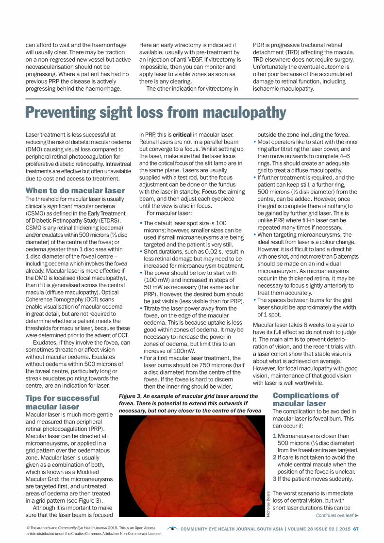
Tips for successful macular laserMacular laser is much more gentle and measured than peripheral retinal photocoagulation (PRP). Macular laser can be directed at microaneurysms, or applied in a grid pattern over the oedematous zone. Macular laser is usually given as a combination of both, which is known as a Modified Macular Grid: the microaneurysms are targeted first, and untreated areas of oedema are then treated in a grid pattern (see Figure 3).
Although it is important to make sure that the laser beam is focused
in PRP, this is critical in macular laser. Retinal lasers are not in a parallel beam but converge to a focus. Whilst setting up the laser, make sure that the laser focus and the optical focus of the slit lamp are in the same plane. Lasers are usually supplied with a test rod, but the focus adjustment can be done on the fundus with the laser in standby. Focus the aiming beam, and then adjust each eyepiece until the view is also in focus.
For macular laser:
• The default laser spot size is 100 microns; however, smaller sizes can be used if small microaneurysms are being targeted and the patient is very still.
• Short durations, such as 0.02 s, result in less retinal damage but may need to be increased for microaneurysm treatment.
• The power should be low to start with (100 mW) and increased in steps of 50 mW as necessary (the same as for PRP). However, the desired burn should be just visible (less visible than for PRP).
• Titrate the laser power away from the fovea, on the edge of the macular oedema. This is because uptake is less good within zones of oedema. It may be necessary to increase the power in zones of oedema, but limit this to an increase of 100mW.
• For a first macular laser treatment, the laser burns should be 750 microns (half a disc diameter) from the centre of the fovea. If the fovea is hard to discern then the inner ring should be wider,
outside the zone including the fovea.• Most operators like to start with the inner
ring after titrating the laser power, and then move outwards to complete 4–6 rings. This should create an adequate grid to treat a diffuse maculopathy.
• If further treatment is required, and the patient can keep still, a further ring, 500 microns (⅓ disk diameter) from the centre, can be added. However, once the grid is complete there is nothing to be gained by further grid laser. This is unlike PRP, where fill-in laser can be repeated many times if necessary.
• When targeting microaneurysms, the ideal result from laser is a colour change. However, it is difficult to land a direct hit with one shot, and not more than 5 attempts should be made on an individual microaneurysm. As microaneurysms occur in the thickened retina, it may be necessary to focus slightly anteriorly to treat them accurately.
• The spaces between burns for the grid laser should be approximately the width of 1 spot.
Macular laser takes 8 weeks to a year to have its full effect so do not rush to judge it. The main aim is to prevent deterio-ration of vision, and the recent trials with a laser cohort show that stable vision is about what is achieved on average. However, for focal maculopathy with good vision, maintenance of that good vision with laser is well worthwhile.
Complications of macular laserThe complication to be avoided in macular laser is foveal burn. This can occur if:
1 Microaneurysms closer than 500 microns (⅓ disc diameter) from the foveal centre are targeted.2 If care is not taken to avoid the whole central macula when the position of the fovea is unclear.3 If the patient moves suddenly.
The worst scenario is immediate loss of central vision, but with short laser durations this can be
Continues overleaf ➤
Figure 3. An example of macular grid laser around the fovea. There is potential to extend this outwards if necessary, but not any closer to the centre of the fovea
can afford to wait and the haemorrhage will usually clear. There may be traction on a non-regressed new vessel but active neovascularisation should not be progressing. Where a patient has had no previous PRP the disease is actively progressing behind the haemorrhage.
Here an early vitrectomy is indicated if available, usually with pre-treatment by an injection of anti-VEGF. If vitrectomy is impossible, then you can monitor and apply laser to visible zones as soon as there is any clearing.
The other indication for vitrectomy in
PDR is progressive tractional retinal detachment (TRD) affecting the macula. TRD elsewhere does not require surgery. Unfortunately the eventual outcome is often poor because of the accumulated damage to retinal function, including ischaemic maculopathy.
Nic
hola
s B
eare
68 COMMUNITY EYE HEALTH JOURNAL SOUTH ASIA | VOLUME 28 ISSUE 92 | 2015
Diabetic retinopathy is one of the many complications of diabetes. Because there are no symptoms initially, patients will not realise that they have the condition until it is at a proliferative stage or they develop macular oedema, when their vision becomes affected. Unfortunately, vision that has been lost may never be regained.
To prevent visual loss, early detection is needed at the pre-proliferative stage. This can only be achieved if the person with diabetes has regular (often annual) examination of the retina, starting from when they are first diagnosed. Screening of diabetes patients therefore has to be timely and in accordance with locally agreed guidelines for detection, referral and treatment. The challenge faced across many programmes is that people with diabetes:
• Do not always attend regular DR screening
• Present with late-stage retinopathy which results in a poor visual outcome
• Have poor acceptance of laser treatment.
In national population-based screening programmes, the desirable target uptake is 80%, which is difficult to achieve. The UK National Screening Programme took five years from the start of the programme in 2006 to reach this target. Attendance for initial laser treatment is reportedly around 70%, but in some studies as few as 21–45% of those patients who started laser treatment had completed the course of laser when they were followed up 6 months later.
Why do patients not attend?Reasons for non-attendance in various setting have been studied qualitatively and quantitatively and common themes arise.
Patient-related reasonsThese can be remembered using the first 7 letters of the alphabet.
• Awareness about diabetes and eye complications is often limited. Patients may not be aware of local screening centres
• Belief that they do not require retinal examinations or treatment as their vision is good, or they have a mild form of diabetes, or are too old
• Cost: direct and indirect (e.g. travel)• Distance from screening/treatment
centres and discomfort from dilating drops• Effort to attend yet another clinic.
People with diabetes often have multiple hospital appointments
• Fear of laser treatment and fear of its impact on quality of life and jobs. A lack of family support
• Guilt surrounding failure to control blood glucose levels. People fear that an eye examination, or being told they need laser treatment, will confirm their guilt and make them feel even worse.
Provider-related reasons• The existence of poor counselling and
advisory services about ocular complications for people with diabetes
• An inefficient system for getting patients to come, and to then come back if needed (‘call and recall’ systems)
• Long waiting times for screening or treatment
• Complicated referral mechanisms or inaccessible locations where services are offered.
Assessing the situationThe different factors in patients’ experience – which either prevent or encourage their engagement – should determine what interventions might improve uptake of services. By assessing the situation and identifying key stages in the process (from screening to completion of treatment) we can target interventions at those most at risk of vision loss.
The non-attendance rateThis is the proportion or percentage of patients who do not attend their appoint-ments, whether for their yearly eye examination or for laser treatment. We should aim to make this figure as low as possible.
At a clinic level, work out the non-attendance rate, say for 1 month, by dividing the number of patients who did not attend their appointment (for screening, the eye clinic or laser treatment) by the number of patients who have appoint-ments in that time period. Multiply by 100 to obtain the percentage.
CoverageCoverage is the percentage or proportion of the target population who undergo screening. In the case of diabetic eye disease, ‘screening’ means yearly eye
PATIENT COMPLIANCE
Improving patient compliance with diabetic retinopathy screening and treatment
Karinya LewisSpecialist Registrar, University Hospitals Southampton, Southampton, UK. [email protected]
MACULOPATHY Continued
mitigated. Sometimes patients are aware of paracentral scotomas from the laser burns if they are close to the foveal centre – it is therefore important to listen to what they have to say.
Role of intravitreal treatment in maculopathyRecent studies have shown that anti-VEGF treatment produces greater visual improvements than laser in patients with central diabetic macular oedema (DMO) whose vision is reduced to 6/12 or worse. These intravitreal injections reduce DMO rapidly and effectively. Laser treatment usually prevents loss of vision but does not often lead to visual improvement. Repeated injections of bevacizumab in eyes with visual acuity of less than 6/12 give an average improvement of two lines on the Snellen chart, and about a quarter of patients will improve by three lines. However, they have a number of problems – notably cost, the treatment burden of monthly injections, and the risk of infection/endophthalmitis. Even the relatively low cost of bevacizumab is prohibitive to many patients in low- and middle-income countries. Furthermore, a reliable pharmacy is required, one which can divide the intravenous dose into intravitreal doses in sterile condi-tions. Clusters of endophthalmitis cases in the US and UK have led to a suspicion of contaminated batches of anti-VEGF preparations. This is a definite concern in less well-regulated areas.
Evidence from clinical trials suggests that patients require 9–12 injections in the first year, so the treatment regime is intense – requiring frequent revisits in order to maximise the benefits. After the first year, the overall treatment burden is less, but some patients have recurring DMO requiring ongoing retreatments.
Despite these problems, the greater effectiveness of intravitreal injections means that they may be valuable for some patients, particularly those who can afford the drug costs and live sufficiently near to the clinic to attend for repeated treatment.
An alternative intravitreal treatment is steroid, the least costly being triamci-nolone. This is effective, but visual gains are reduced by induced cataract, and even after cataract surgery on average the vision does not catch up with that from anti-VEGF therapy. This may be due to exacerbated DMO or postoperative cystoid macular oedema. Intravitreal steroid is an option, especially in patients who are already pseudophakic. Post-injection intraocular pressure (IOP) rise can be a problem, so this needs to be monitored and treated accordingly.
COMMUNITY EYE HEALTH JOURNAL SOUTH ASIA | VOLUME 28 ISSUE 92 | 2015 69© The author/s and Community Eye Health Journal 2015. This is an Open Access article distributed under the Creative Commons Attribution Non-Commercial License.
PATIENT COMPLIANCE
examinations for everyone diagnosed with diabetes. Coverage is an important measure of the quality of a programme, and we should aim to make this figure as high as possible.
To work out the coverage offered by your clinic or programme, divide the number of patients who attended screening on a yearly basis by the number of patients with diabetes in your catchment population; multiply by 100 to calculate the yearly coverage of screening as a percentage.
The pathwayCan you identify which part of the pathway, from screening to treatment, is most affected by non-attendance?
Is there a particular geographic location where assessments or treatment take place, where non-attendance is higher?
Who is not coming?Among the diabetes patients, can you identify any particular subgroup who would benefit most from a targeted inter-vention? (For example, younger patients, those newly diagnosed, people with language barriers, or people with low social economic status or poor education.)
Addressing the challengesThe following practical suggestions are gathered from patient recommendations, models of good practice and successful interventions. Together, they improve the overall patient experience, improve ease of access to services, and encourage and engage the patient through education.
Empower health professionals• Encourage all allied health professionals
working with diabetes to personally recommend annual retinal checks to patients with diabetes. Train health workers to offer intensive patient education programmes to all newly diagnosed patients, covering specifically diabetes, potential blindness and the eye.
• Encourage health workers to support patients with diabetes (especially those with poor control) and work with them to find solutions to the challenges of having diabetes. It is vital not to blame the patient or make them feel guilty.
Strengthen patient communicationA diabetes ‘passport’ has been a useful tool to encourage patients to feel ownership of their disease and facilitate communication between health profes-sionals and the patient. The patient brings the passport (a specially designed booklet or file) to every appointment and health professionals record current medications and results (blood sugar, blood pressure, cholesterol, kidney function, podiatry assessment and retinopathy grading) as well as when next assessments are due. The passport helps to start conversations with the patient about their diabetes.
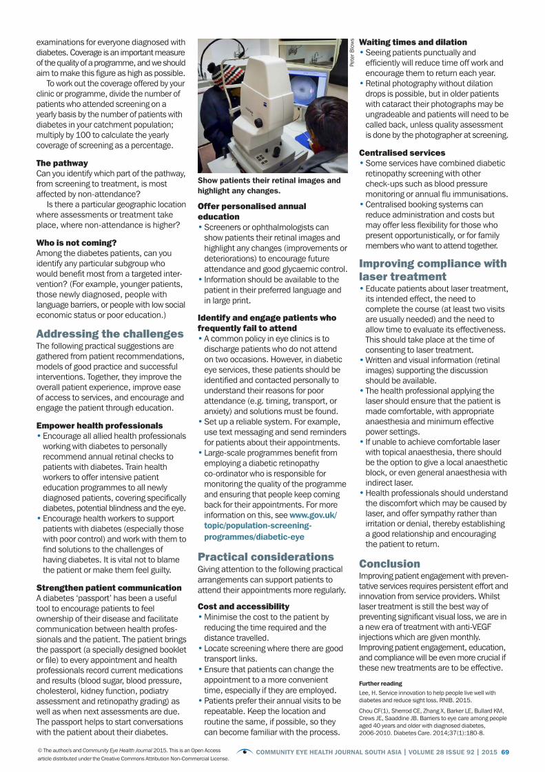
Offer personalised annual education• Screeners or ophthalmologists can
show patients their retinal images and highlight any changes (improvements or deteriorations) to encourage future attendance and good glycaemic control.
• Information should be available to the patient in their preferred language and in large print.
Identify and engage patients who frequently fail to attend• A common policy in eye clinics is to
discharge patients who do not attend on two occasions. However, in diabetic eye services, these patients should be identified and contacted personally to understand their reasons for poor attendance (e.g. timing, transport, or anxiety) and solutions must be found.
• Set up a reliable system. For example, use text messaging and send reminders for patients about their appointments.
• Large-scale programmes benefit from employing a diabetic retinopathy co-ordinator who is responsible for monitoring the quality of the programme and ensuring that people keep coming back for their appointments. For more information on this, see www.gov.uk/topic/population-screening-programmes/diabetic-eye
Practical considerationsGiving attention to the following practical arrangements can support patients to attend their appointments more regularly.
Cost and accessibility• Minimise the cost to the patient by
reducing the time required and the distance travelled.
• Locate screening where there are good transport links.
• Ensure that patients can change the appointment to a more convenient time, especially if they are employed.
• Patients prefer their annual visits to be repeatable. Keep the location and routine the same, if possible, so they can become familiar with the process.
Waiting times and dilation• Seeing patients punctually and
efficiently will reduce time off work and encourage them to return each year.
• Retinal photography without dilation drops is possible, but in older patients with cataract their photographs may be ungradeable and patients will need to be called back, unless quality assessment is done by the photographer at screening.
Centralised services• Some services have combined diabetic
retinopathy screening with other check-ups such as blood pressure monitoring or annual flu immunisations.
• Centralised booking systems can reduce administration and costs but may offer less flexibility for those who present opportunistically, or for family members who want to attend together.
Improving compliance with laser treatment• Educate patients about laser treatment,
its intended effect, the need to complete the course (at least two visits are usually needed) and the need to allow time to evaluate its effectiveness. This should take place at the time of consenting to laser treatment.
• Written and visual information (retinal images) supporting the discussion should be available.
• The health professional applying the laser should ensure that the patient is made comfortable, with appropriate anaesthesia and minimum effective power settings.
• If unable to achieve comfortable laser with topical anaesthesia, there should be the option to give a local anaesthetic block, or even general anaesthesia with indirect laser.
• Health professionals should understand the discomfort which may be caused by laser, and offer sympathy rather than irritation or denial, thereby establishing a good relationship and encouraging the patient to return.
ConclusionImproving patient engagement with preven-tative services requires persistent effort and innovation from service providers. Whilst laser treatment is still the best way of preventing significant visual loss, we are in a new era of treatment with anti-VEGF injections which are given monthly. Improving patient engagement, education, and compliance will be even more crucial if these new treatments are to be effective.
Further readingLee, H. Service innovation to help people live well with diabetes and reduce sight loss. RNIB. 2015.
Chou CF(1), Sherrod CE, Zhang X, Barker LE, Bullard KM, Crews JE, Saaddine JB. Barriers to eye care among people aged 40 years and older with diagnosed diabetes, 2006-2010. Diabetes Care. 2014;37(1):180-8.
Pete
r Blo
ws
Show patients their retinal images and highlight any changes.
70 COMMUNITY EYE HEALTH JOURNAL SOUTH ASIA | VOLUME 28 ISSUE 92 | 2015
Diab
etic
retin
opat
hy (D
R): m
anag
emen
t and
refe
rral
Dia
beti
c re
tino
path
y st
age
Clin
ical
sig
nsW
hat t
o do
(s
cree
ning
/pri
mar
y ey
e ca
re)
Wha
t to
do(r
etin
al c
linic
)W
hat y
ou c
ould
say
to y
our p
atie
nts
No
diab
etic
re
tino
path
yN
o ab
norm
aliti
esEn
cour
age
patie
nt to
com
e ag
ain
in
12 m
onth
sRe
view
in
12 m
onth
sD
iabe
tes
can
affe
ct th
e in
side
of y
our e
yes
at a
ny ti
me.
It is
impo
rtan
t th
at y
ou c
ome
back
in tw
elve
mon
ths
so w
e ca
n ex
amin
e yo
u ag
ain.
Th
is w
ill h
elp
to p
reve
nt y
ou lo
sing
vis
ion
or g
oing
blin
d.
Mild
non
- pr
olife
rati
ve
diab
etic
reti
nopa
thy
Mic
roan
eury
sms
only
Enco
urag
e pa
tient
to c
ome
agai
n in
12
mon
ths
Revi
ew in
12
mon
ths
Your
dia
bete
s is
affe
ctin
g yo
ur e
yes.
At t
he m
omen
t you
r vis
ion
is g
ood,
bu
t we
mus
t che
ck y
our e
yes
in 1
2 m
onth
s’ ti
me
to s
ee if
thes
e ch
ange
s ar
e ge
ttin
g w
orse
. If t
he d
amag
e be
com
es s
ever
e, w
e w
ill
need
to tr
eat y
our e
yes
to s
top
the
diab
etes
affe
ctin
g yo
ur s
ight
.
Mod
erat
e no
n-
prol
ifera
tive
di
abet
ic re
tino
path
y
Mor
e th
an ju
st m
icro
-an
eury
sms
but l
ess
than
se
vere
non
-pro
lifer
ativ
e re
tinop
athy
Enco
urag
e pa
tient
to c
ome
agai
n in
6–
12 m
onth
s Re
view
in
6–12
mon
ths
Your
dia
bete
s is
dam
agin
g yo
ur e
yes.
At t
he m
omen
t you
r vis
ion
is
good
, but
we
mus
t che
ck y
our e
yes
in s
ix m
onth
s’ ti
me
as it
is li
kely
th
at th
ese
chan
ges
will
get
wor
se. I
f the
dam
age
beco
mes
sev
ere,
we
will
nee
d to
trea
t you
r eye
s to
sto
p th
e di
abet
es a
ffect
ing
your
sig
ht.
Unl
ess
you
are
trea
ted
prom
ptly,
you
risk
losi
ng v
isio
n or
goi
ng b
lind.
Sev
ere
non-
pr
olife
rati
ve
diab
etic
reti
nopa
thy
Mor
e th
an 2
0 ha
emor
rhag
es
in e
ach
quad
rant
; or v
enou
s be
adin
g in
two
quad
rant
s; o
r in
trar
etin
al m
icro
vasc
ular
ab
norm
aliti
es (I
RMA)
Refe
r to
retin
al c
linic
. All
patie
nts
with
sev
ere
non-
prol
ifera
tive
DR
sh
ould
be
in th
e ca
re o
f an
opht
halm
olog
ist.
The
patie
nt s
houl
d be
re-e
xam
ined
eve
ry s
ix m
onth
s
Perfo
rm p
erip
hera
l re
tinal
pho
toco
agul
-at
ion
if fo
llow
-up
is
unre
liabl
e; o
ther
wis
e re
view
in 6
mon
ths
Your
dia
bete
s ha
s da
mag
ed y
our e
yes
quite
sev
erel
y, a
lthou
gh y
our
visi
on is
stil
l goo
d. Y
ou a
re li
kely
to n
eed
trea
tmen
t soo
n to
ens
ure
that
yo
u do
n’t l
ose
visi
on o
r go
blin
d. W
e m
ust c
heck
you
r eye
s in
six
mon
ths’
tim
e. H
owev
er, i
f you
thin
k yo
u m
ay n
ot b
e ab
le to
com
e th
en, w
e m
ay
trea
t you
r eye
s no
w, s
o w
e ca
n be
sur
e yo
u do
n’t l
ose
visi
on la
ter.
Pro
lifer
ativ
e di
abet
ic re
tino
path
yAn
y ne
w v
esse
ls a
t the
dis
c or
els
ewhe
re, v
itreo
us/
pre-
retin
al h
aem
orrh
age
Urg
ent r
efer
ral t
o re
tinal
clin
icPe
riphe
ral r
etin
al
phot
ocoa
gula
tion
or
vitr
ecto
my
if th
ere
is
vitr
eous
hae
mor
rhag
e or
retin
al d
etac
hmen
t
Your
dia
bete
s ha
s da
mag
ed y
our e
yes
very
sev
erel
y. A
lthou
gh y
our
visi
on m
ay b
e go
od, y
ou a
re a
t gre
at ri
sk o
f los
ing
your
sig
ht o
ver t
he
next
yea
r. Yo
u ne
ed u
rgen
t tre
atm
ent t
o sa
ve y
our s
ight
. Tre
atm
ent w
ill
not i
mpr
ove
your
eye
sigh
t, bu
t sho
uld
pres
erve
the
visi
on y
ou h
ave.
Mac
ular
oed
ema
Mac
ular
oed
ema
abse
ntN
o ex
udat
es o
r ret
inal
th
icke
ning
in p
oste
rior p
ole
Rev
iew
in 1
2 m
onth
sRe
view
in
12 m
onth
sAs
for “
No
diab
etic
retin
opat
hy”
abov
e.
Mild
mac
ular
oe
dem
aEx
udat
es o
r ret
inal
th
icke
ning
at p
oste
rior
pole
, >1d
d fro
m fo
vea
Rev
iew
in 6
mon
ths
Revi
ew in
6
mon
ths
Your
dia
bete
s is
dam
agin
g yo
ur e
yes.
At t
he m
omen
t you
r vis
ion
is
good
, but
we
mus
t che
ck y
our e
yes
in s
ix m
onth
s’ ti
me
as it
is li
kely
th
at th
ese
chan
ges
will
get
wor
se. I
f the
dam
age
beco
mes
sev
ere,
w
e w
ill n
eed
to tr
eat y
our e
yes
to s
top
the
diab
etes
affe
ctin
g yo
ur
sigh
t. U
nles
s yo
u ar
e tr
eate
d pr
ompt
ly, y
ou ri
sk lo
sing
vis
ion
or
goin
g bl
ind.
Mod
erat
e m
acul
ar
oede
ma
Exud
ates
or r
etin
al
thic
keni
ng a
t pos
terio
r po
le, 1
dd o
r les
s fro
m
fove
a, b
ut n
ot a
ffect
ing
fove
a
Ref
er to
retin
al c
linic
. Enc
oura
ge
patie
nt to
man
age
thei
r blo
od s
ugar
an
d bl
ood
pres
sure
, and
refe
r the
m
to a
vaila
ble
serv
ices
for h
elp
if th
ey
are
not s
ure
how
to d
o th
is
Lase
r tre
atm
ent i
f cl
inic
ally
sig
nific
ant
mac
ular
oed
ema
(CSM
O).
Rev
iew
in 6
m
onth
s if
no C
SMO
Your
dia
bete
s ha
s da
mag
ed y
our e
yes
seve
rely.
Alth
ough
you
r vis
ion
may
be
good
at p
rese
nt, i
t is
likel
y to
get
wor
se o
ver t
he n
ext y
ear o
r tw
o. Y
ou n
eed
lase
r tre
atm
ent t
o st
op y
our s
ight
det
erio
ratin
g. T
he
trea
tmen
t will
not
impr
ove
your
eye
sigh
t, bu
t sho
uld
pres
erve
the
visi
on
you
have
.
Sev
ere
mac
ular
oe
dem
aEx
udat
es o
r ret
inal
th
icke
ning
affe
ctin
g ce
ntre
of
fove
a
Ref
er to
retin
al c
linic
Lase
r tre
atm
ent o
r in
trav
itrea
l inj
ectio
ns o
f an
ti-VE
GF
drug
s
You
have
pro
babl
y no
ticed
you
r eye
sigh
t has
got
wor
se. T
his
is b
ecau
se
your
dia
bete
s ha
s da
mag
ed y
our e
yes
very
sev
erel
y. Y
ou n
eed
urge
nt
trea
tmen
t to
prev
ent f
urth
er lo
ss o
f vis
ion.
The
trea
tmen
t may
not
im
prov
e yo
ur e
yesi
ght,
but i
f you
are
not
trea
ted,
you
r vis
ion
will
get
w
orse
and
you
may
eve
n be
com
e bl
ind.
If y
ou c
anno
t see
the
reti
na d
ue to
cat
arac
t or v
itre
ous
hae
mor
rhag
e, re
fer t
o an
oph
thal
mol
ogis
t for
cat
arac
t sur
gery
or a
reti
nal s
urge
on fo
r vit
rect
omy.
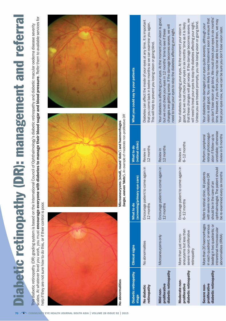
This
dia
betic
retin
opat
hy (D
R) g
radi
ng s
yste
m is
bas
ed o
n th
e In
tern
atio
nal C
ounc
il of
Oph
thal
mol
ogy’
s di
abet
ic re
tinop
athy
and
dia
betic
mac
ular
oed
ema
dise
ase
seve
rity
scal
es. A
t wha
teve
r lev
el y
ou w
ork,
you
mus
t enc
oura
ge e
very
one
wit
h di
abet
es to
man
age
thei
r blo
od s
ugar
and
blo
od p
ress
ure.
Ref
er th
em to
ava
ilabl
e se
rvic
es fo
r he
lp if
they
are
not
sur
e ho
w to
do
this
, or i
f the
ir co
ntro
l is
poor
.
No
abno
rmal
itie
sM
icro
aneu
rysm
s (s
mal
l, ro
und
‘dot
s’) a
nd h
aem
orrh
ages
(la
rger
, une
ven
‘blo
ts’).
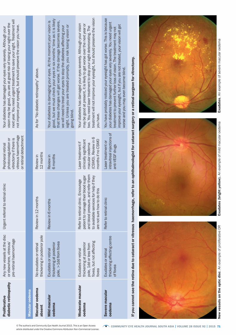
An
exam
ple
of m
oder
ate
non-
prol
ifera
tive
DR
70 COMMUNITY EYE HEALTH JOURNAL SOUTH ASIA | VOLUME 28 ISSUE 92 | 2015
COMMUNITY EYE HEALTH JOURNAL SOUTH ASIA | VOLUME 28 ISSUE 92 | 2015 71© The author/s and Community Eye Health Journal 2015. This is an Open Access article distributed under the Creative Commons Attribution Non-Commercial License.
Dia
beti
c re
tino
path
y st
age
Clin
ical
sig
nsW
hat t
o do
(s
cree
ning
/pri
mar
y ey
e ca
re)
Wha
t to
do(r
etin
al c
linic
)W
hat y
ou c
ould
say
to y
our p
atie
nts
No
diab
etic
re
tino
path
yN
o ab
norm
aliti
esEn
cour
age
patie
nt to
com
e ag
ain
in
12 m
onth
sRe
view
in
12 m
onth
sD
iabe
tes
can
affe
ct th
e in
side
of y
our e
yes
at a
ny ti
me.
It is
impo
rtan
t th
at y
ou c
ome
back
in tw
elve
mon
ths
so w
e ca
n ex
amin
e yo
u ag
ain.
Th
is w
ill h
elp
to p
reve
nt y
ou lo
sing
vis
ion
or g
oing
blin
d.
Mild
non
- pr
olife
rati
ve
diab
etic
reti
nopa
thy
Mic
roan
eury
sms
only
Enco
urag
e pa
tient
to c
ome
agai
n in
12
mon
ths
Revi
ew in
12
mon
ths
Your
dia
bete
s is
affe
ctin
g yo
ur e
yes.
At t
he m
omen
t you
r vis
ion
is g
ood,
bu
t we
mus
t che
ck y
our e
yes
in 1
2 m
onth
s’ ti
me
to s
ee if
thes
e ch
ange
s ar
e ge
ttin
g w
orse
. If t
he d
amag
e be
com
es s
ever
e, w
e w
ill
need
to tr
eat y
our e
yes
to s
top
the
diab
etes
affe
ctin
g yo
ur s
ight
.
Mod
erat
e no
n-
prol
ifera
tive
di
abet
ic re
tino
path
y
Mor
e th
an ju
st m
icro
-an
eury
sms
but l
ess
than
se
vere
non
-pro
lifer
ativ
e re
tinop
athy
Enco
urag
e pa
tient
to c
ome
agai
n in
6–
12 m
onth
s Re
view
in
6–12
mon
ths
Your
dia
bete
s is
dam
agin
g yo
ur e
yes.
At t
he m
omen
t you
r vis
ion
is
good
, but
we
mus
t che
ck y
our e
yes
in s
ix m
onth
s’ ti
me
as it
is li
kely
th
at th
ese
chan
ges
will
get
wor
se. I
f the
dam
age
beco
mes
sev
ere,
we
will
nee
d to
trea
t you
r eye
s to
sto
p th
e di
abet
es a
ffect
ing
your
sig
ht.
Unl
ess
you
are
trea
ted
prom
ptly,
you
risk
losi
ng v
isio
n or
goi
ng b
lind.
Sev
ere
non-
pr
olife
rati
ve
diab
etic
reti
nopa
thy
Mor
e th
an 2
0 ha
emor
rhag
es
in e
ach
quad
rant
; or v
enou
s be
adin
g in
two
quad
rant
s; o
r in
trar
etin
al m
icro
vasc
ular
ab
norm
aliti
es (I
RMA)
Refe
r to
retin
al c
linic
. All
patie
nts
with
sev
ere
non-
prol
ifera
tive
DR
sh
ould
be
in th
e ca
re o
f an
opht
halm
olog
ist.
The
patie
nt s
houl
d be
re-e
xam
ined
eve
ry s
ix m
onth
s
Perfo
rm p
erip
hera
l re
tinal
pho
toco
agul
-at
ion
if fo
llow
-up
is
unre
liabl
e; o
ther
wis
e re
view
in 6
mon
ths
Your
dia
bete
s ha
s da
mag
ed y
our e
yes
quite
sev
erel
y, a
lthou
gh y
our
visi
on is
stil
l goo
d. Y
ou a
re li
kely
to n
eed
trea
tmen
t soo
n to
ens
ure
that
yo
u do
n’t l
ose
visi
on o
r go
blin
d. W
e m
ust c
heck
you
r eye
s in
six
mon
ths’
tim
e. H
owev
er, i
f you
thin
k yo
u m
ay n
ot b
e ab
le to
com
e th
en, w
e m
ay
trea
t you
r eye
s no
w, s
o w
e ca
n be
sur
e yo
u do
n’t l
ose
visi
on la
ter.
Pro
lifer
ativ
e di
abet
ic re
tino
path
yAn
y ne
w v
esse
ls a
t the
dis
c or
els
ewhe
re, v
itreo
us/
pre-
retin
al h
aem
orrh
age
Urg
ent r
efer
ral t
o re
tinal
clin
icPe
riphe
ral r
etin
al
phot
ocoa
gula
tion
or
vitr
ecto
my
if th
ere
is
vitr
eous
hae
mor
rhag
e or
retin
al d
etac
hmen
t
Your
dia
bete
s ha
s da
mag
ed y
our e
yes
very
sev
erel
y. A
lthou
gh y
our
visi
on m
ay b
e go
od, y
ou a
re a
t gre
at ri
sk o
f los
ing
your
sig
ht o
ver t
he
next
yea
r. Yo
u ne
ed u
rgen
t tre
atm
ent t
o sa
ve y
our s
ight
. Tre
atm
ent w
ill
not i
mpr
ove
your
eye
sigh
t, bu
t sho
uld
pres
erve
the
visi
on y
ou h
ave.
Mac
ular
oed
ema
Mac
ular
oed
ema
abse
ntN
o ex
udat
es o
r ret
inal
th
icke
ning
in p
oste
rior p
ole
Rev
iew
in 1
2 m
onth
sRe
view
in
12 m
onth
sAs
for “
No
diab
etic
retin
opat
hy”
abov
e.
Mild
mac
ular
oe
dem
aEx
udat
es o
r ret
inal
th
icke
ning
at p
oste
rior
pole
, >1d
d fro
m fo
vea
Rev
iew
in 6
mon
ths
Revi
ew in
6
mon
ths
Your
dia
bete
s is
dam
agin
g yo
ur e
yes.
At t
he m
omen
t you
r vis
ion
is
good
, but
we
mus
t che
ck y
our e
yes
in s
ix m
onth
s’ ti
me
as it
is li
kely
th
at th
ese
chan
ges
will
get
wor
se. I
f the
dam
age
beco
mes
sev
ere,
w
e w
ill n
eed
to tr
eat y
our e
yes
to s
top
the
diab
etes
affe
ctin
g yo
ur
sigh
t. U
nles
s yo
u ar
e tr
eate
d pr
ompt
ly, y
ou ri
sk lo
sing
vis
ion
or
goin
g bl
ind.
Mod
erat
e m
acul
ar
oede
ma
Exud
ates
or r
etin
al
thic
keni
ng a
t pos
terio
r po
le, 1
dd o
r les
s fro
m
fove
a, b
ut n
ot a
ffect
ing
fove
a
Ref
er to
retin
al c
linic
. Enc
oura
ge
patie
nt to
man
age
thei
r blo
od s
ugar
an
d bl
ood
pres
sure
, and
refe
r the
m
to a
vaila
ble
serv
ices
for h
elp
if th
ey
are
not s
ure
how
to d
o th
is
Lase
r tre
atm
ent i
f cl
inic
ally
sig
nific
ant
mac
ular
oed
ema
(CSM
O).
Rev
iew
in 6
m
onth
s if
no C
SMO
Your
dia
bete
s ha
s da
mag
ed y
our e
yes
seve
rely.
Alth
ough
you
r vis
ion
may
be
good
at p
rese
nt, i
t is
likel
y to
get
wor
se o
ver t
he n
ext y
ear o
r tw
o. Y
ou n
eed
lase
r tre
atm
ent t
o st
op y
our s
ight
det
erio
ratin
g. T
he
trea
tmen
t will
not
impr
ove
your
eye
sigh
t, bu
t sho
uld
pres
erve
the
visi
on
you
have
.
Sev
ere
mac
ular
oe
dem
aEx
udat
es o
r ret
inal
th
icke
ning
affe
ctin
g ce
ntre
of
fove
a
Ref
er to
retin
al c
linic
Lase
r tre
atm
ent o
r in
trav
itrea
l inj
ectio
ns o
f an
ti-VE
GF
drug
s
You
have
pro
babl
y no
ticed
you
r eye
sigh
t has
got
wor
se. T
his
is b
ecau
se
your
dia
bete
s ha
s da
mag
ed y
our e
yes
very
sev
erel
y. Y
ou n
eed
urge
nt
trea
tmen
t to
prev
ent f
urth
er lo
ss o
f vis
ion.
The
trea
tmen
t may
not
im
prov
e yo
ur e
yesi
ght,
but i
f you
are
not
trea
ted,
you
r vis
ion
will
get
w
orse
and
you
may
eve
n be
com
e bl
ind.
If y
ou c
anno
t see
the
reti
na d
ue to
cat
arac
t or v
itre
ous
hae
mor
rhag
e, re
fer t
o an
oph
thal
mol
ogis
t for
cat
arac
t sur
gery
or a
reti
nal s
urge
on fo
r vit
rect
omy.
New
ves
sels
on
the
opti
c di
sc. A
n ex
ampl
e of
pro
lifer
ativ
e D
REx
udat
es (b
righ
t ye
llow
). A
n ex
ampl
e of
mild
mac
ular
oed
ema
Exud
ates
. An
exam
ple
of s
ever
e m
acul
ar o
edem
a
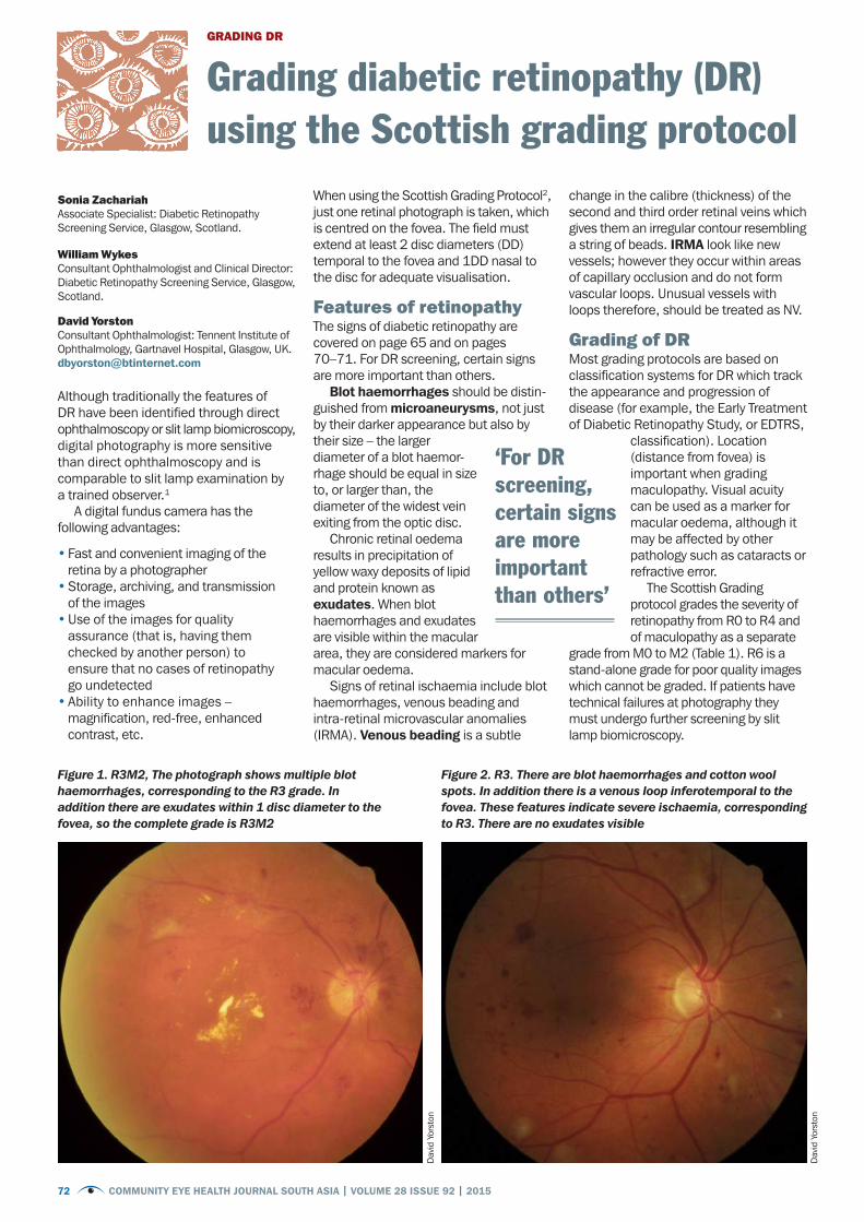
72 COMMUNITY EYE HEALTH JOURNAL SOUTH ASIA | VOLUME 28 ISSUE 92 | 2015
Although traditionally the features of DR have been identified through direct ophthalmoscopy or slit lamp biomicroscopy, digital photography is more sensitive than direct ophthalmoscopy and is comparable to slit lamp examination by a trained observer.1
A digital fundus camera has the following advantages:
• Fast and convenient imaging of the retina by a photographer
• Storage, archiving, and transmission of the images
• Use of the images for quality assurance (that is, having them checked by another person) to ensure that no cases of retinopathy go undetected
• Ability to enhance images – magnification, red-free, enhanced contrast, etc.
When using the Scottish Grading Protocol2, just one retinal photograph is taken, which is centred on the fovea. The field must extend at least 2 disc diameters (DD) temporal to the fovea and 1DD nasal to the disc for adequate visualisation.
Features of retinopathyThe signs of diabetic retinopathy are covered on page 65 and on pages 70–71. For DR screening, certain signs are more important than others.
Blot haemorrhages should be distin-guished from microaneurysms, not just by their darker appearance but also by their size – the larger diameter of a blot haemor-rhage should be equal in size to, or larger than, the diameter of the widest vein exiting from the optic disc.
Chronic retinal oedema results in precipitation of yellow waxy deposits of lipid and protein known as exudates. When blot haemorrhages and exudates are visible within the macular area, they are considered markers for macular oedema.
Signs of retinal ischaemia include blot haemorrhages, venous beading and intra-retinal microvascular anomalies (IRMA). Venous beading is a subtle
change in the calibre (thickness) of the second and third order retinal veins which gives them an irregular contour resembling a string of beads. IRMA look like new vessels; however they occur within areas of capillary occlusion and do not form vascular loops. Unusual vessels with loops therefore, should be treated as NV.
Grading of DR Most grading protocols are based on classification systems for DR which track the appearance and progression of disease (for example, the Early Treatment of Diabetic Retinopathy Study, or EDTRS,
classification). Location (distance from fovea) is important when grading maculopathy. Visual acuity can be used as a marker for macular oedema, although it may be affected by other pathology such as cataracts or refractive error.
The Scottish Grading protocol grades the severity of retinopathy from R0 to R4 and of maculopathy as a separate
grade from M0 to M2 (Table 1). R6 is a stand-alone grade for poor quality images which cannot be graded. If patients have technical failures at photography they must undergo further screening by slit lamp biomicroscopy.
GRADING DR
Grading diabetic retinopathy (DR) using the Scottish grading protocol
Sonia ZachariahAssociate Specialist: Diabetic Retinopathy Screening Service, Glasgow, Scotland.
William WykesConsultant Ophthalmologist and Clinical Director: Diabetic Retinopathy Screening Service, Glasgow, Scotland.
David YorstonConsultant Ophthalmologist: Tennent Institute of Ophthalmology, Gartnavel Hospital, Glasgow, UK. [email protected]
Dav
id Y
orst
on
Dav
id Y
orst
on
Figure 1. R3M2, The photograph shows multiple blot haemorrhages, corresponding to the R3 grade. In addition there are exudates within 1 disc diameter to the fovea, so the complete grade is R3M2
Figure 2. R3. There are blot haemorrhages and cotton wool spots. In addition there is a venous loop inferotemporal to the fovea. These features indicate severe ischaemia, corresponding to R3. There are no exudates visible
‘For DR screening, certain signs are more important than others’
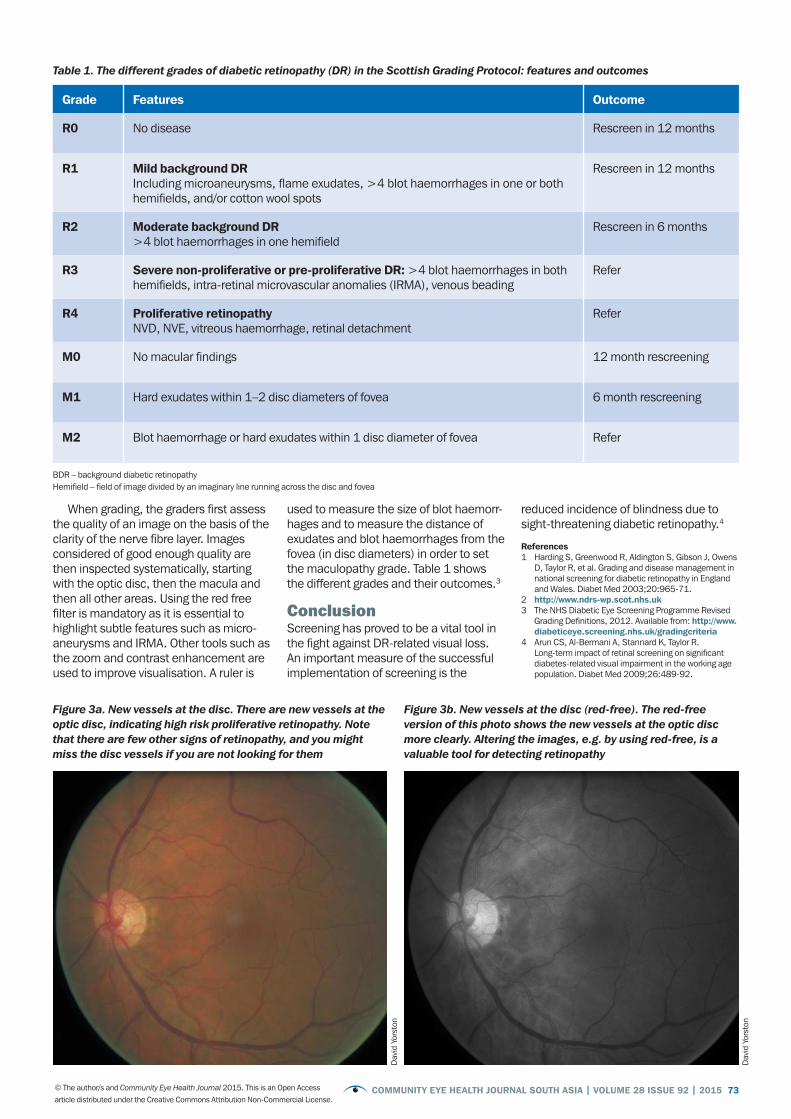
COMMUNITY EYE HEALTH JOURNAL SOUTH ASIA | VOLUME 28 ISSUE 92 | 2015 73© The author/s and Community Eye Health Journal 2015. This is an Open Access article distributed under the Creative Commons Attribution Non-Commercial License.
Grade Features Outcome
R0 No disease Rescreen in 12 months
R1 Mild background DR Including microaneurysms, flame exudates, >4 blot haemorrhages in one or both hemifields, and/or cotton wool spots
Rescreen in 12 months
R2 Moderate background DR >4 blot haemorrhages in one hemifield
Rescreen in 6 months
R3 Severe non-proliferative or pre-proliferative DR: >4 blot haemorrhages in both hemifields, intra-retinal microvascular anomalies (IRMA), venous beading
Refer
R4 Proliferative retinopathyNVD, NVE, vitreous haemorrhage, retinal detachment
Refer
M0 No macular findings 12 month rescreening
M1 Hard exudates within 1–2 disc diameters of fovea 6 month rescreening
M2 Blot haemorrhage or hard exudates within 1 disc diameter of fovea Refer
BDR – background diabetic retinopathyHemifield – field of image divided by an imaginary line running across the disc and fovea
Dav
id Y
orst
on
Dav
id Y
orst
on
Table 1. The different grades of diabetic retinopathy (DR) in the Scottish Grading Protocol: features and outcomes
Figure 3a. New vessels at the disc. There are new vessels at the optic disc, indicating high risk proliferative retinopathy. Note that there are few other signs of retinopathy, and you might miss the disc vessels if you are not looking for them
Figure 3b. New vessels at the disc (red-free). The red-free version of this photo shows the new vessels at the optic disc more clearly. Altering the images, e.g. by using red-free, is a valuable tool for detecting retinopathy
When grading, the graders first assess the quality of an image on the basis of the clarity of the nerve fibre layer. Images considered of good enough quality are then inspected systematically, starting with the optic disc, then the macula and then all other areas. Using the red free filter is mandatory as it is essential to highlight subtle features such as micro-aneurysms and IRMA. Other tools such as the zoom and contrast enhancement are used to improve visualisation. A ruler is
used to measure the size of blot haemorr-hages and to measure the distance of exudates and blot haemorrhages from the fovea (in disc diameters) in order to set the maculopathy grade. Table 1 shows the different grades and their outcomes.3
ConclusionScreening has proved to be a vital tool in the fight against DR-related visual loss. An important measure of the successful implementation of screening is the
reduced incidence of blindness due to sight-threatening diabetic retinopathy.4 References1 Harding S, Greenwood R, Aldington S, Gibson J, Owens
D, Taylor R, et al. Grading and disease management in national screening for diabetic retinopathy in England and Wales. Diabet Med 2003;20:965-71.
2 http://www.ndrs-wp.scot.nhs.uk 3 The NHS Diabetic Eye Screening Programme Revised
Grading Definitions, 2012. Available from: http://www.diabeticeye.screening.nhs.uk/gradingcriteria
4 Arun CS, Al-Bermani A, Stannard K, Taylor R. Long-term impact of retinal screening on significant diabetes-related visual impairment in the working age population. Diabet Med 2009;26:489-92.
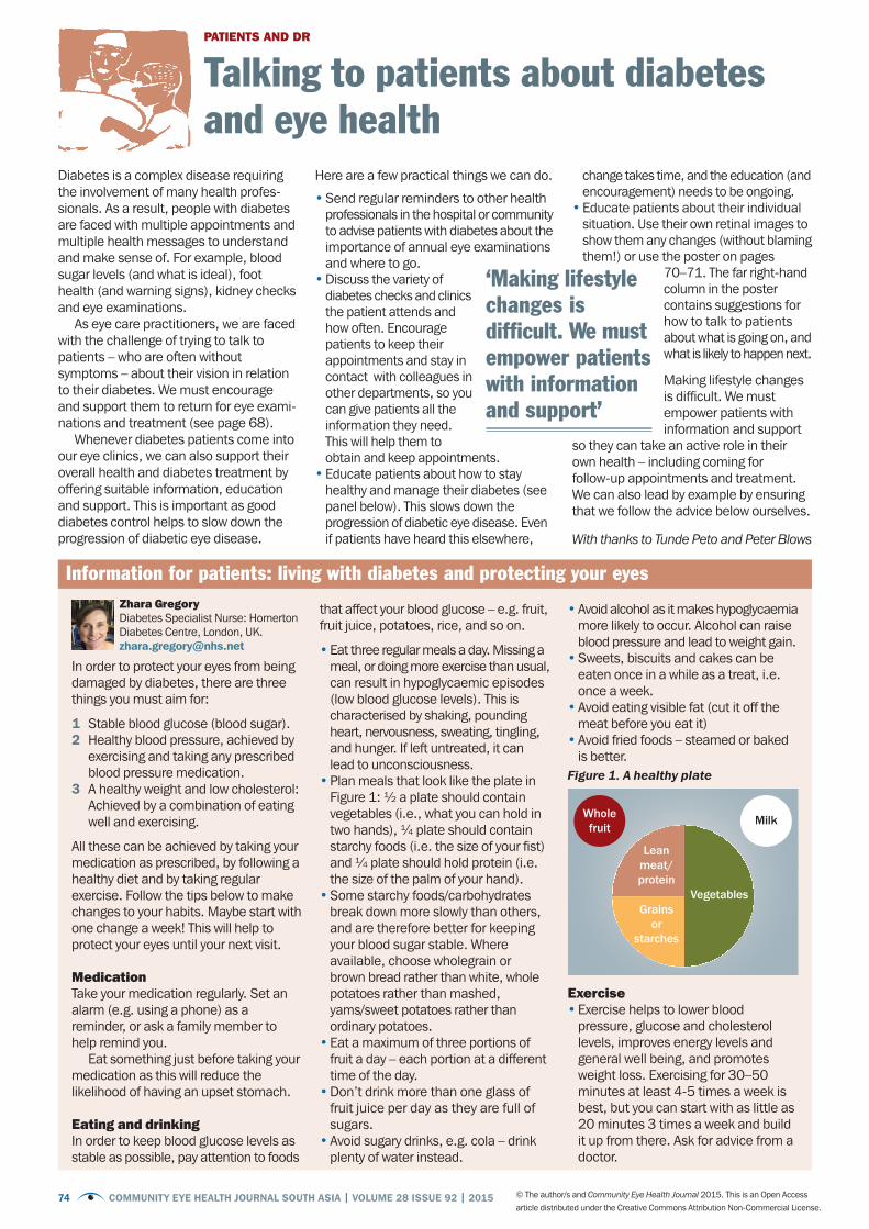
74 COMMUNITY EYE HEALTH JOURNAL SOUTH ASIA | VOLUME 28 ISSUE 92 | 2015
Diabetes is a complex disease requiring the involvement of many health profes-sionals. As a result, people with diabetes are faced with multiple appointments and multiple health messages to understand and make sense of. For example, blood sugar levels (and what is ideal), foot health (and warning signs), kidney checks and eye examinations.
As eye care practitioners, we are faced with the challenge of trying to talk to patients – who are often without symptoms – about their vision in relation to their diabetes. We must encourage and support them to return for eye exami-nations and treatment (see page 68).
Whenever diabetes patients come into our eye clinics, we can also support their overall health and diabetes treatment by offering suitable information, education and support. This is important as good diabetes control helps to slow down the progression of diabetic eye disease.
Here are a few practical things we can do.
• Send regular reminders to other health professionals in the hospital or community to advise patients with diabetes about the importance of annual eye examinations and where to go.
• Discuss the variety of diabetes checks and clinics the patient attends and how often. Encourage patients to keep their appointments and stay in contact with colleagues in other departments, so you can give patients all the information they need. This will help them to obtain and keep appointments.
• Educate patients about how to stay healthy and manage their diabetes (see panel below). This slows down the progression of diabetic eye disease. Even if patients have heard this elsewhere,
change takes time, and the education (and encouragement) needs to be ongoing.
• Educate patients about their individual situation. Use their own retinal images to show them any changes (without blaming them!) or use the poster on pages
70–71. The far right-hand column in the poster contains suggestions for how to talk to patients about what is going on, and what is likely to happen next.
Making lifestyle changes is difficult. We must empower patients with information and support
so they can take an active role in their own health – including coming for follow-up appointments and treatment. We can also lead by example by ensuring that we follow the advice below ourselves.
With thanks to Tunde Peto and Peter Blows
PATIENTS AND DR
Talking to patients about diabetes and eye health
In order to protect your eyes from being damaged by diabetes, there are three things you must aim for:
1 Stable blood glucose (blood sugar).2 Healthy blood pressure, achieved by
exercising and taking any prescribed blood pressure medication.
3 A healthy weight and low cholesterol: Achieved by a combination of eating well and exercising.
All these can be achieved by taking your medication as prescribed, by following a healthy diet and by taking regular exercise. Follow the tips below to make changes to your habits. Maybe start with one change a week! This will help to protect your eyes until your next visit.
MedicationTake your medication regularly. Set an alarm (e.g. using a phone) as a reminder, or ask a family member to help remind you.
Eat something just before taking your medication as this will reduce the likelihood of having an upset stomach.
Eating and drinkingIn order to keep blood glucose levels as stable as possible, pay attention to foods
that affect your blood glucose – e.g. fruit, fruit juice, potatoes, rice, and so on.
• Eat three regular meals a day. Missing a meal, or doing more exercise than usual, can result in hypoglycaemic episodes (low blood glucose levels). This is characterised by shaking, pounding heart, nervousness, sweating, tingling, and hunger. If left untreated, it can lead to unconsciousness.
• Plan meals that look like the plate in Figure 1: ½ a plate should contain vegetables (i.e., what you can hold in two hands), ¼ plate should contain starchy foods (i.e. the size of your fist) and ¼ plate should hold protein (i.e. the size of the palm of your hand).
• Some starchy foods/carbohydrates break down more slowly than others, and are therefore better for keeping your blood sugar stable. Where available, choose wholegrain or brown bread rather than white, whole potatoes rather than mashed, yams/sweet potatoes rather than ordinary potatoes.
• Eat a maximum of three portions of fruit a day – each portion at a different time of the day.
• Don’t drink more than one glass of fruit juice per day as they are full of sugars.
• Avoid sugary drinks, e.g. cola – drink plenty of water instead.
• Avoid alcohol as it makes hypoglycaemia more likely to occur. Alcohol can raise blood pressure and lead to weight gain.
• Sweets, biscuits and cakes can be eaten once in a while as a treat, i.e. once a week.
• Avoid eating visible fat (cut it off the meat before you eat it)
• Avoid fried foods – steamed or baked is better.
Exercise• Exercise helps to lower blood
pressure, glucose and cholesterol levels, improves energy levels and general well being, and promotes weight loss. Exercising for 30–50 minutes at least 4-5 times a week is best, but you can start with as little as 20 minutes 3 times a week and build it up from there. Ask for advice from a doctor.
Information for patients: living with diabetes and protecting your eyesZhara GregoryDiabetes Specialist Nurse: Homerton Diabetes Centre, London, UK. [email protected]
‘Making lifestyle changes is difficult. We must empower patients with information and support’
Figure 1. A healthy plate
Whole fruit
Lean meat/protein
Grains or
starches
Vegetables
Milk
© The author/s and Community Eye Health Journal 2015. This is an Open Access article distributed under the Creative Commons Attribution Non-Commercial License.
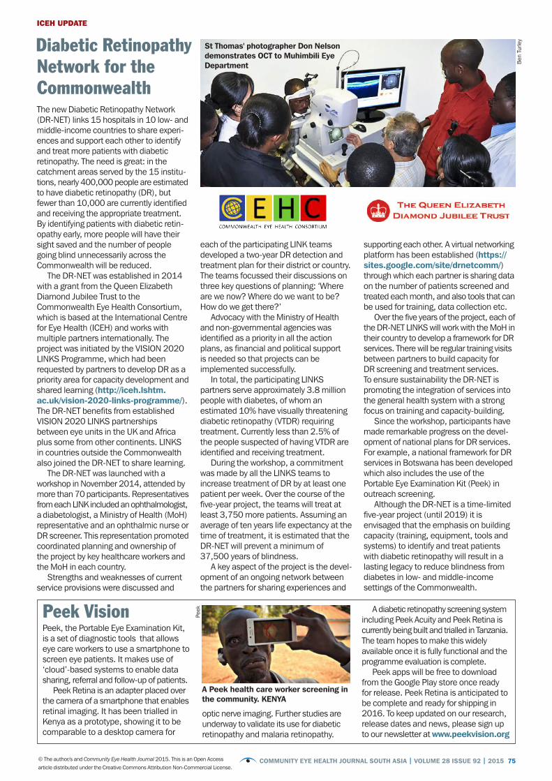
COMMUNITY EYE HEALTH JOURNAL SOUTH ASIA | VOLUME 28 ISSUE 92 | 2015 75© The author/s and Community Eye Health Journal 2015. This is an Open Access article distributed under the Creative Commons Attribution Non-Commercial License.
ICEH UPDATE
The new Diabetic Retinopathy Network (DR-NET) links 15 hospitals in 10 low- and middle-income countries to share experi-ences and support each other to identify and treat more patients with diabetic retinopathy. The need is great: in the catchment areas served by the 15 institu-tions, nearly 400,000 people are estimated to have diabetic retinopathy (DR), but fewer than 10,000 are currently identified and receiving the appropriate treatment. By identifying patients with diabetic retin-opathy early, more people will have their sight saved and the number of people going blind unnecessarily across the Commonwealth will be reduced.
The DR-NET was established in 2014 with a grant from the Queen Elizabeth Diamond Jubilee Trust to the Commonwealth Eye Health Consortium, which is based at the International Centre for Eye Health (ICEH) and works with multiple partners internationally. The project was initiated by the VISION 2020 LINKS Programme, which had been requested by partners to develop DR as a priority area for capacity development and shared learning (http://iceh.lshtm.ac.uk/vision-2020-links-programme/). The DR-NET benefits from established VISION 2020 LINKS partnerships between eye units in the UK and Africa plus some from other continents. LINKS in countries outside the Commonwealth also joined the DR-NET to share learning.
The DR-NET was launched with a workshop in November 2014, attended by more than 70 participants. Representatives from each LINK included an ophthalmologist, a diabetologist, a Ministry of Health (MoH) representative and an ophthalmic nurse or DR screener. This representation promoted coordinated planning and ownership of the project by key healthcare workers and the MoH in each country.
Strengths and weaknesses of current service provisions were discussed and
each of the participating LINK teams developed a two-year DR detection and treatment plan for their district or country. The teams focussed their discussions on three key questions of planning: ‘Where are we now? Where do we want to be? How do we get there?’
Advocacy with the Ministry of Health and non-governmental agencies was identified as a priority in all the action plans, as financial and political support is needed so that projects can be implemented successfully.
In total, the participating LINKS partners serve approximately 3.8 million people with diabetes, of whom an estimated 10% have visually threatening diabetic retinopathy (VTDR) requiring treatment. Currently less than 2.5% of the people suspected of having VTDR are identified and receiving treatment.
During the workshop, a commitment was made by all the LINKS teams to increase treatment of DR by at least one patient per week. Over the course of the five-year project, the teams will treat at least 3,750 more patients. Assuming an average of ten years life expectancy at the time of treatment, it is estimated that the DR-NET will prevent a minimum of 37,500 years of blindness.
A key aspect of the project is the devel-opment of an ongoing network between the partners for sharing experiences and
supporting each other. A virtual networking platform has been established (https://sites.google.com/site/drnetcomm/) through which each partner is sharing data on the number of patients screened and treated each month, and also tools that can be used for training, data collection etc.
Over the five years of the project, each of the DR-NET LINKS will work with the MoH in their country to develop a framework for DR services. There will be regular training visits between partners to build capacity for DR screening and treatment services. To ensure sustainability the DR-NET is promoting the integration of services into the general health system with a strong focus on training and capacity-building.
Since the workshop, participants have made remarkable progress on the devel-opment of national plans for DR services. For example, a national framework for DR services in Botswana has been developed which also includes the use of the Portable Eye Examination Kit (Peek) in outreach screening.
Although the DR-NET is a time-limited five-year project (until 2019) it is envisaged that the emphasis on building capacity (training, equipment, tools and systems) to identify and treat patients with diabetic retinopathy will result in a lasting legacy to reduce blindness from diabetes in low- and middle-income settings of the Commonwealth.
Ben
Tur
ley
Peek VisionPeek, the Portable Eye Examination Kit, is a set of diagnostic tools that allows eye care workers to use a smartphone to screen eye patients. It makes use of ‘cloud’-based systems to enable data sharing, referral and follow-up of patients.
Peek Retina is an adapter placed over the camera of a smartphone that enables retinal imaging. It has been trialled in Kenya as a prototype, showing it to be comparable to a desktop camera for
optic nerve imaging. Further studies are underway to validate its use for diabetic retinopathy and malaria retinopathy.
A diabetic retinopathy screening system including Peek Acuity and Peek Retina is currently being built and trialled in Tanzania. The team hopes to make this widely available once it is fully functional and the programme evaluation is complete.
Peek apps will be free to download from the Google Play store once ready for release. Peek Retina is anticipated to be complete and ready for shipping in 2016. To keep updated on our research, release dates and news, please sign up to our newsletter at www.peekvision.org
A Peek health care worker screening in the community. KENYA
St Thomas' photographer Don Nelson demonstrates OCT to Muhimbili Eye Department
Peek
Diabetic Retinopathy Network for the Commonwealth
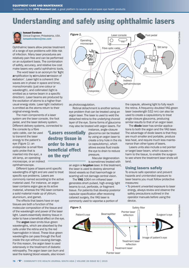
76 COMMUNITY EYE HEALTH JOURNAL SOUTH ASIA | VOLUME 28 ISSUE 92 | 2015
Ophthalmic lasers allow precise treatment of a range of eye problems with little risk of infection. Many laser procedures are relatively pain free and can be performed on an outpatient basis. The combination of safety, accuracy, and relative low cost make lasers very useful ophthalmic tools.
The word laser is an acronym for ‘light amplification by stimulated emission of radiation’. Laser light is coherent (the waves are in phase in space and time), monochromatic (just one colour or wavelength), and collimated (light is emitted as a narrow beam in a specific direction). Laser beams are produced by the excitation of atoms to a higher than usual energy state. Laser light (radiation) is emitted as the atoms return to their original energy levels.
The main components of a laser system are the laser console, the foot pedal, and the laser delivery system. Different delivery systems, connected to the console by a fibre optic cable, can be used to transmit the laser energy to the patient’s eye (Figure 1): an endoprobe (a small fibre optic probe that is inserted into the eye), a slit lamp, an operating microscope, or an indirect ophthalmoscope.
Different types of lasers emit specific wavelengths of light and are used to treat specific eye problems. Lasers are commonly named according to the active material used. For instance, an argon laser contains argon gas as its active material, whereas the YAG laser contains a solid material made up of yttrium, aluminium, and garnet.
The effects that lasers have on eye tissues are both a function of the molecular composition of the tissue and of the wavelength and power of the laser light. Lasers essentially destroy tissue in order to have a beneficial effect on the eye.
The argon laser emits blue-green wavelengths, which are absorbed by the cells under the retina and by the red haemoglobin in blood. These blue-green wavelengths can pass through the fluid inside the eye without causing damage. For this reason, the argon laser is used extensively in the treatment of diabetic retinopathy. The argon laser can burn and seal the leaking blood vessels, also known
as photocoagulation. Retinal detachment is another serious
eye problem that can be treated using an argon laser. The laser is used to weld the detached retina to the underlying choroid layer of the eye. Some forms of glaucoma may also be treated with argon lasers. For
instance, angle-closure glaucoma can be treated by using an argon laser to create a tiny hole in the iris (a capsulotomy), which allows excess fluid inside the eye to drain to reduce pressure.
Macular degeneration is sometimes treated with
an argon or krypton laser. In this treatment, the laser is used to destroy abnormal blood vessels so that haemorrhage or scarring will not damage central vision.
The YAG 1064 nm infrared laser generates short-pulsed, high-energy light beams to cut, perforate, or fragment tissue. For patients that develop posterior capsular opacification after receiving cataract surgery, the YAG laser is commonly used to vaporise a portion of
the capsule, allowing light to fully reach the retina. A frequency-doubled YAG green laser (wavelength 532 nm) can also be used to create a capsulotomy to treat angle-closure glaucoma, producing similar results to that of an argon laser.
The diode laser has similar applica-tions to both the argon and the YAG laser. The advantage of diode lasers is that they are much smaller and portable, produce less heat, and require much less mainte-nance than other types of lasers.
Lasers units also include a red pointer or target laser beam, which causes no harm to the tissue, to enable the surgeon to see where the treatment laser shots will land.
Using lasers safelyTo ensure safe operation and prevent hazards and unintended exposure to laser beams you must follow protective measures:• To prevent unwanted exposure to laser
energy, always review and observe the safety precautions outlined in the operator manuals before using the device.
Understanding and safely using ophthalmic lasersIsmael CorderoClinical Engineer, Philadelphia, [email protected]
EQUIPMENT CARE AND MAINTENANCESponsored by the IAPB Standard List: a great platform to source and compare eye health products www.iapb.standardlist.org
Foot pedal
Slit lamp
Laser console
Safety filter
Treatment laser
Pointer laser
Patient’s eye Surgeon’s eye
Image of patient’s eye
Beamsplitter
Treatment laser reflection
Pointer laser reflection
Fibre optic cable
Operating microscope
Endoprobe
Indirect opthalmoscope
Figure 1.
Figure 3.
‘Lasers essentially destroy tissue in order to have a beneficial effect on the eye’
Ism
ael C
orde
roIs
mae
l Cor
dero
76 COMMUNITY EYE HEALTH JOURNAL SOUTH ASIA | VOLUME 28 ISSUE 92 | 2015
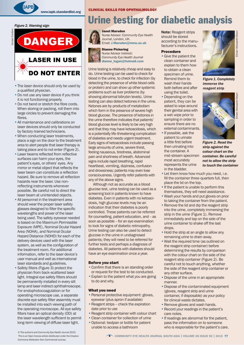
COMMUNITY EYE HEALTH JOURNAL SOUTH ASIA | VOLUME 28 ISSUE 92 | 2015 77© The author/s and Community Eye Health Journal 2015. This is an Open Access article distributed under the Creative Commons Attribution Non-Commercial License.
Urine testing is relatively cheap and easy to do. Urine testing can be used to check for blood in the urine, to check for infection (by detecting the presence of white blood cells or protein) and can show up other systemic problems such as liver problems (by showing abnormal bilirubin levels). Urine testing can also detect ketones in the urine.Ketones are by-products of metabolism which form in the presence of severe high blood glucose. The presence of ketones in the urine therefore indicates that patients’ blood glucose level is likely to be very high and that they may have ketoacidosis, which is a potentially life-threatening complication of diabetes and needs urgent treatment. Early signs of ketoacidosis include passing large amounts of urine, severe thirst, feeling nauseous, tiredness, abdominal pain and shortness of breath. Advanced signs include rapid breathing, rapid heartbeat, vomiting, dizziness, confusion and drowsiness; patients may even lose consciousness. Urgently refer patients with any of the above signs.
Although not as accurate as a blood glucose test, urine testing can be used as a screening tool in patients known to have diabetes. Even in patients with no ketoaci-dosis, high glucose levels may be an indication that their diabetes is poorly controlled. These patients can be referred for counselling, patient education, and – as soon as possible – for an eye examination to look for signs of diabetic retinopathy. Urine testing can also be used to detect glucose in the urine in undiagnosed patients; they will need to be referred for further tests and perhaps a diagnosis of diabetes. All patients with diabetes should have an eye examination once a year.
Before you start• Confirm that there is an standing order
or request for the test to be conducted.• Explain to the patient what you are going
to do and why.
What you need• Personal protective equipment: gloves,
eyewear (plus apron if available)• Reagent strips – check the expiration
date prior to use• Reagent strip container with colour chart• Clean container for collection of urine• Optional: bedpan or bottle for patient
unable to access a bathroom
Note: Reagent strips should be stored according to the manu-facturer’s instructions.
Procedure• Give the patient the
clean container and explain to them how to obtain a clean specimen of urine. Remind them to wash their hands both before and after using the toilet.
• Depending on the patient, they can be asked to wipe around their genital area with a wet-wipe prior to sampling in order to ensure there are no external contaminants.
• If possible, ask the patient to urinate a little first before then urinating into the container. A mid-stream specimen most accurately represents the urine in the bladder.
• Let them know how much you need, i.e. fill the container three-quarters full, then place the lid on the top.
• If the patient is unable to perform this themselves, they will need assistance.
• Wash your hands and put gloves on prior to taking the container from the patient.
• Remove the lid and dip the reagent strip into the urine, completely immersing the strip in the urine (Figure 1). Remove immediately and tap on the side of the urine container to shake off the last drops.
• Hold the strip at an angle to allow any remaining urine to drain away.
• Wait the required time (as outlined on the reagent strip container) before determining the results by comparison with the colour chart on the side of the reagent strip container (Figure 2). Be careful not to touch anything, whether the side of the reagent strip container or any other surface.
• Dispose of the urine in an appropriate manner.
• Dispose of the contaminated equipment (gloves, reagent strip and urine container, if disposable) as your policy for clinical waste dictates.
• Remove gloves and wash hands.• Record your readings in the patient’s
care notes.• If readings are abnormal for the patient,
pass the information on to someone who is responsible for the patient’s care.
CLINICAL SKILLS FOR OPHTHALMOLOGY
Urine testing for diabetic analysisEQUIPMENT CARE AND MAINTENANCESponsored by the IAPB Standard List: a great platform to source and compare eye health products www.iapb.standardlist.org
Figure 2. Warning sign
• The laser device should only be used by a qualified physician.
• Do not use any laser device if you think it is not functioning properly.
• Do not bend or stretch the fibre cords. When storing or packing, roll them into large circles to prevent damaging the fibres.
• All maintenance and calibrations on laser devices should only be conducted by factory-trained technicians.
• When conducting laser treatments, place a sign on the door to the treatment area to alert people that laser therapy is taking place and to not enter (Figure 2).
• Laser beams reflected from reflective surfaces can harm your eyes, the patient’s eyes, or others’ eyes. Any mirror or metal object that reflects the laser beam can constitute a reflection hazard. Be sure to remove all reflection hazards near the laser. Use non- reflecting instruments whenever possible. Be careful not to direct the laser beam at unintended objects.
• All personnel in the treatment area should wear the proper laser safety glasses designed to filter the specific wavelengths and power of the laser being used. The safety eyewear needed is based on the Maximum Permissible Exposure (MPE), Nominal Ocular Hazard Area (NOHA), and Nominal Ocular Hazard Distance (NOHD) for each of the delivery devices used with the laser system, as well as the configuration of the treatment room. For additional information, refer to the laser device’s user manual and well as international laser standards and guidelines.
• Safety filters (Figure 3) protect the physician from back-scattered laser light. Integral eye safety filters should be permanently installed in every slit lamp and laser indirect ophthalmoscope. For endophotocoagulation or for operating microscope use, a separate discrete eye safety filter assembly must be installed into each viewing path of the operating microscope. All eye safety filters have an optical density (OD) at the laser wavelength sufficient to permit long-term viewing of diffuse laser light.
Figure 2. Read the strip against the colour chart on the container. Be careful not to allow the strip to touch the container
Figure 1. Completely immerse the reagent strip
Dianne PickeringNurse Advisor (retired): Community Eye Health [email protected]
Janet MarsdenNurse Advisor: Community Eye Health Journal, London, UK. Email: [email protected]
Elm
ien
Wol
vaar
dt E
lliso
nEl
mie
n W
olva
ardt
Elli
son
Ism
ael C
orde
ro
COMMUNITY EYE HEALTH JOURNAL SOUTH ASIA | VOLUME 28 ISSUE 92 | 2015 77© The author/s and Community Eye Health Journal 2015. This is an Open Access article distributed under the Creative Commons Attribution Non-Commercial License.
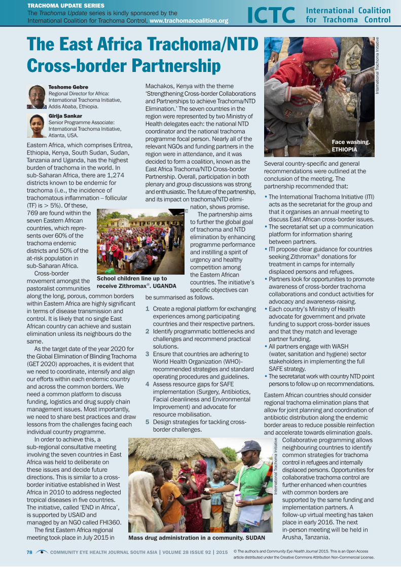
78 COMMUNITY EYE HEALTH JOURNAL SOUTH ASIA | VOLUME 28 ISSUE 92 | 2015
The East Africa Trachoma/NTD Cross-border Partnership
TRACHOMA UPDATE SERIESThe Trachoma Update series is kindly sponsored by the International Coalition for Trachoma Control, www.trachomacoalition.org
Eastern Africa, which comprises Eritrea, Ethiopia, Kenya, South Sudan, Sudan, Tanzania and Uganda, has the highest burden of trachoma in the world. In sub-Saharan Africa, there are 1,274 districts known to be endemic for trachoma (i.e., the incidence of trachomatous inflammation – follicular (TF) is > 5%). Of these, 769 are found within the seven Eastern African countries, which repre-sents over 60% of the trachoma endemic districts and 50% of the at-risk population in sub-Saharan Africa.
Cross-border movement amongst the pastoralist communities along the long, porous, common borders within Eastern Africa are highly significant in terms of disease transmission and control. It is likely that no single East African country can achieve and sustain elimination unless its neighbours do the same.
As the target date of the year 2020 for the Global Elimination of Blinding Trachoma (GET 2020) approaches, it is evident that we need to coordinate, intensify and align our efforts within each endemic country and across the common borders. We need a common platform to discuss funding, logistics and drug supply chain management issues. Most importantly, we need to share best practices and draw lessons from the challenges facing each individual country programme.
In order to achieve this, a sub-regional consultative meeting involving the seven countries in East Africa was held to deliberate on these issues and decide future directions. This is similar to a cross-border initiative established in West Africa in 2010 to address neglected tropical diseases in five countries. The initiative, called ‘END in Africa’, is supported by USAID and managed by an NGO called FHI360.
The first Eastern Africa regional meeting took place in July 2015 in
Machakos, Kenya with the theme ‘Strengthening Cross-border Collaborations and Partnerships to achieve Trachoma/NTD Elimination.’ The seven countries in the region were represented by two Ministry of Health delegates each: the national NTD coordinator and the national trachoma programme focal person. Nearly all of the relevant NGOs and funding partners in the region were in attendance, and it was decided to form a coalition, known as the East Africa Trachoma/NTD Cross-border Partnership. Overall, participation in both plenary and group discussions was strong and enthusiastic. The future of the partnership, and its impact on trachoma/NTD elimi-
nation, shows promise. The partnership aims
to further the global goal of trachoma and NTD elimination by enhancing programme performance and instilling a spirit of urgency and healthy competition among the Eastern African countries. The initiative’s specific objectives can
be summarised as follows.
1 Create a regional platform for exchanging experiences among participating countries and their respective partners.
2 Identify programmatic bottlenecks and challenges and recommend practical solutions.
3 Ensure that countries are adhering to World Health Organization (WHO)- recommended strategies and standard operating procedures and guidelines.
4 Assess resource gaps for SAFE implementation (Surgery, Antibiotics, Facial cleanliness and Environmental Improvement) and advocate for resource mobilisation.
5 Design strategies for tackling cross-border challenges.
Several country-specific and general recommendations were outlined at the conclusion of the meeting. The partnership recommended that:
• The International Trachoma Initiative (ITI) acts as the secretariat for the group and that it organises an annual meeting to discuss East African cross-border issues.
• The secretariat set up a communication platform for information sharing between partners.
• ITI propose clear guidance for countries seeking Zithromax® donations for treatment in camps for internally displaced persons and refugees.
• Partners look for opportunities to promote awareness of cross-border trachoma collaborations and conduct activities for advocacy and awareness-raising.
• Each country’s Ministry of Health advocate for government and private funding to support cross-border issues and that they match and leverage partner funding.
• All partners engage with WASH (water, sanitation and hygiene) sector stakeholders in implementing the full SAFE strategy.
• The secretariat work with country NTD point persons to follow up on recommendations.
Eastern African countries should consider regional trachoma elimination plans that allow for joint planning and coordination of antibiotic distribution along the endemic border areas to reduce possible reinfection and accelerate towards elimination goals.
Collaborative programming allows neighbouring countries to identify common strategies for trachoma control in refugees and internally displaced persons. Opportunities for collaborative trachoma control are further enhanced when countries with common borders are supported by the same funding and implementation partners. A follow-up virtual meeting has taken place in early 2016. The next in-person meeting will be held in Arusha, Tanzania.
Teshome GebreRegional Director for Africa: International Trachoma Initiative, Addis Ababa, Ethiopia.
Girija SankarSenior Programme Associate: International Trachoma Initiative, Atlanta, USA.
Mass drug administration in a community. SUDAN
School children line up to receive Zithromax®. UGANDA
Face washing. ETHIOPIA
Inte
rnat
iona
l Tra
chom
a In
itiat
ive
Inte
rnat
iona
l Tra
chom
a In
itiat
ive
ITI
78 COMMUNITY EYE HEALTH JOURNAL SOUTH ASIA | VOLUME 28 ISSUE 92 | 2015 © The author/s and Community Eye Health Journal 2015. This is an Open Access article distributed under the Creative Commons Attribution Non-Commercial License.

COMMUNITY EYE HEALTH JOURNAL SOUTH ASIA | VOLUME 28 ISSUE 92 | 2015 79© The author/s and Community Eye Health Journal 2015. This is an Open Access article distributed under the Creative Commons Attribution Non-Commercial License.
CONTINUING PROFESSIONAL DEVELOPMENT (CPD)
Test your knowledge and understanding
This page is designed to help you test your own understanding of the concepts covered in this issue, and to reflect on what you have learnt. We hope that you will also discuss the questions with your colleagues and other members of the eye care team, perhaps in a journal club. To complete the activities online – and get instant feedback – please visit www.cehjournal.org
REFLECTIVE LEARNINGVisit www.cehjournal.org to complete the online ‘Time to reflect’ section.
1. Consider diabetes and its link to diabetic retinopathy. Which statement is correct?
Select one
a Diabetes kills fewer people than TB and HIV
b The risk of developing type 2 diabetes can be reduced by keeping to a healthy weight, taking regular exercise and minimising sugar intake
c Once diabetic retinopathy occurs, the patient will inevitably go blind
d If a patient has diabetic retinopathy, they are usually aware of it
2. Consider diabetic retinopathy in individual patients. Which statement is correct?
Select one
a Diabetic retinopathy (DR) affects only people with high blood pressure
b If a patient’s diabetes and blood sugar levels are well controlled, there is no risk of DR
c Laser treatment will improve vision in people with DR
d The risk of blindness from DR can be avoided if patients attend hospital appointments as required and accept treatment
3. Consider diabetic eye disease as a public health problem. Which statement is correct?
Select one
a Blindness from diabetic retinopathy can be prevented by a screening programme
b Blindness from diabetic retinopathy is mostly linked with patients unable to afford services
c The risk of blindness from diabetic retinopathy can be reduced through early detection and treatment
d Blindness from diabetic retinopathy occurs mostly in high-income countries
4. Consider the prevention of diabetic eye disease in your district. Which of the following is essential?
Select one
a Good links with other health workers involved in caring for patients with diabetes (e.g., physicians, diabetes nurses)
b A community-based survey to determine the number of people with diabetes in your country
c A national diabetic retinopathy screening programme
d Enough ophthalmologists to examine every patient with diabetes at least once every year
1 Correct answer: b. The risk cannot be abolished by a healthy lifestyle, but it can be greatly reduced. Diabetes kills more people than TB, malaria, and HIV put together. If diabetic retinopathy is diagnosed and treated promptly, blindness can be prevented in almost all cases. Diabetic retinopathy is usually asympto-matic in its early stages.
2 Correct answer: d. By regularly attending appointments, DR can be detected early and treated before it causes visual loss. Lifestyle changes can reduce, but not eliminate, the risk of complications. Anyone with diabetes can develop diabetic eye disease. The duration of diabetes and high blood glucose levels are the most important risk factors. Good control of blood pressure is, however, also very important to reduce the risk of diabetic retinopathy. Blood glucose control reduces the risk of complications, but does not eliminate it. Complications may also be the result of poor control in previous years, even if control is good now. Laser treatment can stabilise the vision, but usually does not improve it. (Laser treatment may be used to prevent the development of proliferative retinopathy or to treat diabetic macular oedema.)
3 Correct answer: c. Both detection and treatment are needed to reduce the risk of blindness. Screening alone will not prevent blindness. There must be a referral network to confirm the diagnosis and to deliver effective treatment. Affordability is not the only challenge. Awareness of, access to, and availability of services are also required to prevent blindness. Blindness caused by DR is increasingly common in low-income countries.
4 Correct answer: a. The support of other health workers is essential. The number of people with diabetes can be estimated from the global atlas of diabetes. Local, district-level programmes are simpler and cheaper than a national programme. Ophthalmologists may be needed to examine patients with the most advanced retinopathy, but a screening programme may use photographers or other health care workers to examine patients with no or minimal retinopathy.
Picture quiz
Alle
n Fo
ster
A 45-year-old man presents with blurring of vision in his good eye. The other eye has only light perception with a traction retinal detachment.
Q1. What is the most likely diagnosis? a. Papilloedema due to a brain tumour b. Proliferative diabetic retinopathy c. Hypertensive retinopathy d. Coat’s disease e. Non-proliferative diabetic retinopathy
Q2. Which of the following clinical signs are present? a. Vitreous haemorrhage b. Neovascularisation of the retina c. Retinal haemorrhages d. Retinal hard exudates e. Retinal “cotton wool spots”
There may be more than one correct answer.
Q3. What treatments might be useful in managing this condition?
a. Pattern laser treatment to the macular area b. Peripheral retinal photocoagulation c. Investigation of papilloedema d. Watch and review in 3 months e. Anti-vascular endothelial growth factor
intra-vitreal injections
1. Answer b.This is severe proliferative diabetic retinopathy (PDR). The traction RD in the fellow eye is probably due to old PDR.2. Answer b, c, d and e.In addition to haemorrhages and hard exudates, there are cotton-wool spots and widespread neovascularisation (new vessels) indicating retinal ischaemia.3. Answer b or possibly e if laser is not available. The treatment of PDR is peripheral retinal photocoagulation (laser treatment). This needs to be done as soon as possible.
ANSWERSANSWERS
COMMUNITY EYE HEALTH JOURNAL SOUTH ASIA | VOLUME 28 ISSUE 92 | 2015 79 © The author/s and Community Eye Health Journal 2015. This is an Open Access article distributed under the Creative Commons Attribution Non-Commercial License.
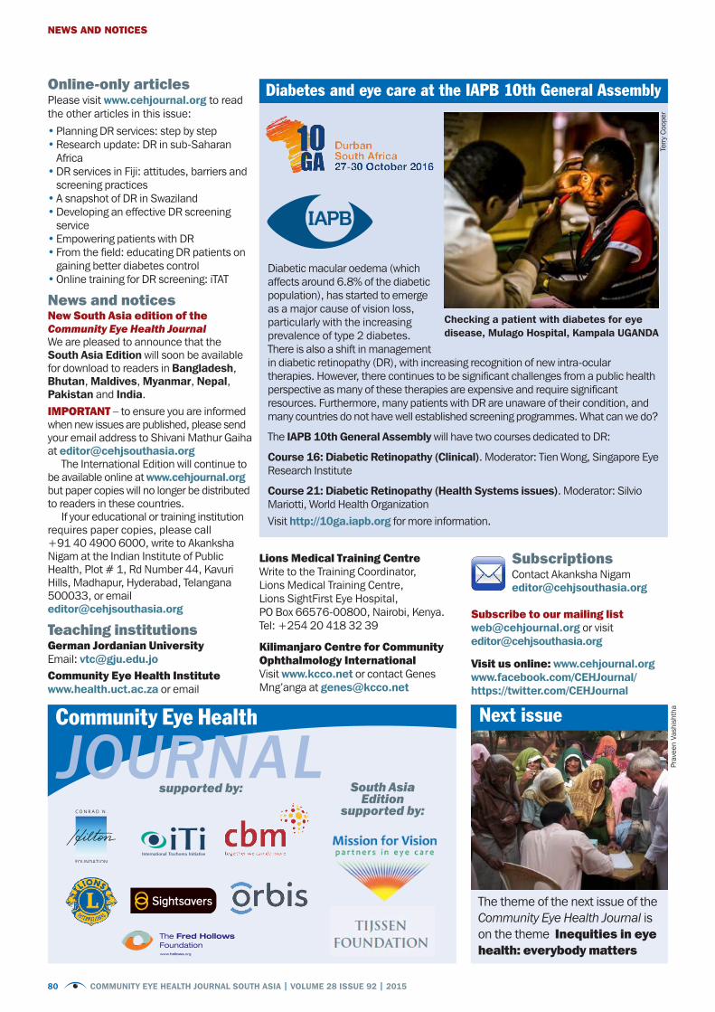
80 COMMUNITY EYE HEALTH JOURNAL SOUTH ASIA | VOLUME 28 ISSUE 92 | 2015
Lions Medical Training Centre Write to the Training Coordinator, Lions Medical Training Centre, Lions SightFirst Eye Hospital, PO Box 66576-00800, Nairobi, Kenya. Tel: +254 20 418 32 39
Kilimanjaro Centre for Community Ophthalmology InternationalVisit www.kcco.net or contact Genes Mng’anga at [email protected]
SubscriptionsContact Akanksha [email protected]
Subscribe to our mailing list [email protected] or visit [email protected]
Visit us online: www.cehjournal.orgwww.facebook.com/CEHJournal/https://twitter.com/CEHJournal
NEWS AND NOTICES
Online-only articlesPlease visit www.cehjournal.org to read the other articles in this issue:
• Planning DR services: step by step• Research update: DR in sub-Saharan
Africa• DR services in Fiji: attitudes, barriers and
screening practices• A snapshot of DR in Swaziland• Developing an effective DR screening
service• Empowering patients with DR• From the field: educating DR patients on
gaining better diabetes control• Online training for DR screening: iTAT
News and noticesNew South Asia edition of the Community Eye Health JournalWe are pleased to announce that the South Asia Edition will soon be available for download to readers in Bangladesh, Bhutan, Maldives, Myanmar, Nepal, Pakistan and India. IMPORTANT – to ensure you are informed when new issues are published, please send your email address to Shivani Mathur Gaiha at [email protected]
The International Edition will continue to be available online at www.cehjournal.org but paper copies will no longer be distributed to readers in these countries.
If your educational or training institution requires paper copies, please call +91 40 4900 6000, write to Akanksha Nigam at the Indian Institute of Public Health, Plot # 1, Rd Number 44, Kavuri Hills, Madhapur, Hyderabad, Telangana 500033, or email [email protected]
Teaching institutionsGerman Jordanian University Email: [email protected] Eye Health Institute www.health.uct.ac.za or email
s Community Eye Health
JOURNALSouth Asia
Editionsupported by:
The theme of the next issue of the Community Eye Health Journal is on the theme Inequities in eye health: everybody matters
Diabetic macular oedema (which affects around 6.8% of the diabetic population), has started to emerge as a major cause of vision loss, particularly with the increasing prevalence of type 2 diabetes. There is also a shift in management in diabetic retinopathy (DR), with increasing recognition of new intra-ocular therapies. However, there continues to be significant challenges from a public health perspective as many of these therapies are expensive and require significant resources. Furthermore, many patients with DR are unaware of their condition, and many countries do not have well established screening programmes. What can we do?
The IAPB 10th General Assembly will have two courses dedicated to DR:
Course 16: Diabetic Retinopathy (Clinical). Moderator: Tien Wong, Singapore Eye Research Institute
Course 21: Diabetic Retinopathy (Health Systems issues). Moderator: Silvio Mariotti, World Health OrganizationVisit http://10ga.iapb.org for more information.
Diabetes and eye care at the IAPB 10th General Assembly
Next issue
Terr
y C
oope
r
Checking a patient with diabetes for eye disease, Mulago Hospital, Kampala UGANDA
Prav
een
Vash
isht
ha
supported by:
Related Documents