The Cytoplasmic Domain of CD4 Promotes the Development of CD4 Lineage T Cells By Andrea Itano, Patrick Salmon, Dimitris Kioussis,* Mauro Tolaini,* Paola Corbella,* and Ellen Robey From the Department of Molecular and Cell Biology, University of California, Berkeley, California 94720; and the *Division of Molecular Immunology, National Institute for Medical Research, Mill Hill, London, NW71AA United Kingdom Summary Thymocytes must bind major histocompatibility complex (MHC) proteins on thymic epithelial cells in order to mature into either CD8 + cytotoxic T cells or CD4 + helper T cells. Thymic precursors express both CD8 and CD4, and it has been suggested that the intracellular signals generated by CD8 or CD4 binding to class I or II MHC, respectively, might influence the fate of uncommitted cells. Here we test the notion that intracellular signaling by CD4 directs the development of thymocytes to a CD4 lineage. A hybrid protein consisting of the CD8 extra- cellular and transmembrane domains and the cytoplasmic domain of CD4 (CD884) should bind class I MHC but deliver a CD4 intracellular signal. We find that expression of a hybrid CD884 protein in thymocytes oftransgenic mice leads to the development of large numbers of class I MHC-specific, CD4 lineage T cells. We discuss these results in terms of current models for CD4 and CD8 lineage commitment. T he differentiation of immature thymocytes into either CD4 § or CD8 + mature T cells is linked to the speci- ficity of the TCRs expressed on developing thymocytes. Recognition of class I MHC by developing thymocytes re- quires the coordinate binding of both a class I-specific TCR and a CD8 coreceptor, and can give rise to mature CD8 + T cells. In contrast, recognition of class II MHC re- quires corecognition by a class II-specific TCR and the CD4 coreceptor and can give rise to mature CD4 + T cells. Al- though immature thymocytes express both CD4 and CD8, as thymocytes mature, they downregulate expression of one of the coreceptors, giving rise to mature CD4+CD8 - or CD4-CD8 § T cells. Thus lineage commitment in the thy- mus is linked to both the specificity of the TCR and the expression of the CD4 and CD8 coreceptors. The observation that the specificity of TCR for class I or II MHC influences the choice between CD4 and CD8 lin- eages is consistent with a model in which binding of class I MHC by a CD4+CD8 + thymocyte leads to the downregu- lation of CD4 expression and commitment to a CD8 fin- eage. Likewise, binding of class II MHC may lead to the downregulation of CD8 expression and commitment to a CD4 lineage (1). This model fits well with observations from TCR transgenic mice, in which expression of a class I-specific TCR generally leads to an increase in the pro- duction of mature CD8 cells, and expression of a class II- specific TCR generally leads to an increase in the produc- tion of mature CD4 cells (2-5). The analysis of putative transitional intermediates in MHC- deficient mice has been interpreted as evidence against an in- structive model. For example, a population of CD4+CD8 l~ thymocytes is observed in mice mutant for class II MHC, but is missing in mice mutant for both class I and II MHC (6, 7). It has been proposed (7) that this population repre- sents class I-selected transitional cells that are committed to a CD4 lineage, a population that would not be predicted by an instructive model. There is, however, no direct evidence that the CD4+CD8 l~ cells are the precursors of CD4 lineage cells. Indeed, recent experiments indicate that at least some CD4+CD8 ]~ thymocytes give rise to CD8 lineage cells (8), raising questions about the implications of this population for the mechanism of CD4/CD8 lineage commitment. Another line of evidence that lineage commitment need not correlate with MHC specificity comes from mice ex- pressing constitutive CD8 or CD4 transgenes (9-15). Al- though these experiments provide evidence for a stochastic component to lineage commitment, it is striking that the generation of T cells of the "wrong" lineage (i.e., CD4 § with class I-specific TCRs or CD8 + with class II-specific TCRs) is invariably inefficient. For example, class II MHC mutant, CD8 transgenic mice have only ~3% of the ma- ture CD4 § thymocytes that are found in mice that are wild type for class II MHC (10). Moreover, another class I-spe- cific TCR, anti-HY, failed to give rise to any detectable CD4 lineage cells when coexpressed with a CD8 transgene (16-18), implying that not all class I-specific TCRs can permit the development of CD4 lineage cells. In light of these considerations, we decided to examine more directly the question of whether intracellular signals generated upon MHC recognition might influence the fate 731 J. Exp. Med.9 The Rockefeller University Press 9 0022-1007/96/03/731/11 $2.00 Volume 183 March 1996 731-741 on May 20, 2015 jem.rupress.org Downloaded from Published March 1, 1996

Welcome message from author
This document is posted to help you gain knowledge. Please leave a comment to let me know what you think about it! Share it to your friends and learn new things together.
Transcript
The Cytop lasmic D o m a i n o f CD4 P r o m o t e s the D e v e l o p m e n t o f CD4 Lineage T Cells
By Andrea Itano, Patrick Salmon, Dimitris Kioussis,* Mauro Tolaini,* Paola Corbella,* and Ellen Robey
From the Department of Molecular and Cell Biology, University of California, Berkeley, California 94720; and the *Division of Molecular Immunology, National Institute for Medical Research, Mill Hill, London, NW71AA United Kingdom
Summary Thymocytes must bind major histocompatibility complex (MHC) proteins on thymic epithelial cells in order to mature into either CD8 + cytotoxic T cells or CD4 + helper T cells. Thymic precursors express both CD8 and CD4, and it has been suggested that the intracellular signals generated by CD8 or CD4 binding to class I or II M H C , respectively, might influence the fate o f uncommitted cells. Here we test the notion that intracellular signaling by CD4 directs the development o f thymocytes to a CD4 lineage. A hybrid protein consisting o f the CD8 extra- cellular and transmembrane domains and the cytoplasmic domain o f CD4 (CD884) should bind class I M H C but deliver a CD4 intracellular signal. We find that expression o f a hybrid CD884 protein in thymocytes oftransgenic mice leads to the development o f large numbers o f class I MHC-specif ic, CD4 lineage T cells. We discuss these results in terms o f current models for CD4 and CD8 lineage commitment.
T he differentiation o f immature thymocytes into either CD4 § or CD8 + mature T cells is linked to the speci-
ficity o f the T C R s expressed on developing thymocytes. Recogni t ion o f class I M H C by developing thymocytes re- quires the coordinate binding of both a class I-specific T C R and a CD8 coreceptor, and can give rise to mature CD8 + T cells. In contrast, recognition o f class II M H C re- quires corecognition by a class II-specific T C R and the CD4 coreceptor and can give rise to mature CD4 + T cells. Al- though immature thymocytes express both CD4 and CD8, as thymocytes mature, they downregulate expression of one o f the coreceptors, giving rise to mature C D 4 + C D 8 - or C D 4 - C D 8 § T cells. Thus lineage commitment in the thy- mus is linked to both the specificity o f the T C R and the expression of the CD4 and CD8 coreceptors.
The observation that the specificity o f T C R for class I or II M H C influences the choice between CD4 and CD8 lin- eages is consistent with a model in which binding o f class I M H C by a C D 4 + C D 8 + thymocyte leads to the downregu- lation o f CD4 expression and commitment to a CD8 fin- eage. Likewise, binding of class II M H C may lead to the downregulation o f CD8 expression and commitment to a CD4 lineage (1). This model fits well with observations from T C R transgenic mice, in which expression o f a class I-specific T C R generally leads to an increase in the pro- duction o f mature CD8 cells, and expression of a class I I - specific T C R generally leads to an increase in the produc- tion o f mature CD4 cells (2-5).
The analysis o f putative transitional intermediates in M H C - deficient mice has been interpreted as evidence against an in-
structive model. For example, a population of CD 4 +CD8 l~ thymocytes is observed in mice mutant for class II M H C , but is missing in mice mutant for both class I and II M H C (6, 7). It has been proposed (7) that this population repre- sents class I-selected transitional cells that are committed to a CD4 lineage, a population that would not be predicted by an instructive model. There is, however, no direct evidence that the CD4+CD8 l~ cells are the precursors o f CD4 lineage cells. Indeed, recent experiments indicate that at least some C D 4 + C D 8 ]~ thymocytes give rise to CD8 lineage cells (8), raising questions about the implications o f this population for the mechanism of C D 4 / C D 8 lineage commitment.
Another line o f evidence that lineage commitment need not correlate with M H C specificity comes from mice ex- pressing constitutive CD8 or CD4 transgenes (9-15). Al- though these experiments provide evidence for a stochastic component to lineage commitment, it is striking that the generation o f T cells o f the "wrong" lineage (i.e., CD4 § with class I-specific T C R s or CD8 + with class II-specific TCRs) is invariably inefficient. For example, class II M H C mutant, CD8 transgenic mice have only ~ 3 % of the ma- ture CD4 § thymocytes that are found in mice that are wild type for class II M H C (10). Moreover, another class I-spe- cific T C R , anti-HY, failed to give rise to any detectable CD4 lineage cells when coexpressed with a CD8 transgene (16-18), implying that not all class I-specific T C R s can permit the development o f CD4 lineage cells.
In light o f these considerations, we decided to examine more directly the question o f whether intracellular signals generated upon M H C recognition might influence the fate
731 J. Exp. Med.�9 The Rockefeller University Press �9 0022-1007/96/03/731/11 $2.00 Volume 183 March 1996 731-741
on May 20, 2015
jem.rupress.org
Dow
nloaded from
Published March 1, 1996
o f uncommit ted thymocytes. Here we directly test the hy- pothesis that CD4 intracellular signals p romote the devel- opment o f CD4 lineage T cells. A hybrid molecule consist- ing o f the extracellular domain o f CD8 and the cytoplasmic domain o f CD4 would be expected to bind class I M H C , yet deliver a CD4 intracellular signal. I f C D 4 signals direct thymic precursors to choose~the C D 4 lineage, then the rec- ognit ion o f class II M H C by thymocytes expressing such a hybrid molecule would direct development to a CD4 in- stead o f a CD8 lineage. Indeed, we find that coexpression o f a hybrid C D 8 / 4 molecule wi th the F5 T C R leads to a dramatic increase in mature CD4 cells, and a decrease in mature CD8 cells. These results indicate that the cytoplas- mic domain o f CD4 delivers a signal that favors the devel- opment o f CD4 lineage cells.
Materials and Methods
Generation of Transgenic Mice Expressing CD884 Hybrid Moleo cules. A Nael site was introduced into the CD8.1 cDNA (16) at amino acid 196 by site-directed mutagenesis. The sequence of the mutagenic oligonucleotide was 5'-aac acg ctg ccg gct cct gtg-3'. This Nael site was ligated to the existing Nael site at amino acid 400 of the murine CD4 gene. The resulting hybrid cDNA en- codes a fusion protein whose extracellular and transmembrane domains derive from the CD8 gene (through amino acid Ser 195 [19]), and whose cytoplasmic domain derives from CD4 (from amino acid Arg 400 [20]). The hybrid cDNA was inserted into the EcoR.I site of the human CD2 expression cassette, to produce a transgenic construct that is analogous to the " T l l - 8 " construct that was used to generate CD8.1 transgenic mice (16). This con- struct was coinjected along with a CD813.1 genomic clone (21) into C57B1/10 embryos. One founder which expresses transgenic CD8.~xl and CD813.1 at levels comparable to the endogenous CD8 genes was chosen for further analysis. Hematopoietic stem cell chimeras were generated by injecting mixtures o fT cell-depleted bone marrow cells from F5 T C R and F5 TCR/CD884 trans- genic mice (2 • 107 cell/recipient) into unirradiated P,.agl mu- tant mice (22).
Analysis of Lck Association. Thymocytes (107 cells/sample) were lysed in 1 ml oflysis buffer (50 mM Tris, pH 7.5, 0.1% NP-40, 1 mM Na3VO4, 10 mM NaF, 10 mM Na4P207, 1 mM PMSF, 10 l~g/ml aprotinin, and 10 p~g/ml leupeptin). Lysates were cen- trifuged for 1 min at 14,000 rpm and the supematants were incu- bated with 25 Ixl protein G beads (GammaBind G Sepharose; Pharmacia, Piscataway, NJ) that had been precoated with either anti-CD4 (GK1.5) or anti-CD8 (53-6.72). After 3 h at 4~ beads were washed twice with lysis buffer without detergent. Nonreduc- ing SDS loading buffer was added to the samples, which were heated for 5 min at 95~ Samples were run on an SDS polyacryl- amide gel and transferred to nitrocellulose (Hybond-ECL; Amer- sham Corp., Arlington Heights, IL). Irnmunoblotting was per- formed using the enhanced chemiluminescence system (ECL; Amersham Corp.) according to the manufacturer's instructions. Anti-Lck antibody was kindly provided by Dr. Joe Bolen (Bris- tol-Myers Squibb, Princeton, NJ) and was visualized using horse- radish peroxidase goat anti-mouse Ig (Southern Biotechnology Associates, Birmingham, AL).
Analysis of Lck Activation. CD8 and CD884 constructs were cloned into a SV40 expression cassette (23) and introduced by electroporation into Jurkat cells along with a selectable plasmid.
Individual hygromycin B resistant clones expressing comparable cell surface levels of CD8 were selected for cross-linking experi- ments. Cross-linking was performed as follows. 107 cells were in- cubated with 20 Ixg rat anti-murine CD8 IgG (clone 53-6.72) for 15 min on ice, then with 60 Ixg goat anti-rat IgG (Cappel Labo- ratories, Durham, NC) for 15 rain on ice. Cells were then incu- bated for 10 min at 37~ rinsed with cold PBS containing 1 mM Na3VO 4 and 10 mM NaF, and lysed for 20 min in lysis buffer on ice. Lysates were cleared by centrifugation for 10 min at 10,000 g and incubated with 25 Ixl protein G beads for 6 h at 4~ Beads were then washed four times with lysis buffer (without Na4P207 and PMSF), once with kinase buffer (50 mM Hepes, pH 7.5, 100 mM NaC1, 5 mM MgC12, and 5 mM MnC12) , and resuspended in 30 Ixl kinase buffer containing 10 I~Ci ~/-[32p]ATP and 10 Ixg acid-denatured enolase (Sigma Chemical Co., St. Louis, MO). Reactions were run for 10 min at 30~ quenched with 15 I~1 3• sample buffer (150 mM Tris, pH 6.8, 30% glycerol, 6% SDS, 3% 2-ME and 0.05% bromophenol blue), and reaction products were analyzed by SDS-PAGE. The gel was then fixed and dried for au- toradiography. Quantification of radioactive bands was performed using the Phosphorlmager/ImageQuant system (Molecular Dy- namics, Sunnyvale, CA). For the in vitro kinase assay on thy- mocytes, immunoprecipitations were performed as described above for the Western blot. Immunoprecipitates were resuspended in kinase buffer and assays were performed as described above.
Analysis of T Cell Populations. Class II MHC mutant, anti-HY and F5 T C R transgenic mice have been previously described (3, 24, 25). All mice were homozygous for H-2 b. Cell suspensions of thymocytes and lymph node T cells were prepared and labeled with fluorescent antibodies as previously described (11). For anal- ysis of mature thymocytes, thymocytes were treated with anti- heat stable antigen (HSA) 1 and complement as previously de- scribed (10). Antibodies used were T3.70 (culture supematant), PE-labeled goat anti-mouse IgG1 (Caltag Laboratories, South San Francisco, CA), PE-labeled CD4 (Beckton Dickinson & Co., Mountain View, CA), anti-CD8.2~ FITC (2.43), Tricolor xM- labeled streptavidin (Caltag), and rat gamma globulin (Calbiochem- Novabiochem Corp., San Diego, CA), anti-CD8.1 (49-31.1; Cedarlane Laboratories, Westbury, NY), FITC-labeled goat anti- mouse IgG3 (Caltag), biotinylated anti-CD4 (YTS 191.1; Cal- tag), biotinylated anti-Vet2 (PharMingen, San Diego, CA), and biotinylated anti-VI311 (KT11). Data (50,000 events) were col- lected and analyzed using a FACScan | flow cytometer (Becton Dickinson & Co.) or X-cell flow cytometer (Coulter Corp., Hi- aleah, FL). Dead cells were excluded on the basis of forward and side scatter.
Results
Transgenic Mice Expressing a Hybrid C D 8 / C D 4 Transgene. To redirect a CD4 intracellular signal, we constructed a hy- brid c D N A consisting o f the extracellular and transmem- brane domains o f C D 8 a and the cytoplasmic domain of CD4 (CD884). The CD8o~ c D N A used encodes the CD8.1 al- lele, which can be distinguished from the endogenous CD8 allele (CD8.2) using monoclonal antibodies. The hybrid c D N A (Fig. 1 a) was inserted into an expression cassette containing the human CD2 promoter , minigene, and 3' locus controll ing region, to generate a construct analogous
1Abbreviations used in this paper: HSA, heat stable antigen; NP, nucleopro- tein.
732 Development of CD4 Lineage T Cells
on May 20, 2015
jem.rupress.org
Dow
nloaded from
Published March 1, 1996
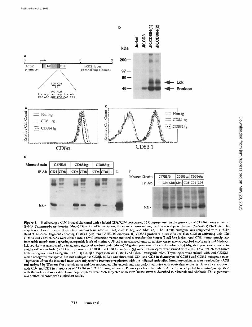
Figure 1. Redirecting a CD4 intraceUular signal with a hybrid CD8/CD4 coreceptor. (a) Construct used in the generation of CD884 transgenic mice. (White) Transmembrane domain. (Arrow) Direction of transcription; the sequence surrounding the fusion is depicted below. (Underlined) Nael site. The map is not drawn to scale. Restriction endonuclease sites: Sall (S), BamH1 (/3), and Xbal (X). The CD884 transgene was coinjected with a 15-kb BamH1 genonfic fragment encoding CD813.1 (21) into C57B1/10 embryos. (b) CD884 protein is more effective than CD8 in activating Lck. The CD884 and CD8 cDNAs were cloned into a SV40 expression vector and used to transfect the human T cell line Jurkat. Anti-CD8 immunoprecipitates from stable transfectants expressing comparable levels ofmurine CD8 cell were analyzed using an in vitro kinase assay as described in Materials and Methods. Lck activity was quantitated by integrating signals of enolase bands. (Arrows) Migration positions of Lck and enolase. (Le~) Migration positions of molecular weight (kDa) standards. (c) CD8a expression on CD884 and CD8.1 transgenic (tg) mice. Thymocytes were stained with anti-CD8~t, which recognized both endogenous and transgenic CD8. (d) CD813.1 expression on CD884 and CD8.1 transgenic mice. Thymocytes were stained with anti-CD8[3.1, which recognizes transgenic, but not endogenous CD813. (e) Lck associated with CD4 and CD8 in thyrnocytes of CD884 and CD8.1 transgenic mice. Thymocytes from the indicated mice were subjected to immunoprecipitation with the indicated antibodies. Immunoprecipitates were resolved by PAGE and analyzed by Western blot analysis using anti-Lck antibodies. The experiment was performed twice with equivalent results. (jr) Active Lck associated with CD4 and CD8 in thymocytes of CD884 and CD8.1 transgenic mice. Thymocytes from the indicated mice were subjected to immunoprecipitation with the indicated antibodies, lmmunoprecipitates were then subjected to in vitro kinase assays as described in Materials and Methods. The experiment was performed twice with equivalent results.
733 Itano et al.
on May 20, 2015
jem.rupress.org
Dow
nloaded from
Published March 1, 1996
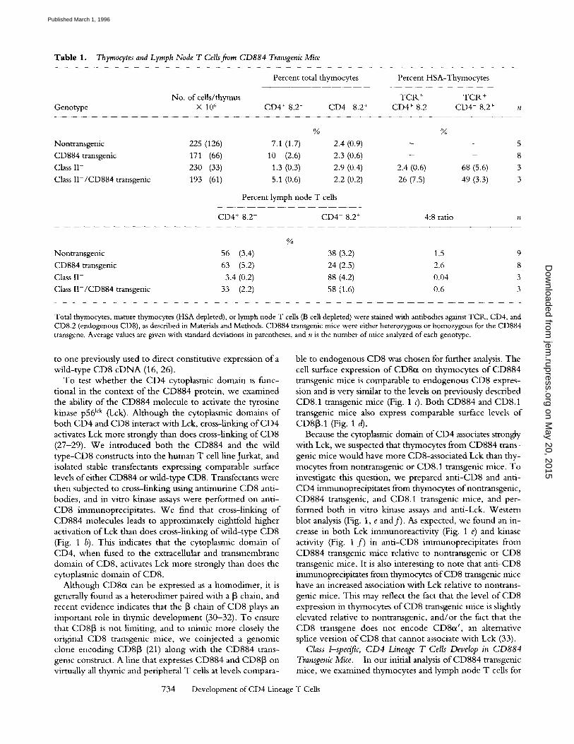
Table 1. Thymocytes and Lymph Node T Cells fiom CD884 Transgenic Mice
Percent total thymocytes Percent HSA-Thymocytes
No. of cells/thymus TCR. + TCR + Genotype X 10 6 CD4 + 8.2- CD4- 8.2 + CD4 + 8.2- CD4- 8.2 +
% %
Nontransgenic 225 (126) 7.1 (1.7) 2.4 (0.9) - - 5 CD884 transgenic 171 (66) 10 (2.6) 2.3 (0.6) - - 8 Class II- 230 (33) 1.3 (0.3) 2,9 (0.4) 2.4 (0.6) 68 (5.6) 3
Class II-/CD884 transgenic 193 (61) 5.1 (0.6) 2,2 (0.2) 26 (7.5) 49 (3.3) 3
Percent lymph node T cells
CD4 + 8.2- CD4- 8.2 + 4:8 ratio n
%
Nontransgenic 56 (3,4) 38 (3.2) 1,5 9 CD884 transgenic 63 (5.2) 24 (2.5) 2.6 8 Class II- 3.4 (0.2) 88 (4.2) 0.04 3 Class II-/CD884 transgenic 33 (2.2) 58 (1,6) 0.6 3
Total thymocytes, mature thymocytes (HSA depleted), or lymph node T cells (B cell depleted) were stained with antibodies against TCR, CD4, and CD8.2 (endogenous CD8), as described in Materials and Methods. CD884 transgenic mice were either heterozygous or homozygous for the CD884 transgene. Average values are given with standard deviations in parentheses, and n is the number of mice analyzed of each genotype.
to one previously used to direct constitutive expression of a wild-type CD8 c D N A (16, 26).
To test whether the CD4 cytoplasmic domain is func- tional in the context o f the CD884 protein, we examined the ability of the CD884 molecule to activate the tyrosine kinase p56 lck (Lck). Although the cytoplasmic domains o f both CD4 and CD8 interact with Lck, cross-linking of CD4 activates Lck more strongly than does cross-linking of CD8 (27-29). W e introduced both the CD884 and the wild type-CD8 constructs into the human T cell line Jurkat, and isolated stable transfectants expressing comparable surface levels of either CD884 or wild-type CD8. Transfectants were then subjected to cross-linking using antimurine CD8 anti- bodies, and in vitro kinase assays were performed on anti- CD8 immunoprecipitates. We find that cross-linking o f CD884 molecules leads to approximately eightfold higher activation o f Lck than does cross-linking of wild-type CD8 (Fig. 1 b). This indicates that the cytoplasmic domain o f CD4, when fused to the extracellular and transmembrane domain o f CD8, activates Lck more strongly than does the cytoplasmic domain o f CD8.
Although CD8ct can be expressed as a homodimer, it is generally found as a heterodimer paired with a [~ chain, and recent evidence indicates that the [~ chain of CD8 plays an important role in thymic development (30-32). To ensure that CD8[3 is not limiting, and to mimic more closely the original CD8 transgenic mice, we coinjected a genomic clone encoding CD813 (21) along with the CD884 trans- genic construct. A line that expresses CD884 and CDSJ3 on virtually all thymic and peripheral T cells at levels compara-
ble to endogenous CD8 was chosen for further analysis. The cell surface expression of CD8~x on thymocytes o f CD884 transgenic mice is comparable to endogenous CD8 expres- sion and is very similar to the levels on previously described CD8.1 transgenic mice (Fig. 1 c). Both CD884 and CD8.1 transgenic mice also express comparable surface levels o f CD813.1 (Fig. 1 d).
Because the cytoplasmic domain of CD4 associates strongly with Lck, we suspected that thymocytes from CD884 trans- genic mice would have more CD8-associated Lck than thy- mocytes from nontransgenic or CD8.1 transgenic mice. To investigate this question, we prepared anti-CD8 and anti- CD4 immunoprecipitates from thymocytes ofnontransgenic, CD884 transgenic, and CD8.1 transgenic mice, and per- formed both in vitro kinase assays and anti-Lck. Western blot analysis (Fig. 1, e and f) . As expected, we found an in- crease in both Lck immunoreactivity (Fig. 1 e) and kinase activity (Fig. 1 f ) in anti-CD8 immunoprecipitates from CD884 transgenic mice relative to nontransgenic or CD8 transgenic mice. It is also interesting to note that anti-CD8 immunoprecipitates from thymocytes o f C D 8 transgenic mice have an increased association with Lck relative to nontrans- genic mice. This may reflect the fact that the level of CD8 expression in thymocytes o f CD8 transgenic mice is slightly elevated relative to nontransgenic, and/or the fact that the CD8 transgene does not encode CD8od, an alternative splice version o f CD8 that cannot associate with Lck (33).
Class 1-specific, CD4 Lineage T Cells Develop in CD884 Transgenic Mice. In our initial analysis o f C D 8 8 4 transgenic mice, we examined thymocytes and lymph node T cells for
734 Development of CD4 Lineage T Cells
on May 20, 2015
jem.rupress.org
Dow
nloaded from
Published March 1, 1996
T o t a l T h y m u s F5 TCR tg F5 TCR/
CD8.1 tg
8 12.3_~ r '~t4!
Z
CD8.2
F5 TCR
F5 TCR/ CD884 tg
CD8.1
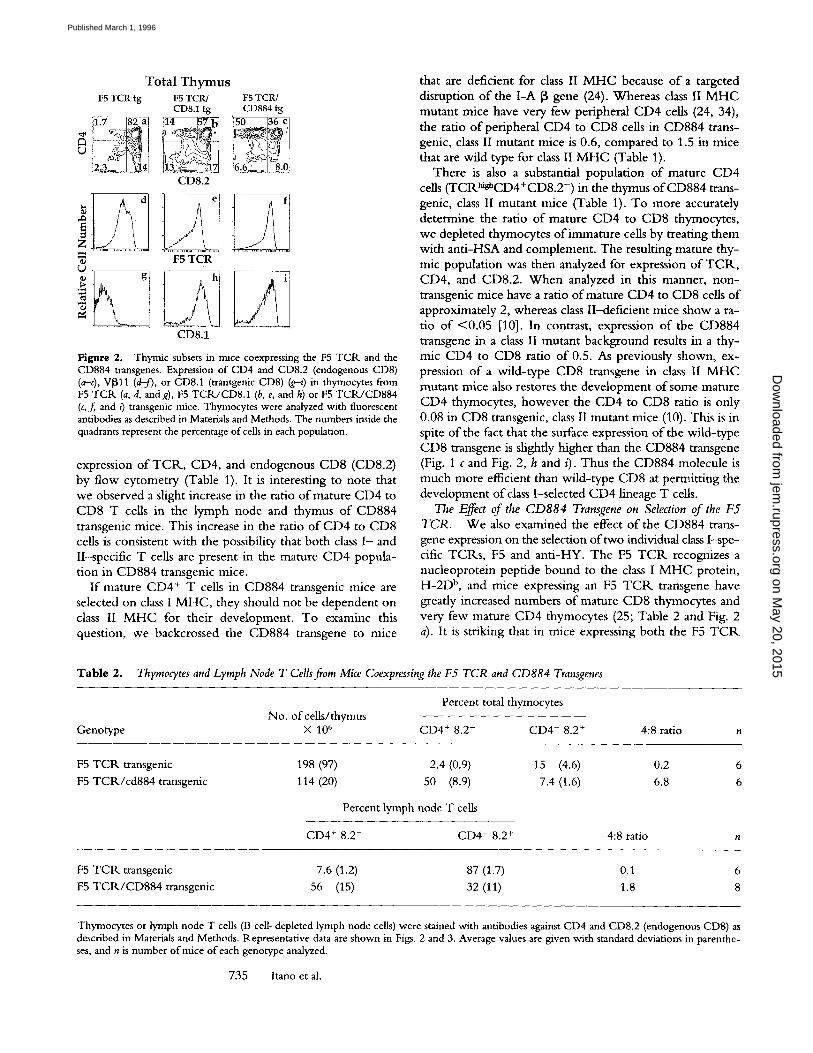
Figure 2. Thymic subsets in mice coexpressing the F5 TCR and the CD884 transgenes. Expression of CD4 and CD8.2 (endogenous CD8) (a-c), V1311 (d-f), or CD8.1 (transgenic CDS) (g--t) in thymocytes from F5 TCR (a, d, andg), F5 TCIq./CD8.1 (b, e, and h) or F5 TCR/CD884 (c, f, and t) transgenic mice. Thymocytes were analyzed with fluorescent antibodies as described in Materials and Methods. The numbers inside the quadrants represent the percentage of cells in each population.
expression o f TCR., CD4, and endogenous CD8 (CD8.2) by flow cytometry (Table 1). It is interesting to note that we observed a slight increase in the ratio o f mature CD4 to CD8 T cells in the lymph node and thymus o f CD884 transgenic mice. This increase in the ratio o f CD4 to CD8 cells is consistent with the possibility that both class I - and II-specific T cells are present in the mature CD4 popula- t ion in CD884 transgenic mice.
I f mature CD4 + T cells in CD884 transgenic mice are selected on class I M H C , they should not be dependent on class II M H C for their development . To examine this question, we backcrossed the CD884 transgene to mice
that are deficient for class II M H C because o f a targeted disruption o f the I -A 13 gene (24). Whereas class II M H C mutant mice have very few peripheral CD4 cells (24, 34), the ratio o f peripheral CD4 to CD8 cells in CD884 trans- genic, class II mutant mice is 0.6, compared to 1.5 in mice that are wi ld type for class II M H C (Table 1).
There is also a substantial populat ion o f mature CD4 cells (TCRhig~CD4+CD8.2 -) in the thymus o fCD884 trans- genic, class II mutant mice (Table 1). T o more accurately determine the ratio o f mature CD4 to CD8 thymocytes, we depleted thymocytes o f immature cells by treating them with ant i -HSA and complement . The resulting mature thy- mic populat ion was then analyzed for expression o f TCR., CD4, and CD8.2. W h e n analyzed in this manner, non- transgenic mice have a ratio o f mature CD4 to CD8 cells o f approximately 2, whereas class I I -def ic ient mice show a ra- tio o f <0 .05 [10]. In contrast, expression o f the CD884 transgene in a class II mutant background results in a thy- mic CD4 to CD8 ratio o f 0.5. As previously shown, ex- pression o f a wi ld- type CD8 transgene in class II M H C mutant mice also restores the development o f some mature CD4 thymocytes, however the CD4 to CD8 ratio is only 0.08 in CD8 transgenic, class II mutant mice (10). This is in spite o f the fact that the surface expression o f the wi ld- type CD8 transgene is slightly higher than the CD884 transgene (Fig. 1 c and Fig. 2, h and i). Thus the CD884 molecule is much more efficient than wi ld- type CD8 at permit t ing the development o f class I-selected CD4 lineage T cells.
The Effect of the CD884 Transgene on Selection of the F5 TCR. W e also examined the effect o f the CD884 trans- gene expression on the selection o f two individual class 1-spe- cific T C R s , F5 and ant i -HY. The F5 T C R recognizes a nucleoprotein pept ide bound to the class I M H C protein, H - 2 D b, and mice expressing an F5 T C R transgene have greatly increased numbers o f mature CD8 thymocytes and very few mature CD4 thymocytes (25; Table 2 and Fig. 2 a). It is striking that in mice expressing both the F5 T C R
Table 2. Thymocytes and Lymph Node T Cells fiom Mice Coexpressing the F5 TCR and CD884 Transgenes
Percent total thymocytes No. of ceUs/thymus
Genotype • 106 CD4 + 8.2- CD4- 8.2 + 4:8 ratio n
F5 T C R transgenic 198 (97) 2.4 (0.9) 15 (4.6) 0.2 6
F5 TCR/cd884 transgenic 114 (20) 50 (8.9) 7.4 (1.6) 6.8 6
Percent lymph node T cells
CD4 + 8.2- CD4- 8.2 + 4:8 ratio n
F5 T C R transgenic
F5 TCR/CD884 transgenic 7.6 (1.2) 87 (1.7) 0.1 6
56 (15) 32 (11) 1.8 8
Thymocytes or lymph node T cells (B cell-depleted lymph node cells) were stained with antibodies against CD4 and CD8.2 (endogenous CDS) as described in Materials and Methods. Representative data are shown in Figs. 2 and 3. Average values are given with standard deviations in parenthe- ses, and n is number of mice of each genotype analyzed.
735 Itano et al.
on May 20, 2015
jem.rupress.org
Dow
nloaded from
Published March 1, 1996

and the CD884 transgenes, this pattern is reversed (Fig. 2 c and Table 2). In the thymus o fF5 T C R / C D 8 8 4 transgenic mice, the mature CD4 subset represents "~50% of thy- mocytes and the mature CD8 subset is reduced approxi- mately twofold compared to F5 T C R transgenic mice. Ex- pression o f a wild-type CD8 transgene also leads to an increased number o f mature CD4 cells (11, and Fig. 2 b); however, this effect is much less dramatic than that ob- served with the CD884 transgene, in spite o f the higher level o f expression of the CD8 transgene (Fig. 1, c and d and Fig. 2, h and 0- Moreover, in F5 T C R / C D 8 8 4 trans- genic mice, the increase in the CD4 subset is accompanied by a decrease in the mature CD8 subset.
A similar trend is observed in the lymph node of F5 T C R / C D 8 8 4 transgenic mice (Fig. 3 and Table 2). The average ratio o f CD4 to CD8 cells is 1.8 in F5 T C R / C D 8 8 4 transgenic mice, compared to 0.1 in F5 T C R and 0.3 in F5 T C R / C D 8 . 1 transgenic mice (11). The majority o f the CD4 cells in the lymph node of F5 T C R / C D 8 8 4 trans- genic mice also express V~I 1 (Fig. 3 f ) , consistent with the notion that they express the F5 T C R . Because some rear- rangements o f the endogenous T C R genes can occur in T C R transgenic mice, however, it is important to confirm that the CD4 + T cells express the F5 T C R rather than en- dogenous TCRs. Because there is no anticlonotypic antibody
F5 TCR/ F5TCR/ F 5 T C R t g CD8.1tg CD884tg
7 . ' 5 ~ 70 CD8.2
~ d I ~68 I ~ el 116 __[r f]
CD4
eq CD8.2 >
lj 1(13fo)lk , ~9.6~/o~ :11 ~2.3~
CD4
Figure 3. Lymph node T cells in mice coexpressing the F5 TCR and the CD884 transgenes. Expression of CD4, CD8.2 (endogenous CD8), V[B11 (transgenic TCR[3), and Vct2 (endogenous V~x) in lymph node T cells from F5 TCR (a, d,g, and/), F5 TCR/CD8.1 (b, e, h, and k), or F5/ CD884 (c, f, i, and/) transgenic mice. The numbers inside the quadrants represent the percentage of cells in each population. Numbers in paren- theses represent the frequency ofV~x2 + cells as a percentage of CD8.2 + T cells (g-i) or as a percentage of CD4 + T cells (/-q). T cells (B-depleted lymph node cells) were analyzed with fluorescent antibodies as described in Materials and Methods.
8
r~
Spleen
F5 TCR tg FS TCR/
CD4-CD8.2+
Thymus
F5 TCR tg F5 TCR/ CD884 tg
CD4-CD8.2+ CD4-CD8.2+
CD884 tg
CD4-CD8.2+
CD4+CD8.2- CD4+CD8.2- CD4+CD8.2- CD4+CD8.2-
Anti-IL-2 receptor
Figure 4. Expression of [L-2 receptor after in vitro stimulation with NP peptide. Splenocytes (a-d) or thymocytes (e-h) from F5 TCR or F5 TCR/CD884 transgenic mice were incubated with NP peptide and H-2 b splenocytes for 48 h and analyzed for expression of CD4, endogenous CD8 (CD8.2), and IL-2 receptor as described in Materials and Methods. IL-2 receptor levels on gated CD4+CD8.2 - and CD4-CD8.2 + popula- tions are shown.
available to the F5 T C R , we took two alternative ap- proaches to address this question. First, we examined ex- pression o f an endogenous Vow, Vet2 (Fig. 3, g-/). Whereas V0~2 is normally expressed on 8-15% o f mature T cells from normal mice (35, and data not shown), <0.5% of the CD8 + T cells from F5 T C R , F5 T C R / C D 8 . 1 , or F5 T C R / CD884 transgenic mice express Vet2 (11, and Fig. 3, g-0 , consistent with the notion that the F5 T C R ci and [3 chains are expressed on these cells. In contrast, 13% of the CD4 + T cells from F5 T C R transgenic mice express VoL2 (Fig. 3 j), consistent with the notion that these cells were selected using endogenous TCRs. It is striking that only 2% o f the CD4 + T cells in F5 T C R / C D 8 8 4 transgenic mice express Va2 (Fig. 3 I), implying they, like the CD8 + T cells, ex- press the F5 T C R .
As an additional confirmation that CD4 + T cells in F5 T C R / C D 8 8 4 transgenic mice express the F5 T C R , we also examined the response o f T cells from mice to nucleo- protein (NP) peptide as measured by IL-2 receptor induc- tion. Although >90% o f C D 8 + T cells from both F5 T C R and F5 T C R / C D 8 8 4 transgenic mice expression IL-2 re- ceptor upon stimulation with NP peptide (Fig. 4, a-f), only '~10% o f C D 4 + T cells from F5 T C R are IL-2 recep- tor positive after stimulation (11, and Fig. 4, c and g). In contrast, >90% of CD4 § T cells from F5 T C R / C D 8 8 4 transgenic mice express IL-2 receptor in response to NP peptide (Fig. 4, d and h), confirming that they express the F5 T C R .
Competition for Limiting "Niches" Does Not Account for the Reduction in CD8 Lineage Cells in F5 TCR Transgenic Mice. The decrease in the mature CD8 thymocytes in F5 T C R / CD884 transgenic mice (Table 2) is consistent with the no- tion that the CD884 transgene is causing uncommitted thymocytes to switch fates and choose the CD4 instead of
736 Development of CD4 Lineage T Cells
on May 20, 2015
jem.rupress.org
Dow
nloaded from
Published March 1, 1996
the CD8 lineage. We also considered an alternative explana- tion for the decrease in the mature CD8 population; that the large number o f mature CD4 cell thymocytes in F5 T C R / C D 8 8 4 transgenic mice are preventing CD8 lineage cells from being selected because o f competition between CD4 and CD8 lineage cells for limiting niches in the thy- mus (36). I f cellular competition for limiting niches is the explanation for the reduction in the CD8 population, we could expect the proportion o f CD8 lineage cells within the F5 T C R / C D 8 8 4 transgenic cells to increase as the pro- portion o f F5 T C R + C D 8 8 4 + thymocytes decreases. O n the other hand, if the CD884 transgene is diverting thy- mocytes from the CD8 into the CD4 lineage, we would expect the ratio o f mature CD4 to CD8 lineage cells to re- main constant as the proportion o fF5 T C R / C D 8 8 4 trans- genic thymocytes decreases. To investigate this question, we constructed mixed hematopoietic stem cell chimeras from bone marrow o f F5 T C R and F5 T C R / C D 8 8 4 transgenic mice. 3 wk after reconstitution, we analyzed thymocytes for expression o f CD4, endogenous CD8 (CD8.2), and transgenic CD8 (CD8.1). As shown in Table 3, there is no correlation between the proportion o f thymocytes that are derived from F5 T C R / C D 8 8 4 transgenic bone marrow and the relative proportion o f mature CD4 and CD8 lin- eage cells within this population. These data indicate that cellular competit ion for limiting niches does not account
Table 3. Ratio of Mature CD4 to CD8 Lineage Thymocytes in Mixed Hematopoetic Stem Cell Chimeras fiom F5 TCR/CD884 Transgenic and F5 TCR Transgenic Mice
Percent thymocytes derived from F5 TCR/CD884 transgenic donor (CD8.1 +)
Ratio of CD4 +CD8.2-/CD4-CD8.2 +
thymocytes within the F5 TCR/CD884-
derived subset (CD8.1+
3.4 3.6 20 2.8 20 12 23 3.8 32 6.3 54 4.2 81 3.7 87 4.8 89 3.6
Mixed hematopoetic stem cell chimeras were generated by reconstitut- ing the immune systems of Rag1 mutant mice with mixtures of bone marrow from F5 TCR/CD884 and F5 TCR transgenic mice. After 3-4 wk to allow reconstitution, thymocytes were analyzed by flow cy- tometry for expression of CD8.1 (as a marker for F5 TCR/CD884 transgene-derived cells), CD4, and endogenous CD8 (CD8.2). Thy- mocytes that were CD4- and CD8.2- (double negative) were ex- cluded from the analysis because their genotype could not be assessed. The percentage of remaining thymocytes that expressed the CD884 transgene (CD8.1 +) is indicated. Each line represents the data from an individual chimera.
for the reduction in the mature CD8 population in F5 T C R / C D 8 8 4 transgenic mice.
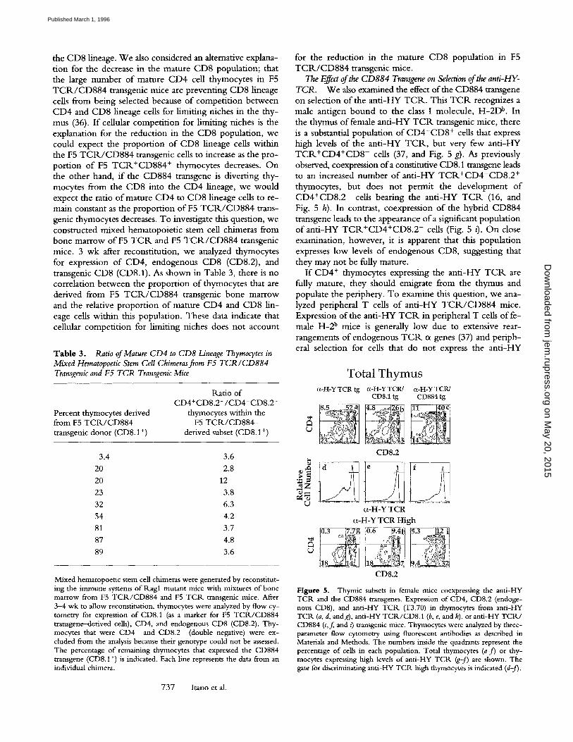
The Effect of the CD884 Transgene on Selection of the anti-HY- TCR. We also examined the effect o f the CD884 transgene on selection o f the anti-HY T C R . This T C R recognizes a male antigen bound to the class I molecule, H - 2 D b. In the thymus o f female ant i -HY T C R transgenic mice, there is a substantial population o f C D 4 - C D 8 + cells that express high levels o f the anti-HY T C R , but very few anti-HY T C R + C D 4 + C D 8 - cells (37, and Fig. 5 g). As previously observed, coexpression of a constitutive CD8.1 transgene leads to an increased number o f anti-HY T C R + C D 4 - C D 8 . 2 + thymocytes, but does not permit the development o f CD4+CD8.2 - cells bearing the anti-HY T C R (16, and Fig. 5 h). In contrast, coexpression o f the hybrid CD884 transgene leads to the appearance o f a significant population o f anti-HY T C R + C D 4 + C D 8 . 2 - cells (Fig. 5 t). O n close examination, however, it is apparent that this population expresses low levels o f endogenous CD8, suggesting that they may not be fully mature.
I f CD4 + thymocytes expressing the anti-HY T C R are fully mature, they should emigrate from the thymus and populate the periphery. To examine this question, we ana- lyzed peripheral T cells o f ant i -HY T C R / C D 8 8 4 mice. Expression of the anti-HY T C R in peripheral T cells o f fe- male H-2 b mice is generally low due to extensive rear- rangements of endogenous T C R c~ genes (37) and periph- eral selection for cells that do not express the anti-HY
Total Thymus c~-FI-Y TCR tg c~-H-Y TCR/ c~-H-Y TCR/
CD8.1 tg CD884 tg
CD8.2
a-H-Y TCR a-H-Y TCR High
,~ ]0.3 !7 .7g } 0 . 6 / 4 4 i ~ ~
CD8.2
Figure 5. Thymic subsets in female mice coexpressing the anti-HY TCR and the CD884 transgenes. Expression of CD4, CD8.2 (endoge- nous CD8), and anti-HY TCR (T3.70) in thymocytes from anti-HY TCR (a, d, andg), anti-HY TCR/CD8.1 (b, e, and h), or anti-HY TCR/ CD884 (c,f,, and t) transgenic mice. Thymocytes were analyzed by three- parameter flow cytometry using fluorescent antibodies as described in Materials and Methods. The numbers inside the quadrants represent the percentage of cells in each population. Total thymocytes (a-j) or thy- mocytes expressing high levels of anti-HY TCR (g-J) are shown. The gate for discriminating anti-HY TCR high thymocytes is indicated (d-f).
737 Itano et al.
on May 20, 2015
jem.rupress.org
Dow
nloaded from
Published March 1, 1996
L y m p h N o d e T Cells
(~-H-Y TCR/ c{-H-Y TCR/ (z-H-Y TCR tg CD8.1 tg CD884 tg
CD8.2
CD8.2
CD4
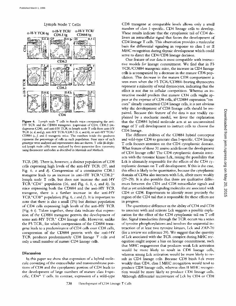
Figure 6. Lymph node T cells in female mice coexpressing the anti- HY TCR and the CD884 transgenes. Expression of CD4, CD8.2 (en- dogenous CD8), and anti-HY TCR in lymph node T cells from anti-HY TCR (a, d, andg), anti-HY TCR/CD8.1 (b, e, and h), or anti-HY TCR/ CD884 (c, f and 0 transgenic mice. The numbers inside the quadrants represent the percentage of cells in each population. Four mice of each genotype were analyzed and representative data are shown. T cells (B-deple- ted lymph node cells) were analyzed by three-parameter flow cytometry using fluorescent antibodies as described in Materials and Methods.
TC1K (38). There is, however, a distinct population o f CD8 cells expressing high levels o f the anti-HY TCP, (37, and Fig. 6, a and d). Coexpression of a constitutive CD8.1 transgene leads to an increase in anti-HY T C R + C D 8 . 2 + lymph node T cells, but does not increase the anti-HY T C R + C D 4 + population (16, and Fig. 6, b, e, and h). In mice expressing both the CD884 and the anti-HY T C R transgene, there is a further increase in the anti-HY TCP,+CD8 + population (Fig. 6, c and f ) . It is important to note that there is also a small (1%) but distinct population o f CD4 cells expressing high levels o f the anti-HY TCIK (Fig. 6 I]. Taken together, these data indicate that expres- sion o f the CD884 transgene permits the development o f some anti-HY T C R + CD4 lineage cells. However, unlike the F5 T C R , for which coexpression of the CD884 trans- gene leads to a predominance o f CD4 cells over CD8 cells, coexpression o f the CD884 protein with the anti-HY TCP, produces predominately CD8 lineage T cells and only a small number o f mature CD4 lineage cells.
Discussion
In this paper we show that expression o f a hybrid mole- cule consisting o f the extracellular and transmembrane por- tions o f CD8 and the cytoplasmic portion o f CD4 leads to the development o f large numbers o f mature class I~spe- cific, CD4 + T cells. In contrast, expression of a wild-type
CD8 transgene at comparable levels allows only a small number o f class 1--specific, CD4 lineage cens to develop. These results indicate that the cytoplasmic tail o f CD4 de- livers an intracellular signal that favors the development o f CD4 lineage T cells. This observation provides a molecular basis for differential signaling in response to class I or II M H C recognition during thymic development which could serve to direct the C D 4 / C D 8 lineage decision.
One feature o f our data is most compatible with instruc- tive models for lineage commitment. We find that in F5 T C R / C D 8 8 4 transgenic mice, the increase in CD4 lineage cells is accompanied by a decrease in the mature CD8 pop- ulation. This decrease in the mature CD8 compartment is seen even when the F5 T C R / C D 8 8 4 - b e a r i n g thymocytes represent a minority o f total thymocytes, indicating that the effect is not due to cellular competition. Whereas an in- structive model predicts that mature CD4 cells might ap- pear at the expense o f CD8 cells, i fCD884 expression "res- cues" already committed CD4 lineage cells, it is not obvious why the development o f CD8 lineage cells should be im- paired. Because this feature o f the data is not readily ex- plained by a stochastic model, we favor the explanation that the CD884 hybrid molecule acts at an uncommitted stage o f T cell development to instruct cells to choose the CD4 lineage.
The different abilities o f the CD884 hybrid coreceptor and wild-type CD8 t o generate class I-specific, CD4 lineage T cells focuses attention on the CD4 cytoplasmic domain. What feature o f these 31 amino acids favors the development o f CD4 lineage cells? The CD4 cytoplasmic domain inter- acts with the tyrosine kinase Lck, raising the possibility that Lck is ultimately responsible for the effects o f the CD4 cy- toplasmic domain on T cell development. If this is the case, this effect is likely to be quantitative, because the cytoplasmic domain o f CD8o~ also interacts with Lck, albeit more weakly (27-29). It is also possible that there are qualitative differ- ences between the CD4 and CD8 intracellular signals and that as yet unidentified signaling molecules are associated with CD4 or CD8. Experiments to identify more precisely the region of the CD4 tail that is responsible for these effects are in progress.
The quantitative difference in the ability o f CD4 and CD8 to associate with and activate Lck suggests a possible expla- nation for the effect o f the CD4 cytoplasmic tail on T cell fate. Signal transduction through the T C R occurs via a series o f tyrosine phosphorylations and involves the sequential in- teraction o f at least two tyrosine kinases, Lck and ZAP-70 (for a review see reference 39). We suggest that the quantity o f Lck associated with the T C R complex during M H C rec- ognition might impose a bias on lineage commitment, such that M H C engagement that produces weak Lck activation would be more likely to result in CD8 lineage cells, whereas strong Lck activation would be more likely to re- sult in CD4 lineage cells. Because CD8 binds Lck more weakly than CD4, class I M H C recognition would tend to produce CD8 lineage cells, whereas class II M H C recogni- tion would be more likely to produce CD4 lineage cells. Although differential recruitment of Lck by CD4 or CD8
738 Development of CD4 Lineage T Cells
on May 20, 2015
jem.rupress.org
Dow
nloaded from
Published March 1, 1996
would have a strong impact on lineage commitment, the level of Lck associated with the T C R could be affected by other factors as well. It has been shown that phosphoryla- tion of the ~ chain of the T C R by Lck and recruitment of ZAP-70 can occur in the absence of CD4 and CD8, im- plying that there are multiple pathways for activating Lck (for a review see reference 39). Although other means of recruiting Lck to the T C R complex have not yet been characterized, it is possible that additional factors, such as the extent of TCP,, cross-linking by MHC, could affect the level of Lck in the T C R complex and influence the C D 4 / CD8 lineage decision independently of coreceptor engage- m e r i t .
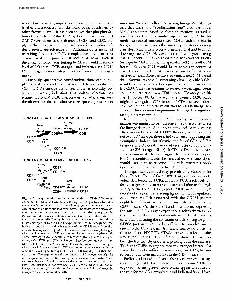
Obviously, quantitative considerations alone cannot ex- plain the strict correlation between T C R specificity and CD4 or CD8 lineage commitment that is normally ob- served. However, indications that positive selection may require prolonged T C R engagement (40, 41), along with the observation that constitutive coreceptor expression can
THYMOCY'I'ES WITH CLASS II SPECIFIC TCRs strong
strong Ick Ick CD4 + s igna l CD4+ S lWCD81Ow CO8-
CD4+ weak
CD8+ Ick ........ ~_~signal
.......... ~ CD41OW
CD8+
THYMOCYTES WITH CLASS I SPEQFIC TCRs strong Ick CD4+
CD4+ s y CD81OW weak
CDS+ Ick weak ~ i g n a I Ick
CD41OW signal CD4" �9 CD8 + C D 8 +
Figure 7. A quantitative, instructive model for the C D 4 / C D 8 hneage decision. This model is based on the assumption that positive selection is not a "single-hit" event, and that M H C engagement influences the lin- eage choice of an uncommitted thymocyte. The width of the arrow de- notes the proportion of thymocytes that take a particular pathway and the the darkness o f the arrow indicates the extent o f Lck activation. Accord- ing to this model, M H C recognition that leads to weak activation of Lck biases development to the CD8 lineage, whereas M H C recognition that leads to strong Lck activation biases toward the CD4 lineage. Most thy- mocytes bearing class II-specific T C R s would receive a strong Lck signal (due to Lck activation by CD4) and would begin to downregulate CD8. These tliymocytes would continue to receive a strong intraceflular signal through the T C R and CD4, and would therefore continue to mature. Most cells bearing class I-specific T C R s would receive a weaker signal (due to weak Lck activation by CD8) and would downregnlate CD4. A continued weak signal through TCP,. and CD8 would permit their final maturation. The requirement for continued M H C recognition after the downregulation of one of the coreceptors serves as a "confirmation" step to insure that cells that downregulate the wrong coreceptor do not ma- ture. Note that if cells that have begun CD8 downregnlation are not yet lineage colrlmitted (8), then the confirmation step could also influence the lineage choice of uncommitted cells.
739 Itano et al.
sometime "rescue" cells of the wrong lineage (9-15), sug- gest that there is a "confirmation step" after the initial M H C encounter. Based on these observations, as well as our data, we favor the model depicted in Fig. 7. In this model, the initial encounter with M H C leads to a bias in lineage commitment such that most thymocytes expressing class II-specific TCRs receive a strong signal and begin to downregulate CD8. However, some thymocytes bearing class II-specific TCRs (perhaps those with weaker avidity for pept ide/MHC on thymic epithelial cells) turn off CD4 instead. Because CD4 would be required for continued class II-specific TCRs that retain expression of CD4 could survive, whereas those that have downregulated CD4 would die, Likewise, most cells expressing class I-specific TCR.s would receive a weaker Lck signal and would downregu- late CD4. Cells that continue to receive a weak signal could complete maturation to a CD8 lineage. Thymocytes with class I-specific TCRs that receive a stronger T C k signal might downregulate CD8 instead of CD4, however those cells would not complete maturation to a CD4 hneage be- cause of the continued requirement for class I recognition throughout maturation.
It is interesting to consider the possibility that the confir- mation step might also be instructive, i.e., that it may affect the lineage decision of an uncommitted cell. Although it is often assumed that CD4+CD8 l~ thymocytes are commit- ted to a CD4 lineage, there is litde evidence supporting this assumption. Indeed, intrathymic transfer of CD4+CD8 l~ thymocytes indicates that some of these cells can differenti- ate into CD8 lineage cells (8). If CD4+CD8 l~ thymocytes are uncommitted, then the signal that they receive upon M H C recognition might be instructive. A strong signal would lead them to become CD4 cells, whereas a weak signal would divert them to the CD8 lineage.
This quantitative model may provide an explanation for the different effects of the CD884 transgene on two indi- vidual class I-specific TCR.s. If the F5 T C R is relatively ef- fective at generating an intracellular signal (due to the high avidity of the F5 TCR. for pept ide/MHC or due to a high density of the positive-selecting ligand on thymic epithelial cells), then the Lck associated with the CD884 protein might be sufficient to divert the majority of cells to the CD4 lineage. On the other hand, thymocytes expressing the anti-HY TCP,. might experience a relatively weak in- tracellular signal during positive selection. If that were the case, then increasing the activation of Lck by engaging the CD884 protein might not be sufficient to complete matu- ration to the CD4 lineage. It is interesting to note that the thymus ofant i -HY TCRJCD884 transgenic mice contains a very prominent CD4+CD8 l~ population. This may re- flect the fact that thymocytes expressing both the anti-HY T C R and CD884 transgenes receive a stronger intracellular signal that may be sufficient to downregulate CDS, but not to permit complete maturation to the CD4 lineage.
Earlier studies (42) indicated that CD4 intracellular sig- nals are dispensable for the development of some CD4 lin- eage cells. At first glance, these results appear to contradict the role for the CD4 cytoplasmic tail indicated here. How-
on May 20, 2015
jem.rupress.org
Dow
nloaded from
Published March 1, 1996
ever, it is possible that the CD4 cytoplasmic tail could pro- mote the development o f CD4 lineage cells and still not be essential for the development o f all CD4 cells. The CD4 cells that develop in the absence o f a CD4 cytoplasmic tail may bear high avidity T C R s that would generate strong intracellular signals. Such cells might not require the Lck associated with CD4 in order to develop into CD4 lineage T cells. The fact that the substitution of physiological levels o f tail-less CD4 for endogenous CD4 restores only a frac- tion o f the normal numbers o f mature CD4 cells is consis- tent with this explanation.
An earlier report describing a hybrid C D 8 / C D 4 trans- gene (18) differs from our results in a number o f important respects. In the previous report, a population o f peripheral T cells expressing both CD4 and endogenous CD8 was ob- served. We see no evidence for such a population in our studies. Moreover, in a preliminary study, expression of the previously described C D 8 / C D 4 hybrid transgene in class II mutant mice did not produce an increase in the mature CD4 population (Jane Pames, personal communication).
The failure to generate class I-specific CD4 cells using this particular hybrid coreceptor could be due to the fact that CD813 was not coinjected with the C D 8 / C D 4 hybrid trans- gene or due to the level or timing of expression of the transgene. It may also be relevant that our C D 8 / C D 4 hy- brid transgene uses the transmembrane segment o f CDS, whereas the one described in the earlier study uses the transmembrane segment o f CD4.
A number o f recent studies have emphasized the stochas- tic nature o f the process of C D 4 / C D 8 lineage commitment and the lack o f an absolute requirement for CD4 and CD8 intracellular domains in thymic development. In spite of this, the critical issue o f whether intracellular signals gener- ated upon M H C recognition influence the fate of uncom- mitted thymocytes remains unresolved. The data in our pa- per clearly demonstrate that the CD4 intraceUular domain does play an important role in the C D 4 / C D 8 lineage deci- sion. Moreover, our data are most compatible with the no- tion that CD4 intracellular signals act on uncommitted thy- mocytes to influence their lineage decision.
We thank Mimi Mong for technical assistance, Peter Schow for assistance with flow cytometry, Paul Gott- lieb (University of Texas, Austin, TX) for providing the CD8~.I genomic clone, David Baltimore (MIT, Boston, MA) for providing Ragl mutant mice, Tak Mak (Amgen, Toronto, Canada) for providing CD8cx mutant mice, Laurie Glimcher (Harvard University, Boston, MA) for providing class II MHC mutant mice, Harald yon Boehmer (Basel Institute, Switzerland) for providing anti-HY TCR transgenic mice, Joe Bolen (Bristol-Myers Squibb, Princeton, NJ) for providing anti-Lck antisera, and R. Axel (Columbia University, New York), F. Ramsdell (Darwin Molecular, Seattle, WA), D. Raulet (University of California, Berkeley, CA), and members of the Robey laboratory for comments on the manuscript.
This work was supported by National Institutes of Health (NIH) grant AI-32985 to E. Robey. R. Salmon was supported by Fondation pour la Recherche M6dicale. The initial phases of this work were carried out in Richard Axel's laboratory (Columbia University, New York), and were supported by the NIH.
Address correspondence to Dr. Ellen Robey, Department of Molecular and Cell Biology, Division of Im- munology, Room 471, University of California, Berkeley, CA 94720.
Received for publication 25 August 1995 and in revised form 2 November 1995.
References 1. von Boehmer, H. 1986. The selection of the alpha beta het-
erodimeric T cell receptor for antigen. Immunol. Today. 7: 333-336.
2. Sha, W.C., C.A. Nelson, R.D. Newherry, D.M. Kranz, J.H. Russell, and D.Y. Loh. 1988. Selective expression of an anti- gen receptor on CD8-bearing T lymphocytes in transgenic mice. Nature (Lond.). 335:271-274.
3. Kisielow, P., H.S. Teh, H. Bluthmann, and H. von Boehmer. 1988. Positive selection of antigen-specific T cells in thymus by restricting MHC molecules. Nature (Lond.). 335:730-733.
4. Berg, L.J., A. Pullen, and M.M. Davis. 1989. Antigen/ MHC-specific T cells are preferentially exported from the thymus in the presence of their MHC ligand. Cell. 58:1035- 1046.
5. Kaye, J., M.L. Hsu, M.E. Sauron, S.C. Jameson, N.R. Gas- coigne, and S.M. Hedrick. 1989. Selective development of CD4 § T cells in transgenic mice expressing a class II M H C - restricted antigen receptor. Nature (Lond.). 341:746-749.
6. Gmsby, M., H.J. Auchincloss, R. Lee, R. Johnson, J. Spen-
cer, M. Zijlstra, R. Jaenisch, V. Papaioannou, and L. Glim- cher. 1993. Mice lacking major histocompatibility complex class I and class II molecules. Proc. Natl. Acad. Sci. USA. 90: 3913-3917.
7. Chan, S.H., O. Cosgrove, C. Waltinger, C. Benoist, and O. Mathis. 1993. Another view of the selective model of thy- mocyte selection. Cell. 73:225-236.
8. Lundberg, K., W. Heath, F. Kontgen, F. Carhone, and K. Shortman. 1995. Intermediate steps in positive selection: differentiation of CD4+CD8intTCR mt thymocytes into CD4-CD8+TCR hi thymocytes. J. Exp. Med. 181:1643- 1651.
9. Davis, C.B., N. Killeen, M.E.C. Crooks, D. Raulet, and D.R. Littman. 1993. Evidence for a stochastic mechanism in the differentiation of mature subsets of T lymphocytes. Cell. 73:237-247.
10. Robey, E., A. Itano, W.C. Fanslow, and B.J. Fowlkes. 1994. Constitutive CD8 expression allows inefficient maturation of CD4 + helper T cells in class II MHC mutant mice. J. Exp.
740 Development of CD4 Lineage T Cells
on May 20, 2015
jem.rupress.org
Dow
nloaded from
Published March 1, 1996
Med. 179:1997-2004. 11. Itano, A., D. Kioussis, and E. Robey. 1994. Stochastic com-
ponent to the development of class I MHC specific T cells. Pro& Natl. Acad. Sci, USA. 91:220-224.
12. Paterson, R., L. Burkly, D. Kurahara, A. Dunlap, R. Flavell, and T. Finkel. 1994. Thymic development in human CD4 transgenlc mice.J. Immunol. 153:3491-3503.
13. Chan, S., C. Walzinger, A. Baron, C. Benoist, and D. Mathis. 1994. Role of coreceptors in positive selection and lineage commitment. EMBO (Eur. Mol. Biol. Organ.) J. 13: 4482-4489.
14. Baron, A., K. Hafen, and H. von Boehmer. 1994. A human CD4 transgene rescues CD4-CD8 + cells in beta 2-micro- globulin--deficient mice. Eur. J. Immunol. 24:1933-1936.
15. Corbella, P., D. Moskophidis, E. Spanopoulou, C. Ma- malaki, M. Tolaini, A. Itano, D. Lans, D. Baltimore, E. Robey, and D. Kioussis. 1994. Functional commitment of helper T cell lineage precedes positive selection and is inde- pendent of T cell receptor MHC specificity. Immunity. 1: 269--276.
16. Robey, E.A., B.J. Fowlkes, J.W. Gordon, D. Kioussis, H. yon Boehmer, F. Ramsdell, and R. Axel. 1991. Thymic se- lection in CD8 transgenic mice supports an instructive model for commitment to a CD4 or CD8 lineage. Cell. 64:99-107.
17. Borgulya, P., H. Kishi, U. Muller, J. Kirberg, and H. von Boehmer. 1991. Development of the CD4 and CD8 lineage of T cells: instruction versus selection. EMBO (Eur. Mol. Biol. Organ.)J. 10:913-918.
18. Seong, R.H. ,J .W. Chamberlain, andJ.R. Pames. 1992. Sig- nal for T-cell differentiation to a CD4 cell lineage is delivered by CD4 transmembrane region and/or cytoplasmic tail. Na- ture (Lond.). 356:718-720.
19. Zamoyska, R., A.C. Vollmer, K.C. Sizer, C.W. Liaw, andJ.R. Parnes. 1985. Two Lyt-2 polypeptides arise from a single gene by alternative splicing patterns of mRNA. Cell. 43:153-163.
20. Littman, D., and S. Gettner. 1987. Unusual intron in the im- munoglobulin domain of the newly isolated murine CD4 (L3T4) gene. Nature (Lond.). 325:453-455.
21. Youn, H.J., J.V. Harriss, and P.D. Gottlieb. 1988. Structure and expression of the Lyt-3a gene of C.AKR mice. Immuno- genetics. 28:353-361.
22. Spanopoulou, E., C. Roman, L. Corcoran, M. Schlissel, D. Silver, D. Nemazee, M. Nussenzweig, S. Shinton, R. Hardy, and D. Baltimore. 1994. Functional immunoglobulin trans- genes guide ordered B-cell differentiation in Rag-l-deficient mice. Genes & Dev. 8:1030-1042.
23. Green, S., I. Issemann, and E. Sheer. 1988. A versatile in vivo and in vitro eukaryotic expression vector for protein engineer- ing. Nucleic Acids Res. 16:369-372.
24. Grusby, M.J., R.S. Johnson, V.E. Papaioannou, and L.H. Glimcher. 1991. Depletion of CD4 + T cells in major histo- compatibility complex class II-deficient mice. Science (Wash. DC). 253:1417-1420.
25. Mamalaki, C., J. Elliott, T. Norton, N. Yannoutsos, A.R. Townsend, P. Chandler, E. Simpson, and D. Kioussis. 1993. Positive and negative selection in transgenic mice expressing a T-cell receptor specific for influenza nucleoprotein and en- dogenous superantigen. Dev. Immunol. 3:159-174.
26. Greaves, D.R., F.D. Wilson, G. Lang, and D. Kioussis. 1989. Human CD2 3'-flanking sequences confer high-level T cell- specific position-independent gene expression in transgenic
mice. Cell. 56:979-986. 27. Veillette, A., M.A. Bookman, E.M. Horak, and J.B. Bolen.
1988. The CD4 and CD8 T cell surface antigens are associ- ated with the internal membrane tyrosine-protein kinase p561ck. Cell. 55:301-308.
28. Wiest, D., L. Yuan, J. Jefferson, P. Benveniste, M. Tsokos, R. Klausner, L. Glimcher, L. Samelson, and A. Singer. 1993. Regulation of T cell receptor expression in immature CD4+CD8 + thymocytes by p561ck tyrosine kinase: basis for differential signaling by CD4 and CD8 in immature thy- mocytes expressing both coreceptor molecules. J. Exp. Med. 178:1701-1712.
29. Ravichandran, K., and S. Burakoff. 1994. Evidence for dif- ferential intracellular signaling via CD4 and CD8 molecules. J. Exp. Med. 179:727-732.
30. Nakayama, K., K. Nakayama, I. Negishi, K. Kuida, M. Louie, O. Kanagawa, H. Nakauchi, and D. Loh. 1994. Require- ment for CD8 beta chain in positive selection of CD8-1in- eage T cells. Science (Wash. DC). 263:1131-1133.
31. Crooks, M.E.C., and D.R. Littman. 1994. Disruption of T lymphocyte positive and negative selection in mice lacking the CD8 beta chain. Immunity. 1:277-286.
32. Itano, A., D. Cado, F.K.M. Chan, and E. Robey. 1994. A role for the cytoplasmic tail of the beta chain of CD8 in thy- mic selection. Immunity. 1:287-290.
33. Zamoyska, R., P. Derham, S.D. Gorman, P. von Hoegen, J.B. Bolen, A. Veillette, and J.R. Pames. 1989. Inability of CD8 alpha' polypeptides to associate with p561ck correlates with impaired function in vitro and lack of expression in vivo. Nature (Lond.). 342:278-281.
34. Cosgrove, D., D. Gray, A. Dierich, J. Kaufman, M. Lemeur, C. Benoist, and D. Mathis. 1991. Mice lacking MHC class I1 molecules. Cell. 66:1051-1066.
35. Pircher, H., N. Rebai, M. Groettrup, C. Gregoire, D.E. Speiser, M.P. Happ, E. Palmer, R.M. Zinkernagel, H. Hen- gartner, and B. Malissen. 1992. Preferential positive selection of V alpha 2+CD8 + T cells in mouse strains expressing both H-2k and T cell receptor V alpha a haplotypes: determination with a V alpha 2-specific monoclonal antibody. Eur. J. lmmu- nol. 22:1399-1404.
36. Huesmann, M., B. Scott, P. Kisielow, and H. von Boehmer. 1991. Kinetic and efficacy of positive selection in the thymus of normal and T cell receptor transgenic mice. Cell. 66:533- 540.
37. von Boehmer, H. 1990. Developmental biology ofT cells in T cell-receptor transgenic mice. Annu. Rev. Immunol. 8:531-556.
38. Rocha, B., and H. von Boehmer. 1991. Peripheral selection of the T cell repertoire. Science (Wash. DC). 251:1225-1228.
39. Weiss, A., and D. Littman. 1994. Signal transduction by lym- phocyte antigen receptors. Cell. 76:263-274.
40. Brandle, D., S. Muller, C. Muller, H. Hengartner, and H. Pircher. 1994. Regulation of R_AG-1 and CD69 expression in the thymus during positive and negative selection. Eur. J. Immunol. 24:145-151.
41. Kisielow, P., and A. Miazek. 1995. Positive selection of T cells: rescue from programmed cell death and differentiation require continual engagement of the T cell receptor. J. Exp. Med. "181:1975-1984.
42. Killeen, N., and D. Littman. 1993. Helper T-cell develop- ment in the absence of CD4-p561ck association. Nature (Lond.). 364:729-732.
741 Itano et al.
on May 20, 2015
jem.rupress.org
Dow
nloaded from
Published March 1, 1996
Related Documents
![High Fat Diet Rapidly Suppresses B Lymphopoiesis by ......myeloid lineage cells on a single plot [21]. T-cells were stained with CD4 and CD8, and myeloid cells were stained with Mac-1](https://static.cupdf.com/doc/110x72/5ffe554f4b37640a6277a79b/high-fat-diet-rapidly-suppresses-b-lymphopoiesis-by-myeloid-lineage-cells.jpg)