4*3 The Cytoplasmic Cytology of Sarcoma 180 By LEONARD G. WORLEY AND HERMAN W. SPATER (From Brooklyn College, Brooklyn, N. Y.) With four plates (figs. 1-4) SUMMARY Typical sarcoma 180 cells present a large spherical lipoid or Golgi zone adjacent to the nucleus within which discrete, homogeneous and vesicular, Golgi bodies (lipo- chondria) are distributed. No Golgi network can be detected in the living cell, but such networks may be produced artificially by means of hypertonic solutions and by shrinking vitally stained cells. All of the cytoplasm, except the lipoid zone, is strongly basiphil owing to the presence of ribonucleic acid. Mitochondria, mostly rod shaped, form a halo of variable width surrounding the lipoid zone except adjacent to the nucleus. These lie partly within the ribonucleic acid containing cytoplasm. Tests for glycogen, alkaline, and acid phosphatase, and for lipase are largely negative. A positive reaction to Bourne's test for ascorbic acid is given by many of the lipochondria. S ARCOMA 180 is one of the transplantable, non-metastasizing, connective tissue tumours of the mouse. A bit of sarcoma material, not exceeding 2 mm. in diameter, implanted beneath the skin of the breast as described by Zahl and Drasher (1947), grows within a fortnight into a conspicuous malig- nant tissue of such proportions that the host usually soon succumbs. Although studies have been made on the nuclear and cytoplasmic cytology of this and closely related tumours (Levine, 1939; MacCardle, 1951, and others), and Ludford and Smiles (1950) have recently compared the structure of mouse sarcoma cells with that of normal fibroblasts as seen in phase-contrast and ultra-violet micrographs, little pertinent work has been published regarding the finer cytological details and there is essentially no information available regarding the cytochemistry of the cytoplasm and its inclusions. In the early part of the century there was a flurry of interest in the morpho- logy of the cytoplasm of cancer cells, particularly with reference to the Golgi apparatus (see Cowdry, 1924, and Ludford, 1951, for many references). In these studies the Golgi network was frequently reported to be unusually elaborate in cancer cells in general. However, few modern cytologists support the 'network' theory of Golgi body structure and, considering the importance of the tumour cell in the economy of the animal body, a re-examination of the morphology of the cytoplasm of these cells, particularly with reference to the Golgi material, is desirable. Sarcoma 180 cells are particularly appropriate for study in this connexion because of the extreme malignancy of this tissue and the ease with which material of known age may be obtained and examined promptly or fixed for subsequent study. [Quarterly Journal of Microscopical Science, Vol. 93, part 4, pp. 413-425, Dec. 1952.]

Welcome message from author
This document is posted to help you gain knowledge. Please leave a comment to let me know what you think about it! Share it to your friends and learn new things together.
Transcript

4*3
The Cytoplasmic Cytology of Sarcoma 180
By LEONARD G. WORLEY AND HERMAN W. SPATER
(From Brooklyn College, Brooklyn, N. Y.)
With four plates (figs. 1-4)
SUMMARY
Typical sarcoma 180 cells present a large spherical lipoid or Golgi zone adjacent tothe nucleus within which discrete, homogeneous and vesicular, Golgi bodies (lipo-chondria) are distributed. No Golgi network can be detected in the living cell, but suchnetworks may be produced artificially by means of hypertonic solutions and byshrinking vitally stained cells. All of the cytoplasm, except the lipoid zone, is stronglybasiphil owing to the presence of ribonucleic acid. Mitochondria, mostly rod shaped,form a halo of variable width surrounding the lipoid zone except adjacent to thenucleus. These lie partly within the ribonucleic acid containing cytoplasm. Tests forglycogen, alkaline, and acid phosphatase, and for lipase are largely negative. A positivereaction to Bourne's test for ascorbic acid is given by many of the lipochondria.
SARCOMA 180 is one of the transplantable, non-metastasizing, connectivetissue tumours of the mouse. A bit of sarcoma material, not exceeding
2 mm. in diameter, implanted beneath the skin of the breast as described byZahl and Drasher (1947), grows within a fortnight into a conspicuous malig-nant tissue of such proportions that the host usually soon succumbs. Althoughstudies have been made on the nuclear and cytoplasmic cytology of this andclosely related tumours (Levine, 1939; MacCardle, 1951, and others), andLudford and Smiles (1950) have recently compared the structure of mousesarcoma cells with that of normal fibroblasts as seen in phase-contrast andultra-violet micrographs, little pertinent work has been published regardingthe finer cytological details and there is essentially no information availableregarding the cytochemistry of the cytoplasm and its inclusions.
In the early part of the century there was a flurry of interest in the morpho-logy of the cytoplasm of cancer cells, particularly with reference to the Golgiapparatus (see Cowdry, 1924, and Ludford, 1951, for many references). Inthese studies the Golgi network was frequently reported to be unusuallyelaborate in cancer cells in general. However, few modern cytologists supportthe 'network' theory of Golgi body structure and, considering the importanceof the tumour cell in the economy of the animal body, a re-examination ofthe morphology of the cytoplasm of these cells, particularly with reference tothe Golgi material, is desirable. Sarcoma 180 cells are particularly appropriatefor study in this connexion because of the extreme malignancy of this tissueand the ease with which material of known age may be obtained and examinedpromptly or fixed for subsequent study.[Quarterly Journal of Microscopical Science, Vol. 93, part 4, pp. 413-425, Dec. 1952.]

414 Worley and Spater—The Cytoplasmic Cytology of Sarcoma 180
MATERIAL AND METHODS
All mice used in this study Were males of the inbred Rockland Swiss strain,18-22 grammes in weight, and were provided by the Haskins Laboratories,New York City. Mice were received within a few hours after the transfer ofsarcoma tissue at the Haskins Laboratories. Slices of growing tumours werefixed, or studied without fixation, at varying intervals of from 1 to 12 days afterimplantation; but tumours 5 days after transfer have received the greatestamount of attention, for although these are generally not too large for optimumcytological fixation, a sufficient time after implantation has elapsed to ensurethat the tumour is developing typically from the standpoint both of form andrate of growth. Furthermore, 5-day tumours have reached the steepest partof the weight-growth curve for this tumour (Zahl and Drasher, 1947). The5-day tumour is usually roughly spherical or slightly ovoid and typically showsnone of the irregularities in growth that are soon to develop.
The following methods were employed:
Vital staining. Bits of fresh tumour tissue were cultivated for from 2 to 5hours at either 50 C. or 37° C , in either Tyrode's or Locke-Lewis's solutionto which methylene blue, 1 part in 25,000, had been added. Other fragmentswere kept in Janus green B of similar concentration, and others for shorterintervals, up to one hour, in neutral red, 1 part in 50,000.
No appreciable difference was noted in the results obtained at the twodifferent temperatures and in both cases many cells could be found that werebelieved to be viable because of the localization of the stain in discrete in-clusions in the cytoplasm, without diffuse staining of either the cytoplasm orthe nucleus. In the case of methylene blue and Janus green B, cells wereinterpreted as viable so long as the ground cytoplasm and nucleus continued toreduce the stain to the colourless form. Our observations on fresh material havebeen confined to those that could be made during the first 20 minutes afterthe preparation of a fresh mount.
Neutral red and methylene blue, used vitally, stain what we believe to bethe same cytoplasmic elements to be described later, but while neutral red actsrapidly and methylene blue stains much more slowly, neutral red also ratherquickly brings about an artificial enlargement of the inclusions to such adegree that such preparations are considered unreliable. Methylene blue, onthe other hand, by behaving much more slowly, permits at least some observa-tion to be made after the inclusions are stained but before any appreciableartificial enlargement occurs. There is no doubt, however, but that both ofthese vital dyes produce abnormalities when care is not taken to employ themat the minimum concentration necessary to stain the inclusions (see Dustin,1947). It appears to us that much of the criticism of vital staining methods hasbeen made by those who have been careless or indifferent in using them.
Our experience with Janus green B has been disappointing. Seldom havewe been able to stain the mitochondria in cells which, through the failure ofother elements to stain, could be considered viable. With the death of the

Worley and Spater—The Cytoplasmic Cytology of Sarcoma 180 415
cell, however, as indicated by diffuse staining of both the cytoplasm and thenucleus, the mitochondria often appear deeply stained. Our experience withthe methods we have employed is that the 'vital' staining of the mitochondriais typically a post-vital phenomenon in these cells. This, however, may be dueto deficiencies in the brands of Janus green B we have used. Such preparationsprovide adequate data on the distribution within the cell of these inclusions,but considering the extreme lability of the mitochondria we are doubtful ofthe adequacy of such methods in determining mitochondrial form.
Unfixed and unstained tissue. Unstained living cells were also examined forcomparison with the vitally stained and fixed tissue. These were studied bothby ordinary microscopic methods and with phase-contrast equipment. Ingeneral, these methods have served largely to verify observations made byother means.
Permanent and semi-permanent preparations
For the demonstration of lipides, tissues were fixed in 10 per cent, formalinand in Baker's (1946) formaldehyde-calcium. For total fat content, frozensections of tissue embedded in gelatine were stained with sudan IV, sudanblack B in 70 per cent, alcohol, and in nile blue (sulphate) in distilled water.Sections were left in sudan IV overnight, were then washed in 70 per cent,alcohol and mounted in Farrants's medium. Sudan black B acted much morequickly, staining the lipides in 10 minutes or less. Sections were differentiatedin 70 per cent, alcohol for a few minutes and then mounted in Farrants'smedium for observation. A 1 per cent, solution of nile blue stained othersections in 5 minutes. These were then differentiated for 30 seconds to 1minute in a 1 per cent, aqueous solution of glacial acetic acid and were thenmounted for study in Farrants's medium.
Ice-cold acetone was employed as a fixative in a search for acid phosphatase,according to the method of Gomori (1941), as modified by Wolf, Kabat, andNewman (1943), and for the detection of lipase(esterase), following the methodof Gomori (1945) and that of Wachstein (1946). Ice-cold acetone was alsoused as a fixative in an attempt to demonstrate sites of alkaline phosphataseactivity by the azo-dye method of Seligman and Manheimer (1948). Acidphosphatase and esterase activity were also sought for by the azo-dye tech-nique of Seligman and Manheimer (1949) and Seligman and Nachlas (1949).Rat duodenum, known for its high alkaline phosphatase activity, and ratliver, a tissue with high acid phosphatase and esterase activity, were usedas controls.
Eighty per cent, alcohol was used as a fixative in the demonstration of or-ganic iron, after the method of Mallory (1938). After acid hydrolysis Turnbullblue and Prussian blue preparations were exposed to MacCallum's haema-toxylin to intensify the reaction. These preparations were subsequently sub-jected to treatment with hot ether/absolute ethyl alcohol (equal parts) for30-40 minutes to extract all haematoxylin not in combination with ironcompounds.

4i6 Worley and Spater—The Cytoplasmic Cytology of Sarcoma 180
Helly's fixative was followed by Mallory's eosin methylene blue technique,by Sonnenblick's toluidin blue method, by haematoxylin/eosin/azure II, bythe Masson method, and by the Feulgen and Giemsa methods. These pre-parations were used for general histological orientation, the demonstrationof cytoplasmic basiphilia, and for determining the distribution of aldehydes.Slides incubated for 3 hours at 60° C. in 0-005 per cent, ribo-nuclease with aphosphate buffer were used as a check to determine whether or not thecytoplasmic basiphilia could be attributed to the ribonucleic acid content.Sections treated in this manner lacked the cytoplasmic basiphilia whenstained subsequently with toluidin blue, eosin/methylene blue, and Giemsa.The basiphilia was retained in parallel control slides treated simultaneouslyin exactly the same manner except for the omission of ribonuclease from thehot buffer.
Flemming's solution (containing only 2 drops of glacial acetic acid per5 ml. of fluid), followed by a 5-day treatment at 37° C. in 2 per cent, osmiumtetroxide was used for the demonstration in permanent form of the Golgimaterial. It has been our experience that this technique generally results inlittle or no distortion of the Golgi elements as seen in fresh material. Theirform in such preparations is also essentially identical to that found by vitalstaining with methylene blue. This procedure is, therefore, consideredsuperior to the methods of Mann-Kopsch, Kolatchev, and Da Fano.
Tissue similarly fixed, but not post-osmicated, was followed by ironhaematoxylin staining for the general relationship of cellular inclusionsand nuclear structure. Similarly fixed tissue, post-chromed at 370 C. for4-5 days in 3 per cent, potassium dichromate, was stained with Bensley'saniline acid fuchsin and methyl green in making permanent preparations ofmitochondria.
Regaud's fluid, followed by an 8-day post-chroming in 3 per cent, potassiumdichromate, was also used for the study of mitochondria after staining withaniline acid fuchsin and methyl green.
Rossman's fixing fluid, followed by the Feulgen-Bauer staining method(Bensley and Bensley, 1938) was used for the demonstration of glycogen.
The technique of Bourne (1936) was used on fresh frozen tissue in thedemonstration of what we presume to be ascorbic acid.
OBSERVATIONS
Sections of 5-7 day tumours present a rather bewildering array of cellsshowing great variation in size and general structure. Zahl and Drasher(1947), whose terminology is followed here and whose description should beconsulted for general structure of sarcoma 180, recognize at least two typesof viable and presumably malignant cells which can be identified in frag-mented, unstained fresh tissue. This is, however, a conservative classificationwhich under-emphasizes the great variability particularly with regard to thecytoplasmic content as seen in suitably fixed preparations. There is, forexample, a marked difference in total fat content in cells from different

Worley and Spater—The Cytoplasmic Cytology of Sarcoma 180 417
regions. This component increases greatly as one passes in observationthrough the malignant cortex and approaches the boundary between thisarea and the necrotic centre. Cells of the intermediate zone between these twomajor areas have accumulated fat to a very marked degree. In sudan IV andnile blue preparations, such cells comprise a conspicuous band immediatelysurrounding the caseous central mass (fig. 1, A and B).
Nevertheless, when the peripheral areas of the tumour at points of reactionwith host tissues, the intermediate zone, and the caseous centre consistinglargely of dead and dying cells (class B cells of Zahl and Drasher, 1947) aredisregarded and attention is confined to the firm rind, cortex or peripheralzone, a degree of uniformity can be noted and the problem is simplifiedmaterially. Since it is this peripheral region that is considered to possess thegreatest degree of malignancy and is therefore selected as the source of frag-ments for transfer, cells of this area may be considered more typical repre-sentatives of the malignant process from the standpoint of structure, tumourgrowth, and multiplication of cells. This is the region that has taken most ofour attention, but it should be noted that a study which embraced all of thezones of the tumour from extreme periphery to caseous centre might proveexceedingly interesting since there is some indication that the position of thecell within the malignant mass is a rough index of its functional age.
Cytoplasmic basiphilia. Since the cytoplasmic basiphilia in these sarcomacells can be abolished by treatment with ribonuclease, the basiphilia ispresumed to be due to the presence of pentose nucleic acid. One of the mostcharacteristic features of these cells is the pronounced basiphilia of the cyto-plasm. This is usually diffuse and resembles the chromophilic substance whichis found in the basal portion of normal pancreas acinar cells, although cellshave been observed where the material appears to be arranged in the form ofclub-like rods, possibly to be identified as mitochondria. The basiphilicmaterial appears to be more heavily concentrated at the periphery of the celland towards the ends of cells which are unusually elongate.
Some, especially small, cells show the entire cytoplasm intensely basiphil,but in cells considered more typical the basiphilia is either much less intenseor lacking altogether in a spherical area of variable size near the nucleus. Thisis the area where lipoid is concentrated. Cells stained for basiphil cytoplasmicsubstances do not show the lipoid as such, but its location is characteristicallyindicated by this very pale spherical area lying in the otherwise stronglybasiphil cytoplasm (fig. 1, c and D).
These statements apply to the cells of the peripheral zone. As one passes in-ward toward the more necrotic central area, the cells lose their basiphiliarather abruptly and in the cells of the caseous centre, the cytoplasm andfrequently the nucleus, are quite uniformly acidophilic. Pycnotic nuclei andformless nuclear materials remain basiphil, extracellular nuclear remainsare strongly basiphil, but intracellular, possibly degenerating, nuclei oftengive a basic reaction. Cells in mitosis may show either strongly or weaklybasiphil cytoplasm. Cells with acidophil nuclei have never been observed

418 Worley and Spater—The Cytoplasmic Cytology of Sarcoma 180
undergoing mitosis. Amitotic division in cells of the caseous centre, however,is sometimes noted (fig. 2, A).
Golgi complex. Another striking feature of the principal cells of the sarcomacortex is the elaborate character of the Golgi complex. It consists of homo-geneous lipoid granules, recalling the Golgi 'presubstance' of Hirsch (1939),and larger bipartite spheres with osmiophilic surfaces and osmiophobiccentres, recalling the Golgi 'systems' described by Hirsch (1939) for manykinds of animal cells. In life, these granules and droplets are pale yellow; theyblacken with prolonged osmication (fig. 2, B and c); they stain very dark blue-grey with Baker's (1946) acid haematein test for phospholipides; they aregenerally dark blue with nile blue; they are readily blackened with sudanblack B and they are stainable vitally both with neutral red and with methy-leneblue (fig. 2, D). These are typical examples of what Baker (1950) has calledlipochondria. The striking similarity in the form and distribution of theseGolgi bodies between suitably prepared osmicated cells and living cellsvitally stained with methylene blue is illustrated in fig. 2, c and D (A, Ax;B, Bx; C, Cx). When the same picture is presented by fixed preparations andvitally stained living cells, one is justified in having a degree of confidence inthe methods employed.
It is characteristic for these Golgi elements to occupy a roughly sphericalarea at one side of, and frequently partly within, a depression or hilus in thenucleus (fig. 3, A). This large area appears to consist largely of diffuse lipoid,probably composed of lipoid particles of submicroscopic dimensions. Baker'sacid haematein test colours this area a diffuse grey-blue (fig 3, B) and the wholespherical area with the discrete granules of lipoid suspended within is veryreminiscent of Baker's (1944) interpretation of the 'Golgi element', in whichboth diffuse and concentrated lipoid was recognized. In some sarcoma cells,especially those that are unusually large, the lipoid zone occupies almost allof the cytoplasm (figs. 1, D and 3, c). The relative firmness of this lipoid mass isbelieved to be responsible for the frequently observed apparent distortion ofthe nucleus which involves the formation of a hilus (fig. 1, D).
No discrete lipoid zone seems to be identifiable in cells undergoing division.In such cells, individual Golgi bodies or spheres are widely scattered through
FIG. I
A. Low power ( X ioo) view of a frozen section through the centre of sarcoma 180 showinghigh total lipide concentration in cells of intermediate zone {A) immediately surroundingcaseous, necrotic portion (B). The cells on each side of this marginal zone are relatively free ofneutral fats. Nile blue.
B. High power ( X 2000) view of lipide-laden intermediate-zone cells closely surroundingcaseous centre. Flemming's fluid followed by 5-day treatment with 2 per cent, osmiumtetroxide.
c. Cytoplasmic basiphilia of cells of malignant cortex or peripheral zone. Note the peri-pheral distribution in each cell of the basiphilia and its absence from a large spherical areawithin the cytoplasm. Zenker fixation; Giemsa staining. Preparation of T. Sardinsky ( X 1200).
D. Same ( x 2000) showing similar relationship of parts in a giant tumour cell with unusuallylarge pale lipide zone and peripheral dark-staining ribonucleic acid containing cytoplasm.Zenker fixation; Giemsa staining. Preparation of T. Sardinsky.

Worley and Spater—The Cytoplasmic Cytology of Sarcoma 180 419
the cytoplasm and are not concentrated in a definite area. Both homogeneousgranules and larger more complex forms are found, but the general impressionis that cells undergoing mitosis contain fewer and smaller Golgi bodies. Thisis believed to account for the difficulty some investigators have experienced(Dalton, 1951) in demonstrating a Golgi 'network' during division. Smallhomogeneous Golgi bodies are not so readily distorted as are the larger forms.However, some Golgi material has been present in all of the cells of the cortexwe have examined. During mitosis, the cytoplasm and cytoplasmic inclusionsgive the impression of being relatively inactive and there is no evidencewhatever, from our observations, that the Golgi bodies divide during celldivision.
With regard to the character and number of these lipoid granules andspheres, two general areas of the malignant peripheral zone of the tumour canbe recognized. In the more external areas, in which the cells themselves aresmaller, the number and size of the Golgi bodies in each cell are proportionallyless than in the deeper zone of somewhat larger cells. Likewise, in the deepercells, the number of vesicular Golgi bodies (Golgi 'systems' of Hirsch, 1939)is correspondingly increased. Sudan black affects only the surface of thesevesicular elements, leaving their centres unstained. With their increased num-bers they occupy a greater mass of the cytoplasm.
The Golgi picture presented here deviates from the classical view of thisinclusion as an 'apparatus' or 'network', but it is in general accord with themore modern accounts of its structure as presented by Hirsch (1939), Baker(1944), Thomas (1947), and others, contrary to the opinion of Dalton (1951)and MacCardle (1951). Our studies fail to reveal anything resembling anetwork in these sarcoma cells when they are examined either in the freshcondition or in most fixed preparations. However, such a network can beproduced artificially in several ways. When the lipoid inclusions are vitallystained with methylene blue, or post-vitally stained with nile blue, or withsudan black B, and the cell is subsequently shrunken by treatment with ahypertonic solution, the network makes its appearance and is the same colouras the colour of the stain employed. Moreover, this transformation can befollowed while the Golgi material is under direct observation, proving clearlyto the observer that the result is due to the shrinkage treatment (fig. 3, D).
FIG. 2A. Cell of caseous centre (arrow) undergoing amitotic division ( X 1200). Baker's acid haema-
tein method.B. Cells of malignant cortex ( X 1200) showing characteristic distribution of Golgi material
in spherical zone near nucleus. Flemming's fixation, followed by 5-day treatment with osmiumtetroxide.
C. Same as B ( X 1800), especially for comparison with vitally stained cells of D. Note the closecorrespondence in the distribution of the osmiophilic (chromophilic) substances (A-Ax;B-Bi; C-Cj).
D. Cells vitally stained with methylene blue ( X 1200) for comparison with osmicated materialof c. Note the close correspondence in distribution and appearance between vitally stainedinclusions and osmiophilic substances of c (A—Ax; B—Bx; C-Cx).

420 Worley and Spater—The Cytoplastnic Cytology of Sarcoma 180
Here, too, the Golgi 'network' is an artifact and it appears to us that con-clusions drawn concerning the Golgi material, when this structure is inter-preted as a 'network' such as those of MacCardle (1951), have little value.
Mitochondria. There is considerable variability in the mitochondrial picturein various cells of the tumour, just as there is in the other cytoplasmic com-ponents. Generally speaking, the mitochondria in fixed preparations appearspherical or rod-shaped. In cells post-vitally stained with Janus green B theyare chiefly in the form of rods. Filamentous mitochondria have been observedonly in cells in hypertonic media and we doubt if such forms occur in theliving sarcoma cells. In what are considered to be the more typical cells, themitochondria occupy a peripheral position, avoiding the more central Golgizone. They form an incomplete halo around the lipoid zone with an inter-ruption on the side of the lipoid area facing the nucleus (fig. 4, A). This regionof the cell, as pointed out above, contains the heavy concentration of ribo-nucleic acid, as shown by its strongly basiphil character. In most cells, stainedwith acid fuchsin, a pale spherical area lying near the nucleus, apparentlycompletely devoid of mitochondria but surrounded by them, is seen. In suchcells a very clear and positive differentiation between the Golgi bodies andthe mitochondria is readily apparent. Viewing such cells, one cannot help butthink of the spherical Golgi bodies as representing aggregations of lipoidalsubstance in areas of diffuse lipoid, and of the mitochondria as somewhatsimilar aggregations of material within the ribonucleic acid containing cyto-plasm (fig. 4, A).
Thus far things are fairly clear, but the relationship becomes less welldefined when other tumour cells are examined. In some cells, particularlythose of the intermediate zone between cortex and centre, Golgi bodies occupymost of the cytoplasm, while the volume of cytoplasm containing ribonucleicacid is greatly reduced. In these cells, the typical mitochondria are correspond-ingly reduced in number, but curiously many of what in other preparationsare clearly to be classified as Golgi bodies are now fuchsinophilic. Golgibodies are not fuchsinophil in the principal cells of the tumour, but, as is wellknown, they sometimes show this tinctorial property in other tissues. Whatinterpretation is to be placed on this observation is not clear, but since thenumber of typical mitochondria in these cells is greatly reduced and because
FIG. 3A. Cells of malignant cortex (x 1200) showing relation frequently observed between nucle-
olus and extranuclear Golgi zone (arrows). In those cases where the nucleolus lies close to thenuclear membrane, the lipide zone almost invariably is found adjacent to the nucleolus.Flemming's fixation; Heidenhain's iron haematoxylin staining.
B. Cells of the malignant cortex (x 2000) showing Golgi zone as seen in frozen section stainedwith Baker's acid haematein method. The nucleolus also gives a strong reaction.
c. Giant cell, similar to that of fig. D. In this case the Golgi zone is blackened with osmiumtetroxide, which leaves the peripheral region of the cell unstained (x 1200).
D. Golgi apparatus as seen in vitally stained material subsequently treated with hypertonicsalt solution. The figures are typical of the classical conception of the Golgi apparatus, but theconfiguration is entirely artificial (x 1200).

Worley and Spater—The Cytoplasmic Cytology of Sarcoma 180 421
of the fuchsinophilic character of the Golgi bodies, it appears possible that asthe lipoidal Golgi elements have extended out into the peripheral cytoplasm,some of the mitochondria have been adsorbed on their surfaces. Other possi-bilities are considered in the discussion.
Ascorbic acid. Bourne's (1936) technique for ascorbic acid is regarded asvalid by some investigators, but has been criticized by others. We are not in aposition to assess the value of this method as a histochemical test for thissubstance. However, a few years ago, one of us (Worley and Harary, 1946)noted a marked increase in the amount of argentophilic substances in theadrenal glands of animals subjected to treatment with large dosages of ascorbicacid. This is circumstantial evidence in support of the validity of the method.
Although Sluiter (1948) has been unable to confirm for the acinar cells ofthe pancreas the localization of ascorbic acid in the Golgi apparatus, contraryto the earlier findings of Hirsch (1939), Bourne (1936), Tonutti (1938), andothers, there is a clear-cut positive reaction of the granules of the lipoid zoneof these tumour cells to the ascorbic acid test (fig. 4, B). Superficially, the testappears to be almost as satisfactory for the demonstration of the phospholipidspheres in the tumour cells as are the methods used for the demonstration oflipides. However, the lipide spheres demonstrated by the acidified silvermethod are less numerous, suggesting possibly different chemical propertiesfor the more peripheral lipide inclusions. These inclusions also appearsmaller with the ascorbic acid technique.
Lipase {esterase) and acid phosphatase. Our tests for lipase (esterase) and foracid phosphatase have been entirely negative for the cells of sarcoma 180.Tests for these substances were controlled by running parallel tests on tissuesknown to contain these materials. Positive results were obtained for thelatter. Subsequently, Professor William Montagna, of Brown University, atour request, examined the sarcoma from this standpoint and he confirmedour findings. The junior author has just completed an extensive search forthese substances in Professor Alex Novikoff's laboratory at the University ofVermont, using the azo-dye method of Seligman and Manheimer (1949), butagain the results have been completely negative. We are forced to concludethat lipase (esterase) and acid phosphatase are not present in the cells ofsarcoma 180 in quantities detectable by the cytochemical methods at presentavailable.
FIG. 4A. Large cell of malignant cortex of sarcoma i8o showing distribution of mitochondria in
halo around the spherical Golgi zone (X 1200). The relatively small size of the Golgi zone isbelieved to be due to the fact that the more peripheral Golgi spheres are fuchsinophilic (seetext). Regaud fixation; Altmann aniline acid fuchsin staining. Preparation of N. Eisenberg.
B. Frozen section showing reaction of lipide zone to Bourne's technique for ascorbic acid( X 600). The technique blackens the Golgi bodies.
C. Caseous centre (X 900) showing extracellular Feulgen-positive substances released fromnecrotic cells. Zenker fixation; Feulgen technique. Compare with D.
D. Caseous centre (X 900) as seen in vitally stained material showing extracellular droplets(arrows) believed to be the same as the Feulgen-positive material shown in c.
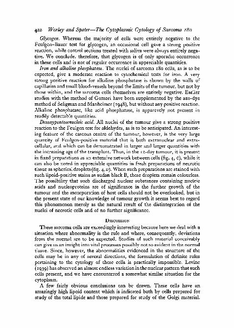
422 Worley and Spater—The Cytoplasmic Cytology of Sarcoma 180
Glycogen. Whereas the majority of cells were entirely negative to theFeulgen-Bauer test for glycogen, an occasional cell gave a strong positivereaction, while control sections treated with saliva were always entirely nega-tive. We conclude, therefore, that glycogen is of only sporadic occurrencein these cells and is not of regular occurrence in appreciable quantities.
Iron and alkaline phosphatase. The nuclei of sarcoma 180 cells, as is to beexpected, give a moderate reaction to cytochemical tests for iron. A verystrong positive reaction for alkaline phosphatase is shown by the walls ofcapillaries and small blood-vessels beyond the limits of the tumour, but not bythose within, and the sarcoma cells themselves are entirely negative. Earlierstudies with the method of Gomori have been supplemented by the azo-dyemethod of Seligman and Manheimer (1948), but without any positive reaction.Alkaline phosphatase, like acid phosphatase, is apparently not present inreadily detectable quantities.
Desoxypentosenucleic acid. All nuclei of the tumour give a strong positivereaction to the Feulgen test for aldehydes, as is to be anticipated. An interest-ing feature of the caseous centre of the tumour, however, is the very largequantity of Feulgen-positive material that is both extranuclear and extra-cellular, and which can be demonstrated in larger and larger quantities withthe increasing age of the transplant. Thus, in the 12-day tumour, it is presentin fixed preparations as an extensive network between cells (fig. 4, c), while itcan also be noted in appreciable quantities in fresh preparations of necrotictissue as spherical droplets (fig. 4, D). When such preparations are stained withsuch lipoid-positive stains as sudan black B, these droplets remain colourless.The possibility that such discharged nuclear substances containing nucleicacids and nucleoproteins are of significance in the further growth of thetumour and the incorporation of host cells should not be overlooked, but inthe present state of our knowledge of tumour growth it seems best to regardthis phenomenon merely as the natural result of the distintegration of thenuclei of necrotic cells and of no further significance.
DISCUSSION
These sarcoma cells are exceedingly interesting because here we deal with asituation where abnormality is the rule and where, consequently, deviationsfrom the normal are to be expected. Studies of such material conceivablycan give us an insight into vital processes possibly not so evident in the normaltissue. Since, however, the abnormalities evidenced in the structure of thecells may be in any of several directions, the formulation of definite rulespertaining to the cytology of these cells is practically impossible. Levine(1939) has observed an almost endless variation in the nuclear pattern that suchcells present, and we have encountered a somewhat similar situation for thecytoplasm.
A few fairly obvious conclusions can be drawn. These cells have anamazingly high lipoid content which is indicated both by cells prepared forstudy of the total lipide and those prepared for study of the Golgi material.
Worley and Spater—The Cytoplasmic Cytology of Sarcoma 180 423
Among the cells of vertebrates with which we are familiar, only oocytes andgland cells show such a concentration and degree of elaborateness as isdemonstrated by the Golgi complex in these cells. Indeed, we have beenrepeatedly impressed with the general morphological resemblance betweensarcoma cells and such gland cells as, for example, those of normal pancreaticalveoli.
We have also been impressed by the high ribonucleic acid content of thecytoplasm of these cells, again recalling the situation in vertebrate sero-zymogenic cells. Probably both of these conditions merely reflect the intensemetabolism that is taking place and we have no direct evidence that anysecretory product is being discharged. That certain storage products arebeing produced, however, is evident from the observation of the increasedaccumulation of lipides in cells as one approaches the caseous centre of thetumour.
In a general way, it is possible to distinguish three zones in the malignantportion of the tumour outside the caseous centre. (1) A more superficial zone,in which the cells and their nuclei are smaller on the average and in which theGolgi bodies or lipochondria are principally in the form of small, homo-geneous granules. These cells closely resemble normal fibroblasts. (2) A broad,more central zone of larger cells, somewhat resembling normal fibroblasts butmuch larger, and in which many of the Golgi bodies display chromophobicinteriors. (3) An intermediate zone, adjacent to the necrotic centre, inwhich the Golgi bodies are fewer in number but in which neutral fats andlipides are abundant. We suggest that the cells of zone 1 are to be interpretedas relatively young cells while those of zone 3 are much older cells at a stagejust before they become necrotic, become incorporated into the caseous centre,and eventually die or disintegrate. The tumour as a whole, in the meantime,is expanding farther and farther into the host tissue.
Our observations support Hirsch's (1939) view that the Golgi bodies under-go transformation and can be classified into different types which are chrono-logically related. Clearly they show variation in size which can be noted inalmost any type of preparation. Now, when the same technique is applied tothese elements of different sizes, the smaller ones appear homogeneous, butthe larger ones exhibit pale 'chromophobic' or 'osmiophobic' centres. It isbelieved that when these larger elements are 'fixed' or otherwise disturbed,a chromophilic or osmiophilic envelope surrounding the chromophobic coreis artificially contracted or slipped off from the latter, thus rendering it visible.Such a body, consisting of a chromophobic sphere, capped or partly sur-rounded by a chromophilic knob, crescent, or ring, is believed to be anartifact; but this should not be taken to mean that the chromophobic core incontact with the chromophilic surface does not exist, as some investigatorshave either implied or suggested.
Holtfreter (1946) suggested that bipartite Golgi bodies of this sort repre-sented 'desolvated' lipoid spheres. Others have referred to such bodies as'cavulated' mitochondria. Neither interpretation demands that 'desolvation'
2421.4 F f

424 Worley and Spater—The Cytoplasmic Cytology of Sarcoma 180
or 'cavulation' is always to be considered an artificial process, although it hasbeen shown that such appearances can be produced artificially by certainappropriate external agents. In view of the fact that 'desolvation' seems onlyto involve the larger, and perhaps more 'mature' lipoid spheres, and con-sidering that in the eggs of molluscs (Worley and Worley, 1943) and elsewherethese chromophobic cores eventually become free of their chromophilicenvelopes during the course of development or activity, for the time being itwould seem pertinent to regard 'cavulation' or 'desolvation' as a process thatmay occur naturally in the cell from time to time as the natural metabolic pro-cesses of the cell warrant and permit. 'Cavulation' occurs so readily in thelarger Golgi bodies whenever the cell is disturbed in any manner that it seemsunlikely that it does not occur naturally from time to time in the normalhealthy cell under certain given conditions. Xeros (1951) has recently con-firmed that chromophobic centres of this sort are found in some of the Golgibodies of the pancreas. Many investigators (Hirsch, 1939; Thomas, 1947;Siang-Hsu, 1947; Bourne, 1951, and others) have regarded this as the naturalmanner in which the secretory granules and certain storage products arise.In the present state of our knowledge of lipide metabolism, however, it wouldbe premature to offer comment regarding the nature of the chemical pro-cesses involved.
Some of the observations recorded here suggest that Golgi bodies and mito-chondria may at times form complexes between themselves or that mito-chondrial material may become incorporated into the Golgi spheres. Certainworkers in the past, notably Parat (1928) and more recently Adamstone (1950),have made similar suggestions. Oliver (1948) has furnished a most convincingdemonstration of this in cells of the mammalian kidney where the neutral redstaining inclusions apparently incorporate mitochondrial stuffs. This is not theusual interpretation, but in these cancerous tissues we are dealing with ab-normal cells where peculiar happenings should not be unexpected. The factthat such an occurrence may take place in abnormal cells suggests the possi-bility that normal cells may occasionally behave in a similar manner, at leastin particular instances. In this connexion, it is worth while to recall the con-ditions in the mammalian mast cell. As is well known, such cells are loadedwith mast granules. Techniques for the Golgi material demonstrate only themast granules while when techniques for mitochondria are employed, againit is the mast granules that react (Montagna and Noback, 1948). Here is a casewhere it appears that one type of inclusion is structurally, and probablyfunctionally, endowed with the attributes that in most cells are associatedwith two distinct types of inclusions. It would not seem appropriate to assume,a priori, that in all animal cells different functions are invariably carried out bystructurally unrelated cytoplasmic organelles.
The observation that in these tumour cells the nucleolus is often seen pressedagainst the nuclear membrane adjacent to the extranuclear lipoid mass callsfor brief comment. This seems to be not an uncommon situation in cells wherelipoid metabolism is proceeding at an unusually high rate. Such or a similar
Worley and Spater—The Cytoplasmic Cytology of Sarcoma 180 425
situation has been repeatedly detected in developing oocytes (Gardiner, 1927;Saguchi, 1927, 1928; Hirschler, 1929; Singh and Boyle, 1938; Fahmy, 1949);these are cells in which intense lipoid metabolism is associated with theaccumulation of cytoplasmic 'fatty' yolk. Although the nucleolus itself is notgenerally considered to contain much if any lipoid, this intimate associationoccurs far too frequently to be considered purely coincidental.
The authors wish to thank the personnel of the Haskins Laboratories, NewYork City, for supplying the material used in this study. We particularlyacknowledge the constant enthusiastic encouragement of Dr. Paul A. Zahl ofthat laboratory.
REFERENCESADAMSTONE, F. B., 1950. Anat. Rec, 108, 50.BAKER, J. R., 1944. Quart. J. micr. Sci., 85, 1.
1946. Ibid., 87, 441.1950. Proc. Linnean Soc. London, 162, 67.
BENSLEY, R. R., and BENSLEY, S. H., 1938. Handbook of histological and cytological technique.Chicago (University Press).
BOURNE, G., 1936. Anat. Rec, 66, 369.1951- Cytology and cell physiology. Oxford (Clarendon Press).
COVVDRY, E. V., 1924. General cytology. Chicago (University Press).DALTON, A. J., 1951. Ann. N.Y. Acad. Sci., 51, 1295.DUSTIN, JR., P., 1947. In Nucleic acid. Symposium Soc. exp. Biol., Cambridge (University
Press).FAHMY, O. G., 1949. Quart. J. micr. Sci., go, 159.GARDINER, M. S., 1927. J. Morph., 44, 217.GOMORI, G., 1941. Arch Path., 32, 189.• 1945- Proc. Soc. exp. Biol. Med., 58, 362.HIRSCH, G. C, 1939. Form- und Stoffwechsel der Golgi-Korper. Berlin (Borntraeger).HIRSCHLER, J., 1929. C.R. Soc. Biol., 101, 250.HOLTFRETER, J., 1946. J. exp. Zool., 102, 51.LEVINE, M., 1939. Am. J. Cancer, 36, 276, 386, 581; 37, 69.LUDFORD, R. J., 1951. In Bourne's Cytology and cell physiology. Oxford (Clarendon Press).LUDFORD, R. J., and SMILES, J., 1950. J. roy. micr. Soc, 70, 186.MACCARDLE, R. C, 1951. Ann. N.Y. Acad. Sci., 51, 1489.MALLORY, F. B., 1938. Pathological technique. Philadelphia (Saunders).MONTAGNA, W., and NOBACK, C. R., 1948. Anat. Rec, 100, 535.OLIVER, J., 1948. J. Mt. Sinai Hospital, 15, 175.PARAT, M., 1928. Arch. d'Anat. micr., 24, 73.SAGUCHI, S., 1927. Zytologische Studien, 1, 1.
1928. Ibid., 2, 1.SELICMAN, A. M., and MANHEIMER, L. H., 1948. J. Nat. Cancer Inst., 9, 181.— J949' Ibid., 9, 427.SELIGMAN, A. M., and NACHLAS, M. M., 1949. Ibid., 9, 415.SIANG-HSU, W., 1947. J. Morph., 8o, 161.SINCH, B. N., and BOYLE, W., 1938. Quart. J. micr. Sci., 81, 81.SLUITER, J. W., 1948. Proc. Koninklijke nederlandsche Akad. van Wetenschoppen, 51, 1.THOMAS, O. L., 1947. Quart. J. micr. Sci., 88, 445.TONUTTI, E., 1938. Protoplasma, 31, 151.WACHSTEIN, M., 1946. J. exp. Med., 84, 25.WOLF, A., KABAT, A. E., and NEWMAN, W., 1943. Am. J. Path., 19, 423.WORLEY, L. G., and HARARY, I., 1946. Anat. Rec, 94, 47.WORLEY, L. G., and WORLEY, E. K., 1943. J. Morph., 73, 365.XEROS, N., 1951. Nature, 167, 448.ZAHL, P. A., and DRASHER, M. L., 1947. Cancer Res., 7, 658.

FIG. I
L. G. WORLEY and H. W. SPATER

FIG. 2
L. G. WORLEY and H. W. SPATER

Fie. 3
L. G. WORLEY and H. W. SPATER
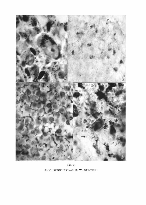
FIG. 4
L. G. WORLEY and H. W. SPATER
Related Documents