The Crystal Structure of Triosephosphate Isomerase (TIM) From Thermotoga maritima: A Comparative Thermostability Structural Analysis of Ten Different TIM Structures Dominique Maes, 1 * Johan P. Zeelen, 2 Narmada Thanki, 2 Nicola Beaucamp, 3 Marco Alvarez, 4 Minh Hoa Dao Thi, 1 Jan Backmann, 1 Joseph A. Martial, 4 Lode Wyns, 1 Rainer Jaenicke, 3 and Rik K. Wierenga 2,5 1 Ultrastructure Unit, Vlaams Interuniversitair Instituut voor Biotechnologie, Vrije Universiteit Brussel, Sint-Genesiu-Rode, Belgium 2 European Molecular Biology Laboratory, Heidelberg, Germany 3 Institut fu ¨ r Biophysik und physikalische Biochemie, Universita ¨ t Regensburg, Regensburg, Germany 4 Laboratoire de Biologie Mole ´culaire et de Ge ´nie Ge ´ne ´tique, Universite ´ de Lie `ge, Lie `ge, Belgium 5 Department of Biochemistry, University of Oulu, Oulu, Finland ABSTRACT The molecular mechanisms that evolution has been employing to adapt to environ- mental temperatures are poorly understood. To gain some further insight into this subject we solved the crystal structure of triosephosphate isomerase (TIM) from the hyperthermophilic bacterium Thermotoga maritima (TmTIM). The enzyme is a tetramer, as- sembled as a dimer of dimers, suggesting that the tetrameric wild-type phosphoglycerate kinase PGK-TIM fusion protein consists of a core of two TIM dimers covalently linked to 4 PGK units. The crystal structure of TmTIM represents the most thermostable TIM presently known in its 3D-struc- ture. It adds to a series of nine known TIM struc- tures from a wide variety of organisms, spanning the range from psychrophiles to hyperthermo- philes. Several properties believed to be involved in the adaptation to different temperatures were calcu- lated and compared for all ten structures. No se- quence preferences, correlated with thermal stabil- ity, were apparent from the amino acid composition or from the analysis of the loops and secondary structure elements of the ten TIMs. A common fea- ture for both psychrophilic and T. maritima TIM is the large number of salt bridges compared with the number found in mesophilic TIMs. In the two ther- mophilic TIMs, the highest amount of accessible hydrophobic surface is buried during the folding and assembly process. Proteins 1999;37:441–453. r 1999 Wiley-Liss, Inc. Key words: hydrophobicity; hyperthermophile; psy- chrophile; salt bridges; thermophile INTRODUCTION The thermal stability of proteins is the result of evolution- ary pressure, depending on specific conditions of the environment. Consequently, large variations are observed if one compares proteins from psychrophilic organisms with ultrastable proteins from hyperthermophiles. 1 The strategies of thermal adaptation of proteins are still poorly understood. The study of protein thermal stability is important not only theoretically but also because of its potential biotechnological applications. For this purpose it is essential to obtain full understanding of the factors that influence stability and catalytic activity at low and high temperatures. Detailed structural information from pro- teins of protein families with representatives in psychro- philic and thermophilic organisms is essential. Only three structures of psychrophilic organisms have been reported so far. 2–4 In contrast, numerous examples of homologous enzymes from mesophiles, thermophiles, and hyperthermo- philes have been documented. Analysis of high resolution X-ray structures of proteins from a variety of thermophiles and hyperthermophiles has suggested that the adaptation to ambient temperatures, rather than being the consequence of one dominant type of interaction, arises from the addition of increments contrib- uted by, e.g., salt bridges, decrease in accessible hydropho- bic surface area, improved packing, helix stabilization, and higher states of subunit assembly. 1,5–7 To gain further insight into the molecular mechanisms that nature uses to adapt to environmental temperatures, we have carried out a comparative structural analysis on triosephosphate isomerase (TIM; EC 5.3.1.1), a dimeric glycolytic enzyme consisting of two identical subunits, each about 250 residues long. It catalyzes the interconver- sion of dihydroxyacetone phosphate and D-glyceraldehyde- Abbreviations: ASA, accessible surface area; AUC, analytical ultra- centrifugation; BsTIM, Bacillus stearothermophilus TIM; ChTIM, chicken TIM; EcTIM, Escherichia coli TIM; HuTIM, human TIM; LeTIM, Leish- mania mexicana TIM; PfTIM, Plasmodium falciparum TIM; PGK, phosphoglycerate kinase; TbTIM, Trypanosoma brucei TIM; TIM, triosephosphate isomerase; TmTIM, Thermotoga maritima TIM; VmTIM, Vibrio marinus TIM; YeTIM, yeast TIM. Grant sponsor: European Union; Grant number: BIO4-CT96–0670; Grant sponsor: PRODEX ESA; Grant number: 1298/98/NL/VJ (IC); Grant sponsor: Actions de Recherche Concerte ´es; Grant number: 95/00– 193; Grant sponsor: Services Fe ´de ´reaux des Affaires Scientifiques Techniques et Culturelles; Grant number: SSTC-PAI P4–30; Grant sponsor: Fond National de la Recherche Scientifique; Grant number: FRFC 2.4545.96; Grant sponsor: Deutsche Forschungsgemeinschaft; Grant sponsor: Fonds der Chemischen Industrie; Grant sponsor: Patri- moine de l’Universite ´ de Lie `ge. *Correspondence to: Dominique Maes, ULTR, IMOL, Vrije Universi- teit Brussel, Paardenstraat 65, B-1640 Sint-Genesius-Rode, Belgium. E-mail: [email protected] Received 16 March 1999; Accepted 18 June 1999 PROTEINS: Structure, Function, and Genetics 37:441–453 (1999) r 1999 WILEY-LISS, INC.

Welcome message from author
This document is posted to help you gain knowledge. Please leave a comment to let me know what you think about it! Share it to your friends and learn new things together.
Transcript
The Crystal Structure of Triosephosphate Isomerase (TIM)From Thermotoga maritima:AComparative ThermostabilityStructural Analysis of Ten Different TIM StructuresDominique Maes,1* Johan P. Zeelen,2 Narmada Thanki,2 Nicola Beaucamp,3 Marco Alvarez,4 Minh Hoa Dao Thi,1Jan Backmann,1 Joseph A. Martial,4 Lode Wyns,1 Rainer Jaenicke,3 and Rik K. Wierenga2,5
1Ultrastructure Unit, Vlaams Interuniversitair Instituut voor Biotechnologie, Vrije Universiteit Brussel, Sint-Genesiu-Rode, Belgium2European Molecular Biology Laboratory, Heidelberg, Germany3Institut fur Biophysik und physikalische Biochemie, Universitat Regensburg, Regensburg, Germany4Laboratoire de Biologie Moleculaire et de Genie Genetique, Universite de Liege, Liege, Belgium5Department of Biochemistry, University of Oulu, Oulu, Finland
ABSTRACT The molecular mechanisms thatevolution has been employing to adapt to environ-mental temperatures are poorly understood. To gainsome further insight into this subject we solved thecrystal structure of triosephosphate isomerase (TIM)from the hyperthermophilic bacterium Thermotogamaritima (TmTIM). The enzyme is a tetramer, as-sembled as a dimer of dimers, suggesting that thetetrameric wild-type phosphoglycerate kinasePGK-TIM fusion protein consists of a core of twoTIM dimers covalently linked to 4 PGK units. Thecrystal structure of TmTIM represents the mostthermostable TIM presently known in its 3D-struc-ture. It adds to a series of nine known TIM struc-tures from a wide variety of organisms, spanningthe range from psychrophiles to hyperthermo-philes. Several properties believed to be involved inthe adaptation to different temperatures were calcu-lated and compared for all ten structures. No se-quence preferences, correlated with thermal stabil-ity, were apparent from the amino acid compositionor from the analysis of the loops and secondarystructure elements of the ten TIMs. A common fea-ture for both psychrophilic and T. maritima TIM isthe large number of salt bridges compared with thenumber found in mesophilic TIMs. In the two ther-mophilic TIMs, the highest amount of accessiblehydrophobic surface is buried during the foldingand assembly process. Proteins 1999;37:441–453.r 1999 Wiley-Liss, Inc.
Key words: hydrophobicity; hyperthermophile; psy-chrophile; salt bridges; thermophile
INTRODUCTION
The thermal stability of proteins is the result of evolution-ary pressure, depending on specific conditions of theenvironment. Consequently, large variations are observedif one compares proteins from psychrophilic organismswith ultrastable proteins from hyperthermophiles.1 Thestrategies of thermal adaptation of proteins are still poorlyunderstood. The study of protein thermal stability isimportant not only theoretically but also because of its
potential biotechnological applications. For this purpose itis essential to obtain full understanding of the factors thatinfluence stability and catalytic activity at low and hightemperatures. Detailed structural information from pro-teins of protein families with representatives in psychro-philic and thermophilic organisms is essential. Only threestructures of psychrophilic organisms have been reportedso far.2–4 In contrast, numerous examples of homologousenzymes from mesophiles, thermophiles, and hyperthermo-philes have been documented.
Analysis of high resolution X-ray structures of proteinsfrom a variety of thermophiles and hyperthermophiles hassuggested that the adaptation to ambient temperatures,rather than being the consequence of one dominant type ofinteraction, arises from the addition of increments contrib-uted by, e.g., salt bridges, decrease in accessible hydropho-bic surface area, improved packing, helix stabilization,and higher states of subunit assembly.1,5–7
To gain further insight into the molecular mechanismsthat nature uses to adapt to environmental temperatures,we have carried out a comparative structural analysis ontriosephosphate isomerase (TIM; EC 5.3.1.1), a dimericglycolytic enzyme consisting of two identical subunits,each about 250 residues long. It catalyzes the interconver-sion of dihydroxyacetone phosphate and D-glyceraldehyde-
Abbreviations: ASA, accessible surface area; AUC, analytical ultra-centrifugation; BsTIM, Bacillus stearothermophilus TIM; ChTIM, chickenTIM; EcTIM, Escherichia coli TIM; HuTIM, human TIM; LeTIM, Leish-mania mexicana TIM; PfTIM, Plasmodium falciparum TIM; PGK,phosphoglycerate kinase; TbTIM, Trypanosoma brucei TIM; TIM,triosephosphate isomerase; TmTIM, Thermotoga maritima TIM;VmTIM, Vibrio marinus TIM; YeTIM, yeast TIM.
Grant sponsor: European Union; Grant number: BIO4-CT96–0670;Grant sponsor: PRODEX ESA; Grant number: 1298/98/NL/VJ (IC);Grant sponsor: Actions de Recherche Concertees; Grant number: 95/00–193; Grant sponsor: Services Federeaux des Affaires ScientifiquesTechniques et Culturelles; Grant number: SSTC-PAI P4–30; Grantsponsor: Fond National de la Recherche Scientifique; Grant number:FRFC 2.4545.96; Grant sponsor: Deutsche Forschungsgemeinschaft;Grant sponsor: Fonds der Chemischen Industrie; Grant sponsor: Patri-moine de l’Universite de Liege.
*Correspondence to: Dominique Maes, ULTR, IMOL, Vrije Universi-teit Brussel, Paardenstraat 65, B-1640 Sint-Genesius-Rode, Belgium.E-mail: [email protected]
Received 16 March 1999; Accepted 18 June 1999
PROTEINS: Structure, Function, and Genetics 37:441–453 (1999)
r 1999 WILEY-LISS, INC.

3-phosphate.8 TIM has been the subject of extensivebiophysical, enzymological, and computational studies.Each TIM monomer is formed by an eightfold repeat of a[b-strand, loop, a-helix, loop] motif. The ba-units fold up ina regular way, such that the b-strands (numbered b1 to b8)form an eight-stranded b-barrel, surrounded by the eighta-helices (numbered a1 to a8) on the outside. This foldingmotif is also referred to as the TIM-barrel motif. Thecatalytic residues are located in the b-strands and in theloops connecting the b-strands to subsequent a-helices,numbered loop 1 to loop 8 in correspondence with thenumbering of the preceding b-strands. Binding of sub-strate or substrate analogues is accompanied by a changein conformation of loop 5, loop 6, and loop 7,9,10 going froman open state (unliganded) into a closed state (liganded) toprevent an unwanted phosphate elimination reaction thatmay otherwise occur.8 The dimer interface is formedmainly by loops 1, 2, 3, and 4; the protruding loop 3 of onesubunit docks into a deep pocket between loop 1 and loop 4of the other subunit.11
To date, x-ray structures of TIM have been solved (wildtype and/or in complex with substrate analogues) frompsychrophilic, mesophilic, thermophilic, and hyperthermo-philic organisms. With respect to mesophilic organisms,the following structures have been reported: chicken(ChTIM),12,13 yeast (YeTIM),14 Trypanosoma brucei(TbTIM),15 Escherichia coli (EcTIM),16 human (HuTIM),17
Plasmodium falciparum (PfTIM),18 and Leishmania mexi-cana (LmTIM).19 Very recently, the crystal structure ofT. cruzei TIM20 also has been described. The following TIMstructures of extremophilic organisms have been solved:that of the thermophile Bacillus stearothermophilus(BsTIM) 21 and that of the psychrophile Vibrio marinus(VmTIM).3 Here we report the structure of TIM from thehyperthermophile Thermotoga maritima (TmTIM). T. mar-itima belongs to one of the deepest and slowly evolvingbacterial branches of the phylogenetic tree. This hyperther-mophilic, strictly anaerobic, fermentative bacterium growsin the temperature range between 55° and 90°C with anoptimum growth at 80°C. The N-terminus of TmTIM hasbeen shown to be covalently linked to the C-terminus ofphosphoglycerate kinase (PGK), forming a bifunctionalPGK-TIM fusion protein.22 This fusion protein is a tetra-mer consisting of four PGK-TIM chains. Recently, theseparate TIM-enzyme has been cloned, expressed in E.coli, and compared with its wild-type in the PGK-TIMfusion protein. It turns out that the fusion enhances theintrinsic stability and the catalytic efficiency of TIM, butthe separate TIM exhibits high intrinsic stability and canstill be categorized as a hyperthermophilic enzyme.23 Inthis article we also describe the results of the detailedcomparison of ten TIM structures, which was aimed atfinding structural determinants of thermostability.
MATERIALS AND METHODSExpression and Purification
TmTIM was produced and purified as described previ-ously.23
Sedimentation Analysis
Sedimentation velocity and sedimentation equilibriumexperiments made use of a Beckman Spinco E analyticalultracentrifuge equipped with a high-sensitivity lightsource and a multiplexer photoelectric scanning system(AnG and AnF-Ti rotors, 12-mm double-sector cells withsapphire windows, scanning wavelength 235 and 280 nm).Before the experiments, the enzyme solutions were sub-jected to equilibrium dialysis against 50 mM Tris/HClbuffer pH8.6 with 2 mM EDTA. Sedimentation velocityexperiments were performed at 44,000 revolutions/minuteand room temperature; sedimentation coefficients werecorrected for 20°C and water viscosity. For sedimentationequilibria, the meniscus-depletion technique24 was appliedto quantify possible deviations from homogeneity; experi-mental conditions were as follows: 15,000 and 18,000revolutions/minute, 20.0° to 21.3°C. The quantitative evalu-ation was based on the linearization of the scans in ln cversus r2 plots, with the use of a computer programprovided by G. Bohm (University of Halle); for curvefitting, a monomer molecular mass of 26850 D was as-sumed.
Crystallization
Crystals were grown at 20°C using the hanging dropvapor diffusion technique with the aid of Hampton screen-ing solutions. The protein solution used in the crystalliza-tion set-ups contained 8.5mg/mL protein in 70 mM TRISHCl (pH 8.0), 2 mM EDTA, 5 mM cysteamine, and 400 mMNaCl. Drops were prepared by mixing 2.5 µl of TmTIMsolution with 2.5 µl reservoir solution of 100 mM TRIS/HClbuffer (pH 8.5) with 2.0 M ammonium sulfate.
Data Collection
Crystals diffracted to 2.8 A on a rotating anode x-raysource and data were collected at 25°C on a BigMar imageplate detector mounted on an Enraf Nonius rotating anodeFR571. The data were processed with DENZO.25 Scaling,merging, and reduction of the integrated intensities wasperformed using SCALEPACK25and TRUNCATE.26 TheB-factor of the Wilson plot is 39 A2. The crystal lattice isprimitive trigonal; with a 5 b 5 125.95 A; c5103.72 A, andtwo possibilities for the space group, i.e., P3221 and P3121.Calculation of Matthews parameters gave a VM of 4.4 and2.2 A3/D for one and two dimers in the asymmetric unit,respectively.
Molecular Replacement
To find the orientation(s) of the TIM dimer in the unitcell, molecular replacement was performed with the pro-gram AMORE27 using the crystal structure of BsTIM(1BTM; Brookhaven Protein Data Bank) as the searchmodel. The rotation function showed one unique peakabove 19 s, while the next peak was only 7 s. A translationwas calculated for this rotation peak in both possible spacegroups (P3221 and P3121). A good translation functionsolution was found for space group P3221. The R-factor fordata between 20 and 3.5 A for this solution dropped to 43%
442 D. MAES ET AL.

after rigid body refinement. There was no indication forthe presence of a second dimer in the asymmetric unit. Thepacking of the dimers is very loose, with large, triangularsolvent channels, resulting in a crystal solvent content ofmore than 70%.
Refinement and Quality of the Structure
The starting model for the refinement was the BsTIMmodel properly positioned in the TmTIM cell. Loop 6 wasdeleted in both subunits, and the sequence was changedinto the TmTIM sequence. Several rounds of visual inspec-tion and improvement followed by computer refinementwere done. On one hand, the model was optimized toimprove its fit in a 2Fo-Fc density map with the programO28 running on an SGI-workstation; on the other hand,refinement was pursued with a mixture of simulatedannealing X-ray refinement and conventional positionaland thermal factor refinement using the X-PLOR pack-age29 for the initial refinement calculations. For refine-ment, a subset of 5% of the data (the test set) was used forR-free calculations. A bulk solvent correction was applied.Electron density maps indicated a closed conformation forloop 6, which was manually rebuilt into the density. Inaddition, a sulfate ion was found in the active site of eachsubunit; its coordinates were included in the model. Thefinal stages of the refinement were carried out with CNS;30
water molecules were added at sites displaying a peaklarger than three standard deviations above the mean inan Fo-Fc map and having a potential hydrogen-bondingpartner. In total, 52 water molecules were added perdimer. The final R-factor was 21.1%, and R-free was 24.9 %for all data in a resolution range from 20 to 2.85 A (Table I).The quality of the structure was analyzed using theprograms PROCHECK31 and WHAT IF.32
Structure Comparison and Sequence Alignment
The ten structures used in the comparison are found inthe PDB with entry codes summarized in Table II. For thepurpose of analysis, TIM structures were superimposed onthe basis of the 129 Ca-atoms forming the eight frameworka-helices and b-strands and using the lsq option in O.28
The sequence alignment was made with Pileup of the GCGpackage33 and checked by superimposing of the structures.
Secondary Structure Assignment
The assignment of the b-strands was done with theDSSP-software.34 The beginnings (the N-cap positions)and the ends (the C-cap positions) of the helices wereidentified manually as defined by Richardson and Richard-son.35 The N- and C-caps were found by positioning anideal helix on the TIM helix. The N-cap is the first residueof the helix for which Ca is on the ideal helical spiral,similarly the C-cap is the last residue on the helical spiral.
Volumes, Cavities and B-Factors
Molecular volumes were calculated with the MSP soft-ware.36 Atomic radii were as described previously.13 Cavi-ties of which more than 70% of the surface is hydrophobic
are classified as hydrophobic cavities. Carbon and sulfuratoms are considered hydrophobic, whereas nitrogen andoxygen atoms are considered hydrophilic.
Hydrophobicity
The accessible surface area and the difference in acces-sible surface area upon protein folding and assembly werecalculated with the NACCESS software37 using a proberadius of 1.4 A. For the unfolded state, accessibility wascalculated using an extended Ala-X-Ala peptide. Carbonand sulfur atoms are considered hydrophobic, whereasnitrogen and oxygen atoms are considered hydrophilic.
Hydrogen Bonds and Salt Bridges
Hydrogen bonds between protein atoms were calculatedfor all proteins using the HBPLUS routine38 with thedefault parameters for distances and angles. When two
TABLE I. Crystallographic Data and RefinementStatistics of the TmTIM Structure
Space group P3221Cell dimensions (Å) 125.95 125.95 103.72Cell dimensions (°) 90 90 120Subunits per asymmetric unit 2Vm (Å3/D) 4.4Data collection statisticsObserved reflections 88,489Unique reflections 22,020Overall range (Å) 20–2.85Overall R-merge (%) 8.6Overall completeness (%) 97.8Last shell range (Å) 2.90–2.85Last shell R-merge (%) 29.9Last shell completeness (%) 90.0B-factor from Wilson plot (Å2) 38.7Refinement statisticsProtein atoms 3974Ligand atoms 10Solvent atoms 52Resolution range (Å) 20.0–2.85R-factor (%) 21.1R-free (%) 24.9rms bond length deviations (Å) 0.006rms bond angle deviations (°) 1.30x1x2 imperfection (°)a 31.7rms DB for covalently bonded atoms (Å2) 3.47average B-factor, all protein atoms (Å2) 54.7Average B-factor, backbone atoms (Å2) 52.9Average B-factor, side chain atoms (Å2) 56.6Average B-factor, ligand atoms (Å2) 74.4Average B-factor, solvent atoms (Å2) 46.2Ramachandran plotb
Most favored regions (%) 84.1Additional allowed regions (%) 14.3Generously allowed regions (%) 1.1Disallowed regions (%) 0.5aThe x1x2 imperfection value9 is the rms between observed x1x2 valuesand the nearest preferred values as found in a database of well-refinedstructures.bAs defined by PROCHECK.31
443STRUCTURE AND STABILITY OF T. MARITIMA TIM

atoms of opposite charge were observed to be within 4 A,they were assigned to a salt bridge. Barlow and Thornton39
recommended this distance threshold after extensive analy-sis of several protein structures. Positively charged atomsincluded side-chain nitrogens in Lys and Arg, whereasnegatively charged atoms are the side-chain oxygens ofAsp and Glu. Salt-bridge networks consist of three or moresalt-bridged residues.
Charge Distribution at N- and C-Terminal Endsof Helices
For this analysis only the framework helices were used,except for the C-terminal end of helix 8, which is notwell-defined. The center point of the N-terminal end of thehelix is calculated from the coordinates of the main chainCa, N and C atoms of the N-cap, N-cap 11, N-cap 12, andN-cap 13 residues, belonging to the first turn of the helix.In an analogous way the center point of the C-terminal endwas calculated, using the C-cap-3, C-cap-2, C-cap-1, andC-cap residues. All charged atoms within a distance of 7 Aof this center were calculated. Positively charged atomsincluded side-chain nitrogens in Lys and Arg, with a netcontribution of 11 and 11/3, respectively for each nitrogenatom, whereas negatively charged atoms concern the side-chain oxygens of Asp and Glu, with a net negative chargeof 21⁄2 for each oxygen atom. The average charge at theN-terminal end of a helix is calculated by first summing allcharges within the defined spheres of all the availablehelices then dividing by the number of helices. The aver-age charge at the C-terminal is calculated the same way.
RESULTS AND DISCUSSIONTmTIM X-Ray Structure
The structure of T. maritima TIM has been refined at2.85 A resolution to a model with good geometry andcrystallographic quality (Table I). In this crystal formthere is one TmTIM dimer per asymmetric unit: the modelfor the TmTIM dimer consists of residues 2–253 forsubunit A and residues 1– 255 for subunit B. There was noclear density for the N- and C-terminus of the first subunit.The residue-stretch composed of residues 33 to 36 in bothsubunits as well as loop 6 in subunit B display highthermal B-factors; nevertheless, density was visible in the
omit map, and the coordinates were incorporated in thestructure. The stereochemical quality of the structure wasanalyzed with the program PROCHECK.31 The Rama-chandran plot shows only two outliers, concerning theLys13 residue of both subunits, which are in a strainedconformation in each TIM structure. All stereochemicalparameters for the main chain and side-chain atoms arebetter than those for proteins refined to a similar resolu-tion. The thermal B-factors of the complete structure arehigh with an average of 52.9, 56.6, and 54.7 A2 for mainchain, side-chain, and all protein atoms, respectively. Thehigh average B-value agrees with the high B-factor of theWilson plot (Table I) and correlates with the high solventcontent of the crystal. Loops 5, 6, and 7 are in the closedconformation in both subunits. A sulfate ion occupies theactive site of both subunits (with an average thermalB-factor of 74.4 A2). Fifty-two waters were included in thestructure with an average B-factor of 46.2 A2. The packingof the TIM dimers is very loose with triangular solventchannels with a diameter of 89 A. The TmTIM structure isvery similar to all other TIM structures with a Ca rmsd forthe framework residues ranging from 0.8 to 1.4 A (TableIII). Both subunits are very similar with a Ca rmsd of 0.5 Afor all main chain atoms.
Tetramerization
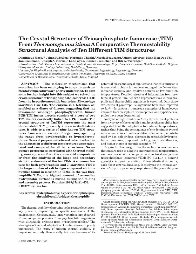
The crystallographic twofold axis perpendicular to thethreefold screw axis rotates the TIM dimer in such a waythat a dimer of dimers with tetrahedral symmetry isformed. Figure 1A shows the tetrameric organization ofTmTIM. The noncrystallographic twofold axis (k 5 179.5°)of the canonical dimer is perpendicular to the crystallo-graphic twofold, resulting in the near 222 symmetry. Thedimer of dimers is stabilized by hydrophobic and polarinteractions of residues in loop 5 and helix 5. The majorinterdimeric contacts consist of two disulfide bridges be-tween the Cys142 of each monomer and its symmetryequivalent. Furthermore, there is a hydrophobic contactbecause of Leu139, a salt link between Glu150 and Lys137,and some hydrogen bonds, shown in detail in Figure 1B.Because of the twofold symmetry, the latter contacts arefound twice, on either side of the disulfide bridge. The saltbridges each belong to a salt-bridge network involving
TABLE II. The TIM PDB Files Used in the Structure Comparison
Organism CodeTemperature
rangePDBcode
Resolution(Å)
Asymmetricunit
Conformation ofactive site loops
ReferenceSubunit A Subunit B
V. marinus Vm Psychrophile 1AW1 2.7 4 dimers Closed Closed 3T. brucei Tb Mesophile 5TIM 1.8 Dimer Open Almost closed 15L. mexicana Lm Mesophile 1AMK 1.8 Monomer Closed Closed 19P. falciparum Pf Mesophile 1YDV 2.2 Dimer Open Open 18Human Hu Mesophile 1HTI 2.8 Dimer Open Closed 17Chicken Ch Mesophile 8TIM 2.5 Dimer Open Open 12, 13Yeast Ye Mesophile 7TIM 1.9 Dimer Closed Closed 14E. coli Ec Mesophile 1TRE 2.6 Dimer Open Open 16B. stearothermophilus Bs Thermophile 1BTM 2.8 Dimer Closed Closed 21T. maritima Tm Hyperthermophile 1B9B 2.8 Dimer Closed Closed
444 D. MAES ET AL.

Glu133 and Lys137 of one subunit, connected to Glu150,Lys146, and Asp107 of the other subunit. This new contactarea buries 537 A2 of accessible surface area (ASA) (permonomer), of which 69% is hydrophobic. These valueshave to be compared with 1,758 A2 (per monomer), ofwhich 70% is hydrophobic, buried between the monomersin the ‘‘canonical dimer.’’
The tetrameric assembly is also confirmed by analyticalultracentrifugation (AUC) studies (Table IV). The resultsof sedimentation velocity experiments in the absence andin the presence of cysteamine resulted in s-values around5.4 S, corresponding to a molecular mass of globularproteins between 90 and 100 kD.40 Sedimentation equilib-rium experiments were fitted, taking into account thecalculated partial specific volume of the native enzyme(Vp 5 0.750 cm3g21) on the one hand and tentative valuesfor the partial volume and the density in the presence of 6M GdmCl (Vp8 5 0.740 cm3 g 21, r8 5 1.145 g cm23) on theother hand. The latter value for Vp is based on modelstudies using bovine serum albumine as a standard;41
whether this is appropriate for proteins without intramo-lecular cystine cross-links is unclear. Because the boyancyterm causes a large range of error at high solvent densi-
ties, MW in the presence of 6M GdmCl may be too high by afactor of # 2. The results in dilute buffer in the absenceand in the presence of reducing agent clearly show that invitro TmTIM forms a tetramer (Table IV).
The TmTIM structure also indicates a mode of assemblyfor the tetrameric PGK-TIM fusion protein. In the TmTIMtetramer (Figure 1A) the N-termini of each subunit aresolvent exposed. This suggests that in the wild-type fusionprotein the TmTIM tetramer forms the core and eachTmTIM subunit is covalently linked at its N-terminus tothe C-terminus of a PGK unit.
Two other hyperthermophilic archaeal TIMs were re-ported to exist also in a tetrameric form.42,43 Also, variousother enzymes from thermophilic and hyperthermophilicenzymes are known to exist as higher-order associationstates compared with their mesophilic analogues, suggest-ing that the formation of higher-order oligomers is one wayby which hyperthermostability is achieved in nature.1
Conserved Salt Bridge
The sequences of the ten TIM structures are tabulatedin Figure 2. The fully conserved residues are all, exceptone, near the active site pocket. The conservation of these
TABLE III. Structural and Sequence Comparison
Vm Tb Lm Pf Hu Ch Ye Ec Bs Tm
Vm 100 38.9 39.3 39.4 41.6 41.2 43.1 65.7 41.3 39.90.23 0.98 0.91 1.13 0.86 0.84 0.81 0.58 1.13 0.920.25 1.41 1.29 1.57 1.42 1.33 1.27 0.60 1.53 1.13
Tb 38.9 100 69.6 43.1 52.6 52.0 49.2 42.9 40.7 42.20.98 0.26 0.42 0.79 0.85 0.79 1.03 0.90 1.27 1.231.41 0.26 0.46 1.23 1.18 1.12 1.26 1.36 1.44 1.40
Lm 39.3 69.6 100 45.5 50.6 50.4 45.9 42.5 43.5 42.20.91 0.42 0.0 0.72 0.90 0.81 0.99 0.86 1.29 1.221.29 0.46 0.0 1.20 1.19 1.12 1.19 1.27 1.46 1.37
Pf 39.4 43.1 45.5 100 42.5 43.3 41.5 40.2 38.1 40.71.13 0.79 0.72 0.27 1.11 1.08 1.26 1.02 1.46 1.371.57 1.23 1.20 0.37 1.24 1.19 1.45 1.54 1.53 1.57
Hu 41.6 52.6 50.6 42.5 100 89.1 53.3 44.9 39.0 41.70.86 0.85 0.90 1.11 0.44 0.42 0.80 0.88 1.15 1.051.42 1.18 1.19 1.24 0.46 0.50 0.99 1.45 1.42 1.40
Ch 41.2 52.0 50.4 43.3 89.1 100 53.3 45.3 40.2 43.30.84 0.79 0.81 1.08 0.42 0.35 0.72 0.82 1.11 0.991.33 1.12 1.12 1.19 0.50 0.36 0.86 1.35 1.41 1.35
Ye 43.1 49.2 45.9 41.5 53.3 53.3 100 45.1 37.9 42.10.81 1.03 0.99 1.26 0.80 0.72 0.24 0.85 1.39 1.061.27 1.26 1.19 1.45 0.99 0.86 0.26 1.32 1.81 1.46
Ec 65.7 42.9 42.5 40.2 44.9 45.3 45.1 100 39.7 42.70.58 0.90 0.86 1.02 0.88 0.82 0.85 0.39 1.11 0.940.60 1.36 1.27 1.54 1.45 1.35 1.32 0.41 1.52 1.10
Bs 41.3 40.7 43.5 38.1 39.0 40.2 37.9 39.7 100 50.01.13 1.27 1.29 1.46 1.15 1.11 1.39 1.11 0.27 0.831.53 1.44 1.46 1.53 1.42 1.41 1.81 1.52 0.28 1.09
Tm 39.9 42.2 42.2 40.7 41.7 43.3 42.1 42.7 50.0 1000.92 1.23 1.22 1.37 1.05 0.99 1.06 0.94 0.83 0.301.13 1.40 1.37 1.57 1.40 1.35 1.46 1.10 1.09 0.30
The numbers in each block refer to: % sequence identity after alignment (calculated from the sequence alignment shown in Fig. 3),rmsd of framework C-atoms (129 atoms) (subunit A), and rmsd of all equivalent C-atoms after alignment (subunit A), excluding loops5, 6, and 7, which are known to adopt different conformations in the presence or absence of ligand9 and excluding insertions/deletions(including one residue before and after). On the diagonal the same quantities are given for subunit A on subunit B of the same species.
445STRUCTURE AND STABILITY OF T. MARITIMA TIM
residues is therefore correlated with the architecture ofthe catalytic site. The only exception is the conservedarginine at position 192. This arginine is located at theother end of the TIM barrel and is involved in a conservedsalt bridge with Asp230 and/or Asp228 (Fig. 3). This saltbridge anchors the C-terminal ends of helix 6 and helix 7 toeach other, and this anchoring is completely conserved inall available structures. Although the location of thisinteraction is at a distance of 20 A from the active site, thestrict conservation of this salt bridge points to an impor-tant function. The importance of this anchoring interac-tion might be related to the conformational flexibility of
loop 6 and loop 7, located at the N-terminal ends of helix 6and helix 7, respectively, which is crucial for the catalyticproperties of TIM.9
Comparative Analysis
Which structural features can be correlated with thedifferences in environmental temperature in the TIMfamily? If all TIMs would use the same mechanism ofthermal adaptation, a trend would be visible; in this casethe relevant variable(s) would increase or decrease ingoing from a psychrophilic TIM to a hyperthermophilicTIM. However, it is also possible that different approaches
Fig. 1. A: Stereo view of the Ca-trace of the TmTIM tetramer,composed of two canonical dimers (blue and green), related by acrystallographic twofold axis. The contacts between the two canonicaldimers are between residues of loop 5 and helix 5. The N- and C-termini ofeach subunit are indicated with N1, C1, N2, C2, N3, C3, N4, and C4. The
active sites are marked by yellow spheres. B: Stereo view of theinterdimeric interface. The labeled residues are involved in interdimericcontacts. Each residue label has the same color as the correspondingsubunit in Figure 1A.
446 D. MAES ET AL.

are being used to adapt to psychrophilic conditions andthermophilic conditions; in this case, unique properties forpsychrophilic and thermophilic enzymes should becomeapparent. The thermal stability of mesophilic proteins isonly marginal; in the range of 10 to 15 kcal/mol.6 Hence,the addition or deletion of a few well chosen, weakinteractions is sufficient to convert a mesophilic enzymeinto a thermostable enzyme.19,44,45 It seems, therefore,likely that for ultrastable wild-type proteins also smalladjustments can contribute significantly to the adaptation
toward extremes of environmental temperature. Unfortu-nately, such specific adaptations may not be detectablewhen systematic comparisons of TIMs from different spe-cies are done because there are just too many exchanges.46
Extremophilic enzymes not only have optimal stability butalso must fold, assemble, and function at the extremetemperatures of their environment. Therefore, some uniquefeatures of the sequences or structures of these enzymesmight, in fact, be adaptations required for optimal foldingand function but not for optimal stability.
Fig. 2. Sequence alignment of the ten TIMs. The b-strands (blue) and the a-helices (red) arelabeled b1 to b8 and a1 to a8, respectively. The conserved residues in these ten TIMs are shown onthe ‘‘Consensus’’ line. The residues involved in the conserved salt bridge (Fig. 3) are indicated by anasterisk.
447STRUCTURE AND STABILITY OF T. MARITIMA TIM

The ten different structures used for the comparison aresummarized in Table II; their sequence alignment isdepicted in Figure 2. The secondary structure of theframework b-strands and a-helices is also shown; the startand end of these secondary structural elements for thedifferent species vary. The percentage sequence identity isgiven in Table III. The TIM sequences are highly con-served, despite the wide range of species included in thecomparison. The lowest sequence identity (38%) is foundfor the comparison of BsTIM and YeTIM; the highest
sequence identity (89%) is between ChTIM and HuTIM.VmTIM and EcTIM have a high sequence identity (66%);the peculiar structural features of VmTIM also occur inEcTIM.3 TmTIM has the highest sequence identity (50%)with BsTIM; both are thermostable. A good correlationexists between the percentage sequence identity and theCa rmsd for the superimposed framework residues. For thecomparisons with low sequence identity (less than 40%),the rms is near 1.4 A, and for the comparisons with highsequence identity (70% to 90%), the rms value is between0.3 and 0.4 A.
Fig. 3. The conserved salt bridge between the C-terminal ends of helix6 and helix 7 for the ten TIM structures. The side-chains of Arg192,Asp228, and Asp230 of TmTIM and their equivalents in the otherstructures are shown explicitly. The main chain of the ten structures areshown in different colors.
Fig. 4. Graphs displaying various parameters for each of the ten TIMstructures. The horizontal axis displays the ten TIMs with increasingthermal stability. A: Average charge within a sphere of 7 A at the N-terminalends (♦) and C-terminal ends (j) of the framework helices. B: Hydropho-bic DASA of the folded monomer buried on assembly (A2) (see also TableVII). C: Number of hydrogen bonds (:10) (j), number of salt bridges (m),number of residues in salt bridges (♦), and number of residues in saltbridge networks (x) (the numbers have been normalized per dimer).
TABLE IV. Results ofAnalytical UltracentrifugationExperiments With TmTIM in 50 mM Tris pH 8.6,
2 mM EDTA
Conditions S-value (S) MW (D)Correlationcoefficienta
Buffer,no cysteamine 5.43 6 0.15 103,827 6 3,065 0.9999
Buffer, 5 mMcysteamine 5.31 6 0.15 108,753 6 6,796 0.9995
Buffer, 6 M GdmCl,5 mM cysteamine ndb 102,321 6 9,127 0.9994
aThe given correlation coefficients refer to the quality of the ln c vs r2
linearization. The true range of error in MW in dilute buffer amounts toapproximately 5%.bValue not determined.
448 D. MAES ET AL.

Sequences
Previous studies have attempted to correlate amino acidcompositions and sequence alignments with thermal stabil-ity.47 Table V lists the amino acid composition of the tenTIM structures. The largest deviation from the average isthe high alanine content of VmTIM (48 of 256 residues).This seems to be a property of the phylogenetic family,because EcTIM also has a high (45 of 255 residues) alaninecontent. Consequently, for both TIMs, the alanine contentof the framework helices is also high (27 in VmTIM and 22in EcTIM) (Table VI). This high alanine content in thehelices of VmTIM and the low content in TmTIM issurprising because alanine is known to stabilize helices.48
The glycine and proline content, affecting the flexibility ofthe unfolded state as well as the content of thermolabileresidues (e.g., Cys, Asn, and Gln),49 cannot be correlatedwith thermal stability. The slightly deviating content ofsome residues in TmTIM, like the high percentage ofcharged residues (28%) compensated by a low percentageof polar residues (16%), is not found in the thermophilicBsTIM; the opposite feature is also not found in VmTIM.Hence, it seems that the discrimination between psychro-philic, mesophilic, and (hyper)thermophilic TIMs cannotbe correlated with well-defined shifts in specific amino acidcompositions.
Secondary structure
The only sound basis for the discussion of extremophilicadaptation is the 3D structure, permitting the forcesunderlying protein stability under extremes of physicalconditions to be defined in an unambiguous way. a-Helicesand extended b-sheet structures contribute significantly toprotein stability; loops in thermostable enzymes havesometimes been observed to be shorter than those in theirmesophilic counterparts, thus stabilizing the folded ordestabilizing the unfolded state. In the case of the tendifferent TIM structures, no substantial differences can bedetected regarding the fraction of amino acids involved insecondary structure, taking into account also the helicalarrangement in some of the loops connecting the frame-work helices and strands.
Recent studies50,51 independently concluded that en-hanced stabilization of helices is a necessary, but not asufficient, condition for protein thermostability. Two typesof contributions were distinguished: (a) ‘‘intrinsic helicalpropensities,’’ associated with the loss of conformationalentropy of amino acid residues in helical arrangements,and (b) specific amino acid patterns at the helical ends. Asfar as the first of these two is concerned, it was found thatthe helices of thermophilic proteins contain a lower percent-age of b-branched residues (i.e., Val, Ile, and Thr) com-pared with their mesophilic equivalents. The rotamericconformations of these residues are constrained by thehelical conformation. Table VI shows the amino acidcomposition of the framework a-helices (in absolute valuesand normalized values); all mesophilic sequences aregrouped together. The only striking feature is the highalanine content of the VmTIM helices and the low alaninecontent of the TmTIM helices, as discussed earlier. Summa-
tion of the contents of b-branched residues gives normal-ized values of 0.7, 1.2, 1.1, and 1.1 for psychrophilic,mesophilic, thermophilic. and hyperthermophilic TIMs,respectively. Hence, a higher percentage of these residuesis not present in the (hyper)thermophilic TIMs, but theopposite is true for the psychrophilic TIM. In regard to theearlier mentioned point (b), helix-stabilizing factors havealso been identified for the N- and C-capping boxes.35,51 Weanalyzed the amino acid compositions at the N-cap andC-cap positions and their neighborhood (N24 5. N14and C14 5. C24): no significant differences in preferencewere found between the mesophiles and the extremophiles(data not shown). This analysis is unfortunately hamperedby the availability of only one psychrophilic, thermophilic,and hyperthermophilic structure, which prevents a reli-able statistical study.
Interactions of charged and polar groups with the helixmacrodipole can increase the stability of a protein.52,53
Therefore we analyzed the charge distribution at theN-terminus and C-terminus of the framework a-helices.For this, the presence of charged side-chain atoms within asphere of 7 A from the N- and C-cap of these helices wascalculated (Fig. 4A). The analysis shows that this chargedistribution is not different in the psychrophilic andthermophilic enzymes. The analysis also indicates that aremarkable favorable charge preference is present at theN-terminus but not at the C-terminus.
Volumes, cavities, and B-factors
As reviewed by Lee et al.,54 variation of cavity volumesand compactness contribute to optimal stability at differ-ent ambient temperatures. The molecular volumes and thecavity volumes for the ten TIMs are summarized in TableVII. The total cavity volume varies between 0.6% and 1.2%of the volume of the protein itself. The substantial varia-tions of the cavity volumes within the mesophilic family aswell as in the complete set of the ten TIM structures makesit unlikely that cavities play a critical role in the stabiliza-tion of the enzyme. Analysis of the size of predominantlyhydrophobic cavities shows no correlation with thermalstability. The compactness, calculated as the averagevolume occupied by each atom, also is not related tothermal stability.
The dynamic properties of the TIM-structures wereanalyzed by comparing the B-factor distribution for eachstructure. Such B-factor distribution will also be influ-enced by solvent content and the packing of the crystal,which vary significantly for these structures. The meanB-factors as well as the distributions of the B-factor(expressed as the rmsd with respect to the mean) werecalculated. A broad range of values was obtained, whichdid not correlate with the thermal stability.
Hydrophobic interactions
The importance of the hydrophobic effect on proteinfolding and stability is generally accepted.55 Because thecorrect analytical solution for this interaction is complex, asimplification based on accessible surface calculations is
449STRUCTURE AND STABILITY OF T. MARITIMA TIM

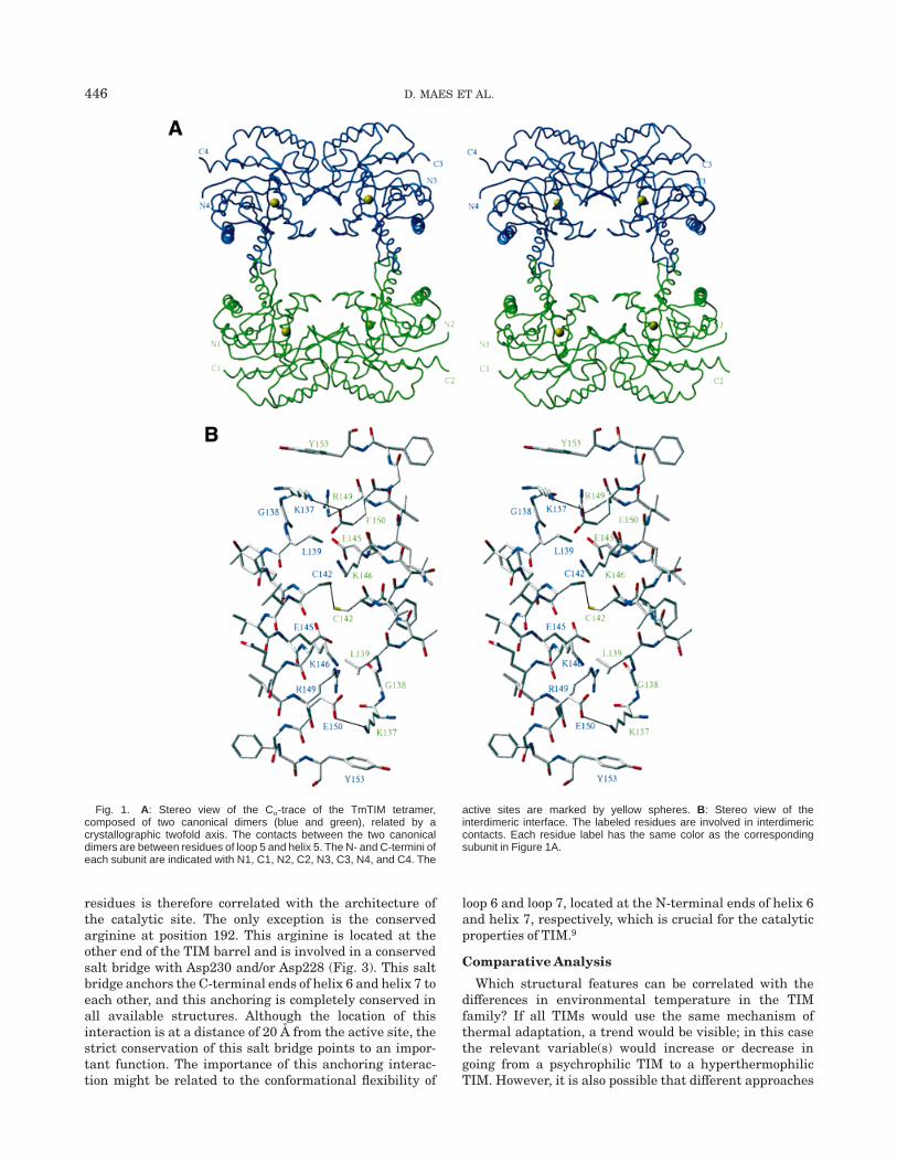
used.56,57 Three different comparisons of hydrophobic sur-faces are given in Table VII. First, the hydrophobicity ofthe protein envelope is calculated: no significant differ-ences related to thermostability are observed when the tendifferent TIM structures are compared. Second, values forthe total accessible surface area (DASA) and the hydropho-bic DASA, buried when the unfolded chain is folded intothe monomer structure, were calculated.58 The burial ofhydrophobic surface area shows its maximum value forBsTIM and TmTIM: for TmTIM 17,378 A2 of hydrophobicDASA is buried upon monomer folding. For BsTIM it is 584A2 (3%), and for TmTIM it is 1,363 A2 (8%) more comparedwith VmTIM. It is interesting to note that the amount ofburied hydrophobic area is a constant fraction of the totalDASA buried upon folding (varying between 59.0% and59.7%), in agreement with previous studies.59 Third, thetotal DASA and hydrophobic DASA buried on assembly ofthe folded monomers into oligomers was calculated permonomer. These values are much higher for both thermo-philes; for TmTIM 1,599 A2 is buried (per monomer) onassembly. For BsTIM it is 117 A2 (7%), and for TmTIM it is528 A2 (50%) more compared with VmTIM. The largeincrease seen for TmTIM (Fig. 4B) is largely because of theformation of the tetramer. The buried DASA on assemblyhas the highest percentage of hydrophobicity for boththermophilic TIMs (67.8% and 69.7%). These hydrophobic-ity calculations confirm the proposal that the hydrophobicterm makes a major contribution to the enhanced stabilityof thermostable TIMs.21 For TmTIM the increase of buriedhydrophobicity is achieved at three levels: upon folding the
TABLE V. TotalAminoAcid Composition
Vm Tb Lm Pf Hu Ch Ye Ec Bs Tm
A-Ala 48 34 33 15 28 28 25 45 29 15C-Cys 2 3 4 4 5 4 2 3 4 3D-Asp 12 8 8 14 12 13 15 10 12 9E-Glu 22 13 15 19 17 17 17 21 20 29F-Phe 8 8 6 13 8 8 11 6 9 12G-Gly 23 19 16 14 25 27 22 22 22 24H-His 6 5 6 5 4 8 3 8 6 4I-Ile 17 19 19 19 15 17 15 19 16 23K-Lys 13 17 17 22 20 22 21 16 12 19L-Leu 18 17 17 19 15 17 19 17 19 23M-Met 6 3 3 2 2 2 0 7 5 5N-Asn 12 10 11 17 8 7 12 9 5 6P-Pro 8 7 10 4 10 7 7 7 13 7Q-Gln 8 13 13 12 11 9 7 11 14 8R-Arg 7 9 8 8 8 7 8 8 11 14S-Ser 10 18 13 18 12 12 16 11 13 12T-Thr 11 12 16 13 14 11 12 9 12 10V-Val 19 25 25 21 25 22 26 19 25 24W-Trp 2 5 5 2 5 5 3 2 2 2Y-Tyr 4 5 6 7 4 4 6 5 4 6Total 256 250 251 248 248 247 247 255 253 255% Charged 21.1 18.8 19.1 25.4 23.0 23.9 24.7 21.6 21.7 27.8% Polar 18.4 23.2 23.5 26.2 19.8 19.0 20.2 18.8 19.8 15.7% Apolar 51.6 50.4 51.0 42.7 47.2 46.2 46.2 51.0 49.8 47.1% Glycines 9.0 7.6 6.4 5.6 10.1 10.9 8.9 8.6 8.7 9.4Charge 214 15 12 23 21 21 23 27 29 25
TABLE VI.AminoAcid Composition of theFramework Helices
Numberof residues
Normalizedpreferencesa
VmMeso-philicb Bs Tm Vm
Meso-philicb Bs Tm
A-Ala 27 86 14 6 2.73 1.42 1.59 0.64C-Cys 1 7 1 1 0.55 0.62 0.61 0.57D-Asp 6 41 7 2 0.93 1.03 1.21 0.32E-Glu 12 54 7 13 1.66 1.22 1.08 1.88F-Phe 7 22 6 7 1.60 0.82 1.54 1.67G-Gly 4 22 3 6 0.45 0.40 0.38 0.71H-His 2 19 1 2 0.82 1.27 0.46 0.86I-Ile 5 57 2 8 0.86 1.59 0.38 1.44K-Lys 8 66 5 7 1.04 1.40 0.73 0.96L-Leu 9 60 11 14 0.97 1.05 1.33 1.58M-Met 3 8 1 3 1.28 0.55 0.48 1.34N-Asn 5 26 4 3 1.01 0.85 0.90 0.63P-Pro 1 13 3 2 0.20 0.42 0.66 0.41Q-Gln 4 43 5 6 0.91 1.58 1.27 1.42R-Arg 4 21 5 5 0.74 0.63 1.03 0.96S-Ser 4 43 6 7 0.54 0.94 0.91 0.99T-Thr 4 41 6 3 0.61 1.01 1.02 0.48V-Val 6 53 14 11 0.76 1.09 1.98 1.45W-Trp 0 4 0 0 0.00 0.49 0.00 0.00Y-Tyr 1 8 0 2 0.27 0.35 0.00 0.56Totals 113 694 101 108aThe normalization used the natural occurrences in percentage, ascalculated by Nakashima et al.65 (A: 8.74; C: 1.62; D: 5.72; E: 6.39;F: 3.87; G: 7.82; H: 2.15; I: 5.15; K: 6.78; L: 8.20; M: 2.08; N: 4.39;P: 4.49; Q: 3.19; R: 4.81; S: 6.56; T: 5.84; V: 7.01; W: 1.17; Y: 3.33).bThe mesophilic TIMs are grouped together.
450 D. MAES ET AL.
monomer, in the canonical monomer-monomer interface,and in the new dimer-dimer interface (Table VII).
Polar interactions
For some thermostable proteins a correlation betweenthermostability and the number of hydrogen bonds hasbeen reported.60 The respective numbers for the ten TIMstructures, presented in Figure 4C, turn out to be closelysimilar; no general trend related to thermostability can beobserved. Also splitting up the total number of H-bondsinto main chain–main chain, main chain–side-chain andside-chain–side-chain interactions does not allow signifi-cant differences to be detected (data not shown).
Finally, salt bridges and salt-bridge networks have beenproposed to play a key role in the maintenance of enzymestability at high temperatures.61–63 The number of saltbridges and the number of residues involved, as well as thenumber of residues involved in salt-bridge networks, arepresented in Figure 4C. Superstable TmTIM has thehighest number of salt bridges (0.11 per residue), which ismuch higher than the average observed in mesophilicTIMs (0.07 per residue). In other superstable proteins, avalue of 0.11 salt bridges per residue has been found,
compared with 0.06 salt bridges per residue in the meso-philic homologues.63 In thermostable BsTIM no increase insalt bridges is observed. However, also in psychrophilicVmTIM a high number of salt bridges is present (0.09 perresidue). An increase in salt bridges was also observed inpsychrophilic citrate synthase,2 but not in psychrophilica-amylase.4 It has been speculated that the increase of saltbridges in psychrophilic citrate synthase could be impor-tant for preventing cold denaturation.
The higher number of residues involved in salt-bridgenetworks in TmTIM is mainly due to the four intersubunitnetworks in the new dimer-dimer interfaces. The existenceof these networks corroborates the observation that exten-sive intersubunit salt bridge networks play a major role inthe stabilization of multisubunit enzymes.64
CONCLUSIONS
The stability of a protein is determined by the enthalpicand entropic terms of the free energy difference (DG)between the folded state and the unfolded state. Themarginal stability of proteins depends on favorable electro-static and hydrophobic interactions. The temperaturedependence of these interactions is complex. It is poorly
TABLE VII. Volumes, Cavities, DASA, and Hydrophobicity
Vm Tb Lm Pf Hu Ch Ye Ec Bs Tm
Molecular volume of theoligomer (Å3) 65473 66800 67378 70203 65805 65012 65723 65522 65346 141695
Total volume of cavities inthe oligomer (Å3) 374 773 589 535 616 656 544 409 453 1594
Total volume of the hydro-phobic cavities in theoligomer (Å3) 204 287 248 206 308 425 216 287 252 896
Total number of structuredatoms in the oligomer 3733 3766 3812 3914 3736 3734 3766 3772 3765 7948
Compactnessa (Å3) 17.44 17.53 17.52 17.80 17.45 17.24 17.31 17.26 17.24 17.63Total ASA of the folded
oligomer (Å2) 18586 19198 19230 20378 18725 19623 19017 19510 17671 37073Hydrophobic ASA of the
folded oligomer (Å2) 10693 11304 11573 10455 10873 11330 10787 11430 9993 21050% Hydrophobicityb 57.5 58.9 60.2 51.3 58.1 57.7 56.7 58.6 56.5 56.8Total DASA buried on folding
of the monomer (Å2) 27060 27376 27683 27569 26844 26378 26960 27165 28006 29457Hydrophobic DASA buried
on folding of the monomer(Å2) 16015 16148 16322 16258 15867 15650 16093 16043 16599 17378
% Hydrophobicityc 59.2 59.0 59.0 59.0 59.1 59.3 59.7 59.1 59.3 59.0Total DASA of the folded
monomer buried onassembly (Å2) 1663 1533 1477 1648 1686 1603 1622 1635 1752 2295e
Hydrophobic DASA of thefolded monomer buried onassembly (Å2) 1071 1003 989 1092 1058 1032 932 965 1188 1599f
% Hydrophobicityd 64.4 65.5 66.9 66.2 62.8 64.4 57.4 59.0 67.8 69.7aCompactness 5 molecular volume (excluding the cavity volumes) of the oligomer/total number of structured atoms in the oligomer.b% Hydrophobicity 5 (hydrophobic ASA of the oligomer/total ASA of the oligomer) · 100%.c% Hydrophobicity 5 (hydrophobic DASA buried on folding of the monomer/total DASA buried on folding of the monomer) · 100%.d% Hydrophobicity 5 (hydrophobic DASA of the folded monomer buried on assembly/total DASA of the folded monomer buried on assembly) ·100%.e1758 Å2 at the canonical monomer-monomer interface, 537 Å2 at the new dimer-dimer interface.f1229 Å2 at the canonical monomer-monomer interface, 370 Å2 at the new dimer-dimer interface.
451STRUCTURE AND STABILITY OF T. MARITIMA TIM
understood how the adaptation of protein stability toambient temperatures came about during evolution. Fromthe marginal differences in DG we know that small adjust-ments are sufficient to achieve this goal. Some of theseadjustments follow an average trend and some are adapta-tions unique for the particular structure. To address thisquestion we have solved the crystal structure of a super-stable TIM and compared ten TIM crystal structures,including representatives adapted to both the lower andupper temperature limits of life.
The TmTIM structure stands out from the mesophilicTIM structures, possessing more salt bridges and moreburied hydrophobicity upon both folding and assembly.The importance of these two interactions for superstabilityis supported by the observation that at the extra interfacein the tetramer, both extra salt bridges and a significantamount of buried hydrophobicity are observed.
The comparative analysis of the ten TIM structures didnot indicate a common adaptive mechanism for the com-plete temperature scale. It was found that in psychrophilicTIM and hyperthermophilic TIM salt-bridge interactionsare increased compared with interactions in mesophilicTIMs. With respect to adaptation at high temperature, it isobvious that in both B. stearothermophilus and T. mar-itima TIM, a change of the distribution of hydrophobicresidues is detectable, such that in the thermophilic TIMsmore hydrophobicity is buried inside the folded and as-sembled protein. In T. maritima TIM, as in some otherthermophilic proteins, this effect is among others achievedby changing the quaternary structure to a higher state ofassociation.
ACKNOWLEDGMENTS
D.M. is a research associate of the Belgian NationalScience Foundation (FWO). She thanks the EMBL inHeidelberg, where part of this work was carried out. In thecontext of this study TmTIM data were also collected atthe EMBL X11 beamline at the DORIS storage ring, Desy,Hamburg. Dr. Artymiuk and Dr. Murthy kindly madeavailable to us the coordinates of the refined chicken andmalaria TIM structure, respectively, before deposition atthe Brookhaven database. The TmTIM x-ray coordinateswere submitted at the Brookhaven database and haveentry code 1B9B. M.A. acknowledges grants from theActions de Recherche Concertees, the Services Federeauxdes Affaires Scientifiques Techniques et Culturelles,PRODEX ESA, the Fond National de la Recherche Scienti-fique, and the Patrimoine de l’Universite de Liege.
REFERENCES1. Jaenicke R, Bohm G. The stability of proteins in extreme environ-
ments. Curr Opin Struct Biol 1998;8:738–748.2. Russell RJM, Gerike U, Danson MJ, Hough DW, Taylor GL.
Structural adaptations of the cold-active citrate synthase from anAntarctic bacterium. Structure 1998;6:351–361.
3. Alvarez M, Zeelen JP, Mainfroid V, et al. Triose-phophate isomer-ase (TIM) of the psychrophilic bacterium Vibrio marinus. J BiolChem 1998;273:2199–2206.
4. Aghajari N, Feller G, Gerday C, Haser R. Structure of thepsychrophilic Alteromonas haloplanctisa-amylase give insight intocold adaptation at a molecular level. Structure 1998;6:1503–1516.
5. Querol E, Perez-Pons JA, Mozo-Villarias A. Analysis of proteinconformational characteristics related to thermostability. ProteinEng. 1996;9:265–271.
6. Jaenicke R, Schurig H, Beaucamp N, Ostendorp R. Structure andstability of hyperstable proteins:glycolytic enzymes from hyper-thermophilic bacterium Thermotoga maritima. Adv Protein Chem1996;48:181–269.
7. Voght G, Argos P. Protein thermal stability: hydrogen bonds orinternal packing. Folding Design 1997;2:S40–S46.
8. Knowles JR. To build an enzyme. Philos Trans R Soc Lond B BiolSci 1991;B332:115–121.
9. Noble MEM, Zeelen JP, Wierenga RK. Structure of the open andclosed state of trypanosomal triosephosphate isomerase, as ob-served in a new crystal form: implications for the reactionmechanism. Proteins 1993;16:311–326.
10. Joseph D, Petsko GA, Karplus M. Anatomy of a conformationalchange: Hinged ‘‘lid’’ motion of the triose phosphate isomeraseloop. Science 1990;249:1425–1428.
11. Schliebs W, Thanki, N, Eritja R, Wierenga R. Active site propertiesof monomeric triosephosphate isomerase (monoTIM) as deducedfrom mutational and structural studies. Protein Sci 1996;5:229–239.
12. Banner DW, Bloomer AC, Petsko GA, et al. Structure of chickenmuscle triose phosphate isomerase determined crystallographi-cally at 2.5 A resolution. Nature 1975;255:609–614.
13. Wierenga RK, Noble MEM, Davenport RC. Comparison of therefined crystal structures of liganded and the unliganded chicken,yeast and trypanosomal triosephosphate isomerase. J Mol Biol1992;224:1115–1126.
14. Lolis E, Alber T, Davenport RC, Rose D, Hartman FC, Petsko GA.Structure of yeast triosephosphate isomerase at 1.9 A resolution.Biochemistry 1990;29:6609–6618.
15. Wierenga RK, Noble MEM, Vriend G, Nauche S, Hol WGJ.Refined 1.83A structure of trypanosomal triosephosphate isomer-ase crystallized in the presence of 2.4M-ammonium sulphate. JMol Biol 1991;220:995–1015.
16. Noble MEM, Zeelen JP, Wierenga RK. Structure of triosephos-phate isomerase from Escherichia coli determined at 2.6A resolu-tion. Acta Crystallogr 1993;D49:403–417.
17. Mande SC, Mainfroid V, Kalk KH, Goraj K, Martial JA, Hol WGJ.Crystal structure of recombinant human triosphosphate isomer-ase at 2.8 A resolution. Triosephosphate isomerase-related humangenetic disorders and comparison with the trypanosomal enzyme.Protein Sci 1994;3:810–821.
18. Velanker SS, Ray SS, Gokhale RS, Hemalatha Balaram SS,Murthy MRN. Triosephosphate isomerase from Plasmodium falci-parum: the crystal structure provides insights into antimalarialdrug design. Structure 1997;5:751–761.
19. Williams JC, Zeelen JP, Neubauer G, Vriend G, Backmann J,Michels PA, Lambeir AM, Wierenga RK. Structural and muta-tional studies of leishmania triosephosphate isomerase: a pointmutation can convert a mesophilic enzyme into a superstableenzyme without losing catalytic power. Prot Eng 1999;12:243.
20. Maldonado E, Soriano-Garcia M, Moreno A, et al. Differences inthe intersubunit contacts in triosephosphate isomerase from twoclosely related pathogenic trypanosomes. J Mol Biol 1998;283:193–203.
21. Delboni LF, Mande SC, Rentier-Delrue F, et al. Crystal structureof recombinant triosephosphate isomerase from Bacillus stearo-thermophilus. An analysis of potential thermostability factors insix isomerases with known three-dimensional structures points tothe importance of hydrophobic interactions. Protein Sci 1995;4:2594–2604.
22. Schurig H, Beaucamp N, Ostendorp R, Jaenicke R, Adler E,Knowles JR. PGK and TIM from the hyperthermophilic bacteriumThermotoga maritima form a covalent bifunctional enzyme com-plex. EMBO J 1995;14:442–452.
23. Beaucamp N, Hofmann A, Kellerer B, Jaenicke R. Dissection ofthe gene of the bifunctional PGK-TIM fusion protein from thehyperthermophilic bacterium Thermotoga maritima: design andcharacterization of the separate triosephosphate isomerase. Pro-tein Sci 1997;6:2159–2165.
24. Yphantis DA. Equilibrium ultracentrifugation of dilute solutions.Biochemistry 1994;3:297–317.
25. Otwinowski Z. DENZO. An oscillation data processing program
452 D. MAES ET AL.

for macromolecular crystallography. New Haven: Yale UniversityPress; 1993.
26. Collaborative Computational Project Number 4. The CCP4 suite:programs for protein crystallography.Acta Crystallogr D 1994;D50:760–763.
27. Navazza J. AMoRe: an automated package for molecular replace-ment. Acta Crystallogr 1994;A50:157–163.
28. Jones TA, Zou JY, Cowan SW, Kjelgaard M. Improved methods forbuilding protein models in electron density amps and the locationof errors in these models. Acta Crystallogr 1991;A47:110–119.
29. Brunger AT. X-PLOR. Version 3.1. A system for X-ray crystallogra-phy and NMR. New Haven: Yale University Press; 1992.
30. Brunger AT, Adams PD, Clore MG, et al. Crystallography & NMRsystem: a new software suite for macromolecular structure deter-mination. Acta Crystallogr D 1998;D54:905–921.
31. Laskowski RA, MacArthur MW, Moss DS, Thornton JM. PRO-CHECK: a program to check the stereochemical quality of proteinstructures. J Appl Crystallogr 1993;26:283–291.
32. Vriend G. WHAT IF: a molecular modeling and drug designprogram. J Mol Graph 1990;8:52–56.
33. Devereux J, Haeberli P, Smithies O. A comprehensive set ofsequence analysis programs for the VAX. Nucleic Acids Res1984;12:387–395.
34. Kabsch W, Sander C. Dictionary of protein secondary structure:pattern recognition of hydrogen-bonded and geometrical features.Biopolymers 1983;22:2577–2637.
35. Richardson JS, Richardson DC. Amino acid preferences for spe-cific locations at the ends of a helices. Science 1988;240:1648–1652.
36. Connolly ML. Computation of molecular volume. J Am Chem Soc1985;107:1118–1124.
37. Hubbard SJ, Thornton JM. NACCESS [computer program]. De-partment of Biochemistry and Molecular Biology, UniversityCollege, London; 1993.
38. McDonald I, Thornton J. Satisfying hydrogen bonding potential inproteins. J Mol Biol 1994;238:777–793.
39. Barlow DJ, Thornton MJ. Ion-pairs in proteins. J Mol Biol1983;168:867–885.
40. Edsall JT. The size, shape and hydration of protein molecules. In:The proteins. Chemistry, biological activity and methods. NewYork: Neurath & Bailey;1953. p 636.
41. Durchschlag H, Jaenicke R. Partial specific volume changes ofproteins: densimetric studies. Biochem Biophys Res Commun1982;108:1074–1079.
42. Kohlhoff M, Dahm A, Hensel R. Tetrameric triosephosphateisomerase from hyperthermophilic Archaea. FEBS Lett 1996;383:245–250.
43. Bell GS, Russell RJM, Kohlhoff M, et al. Preliminary crystallo-graphic studies of triosephosphate isomerase (TIM) from thehyperthermophilic Archaeon Pyrococcus woesei. Acta Crystallogr1998;D54:1419–1421.
44. Das G, Hickey DR, McLendon D, McLendon G, Sherman F.Dramatic thermostabilisation of yeast iso-1-cytochrome c by anasparagine 5. isoleucine replacement at position 57. Proc Nat.Acad Sci USA 1989;86:496–499.
45. Van den Burg B, Vriend G, Veltman OR, Venema G, Eijsink VGH.Engineering an enzyme to resist boiling. Biochemistry 1998;95:2056–2060.
46. Usher KC, de la Cruz AFA, Dahlquist FW, Swanson RV, Simon MI,Remington SJ. Crystal structures of CheY from Thermotogamaritima do not support conventional explanations for the struc-tural basis of enhanced thermostability. Protein Sci 1998;7:403–412.
47. Bohm G, Jaenicke R. Relevance of sequence statistics for theproperties of extremophilic proteins. Int J Pept Protein Res1994;43:97–106.
48. Matthews BW, Nicholson H, Becktel WJ. Enhanced thermostabil-ity from site-directed mutations that decrease the entropy ofunfolding. Proc Natl Acad Sci USA 1987;84:6663–6667.
49. Russell RJM, Taylor GL. Engineering thermostability: lessonsfrom thermophilic proteins. Curr Opin Biotech 1995;9:370–374.
50. Petukhov L, Kil Y, Kuramitsu S, Lanzov V. Insights into thermalresistance of proteins from intrinsic stability of their a-helices.Proteins 1997;29:309–320.
51. Facchiano AM, Colonna G, Ragone R. Helix stabilizing factors andstabilization of thermophilic proteins: an X-ray based study.Protein Eng 1998;11:753–760.
52. Nicholson H, Becktel W, Matthews BW. Enhanced protein thermo-stability from designed mutations that interact with a-helixdipoles. Nature 1988;336:651–656.
53. Serrano L, Fersht AR. Capping and a-helix stability. Nature1989;342:296–299.
54. Lee B, Vasmatzis G. Stabilization of protein structures. Curr OpinStruct Biol 1997;8:423–428.
55. Dill KA. Dominant forces in protein folding. Biochemistry 1990;29:7133–7155.
56. Eisenberg D, McLachlan AD. Solvation energy in protein foldingand binding. Nature 1986;319:199–203.
57. Xie D, Freire E. Molecular basis of cooperativity in protein folding.Thermodynamic and structural conditions for the stabilization ofcompact denatured states. Proteins 1994;19:291–301.
58. Dahiyat BI, Mayo SL. Protein design automation. Protein Sci1996;5:895–903.
59. Backmann J, Schafer G, Wyns L, Bonisch H. Thermodynamicsand kinetics of unfolding of the trimeric adenylate kinase from thearcheon Sulfolobus acidocaldarius. J Mol Biol 1998;284:817–833.
60. Tanner JT, Hecht RM, Krause KL. Determinants of enzymethermostability observed in the molecular structure of Thermusaquatus D-glyceraldehyde-3-phosphate dehydrogenase at 2.5Aresolution. Biochemistry 1996;35:2597–2609.
61. Perutz MF, Raidt H. Stereochemical basis of heat stability inbacterial ferredoxins and in haemoglobin A2. Nature 1975;255:256–259.
62. Goldman A. How to make my blood boil. Structure 1995;3:1277–1279.
63. Yip KSP, Stillman TJ, Britton KL, et al. The structure of Pyrococ-cus furiosus glutamate dehydrogenase reveals a key role forion-pair networks in maintaining enzyme stability at extremetemperatures. Structure 1995;3:1147–1158.
64. Vetriani C, Maeder DL, Tolliday N, et al. Protein thermostabilityabove 100°C: a key role for ionic interactions. Proc Natl Acad SciUSA 1998;95:12300–12305.
65. Nakashima H, Nishikwa K, Ooi T. The folding type of a protein isrelevant to the amino acid composition. J Biochem 1986;99:153–162.
453STRUCTURE AND STABILITY OF T. MARITIMA TIM
Related Documents


