The Crystal Structure of Human Transketolase and New Insights into Its Mode of Action * Received for publication, May 31, 2010, and in revised form, July 14, 2010 Published, JBC Papers in Press, July 28, 2010, DOI 10.1074/jbc.M110.149955 Lars Mitschke ‡1 , Christoph Parthier ‡1 , Kathrin Schro ¨ der-Tittmann ‡ , Johannes Coy § , Stefan Lu ¨ dtke ‡¶ , and Kai Tittmann ‡¶2 From the ‡ Institute of Biochemistry and Biotechnology, Martin-Luther-University Halle-Wittenberg, 06120 Halle, Germany, § TAVARGENIX GmbH, 64293 Darmstadt, Germany, and the ¶ Albrecht-von-Haller-Institute and Go ¨ttingen Center for Molecular Biosciences, Department of Bioanalytics, Georg-August-University Go ¨ttingen, D-37077 Go ¨ttingen, Germany The crystal structure of human transketolase (TKT), a thia- mine diphosphate (ThDP) and Ca 2 -dependent enzyme that catalyzes the interketol transfer between ketoses and aldoses as part of the pentose phosphate pathway, has been determined to 1.75 A ˚ resolution. The recombinantly produced protein crystal- lized in space group C2 containing one monomer in the asym- metric unit. Two monomers form the homodimeric biological assembly with two identical active sites at the dimer interface. Although the protomer exhibits the typical three (/)-domain structure and topology reported for TKTs from other species, structural differences are observed for several loop regions and the linker that connects the PP and Pyr domain. The cofactor and substrate binding sites of human TKT bear high resemblance to those of other TKTs but also feature unique properties, including two lysines and a serine that interact with the -phosphate of ThDP. Furthermore, Gln 189 spans over the thiazolium moiety of ThDP and replaces an isoleu- cine found in most non-mammalian TKTs. The side chain of Gln 428 forms a hydrogen bond with the 4-amino group of ThDP and replaces a histidine that is invariant in all non- mammalian TKTs. All other amino acids involved in sub- strate binding and catalysis are strictly conserved. Besides a steady-state kinetic analysis, microscopic equilibria of the donor half-reaction were characterized by an NMR-based inter- mediate analysis. These studies reveal that formation of the cen- tral 1,2-dihydroxyethyl-ThDP carbanion-enamine intermedi- ate is thermodynamically favored with increasing carbon chain length of the donor ketose substrate. Based on the structure of human transketolase and sequence alignments, putative func- tional properties of the related transketolase-like proteins TKTL1 and -2 are discussed in light of recent findings suggest- ing that TKTL1 plays a role in cancerogenesis. Transketolase (TKT 3 ; EC 2.2.1.1) is a ubiquitous enzyme in cellular carbon metabolism and requires thiamine diphosphate (ThDP), the biologically active derivative of vitamin B1, and Ca 2 ions as cofactors for enzymatic activity (1, 2). TKT cata- lyzes the reversible transfer of two-carbon (1,2-dihydroxyethyl) units from ketose phosphates to the C1 position of aldose phos- phates and thus provides, together with the Schiff base-forming transaldolase, a reversible link between glycolysis and the pen- tose phosphate pathway. This shunt permits cells a flexible adaptation to different metabolic needs as the pentose phos- phate pathway supplies intermediates for other metabolic path- ways; generates precursors for biosynthesis of nucleotides, aro- matic amino acids, and vitamins; and further produces NADPH for sustaining the glutathione level and for reductive biosyn- thetic pathways of, for example, cholesterol and fatty acids. TKT acts on different ketose phosphate (donor) and aldose phosphate (acceptor) substrates of variable carbon chain length (3–7 carbons) in two major, essentially reversible reactions. X5P R5P N S7P G3P X5P E4P N F6P G3P REACTIONS 1 AND 2 A simplified reaction scheme for the TKT-catalyzed conver- sion of substrates X5P and R5P into products S7P and G3P is shown in Fig. 1. The reaction cycle can be subdivided into a donor half-reaction (donor ligation and cleavage) and an accep- tor half-reaction (acceptor ligation and product liberation) (3). After formation of the reactive ylide form of ThDP, the C2 carbanion of ThDP attacks the carbonyl of donor X5P in a nucleophilic manner to yield the covalent donor-ThDP adduct X5P-ThDP (step 1). Ionization of C3-OH and cleavage of the scissile C2–C3 bond of X5P-ThDP results in the formation of product G3P and of the 1,2-dihydroxylethyl-ThDP (DHE- ThDP) carbanion/enamine intermediate (step 2). This inter- mediate may then react with either G3P (reverse reaction of step 2) or R5P in competing equilibria. In the latter case, C2 of DHE-ThDP ligates to C1 of R5P (in the acyclic form), yielding * This work was supported by a grant from the “Fonds der Chemischen Indus- trie” (stipend to S. L.) and the Deutsche Forschungsgemeinschaft-funded Go ¨ ttingen Graduate School for Neurosciences and Molecular Biosciences (to K. T.). The atomic coordinates and structure factors (code 3MOS) have been deposited in the Protein Data Bank, Research Collaboratory for Structural Bioinformat- ics, Rutgers University, New Brunswick, NJ (http://www.rcsb.org/). 1 Both authors contributed equally to this work. 2 To whom correspondence should be addressed: Ernst-Caspari-Haus, Justus- von-Liebig-Weg 11, D-37077 Go ¨ ttingen, Germany. Tel.: 49-551-3914430; Fax: 49-551-395749; E-mail: [email protected]. 3 The abbreviations used are: TKT, transketolase; G3P, D-glyceraldehyde 3-phosphate; E4P, D-erythrose 4-phosphate; X5P, D-xylulose 5-phosphate; R5P, D-ribose 5-phosphate; F6P, D-fructose 6-phosphate; S7P, D-sedohep- tulose 7-phosphate; ThDP, thiamine diphosphate; DHE-ThDP, 1,2-dihy- droxyethyl-ThDP; X5P-ThDP, covalent adduct of X5P and ThDP; F6P-ThDP, covalent adduct of F6P and ThDP; S7P-ThDP, covalent adduct of S7P and ThDP. THE JOURNAL OF BIOLOGICAL CHEMISTRY VOL. 285, NO. 41, pp. 31559 –31570, October 8, 2010 © 2010 by The American Society for Biochemistry and Molecular Biology, Inc. Printed in the U.S.A. OCTOBER 8, 2010 • VOLUME 285 • NUMBER 41 JOURNAL OF BIOLOGICAL CHEMISTRY 31559 by guest on February 14, 2018 http://www.jbc.org/ Downloaded from

Welcome message from author
This document is posted to help you gain knowledge. Please leave a comment to let me know what you think about it! Share it to your friends and learn new things together.
Transcript

The Crystal Structure of Human Transketolase and NewInsights into Its Mode of Action*
Received for publication, May 31, 2010, and in revised form, July 14, 2010 Published, JBC Papers in Press, July 28, 2010, DOI 10.1074/jbc.M110.149955
Lars Mitschke‡1, Christoph Parthier‡1, Kathrin Schroder-Tittmann‡, Johannes Coy§, Stefan Ludtke‡¶,and Kai Tittmann‡¶2
From the ‡Institute of Biochemistry and Biotechnology, Martin-Luther-University Halle-Wittenberg, 06120 Halle, Germany,§TAVARGENIX GmbH, 64293 Darmstadt, Germany, and the ¶Albrecht-von-Haller-Institute and Gottingen Center for MolecularBiosciences, Department of Bioanalytics, Georg-August-University Gottingen, D-37077 Gottingen, Germany
The crystal structure of human transketolase (TKT), a thia-mine diphosphate (ThDP) and Ca2�-dependent enzyme thatcatalyzes the interketol transfer between ketoses and aldoses aspart of the pentose phosphate pathway, has been determined to1.75 A resolution. The recombinantly produced protein crystal-lized in space group C2 containing one monomer in the asym-metric unit. Two monomers form the homodimeric biologicalassembly with two identical active sites at the dimer interface.Although the protomer exhibits the typical three (�/�)-domainstructure and topology reported for TKTs from other species,structural differences are observed for several loop regionsand the linker that connects the PP and Pyr domain. Thecofactor and substrate binding sites of human TKT bear highresemblance to those of other TKTs but also feature uniqueproperties, including two lysines and a serine that interactwith the �-phosphate of ThDP. Furthermore, Gln189 spansover the thiazolium moiety of ThDP and replaces an isoleu-cine found in most non-mammalian TKTs. The side chain ofGln428 forms a hydrogen bond with the 4�-amino group ofThDP and replaces a histidine that is invariant in all non-mammalian TKTs. All other amino acids involved in sub-strate binding and catalysis are strictly conserved. Besides asteady-state kinetic analysis, microscopic equilibria of thedonor half-reactionwere characterized by anNMR-based inter-mediate analysis. These studies reveal that formation of the cen-tral 1,2-dihydroxyethyl-ThDP carbanion-enamine intermedi-ate is thermodynamically favored with increasing carbon chainlength of the donor ketose substrate. Based on the structure ofhuman transketolase and sequence alignments, putative func-tional properties of the related transketolase-like proteinsTKTL1 and -2 are discussed in light of recent findings suggest-ing that TKTL1 plays a role in cancerogenesis.
Transketolase (TKT3; EC 2.2.1.1) is a ubiquitous enzyme incellular carbonmetabolism and requires thiamine diphosphate(ThDP), the biologically active derivative of vitamin B1, andCa2� ions as cofactors for enzymatic activity (1, 2). TKT cata-lyzes the reversible transfer of two-carbon (1,2-dihydroxyethyl)units from ketose phosphates to the C1 position of aldose phos-phates and thus provides, togetherwith the Schiff base-formingtransaldolase, a reversible link between glycolysis and the pen-tose phosphate pathway. This shunt permits cells a flexibleadaptation to different metabolic needs as the pentose phos-phate pathway supplies intermediates for othermetabolic path-ways; generates precursors for biosynthesis of nucleotides, aro-matic amino acids, and vitamins; and further producesNADPHfor sustaining the glutathione level and for reductive biosyn-thetic pathways of, for example, cholesterol and fatty acids.TKT acts on different ketose phosphate (donor) and aldose
phosphate (acceptor) substrates of variable carbon chain length(3–7 carbons) in two major, essentially reversible reactions.
X5P � R5P N S7P � G3P
X5P � E4P N F6P � G3PREACTIONS 1 AND 2
A simplified reaction scheme for the TKT-catalyzed conver-sion of substrates X5P and R5P into products S7P and G3P isshown in Fig. 1. The reaction cycle can be subdivided into adonor half-reaction (donor ligation and cleavage) and an accep-tor half-reaction (acceptor ligation and product liberation) (3).After formation of the reactive ylide form of ThDP, the C2
carbanion of ThDP attacks the carbonyl of donor X5P in anucleophilic manner to yield the covalent donor-ThDP adductX5P-ThDP (step 1). Ionization of C3-OH and cleavage of thescissile C2–C3 bond of X5P-ThDP results in the formation ofproduct G3P and of the 1,2-dihydroxylethyl-ThDP (DHE-ThDP) carbanion/enamine intermediate (step 2). This inter-mediate may then react with either G3P (reverse reaction ofstep 2) or R5P in competing equilibria. In the latter case, C2� ofDHE-ThDP ligates to C1 of R5P (in the acyclic form), yielding
* This work was supported by a grant from the “Fonds der Chemischen Indus-trie” (stipend to S. L.) and the Deutsche Forschungsgemeinschaft-fundedGottingen Graduate School for Neurosciences and Molecular Biosciences(to K. T.).
The atomic coordinates and structure factors (code 3MOS) have been depositedin the Protein Data Bank, Research Collaboratory for Structural Bioinformat-ics, Rutgers University, New Brunswick, NJ (http://www.rcsb.org/).
1 Both authors contributed equally to this work.2 To whom correspondence should be addressed: Ernst-Caspari-Haus, Justus-
von-Liebig-Weg 11, D-37077 Gottingen, Germany. Tel.: 49-551-3914430;Fax: 49-551-395749; E-mail: [email protected].
3 The abbreviations used are: TKT, transketolase; G3P, D-glyceraldehyde3-phosphate; E4P, D-erythrose 4-phosphate; X5P, D-xylulose 5-phosphate;R5P, D-ribose 5-phosphate; F6P, D-fructose 6-phosphate; S7P, D-sedohep-tulose 7-phosphate; ThDP, thiamine diphosphate; DHE-ThDP, 1,2-dihy-droxyethyl-ThDP; X5P-ThDP, covalent adduct of X5P and ThDP; F6P-ThDP,covalent adduct of F6P and ThDP; S7P-ThDP, covalent adduct of S7P andThDP.
THE JOURNAL OF BIOLOGICAL CHEMISTRY VOL. 285, NO. 41, pp. 31559 –31570, October 8, 2010© 2010 by The American Society for Biochemistry and Molecular Biology, Inc. Printed in the U.S.A.
OCTOBER 8, 2010 • VOLUME 285 • NUMBER 41 JOURNAL OF BIOLOGICAL CHEMISTRY 31559
by guest on February 14, 2018http://w
ww
.jbc.org/D
ownloaded from

the covalent S7P-ThDP adduct (step 3). Eventual liberation ofproduct S7P completes the reaction cycle (step 4).TKTs from different species show a remarkably high degree
of sequence similarity. Whereas the bacterial, yeast, and plantenzymes comprise about �45–50% identical amino acids,mammalian TKTs share less identity with TKTs from otherorganisms (4). Human TKT shows �27% sequence identitywith TKT from yeast, Escherichia coli (tktA), and maize.Sequence analysis further revealed that �50 amino acids areinvariant across all species, including many residues shown tobe involved in cofactor and substrate binding, such as a clusterof histidine and arginine residues. However, there are approxi-mately 2 dozen residues that differ between the mammaliansequences and those of all other species. Most intriguingly, anactive center histidine (His481 in yeast TKT) that is invariant inall non-mammalian TKTs is substituted by glutamine in themammalian enzymes, ruling out the possibility that this residueacts as an acid/base catalyst (4, 5).All TKTs characterized to date are homodimers related by a
2-fold rotational symmetry and consist of subunits of �70–75kDa (2). The V-shaped subunit consists of three �/�-typedomains: the N-terminal PP domain, the middle PYR domain,and the C-terminal domain. Two identical active sites areformed at the dimer interface between the PP domain and thePyr domain of the neighboring subunit. The biological functionof the C-terminal domain is unknown.The high resolution structures of TKT from yeast, E. coli,
maize, Thermus thermophilus, and several pathogenic bacteriawere determined by x-ray crystallography (6–9). In the caseof the yeast and bacterial enzymes, crystallographic snap-shots of reaction intermediates delineated the stereochemi-cal course of substrate binding and processing (10–13).However, there is no structural information available forany mammalian TKT, including the human enzyme. Alter-ations in the activity of human TKT have been reported to
cause and/or accompany different pathological disorders,including the Wernicke-Korsakoff syndrome, Alzheimer dis-ease, or diabetes (14–16). Furthermore, TKT was suggested tobe a critical determinant for impaired hippocampal neurogen-esis, lymphatic metastasis of hepatocarcinoma, fibromyalgia,tumor cell growth, resistance of colon cancer toward 5-fluoru-racil, and female fertility (17–21).Mammalian TKT is expressed in all tissues highlighting its
central metabolic function (22, 23). Interestingly, the highestexpression level for TKT in mouse was found in cornea, whereTKT amounts for �10% of the total soluble protein, suggestingthat it functions as an enzyme-crystallin (23). Furthermore,cumulative evidence was mounted that the vast majority ofnucleic acid ribose in cancer cells is provided by the non-oxida-tive part of PPP through activity of TKT and transaldolase. Inview of the central role of TKT in normal and different diseasestates, human TKT appears to be a promising drug target. Inthis context, it was previously demonstrated that the activity ofhuman TKT could be effectively decreased both in vivo and invitro by the addition of inactive cofactor analogues (24–27).There is also evidence of a direct correlation between impairedTKT activity and reduced cancer growth (24).Besides TKT, the human genome encodes for two closely
related proteins, which were termed TKTL1 and TKTL2 (tran-sketolase-like proteins 1 and 2) (28). Human TKT shares a highsequence identity with both TKTL1 (61%) and TKTL2 (66%).However, a marked difference between TKT and TKTL1 is adeletion of 38 amino acids in the N-terminal PP domain (resi-dues 76–113 in TKT), including 4 residues (Tyr83, Gly90,His110, and Pro111) that are totally invariant among all transke-tolase sequences (4). Recently, it was suggested that TKTL1might play an important role for cancerogenesis, because numer-ous studies reported a direct correlation between the expressionlevel of TKTL1 and invasion efficiency of cancer cells and the
FIGURE 1. Minimal reaction mechanism of TKT for the conversion of donor ketose X5P and acceptor aldose R5P into S7P and G3P with intermediatesand elementary steps of catalysis identified.
Crystal Structure of Human Transketolase
31560 JOURNAL OF BIOLOGICAL CHEMISTRY VOLUME 285 • NUMBER 41 • OCTOBER 8, 2010
by guest on February 14, 2018http://w
ww
.jbc.org/D
ownloaded from

corresponding mortality of patients (29). The biological func-tion of TKTL2 is unknown.Herein, we report on the newly determined crystal struc-
ture of human TKT as the first structure of a mammalianTKT. Structural differences between the human enzyme andits orthologs from yeast, bacteria, and plants are discussed. Inaddition, the macroscopic and microscopic kinetics of humanTKT are studied by different assays. Finally, we hypothesizeabout the functional relationship of TKT and the proteinsTKTL1 and TKTL2.
EXPERIMENTAL PROCEDURES
Reagents—Chemicals were purchased from Sigma-Aldrich,Carl Roth & Co., Merck, and AppliChem. Yeast extract forfed batch fermentation was purchased fromDeutscheHefewerkeGmbH & Co. (Hamburg, Germany). Restriction enzymeswere obtained from MBI Fermentas. Thrombin cleavage kit,TKT substrates (X5P, R5P, and �-hydroxypyruvate), andauxiliary enzymes triose-phosphate isomerase and sn-glycer-ol-3-phosphate:NAD� 2-oxidoreductase were from Sigma-Aldrich. Quartz double-distilled water was used throughoutthe experiments.Chemoenzymatic Synthesis of S7P—The donor sugar S7Pwas
chemoenzymatically synthesized, relying on the protocol givenbyCharmantray et al. (30) but usingwild-typeTKT fromE. coli.In brief, TKT was reacted with artificial donor �-hydroxypyru-vate and the physiological acceptor R5P,making the donor half-reaction irreversible. Correct synthesis and purity of S7P wereconfirmed by nanoelectrospray ionization mass spectrometry.Cloning, Expression, and Purification of Human Trans-
ketolase—The cDNA encoding for human TKT was optimizedfor bacterial (E. coli) expression and synthesized byGeneartAG(Regensburg, Germany). The synthetic DNA was inserted intovector pET28a(�) using NcoI and XhoI restriction sites andtransformed into E. coli strain Top 10. The plasmid DNA of theclones was isolated using the QIAprep SpinMiniprep kit. DNAsequence identity was confirmed by sequencing of the wholeplasmid (Eurofins MWGOperon).The final expression vector comprises an additional sequence
encoding for a C-terminal thrombin cleavage site followed by ahexahistidine tag: 5�-pET 28a-NcoI-human TKT-(thrombincleavage site)-XhoI-hexa-His-3�. This tag extends human TKTby an additional 14 amino acids at the C terminus, TKT-(Leu-Val-Pro-Arg-Gly-Ser-Leu-Glu)-(His-His-His-His-His-His)and 4 residues after thrombin cleavage, TKT-Leu-Val-Pro-Arg.The pET28a(�) vector containing the tkt gene was trans-
formed into E. coli BL21C� (Invitrogen) cells by electropora-tion. BL21C� cells containing the pET28a(�)-tkt plasmid weregrown on LB plates containing 35 �g/ml kanamycin overnightat 37 °C. A single colony was then used to inoculate a 300-mlovernight culture (LB medium, 35 �g/ml kanamycin) for cellcultivation in a biofermenter (B. Braun Biotech) at 37 °C. Theovernight culturewas centrifuged (6000 rpm, 20min, 4 °C), andthe cell pellet was resuspended in 300 ml of LB medium con-taining 35 �g/ml kanamycin. This cell suspension was used toinoculate 6 liters of fermentation medium (300 g of yeastextract, 3 g of NH4Cl, 30 g of glucose, 4 g of MgSO4, 66 g ofK2HPO4, 1 ml of antifoam) supplemented with 35 �g/ml kana-
mycin. The culturewas stirredwith 600–1500 rpmat a p(O2) of100% at 37 °C. After depletion of the glucose, feeding wasstarted using a glucose feeding solution (2 liters containing600 g of yeast extract and 600 g of glucose). The air flow wasgradually increased during feeding from 3 to 12 liters/min. Aconstant pH of 7 was maintained by automatic titration of 10%NaOHand 10%H3PO4. At anA600 of�50, the temperaturewasrapidly decreased to 20 °C, and thiamine hydrochloride (70�M)was added to the fermentation medium. Gene expression wasinduced by the addition of 100�M IPTG.After 3 h of expression(A600 �78), cells were pelleted by centrifugation (6000 rpm, 20min, 4 °C). The cells (wet weight of �1.1 kg) were stored at�70 °C until usage.For purification, �100 g of cells were thawed and resus-
pended on ice in 300 ml of buffer containing 25 mM Tris-HCl,pH 8.0, 300 mM NaCl, 20 mM imidazole, 1 mM CaCl2, and 0.1mM ThDP. In addition, DNase (5 �g/ml), MgSO4 (1 mM),lysozyme (0.6 mg/ml), and PMSF (1 mM) were added. The sus-pension was then gently stirred on ice for 30 min. Cells weredisrupted by three passages in a French press device. The celldebris was separated from the soluble fraction by ultracen-trifugation (30,000 rpm, 45 min, 4 °C). The clear supernatantwas then loaded onto a Ni2�-NTA column previously equil-ibrated with 200 ml of 25 mM Tris-HCl, pH 8.0, 300 mM
NaCl, 20 mM imidazole, 1 mM CaCl2, and 0.1 mM ThDP.Human TKT was eluted from the column using a linear gra-dient (0–100%) elution buffer (25 mM Tris-HCl, pH 8.0, 300mMNaCl, 200mM imidazole, 1mMCaCl2) over a volume of 200ml. Fractions containing TKT were pooled and loaded onto aG-25 HighPrep desalting column previously equilibrated with50 mM glycyl-glycine (either pH 7.6 for functional analysis orpH 7.9 for crystallization). TKT was finally adjusted to a con-centration of 20–30 mg/ml by ultrafiltration using microcon-centrators (VIVASPIN, molecular weight cut-off of 30,000,4000 rpm, 4 °C).Thrombin Cleavage of C-terminal His6 Tag—The C-termi-
nal His6 tag of TKT was cleaved off using the THROMBINCleanCleaveTM kit (Sigma-Aldrich). A volume of 1 ml ofthrombin-agarose was applied per mg of TKT. Proteolyticdigestion was carried out for 12 h at 20 °C. Afterward, the reac-tion mixture was centrifuged at 500 � g for 5 min at 20 °C. Thesupernatant containing processed TKTwas separated from theagarose. The beads were washed again with 5 ml of 50 mM
glycyl-glycine and recentrifuged at 500 � g for 5 min at 20 °C.The supernatant was added to that of the previous step andloaded onto a 1-ml Ni2�-NTA column. Although unprocessedprotein and His tag-containing peptides bind to the column,processed TKT passes the column and was only detected in theflow-through as judged by SDS-PAGE analysis.Steady-state Kinetics Analysis—The enzymatic activity of
human TKT (with and without His6 tag) for conversion ofX5P and R5P into S7P and G3P (Reaction 1) was determinedin a spectrophotometric steady-state assay using the auxiliaryenzymes triose-phosphate isomerase and sn-glycerol-3-phos-phate:NAD� 2-oxidoreductase to detect formation of G3P,which derives from cleavage of X5P. The concomitant oxida-tion of NADH was followed spectrophotometrically at 340 nmin 50 mM glycyl-glycine, pH 7.6, at 30 °C. The assay further
Crystal Structure of Human Transketolase
OCTOBER 8, 2010 • VOLUME 285 • NUMBER 41 JOURNAL OF BIOLOGICAL CHEMISTRY 31561
by guest on February 14, 2018http://w
ww
.jbc.org/D
ownloaded from

contained 0.22 mM NADH, 3.6 units of sn-glycerol-3-phos-phate:NAD� 2-oxidoreductase/triose-phosphate isomerase,100 �M ThDP, 5 mM CaCl2, 0.1–0.5 mg/ml human TKT, andvariable concentrations of X5P and R5P. One unit is defined asthe formation of 1 �mol of G3P/min. In order to determine theKm values for X5P and R5P, the concentration of one substratewas kept constant at 2 mM. The dependence of the initial rateson the substrate concentration was analyzed according to theMichaelis-Menten equation.The TKT activity in the so-called one-substrate reaction was
analyzed using the same assay and conditions but in the pres-ence of X5P as sole substrate (i.e. devoid of acceptor R5P). Itcould be demonstrated for TKTs from other organisms thatX5P is slowly converted to G3P and erythrulose (carboligationproduct of two glycolaldehyde molecules) in the absence of anacceptor (31).Analysis of Reaction Intermediates under Equilibrium Con-
ditions by 1H NMR Spectroscopy after Acid Quench Isolation—The donor half-reactions of human TKT with donor ketosesX5P, F6P, and S7P were analyzed by one-dimensional 1HNMRspectroscopy after acid quench isolation of reaction intermedi-ates (12, 32). This approach allows us to quantitatively assessthe microscopic equilibria of the donor half-reaction compris-ing formation of (i) the substrate Michaelis complex (K1), (ii)the covalent donor-ThDP adduct (X5P-ThDP, F6P-ThDP,S7P-ThDP) (K2), and (iii) cleavage of the latter intermediateinto DHE-ThDP and an aldose phosphate product (K3) asshown below.
E-ThDP � donor �X5P/F6P/S7P�K1
N
E-ThDP � donor �Michaelis complex�K2
N
covalent donor-ThDP adductK3
N
E-DHE-ThDP � product �G3P/E4P/R5P�
REACTION 3
The relative concentrations of the intermediates were esti-mated by 1HNMR spectroscopy using theC2-H signal of ThDP(9.70 ppm) and the C6�-H proton signals of chemically synthe-sized DHE-ThDP (7.31 ppm), and of chemoenzymatically syn-thesized X5P-ThDP (7.35 ppm) and F6P-ThDP (7.34 ppm)adducts as standards. As shown under “Results and Discus-sion,” the covalent S7P-ThDP intermediate could be detectedand assigned for the first time in this study.In a typical experiment, 15 mg/ml holoenzyme (219 �M
active sites) in 50 mM glycyl-glycine, pH 7.6, was mixed with 50mM substrate solution (X5P, F6P, or S7P in the same buffer) ina 1 � 1mixing ratio (200�l each) for 30 s at 30 °C. The reactionwas then stopped by the addition of 200 �l of TCA/HCl asdetailed (32). NMR data acquisition and processing were per-formed as described previously (32).Crystallization of Human TKT—Human transketolase bear-
ing a C-terminal His6 tag was successfully crystallized by thehanging drop vapor diffusionmethod. Remarkably, all attemptsto crystallize thrombin-processed TKT (devoid of His tag)
failed. For initial crystallization trials, we relied on conditionsestablished for crystallization of TKTs from E. coli and S. cer-evisiae (8, 33). The quality of these initially obtained crystalswas not sufficient for an x-ray structural analysis. In order toimprove the crystal quality, several additive screenswere tested.These optimization trials revealed a reservoir mixture of 13.5–15% (w/v) PEG 6000, 4% (v/v) PEG 400, and 2% (v/v) glycerol in50 mM glycyl-glycine, pH 7.9, to be most suitable for a repro-ducible crystallization of single crystals of human TKT. To setup crystallization, 3�l of protein solution (8–12mg/ml, 0.6mM
ThDP, 5mMCaCl2 in 50mM glycyl-glycine, pH 7.9) weremixedwith 3 �l of the reservoir solution. Droplets were allowed toequilibrate with 500 �l of mother liquor at 8 °C. Crystal growthoccurred within 3–5 days.Data Collection, Structure Determination, and Refinement—
Single crystals of human TKT were mounted to nylon cry-oloops (Hampton Research) and transferred to cryoprotectantsolution containing 20% (w/v) PEG 6000, 30% (v/v) ethyleneglycol, 5 mM ThDP, 10 mM CaCl2 in 50 mM glycyl-glycine, pH7.9. After an incubation time of �10 s, crystals were immedi-ately flash-cooled by direct immersion into liquid nitrogen andtransferred to the goniometer head.A data set of a single crystal was collected in house in a 100 K
nitrogen cryostream (XSTREAM2000, Rigaku/MSC, Japan) usingcopper K� radiation (� � 1.5418Å)with an R-AXIS IV�� imageplate detector (Rigaku/MSC, Japan) on a RigakuMicroMax 007rotating anode generator. The diffraction data extended to aresolution of 1.75 Å. Oscillation images were integrated,merged, and scaled using the XDS program package (34).Molecular replacement was carried out with Phaser using datafrom 20 to 1.75 Å and using a monomer of transketolase fromSaccharomyces cerevisiae as a searchmodel (Protein Data Bankcode 1TRK) (35). Human transketolase crystallized in themon-oclinic space group C2 with one monomer in the asymmetricunit forming half of the biologically functional dimer. Thestructure was manually rebuilt and verified against 2Fo � Fcelectron density maps using Coot (36). Refinement was carriedout with Refmac5, applying overall anisotropic B-factor andbulk solvent correction (35). Ligands and water molecules wereadded to the model at a late stage of refinement. The crystallo-graphic R-factor was used to monitor the stage of refinementomitting 5% of the structure factors for calculation of Rfree. Thefinal model consists of residues 3–618 (human TKT comprises623 residues), 1 ThDP molecule, 1 Ca2� ion, 1 Na� ion, 101,2-ethanediol molecules, and 488 waters and was refined toRwork and Rfree values of 0.169 and 0.205, respectively. The ste-reochemistry of the model was assessed by MolProbity (37).Preparation of the crystallographic images was carried out withPyMOL (thePyMOLMolecularGraphics System,Version 0.99,Schrodinger, LLC). Sequences were aligned with ClustalW2,and sequence figures were generated with ESPript 2.2 (38, 39).
RESULTS AND DISCUSSION
Previous studies had revealed that a heterologous expressionof human TKT in bacterial hosts resulted in poor yields due toverymodest expression levels, which allowed a functional char-acterization but did not provide sufficient amounts for crystal-lization and eventual x-ray structural analysis (40). Here, a syn-
Crystal Structure of Human Transketolase
31562 JOURNAL OF BIOLOGICAL CHEMISTRY VOLUME 285 • NUMBER 41 • OCTOBER 8, 2010
by guest on February 14, 2018http://w
ww
.jbc.org/D
ownloaded from

thetic codon-optimized tkt-cDNA was expressed in E. coliBL21C� cells under high cell density conditions in a biofer-menter. Approximately 40–50 mg of human TKT bearing aC-terminal His6 tag and a preceding thrombin cleavage sitewere purified to homogeneity from 100 g of cells. The His6 tagcould be quantitatively removed by thrombin digestion.The activity of human transketolase with and without the
C-terminal His tag was determined relying on a coupledassay with auxiliary enzymes triose-phosphate isomeraseand G3PDH to monitor conversion of donor X5P and accep-tor R5P into products G3P and S7P. The activity of humanTKT is not dependent on the presence of excess ThDP andCa2� in the assay mixture, indicating that the as isolatedform of the protein is fully saturated with cofactors parallel-ing earlier observations (40). This quasi-irreversible cofactorbinding is a marked difference between the human enzymeand bacterial or yeast TKT, which bind ThDP and Ca2� in areversible manner. The steady-state kinetic constants ofrecombinantly produced human TKT (Table 1) are in fairagreement with the reported values for material purifiedfrom native sources or obtained by recombinant expressionusing an N-terminal His6 tag. Under the chosen experimen-tal conditions (pH 7.6, 30 °C), the His-tagged protein exhib-its a specific activity of 2.7 0.1 units/mg and Km values of610 36 �M for R5P and 303 79 �M for X5P. Both thespecific activity and substrate affinity of TKT after His tagremoval are slightly increased and amount to 5.5 0.1 units/mg, 480 41 �M (Km for R5P), and 255 37 �M (Km forX5P). This result indicates that the C-terminal His tagslightly impairs substrate binding and catalysis.Kinetic analysis of the one-substrate reaction (depletion of
X5P in the absence of R5P) revealed a maximal activity of0.68 0.09 unit/mg and a Km
app for X5P of 6.3 1.6 mM (datanot shown). At a concentration of 2 mM X5P, the observedactivity of 0.14 unit/mg corresponds to a mere 2.8% activity ofthe native reaction (�5 units/mg, 2 mM X5P and R5P).Analysis of Reaction Intermediates under Equilibrium Con-
ditions by 1H NMR Spectroscopy after Acid Quench Isolation—When TKT is reacted with a donor ketose (X5P, F6P, or S7P),the donor half-reaction comprising three reversible reactionsteps and four different enzyme states (Reaction 3) will settle toequilibrium and stall halfway the catalytic cycle (3, 13). Becausewe have employed saturating substrate concentration of 25mM
in each case, the fraction of free enzyme is negligible underthese conditions. Hence, three different catalytic states of theenzyme are mainly populated in sequential equilibria: (i) thesubstrate Michaelis complex, (ii) the covalent donor-ThDPadduct, and (iii) the DHE-ThDP carbanion/enamine (detectedas the conjugate acid in the NMR experiments).
The comparative quantitative analysis of reaction inter-mediates by 1H NMR after acid quench isolation revealsclear differences in the equilibrium positions (K2 and K3 inReaction 3) for the donor half-reactions of human TKTwhenemploying alternative donor substrates X5P, F6P, and S7P(Fig. 2 and Table 2). In the presence of 25 mM X5P, �75% ofactive sites are occupied with the covalent X5P-ThDP interme-diate, and 25%of active sites containC2-unsubstitutedThDPasa reporter for the substrate Michaelis complex. The fraction ofthe DHE-ThDP intermediate is too small (5% active sites) toallow a reliable quantitation by NMR spectroscopy. Whenhuman TKT is reacted with 25 mM F6P, however, we observe afraction of�12%DHE-ThDP along with�72% F6P-ThDP and�16% ThDP (Michaelis complex). The fraction of DHE-ThDP
FIGURE 2. 1H NMR-based analysis of the intermediate distribution atequilibrium after reaction of human TKT with saturating concentrationsof either X5P, F6P, or S7P and subsequent acid quench isolation usingthe characteristic C2-H and C6�-H proton signals of ThDP and of chemi-cally or chemoenzymatically synthesized intermediates as standards.Due to acid quench isolation of reaction intermediates, the conjugate acid ofthe DHE-ThDP carbanion/enamine intermediate that is the C2� protonatedform is detected.
TABLE 1Steady-state kinetic constants of recombinantly expressed and native human TKTAssay conditions are detailed under “Experimental Procedures.”
Aspec kcat Km(X5P) Km(R5P)
units/mg s�1 �M �M
Recombinant human TKT-His6 (30 °C, this study) 2.7 0.1 3.1 0.1 303 79 610 36Recombinant human TKT (30 °C, this study) 5.5 0.1 6.3 0.1 255 37 480 41Recombinant human His6-TKT (37 °C)a 13.5 15.5 270 20 510 50Native human TKT (37 °C)a 12–17 14–19 490 140 530 40
a Data from Ref. 1.
Crystal Structure of Human Transketolase
OCTOBER 8, 2010 • VOLUME 285 • NUMBER 41 JOURNAL OF BIOLOGICAL CHEMISTRY 31563
by guest on February 14, 2018http://w
ww
.jbc.org/D
ownloaded from
is even higher in the presence of saturating amounts of S7P.Here, �40% of active sites contain DHE-ThDP, and 50% ofactive sites are occupied with the S7P-ThDP adduct and 10%with ThDP. It has to be noted that we do not have a chemicalstandard for an unequivocal assignment of the S7P-ThDPadduct by 1H NMR, but the chemical reaction and the NMRspin system suggest that the singlet signal at 7.35 ppm origi-nates from the latter intermediate.Two main conclusions can be drawn from our studies. First
and foremost, the fraction of the DHE-ThDP carbanion/enam-ine intermediate under true equilibrium conditions is depen-dent on the carbon chain length of the donor ketose substrate.Although this intermediate is not (donor X5P) or barely (donorF6P) detectable for 5- or 6-carbon substrates, there is a substan-tial fraction accumulated in the case of 7-carbon substrate S7P.Thedifferent equilibriumposition between the covalent donor-ThDP adduct on one hand and the DHE-ThDP carbanion/enamine and aldose product on the other (K3 in Reaction 3)(Reactions 4–6) could either result from a different reactantstate stabilization of the respective donor-ThDP intermediatesrelative to the common DHE-ThDP carbanion/enamine inter-mediate or, alternatively, fromdifferent affinities ofTKT for thealdose acceptor (G3P, E4P, or R5P) formed upon cleavage of thedonor-ThDP adduct.
X5P: E-X5P-ThDPK3
N E-DHE-ThDP � G3P
F6P: E-F6P-ThDP N E-DHE-ThDP � E4P
S7P: E-S7P-ThDP N E-DHE-ThDP � R5PREACTIONS 4–6
The latter scenario appears unlikely because TKT exhibits ahigh affinity for R5P and E4PwithKm values in the submillimo-lar range, whereas G3P is a poor substrate with Km in the lowermillimolar range. Hence, one would expect the largest fractionof DHE-ThDP in the case of X5P, which is clearly not the case.Second, the fraction of C2-unsubstituted ThDP as a quantita-tive measure of the Michaelis complex decreases with increas-ing carbon chain length of the donor substrate (i.e. the largestfraction is observed for X5P as substrate, indicating a strongerreactant state stabilization in the case of this substrate relativeto F6P and S7P).Crystal Structure of Human TKT and Comparison with
Orthologs from Different Species—The crystal structure ofhumanTKTbearing aC-terminalHis6 tag has been determinedby molecular replacement phasing using the structure of
yeast TKT (Protein Data Bank code 1TRK) as search modeland was refined to an Rwork/Rfree of 0.169/0.205 against datato 1.75 Å resolution (Table 3). The protein crystallized inspace group C2, comprising one monomer in the crystallo-graphic asymmetric unit. Twomonomers form the biologicallyfunctional homodimer with C2 symmetry (Fig. 3A). Each sub-unit comprises in total 623 residues plus 14 additional residuesof the C-terminal His tag, of which residues 3–618 are welldefined by their electron density maps. The first 2 N-terminaland the last 5 C-terminal native residues as well as the His tagcould not be traced, indicating these parts to be flexible. Thefinal model of the asymmetric unit comprises 616 amino acids,one thiamine diphosphate and Ca2� ion as cofactors, one Na�
ion, and 10 ethanediol and 488 water molecules. The tempera-ture factors of enzyme-bound ThDP (average B-value of 12.5Å2) are comparable with those of neighboring protein residues,indicating full occupancy with cofactor (Table 3).The two active sites of theTKThomodimer are formed at the
interface of two neighboring subunits and are deeply buried atthe bottom of a funnel-shaped substrate channel of �14–16 Åin length and a width of 10–12 Å at the entrance and 7–9 Å atthe interior (Fig. 3B). It is noteworthy that the substrate chan-nels in all other structurally characterized non-mammalianTKTs are markedly wider than that of the human enzyme, pro-viding a rationale for the broader substrate range of the non-mammalian enzymes (1, 7, 9, 41). In the resting state, the reac-tive C2 atom of the thiazolium portion and the 4-amino groupof the aminopyrimidine are solvent-accessible. The entrance to
TABLE 2Quantitative distribution of reaction intermediates in the donor half-reaction of human TKT under equilibrium conditions as estimated by1H NMR spectroscopy after acid quench isolationNote that the conjugate acid of the DHE-ThDP carbanion/enamine intermediate isdetected by NMR spectroscopy.
Donorsubstrate
C2-unsubstitutedThDP
Donor-ThDPadduct DHE-ThDP
% % %X5P 25 75 5F6P 16 72 12S7P 10 50 40
TABLE 3Crystallographic statisticsValues for the highest resolution shell (1.85-1.75 Å) are given in parentheses.
Parameter Value
Data collectionWavelength (Å) 1.5418Beamline Home source (rotating anode)Space group C2Cell dimensionsa (Å), b (Å), c (Å) 113.63, 85.33, 72.74� (degrees), � � � � 90° 125.7
Resolution (Å) 20-1.75 (1.85-1.75)Rmerge 5.0 (39.4)Rmeas 6.3 (49.4)I/�I 14.8 (2.8)Completeness (%) 98.9 (96.2)Redundancy 2.5 (2.4)B-Factor fromWilson plot (Å2) 27.5
RefinementResolution (Å) 20-1.75No. of reflections (work/test set) 53,132/2796Rwork 16.9Rfree 20.5No. of atomsProtein 4730Ligands 68Water 488
Average B-factors (Å2)Protein 20.2ThDP 12.51,2-Ethanediol 25.9Water 31.7
Root mean square deviationsBond lengths (Å) 0.014Bond angles (degrees) 1.44
Ramachandran plot (%)Favored 98.2Allowed 99.8
Molprobity Clashscore, all atoms 4.7
Crystal Structure of Human Transketolase
31564 JOURNAL OF BIOLOGICAL CHEMISTRY VOLUME 285 • NUMBER 41 • OCTOBER 8, 2010
by guest on February 14, 2018http://w
ww
.jbc.org/D
ownloaded from
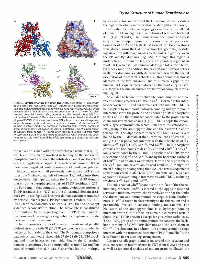
the active site is lined with positively charged residues (Fig. 3B),which are presumably involved in binding of the substrates’phosphatemoiety, whereas the substrate channel and the activesite are negatively charged. The surface of human TKT ismostly uncharged but contains several acidic and basic patches.In accordance with all previously determined TKT struc-
tures, the V-shaped subunit of human TKT folds into threeconsecutive �/�-type domains: the N-terminal PP domainthat binds the pyrophosphate part of ThDP (residues 1–276),the Pyr domain that contacts the aminopyrimidine portion ofThDP (residues 316–472), and the C-terminal domain (resi-dues 493–623) (Fig. 3C). The three domains are interconnectedby flexible linker regions (PP-Pyr domains, residues 277–315;Pyr-C-terminal domains, residues 473–492) that do not adopta defined secondary structure. The active site is constitutedfrom multiple loops originating from the PP domain and thePyr domain of two neighboring subunits, explaining the di-meric nature of the enzyme.The PP domain consists of a central five-stranded parallel
�-sheet structure with �1 �2 �3 �5 �4 topology surrounded byhelices on both sides of the sheet. The Pyr domain comprises aparallel six-stranded �-sheet with �7 �6 �8 �9 �11 �10 topol-ogy and three helices on each side. Finally, the C-terminaldomain is constituted by one antiparallel strand (�12) and fourparallel strands (�14 �13 �15 �16) embedded into multiple
helices. B-Factors indicate that the C-terminal domain exhibitsthe highest flexibility in the crystalline state (data not shown).Both subunit and domain topology as well as the overall fold
of human TKT are highly similar to those of yeast and bacterialTKT (Figs. 3D and 4). The subunits from the human and yeastenzyme can be superimposed with a root mean square devia-tion value of 2.1 Å and a highDaliZ score of 37.5 (579C� atomswere aligned) using theDaliLite version 3 program (42). A strik-ing structural difference locates to the linker region betweenthe PP and Pyr domains (Fig. 3D). Although this region isunstructured in human TKT, the corresponding segment inyeast TKT, which is �20 amino acids longer, folds into a helix-turn-helix motif. In addition, the orientation of several helicesin all three domains is slightly different. Remarkably, the spatialorientation of the central�-sheets in all three domains is almostidentical in the two enzymes. Due to numerous gaps in thehuman TKT sequence when aligned to the yeast enzyme, sev-eral loops in the human enzyme are shorter or completelymiss-ing (Fig. 4).As alluded to before, the active site containing the non-co-
valently bound cofactors ThDP andCa2� is formed at the inter-face between the PP and Pyr domains of both subunits. ThDP isheld in place by numerous hydrogen bonding and hydrophobicinteractions with the protein and by bonding of its diphosphateto the Ca2� ion that is further coordinated by the protein mainchain and several side chains (Fig. 5). ThDP adopts the canon-ical V-type conformation, which juxtaposes the catalytic 4�-NH2 group of the aminopyrimidine and the reactive C2 of thethiazolium. The diphosphate moiety of ThDP is exclusivelybound by the PP domain at the C-terminal ends of strands �1and �2. The �-phosphate interacts with the side chains of res-idues Ser40, Lys75, His77, Asn185, and Lys244. The �-phosphatecontacts the backbone amides of Gly156 and Glu157. The Ca2�
ion is coordinated by the �- and �-phosphate of ThDP, by theside chains of Asp155 andAsn185, and by the backbone carbonylof Leu187. In addition, a water interacts with the �-phosphate,the Ca2� ion, and several amino acid residues. Although themetal binding site comprising the GDG consensus sequence isstrictly conserved in all TKTs (4, 43), mammalian TKTs haveapparently evolved unique interactions with ThDP, includingresidues Ser40, Lys77, and Lys244.
The side chain of Gln189 spans over the re-face of the thiazo-lium ring, whereas Leu125 is located at the opposite face andserves as a fulcrum, over which the methylene bridge that linksthe thiazolium and aminopyrimidine rings is bent. Further-more, His258 is bound in close vicinity to the thiazolium and ispresumably involved in substrate binding and catalysis. TheN1� atom of the aminopyrimidine is in hydrogen-bondinginteraction with Glu366 of the Pyr domain, a conserved residuefound in all ThDP enzymes except for glyoxylate carboligase.The 4�-NH2 group of the aminopyrimidine contacts the back-bone carbonyl of Gly123 (PP domain) and the side chain ofGln428 (Pyr domain). In addition, the aminopyrimidine ringsinteractswith the aromatic side chains of Phe389 andPhe392, thelatter bound in a �-stacking arrangement.Recent crystallographic studies on several non-covalent and
covalent reaction intermediates in TKT from E. coli and yeastas well as functional analysis of mutant proteins allowed the
FIGURE 3. Crystal structure of human TKT. A, structure of the TKT dimer withbound cofactors ThDP (yellow) and Ca2� (magenta) in a schematic representa-tion. The individual subunits are shown in blue and red, respectively. B, surfacerepresentation of TKT dimer in the same orientation as in A. The surface elec-trostatic potential is color-coded, representing electrostatic potentials from�7 (red) to �7 (blue) kBT. The surface potential was calculated with the APBSplugin of PyMOL. C, domain structure of TKT subunit in a schematic represen-tation showing the three domains in a different color code: N-terminal PPdomain in yellow, middle Pyr domain in magenta, and C-terminal domain ingreen. The monomer is shown in the same orientation as in A. D, superpositionof subunits from human TKT (same color code as in C) and TKT from yeast(gray; Protein Data Bank code 1TRK) in a schematic representation. The struc-tures are rotated �90° around the 2-fold symmetry axis compared with pre-vious panels.
Crystal Structure of Human Transketolase
OCTOBER 8, 2010 • VOLUME 285 • NUMBER 41 JOURNAL OF BIOLOGICAL CHEMISTRY 31565
by guest on February 14, 2018http://w
ww
.jbc.org/D
ownloaded from

Crystal Structure of Human Transketolase
31566 JOURNAL OF BIOLOGICAL CHEMISTRY VOLUME 285 • NUMBER 41 • OCTOBER 8, 2010
by guest on February 14, 2018http://w
ww
.jbc.org/D
ownloaded from

identification of critical interactions between the substrate/in-termediates and the protein (10–12). A superposition of theactive sites of human and E. coli TKT (Fig. 6) reveals that mostof the active site residues are strictly conserved and occupyalmost identical positions in the two proteins, including twoarginines (Arg474 and Arg318 in human TKT), a serine (Ser345),and a histidine (His461) all shown to be involved in binding of
the phosphate portion of the substrates. The same observationholds true for a cluster of histidines (His110, His37, His258) andAsp424, which are likely to form hydrogen bonds with the sub-strate hydroxyl groups, as shown for the homologous residuesin bacterial TKT. Despite these commonalties, the humanenzyme exhibits some unique features. Most intriguingly,Gln428 replaces a histidine (His481 in yeast TKT,His473 in E. coli
FIGURE 4. Sequence alignment of human TKT versus orthologs from mouse, E. coli, maize, and yeast using the programs ClustalW2 and ESPript 2.2.Numbering corresponds to the sequence of human TKT. Identical residues are indicated by a red background, and conserved residues are indicated by redcharacters. The secondary structure elements of human TKT are shown above the sequences, and those of yeast TKT are shown below.
FIGURE 5. Structure of the cofactor binding site in human TKT. A, stereo drawing showing cofactor ThDP and selected amino acid residues in stick represen-tation and Ca2� as a magenta sphere. Amino acids contributed by different subunits are indicated by different color-coding. The final 2Fo � Fc electron densitymap of ThDP is contoured at 1.0�. B, schematic diagram of the interactions of bound ThDP and Ca2� at the active site of human TKT. Possible hydrogen-bondingand hydrophobic interactions are indicated by dashed lines. Residues from the neighboring subunit are indicated by an asterisk.
FIGURE 6. Superposition of the active sites of human (yellow) and E. coli (purple) TKT in stereo view showing selected amino acid residues and boundThDP (green) in stick representation and Ca2� as a magenta sphere. Residues of human TKT are labeled.
Crystal Structure of Human Transketolase
OCTOBER 8, 2010 • VOLUME 285 • NUMBER 41 JOURNAL OF BIOLOGICAL CHEMISTRY 31567
by guest on February 14, 2018http://w
ww
.jbc.org/D
ownloaded from

TKT) found to be invariant in all non-mammalian TKTs. Thisfinding rules out the possibility that this residue acts as a gen-eral acid/base catalyst and rather suggests a role for substratebinding and orbital alignment in intermediates. Second, resi-due Gln189 sitting atop the thiazolium nucleus as a backstopreplaces an isoleucine found in most non-mammalian TKTs.Gln189 but not the equivalent isoleucine in non-mammalianTKTs partially occludes the thiazolium ring, inviting specula-tion that this Gln residue could sterically hinder cofactor disso-ciation, thus explaining the quasi-irreversible binding of ThDPin mammalian enzymes. Third, the human enzyme contains alysine residue (Lys260), which is located at the entrance to thesubstrate channel and is presumably involved in binding of thesubstrate phosphate. There is no equivalent residue found innon-mammalian TKTs.
Sequence Alignment of Transketolase and Transketolase-likeProteins 1 and 2 and Implications for Putative Functions—Be-sides TKT, the human genome encodes for the two sequence-wise closely related proteins, TKTL1 andTKTL2, which share asequence identity on the amino acid level of 61% (TKTL1) andof 66% (TKTL2) compared with TKT. Although the enzymaticfunction of TKT is well characterized, much less is knownabout the biochemical and enzymatic properties of TKTL1 andTKTL2. To date, there are no reports on enzymatic or cellularfunctions of TKTL2. The elucidation of the cellular function ofTKTL1 is of particular interest because overexpression ofTKTL1 mRNA and protein were observed in different humancancers and have been linked tometastasis and poor survival ofcancer patients (29, 44). In contrast to TKTL1, TKT andTKTL2 are not overexpressed in tumor cells, indicating a func-
FIGURE 7. Sequence alignment of human TKT, TKTL1, and TKTL2 (hTKT, hTKTL1, and hTKTL2). Residues highlighted in gray are invariant among all threeproteins. Residues involved in cofactor and/or substrate binding are highlighted in yellow. Residues invoked in substrate binding and catalysis in TKT but notconserved in TKTL1 or TKTL2 are indicated in red. Sequences were aligned with ClustalW2.
Crystal Structure of Human Transketolase
31568 JOURNAL OF BIOLOGICAL CHEMISTRY VOLUME 285 • NUMBER 41 • OCTOBER 8, 2010
by guest on February 14, 2018http://w
ww
.jbc.org/D
ownloaded from
tional divergence of TKTL1 compared with TKT and TKTL2(45). The TKTL1 gene is activated by promoter hypomethyla-tion and contributes to carcinogenesis through increased aero-bic glycolysis and stabilization of HIF1� (46). Through this sta-bilization, TKTL1 leads to a shift from a mitochondria-basedoxidative energy release to a fermentative energy release con-comitant with a suppression of radical- and apoptosis-inducedcell death (29). The protective function of TKTL1 in normalcells by reactive oxygen species detoxification and preventionof tissue damage has been demonstrated using TKTL1-defi-cient mice.4 Despite these differences from TKT, TKTL1expression has been shown to compensate for inhibition ofTKT translation, thereby restoring cancer drug resistance toimatinib (47).Based on the newly determined crystal structure of human
TKT and a sequence alignment between TKT, TKTL1, andTKTL2, we have analyzed whether amino acid residues shownto be involved in cofactor and substrate binding in TKT are alsoconserved in TKTL1 and -2 (Fig. 7). As mentioned before,TKTL1 has a deletion of 38 amino acids in the N-terminal PPdomain (residues 76–113 in TKT). This segment folds into aloop-helix-turn-helix-loop motif in human TKT. Both loopregions constitute part of the active and cofactor binding siteand harbor numerous residues that are likely to play importantroles for cofactor binding (His77) and catalysis (His77/His110).Both residues are invariant in all TKTs, and the homologousresidues of His110 in yeast and bacterial TKT were shown toform an essential hydrogen-bonding interaction with 1-OH ofthe X5P-ThDP, F6P-ThDP, and DHE-ThDP carbanion/enam-ine intermediates (11, 12). Mutagenesis studies revealed thatany substitution of His110 renders human TKT inactive (5). Inaddition, there is a mutation in the GDG (residues 154–156 inTKT) cofactor binding consensus sequence; TKTL1 features aSDG sequence instead. The GDG consensus sequence is highlyconserved among all TKTs (4); there is only one annotatedsequence in which the first glycine is replaced (TKT fromMycobacterium leprae) by another residue. However, the enzy-matic function of this particular protein has not been provenexperimentally but was inferred from sequence similarity.Another difference in sequence of the putative cofactor bindingsite is found for Gln189, which serves as a backstop for the thia-zolium ring of ThDP in TKT and is replaced by a histidine inTKTL1. A third crucial difference can be identified for a shortloop (Gly123-Ser124-Leu125) involved in binding of the amin-opyrimidine. In TKTL1, the bulky tryptophan replaces the oth-erwise highly conserved serine (or proline) residue. Takentogether, TKTL1 is lacking several important residues provento be important for cofactor binding and catalysis in all hithertocharacterized TKTs, suggesting that TKTL1 may not possess acommon transketolase activity. Moreover, it remains to bestudied whether TKTL1 does bind ThDP at all because severalcritical determinants of cofactor binding in TKTs are eithermissing in TKTL1 or nonconservatively replaced.In the sequence of transketolase-like protein 2, there are also
several substitutions of residues belonging to the active site and
cofactor binding pocket in TKT. Lys75 involved in bonding ofthe diphosphate anchor of ThDP in TKT is conservativelyreplaced by an arginine residue. However, a histidine (His37 inhuman TKT) and two arginines (Arg474 and Arg318), which aretotally invariant among all TKTs are non-homologously re-placed in TKTL2 by glutamine residues in each case. Structuralstudies revealed these side chains to interact either with sub-strate hydroxyl groups (histidine) or with the phosphatemoiety(arginines) (10, 12). Substitutions of the invariant histidineabolished activity, whereas binding of phosphorylated sugarsubstrates is significantly compromised in variants with substi-tutions of the conserved arginine residues (48, 49). In conclu-sion, TKTL2 lacks numerous residues that are important forsubstrate binding and catalysis in TKTs and can thus beexpected to possess only little TKT activity if any.
CONCLUSIONS
The crystal structure of human TKT has been determinedas the first structure of a mammalian TKT. Although subunittopology, dimer assembly, and active site architecture arehighly conserved among all TKTs, mammalian TKTs haveevolved numerous unique interactions with the cofactor andthe substrate/reaction intermediates. The related proteinsTKTL1 and TKTL2 lack numerous invariant residues involvedin cofactor and substrate binding and are therefore notexpected to possess TKT activity.
Acknowledgment—We thank Dr. C. Gobel for mass spectrometricanalysis of chemoenzymatically synthesized S7P.
REFERENCES1. Schenk, G., Duggleby, R. G., and Nixon, P. F. (1998) Int. J. Biochem. Cell
Biol. 30, 1297–13182. Schneider, G., and Lindqvist, Y. (1998) Biochim. Biophys. Acta 1385,
387–3983. Kluger, R., and Tittmann, K. (2008) Chem. Rev. 108, 1797–18334. Schenk, G., Layfield, R., Candy, J. M., Duggleby, R. G., and Nixon, P. F.
(1997) J. Mol. Evol. 44, 552–5725. Singleton, C. K.,Wang, J. J., Shan, L., andMartin, P. R. (1996)Biochemistry
35, 15865–158696. Lindqvist, Y., Schneider, G., Ermler, U., and Sundstrom,M. (1992) EMBO
J. 11, 2373–23797. Nikkola, M., Lindqvist, Y., and Schneider, G. (1994) J. Mol. Biol. 238,
387–4048. Littlechild, J., Turner, N., Hobbs, G., Lilly, M., Rawas, A., and Watson, H.
(1995) Acta. Crystallogr. D Biol. Crystallogr. 51, 1074–10769. Gerhardt, S., Echt, S., Busch, M., Freigang, J., Auerbach, G., Bader, G.,
Martin, W. F., Bacher, A., Huber, R., and Fischer, M. (2003) Plant Physiol.132, 1941–1949
10. Nilsson, U., Meshalkina, L., Lindqvist, Y., and Schneider, G. (1997) J. Biol.Chem. 272, 1864–1869
11. Fiedler, E., Thorell, S., Sandalova, T., Golbik, R., Konig, S., and Schneider,G. (2002) Proc. Natl. Acad. Sci. U.S.A. 99, 591–595
12. Asztalos, P., Parthier, C., Golbik, R., Kleinschmidt, M., Hubner, G., Weiss,M. S., Friedemann, R.,Wille, G., and Tittmann, K. (2007) Biochemistry 46,12037–12052
13. Tittmann, K., and Wille, G. (2009) J. Mol. Catal. B Enzym. 61, 93–9914. Nixon, P. F., Kaczmarek, M. J., Tate, J., Kerr, R. A., and Price, J. (1984) Eur.
J. Clin. Invest. 14, 278–28115. Heroux, M., Raghavendra Rao, V. L., Lavoie, J., Richardson, J. S., and But-
terworth, R. F. (1996)Metab. Brain Dis. 11, 81–8816. Hammes, H. P., Du, X., Edelstein, D., Taguchi, T., Matsumura, T., Ju, Q.,
4 S. Bentz, T. Pesch, L. Wolfram, C. de Valliere, K. Leucht, M. Fried, J. Coy, M.Hausmann, and G. Rogler, submitted for publication.
Crystal Structure of Human Transketolase
OCTOBER 8, 2010 • VOLUME 285 • NUMBER 41 JOURNAL OF BIOLOGICAL CHEMISTRY 31569
by guest on February 14, 2018http://w
ww
.jbc.org/D
ownloaded from

Lin, J., Bierhaus, A., Nawroth, P., Hannak, D., Neumaier, M., Bergfeld, R.,Giardino, I., and Brownlee, M. (2003) Nat. Med. 9, 294–299
17. Zhao, Y., Pan, X., Zhao, J.,Wang, Y., Peng, Y., and Zhong, C. (2009) J. Neu-rochem. 111, 537–546
18. Liu, S., Sun, M. Z., Tang, J. W., Wang, Z., Sun, C., and Greenaway, F. T.(2008) Rapid Commun. Mass Spectrom. 22, 3172–3178
19. Cascante, M., Centelles, J. J., Veech, R. L., Lee, W. N., and Boros, L. G.(2000) Nutr. Cancer 36, 150–154
20. Shin, Y. K., Yoo, B. C., Hong, Y. S., Chang,H. J., Jung, K.H., Jeong, S. Y., andPark, J. G. (2009) Electrophoresis 30, 2182–2192
21. Xu, Z. P., Wawrousek, E. F., and Piatigorsky, J. (2002) Mol. Cell. Biol. 22,6142–6147
22. Calingasan, N. Y., Sheu, K. F., Baker, H., Jung, E. H., Paoletti, F., andGibson, G. E. (1995) J. Neurochem. 64, 1034–1044
23. Sax, C. M., Salamon, C., Kays,W. T., Guo, J., Yu, F. X., Cuthbertson, R. A.,and Piatigorsky, J. (1996) J. Biol. Chem. 271, 33568–33574
24. Boros, L.G., Puigjaner, J., Cascante,M., Lee,W.N., Brandes, J. L., Bassilian,S., Yusuf, F. I.,Williams, R. D.,Muscarella, P.,Melvin,W. S., and Schirmer,W. J. (1997) Cancer Res. 57, 4242–4248
25. Thomas, A. A., De Meese, J., Le Huerou, Y., Boyd, S. A., Romoff, T. T.,Gonzales, S. S., Gunawardana, I., Kaplan, T., Sullivan, F., Condroski, K.,Lyssikatos, J. P., Aicher, T. D., Ballard, J., Bernat, B., DeWolf, W., Han, M.,Lemieux, C., Smith, D., Weiler, S., Wright, S. K., Vigers, G., and Brandhu-ber, B. (2008) Bioorg. Med. Chem. Lett. 18, 509–512
26. Thomas, A. A., Le Huerou, Y., De Meese, J., Gunawardana, I., Kaplan, T.,Romoff, T. T., Gonzales, S. S., Condroski, K., Boyd, S. A., Ballard, J., Bernat,B., DeWolf, W., Han, M., Lee, P., Lemieux, C., Pedersen, R., Pheneger, J.,Poch, G., Smith, D., Sullivan, F., Weiler, S., Wright, S. K., Lin, J., Brandhu-ber, B., and Vigers, G. (2008) Bioorg. Med. Chem. Lett. 18, 2206–2210
27. Le Huerou, Y., Gunawardana, I., Thomas, A. A., Boyd, S. A., de Meese, J.,Dewolf, W., Gonzales, S. S., Han, M., Hayter, L., Kaplan, T., Lemieux, C.,Lee, P., Pheneger, J., Poch, G., Romoff, T. T., Sullivan, F., Weiler, S.,Wright, S. K., and Lin, J. (2008) Bioorg. Med. Chem. Lett. 18, 505–508
28. Coy, J. F., Dubel, S., Kioschis, P., Thomas, K., Micklem, G., Delius, H., andPoustka, A. (1996) Genomics 32, 309–316
29. Xu, X., ZurHausen, A., Coy, J. F., and Lochelt,M. (2009) Int. J. Cancer 124,1330–1337
30. Charmantray, F., Helaine, V., Legeret, B., and Hecquet, L. (2009) J Mol.Catal. B Enzym. 57, 6–9
31. Bykova, I. A., Solovjeva, O. N., Meshalkina, L. E., Kovina, M. V., and
Kochetov, G. A. (2001) Biochem. Biophys. Res. Commun. 280, 845–84732. Tittmann, K., Golbik, R., Uhlemann, K., Khailova, L., Schneider, G., Patel,
M., Jordan, F., Chipman, D. M., Duggleby, R. G., and Hubner, G. (2003)Biochemistry 42, 7885–7891
33. Schneider, G., Sundstrom, M., and Lindqvist, Y. (1989) J. Biol. Chem. 264,21619–21620
34. Kabsch, W. (1993) J. Appl. Crystallogr. 26, 795–80035. Collaborative Computational Project 4 (1994) Acta Crystallogr. D Biol.
Crystallogr. 50, 760–76336. Emsley, P., and Cowtan, K. (2004)Acta Crystallogr. D Biol. Crystallogr. 60,
2126–213237. Davis, I. W., Leaver-Fay, A., Chen, V. B., Block, J. N., Kapral, G. J., Wang,
X., Murray, L. W., Arendall, W. B., 3rd, Snoeyink, J., Richardson, J. S., andRichardson, D. C. (2007) Nucleic Acids Res. 35,W375–W383
38. Thompson, J. D., Gibson, T. J., andHiggins, D. G. (2002)Current Protocolsin Bioinformatics, Chapter 2, Unit 2.3, JohnWiley & Sons, Inc., New York
39. Gouet, P., Courcelle, E., Stuart, D. I., and Metoz, F. (1999) Bioinformatics15, 305–308
40. Schenk, G., Duggleby, R. G., and Nixon, P. F. (1998) Int. J. Biochem. CellBiol. 30, 369–378
41. Sprenger, G. A., Schorken, U., Sprenger, G., and Sahm, H. (1995) Eur.J. Biochem. 230, 525–532
42. Holm, L., Kaariainen, S., Rosenstrom, P., and Schenkel, A. (2008) Bioin-formatics 24, 2780–2781
43. Hawkins, C. F., Borges, A., and Perham, R. N. (1989) FEBS Lett. 255,77–82
44. Furuta, E., Okuda, H., Kobayashi, A., and Watabe, K. (2010) Biochim.Biophys. Acta Rev. Cancer 1805, 141–152
45. Sigrun, L., Zerilli, M., Zur, H. A., Popa, J., Steidler, A., Alken, P., Stassi, G.,Schubert, P., and Coy, J. F. (2006) Tumor Biol. 27, 48–48
46. Sun, W., Liu, Y., Glazer, C. A., Shao, C., Bhan, S., Demokan, S., Zhao, M.,Rudek, M. A., Ha, P. K., and Califano, J. A. (2010) Clin. Cancer Res. 16,857–866
47. Zhao, F., Mancuso, A., Bui, T. V., Tong, X., Gruber, J. J., Swider, C. R.,Sanchez, P. V., Lum, J. J., Sayed, N., Melo, J. V., Perl, A. E., Carroll, M.,Tuttle, S. W., and Thompson, C. B. (2010) Oncogene 29, 2962–2972
48. Wikner, C., Nilsson, U., Meshalkina, L., Udekwu, C., Lindqvist, Y., andSchneider, G. (1997) Biochemistry 36, 15643–15649
49. Soh, Y., Song, B. J., Jeng, J., and Kallarakal, A. T. (1998) Biochem. J. 333,367–372
Crystal Structure of Human Transketolase
31570 JOURNAL OF BIOLOGICAL CHEMISTRY VOLUME 285 • NUMBER 41 • OCTOBER 8, 2010
by guest on February 14, 2018http://w
ww
.jbc.org/D
ownloaded from
Lüdtke and Kai TittmannLars Mitschke, Christoph Parthier, Kathrin Schröder-Tittmann, Johannes Coy, Stefan
ActionThe Crystal Structure of Human Transketolase and New Insights into Its Mode of
doi: 10.1074/jbc.M110.149955 originally published online July 28, 20102010, 285:31559-31570.J. Biol. Chem.
10.1074/jbc.M110.149955Access the most updated version of this article at doi:
Alerts:
When a correction for this article is posted•
When this article is cited•
to choose from all of JBC's e-mail alertsClick here
http://www.jbc.org/content/285/41/31559.full.html#ref-list-1
This article cites 48 references, 9 of which can be accessed free at
by guest on February 14, 2018http://w
ww
.jbc.org/D
ownloaded from
Related Documents