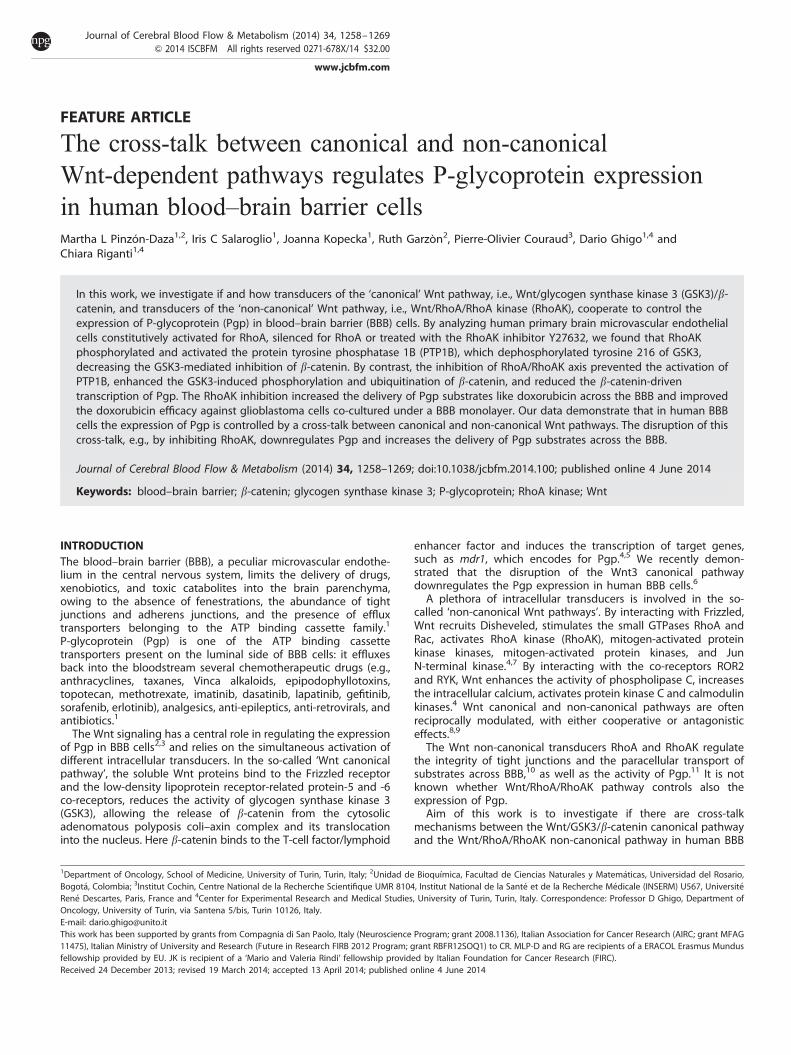
FEATURE ARTICLE The cross-talk between canonical and non-canonical Wnt-dependent pathways regulates P-glycoprotein expression in human blood–brain barrier cells Martha L Pinzo ´ n-Daza 1,2 , Iris C Salaroglio 1 , Joanna Kopecka 1 , Ruth Garzo `n 2 , Pierre-Olivier Couraud 3 , Dario Ghigo 1,4 and Chiara Riganti 1,4 In this work, we investigate if and how transducers of the ‘canonical’ Wnt pathway, i.e., Wnt/glycogen synthase kinase 3 (GSK3)/b- catenin, and transducers of the ‘non-canonical’ Wnt pathway, i.e., Wnt/RhoA/RhoA kinase (RhoAK), cooperate to control the expression of P-glycoprotein (Pgp) in blood–brain barrier (BBB) cells. By analyzing human primary brain microvascular endothelial cells constitutively activated for RhoA, silenced for RhoA or treated with the RhoAK inhibitor Y27632, we found that RhoAK phosphorylated and activated the protein tyrosine phosphatase 1B (PTP1B), which dephosphorylated tyrosine 216 of GSK3, decreasing the GSK3-mediated inhibition of b-catenin. By contrast, the inhibition of RhoA/RhoAK axis prevented the activation of PTP1B, enhanced the GSK3-induced phosphorylation and ubiquitination of b-catenin, and reduced the b-catenin-driven transcription of Pgp. The RhoAK inhibition increased the delivery of Pgp substrates like doxorubicin across the BBB and improved the doxorubicin efficacy against glioblastoma cells co-cultured under a BBB monolayer. Our data demonstrate that in human BBB cells the expression of Pgp is controlled by a cross-talk between canonical and non-canonical Wnt pathways. The disruption of this cross-talk, e.g., by inhibiting RhoAK, downregulates Pgp and increases the delivery of Pgp substrates across the BBB. Journal of Cerebral Blood Flow & Metabolism (2014) 34, 1258–1269; doi:10.1038/jcbfm.2014.100; published online 4 June 2014 Keywords: blood–brain barrier; b-catenin; glycogen synthase kinase 3; P-glycoprotein; RhoA kinase; Wnt INTRODUCTION The blood–brain barrier (BBB), a peculiar microvascular endothe- lium in the central nervous system, limits the delivery of drugs, xenobiotics, and toxic catabolites into the brain parenchyma, owing to the absence of fenestrations, the abundance of tight junctions and adherens junctions, and the presence of efflux transporters belonging to the ATP binding cassette family. 1 P-glycoprotein (Pgp) is one of the ATP binding cassette transporters present on the luminal side of BBB cells: it effluxes back into the bloodstream several chemotherapeutic drugs (e.g., anthracyclines, taxanes, Vinca alkaloids, epipodophyllotoxins, topotecan, methotrexate, imatinib, dasatinib, lapatinib, gefitinib, sorafenib, erlotinib), analgesics, anti-epileptics, anti-retrovirals, and antibiotics. 1 The Wnt signaling has a central role in regulating the expression of Pgp in BBB cells 2,3 and relies on the simultaneous activation of different intracellular transducers. In the so-called ‘Wnt canonical pathway’, the soluble Wnt proteins bind to the Frizzled receptor and the low-density lipoprotein receptor-related protein-5 and -6 co-receptors, reduces the activity of glycogen synthase kinase 3 (GSK3), allowing the release of b-catenin from the cytosolic adenomatous polyposis coli–axin complex and its translocation into the nucleus. Here b-catenin binds to the T-cell factor/lymphoid enhancer factor and induces the transcription of target genes, such as mdr1, which encodes for Pgp. 4,5 We recently demon- strated that the disruption of the Wnt3 canonical pathway downregulates the Pgp expression in human BBB cells. 6 A plethora of intracellular transducers is involved in the so- called ‘non-canonical Wnt pathways’. By interacting with Frizzled, Wnt recruits Disheveled, stimulates the small GTPases RhoA and Rac, activates RhoA kinase (RhoAK), mitogen-activated protein kinase kinases, mitogen-activated protein kinases, and Jun N-terminal kinase. 4,7 By interacting with the co-receptors ROR2 and RYK, Wnt enhances the activity of phospholipase C, increases the intracellular calcium, activates protein kinase C and calmodulin kinases. 4 Wnt canonical and non-canonical pathways are often reciprocally modulated, with either cooperative or antagonistic effects. 8,9 The Wnt non-canonical transducers RhoA and RhoAK regulate the integrity of tight junctions and the paracellular transport of substrates across BBB, 10 as well as the activity of Pgp. 11 It is not known whether Wnt/RhoA/RhoAK pathway controls also the expression of Pgp. Aim of this work is to investigate if there are cross-talk mechanisms between the Wnt/GSK3/b-catenin canonical pathway and the Wnt/RhoA/RhoAK non-canonical pathway in human BBB 1 Department of Oncology, School of Medicine, University of Turin, Turin, Italy; 2 Unidad de Bioquı ´mica, Facultad de Ciencias Naturales y Matema ´ticas, Universidad del Rosario, Bogota ´, Colombia; 3 Institut Cochin, Centre National de la Recherche Scientifique UMR 8104, Institut National de la Sante ´ et de la Recherche Me ´ dicale (INSERM) U567, Universite ´ Rene ´ Descartes, Paris, France and 4 Center for Experimental Research and Medical Studies, University of Turin, Turin, Italy. Correspondence: Professor D Ghigo, Department of Oncology, University of Turin, via Santena 5/bis, Turin 10126, Italy. E-mail: [email protected] This work has been supported by grants from Compagnia di San Paolo, Italy (Neuroscience Program; grant 2008.1136), Italian Association for Cancer Research (AIRC; grant MFAG 11475), Italian Ministry of University and Research (Future in Research FIRB 2012 Program; grant RBFR12SOQ1) to CR. MLP-D and RG are recipients of a ERACOL Erasmus Mundus fellowship provided by EU. JK is recipient of a ‘Mario and Valeria Rindi’ fellowship provided by Italian Foundation for Cancer Research (FIRC). Received 24 December 2013; revised 19 March 2014; accepted 13 April 2014; published online 4 June 2014 Journal of Cerebral Blood Flow & Metabolism (2014) 34, 1258–1269 & 2014 ISCBFM All rights reserved 0271-678X/14 $32.00 www.jcbfm.com

Welcome message from author
This document is posted to help you gain knowledge. Please leave a comment to let me know what you think about it! Share it to your friends and learn new things together.
Transcript
FEATURE ARTICLE
The cross-talk between canonical and non-canonicalWnt-dependent pathways regulates P-glycoprotein expressionin human blood–brain barrier cellsMartha L Pinzon-Daza1,2, Iris C Salaroglio1, Joanna Kopecka1, Ruth Garzon2, Pierre-Olivier Couraud3, Dario Ghigo1,4 andChiara Riganti1,4
In this work, we investigate if and how transducers of the ‘canonical’ Wnt pathway, i.e., Wnt/glycogen synthase kinase 3 (GSK3)/b-catenin, and transducers of the ‘non-canonical’ Wnt pathway, i.e., Wnt/RhoA/RhoA kinase (RhoAK), cooperate to control theexpression of P-glycoprotein (Pgp) in blood–brain barrier (BBB) cells. By analyzing human primary brain microvascular endothelialcells constitutively activated for RhoA, silenced for RhoA or treated with the RhoAK inhibitor Y27632, we found that RhoAKphosphorylated and activated the protein tyrosine phosphatase 1B (PTP1B), which dephosphorylated tyrosine 216 of GSK3,decreasing the GSK3-mediated inhibition of b-catenin. By contrast, the inhibition of RhoA/RhoAK axis prevented the activation ofPTP1B, enhanced the GSK3-induced phosphorylation and ubiquitination of b-catenin, and reduced the b-catenin-driventranscription of Pgp. The RhoAK inhibition increased the delivery of Pgp substrates like doxorubicin across the BBB and improvedthe doxorubicin efficacy against glioblastoma cells co-cultured under a BBB monolayer. Our data demonstrate that in human BBBcells the expression of Pgp is controlled by a cross-talk between canonical and non-canonical Wnt pathways. The disruption of thiscross-talk, e.g., by inhibiting RhoAK, downregulates Pgp and increases the delivery of Pgp substrates across the BBB.
Journal of Cerebral Blood Flow & Metabolism (2014) 34, 1258–1269; doi:10.1038/jcbfm.2014.100; published online 4 June 2014
Keywords: blood–brain barrier; b-catenin; glycogen synthase kinase 3; P-glycoprotein; RhoA kinase; Wnt
INTRODUCTIONThe blood–brain barrier (BBB), a peculiar microvascular endothe-lium in the central nervous system, limits the delivery of drugs,xenobiotics, and toxic catabolites into the brain parenchyma,owing to the absence of fenestrations, the abundance of tightjunctions and adherens junctions, and the presence of effluxtransporters belonging to the ATP binding cassette family.1
P-glycoprotein (Pgp) is one of the ATP binding cassettetransporters present on the luminal side of BBB cells: it effluxesback into the bloodstream several chemotherapeutic drugs (e.g.,anthracyclines, taxanes, Vinca alkaloids, epipodophyllotoxins,topotecan, methotrexate, imatinib, dasatinib, lapatinib, gefitinib,sorafenib, erlotinib), analgesics, anti-epileptics, anti-retrovirals, andantibiotics.1
The Wnt signaling has a central role in regulating the expressionof Pgp in BBB cells2,3 and relies on the simultaneous activation ofdifferent intracellular transducers. In the so-called ‘Wnt canonicalpathway’, the soluble Wnt proteins bind to the Frizzled receptorand the low-density lipoprotein receptor-related protein-5 and -6co-receptors, reduces the activity of glycogen synthase kinase 3(GSK3), allowing the release of b-catenin from the cytosolicadenomatous polyposis coli–axin complex and its translocationinto the nucleus. Here b-catenin binds to the T-cell factor/lymphoid
enhancer factor and induces the transcription of target genes,such as mdr1, which encodes for Pgp.4,5 We recently demon-strated that the disruption of the Wnt3 canonical pathwaydownregulates the Pgp expression in human BBB cells.6
A plethora of intracellular transducers is involved in the so-called ‘non-canonical Wnt pathways’. By interacting with Frizzled,Wnt recruits Disheveled, stimulates the small GTPases RhoA andRac, activates RhoA kinase (RhoAK), mitogen-activated proteinkinase kinases, mitogen-activated protein kinases, and JunN-terminal kinase.4,7 By interacting with the co-receptors ROR2and RYK, Wnt enhances the activity of phospholipase C, increasesthe intracellular calcium, activates protein kinase C and calmodulinkinases.4 Wnt canonical and non-canonical pathways are oftenreciprocally modulated, with either cooperative or antagonisticeffects.8,9
The Wnt non-canonical transducers RhoA and RhoAK regulatethe integrity of tight junctions and the paracellular transport ofsubstrates across BBB,10 as well as the activity of Pgp.11 It is notknown whether Wnt/RhoA/RhoAK pathway controls also theexpression of Pgp.
Aim of this work is to investigate if there are cross-talkmechanisms between the Wnt/GSK3/b-catenin canonical pathwayand the Wnt/RhoA/RhoAK non-canonical pathway in human BBB
1Department of Oncology, School of Medicine, University of Turin, Turin, Italy; 2Unidad de Bioquımica, Facultad de Ciencias Naturales y Matematicas, Universidad del Rosario,Bogota, Colombia; 3Institut Cochin, Centre National de la Recherche Scientifique UMR 8104, Institut National de la Sante et de la Recherche Medicale (INSERM) U567, UniversiteRene Descartes, Paris, France and 4Center for Experimental Research and Medical Studies, University of Turin, Turin, Italy. Correspondence: Professor D Ghigo, Department ofOncology, University of Turin, via Santena 5/bis, Turin 10126, Italy.E-mail: [email protected] work has been supported by grants from Compagnia di San Paolo, Italy (Neuroscience Program; grant 2008.1136), Italian Association for Cancer Research (AIRC; grant MFAG11475), Italian Ministry of University and Research (Future in Research FIRB 2012 Program; grant RBFR12SOQ1) to CR. MLP-D and RG are recipients of a ERACOL Erasmus Mundusfellowship provided by EU. JK is recipient of a ‘Mario and Valeria Rindi’ fellowship provided by Italian Foundation for Cancer Research (FIRC).Received 24 December 2013; revised 19 March 2014; accepted 13 April 2014; published online 4 June 2014
Journal of Cerebral Blood Flow & Metabolism (2014) 34, 1258–1269& 2014 ISCBFM All rights reserved 0271-678X/14 $32.00
www.jcbfm.com

cells, and how these cross-talks control the expression of Pgp andthe delivery of Pgp substrates across BBB.
MATERIALS AND METHODSChemicalsThe plasticware for cell cultures was from Falcon (Becton Dickinson,Franklin Lakes, NJ, USA). Wnt activators (WntA) [2-amino-4-(3,4-(methyle-nedioxy)benzylamino)-6-(3-methoxyphenyl)pyrimidine] and Y27632 werepurchased from Calbiochem (San Diego, CA, USA). Human recombinantDickkopf-1 (Dkk-1) was from R&D Systems (Minneapolis, MN, USA). Rhoactivator II, a synthetic derivative of cytotoxic necrotizing factor fromEscherichia coli that maintains RhoA constitutively activated,12 was fromCytoskeleton (Denver, CO, USA). The electrophoresis reagents wereobtained from Bio-Rad Laboratories (Hercules, CA, USA). The proteincontent of cell lysates was assessed with the BCA kit from Sigma Chemicals(St Louis, MO, USA). When not otherwise specified, all the other reagentswere purchased from Sigma Chemicals.
CellsThe hCMEC/D3 cells, a primary human brain microvascular endothelial cellline that retains the BBB characteristics in vitro,13 were seeded at 50,000/cm2 density, grown for 7 days up to confluence in Petri dishes or Transwelldevices (0.4mm diameter pore size, Corning Life Sciences, Chorges, France),and cultured as previously reported.13
The primary human glioblastoma cells (CV17, 01010627, Nov3) wereobtained from surgical samples of patients from the Neuro-BiooncologyCenter, Vercelli, Italy and the DIBIT San Raffaele Scientific Institute, Milan,Italy. The tumor diagnosis was performed according to WHO guidelines.The U87-MG cell line was purchased from ATCC (Manassas, VA, USA). Thecells were cultured as already reported.6 The experimental protocols wereapproved by the Bioethics Committee (‘Comitato di Bioetica d’Ateneo’),University of Turin, Italy.
In co-culture experiments, 500,000 (for intracellular doxorubicinaccumulation, cytotoxicity assays and cell cycle analysis) or 1,000 (forproliferation assay) glioblastoma cells were added in the lower chamber ofTranswell devices 4 days after seeding hCMEC/D3 cells in the Transwellinsert. After 3 days of co-culture, the medium of the upper chamber wasreplaced with fresh medium or with medium containing Y27632 anddoxorubicin, alone or in combination, as detailed in the Figure legends.
Western Blot AnalysisThe cells were rinsed with lysis buffer (50 mmol/L Tris, 10 mmol/L EDTA, 1%v/v Triton X-100), supplemented with the protease inhibitor cocktail set III(80mmol/L aprotinin, 5 mmol/L bestatin, 1.5 mmol/L leupeptin, 1 mmol/Lpepstatin; Calbiochem), 2 mmol/L phenylmethylsulfonyl fluoride and 1 mmol/L NaVO4, then sonicated and centrifuged at 13,000 g for 10 minutes at 41C.A quantity of 20mg protein extracts were subjected to SDS–PAGE andprobed with the following antibodies: anti-GSK3 (BD Biosciences, FranklinLakes, NJ, USA); anti-phospho(Tyr216)GSK3 (BD Biosciences); anti-b-catenin(BD Biosciences); anti-phospho(Ser33/Ser37/Thr41)b-catenin (Cell SignalingTechnology, Danvers, MA, USA); anti-claudin 3 (Invitrogen Life Technolo-gies, Monza, Italy); anti-claudin 5 (Invitrogen Life Technologies); anti-occludin (Invitrogen Life Technologies); anti-zonula occludens-1 (InvitrogenLife Technologies); anti-protein tyrosine phosphatase 1B (PTP1B; Abcam,Cambridge, UK); anti-phospho(Ser50)PTP1B (Abcam); anti-Pgp (clone C219,Calbiochem); anti-multidrug resistance-related protein 1 (MRP1; Abcam);anti-breast cancer resistance protein (BCRP; Santa Cruz Biotechnology,Santa Cruz, CA, USA); anti-caspase-3 (C33, GeneTex, Hsinhu City, Taiwan);anti-b-tubulin (Santa Cruz Biotechnology), followed by a peroxidase-conjugated secondary antibody (Bio-Rad). The membranes were washedwith Tris-buffered saline-Tween 0.1% v/v, and the proteins were detectedby enhanced chemiluminescence (Bio-Rad).
To assess the presence of ubiquitinated b-catenin, 100mg of proteinsfrom whole-cell extracts were immunoprecipitated with the anti-b-cateninantibody, using the PureProteome protein A and protein G Magnetic Beads(Millipore, Billerica, MA, USA). The immunoprecipitated proteins wereseparated by SDS–PAGE and probed with an anti-ubiquitin antibody (EnzoLife Science, Farmingdale, NY, USA), followed by a peroxidase-conjugatedsecondary antibody.
A quantity of 10mg of nuclear extracts, obtained with the NuclearExtraction Kit (Active Motif, Rixensart, Belgium), was subjected to westernblot analysis using an anti-b-catenin antibody. To check the equal control
loading in nuclear fractions, the samples were probed with an anti-TATA-binding protein (TBP/TFIID) antibody (Santa Cruz Biotechnology). Toexclude any cytosolic contamination of nuclear extracts, we verified thatb-tubulin was undetectable in nuclear samples (not shown).
The densitometric analysis of western blots was performed withthe ImageJ software (http://rsb.info.nih.gov/ij/) and expressed asarbitrary units, where ‘1 unit’ is the mean band density of untreatedhCMEC/D3 cells.
Chromatin ImmunoprecipitationThe chromatin immunoprecipitation experiments were performed usingthe Magna ChIP A/G Chromatin Immunoprecipitation Kit (Millipore). Thesamples were immunoprecipitated with 5 mg of an anti-b-catenin antibodyor with no antibody. The immunoprecipitated DNA was washed and elutedtwice with 100mL of elution buffer (0.1 mol/L NaHCO3, 1% w/v SDS), thecross-linking was reversed by incubating the samples at 651C for 6 hours.The samples were then treated with 1mL proteinase K for 1 hour at 551C.The DNA was eluted in 50 mL of H2O and analyzed by quantitative real-timePCR (qRT–PCR). The putative b-catenin site on mdr1 promoter wasvalidated by the MatInspector software (http://www.genomatix.de/); theprimers sequences were: 50-CGATCCGCCTAAGAACAAAG-30 ; 50-AGCACAAATTGAAGGAAGGAG-30 . As negative control, the immunoprecipitatedsamples were subjected to PCR with primers matching a region10,000 bp upstream the mdr1 promoter, using the following primers: 50-GTGGTGCCTGAGGAAGAGAG-30 ; 50-GCAACAAGTAGGCACAAGCA-30 . TheqRT–PCR was carried out using an IQ SYBR Green Supermix (Bio-Rad);the data were analyzed with a Bio-Rad Software Gene ExpressionQuantitation (Bio-Rad).
qRT–PCRTotal RNA was extracted and reverse transcribed using the QuantiTectReverse Transcription Kit (Qiagen, Hilden, Germany). The qRT–PCR wasperformed with the IQ SYBR Green Supermix (Bio-Rad). The same cDNApreparation was used to quantify the genes of interest and the house-keeping gene b-actin. The primer sequences, designed with the Primer3software (http://frodo.wi.mit.edu/primer3), were: for mdr1: 50-TGCTGGAGCGGTTCTACG-30 ; 50-ATAGGCAATGTTCTCAGCAATG-30 ; for b-actin: 50- GCTATCCAGGCTGTGCTATC-30 ; 50- TGTCACGCACGATTTCC-30 . The relative quanti-fication was performed by comparing each PCR product with thehousekeeping PCR product, using the Bio-Rad Software Gene ExpressionQuantitation (Bio-Rad).
RhoA and RhoA Kinase ActivityTo evaluate the RhoA activity, the RhoA-GTP-bound fraction, taken as anindex of monomeric G-proteins activation,11 was measured using theG-LISA RhoA Activation Assay Biochem Kit (Cytoskeleton), according to themanufacturer’s instructions. The absorbance was read at 450 nm, using aPackard EL340 microplate reader (Bio-Tek Instruments, Winooski, VT, USA).For each set of experiments, a titration curve was prepared, using serialdilutions of the Rho-GTP positive control of the kit. The data wereexpressed as U absorbance/mg cell proteins. The RhoAK activity wasmeasured using the CycLex Rho Kinase Assay Kit (CycLex, Nagano, Japan),following the manufacturer’s instructions. For each set of experiments, atitration curve was set, using serial dilutions of recombinant RhoAK (MBL,Woburn, MA, USA). The data were expressed as U absorbance/mg cellproteins.
RhoA Small Interfering RNA TransfectionA quantity of 200,000 cells were transfected with 400 nmol/L of 20 to 25nucleotide non-targeting scrambled small interfering RNAs (Control siRNA-A, Santa Cruz Biotechnology) or specific RhoA siRNAs (Santa CruzBiotechnology), as reported previously.14 To verify the silencing efficacy,48 hours after the transfection, the cells were lysed and checked for theexpression of RhoA by western blotting, using an anti-RhoA antibody(Santa Cruz Biotechnology).
Protein Tyrosine Phosphatase 1B ActivityTo measure the activity of endogenous PTB1B in cell lysates, the PTP1BInhibitor Screening Assay kit (Abcam) was used. Cells untreated or treatedwith RhoA activator II, Y27632 or both, were washed twice in ice-cold PBS,detached by trypsin/EDTA, rinsed with 0.5 mL of PTP1B assay bufferprovided by the kit and sonicated. Cell lysate (200mL), each containing
Wnt controls P-glycoprotein in blood–brain barrierML Pinzon–Daza et al
1259
& 2014 ISCBFM Journal of Cerebral Blood Flow & Metabolism (2014), 1258 – 1269
100mg proteins, were transferred into a 96-wells plate, in the presence of100mmol/L PTP1B substrate from the kit. The plates were incubated for30 minutes at 371C, then 100mL of the Red Assay reagent of the kit wasadded for 20 minutes. The absorbance at 620 nm was read using a PackardEL340 microplate reader.
The activity of purified PTP1B was measured in a cell-free system: 5 U ofhuman recombinant PTP1B protein (Abcam), diluted in 100mL of reactionbuffer (10 mmol/L Tris/HCl, 50 mmol/L NaCl, 2 mmol/L dithiothreitol,1 mmol/L MnCl2; pH 7.5), was incubated for 10 minutes at 371C, with10mg of a recombinant peptide from human GSK3 containing phosphory-lated tyrosine 216 (Abcam). To test if RhoAK affects the dephosphorylationof GSK3 by PTP1B, in a parallel set of experiments, PTP1B protein waspreincubated for 30 minutes at 371C with 10 U of human recombinantRhoAK (MBL), diluted in 100mL of the Rho Kinase buffer (from CycLex RhoKinase Assay Kit) containing 25 mmol/L ATP. When indicated, 10 mmol/L ofthe RhoAK inhibitor Y27632 was added. After this preincubation step, a10mL aliquot from each sample was removed and used to measurethe phosphorylation of PTP1B on serine 50 by western blot analysis.The remaining sample was incubated for 10 minutes at 371C with thephospho(Tyr 216)-GSK3 peptide, as reported above. In all samples, thereaction was stopped by adding 100mL of the Red Assay reagent from thePTP1B Inhibitor Screening Assay kit; the absorbance at 620 nm was readafter 20 minutes. For each set of experiments, a titration curve wasprepared, using serial dilutions of the phosphate standard from the PTP1BInhibitor Screening Assay kit. Data were expressed as nmol phosphate/mL.The activity of endogenous PTP1B was then expressed as percentage ofthe activity of PTP1B of each sample versus the activity of PTP1B measuredin untreated cells. The activity of purified PTP1B was expressed aspercentage of the activity of PTP1B measured after the preincubation stepwith RhoAK versus the activity of PTP1B measured without thepreincubation step with RhoAK.
Permeability Assays Across Blood–Brain BarrierThe permeability to dextran–fluorescein isothiocyanate (molecular weight70 kDa), [14C]-sucrose (molecular weight: 342.30 Da; 589 mCi/mmol; Perki-nElmer, Waltham, MA, USA), [14C]-inulin (molecular weight range: 5.0 to5.5 kDa; 10 mCi/mmol; PerkinElmer), sodium fluorescein (molecular weight:376.27 Da) was taken as a parameter of tight junction integrity11,15 andmeasured as previously reported.6,16
To measure the permeability coefficient of doxorubicin across the BBBmonolayer, wild-type or RhoA-silenced hCMEC/D3 cells were grown for7 days up to confluence in 6-multiwell Transwell devices, and treated asreported under the Results section. Doxorubicin (5mmol/L) was added inthe upper Transwell chamber for 3 hours, then the medium in the lowerchamber was collected and the amount of doxorubicin was measuredfluorimetrically, using an LS-5 Spectrofluorimeter (PerkinElmer). Theexcitation and emission wavelengths were 475 nm and 553 nm, respec-tively. The fluorescence was converted in nmol doxorubicin/cm2, using acalibration curve prepared previously. The permeability coefficients werecalculated as reported earlier.17
Intratumor Doxorubicin Accumulation in Co-Culture ModelsAfter 3 days of co-culture, doxorubicin (5mmol/L for 3 hours) or Y27632(10mmol/L for 3 hours) followed by doxorubicin (5mmol/L for 3 hours) wereadded to the upper chamber of a Transwell insert containing an hCMEC/D3cell monolayer. Then glioblastoma cells were collected from the lowerchamber, rinsed with PBS, re-suspended in 0.5 mL ethanol/HCl 0.3 N (1:1 v/v)and sonicated. A 50 mL aliquot was used to measure the protein content;the remaining sample was used to quantify fluorimetrically the intracellulardoxorubicin content, as described above. The results were expressed asnmol doxorubicin/mg cell proteins.
For fluorescence microscope analysis, the glioblastoma cells in the lowerchamber were seeded on sterile glass coverslips and treated as reportedabove. At the end of the incubation period, the cells were rinsed with PBS,fixed in 4% w/v paraformaldehyde for 15 minutes, washed three times withPBS and incubated with 40 ,6-diamidino-2-phenylindole dihydrochloride for3 minutes at room temperature in the dark. The cells were washed threetimes with PBS and once with water, then the slides were mountedwith 4mL of Gel Mount Aqueous Mounting and examined with a LeicaDC100 fluorescence microscope (Leica Microsystems, Wetzlar, Germany).For each experimental point, a minimum of five microscopic fields wereexamined.
Cytotoxicity, Cell Cycle Analysis and Cell Proliferation ofGlioblastoma Cells in Co-Culture ModelsFor cytotoxicity, apoptosis, and cell cycle analysis, after 3 days ofco-culture, doxorubicin (5 mmol/L for 24 hours) or Y27632 (10mmol/L for3 hours) followed by doxorubicin (5 mmol/L for 24 hours) were added tothe upper chamber of Transwell inserts containing an hCMEC/D3 cellmonolayer.
The release of lactate dehydrogenase in the supernatant of glioblastomacells, used as an index of cell damage and necrosis, was measuredspectrophotometrically as described earlier.18 The apoptosis of gliobla-stoma cells was assessed by analyzing the cleavage of caspase-3 bywestern blotting, as described above. For cell cycle distribution, theglioblastoma cells were washed twice with fresh PBS, incubated in 0.5 mLof ice-cold 70% v/v ethanol for 15 minutes, then centrifuged at 1,200 g for5 minutes at 41C and rinsed with 0.3 mL of citrate buffer (50 mmol/LNa2HPO4, 25 mmol/L sodium citrate, 1% v/v Triton X-100), containing10mg/mL propidium iodide and 1 mg/mL RNAse (from bovine pancreas).After 15-minute incubation in the dark, the intracellular fluorescence wasdetected by a FACSCalibur flow cytometer (Becton Dickinson). For eachanalysis, 10,000 events were collected and analyzed by the Cell Questsoftware (Becton Dickinson).
To monitor the long-term cell proliferation, 1,000 glioblastoma cellswere seeded in the lower chamber of Transwell, containing confluenthCMEC/D3 cells in the insert. This time was considered ‘day 0’ in theproliferation assay. After 3 days of co-culture, the upper chamber of theTranswell insert was filled with fresh medium or medium containing10mmol/L Y27632 for 3 hours, 5 mmol/L doxorubicin for 24 hours, 10 mmol/LY27632 for 3 hours followed by 5mmol/L doxorubicin for 24 hours. Thetreatments were repeated every 7 days, for 4 weeks. At day 7, 14, 21, and28 the glioblastoma cells were collected, transferred into a 96-wells plate,fixed with 4% w/v paraformaldehyde and stained with 0.5% w/v crystalviolet solution for 10 minutes at room temperature. The plate was washedthree times in water, then 100mL of 0.1 mmol/L sodium citrate in 50% v/vethanol was added to each well and the absorbance was read at 570 nm.The absorbance units were converted into the number of cells, accordingto a titration curve obtained with serial cell dilutions of each cell line. Tocheck if the hCMEC/D3 cells retained BBB properties, the permeability todextran and inulin was measured weekly in a parallel set of Transwell. Nosignificant changes in the permeability coefficients were detected duringthe whole experiment (not shown).
Pgp ATPase AssayThe rate of ATP hydrolysis was measured spectrophotometrically on Pgpimmunoprecipitated from the membrane of hCMEC/D3 cells as describedpreviously.11
Statistical AnalysisAll data in the text and figures are provided as means±s.d. The resultswere analyzed by a one-way analysis of variance. A Po0.05 was consideredsignificant.
RESULTSWnt Controls the GSK3/b-Catenin and RhoA/RhoA Kinase Activitiesin Human Blood–Brain Barrier CellsThe hCMEC/D3 cells exhibited a GSK3 constitutively phosphory-lated on tyrosine 216, i.e., activated, and a b-catenin constitutivelyphosphorylated on serine 33, serine 37 and threonine 41(Figure 1A), i.e., primed for ubiquitination. Notwithstanding, wedetected in the untreated hCMEC/D3 cells a basal amount ofb-catenin translocated into the nucleus (Figure 1B) and bound tothe promoter of mdr1 gene (Figure 1C), which encodes for Pgp. Inline with previous findings obtained on hCMEC/D3 cells andprimary human brain microvascular endothelial cells,6 the Wntactivator WntA decreased the phosphorylation/activation of GSK3,strongly reduced the phosphorylation of b-catenin (Figure 1A),increased the nuclear translocation and the binding of b-cateninto the mdr1 promoter (Figures 1B and 1C); the Wnt inhibitor Dkk-1produced opposite effects (Figures 1A–C). In keeping with theseresults, WntA increased and Dkk-1 decreased the mRNA level ofmdr1 in hCMEC/D3 cells (Figure 1D). In parallel, Wnt modulated
Wnt controls P-glycoprotein in blood–brain barrierML Pinzon–Daza et al
1260
Journal of Cerebral Blood Flow & Metabolism (2014), 1258 – 1269 & 2014 ISCBFM

the activity of RhoA and RhoAK: as shown in Figure 1E, WntAincreased and Dkk-1 decreased the GTP binding to RhoA and theactivity of RhoAK. These data suggest that both Wnt/GSK3canonical pathway and Wnt/RhoA/RhoAK non-canonical pathwayare active in the hCMEC/D3 cells and vary their activity in responseto Wnt activators and inhibitors at the same time.
RhoA Modulates the GSK3/b-Catenin-Driven Transcription of Pgpin Human Blood–Brain Barrier CellsTo investigate whether the activity of non-canonical Wnt/RhoA/RhoAK pathway controls the activity of canonical Wnt/GSK3pathway in hCMEC/D3 cells, we constitutively activated RhoA withthe RhoA activator II12 (Figure 2A) and silenced RhoA (Figure 2B),respectively. The cells with active RhoA showed a reducedphosphorylation of GSK3 and b-catenin (Figure 2C), and an
increased b-catenin nuclear translocation (Figure 2D). By contrast,the RhoA-silenced cells exhibited a higher amount of phosphory-lated GSK3 and b-catenin (Figure 2C), and a reduced b-cateninnuclear translocation (Figure 2D). These data suggest that theactivity of the Wnt non-canonical transducer RhoA controls theactivation of the Wnt canonical transducers GSK3/b-catenin in ourmodel.
Of note for the aim of this work, the RhoA activation increased,while the RhoA silencing decreased the binding of b-catenin tomdr1 promoter (Figure 2E) and the levels of mdr1 mRNA(Figure 2F) in the hCMEC/D3 cells. The increase of mdr1 expressioninduced by WntA or RhoA activator II was not paralleled by anincrease in permeability to small molecules, such as sucrose andsodium fluorescein (Supplementary Figure 1), thus ruling out aWnt- or RhoA-mediated increase of the monolayer passivepermeability.
Figure 1. Wnt controls the b-catenin-induced transcription of P-glycoprotein (Pgp) and RhoA activity in human blood–brain barrier cells. ThehCMEC/D3 cells were grown in fresh medium (ctrl), with the Wnt activator 2-amino-4-(3,4-(methylenedioxy)benzylamino)-6-(3-methoxyphenyl)pyrimidine (WntA; 20 mmol/L for 24 hours) or the Wnt inhibitor Dickkopf-1 (Dkk-1) protein (Dkk; 1mg/mL for 24 hours).(A) Western blot analysis of phospho(Tyr216)GSK3 (glycogen synthase kinase 3) (pGSK3), GSK3, phospho(Ser33/Ser37/Thr41)b-catenin (pcat),b-catenin (cat) in whole-cell lysates. The b-tubulin expression was used as a control of equal protein loading. The figure is representative ofthree experiments with similar results. The band density ratio between each protein and b-tubulin was expressed as arbitrary units. Versus ctrlcells: *Po0.02. (B) Nuclear extracts were analyzed for the amount of b-catenin (nucl cat). The expression of TATA-binding protein (TBP) wasused as a control of equal protein loading. The figure is representative of three experiments with similar results. The band density ratiobetween each protein and TBP was expressed as arbitrary units. Versus ctrl cells: *Po0.02. (C) Chromatin Immunoprecipitation assay. Thegenomic DNA was extracted, immunoprecipitated with an anti-b-catenin antibody and analyzed by quantitative real-time PCR (qRT–PCR),using primers for the b-catenin binding site on mdr1 promoter (open bars) or for an upstream region (black bars), chosen as a negativecontrol. Results are presented as means±s.d. (n¼ 4). Versus ctrl: *Po0.05. (D) The mdr1 expression was detected by qRT–PCR. Data arepresented as means±s.d. (n¼ 4). Versus ctrl: *Po0.02. (E) RhoA/RhoA kinase (RhoAK) activity. The samples were subjected to enzyme-linkedimmunosorbent assays to measure the amount of RhoA-GTP (open bars) and the activity of RhoAK (black bars). Data are presented asmeans±s.d. (n¼ 4). Versus ctrl: *Po0.05.
Wnt controls P-glycoprotein in blood–brain barrierML Pinzon–Daza et al
1261
& 2014 ISCBFM Journal of Cerebral Blood Flow & Metabolism (2014), 1258 – 1269

The RhoA Kinase Inhibition Reduces the Pgp Transcription inBlood–Brain Barreir Cells, by Inhibiting the Protein TyrosinePhosphatase 1B Activity and Increasing the Glycogen SynthaseKinase 3-Mediated Phosphorylation and Ubiquitination ofb-Catenin
As in hCMEC/D3 cells, Wnt controls the activity of RhoA and itsdownstream effector RhoAK (Figure 1E), we next investigatedwhether RhoAK mediated the cross-talk between the Wnt/GSK3canonical pathway and the Wnt/RhoA non-canonical pathway.Time-dependence experiments with the RhoAK inhibitor Y27632
showed that at 10mmol/L, this compound effectively inhibited theRhoAK activity at each time point considered (SupplementaryFigure 2A) and decreased the b-catenin nuclear translocation atthe time points from 3 to 24 hours (Supplementary Figure 2B).After 3 hours, when the reduction of RhoAK activity and b-cateninwas maximal, Y27632 did not change the expression of claudin-3,claudin-5, occludin, zonula occludens-1 (Supplementary Figure 2C)and did not alter the permeability coefficient of dextran, inulinand sucrose (Supplementary Figure 2D), suggesting that theRhoAK inhibition did not affect the integrity of tight junctions and
Figure 2. The RhoA activity controls the glycogen synthase kinase 3 (GSK3)/b-catenin-driven transcription of P-glycoprotein (Pgp) in humanblood–brain barrier cells. (A) The hCMEC/D3 cells were grown in fresh medium in the absence (ctrl) or in the presence of the RhoA activator II(RhoAc; 5 mg/mL for 3 hours), then the activity of RhoA was measured by an enzyme-linked immunosorbent assay. Data are presented asmeans±s.d. (n¼ 4). Versus ctrl: *Po0.005. (B) The cells were cultured for 48 hours with fresh medium (ctrl), treated with a non-targetingscrambled small interfering RNA (siRNA) (scr) or a RhoA-targeting specific siRNA pool (siRhoA). The expression of RhoA was measured inwhole-cell lysates by western blotting. The b-tubulin expression was used as a control of equal protein loading. The figure is representative ofthree experiments with similar results. The band density ratio between each protein and b-tubulin was expressed as arbitrary units. Versus ctrlcells: *Po0.005. (C) Western blot analysis of phospho(Tyr216)GSK3 (pGSK3), GSK3, phospho(Ser33/Ser37/Thr41)b-catenin (pcat), b-catenin (cat)in whole-cell lysates of hCMEC/D3 cells treated as described in (A and B). The b-tubulin expression was used as a control of equal proteinloading. The figure is representative of three experiments with similar results. The band density ratio between each protein and b-tubulin wasexpressed as arbitrary units. Versus ctrl cells: *Po0.05. (D) Nuclear extracts from cells treated as described in (A and B) were analyzed for theamount of b-catenin (nucl cat). The expression of TATA-binding protein (TBP) was used as a control of equal protein loading. The band densityratio between each protein and TBP was expressed as arbitrary units. Versus ctrl cells: *Po0.005. (E) Cells were cultured as reported in (A andB). After 3 hours (for the RhoAc II-treated cells) or 48 hours (for the scrambled- and RhoA-targeting siRNA-treated cells), the genomic DNA wasextracted, immunoprecipitated with an anti-b-catenin antibody and analyzed by quantitative real-time PCR (qRT–PCR), using primers for theb-catenin binding site on the mdr1 promoter (open bars) or for an upstream region (black bars), chosen as a negative control. The ctrl bars inthe figure correspond to the DNA extracted after 48 hours from hCMEC/D3 cells; the results were superimposable for the DNA extracted after3 hours (not shown in the figure). Results are expressed as means±s.d. (n¼ 4). Versus ctrl: *Po0.01. (F) The cells were treated as detailed in (Aand B). After 3 hours (for the RhoAc II-treated cells) or 48 hours (for the scrambled- and RhoA-targeting siRNA-treated cells), the mdr1expression was detected by qRT–PCR. Data are presented as means±s.d. (n¼ 4). Versus ctrl *Po0.005.
Wnt controls P-glycoprotein in blood–brain barrierML Pinzon–Daza et al
1262
Journal of Cerebral Blood Flow & Metabolism (2014), 1258 – 1269 & 2014 ISCBFM
the paracellular transport processes. In the light of these results,Y27632 was used at 10 mmol/L for 3 hours in all the followingexperiments: in these conditions, Y27632 increased the phosphor-ylation of GSK3 and b-catenin (Figure 3A) and reduced theb-catenin nuclear translocation (Figure 3B), also in the presence ofa constitutively activated RhoA (Figures 3A and 3B).
The tyrosine phosphatase PTP1B reduces the activity of GSK3 bydephosphorylating tyrosine 216, which is critical for the GSK3activity.19 PTP1B is in its turn activated by the phosphorylationon serine 50, operated by serine/threonine kinases.20 We thuswondered whether RhoAK may modulate the GSK3 phosphory-lation via PTP1B.
PTP1B was basally phosphorylated on serine 50 in the hCMEC/D3 cells (Figure 3C). Interestingly, the cells with activated RhoAhad increased levels of phospho(Ser50)PTP1B, which was stronglyreduced in cells treated with the RhoAK inhibitor Y27632. Thelatter also abolished the phosphorylation of PTP1B induced byactive RhoA (Figure 3C). The endogenous activity of PTP1B wassignificantly increased in cells with activated RhoA and signifi-cantly decreased in Y27632-treated cells (Figure 3D). To testwhether RhoAK, by phosphorylating PTP1B on serine 50, maydecrease the phosphorylation of GSK3 on tyrosine 216, we set upa cell-free system and measured the activity of recombinantPTP1B protein, using as a substrate a synthetic peptide derivedfrom GSK3, containing the phosphorylated tyrosine 216 (Figures3E and 3F). PTP1B, when preincubated with the recombinantRhoAK in this cell-free system, was phosphorylated on serine 50,an effect that was prevented by Y27632 (Figure 3E). In keepingwith this observation, the preincubation with RhoAK increased thePTP1B-mediated dephosphorylation of the recombinant phos-pho(Tyr 216)GSK3 peptide; such a dephosphorylation wassignificantly reduced by the RhoAK inhibitor Y27632 (Figure 3F).
These data suggest that RhoAK activates PTP1B, promotes thetyrosine dephosphorylation of GSK3 and its inhibition, whereasthe RhoAK inhibition produces opposite effects.
As an active GSK3 promotes the phosphorylation of b-catenin,priming it for the subsequent ubiquitination and proteasomaldegradation,4,5 we next measured the b-catenin ubiquitination inthe presence of RhoA/RhoAK activators or inhibitors. Theuntreated hCMEC/D3 cells showed a basal level of b-cateninubiquitination (Figure 4A), which was in line with the basalphosphorylation of the protein on serine 33, serine 37, andthreonine 41 (Figure 1A). The ubiquitination of b-catenin wasreduced in cells with active RhoA and increased by the RhoAsilencing or the RhoAK inhibitor Y27632 (Figure 4A). These datasuggest that an active RhoAK prevents the ubiquitination ofb-catenin and highlight the possibility to regulate the transcrip-tion of b-catenin-target genes by modulating the RhoAK activity.As the silencing of RhoA did (Figures 2E and 2F), also Y27632decreased the binding of b-catenin to the mdr1 promoter(Figure 4B) and the levels of mdr1 mRNA (Figure 4C). Both RhoAsilencing and RhoAK inhibition reduced the Pgp protein levels,whereas RhoA increased them; by contrast, these treatments didnot change the amount of MRP1 and BCRP, two other ATP bindingcassette transporters present on the luminal side of BBB cells1
(Figure 4D).To verify whether the inhibition of RhoA and RhoAK increases
the delivery of Pgp substrates across the BBB, we useddoxorubicin,21 which exhibited a low permeability across thehCMEC/D3 cell monolayer (Figure 4E), owing to the high level ofPgp on the luminal side of these cells.22 The doxorubicinpermeability was further decreased by active RhoA, but it wasincreased by RhoA silencing or Y27632 (Figure 4E). The latter,which counteracted the effect of RhoA activator II on b-cateninubiquitination (Figure 4A), also prevented the effects of RhoAactivation on b-catenin binding to mdr1 promoter (Figure 4B),mdr1 transcription (Figure 4C), Pgp protein levels (Figure 4D) anddoxorubicin permeability (Figure 4E).
The Inhibition of RhoA Kinase Increases the Doxorubicin Deliveryand Cytotoxicity in Human Glioblastoma Cells Co-Cultured withBlood–Brain Barrier CellsAs the inhibition of RhoAK increased the doxorubicin permeabilityacross the hCMEC/D3 monolayer, we wondered whether primingthe BBB cells with Y27632 improves the delivery of doxorubicin toglioblastoma cells grown under the BBB monolayer.
The doxorubicin accumulation within glioblastoma cells (CV17,01010627, Nov3 and U87-MG) co-cultured with hCMEC/D3 cellswas low, as evaluated by fluorimetric assays (Figure 5A) andfluorescence microscope analysis (Figure 5B). The pretreatment ofthe hCMEC/D3 cells with Y27632 significantly increased thedoxorubicin retention within glioblastoma cells (Figures 5A and5B). Doxorubicin alone did not produce significant cell damages interms of release of lactate dehydrogenase in the extracellularmedium of glioblastoma cells (Figure 5C), and induced weak signsof apoptosis, as suggested by the low level of cleaved caspase-3(Figure 5D). When effective, the drug is expected to induce a G2/M-phase arrest, which was not observed in the 01010627glioblastoma cells co-cultured under the hCMEC/D3 monolayerexposed to doxorubicin alone (Figure 5E). The exposure to Y27632followed by doxorubicin strongly increased the release of lactatedehydrogenase (Figure 5C), the cleavage of caspase-3 (Figure 5D),the percentage of cells arrested in G2/M phase (Figure 5E). Inparallel, such combination increased the amount of cells in pre-G1phase, an index of apoptotic cells, and decreased the number ofcells in S phase (Figure 5E). Of note, used at 10 mmol/L for 3 hours,Y27632 alone was not cytotoxic for glioblastoma cells (Figures 5C–E). The repeated administration of Y27632 or doxorubicin as singleagents on the luminal side of the hCMEC/D3 monolayer did notreduce the proliferation of 01010627 glioblastoma cells growingunder this model of BBB (Figure 5F); only the pretreatment ofhCMEC/D3 cells with Y27632 followed by doxorubicin significantlydecreased the long-term proliferation of tumor cells (Figure 5F).
Interestingly, the pretreatment with Y27632 produced the sameeffects of verapamil, a strong inhibitor of Pgp activity in thehCMEC/D3 cells (Supplementary Figure 3A); when co-incubatedwith doxorubicin, verapamil increased the drug permeabilityacross the BBB monolayer (Supplementary Figure 3B) and itsaccumulation in co-cultured glioblastoma cells (SupplementaryFigure 3C), as well as lactate dehydrogenase release (Supplemen-tary Figure 3D), caspase-3 activation (Supplementary Figure 3E),G2/M-arrest (Supplementary Figure 3F) in glioblastoma cells.
DISCUSSIONIn this work, we demonstrate that the expression of Pgp in humanBBB cells is controlled by a cross-talk between the Wnt/GSK3canonical pathway and the Wnt/RhoA/RhoAK non-canonicalpathway. The activation of Wnt/GSK3/b-catenin axis is knownto increase the expression of Pgp in hCMEC/D3 cells.2,3,6 Theactivation of RhoA and RhoAK is known to enhance the Pgpactivity in BBB cells.11 It is not known: (1) whether the Wnt/RhoA/RhoAK axis controls also the Pgp expression; (2) whether thecanonical and non-canonical Wnt pathways cooperate inregulating the Pgp levels in BBB cells.
We observed that the Wnt activation increases at the same timethe activity of GSK3 and the activity of RhoA/RhoAK in hCMEC/D3cells, while the Wnt inhibition reduces them. The positivecooperation between Wnt/GSK3 and Wnt/RhoA/RhoAK axis, whichwe detected in BBB cells, has been described in pulmonary aorticendothelial cells: here the bone morphogenetic protein 2increases the activity of GSK3/b-catenin axis and at the sametime recruits the Wnt-downstream effector Disheveled, whichactivates RhoA.23 As WntA activates Disheveled, and Dkk-1 inhibitsit in most mammalian cells,4 it is likely that also in our model thechanges in RhoA/RhoAK activity in response to WntA and Dkk-1were due to the changes in Disheveled activity.
Wnt controls P-glycoprotein in blood–brain barrierML Pinzon–Daza et al
1263
& 2014 ISCBFM Journal of Cerebral Blood Flow & Metabolism (2014), 1258 – 1269
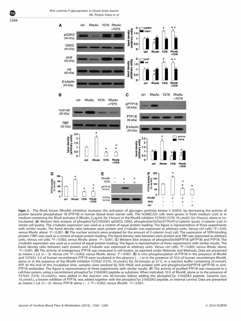
Figure 3. The RhoA kinase (RhoAK) inhibition increases the activation of glycogen synthase kinase 3 (GSK3), by decreasing the activity ofprotein tyrosine phosphatase 1B (PTP1B) in human blood–brain barrier cells. The hCMEC/D3 cells were grown in fresh medium (ctrl) or inmedium containing the RhoA activator II (RhoAc; 5 mg/mL for 3 hours) or the RhoAK inhibitor Y27632 (Y276; 10mmol/L for 3 hours), alone or co-incubated. (A) Western blot analysis of phospho(Tyr216)GSK3 (pGSK3), GSK3, phospho(Ser33/Ser37/Thr41)b-catenin (pcat), b-catenin (cat) inwhole-cell lysates. The b-tubulin expression was used as a control of equal protein loading. The figure is representative of three experimentswith similar results. The band density ratio between each protein and b-tubulin was expressed as arbitrary units. Versus ctrl cells: *Po0.05;versus RhoAc alone: 1Po0.001. (B) The nuclear extracts were analyzed for the amount of b-catenin (nucl cat). The expression of TATA-bindingprotein (TBP) was used as a control of equal protein loading. The band density ratio between each protein and TBP was expressed as arbitraryunits. Versus ctrl cells: *Po0.002; versus RhoAc alone: 1Po0.001. (C) Western blot analysis of phospho(Ser50)PTP1B (pPTP1B) and PTP1B. Theb-tubulin expression was used as a control of equal protein loading. The figure is representative of three experiments with similar results. Theband density ratio between each protein and b-tubulin was expressed as arbitrary units. Versus ctrl cells: *Po0.005; versus RhoAc alone:1Po0.001. (D) The activity of endogenous PTP1B was measured in cell lysates, as reported under Materials and Methods. Data are presentedas means±s.d. (n¼ 3). Versus ctrl: *Po0.002; versus RhoAc alone: 1Po0.001. (E) In vitro phosphorylation of PTP1B in the presence of RhoAKand Y27632. 5 U of human recombinant PTP1B were incubated in the absence (� ) or in the presence of 10U of human recombinant RhoAK,alone or in the presence of the RhoAK inhibitor Y27632 (Y276; 10 mmol/L) for 30minutes at 371C, in a reaction buffer containing 25mmol/LATP. At the end of this incubation time, samples were resolved by SDS–PAGE and probed with anti-phospho(Ser50)PTP1B (pPTP1B) or anti-PTP1B antibodies. The figure is representative of three experiments with similar results. (F) The activity of purified PTP1B was measured in acell-free system, using a recombinant phospho(Tyr 216)GSK3 peptide as substrate. When indicated, 10U of RhoAK, alone or in the presence ofY27632 (Y276; 10mmol/L), were added in the reaction mix 30minutes before adding the phospho(Tyr 216)GSK3 peptide. Suramin (sur;10mmol/L), a known inhibitor of PTP1B, was added together with the phospho(Tyr 216)GSK3 peptide, as internal control. Data are presentedas means±s.d. (n¼ 3). Versus PTP1B alone (� ): *Po0.002; versus RhoAK: 1Po0.001.
Wnt controls P-glycoprotein in blood–brain barrierML Pinzon–Daza et al
1264
Journal of Cerebral Blood Flow & Metabolism (2014), 1258 – 1269 & 2014 ISCBFM
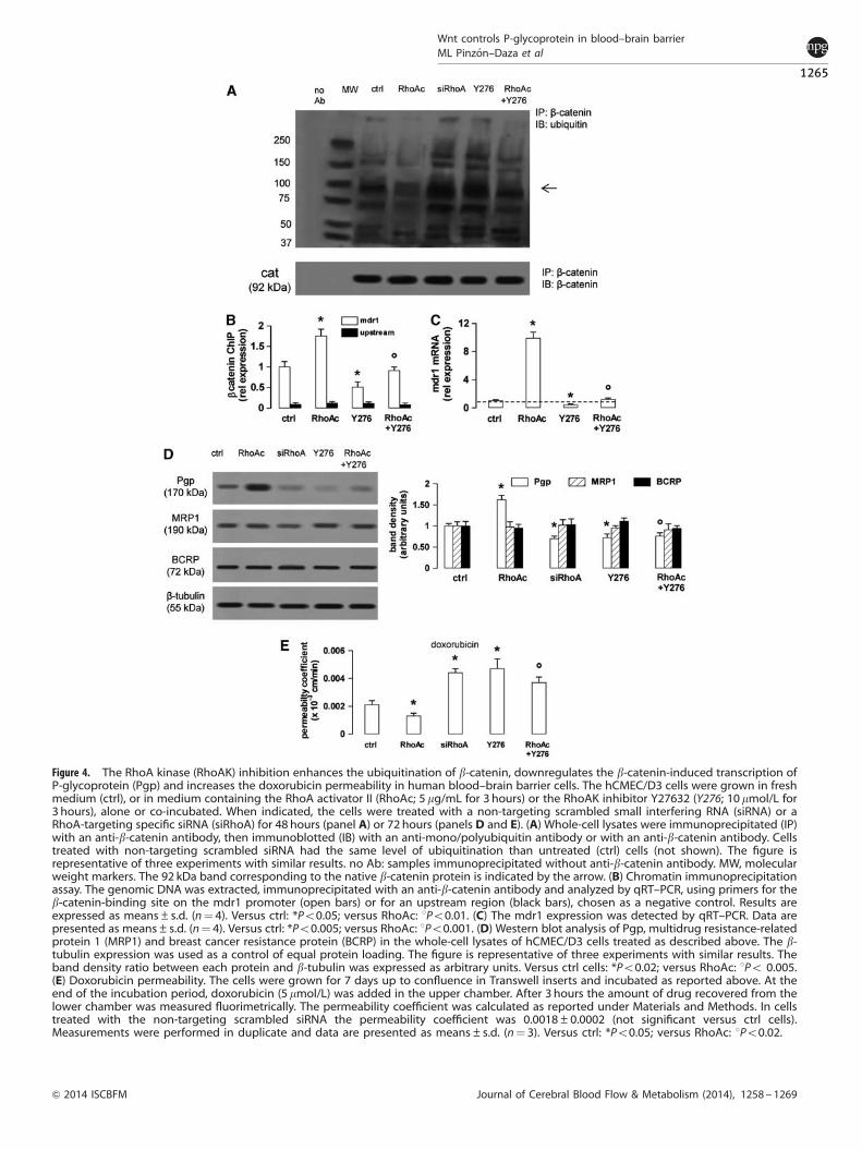
Figure 4. The RhoA kinase (RhoAK) inhibition enhances the ubiquitination of b-catenin, downregulates the b-catenin-induced transcription ofP-glycoprotein (Pgp) and increases the doxorubicin permeability in human blood–brain barrier cells. The hCMEC/D3 cells were grown in freshmedium (ctrl), or in medium containing the RhoA activator II (RhoAc; 5 mg/mL for 3 hours) or the RhoAK inhibitor Y27632 (Y276; 10 mmol/L for3 hours), alone or co-incubated. When indicated, the cells were treated with a non-targeting scrambled small interfering RNA (siRNA) or aRhoA-targeting specific siRNA (siRhoA) for 48 hours (panel A) or 72 hours (panels D and E). (A) Whole-cell lysates were immunoprecipitated (IP)with an anti-b-catenin antibody, then immunoblotted (IB) with an anti-mono/polyubiquitin antibody or with an anti-b-catenin antibody. Cellstreated with non-targeting scrambled siRNA had the same level of ubiquitination than untreated (ctrl) cells (not shown). The figure isrepresentative of three experiments with similar results. no Ab: samples immunoprecipitated without anti-b-catenin antibody. MW, molecularweight markers. The 92 kDa band corresponding to the native b-catenin protein is indicated by the arrow. (B) Chromatin immunoprecipitationassay. The genomic DNA was extracted, immunoprecipitated with an anti-b-catenin antibody and analyzed by qRT–PCR, using primers for theb-catenin-binding site on the mdr1 promoter (open bars) or for an upstream region (black bars), chosen as a negative control. Results areexpressed as means±s.d. (n¼ 4). Versus ctrl: *Po0.05; versus RhoAc: 1Po0.01. (C) The mdr1 expression was detected by qRT–PCR. Data arepresented as means±s.d. (n¼ 4). Versus ctrl: *Po0.005; versus RhoAc: 1Po0.001. (D) Western blot analysis of Pgp, multidrug resistance-relatedprotein 1 (MRP1) and breast cancer resistance protein (BCRP) in the whole-cell lysates of hCMEC/D3 cells treated as described above. The b-tubulin expression was used as a control of equal protein loading. The figure is representative of three experiments with similar results. Theband density ratio between each protein and b-tubulin was expressed as arbitrary units. Versus ctrl cells: *Po0.02; versus RhoAc: 1Po 0.005.(E) Doxorubicin permeability. The cells were grown for 7 days up to confluence in Transwell inserts and incubated as reported above. At theend of the incubation period, doxorubicin (5 mmol/L) was added in the upper chamber. After 3 hours the amount of drug recovered from thelower chamber was measured fluorimetrically. The permeability coefficient was calculated as reported under Materials and Methods. In cellstreated with the non-targeting scrambled siRNA the permeability coefficient was 0.0018±0.0002 (not significant versus ctrl cells).Measurements were performed in duplicate and data are presented as means±s.d. (n¼ 3). Versus ctrl: *Po0.05; versus RhoAc: 1Po0.02.
Wnt controls P-glycoprotein in blood–brain barrierML Pinzon–Daza et al
1265
& 2014 ISCBFM Journal of Cerebral Blood Flow & Metabolism (2014), 1258 – 1269

It has not been clarified however whether the canonical Wntpathway controls the activity of the non-canonical Wnt pathwaysor vice versa. In gastric cancer cells, the activation of RhoA inresponse to Wnt5a is dependent on the activation of the PI3K/Akt/GSK3 axis.24 Our results demonstrated that an active RhoAdecreases the activity of GSK3, prevents the GSK3-mediatedphosphorylation of b-catenin, and favors its nuclear translocationand the subsequent transcription of Pgp, whereas the RhoAsilencing produces opposite effects. We therefore hypothesizethat the RhoA activity controls the GSK3/b-catenin axis in hCMEC/D3 cells. This hypothesis is in contrast with results obtained inmurine cerebrovascular endothelial cells, where the activation ofRhoA promotes the phosphorylation of b-catenin and reduces itstranscriptional activity.9 As murine and human brain microvascularcells have often striking differences in the expression and activityof Pgp,25 it is not surprising that they also differ in the upstream
pathways controlling Pgp expression. For instance, the mechanismby which RhoA modulates GSK3 activity is quite different inmurine and human cerebrovascular endothelial cells: in murinecells, RhoA controls the GSK3 activity in a PTEN- and protein kinaseCd-dependent way and changes the phosphorylation of GSK3 onserine 9.9 This phosphorylation has inhibitory effects on theenzymatic activity of GSK3.26 We cannot exclude that the RhoAactivity may change the phosphorylation on serine 9 of GSK3 alsoin human hCMEC/D3 cells; however, we observed that in ourmodel, the activation of RhoA decreases—and the silencing ofRhoA increases—the phosphorylation of GSK3 on tyrosine 216,which is a proactivating phosphorylation.26 When phosphorylatedon tyrosine 216, GSK3 induces b-catenin phosphorylation anddegradation. To our knowledge, this is the first work showingthat RhoA activity modulates the phosphorylation on tyrosine 216of GSK3.
Figure 5. The RhoA kinase (RhoAK) inhibitor Y27632 increases the doxorubicin delivery and cytotoxicity in glioblastoma cells co-cultured withblood–brain barrier cells. The hCMEC/D3 cells were grown for 7 days up to confluence in Transwell inserts; CV17, 01010627, Nov3, and U87-MG cells were seeded at day 4 in the lower chamber. After 3 days of co-culture, the supernatant in the upper chamber was replaced with freshmedium without (� or ctrl) or with Y27632 (Y276; 10 mmol/L for 3 hours). After this incubation time, doxorubicin (dox; 5mmol/L) was added inthe upper chamber for 3 hours (panels A and B) or 24 hours (panels C–F), then the following investigations were performed. (A) Fluorimetricquantification of intracellular doxorubicin in glioblastoma cells. Data are presented as means±s.d. (n¼ 4). Versus untreated (� ) cells:*Po0.001. (B) The 01010627 cells were seeded on sterile glass coverslips, treated as reported above, then stained with 40,6-diamidino-2-phenylindole dihydrochloride (DAPI) and analyzed by fluorescence microscopy to detect the intracellular accumulation of doxorubicin.Magnification: � 63 objective (1.4 numerical aperture); � 10 ocular lens. The micrographs are representative of three experiments with similarresults. Scale bar, 20mm. (C) The glioblastoma cells were checked spectrophotometrically for the extracellular release of lactate dehydrogenase(LDH) activity. Data are presented as means±s.d. (n¼ 4). Versus untreated (� ) cells: *Po0.001. (D) The whole-cell lysates from 01010627 cellswere resolved by SDS–PAGE and immunoblotted with an anti-caspase 3 antibody (recognizing both pro-caspase and cleaved active caspase).The b-tubulin expression was used as a control of equal protein loading. The figure is representative of three experiments with similar results.(E) Cell cycle analysis. The distribution of the 01010627 cells in sub-G1, G0/G1, S, G2/M phase was analyzed by flow cytometry, as detailedunder Materials and Methods. Data are presented as means±s.d. (n¼ 4). Versus ctrl: *Po0.005. (F). After 3 days of co-culture between hCMEC/D3 and 01010627 cells, the medium of the upper chamber was replaced with fresh medium (open circles) or medium containing Y27632(Y276; 10 mmol/L for 3 hours, solid circles), doxorubicin (dox; 5 mmol/L for 24 hours, open squares), Y27632 (Y276; 10 mmol/L for 3 hours)followed by doxorubicin (doxo; 5 mmol/L for 24 hours, solid squares). Drug treatments were repeated every 7 days, as reported in the Materialsand Methods section. The proliferation of glioblastoma cells was monitored weekly by crystal violet staining. Measurements were performedin triplicate and data are presented as means±s.d. (n¼ 4). Versus ctrl: *Po0.001.
Wnt controls P-glycoprotein in blood–brain barrierML Pinzon–Daza et al
1266
Journal of Cerebral Blood Flow & Metabolism (2014), 1258 – 1269 & 2014 ISCBFM
We next looked for putative downstream effectors of RhoAresponsible for this effect. We focused on RhoAK, whose activityfollowed the same trend of RhoA activity in response to WntA andDkk-1 in hCMEC/D3 cells. Interestingly, the inhibition of RhoAK byY27632 quickly decreased the nuclear translocation of b-catenin,with a maximal efficacy after 3 hours. At longer time points,nuclear b-catenin progressively re-accumulated in the nucleus,although it remained lower than in untreated cells: this may bedue to the short half-life (i.e., less than 12 hours) of b-catenin,27
which produces a fast re-synthesis of new b-catenin ready totranslocate into the nucleus. After 3 hours, Y27632 effectivelyincreased the phosphorylation on tyrosine 216 of GSK3 and thesubsequent GSK3-induced phosphorylation of b-catenin; by actingdownstream RhoA, Y27632 was effective even in cells with aconstitutively activated RhoA. These data suggest that RhoAKlikely controls the GSK3 activity by activating a tyrosinephosphatase, which recognizes GSK3 as substrate. PTP1B, whichis activated by the phosphorylation on serine 50,20 is one of thesephosphatases.19 Our results in hCMEC/D3 cells and in a cell-freesystem demonstrate that the RhoAK phosphorylates PTP1B onserine 50, promoting the dephosphorylation of GSK3 on tyrosine216, an event that was fully abrogated by Y27632. These datasuggest that the RhoAK, by activating PTP1B, inhibits the GSK3activity, prevents the ubiquitination of b-catenin, and allows itsnuclear translocation and transcriptional activity. Our results alsoexplain previous observations, showing that Wnt3 stimulates thetranscription of b-catenin-target genes and RhoA-target genes,with a putative RhoAK-dependent mechanism.8
Some b-catenin-target genes encode for proteins of adherensjunctions and tight junctions; therefore, changes in RhoA/RhoAKactivity may lead to the loss of BBB integrity.10 In our experimentalconditions, no changes occurred in the protein expression andfunctionality of tight junctions in the hCMEC/D3 cells treatedwith Y27632. By contrast, Y27632 was sufficient to reduce theb-catenin-driven transcription of Pgp. As different promoters havedifferent sensitivity to the binding of b-catenin–T-cell factorcomplex, we might speculate that the promoter of Pgp is moresensitive than the promoter of other genes to the variations ofb-catenin/T-cell factor binding.
Working at proper concentrations and incubation times, theRhoAK inhibitor increased the delivery of doxorubicin, a Pgpsubstrate with a very low permeability across the BBB.13 Fasudil,the clinically prescribed analog of Y27632, is used to preventvasospasms after subarachnoid hemorrhage,28 to improve tissueperfusion during cerebral ischemia,29 to prevent the progressionof cerebral aneurisms.30 Our work suggests that Fasudil might beused to improve the delivery of Pgp substrates through the BBB.
In the case of doxorubicin, a drug that is highly effective againstglioblastoma cells in vitro31 and ineffective in the presence of acompetent BBB,6 the pretreatment of hCMEC/D3 cells with Y27632fully restored the doxorubicin delivery and cytotoxicity inglioblastoma cells. Repeated pulses of Y27632 followed bydoxorubicin confirmed that this approach effectively reducedthe long-term proliferation of glioblastoma cells grown under aBBB monolayer. However, Pgp is not the only transporter presenton the luminal side of BBB that can efflux doxorubicin: also MRP1and BCRP, which are detectable in the hCMEC/D3 cells,11 meetthese requisites,1 but their expression did not change in cells withconstitutively active RhoA, silenced for RhoA or treated withY27632. We thus hypothesize that the changes in doxorubicindelivery after RhoA activation or inhibition were consequent tothe expression change of Pgp in hCMEC/D3 cells. To furtherquantify the contribution of this transporter in doxorubicinpermeability, we treated the hCMEC/D3 cells with the Pgpinhibitor verapamil, which produced the same effects—in termsof doxorubicin delivery and toxicity in glioblastoma cells—ofY27632. These results indirectly suggest that Pgp has a major rolein limiting doxorubicin delivery in the central nervous system.
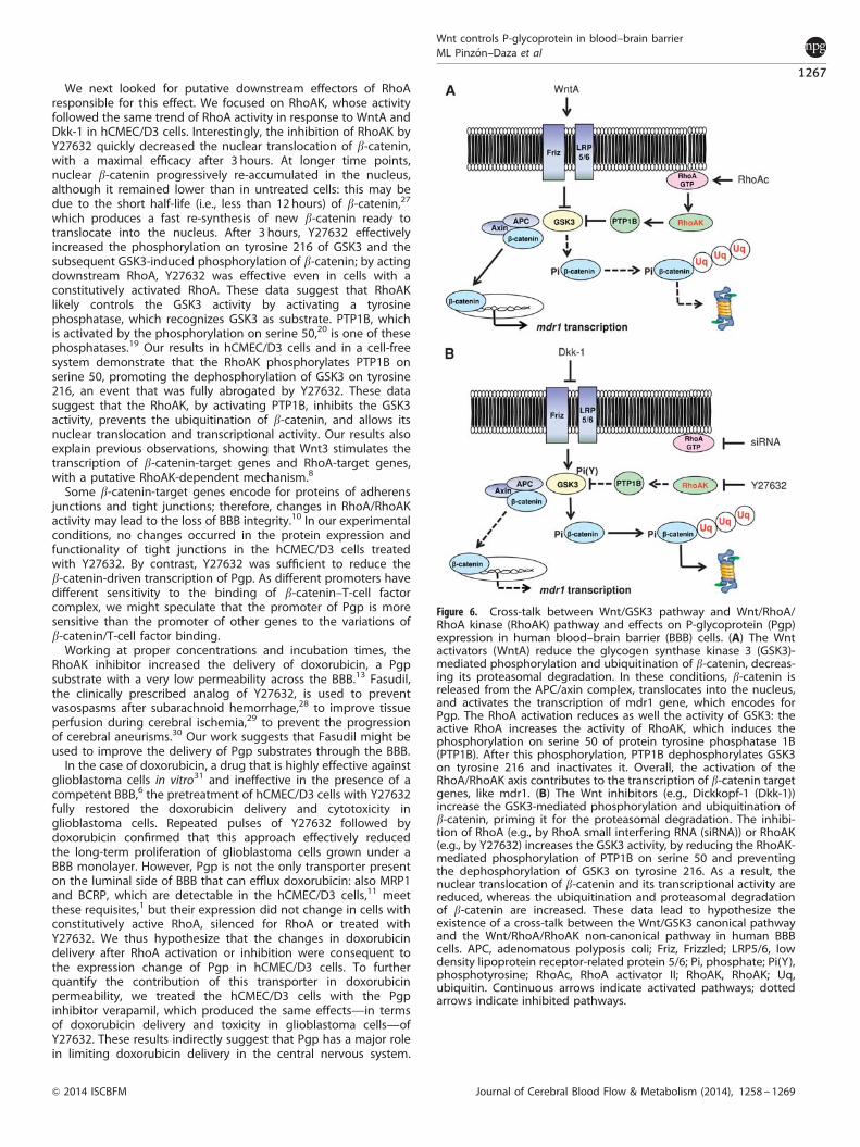
Figure 6. Cross-talk between Wnt/GSK3 pathway and Wnt/RhoA/RhoA kinase (RhoAK) pathway and effects on P-glycoprotein (Pgp)expression in human blood–brain barrier (BBB) cells. (A) The Wntactivators (WntA) reduce the glycogen synthase kinase 3 (GSK3)-mediated phosphorylation and ubiquitination of b-catenin, decreas-ing its proteasomal degradation. In these conditions, b-catenin isreleased from the APC/axin complex, translocates into the nucleus,and activates the transcription of mdr1 gene, which encodes forPgp. The RhoA activation reduces as well the activity of GSK3: theactive RhoA increases the activity of RhoAK, which induces thephosphorylation on serine 50 of protein tyrosine phosphatase 1B(PTP1B). After this phosphorylation, PTP1B dephosphorylates GSK3on tyrosine 216 and inactivates it. Overall, the activation of theRhoA/RhoAK axis contributes to the transcription of b-catenin targetgenes, like mdr1. (B) The Wnt inhibitors (e.g., Dickkopf-1 (Dkk-1))increase the GSK3-mediated phosphorylation and ubiquitination ofb-catenin, priming it for the proteasomal degradation. The inhibi-tion of RhoA (e.g., by RhoA small interfering RNA (siRNA)) or RhoAK(e.g., by Y27632) increases the GSK3 activity, by reducing the RhoAK-mediated phosphorylation of PTP1B on serine 50 and preventingthe dephosphorylation of GSK3 on tyrosine 216. As a result, thenuclear translocation of b-catenin and its transcriptional activity arereduced, whereas the ubiquitination and proteasomal degradationof b-catenin are increased. These data lead to hypothesize theexistence of a cross-talk between the Wnt/GSK3 canonical pathwayand the Wnt/RhoA/RhoAK non-canonical pathway in human BBBcells. APC, adenomatous polyposis coli; Friz, Frizzled; LRP5/6, lowdensity lipoprotein receptor-related protein 5/6; Pi, phosphate; Pi(Y),phosphotyrosine; RhoAc, RhoA activator II; RhoAK, RhoAK; Uq,ubiquitin. Continuous arrows indicate activated pathways; dottedarrows indicate inhibited pathways.
Wnt controls P-glycoprotein in blood–brain barrierML Pinzon–Daza et al
1267
& 2014 ISCBFM Journal of Cerebral Blood Flow & Metabolism (2014), 1258 – 1269
Although Pgp tightly cooperates with BCRP in controlling thedelivery of several drugs across the BBB,32 doxorubicin has highaffinity for Pgp, which represents the main transporter of this drugin BBB cells.33
The downregulation of the canonical Wnt/GSK3/b-cateninpathway is known to reduce the Pgp expression and to inducechemosensitization in colon34 and glioblastoma17 tumor stemcells, neuroblastoma,35 chronic myeloid leukemia,36 and cholan-giocarcinoma.37 Also, the inhibition of the non-canonical Wnt3/RhoA/RhoAK pathway chemosensitizes multiple myeloma cells todoxorubicin.38 We might speculate that the same cross-talkbetween canonical and non-canonical Wnt pathways, observedin BBB cells, cooperates in regulating the Pgp expression also incancer cells, and may represent a critical target of therapeuticintervention for Pgp-rich tumors.
We suggest that the expression of Pgp is controlled by a cross-talk between canonical and non-canonical Wnt pathways inhuman BBB cells. A crucial regulator of this cross-talk is RhoAK,which inhibits the GSK3-induced phosphorylation of b-catenin byactivating PTP1B. Targeted therapies against RhoA/RhoAK candisrupt the cross-talk and downregulate the Pgp expression(Figure 6). Improving the BBB permeability of anticancer drugsthat are effluxed by Pgp thus enhancing their delivery to braintumors is still an unsolved challenge.39 Moreover, the highexpression of Pgp on BBB cells decreases not only the deliveryof chemotherapeutic agents, but also the passage of drugs usedto treat infective diseases, neurodegenerative diseases, andepilepsy.40 By unveiling a new mechanism that regulates Pgpexpression in the BBB cells, our work may pave the way topreclinical investigations using RhoAK inhibitors as adjuvant toolsin these central nervous system diseases.
DISCLOSURE/CONFLICT OF INTERESTThe authors declare no conflict of interest.
ACKNOWLEDGMENTSThe authors thank Mr Costanzo Costamagna, Department of Oncology, University ofTurin for the technical assistance. We are grateful to Professor Davide Schiffer (Neuro-Biooncology Center, Vercelli, Italy) and Dr Rossella Galli (DIBIT, San Raffaele ScientificInstitute, Milan, Italy) for providing the primary glioblastoma samples.
REFERENCES1 Agarwal S, Sane R, Oberoi R, Ohlfest JR, Elmquist WF. Delivery of molecularly
targeted therapy to malignant glioma, a disease of the whole brain. Expert RevMol Med 2011; 13: e17.
2 Lim JC, Kania KD, Wijesuriya H, Chawla S, Sethi JK, Pulaski L et al. Activation ofb-catenin signalling by GSK-3 inhibition increases p-glycoprotein expression inbrain endothelial cells. J Neurochem 2008; 106: 1855–1865.
3 Kania KD, Wijesuriya HC, Hladky SB, Barrand MA. Beta amyloid effects onexpression of multidrug efflux transporters in brain endothelial cells. Brain Res2011; 1418: 1–11.
4 Katoh M, Katoh M. WNT signalling pathway and stem cells signaling network. ClinCancer Res 2007; 13: 4042–4045.
5 MacDonald BT, Tamai K, He X. Wnt/b-catenin signaling: components, mechan-isms, and diseases. Dev Cell 2009; 17: 9–26.
6 Riganti C, Salaroglio IC, Pinzon-Daza ML, Caldera V, Campia I, Kopecka J et al.Temozolomide down-regulates P-glycoprotein in human blood-brain barrier cellsby disrupting Wnt3-signalling. Cell Mol Life Sci 2014; 71: 499–516.
7 Tsuji T, Ohta Y, Kanno Y, Hirose K, Ohashi K, Mizuno K. Involvement of p114-RhoGEF and Lfc in Wnt-3a- and dishevelled-induced RhoA activation and neuriteretraction in N1E-115 mouse neuroblastoma cells. Mol Biol Cell 2010; 21:3590–3600.
8 Rossol-Allison J, Stemmle LN, Swenson-Fields KI, Kelly P, Fields PE, McCall SJ et al.Rho GTPase activity modulates Wnt3a/beta-catenin signaling. Cell Signal 2009; 21:1559–1568.
9 Chang CC, Lee PS, Chou Y, Hwang LL, Juan SH. Mediating effects of aryl-hydro-carbon receptor and RhoA in altering brain vascular integrity: the therapeuticpotential of statins. Am J Pathol 2012; 181: 211–221.
10 Allen C, Srivastava K, Bayraktutan U. Small GTPase RhoA and its effector rho kinasemediate oxygen glucose deprivation-evoked in vitro cerebral barrier dysfunction.Stroke 2010; 41: 2056–2063.
11 Pinzon-Daza ML, Garzon R, Couraud PO, Romero IA, Weksler B, Ghigo D et al. Theassociation of statins plus LDL receptor-targeted liposome-encapsulated doxor-ubicin increases the in vitro drug delivery across blood-brain barrier cells. Brit JPharmacol 2012; 167: 1431–1447.
12 Flatau G, Lemichez E, Gauthier M, Chardin P, Paris S, Fiorentini C et al. Toxin-induced activation of the G protein p21 Rho by deamidation of glutamine. Nature1997; 387: 729–733.
13 Weksler BB, Subileau EA, Perriere N, Charneau P, Holloway K, Leveque M et al.Blood-brain barrier-specific properties of a human adult brain endothelial cell line.FASEB J 2005; 19: 1872–1894.
14 Doublier S, Riganti C, Voena C, Costamagna C, Aldieri E, Pescarmona G et al. RhoAsilencing reverts the resistance to doxorubicin in human colon cancer cells. MolCancer Res 2008; 6: 1607–1620.
15 Monnaert V, Betbeder D, Fenart L, Bricout H, Lenfant AM, Landry C et al. Effectsof g- and hydroxypropyl-g-cyclodextrins on the transport of doxorubicinacross an in vitro model of blood-brain barrier. J Pharmacol Exp Ther 2004; 311:1115–1120.
16 Eigenmann DE, Xue G, Kim KS, Moses AV, Hamburger M, Oufir M. Comparativestudy of four immortalized human brain capillary endothelial cell lines, hCMEC/D3, hBMEC, TY10, and BB19, and optimization of culture conditions, for an in vitroblood-brain barrier model for drug permeability studies. Fluids Barriers CNS 2013;10: e33.
17 Siflinger-Birnboim A, Del Vecchio PJ, Cooper JA, Blumenstock FA, Shepard JM,Malik AB. Molecular sieving characteristics of the cultured endothelial monolayer.J Cell Physiol 1987; 132: 111–117.
18 Riganti C, Salaroglio IC, Caldera V, Campia I, Kopecka J, Mellai M et al. Temozo-lomide down-regulates P-glycoprotein expression in glioblastoma stem cells byinterfering with the Wnt3a/GSK3/b-catenin pathway. Neuro Oncol 2013; 15:1502–1517.
19 Mobasher MA, Gonzalez-Rodriguez A, Santamarıa B, Ramos S, Martın MA,Goya L et al. Protein tyrosine phosphatase 1B modulates GSK3b/Nrf2 and IGFIRsignaling pathways in acetaminophen-induced hepatotoxicity. Cell Death Dis2003; 4: e626.
20 Moeslein FM, Myers MP, Landreth GE. The CLK family kinases, CLK1 and CLK2,phosphorylate and activate the tyrosine phosphatase, PTP-1B. J Biol Chem 1999;274: 26697–26704.
21 Gottesman MM, Fojo T, Bates SE. Multidrug resistance in cancer: role of ATP-dependent transporters. Nat Rev Cancer 2002; 2: 48–58.
22 Tai LM, Loughlin AJ, Male DK, Romero IA. P-glycoprotein and breastcancer resistance protein restrict apical-to-basolateral permeability ofhuman brain endothelium to amyloid-b. J Cereb Blood Flow Metab 2009; 29:1079–1083.
23 de Jesus Perez VA, Alastalo TP, Wu JC, Axelrod JD, Cooke JP, Amieva M et al. Bonemorphogenetic protein 2 induces pulmonary angiogenesis via Wnt-beta-cateninand Wnt-RhoA-Rac1 pathways. J Cell Biol 2009; 184: 83–99.
24 Liu J, Zhang Y, Xu R, Du J, Hu Z, Yang L et al. PI3K/Akt-dependent phosphorylationof GSK3b and activation of RhoA regulate Wnt5a-induced gastric cancer cellmigration. Cell Signal 2013; 25: 447–456.
25 Uchida Y, Ohtsuki S, Katsukura Y, Ikeda C, Suzuki T, Kamiie J et al. Quantitativetargeted absolute proteomics of human blood-brain barrier transporters andreceptors. J Neurochem 2011; 117: 333–345.
26 Jope RS, Johnson GV. The glamour and gloom of glycogen synthase kinase-3.Trends Biochem Sci 2004; 29: 95–102.
27 Bareiss S, Kim K, Lu Q. Delta-catenin/NPRAP: A new member of the glycogensynthase kinase-3beta signaling complex that promotes beta-catenin turnover inneurons. J Neurosci Res 2010; 88: 2350–2363.
28 Zoerle T, Ilodigwe DC, Wan H, Lakovic K, Sabri M, Ai J et al. Pharmacologicreduction of angiographic vasospasm in experimental subarachnoid hemorrhage:systematic review and meta-analysis. J Cereb Blood Flow Metab 2012; 32:1645–1658.
29 Shin HK, Huang PL, Ayata C. Rho-kinase inhibition improves ischemic perfusiondeficit in hyperlipidemic mice. J Cereb Blood Flow Metab 2014; 34: 284–287.
30 Eldawoody H, Shimizu H, Kimura N, Saito A, Nakayama T, Takahashi A et al.Fasudil, a Rho-kinase inhibitor, attenuates induction and progression of cerebralaneurysms: experimental study in rats using vascular corrosion casts. Neurosci Lett2010; 470: 76–80.
31 Hau P, Fabel K, Baumgart U, Rummele P, Grauer O, Bock A et al. Pegylatedliposomal doxorubicin-efficacy in patients with recurrent high-grade glioma.Cancer 2004; 100: 1199–1207.
32 Agarwal S, Hartz AM, Elmquist WF, Bauer B. Breast cancer resistance protein andP-glycoprotein in brain cancer: two gatekeepers team up. Curr Pharm Des 2011;17: 2793–2802.
Wnt controls P-glycoprotein in blood–brain barrierML Pinzon–Daza et al
1268
Journal of Cerebral Blood Flow & Metabolism (2014), 1258 – 1269 & 2014 ISCBFM

33 Qu Q, Chu JW, Sharom FJ. Transition state P-glycoprotein binds drugs andmodulators with unchanged affinity, suggesting a concerted transport mechan-ism. Biochemistry 2003; 42: 1345–1353.
34 Chikazawa N, Tanaka H, Tasaka T, Nakamura M, Tanaka M, Onishi H et al. Inhi-bition of Wnt signaling pathway decreases chemotherapy-resistant side-popula-tion colon cancer cells. Anticancer Res 2010; 30: 2041–2048.
35 Flahaut M, Meier R, Coulon A, Nardou KA, Niggli FK, Martinet D et al. The Wntreceptor FZD1 mediates chemoresistance in neuroblastoma through activation ofthe Wnt/beta-catenin pathway. Oncogene 2009; 28: 2245–2256.
36 Correa S, Binato R, Du Rocher B, Castelo-Branco MT, Pizzatti L, Abdelhay E. Wnt/b-catenin pathway regulates ABCB1 transcription in chronic myeloid leukemia. BMCCancer 2012; 12: e303.
37 Shen DY, Zhang W, Zeng X, Liu CQ. Inhibition of Wnt/b-catenin signalingdownregulates P-glycoprotein and reverses multi-drug resistance of cholangio-carcinoma. Cancer Sci 2013; 104: 1303–1308.
38 Kobune M, Chiba H, Kato J, Kato K, Nakamura K, Kawano Y et al. Wnt3/RhoA/ROCKsignaling pathway is involved in adhesion-mediated drug resistance of multiplemyeloma in an autocrine mechanism. Mol Cancer Ther 2007; 6: 1774–1784.
39 Serwer LP, James CD. Challenges in drug delivery to tumors of the central nervoussystem: an overview of pharmacological and surgical considerations. Adv DrugDeliv Rev 2012; 64: 590–597.
40 Pinzon-Daza ML, Campia I, Kopecka J, Garzon R, Ghigo D, Riganti C. Nanoparticle-and liposome-carried drugs: new strategies for active targeting and drug deliveryacross blood-brain barrier. Curr Drug Metab 2013; 14: 625–640.
Supplementary Information accompanies the paper on the Journal of Cerebral Blood Flow & Metabolism website (http://www.nature.com/jcbfm)
Wnt controls P-glycoprotein in blood–brain barrierML Pinzon–Daza et al
1269
& 2014 ISCBFM Journal of Cerebral Blood Flow & Metabolism (2014), 1258 – 1269
Related Documents