Welcome message from author
This document is posted to help you gain knowledge. Please leave a comment to let me know what you think about it! Share it to your friends and learn new things together.
Transcript

ANGIOGENESIS AND DIRECT MYOCARDIAL VASCULARIZATION

CONTEMPORARY CARDIOLOGY
Angiogenesis and Direct MyocardialRevascularization, edited by Roger J.Laham, MD, and Donald S. Baim, MD,2005
Cardiovascular Disease in the Elderly,edited by Gary Gerstenblith, MD, 2005
Platelet Function: Assessment, Diagnosis,and Treatment, edited by MartinQuinn, MB BCh BAO, PhD, andDesmond Fitzgerald, MD, FRCPI, 2005
Diabetes and Cardiovascular Disease,Second Edition, edited by MichaelT. Johnstone, MD, CM, FRCP(C), andAristidis Veves, MD, DSc, 2005
Interventional Cardiology: PercutaneousNoncoronary Intervention, editedby Howard C. Herrmann, MD, 2005
Principles of Molecular Cardiology,edited by Marschall S. Runge, MD,and Cam Patterson, MD, 2005
Heart Disease Diagnosis and Therapy: APractical Approach, Second Edition,by M. Gabriel Khan, MD, FRCP(C),FRCP(LONDON), FACP, FACC, 2005
Cardiovascular Genomics: Gene Miningfor Pharmacogenomics and GeneTherapy, edited by Mohan K.Raizada, PhD, Julian F. R. Paton,PhD, Michael J. Katovich, PhD, andSergey Kasparov, MD, PhD, 2005
Surgical Management of CongestiveHeart Failure, edited by James C.Fang, MD and Gregory S. Couper,MD, 2005
Cardiopulmonary Resuscitation, edited byJoseph P. Ornato, MD, FAP, FACC andMary Ann Peberdy, MD, FACC, 2005
CT of the Heart: Principles andApplications, edited by U. JosephSchoepf, MD, 2005
Heart Disease and Erectile Dysfunction,edited by Robert A. Kloner, MD, PhD,2004
Cardiac Transplantation: The ColumbiaUniversity Medical Center/NewYork-Presbyterian HospitalManual, edited by Niloo M.Edwards, MD, Jonathan M. Chen,MD, and Pamela A. Mazzeo, 2004
Coronary Disease in Women: Evidence-Based Diagnosis and Treatment,edited by Leslee J. Shaw, PhD andRita F. Redberg, MD, FACC, 2004
Complementary and AlternateCardiovascular Medicine, editedby Richard A. Stein, MD andMehmet C. Oz, MD, 2004
Nuclear Cardiology, The Basics: How toSet Up and Maintain a Laboratory,by Frans J. Th. Wackers, MD, PhD,Wendy Bruni, BS, CNMT, and Barry L.Zaret, MD, 2004
Minimally Invasive Cardiac Surgery,Second Edition, edited by DanielJ. Goldstein, MD, and Mehmet C.Oz, MD 2004
Cardiovascular Health Care Economics,edited by William S. Weintraub, MD,2003
Platelet Glycoprotein IIb/IIIa Inhibitorsin Cardiovascular Disease, SecondEdition, edited by A. MichaelLincoff, MD, 2003
Heart Failure: A Clinician’s Guide toAmbulatory Diagnosis and Treatment,edited by Mariell L. Jessup, MD andEvan Loh, MD, 2003
Management of Acute CoronarySyndromes, Second Edition,edited by Christopher P. Cannon,MD 2003
Aging, Heart Disease, and Its Manage-ment: Facts and Controversies,edited by Niloo M. Edwards, MD,Mathew S. Maurer, MD, and RachelB. Wellner, MPH, 2003
CHRISTOPHER P. CANNON, MDSERIES EDITOR

ANGIOGENESIS
AND DIRECT
MYOCARDIAL
REVASCULARIZATION
Edited by
ROGER J. LAHAM, MDBeth Israel Deaconess Medical CenterBoston, MA
DONALD S. BAIM, MDBrigham and Women’s HospitalBoston, MA

© 2005 Humana Press Inc.999 Riverview Drive, Suite 208Totowa, New Jersey 07512
humanapress.com
For additional copies, pricing for bulk purchases, and/or information about other Humana titles, contactHumana at the above address or at any of the following numbers: Tel.: 973-256-1699; Fax: 973-256-8341,E-mail: [email protected]; or visit our Website: www.humanapress.com
All rights reserved. No part of this book may be reproduced, stored in a retrieval system, or transmitted in anyform or by any means, electronic, mechanical, photocopying, microfilming, recording, or otherwise withoutwritten permission from the Publisher.
All articles, comments, opinions, conclusions, or recommendations are those of the author(s), and do notnecessarily reflect the views of the publisher.
Due diligence has been taken by the publishers, editors, and authors of this book to assure the accuracy of theinformation published and to describe generally accepted practices. The contributors herein have carefullychecked to ensure that the drug selections and dosages set forth in this text are accurate and in accord with thestandards accepted at the time of publication. Notwithstanding, as new research, changes in government regu-lations, and knowledge from clinical experience relating to drug therapy and drug reactions constantly occurs,the reader is advised to check the product information provided by the manufacturer of each drug for any changein dosages or for additional warnings and contraindications. This is of utmost importance when the recom-mended drug herein is a new or infrequently used drug. It is the responsibility of the treating physician todetermine dosages and treatment strategies for individual patients. Further it is the responsibility of the healthcare provider to ascertain the Food and Drug Administration status of each drug or device used in their clinicalpractice. The publisher, editors, and authors are not responsible for errors or omissions or for any consequencesfrom the application of the information presented in this book and make no warranty, express or implied, withrespect to the contents in this publication.
Production Editor: Tracy Catanese
Cover design by Patricia F. Cleary
Cover Illustration: Angiogenesis from bench to bedside: tube formation on in vitro matrigel (upper left) CD31 staining showing increased capillary formation (upper right) in vivo hind limb ischemia model withincreased arterial collaterals (lower left) cardiac magnetic resonance imaging to detect improvement inperfusion and function (lower right). This translational paradigm with agent discovery rapidly testing in vitrofollowed by animal models to investigate delivery modalities and efficacy leading to clinical testing withsensitive outcome measures should lead to functionally significant angiogenesis and myogenesis in patientswith end-stage ischemic disease and heart failure.
This publication is printed on acid-free paper.
ANSI Z39.48-1984 (American National Standards Institute) Permanence of Paper for Printed Library Materials.
Photocopy Authorization Policy:Authorization to photocopy items for internal or personal use, or the internal or personal use of specific clients,is granted by Humana Press Inc., provided that the base fee of US $30.00 per copy is paid directly to theCopyright Clearance Center at 222 Rosewood Drive, Danvers, MA 01923. For those organizations that havebeen granted a photocopy license from the CCC, a separate system of payment has been arranged and isacceptable to Humana Press Inc. The fee code for users of the Transactional Reporting Service is: [1-58829-153-7/05 $30.00].Printed in the United States of America. 10 9 8 7 6 5 4 3 2 1eISBN: 1-59259-934-6Library of Congress Cataloging-in-Publication DataAngiogenesis and direct myocardial revascularization / edited by Roger J. Laham. p. ; cm. -- (Contemporary cardiology) Includes bibliographical references and index. ISBN 1-58829-153-7 (alk. paper) 1. Coronary heart disease. 2. Neovascularization.
[DNLM: 1. Neovascularization, Physiologic. 2. Myocardial Revascularization--methods. WG 500 A58422005] I. Laham, Roger J. II.Series: Contemporary cardiology (Totowa, N.J. : Unnumbered). RC685.C6A595 2005 616.1'23--dc22
2004026636

PREFACE
v
Si facile esset, iam factum sit.
Atherosclerotic disease remains the leading cause of death in theWestern Hemisphere, and its prevalence continues to increase as thepopulation ages. Despite progress in surgical and catheter-basedrevascularization, an ever increasing number of patients are either notcandidates for these therapies or remain symptomatic despite priorrevascularization and maximal ongoing medical treatment. Thus, it isclear that an alternative treatment strategy such as therapeutic angiogen-esis and myogenesis is needed for these “no-option” patients.
The field of angiogenesis/myogenesis, however, has followed thesame development pattern seen with other novel therapeutic interven-tions: early spectacular and “too-good-to-be-true” results leading tounrealistic expectations, followed by sobering complications and disap-pointments, only later maturing to cautious optimism when better under-standing of the biological and logistic obstacles is achieved. We believethat this is such a time for therapeutic angiogenesis/myogenesis, puttingbehind us the early picture of angiogenesis as “an attempt to influencea process we do not understand, with the agents we do not know how touse and deliver, relying on the end-points we cannot assess.” Unfortu-nately, this led to failure of early studies and a negative view of the field,at a time when we are finally developing a good understanding of thebiology and therapeutic targets, have multiple available and well-stud-ied therapeutic strategies, and have developed the necessary imaging tomeasure outcomes. From here, much work still needs to be done toeventually achieve functionally significant angiogenesis/myogenesis,but clearly we have turned at least the first developmental corner with theidentification of novel therapeutic targets and pathways, the investiga-tion of transcriptional factors, master switch molecules, cell-basedapproaches, chemokines, a better understanding of the effects of aging,endothelial dysfunction, and hypercholesterolemia in response toangiogenic stimuli, as well as a better understanding of delivery prob-lems. Each development has brought us one step closer to our goal ofhelping patients with end-stage ischemic heart disease, peripheral vas-cular disease, and congestive heart failure.

vi Preface
Angiogenesis and Direct Myocardial Revascularization representsan interdisciplinary effort to balance the basic, preclinical, and clinicalaspects in this field. The various sections are each written by pioneersand opinion leaders in angiogenesis/myogenesis. Their chapters reflectthe latest developments in this rapidly evolving field, including the in-troduction of cell-based therapy for angiogenesis and myocardial repair.Wherever this field takes us, we hope that this book will be a usefulwaypoint, and that we can go forward balancing optimistic enthusiasmwith a healthy dose of scientific skepticism, in order to finally realize thepromise that such therapies may hold for patients with advanced cardio-vascular disease.
Roger J. Laham, MD
Donald S. Baim, MD

Preface ..................................................................................................v
Contributors ........................................................................................ ix
Color Plates ........................................................................................ xi
1 No-Option Patients: A Growing Problem ........................1Roger J. Laham and Donald S. Baim
2 Transcriptional Regulation of Angiogenesis .................19Peter Oettgen
3 Preclinical Models and Experience to Date ...................37Aysegul Yegin and Nicolas A. Chronos
4 The Coronary Microcirculation and Angiogenesis .......65Pierre Voisine, Joanna J. Wykrzykowska,
Munir Boodhwani, David G. Harrison,Roger J. Laham, and Frank W. Sellke
5 Local and Regional Vascular Deliveryfor Therapeutic Angiogenesisand Myogenesis ..................................................107
Erik T. Price, Alan C. Yeung,and Mehrdad Rezaee
6 Imaging Angiogenesis: A Guide for ClinicalManagement and Therapeutic Trials .................143
Justin D. Pearlman
7 Myocardial Angiogenesis:Protein Growth Factors .....................................185
Kwang Soo Cha, Robert S. Schwartz,and Timothy D. Henry
8 Gene Therapy for Angiogenesis in the Treatmentof Cardiovascular and PeripheralArterial Disease ..................................................215
Pinak B. Shah, Kapildeo Lotun,and Douglas W. Losordo
CONTENTS
vii

9 Therapeutic Angiogenesis in PeripheralArterial Disease: Current Approachesand Future Directions ........................................245
Richard E. Waters and Brian H. Annex
10 Bone Marrow Cell Transplantationfor Myocardial Regenerationand Therapeutic Angiogenesis ...........................261
Hung-Fat Tse, Pui-Yin Lee, and Chu-Pak Lau
11 Transplantation of Embryonic Stem Cellsfor Myocardial Regenerationand Angiogenesis ................................................283
Yong-Fu Xiao, Jiang-Yong Min,and James P. Morgan
12 Skeletal Myoblast Transplantationfor Cardiac Repair ..............................................311
Audrey Rosinberg, Jamal S. Rana,and Roger J. Laham
13 Transmyocardial Laser Revascularization ...................329Keith A. Horvath
Index .................................................................................................349
viii Contents

CONTRIBUTORS
BRIAN H. ANNEX, MD, Division of Cardiology, Departmentof Medicine, Duke University School of Medicine, Durham, NC
DONALD S. BAIM, MD, Division of Cardiology, Brigham and Women’sHospital and Harvard Medical School, Boston, MA
MUNIR BOODHWANI, MD, Division of Cardiac Surgery, Universityof Ottawa Heart Institute, Ottawa, ON, Canada
KWANG SOO CHA, MD, Dong-A University Hospital, Busan, South Korea,and Minneapolis Heart Institute Foundation, Minneapolis, MN
NICHOLAS A. CHRONOS, MD, American Cardiovascular ResearchInstitute, St. Joseph’s Hospital of Atlanta, Atlanta, GA
DAVID G. HARRISON, MD, Division of Cardiology, Departmentof Medicine, Emory University School of Medicine, Atlanta, GA
TIMOTHY D. HENRY, MD, Minneapolis Heart Institute Foundation,Minneapolis, MN
KEITH A. HORVATH, MD, National Heart, Lung and Blood Institute,National Institutes of Health, Bethesda, MD
ROGER J. LAHAM, MD, Department of Medicine, AngiogenesisResearch Center, Beth Israel Deaconess Medical Center andHarvard Medical School, Boston, MA
CHU-PAK LAU, MD, Cardiology Division, Department of Medicine,Queen Mary Hospital, The University of Hong Kong, Hong Kong,China
PUI-YIN LEE, MBBS, Cardiology Division, Department of Medicine,Queen Mary Hospital, The University of Hong Kong, Hong Kong,China
DOUGLAS W. LOSORDO, MD, Division of Cardiovascular Medicine, St.Elizabeth’s Medical Center, Boston, MA
KAPILDEO LOTUN, MD, Division of Cardiovascular Medicine, St.Elizabeth’s Medical Center, Boston, MA
JIANG-YONG MIN, MD, Cardiovascular Division, Departmentof Medicine, Beth Israel Deaconess Medical Center and HarvardMedical School, Boston, MA
JAMES P. MORGAN, MD, PhD, Cardiovascular Division, Departmentof Medicine, Beth Israel Deaconess Medical Center and HarvardMedical School, Boston, MA
ix

x Contributors
PETER OETTGEN, MD, Division of Cardiology, Department of Medicine,Beth Israel Deaconess Medical Center and Harvard MedicalSchool, Boston, MA
JUSTIN D. PEARLMAN, MD, ME, PhD, Advanced Cardiovascular ImagingCenter, Dartmouth Hitchcock Medical Center, Lebanon, NH
ERIK T. PRICE, MD, Division of Cardiovascular Medicine, StanfordUniversity Medical Center, Stanford, CA
JAMAL S. RANA, MD, Divisions of Cardiology and CardiothoracicSurgery, Angiogenesis Research Center, Beth Israel DeaconessMedical Center and Harvard Medical School, Boston, MA
MEHRDAD REZAEE, MD, PhD, Division of Cardiovascular Medicine,Stanford University Medical Center, Stanford, CA
AUDREY ROSINBERG, MD, Divisions of Cardiology and CardiothoracicSurgery, Angiogenesis Research Center, Beth Israel DeaconessMedical Center and Harvard Medical School, Boston, MA
ROBERT S. SCHWARTZ, MD, Minneapolis Heart Institute Foundation,Minneapolis, MN
FRANK W. SELLKE, MD, Division of Cardiothoracic Surgery, BethIsrael Deaconess Medical Center and Harvard Medical School,Boston, MA
PINAK B. SHAH, MD, Division of Cardiovascular Medicine, St.Elizabeth’s Medical Center, Boston, MA
HUNG FAT-TSE, MD, Cardiology Division, Department of Medicine,Queen Mary Hospital, The University of Hong Kong, Hong Kong,China
PIERRE VOISINE, MD, Division of Cardiothoracic Surgery, Beth IsraelDeaconess Medical Center and Harvard Medical School, Boston, MA
RICHARD E. WATERS, MD, Division of Cardiology, Department ofMedicine, Duke University School of Medicine, Durham, NC
JOANNA J. WYKRZYKOWSKA, MD, Department of Medicine,Massachusetts General Hospital, Harvard Medical School,Boston, MA
YONG-FU XIAO, MD, Cardiovascular Division, Department ofMedicine, Beth Israel Deaconess Medical Center and HarvardMedical School, Boston, MA
AYSEGUL YEGIN, MD, American Cardiovascular Research Institute, St.Joseph’s Hospital of Atlanta, Atlanta, GA
ALAN C. YEUNG, MD, Division of Cardiovascular Medicine, StanfordUniversity Medical Center, Stanford, CA

COLOR PLATES
Color plates 1–6 appear in an insert following p. 116.
Plate 1 Fig. 2 from Chapter 5; for full caption see p. 117.Plate 2 Fig. 3 from Chapter 5; for full caption see p. 120.Plate 3 Fig. 5 from Chapter 5; for full caption see p. 128.Plate 4 Fig. 1 from Chapter 11; for full caption see p. 287.Plate 5 Fig. 2 from Chapter 11; for full caption see p. 297.Plate 6 Fig. 3 from Chapter 11; for full caption see p. 299.
xi

Chapter 1 / No-Option Patients 1
1
From: Contemporary Cardiology: Angiogenesis and Direct Myocardial RevasularizationEdited by: R. J. Laham and D. S. Baim © Humana Press Inc., Totowa, NJ
INTRODUCTION
Despite advances in preventive health care, medical management,interventional cardiology, and cardiovascular surgery, atheroscleroticdisease remains the leading cause of morbidity and mortality in theWestern Hemisphere. Cardiovascular disease accounted for 38.5% of alldeaths or 1 of every 2.6 deaths in the United States in 2001. Cardiovas-cular disease mortality was about 60% of “total mortality,” i.e., of over2,400,000 deaths from all causes, cardiovascular disease was listed as aprimary or contributing cause on about 1,408,000 death certificates.Since 1900, cardiovascular disease has been the number one killer in theUnited States every year except 1918 (1). Treatment of coronary arterydisease (CAD) includes risk factor modification, use of antiplateletagents, medical therapy by decreasing myocardial oxygen demand andcoronary vasodilation, and restoring myocardial perfusion using percu-taneous coronary interventions (PCI) and coronary artery bypass graft-ing (CABG). Although significant advances have reduced the mortality
No-Option PatientsA Growing Problem
Roger J. Laham, MD
and Donald S. Baim, MD
CONTENTS
INTRODUCTION
NO-OPTION PATIENTS
ADVANCED REVASCULARIZATION STRATEGIES
AND ANGIOGENESIS
ALTERNATIVE TREATMENT STRATEGIES
1

2 Laham and Baim
of cardiovascular disease, the number of cardiac interventions continuesto grow: a total of 1.3 million inpatient cardiac catheterizations, 561,000percutaneous transluminal coronary angioplasty (PTCA) procedures,and 519,000 coronary artery bypass procedures were performed in 2000in the United States alone (1). This is because atherosclerotic disease isprogressive and the effects of many cardiovascular procedures are notpermanent. Finally, the cost of cardiovascular diseases and stroke in theUnited States in 2004 was estimated at $368.4 billion. This figure in-cludes health expenditures and lost productivity resulting from morbid-ity and mortality.
In addition, ischemic heart disease remains the leading cause of con-gestive heart failure (CHF), which has reached epidemic proportion inthe United States. Based on the 44-yr follow-up of the National Heart,Lung, and Blood Institute’s (NHLBI) Framingham Heart Study, CHFincidence approaches 10 per 1000 population after age 65, with 22% ofmale and 46% of female myocardial infarction patients becoming dis-abled with heart failure. Hospital discharges for CHF rose from 377,000in 1979 to 995,000 in 2001 (1).
A significant number of patients (5–21%) with ischemic heart diseaseare not optimal candidates for revascularization (PCI/CABG) or receiveincomplete revascularizations with these procedures (2–6), and manyhave residual angina despite maximal medical therapy. Thus, an alterna-tive treatment strategy is needed, and therapeutic angiogenesis may playthat role by providing new venues for blood flow (7–18). Similarly, asignificant number of patients with peripheral vascular disease (5%)have residual symptoms despite medical and surgical therapy and maybenefit from such therapy (17,19–24). Furthermore, CHF is a pro-gressive disease that results from irreversible myocyte loss and maybenefit from strategies that would enable myocyte regeneration, i.e.,myogenesis.
However, it is important first to define the target patient populationfor such therapies and to discuss available and experimental strategiesthat may provide relief to these patients, more commonly known as “no-option” patients. In this chapter we will concentrate on end-stage is-chemic heart disease, since it is the most widely studied, and discusstherapeutic options not covered in this book.
NO-OPTION PATIENTS
An increasing number of patients are no longer candidates for percu-taneous or surgical revascularization or have exhausted or failed thesemodalities. In a study of 500 patients at the Cleveland Clinic, 59 patients

Chapter 1 / No-Option Patients 3
(12%) were considered ineligible for PCI/CABG, a study commonlycited to describe this patient population (6,25,26). However, wide re-gional and institutional variability in treatment patterns of coronary dis-ease including more or less aggressive revascularization practicescontributes to different estimates of the magnitude of the problem, rang-ing from 5 to 21% of patients with CAD. Table 1 details the most com-mon underlying reasons for residual unrevascularized yet ischemicmyocardial territories.
Current management strategies for these patients are limited. Medi-cations used concomitantly to help control symptoms include antiplateletagents, nitrates, -blockers, angiotensin-converting enzyme inhibitors,angiotensin II receptor blockers, and calcium channel blockers, and manypatients continue to have symptoms on maximal medical therapy. Thetreatment of these patients is also a moving target since advances ininterventional and surgical techniques have helped improve their qualityof life. Most notably, the development of drug (e.g., sirolimus,paclitaxel)-eluting stents has all but solved the problem of recurrentrestenosis (27–34).
ADVANCED REVASCULARIZATION STRATEGIESAND ANGIOGENESIS
The eligibility of patients for percutaneous or surgical revascularizationis subject to wide geographic and institutional variability, highlightingdiffering practice patterns and offering patients referrals to advancedcoronary revascularization centers. In addition, the development of vari-ous procedures such as endovascular cardiopulmonary bypass (35), ro-
Table 1Conditions Resulting in No-Option Status
Condition Incidence (%)
Recurrent in-stent restenosis 24Prohibitive expected failure 28Chronic total occlusion 29Poor targets for coronary artery bypass grafting/ 50
percutaneous coronary interventionSaphenous graft total occlusion with patent left internal 28
mammary artery graft to left anterior descending arteryDegenerated saphenous vein grafts 15No conduits/calcified aorta 5Comorbidities 3

4 Laham and Baim
tational atherectomy for calcified undilatable lesions (36,37), distal pro-tection for vein graft interventions (38–40), and chronic total occlusionwires and devices (41) has made possible the treatment of many patientspreviously deemed to be no-option. Figures 1–3 illustrate the advancedtreatment of patients with undilatable lesions, chronic total occlusion,and degenerated vein grafts using rotational atherectomy, a frontrunnertotal occlusion catheter, and balloon distal protection. Thus, prior toconsidering experimental therapies in these patients, consideration ofadvanced intervention or referral to an aggressive revascularization pro-gram is warranted.
If all these options are exhausted, then patients are deemed trulywithout any options and alternative treatment strategies are needed.Therapeutic angiogenesis may provide a treatment strategy for thesepatients by providing new venues for blood flow. Angiogenesis is acomplex process that involves stimulation of endothelial cell prolifera-tion and migration, stimulation of extracellular matrix breakdown, at-traction of pericytes and macrophages, stimulation of smooth musclecell proliferation and migration, formation and “sealing” of new vascu-lar structures, and deposition of new matrix (7–12,15,16,42–45). It islikely that coordinated action of several mitogens and cascades is neededto achieve this process. Gradual occlusion of coronary arteries is fre-quently associated with development of collateral circulation in patientswith atherosclerosis (Fig. 4) (7–9,12,15–17,24,42,43,45–48). Althoughthe existence of collateral circulation in such patients is associated withimproved clinical outcomes, the net effect is rarely adequate to compen-sate fully for the flow lost as the result of the occlusion of native epicar-dial coronary arteries. The large number of revascularization proceduresperformed attests to the inadequacy of native collateralization. Myocar-dial ischemia is a potent angiogenic stimulus, and a number of growthfactors have been isolated from ischemic myocardium, suggesting thatthese molecules may play a role in ischemia-induced angiogenesis.Among these growth factors, fibroblast growth factors (acidic [aFGF]and basic [bFGF]) and vascular endothelial growth factor (VEGF) arethe most widely studied. Although often referred to as angiogenesis, theprocess of neovascularization can occur via three different mechanisms:vasculogenesis, angiogenesis, and arteriogenesis. Vasculogenesis is theformation of new vascular structures from stem cells during embryogen-esis and may contribute to adult neovascularization (49,50). Angiogen-esis refers to the formation of thin-walled endothelium-lined structureslacking a smooth muscle layer from preexisting vessels (sprouting frompostcapillary venules). For example, angiogenesis is the manner bywhich capillaries proliferate in healing wounds and along the border of

Chapter 1 / No-Option Patients 5
Fig
. 1. S
even
ty-f
ive-
year
-old
pat
ient
wit
h an
gina
, sev
ere
left
ant
erio
r de
scen
ding
dis
ease
, and
a c
alci
fied
aor
ta a
nd c
oron
ary
arte
ries
.(A
) Nin
ety
perc
ent l
eft a
nter
ior d
esce
ndin
g di
seas
e (a
rrow
). (B
) Bal
loon
infl
ated
to 1
0 at
m w
ith
inab
ilit
y to
dil
ate
lesi
on d
ue to
ext
ensi
veca
lcif
icat
ion.
(C) R
otat
iona
l ath
erec
tom
y us
ing
a 1.
5-m
m B
urr (
arro
w).
(D) P
osta
ther
etom
y an
giog
ram
sho
win
g re
sidu
al s
teno
sis.
(E,
F)
Dil
atat
ion
wit
h a
ball
oon
foll
owed
by
sten
ting
yie
lds
an e
xcel
lent
res
ult.
5

6 Laham and Baim
Fig. 2. Use of frontrunner (B) blunt dissection device for chronic total occlusion.(A) Angiography shows a chronically occluded right coronary artery, whichcould not be crossed using standard wires. (C) Frontrunner catheter (arrow) isused to cross the occlusion, which is stented (D), resulting in 0% residual stenosis.
myocardial infarctions. Arteriogenesis is the formation of vessels witha complete smooth muscle wall as seen in the development ofangiographically visible collaterals in patients with advanced obstruc-tive arterial disease. Arteriogenesis is believed to result from remodelingof existing collateral vessels as well as formation of new vessels (notwell established). In addition, significant variability has been observedin intrinsic angiogenesis, with some patients having robust collateralswhile others have none. The lack of an adequate angiogenic responsemay be related in part to reduced production of angiogenic factors orresistance to these factors. Comorbidities such as diabetes and hyperc-holesterolemia commonly accompany atherosclerotic occlusive disease.

Chapter 1 / No-Option Patients 7
Fig
. 3. T
reat
men
t of
dege
nera
ted
vein
gra
ft u
sing
dis
tal p
rote
ctio
n, th
us a
void
ing
no r
eflo
w a
nd m
yoca
rdia
l dam
age.
(A)
Sap
heno
usve
in b
ypas
s gr
aft t
o le
ft c
ircu
mfl
ex w
ith
seri
al s
teno
ses
(arr
ows)
. (B
) Dis
tal p
rote
ctio
n ba
lloo
n is
use
d to
occ
lude
the
graf
t (ar
row
), w
hile
a st
ent i
s pl
aced
and
dep
loye
d (b
lock
arr
ow)
foll
owed
by
rem
oval
of
debr
is a
nd e
xcel
lent
ang
iogr
aphi
c re
sult
wit
h no
rmal
flo
w (C
).
7

8 Laham and Baim
Fig
. 4. C
oron
ary
angi
ogra
phy
in tw
o pa
tien
ts w
ho h
ad a
sym
ptom
atic
occ
lusi
on o
f th
e ri
ght c
oron
ary
arte
ry w
ith
exte
nsiv
e co
llat
eral
sfr
om th
e le
ft c
oron
ary
syst
em. T
he ri
ght c
oron
ary
arte
ry (b
lack
arr
ows)
fill
s by
intr
amyo
card
ial c
olla
tera
ls (l
eft,
whi
te a
rrow
s) o
r lar
ge-
bore
epi
card
ial
coll
ater
als
(rig
ht, w
hite
arr
ows)
, und
ersc
orin
g th
e na
tive
col
late
rali
zati
on p
roce
ss.
8

Chapter 1 / No-Option Patients 9
These conditions have been associated with decreased growth factorproduction (51,52), and may contribute to the marginal results seen inclinical trials of therapeutic angiogenesis (53).
Our group investigated the effect of endothelial dysfunction second-ary to hypercholesterolemia on therapeutic angiogenesis. In a pig modelof chronic myocardial ischemia, animals were fed either a high-choles-terol or a normal diet. Four weeks after placement of an ameroid con-strictor on the left coronary circumflex artery, FGF-2 loaded in heparinalginate beads for slow release was implanted in the circumflex territory.The hypercholesterolemic group showed significant endothelial dys-function and impaired angiogenesis manifest as decreased circumflexperfusion compared to the control, normal diet group. FGF receptor-1expression was upregulated in the control group, but decreased in thehypercholesterolemic animals (54). Decreased production of growthfactors may contribute to a lack of compensatory neovascularization insome patients with ischemic cardiovascular disease.
A number of growth factors have been evaluated for their angiogenicpotential including fibroblast growth factors, vascular endothelialgrowth factors, hepatocyte growth/scatter factor (HGF/SF), chemokinessuch as interleukin (IL)-8 and monocyte chemotactic protein (MCP)-1,growth factors involved in maturation of vascular tree such asangiopoietins and platelet-derived growth factor (PDGF) (34,35), aswell as transcription factors that stimulate expression of angiogeniccytokines and their receptors such as hypoxia-induced factor (HIF)-1 .
The problems that must be surmounted to achieve successful angio-genesis are detailed throughout the text, but they will be briefly dis-cussed here. As with any biological therapy, the necessary steps areunderstanding the biology, developing therapeutic agents and vectors,site-specific delivery of therapeutic agents, and developing outcomemeasures to evaluate the benefits of the therapeutic intervention. Mostof these will be discussed in separate chapters, but significant advancesmust be made in each step prior to achieving successful and functionalangiogenesis. This problem is confounded by a very powerful placeboeffect in this patient population, necessitating blinded studies and morepowerful imaging and outcome measures to detect the small benefitsexpected with such therapies. All these will be discussed in detail in theensuing chapters.
ALTERNATIVE TREATMENT STRATEGIES
For the sake of completeness, it is important to discuss three treatmentmodalities that could be offered to no-option patients with angina: spinal

10 Laham and Baim
cord stimulation, extracorporeal counterpulsation, and metabolic modu-lation with ranolazine and trimetazidine.
Spinal Cord StimulationSpinal cord stimulation (SCS) has been proposed as a novel treatment
strategy that may be effective in end-stage ischemic heart disease pa-tients with intractable angina. The efficacy of spinal cord stimulation onthe relief of otherwise intractable angina pectoris was studied in a 2-morandomized study with 1-yr follow-up by quality-of-life parameters,cardiac parameters, and complications. Twenty-four patients were ran-domized to either an actively treated group A (12 patients received thedevice within a 2-wk period) or a control group B (10 patients hadimplantation after the study period). Spinal cord stimulation improvedboth quality-of-life and cardiac parameters. The latter included a trendtowards reduction in ischemia after implantation of the device in bothexercise testing with a treadmill (ETT) and 24-h ambulatory Holter re-cordings, with a concomitant improvement in exercise capacity (55).Indices of ischemia were studied with and without SCS in 10 patientswith otherwise intractable angina and evidence of myocardial ischemiaon 48-h ambulatory electrocardiograph (ECG) recording. During SCS,the total ischemic burden of the entire group was significantly reducedfrom a median of 27.9 (1.9–278.2) before SCS to 0 (0–70.2) mm × minwith SCS (p < 0.03) (56).
The efficacy of SCS as a treatment for chronic intractable anginapectoris was further studied for 6 wk in 13 treated patients and 12 controlpatients with chronic angina. Assessments were exercise capacity andischemia, daily frequency of anginal attacks and nitrate tablet consump-tion, and quality of life. Compared with control, exercise duration (p =0.03) and time to angina (p = 0.01) increased; anginal attacks and sub-lingual nitrate consumption (p = 0.01) and ischemic episodes on 48-helectrocardiogram (p = 0.04) decreased. ST-segment depression on theexercise electrocardiogram decreased at comparable workload (p =0.01). Anginal attacks and consumption of sublingual nitrates decreased(p = 0.01), perceived quality of life increased (p = 0.03), and pain de-creased (p = 0.01) (57). Ninteen consecutive patients implanted for spi-nal cord stimulation were studied. Annual admission rate afterrevascularization was 0.97/patient/yr, compared with 0.27 after spinalcord stimulation (p = 0.02). Mean time in hospital/patient/yr afterrevascularization was 8.3 vs 2.5 d after spinal cord stimulation (p = 0.04)(58). A major unanswered question regarding SCS is whether its effectis predominantly the result of a placebo effect and whether it is indeeda revascularization strategy, or if it only provides symptomatic relief

Chapter 1 / No-Option Patients 11
without any effects on survival, myocardial infarction, need for repeatrevascularization, or left-ventricular function. These questions are beinganswered in a randomized Medtronic-sponsored study and in a plannedANS study.
Enhanced External CounterpulsationEnhanced external counterpulsation (EECP) is an approved device
for use in patients with disabling, chronic angina as well as heart failure.The device comprises inflatable cuffs that encompass the calf, thigh, andupper thigh and squeeze sequentially from low to high during diastoleand then rapidly and simultaneously deflate at the onset of systole, withECG gating. The arterial hemodynamics generated by EECP may simu-late intra-aortic balloon pump counterpulsation with the generation of aretrograde arterial wave pulse. The usual course of treatment is 35 1-hsessions. This treatment modality has flourished on the fringes of main-stream academic cardiology, with most patients treated in the officesetting, and has been supported by several registries and randomizedclinical trials (59–64). The International EECP Patient Registry (IEPR)was started in 1998 and fashioned on the basis of the NHLBI angioplastyregistry in order to study the outcome of patients undergoing EECP (64).This study investigated the long-term outcomes of EECP in relievingangina and improving the quality of life in a large cohort of patients withchronic angina pectoris. Seventy-three percent had a reduction by �1angina class at the end of treatment, and 50% reported an improvementin quality-of-life assessment. However, there has been only one random-ized, placebo-controlled trial to study the effect and safety of EECP inpatients with chronic angina (65). One hundred and thirty-nine patientswere enrolled and had differing pressures applied to the cuffs, raisingserious concerns about adequate blinding. Both groups had improve-ment in exercise duration, with the active group exercising longer (notstatistically significant). The active group did show a statistically sig-nificant improvement in time to ST-segment depression. These effectswere less impressive than were found for patients in the registry (65). Webelieve that available data are not robust enough to support widespreaduse of EECP, but it remains an alternative yet unproven treatment strat-egy for no-option patients.
Metabolic ModulationConsiderable progress has been made over the last 25 yr in expanding
the therapeutic options available in ischemic heart disease, includingboth pharmacological and interventional measures that improve symp-toms and prognosis. However, many patients continue to experience

12 Laham and Baim
intractable symptoms despite being on “optimal” medical therapy. Inaddition, an increasing number of patients, particularly elderly ones, aredeemed unsuitable for coronary revascularization. A novel medical treat-ment would be particularly beneficial in relieving the significant mor-bidity that exists in this group.
The modes of action of most prophylactic antianginal agents involvehemodynamic changes, such as a reduction in systemic vascular resis-tance, coronary vasodilatation, or negative inotropism, thus improvingthe imbalance in myocardial oxygen supply and demand. Recently it hasbecome apparent that certain antianginal treatments exert a primarilymetabolic action and have little or no effect on coronary hemodynamics.These drugs have considerable potential as adjunctive therapy for an-gina, particularly in patients refractory to standard therapies, and may bea primary therapeutic option in certain circumstances. They generally donot adversely affect blood pressure, pulse rate, or left ventricular systolicfunction, offering a significant advantage in patients in whom conven-tional agents may induce symptomatic hypotension, inappropriate brady-cardia, or worsening heart failure. The purpose of this review is to drawattention to some of these “metabolic” agents, while at the same timesurveying the current level of evidence supporting their clinical use andmode of action. Two commonly used treatments for ischemic heart dis-ease that also exert metabolic effects have been included ( -blockers andglucose–insulin–potassium).
Myocardial Metabolism
Under aerobic conditions, the predominant substrate used by the nor-mal adult human heart are free fatty acids, accounting for 60–90% of theenergy generated (66–71). Carbohydrate metabolism, on the other hand,contributes only about 10–40% of energy generated by the healthy adulthuman heart (66–71). Glucose taken up by the myocardial cell is eitherstored as glycogen or converted into pyruvate by glycolysis. Pyruvate isthen oxidized within the mitochondria by pyruvate dehydrogenase intoacetyl CoA.
In contrast to the adult heart, the fetal heart (which operates underhypoxic conditions) uses glucose as its predominant fuel. The energeticadvantages of incremental glucose utilization arise from the fact thatthough fatty acid oxidation yields more ATP than glycolysis in aerobicconditions, this occurs at the expense of greater oxygen consumption.Fatty acids require approx 10–15% more oxygen to generate an equiva-lent amount of ATP when compared to glucose. Two drugs, trimetazidine(available in Europe) and ranolazine (studied in the United States and

Chapter 1 / No-Option Patients 13
Europe), are p-FOX inhibitors, which inhibit fatty acid metabolism andpromote glycolysis, potentially making the heart more energy efficient.Several clinical trials have demonstrated the potential benefits oftrimetazidine in ischemic heart disease (66–69). However, a large, ran-domized, placebo-controlled trial recruiting 19,725 patients with acutemyocardial infarction did not demonstrate short- or long-term mortalitybenefit (72,73). More recently, a small, double-blind, randomized, pla-cebo-controlled study demonstrated improved exercise capacity and ST-segment depression during post-myocardial infarction exercise testing (74).
Ranolazine (66,67,70,71) is a substituted piperazine compound simi-lar to trimetazidine. On the basis of recently completed phase 3 clinicaltrials, it appears to offer considerable potential.
The Monotherapy Assessment of Ranolazine in Stable Angina(MARISA) study (75) is a randomized, double-blind, crossover studythat evaluated 191 patients with chronic stable angina given ranolazineas monotherapy following withdrawal of all other antianginal drugs.During follow-up ETT, patients had a significantly longer time to anginaand 1-mm ST-segment depression while on ranolazine than with placebo.
The Combination Assessment of Ranolazine in Stable Angina(CARISA) trial (70) studied 823 patients with chronic stable angina onbackground antianginal therapy of either a -blocker or calcium-chan-nel blocker who were randomized to either ranolazine (750 or 1000 mgtwice daily) or placebo. At follow-up ETT, patients randomized toranolazine had a significantly increased duration of exercise, time toonset of ST-segment depression, and time to angina, while also reportingfewer weekly angina episodes when compared to the placebo group.There was a minor prolongation of QT interval in the ranolazine group.Both the MARISA and CARISA clinical trials offer encouraging dataand indicate that ranolazine has a significant antianginal effect both asmonotherapy and in combination with other antianginal agents. How-ever, its long-term safety, particularly in relation to QT prolongation,remains to be established; in addition, enrollment in the pivotal CARISAstudy was predominantly in Eastern Europe, where the pattern of CADtreatment differs significantly from the US standard. Thus, metabolicantianginal therapies induce a shift from utilization by the myocardiumof free fatty acid to predominantly glucose to increase ATP generationper unit oxygen consumption. These promising results have yet to beproven in large-scale clinical trials.
ACKNOWLEDGMENT
Supported in part by NIH grant HL 63609 (RJL).

14 Laham and Baim
REFERENCES
1. American Heart Association. AHA statistics. (http://www.americanheart.org/presenter.jhtml?identifier=4478).
2. McNeer JF, Conley MJ, Starmer CF, et al. Complete and incomplete revascular-ization at aortocoronary bypass surgery: experience with 392 consecutive patients.Am Heart J 1974;88(2):176–182.
3. Jones EL, Craver JM, Guyton RA, et al. Importance of complete revascularizationin performance of the coronary bypass operation. Am J Cardiol 1983;51(1):7–12.
4. Atwood JE, Myers J, Colombo A, et al. The effect of complete and incompleterevascularization on exercise variables in patients undergoing coronary angioplasty.Clin Cardiol 1990;13(2):89–93.
5. de Feyter PJ. PTCA in patients with stable angina pectoris and multivessel disease:is incomplete revascularization acceptable? Clin Cardiol 1992;15(5):317–322.
6. Mukherjee D, Bhatt DL, Roe MT, Patel V, Ellis SG. Direct myocardial revascular-ization and angiogenesis—how many patients might be eligible? Am J Cardiol1999;84(5):598–600, A8.
7. Laham RJ, Simons M, Tofukuji M, Hung D, Sellke FW. Modulation of myocardialperfusion and vascular reactivity by pericardial basic fibroblast growth factor: in-sight into ischemia-induced reduction in endothelium-dependent vasodilatation. JThorac Cardiovasc Surg 1998;116(6):1022–1028.
8. Laham RJ, Simons M, Sellke F. Gene transfer for angiogenesis in coronary arterydisease. Annu Rev Med 2001;52:485–502.
9. Laham RJ, Simons M. Growth Factor Therapy in Ischemic Heart Disease. In:Rubanyi G, ed. Angiogenesis in Health and Disease. New York: Marcel Decker,2000:451–475.
10. Laham RJ, Post M, Sellke FW, Simons M. Therapeutic angiogenesis using localperivascular and pericardial delivery. Curr Interv Cardiol Rep 2000;2(3):213–217.
11. Laham RJ, Rezaee M, Post M, et al. Intracoronary and intravenous administrationof basic fibroblast growth factor: myocardial and tissue distribution. Drug MetabDispos 1999;27(7):821–826.
12. Laham RJ, Oettgen P. Bone marrow transplantation for the heart: fact or fiction?Lancet 2003;361(9351):11–12.
13. Laham RJ, Hung D, Simons M. Therapeutic myocardial angiogenesis using percu-taneous intrapericardial drug delivery. Clin Cardiol 1999;22(1 Suppl 1):I-6–9.
14. Laham RJ, Garcia L, Baim DS, Post M, Simons M. Therapeutic angiogenesis usingbasic fibroblast growth factor and vascular endothelial growth factor using variousdelivery strategies. Curr Interv Cardiol Rep 1999;1(3):228–233.
15. Laham RJ, Chronos NA, Pike M, et al. Intracoronary basic fibroblast growth factor(FGF-2) in patients with severe ischemic heart disease: results of a phase I open-label dose escalation study. J Am Coll Cardiol 2000;36(7):2132–2139.
16. Laham R, Rezaee M, Post M, et al. Intrapericardial delivery of fibroblast growthfactor-2 induces neovascularization in a porcine model of chronic myocardial is-chemia. J Pharmacol Exp Ther 2000;292:795–802.
17. Isner JM. Angiogenesis for revascularization of ischaemic tissues [editorial]. EurHeart J 1997;18(1):1–2.
18. Isner JM, Pieczek A, Schainfeld R, et al. Clinical evidence of angiogenesis afterarterial gene transfer of phVEGF165 in patient with ischaemic limb. Lancet1996;348(9024):370–374.
19. Asahara T, Bauters C, Zheng LP, et al. Synergistic effect of vascular endothelialgrowth factor and basic fibroblast growth factor on angiogenesis in vivo. Circula-tion 1995;92(9 Suppl):II365–371.

Chapter 1 / No-Option Patients 15
20. Baumgartner I, Rauh G, Pieczek A, et al. Lower-extremity edema associated withgene transfer of naked DNA encoding vascular endothelial growth factor. AnnIntern Med 2000;132(11):880–884.
21. Bauters C, Asahara T, Zheng LP, et al. Physiological assessment of augmentedvascularity induced by VEGF in ischemic rabbit hindlimb. Am J Physiol1994;267:H1263–1271.
22. Bauters C, Asahara T, Zheng LP, et al. Site-specific therapeutic angiogenesis aftersystemic administration of vascular endothelial growth factor. J Vasc Surg1995;21(2):314–325.
23. Isner JM, Feldman LJ. Gene therapy for arterial disease. Lancet 1994;344(8938):1653–1654.
24. Isner JM. Therapeutic angiogenesis: a new frontier for vascular therapy. Vasc Med1996;1(1):79–87.
25. Hennebry TA, Saucedo JF. “No-pption” patients: a nightmare today, a future withhope. J Inv Cardiol 2004;17(2):93–94.
26. Rosinberg A, Khan TA, Sellke FW, Laham RJ. Therapeutic angiogenesis for myo-cardial ischemia. Expert Rev Cardiovasc Ther 2004;2(2):271–283.
27. Waugh J, Wagstaff AJ. The paclitaxel (TAXUS)-eluting stent: a review of its usein the management of de novo coronary artery lesions. Am J Cardiovasc Drugs2004;4(4):257–268.
28. Doggrell SA. Sirolimus- versus paclitaxel-eluting stents in patients with stenosis ina native coronary artery. Expert Opin Pharmacother 2004;5(6):1431–1434.
29. Grube E, Gerckens U, Muller R, Bullesfeld L. Drug eluting stents: initial experi-ences. Z Kardiol 2002;91(Suppl 3):44–48.
30. Wong A, Chan C. Drug-eluting stents: the end of restenosis? Ann Acad MedSingapore 2004;33(4):423–431.
31. Serruys PW, Lemos PA, van Hout BA. Sirolimus eluting stent implantation forpatients with multivessel disease: rationale for the Arterial Revascularisation Thera-pies Study part II (ARTS II). Heart 2004;90(9):995–998.
32. McClure S, Webb J. Drug-eluting stents and saphenous vein graft intervention. JInvasive Cardiol 2004;16(5):234–235.
33. Hoye A, Tanabe K, Lemos PA, et al. Significant reduction in restenosis after the useof sirolimus-eluting stents in the treatment of chronic total occlusions. J Am CollCardiol 2004;43(11):1954–1958.
34. Grube E, Buellesfeld L. Everolimus for stent-based intracoronary applications. RevCardiovasc Med 2004;5(Suppl 2):S3–8.
35. Reichenspurner H, Boehm DH, Welz A, et al. Minimally invasive coronary arterybypass grafting: port-access approach versus off-pump techniques. Ann ThoracSurg 1998;66(3):1036–1040.
36. Medina A, de Lezo JS, Melian F, Hernandez E, Pan M, Romero M. Successful stentablation with rotational atherectomy. Catheter Cardiovasc Interv 2003;60(4):501–504.
37. Mauri L, Reisman M, Buchbinder M, et al. Comparison of rotational atherectomywith conventional balloon angioplasty in the prevention of restenosis of small coro-nary arteries: results of the Dilatation vs Ablation Revascularization Trial TargetingRestenosis (DART). Am Heart J 2003;145(5):847–854.
38. Lev E, Teplitsky I, Fuchs S, Shor N, Assali A, Kornowski R. Clinical experiencesusing the FilterWire EX for distal embolic protection during complex percutaneouscoronary interventions. Int J Cardiovasc Intervent 2004;6(1):28–32.
39. Stone GW, Rogers C, Hermiller J, et al. Randomized comparison of distal protectionwith a filter-based catheter and a balloon occlusion and aspiration system duringpercutaneous intervention of diseased saphenous vein aorto-coronary bypass grafts.Circulation 2003;108(5):548–553.

16 Laham and Baim
40. Baim DS, Wahr D, George B, et al. Randomized trial of a distal embolic protectiondevice during percutaneous intervention of saphenous vein aorto-coronary bypassgrafts. Circulation 2002;105(11):1285–1290.
41. Tadros P. Successful revascularization of a long chronic total occlusion of the rightcoronary artery utilizing the frontrunner X39 CTO catheter system. J InvasiveCardiol 2003;15(11):3.
42. Laham RJ, Simons M, Pearlman JD, Ho KK, Baim DS. Magnetic resonance imag-ing demonstrates improved regional systolic wall motion and thickening and myo-cardial perfusion of myocardial territories treated by laser myocardial revascular-ization. J Am Coll Cardiol 2002;39(1):1–8.
43. Laham RJ, Simons M. Basic fibroblast growth factor protein for coronary arterydisease. In: Handbook of Myocardial Revascularization and Angiogenesis. NewYork: Martin Dunitz Ltd, 1999:175–187.
44. Laham RJ, Mannam A, Post MJ, Sellke F. Gene transfer to induce angiogenesis inmyocardial and limb ischaemia. Expert Opin Biol Ther 2001;1(6):985–994.
45. Laham R, Sellke F, Pearlman J. Magnetic resonance blood-arrival maps providesacccurate assessment of myocardial perfusion and collaterization in therapeuticangiogenesis. Circulation 1998;98:I–373.
46. Folkman J, Shing Y. Angiogenesis. J Biol Chem 1992;267:10931–10934.47. Folkman J. Angiogenic therapy of the human heart. Circulation 1998;97(7):628–629.48. Folkman J. Therapeutic angiogenesis in ischemic limbs. Circulation 1998;97(12):
1108–1010.49. Asahara T, Murohara T, Sullivam A, et al. Isolation of putative progenitor endot-
helial cells for angiogenesis. Science 1997;275:964–967.50. Asahara T, Isner JM. Endothelial progenitor cells for vascular regeneration. J
Hematother Stem Cell Res 2002;11(2):171–178.51. Rivard A, Silver M, Chen D, et al. Rescue of diabetes-related impairment of angio-
genesis by intramuscular gene therapy with adeno-VEGF. Am J Pathol1999;154(2):355–363.
52. Couffinhal T, Silver M, Kearney M, et al. Impaired collateral vessel developmentassociated with reduced expression of vascular endothelial growth factor in ApoE-/- mice. Circulation 1999;99(24):3188–3198.
53. Simons M, Bonow RO, Chronos NA, et al. Clinical trials in coronary angio-genesis: issues, problems, consensus: an expert panel summary. Circulation2000;102(11):E73–86.
54. Ruel M, Wu GF, Khan TA, et al. Inhibition of the cardiac angiogenic response tosurgical FGF-2 therapy in a swine endothelial dysfunction model. Circulation2003;108(Suppl 1):II335–340.
55. de Jongste MJ, Staal MJ. Preliminary results of a randomized study on the clinicalefficacy of spinal cord stimulation for refractory severe angina pectoris. ActaNeurochir Suppl (Wien) 1993;58:161–164.
56. de Jongste MJ, Haaksma J, Hautvast RW, et al. Effects of spinal cord stimulationon myocardial ischaemia during daily life in patients with severe coronary arterydisease. A prospective ambulatory electrocardiographic study. Br Heart J1994;71(5):413–418.
57. Hautvast RW, DeJongste MJ, Staal MJ, van Gilst WH, Lie KI. Spinal cord stimu-lation in chronic intractable angina pectoris: a randomized, controlled efficacy study.Am Heart J 1998;136(6):1114–1120.
58. Murray S, Carson KG, Ewings PD, Collins PD, James MA. Spinal cord stimulationsignificantly decreases the need for acute hospital admission for chest pain in pa-tients with refractory angina pectoris. Heart 1999;82(1):89–92.

Chapter 1 / No-Option Patients 17
59. Linnemeier G, Rutter MK, Barsness G, Kennard ED, Nesto RW. Enhanced externalcounterpulsation for the relief of angina in patients with diabetes: safety, efficacyand 1-year clinical outcomes. Am Heart J 2003;146(3):453–458.
60. Linnemeier G, Michaels AD, Soran O, Kennard ED. Enhanced external counterpul-sation in the management of angina in the elderly. Am J Geriatr Cardiol2003;12(2):90–96.
61. Humphreys DR. Treating angina with EECP therapy. Nurse Pract 2003;28(2):7.62. Blazing MA, Crawford LE. Enhanced external counterpulsation (EECP): enough
evidence to support this and the next wave? Am Heart J 2003;146(3):383–384.63. Michaels AD, Accad M, Ports TA, Grossman W. Left ventricular systolic unloading
and augmentation of intracoronary pressure and Doppler flow during enhancedexternal counterpulsation. Circulation 2002;106(10):1237–1242.
64. Michaels AD, Linnemeier G, Soran O, Kelsey SF, Kennard ED. Two-year outcomesafter enhanced external counterpulsation for stable angina pectoris (from the Inter-national EECP Patient Registry [IEPR]). Am J Cardiol 2004;93(4):461–464.
65. Arora RR, Chou TM, Jain D, et al. The multicenter study of enhanced externalcounterpulsation (MUST-EECP): effect of EECP on exercise-induced myocardialischemia and anginal episodes. J Am Coll Cardiol 1999;33(7):1833–1840.
66. Lee L, Horowitz J, Frenneaux M. Metabolic manipulation in ischaemic heart dis-ease, a novel approach to treatment. Eur Heart J 2004;25(8):634–641.
67. Pauly DF, Pepine CJ. Ischemic heart disease: metabolic approaches to management.Clin Cardiol 2004;27(8):439–441.
68. Slavov S, Djunlieva M, Ilieva S, Galabov B. Quantitative structure-activity relation-ship analysis of the substituent effects on the binding affinity of derivatives oftrimetazidine. Arzneimittelforschung 2004;54(1):9–14.
69. Feola M, Biggi A, Francini A, et al. Trimetazidine improves myocardial perfusionand left ventricular function in ischemic left ventricular dysfunction. Clin Nucl Med2004;29(2):117–118.
70. Chaitman BR, Pepine CJ, Parker JO, et al. Effects of ranolazine with atenolol,amlodipine, or diltiazem on exercise tolerance and angina frequency in patients withsevere chronic angina: a randomized controlled trial. JAMA 2004;291(3):309–316.
71. Louis AA, Manousos IR, Coletta AP, Clark AL, Cleland JG. Clinical trials update:The Heart Protection Study, IONA, CARISA, ENRICHD, ACUTE, ALIVE,MADIT II and REMATCH. Impact of Nicorandil on Angina. Combination Assess-ment of Ranolazine in Stable Angina. ENhancing Recovery in Coronary HeartDisease Patients. Assessment of Cardioversion Using Transoesophageal Echo-cardiography. AzimiLide post-Infarct surVival Evaluation. Randomised Evalua-tion of Mechanical Assistance for Treatment of Chronic Heart failure. Eur J HeartFail 2002;4(1):111–116.
72. Marzilli M, Mariani M. About EMIP-FR and reperfusion damage in AMI: a com-ment to the comment. Eur Heart J 2001;22(11):973–975; author reply 978.
73. Effect of 48-h intravenous trimetazidine on short- and long-term outcomes of pa-tients with acute myocardial infarction, with and without thrombolytic therapy; adouble-blind, placebo-controlled, randomized trial. The EMIP-FR Group. Euro-pean Myocardial Infarction Project—Free Radicals. Eur Heart J 2000;21(18):1537–1546.
74. Guler N, Eryonucu B, Gunes A, Guntekin U, Tuncer M, Ozbek H. Effects oftrimetazidine on submaximal exercise test in patients with acute myocardial infarc-tion. Cardiovasc Drugs Ther 2003;17(4):371–374.
75. Chaitman BR, Skettino SL, Parker JO, et al. Anti-ischemic effects and long-termsurvival during ranolazine monotherapy in patients with chronic severe angina. JAm Coll Cardiol 2004;43(8):1375–1382.

Chapter 2 / Transcriptional Regulation of Angiogenesis 19
19
From: Contemporary Cardiology: Angiogenesis and Direct Myocardial RevasularizationEdited by: R. J. Laham and D. S. Baim © Humana Press Inc., Totowa, NJ
2 Transcriptional Regulationof Angiogenesis
Peter Oettgen
CONTENTS
INTRODUCTION
ANIMAL MODELS OF VASCULAR DEVELOPMENT
CONSERVATION OF TRANSCRIPTION FACTORS INVOLVED
IN VASCULAR DEVELOPMENT
TRANSCRIPTIONALLY MEDIATED HYPOXIA RESPONSES
DURING ANGIOGENESIS AND LATER STAGES
OF BLOOD VESSEL DEVELOPMENT
INDUCTION OF ANGIOGENESIS IN THE SETTING
OF INFLAMMATION
TARGETED DISRUPTION AND OVEREXPRESSION STUDIES
OF ADDITIONAL TRANSCRIPTION FACTORS
ENDOTHELIAL DIFFERENTIATION
ENDOTHELIAL TUBE FORMATION
SMOOTH MUSCLE CELL DIFFERENTIATION
OVERLAPPING TRANSCRIPTIONAL MECHANISMS
BETWEEN THE HEMATOPOIETIC AND ENDOTHELIAL
LINEAGES
TEMPORAL AND SPATIAL ASPECTS OF VASCULAR
DEVELOPMENT
CLINICAL IMPLICATIONS
SUMMARY

20 Oettgen
INTRODUCTION
Until recently, the transcription factors necessary for regulating vas-cular development were largely unknown. This is in sharp contrast withother developmental processes, such as hematopoiesis and myogenesis,in which several cell- or tissue-specific transcription factors have beenidentified. Vascular development requires the differentiation of endot-helial cells from pluripotent stem cells. Progress in identifying the mo-lecular mechanisms underlying vascular development has laggedconsiderably, in large part the model systems for studying vascular bloodvessel development are more limited. The identification of several vas-cular-specific genes involved in vasculogenesis and the genomic regu-latory regions required for directing their expression over the past decadehas facilitated the identification of the transcriptional mechanisms re-quired for vascular-specific gene expression. Targeted disruption ofadditional transcription factors that have been associated with vasculardefects led to the elucidation of a role for these factors in vascular devel-opment. Angiogenesis, the development of additional blood vessels froma primary vascular network, may recapitulate many of the molecularevents occurring during vascular development.
ANIMAL MODELS OF VASCULAR DEVELOPMENT
One of the major difficulties in identifying the specific transcriptionfactors involved in regulating vascular-specific gene expression, par-ticularly as it relates to blood vessel development, is the difficulty inisolating either embryonic or extraembryonic blood vessels duringmouse embryogenesis. Because the process of blood vessel develop-ment is highly conserved over evolution, the use of alternate modelsystems has permitted easier access to studying blood vessel develop-ment. Two animal models that have been particularly useful for thesestudies are the developing zebrafish and chicken. Both have the advan-tages of allowing direct visualization of blood vessels. Two genes thathave been identified in zebrafish and appear to be critical early regula-tors for initiating vascular development are cloche and spade tail (1).Similarly, the stem cell leukemia transcription factor, SCL, was alsoshown to promote vasculogenesis, hematopoiesis, and endothelial dif-ferentiation when expressed ectopically in zebrafish mesoderm (2). TheETS transcription factor Fli-1 has also been shown to be enriched in thedeveloping blood vessels of zebrafish embryos (3). As an alternativemodel of blood vessel development, several investigators have used thedeveloping chicken because of the easier access to developing bloodvessels, particularly in the extraembryonic chorioallantoic membrane.

Chapter 2 / Transcriptional Regulation of Angiogenesis 21
These blood vessels can be microdissected at different stages of devel-opment, facilitating the determination of whether specific genes areupregulated or enriched in developing blood vessels. This approach wasused to identify which of the members of the ETS transcription factorfamily are upregulated during blood vessel development. A novel rolefor the ETS factor E74-like factor (ELF)-1 in vascular development wasidentified using this approach (4). In situ hybridization and immunohis-tochemical experiments confirmed the enriched expression of this factorin extraembryonic and embryonic blood vessels of the developingchicken embryo (4). The ETS factor ETS-1 has also been shown to beenriched in the developing blood vessels of the chicken, and antisenseoligonucleotides have been shown to inhibit angiogenesis when deliv-ered to the chicken chorioallantoic membrane (5).
CONSERVATION OF TRANSCRIPTION FACTORSINVOLVED IN VASCULAR DEVELOPMENT
One potential criticism of using nonmammalian models to identifythe transcription factors involved in regulating blood vessel develop-ment is that the same factors may not be evolutionarily conserved. Ar-guing against this is the fact that studies in the chicken and zebrafish havedemonstrated that the factors not only are conserved with regard to pro-tein sequence, but also show a similar enriched expression pattern duringvascular development. For example, the helix–loop–helix transcriptionfactor, SCL, is expressed in developing blood vessels and in the vascu-lature of both the developing mouse and zebrafish (2,6). The ETS factorELF-1, which has previously been identified for its role for T-cell spe-cific gene expression, has also been shown to be a strong transactivatorof the Tie1 and Tie2 genes and is highly enriched in developing bloodvessels of the developing chicken embryo. The overall homology be-tween the chicken and human ELF-1 protein is 80% (4). Similarly, theETS factor Fli-1 has recently been shown to be a critical regulator ofblood vessel development, not only in zebrafish, but also in the mouse(3,7). In situ hybridization studies of the developing mouse have alsodemonstrated that ETS-1 is expressed in developing blood vessels asso-ciated with tumor angiogenesis (8). Targeted disruption of Fli-1 in miceresults in a loss of vascular integrity accompanied by bleeding andembryonic lethality at d 11.5 (7). Expression of the Tie2 gene is alsodown regulated in these mice. The expression of two GATA factors,GATA-2 and GATA-3, has recently been examined in human fetal tis-sues. Both factors are enriched in the developing dorsal aorta at 5 wkof age (9).

22 Oettgen
TRANSCRIPTIONALLY MEDIATED HYPOXIARESPONSES DURING ANGIOGENESIS AND LATER
STAGES OF BLOOD VESSEL DEVELOPMENT
After the development of a primary vascular network, the developingembryo requires the formation of additional blood vessels or angiogen-esis. This process is largely driven by hypoxia, which serves as a stimu-lus for the release of angiogenic growth factors. One of the main classesof transcription factors that promote this process is the basic helix–loop–helix (bHLH) PAS domain family. A prototype member of this familyis the arylhydrocarbon-receptor nuclear translocator (ARNT) (10).ARNT forms a heterodimeric complex with another PAS transcriptionfactor, hypoxia-induced factor (HIF)-1 (11). In response to oxygendeprivation, these transcription factors stimulate the expression of suchangiogenic factors as vascular endothelial growth factor (VEGF) (12).Targeted disruption of the ARNT gene results in embryonic lethality byd 10.5 (13). Although a primary vascular network forms, the predomi-nant defective angiogenesis occurs in the yolk sac and branchial arches,and overall growth of the embryos is stunted. These defects are similarto those observed in VEGF or tissue factor-deficient mice (14,15). Thus,although the primary vascular network develops, the angiogenic re-sponses to hypoxia are severely impaired. Similar findings are observedin HIF-1 knockout mice in which embryonic lethality occurs by d 10.5as a result of cardiac and vascular malformations (16). Although neitherof these transcription factors is expressed in a vascular-specific way,their roles in angiogenesis and vascular development are primarily re-lated to their ability to stimulate the production of angiogenic factorssuch as VEGF in response to hypoxia. A third member of this family oftranscription factors, endothelial PAS domain protein 1 (EPAS1), wasrecently identified (17). EPAS is predominantly expressed in endothe-lial cells and can also heterodimerize with ARNT. Targeted disruptionof the EPAS gene has been evaluated by two different groups, resultingin two different phenotypes (18,19). Tian et al. (18) detected abnormali-ties in catecholamine homeostasis in EPAS–/– mice and no distinctabnormalities in blood vessel formation, whereas Peng et al. (19) iden-tified vascular defects at later stages of embryogenesis during vascularremodeling in their EPAS–/– mice. The differences in the phenotypecannot be attributed to differences in targeting construct, since bothgroups disrupted the expression of the bHLH domain, but were morelikely attributed to differences in the strain of the mice or subtle differ-ences in the embryonic stem (ES) cells used. Although the formation ofa primary vascular network or vasculogenesis occurs, later defects in

Chapter 2 / Transcriptional Regulation of Angiogenesis 23
vascular remodeling are observed during large vessel formation associ-ated with hemorrhaging and the inability of the vessels to fuse properly.This suggests that all three of these PAS family members play a similarrole in facilitating later stages of vascular remodeling and angiogenesisin the developing embryo.
Modulation of the function of HIF-1 is also achieved by interactionwith other proteins. The transcriptional adapter proteins p300 and CREB-binding protein (CBP) form a multiprotein/DNA complex together withHIF-1 on the promoters of the VEGF and erythropoietin genes to pro-mote expression of these genes in response to hypoxia (20). CBP-defi-cient mice exhibit abnormalities in both vasculogenesis and angiogenesis(21). In contrast, the von Hippel–Lindau tumor suppressor protein(pVHL) has been shown to promote proteolysis of HIF-1 throughubiquitylation under normoxic conditions. Defective VHL function isassociated with cancers that exhibit dysregulated angiogenesis andupregulation of hypoxia inducible genes (22).
The signaling mechanisms by which hypoxia activates HIF-1 arebeginning to be elucidated. The catalytic subunit of PI3-kinase, p110,plays a pivotal role in the induction of HIF-1 activity in response tohypoxia (23). Both induction of VEGF gene expression and HIF-1activity in response to hypoxia could be blocked by the addition of a PI3-kinase inhibitor. Further support of this concept comes from experi-ments in which VEGF gene expression and HIF-1 activity is induced bycotransfection of p110. Other studies have recently demonstrated thatHIF-1 activity may also be modulated by the mitogen-activated proteinkinases p42 and p44 (24).
INDUCTION OF ANGIOGENESIS IN THE SETTINGOF INFLAMMATION
In addition to hypoxia, inflammation is a potent stimulus of angiogen-esis. Inflammation is associated with the release of inflammatorycytokines such as interleukin (IL)-1 or tumor necrosis factor (TNF)-
. These inflammatory cytokines have been shown to promote the in-duction of a number of angiogenic growth factors including VEGF,growth factor (FGF), and, more recently angiopoietin-1 (AP-1). Theclassic transcription factor involved in mediating several inflammatoryresponses is nuclear factor (NF)- B. One of the main sources of VEGFin the setting of inflammation is the macrophage. The induction of VEGFin response to inflammatory cytokines in the macrophage has recentlybeen shown to be largely dependent on the activation of NF- B (25). Theregulatory elements responsible for AP-1 gene induction do not contain

24 Oettgen
classical NF- B sites used for personal communication. In contrast, wehave identified a role for the Ets factor ESE-1 as a transcriptional media-tor of AP-1 induction in the setting of inflammation. We have previouslyshown that ESE-1 is induced in a number of cell types in response toinflammatory cytokines and interacts with NF- B to regulate severalgenes, including nitric oxide synthase (26). This suggests that the mo-lecular mechanisms by which angiogenic growth factors are activated atthe transcriptional level may be very different from those in the settingof hypoxia and that each angiogenic factor is independently regulated atthe transcriptional level.
TARGETED DISRUPTION AND OVEREXPRESSIONSTUDIES OF ADDITIONAL TRANSCRIPTION FACTORS
An alternative approach that has resulted in the identification of othertranscription factors required for blood vessel development is throughtargeted disruption. In many cases this has unexpectedly resulted indetermining a novel role for a particular factor in blood vessel develop-ment. An example is targeted disruption of the AP-1 transcription factorfamily member Fra1, which leads to abnormalities in extraembryonicvascularization (27). The zinc finger transcription factor, lung krueppel-like factor (LKLF), is expressed in a variety of vascular and nonvascularcell types. However, targeted disruption of this transcription factor leadsto abnormalities in later stages of blood vessel development (28). Al-though the early events of both angiogenesis and vasculogenesis werenormal in LKLF-deficient mice, they develop abnormalities in thesmooth muscle architecture of the tunica media, leading to aneurysmaldilatation of the blood vessels with eventual blood vessel rupture. Di-minished numbers of endothelial cells, pericytes, and extracellular ma-trix deposition are also seen. The transcription factor Tfeb, a bHLHtranscription factor, was recently shown to be required for vasculariza-tion of the placenta (29). The homeobox gene Hox D3 is induced inendothelial cells in response to basic fibroblast growth factor (bFGF),and antisense oligonucleotides to Hox D3 block the ability of bFGF toinduce urokinase plasminogen activator (uPA). Overexpression of HoxD3 increases integrin expression in endothelial cells (30). Anotherhomeobox transcription factor that may contribute to both hematopoeisisand endothelial differentiation is hhex. Overexpression of this factor inzebrafish embryos leads to enhanced endothelial and erythroid differen-tiation (31).

Chapter 2 / Transcriptional Regulation of Angiogenesis 25
ENDOTHELIAL DIFFERENTIATIONOne of the first steps during vascular development is the differentia-
tion of endothelial cells from pluripotent stem cells. This process ini-tially involves the expression of other endothelial-specific markers suchas CD31(PECAM-1). VE-cadherin is associated with the differentiationof these cells into mature endothelial cells. The specific transcriptionfactors that mediate these events have not yet been identified. However,because there are conserved binding sites for several of the transcriptionfactors involved in hematopoiesis in the regulatory regions of vascular-specific genes, it is suggested that members of the same transcriptionfactor families are also involved in the process of endothelial differentiation.
Several studies have recently suggested the existence of a commonprecursor for both endothelial cells and cells of hematopoietic origin.The possible existence of a common precursor was originally suggestedbecause of the close association of hematopoietic cells and endothelialcells in the developing embryos in the so-called blood islands. Hemato-poietic and endothelial cells coexpress a number of genes. One of theearliest markers expressed on cells of endothelial and hematopoieticorigin is the VEGF receptor flk-1. Further support for the existence of thehemangioblast comes from differentiation of pluripotent embryonic stemcells along endothelial and hematopoietic lineages (32,33). When indi-vidual blast colonies are allowed to differentiate further, they form ad-herent cells that express more endothelial-specific markers such asPECAM-1 and Tie2, whereas many of the nonadherent cells presumedto be hematopoietic origin express such genes as -H1 and major con-sistent with cells derived from the erythroid lineage. Furthermore, whenFlk-1-positive cells were isolated from ES cells and allowed to differen-tiate in vitro, they could be sorted into cells of both endothelial andhematopoietic origin by flow cytometry using surface markers specificfor endothelial or hematopoietic cells (34). Some of the specific tran-scription factors required for endothelial differentiation have recentlybeen identified. The vascular defects seen in mice with targeted disrup-tion of the immediate-early gene Fra1 were partially attributed to amarked reduction in the number of endothelial cells. The defects weremainly seen in the placenta with severely impaired vascular develop-ment leading to embryonic lethality between E10.0 and E10.5 (27). Thezinc finger transcription factor Vezf1 is expressed solely in vascularendothelial cells and their precursors (35). Endothelial-specific expres-sion of Vezf1 was also observed in endothelial cells of the developingdorsal aorta, the branchial arch artery, and endocardium and co-local-ized with Flk-1 expression.

26 Oettgen
ENDOTHELIAL TUBE FORMATION
Following their differentiation from pluripotent stem cells, endothe-lial cells migrate and form primitive tubes. The bHLH transcriptionfactor HESR1 has recently been shown to be upregulated during endot-helial tube formation (36). Overexpression of this gene in endothelialcells results in downregulation of Flk-1, which may result in inhibitingendothelial cell proliferation by diminishing endothelial responsivenessto VEGF. Antisense oligonucleotides directed against HESR1 were ableto block the formation of capillary tubes. The homolog of this factor inzebrafish is called gridlock and is a critical mediator of the developmentof arteries such as the aorta but not of veins (37). The homeobox geneHOX B3 has recently been shown to be involved in facilitating capillarymorphogenesis (38). Overexpression of this factor in the chicken chorio-allantoic membrane leads to increased capillary vascular density, andantisense oligonucleotides inhibit endothelial tube formation of mi-crovascular endothelial cells cultured on extracellular matrix. Anothertranscription factor involved in endothelial tube formation is nuclearreceptor peroxisome proliferator-activated receptor (PPAR)- . In con-trast to HESR1 and HOX B3, ligand activation of this transcription factorblocks endothelial tube formation and endothelial proliferation (39).
SMOOTH MUSCLE CELL DIFFERENTIATION
After initial endothelial tube formation, vessel maturation requiresthe subsequent recruitment of surrounding mesenchymal cells and theirdifferentiation into vascular smooth muscle cells. This process involvesthe interaction of endothelial cells with mesenchymal cells and the re-lease of specific growth factors such as platelet-derived growth factor(PDGF) (40,41). A number of transcription factors have also been shownto be critical for smooth muscle differentiation (Table 1). One suchfamily is the MADS-box transcription factor family. Two members ofthis family, SMAD5 and MEF2C, are important in vascular develop-ment and in smooth muscle cell differentiation (42,43). Targeted disrup-tion of SMAD5 leads to vascular defects resulting in embryonal lethalityat d 10.5–11.5. The defects included enlarged blood vessels with dimin-ished numbers of vascular smooth muscle cells. The absence of SMAD5results in apoptosis of mesenchymal cells and marked reduction in thedifferentiation of mesenchymal cells into vascular smooth muscle cells(43). Similarly, the targeted disruption of MEF2C leads to abnormalitiesin smooth muscle cell differentiation and the inability of endothelialcells to form into vascular structures (42). LKLF is a member of thekrueppel-like family of zinc finger transcription factors. Targeted dis-

Chapter 2 / Transcriptional Regulation of Angiogenesis 27
ruption of this gene leads to vascular defects. Most notably, there is areduction in the number of differentiated smooth muscle cells andpericytes. These defects result in aneurysmal dilatation of the large ves-sels and eventual rupture with intra-amniotic hemorrhage (28). A similarphenotype was recently reported for mice lacking the cytoplasmic do-main of Ephrin B2, suggesting that signaling through ephrin B2 mayinvolve activation of LKLF or similar transcription factors during laterstages of blood vessel development (44). The bHLH transcription factordHAND has recently been shown to be crucial for yolk sac vasculardevelopment. In dHAND null mice, endothelial cell differentiation and
Table 1Transcription Factors, Their Families, and Their Roles
Transcription factor (ref.) Family Role
AML-1 (53) CBF AngiogenesisELF-1 (4) ETS (wHTH) Tie2 gene regulationEts-1 (5) ETS (wHTH) AngiogenesisFli-1 (7) ETS (wHTH) Vascular development, Tie2
gene regulationNERF2 (48) ETS (wHTH) Tie2 gene regulationTEL (49) ETS (wHTH) Yolk sac angiogenesisMEF2 (42) MADS box Vascular development, smooth
muscle cell differentiationSMAD5 (43) MADS box Smooth muscle differentiation,
angiogenesisSCL/tal-1 (6) bHLH Vascular developmentdHAND (45) bHLH Vascular smooth muscle differen-
tiationTfeb (29) bHLH-Zip Placental vascularizationHESR1, gridlock (36,37) bHLH Aorta development, endothelial
tube formationEPAS (18,19) PAS-bHLH AngiogenesisHIF-1a (16) PAS-bHLH AngiogenesisARNT (13) PAS-bHLH AngiogenesisFra1 (27) bZip Endothelial differentiationVezf1 (35) Zinc finger Endothelial differentiationLKLF (28) Zinc finger Vascular smooth muscle differen-
tiationHOXD3 (30) Homeobox Endothelial response to angio-
genic factorsCOUP-TFII (54) Nuclear receptor Yolk sac angiogenesis
See text for abbreviations.

28 Oettgen
recruitment of surrounding mesenchymal cells occurs normally. How-ever, the mesenchymal cells fail to differentiate into vascular smoothmuscle cells (45). One of the genes that was shown to be downregulatedin these mice was the VEGF165 receptor neuropilin, suggesting thatdHAND may be a critical mediator of the VEGF signaling pathway.
OVERLAPPING TRANSCRIPTIONAL MECHANISMSBETWEEN THE HEMATOPOIETIC
AND ENDOTHELIAL LINEAGES
One of the most recent findings regarding the transcriptional regula-tion of vascular development was the determination that the transcrip-tion factor SCL/tal-1, which was originally thought to play a role strictlyin hematopoiesis, also appears to be critical for embryonic blood vesseldevelopment. Targeted disruption of this gene leads to embryonic lethal-ity by d 9.5 as a result of an absence of yolk sac erythropoiesis (46).However, it was unclear whether this gene might also contribute tononhematopoietic pathways at later stages of development. By perform-ing transgenic experiments in which the GATA-1 promoter is used torestore SCL gene expression in hematopoietic lineages in SCL–/– mice,the mice develop striking abnormalities in yolk sac angiogenesis (6).This suggests that certain transcription factors may be critical for boththe normal development of hematopoietic cells and blood vessels andthat there may be a common stem cell precursor for both lineages. Themost striking defects were a disorganized array of capillaries and ab-sence of normal vitelline blood vessel formation. Although the largervitelline blood vessels were not present, a smaller network of intercon-necting vessels did exist. The architecture of these vessels revealednormal-appearing endothelial cells as well as the smooth muscle cells orpericytes that constituted the outer lining of the blood vessels. The ex-pression of a number of vascular-specific genes including Tie-1, Tie-2,Flk-1, and Flt-1 also appeared normal. Members of the ETS transcrip-tion factor family that were originally described for their role in lym-phoid development have now also been shown to regulate vascularspecific genes. The ETS factor NERF was originally identified for itsrole in regulating the expression of B-cell-specific genes such as thetyrosine kinase blk (47). The NERF gene is expressed as at least threeisoforms, NERF1a, NERF1b, and NERF2. Whereas NERF2 is a potenttransactivator, the NERF1 isoforms have a truncated transactivationdomain and act as natural dominant negative forms of NERF2. Theseisoforms are differentially expressed in different cell types. WhereasNERF1a and 1b are expressed in B-cells, NERF2 is highly expressed in

Chapter 2 / Transcriptional Regulation of Angiogenesis 29
endothelial cells and is a strong transactivator of the endothelial-specificTie1 and Tie2 genes (48). Similarly, the related ETS factor ELF-1, whichwas originally shown to regulate T-cell-specific genes, was also shownto be enriched in developing blood vessels of the chicken (4). The ETSfactor Tel was originally identified for its role as a proto-oncogene in thedevelopment of human leukemias. Targeted disruption of this factor ledto defects not only in hematopoiesis, but also in extraembryonic angio-genesis (49).
Another mechanism for providing cell type specificity, even thoughthe particular factor may be expressed in several cell types, is throughdifferential expression of functionally different isoforms of the tran-scription factor in different cell types. The ETS transcription factorNERF, for example, which was originally identified as being importantin B-cell function by regulating the B-cell-specific tyrosine kinase blk,has also subsequently been shown to regulate the Tie2 tyrosine kinase inendothelial cells (47,48). The NERF gene has multiple isoforms that aredifferentially expressed in B-cells compared with endothelial cells (48).
TEMPORAL AND SPATIAL ASPECTSOF VASCULAR DEVELOPMENT
Differentiating cells migrate to the proper location in the correct spa-tial and temporal organization to form specific structures such as organsor tissues. Blood vessel development similarly involves the correct spa-tial organization of differentiating endothelial and vascular smoothmuscle cells. Endothelial differentiation is an early event followed bythe formation of primitive tubes. The subsequent recruitment of sur-rounding mesenchymal cells and their differentiation into vascularsmooth muscle cells is a later event leading to the formation of stableblood vessels. Growth factors including PDGF, bFGF, VEGF, AP-1,and transforming growth factor (TGF)- are key mediators of theseevents, promoting the proliferation and migration of cells. Several of thetranscription factors described above are key regulators of the expres-sion of either growth factors or their receptors or mediators of the cellu-lar responses to these growth factors. A summary of the temporal role forthese transcription factors is shown in Fig. 1. One of the earliest tran-scription factors required for the differentiation of a pluripotent stem cellinto a hemangioblast is SCL/tal-1 (50). Knockout studies suggest thattwo transcription factors that may be required for differentiation or sur-vival of endothelial cells early in development are Fra1 and Vezf1(27,35). Another early step in the differentiation of endothelial cellsis the expression of VEGF receptors that promote not only the

30 Oettgen
Fig
1. T
he r
ole
of t
rans
crip
tion
fac
tors
dur
ing
diff
eren
t st
ages
of
vasc
ular
dev
elop
men
t.
30

Chapter 2 / Transcriptional Regulation of Angiogenesis 31
differentiaion but also the proliferation of endothelial cells. Regulationof the VEGF receptor’s gene expression is mediated by the Ets transcrip-tion factors, GATA factors, and bHLH factor dHAND (45,51,52). Theexpression of VEGF is largely mediated by the PAS domain family oftranscription factors, including HIF-1 , EPAS, and ARNT, in responseto hypoxia. The next stage of blood vessel development involves theproliferation and migration of endothelial cells and their formation intoprimitive tubes. Endothelial tube formation is regulated at least in partby the transcription factors HESR1 and PPAR- (36,39). Maturation ofprimitive endothelial tubes into mature blood vessels requires the re-cruitment of surrounding mesenchymal cells or pericytes and their dif-ferentiation into vascular smooth muscle cells. This process is largelymediated by the angiopoietins and the Tie2 receptor. Tie2 gene expres-sion has been shown to be regulated by the ETS factors NERF, ELF-1,and Fli-1 (4,7,48). One of the key regulators of AP-1 expression is thetranscription factor AML1. Targeted disruption of this factor led toabnormalities in angiogenesis that could be rescued by administration ofAP-1 (53). Another transcription factor that also appears to regulate AP-1 levels is the nuclear receptor COUP-TFII (54). Targeted disruption ofthis gene is associated with angiogenic defects and marked reductions inthe level of AP-1. The differentiation of mesechymal cells into vascularsmooth muscle cells is also a highly orchestrated process. Members ofthe MADS-box factors such as SMAD5 and MEF2C mediate the effectsof TGF- , thereby promoting endothelial mesenchymal interactions andsmooth muscle cell differentiation. Crucial gaps in our understanding ofthe role of specific transcription factors in this process include the lackof identification of transcriptional mediators that mediate endothelialresponses to growth factors such as VEGF and AP-1. The list of factorsmentioned above likely represents only a small subset of the factorsrequired for vascular development. Several additional factors likely existfor the different stages of vascular development.
CLINICAL IMPLICATIONS
The identification of the genomic regulatory regions and the specifictranscription factors required for vascular-specific gene expression hasseveral implications regarding the potential treatment of several dis-eases. First, the identification of vascular-specific fragments allows thepossibility of delivering genes and their protein products specifically toblood vessels. The Tie1 promoter has been used not only to direct theexpression of the -galactosidase gene in an endothelial-specific fashionbut has also been used to express growth hormone (55). Although these

32 Oettgen
experiments were performed in transgenic animals, they could similarlybe used in viral vectors to direct endothelial-specific gene expression.Potential therapeutic uses of these vectors include the expression ofmodulators of inflammation or cell growth in diseases such as therestenosis associated with angioplasty, vasculopathy related to cardiactransplantation, and chronic inflammation associated with atherosclero-sis. An example of a protein that has been successfully used to treatrestenosis is the Fas ligand, which promotes cell death (56). However,if this gene was expressed in nonvascular cells, it could lead to signifi-cant adverse effects. In addition to inflammatory conditions, a vascular-specific promoter might also be used to block vascular growth duringtumor growth, since most endothelial cells are not actively proliferating.The identification of transcription factors that may serve as masterswitches of endothelial differentiation or angiogenesis may also allowthe use of these factors to be used in a therapeutic manner or serve as atherapeutic target for blocking angiogenesis. The ability of two tran-scription factors to direct angiogenesis was recently shown in two stud-ies. In the first study the delivery of the early response gene transcriptionfactor egr-1 in a wound-healing model enhanced the degree of angiogen-esis and promoted normal healing (57). Similarly, the administration ofa constitutively active form of the transcription factor HIF-1 augmentedthe angiogenic response by expression of this transcription factor in vivoin a rabbit model of hindlimb ischemia (58). The fact that several othertranscription factors have been shown to be enriched during blood vesseldevelopment or that the targeted disruption of these genes is associatedwith significant vascular defects, as described above, suggests that thesefactors may also be used therapeutically to promote angiogenesis. Alter-natively, several of these newly identified transcription factors couldserve as targets for inhibiting blood vessel development or angiogenesis.Drugs used to augment or interfere with the function of these factorscould enhance the development of angiogenesis in diseases such as is-chemic heart disease, where the development of new blood vessel devel-opment may be beneficial. Downregulation or blockade of the functionof these factors might also be effective in inhibiting the angiogenesis thatpromotes such diseases as cancer, rheumatoid arthritis, or diabeticretinopathy.
SUMMARY
Angiogenesis requires the carefully orchestrated proliferation andmigration of endothelial cells, followed by their formation into primitivetube-like structures. Maturation of these primitive tubes into fully devel-

Chapter 2 / Transcriptional Regulation of Angiogenesis 33
oped blood vessels requires the recruitment of surrounding pericytes andtheir differentiation into vascular smooth muscle cells. Many of theevents that occur during angiogenesis recapitulate events that occurduring embryonic blood vessel development. More recently, it has alsobeen shown that endothelial progenitors can be mobilized from the bonemarrow to active sites of angiogenesis, thereby providing another sourceof endothelial cells. Two of the main stimuli that promote angiogenesisare hypoxia and inflammation. Transcription factors have been shown toserve as master switches for regulating a number of developmental pro-cesses, such as vascular development, and similarly act to orchestrateangiogenesis. The purpose of this review is to provide an update on theprogress that has been made in our understanding of the transcriptionalregulation of angiogenesis over the past few years. Ultimately, a betterunderstanding of the molecular mechanisms underlying angiogenesismay provide insights into novel and better therapeutic approaches topromote angiogenesis in the setting of ischemic heart disease.
REFERENCES
1. Thompson MA, Ransom DG, Pratt SJ, et al. The cloche and spadetail genes differ-entially affect hematopoiesis and vasculogenesis. Dev Biol 1998;197:248–269.
2. Gering M, Rodaway AR, Gottgens B, Patient RK, Green AR. The SCL gene speci-fies haemangioblast development from early mesoderm. EMBO J 1998;17:4029–4045.
3. Brown LA, Rodaway AR, Schilling TF, et al. Insights into early vasculogenesisrevealed by expression of the ETS-domain transcription factor Fli-1 in wild-typeand mutant zebrafish embryos. Mech Dev 2000;90:237–252.
4. Dube A, Thai S, Gaspar J, et al. Elf-1 is a transcriptional regulator of the Tie2 geneduring vascular development. Circ Res 2001;88:237–244.
5. Wernert N, Stanjek A, Hugel A, Giannis A. [Inhibition of angiogenesis on thechicken chorioallantoic membrane by Ets 1 antisense oligodeoxyribonucleotides].Verh Dtsch Ges Pathol 1999;83:212–215.
6. Visvader JE, Fujiwara Y, Orkin SH. Unsuspected role for the T-cell leukemia pro-tein SCL/tal-1 in vascular development. Genes Dev 1998;12:473–479.
7. Hart A, Melet F, Grossfeld P, et al. Fli-1 is required for murine vascular and mega-karyocytic development and is hemizygously deleted in patients with thrombocy-topenia. Immunity 2000;13:167–177.
8. Vandenbunder B, Wernert N, Stehelin D. [Does oncogene c-ets 1 participate in theregulation of tumor angiogenesis?]. Bull Cancer 1993;80:38–49.
9. Minegishi N, Ohta J, Yamagiwa H, et al. The mouse GATA-2 gene is expressed inthe para-aortic splanchnopleura and aorta-gonads and mesonephros region. Blood1999;93:4196–4207.
10. Burbach KM, Poland A, Bradfield CA. Cloning of the Ah-receptor cDNA revealsa distinctive ligand-activated transcription factor. Proc Natl Acad Sci USA1992;89:8185–8159.
11. Wang GL, Jiang BH, Rue EA, Semenza GL. Hypoxia-inducible factor 1 is a basic-helix-loop-helix-PAS heterodimer regulated by cellular O2 tension. Proc Natl AcadSci USA 1995;92:5510–5514.

34 Oettgen
12. Forsythe JA, Jiang BH, Iyer NV, et al. Activation of vascular endothelialgrowth factor gene transcription by hypoxia-inducible factor 1. Mol Cell Biol1996;16:4604–4613.
13. Maltepe E, Schmidt JV, Baunoch D, Bradfield CA, Simon MC. Abnormal angio-genesis and responses to glucose and oxygen deprivation in mice lacking the proteinARNT. Nature 1997;386:403–407.
14. Carmeliet P, Ferreira V, Breier G, et al. Abnormal blood vessel development andlethality in embryos lacking a single VEGF allele. Nature 1996;380:435–439.
15. Carmeliet P, Mackman N, Moons L, et al. Role of tissue factor in embryonic bloodvessel development. Nature 1996;383:73–75.
16. Kotch LE, Iyer NV, Laughner E, Semenza GL. Defective vascularization of HIF-1alpha-null embryos is not associated with VEGF deficiency but with mesenchymalcell death. Dev Biol 1999;209:254–267.
17. Tian H, McKnight SL, Russell DW. Endothelial PAS domain protein 1 (EPAS1),a transcription factor selectively expressed in endothelial cells. Genes Dev1997;11:72–82.
18. Tian H, Hammer RE, Matsumoto AM, Russell DW, McKnight SL. The hypoxia-responsive transcription factor EPAS1 is essential for catecholamine homeostasisand protection against heart failure during embryonic development. Genes Dev1998;12:3320–3324.
19. Peng J, Zhang L, Drysdale L, Fong GH. The transcription factor EPAS-1/hypoxia-inducible factor 2alpha plays an important role in vascular remodeling. Proc NatlAcad Sci USA 2000;97:8386–8391.
20. Arany Z, Huang LE, Eckner R, et al. An essential role for p300/CBP in the cellularresponse to hypoxia. Proc Natl Acad Sci USA 1996;93:12969–12973.
21. Oike Y, Takakura N, Hata A, et al. Mice homozygous for a truncated form of CREB-binding protein exhibit defects in hematopoiesis and vasculo-angiogenesis. Blood1999;93:2771–2779.
22. Cockman ME, Masson N, Mole DR, et al. Hypoxia inducible factor-alpha bindingand ubiquitylation by the von Hippel-Lindau tumor suppressor protein. J Biol Chem2000;275:25733–25741.
23. Mazure NM, Chen EY, Laderoute KR, Giaccia AJ. Induction of vascular endothe-lial growth factor by hypoxia is modulated by a phosphatidylinositol 3-kinase/Aktsignaling pathway in Ha-ras-transformed cells through a hypoxia inducible factor-1 transcriptional element. Blood 1997;90:3322–3331.
24. Richard DE, Berra E, Gothie E, Roux D, Pouyssegur J. p42/p44 mitogen-activatedprotein kinases phosphorylate hypoxia-inducible factor 1alpha (HIF-1alpha) andenhance the transcriptional activity of HIF-1. J Biol Chem 1999;274:32631–32637.
25. Kiriakidis S, Andreakos E, Monaco C, Foxwell B, Feldmann M, Paleolog E. VEGFexpression in human macrophages is NF-kappaB-dependent: studies usingadenoviruses expressing the endogenous NF-kappaB inhibitor IkappaBalpha and akinase-defective form of the IkappaB kinase 2. J Cell Sci 2003;116:665–674.
26. Rudders S, Gaspar J, Madore R, et al. ESE-1 is a novel transcriptional mediator ofinflammation that interacts with NF-kappa B to regulate the inducible nitric-oxidesynthase gene. J Biol Chem 2001;276:3302–3309.
27. Schreiber M, Wang Z, Jochum W, Fetka I, Elliott C, Wagner EF. Placentalvascularisation requires the AP-1 component fra1. Development 2000;127:4937–4948.
28. Kuo CT, Veselits ML, Barton KP, Lu MM, Clendenin C, Leiden JM. The LKLFtranscription factor is required for normal tunica media formation and blood vesselstabilization during murine embryogenesis. Genes Dev 1997;11:2996–3006.

Chapter 2 / Transcriptional Regulation of Angiogenesis 35
29. Steingrimsson E, Tessarollo L, Reid SW, Jenkins NA, Copeland NG. The bHLH-Zip transcription factor Tfeb is essential for placental vascularization. Development1998;125:4607–4616.
30. Boudreau N, Andrews C, Srebrow A, Ravanpay A, Cheresh DA. Induction of theangiogenic phenotype by Hox D3. J Cell Biol 1997;139:257–264.
31. Liao W, Ho C, Yan YL, Postlethwait J, Stainier DY. Hhex and scl function inparallel to regulate early endothelial and blood differentiation in zebrafish. Devel-opment 2000;127:4303–4313.
32. Kennedy M, Firpo M, Choi K, et al. A common precursor for primitive erythropoie-sis and definitive haematopoiesis. Nature 1997;386:488–493.
33. Choi K, Kennedy M, Kazarov A, Papadimitriou JC, Keller G. A common precursorfor hematopoietic and endothelial cells. Development 1998;125:725–732.
34. Nishikawa SI, Nishikawa S, Hirashima M, Matsuyoshi N, Kodama H. Progressivelineage analysis by cell sorting and culture identifies FLK1+VE-cadherin+ cellsat a diverging point of endothelial and hemopoietic lineages. Development1998;125:1747–1757.
35. Xiong JW, Leahy A, Lee HH, Stuhlmann H. Vezf1: a Zn finger transcription factorrestricted to endothelial cells and their precursors. Dev Biol 1999;206:123–141.
36. Henderson AM, Wang SJ, Taylor AC, Aitkenhead M, Hughes CC. The basic helix-loop-helix transcription factor HESR1 regulates endothelial cell tube formation. JBiol Chem 2001;276:6169–6176.
37. Zhong TP, Rosenberg M, Mohideen MA, Weinstein B, Fishman MC. gridlock, anHLH gene required for assembly of the aorta in zebrafish. Science 2000;287:1820–1824.
38. Myers C, Charboneau A, Boudreau N. Homeobox B3 promotes capillary morpho-genesis and angiogenesis. J Cell Biol 2000;148:343–351.
39. Xin X, Yang S, Kowalski J, Gerritsen ME. Peroxisome proliferator-activated recep-tor gamma ligands are potent inhibitors of angiogenesis in vitro and in vivo. J BiolChem 1999;274:9116–9121.
40. Hirschi KK, Rohovsky SA, D’Amore PA. PDGF, TGF-beta, and heterotypic cell-cell interactions mediate endothelial cell-induced recruitment of 10T1/2 cells andtheir differentiation to a smooth muscle fate. J Cell Biol 1998;141:805–814.
41. Hirschi KK, Rohovsky SA, Beck LH, Smith SR, D’Amore PA. Endothelial cellsmodulate the proliferation of mural cell precursors via platelet-derived growth fac-tor-BB and heterotypic cell contact. Circ Res. 1999;84:298–305.
42. Lin Q, Lu J, Yanagisawa H, et al. Requirement of the MADS-box transcriptionfactor MEF2C for vascular development. Development 1998;125:4565–4574.
43. Yang X, Castilla LH, Xu X, et al. Angiogenesis defects and mesenchymal apoptosisin mice lacking SMAD5. Development 1999;126:1571–1580.
44. Adams RH, Diella F, Hennig S, Helmbacher F, Deutsch U, Klein R. The cytoplas-mic domain of the ligand ephrinB2 is required for vascular morphogenesis but notcranial neural crest migration. Cell 2001;104:57–69.
45. Yamagishi H, Olson EN, Srivastava D. The basic helix-loop-helix transcriptionfactor, dHAND, is required for vascular development. J Clin Invest 2000;105:261–270.
46. Shivdasani RA, Mayer EL, Orkin SH. Absence of blood formation in mice lackingthe T-cell leukaemia oncoprotein tal-1/SCL. Nature 1995;373:432–434.
47. Oettgen P, Akbarali Y, Boltax J, Best J, Kunsch C, Libermann TA. Characterizationof NERF, a novel transcription factor related to the Ets factor ELF-1. Mol Cell Biol1996;16:5091–5106.

36 Oettgen
48. Dube A, Akbarali Y, Sato TN, Libermann TA, Oettgen P. Role of the Ets transcrip-tion factors in the regulation of the vascular-specific Tie2 gene [see comments]. CircRes. 1999;84:1177–1185.
49. Wang LC, Kuo F, Fujiwara Y, Gilliland DG, Golub TR, Orkin SH. Yolk sac angio-genic defect and intra-embryonic apoptosis in mice lacking the Ets-related factorTEL. EMBO J 1997;16:4374–4383.
50. Porcher C, Liao EC, Fujiwara Y, Zon LI, Orkin SH. Specification of hematopoieticand vascular development by the bHLH transcription factor SCL without directDNA binding. Development 1999;126:4603–4615.
51. Wakiya K, Begue A, Stehelin D, Shibuya M. A cAMP response element and an Etsmotif are involved in the transcriptional regulation of flt-1 tyrosine kinase (vascularendothelial growth factor receptor 1) gene. J Biol Chem 1996;271:30823–30938.
52. Kappel A, Schlaeger TM, Flamme I, Orkin SH, Risau W, Breier G. Role of SCL/Tal-1, GATA, and ets transcription factor binding sites for the regulation of flk-1expression during murine vascular development. Blood 2000;96:3078–3085.
53. Takakura N, Watanabe T, Suenobu S, et al. A role for hematopoietic stem cells inpromoting angiogenesis. Cell 2000;102:199–209.
54. Pereira FA, Qiu Y, Zhou G, Tsai MJ, Tsai SY. The orphan nuclear receptor COUP-TFII is required for angiogenesis and heart development. Genes Dev 1999;13:1037–1049.
55. Iljin K, Dube A, Kontusaari S, et al. Role of ets factors in the activity and endothelialcell specificity of the mouse Tie gene promoter. FASEB J 1999;13:377–386.
56. Sata M, Perlman H, Muruve DA, et al. Fas ligand gene transfer to the vessel wallinhibits neointima formation and overrides the adenovirus-mediated T cell response.Proc Natl Acad Sci USA 1998;95:1213–1217.
57. Bryant M, Drew GM, Houston P, Hissey P, Campbell CJ, Braddock M. Tissue repairwith a therapeutic transcription factor. Hum Gene Ther 2000;11:2143–2158.
58. Vincent KA, Shyu KG, Luo Y, et al. Angiogenesis is induced in a rabbit model ofhindlimb ischemia by naked DNA encoding an HIF-1alpha/VP16 hybrid transcrip-tion factor. Circulation 2000;102:2255–2261.

Chapter 3 / Preclinical Models and Experience to Date 37
37
From: Contemporary Cardiology: Angiogenesis and Direct Myocardial RevasularizationEdited by: R. J. Laham and D. S. Baim © Humana Press Inc., Totowa, NJ
INTRODUCTION
In the last decade, therapeutic angiogenesis in cardiovascular dis-eases has been extensively investigated using a variety of animal mod-els. Recently, there have been great efforts to begin such research inhuman trials. There is still, however, an ongoing need for continuedpreclinical investigations to illuminate the complexity of angiogenicagents. Therapeutic modalities including autologous stem cell trans-plantation, targeted protein delivery, prolonged protein half-life, andvarious combination therapies are only some of the areas that requirefurther investigation.
3 Preclinical Modelsand Experience to Date
Aysegul Yegin, MD
and Nicolas A. Chronos, MD
CONTENTS
INTRODUCTION
ANIMAL MODEL SELECTION CRITERIA
MODELS OF MYOCARDIAL ISCHEMIA
HINDLIMB ISCHEMIA MODEL
EXPERIENCE IN PROTEIN THERAPY
UPDATE IN GENE THERAPY
CELL THERAPY STUDIES
NONINVASIVE IMAGING TECHNIQUES
COLLATERAL-SENSITIVE MRI IN SWINE MODEL
OF CHRONIC ISCHEMIA
ELECTROMECHANICAL LV MAPPING/INJECTION SYSTEM
ANIMAL MODELS FOR ANGIOGENESIS:DEPENDABLE BASIS FOR HUMAN STUDIES?
SUMMARY AND CONCLUSIONS

38 Yegin and Chronos
This chapter presents a current update of preclinical angiogenesisstudies using animal models. A number of important studies have beencompleted, each addressing many unknowns and providing further guid-ance for future studies. Our goal is to provide scientists with the neces-sary information to design future studies and better understand completedstudies. The primary role of today’s scientist is to be able to evaluateresults in an objective manner and to develop angiogenic solutions forthose cardiac patients who are unresponsive to conventional therapies.
ANIMAL MODEL SELECTION CRITERIAEach animal model has its own strengths and weaknesses. Selecting
the best model to study a particular human disease depends on the extentto which the experimental animal system mimics the disease. The majorpoints to consider when selecting models for preclinical angiogenesisstudies include (1) the experimental advantages of one species overanother; (2) the reproducibility characteristics of the model in the labo-ratory; and (3) the potential for that particular model to produce thedesired angiogenic response without major limiting side effects. Sec-ondary issues influencing the model selection are experimental goals,available expertise/equipment/technology, and the study budget. Anideal animal model for angiogenesis studies should have the followingcharacteristics (1):
1. Small, yet easily implemented and maintained2. Coronary anatomy and innate collateral circulation similar to humans3. Collateral development in the virtual absence of infarction4. Consistent, regional abnormalities in left-ventricular (LV) perfusion
and function5. Easily assessed in response to simple interventions
Although this optimal model may not always be attainable, modelsshould strive to reflect the “ideal” based on the above criteria and follow-ing the basic knowledge of anatomy, physiology, and function (Table 1).
Anatomy of Coronary CirculationThe anatomy of the porcine coronary circulation is analogous to a
human’s, with three major coronary arteries. In contrast, dogs have essen-tially a two-vessel system, with a nondominant right coronary arterysupplying only the right ventricle, as is the case in the vast majority ofanimals.
Pre-Existing Interconnecting Arteriolesand Collateral Circulation
Collateral vessels develop from pre-existing interconnecting arteri-oles, but not all species are endowed with a sufficient number to react

Chapter 3 / Preclinical Models and Experience to Date 39
rapidly to critical coronary stenosis. Morphological studies of coronaryvessels in different species have shown that while rats, rabbits, and pigshave anatomical end arteries with no arteriolar connections, dogs andcats are well endowed with these vessels and guinea pigs hearts exhibita truly abundant arteriolar network (2). The normal human heart hasfewer interconnecting arterioles than a dog heart (3,4). Pigs have a verylimited innate collateral circulation, with only sparse endocardial con-nections; however, dogs have numerous, generally epicardial, innateanastomoses, which are thought to have greater potential for develop-ment than those of pigs (5). This difference has resulted in a preferencefor the pig model for angiogenesis studies.
The canine ameroid model is characterized by the development oflarge, muscular collateral arteries with limited new capillary growth andis the ideal model to study neoarteriogenesis (6). In contrast, neovas-cularization in response to ischemia in the pig ameroid model consistsmainly of small vessels about the size of capillaries that lack an arterialcoat. These vessels mostly develop around areas of focal necrosis (7),although they can be found throughout the ischemic territory. The lowpressure in the collateral system is perhaps one of the reasons why thesevessels remain thin-walled but do not leak. Another explanation couldbe that porcine myocardium does not have an efficient smooth muscle-recruiting mechanism such as the angiopoietin-tie-2 system (8).
MODELS OF MYOCARDIAL ISCHEMIA
Collateral vessels develop in response to a gradually developing high-grade coronary stenosis or occlusion growing at the interface betweennormal and ischemic tissue. These vessels may be sufficient to preservewall motion and prevent or reduce ischemia at rest or during stress.
Table 1Comparison of Characteristics of Pig and Dog Models in Angiogenesis Studies
Model Pre-exist- Infarct type Coronary Innate Response Preferenceing inter- with coro- system collateral to ameroid in angiogen-
connecting nary ligation circulation constrictor esis studiesarterioles
Pig Few Transmural 3-coronary Very limited Small cap- Highsystem endocardial illary size
vesselsDog Numerous Nontrans- 2-coronary Numerous Large Low
mural system epicardial musculararteries

40 Yegin and Chronos
Enhancing this process by delivering angiogenic factors to promoteneovascularization may therefore be a useful therapeutic strategy (9).
Myocardial Ischemia or InfarctionReversible myocardial ischemia and myocardial infarction are funda-
mentally different pathophysiological events. Following an abrupt inter-ruption of coronary perfusion, there is a limited time for myocyte survival(hours) compared to the time required for vascular development (days).It is unlikely that angiogenic mechanisms can importantly affect theinitial events in myocardial infarction. It is also unclear if these mecha-nisms can alter the healing response to myocardial infarction. Variousexperimental models that cause acute myocardial infarction (AMI)through direct coronary occlusion, circulatory embolization withmicrospheres, or inorganic mercury/thrombus have limited applicabil-ity to studies of collateral development. Therefore, the preferred ap-proach to study coronary collateral development involves chronicmodels of intermittent or progressive coronary occlusion (10).
Ameroid Constrictor ModelAn ameroid constrictor model was initially used by Litvak in the
1950s (11) and extensively characterized by Schaper and colleagues inthe 1960s and 1970s (6). This model has been used to investigate chroniccollateral structure/function and remains a useful tool in preclinicalangiogenesis studies. Ameroid constrictors are implanted mostly inlarger animals, primarily dogs and pigs.
STRUCTURE AND MECHANISM OF FUNCTION
There are different types and sizes of ameroid constrictors. The de-vice is usually placed around the left circumflex coronary artery (LCX)via left lateral thoracotomy through the fourth intercostal space undergeneral anesthesia (Fig. 1). The ameroid constrictor is a C-shaped deviceconsisting of a hygroscopic material encased in an inflexible stainlesssteel outer ring. When implanted around an artery, the inner hygroscopicmaterial swells over a period of days to weeks. Because the outwardmovement is limited by the steel sleeve, swelling is directed inward,causing arterial compression. Ameroids generally cause complete coro-nary occlusion within 2–3 wk (Fig. 2). The common perception thatameroids cause gradual coronary occlusion leading to myocardial is-chemia with eventual collateral development may be an oversimplifica-tion. It should be noted that they may also cause mechanical trauma,which can lead to endothelial damage, platelet aggregation and/or throm-

Chapter 3 / Preclinical Models and Experience to Date 41
Fig
1. A
mer
oid
cons
tric
tor
impl
ante
d ar
ound
lef
t ci
rcum
flex
cor
onar
y ar
tery
(L
CX
) in
sw
ine.
41

42 Yegin and Chronos
bus formation, and foreign body reaction with local scar formation (6).The degree to which ameroid devices cause ischemia is dependent ongeneral coronary artery topography, the location of the constrictor alongthe coronary artery (i.e., proximal vs distal), its placement relative to sidebranches, the extent of pre-existing collaterals, the animal’s level ofactivity, and genetic/species differences.
In swine, gradual occlusion of the coronary artery over 7–14 d isaccompanied by the rapid development of collateral vessels and <2%infarction of the “at-risk” vascular bed (12,13). Pigs implanted withameroid constrictors have collateral-dependent myocardium distal tothe occlusion. The collateral vessels function adequately at rest, andusually ischemia is not observed. However, during stress conditions,such as feeding, treadmill exercise, or rapid atrial pacing, the collateral-dependent myocardium becomes ischemic. This remains stable for atleast 6 mo, and the animals show stable, stress-induced ischemia with nofurther evidence of collateral formation (14).
White and colleagues extensively quantified the development of thecoronary collateral circulation in the pig using the ameroid model (15).They described the ameroid occluder to cause gradual occlusion result-ing in small uniform infarcts and minimal contractile dysfunction.Yucatan minipigs, approx 7 mo old, were used in their study. The ameroidoccluder (2–2.5 mm) was placed around the LCX near its origin. Therewas a total of four subgroups, including sham-operated and 3-wk, 8-wk,and 16-wk groups, each interval representing the period after placementof the ameroid. The coronary collateral circulation was evaluated byinjection of multiple colors of a silicone polymer into the coronary
Fig 2. Coronary angiography indicating 100% left circumflex artery (LCX)occlusion 4 wk after implantation of an ameroid constrictor in Yucatan pig.

Chapter 3 / Preclinical Models and Experience to Date 43
arteries and the aorta. Overall, intercoronary and extracardiac collaterals(originating from bronchial and internal mammary arteries) were quan-tified. Comparison of the preexisting collateral flow in the LCX region(0.06 ± 0.01 mL/min/g) with the collateral flow in the endocardium ofthe 3-wk ameroid group (0.86 ± 0.08 mL/min/g) indicated a 14-foldincrease in collateral-dependent flow in the ameroid group. There weredifferences in the endocardial and epicardial blood flows between thesham and ameroid animals. Under resting conditions, blood flow valuesin the sham and 16-wk animals were slightly higher in the endocardiumthan in the epicardium. However, blood flow values in the endocardialregion of the 3- and 8-wk ameroid groups were lower than the epicardialregion. Only the 3-wk group experienced a relative underperfusion ofthe endocardial region during nonvasodilated conditions. Endocardialblood flow showed a decrease in the 3-wk group, but then increased inthe 8- and 16-wk groups. The investigators observed a relativeunderperfusion only in the 3-wk group, and only in the endocardialregion. Normal blood flow was maintained in the occluded region by 8wk after ameroid implantation. Most of the collateral developmentoccured by 3 wk, and further collateral growth was minimal after thistime. In addition, extracardiac collateral flow, which contributed 30–40% of the total collateral flow, was found to be higher in the epicardiumthan in the endocardium. By 16 wk after ameroid implantation, theextracardiac collaterals showed moderate smooth muscle development,whereas the intercoronary collaterals developed primarily in themidmyocardial and endocardial regions.
Recently, use and function of the porcine ameroid constrictor modelfor therapeutic angiogenesis was investigated by Radke et al. (16). Thegroup studied a total of 94 animals with ameroid constrictor placedaround the LCX. An extensive evaluation, including echocardiography,coronary angiography, and blood flow measurements, was performed at26 ± 5 d after ameroid placement. Complete LCX occlusion was ob-served in 36% of animals, with demonstrated ischemia of the lateral wallat rest and under stress. By applying an additional set of angiographiccriteria (TIMI < 2 flow in LCX, or collateral flow Rentrop class >1),another 29% of animals were identified to have myocardial ischemiaunder stress conditions. The researchers reported that echocardiographicparameters of regional and global myocardial function were not associ-ated with myocardial blood flow or ischemia level. No relation wasfound between the collateral formations as assessed by angiography,echocardiography, or blood flow parameters. The investigators con-cluded that the ameroid constrictor model is not an optimal prerequisitefor establishing the pathophysiology of chronic myocardial ischemia.

44 Yegin and Chronos
ADVANTAGES OF THE DEVICE
Advantages include:
1. Provides slow and progressive coronary artery occlusion, mimickingchronic ischemia
2. Simple development of arterial occlusion after implantation around acoronary artery
DISADVANTAGES OF THE DEVICE
Disadvantages include:
1. The occlusion process may be influenced by vascular tone, plateletaggregability, thrombogenicity, inflammation, and fibrosis.
2. A variety of angiogenic growth factors and anti-inflammatory/antithrombotic/antiplatelet agents may directly affect the process ofameroid-induced coronary occlusion, which could impact the dynam-ics of collateral expansion. Basic fibroblast growth factor (bFGF)-2 hasbeen shown to be a nitric oxide-dependent vasodilator and possibly acoronary vasodilator as well, a property that might affect the process ofameroid-induced coronary occlusion in vivo.
3. A variable amount of infarction (mostly subendocardial), as well assudden death in a significant fraction of animals, occurs (6). The pro-duction of scar tissue and the variation in its extent affect the overallvariability of the ameroid model, because scarring profoundly affectsthe parameters used to assess collateral function including vasculardensity, cell proliferation, myocardial perfusion, and function. Sincethe effect of the angiogenic intervention being assessed may be modest,even limited LV scarring may have the potential to confound the assess-ment of collateral function. Therefore, larger sample sizes are requiredto overcome variability and achieve statistical significance.
RABBIT MODEL OF AMEROID CONSTRICTOR
Operschall et al. recently developed a novel ameroid device in rabbits(17). In this model, ameroid material simply rides on the epicardialsurface, and a nonabsorbable suture material, threaded twice through theameroid and the subjacent epicardium, is used to entrap the underlyingLCX. Because the suture penetrates the myocardium deep to the circum-flex, the artery is not subject to the trauma of direct dissection. A Dopplerflow probe is used both to localize the artery and adjust the baselinetension in the suture. Using coronary cineangiography and corrosioncasts, the investigators demonstrated total arterial occlusion/severestenosis in eight animals after 21 d. Mortality rate was 22%, with anumber of these animals sustaining large infarctions. In surviving rab-bits, infarct size was fairly substantial. Perfusion in the risk area reduced

Chapter 3 / Preclinical Models and Experience to Date 45
at d 7 and progressively increased on d 14 and 21. On d 21, endocardialperfusion was approximately half that of sham-operated rabbits, whereasepicardial perfusion was equivalent. Although the feasibility of the modelwas demonstrated, an apparent disadvantage was the propensity forinfarcts. Suggested potential solutions included implanting the constric-tor more distally or using less hygroscopic ameroid material that swellsmore slowly. This model, when better characterized, may have a poten-tial to provide investigators with a small-animal model of chronic, single-vessel coronary occlusion.
Intermittent Occlusion Model
It has been demonstrated that brief and repetitive total coronary occlu-sions provide a potent stimulus for coronary collateral development(18). To create this model, the coronary artery is subjected to repetitiveproximal occlusions through a periodic inflation of a chronically im-planted hydraulic balloon occluder. Sonomicrometer crystals embeddedin the myocardium are used to assess regional LV function in the terri-tory of the instrumented coronary artery.
The steps leading to collateral development are as follows: (1) initialcoronary occlusions cause significant LV dysfunction in the territory ofthe occluded artery; (2) as collateral expansion progresses, the degree ofLV dysfunction associated with each occlusion diminishes until the lackof LV dysfunction in response to coronary occlusion signals the devel-opment of an adequate collateral supply; and (3) the rapidity of collateraldevelopment is inversely related to the number of coronary occlusionsrequired before the changes in regional LV function in response to bal-loon occlusion are abolished. This model has been criticized owing toalternations between total arterial occlusion and complete patency beingartificial and unable to mimic the pathophysiology of human coronarydisease. However, the principal advantage of this model is the invariabil-ity of myocardial infarction, as coronary occlusion is predictablyfollowed by reperfusion, avoiding irreversible injury. The major disad-vantages of the model are its technical complexity and labor-intensive-ness, which may explain the lack of wide acceptance.
Variation of Vineberg Direct Arterial Implantation ProcedureVineberg pioneered a palliative treatment for ischemic heart disease
in which an internal mammary artery (IMA) and its intercostal brancheswere transected and tunneled directly into the myocardium with thesupposition that anastomoses would form between the implanted arteryand the coronary circulation (19). The operation was used clinically fora number of years prior to the advent of coronary artery bypass grafting

46 Yegin and Chronos
(CABG) surgery. By combining the ameroid model with the Vinebergoperation, a systemic artery can be brought into contact with the coro-nary circulation in a collateral-dependent area. This artery has the poten-tial to develop collateral circulation in communication with themyocardium and can be implanted with an infusion pump to administerangiogenic agents and enhance the development of systemic-to-coro-nary anastomoses. This model is unique because it can be used in pa-tients with severe multivessel disease and no patent feeder artery. Itsmain disadvantage is variability in the extent of internal mammary-to-coronary anastomoses.
Minimally Invasive Coronary Stenosis Model for ImagingRecently, a minimally invasive method of coronary stenosis has been
developed in swine to investigate myocardial perfusion in magneticresonance imaging (MRI) and single photon emission computed tomog-raphy (SPECT) studies (20). The aim is to overcome the shortcomingsof the open chest and pericardium models such as the impact on nor-mal cardiac mechanics, coronary autoregulation, and the required pe-riod for the development of significant stenosis. Coronary stenosis wasdeveloped in a closed-chest model by using a biocompatible and rela-tively nonthrombogenic, radiolucent, and MRI-compatible coronaryflow-reduction device. Pigs were implanted with a coronary flow-reduc-tion fitting in the left anterior descending artery (LAD). These fittingswere turned from a nylon rod, tapered from a maximum outer diameterof 3 mm, and drilled to a specific inner diameter depending on the degreeof required coronary stenosis. The flow-reducing fittings were advancedto a wedge position in the proximal LAD with an angioplasty catheter viacarotid artery approach. Perfusion determined by contrast-enhancedMRI at peak dipyridamole stress was compared with that obtained by99mTc-sestamibi SPECT. Radiolabeled microspheres were injected atrest, after stenosis implantation, and at peak pharmacological stress toassess the coronary lesion severity. There was significant coronary steno-sis (80–90%) in 7 animals and mild stenosis (60%) in 4 animals out of11 animals studied. It was concluded that this new technique for creatingangioplasty-guided coronary stenosis may be a less invasive alternativeto open-chest techniques such as hydraulic occluders and ameroid con-strictors.
Cryothermia-Induced Myocardial Infarction ModelA rat cryothermia-induced myocardial infarction model was used to
study microvascularization and ventricular function after local alginate-encapsulated angiogenic growth factor treatment in rat model by Huwer

Chapter 3 / Preclinical Models and Experience to Date 47
et al. (21). After exposing the hearts of Sprague-Dawley rats through aleft lateral thoracotomy, cryothermia was induced to the LV wall usinga 5-mm cryoprobe cooled to –120°C, and 0.2 mL of calcium-algineatebeads were injected into the cryoinjured tissue. The beads contained 0.4 μgbFGF, 0.1 μg vascular endothelial growth factor (VEGF), or 4.2 μg epider-mal growth factor (EGF). Four weeks later the chest was reopened andthe formation of microvessels within the myocardial lesion, hemody-namics, and LV function were evaluated. The results of the study indi-cated that although the functional capillary density did not improve,there was a significant increase in the number of microvessels larger thancapillaries. The increased number of microvessels within the infarctedtissue only marginally improved the LV function. The same group ear-lier investigated angiogenesis and microvascularization after cryothermia-induced myocardial infarction using intravital fluorescence microscopictechniques in rat model (22). A standardized cryolesion was induced tothe right ventricle by freezing for 5 min to –160°C. Myocardial angio-genesis and microvascularization were analyzed quantitatively on d 7 or28. Seven days after cryothermia, the central area of the injured myocar-dium indicated a complete lack of capillary perfusion, while the periph-ery of the lesion revealed a heterogeneous capillary perfusion patternwith a density of 301 ± 39 cm–1. Adjacent myocardial tissue showedintact capillary perfusion (density: 563 ± 44 cm–1) comparable with thatof sham-operated controls. After 28 d, the area lacking capillary perfu-sion was found to be significantly reduced, still surrounded by a hetero-geneously perfused area of myocardial tissue, indicating partialrestitution of capillary perfusion. Although at d 7 capillary perfusion wascompletely shut down within the central zone of the cryolesions, perfu-sion of microvessels larger than capillaries was maintained, but with amarkedly lower density when compared with that of sham controls.After 28 d, the number of these larger-sized microvessels increasedsignificantly with values of density even higher compared with thoseobserved in controls, indicating new vessel formation. The results of thiswork indicated partial restitution and function of the microvascularnetwork within infarcted myocardial tissue, which may serve as an ap-propriate prerequisite for successful application of novel therapeuticstrategies to improve myocardial function.
Microembolization ModelThis model is created by injecting microspheres into a discrete vascu-
lar bed. The diameter of the beads can be selected to correspond toapproximate the diameter of vessels ranging from arteries to capillaries.In time, repeated administration of smaller microspheres, referred to as

48 Yegin and Chronos
“repetitive microembolization,” has become the method of choice.Microspheres can produce regional ischemia that is less severe than acomplete blockage at a more proximal point. They are administered viacatheters while the animal is under general anesthesia. The microspheres(usually ~15 μm in diameter) can be made of glass, polystyrene, or otherplastics. The microembolization model has been used in studies measur-ing both short- and long-term capillary growth (23–25). Dogs and pigsare the most animals commonly used for this model. This model is quiteflexible since the bead size, frequency, and quantity of injection can bealtered. Smaller size beads and repetitive embolizations are more likelyto produce significant and chronic LV dysfunction and mimic humancoronary atherosclerosis.
Myocardial Infarction (Ligation) Model
This is a rather simplistic model that guarantees the onset of ischemia.Coronary ligation produces transmural infarction in pigs and smalleranimals and nontransmural infarctions in dogs (26). Although it can beused in dogs and rats, pigs will not survive this procedure. The appear-ance of myocardium can be complicated by cell death, myocyte hyper-trophy, and fibroblastic growth; however, border areas and nonischemicmyocardium are easier to interpret. This model and the baseline dataprovided by Li et al. (27) can be useful in angiogenesis studies.
HINDLIMB ISCHEMIA MODEL
This model is created by dissection, ligation, and complete excisionof a portion of the femoral artery in one hindlimb. As a result, the distallimb becomes entirely dependent on collateral arteries. Advantages ofthis technique are as follows:
1. Relatively simple surgical technique2. Easy-to-measure indices of collateral development such as blood pres-
sure ratios between ischemic and normal limbs, pressure gradientsacross obliterated portions of the femoral artery, etc.
3. Readily defined zones of collateral development and easily harvestedsamples
This model has been used in multiple angiogenesis studies that inves-tigated the outcome of angiogenic gene and protein therapies in rats andrabbits (28,35,43). Other groups have studied various cell therapy strat-egies including autologous bone marrow-derived mononuclear cells,endothelial progenitor cells, and peripheral leukocytes/platelets in ratand rabbit models of hindlimb ischemia (46,49,50,55).

Chapter 3 / Preclinical Models and Experience to Date 49
EXPERIENCE IN PROTEIN THERAPY
To date, the angiogenic potency of a number of growth factors, in-cluding FGF, VEGF, and hepatocyte growth factor (HGF), has beeninvestigated using various animal models (14). Some of these studies arelisted in Tables 2 and 3. Although preclinical evidence of in vivo efficacyhas been obtained for all of the major angiogenic growth factors, studiesof FGF-2 and VEGF-A are the most extensive to date (29). FGF-2 be-longs to the FGF family, which includes 22 members. The ability ofFGF-2 to induce angiogenesis in mature tissues was suggested by stud-ies that documented significantly higher vessel counts followingintracoronary injections in the setting of acute coronary thrombosis indogs and pigs (30–32). These studies were followed by a more detailedevaluations of therapeutic efficacy of FGF-2 using an ameroid constric-tor model to create chronic myocardial ischemia accompanied by myo-cardial hibernation in the affected coronary territory with limitedsubendocardial infarction (15,33). Most of these studies indicated anincrease in collateral blood flow or a tendency toward the preservationof ventricular function under strain. However, there are many questionsto be answered, such as the range of peptide concentrationsnecessary fora measurable effect and the timing of administration during the processof collateralization.
Table 2Preclinical Angiogenesis Studies Investigating Growth
Factor Proteins Using Various Animal Models
Species Investigator/ref. Agent
Pig Battler et al., 1993 (31) FGF-2Harada et al., 1994 (70) FGF-2Lopez et al., 1997 (71) FGF-2Sato et al., 2000 (72) FGF-2Laham et al., 2000 (73) FGF-2
Dog Yanagisawa-Miwa et al., 1992 (32) FGF-2Banai et al., 1994 (34) VEGFUnger et al., 1994 (74) FGF-2Lazarous et al., 1995 (75) FGF-2Uchida et al., 1995 (76) FGF-2Shou et al., 1997 (77) FGF-2Villanueva et al., 2002 (78) VEGF-121
Rabbit Baffour et al., 1992 (79) FGF-2Takeshita et al., 1994 (80) VEGF
Rat Edelman et al., 1992 (81) FGF-2

50 Yegin and ChronosT
able
3P
recl
inic
al S
tudi
es o
f Ang
ioge
nesi
s U
sing
Dif
fere
nt A
nim
al M
odel
s, G
row
th F
acto
rs, a
nd M
etho
dolo
gies
Inve
stig
ator
/ref
.Sp
ecie
s, is
chem
-A
ngio
geni
c ag
ent/
rout
eH
isto
path
olog
ical
Eva
luat
ion
ofia
-inf
arct
mod
elof
del
iver
yas
sess
men
tpe
rfus
ion
and
func
tion
Yan
agis
awa-
Miw
aD
ogF
GF
-2A
rter
iole
/cap
illa
ryet
al.,
199
2 (3
2)In
trac
oron
ary
(IC
)co
unt
Bat
tler
et a
l.,P
ig b
eads
FG
F-2
(sl
ow-r
elea
se)
Imm
unoh
isto
chem
istr
yS
eria
l ech
ocar
diog
raph
y19
93(3
1)H
arad
a et
al.,
Pig
, am
eroi
dF
GF
-2%
vol
ume
of in
farc
ted
LV
Col
ored
mic
rosp
here
s—19
94(7
0)E
xtra
lum
inal
adm
inis
trat
ion
flow
, ech
ocar
diog
raph
yby
bea
dsU
chid
a et
al.
Dog
FG
F-2
(30
μg)
/hep
arin
Ves
sel c
ount
1995
(76)
sulf
ate
(3 m
g)T
rans
cath
eter
intr
aper
icar
dial
Yan
g et
al.,
Rat
IV/V
EG
F (
250 μ
g/kg
)H
R, M
AP
, CO
, SV
, LV
t19
96dp
/dt,
hem
atoc
riL
opez
et a
l.,P
ig, a
mer
oid
FG
F-2
(10
μg/
100μ
g)L
CX
-LA
D s
ecti
ons:
Cor
onar
y an
giog
raph
y,19
97(7
1)H
epar
in-a
lgin
ate
mic
rosp
here
sne
oint
ima
form
atio
nec
hoca
rdio
grap
hy c
olor
edm
icro
sphe
res—
flow
Lop
ez e
t al.,
Pig
, am
eroi
dIC
Loc
al/I
C b
olus
infu
sion
/C
oron
ary
angi
ogra
phy,
1998
epic
ardi
al o
smot
ic d
eliv
ery
syst
em:
colo
red
mic
rosp
here
s—V
EG
F (
20 μ
g)fl
ow, M
RI
(fun
ctio
n,pe
rfui
son,
infa
rct s
ize)
Lah
am e
t al.,
Pig
, am
eroi
dS
ingl
e in
trap
eric
ardi
al (
IP):
Mac
rosc
opy
and
hist
olog
yC
oron
ary
angi
ogra
phy,
2000
(73)
Hep
arin
(3
mg)
/Hep
arin
(3
mg)
+(n
umbe
r of
cap
illa
ries
)co
lore
d m
icro
sphe
res—
FG
F-2
(30
μg)
/FG
F-2
(20
0 μ
g)/
flow
, MR
I (g
loba
l and
re-
FG
F-2
(2 μ
g)gi
onal
fun
ctio
n, p
erfu
sion
)V
illn
anue
vaD
og, a
mer
oid
C: V
EG
F-1
21 (
108 μ
g)/
Imm
unoh
isto
chem
istr
y,M
yoca
rdia
l con
tras
tet
al.,
200
2 (7
8)IS
C: V
EG
F-1
21 (
1 m
g)ar
teri
ole
coun
tec
hoca
rdio
grap
hy, r
adio
-la
bele
d m
icro
sphe
res—
flow
50

Chapter 3 / Preclinical Models and Experience to Date 51
VEGF was studied by Unger’s group in the dog model (34) in whichthe injections were made into the peripheral stump of the occluded LAD.The results indicated significantly higher microsphere counts in thecollateral dependent bed at d 24 and 38, but not at d 31. Although VEGFis known to be a specific mitogen for endothelial cells, capillary densityremained unchanged, but the number of muscular distribution vesselsincreased in the collateral-dependent region. The authors suggested thatVEGF-activated endothelium might have produced platelet-derivedgrowth factor (PDGF). Although 2 wk of daily injections of high VEGFconcentrations were needed to achieve a relatively modest effect inUnger’s model, Isner’s group (35) showed that a single intra-arterialdose of VEGF had a beneficial effect on hindlimb collaterals in therabbit. The primary findings indicated an increase in calf blood pressure,visual angiographic scores, and capillary density. Angiograms showedcorkscrew-type collaterals suggestive of muscular arteries, which issomewhat unexpected for an endothelium-specific mitogen.
Unger et al. concluded that bFGF exerted its most convincing actionat the time of presumed closure of the ameroid. This may be reflectingthe tendency of the FGF receptors to downregulate under physiologicalconditions. A pathological stimulus is requiredto upregulate them again.However, this view is not shared by Baird et al., who suggest that thebioavailability of the growth factor is of exclusive importance (36).
UPDATE IN GENE THERAPY
Intramyocardial Injection Via ThoracotomyMack et al. (37) investigated the direct intramyocardial injection of an
adenovirus encoding an angiogenic gene in porcine myocardial ischemia(ameroid) model. Regional function (echocardiography) and perfusion(radionuclide imaging) were assessed 3 wk after proximal LCX ameroidplacement. Adenovirus encoding VEGF-121 (Ad.VEGF-121) or aden-ovirus vector was administered by needle into the ventricular wall at 10injection sites (each with 108 plaque-forming units [pfu]). At 4 wk, bothregional function and perfusion during stress were improved inAd.VEGF-121-treated animals. The results of this study indicated thatgene transfer by relatively low amounts of adenovirus results in positiveimprovements.
Patel et al. (38) used the porcine ameroid model of stress-inducedmyocardial ischemia to determine what toxic effects might result fromintramyocardial injection of Ad.VEGF-121 at 21 d after LCX ameroidplacement. The dose used per injection site was 108 or 109 pfu.Echocardiography, blood analysis, survival, and myocardial/liver his-

52 Yegin and Chronos
tology were evaluated at 3 and 28 d after vector administration. Minimalinflammation and necrosis were observed in the hearts of animals thatreceived gene transfer. The mild myocardial inflammation and necrosisobserved was statistically significant and dose-related, with increasedamounts in the animals that received 109 pfu per site. This amount ofadenovirus is 75% less than the dose associated with marked inflamma-tion as reported by other investigators (39).
Catheter-Based Transendocardial InjectionKornowski et al. (40) used the porcine ameroid model of regional
stress-induced myocardial ischemia to evaluate the effects of trans-endocardial delivery of Ad.VEGF-121. Using an electromagnetic-basedcatheter guidance system, recombinant adenovirus encoding Ad.VEGF-121 or lacZ was injected at two to six sites at a dose of 1 × 10¹⁰ virusparticles (vp) per site through a retractable needle. The injection sitesexhibited VEGF-121 expression comparable to that obtained bytransepicardial Ad.VEGF-121 gene transfer with thoracotomy. Trans-gene expression was observed to be adjacent to the site of injection, and5–10% of injections showed no detectable gene transfer, possibly as aresult of systemic delivery of the vector. This study showed that theapproach could be used for gene transfer. However, there were no dataregarding histological changes in the heart, regional myocardial perfu-sion, function, or angiogenesis.
Intramyocardial/Intramuscular InjectionTio et al. (41), using the porcine ameroid model, investigated whether
intramyocardial injection of a plasmid vector encoding VEGF-165 in-creases regional myocardial blood flow (MBF). Microspheres were usedto assess regional blood flow at rest and during adenosine infusion. Genetransfer was achieved by minimally invasive thoracotomy and needleinsertion into the wall of the heart in which four injections delivering atotal of 200 μg of plasmid in a volume of 2.0 mL (0.5 mL per injectionsite) were performed 3–4 wk post ameroid placement. The results dem-onstrated the feasibility of thoracotomy to administer the plasmid andalso showed increased plasma VEGF levels. In addition, the investiga-tors observed an increase in LCX blood flow during adenosine infusionafter gene transfer. The actual degree of gene transfer with this methodwas substantially less than that achieved with other vectors and routes ofdelivery, and the results indicated that small amounts of transgene pro-tein can have physiologically positive effects.
Recently, the angiogenic potential of human HGF gene transfer wasrecently studied in the normal and infarcted rat myocardium (42). HGF

Chapter 3 / Preclinical Models and Experience to Date 53
or control vectors were injected into rat hearts via thoracotomy. Trans-fection of the HGF gene resulted in increased immunoreactive HGF andPCNA-positive endothelial cells compared with control vector. HGF genetransfer, in both normal and infarcted myocardium, activated the angio-genic transcription factor, etc. Reduced HGF concentration in infarctedhearts increased to normal levels 4 d after HGF gene transfer. Angiogen-esis was assessed by a light microscopic analysis following perfusion–fixation. In both normal and infarcted tissue, HGF-treated animalsexhibited greater numbers of vessels per section than animals that hadreceived injections of control vector. Surface myocardial blood flowassessed by laser Doppler at thoracotomy revealed higher values forHGF-treated normal and infarcted rats compared to controls. This wasa study that documented angiogenesis in a blinded and controlled manner.
The feasibility of gene therapy using HGF to treat peripheral arterialdisease (PAD) in diabetic rats was also investigated (43). Intramuscularinjection of hemagglutinating virus of Japan (HVJ)-liposome was usedto transfect the human HGF gene into the diabetic rat hindlimb model.A significant increase in blood flow as assessed by laser Doppler imag-ing and increased capillary density accompanied the detection of humanHGF protein. The degree of natural recovery of blood flow was signifi-cantly greater in nondiabetic rats than in diabetic rats. In an in vitroculture system, the molecular mechanisms of how diabetes delayedangiogenesis were also studied. High D-glucose treatment of endothelialcells resulted in a significant decrease in matrix metalloproteinase(MMP)-1 protein and ets-1 expression in human aortic endothelial cells.Similarly, high D-glucose significantly decreased the mRNA and proteinof HGF in endothelial cells. The investigators concluded that intramus-cular injection of human HGF plasmid induced therapeutic angiogenesisin a diabetic rat ischemic hindlimb model as a potential therapy for PAD.It has been suggested that the delay of angiogenesis in diabetics is dueto downregulation of MMP-1 and ets-1 through a decrease in HGF byhigh D-glucose.
Intrapericardial DeliveryLazarous and colleagues explored the effects on collateral vessel
development following intrapericardial delivery of an adenovirus en-coding VEGF-165 (Ad.VEGF-165) using the ameroid model of ischemiain dogs (44). Ten days after ameroid placement, Ad.VEGF165 (6 × 10⁹
pfu), an adenovirus encoding lacZ (6 × 10⁹ pfu), or control was injectedinto the pericardial space through an indwelling catheter placed at theinitial thoracotomy. Twenty-eight days later, maximal MBF was mea-sured using the microsphere technique. Gene expression was abundant

54 Yegin and Chronos
in pericardium and epicardium but not evident in the midmyocardium orendocardium. VEGF expression was detectable in fluid samples fromthe pericardial space, with a peak occurring 3 d after gene transfer withsubsequent decline. Plasma VEGF was not increased. Maximal myocar-dial blood flow was equivalent in all groups and unchanged by VEGFgene transfer. Thus, despite sustained increased amounts of VEGF pro-duced and released into the pericardial space and gene transfer in peri-cardium and epicardium, increased myocardial blood flow did not occur.Ad.VEGF-165 gene transfer was associated with large pericardial effu-sions, requiring drainage and resulting in death due to pericardial tam-ponade in one animal. The authors suggested that large pericardialeffusions might be a result of the effect of VEGF on permeability.
Intracoronary DeliveryGiordano et al. (45) used the porcine ameroid model to perform an
intracoronary injection of a recombinant adenovirus expressing a humanfibroblast growth factor (Ad.FGF-5). The study compared the efficacyof intracoronary Ad.FGF-5 and intracoronary adenovirus encoding lacZ,both at 2 × 10¹¹ vp, in the treatment of already existing stress-inducedregional myocardial ischemia. Two weeks after gene transfer, regionalabnormalities in stress-induced function and perfusion were examinedby transthoracic echocardiography. Improved function and perfusionwere associated with evidence of angiogenesis. This report documentedsuccessful treatment of abnormalities in myocardial blood flow andfunction following gene transfer. Although capillary angiogenesis wasdocumented, evidence for increased numbers of larger caliber vesselswas not reported, thus limiting study results.
In brief, previous animal studies have proven the feasibility of en-hancing angiogenesis by delivering various factors to the myocardiumvia different routes. It is still not known which is the most effective andsafe delivery strategy to induce therapeutic angiogenic responses in is-chemic myocardium. With intracoronary injection, during its first passa significant amount of the angiogenic factor will not be taken up fromthe vascular compartment by the heart and will be delivered to othertissues. Therefore, it may be preferable to deliver the angiogenic agentdirectly into the target tissue. These studies indicate that the model,vector, and route of administration are the major critical elements ofsuccess in angiogenesis studies.
CELL THERAPY STUDIES
Since the initial encouraging work utilizing gene and protein thera-pies, various studies are currently being conducted to look at the possi-

Chapter 3 / Preclinical Models and Experience to Date 55
bility of a more natural angiogenic agent—cells. Different cell types,including allogeneic vs autologous, and different cell sources, such asskeletal muscle, bone marrow, and peripheral blood, are under swiftinvestigation. Some examples of recent cell therapy studies in variousanimal models are given in Table 4 (46–56).
NONINVASIVE IMAGING TECHNIQUES
There has been a tremendous improvement in the imaging techniquesutilized for evaluation of therapeutic angiogenesis strategies. Recently,important trends have been observed in the use of noninvasive and high-tech approaches such as MRI. In the past, the identification of collateralcirculation has been limited because of the insensitivity of utilized tech-niques. One of these techniques is conventional angiography, which is
Table 4Preclinical Studies of Angiogenesis Using Various Animal Models and Cell Types
Investigator/ref. Animal model Injury model Cell type/locationof delivery
Asahara et al., Mouse + rabbit Hindlimb ischemia EPC/iv injection1997 (46)
Tomita et al., Adult rat Cryoinjury Autologous BMC/scar1999 (47)
Kobayashi et al., Rat LAD ligation Autologous BMC/ischemic2000 (48) area
Schatteman et Diabetic rat Hindlimb ischemia CD34+ cells/intramuscularal., 2000 (49)
Shintani et al., Rabbit Hindlimb ischemia Autologous BMC/ischemic2001 (50) area
Orlic et al., Rat Coronary ligation BMC/ischemic area2001 (51)
Fuchs et al., Pig LCX ameroid Autologous BMC/intra- 2001 (52) myocardialKim et al., Rat Cryoinjury EPC/scar
2001 (53)Kocher et al., Rat LAD ligation CD34+ cells/ischemic
2001 (54) areaKobayashi et al., Rat Hindlimb ischemia Peripheral leukocytes
2002 (55) and platelets/scarHamano et al., Dog LAD ligation Autologous BMC/normal,
2002 (56) marginal, infarct areas
EPC, endothelial progenitor cells; BMC, bone marrow cells; LAD, left anterior descendingartery; LCX, left circumflex artery.

56 Yegin and Chronos
capable of identifying only vessels larger than 180 μm in diameter. Thislimits detection to a subset of epicardial collateral vessels, making itimpossible to detect smaller intramyocardial vessels.
Demonstrating physiological improvements in myocardial perfusionor function is critically important in therapeutic angiogenesis studies.Currently, MRI appears to be the most promising and sensitive imagingmodality (57). Nuclear imaging techniques (e.g., SPECT, positron emis-sion tomography [PET]) and x-ray angiography have been used to assesschanges in perfusion and anatomical appearance, respectively. Therehave been concerns related to SPECT’s sensitivity and spatial resolutionin detecting subtle improvement in perfusion. On the other hand, withPET imaging, attenuation correction significantly improves image qual-ity and has the potential to detect subtle changes in blood flow and flowreserve. However, the limited availability and expense of PET are somefactors preventing its application (58). New MRI techniques are able toidentify early changes in vivo and are more sensitive in detecting theeffects of new vessel growth than x-ray angiography or nuclear imaging(59). Cardiac magnetic resonance (CMR), with its higher sensitivity andspecificity indices in identifying coronary artery disease (CAD), has notbeen extensively used in earlier trials of angiogenic therapies. UsingCMR as a noninvasive test that assesses myocardial function and perfu-sion in one session may considerably lower the cost of such trials (60).
COLLATERAL-SENSITIVE MRI IN SWINEMODEL OF CHRONIC ISCHEMIA
The novel technique of collateral-sensitive MRI appears promising asa noninvasive quantitative measure of the progressof collateral develop-ment. Recently, the ability of collateral-sensitive MR imaging to exam-ine the presence and quantify the extent of neovas- cularization has beeninvestigated in chronic ischemic porcine myocardium (61). In pigs withvascular occluders, the extent of collaterals was determined with collat-eral sensitive imaging and correlated with measurements by three-dimensional (3D) computed tomography (CT), coronary blood flowdistribution (microspheres), and histological examination. The presenceof intramyocardial collateral microvessels was accurately determinedwith collateral-sensitive MRI. The histological slices stained with anti-von Willebrand factor antibody to determine new vessel developmentdemonstratedthe presence of typical thin-walled intramyocardial collat-eral microvessels in the areas depicted with the dark flare but not in thenonischemic myocardium. The comparison of twostudies demonstratedthat thallium imaging had no predictive value for coronary collateralmicrovessels.

Chapter 3 / Preclinical Models and Experience to Date 57
Advantages of this technique include:
1. Depicts small areas of neovascularization2. Provides quantitative assessment of extent of neovascularization3. Enables serial noninvasive studies of collateral development
Disadvantages of this technique include:
1. Is not useful in determining the time of arrival of the contrast agent inthe LV
2. Identifies new vessels that are much smaller than the image resolution,owing to the signal flare of magnetic susceptibility, which could resultin an overestimation of the extent of collateral development
The extent of collateral territory determined with collateral-sensitiveMRI was compared to that determined with the ex vivo images acquiredwith elastic-match 3D CT after intracoronary contrast agent injection.There was a remarkable visual identity between both sets of images, andquantitative analysis of the territory extent also demonstrated a closecorrelation between the two techniques. Similarly, the extent of collat-eral perfusion determined with collateral-sensitive MRI was compa-rable to that determined with coronary microsphericdata, demonstratinga close correlation between the techniques. The presence of a dark flaresignal detected with collateral-sensitive MRI was associated with thehistological evidence of intramyocardial collateral microvessels (theextent corresponding to the anatomical extent of intramyocardialneovascularization), suggesting that the latter is responsible for this ef-fect. The increase in dark flare area in the VEGF-treated animals wascomparable to the control group, further supporting the technique’s sen-sitivity to the extent of collateral perfusion. The signal at collateral-sensitiveMRI is not obscured by LV filling withmagnetic susceptibilitycontrast agent, does not obscure myocardialsignal, and is not influencedby variations in ventricular wall thickness.
Catheter-Based Endomyocardial InjectionsWith Real-Time MRI in Swine
Technological advances in MRI allow for rapid image acquisition anddisplay. Lederman et al. recently tested the feasibility of targeted LVmural injections using real-time MRI (rtMRI) in swine (62). They useda 1.5T MRI scanner customized with a fast reconstruction engine,transfemoral guiding catheter-receiver coil, MRI-compatible needle, andtableside consoles. It was concluded that percutaneous endomyocardialdelivery is feasible with the aid of rtMRI, which permits precise 3Dlocalization of injection within the LV wall.

58 Yegin and Chronos
ELECTROMECHANICAL LV MAPPING/INJECTIONSYSTEM
Angiogenesis can be induced by direct injection of growth factors intoischemic myocardium during open-heart surgery. Catheter-basedtransendocardial injection of angiogenic factors may provide equivalentbenefit without need for surgery. The feasibility of catheter-mediateddirect injection of an Ad.vector inducing gene expression in the ventriclehas been previously demonstrated (63). Recently, electromechanicalmapping and a percutaneous approach has been used to deliverproangiogenic transgenes into the pig myocardium (64). This includesa new guidance system utilizing magnetic fields and catheter-tip sensorsto locate the position in space and reconstruct 3D LV electromechanicalmaps without using fluoroscopy.
Structure and Mechanism of FunctionAn electromechanical mapping system includes the following parts:
1. A location pad containing three coils generating ultralow magneticfield energy
2. A stationary reference catheter with a miniature magnetic field sensorlocated on the body surface
3. A navigation sensor mapping catheter with a deflectable tip and elec-trodes providing endocardial signals
4. A workstation for information processing and 3D LV reconstruction
One advantage of this approach is that it precisely localizes genetransfer and directs it towards the most ischemic myocardial regions.The mapping catheter is introduced retrograde across the aortic valveinto the LV. The initial three points outlining the boundaries of the LV(apex, aortic outflow, and mitral inflow) are acquired with fluoroscopicguidance. The mapping process proceeds after stabilizing the cathetertip on the endocardial surface as evidenced by a local activation time,location, loop, and cycle length stability parameters. Once all endocar-dial regions are represented on the map, an injection catheter replaces themapping catheter. The needle is controlled by a handle mechanism lo-cated proximal to the standard deflection handle. Standard manual op-eration of the syringe attached to the injection handle delivers the agentto the myocardium. The exact catheter-tip location, orientation, andinjection sites are indicated in real time on the LV map, and local elec-trical and location signals are traced to assure catheter stability andoptimal endocardial contact. Arrhythmia is evaluated from the LV map-ping catheter data during mapping and by electrocardiographic record-ing after injection.

Chapter 3 / Preclinical Models and Experience to Date 59
A study by Vale et al. (64) was designed to test the feasibility ofmyocardial angiogenic gene expression using a novel catheter-basedtransendocardial injection system. In 12 pigs, the catheter was used toinject 0.1 mL of methylene-blue (MB) dye, and 8 pigs had myocardialinjections of adenoviral vector (1 × 10¹⁰ particles per site) containing theLacZ transgene. Ten pigs underwent catheter-based transendocardialinjection, and six pigs were injected using a transepicardial approachwith the gene encoding adenovirus vascular endothelial growth factor-121 (Ad.VEGF-121, 1 × 10¹⁰ viral particles × 6 sites) and sacrificed at24 h. Injection sites were identified with ultraviolet light by coinjectionof fluorescent beads. Tissue staining was 7.1 ± 2.1 mm in depth and 2.3± 1.8 mm in width. No animal had pericardial effusion or tamponade.Gross pathology showed positive staining in injected zones, and histol-ogy confirmed positive myocyte staining. Ad.VEGF-121 injected sitesshowed high levels of VEGF-121 production, which were of similarmagnitude whether injected via transendocardial or transepicardialdelivery.
This less invasive catheter-based system offers a similar gene-deliv-ery efficiency and, thus, may have clear advantages when compared toa surgically based transepicardial injection approach. Importantly, theVEGF-121 results showed much greater expression at the site of injec-tion, with a significant drop-off of VEGF production even 1 cm from theinjection site. This study showed that the catheter-based approach forgene delivery makes possible equivalent gene transfection efficiencyand gene expression compared with a surgically based transepicardialinjection approach.
ANIMAL MODELS FOR ANGIOGENESIS:DEPENDABLE BASIS FOR HUMAN STUDIES?
Studies using small and large animal models of ischemia have shownthat significant development, the both in cardiac and peripheral circula-tion, can be achieved through the administration of angiogenic factors.Obviously, it would be premature to expect that the encouraging resultsobtained in animals will necessarily be observed in humans (65). Thereare a number of points to be considered in translating the results of thepreclinical studies into the clinical arena:
1. Animals do not have atherosclerotic vascular disease. The presence ofsuch disease may adversely affect the response to growth factors.
2. Patients in angiogenesis trials are typically older, whereas a typicalanimal in a preclinical study is usually a young adult or is still growing.A limited amount of data suggest that angiogenic potential and respon-siveness to therapeutic angiogenesis decreases with age (66–68).

60 Yegin and Chronos
3. A number of commonly used medications can potentially interfere withangiogenic response.
4. Laboratory animals represent an unselected population, whereas a typi-cal patient in therapeutic angiogenesis trials has been selected becausehe or she demonstrated a failure to develop adequate collateral circula-tion and/or respond to prior therapeutic interventions, also known as a“no-option” patient.
All of these factors combined suggest that a lack of response in a largeanimal study should translate into a lack of response in a clinical trial.Similarly, a positive animal studydoes not guarantee a positive outcomein a patient trial (69).
SUMMARY AND CONCLUSIONS
Preclinical efforts have moved therapeutic angiogenesis forward intwo major areas. The first and most important area attempts to answer thequestion, ‘how do we treat,’ which includes other questions such as‘which animal model,’ ‘which route,’ and ‘what dose.’ The second area,which has emerged more recently, attempts to answer the question, ‘howdo we capture the treatment effect.’ Obviously, without the most preciseassessment tools, it may not be possible to clearly understand if the fieldis moving on the right track. Furthermore, we need to investigate anddecide upon the best time points at which to use these tools for the mostaccurate results. On the other hand, given the differences and complexityof the pathophysiology and progress of human cardiovascular diseasescompared to relatively simplistic animal models, translation of theseresults to clinical practice may not be as fast or easy as originally believed.
REFERENCES1. Unger EF. Experimental evaluation of coronary collateral development. Cardiovasc
Res 2001;49:497–506.2. Schaper W. Experimental infarcts and the microcirculation. In: Hearse DJ, Yellon
DM, eds. Therapeutic Approaches to Myocardial Infarct Size Limitation. Raven,New York, NY: 1984:79–90.
3. Baroldi G, Scomazzoni G. Coronary circulation in the normal and the pathologicheart. Office of the Surgeon General, Department of the Army, 1967.
4. Baroldi G, Radice F, Schmid G, et al. Morphology of acute myocardial infarctionin relation to coronary thrombosis. Am Heart J 1974;87:65–75.
5. Cohen MV. Coronary Collaterals, Clinical and Experimental Observations. Futura,Mount Kisco, NY: 1985.
6. Schaper W. The Collateral Circulation of the Heart. Elsevier, Amsterdam, Nether-lands: 1971.
7. Schaper W, Schaper J. Collateral Circulation—Heart, Brain, Kidney, Limbs.Dordrecht: Kluwer Academic, 1993.
8. Folkman J, D’Amore A. Blood vessel formation: what is its molecular basis? Cell1996;87:1153–1155.

Chapter 3 / Preclinical Models and Experience to Date 61
9. Freedman SB, Isner JM. Therapeutic angiogenesis for coronary artery disease. AnnIntern Med 2002;136:54–71.
10. Unger EF. Experimental evaluation of coronary collateral development. CardiovascRes 2001;49:497–506.
11. Litvak J, Siderides LE, Vineberg AM. Experimental production of coronary arteryinsufficiency and occlusion. Am Heart J 1957;53:505–518.
12. Roth DM, Maruoka Y, Rogers J, et al. Development of collateral circulation in leftcircumflex ameroid-occluded swine myocardium. Am J Physiol 1987;253:H1279–H1288.
13. Roth DM, White FC, Nichols ML, et al. Effect of long-term exercise on regionalmyocardial function and coronary collateral development after gradual coronaryartery occlusion in pigs. Circulation 1990;82:1778–1789.
14. Simons M. Myocardial ischemia and growth factor therapy. In: Dormandy JA, DoleWP, Rubanyi GM, eds. Therapeutic Angiogenesis. Springer, Berlin: 1999:125.
15. White FC, Carroll SM, Magnet A, et al. Coronary collateral development in swineafter coronary artery occlusion. Circ Res 1992;71:1490–1500.
16. Radke PW, Heinl-Green A, Frass OM, et al. Evaluation of the porcine ameroidconstrictor model of chronic myocardial ischemia for therapeutic angiogenesis stud-ies. JACC 2003;41(6):331A.
17. Operschall C, Falivene L, Clozel JP, Roux S. A new model of chronic cardiacischemia in rabbits. J Appl Physiol 2000;88:1438–1445.
18. Fujita M, Mikuniya A, Takayashi M, et al. Acceleration of coronary collateraldevelopment by heparin in conscious dogs. Jpn Circ J 1987;51:395–402.
19. Vineberg AM. The vineberg operation. 1. Revascularization of the heart. J Am MedAssoc 1966;195(Suppl):43–47c.
20. Kraitchman DL, Bluemke D, Chin BB, et al. A minimally invasive method forcreating coronary stenosis in a swine model for MRI and SPECT imaging. InvestRadiol 2000;35:445–451.
21. Huwer H, Winning J, Vollmar B, et al. Microvascularization and ventricular func-tion after local alginate-encapsulated angiogenic growth factor treatment in a ratcryothermia-induced myocardial infarction model. Microvasc Res 2001;62:211.
22. Huwer H, Rissland J, Vollmar B, et al. Angiogenesis and microvascularization aftercryothermia-induced myocardial infarction: a quantitative fluorescence microscopicstudy in rats. Basic Res Cardiol 1999;94:85–93.
23. Chilian WM, Mass HJ, Williams SE, et al. Microvascular occlusions promote coro-nary collateral growth. Am J Physiol 1990;258:H1103–H1111.
24. Sabbah HN, Stein PD, Kono T, et al. A canine model of chronic heart failureproduced by multiple sequential coronary microembolizations. Am J Physiol1991;260:H1379–H1384.
25. Zimmermann R, Arras M, Ullmann C, et al. Time course of mitosis and collateralgrowth following coronary embolization in the porcine heart. Cell Tissue Res1997;287:583–590.
26. Schaper W, Munoz-Chapuli R, Wolf C, et al. Collateral circulation of the heart. In:Ware JA, Simons M, eds. Angiogenesis and Cardiovascular Disease. Oxford Uni-versity Press, Oxford, UK: 1999:159–198.
27. Li J, Brown LF, Hibberd MG, et al. VEGF, flk-1, and flt-1 expression in a ratmyocardial model of angiogenesis. Am J Physiol 1996;270:H1803–H1811.
28. Takeshita S, Rossow S, Kearney M, et al. Time course of increased cellular prolif-eration in collateral arteries after administration of vascular endothelial growthfactor in a rabbit model of lower limb vascular insufficiency. Am J Pathol1995;147:1649–1660.

62 Yegin and Chronos
29. Ware JA, Simons M. Angiogenesis and Cardiovascular Disease. Oxford UniversityPress, Oxford, UK: 1999.
30. Xie MH, Holcomb I, Deuel B, et al. FGF-19, a novel fibroblast growth factor withunique specificity for FGFR4. Cytokine 1999;11:729–735.
31. Battler A, Scheinowitz M, Bor A, et al. Intracoronary injection of basic growthfactor enhances angiogenesis in infarcted swine myocardium. J Am Coll Cardiol1993;22:2001–2006.
32. Yanagisawa-Miwa A, Uchida Y, Nakamura F, et al. Salvage of infarctedmyocardium by angiogenic action of basic fibroblast growth factor. Science1992;257:1401–1403.
33. Roth DM, Maruoka Y, Rogers J, et al. Development of coronary collateral circula-tion in left circumflex ameroid-occluded swine myocardium. Am J Physiol1987;253:H1279–H1288.
34. Banai S, Jaklitsch MT, Shou M, et al. Angiogenic-induced enhancement of collat-eral blood flow to ischemic myocardium by vascular endothelial growth factor indogs. Circulation 1994;89:2183–2189.
35. Takeshita S, Zheng LP, Brogi E, et al. Therapeutic angiogenesis: a single intraar-terial bolus of vascular endothelial growth factor augments revascularization in arabbit ischemic hind limb model. J Clin Invest 1994;93:662–670.
36. Baird A, Walicke P. Fibroblast growth factors. Br Med Bull 1989;45:438–452.37. Mack CA, Patel SR, Schwartz EA, et al. Biologic bypass with the use of adenovirus-
mediated gene transfer of the complementary deoxyribonucleic acid for vascularendothelial growth factor 121 improves myocardial perfusion and function in theischemic porcine heart. J Cardiovasc Surg 1998;115:168–177.
38. Patel SR, Lee LY, Mack CA, et al. Safety of direct myocardial administration of anadenovirus vector encoding vascular endothelial growth factor 121. Hum Gene Ther1999;10:1331–1348.
39. French BA, Mazur W, Bolli R. Direct in vivo gene transfer into porcine myocardiumusing replication-deficient adenoviral vectors. Circulation 1994;90:2414–2424.
40. Kornowski R, Leon MB, Fuchs S, et al. Electromagnetic guidance for catheter-based transendocardial injection: a platform for intramyocardial angiogenesistherapy. J Am Coll Cardiol 2000;35:1031–1039.
41. Tio RA, Tkebuchava T, Scheuerman TH, et al. Intramyocardial gene therapy withnaked DNA encoding vascular endothelial growth factor improves collateral flowto ischemic myocardium. Hum Gene Ther 1999;10:2953–2960.
42. Aoki M, Morishita R, Taniyame Y, et al. Angiogenesis induced by hepatocytegrowth factor in noninfarcted myocardium and infarcted myocardium: up-regula-tion of essential transcription factor for angiogenesis. Gene Ther 2000;7:417–427.
43. Taniyama Y, Morishita R, Hiraoka K, et al. Therapeutic angiogenesis induced byhuman hepatocyte growth factor gene in rat diabetic hind limb ischemiamodel: molecular mechanisms of delayed angiogenesis in diabetes. Circulation2001;104:2344–2350.
44. Lazarous DF, Shou M, Stiber JA, et al. Adenoviral-mediated gene transfer inducessustained pericardial VEGF expression in dogs: effect on myocardial angiogenesis.Cardiovasc Res 1999;44:294–302.
45. Giordano F, Ping P, McKirnan MD, et al. Intracoronary gene transfer of fibroblastgrowth factor-5 increases blood flow and contractile function in an ischemic regionof the heart. Nat Med 1996;2:534–539.
46. Asahara T, Murohara T, Sullivan A, et al. Isolation of putative progenitor endothe-lial cells for angiogenesis. Science 1997;275:964–967.
47. Tomita S, Li RK, Weisel RD, et al. Autologous transplantation of bone marrow cellsimproves damaged heart function. Circulation 1999;100(19 Suppl):II247–II256.

Chapter 3 / Preclinical Models and Experience to Date 63
48. Kobayashi T, Hamano K, Li TS, et al. Enhancement of angiogenesis by the im-plantation of self bone marrow cells in a rat ischemic heart model. Surg Res2000;89:189–195.
49. Schatteman GC, Hanlon HD, Jiao C, et al. Blood-derived angioblasts accelerateblood-flow restoration in diabetic mice. J Clin Invest 2000;106:571–578.
50. Shintani S, Murohara T, Ikeda H, et al. Augmentation of postnatal neovascularizationwith autologous bone marrow transplantation. Circulation 2001;103:897–903.
51. Orlic D, Kajstura J, Chimenti S, et al. Bone marrow cells regenerate infarctedmyocardium. Nature 2001;410:701–705.
52. Fuchs S, Baffour R, Zhou YF, et al. Transendocardial delivery of autologous bonemarrow enhances collateral perfusion and regional function in pigs with chronicexperimental myocardial ischemia. J Am Coll Cardiol 2001;37:1726–1732.
53. Kim EJ, Li RK, Weisel RD, et al. Angiogenesis by endothelial cell transplantation.J Thorac Cardiovasc Surg 2001;122:963–971.
54. Kocher AA, Schuster MD, Szabolcs MJ, et al. Neovascularization of ischemicmyocardium by human bone-marrow-derived angioblasts prevents cardio-myocyte apoptosis, reduces remodeling and improves cardiac function. Nat Med2001;7:430–436.
55. Kobayashi T, Hamano K, Li TS, et al. Angiogenesis induced by the injection ofperipheral leukocytes and platelets. J Surg Res 2002;103:279–286.
56. Hamano K, Li TS, Kobayashi T, et al. Therapeutic angiogenesis induced by localautologous bone marrow cell implantation. Ann Thorac Surg 2002;73:1210–1215.
57. Post MJ, Laham R, Sellke FW, Simons M. Therapeutic angiogenesis in cardiologyusing protein formulations. Cardiovasc Res 2001;49:522–531.
58. Simons M, Bonow RO, Chronos NA. Clinical trials in coronary angiogenesis: is-sues, problems, consensus: an expert panel summary. Circ 2000;102:E73–E86.
59. Pearlman JD, Laham RJ, Post M, et al. Medical imaging techniques in the evaluationof strategies for therapeutic angiogenesis. Curr Pharm Des 2002;8:1467–1496.
60. Wilke NM, Zenovich AG, Jerosch-Herold M, Henry TD. Cardiac magnetic reso-nance imaging for the assessment of myocardial angiogenesis. Curr Interv CardiolRep 2001;3:205–212.
61. Pearlman JD, Laham RJ, Simons M. Coronary angiogenesis: detection in vivo withMR imaging sensitive to collateral neocirculation—preliminary study in pigs. Ra-diology 2000;214:801–807.
62. Lederman RJ, Guttman MA, Peters DC. Catheter-based endomyocardial injectionwith real-time magnetic resonance imaging. Circulation 2002;105:1282–1284.
63. Li JJ, Ueno H, Pan Y, et al. Percutaneous transluminal gene transfer intocanine myocardium in vivo by replication-defective adenovirus. Cardiovasc Res1995;30:97–105.
64. Vale PR, Losordo DW, Tkebuchava T, et al. Catheter-based myocardial gene trans-fer utilizing nonfluoroscopic electromechanical left ventricular mapping. J Am CollCardiol 1999;34:246–254.
65. Goncalves LM, Epstein SE, Piek JJ. Controlling collateral development: the diffi-cult task of mimicking mother nature. Cardiovasc Res 2001;49:495–496.
66. Reed MJ, Corsa A, Pendergrass W, et al. Neovascularization in aged mice: delayedangiogenesis is coincident with decreased levels of transforming growth factorbeta1 and type I collagen. Am J Pathol 1998;152:113–123.
67. Swift ME, Kleinman HK, DiPietro LA. Impaired wound repair and delayed angio-genesis in aged mice. Lab Invest 1999;79:1479–1487.
68. Reed MJ, Corsa AC, Kudravi SA, et al. A deficit in collagenase activity contributesto impaired migration of aged microvascular endothelial cells. J Cell Biochem2000;77:116–126.

64 Yegin and Chronos
69. Simons M. Therapeutic coronary angiogenesis: a fronte praecipitium a tergo lupi?Am J Physiol Heart Circ Physiol 2002;280:H1923–H1927.
70. Harada K, et al. Basic fibroblast growth factor improves myocardial function inchronically ischemic porcine hearts. J Clin Invest 1994;94:623–630.
71. Lopez JJ, Edelman ER, Stamler A, et al. Basic fibroblast growth factor in aporcine model of chronic myocardial ischemia: a comparison of angiographic,echocardiographic and coronary flow parameters. J Pharmacol Exp Ther 1997;282:385–390.
72. Sato K, Laham RJ, Pearlman JD, et al. Efficacy of intracoronary versus intravenousFGF-2 in a pig model of chronic myocardial ischemia. Ann Thorac Surg 2000;70:2113–2118.
73. Laham RJ, Rezaee M, Post M, et al. Delivery of fibroblast growth factor-2 inducesneovascularization in a porcine model of chronic myocardial ischemia. J PharmacolExp Ther 2000;292:795–802.
74. Unger EF, Banai S, Shou M, et al. Basic fibroblast growth factor enhances myocar-dial collateral flow in a canine model. Am J Physiol Heart Circ Physiol 1994;266:H1588–H1595.
75. Lazarous DF, Scheinowitz M, Shou M, eta l. Effects of chronic systemic adminis-tration of basic fibroblast growth factor on collateral development in the canineheart. Circulation 1995;91:145–153.
76. Uchida Y, Yanagisawa-Miwa A, Nakamura F, et al. Angiogenic therapy of acutemyocardial infarciton by intrapericardial injection of basic fibroblast growth factorand heparin sulfate: an experiment study. Am Heart J 1995;130:1182–1188.
77. Shou M, Thirumurti V, Rajanayagam S. Effect of basic fibroblast growth factor onmyocardial angiogenesis in dogs with mature collateral vessels. J Am Coll Cardiol1997;29:1102–1106.
78. Villanueva FS, Abraham JA, Schreiner GF, et al. Myocardial Contrast Echocardio-graphy can be used to assess the microvascular response to vascular endothelialgrowth factor-121. Circulation 2002;105:759–765.
79. Baffour R, Berman J, Garb JL, Rhee SW, Kaufman J, Friedmann P. Enhancedangiogenesis and growth of collaterals by in vivo administration of recombinantbasic fibroblast growth factor in a rabbit model of acute lower limb ischemia: dose-response effect of basic fibroblast growth facotr. J Vasc Surg 1992;16:181–191.
80. Takeshita S, Pu LQ, Stein LA, et al. Intramuscular administration of vascular endot-helial growth factor induces dose-dependent collateral artery augmentation in arabbit model of chronic limb ischemia. Circulation 1994;90:II228–II234.
81. Edelman ER, Nugent MA, Smith LT, Karnovsky MJ. Basic fibroblast growth factorenhances the coupling of intimal hyperplasia and proliferation of vasa vasorum ininjured rat arteries. J Clin Invest 1992;89:465–473.

Chapter 4 / Coronary Microcirculation and Angiogenesis 65
65
From: Contemporary Cardiology: Angiogenesis and Direct Myocardial RevasularizationEdited by: R. J. Laham and D. S. Baim © Humana Press Inc., Totowa, NJ
4 The Coronary Microcirculationand Angiogenesis
Pierre Voisine, Joanna J. Wykrzykowska,Munir Boodhwani, David G. Harrison,Roger J. Laham, and Frank W. Sellke
CONTENTS
INTRODUCTION
DEFINITIONS: THE CORONARY RESISTANCE CIRCULATION
AS DEFINED BY PRESSURE GRADIENTS
REGULATION OF CORONARY VASOMOTOR TONE:ENDOGENOUS AND EXOGENOUS CONTROL
CONSIDERATIONS REGARDING THE CORONARY VENULES
IN MODULATION OF OVERALL CORONARY VASCULAR
RESPONSIVENESS
ROLE OF ENDOTHELIAL FACTORS IN VASCULAR
GROWTH, DEVELOPMENT, AND RESPONSE TO INJURY
ROLE OF NITRIC OXIDE IN THE ANGIOGENIC PROCESS
THE CORONARY MICROCIRCULATION IN DISEASE STATES
ACUTE MICROVASCULAR EFFECTS OF GROWTH FACTORS
PHARMACOLOGY OF THE CORONARY MICROCIRCULATION
SUMMARY
INTRODUCTION
Resistance circulation of the heart is important in regulating the de-livery of blood and nutrients to the myocardium. There has been a long-standing interest in studying its properties; however, prior to themid-1980s, technical limitations made it difficult to directly study coro-

66 Voisine et al.
nary microvessels either in situ or in vitro. Traditionally, studies of thecoronary microcirculation had been limited to indirect assessments us-ing measurements of coronary flow and calculations of coronary resis-tance, which provided a great deal of insight into the properties of theintact coronary circulation. Significantly more has been learned in thelast 15 yr as new in vivo and in vitro approaches have been developed fordirect study of coronary microvessels (1–4). Furthermore, developmentof microangiography methods allowed for visualization of the coronarymicrocirculation in mammals (5,6). The most important finding of thesestudies was the inhomogeneity of the resistance vessels (7).
As are other vascular beds, the coronary microcirculation is com-posed of resistance arterioles, capillaries, and small veins (venules). Theunique features of the coronary microcirculation allow it to function inthe setting of a contracting support structure, to interact with the sur-rounding tissue, and to respond to dynamic changes in requirements fornutrients. These features of the coronary microcirculation have beendescribed previously in extensive review articles (8–11) and entire books(12,13). Thorough re-analysis of all facets of coronary physiology iscovered in these prior reviews. The following paragraphs will discussand emphasize some of the critical aspects of coronary circulation physi-ology, particularly as they relate to the coronary microcirculation. Wewill focus on how the physiology and pharmacology of the coronarymicrocirculation pertains to angiogenesis.
DEFINITIONS: THE CORONARY RESISTANCECIRCULATION AS DEFINED BY PRESSURE GRADIENTS
Resistance vessels are those over which pressure losses occur. Tradi-tionally, resistance vessels were considered to be precapillary arterioles(25–50μm). Vessels of larger dimensions were thought to have little rolein the regulation of perfusion. For the coronary circulation, this conceptwas radically changed in the 1980s by Nellis et al. (1) and, subsequently,Chilian and co-workers (2). Their experiments demonstrated that approx50% of the total coronary vascular resistance is present in vessels largerthan 100 μm in diameter, particularly under conditions of ischemia andhypoxia (14). The pressure decreases could be observed in vessels aslarge as 300 μm. The distribution of vascular resistance is not static andthe size of vessels regulating vascular resistance depends on the tone ofthe vasculature (Fig. 1) (2,15). Under conditions of vasodilatation fol-lowing intravenous administration of dipyridamole, a significant redis-tribution of microvascular resistance occurs. Similarly to what is seen inmyocardial ischemia (>50% stenosis), a greater proportion of vascularresistance is then attributable to larger arteries and veins (16). As much

Chapter 4 / Coronary Microcirculation and Angiogenesis 67
as 30% of resistance may reside in the venous circulation under condi-tions of maximal vascular dilatation, in contrast with predictions derivedfrom older traditional theories of vascular regulation. These observa-tions have led to numerous studies, both in vivo and in vitro, examiningthe properties of these larger (100–300 μm) microvessels.
Another unique feature of the coronary circulation is that pressurelosses are observed not only as vessel sizes decrease, but also as theypenetrate from the epicardium to the endocardium (17). The influence ofextravascular forces is differentially distributed across the myocardiumand becomes especially important at the subendocardial level (15).During maximal vasodilatation, this pressure gradient increases from afew (8–10) to more than 20 mmHg, and can further increase in the settingof cardiac hypertrophy (Fig. 2) (18). The net result is a reduction ofperfusion pressure in the subendocardium, providing a potential expla-nation to the susceptibility of hypertrophied hearts to develop suben-docardial ischemia.
REGULATION OF CORONARY VASOMOTOR TONE:ENDOGENOUS AND EXOGENOUS CONTROL
Vasomotor tone results from the complex interaction of circulatingsubstances, properties intrinsic to the vessel wall, surrounding paren-chymal tissue, neuronal influences, and extra-vascular factors. Proper-
Fig. 1. Intravascular pressures in the coronary microcirculation under basalconditions and during vasodilation with dipyridamole. Dipyridamole infusionreduced pressures in small arteries <170 μm while increasing venular pressuresresulting in redistribution of microvascular resistance. Adapted from refs. 15and 16.

68 Voisine et al.
Fig. 2. Transmural losses of coronary perfusion pressure in normal and hyper-trophied hearts. Pressures were measured using micropuncture-servo null tech-niques in hearts perfused via the left main coronary artery at 100 mmHg. Adaptedfrom ref. 18.
ties intrinsic to the vessel wall and interactions with adjacent tissues maywork together to promote metabolic regulation and autoregulation. Inthe coronary circulation, there is evidence that all of these are integratedto play a role in setting microvascular tone, and there are longitudinalgradients of metabolic, myogenic, and flow-dependent responses (19).The major contributing factors are summarized in Fig. 3 (15).
Myogenic ToneMyogenic tone is a property of the vascular smooth muscle in most
vessels, including coronary microvessels (3). The myogenic response isan increase in wall tension, or a decrease in vessel diameter, in responseto an increase in vascular transmural pressure according to Laplace’slaw. Vessels contract in response to an increased intraluminal pressureto normalize wall tension and prevent vascular injury (20). Myogenicreactivity lends an important contribution to regulation of blood flowand maintenance of basal vascular tone. It has been postulated as onemechanism of autoregulation. We will discuss the molecular mecha-nisms that mediate myogenic response later.
Depolarization of the vascular smooth muscle cells (VSMCs) inresponse to increased intraluminal pressure or stretch causes influx ofCa2+ (22–24). The 30-pS nonselective mechanosensitive cation chan-

Chapter 4 / Coronary Microcirculation and Angiogenesis 69
Fig. 3. Major factors contributing to regulation of coronary microvascular tone(A) mechanosensor induced calcium influx, the role of L-type calcium channelsand the downstream mediators; (B) Diacylglycerol and 20-hydroxyeico-satrienoic acid pathways. Adapted from ref. 11.
nel belonging to the transient receptor potential channels (TRPCs) fam-ily has recently been discovered as a potential effector of this initialcalcium influx (25). As calcium enters the vascular muscle cell anddepolarizes it, this in turn activates L-type (voltage-gated) calcium chan-nels, leading to increased intracellular calcium and vasoconstriction.Calcium-activated as well as voltage-dependent potassium channels,responsible for compensatory potassium efflux and hyperpolarization,provide a feedback mechanism for depolarization and vasoconstriction(21,26). The density of L-type calcium channels is greatest in arterieswith smallest diameters (27). This mechanism plays a pivotal role inmyogenic microvascular constriction (24).
In addition to the stretch-activated calcium channels, activation ofphospholipase C (28,29) also plays a role in vasomotor tone, resulting inthe production of diacylglycerol and inositol 1,4,5-trisphosphate (IP3).While IP3 triggers the release of Ca2+ from the sarcoplasmic reticulum(30), diacylglycerol activates protein kinase C (PKC) (31,32). PKCpositively feedbacks on calcium influx.
20-Hydroxyeicosatetraenoic acid (20-HETE) and epoxyeicosatri-enoic acids (EETs), the metabolic products of arachidonic acid by cyto-chrome P-450 enzymes, also play critical roles in the regulation of vas-cular tone (33). Whereas EETs are endothelium-derived vasodilatorsthat hyperpolarize VSMCs through activation of K+ channels, 20-HETEis a vasoconstrictor produced in VSMCs that reduces the open-stateprobability of Ca2+-activated K+ channels (33). The involvement of 20-

70 Voisine et al.
HETE in the regulation of coronary vascular tones is suggested by recentdata showing that it can induce contraction of small porcine coronaryarteries by two mechanisms, one endothelium-dependent, involving thecyclooxygenase-dependent generation of vasoconstrictor prostanoids,and the other endothelium-independent, involving the activation of Rho-kinase, phosphorylation of myosin light chain MLC20, and sensitizationof the contractile apparatus to Ca2+ (34).
Mitogen-activated protein (MAP) kinase pathways have also beeninvestigated for their contributing role in myogenic tone. They are in-volved in the mechanotransduction of wall tension of rat skeletal musclearterioles exposed to pressure (35). In a recent study, vasomotor dys-function of the coronary microcirculation following cardiopulmonarybypass and cardioplegic arrest in humans was found to be mediated inpart by alterations in the MAP/extracellular signal regulated kinase(ERK)1/2 pathway (36).
Myogenic tone in coronary vessels is independent of the endothelium(37); however, it varies among species and is influenced both by vesselsize and ventricular transmural heterogeneity. Spontaneous tone andmyogenic responses are often observed in porcine and primate coronaryvessels. In contrast, they are uncommonly seen in canine coronarymicrovessels. The magnitude of the myogenic response of porcine arte-rioles has been shown to be less significant in microvessels larger than150 μm (38), and of maximal importance in intermediate arterioles (50–80 μm) (39). Myogenic responses to increases in pressure are greater incoronary microvessels from the subepicardium than in vessels from thesubendocardium (3). This increased dilatation capacity of subepicardialarterioles functions as a possible protective mechanism to ischemia.Interestingly, exercise training also seems to increase the capacity forcoronary arterioles to generate myogenic tone (40). The physiologicalrelevance of this remains unclear, but recent evidence suggests thatexercise-induced alterations in PKC signaling underlie the enhancedmyogenic contraction (32).
Metabolic Regulation and Autoregulation
The tone of the coronary microcirculation and, consequently, levelsof myocardial perfusion, are tightly coupled to the state of myocardialoxygen consumption. When myocardial oxygen needs are increased,coronary flow rises accordingly. This is partly a result of myocardialoxygen extraction being near maximum even under resting conditions.Thus, myocardial ability to extract additional oxygen with increaseddemand is limited.

Chapter 4 / Coronary Microcirculation and Angiogenesis 71
Autoregulation refers to the ability of a vascular bed to constrict anddilate, in order to maintain flow constant during changes in perfusionpressure. In the coronary circulation, autoregulation is most effectivebetween pressures of 40 and 160 mmHg. The range of pressures overwhich autoregulation can be observed is different for the subendo-cardium, as compared with the subepicardium. Thus, flow will begin todecrease at pressures <70–75 mmHg in the subendocardium, as opposedto substantially lower pressures in the more superficial layers of the heart(41). Importantly, chronic hypertension shifts the range of pressuresover which autoregulation occurs in the subendocardium such that flowwill begin to decline at even higher pressures. This may be related tochanges in subendocardial perfusion pressure (discussed earlier) andthus may also explain the propensity for the subendocardium to developischemia in the setting of myocardial hypertrophy. Of note, during bothautoregulation and metabolic regulation, studies using direct observa-tions of the coronary microcirculation indicate that the predominantchanges in vasomotion occur in vessels <100 μm in diameter.
The signaling molecules linking flow to demand and participating inthe autoregulation process have been the subject of extensive investiga-tion, but remain poorly defined (42). Prostaglandins (43), nitric oxide(NO) (44), tissue levels of oxygen, carbon dioxide, hydrogen ions, andpotassium have all been considered as candidates (45). Flow-induceddilation, in contrast with myogenic response, is endothelium-dependentand is dependent on as-yet unidentified mechanoreceptors. Extensiveresearch spanning 40 yr has been devoted to understanding a potentialrole of adenosine as a mediator of either autoregulation or metabolicregulation. Adenosine was thought to be a likely candidate because it isa potent vasodilator, and accumulates as a result of increased cardiacwork or ischemia. Several earlier studies showed adenosine levels in theheart and coronary sinus correlating with myocardial work and coronaryperfusion (46). Despite this, antagonists of the adenosine receptor anddegradation of adenosine with adenosine deaminase have only minoreffects on myocardial perfusion at rest or during a variety of interven-tions. More recent studies in canine models of either pacing-inducedmetabolic stimulation under -blockade and anesthesia (46) or exercise-induced stimulation in the steady state (47) showed that adenosine is notrequired for the local metabolic control of coronary flow. These resultssuggest that reactive hyperemia relies primarily on mediators other thanadenosine.
A role for ATP-sensitive potassium channels in regulation of myocar-dial perfusion has also been described, and linked to adenosine-induced

72 Voisine et al.
vasodilation. Studies in pig subepicardial coronary arterioles have led tothe proposition that adenosine activates both endothelial and smoothmuscle cell pathways via their respective KATP channels. The opening ofKATP channels through activation of pertussis toxin-sensitive G proteinsin the endothelium leads to the production and release of NO. Subse-quently, NO activates smooth muscle guanylyl cyclase resulting in va-sodilation. The activation of smooth muscle KATP channels, on the otherhand, leads to vasodilation through hyperpolarization, independently ofG proteins and cAMP/cGMP pathways (48).Blockade of these channelswith glibenclamide inhibits vasodilation of coronary microvessels lessthan 100 μm in diameter caused by reduction in perfusion pressure (49).Blocking KATP channels also reduces basal coronary perfusion in vivo(50,51). These two separate pathways of potassium channel opening andadenosine receptor activation interact to modulate myocardial flowduring exercise and during changes in myocardial perfusion pressure(51). The concept of redundancy in control of such an important processas coronary metabolic and autoregulation is extremely important. It mayexplain why previous studies examining these pathways in isolationwere negative. More recently, the role of KATP channels in coronaryvasodilation and modulation of coronary blood flow was also demon-strated in patients (52). It was also demonstrated that KATP channel-mediated vasodilation is impaired in diabetics (53). The role ofendothelial function in autoregulation as well as coronary vascular toneas a whole is well recognized and considered in a further section. Incontrast with the importance of vasodilatory substances produced bymyocytes in relation to oxygen consumption, constrictors such asendothelin-1 seem to have decreased influence on vascular tone duringincreased metabolism (54).
Extravascular ForcesThe coronary circulation is exposed to a large number of extravascu-
lar forces produced by contraction of adjacent myocardium and intra-ventricular pressures. Extravascular influences may become moreevident during ischemia or in the setting of other pathological processesleading to decreased tissue compliance or increased tissue edema. Forexample, collateral perfusion is particularly sensitive to changes in heartrate (more frequent extravascular compression) and ventricular diam-eter (stretch) (13,55).
Extravascular pressure might collapse coronary vessels under certaincircumstances. In 1978, Bellamy reported that flow through the epicar-dial coronary arteries halted when aortic pressure fell to values rangingfrom 25 to 50 mmHg (56). This observation and others highlighted the

Chapter 4 / Coronary Microcirculation and Angiogenesis 73
possibility that extravascular forces might be sufficiently high to col-lapse vessels when intraluminal pressures declined to values below this“critical closing pressure.” It soon became apparent, however, that flowin the coronary microcirculation continued even when the arterial driv-ing pressure was minimally higher than coronary venous pressure.Modeling and various experimental interventions determined that thiscontinued forward flow in microvessels despite decreases of antegradeblood flow in larger upstream arteries was explained by capacitance ofthe coronary circulation (57). Kanatsuka and colleagues used a floatingmicroscope to visualize epicardial capillaries. They were able to showthat red cells continued to flow, even after perfusion had stopped in themore proximal vessels. Using this approach, they showed that the “stop-flow” pressure in the epicardial coronary microvessels was only a fewmmHg higher than right atrial pressure (58). They did not observe ces-sation of epicardial coronary microvessel flow at any pressure. There-fore, it seems likely that the concept of “critical closing pressure” is notapplicable to all vessels in the coronary circulation. It is conceivable thatvessels deeper in the subendocardium may collapse as a result of pres-sure transmitted from the ventricular chamber, particularly whenleft-ventricular diastolic pressure is very high. Myocardial contrastechocardiography studies in dogs show an increase in capillary resis-tance as coronary blood flow decreases. They provide an alternativeexplanation for the critical coronary closing pressure (59). Most recentdata from Sun and co-workers (60) shows that cardiac contraction withconsequent vessel deformation causes increase in endothelial nitric oxidesynthase (eNOS) phosphorylation and increase in NO production andvasodilation. This NO release is greater in the subendocardium that inthe subepicardium.
Neurohumoral Control of the CirculationIn the past three decades, there has been a significant amount of re-
search on the role of the sympathetic and parasympathetic nervous sys-tems in regulating coronary perfusion (61). In awake animals, -adrenergicstimulation produces rather marked reductions in coronary flow, suggest-ing constriction of coronary resistance vessels (57). Administration of -adrenergic stimuli results in marked coronary vasodilation, because of adirect effect on the -adrenergic receptors in the coronary microvesselsand indirectly by increasing myocardial metabolic demand. Sympatheticnerve stimulation results in coronary vasodilation. -Adrenergic antago-nists, on the other hand, cause transient vasoconstriction. In vitro, however,
-adrenergic stimulation has minimal contractile effects on coronarymicrovessels (62). Selective pharmacological 2 adrenergic stimula-

74 Voisine et al.
tion results in is rather potent vasodilation of all sized coronarymicrovessels, predominantly as a result of a release of endothelium-derived NO. -Adrenergic stimulation produces a potent relaxation ofall coronary arteries, but especially small resistance vessels (62). Invitro, the 2-adrenergic receptor-subtype predominates in vessels lessthan 100 microns in diameter (62). In vivo, however, mixed 1- or 2-adrenergic receptor population controls vascular resistance. Larger coro-nary vessels are regulated by a mixed 1- and 2-adrenoceptor subtypepopulation, or a predominant 1-adrenergic mechanism.
Activation of cholinergic receptors by either vagal stimulation or theinfusion of acetylcholine produces a uniform vasodilation of coronaryvessels (63). This vasodilation is predominantly mediated by endothe-lium-derived NO. The release of a hyperpolarizing factor (discussedlater) (64) and prostaglandin substances (65) may also play a role invessel response to vasoactive substances. The role of endothelium-de-rived factors, in regulation of coronary microvascular tone, is discussedmore thoroughly later in this chapter.
Effects of Humoral Agents on the Coronary Microcirculation
The response of the coronary microcirculation to a variety of humoralagents is very heterogeneous. For example, serotonin (66) constrictsvessels greater than 100 microns in diameter, but causes potent vasodi-lation of smaller arteries. In contrast, vasopressin produces greater con-striction in microvessels less than 100 microns in diameter than in largermicrovessels (66,67). In the larger epicardial coronary arteries, vaso-pressin causes predominantly vasodilation. Endothelin-1 acts as a vaso-constrictor when administered to the adventitial surface of coronarymicrovessels. The degree of this constriction is inversely related to thesize of the vessels. Paradoxically, when endothelin-1 is administeredintra-arterially it acts as a vasodilator, presumably via release of NO(68). After myocardial infarction, the effect of endothelin on coronaryvasomotor tone appears to be attennuated (69). Activation of other re-ceptors, such as the thromboxane receptor (65), results in uniform con-striction of all coronary arterioles and veins.
Endothelial Regulation of the Coronary Microcirculation
In addition to direct influences on the vascular smooth muscle, numer-ous neurohumoral stimuli modulate coronary vascular tone via their effecton the endothelium. As in all other circulations, the coronary endothe-lium releases a variety of substances, which modulate tone of the resistancevessels. These substances include NO, prostaglandins, a hyperpolarizing

Chapter 4 / Coronary Microcirculation and Angiogenesis 75
factor, endothelin, and reactive oxygen species. Among these variousfactors, NO plays a predominant role. The enzyme responsible for pro-duction of NO is eNOS (or NOS-3), a 133-kDa protein constitutivelyexpressed by endothelial cells. The biochemical mechanisms respon-sible for function of the NO synthases have recently been elucidated. Forall isoforms, an electron donor (nicotinamide adenine dinucleotide phos-phate, or NADPH) binds to a site at the carboxyl terminus of the pro-tein. Electrons are then transferred from NADPH to the flavinsflavin-adenine dinucleotide (FAD) and flavin mononucleotide (FMN)noncovalently bound within the reductase domain. For the neuronal NOSand eNOS, electrons are stored on the flavins until the enzyme is activatedby calcium/calmodulin (Ca/CaM). When calmodulin binds to the en-zyme, electrons are transferred to a prosthetic heme group in the oxyge-nase domain. Upon heme reduction, catalysis of arginine to citrullineand NO occurs. The NO thus formed diffuses to underlying vascularsmooth muscle, where it stimulates soluble guanylate cyclase. Thisincreases cyclic guanosine monophosphate (cGMP) and results invasodilation via activation of cGMP-dependent protein kinase (70).There is substantial evidence that NO may undergo reactions with othermolecules, such as those containing compounds to form biologicallyactive nitroso intermediates (71). Calcium/calmodulin binding is a pre-requisite for activity of eNOS. There is evidence, however, that phos-phorylation (72), membrane binding (73), and association with theintegral membrane protein caveolin (74), can also modulate eNOS ac-tivity. eNOS is constitutively expressed in the endothelium, but its ex-pression is subject to regulation. Factors such as shear stress (75), thestate of endothelial cell growth (76), hypoxia (77), exposure to oxidizedlow density lipoprotein, and exposure to cytokines all affect expressionof eNOS. eNOS expression is regulated by changes in mRNA half-liferather than changes in the rate of its transcription. Moreover, thebioavailability of NO can also be decreased by a lack of substrate orcofactors for eNOS (78), alterations of cellular signaling resulting ininappropriate activation of eNOS (79), and accelerated degradation ofNO by reactive oxygen species (80). An endogenous competitor of L-arginine for eNOS, called asymmetric dimethylarginine, can also de-crease NO availability. Dimethylarginine level is elevated in conditionssuch as hypercholesterolemia associated with both endothelial dysfunc-tion and impaired angiogenic response (81). In the coronary circulation,the release of NO confers a state of basal vasodilation, and administra-tion of NO synthase antagonists produce an increase in resting coronaryresistance (82). When substances such as acetylcholine and bradykininare administered, coronary microvessels of all sizes dilate.

76 Voisine et al.
In smaller vessels of both the coronary and peripheral circulations,factors other than NO can modulate endothelium-dependent vascularrelaxation. One such factor is the endothelium-derived hyperpolarizingfactor (EDHF). Even before the endothelium was found to be critical inmodulating vascular tone, it was known that certain relaxing substanceswould hyperpolarize vascular smooth muscle. It was subsequently shownthat this phenomenon was endothelium-dependent (83). This hyperpo-larizing effect occurs via opening of vascular smooth muscle potassiumchannels, and the channel type involved has been the subject of substan-tial interest. These have largely been characterized using pharmacologi-cal means. In cerebral vessels, a voltage-regulated potassium channelhas been implicated in endothelium-dependent hyperpolarization (84),whereas others have suggested that the EDHF acts on large conductancepotassium channels. When the vascular smooth muscle is hyperpolar-ized, voltage-sensitive calcium channels are closed, leading to a reduc-tion in intracellular calcium. There is debate as to the nature of thehyperpolarizing factor, and some investigators have suggested that it issimply NO acting in a fashion independent of guanylate cyclase. Recentdata supports the concept that the hyperpolarizing factor is a cytochromeP450 fatty acid metabolite (85), although this remains controversial (86).Endothelium-derived potassium (87), electrical communications throughgap junctions between endothelial cells and VSMCs (88), L-S-nitrosothiols(89), and hydrogen peroxide (90) have also been suggested. In the coro-nary circulation, the importance of the hyperpolarizing factor in modu-lating endothelium-dependent vascular relaxation seems to increase asvessel size decreases (91).
Prostaglandin synthesis by the endothelium also contributes to modu-lation of tone in the coronary microcirculation. Interestingly, the pro-duction of prostaglandins seems to inhibit production of NO duringhypoxia (92), although the mechanism for this has not been clarified.
CONSIDERATIONS REGARDING THE CORONARYVENULES IN MODULATION OF OVERALLCORONARY VASCULAR RESPONSIVENESS
The arterial microcirculation is considered to be the predominantregulator of coronary blood flow. However, venules may have a consid-erable importance under conditions of vascular dilation, as noted previ-ously (16), such as exercise, metabolic stress, or reperfusion aftermyocardial ischemia. The venous circulation may also influence myo-cardial stiffness and diastolic properties of the heart. Veins can responddifferently to agonists and neuronal stimulation compared with arteriesin the same vascular bed (65,93). Thus, a consideration of the venous

Chapter 4 / Coronary Microcirculation and Angiogenesis 77
circulation apart from the arterial circulation may be warranted undercertain physiologic and clinical conditions.
Not only is vasomotor regulation differentially controlled betweenthe venous and arterial microcirculations, but certain reactions to patho-logic stimuli occur preferentially on one side of the capillary bed. Forexample, postcapillary venules are the initiating site of neutrophil adher-ence and transmigration (94), whereas arterioles seldom manifest theseinitial changes in the inflammatory response. In addition, complementfragment C5a causes neutophil adherence in venules but not in arteri-oles, suggesting that different mechanisms mediate neutrophil–endot-helial adherence in the two vessel types. The mechanism of C5a-inducedneutrophil adherence has recently been shown to involve the activationof Src kinase, Src/ -catenin association, and -catenin phosphorylation(95). Neutrophil adherence, in turn, causes dysfunction of the endothe-lial cell barrier leading to hyperpermeability which is seen in ischemiaand other inflammatory disease states (96). While ischemia-reperfusionhas been determined to cause endothelial dysfunction in veins (97),under similar conditions arterioles appear to be more susceptible to a reduc-tion in endothelium-dependent relaxation than coronary venules (98).
ROLE OF ENDOTHELIAL FACTORSIN VASCULAR GROWTH, DEVELOPMENT,
AND RESPONSE TO INJURY
The role of endothelial mediators, in particular the role of NO andNO-related factors on the growth of vascular cells and blood vessels, hasbeen a subject of recent interest. This research was largely spurred byobservations made in 1989 by Garg and Hassid that nitrovasodilatorsand NO-donors, such as sodium nitroprusside and S-nitrosopena-cillamine, reduced growth of VSMCs and fibroblasts in culture (99). Inthese initial studies, very large concentrations of the nitrovasodilatorsseemed to be necessary to produce this effect, and there was initiallysome skepticism regarding the physiological importance of this finding.Subsequent studies have largely supported their premise, implicatingNO as an inhibitor of smooth muscle growth. Treatment of rabbits withL-nitroarginine methyl ester (L-NAME, which inhibits NO formation)markedly increases the neointimal development following vascular bal-loon injury (100). Likewise, local transfection of the rat carotid arterywith the eNOS cDNA reduces the intimal proliferation that followsballoon injury (101). The vascular response to injury is enhanced in micedeficient in eNOS (102). This effect of NO on vascular smooth musclegrowth is mediated by cGMP and can be mimicked by cGMP analogs

78 Voisine et al.
(103). Interestingly, atrial natriuretic factor (which increases cGMP viaactivation of a particulate guanylate cyclase) shares this property ofnitric oxide (104). Similarly, C-type natriuretic peptide (CNP), pro-duced by endothelial cells, can also inhibit smooth muscle growth (103).There is some debate whether the antigrowth effects of NO or the natri-uretic peptide are mediated by cAMP or cGMP dependent protein ki-nases (103,105). Protein kinase A, with its effects on the protein levelsof p21 and p53, has also been implicated as a possible mechanism ofgrowth inhibition of VSMCs by eNOS (106). NO exerts its effects onvascular smooth muscle not only through growth inhibition, but alsothrough promotion of apoptosis (107).
Studies evaluating the effects of NO on the progression of atheroscle-rosis have shown that it can have both protective and atherogenic effects.Studies in eNOS deficient mice fed an atherogenic diet showed reducedfatty streaks compared to controls (108), and it has also been demon-strated that eNOS over-expression leads to accelerated atherosclerosisin apoE-deficient mice (109). On the other hand, Kuhlencordt et al.demonstrated distal coronary arteriosclerosis with myocardial ischemiaand left-ventricular failure in apoE and eNOS double knockout mice,suggesting a protective effect of NO (110). These potentially conflictingresults may be related to different cellular and molecular mechanismsthrough which NO exerts its effects as well as the differing spatio-tem-poral profile of NO within the vasculature. A particular cGMP depen-dent protein kinase (cGKI) has been implicated in the proatherogeniceffects of nitric oxide on VSMCs (111). Further elucidation of NO sig-naling pathways may reveal novel molecular targets for the treatment ofatherosclerosis.
These effects of NO on VSMCs have obvious implications forneointimal formation following vessel injury as well as atherosclerosis.Consequently, there has been substantial interest in using NO donors, ororganic nitrates, as approachs to modify the atherosclerotic process or toprevent restenosis following angioplasty. Studies to date using thesedrugs have not shown any obvious benefit. However, the potential useof these agents as modulators of mitogenesis and proliferation may notyet be realized.
Although NO and cGMP-elevating agents inhibit the growth of fibro-blasts and vascular smooth muscle, they do not alter the rate of growthof endothelial cells as assessed by cell number or [3H]-thymidine incor-poration (76). This is important because, as discussed previously, one ofthe most potent stimuli for increasing expression of the eNOS is prolif-eration. Proliferating cells express about sixfold as much eNOS mRNAas confluent cells (76). This is associated with a threefold increase in

Chapter 4 / Coronary Microcirculation and Angiogenesis 79
eNOS protein and NO production by the proliferating cells as comparedwith nongrowing cells. If one considers this in terms of the ability of thevessel to respond to injury, it makes teleological sense. Following endot-helial denudation, the proliferating endothelial cells compensate by pro-ducing large amounts of NO as they grow back to recover the exposedintima. This increased NO production would tend to minimize plateletadhesion and vascular smooth muscle proliferation in the area. Becauseendothelial cell proliferation is relatively insensitive to the growth in-hibitory effects of NO, this permits rapid re-endothelialization of adenuded region, even in the presence of large quantities of NO. In addi-tion to the effects of eNOS, recent studies have also demonstrated a rolefor reactive oxygen species and the NADPH oxidase system in the regu-lation of endothelial cell growth (112). Rat coronary endothelial cellgrowth seems to involve cross-talk between reactive oxygen species andNO and their respective signaling pathways.
ROLE OF NITRIC OXIDE IN THE ANGIOGENIC PROCESS
NO not only influences the rate of vascular smooth muscle growth andthe vascular response to injury, but also has tremendous effects on newvessel growth. The process of blood vessel formation involves severaldistinct steps, including (1) increased vascular permeability and disso-lution of the bond between the endothelium and basement membrane;(2) migration; (3) reattachment of endothelial cells; (4) proliferation andmigration; and (5) the formation of a tubule which is the rudimentaryvascular structure (113). The cellular and molecular changes requiredfor the angiogenic and vasculogenic processes, and the exact roles ofgrowth factors and NO, are as yet poorly understood. Almost univer-sally, pathological conditions that may lead to angiogenesis, such astissue hypoxia and inflammation, are associated with the production andrelease of growth factors. Indeed, increased expression of fibroblastgrowth factor (FGF) receptor-1 and the vascular endothelial growthfactor (VEGF) receptors flt-1 and flk-1 is known to occur in both acute(114) and chronic (115) myocardial ischemia (Fig. 4). This would sug-gest that these substances are critical to the formation of new bloodvessels. However, the process of angiogenesis in vivo is a complex onewhich is regulated by a variety of proangiogenic and anti-angiogenic factors.
There is also a strong relation between the release of NO with subse-quent activation of guanylate cyclase and the regulation of blood vesselgrowth and development. But again, the relationship is not well definedand at times seems contradictory. For example, substance P and growthfactors such as VEGF and FGF, all of which stimulate release of NO

80 Voisine et al.
(115–117), induce new vessel formation in vivo. This is in addition toincreasing the permeability, migration, and proliferation of postcapillaryendothelial cells in tissue culture (116,118). Moreover, VEGF enhancesthe expression of eNOS in native and cultured endothelial cells, an effectthat may be important in the process of VEGF-induced angiogenesis(119). Inhibitors of NOS suppress angiogenesis and the proliferativeeffect of VEGF. Uhlmann et al. (120) measured the proliferation andmigration of choroidal endothelial cells after VEGF stimulation in thepresence or absence of L-NAME, a NO inhibitor. They found that pre-treatment with L-NAME attenuated the VEGF-induced angiogenic re-sponse, in direct correlation with a reduction in basal NO release. Inaddition, mice deficient in eNOS have decreased VEGF expression anddecreased myocardial angiogenesis and capillary development (121).
It was recently reported that VEGF-stimulated NO release is inhibitedby the blockade of VEGF receptor 1 (VEGFR-1). VEGFR-1, via NO-dependent mechanisms, negatively regulates VEGFR-2-mediated en-dothelial cell proliferation and promotes formation of capillary networksin human umbilical vein endothelial cells (122). It was suggested thatVEGFR-1 may be a signaling receptor that promotes endothelial celldifferentiation into vascular tubes, in part by limiting VEGFR-2-medi-ated endothelial cell proliferation via NO, which seems to be a molecularswitch for endothelial cell differentiation. NO may also play a crucial
Fig. 4. Expression of fibroblast growth factor (FGF)-2 (FGFR1) and vascularendothelial growth factor (VEGF) (flt-1,flk-1) receptors in chronically ischemicmyocardium (115).

Chapter 4 / Coronary Microcirculation and Angiogenesis 81
role in the VEGF-mediated angiogenic response of VSMCs. The effectof exogenous and endogenous NO on the synthesis of VEGF by rat andhuman VSMCs was recently examined by exposing cells to exogenousNO donors, or to the genetic augmentation of eNOS or inducible NOS(iNOS) (123). NO-donors potentiated by twofold the generation ofVEGF protein by rat or human VSMCs. Similarly, rat or human VSMCstransiently transfected with plasmid cDNA encoding eNOS or iNOSsynthesized up to threefold more VEGF than those transfected withcontrol plasmid cDNA, an effect which was reversed after treatmentwith L-NAME.
In comparison with VEGF, a lesser number of studies have tied theangiogenic effects of FGF-2 to local NO availability. Still, NO likelyacts as an important signal in the angiogenic response to FGF-2 as well.Presumably NO terminates the proliferative actions of FGF-2 and pro-motes the differentiation of endothelial cells into vascular tubes (124).This role is supported by another study that showed that the inhibitionof endothelial NOS by L-NAME attenuated endothelial cell migration,but not proliferation, in vitro (125). These authors also demonstrated thatendogenous endothelium-derived NO maintains the functional expres-sion of integrin v 3, which is a mediator for endothelial migration,survival, and angiogenesis. This would suggest that endothelium-de-rived NO plays a crucial role in mediating angiogenesis by supportingendothelial cell migration, at least partly via an integrin-dependentmechanism.
It has also been demonstrated that tube development by growingendothelial cells in three-dimensional gels in response to transforminggrowth factor- is dependent on NO and inhibited by antagonists of NOS(126). Moreover, the stimulated synthesis and release of endothelium-derived NO by VEGF and FGF-2 has been shown to be largely regulatedby tyrosine kinases (115). further implicating the role of NO in bloodvessel formation mediated by these two proteins. Interestingly, activityof the tyrosine kinase Src was also found to protect endothelial cells fromapoptosis during VEGF-mediated angiogenesis in chick embryos andmice (127).
Convincing in vivo evidence that endothelial factors play a major rolein mediating the angiogenic response was found in murine studies ofapoE-hypercholesterolemic mice. These mice exhibit attenuated collat-eral vessel formation in response to a FGF-2 disk angiogenesis systemin a hind-limb ischemia model (81,128). This inhibition was fully re-versed by the oral administration of L-arginine, which is the substrate forendothelial NO production. In a porcine model of chronic myocardialischemia, evidence was recently produced that hypercholesterolemia-

82 Voisine et al.
induced endothelial dysfunction blocked the angiogenic response to bothFGF (129) and VEGF (130). Overall, the bulk of evidence suggests thatNO production and perhaps other yet-unidentified endothelial factorsplay a significant role in mediating the endogenous as well as the exog-enous angiogenic responses. This likely accounts for the attenuated effectsof angiogenic therapy observed in humans with end-stage, inoperablecoronary artery disease (CAD) who display significant endothelial dys-function
To summarize, the ultimate effect of NO on vascular cellularity andgrowth is complex. Experimentally, NO clearly suppresses vascularthickening and intimal proliferation following balloon injury, and inhib-its growth of VSMCs in culture. The ultimate effect on vessel growth ofNO and related molecules seems to depend on the model employed andwhether a separate stimulus for angiogenesis is applied. The therapeuticbenefit of NO donors on microcirculatory development in a clinicalsetting (for example, to stimulate new vessel growth in the heart) remainsquestionable. A major problem with the use of the currently available NO-generating compounds in humans is that they cannot be used for pro-longed periods of time because of the development of tolerance (in thecase of the organic nitrates), toxicity (in the case of sodium nitroprusside),or generation of reactive oxygen species (in the case of molsidimine-likedrugs).
THE CORONARY MICROCIRCULATIONIN DISEASE STATES
A variety of systemic and cardiac diseases affect the coronary micro-circulation. These may be considered functional alterations involvingchanges in responsiveness of the coronary microvessels, and structuraleffects such as alterations of the number and diameter of the coronarymicrovessels.
Pathophysiological Alterations of FunctionalProperties of the Coronary Microcirculation
A particularly important aspect of endothelial regulation of vasomo-tion is that endothelial-mediated vasodilation is abnormal in a variety ofpathological conditions. These include atherosclerosis, hypercholester-olemia, diabetes, hypertension, cigarette smoking, and aging. Themechanisms underlying these abnormal endothelium-dependent re-sponses have been the subject of substantial debate. Deficiencies of thesubstrate for eNOS, L-arginine, and the co-factor tetrahydrobiopterinhave all been implicated, as well as the endogenous competitor of L-

Chapter 4 / Coronary Microcirculation and Angiogenesis 83
arginine for eNOS, asymmetric dimethylarginine. Abnormalities of Gprotein signaling, resulting in reduced activation of eNOS in response toendothelial cell receptor activation, have also been shown to occur. Asubstantial body of data suggests that in some of these conditions (hyper-cholesterolemia, hypertension, and diabetes), increased production ofvascular superoxide (•O2
–) occurs. Superoxide reacts very rapidly withNO•, leading to the formation of the toxic peroxynitrite anion. Althoughperoxynitrite can produce vasodilation, it is a very weak vasodilator,and as a result this reaction significantly reduces the amount of bio-available NO.
The initial studies demonstrating abnormal endothelium-dependentvascular relaxation in various disease models were performed in largervessels. Subsequent experiments have shown that most, if not all, ofthese disease processes also affect the coronary microcirculation in asimilar fashion. This is of particular interest in the case of hypercholes-terolemia and atherosclerosis. One of the first examples of an alterationin coronary microvessels in atherosclerosis was made in vessels frommonkeys fed a high-cholesterol diet for 18 mo (131). These animalsdeveloped advanced atherosclerotic lesions in larger vessels, and hadabnormal vasodilation in response to acetylcholine, the calcium iono-phore A23187, and thrombin in those vessels. On the other hand, coro-nary microvessels from the same animals had dramatically impairedrelaxations to the same acetylcholine, bradykinin, and the calcium iono-phore A23187, and in some cases, these agents produced paradoxicalconstrictions (Fig. 5). Similar findings have been made in other animalmodels of diet-induced atherosclerosis. Subsequent studies performedusing in vivo techniques showed that vasoconstriction caused by sero-tonin and ergonovine (both known to be modulated by the endothelium)was markedly enhanced in the coronary microcirculation of hypercho-lesterolemic monkeys (132). These findings are striking because thecoronary microcirculation is spared from the development of overt ath-erosclerosis. Thus, vessels that have been exposed to a high cholesterolmilieu, even in the absence of atherosclerosis, develop abnormal vaso-motion. Although it is difficult to perform such studies in human vessels,investigators have used Doppler techniques to measure coronary flow inhumans. Diminished flow responses to acetylcholine have been demon-strated in humans with hypercholesterolemia (133). Importantly, thisabnormality of vascular function has been corrected by reduction ofserum cholesterol (133), or through anti-oxidant therapy. Similar obser-vations have been made in either humans or experimental models ofhypertension (134), ischemia followed by reperfusion (98,135), anddiabetes (136). Indeed, altered endothelial regulation of vasomotion has

84 Voisine et al.
been found in the coronary arteries of patients with chest pain and normalcoronary arteries, and it is thought that, at least in some instances, thismight contribute to their clinical symptoms. In addition, abnormalitiesin microvascular relaxation resulting from endothelial dysfunction havebeen linked to poorer prognosis in patients with atherosclerotic disease.
A particularly important clinical setting in which endothelial functionis altered in the coronary microcirculation is following cardioplegicarrest and extracorporeal circulation (137). This abnormality persists forsome time after cardiopulmonary bypass, and normalizes thereafter.Obviously, such a deficit in endothelial function may have importantclinical implications because of the frequency in which cardioplegia isused in cardiovascular surgery. It is not uncommon for patients under-going coronary artery bypass grafting, with seemingly complete coro-nary revascularlization, to exhibit signs of myocardial ischemia during
Fig. 5. Effect of atherosclerosis on endothelium-dependent and endothelium-independent vasodilation. Cynomolgus monkeys were made atherosclerotic byfeeding a high cholesterol diet for 18 mo. Coronary microvessels ranging from70 to 140 μm in diameter were studied in a pressurized state using video micros-copy. Following preconstriction, the various vasoactive agents were added in acumulative fashion. Relaxations were expressed as a percent of preconstrictedtension. Data are from ref. 68.

Chapter 4 / Coronary Microcirculation and Angiogenesis 85
the hours following surgery. It is conceivable that alterations of endot-helial function may contribute to this alteration in cardiac function. Inaddition, it is likely that the arteriopathy often observed after cardiactransplantation is in part related to endothelial injury as a result of inad-equate vascular preservation.
A condition that rather strikingly alters coronary vascular reactivityis the development of collateral vessels. When a coronary artery is gradu-ally occluded, flow to the subtended myocardium does not cease, butpersists via perfusion through collateral vessels. When these vesselsfully develop, they are capable of providing normal resting perfusion tothe region previously served by the occluded vessel, albeit at a lowerperfusion pressure. Because collateral vessels represent “new” vessels,and because of their obvious pathophysiological importance, there hasbeen interest in factors that might modulate their reactivity. To performsuch studies, investigators have used ameroid constrictors to producegradual occlusion of coronary arteries in dogs and pigs, and removedmature collateral vessels subsequently. For the most part, these vesselshave demonstrated normal endothelium-dependent vascular relaxationand normal responses to most agents studied in vivo. In mature caninecollateral vessels, however, constrictions to vasopressin are markedlyenhanced when compared to the effect of vasopressin on similar sizednative coronary arteries (138). In vivo, vasopressin has been shown tomarkedly reduce perfusion to collateral-dependent myocardium in dosesthat have no effect on normally perfused myocardium (139). This effectmay be limited to pharmacologic properties of vasopressin. Studies ofpigs with developed collaterals have failed to demonstrate an effect ofa vasopressin antagonist on collateral perfusion during exercise (140).Interestingly, the coronary arterioles nourished by collaterals developmarkedly abnormal vascular reactivity characterized by impaired endot-helium-dependent vascular relaxations and enhanced constrictions tovasopressin (Fig. 6) (138). These observations were originally made invitro in microvessels from a canine model of collateral development, buthave since been reproduced in a porcine model of chronic ischemia(141,142).
The mechanism of the impaired microvascular endothelium-depen-dent relaxation in the collateral-dependent region has not been deter-mined. It may, however, be related to increased local levels in NO as aresult of increased expression of iNOS leading to reduced activity ofeNOS (143). Recent studies have demonstrated a marked increase iniNOS expression in chronically ischemic myocardium (144). Alterna-tively, changes in shear stress or pulsatile flow in the collateral depen-dent microvasculature may contribute to the altered vascular reactivity(145). In a recent study (130), the inhibition of the angiogenic response

86 Voisine et al.
Fig
. 6. A
lter
atio
ns o
f vas
cula
r rea
ctiv
ity
in m
icro
vess
els
from
col
late
ral-
perf
used
myo
card
ium
. Col
late
rals
wer
e pr
oduc
ed b
ypl
acem
ent
of a
n am
eroi
d co
nstr
icto
r on
the
cir
cum
flex
cor
onar
y ar
tery
of
dogs
for
3–6
mo.
Fol
low
ing
this
, co
rona
rym
icro
vess
els r
angi
ng fr
om 1
00 to
220
wer
e st
udie
d in
vit
ro. O
f not
e, v
asod
ilat
ion
in re
spon
se to
the
calc
ium
iono
phor
e A
2318
7an
d ni
trog
lyce
rin
wer
e no
t al
tere
d in
the
se v
esse
ls. D
ata
are
from
ref
. 138
.
86

Chapter 4 / Coronary Microcirculation and Angiogenesis 87
to exogenous VEGF in a porcine model of chronic myocardial ischemia,hypercholesterolemia-induced endothelial dysfunction was associatedwith a decrease in expression of eNOS and VEGF in the ischemicarea. In addition, chronic treatment of hypercholesterolemic animalswith L-arginine reversed the hypercholesterolemia-induced endothelialdysfunction (Fig. 7) and restored the angiogenic response to VEGF,normalizing collateral dependent perfusion in the ischemic territory(146) (Fig. 8). Finally, changes in intracellular calcium mobilizationhave been observed in collateral vessels (147), which may be respon-sible for changes in vascular tone and responses.
Studies have addressed the possibility that collateral growth and coro-nary microvessel function might be altered by the direct perivascularapplication or infusion of angiogenic growth factors such FGF-1 or FGF-2, or VEGF. Indeed, such studies have shown that these therapeuticinterventions are not only associated with improved myocardial func-tion and improved perfusion in chonic ischemic models, but also withnormalization of endothelium-dependent relaxation in the collateral-dependent vasculature (141,142,148). The cause of this enhancement ofendothelium-dependent relaxation is not fully understood, but severalmechanisms may be involved. Both FGF-2 and VEGF release NO (115),which may improve collateral perfusion and decrease tissue ischemia.As stated earlier, expression of receptors for both FGF-2 and VEGF isselectively increased in chronically ischemic myocardium (115), sug-gesting that these growth factors are functionally upregulated. This mayalso explain why enhanced endothelium-dependent relaxation onlyoccurs in the collateral-dependent region and not in the normally per-fused myocardium after the perivascular exogenous administration ofVEGF or FGF-2. Alternatively, FGF-2 and VEGF may counteract theeffects of substances detrimental to vascular function or stabilize NO orNOS. Another possibility is that the growth factors induce enough col-lateral formation to prevent a reduction in myocardial blood flow or inpulsatile perfusion. In summary, treatment of collateral-dependent ves-sels with angiogenic growth factors may enhance endothelium-depen-dent relaxation, in addition to improving other aspects of cardiacperformance. This may, at least in theory, be the basis for a clinicalimprovement in patients after therapeutic angiogenesis suffering inop-erative myocardial ischemia.
ACUTE MICROVASCULAR EFFECTSOF GROWTH FACTORS
One potential problem associated with the intravascular administra-tion of VEGF, FGF-2, and other angiogenic growth factors is the periph-

88 Voisine et al.
88 Fig
. 7.
Mic
rova
scul
ar r
eact
ivit
y st
udie
s af
ter
4 w
k of
vas
cula
r en
doth
elia
l gr
owth
fac
tor
(VE
GF
) tr
eatm
ent
in a
por
cine
mod
el o
fm
yoca
rdia
l isc
hem
ia w
ith
(B,C
)or
wit
hout
(A
)hy
perc
hole
ster
olem
ia-i
nduc
ed e
ndot
heli
al d
ysfu
ncti
on. G
raph
s sh
ow p
erce
nt r
elax
-at
ion
to in
crea
sing
con
cent
rati
ons
of v
asod
ilat
ing
agen
ts f
ollo
win
g pr
econ
stri
ctio
n w
ith
U46
619.
SN
P, s
odiu
m n
itro
prus
side
; AD
P,
aden
osin
e di
phos
phat
e. F
rom
ref
. 146
.

Chapter 4 / Coronary Microcirculation and Angiogenesis 89
eral vasodilation. The angiogenic potential of FGF-2 is largely indepen-dent of the release of NO, whereas VEGF-induced vessel formation ispotently coupled to the release of NO. The intravascular infusions ofFGF-2 and VEGF are poorly tolerated, owing to their vasodilatory ef-fects (115,149) and resulting systemic hypotension. Profound hypoten-sion is obviously not well tolerated by patients with severe and inoperablecoronary artery disease. VEGF-induced relaxation may be inhibited bythe concomitant administration of L-NAME, suggesting a possiblemethod to counter the vasodilatory effect of acute administration ofVEGF. Interestingly, VEGF produces a rapid tachyphylaxis to subse-quent bolus injections of the growth factor, and also to the injection ofother endothelium-dependent vasoactive agents such as serotonin. Therelaxations to sodium nitroprusside and adenosine are not affected, sug-gesting a selectively acquired defect in the endothelial vasodilatorymechanism (149). Examples of these vascular effects are shown in Fig. 9.
Although the predominant effect of VEGF may lie in the NO-guanylate pathway, other pathways may also be important. Relaxationof vessels by VEGF is not affected to the same degree by the tyrosinekinase inhibitor genistein as it is by an inhibitor of NOS, nor is VEGF-
Fig. 8. Post- vs prevascular endothelial growth factor (VEGF) treatment ratiosof ischemic (left circumflex artery [LCX]) vs nonischemic (left anterior descend-ing artery [LAD]) blood flows in a porcine model of myocardial ischemia withhypercholesterolemia-induced endothelial dysfunction (high-cholesterol diet)compared to controls (normal diet), and the effect of L-arginine supplementation(high-cholesterol + L-arginine). From ref. 146.

90 Voisine et al.
induced relaxation totally inhibited by L-NAME (115). This suggeststhat VEGF-induced relaxation may have an endothelial component in-dependent of NO, or that it may release NO through a mechanism un-related to its two different tyrosine kinase receptors (149). The VEGF-induced release of platelet-activating factor (PAF), which may causevasodilation in low concentrations, increases vascular permeability in intactvessels and cultured aortic endothelial cells (150). The cyclooxygenase-2(COX-2) pathway has also been shown to be upregulated in response tohypoxia as well as exogenous VEGF administration, eventually leadingto the synthesis and release of vasodilatory prostanoids (151). Anunderstanding of the acute vascular effects of growth factors may in-crease our understanding of the initial steps in blood vessel developmentand growth, and also help clinicians deal with hypotension associatedwith the intravascular administration of VEGF and other growth factors.
Structural Changes in the Coronary MicrocirculationFor years, it has been observed that patients with cardiac hypertrophy
resulting from a variety of causes have chest pain suggestive of myocar-dial ischemia. This has led to an extensive body of research examining
Fig. 9. Normalization of hypercholesterolemia induced endothelial dysfunc-tion, as measured by endothelial dependent microvascular relaxation, in pigssupplemented chronically with L-arginine. From ref. 145.

Chapter 4 / Coronary Microcirculation and Angiogenesis 91
potential alterations of structure of the coronary microcirculation in avariety of conditions associated with cardiac hypertrophy. In both experi-mental animals and humans with cardiac hypertrophy, there is a reductionin the maximal capacity of the coronary circulation to dilate in responseto either reactive hyperemia or pharmacological stimuli (12,152). Twohypotheses have been proposed to explain this defect in vasodilatorfunction. One is that as the myocardium hypertrophies, the coronaryresistance circulation does not increase to keep pace with the largermyocardial mass. Thus, peak flow normalized to myocardial mass isreduced because of this relative paucity of coronary arterioles. A secondstructural alteration of the microcirculation, which occurs in hyperten-sion, is actual loss of coronary resistance vessels.
It has been assumed that these studies examining a loss of maximalvasodilator reserve reflect a structural alteration of the coronary micro-circulation because they are observed during maximal pharmacologicalstimulation. Thus, the resultant flow must reflect the driving pressure forperfusion and the cross-sectional area of the coronary resistance circula-tion. The pharmacological agents employed in these studies haveincluded adenosine, dipyridamole, or papaverine. Many of these obser-vations were made prior to understanding the importance of the endot-helium in modulation of vasodilation, but in fairness, it is likely that mostof these changes in maximal vasodilation were not due to altered releaseof vasodilator substances from the endothelium. The vasodilation causedby adenosine and papaverine is not greatly influenced by the endothe-lium. In addition, loss of endothelial function is not always associatedwith an impaired maximal vasodilation to adenosine. Nevertheless, it ispossible that some of impaired vasodilator responses, attributed to lossesof vascular cross-sectional area, were in fact the result of changes inendothelial function.
There are structural changes in the coronary microcirculation thatoccur with hypertension and myocardial hypertrophy which alter auto-regulation and the perfusion pressure in the subendocardium, as dis-cussed previously.
PHARMACOLOGY OF THE CORONARYMICROCIRCULATION
The response of the coronary microcirculation to a variety of neuro-humoral stimuli is heterogeneous. Similarly, a variety of pharmacologicagents such as organic nitrates, adenosine, dipyridamole, and certaininhalation anesthetics exert heterogeneous effects on the coronary mi-crocirculation.

92 Voisine et al.
The organic nitrates represent a diverse group of compounds, whichcontain a nitrate ester moiety. Unlike many other nitrovasodilators, theorganic nitrates do not spontaneously release NO, but must undergo athree-electron reduction of the nitrogen atom, which is eventually re-leased as NO (153). Both enzymatic and nonenzymatic mechanisms forthis “biotransformation” have been implicated. Enzymatic processespredominate in vivo. The enzyme systems involved have only beenpartially characterized, and it appears that only certain tissues, such ascoronary circulation, are capable of this enzymatic process. It was notedas early as the 1960s (154,155) that the organic nitrates produced pro-longed vasodilation of the larger epicardial coronary arteries but pro-duced only minimal and short-lived increases in coronary flow. Morerecent in vitro and in vivo studies confirm that coronary microvessels>200 μm in diameter are potently dilated in response to nitroglycerin,whereas vessels <100 μm in diameter are dilated only minimally bysuprapharm- acological concentrations (>1 μmol/L) of the drug(138,156). This property of nitroglycerin is shared by other organicnitrates, and is likely related to the common requirement for biotransfor-mation of the nitrate ester. Nitrovasodilators, such as S-nitrosocysteine(a nitrosothiol) and sodium nitroprusside either yield NO spontaneouslyor upon a one-electron reduction. They potently dilate all size coronarymicrovessels. The smaller coronary microvessels (<100 μm in diameter)can respond to NO, but are simply incapable of biotransforming nitro-glycerin to the free NO gas. Subsequent studies have shown that thisbiotransformation process likely requires glutathione. The ability oflarge, but not small, coronary microvessels to respond to nitroglycerinmay be related to variations in intracellular glutathione levels in differ-ent sized microvessels (157). Figure 10 illustrates the responses of coro-nary microvessels to various nitrovasodilators.
This pharmacological property of the organic nitrates to dilate largercoronary arteries preferentially over the smaller coronary microvesselslikely confers their anti-anginal properties. Drugs that dilate the smaller(<100μm) coronary microvessels have been implicated in producing thecoronary steal phenomenon. By sparing coronary microvessels <100μm in diameter, the organic nitrates avoid this untoward effect. They stillposses the beneficial effects of dilating venous capacitance vessels (re-ducing cardiac preload), epicardial coronary arteries (sites of coronarystenoses), and coronary collateral vessels. This profile of differentialvascular activity may explain the tremendously beneficial effects thesedrugs and other agents with similar heterogeneous vasomotor effects(158) have in the treatment of myocardial ischemia.

Chapter 4 / Coronary Microcirculation and Angiogenesis 93
Fig
. 10
. T
he e
ffec
t of
nit
rogl
ycer
in,
a ni
tric
oxi
de d
onor
, an
d en
doge
nous
ly r
elea
sed
nitr
ic o
xide
on
vari
ous
size
d co
rona
rym
icro
vess
els.
Ves
sels
less
then
100
μm
in d
iam
eter
are
muc
h le
ss r
espo
nsiv
e to
nit
rogl
ycer
in th
an la
rger
cla
sses
of
vess
els.
Asi
mpl
e ni
tric
oxi
de d
onor
, suc
h as
S-n
itro
socy
stei
ne, p
rodu
ces
sim
ilar
deg
rees
of v
asod
ilat
ion
in a
ll s
ized
cor
onar
y m
icro
vess
els,
as d
oes
endo
geno
usly
red
uced
nit
ric
oxid
e. F
rom
ref
. 67.
93

94 Voisine et al.
Adenosine has an effect on the coronary microvessels that is preciselythe opposite of that caused by organic nitrates. Although adenosine is notgenerally considered a pharmacological agent, it is worth mentioninghere because it is used therapeutically for treatment of arrhythmias, anddiagnostically to induce myocardial ischemia. Dipyridamole is also of-ten used for this latter purpose, and its effect is mediated by its ability toboth enhance adenosine’s release and inhibit its degradation. Adenosineproduces potent vasodilation of coronary microvessels <100 μm in di-ameter and only modest dilation of larger vessels.
The dyhydropyridine type calcium channel antagonists produce uni-form vasodilation of all classes of coronary microvessels via their effecton L-type voltage gated channels. The greatest effect is observed insmallest vessels due to the density of L-type channels being inverselyproportional to the vessel diameter. There has not been a reported com-parison of the effect of the other subtypes of calcium channel antago-nists. More recently, calcium channel blockers have been used to preventno-reflow phenomenon after myocardial infarction (159).
As indicated in the section entitled “Metabolic Regulation and Auto-regulation” in this chapter, there is a great deal of interest in the role ofpotassium channels in modulating coronary flow. A variety of potas-sium channel opening agents, principally those that affect the KATP chan-nel, have been studied in terms of their ability to alter coronaryhemodynamics. These agents, which include drugs such as cromakalim,lemakalim, and bemikalim, are potent vasodilators of all vessels, andmarkedly increase coronary flow when administered in vivo. The exactprofile of coronary microvessels dilated by these agents has not beenexamined, but they are capable of hyperpolarizing smooth muscle ofvery small coronary arterioles. A potentially useful therapeutic agent isnicorandil, an organic nitrate with potassium channel opening proper-ties. Not surprisingly, nicorandil dilates all sized coronary microvesselsunder normal conditions; however, it becomes selective for vessels largerthan 100 μm in diameter when KATP channels are blocked by gliben-clamide (160).
Angiotensin-converting enzyme (ACE) inhibitors do not have a di-rect vasodilatory effect on large epicardial arteries but do dilatemicrovessels. Indirectly they potentiate endothelium-dependent vaso-dilatory action of NO and EDHF. ACE is present in the coronary vesselwall. It converts angiotensin I to angiotensin II with concomitant break-down of bradykinins. ACE inhibitors decrease the level of angiotensinII and increase bradykinins, which in turn increase NO (161,162), pros-taglandins and EDHF. In dog experiments, ACE inhibitors increased thecoronary blood flow in ischemic myocardium and normalized endocar-
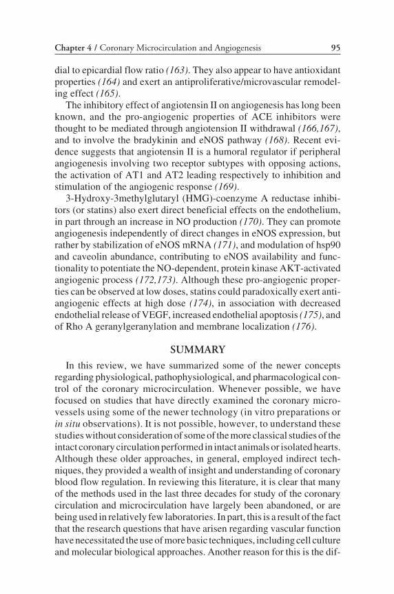
Chapter 4 / Coronary Microcirculation and Angiogenesis 95
dial to epicardial flow ratio (163). They also appear to have antioxidantproperties (164) and exert an antiproliferative/microvascular remodel-ing effect (165).
The inhibitory effect of angiotensin II on angiogenesis has long beenknown, and the pro-angiogenic properties of ACE inhibitors werethought to be mediated through angiotension II withdrawal (166,167),and to involve the bradykinin and eNOS pathway (168). Recent evi-dence suggests that angiotensin II is a humoral regulator if peripheralangiogenesis involving two receptor subtypes with opposing actions,the activation of AT1 and AT2 leading respectively to inhibition andstimulation of the angiogenic response (169).
3-Hydroxy-3methylglutaryl (HMG)-coenzyme A reductase inhibi-tors (or statins) also exert direct beneficial effects on the endothelium,in part through an increase in NO production (170). They can promoteangiogenesis independently of direct changes in eNOS expression, butrather by stabilization of eNOS mRNA (171), and modulation of hsp90and caveolin abundance, contributing to eNOS availability and func-tionality to potentiate the NO-dependent, protein kinase AKT-activatedangiogenic process (172,173). Although these pro-angiogenic proper-ties can be observed at low doses, statins could paradoxically exert anti-angiogenic effects at high dose (174), in association with decreasedendothelial release of VEGF, increased endothelial apoptosis (175), andof Rho A geranylgeranylation and membrane localization (176).
SUMMARY
In this review, we have summarized some of the newer conceptsregarding physiological, pathophysiological, and pharmacological con-trol of the coronary microcirculation. Whenever possible, we havefocused on studies that have directly examined the coronary micro-vessels using some of the newer technology (in vitro preparations orin situ observations). It is not possible, however, to understand thesestudies without consideration of some of the more classical studies of theintact coronary circulation performed in intact animals or isolated hearts.Although these older approaches, in general, employed indirect tech-niques, they provided a wealth of insight and understanding of coronaryblood flow regulation. In reviewing this literature, it is clear that manyof the methods used in the last three decades for study of the coronarycirculation and microcirculation have largely been abandoned, or arebeing used in relatively few laboratories. In part, this is a result of the factthat the research questions that have arisen regarding vascular functionhave necessitated the use of more basic techniques, including cell cultureand molecular biological approaches. Another reason for this is the dif-

96 Voisine et al.
ficulty of these studies and the expense of larger animals used in manyof the physiological experiments. A relatively recent development hasbeen the ability to make many in vivo measurements of coronary hemo-dynamics in human subjects in the catheterization laboratory, bypassingthe absolute need for large animal studies of flow. Nevertheless, as vas-cular biology research examines more fundamental questions, it will beimportant not to lose sight of the need to take basic observations back tothe intact circulation. As emphasized in this chapter, properties of pe-ripheral vessels cannot be extrapolated to the coronary circulation, andproperties of one size or class of coronary microvessel may not be presentin another size or class of coronary microvessel. Future studies will bemost successful when fundamental observations can be tested in intactvessels and circulations, including the coronary circulation.
REFERENCES
1. Nellis SH, Liedtke AJ, Whitesell L. Small coronary vessel pressure and diameter inan intact beating rabbit heart using fixed-position and free-motion techniques. CircRes 1981;49:342–353.
2. Chilian WM, Eastham CL, Marcus ML. Microvascular distribution of coronaryvascular resistance in beating left ventricle. Am J Physiol 1986;251:H779–H788.
3. Kuo L, et al. Myogenic activity in isolated subepicardial and subendocardial coro-nary arterioles. Am J Physiol 1988;255(6Pt2):H1558–H1562
4. Ashikawa K, et al. A new microscopic system for the continuous observation of thecoronary microcirculation in the beating canine left ventricle. Microvasc Res1984;28:387–394.
5. Yada T, et al. In vivo observation of subendocardial microvessels of the beatingporcine heart using a needle probe videomicroscope with a CCD camera. Circ Res1993;72:939–946.
6. Mori H, et al. Visualization of penetrating transmural arteries in situ by monochro-matic synchroton radiation. Circulation 1994;89:863–871.
7. Jones CJH, et al. Regulation of coronary blood flow: coordianation of heterogenouscontrol mechanisms in vascular microdomains. Cardiovasc Res 1995;29:585–596.
8. Feigl EO. Coronary physiology. Physiol Rev 1983;63:1–205.9. Hoffman JI. Transmural myocardial perfusion. Prog Cardiovasc Dis 1987;29:429–464.
10. Duncker DJ, Bache RJ. Regulation of coronary vasomotor tone under normal condi-tions and during acute myocardial hypoperfusion. Pharmacol Ther 2000;86:87–110.
11. Komaru T, Knatsika H, Shirato K. Coronary microcirculation. Physiology andPharmacology. Pharmacol Ther 2000;86:217–261.
12. Marcus M. The coronary circulation in health and disease. First ed. McGraw-Hill,New York, NY: 1983.
13. Schaper W. The pathophysiology of myocardial perfusion. 1 ed. Elsevier/NorthHolland Biomedical, Amsterdam, The Netherlands: 1979.
14. Kanatsuka H, et al. Heterogenous changes in epimyocardial microvascularsize during graded coronary stenosis. Evidence of the microvascular site froautorogulation. Circ Res 1990;66:389–396.
15. Muller JM, et al. Intergrated regulation of pressure and flow in the coronary micro-circulation. Cardiovasc Res 1996;32:668–678.

Chapter 4 / Coronary Microcirculation and Angiogenesis 97
16. Chilian WM, Layne SM, Klausner EC, Eastham CL, Marcus ML. Redistribution ofcoronary microvascular resistance produced by dipyridamole. Am J Physiol1989;256:H383–H390.
17. Chilian WM. Microvascular pressures and resistances in the left ventricularsubepicardium and subendocardium. Circ Res 1991;69:561–570.
18. Fujii M, Nuno DW, Lamping KG, Dellsperger KC, Eastham CL, Harrison DG.Effect of hypertension and hypertrophy on coronary microvascular pressure. CircRes 1992;71:120–126.
19. Kuo L, et al. Longitudinal gradients for endothelium-dependent and independentvascular responses in the coronary microcirculation. Circulation 1995;92:518–525.
20. Miller FJ Jr, Dellsperger KC, Gutterman DD. Myogenic constriction of humancoronary arterioles. Am J Physiol 1997;273:H257–H264.
21. Brayden JE, Nelson MT. Regulation of arterial tone by activation of calcium-depen-dent potassium channels. Science 1992;256:532–535.
22. Nelson MT, et al. Calcium channels, potassium channels, and voltage-dependenceof arterial smooth muscle. Am J Physiol 1990;259(1 Pt 1):C3–C18.
23. Meininger GA, et al. Calcium measurement in isolated arterioles during myogenicand agonist stimulation. Am J Physiol 1991;261(3 Pt 2):H950–H959
24. Hill MA, Meninger GA. Calcium entry and myogenic phenomena in skeletal musclearterioles. Am J Physiol 1994;267:H1088–H1092.
25. Park KW, Dai H-B, Lowenstein E, Darvish E, Sellke FW. Heterogeneous vasomo-tor responses of rabbit microvessels to isoflurane. Anesthesiology 1994;81:1190–1197.
26. Knot HJ, Nelson MT. Regulation of membrane potential and diameter by voltage-dependent K+ channels in rabbit myogenic cerebral arteries. Am J Physiol1995;269:H348–H355.
27. Bowles DK, et al. Heterogeneity of L-type calcium current density in coronarysmooth muscle. Am J Physiol 1997;273(4 Pt 2):H2083–H2089
28. Osol G, Laher I, Cipolla M. Protein kinase C modulates basal myogenic tone inresistance arteries from the cerebral circulation. Circ Res 1991;68:359–367.
29. Narayanan J, Imig M, Roman RJ, Harder DR. Pressurization of isolated renalarteries increases inositol trisphosphate and diacylglycerol. Am J Physiol1994;266:H1840–H1845.
30. Large WA. Receptor-operated Ca2+-permeable nonselective cation channels invascular smooth muscle: a physiologic perspective. J Cardiovasc Electrophysiol2002;13:493–501.
31. Slish DF, Welsh DG, Brayden JE. Diacylglycerol and protein kinase C activatecation channels involved in myogenic tone. Am J Physiol 2002;283:H2196–H2201.
32. Korzick DH, Laughlin MH, Bowles DK. Alterations in PKC signaling underlieenhanced myogenic tone in exercise-trained porcine coronary resistance arteries. JAppl Physiol 2004;96:1425–1432.
33. Roman RJ. P-450 metabolites of arachidonic acid in the control of cardiovascularfunction. Physiol Rev 2002;82:131–185.
34. Randriamboavonjy V, Busse R, Fleming I. 20-HETE-induced contraction ofsmall coronary arteries depends on the activation of Rho-kinase. Hypertension2003;41:801–806.
35. Massett MP, Zoltan U, Csiszar A, Kaley G, Koller A. Different roles of PKC andMAP kinases in arteriolar constrictions to pressure and agonists. Am J Physiol2002;283:H2282–H2287.
36. Khan TA, Bianchi C, Ruel M, Voisine P, Li J, Liddicoat JR, Sellke FW. Mitogen-activated protein kinase inhibition and cardioplegia-cardiopulmonary bypass re-duce coronary myogenic tone. Circulation 2003;108(Suppl 1):II348–II353.

98 Voisine et al.
37. Kuo L, Chilian WM, Davis MJ. Coronary arteriolar myogenic response is indepen-dent of endothelium. Circ Res 1990;66:860–866.
38. Nakayama K, Osol G, Halpern W. Reactivity of isolated porcine coronary resistancearteries to cholinergic and adrenergic drugs and transmural pressure changes. CircRes 1988;62:741–748.
39. Liao JC, Kuo L. Interaction between adenosine and flow-induced dilation in coro-nary microvascular network. Am J Physiol 1997;272:H1571–H1581.
40. Muller JM, Myers PR, Laughlin MH. Exercise training alters myogenic responsesin porcine coronary resistance arteries. J Appl Physiol 1993;75:2677–2682.
41. Boatwright RB, Downey HF, Bashour FA, Crystal GJ. Transmural variation inautoregulation of coronary blood flow in hyperperfused canine myocardium. CircRes 1980;47:599–609.
42. Davis PF. Flow-mediated endothelial mechanotransduction. Physiol Rev 1995;75:519–560.
43. Jimenez AH, et al. Effects of oxygen tension on flow-induced vasodilation in por-cine coronary arterioles. Microvasc Res 1996;51:365–377.
44. Kuo L, et al. Interaction of pressure- and flow-induced responses in porcine coro-nary resistance vessels. Am J Physiol 1991;261(6 Pt 2):H1706–H1715
45. Olsson RA, Bunger R. Metabolic control of coronary blood flow. Prog CardiovascDis 1987;29:369–387.
46. Yada T, Richmond KN, Van Bibber R, Kroll K, Feigl EO. Role of adenosine in localmetabolic coronary vasodilation. Am J Physiol 1999;276:H1425–H1433.
47. Duncker DJ, Stubenitsky R, Verdouw PD. Role of adenosine in the regulation ofcoronary blood flow in swine at rest and during treadmill exercise. Am J Physiol1998;275:H1663–H1672.
48. Hein TW, Kuo L. cAMP-independent dilation of coronary arterioles to adenosisne:role of nitric oxide, G proteins, and KATP channels. Circ Res 1999;85:634–642.
49. Komaru T, Kanatsuka H, Dellsperger K, Takishima T. The role of ATP-sensi-tive potassium channels in regulating coronary microcirculation. Biorheology1993;30:371–380.
50. Duncker DJ, Van Zon NS, Altman JD, Pavek TJ, Bache RJ. Role of K+ ATPchannels in coronary vasodilation during exercise. Circulation 1993;88:1245–1253.
51. Duncker DJ, van Zon NS, Ishibashi Y, Bache RJ. Role of K+ ATP channels andadenosine in the regulation of coronary blood flow during exercise with normal andrestricted coronary blood flow. J Clin Invest 1996;97:996–1009.
52. Farouque HM, et al. Effect of ATP-sensitive channel inhibition on coronary netabolicvasodilation in humans. Arterioscler Thromb Vasc Biol 2004;24(5):905–910.
53. Miura H, et al. Diabetes mellitus impairs vasodilation to hypoxia in human coronaryarterioles: reduced activity of ATP-sensitive potassium channels. Circ Res2003;92(2):151–158.
54. Merkus D, Duncker DJ, Chilian WM. Metabolic regulation of vascular tone: role ofendothelin-1. Am J Physiol 2002;283:H1915–H1921.
55. Conway RS, Kirk ES, Eng C. Ventricular preload alters intravascular and extravas-cular resistances of coronary collaterals. Am J Physiol 1988;254:H532–H541.
56. Bellamy RF. Diastolic coronary artery pressure-flow relations in the dog. Circ Res1978;43:92–101.
57. Eng C, Jentzer JH, Kirk ES. The effects of the coronary capacitance on the interpre-tation of diastolic pressure-flow relationships. Circ Res 1982;50:334–341.
58. Kanatsuka H, Ashikawa K, Komaru T, Suzuki T, Takishima T. Diameter changeand pressure-red blood cell velocity relations in coronary microvessels during longdiastoles in the canine left ventricle. Circ Res 1990;66:503–510.

Chapter 4 / Coronary Microcirculation and Angiogenesis 99
59. Jayaweera AR, Wei K, Coggins M, Bin JP, Goodman C, Kaul S. Role of capill-aries in determining CBF reserve: new insights using myocardial contrast echocar-diography. Am J Physiol 1999;277:H2363–H2372.
60. Sun, et al. Mechanical compression elicits NO-dependent increases in coronaryflow. Am J Physiol Heart Circ Physiol 2004;287:H2454–H2460.
61. Young MA, Knight DR, Vatner SF. Autonomic control of large coronary arteriesand resistance vessels. Prog Cardiovasc Dis 1987;30:211–234.
62. Wang SY, Friedman M, Johnson RG, Weintraub RM, Sellke FW. Adrenergic regu-lation of coronary microcirculation after extracorporeal circulation and crystalloidcardioplegia. Am J Physiol 1994;267:H2462–H2470.
63. Lamping KG, Chilian WM, Eastham CL, Marcus ML. Coronary microvascularresponse to exogenously administered and endogenously released acetylcholine.Microvasc Res 1992;43:294–307.
64. Hammarstrom AK, Parkington HC, Coleman HA. Release of endothelium-derivedhyperpolarizing factor (EDHF) by M3 receptor stimulation in guinea-pig coronaryartery. Br J Pharmacol 1995;115:717–722.
65. Sellke FW, Dai HB. Responses of porcine epicardial venules to neurohumoral sub-stances. Cardiovasc Res 1993;27:1326–1332.
66. Lamping KG, Kanatsuka H, Eastham CL, Chilian WM, Marcus ML. Nonuniformvasomotor responses of the coronary microcirculation to serotonin and vasopressin.Circ Res 1989;65:343–351.
67. Sellke FW, Myers PR, Bates JN, Harrison DG. Influence of vessel size on the sensi-tivity of porcine microvessels to nitroglycerin. Am J Physiol 1990;258:H515–H520.
68. Lamping KG, Clothier JL, Eastham CL, Marcus ML. Coronary microvascular re-sponse to endothelin is dependent on vessel diameter and route of administration.Am J Physiol 1992;263:H703–H709.
69. Merkus D. Contribution of endothelium to coronary vasomotor tone. Am J PhysiolHeart Circ Physiol 2005;28:H871–H880.
70. Murad F. Cyclic guanosine monophosphate as a mediator of vasodilation. J ClinInvest 1986;78:1–5.
71. Myers PR, Minor RL Jr, Guerra R Jr, Bates JN, Harrison DG. The vasorelaxantproperties of the endothelium-derived relaxing factor more closely resemble S-nitrosocysteine than nitric oxide. Nature 1990;345:161–163.
72. Corson M, James N, Latta S, Nerem R, Berk B, Harrison D. Phosphorylation ofendothelial nitric oxide synthse in response to fluid shear stress. Circ Res1996;79:984–991.
73. Venema RC, Sayegh HS, Arnal J-F, Harrison DG. Role of the enzyme calmodulin-binding domain in membrane association and phospholipid inhibition of endothelialnitric oxide synthase. J Biol Chem 1995;270:14705–14711.
74. Michel J, Feron O, Sacks D, Michel T. Reciprocal regulation of endothelial nitric-oxide synthase by Ca2+-calmodulin and caveolin. J Biol Chem 1997;272:15583–15586.
75. Uemetsu M, Ohara Y, Navas JP, et al. Regulation of endothelial cell nitric oxidesynthase mRNA expresiion by shear stress. Am J Physiol 1995;269:C1371–C1378.
76. Arnal J-F, Yamin J, Dockery S, Harrison DG. Regulation of endothelial nitric oxidesynthase mRNA, protein and activity during cell growth. Am J Physiol (Cell Physiol)1994;267:C1381–C1388.
77. McQuillan LP, Leung GK, Marsden PA, Kostyk SK, Kourembanas S. Hypoxiainhibits expression of eNOS via transcriptional and posttranscriptional mechanisms.Am J Physiol 1994;267:H1921–H1927.
78. Pou S, Pou WS, Bredt DS, Snyder SH, Rosen GM. Generation of superoxide bypurified brain nitric oxide synthase. J Biol Chem 1992;267:24173–24176.

100 Voisine et al.
79. Shimokawa H, Flavahan NA, Vanhoutte PM. Loss of endothelial pertussis toxin-sensitive G protein function in atherosclerotic porcine coronary arteries. Circulation1991;83:652–660.
80. Harrison DG. Endothelial function and oxidant stress. Clin Cardiol 1997;20:II-11–II-17.
81. Jang JJ, Ho HKV, Kwan HH, Fajardo LF, Cooke JP. Angiogenesis is impairedby hypercholesterolemia—role of asymmetric dimethylarginine. Circulation2000;102:1414–1419.
82. Amezcua JL, Palmer RM, de Souza BM, Moncada S. Nitric oxide synthesized fromL-arginine regulates vascular tone in the coronary circulation of the rabbit. Br JPharmacol 1989;97:1119–1124.
83. Taylor SG, Weston AH. Endothelium-derived hyperpolarizing factor: a new endog-enous inhibitor from the vascular endothelium. Trends Pharmacol Sci 1988;9:272–274.
84. Petersson J, Zygmunt PM, Hogestatt ED. Characterization of the potassium chan-nels involved in EDHF-mediated relaxation in cerebral arteries. Br J Pharmacol1997;120:1344–1350.
85. Campbell WB, Gebremedhin D, Pratt PF, Harder DR. Identification of epoxyeico-satrienoic acids as endothelium-derived hyperpolarizing factors. Circ Res1996;78:415–423.
86. Fulton D, McGiff JC, Wolin MS, Kaminski P, Quilley J. Evidence against a cyto-chrome P450-derived reactive oxygen species as the mediator of the nitric oxide-independent vasodilator effect of bradykinin in the perfused heart of the rat. JPharmacol Exp Ther 1997;280:702–709.
87. Edwards G, Dora KA, Gardener MJ, Garland CJ, Weston AH. K+ is an endothelium-derived hyperpolarizinf factor in rat arteries. Nature 1998;396:269–272.
88. Taylor HJ, Chaytor AT, Evance WH, Griffith TM. Inhibition of the gap junctionalcomponent of endothelium-dependent relaxations in rabbit iliac artery by 18-alphaglycyrrhetinic acid. Br J Pharmacol 1998;125:1–3.
89. Batenburg WW, et al. L-S nitrosothiols: endothelium-derived hyperpolarizing fac-tors in porcine coronary arteries? J Hypertens 2004;22:1927–1936.
90. Yada T, Shimokawa H, Hiramatsu O, Kajita T, Shigeto F, Goto M, Ogasawara Y,Kajiya F. Hydrogen peroxide, an endogenous endothelium-derived hyperpolarizingfactor, plays an important role in coronary autoregulation in vivo. Circulation2003;107:1040–1045.
91. Shimokawa H, Yasutake H, Fujii K, et al. The importance of the hyperpolarizingmechanism increases as the vessel size decreases in endothelium-dependent relax-ations in rat mesenteric circulation. J Cardiovasc Pharmacol 1996;28:703–711.
92. Xu XP, Tanner MA, Myers PR. Prostaglandin-mediated inhibition of nitric oxideproduction by bovine aortic endothelium during hypoxia. Cardiovasc Res1995;30:345–350.
93. Klassen G, Armour J. Epicardial coronary venous pressure measurements: auto-nomic responses. Can J Physiol Pharmacol 1982;60:698–706.
94. Yuan Y, Mier R, Chilian W, Zawieja D, Granger H. Interaction of neutrophilsand endothelium in isolated coronary venules and arterioles. Am J Physiol1995;268:H490–H498.
95. Tinsey JH, Ustinova EE, Xu W, Yuan SY. Src-dependent, neutrophil-mediatedvascular hyperpermeability and beta-catenin modification. Am J Physiol CellPhysiol 2002;283(6):C1745–C1751.
96. Yuan SY, Wu MH, Ustinova EE, et al. Myosin light chain phosphorylation inneutrophil-stimulated coronary microvascular leakage. Circ Res 2002;90(11):1214–1221.

Chapter 4 / Coronary Microcirculation and Angiogenesis 101
97. Lefer D, Nakanishi K, Vinten-Johansen J, Ma X, Lefer A. Cardiac venous endot-helial dysfunction after myocardial ischemia and reperfusion in dogs. Am J Physiol1992;263:H850–H856.
98. Piana RN, Wang SY, Friedman M, Sellke FW. Angiotensin-converting enzymeinhibition preserves endothelium-dependent coronary microvascular responsesduring short-term ischemia-reperfusion. Circulation 1996;93:544–551.
99. Garg UC, Hassid A. Nitric oxide-generating vasodilators and 8-bromo-cyclic gua-nosine monophosphate inhibit mitogenesis and proliferation of cultured rat vascu-lar smooth muscle cells. J Clin Invest 1989;83:1774–1777.
100. Cayatte AJ, Palacino JJ, Horten K, Cohen RA. Chronic inhibition of nitric oxideproduction accelerates neointima formation and impairs endothelial function inhypercholesterolemic rabbits. Arterioscler Thromb 1994;14:753–759.
101. von der Leyen HE, Gibbons GH, Morishita R, et al. Gene therapy inhibitingneointimal vascular lesion: in vivo transfer of endothelial cell nitric oxide synthasegene. Proc Natl Acad Sci USA 1995;92:1137–1141.
102. Moroi M, Zhang L, Yasuda T, Virmani R, Gold HK, Fisman MC, Huang PL.Interaction of genetic deficiency of endothelial nitric oxide, gender and pregnancyin vascular response to injury in mice. J Clin Invest 1998;101:1225–1232.
103. Yu SM, Hung LM, Lin CC. cGMP-elevating agents suppress proliferation ofvascular smooth muscle cells by inhibiting the activation of epidermal growthfactor signaling pathway. Circulation 1997;95:1269–1277.
104. Itoh H, Pratt RE, Ohno M, Dzau VJ. Atrial natriuretic polypeptide as a novelantigrowth factor of endothelial cells. Hypertension 1992;19:758–761.
105. Cornwell TL, Arnold E, Boerth NJ, Lincoln TM. Inhibition of smooth muscle cellgrowth by nitric oxide and activation of cAMP-dependent protein kinase by cGMP.Am J Physiol 1994;267:C1405–C1413.
106. D’Souza FM, Sparks RL, Chen H, Kadowitz PJ, Jeter JR Jr. Mechanis of eNOSgene transfer inhibition of vascular smooth muscle cell proliferation. Am J PhysiolCell Physiol 2003;284(1):C191–C199
107. Pollman MJ, Yamada T, Horiuchi M, Gibbons GH. Vasoactive substances regu-late vascular smooth muscle cell apoptosis. Countervailing influences of nitricoxide and angiotensin II. Circ Res 1996;79:748–756.
108. Shi W, Wang X, Shih DM, Laubach VE, Vavab M, Lusis AJ. Paradoxicalreducation of fatty streak formation in mice lacking endothelial nitric oxide syn-thase. Circulation 2002;105:2078.
109. Ozaki M, Kawashima S, Yamashita T, et al. Overexpression of endothelial nitricoxide synthase accelerates atherosclerotic lesion formation in apoE-deficient mice.J Clin Invest 2002;110:331–340.
110. Kuhlencordt PJ, Gyurko R, Han F, et al. Accelerated atherosclerosis, aortic aneu-rysm formation, and ischemic heat disease in apolipoprotein E/endothelial nitricoxide synthase double-knockout mice. Circulation 2001;104:448.
111. Wolfsgruber W, Feil S, Brummer S, Kuppinger O, Hofmann F, Feil R. Aproatherogenic role for cGMP-dependant protein kinase in vascular smooth musclecells. PNAS 2003;100(23):13519–13524.
112. Bayraktutan U. Nitric oxide synthase and NAD(P)H oxidase modulate coronaryendothelial cell growth. J Mol Cell Cardiol 2004;36(2):277–286.
113. Ware J, Simons M. Angiogenesis in ischemic heart disease. Nat Med 1997;3:158–164.114. Li J, Brown L, Hibberd M, Grossman J, Morgan J, Simons M. VEGF, flk-1, flt-1
expression in a rat myocardial infarction model of angiogenesis. Am J Physiol1996;270:H1803–H1811.

102 Voisine et al.
115. Sellke FW, Wang SY, Stamler A, et al. Enhanced microvascular relaxations toVEGF and bFGF in chronically ischemic porcine myocardium. Am J Physiol1996;271:H713–H720.
116. Wu H, Yuan Y, McCarthy M, Granger H. Acidic and basic FGF’s dilatearterioles of skeletal muscle through a NO-dependent mechanism. Am J Physiol1996;271:H1087–H1093.
117. Ziche M, Morbidelli L, Masini E, et al. Nitric oxide mediates angiogenesis in vivoand endothelial cell growth and migration in vitro promoted by substance P. J ClinInvest 1994;94:2036–2044.
118. Morbidelli L, Chang CH, Douglas JG, Granger HJ, Ledda F, Ziche M. Nitric oxidemediates mitogenic effect of VEGF on coronary venular endothelium. Am J Physiol1996;270:H411–H415.
119. Bouloumie A, Schini-Kerth VB, Busse R. Vascular endothelial growth factor up-regulates nitric oxide synthase expression in endothelial cells. Cardiovasc Res1999;41:773–780.
120. Uhlmann S, Friedrichs U, Eichler W, Hoffmann S, Wiedemann P. Direct measure-ment of VEGF-induced nitric oxide production by choroidal endothelial cells.Microvasc Res 2001;62:179–189.
121. Zhao X, Lu X, Feng Q. Deficiency in endothelial nitric oxide synthase impairs myo-cardial angiogenesis. Am J Physiol Heart Circ Physiol 2002;283(6):H2371–H2378.
122. Bussolati B, Dunk C, Grohman M, Kontos CD, Mason J, Ahmed A. Vascularendothelial growth factor receptor-1 modulates vascular endothelial growth fac-tor-mediated angiogenesis via nitric oxide. Am J Pathol 2001;159:993–1008.
123. Jozkowicz A, Cooke JP, Guevara I, et al. Genetic augmentation of nitric oxide synthaseincreases the vascular generation of VEGF. Cardiovasc Res 2001;51:773–783.
124. Babaei S, Teichert-Kuliszewska K, Monge JC, Mohamed F, Bendeck MP, StewartDJ. Role of nitric oxide in the angiogenic response in vitro to basic fibroblastgrowth factor. Circ Res 1998;82:1007–1015.
125. Murohara T, Witzenbichler B, Spyridopoulos I, et al. Role of endothelial nitricoxide synthase in endothelial cell migration. Arterioscler Thromb Vasc Biol1999;19:1156–1161.
126. Papapetropoulos A, Desai KM, Rudic RD, et al. Nitric oxide synthase inhibitorsattenuate transforming-growth-factor-beta 1-stimulated capillary organization invitro. Am J Pathol 1997;150:1835–1844.
127. Eliceiri BP, Paul R, Schwartzberg PL, Hood JD, Leng J, Cheresh DA. Selectiverequirement for Src kinases during VEGF-induced angiogenesis and vascular per-meability. Mol Cell 1999;4:915–924.
128. Duan J, Murohara T, Ikeda H, et al. Hypercholesterolemia inhibits angiogenesisin response to hindlimb ischemia: nitric oxide-dependent mechanism. Circulation2000;102:III370–III376.
129. Ruel M, Wu GF, Khan TA, et al. Inhibition of the cardiac angiogenic response tosurgical FGF-2 therapy in a swine endothelial dysfunction model. Circulation2003;108(Suppl 1):II335–II340.
130. Voisine P, Bianchi C, Ruel M, et al. Inhibition of the cardiac angiogenic responseto exogenous vascular endothelial growth factor (VEGF) therapy in a porcinemodel of endothelial dysfunction. Surgery 2004;136:407–415.
131. Sellke FW, Armstrong ML, Harrison DG. Endothelium-dependent vascular relax-ation is abnormal in the coronary microcirculation of atherosclerotic primates.Circulation 1990;81:1586–1593.
132. Chilian WM, Dellsperger KC, Layne SM, et al. Effects of atherosclerosis on thecoronary microcirculation. Am J Physiol 1990;258:H529–H539.

Chapter 4 / Coronary Microcirculation and Angiogenesis 103
133. Drexler H, Zeiher AM, Meinzer K, Just H. Correction of endothelial dysfunctionin coronary microcirculation of hypercholesterolaemic patients by L-arginine.Lancet 1991;338:1546–1550.
134. Treasure CB, Klein JL, Vita JA, et al. Hypertension and left ventricular hypertro-phy are associated with impaired endothelium-mediated relaxation in human coro-nary resistance vessels. Circulation 1993;87:86–93.
135. Quillen JE, Sellke FW, Brooks LA, Harrison DG. Ischemia-reperfusion impairsendothelium-dependent relaxation of coronary microvessels but does not affectlarge arteries. Circulation 1990;82:586–594.
136. Matsunaga T, Okumura K, Ishizaka H, Tsunoda R, Tayama S, Yasue H. Impair-ment of coronary blood flow regulation by endothelium-derived nitric oxide indogs with alloxan-induced diabetes. J Cardiovasc Pharmacol 1996;28:60–67.
137. Sellke FW, Shafique T, Schoen FJ, Weintraub RM. Impaired endothelium-depen-dent coronary microvascular relaxation after cold potassium cardioplegia andreperfusion. J Thorac Cardiovasc Surg 1993;105:52–58.
138. Sellke FW, Quillen JE, Brooks LA, Harrison DG. Endothelial modulation of thecoronary vasculature in vessels perfused via mature collaterals. Circulation1990;81:1938–1947.
139. Peters KG, Marcus ML, Harrison DG. Vasopressin and the mature coronary col-lateral circulation. Circulation 1989;79:1324–1331.
140. Symons JD, Longhurst JC, Stebbins CL. Response of collateral-dependent myo-cardium to vasopressin release during prolonged intense exercise. Am J Physiol1993;264:H1644–H1652.
141. Harada K, Friedman M, Lopez J, et al. Vascular endothelial growth factor admin-istration in chronic myocardial ischemia. Am J Physiol 1996;270:H1791–H1802.
142. Sellke FW, Wang SY, Friedman M, et al. Basic FGF enhances endothelium-dependent relaxation of the collateral-perfused coronary microcirculation. Am JPhysiol 1994;267:H1303–H1311.
143. Ravichandran L, Johns R, Rengasamy A. Direct and reversible inhibition of endot-helial nitric oxide synthase by nitric oxide. Am J Physiol 1995;268:H2216–H2223.
144. Laham RJ, Simons M, Tokufuji M, Hung D, Sellke FW. Modulation of myocardialperfusion and vascular reactivity by pericardial basic fibroblast growth factor;insight into ischemia-induced reduction in endothelium-dependent vasodilation.J Thorac Cardiovasc Surg 1998;116:1022–1028.
145. Uemetsu M, Ohara Y, Navas JP, et al. Regulation of endothelial cell nitric oxidesynthase mRNA expression by shear stress. Am J Physiol 1995;269:C1371–C1378.
146. Voisine P, Bianchi C, Khan TA, et al. Normalization of coronary microvascularreactivity and improvement in myocardial perfusion by surgical VEGF therapycombined with oral supplementation of L-arginine in a porcine model of endothe-lial dysfunction. J Thorac Cardiovasc Surg 2005; in press.
147. Rapps J, Jones A, Sturek M, Magliola L, Parker J. Mechanisms of altered contrac-tile responses to vasopressin and endothelin in canine collateral arteries. Circula-tion 1997;95:231–239.
148. Bauters C, Asahara T, Zheng L, et al. Recovery of disturbed endothelium-depen-dent flow in the collateral-perfused rabbit ischemic hindlimb after administrationof vascular endothelial growth factor. Circulation 1995;91:2802–2809.
149. Lopez J, Laham R, Carrozza J, et al. Hemodynamic effects of intracoronary VEGFdelivery-Evidence of tachyphylaxis and NO dependence of response. Am J Physiol1997;273:H1317–H1323.
150. Sirois M, Edelman E. VEGF effect on vascular permeability is mediated by syn-thesis of platelet-activating factor. Am J Physiol 1997;272:H2746–H2756.

104 Voisine et al.
151. Wu G, Mannam AP, Wu J, et al. Hypoxia induces myocyte-dependent COX-2regulation in endothelial cells: role of VEGF. Am J Physiol Heart Circ Physiol2003;285(6):H2420–H2429
152. Marcus ML, Harrison DG, Chilian WM, e tal. Alterations in the coronary circu-lation in hypertrophied ventricles. Circulation 1987;75:I 19–I 25.
153. Thatcher GRJ, et al. Nitrates and NO release: contemporary aspects in biologicaland medicinal chemistry. Free Radic Biol Med 2004;37(8):1122–1143.
154. Winbury MM, Howe BB, Weiss HR. Effect of nitrates and other coronary dilatorson large and small coronary vessels; an hypothesis for the mechanism of action ofnitrates. J Pharmacol Exp Ther 1969;168:70–95.
155. Fam WM, McGregor M. Effect of nitroglycerin and dipyridamole on regionalcoronary resistance. Circ Res 1968;22:649–659.
156. Kurz MA, Lamping KG, Bates JN, Eastham CL, Marcus ML, Harrison DG. Mecha-nisms responsible for the heterogeneous coronary microvascular response to nitro-glycerin. Circ Res 1991;68:847–855.
157. Wheatley RM, Dockery SP, Kurz MA, Sayegh HS, Harrison DG. Interactions ofnitroglycerin and sulfhydryl-donating compounds in coronary microvessels. AmJ Physiol 1994;266:H291–H297.
158. Park KS, Kim Y, Lee YH, Earm YE, Ho WK. Mechanosensitive cation channelsin arterial smooth muscle cells are activated by diacylglycerol and inhibited byphospholipase C inhibitor. Circ Res 2003;93:557–564.
159. Werner GS, et al. Intracoronary verapamil for reversal of no-reflow duringcoronary angioplasty for acute myocardial infarction. Catheter Cardiovasc Interv2002;57(4):444–451.
160. Akai K, Wang Y, Sato K, et al. Vasodilatory effect of nicorandil on coronaryarterial microvessels: its dependency on vessel size and the involvement of theATP-sensitive potassium channels. J Cardiovasc Pharmacol 1995;26:541–547.
161. Kichuk MR, et al. Regulation of nitric oxide production in human coronarymicrovessels and the contribution of local kinin formation. Circulation 1996;94:44–51.
162. Chen R, et al. important role of nitric oxide in the effect of angiotensin convertingenzyme inhibitor imipaprine on vascular injury. Hypertension 2003;42(4):542–547.
163. Kitakaze M, et al. Beneficial effects of inhibition of angiotensin converting en-zyme on ischemic myocardium during coronary hypoperfusion in dogs. Circula-tion 1995;92:950–961.
164. Piana RN, et al. Angiotensin converting enzyme inhibition preserves endo-thelium-dependent coronary microvascular responses during short-term ischemia-reperfusion. Circulation 1996;93:544–551.
165. Dzau VJ, et al. The relevance of tissue angiotensin converting enzyme: manifes-tations in mechanistic and endpoint data. Am J Cardiol 2001;88(9A):1L–20L.
166. Unger T, Mattfeldt T, Lamberty V, et al. Effect of early onset angiotensin enzymeinhibition on myocardial capillaries. Hypertension 1992;20:478–482.
167. Fabre JE, Rivard A, Magner M, Silver M, Isner JM. Tissue inhibition of angio-tensin-converting enzyme activity stimulates angiogenesis in vivo. Circulation1999;99:3043–3049.
168. Silvestre JS, Bergaya S, Tamarat R, Duriez M, Boulanger CM, Levy BI.Proangiogenic effect of angiotensin-converting enzyme inhibition is mediated bythe bradykinin B(2) receptor pathway. Circ Res 2001;89:678–683.
169. Walther T, Menrad A, Orzechowski HD, Siemeister G, Paul M, Schirner M.Differntial regulation of in vivo angiogenesis by angiotensin II receptors. FASEBJ 2003;17:2061–2067.

Chapter 4 / Coronary Microcirculation and Angiogenesis 105
170. Corsini A, Bellosta S, Baetta R, Funagalli R, Paoletti R, Bernini F. New insightsinto the pharmacodynamic and pharmacokinetic properties of statins. PharmacolTher 1999;84:413–428.
171. Laufs U, Liao JK. Direct vascular effects of HMG-CoA reductase inhibitors. TrendsCardiovasc Med 2000;10:143–148.
172. Brouet A, Sonveauzx P, Dessy C, Moniotte S, Balligand JL, Feron O. Hsp90 andcaveolin are key targets for the proangiogenic nitric oxidemediated effects ofstatins. Circ Res 2001;89:866–873.
173. Kureishi Y, Luo Z, Shiojima I, et al. The HMG-CoA reductase inhibitor simvastatinactivates the protein kinase Akt and promotes angiogenesis in normocho-lesterolemic animals. Nat Med 2000;6:1004–1010.
174. Urbich C, Dernbach E, Zeiher AM, Dimmeler S. Double-edged role of statins inangiogenesis signaling. Circ Res 2002;90:737–744.
175. Weis M, Heeschen C, Glassford AJ, Cooke JP. Statins have biphasic effects onangiogenesis. Circulation 2002;105:739–745.
176. Park HJ, Kong D, Iruela-Arispe L, Begley U, Tang D, Galper JB. 3-hydroxy-3-methylglutaryl coenzyme A reductase inhibitors interfere with angiogenesis byinhibiting the geranylgeranylation fo RhoA. Circ Res 2002;91:143–150.

Chapter 5 / Local Delivery for Angiogenesis and Myogenesis 107
107
From: Contemporary Cardiology: Angiogenesis and Direct Myocardial RevasularizationEdited by: R. J. Laham and D. S. Baim © Humana Press Inc., Totowa, NJ
INTRODUCTION
An estimated 15% of patients with ischemic heart disease are notamenable to conventional methods of revascularization (1–3), and anadditional 12–22% may be limited to incomplete percutaneous coronaryintervention (PCI) or surgical procedures (1,4–7). These so-called no-option patients often progress to end-stage ischemic cardiomyopathywith an annual mortality in excess of 30% (8).Treatment options for this
5 Local and Regional VascularDelivery Strategiesfor Therapeutic Angiogenesisand Myogenesis
Erik T. Price, MD, Alan C. Yeung, MD,and Mehrdad Rezaee, MD, PhD
CONTENTS
INTRODUCTION
SYSTEMIC CARDIAC DELIVERY
DIRECT INTRAMYOCARDIAL INJECTION
PERCUTANEOUS INTRAMYOCARDIAL DELIVERY
INTRAPERICARDIAL AND PERIVASCULAR DELIVERY
INTRACORONARY DELIVERY
RETROGRADE CORONARY VENOUS DELIVERY
DIRECT COMPARISONS OF CARDIAC DELIVERY
MODALITIES
ALTERNATIVE DELIVERY MODALITIES
CONCLUSIONS

108 Price et al.
group remain limited to multidrug medical management, myocardialreduction surgery, left-ventricular (LV) assist device placement, or car-diac transplantation. The current estimation is that 100,000 patients peryear in this group would potentially benefit from alternative therapies(2,3,9). Over the past decade, cardiac angiogenesis and myogenesis haveemerged as such alternatives.
The efforts to develop alternative treatments have been mostly di-rected toward understanding cardiac vascular and muscle regenerationand the various protein, gene, and cellular mediators of these biologicalprocesses. Semantic differences between neovascularization, angiogen-esis, arteriogenesis, and vasculogenesis are important and are furtherdiscussed in recent reviews (4,10,11). For the purposes of this chapter,these processes will be collectively referred to as angiogenesis.Myogenesis and cellular myoplasty refer to mammalian cellular trans-plantation into the myocardium (12–17). A comparative analysis of themany agents that may facilitate cardiac angiogenesis or myogenesis isbeyond the scope of this chapter and is deferred to expert reviews (4,10–29).
The potential availability of myogenic and angiogenic factors hascreated an unusual situation in medicine where potent reagents are athand, but the optimal delivery modalities to target these to the cardiactissue are not yet established (30–33). This chapter will provide an over-view of the delivery modalities that may facilitate the anticipated effi-cacy of therapeutic cardiac angiogenesis and cellular cardiomyoplasty.Conceptualized criteria of an ideal delivery system are presented inTable 1. Safety issues are most pertinent for application in patients withsevere cardiac disease and multiple co-morbidities (4,33–35). Safetyissues addressed, delivery modalities should be applicable to the broad-est patient population. If these treatments are to become routine, thismodality should be practical for both the patient and practitioner andreproducible across a wide spectrum of operator skills. The ideal methodof cardiac delivery must efficiently deliver the desired product to clini-cally relevant regions of targeted myocardium and also promote productretention for sustained biological activity. These parameters may serveas guidelines for future development as well as useful tools for compari-son of currently available modalities.
SYSTEMIC CARDIAC DELIVERYSystemic delivery is well established with respect to cardiac pharma-
ceuticals. Theoretical advantages are evident, with noninvasive proce-dural practicality, cost efficiency, and broad patient applicability. Forthese reasons, the systemic route for protein growth factor and gene

Chapter 5 / Local Delivery for Angiogenesis and Myogenesis 109
product delivery has been actively investigated. In early investigationsof fibroblast growth factor (FGF) and vascular endothelial growth factor(VEGF), systemic intravenous delivery produced consistently poor acuteand chronic myocardial protein uptake with widely variable clinicaleffects (36–39). Follow-up studies have primarily used the systemicroute to establish comparative superiority of alternative methods (32,40–43). In composite, these studies demonstrated acute myocardial reten-tion that was consistently <1% after systemic delivery, with significantfirst-pass product uptake in both liver and lungs and no significant im-provement in collateral development, angiogenesis, myocardial perfu-sion, or function.
Table 1Proposed Parameters for an Ideal Myocardial Delivery System
SafetyMinimal procedure-related complications
(corporal invasiveness, local invasiveness, myocardial damage, coronaryvascular damage, ventricular perforation, pericardial tamponade,arrhythmia)
ApplicabilityBroad spectrum of patientsUseful across a broad range of disease types
(acute ischemia, sub-acute infarction, chronic infarction, ischemiccardiomyopathy, nonischemic cardiomyopathy)
PracticalityFor the patient
(minimally invasive, limited procedural discomfort, limited procedural time,outpatient vs inpatient, single administration)
For the practitioner(standard equipment, standard skill set, procedural efficiency, independence)
EfficiencyProcedural efficiency
(high delivery success rate, reproducible, time efficient)Product delivery efficiency
PrecisionMaximal delivery to targeted areas
(regionality within the myocardium)Minimal delivery to nontargeted areas
(diffusion, wash-out)Retained Activity
Location of delivery allowing prolonged retention for maximal beneficialbiological effect

110 Price et al.
Clinical application in 59 no-option patients treated with either sys-temic (n = 14) or intracoronary (n = 35) FGF at variable doses claimedoverall clinical effectiveness with attenuation of regional stress-inducedischemia (44). However, systemic and intracoronary subsets weregrouped together for final analysis, preventing interpretation of inde-pendent effects of systemic delivery. In a larger (VEGF in Ischemia forVascular Angiogenesis;VIVA) clinical trial of protein growth factordelivery, 178 patients received VEGF by both intravenous andintracoronary routes of administration (45). Although the individualeffects of each route were again not separable, the compositeunderwhelming results of this study may further attest to inadequaciesof the peripheral intravenous delivery of protein growth factor formulations.
The small amount of protein or gene product reaching the myocar-dium may be physiologically predicted, with only 3% of the cardiacoutput per minute perfusing the myocardium (47), and effective filtering(35–50% of total acute delivery) by combined pulmonary, hepatic, re-nal, and reticuloendothelial systems (38,40). Product loss through thepulmonary circulation has been seen to be most prominent for proteingrowth factors. As noted above, the average efficiency of myocardialdelivery using the systemic route is <1% acutely, with myocardial reten-tion falling appreciably within 24 h (32,40,41).
Procedural modifications have not provided significant improvement.Atrial, ventricular cavity, and even pulmonary artery infusion distal toSwan Ganz balloon occlusion demonstrate similarly poor myocardialuptake (32). Higher dosing and repetitive or continuous administrationimprove total myocardial uptake (41,43) but are not easily translated intothe clinical setting. Furthermore, these modifications do not appreciablyalter tissue specificity, with potentially serious clinical consequences ofproduct delivery to unintended organs and potent systemic side effects.An example of this is the dose-dependent hypotension seen with intra-venous VEGF (and to a lesser extent FGF) (43,48–50). The potentialrisks in combination with relative delivery inefficiency continue to chal-lenge intravenous introduction of protein and genetic agents.
Systemic methods of cellular transplant were examined by delivery ofhuman mesenchymal stem cells (hMSCs) into the left ventricular cavityof immunodeficient mice (51). At 4 d the majority of transplanted cellswere identified in the spleen, liver, and lungs, with cells identifiable inthe hearts of 75% of the animals. In the remaining animals, rare trans-planted cells were identified with estimated myocardial delivery of0.44%. Additional studies using radiolabeled MSCs delivered by thesame systemic method resulted in similar <1% total cardiac activity at4 h (52). Independent systemic delivery of endothelial precursor cells

Chapter 5 / Local Delivery for Angiogenesis and Myogenesis 111
(EPCs) demonstrated superiority of ventricular cavity over peripheralvein delivery, with 1-h myocardial retention of up to 4.7 ± 1.55% oftransplanted cells (53). Although the efficiency afforded by the systemicmethod varies between protein, gene, and cellular products, the gener-ally poor myocardial retention and lack of tissue specificity make itunlikely that this approach will find broad utility in future.
DIRECT INTRAMYOCARDIAL INJECTION
Transepicardial needle injection into the myocardium is the moststraightforward and extensively studied of the delivery methods. Directmuscular injection of protein growth factors for the treatment of periph-eral ischemia is well established (54–56), and direct intramyocardialdelivery of these same angiogenic agents has been addressed in a numberof studies demonstrating higher efficiency and preclinical efficacy(42,57–59). Direct intramyocardial injection (DIMI) may provide acutedelivery efficiencies of 5–30%, depending upon the product delivered(21,60). Chronic retention has been less well documented, with sugges-tions of significant tissue efflux over a relatively short period of time(60) and unchanged safety concerns related to nontargeted tissue uptake.Unlike systemic or other vascular delivery, direct injection may mini-mize dose-dependent toxicity (e.g., VEGF- and FGF-mediated hypoten-sion) through focal product concentration and depot tissue release (60).Most studies have not quantitatively examined delivery efficiency, butthe lower than expected retention of protein products by standard injec-tion is thought to be due to active and passive egress through the needletract, incomplete penetration of injection needle, and interstitial path-ways during each cardiac cycle (19,33).
In an attempt to enhance sustained biological effects, several uniquegene transfer approaches have been combined with DIMI (18,61–73).Transepicardial injection of plasmids encoding VEGF (ph.VEGF) inmurine and porcine models demonstrated dose-dependent gene expres-sion, with peak VEGF protein levels between 1 and 5 d, and variabletransgene duration from 3 d to 3 wk (61,62). Whereas some studies havereported beneficial effects related to angiogenesis and enhanced myo-cardial blood flow, others have demonstrated no clinical effects (63,64).
In a recent comparison with viral vectors (adenovirus, adeno-associ-ated virus, and herpes simplex virus), uncomplexed and complexednaked DNA proved inefficient for direct intramyocardial delivery (lessthan one positive cell per heart), whereas all viral vectors producedsignificant transgene expression up to 21 d (66). DIMI of Ad.VEGF torat myocardium produced peak gene expression at 24–72 h that remaineddetectable for up to 3 wk (67). Similar transepicardial delivery of

112 Price et al.
Ad.VEGF to ischemic pigs improved regional stress perfusion and func-tion for up to 4 wk (68). Examining myocardial protective effects,Ad.FGF injected into rabbit myocardium that was infarcted 12 d latershowed evidence of angiogenesis in the pretreated areas with 50% re-duction in the myocardial area at risk (69). This study broadens thepotential application of angiogenic gene transfer from chronic ischemiaand infarct to acute injury and even prophylactic treatment.
Delivery of adeno-associated vectors by DIMI methods has provenequally efficacious in preclinical studies, with documented myocardialtransgene expression lasting for up to 8 wk (70). Also documented wasimproved segmental wall motion concurrent with a greater thanthreefold increase in total arteriolar wall area in animals treated withAAV.FGF compared to controls (71). Others have effectively deliveredHJV.HGF gene product to the myocardium with resultant improvementsin neovascularization, regional function, and perfusion (72,73). In thesestudies the effect of endogenous upregulation of HGF expression wasnot determined.
Successful cellular cardiomyoplasty, by DIMI and other methods, hasrecently undergone comprehensive review (12). These authors exam-ined myocardial delivery of cardiomyocytes (fetal, neonatal, or adult),skeletal myoblasts, and various stem cells (embryonic stem cells, mes-enchymal or bone marrow stem cells, endothelial progenitor cells, anduncharacterized progenitor cell mixtures). No significant differenceswere seen in cell engraftment capabilities among various cell types,although quantitative analyses of acute and chronic transplant efficiencyand cellular distribution were not independently examined. It remainsunclear if cellular transplant facilitates functional improvement throughdifferentiation into physiologically active cardiomyocyte cell types,through angiogenesis, or potentially through physical effects on remod-eling with alteration in the mechanical properties of scar or ischemictissues. The degree to which targeted delivery by transepicardial injec-tion may affect these variables is under exploration, and this modalityremains a viable investigational and treatment approach.
Because of the relative invasiveness of DIMI, clinical experience hasbeen predictably more complex. In a phase I trial in five patients withsymptomatic myocardial ischemia, naked plasma DNA encoding forVEGF was directly injected into myocardial areas of injury via mini-thoracotomy (74). Injections were safe, and at 30 and 60 d postdeliverythere were significant reductions in angina, improvement in regionalperfusion, and angiographic evidence of enhanced collateral flow intreated patients. In a follow-up study, 20 patients received similar treat-ment with improved perfusion and collateral formation again docu-

Chapter 5 / Local Delivery for Angiogenesis and Myogenesis 113
mented up to 60 d, and anginal improvement out to 3 mo (75). DIMI wasalso used to deliver Ad.VEGF to 21 coronary artery disease (CAD)patients, either concurrent with bypass surgery (n = 15) or as stand-alonetherapy via mini-thoracotomy (n = 6) (76). Delivery using both methodswas performed without procedurally related adverse events, demon-strating improvement in angina class and regional ventricular functionat 30 d, but no significant change in perfusion or exercise performance.Direct injection of FGF in 20 patients undergoing coronary bypass sur-gery (in the area of left internal mammary artery [LIMA] to left anteriordescending artery [LAD] anastamosis) resulted in augmented neovas-cularization in the anastomotic region at 1 yr (77). Skeletal myoblasttransplant by DIMI methods concurrent with surgical revascularizationhas likewise proven safe and feasible in a small number of patients (17).
DIMI claims superiority with respect to targeted myocardial distribu-tion. However, although a large amount of product may be introducedinto focal myocardial areas, this process depends on limited epicardialvisualization to identify site(s) of interest. Direct delivery is at best focaland not regional without numerous injections, and limited with respectto septal and deep myocardial access (19). Preclinical and clinical stud-ies support the angiogenic and myogenic potential of DIMI, and thismodality remains an attractive delivery option, especially as part of ahybrid procedure with mechanical revascularization.
PERCUTANEOUS INTRAMYOCARDIAL DELIVERY
Percutaneous intramyocardial delivery (PIMD) has evolved based onthe focal efficiency of intramyocardial injection and the potential tooptimize safety, practicality, and patient applicability. Competitive de-vice development has produced a variety of technologies, each combin-ing catheter-based transendocardial needle injection with uniqueproperties to facilitate intramyocardial localization, delivery, and reten-tion. Integrated electromechanical myocardial mapping, standard fluo-roscopic, ultrasound, or even magnetic resonance imaging (MRI) arebeing applied to target myocardial delivery of angiogenic factors ormammalian cells.
The NOGA™ Biosense system combines three-dimensional (3D)electromechanical ventricular mapping with a catheter-based needleinjection system (Biosense-Webster). Early NOGA-guided PIMD oftracer dye in pigs confirmed procedural safety (78). PIMD ofAd.luciferaseor Ad.VEGF with this system was successful in 91% of attempts, pro-viding focal transgene expression in 97% of successful injections (rapidfall-off beyond 1-cm delivery point radius) and similar VEGF proteinlevels using percutaneous endocardial or transepicardial needle delivery

114 Price et al.
(79). Using similar methods, autologous bone marrow cells have beendelivered to areas of porcine myocardial ischemia, with evidence ofimproved collateral flow and regional myocardial function in areas cor-responding to cellular transplant (80).
Intracardiac echocardiography (ICE) was combined with fluoroscopyas an alternative to NOGA guidance for directed PIMD of gene productsin pigs (81). ICE/fluoroscopy successfully directed intramyocardial in-jection in 88–100% of attempts, with enhanced appreciation for injectateleakage. Focal transgene expression was identified in 95.2% of injectionsites, but cardiomyocyte transfection efficiency was <1% after plasmid-LacZ delivery. Guided PIMD has also proven feasible for the introduc-tion of myoblasts and endothelial progenitor cells in swine, with efficientacute cellular engraftment and significant improvement in regional col-lateral development, capillary density, and LV-ejection fraction (EF)(82,83).
Clinical experience includes a phase I study in six no-option patientsreceiving plasmid DNA encoding for VEGF delivered to ischemicregions by NOGA-Biosense (84). Treated patients had significantlyreduced angina for up to 1 yr. In a follow-up phase I and II, placebo-controlled, double-blind study, 19 similar patients (85) were treated withph.VEGF, resulting in a significant improvement in anginal class. De-livery of autologous bone marrow (ABM) cells with the NOGA-Biosensesystem was performed, targeting areas of myocardial ischemia in 10patients (86). Successful cellular delivery resulted in improvement ofangina score and regional ischemia in injection territories at 3 mo. PIMDof ABM cells was similarly performed in 14 separate patients with end-stage ischemic heart disease using preprocedural SPECT imaging com-bined with real-time NOGA mapping and a NOGA Myostar™ injectioncatheter (Cordis Corporation, Miami Lakes, FL) (87). At 4 mo, treatedpatients had improved EF as compared to pretreatment baseline. Largerphase I and II studies are currently underway, with anticipated efficacystudies using this PIMD method to follow.
The Stiletto™ endomyocardial injection system (Boston Scientific,Natick, MA) has also been tested for adenoviral delivery in a pig modelwith successful injection in 80% of attempted sites and efficient localtransfection (30.1 ± 6.8 -Gal cells/HPF) (88). Separate quantitativedistribution analysis of Stiletto™ endomyocardial injection of neutron-activated microspheres revealed 43 ± 15% acute myocardial retention,with delivery efficiency inversely related to injectate volume (60). Thisdevice has recently been modified to incorporate an MR receiver coilwithin the delivery catheter for successful MR-guided focal myocardialinjection (89). This combined technology has been applied for the first-

Chapter 5 / Local Delivery for Angiogenesis and Myogenesis 115
ever temporal noninvasive tracking of PIMD cell transplant (90). Por-cine mesenchymal stem cells labeled with iron fluorophores were deliv-ered under real-time 3D MR guidance to normal and infarcted pig hearts.Serial MR imaging for up to 21 d was able to identify MR signals downto a minimum dose of 105 cells, with histological evaluation confirmingintact MSCs retaining fluorophores. This novel PIMD modificationprovides the unique opportunity for combined MR myocardial func-tional assessment, rtMRI-guided cellular transplantation, and temporalnoninvasive cellular tracking.
Another PIMD platform, the BioCardia™ catheter delivery system(BioCardia, South San Francisco, CA) uses a deflectable guiding cath-eter to direct endomyocardial approximation of a needle delivery cath-eter to targeted sites of injection (Fig. 1). The delivery catheter containsa helical needle infusion tip for effective anchoring and defined deliverydepth. The feasibility and safety of this delivery system for introductionof biomaterial and gene products have been demonstrated in porcinemodels as well as in patients (91–93). Effective applications for celldelivery have also been documented using this platform to deliver mes-
Fig. 1. Contrast media is injected into the myocardium to illustrate attachmentof helical needle, the injection site, lack of backleak, and the stability of engage-ment during cardiac cycle. The anchoring of the infusion catheter into themyocardium maintains the delivery position with cardiac motion: (A) duringrelaxation; (B) during systolic contraction. The arrow demonstrates the stabilityof the anchoring tip with cardiac motion. This ability to maintain position allowsfor slow infusion of the contrast dye into the myocardium. From ref. 91.

116 Price et al.
enchymal stem cells to areas of infarcted porcine myocardium (94).Results demonstrated successful engraftment for up to 8 wk, with MSCslocated in areas of neovascularization and transplant cell expression ofmarkers suggesting myogenic differentiation. A separate feasibilitystudy in a porcine model of LAD infarct demonstrated accurate myocar-dial cell transplant using the BioCardia system (95). Three weeks afterPIMD, a large number of viable transplanted cells were histologicallyidentifiable, with 98.4% of these cells localized to the anteroseptal walls,95.8% localized to the endocardial half, and 96.8% within infarct or 5-mm border zone (Fig. 2).
The overall delivery efficiencies of PIMD methods are likely equiva-lent if not superior to those of direct transepicardial injection (4,60,79).Moreover, although temporal retention of protein and gene products hasnot been completely addressed, chronic cellular viability has been docu-mented after PIMD transplant (60,90,95). NOGA mapping provides alarge amount of information for precise electromechanical myocardialtargeting, but it remains a complicated system requiring special equip-ment and operator skill and significant procedural time. Furthermore, itis not clear that extensive precision for delivery is required, as smallareas of infarct or ischemia are not indications for these alternative treat-ments. MR-guided Stiletto delivery is attractive but not yet widely avail-able and again requires a high level of technical imaging and per-cutaneous proficiency. Methods utilizing standard fluoroscopic or ultra-sound guidance may optimize procedural practicality.
As needle injection systems, each of these PIMD platforms inducesmyocardial damage associated with both needle insertion and tissuedisruption. The acceptable amount of myocardial damage and the degreeto which this mechanical disruption is harmful or potentially beneficialhave not been determined. Furthermore, spring-loaded systems lack bothdepth control and myocardial anchoring, with potential for under- oroverpenetration of the intended myocardial area. Between 3 and 20% ofintended injections miss with these systems. While transmyocardialextension has not been prominent, most practitioners avoid injectionsinto tissue less than 4 mm thick, often delivering to border-zone areas inpreference over thinned myocardial scar. For the purpose of angiogen-esis, border-zone injection may be inconsequential or even beneficial,but for intended myogenesis in areas of chronic infarct this may limit theefficacy. The BioCardia system may have advantages with fixed en-docardial engagement and a helical injection tract improving acute de-livery success, retention, and delivery depth.
Issues related to myocardial penetration, arrhythmia, product reten-tion, and catheter compatibility will need further clarification as the

Chapter 5 / Local Delivery for Angiogenesis and Myogenesis 117
Fig
. 2. P
ercu
tane
ous
intr
amyo
card
ial d
eliv
ery
(Bio
card
ia™)
cell
ular
del
iver
y. L
eft v
entr
icle
s w
ere
sect
ione
d fr
om b
ase
to a
pex
in 1
-cm
rin
gs (
A) .
Spe
cial
ly p
repa
red
slid
es p
rese
rved
ana
tom
ical
rin
g or
ient
atio
n (B
,C) .
Nat
ive
card
iac
tiss
ue a
nd i
nfar
ct (
H&
E)
wer
ede
line
ated
fro
m l
abel
ed f
ibro
blas
ts (
blue
iro
n cy
anat
e re
acti
on)
(D:×
10;
E:×
20).
Fro
m r
ef. 9
5.
117

118 Price et al.
percutaneous intramyocardial delivery techniques play a larger role inclinical practice (4,60). However, the many advantages of the PIMDsystem make it extremely attractive for future applications in cardiacangiogenesis and cellular cardiomyoplasty.
INTRAPERICARDIAL AND PERIVASCULAR DELIVERY
The pericardial space and adjacent tissues may serve as a reservoir forsustained release to the myocardium, a property that has been exploitedfor various pharmaceutical deliveries (96–99). Access to this space andadjacent epicardial perivascular tissue may be accomplished in a lessinvasive and procedurally practical manner, avoiding intramyocardialpenetration, intravascular or ventricular lumen manipulation, and asso-ciated mechanical, inflammatory, and immunological consequences.Without direct myocardial introduction, this method remains less appli-cable to cellular delivery but retains the potential for facilitated angio-genesis using both surgical and percutaneous techniques.
The surgical approach has focused primarily on delivery of growthfactors complexed to sustained-release particles placed under directvisualization in the perivascular tissue of visceral pericardium and un-derlying epicardium. In a pig left circumflex artery (LCX) ameroid con-strictor model, FGF in ethylene vinyl acetate polymer was delivered toperivascular (pv) areas immediately adjacent to the LCX (100). After 7–9 wk, coronary blood flow was significantly increased in the ischemicterritory, with associated improvement in both regional and global ven-tricular function in FGF-treated animals. In a similar model, FGF hep-arin-alginate beads were delivered to epicardial perivascular areas at thetime of LCX ameroid constrictor placement (101). After 5–8 wk, FGF-bead-treated animals had a fourfold reduction in infarct size and im-proved coronary blood flow and regional mechanical function duringrapid pacing. Early clinical application involved injection of free FGF toepicardial areas adjacent to diseased LAD arteries in 20 patients receiv-ing LIMA grafts (77,102). Follow-up angiography at 3 mo and 3 yrshowed persistent local increase in collateral vessels within the LADterritory. In a randomized, double-blind, placebo-controlled trial in 24patients, heparin-alginate FGF beads (n = 16) or placebo (n = 8) wereimplanted at the time of coronary artery bypass grafting (CABG) inperivascular epicardial locations corresponding to ungraftable myocar-dial territories (103,104). Patients who received pv-delivered FGF hadsignificantly improved angina scores compared to controls at 6 mo andimproved nuclear perfusion in the ungraftable treated territories. As withgene-delivery systems, temporal product levels and distribution havebeen difficult to quantify using sustained-release pv methods.

Chapter 5 / Local Delivery for Angiogenesis and Myogenesis 119
Bolus infusion of radiolabeled basic fibroblast growth factor (bFGF)through a silastic catheter placed within the pericardial space of dogsresulted in “cardiac” recovery of 19% of product 150 min after injection(32). The majority of the label was located in “epimyocardial” tissue,with an order of magnitude drop in epicardial to endocardial distribution.Unlike intravenous (iv) and intracoronary (ic) routes, intrapericardial(ip) administration prevented significant hepatic uptake. Despite theseencouraging feasibility results, the same group demonstrated that con-tinuous pericardial infusion of large amounts of FGF over 1 wk failed toenhance collateral perfusion (41). The distribution and pharmacokinet-ics of ip delivery of FGF has recently been re-examined, with 30.9%“cardiac” uptake at 1 h and 23.9% at 24 h (106). However, “cardiac”measurements included both pericardium and myocardium, with dimin-ished FGF recovery in targeted LAD and LCX intramyocardial territo-ries. Intrapericardial delivery of VEGF through an osmotic pumppositioned over the epicardial LCX artery in a porcine ameroid constric-tor model improved left-to-left collateral index and myocardial perfu-sion (107). Similar delivery of Ad.VEGF through a catheter in thepericardial space of dogs resulted in extensive transgene expression inthe pericardium and epicardial surface (peak 3 d, lasting 8–14 d), but notwithin the mid-myocardial or endocardial tissue (108). Serum VEGFlevels did not increase, and there was no significant difference in collat-eral perfusion between VEGF and control groups.
Percutaneous coronary adventitial delivery has resurfaced as an alter-native route for perivascular therapy. Recently a microsyringe system(MicroSyringe™) (Fig. 3) was developed for regulated drug injectioninto the adventitial space of coronary arteries (109). The feasibility,safety, and distribution pattern of vascular treatment with this modalitywas tested by delivering Oregon green-labeled Paclitaxel (OGP),tacrolimus, microspheres, and mononuclear cells. A single injection ofdrugs or microspheres resulted in complete arterial wall coverage (lon-gitudinal and circumferential), and injecting autologous mononuclearcells (MNCs) resulted in ectopic cell clusters (noncardiomyocyte) withinthe underlying myocardium. Perivascular depot delivery of angiogenicfactors and pleuripotent cells using this MicroSyringe™ system may pro-vide yet another means of facilitating angiogenesis and/or myogenesiswithin targeted myocardial areas.
Although the perivascular and intrapericardial methods may be lesslocally invasive, they are not without potential risks. Perivascular depotdelivery has traditionally required open procedures similar to those forDIMI methods, and therefore are subject to the same procedural hazardsand limitations. Complications with percutaneous intrapericardial de-

120 Price et al.
Fig
. 3.
Cat
hete
r-ba
sed
adve
ntit
ial
drug
-del
iver
y sy
stem
. T
he c
athe
ter
syst
em i
s de
sign
ed f
or r
outi
ne p
ercu
tane
ous
appl
icat
ion
by a
sing
le o
pera
tor
(A) .
(B)
5 F
r in
trav
ascu
lar
devi
ces
that
can
dep
loy
and
retr
act a
150
μm
×1.
0 m
m n
eedl
e m
ount
ed w
ithi
n an
infl
ated
sem
i-ri
gid
poly
mer
act
uato
r. U
pon
actu
ator
inf
lati
on (
at 2
atm
), t
he n
eedl
e is
des
igne
d to
pen
etra
te t
he e
xter
nal
elas
tic
lam
ina,
faci
lita
ting
dir
ect
drug
del
iver
y in
to t
he v
esse
l w
all (
C) .
120

Chapter 5 / Local Delivery for Angiogenesis and Myogenesis 121
livery have been minimal, but hemorrhagic and transudative effusionsleading to tamponade have been noted (108). Also, percutaneousintrapericardial delivery may exclude patients with prior coronary by-pass or pericardial surgery (30). At first glance, the “cardiac” deliveryefficiency of these methods appears on a par with other delivery modali-ties (12–19%), but as emphasized above, transmyocardial penetrationmay be severely limited. With product primarily confined to pericardialor epicardial surface tissues, the angiogenic efficacy of pv and ip deliv-ery remains in question. Modifications affecting procedural access andmyocardial penetration may enhance future applications.
INTRACORONARY DELIVERY
Intracoronary delivery of reagents for the treatment of coronary arteryor myocardial disease may be performed through routine cardiac cath-eterization procedures. However, the efficiency of this route remainsuncertain due to significant anterograde “wash-out,” potentially effect-ing both safety and efficacy in applications for angiogenesis andmyogenesis. Although early studies of FGF delivered by the ic routedemonstrated proof of concept (110–112), subsequent results have beenmore variable. Radiolabeled FGF delivered through the dog LAD re-sulted in acute tissue uptake and an elimination half-life of approx 50min (32). Intraluminal delivery through the left main coronary artery ina canine LCX ameroid model produced significant improvement incollateral perfusion when delivered by repeat bolus injection, but noimprovement using single ic delivery (41). Single ic infusion of radio-labeled FGF in normal and ischemic pigs resulted in a majority of acuteprotein uptake within the liver and a total cardiac activity of only 0.88± 0.89% at 1 h and 0.05 ± 0.04% at 24 h (40), although FGF localized toischemic tissue to a greater extent than normal myocardium.
Feasibility and efficacy of intracoronary administration of VEGFprotein in ischemic pigs have also been examined (43,48,107,113).VEGF administration by both intraluminal single-bolus ic delivery andlocal intravascular delivery through a balloon infusion catheter(InfusaSleeve, Local Med, Palo Alto, CA) produced significant improve-ment in collateral formation and myocardial flow, with correspondingimprovement in regional function compared to controls (107). This wascomplicated by hypotension, which was shown in follow-up studies tobe ameliorated by nitric oxide sythetase inhibition (43).
Successful angiogenic gene transfer by the ic route has also beendocumented in several preclinical investigations (66,69,114–121). Inischemic swine receiving intracoronary Ad.FGF, successful transfec-tion and myocardial transgene expression was evident for up to 2 wk

122 Price et al.
with minimal gene product in noncardiac tissues (>95% first-pass myo-cardial retention) (118). This highly efficient delivery correlated withsignificant improvements in regional ventricular function and blood flowand significantly increased capillary density. These noteworthy resultshave not been reproduced to the same degree in other ic gene transferstudies (66,119). Bolus ic injection of Ad.VEGF in a porcine modeldemonstrated 33% less targeted transgene expression than comparativeDIMI delivery (66). Increase in intracoronary delivery pressure pro-duced by intraluminal arterial occlusion and higher delivery flow ratesimproved ic gene transfer success (120). Pressurized retrograde venousinfusion of saline distal to coronary sinus occlusion also enhanced theeffectiveness of ic gene transfer, supporting the concept of modalitycombination for delivery optimization (121).
Despite only modest preclinical results for intracoronary administra-tion of angiogenic mediators, several clinical studies have followed. Inan uncontrolled, open-label, phase I study, 59 patients with coronaryartery disease not amenable to revascularization received FGF by eitheric (n = 45) or IV (n = 14) routes (44). Combining ic and iv treatmentgroups at all doses, no significant improvement in global perfusion orinducible ischemia was seen. In the follow-up randomized, double blind,placebo-controlled clinical (FIRST) trial, 337 CAD patients received icFGF (122). At 90 d there were trends toward symptomatic improvement,but at 180 d there were no significant differences in any efficacy end-point between the placebo and FGF groups. In the AGENT trial, repli-cation-deficient adenovirus encoding for FGF (n = 60) or placebo (n =19) was introduced through the coronary arteries of “no-option” patients(123). Single ic injection produced no immediate adverse events. Al-though there were trends toward improvement in exercise testing with atreadmill (ETT) times at 4 wk, these results were insignificant across thestudy population. Subset analysis of patients with a baseline ETT of <10min showed significant improvement in exercise tolerance. The secondphase of this trial is currently underway.
Intracoronary delivery of VEGF to patients with CAD has met withsimilarly varied results. In an open-label phase I study, 14 patients re-ceived VEGF by selective coronary injection (124). Stress single photonemission computed tomography (SPECT) imaging before and after treat-ment demonstrated dose-dependent segmental improvement in perfu-sion. In a larger, double-blind, placebo-controlled clinical trial, 178patients received either placebo or VEGF by ic infusion, followed by ivVEGF administration (the VIVA trial) (45). Myocardial perfusion andETT performance at 60 d showed no significant benefit, and only mar-ginal benefit in anginal class was present at 120 d in the high-dose group.

Chapter 5 / Local Delivery for Angiogenesis and Myogenesis 123
Local ic delivery of placebo, plasmid/liposome encoding VEGF, orAd.VEGF was performed using a balloon perfusion-infusion catheter(Dispatch catheter, Boston Scientific) in 103 CAD patients undergoingpercutaneous transluminal coronary angioplasty (PTCA) in Finland(125). Myocardial perfusion at 6 mo was significantly improved in theAd.VEGF-treated group.
Effective transplant of ic-delivered cells has also been demonstrated(126,127). Although the acute myocardial retention of viable cells waslimited, cellular migration to areas of injury and improvement in myo-cardial function were demonstrated. Intracoronary delivery of autolo-gous bone marrow cells distal to PTCA balloon occlusion in 10 patientswith acute myocardial infarction significantly decreased infarct size andimproved regional myocardial function and perfusion at 3 mo (128). Ina different study, 20 patients with percutaneously reperfused acutemyocardial infarction (AMI) were treated with intracoronary infusion ofeither bone marrow-derived (n = 9) or circulating (n = 11) progenitorcells (CD34/CD45+) (129). At 4-mo follow-up, transplantation of pro-genitor cells was associated with significant improvement in global LV-EF, reduced end-systolic LV volume, improved coronary blood flowreserve in the infarct artery, and increased positron emission tomogra-phy (PET) viability within the infarct territory.
In the current era, cardiac catheterization and ic delivery are practical,procedurally safe, minimally invasive, and applicable to a wide cardiacpatient population. However, given that myocardial ischemia is a func-tion of partially or totally occluded coronary arteries, the intended icroute may not be available or may be limited to patients who have under-gone prior revascularization or demonstrate adequate collateral circula-tion. Furthermore, product that is not picked up by the myocardium israpidly cleared by anterograde blood flow. Although delivery distal toballoon occlusion may diminish this “wash-out,” the arterial systemprecludes prolonged occlusion and repetitive or sustained intraluminaladministration. The low efficiency of delivery, especially with proteinand cells, requires technique modification, incorporation of molecularhoming agents, and/or induction of transient local increase in productdiffusion. Despite the limitation of ic delivery, the efficacy results inpreliminary studies are encouraging and require corroboration in largerrandomized trials.
RETROGRADE CORONARY VENOUS DELIVERY
The coronary venous system is also being exploited as a percutaneousvascular route to targeted myocardial areas. The coronary veins havebeen used by cardiac surgeons for over a century to establish retrograde

124 Price et al.
flow of blood or cardioplegia solutions (130–135), and coronary venouslead placement has now assumed a primary role in the treatment of botharrhythmia and heart failure (136–140). Delivery of a wide range ofpharmaceutical and biological agents has likewise been studied usingvarious retrograde coronary venous (RCV) techniques (141–146). Incomposite, these applications identify many unique characteristics ofthe coronary venous route, which may position it as a preferred methodto achieve therapeutic angiogenesis or myogenesis.
Retrograde intravascular delivery requires only venous and right heartaccess, avoiding the potential complications and constraints of the leftheart and arterial systems. Uninvolved in the process of atherogenesis,the coronary veins provide an unobstructed route to the myocardium anda compliant, low-pressure system amenable to safe catheter manipula-tion (Fig. 4). Furthermore, subselect coronary veins drain regional areasof myocardium that overlap arterial distributions in a highly conservedfashion (147). These properties have recently been capitalized upon fordelivery of protein growth factors, transgenes, and mammalian cells.
Fig. 4. Conserved proximity of the anterior interventricular vein (AIV) and leftanterior descending artery (LAD) provides for regional intramyocardial deliv-ery. In this antero-posterior view, co-injection of contrast into the AIV and LADdemonstrates the proximity of the vein and the artery.

Chapter 5 / Local Delivery for Angiogenesis and Myogenesis 125
Intraluminal injection of materials through the coronary sinus or greatcardiac vein results in minimal myocardial uptake, with product lossthrough anterograde venous drainage, veno-venous anastamoses, andalternative Thebesian systems (148,149). Applying selective venousocclusion and intraluminal retrograde pressures that exceed that ofvenous drainage dramatically augments myocardial uptake (150,151).These principles have been directed toward ongoing development ofinterstitial retrograde venous (IRV) delivery through the coronary veins(95,152–155). The coronary IRV method incorporates single high-pres-sure retrograde infusion into selective branches of the coronary sinusand great cardiac veins. Infusion is performed distal to proximal venousballoon occlusion or in the subselected venous space between proximaland distal balloons. Intraluminal infusion pressures between 150 and250 mmHg induce vascular disruption at the small venule and capillarylevel for myocardial extrusion without large epicardial vessel or persis-tent myocardial damage (152).
In a porcine infarct model, IRV delivery of bFGF through anteriorinterventricular vein (AIV) resulted in an acute delivery efficiency of13.4%, with >80% of detected bFGF localized to the ischemic myocar-dium and 21% improvement in flow within the ischemic LAD territoryas compared to a placebo-treated group (154). Furthermore, delivery ofplasmid encoding the angiogenic factor Del-1 to the left ventricularmyocardium using IRV techniques resulted in regional transgene ex-pression in targeted (AIV/LAD) myocardial territories at levels similarto those shown to elicit capillary regrowth in hindlimb muscles of mice(155). Nontargeted myocardial regions and noncardiac organs showedminimal transgene expression. Most recently, IRV techniques were usedto successfully deliver porcine fibroblasts prelabeled with iron oxidenanoparticles to targeted areas of LV infarct (95) by single AIV infusion(150–250 mmHg). Labeled cells were preferentially localized to areas ofLAD infarct and targeted anteroseptal regions (97.6%) as detected byPrussian blue (iron) staining. Cellular delivery efficiency 3 wk after IRVdelivery was estimated to be 15.7 ± 11.6% with subset immunohis-tochemical analysis confirming viability and label retention of trans-planted fibroblasts.
An electrocardiogram synchronized, gas-actuated infusion has beendescribed to augment retrograde venous delivery during diastole andcoronary venous drainage in systole (156–160). This system has beenapplied for reporter gene delivery through the AIV of pigs. Combinedwith brief (10 min) LAD occlusion, this method significantly increasedtransgene expression in the targeted LAD territory, compared to ic de-

126 Price et al.
livery of the same gene products (117). However, in the absence ofconcurrent arterial occlusion there was no further improvement overstandard ic delivery. Cellular delivery using this automated pressure-regulated RCV technique for the purpose of cardiac angiogenesis andmyogenesis is currently being investigated.
Myocardial delivery of autologous bone marrow cells throughsubselect branches of the coronary veins has also been performed usingthe TransVascular™ catheter system, which incorporates a phased-ar-ray ultrasound imaging system mounted next to an extendable nitinolneedle (TransVascular Inc., Menlo Park, CA) (161–163). In preliminarystudies delivering autologous bone marrow cells in a collagen hydrogelto porcine myocardium, transplanted cells were microscopically identi-fiable up to 28 d postdelivery; however, cellular quantification by greenfluorescent protein (GFP) expression was highly variable (2–51% at alltime points) (164). The feasibility of this platform in humans is currentlybeing examined in Europe.
The anatomical complexity of the coronary venous system needs tobe considered prior to developing effective delivery strategies (165,166).Standard retrograde venous delivery efficiency is limited by product lossthrough alternative veno-venous anastamoses or Thebesian circuits.Although the presence of the coronary sinus, great cardiac vein, and theirmajor tributaries (lateral cardiac vein, marginal cardiac vein, and AIV)is highly preserved in animal and human systems (167), the intraluminaldiameter of these vessels and degree of subbranching is widely variable.These differences may limit effective balloon occlusion and dramati-cally alter regional venous capacitance that determines retrograde infu-sion pressure. Further investigations are currently underway to optimizethe delivery parameters and devices for all of the retrograde methodsdescribed above.
DIRECT COMPARISONS OF CARDIACDELIVERY MODALITIES
Comparison of the current systems is required to determine the opti-mal method of cardiac delivery for therapeutic angiogenesis and/ormyogenesis. Several studies have undertaken this task for a given pro-tein, gene, or cellular product. For FGF delivery, various systemic, ic,and ip methods were examined in the same animal model (32). Thisstudy demonstrated minimal acute myocardial retention after iv bolusinjection or Swan Ganz delivery distal to pulmonary arterial occlusion(0.5% of total) or by left atrial infusion (1.3%) as compared with 3–5%by ic infusion and 19% after ip administration (with an order of magni-

Chapter 5 / Local Delivery for Angiogenesis and Myogenesis 127
tude drop in epicardial-to-endocardial distribution with ip delivery). Themajority of labeled FGF was identified within the liver for all but the ipmethod. Independent comparisons of systemic and ic routes of FGFdelivery in pigs revealed similar hepatic predominance, with total 1-hcardiac activities of 0.26 ± 0.08% (iv route), and 0.88 ± 0.89% (ic route),and 24-h values falling to 0.04 ± 0.01%, and 0.05 ± 0.04%, respectively(40). Follow-up analysis of ip delivery revealed 30.9% cardiac uptake(pericardial and myocardial) at 1 h and 23.9% at 24 h (106).
VEGF protein delivery comparisons are limited to studies of thera-peutic effect. VEGF administration by ic delivery (luminal bolus injec-tion and local balloon infusion) compared to ip delivery (osmotic pumpinfusion) demonstrated similar increases in collateral development, re-gional blood flow, and regional ventricular function for all deliverymodalities (107). However, separate comparisons of iv and ic methodsof VEGF delivery in pigs showed that collateral vessel growth, myocar-dial blood flow, and microvascular function improved significantly in icgroups, but not in iv groups (43).
In a rat model, ic and direct transepicardial injection of adenoviralvector coding for VEGF demonstrated superiority of DIMI gene transfermethods over the ic route, with peak gene expression 24–72 h after directinjection (66). Separate investigations of adeno-associated vectors inmice comparing ic and DIMI methods resulted in transgene expressionof less than 1% at 2 wk using ic delivery, but substantial myocardialexpression at 8 wk that was comparable to direct injection (69). Coro-nary venous delivery of an adenoviral vector in swine with transientLAD occlusion increased transgene expression in the targeted LAD areacompared to ic delivery (117). Comparisons of ic and RCV methods ofadenoviral gene transfer in rabbits similarly found that transgene expres-sion by RCV methods occurred only in the setting of simultaneous arte-rial occlusion (121).
Despite the similarity of surgical and percutaneous methods ofintramyocardial injection, few direct comparisons of these modalitieshave been performed. Using a pig model, percutaneous intramyocardialinjection of labeled microspheres resulted in acute retention of 43 ± 15%of delivery product, whereas transepicardial injection demonstrated only15 ± 21% retention (60). Equivalent myocardial transgene expressionusing PIMD or DIMI methods has been reported (4). Even fewer studieshave directly compared separate modalities for cellular delivery. In asmall feasibility study in pigs, fibroblasts were transplanted using theBioCardia PIMD and coronary venous IRV methods (Fig. 5). There wassimilar cellular recovery at 21 d, with PIMD resulting in cellular distri-bution to focal areas of injection and coronary venous IRV resulting in

128 Price et al.
Fig. 5. (top) Percutaneous endocardial delivery (BioCardia™). Composite dis-tribution for percutaneous endocardial delivery in infarcted hearts at 21 d. 98.4%of cells were identified in anteroseptal walls, and 94.8% within a 4-cm longitu-dinal span of the mid left anterior descending artery (LAD) (A,D). The averagedepth of penetration was 3.4 ± 3.9 mm, with 95.8% localized to the endocardialhalf (B,C), and 96.8 within the infarct zone. (bottom) Coronary interstitialretrograde venous (IRV) delivery. Composite distribution for coronary IRV ininfarcted hearts at 21 d. 97.6% of cells were identified in anteroseptal walls, and92.5% within a 4-cm longitudinal span of the mid-LAD (A,D). These cells werean average 21.8 ± 7.9 mm from anterior ventricular vein, with 60.1% localizedto the endocardial half (B,C), and 100% identified within the infarct zone. Fromref. 95.
distribution consistent with regional venous drainage and both methodsdemonstrating preferential cellular recovery in areas of infarct (95).
Although results from these comparisons are based on different deliv-ery techniques, biological agents, doses, and varying methods of analy-sis, they allow for a subjective assessment of how well each method

Chapter 5 / Local Delivery for Angiogenesis and Myogenesis 129
meets ideal delivery parameters (Table 1) as well as objective review ofmethod-dependent efficiency and distribution characteristics (Table 2).
ALTERNATIVE DELIVERY MODALITIES
Several additional delivery methods currently being developed andinvestigated deserve separate consideration. Some of the more intrigu-ing alternatives involve application of therapeutic ultrasound, combinedlaser revascularization and DIMI techniques, electroporation conjugateddelivery, tissue engineering, and autologous cellular mobilization.
External ultrasound focused on areas of vascular tissue in the pres-ence of microbubble contrast agent and naked DNA has been shown toincrease transgene expression by threefold (168). Several variations ofthis method have recently been applied for enhanced myocardial deliv-ery of viral vector and plasmid gene products by coronary arterial andvenous routes (169–172). Disappointing as stand-alone therapies formyocardial ischemia, double micro-ring (DMR) and other laser andradiofrequency technologies may have renewed applications when com-bined with direct angiogenic product delivery. Although percutaneousgrowth factor and transgene delivery does not appear to be augmentedby radiofrequency injury (173,174), transepicardial injection of thesesame products into surgical laser channels produces significant improve-ment in neovascularization and myocardial function (175,176). In aseparate combination of electrical and PIMD technologies, feasibilityand safety have been demonstrated using a modified BioCardia cathetersystem (incorporating electrical pacing and electroporation capabilities)for augmented delivery to electrically stimulated tissue (177).
Table 2Assessment of Delivery Methods
Delivery Procedural Procedural Procedural Delivery Precision/ Retainedmodality safety applicability practicality efficiency Regionality activity
Systemic + + + + + + + + + + + + + + + + + +DIMI + + + + + + + + + + + + + + + +PIMD + + + + + + + + + + + + + + + + + + + + + ++ +pv + + + + + + + + + + + + +ip + + + + + + + + + + + +ic + + + + + + + + + + + + + + + + +RCV + + + + + + + + + + + + + + ++ + + + + + + + +Alternatives + + + + + + + + + + + + + + + + +
DIMI, direct intramyocardial injection; PIMD, percutaneous intramyocardial delivery; pv,perivascular; ip, intrapericardial; ic, intracoronary; RCV, retrograde coronary venous.

130 Price et al.
Tissue engineering has also emerged as a feasible method for facili-tated angiogenesis and/or myogenesis, with myocardial transplant ofmyoblast-derived bioartifical muscles (BAMs) or three-dimensionalfibroblast scaffolds as “epicardial patches,” increasing vascular growthup to threefold (178–180). Finally, angiogenic and myogenic processesmay be augmented through mobilization of endogenous cells that mayhave intrinsic tropism for and biological activity within areas of cardiacinjury. Toward this end, recent studies have examined ischemic injuryalone, granulocyte colony stimulating factor (G-CSF) and granulocyte-macrophage colony stimulating factor (GM-CSF), VEGF, statins, andmany other biological products for progenitor cell mobilization (15,181–185). The efficacy of these methods in promoting cardiac angiogenesisand/or myogenesis has yet to be fully established.
CONCLUSIONS
The ability to utilize potent biological mediators of both angiogenesisand myogenesis requires effective means for local and regional cardio-vascular delivery. All of the modalities and strategies discussed abovenecessitate procedural optimization and further comparative investiga-tion. A single technique will most likely not be sufficient to addressevery clinical situation, and delivery methods may differ in their abilityto provide effective angiogenesis or cellular cardiomyoplasty. In addi-tion to the established modalities described above, alternative deliverymethods need to be more fully explored. Together these may defineoptimal strategies for cardiac delivery for upcoming generations of bio-logical intervention agents.
REFERENCES1. Jones EL, Craver JM, Guyton RA, Bone DK, Hatcher CR Jr, Riechwald N. Impor-
tance of complete revascularization in performance of the coronary bypass opera-tion. Am J Cardiol 1983;51:7–12.
2. Mukherjee D, Bhatt DL, Roe MT, Patel V, Ellis SG. Direct myocardial revasculari-zation and angiogenesis—how many patients might be eligible? Am J Cardiol1999;84:598–600,A8.
3. Koransky ML, Robbins RC, Blau HM. VEGF gene delivery for treatment of is-chemic cardiovascular disease. Trends Cardiovasc Med 2002;12(3):108–114.
4. Laham RJ, Simons M, Sellke F. Gene transfer for angiogenesis in coronary arterydisease. Annu Rev Med 2001;52:485–502.
5. Folkman J. Angiogenic therapy of the human heart. Circulation 1998;97:628–629.6. Bauters C. Growth factors as potential new treatment for ischemic heart disease.
Clin Cardiol 1997;20(II):52–57.7. McNeer JF, Conley MJ, Starmer CF, et al. Complete and incomplete revasculari-
zation at aortocoronary bypass surgery; experience with 392 consecutive patients.Am Heart J 1974;88:176–182.

Chapter 5 / Local Delivery for Angiogenesis and Myogenesis 131
8. Lucchese FA, Frota Filho JD, Blacher C, Pereira W, Lucio E, Beck L, Leonetti LA,Leaes PE. Partial left ventriculectomy: overall and late results in 44 class IV patientswith 4-year follow-up. J Card Surg 2000;15:179–185.
9. Lowe HC, Burkoff D, Khachigian LM, MacNeill BD, Hayase M, Oesterle SN.Beyond angioplasty: novel developments in interventional cardiology. Intern MedJ 2002;32:470–474.
10. Emanueli C, Madeddu P. Angiogenesis gene therapy to rescue ischemic tissues:achievements and future directions. Br J Pharmacol 2001;133(7):951–958.
11. Ware JA, Simons M. Angiogenesis in ischemic heart disease. Nat Med 1997;3(7):158–164.
12. Dowell JD, Rubart M, Pasumarthi KBS, Soonpaa MH, Field LJ. Myocyte andmyogenic stem cell transplantation in the heart. Cardiovasc Res 2003;58:336–350.
13. Perin EC, Geng YJ, Willerson JT. Adult stem cell therapy in perspective. Circula-tion 2003;107:935–938.
14. Orlic D, Hill JM, Arai AE. Stem cells for myocardial regeneration. Circ Res2002;91:1092–1102.
15. Szmitko PE, Fedak PWM, Weisel RD, Stewart DJ, Kutryk MJB, Verma S. Endothe-lial progenitor cells: new hope for a broken heart. Circulation 2003;107:3093–3100.
16. Reffelmann T, Kloner RA. Cellular cardiomyoplasty—cardiomyocytes, skeletalmyoblasts, or stem cells for regenerating myocardium and treatment of heart fail-ure? Cardiovasc Res 2003;58(2):358–368.
17. Menasche P. Skeletal muscle satellite cell transplantation. Cardiovasc Res2003;58:351–357.
18. Hammond HK, McKirnan MD. Angiogenic gene therapy for heart disease: a reviewof animal studies and clinical trials. Cardiovasc Res 2001;49:561–567.
19. Simons M, Bonow RO, Chronos NA, et al. Clinical trials in coronary angio-genesis: issues, problems, consensus: an expert panel summary. Circulation2000;102(11):E73–E86.
20. Reinlib L, Field L. Cell transplantation as a future therapy for cardiovascular dis-ease? A workshop of the National Heart, Lung, and Blood Institute. Circulation2000;101:E182–E187.
21. Post MJ, Laham RJ, Sellke FW, Simons M. Therapeutic angiogenesis in cardiologyusing protein formulations. Cardiovasc Res 2001;49:522–531.
22. Sinnaeve P, Varenne O, Collen D, Janssens S. Gene therapy in the cardiovascularsystem: an update. Cardiovasc Res 1999;44(3):498–506.
23. Allen MD. Myocardial protection: is there a role for gene therapy? Ann Thorac Surg1999;68(5):1924–1928.
24. Leor J, Prentice H, Sartorelli V, Quinones MJ, Patterson M, Kedes LK, Kloner RA.Gene transfer and cell transplant: an experimental approach to repair a ‘brokenheart.’ Cardiovasc Res 1997;35:431–441.
25. Schwartz LB, Moawad J. Gene therapy for vascular disease. Ann Vasc Surg1997;11(2):189–99.
26. Peppel K, Koch WJ, Lefkowitz RJ. Gene transfer studies for augmenting cardiacfunction. Trends Cardiovasc Med 1997;7:145–150.
27. Folkman J. Clinical applications of research on angiogenesis. NEJM 1995;333(26):1757–1763.
28. Rowland RT, Cleveland JC, Meng X, Harken AH, Brown JM. Potential gene therapystrategies in the treatment of cardiovascular disease. Ann Thorac Surg1995;60:721–728.

132 Price et al.
29. Losordo DW, Kawamoto A. Biological revascularization and the interventionalmolecular cardiologist: bypass for the next generation. Circulation 2002;106:3002–3005.
30. Kornowski R, Fuchs S, Leon MB, Epstein SE. Delivery strategies to achieve thera-peutic myocardial angiogenesis. Circulation 2000;101(4):454–458.
31. Sellke FW, Ruel M. Vascular growth factors and angiogenesis in cardiac surgery.Ann Thorac Surg 2003;75(2):S685–S690.
32. Lazarous DF, Shou M, Stiber JA, Dadhania DM, Thirumurti V, Hodge E, et al.Pharmacodynamics of basic fibroblast growth factor: route of administration deter-mines myocardial and systemic distribution. Cardiovasc Res 1997;36(1):78–85.
33. Epstein SE, Fuchs S, Zhou YF, Baffour R, Kornowski R. Therapeutic interventionsfor enhancing collateral development by administration of growth factors: basicprinciples, early results and potential hazards. Cardiovasc Res 2001;49(3):532–542.
34. Epstein SE, Kornowski R, Fuchs S, Dvorak HF. Angiogenesis therapy: amidst thehype, the neglected potential for serious side effects. Circulation 2001;104:115–119.
35. Isner JM, Vale PR, Symes JF, Losordo DW. Assessment of risks associated withcardiovascular gene therapy in human subjects. Circ Res 2001;89:389–400.
36. Unger EF, Banai S, Shou M, et al. Basic fibroblast growth factor enhances myocar-dial collateral flow in a canine model. Am J Physiol 1994;266:H1588–H1595.
37. Lazarous DF, Shou M, Scheinowitz M. Comparative effects of basic fibroblastgrowth factor and vascular endothelial growth factor on coronary collateral devel-opment and the arterial response to injury. Circulation 1996;94:1074–1082.
38. Lazarous DF, Scheinowitz M, Shou M, et al. Effects of chronic systemic adminis-tration of basic fibroblast growth factor on collateral development in the canineheart. Circulation 1995;91:145–153.
39. Thirumurti V, Shou M, Hodge E, Goncalves L, Epstein SE, Lazarous DF, Unger EF.Lack of efficacy of intravenous basic fibroblast growth factor in promoting myocar-dial angiogenesis, J Am Coll Cardiol 1998;31(Suppl 1):54.
40. Laham RJ, Rezaee M, Post M, Sellke FW, Braeckman RA, Hung D, et al.Intracoronary and intravenous administration of basic fibroblast growth factor:myocardial and tissue distribution. Drug Metab Dispos 1999;27(7):821–826.
41. Rajanayagam MA, Shou M, Thirumurti V, et al. Intracoronary basic fibroblastgrowth factor enhances myocardial collateral perfusion in dogs. J Am Coll Cardiol2000;35(2):519–526.
42. Sakakibara Y, Tambara K, Sakaguchi G, et al. Toward surgical angiogenesisusing slow-released basic fibroblast growth factor. Eur J Cardiothorac Surg2003;24(1):105–112.
43. Sato K, Wu T, Laham RJ, et al. Efficacy of intracoronary or intravenous VEGF165in a pig model of chronic myocardial ischemia. J Am Coll Cardiol 2001;37(2):616–623.
44. Udelson JE, Dilsizian V, Laham RJ, et al. Therapeutic angiogenesis with recombi-nant fibroblast growth factor-2 improves stress and rest myocardial perfusion ab-normalities in patients with severe symptomatic chronic coronary artery disease.Circulation 2000;102:1605–1610.
45. Henry TD, Annex BH, McKendall GR, et al. (for the VIVA Investigators). Vascularendothelial growth factor in ischemia for vascular angiogenesis (the VIVA trial).Circulation 2003;107:1359–1365.
46. Li Y, Takemura G, Kosai KI, et al. Postinfarction treatment with an adenoviralvector expressing hepatocyte growth factor relieves chronic left ventricular remod-eling and dysfunction in mice. Circulation 2003;107:2499–2506.
47. Strauer BE, Kornowski R. Stem cell therapies in perspective. Circulation2003;107:929–934.

Chapter 5 / Local Delivery for Angiogenesis and Myogenesis 133
48. Hariawala MD, Horowitz JJ, Esakof D, et al. VEGF improves myocardial bloodflow but produces EDRF-mediated hypotension in porcine hearts. J Surg Res1996;63:77–82.
49. Lopez J, Laham RJ, Carrozza JC, et al. Hemodynamic effects of intracoronaryVEGF delivery: evidence of tachyphylaxis and NO dependence of response. Am JPhysiol 1997;273:H1317–H1323.
50. Yang R, Thomas GR, Bunting S, et al. Effects of vascular endothelial growth factor onhemodynamics and cardiac performance. J Cardiovasc Pharmacol 1996;27:838–844.
51. Toma C, Pittenger MF, Cahill KS, Byrne BJ, Kessler PD. Human mesenchymalstem cells differentiate to a cardiomyocytes phenotype in the adult murine heart.Circulation 2002;105:93–98.
52. Barbash IM, Chouraqui P, Baron J, et al. Systemic delivery of bone marrow-derivedmesenchymal stem cells to the infarcted myocardium: feasibility, cell migration,and body distribution. Circulation 2003;108(7):863–868.
53. Aicher A, Brenner W, Zuhayra M, et al. Assessment of the tissue distribution oftransplanted human endothelial progenitor cells by radioactive labeling. Circulation2003;107:2134–2139.
54. Chleboun JO, Martins RN, Mitchell CA, Chirila TV. bFGF enhances the develop-ment of the collateral circulation after acute arterial occlusion. Biochem BiophysRes Comm 1992;185:510–516.
55. Walder CE, Errett CJ, Bunting S, et al. Vascular endothelial growth factor augmentsmuscle blood flow and function in a rabbit model of hindlimb ischemia. J CardiovascPharmacol 1996;27:91–98.
56. Takeshita S, Rossow ST, Kearney M, et al. Time course of increased cellular pro-liferation in collateral arteries after administration of vascular endothelial growthfactor in a rabbit model of lower limb vascular insufficiency. Am J Pathol1995;147:1649–1660.
57. Watanabe E, Smith DM, Sun J, Smart FW, Delcarpio JB, Roberts TB, et al. Effectof basic fibroblast growth factor on angiogenesis in the infarcted porcine heart.Basic Res Cardiol 1998;93(1):30–37.
58. Jiang ZS, Padua RR, Ju H, Doble BW, Jin Y, Hao J, et al. Acute protection ofischemic heart by FGF-2: involvement of FGF-2 receptors and protein kinase C. AmJ Physiol Heart Circ Physiol 2002;282(3):H1071–H1080.
59. Edelberg JM, Lee SH, Kaur M, Tang L, Feirt NM, McCabe S, et al. Platelet-derivedgrowth factor-AB limits the extent of myocardial infarction in a rat model: feasibil-ity of restoring impaired angiogenic capacity in the aging heart. Circulation2002;105(5):608–613.
60. Grossman PM, Han Z, Palasis M, Barry JJ, Lederman RJ. Incomplete retention afterdirect myocardial injection. Catheter Cardiovasc Interv 2002;55(3):392–397.
61. Sarkar N, Blomberg P, Wardell E, Eskandarpour M, Sylven C, Drvota V, et al.Nonsurgical direct delivery of plasmid DNA into rat heart: time course, dose re-sponse, and the influence of different promoters on gene expression. J CardiovascPharmacol 2002;39(2):215–224.
62. Tio RA, Tkebuchava T, Scheuermann TH, et al. Intramyocardial gene therapy withnaked DNA encoding vascular endothelial growth factor improves collateral flowto ischemic myocardium. Hum Gen Ther 1999;10:2953–2960.
63. Schwartz ER, Speakman MT, Patterson M, et al. Evaluation of the effects ofintramyocardial injection of DNA expressing vascular endothelial growth factor(VEGF) in a myocardial infarction model in the rat—angiogenesis and angiomaformation. J Am Coll Cardiol 2000;35:1323–1330.

134 Price et al.
64. Lee RJ, Springer ML, Blanco-Bose WE, Shaw R, Ursell PC, Blau HM. VEGF genedelivery to myocardium: deleterious effects of unregulated expression. Circulation2000;102:898–901.
65. Lee M, Rentz J, Bikram M, Han S, Bull DA, Kim SW. Hypoxia-inducible VEGFgene delivery to ischemic myocardium using water-soluble lipopolymer. Gene Ther2003;10:1535–1542.
66. Wright MJ, Wightman LM, Lilley C, de Alwis M, Hart SL, Miller A, et al. In vivomyocardial gene transfer: optimization, evaluation and direct comparison of genetransfer vectors. Basic Res Cardiol 2001;96(3):227–236.
67. Lee LY, Patel SR, Hackett NR, Mack CA, Polce DR, El-Sawy T, et al. Focalangiogen therapy using intramyoca rdial delivery of an adenovirus vector coding forvascular endothelial growth factor 121. Ann Thorac Surg 2000;69(1):14–24.
68. Mack CA, Patel SR, Schwartz EA, et al. Biological bypass with the use of adenovi-rus-mediated gene transfer of the complementary deoxyribonucleic acid for vascu-lar endothelial growth factor 121 improves myocardial perfusion and function in theischemic porcine heart. J Cardiovasc Surg 1998;115:168–177.
69. Safi J, DiPaula AF, Riccioni T, et al. Adenovirus-mediated acidic fibroblast growthfactor gene transfer induces angiogenesis in the nonischemic rabbit heart. MicrovascRes 1999;58:238–249.
70. Svensson EC, Marshall DJ, Woodard K, Lin H, Jiang F, Chu L, et al. Efficientand stable transduction of cardiomyocytes after intramyocardial injection orintracoronary perfusion with recombinant adeno-associated virus vectors. Circula-tion 1999;99(2):201–205.
71. Horvath KA, Doukas J, Lu CY, Belkind N, Greene R, Pierce GF, et al. Myocardialfunctional recovery after fibroblast growth factor 2 gene therapy as as-sessed by echocardiography and magnetic resonance imaging. Ann Thorac Surg2002;74(2):481–487.
72. Miyagawa S, Sawa Y, Taketani S, Kawaguchi N, Nakamura T, Matsuura N, et al.Myocardial regeneration therapy for heart failure: hepatocyte growth factor en-hances the effect of cellular cardiomyoplasty. Circulation 2002;105(21):2556–2561.
73. Aoki M, Morishita R, Taniyame Y, et al. Angiogenesis induced by hepatocytegrowth factor in non-infarcted myocardium and infarcted myocardium: up-regula-tion of essential transcription factor for angiogenesis, etc. Gene Ther 2000;7:417–427.
74. Losordo DW, Vale PR, Symes JF, Dunnington CH, Esakof DD, Maysky M, et al.Gene therapy for myocardial angiogenesis: initial clinical results with direct myo-cardial injection of phVEGF165 as sole therapy for myocardial ischemia. Circula-tion 1998;98(25):2800–2804.
75. Symes JF, Losordo JW, Vale PR, et al. Gene therapy with vascular endothelialgrowth factor for inoperable coronary artery disease. Ann Thorac Surg 1999;68:830–837.
76. Rosengardt TK, Lee LY, Patel SR, Sanborn TA, Parikh M, Bergman W, et al.Angiogenesis gene therapy: Phase I assessment of direct intramyocardial adminis-tration of an adenovirus vector expressing VEGF 121 cDNA to individuals withclinically significant severe coronary artery disease. Circulation 1999;100:468–474.
77. Schumacher B, Pecher P, von Specht BU, Stegman T, Induction of neoangiogenesisin ischemic myocardium by human growth factors: first clinical results of a newtreatment of coronary heart disease. Circulation 1998;97:645–650.
78. Kornowski R, Fuchs S, Tio FO, Pierre A, Epstein SE, Leon MB. Evaluation of theacute and chronic safety of the Biosense injection catheter system in porcine hearts.Catheter Cardiovasc Interv 1999;48(4):447–455.

Chapter 5 / Local Delivery for Angiogenesis and Myogenesis 135
79. Kornowski R, Leon MB, Fuchs S, Vodovotz Y, Flynn MA, Gordon DA, et al.Electromagnetic guidance for catheter-based transendocardial injection: a plat-form for intramyocardial angiogenesis therapy. Results in normal and ischemicporcine models. J Am Coll Cardiol 2000;35(4):1031–1039.
80. Fuchs S, Baffour R, Zhou YF, Shou M, Pierre A, Tio FO, et al. Transendocardialdelivery of autologous bone marrow enhances collateral perfusion and regionalfunction in pigs with chronic experimental myocardial ischemia. J Am Coll Cardiol2001;37(6):1726–1732.
81. Park SW, Gwon HC, Jeong JO, Byun J, Kang HS, You JR, et al. Intracardiacechocardiographic guidance and monitoring during percutaneous endomyocardialgene injection in porcine heart. Hum Gene Ther 2001;12(8):893–903.
82. Chazaud B, Hittinger L, Sonnet C, Champagne S, Le Corvoisier P, Benhaiem-Sigaux N, et al. Endoventricular porcine autologous myoblast transplantation canbe successfully achieved with minor mechanical cell damage. Cardiovasc Res2003;58(2):444–450.
83. Kawamoto A, Tkebuchava T, Yamaguchi J, Nishimura H, et al. Intramyo-cardial transplantation of autologous endothelial progenitor cells for therapeuticneovascularization of myocardial ischemia. Circulation 2003;107:461–468.
84. Vale PR, Losordo DW, Milliken CE, McDonald MC, et al. Randomized single-blind, placebo-controlled pilot study of catheter-based myocardial gene transfer fortherapeutic angiogenesis using left ventricle electromechanical mapping in patientswith chronic myocardial ischemia. Circulation 2001;103:2138–2143.
85. Losordo DW, Vale PR, Hendel RC, Milliken CE, Fortuin FD, Cummings N, et al.Phase 1/2 placebo-controlled, double-blind, dose-escalating trial of myocardialvascular endothelial growth factor 2 gene transfer by catheter delivery in patientswith chronic myocardial ischemia. Circulation 2002;105(17):2012–2018.
86. Fuchs S, Satler LF, Kornowski R, Okubagzi P, Weisz G, Baffour R, et al. Catheter-based autologous bone marrow myocardial injection in no-option patientswith advanced coronary artery disease: a feasibility study. J Am Coll Cardiol2003;41(10):1721–1724.
87. Perin EC, Dohmann HFR, Bororjevic R, Silva SA, Sousa ALS, et al. Transendo-cardial, autologous bone marrow cell transplantation for severe, chronic ischemicheart failure. Circulation 2003;107:2294–2302.
88. Naimark WA, Lepore JJ, Klugherz BD, Wang Z, Guy TS, Osman H, et al. Adenovi-rus-catheter compatibility increases gene expression after delivery to porcine myo-cardium. Hum Gene Ther 2003;14(2):161–166.
89. Lederman RJ, Guttman MA, Peters DC, Thompson RB, Sorger JM, Dick AJ, et al.Catheter-based endomyocardial injection with real-time magnetic resonance imag-ing. Circulation 2002;105(11):1282–1284.
90. Hill JM, Dick AJ, Raman VK, et al. Serial cardiac magnetic resonance imaging ofinjected mesenchymal stem cells. Circulation 2003;108:1009–1014.
91. Rezaee M, Yeung AC, Altman P, Lubbe D, Takeshi S, Schwartz RS, et al. Evalu-ation of the percutaneous intramyocardial injection for local myocardial treatment.Catheter Cardiovasc Interv 2001;53(2):271–276.
92. Rezaee M, Yeung AC, Altman P, et al. Percutaneous intramyocardial delivery is anefficient modality for local myocardial treatment. J Am Coll Cardiol 2001;37(Suppl1):1157–1128 (Abstr).
93. Grube E, Gerckens U, Altman PA, Rosenman DC, Rezaee M. The helical infusioncatheter: first clinical evaluation for local intramyocardial therapeutics. Am J Cardiol2002;90(Suppl 6A):120H.

136 Price et al.
94. St. John ME, Xie J, Heldman AW, et al. Catheter-based percutaneous cellularcardiomyoplasty using allogeneic bone marrow derived mesenchymal stem cells.J Am Coll Cardiol 2003;41(Suppl A):1176 (Abstr).
95. Price ET, Ikeno F, Fenn RC, et al. Percutaneous endocardial versus selectivecoronary venous cellular delivery: comparisons of transplant efficiency, distribu-tion, and efficacy in reducing infarct size and improving myocardial function. JAm Coll Cardiol 2003;41(Suppl A):1176–174 (Abstr).
96. Ujhelyi MR, Hadsall KZ, Euler DE, Mehra R. Intrapericardial therapeutics: apharmacodynamic and pharmacokinetic comparison between pericardial and in-travenous procainamide delivery. J Cardiovasc Electrophysiol 2002;13(6):605–611.
97. Hou D, Rogers PI, Toleikis PM, Hunter W, March KL. Intrapericardial paclitaxeldelivery inhibits neointimal proliferation and promotes arterial enlargement afterporcine coronary overstretch. Circulation 2000;102(13):1575– 1581.
98. Waxman S, Moreno R, Rowe KA, Verrier RL. Persistent primary coronary dila-tion induced by transatrial delivery of nitroglycerin into the pericardial space: anovel approach for local cardiac drug delivery. J Am Coll Cardiol 1999;33(7):2073–2077.
99. Baek SH, Hrabie JA, Keefer LK, Hou D, Fineberg N, Rhoades R, et al. Augmen-tation of intrapericardial nitric oxide level by a prolonged-release nitric oxidedonor reduces luminal narrowing after porcine coronary angioplasty. Circulation2002;105(23):2779–2784.
100. Lopez JJ, Edelman ER, Stamler A, Hibberd MG, Prasad P, Thomas KA, et al.Angiogenic potential of perivascularly delivered aFGF in a porcine model ofchronic myocardial ischemia. Am J Physiol 1998;274(3 Pt 2):H930–H936.
101. Harada K, Grossman W, Friedman M, Edelman E, Prasad PV, Keighley CS, Man-ning WJ, Sellke FW, Simons M. Basic fibroblast growth factor improves functionin chronically ischemic porcine hearts. J Clin Invest 1994;94:623–630.
102. Pecher P, Schumacher BA. Angiogenesis in ischemic human myocardium: clini-cal results after 3 years. Ann Thorac Surg 2000;69:1414–1419.
103. Laham RJ, Sellke FW, Edelman ER, Pearlman JD, Ware JA, Brown DL, et al.Local perivascular delivery of basic fibroblast growth factor in patients undergo-ing coronary bypass surgery: results of a phase I randomized, double-blind, pla-cebo-controlled trial. Circulation 1999;100(18):1865–1871.
104. Ruel M, Laham RJ, Parker JA, et al. Long-term effects of surgical angiogenictherapy with FGF-2 protein. J Thorac Cardiovasc Surg 2002;124:28–34.
105. Griscelli F, Belli E, Opolon P, Musset K, Connault E, Perricaudet M, et al. Aden-ovirus-mediated gene transfer to the transplanted piglet heart after intracoronaryinjection. J Gene Med 2003;5(2):109–119.
106. Laham RJ, Rezaee M, Post M, Xu X, Sellke FW. Intrapericardial administrationof basic fibroblast growth factor: myocardial and tissue distribution and compari-son with intracoronary and intravenous administration. Catheter Cardiovasc Interv2003;58(3):375–381.
107. Lopez JJ, Laham RJ, Stamler A, Pearlman JD, Bunting S, Kaplan A, et al.VEGF administration in chronic myocardial ischemia in pigs. Cardiovasc Res1998;40(2):272–281.
108. Lazarous DF, Shou M, Stiber JA, Hodge E, Thirumurti V, Goncalves L, et al. Ad-enoviral-mediated gene transfer induces sustained pericardial VEGF expression indogs: effect on myocardial angiogenesis. Cardiovasc Res 1999;44(2):294–302.
109. Ikeno F, Lyons J, Rezaee M, et al. A novel method for delivering cell therapy tothe heart: safety and feasibility of periadventitial delivery via the EndoBionicsMicroSyringe infusion catheter. Am J Cardiol 2003; Abstract 222:98L.

Chapter 5 / Local Delivery for Angiogenesis and Myogenesis 137
110. Battler A, Scheinowitz M, Bor A, Hasdai D, Vered Z, Di Segni E, et al.Intracoronary injection of basic fibroblast growth factor enhances angiogenesis ininfarcted swine myocardium. J Am Coll Cardiol 1993;22:2001–2006.
111. Horrigan M, MacIsaac A, Nicolini F, et al. Reduction in myocardial infarct size bybasic fibroblast growth factor after temporary coronary occlusion in a caninemodel. Circulation 1996;94:1927–1933.
112. Rajanayagam S, Shou M, Thirumurti V, et al. Two intracoronary doses of basicfibroblast growth factor enhance collateral blood flow in dogs. J Am Coll Cardiol1996;27(Suppl A):36A (Abstr).
113. Banai S, Jaklitsch MT, Shou M, Lazarous DF, Scheinowitz M, Biro S, et al. Angio-genic-induced enhancement of collateral blood flow to ischemic myocardium byvascular endothelial growth factor in dogs. Circulation 1994;89(5):2183–2189.
114. Donahue JK, Kikkawa K, Johns DC, Marban E, Lawrence JH. Ultrarapid, highlyefficient viral gene transfer to the heart. Proc Natl Acad Sci USA 1997;94(9):4664–4668.
115. Shah AS, White DC, Emani S, Kypson AP, Lilly RE, Wilson K, et al. In vivoventricular gene delivery of a beta-adrenergic receptor kinase inhibitor to thefailing heart reverses cardiac dysfunction. Circulation 2001;103(9):1311–1316.
116. Davidson MJ, Jones JM, Emani SM, Wilson KH, Jaggers J, Koch WJ, et al. Cardiacgene delivery with cardiopulmonary bypass. Circulation 2001;104(2):131–133.
117. Boekstegers P, von Degenfeld G, Giehrl W, Heinrich D, Hullin R, Kupatt C, et al.Myocardial gene transfer by selective pressure-regulated retroinfusion of coro-nary veins. Gene Ther 2000;7(3):232–240.
118. Giordano FJ, Ping P, McKirnan D, Nozaki S, DeMaria A, Dillman WH, Mathieu-Costello O, Hammond K. Intracoronary gene transfer of fibroblast growth factor-5 increases blood flow and contractile function in an ischemic region of the heart.Nat Med 1996;2:534–539.
119. McKirnan MD, Lai NC, Waldman L, et al. Intracoronary gene transfer of fibroblastgrowth factor-4 increases regional contractile function and responsiveness to adr-energic stimulation in heart failure. Cardiac Vasc Regen 2000;1:11–21.
120. Emani SM, Shah AS, Bowman MK, Emani S, Wilson K, Glower DD, et al. Cath-eter-based intracoronary myocardial adenoviral gene delivery: importance of in-traluminal seal and infusion flow rate. Mol Ther 2003;8(2):306–313.
121. Logeart D, Hatem SN, Heimburger M, Le Roux A, Michel JB, Mercadier JJ. Howto optimize in vivo gene transfer to cardiac myocytes: mechanical or pharmaco-logical procedures? Hum Gene Ther 2001;12(13):1601–1610.
122. Simons M, Annex BH, Laham RJ, Kleiman N, Henry T, Dauerman H, Udelson JE,Gervino EV, Pike M, Whitehouse MJ, Moon T, Chronos NA. Pharmacologicaltreatment of coronary artery disease with recombinant fibroblast growth factor-2:double-blind, randomized, controlled clinical trial. Circulation 2002;105:788–793.
123. Grines CL, Watkins MW, Helmer G, Penny W, et al. Angiogenic gene therapy(AGENT) trial in patients with stable angina pectoris. Circulation 2002;105:1291–1297.
124. Hendel RC, Henry TD, Rocha-Singh K, Isner JM, Kereiakas DJ, Giordano FJ,Simons M, Bonow RO. Effect of intracoronary recombinant human vascular en-dothelial growth factor on myocardial perfusion: evidence for dose-dependenteffect. Circulation 2000;101:118–121.
125. Hedman M, Hartikainen J, Syvanne M, et al. Safety and feasibility of catheter-based local intracoronary vascular endothelial growth factor gene transfer in theprevention of postangioplasty and in-stent restenosis and in the treatment of chronic

138 Price et al.
myocardial ischemia: phase II results of the Kuopio Angiogenesis Trial (KAT).Circulation 2003;107:2677–2683.
126. Wang JS, Shum-Tim D, Chedrawy E, Chiu RC. The coronary delivery of marrowstromal cells for myocardial regeneration: pathophysiological and therapeutic im-plications. J Thorac Cardiovasc Surg 2001;122(4):699–705.
127. Suzuki K, Murtuza B, Suzuki N, Smolenski RT, Yacoub MH. Intracoronary infu-sion of skeletal myoblasts improves cardiac function in doxorubicin-induced heartfailure. Circulation 2001;104(12 Suppl 1):I213–I217.
128. Strauer BE, Brehm M, Teus T, Kostering M, Hernandez A, Sorg RV, Kogler G,Wernet P. Repair of infarcted myocardium by autologous intracoronary mononuclearbone marrow cell transplantation in humans. Circulation 2002;106:1913–1918.
129. Assmus B, Schachinger V, Teupe C, Britten M, et al. Transplantation of progenitorcells and regeneration enhancement in acute myocardial infarction (TOPCARE-AMI). Circulation 2002;106:3009–3017.
130. Pratt FH. The nutrition of the heart through the vessels of Thebesius and thecoronary veins. Am J Physiol 1898;1:86–103.
131. Meerbaum S. Coronary venous retroperfusion delivery of treatment to ischemicmyocardium. Herz 1986;11(1):41–54.
132. Mohl W. The momentum of coronary sinus interventions clinically. Circulation1988;77(1):6–12.
133. Ruengsakulrach P, Buxton BF. Anatomic and hemodynamic consider-ations influencing the efficiency of retrograde cardioplegia. Ann Thorac Surg2001;71(4):1389–1395.
134. Mohl W. The relevance of coronary sinus interventions in cardiac surgery. ThoracCardiovasc Surg 1991;39(5):245–250.
135. Mohl W. Retrograde cardioplegia via the coronary sinus. Ann Chir Gynaecol1987;76(1):61–67.
136. Gabriele OF. Pacing via coronary sinus. N Engl J Med 1969;280(4):219.137. Hunt D, Sloman G. Long-term electrode catheter pacing from coronary sinus. BMJ
1968;4(629):495–496.138. Gerber TC, Kantor B, Keelan PC, Hayes DL, et al. The coronary venous system:
an alternative portal to the myocardium for diagnostic and therapeutic proceduresin invasive cardiology. Curr Interv Cardiol Rep 2000;2:27–37.
139. Sayad DE, Sawer A, Curkovic V, Gallardo I, Barold SS. Simple access to thecoronary venous system for left ventricular pacing. Pacing Clin Electrophysiol2003;26:1856–1858.
140. Walker S, Levy TM, Coats AJ, Peters NS, Paul VE. Bi-ventricular pacing incongestive cardiac failure: current experience and future directions. Eur Heart J2000;21:884–889.
141. Karagueuzian HS, Ohta M, Drury JK, Fishbein MC, Meerbaum S, Corday E, et al.Coronary venous retroinfusion of procainamide: a new approach for the manage-ment of spontaneous and inducible sustained ventricular tachycardia during myo-cardial infarction. J Am Coll Cardiol 1986;7(3):551–563.
142. Uriuda Y, Wang QD, Li XS, et al. Coronary venous drug infusion in the ischaemic-reperfused isolated rat heart. Cardiovasc Res 1996;31(1):82–92.
143. Tadokoro H, Miyazaki A, Satomura K, et al. Infarct size reduction with coronaryvenous retroinfusion of diltiazem in the acute occlusion/reperfusion porcine heartmodel. J Cardiovasc Pharmacol 1996;28(1):134–141.
144. Hatori N, Miyazaki A, Tadokoro H, et al. Beneficial effects of coronary venousretroinfusion of superoxide dismutase and catalase on reperfusion arrhyth-

Chapter 5 / Local Delivery for Angiogenesis and Myogenesis 139
mias, myocardial function, and infarct size in dogs. J Cardiovasc Pharmacol1989;14(3):396–404.
145. Haga Y, Uriuda Y, Bjorkman JA, Hatori N, et al. Ischemic and nonischemic tissueconcentrations of felodipine after coronary venous retroinfusion during myocar-dial ischemia and reperfusion: an experimental study in pigs. J CardiovascPharmacol 1994;24:298–302.
146. Hatori N, Tadokoro H, Satomura K, Miyazaki A, Fishbein MC, et al. Beneficialeffects of coronary venous retroinfusion but not left atrial administration of super-oxide dismutase on myocardial necrosis in pigs. Eur Heart J 1991;12:442–450.
147. Pakalska E, Kolff WJ. Anatomical basis for retrograde coronary vein perfusion.Venous anatomy and veno-venous anastomoses in the hearts of humans and someanimals. Minn Med 1989;63(11):795–801.
148. Chen SG, Chang BL, Meerbaum S, et al. The pattern of delivery and distributionof coronary venous retroinfusate in canine hearts. Proc Chin Acad Med Sci PekingUnion Med Coll 1989;4(1):19–25.
149. Hochberg MS, Austen WG. Selective retrograde coronary venous perfusion. AnnThorac Surg 1980;29(6):578–578.
150. Punzengruber C, Maurer G, Chang BL, Ong K, Meerbaum S, Corday E. Factorsaffecting penetration of retrograde coronary venous injections into normal andischemic canine myocardium: assessment by contrast echocardiography and digi-tal angiography. Basic Res Cardiol 1990;85(1):21–32.
151. Oh BH, Volpini M, Kambayashi M, et al. Myocardial function and transmuralblood flow during coronary venous retroperfusion in pigs. Circulation 1992;86(4):1265–1279.
152. Herity NA, Lo ST, Oei F, Lee DP, Ward MR, Filardo SD, et al. Selective regionalmyocardial infiltration by the percutaneous coronary venous route: a novel tech-nique for local drug delivery. Catheter Cardiovasc Interv 2000;51(3):358–363.
153. Vicario J, Piva J, Pierini A, Ortega HH, Canal A, Gerardo L, et al. Transcoronarysinus delivery of autologous bone marrow and angiogenesis in pig models withmyocardial injury. Cardiovasc Radiat Med 2002;3:91–94.
154. Rezaee M, Herity N, Lo S, et al. Therapeutic angiogenesis by selective delivery ofbasic FGF in the anterior interventricular vein. J Am Coll Cardiol 2001;37(2):47A(Abstr).
155. Hou D, Maclaughlin F, Thiesse M, Panchal VR, Bekkers BC, Wilson EA, et al.Widespread regional myocardial transfection by plasmid encoding Del-1 follow-ing retrograde coronary venous delivery. Catheter Cardiovasc Interv 2003;58(2):207–211.
156. Farcot JC, Barry M, Bourdarias JP, et al. New catheter-pump system for diastolicsynchronized coronary sinus retroperfusion. Med Prog Technol 1980;8(1):29–37.
157. Chang BL, Drury JK, Meerbaum S, et al. Enhanced myocardial washout andretrograde blood delivery with synchronized retroperfusion during acute myocar-dial ischemia. J Am Coll Cardiol 1987;9(5):1091–1098.
158. Villanueva FS, Spotnitz WD, Glasheen WP, et al. New insights into the physiologyof retrograde cardioplegia delivery. Am J Physiol 1995;268(4 Pt 2):H1555–H166.
159. Boekstegers P, Diebold J, Weiss C. Selective ECG synchronized suction andretroinfusion of coronary veins; first results of studies in acute myocardial is-chemia in dogs. Cardiovasc Res 1990;24:456–464.
160. Boekstegers P, Giehrl W, von Degenfeld G, Steinbeck G. Selective suction andpressure-regulated retroinfusion: an effective and safe approach to retrograde pro-tection against myocardial ischemia in patients undergoing normal and

140 Price et al.
high risk percutaneous transluminal coronary angioplasty. J Am Coll Cardiol1998;31(7):1525–1533.
161. Fitzgerald PJ, Hayase M, Yeung AC, et al. New approaches and conduits: in situvenous arterialization and coronary artery bypass. Curr Interv Cardiol Rep1999;1:127–137.
162. Oesterle SN, Reifart N, Hayase M, et al. Catheter-based coronary bypass: a devel-opment update. Catheter Cardioasc Interv 2003;58:212–218.
163. Oesterle SN, Reifart N, Hauptmann E, Hayase M, Yeung AC. Percutaneous in situcoronary venous arterialization: report of the first human catheter-based coronaryartery bypass. Circulation 2001;103:2539–2543.
164. Thompson CA, Nasseri BA, Makower J, Houser S, McGarry M, Lamson T, et al.Percutaneous transvenous cellular cardiomyoplasty. A novel nonsurgical approachfor myocardial cell transplantation. J Am Coll Cardiol 2003;41(11):1964–1971.
165. Kar S, Nordlander R. Coronary veins: an alternate route to ischemic myocardium.Heart Lung 1992;21(2):148–157.
166. Menasche P, Piwnica A. Cardioplegia by way of the coronary sinus for valvularand coronary surgery. J Am Coll Cardiol 1991;18(2):628–636.
167. Mesisel E, Pfeiffer D, Engelmann L, et al. Investigation of coronary venousanatomy by retrograde venography in patients with malignant ventricular tachy-cardia. Circulation 2001;104:442–447.
168. Lawrie A, Brisken AF, Francis SE, Cumberland DC, Crossman DC, NewmanCM. Microbubble-enhanced ultrasound for vascular gene delivery. Gene Ther2000;7(23):2023–2027.
169. Shohet RV, Chen S, Zhou YT, Wang Z, Meidell RS, Unger RH, Grayburn PA.Echocardiographic destruction of albumin microbubbles directs gene delivery tothe myocardium. Circulation 2000;101:2554–2556.
170. Beeri R, Guerrero JL, Supple G, Sullivan S, Levine RA, Hajjar RJ.New efficient catheter-based system for myocardial gene delivery. Circulation2002;106(14):1756–1759.
171. Mukherjee D, Wong J, Griffin B, Ellis SG, Porter T, Sen S, et al. Ten-fold augmen-tation of endothelial uptake of vascular endothelial growth factor with ultrasoundafter systemic administration. J Am Coll Cardiol 2000;35(6):1678–1686.
172. Bekeredjian R, Chen S, Frenkel PA, Grayburn PA, Shohet RV. Ultrasound-tar-geted microbubble destruction can repeatedly direct highly specific plasmid ex-pression in the heart. Circulation 2003;108:1022–1026.
173. Fuchs S, Baffour R, Shou M, Stabile E, Singh S, Schwartz B, et al. Could plasmid-mediated gene transfer into the myocardium be augmented by left ventricularguided laser myocardial injury? Catheter Cardiovasc Interv 2001;54(4):533–538.
174. Bao J, Naimark W, Palasis M, Laham R, Simons M, Post MJ. Intramyo-cardial delivery of FGF2 in combination with radio frequency transmyocardialrevascularization. Catheter Cardiovasc Interv 2001;53(3):429–434.
175. Yamamoto N, Kohmoto T, Roethy W, et al. Histological evidence that basic fibro-blast growth factor enhances the angiogenic effects of transmyocardial laserrevascularization. Basic Res Cardiol 2000;95:55–63.
176. Sayeed-Shah U, Mann MJ, Martin J, et al. Complete reversal of ischemic wallmotion abnormalities by combined use of gene therapy with transmyocardial laserrevascularization. J Thorac Cardiovasc Surg 1998;116:763–769.
177. Rezaee M, Mead H, Wohlgemuth J, Quertermous T, Rosenman D, Altman P.Enhanced local uptake of genetic material through intramyocardial electroporationwith helix infusion electrode. Mol Ther 2001;13:774 (Abstr).

Chapter 5 / Local Delivery for Angiogenesis and Myogenesis 141
178. Nugent HM, Edelman ER. Tissue engineering therapy for cardiovascular disease.Circ Res 2003;92(10):1068–1078.
179. Lu Y, Shansky J, DelTatto M, Ferland P, Wang X, Vandenburgh H. Recombinantvascular endothelial growth factor secreted from tissue engineered bioartificialmuscles promotes localized angiogenesis. Circulation 2001;104:594–599.
180. Kellar R, Landeen LK, Shepherd BR, Naughton GK, Ratcliffe A, Williams SK.Scaffold-based three-dimensional human fibroblast culture provides astructural matrix that supports angiogenesis in infarcted heart tissue. Circulation2001;104:2063–2068.
181. Shintani S, Murohara T, Ikeda H, Ueno T, et al. Mobilization of endothelial pro-genitor cells in patients with acute myocardial infarction. Circulation 2001;103:2776–2779.
182. Seiler C, Pohl T, Wustmann K, Hutter D, Nicolet PA, Windecker S, Eberli FR,Meier B. Promotion of collateral growth by granulocyte-macrophage colony-stimulating factor in patients with coronary artery disease: a randomized double-blind, placebo-controlled study. Circulation 2001;104:2012–2017.
183. Gill M, Dia S, Hattori K, et al. Vascular trauma induces rapid but transient mobili-zation of VEGFR2+AC133+ endothelial precursor cells. Circ Res 2001;88:167–174.
184. Asahara T, Takahashi T, Masuda H, et al. VEGF contributes to postnatalneovascularization by mobilizing bone marrow-derived endothelial progenitorcells. EMBO J 1999;18:3964–3972.
185. Vasa M, Fichtscherer S, Adler K, Aicher A, Martin H, Zeiher AM, Dimmeler S.Increase in circulating endothelial progenitor cells by statin therapy in patientswith stable coronary artery disease. Circulation 2001;103:2885–2890.

Chapter 6 / Imaging Angiogenesis 143
143
From: Contemporary Cardiology: Angiogenesis and Direct Myocardial RevasularizationEdited by: R. J. Laham and D. S. Baim © Humana Press Inc., Totowa, NJ
INTRODUCTION
Rapid advancements in molecular biology have expanded the candi-date treatments aiming to modify blood supply without surgery. Lasertreatments (1), growth factors, and gene therapies have been touted ascapable of improving blood supply sufficient to resolve symptoms and/or reduce the risks of damage. Pro-angiogenic growth factors that inducethe growth and development of blood vessels may prove useful in (1)protecting myocardium from cell death, (2) protecting extremities fromischemic ulcers and other damage leading to limb loss, (3) providingrelief from ischemic symptoms, and/or (4) improving functional capac-ity. Antiangiogenic agents may prove valuable in slowing or stoppingcancers.
6 Imaging AngiogenesisA Guide for Clinical Managementand Therapeutic Trials
Justin D. Pearlman, MD, ME, PhD
CONTENTS
INTRODUCTION
IN VITRO BENCH METHODS
CELL TRACKING
MOLECULAR IMAGING
IN VIVO BENCH METHODS
LARGE ANIMAL AND HUMAN ANGIOGENESIS STUDIES
SMALL ANIMAL ANGIOGENESIS STUDIES
CONCLUSIONS
SUMMARY

144 Pearlman
Unbridled enthusiasm has yielded to the recognition that these bio-logical systems are complex and require very detailed evaluations. Treat-ment effects can be masked by endogenous inhibitors, by endothelialdysfunction (2), or by various other causes of impaired tissue functionsuch as fibrosis. These complexities can be unraveled by detailed moni-toring of multiple relevant parameters of tissue status (3–5). Relevantfactors include microvascular development, microvessel maturity, blooddelivery, tissue oxygenation, metabolic functions, tissue elasticity, straindevelopment, and systolic and diastolic function.
A therapeutic trial of vessel inhibition around cancer cells (6–10)should consider detrimental effects on the heart and limbs of patientswith ischemia. Conversely, trials of angiogenic stimulation should scru-tinize for possible effects on cancer development. These concerns sup-port attempts to achieve localized activation of therapy.
Trials that focus only on major endpoints such as heart attack or limbloss or death have limited value because they require very large, costlystudies that could be very misleading in the end. Lack of perceivedbenefit in such trials could relate to complex determinants of outcome,inadequate frequency of treatment, ineffective route of delivery, miti-gating sequelae during treatment, changes in progression of underlyingdisease, or other factors that might have been manageable if recognized.Techniques that identify only late endpoints are confounded by numer-ous interim changes, which limit their power to discern therapeutic ef-fects. It is vital to understand in detail all the effects of treatment in thesecomplex patients.
The principal alternative to large and long trials with hard endpointssuch as death, heart attack, or limb loss is the use of surrogate endpoints.Surrogate endpoints can be equally misleading if they are insensitive tothe immediate effects of treatment. It is important to include outcomemeasures that can identify proximate effects and clarify mechanism ofaction in clinical trials on complex patients and quantify these effectswith high sensitivity and specificity.
Studies of angiogenesis proceed at many levels, including cell cul-tures (11–13), genetic knockout mice (14–17), many different animalmodels (18,19). and studies on patients with peripheral ischemia (clau-dication), cardiac ischemia not resolved by current clinical therapies,other tissue ischemia, or cancer. Technologies include immunohis-tochemistry (20), electron microscopy (21), optical imaging (22) withfluorescence (23,24), ultrasound (25), laser Doppler (26,27), radionu-clide imaging (28), including positron emission tomography (PET)(29,30), x-ray angiography (31), computed tomography (CT) (32,33),and magnetic resonance imaging (MRI) (34–38). These different mod-

Chapter 6 / Imaging Angiogenesis 145
els afford different opportunities to observe the impact of interventions.For patient studies, the ability to track and quantify changes in microcir-culation and vessel development proved challenging but solvable.
The key challenge is the identification of microscopic changes inlarge targets that may lie deep within tissues. An early accurate defini-tion of the impact of therapy is essential to avoid the mistake of abandon-ing a therapy that could be very valuable. For example, a therapy thatmight provide sufficient vascular development to prevent a heart attack,with or without a shift in ischemia threshold, might be deemed a failureif the measure of success rests on elimination of inducible ischemia.
The main candidate techniques for clinical trials when the target maylie deep within the subject are radionuclide imaging (single photonemission computed tomography [SPECT] and PET), CT, and MRI.Radionuclide imaging has primarily focused on blood delivery and rec-ognizing induced ischemia. However, symptom-limited stress studiesmay show a similar defect extent despite potentially important benefitsof treatment, because the subject continues stress until ischemia is pro-duced. Improved exercise tolerance is very difficult to detect owing totraining effects, patient bias, high variability in stress tolerance, andother factors. Therefore, it is important to measure vascular effects moredirectly. PET can measure flow reserve (30) but is not widely available.Both MRI and CT can measure blood delivery and the status of vasculardevelopment and are increasingly available. Initial experience demon-strates that MRI offers useful diagnostic and therapeutic guidance notavailable by other techniques (34–38). MRI clarifies the mechanism aswell as impact of therapy. Furthermore, it is cost-effective when usedefficiently, because its high sensitivity to treatment effects enablessmaller trials. Recent advances in molecular and cellular imaging furtherencourage use of MRI and PET (4,29).
When used effectively, new imaging capabilities can document thestatus of collateral circulation, tissue jeopardy, neovascular develop-ment, tissue viability, and functional impairment, as well as response tostress. New imaging techniques can clarify mechanisms of effect andquantify the impact of therapy. Tailoring the imaging method to differ-ent effects of interest can elucidate the relationships between neovasculardevelopment, local blood delivery, rest ischemia, inducible ischemia,cell death, local function, arteriogenesis, global function, and clinicaloutcomes. These relationships are complex and nonlinear (39).
One might argue that only a treatment that produces clear changes inthe late sequelae, such as resolution of inducible ischemia, are important.The fallacy of that argument is best illustrated by a counterexample. Atreatment that stimulates angiogenesis in the heart may protect the pa-

146 Pearlman
tient from infarction and death and may improve the quality of life, butit may not improve the appearance of stress-induced radionuclide perfu-sion defects. Treatment may stimulate the development of new vessels,which can play a very important role at rest and normal activity levels,but these immature microvessels may fail to adjust dynamically to maxi-mal stress the same as an entirely normal blood supply. If stress-induceddefect size is taken as the primary point of a trial, a potentially life-protecting medical treatment might be discarded.
In therapeutic trials, a technique that may be considered costly and notwidely available becomes very cost-effective and highly desirable if itprovides early detection with high sensitivity, specificity, and predictiveaccuracy. The alternative requires much larger trials that greatly limit theability to explore potentially important variations in treatment approach.The ability to discern differences between placebo and various treatmentregimens early after treatment saves money and avoids risk by obtaininguseful information quickly from fewer patients. Patient selection criteriaand control selection can be more stringent, and various avenues oftreatment may be examined in greater depth.
IN VITRO BENCH METHODS
Cell cultures demonstrate organization under the influence of angio-genic factors. Serial microscopic images show progressive organizationof cells into vascular elements. Such images can be acquired digitally atregular time intervals by a camera attachment to the microscope. Alter-natively, photographs may be obtained optically and subsequentlyscanned digitally for analysis by software. The goal is to convert thevisual record of vascular development to a quantitative measure.
Figure 1 shows micrographs demonstrating the progressive organiza-tion of cells into vascular elements. Analysis of such images converts thevisual impression of vascular development to a score. The analysis ap-plies methods of morphometrics. First, the image is filtered and mappedto a binary representation (black-and-white image) of borders and spaces.Morphological operations (operations called open and close) fortify theborders to reduce noise and partial volume (faint edge) effects. Aftercompletion of the preprocessing, software automatically counts thenumber of simple closed regions and measures the area and circumfer-ence of each. For example, area and perimeter can be combined to ashape index (SI) (40) using the formula SI = P2/4 A. For a circle, P = 2 r,so the SI is 1. With progressive deviations from circular, the SI increases.Examination of micrographs of matured angiogenesis demonstrates theappropriate bounds on A and SI. The number N of regions within those

Chapter 6 / Imaging Angiogenesis 147
bounds, divided by the mean SI of those regions, provides a numericalscore that represents a useful measure of vascular development.
A circle can be defined as a closed loop in a plane with the smallestperimeter for the enclosed area. In three dimensions, the correspondingcondition produces a sphere. Experimentally, this is demonstrated byreleasing a drop of liquid under weightless conditions. The surface ten-sion imposes minimization of surface area for the given volume, and thedroplet takes the shape of a sphere. Progressive deviations from circularshape increase the SI.
SI can be applied to hourly images for 24–48 h to establish a curveassessing angiogenic development over time (41). The peak slope, thepeak, and the area under the curve are useful indicators of angiogenicresponse. Area under the curve adds all measurements and is thus noisesuppressing. These indices may be used to compare different cell linesfor their responsiveness to a fixed angiogenic stimulus and to compareresponses of a given cell line to various stimuli to establish and comparedose-response curves. Observation of the changes in these indices inresponse to alteration of the cell line with inhibitors or knockouts maybe used to clarify mechanisms of angiogenic stimulation.
Use of different colored stains provides information about co-local-ization. For example, superposition of the results from two differentstains indicates that macrophages and angiogenic growth factors co-localize, which points to inflammation as an important mediator of an-giogenesis. Examination of angiogenesis rates under normal vs ischemictissues indicates a dramatic increase in ischemic tissue, consistent withthe concept that ischemia induces upregulation of the growth factorreceptors.
Fig. 1. Micrographs demonstrating progressive organization of cells into vas-cular elements. Analysis of such images converts the visual impression of vas-cular development to a score. VEGF, vascular endothelial growth factor.

148 Pearlman
CELL TRACKING
Delivery of stem cells to injured tissue has been posited as a possiblemeans to activate lost functionality, with a potential for the undifferen-tiated cells to assume missing host functions such as contractile muscleand to accelerate formation of vascular elements. To verify delivery ofcells and to track their location, cells can be labeled to produce a distinc-tive signal when the heart is imaged by MRI (42). MRI label optionsinclude T1 agents, T2* (susceptibility) agents, and chemical shift agents.T1 agents facilitate change in magnetization and thus increase signalrecovery (“positive contrast”). T2* or magnetic susceptibility agentsdisturb the uniformity of the magnetic field, accelerating signal loss(“negative contrast”). Chemical shift agents change the resonance fre-quency. Susceptibility agents have been preferred to date because theeffect blooms much larger than the target.
Iron-labeled bone marrow and skeletal muscle cells have been in-jected in the myocardium and successfully imaged with MR after cath-eter-based delivery (43–45) using intracellular magnetic susceptibility,the proportionality between the applied magnetic field strength, and themagnetization established in atoms with an unpaired nucleon. Super-paramagnetic iron oxide particles produce a strong augmentation of thelocal magnetic field. The regional increase in T2 and T2* produces a lossof signal intensity on MR images sensitive to T2* effects.
Figure 2 shows focal hypoenhancement in the heart wall under doublecontrast imaging (T1 agent in normal tissue, T2* in the labeled cells)
Fig. 2. Cell-labeled ischemic area. (Left) Dark-contrast labeled cells within scarin the anteroseptal wall (arrow). (Right) Single frame from a dynamic imageseries with additional positive contrast representing tissue perfusion show dark-contrast labeled cells locate throughout ischemic zones (arrows).

Chapter 6 / Imaging Angiogenesis 149
demonstrating the location of iron-labeled bone marrow cells in areas ofischemic injury. The use of double agents increased the contrast from17.58 ± 8.5% to 27.25 ± 15.8% (p < 0.05) and SNR from 24.87 ± 9.6%to 35.08 ± 15.5% (p < 0.05).
MOLECULAR IMAGING
As we learn more about signaling pathways and specific regulatorsand mediators of angiogenesis, the desire to locate and measure specificmolecules in the pathway increases. PET, MRI, and optical methods canlabel a specific molecule to identify its distribution in tissue (22,46,47).Whereas PET studies substitute a positron-emitting isotope for an atomin the target molecule, MRI relies on bulkier attachment of a nanoparticletypically to an antibody fragment or ligand that associates with the targetmolecule. The techniques can be combined using PET to track agentdistribution and MRI to measure effect (48). The size of a molecular taginfluences the ability of the agent to enter extravascular tissue, withminimal extravasation occurring at and above 120 kDa (49). In vitrostudies demonstrated that cross-linked iron oxide (CLIO), a strong MRIcontrast agent, bound to antibody fragments targeted to human E-selectinachieves 100–200 times higher binding to cells stimulated to overexpressE-selectin (human endothelial umbilical vein cells treated with inter-leukin (IL)-1 than control cells (50). In vivo, ( ) 3-integrin, abiomarker that is highly expressed on activated neovascular endothelialcells and essentially absent on mature quiescent cells, has been trackedin rabbits using paramagnetic nanoparticles to mark the location in MRIand a nonparamagnetic agent to displace the marker (51). Despite theirrelatively large size, nanoparticles penetrated deep into leaky tumorneovasculature, producing signal changes of 56–126%. Ultrasound con-trast agents can also be designed for molecular targets (52). In cancer,molecular markers may vary from case to case, but phage libraries pro-vide a means to screen individual patients for useful markers that canproduce a ligand-receptor-based map of the microvasculature (53).
IN VIVO BENCH METHODS
The easiest way to extend bench methods to in vivo studies is to usea gel that can be placed under the abdominal skin of a mouse (54–56).Alternatively, endothelial cells can be grown as a co-culture with fibro-blasts (57). Tubular elements form at surfaces and also in three dimen-sions (58). Such systems are used in knockout mice to clarify the in vivorelevance of angiogenesis regulators such as caveolin-1 (14). Imageplanes through the structure show circular shapes where the plane inter-

150 Pearlman
sects a tubule in short axis and elliptical elements where the plane inter-sects a tubule obliquely. The distribution of shape index values is widerthan on a flat dish, but still can monitor vascular development in re-sponse to added agents. A chamber can be created for serial opticalassessments (59).
Numerous studies that require autopsy for measurement have beenperformed on small animals to the consternation of animal rights activ-ists. These studies require large numbers in order to generate statisticsthat overwhelm the variations between individuals. In order to look atdifferent stages of development of angiogenesis by autopsy, cohortsmust be treated similarly and sacrificed at different times after interven-tion. Such methods do not enable tracking the changes in an individualover time. Also, it is very difficult to assess the relationship betweendemand and response, or even be confident about details of the sequenceof events, because of variations between individuals in the severity andtiming of injuries and responses.
The ability to follow an individual case over time has tremendousadvantages. Each case serves as its own control, dramatically improvingthe sensitivity of detecting changes over time and in response to treat-ment. The removal of individual variation as a confounding factor makesthe sequence of events and the relationship of response to demand mucheasier to examine.
In a comparison of cohorts of animals sacrificed under different con-ditions, the final result is typically a small population difference thatattains statistical significance due to very large numbers of sacrificedanimals. Such a result leaves us with the question as to whether that smallpopulation difference has any biologic significance. An observed popu-lation difference may be small on average due to individual variation orvery significant for many individuals but with superimposed high vari-ability of disease condition. It could also be a marginal, clinically insig-nificant but consistent change in each case.
LARGE ANIMAL AND HUMAN ANGIOGENESIS STUDIES
Chronic ischemia is produced by encasing a segment of a proximalcoronary artery, e.g., the left circumflex, in ameroid plastic. Tissue re-action and slow progressive swelling of the plastic on absorption ofwater emulate the slow progression of coronary occlusive disease.Agents known to stimulate neovascular development in the Petri dishmay be applied in this model and compared to placebo. Each individualanimal is studied in a highly reproducible manner at different stages oftreatment, as described below. Microsphere distributions, x-rayangiograms, and histology are also obtained.

Chapter 6 / Imaging Angiogenesis 151
Figure 3 shows scout MRI of the heart obtained to determine theprecise position and orientation of the heart inside the chest. Successiveimages are obtained by prescription with respect to the orientation ofprior images. The first goal is to establish the orientation of the long axisof the left ventricle, bisecting the base and passing through the apex ofthe heart.
Figure 4 shows short-axis images perpendicular to the long axis of theleft ventricle. These images provide a symmetry that facilitates recogni-tion of abnormalities in wall thickness, motion, and wall thickness changeduring contraction.
Figure 5 shows a stack of cine images spanning the heart from baseto apex. Cine images consist of a series of images of the heart at succes-sive delays into the contraction cycle. Each cine series is obtained in lessthan 20 s during a breath-hold. We cover the entire heart by obtaining acine series of 5-cm-thick short-axis sections. The prescription is shiftedby 5 mm each time, resulting in a contiguous stack that covers the entireheart without gaps.
Perfusion-Sensitive MRIPerfusion-sensitive imaging is performed by adjusting imaging pa-
rameters so that the heart is dark. Then a series of images are obtainedfor 40 s following the injection of a contrast agent. As shown in Fig. 5,the arrival of contrast-labeled blood is signaled by a bright signal thatarrives first in the right ventricle, then passes through the lungs to fill theleft ventricle, then passes to the aorta and coronary arteries to arrive inthe myocardium.
Based on either long-axis perfusion-sensitive imaging or prior infor-mation, a target level is designated by its fractional distance from baseto apex. That target level is studied in particular detail. Perfusion-sensi-tive short-axis images are obtained specifically at that target short-axislevel. At that level and at neighboring levels cine images are obtainedthat track the out-of-plane motion of the target level resulting from thecontraction of the left ventricle. Detailed motion assessment also tracksthe twisting motion of the heart from diastole to systole. The tracking isperformed both prospectively and retrospectively using the Serial Mo-tion Assessment by Reference Tracking (SMART) technique (60).
Impaired blood arrival to the myocardium is measured by tracking thesignal change in every pixel using a space–time map (61). Figure 6illustrates how the pixels corresponding to the target myocardium areremapped from their annular position around the heart to a vertical stripeso that the views from successive heartbeats may be laid side-by-side toreveal the entire contrast arrival history in a single derived space–time

152 Pearlman
Fig
. 3. S
cout
im
ages
sho
w c
oil
plac
emen
t an
d ca
rdia
c th
ree-
dim
ensi
onal
ori
enta
tion
.
152

Chapter 6 / Imaging Angiogenesis 153
Fig. 4. A pair of short-axis images perpendicular to the long axis of the leftventricle. These images provide symmetry that facilitates recognition of abnor-malities in wall thickness, motion, and wall thickness change between systoleand diastole.
Fig. 5. A stack of images, with each new image perpendicular to the long axisof the heart. A subset is shown for clarity; image typically consists of contiguousslices (no gap) of thickness 3–10 mm as desired. Thinner slices have lowersignal:noise ratio.

154 Pearlman
map. The term “space–time map” indicates that the vertical axis of themap reports angular position around the heart and the immediate hori-zontal position reports position from endocardial layer to epicardial layer(space localization information), whereas the large scale of the horizon-tal axis represents time (successive heartbeats).
Figure 7 shows examples of space–time maps and the correspondingimage frame that best exhibits the pathology. Our software automati-cally links the two so that placement of the mouse cursor on any pointin the space-time map automatically displays the corresponding imageframe and location within the myocardium.
Analysis of 30 different methods for analyzing blood arrival to themyocardium revealed the method of greatest accuracy in terms of re-ceiver operator characteristics. The complement of specificity vs sensi-tivity was plotted for each of the 30 measurement methods. The largestarea under the curve corresponds to best performance in terms of bestspecificity vs sensitivity independent of diagnostic threshold (62–64).
Fig. 6. Space–time map: heart wall is mapped from a closed ring to a vertical bar,with the cut ends corresponding to the posterior third of the interventricularseptum and the left edge corresponding to endocardium. The bars are placed oneafter another corresponding to the time series to display arrival of contrast.Equitime is superimposed. Delayed blood delivery is evident as an indentationto the right, reporting spatial extent (arrows) and also severity by way of thedepth of the indentation. Indentation �2 bars is abnormal. Inverse delay corre-lates with flow per gram of tissue. From ref. 61.

Chapter 6 / Imaging Angiogenesis 155
Table 1 summarizes the different methods for assessing blood arrivalto tissue. The measurements are based on either the time-intensity rela-tion for pixels in impaired vs normal supply zones or derived time-concentration curves that take into account MRI relaxivity andhematocrit (65). One category of measurements is purely descriptive,focusing, for example, on the upslope or time to peak. Another categoryof measurements is based on mathematical modeling of contrast arrivalbased on dye dilution principles of convolution (66–68). A third cat-egory is based on a more comprehensive model of blood arrival based ondifferential equations reflecting multiple pathways, multiple compart-ments, conservation of mass, and stable exchange rates between com-partments (69,70). A major problem with modeling is reliance onsimplifying assumptions such as stable exchange rates, assumed detailsof the pathway distributions, and difficulties accounting for change inhematocrit in the microcirculation. Another problem is noise and errorpropagation. After substantial experience assessing multicenter preclini-cal and clinical trials of therapeutic angiogenesis, we devised a newparameter for blood arrival called “Equitime.” It combines the strengthsof different measurements while minimizing the impact of error andnoise propagation.
Figure 8 illustrates the Equitime measurement. Basically, the time-intensity of arrival to the left ventricle provides calibration by specifyingthe amount of contrast agent available for delivery. Equitime then com-putes how long it takes for the reference amount to accumulate in thetarget tissue. For noise suppression, the reference is computed as theintegral of signal intensity as it rises from 10 to 90% (from t1 to t3 for LV)of baseline to peak. The integral of contrast arrival in the tissue (t2 to t4)reduces noise by the effect of ensemble averaging. It combines the ef-fects of slow arrival (low slope), low peak, delayed peak, and otherfeatures that distinguish impaired blood delivery from normal.
Receiver operating characteristic (ROC) analysis shows that Equitimeproved superior to all other measures for overall detection of impairedblood delivery with respect to the pooled truth from consensus interpre-tation of microspheres, histology, and angiographic data. A similar analy-sis comparing different methods in clinical studies with inducibleradionuclide defects as standard likewise showed Equitime to be supe-rior to other methods. It offers >95% sensitivity and specificity. In fact,Equitime delay proved equally sensitive as nuclear imaging is identify-ing impaired blood arrival at rest under stress conditions.
By analogy, consider whether there are enough lunchroom stations tofeed the entire staff in 1 h. One could poll the staff to see who wenthungry on a calm day. One could also poll the staff on a hectic stressfulday when the demand for quick access to lunch is markedly accentuated.

156 Pearlman
156

Chapter 6 / Imaging Angiogenesis 157
Fig. 7. (Opposite page) Space–time map example: magnetic resonance imagingis performed in a manner that makes the heart wall very dark in the absence ofcontrast agent arrival (A). As the contrast agent arrives, normal zones enhance(B), followed by enhancement also of impaired zones (C). From the completetime series of images (not shown), the space time map is produced (D), in thiscase demonstrating that the patient has two loci of delayed blood-delivery (ar-rows, D). Pointing to them with a mouse automatically displays the correspond-ing image frame (B) exhibiting lack of contrast in the affected zones whennormal zones have accumulated contrast already. Pointing further to the rightin D brings up a frame in which contrast agent has had further time to accumulate(C). Typically, blood eventually fills in the viable zones, but in this case, thereis a persisting deficit in the posterolateral wall indicating a more profoundblood-delivery problem in the posterolateral zone.
Fig. 8. Equal-time measure of perfusion: area A represents the bolus of contrastagent in the left ventricle (LV). Area B represents equal area of contrast accu-mulation in a region of myocardium. The time interval t2–t4 reports how long ittakes to accumulate the designated amount of available bolus of contrast in theregion of interest of the myocardium. This measure is noise-reducing, fullyautomated, and highly sensitive and specific to impaired blood delivery.
Those two polls are comparable to rest and stress nuclear imaging. Arethere any areas routinely underfed under calm conditions? Are there anyareas of stress-inducible underfeeding? Underfeeding corresponds toischemia.
Alternatively, one could measure how long it takes to get through thelines in the lunchroom. Prolonged service time identifies a problem evenbefore actual hunger. Foci of delays can identify a problem at rest evenif everyone gets fed. Where there are long delays at rest, one can expectinducible problems of feeding at peak demand.
Analogously, delayed Equitime greater than 1 s identifies impairedblood delivery. Impaired delivery present under resting conditions maystill suffice to feed the myocardium, albeit just barely, so it does notsignify ischemia, just impaired blood arrival. We therefore hypothesized

158 Pearlman
that regions of impaired blood arrival correspond to regions of inducibleischemia. That hypothesis was tested by comparing impaired blood ar-rival MRI space–time maps to rest and stress nuclear perfusion studyresults in a double-blind prospective trial.
Figure 9 shows the results of delayed blood arrival vs rest/stress ra-dionuclide imaging. The rest distribution was assessed by thallium-201.The stress distribution was determined by Tc99m-sestamibi. The myo-cardium was divided into eight zones starting at the anterior junction ofthe right and left ventricles. MRI data were evaluated blinded to nucleardata, and vice versa. Deficit of blood distribution was scored 0–3 (0indicates no reduction, 1 mild, 2 moderate, and 3 severe) by experiencedobservers blinded to the MRI data and also by automated analysis software.
The results show that impaired blood arrival by Equitime MRI at restcorresponds to zones of inducible ischemia that require stress for detec-tion by radionuclide imaging (Fig. 10). Furthermore, the extent of delaycorrelates with the severity of defect by stress imaging. Detection ofthese zones at rest is safer than forcing stress ischemia on patients andis also likely to prove more reproducible. The phrase “zones of impairedblood delivery” should be used, rather than “ischemic zones,” to accu-rately reflect the method. We have determined that a delay of greaterthan two or more cardiac cycles corresponds to inducible abnormalities.
Technically there are two basic approaches to perfusion-sensitiveMRI. The fundamental goal is to establish a distinction between signalintensity at baseline vs when contrast-labeled blood arrives to the targettissue. That is accomplished by adjusting the imaging conditions so thatthe myocardium is dark and becomes bright when contrast-labeled bloodarrives in the tissue. The reverse is also possible—starting bright andaiming for darkening—but there are technical disadvantages. Onemethod of setting the baseline myocardium dark is to use saturation toremove longitudinal magnetization from the tissue just prior to imaging.An alternative is to invert magnetization and then acquire the imagingsignal just as the magnetization flips from negative towards positive(“zero-crossing”). These two methods are called saturation recovery andinversion recovery, respectively.
Saturation recovery is easier to perform than inversion recovery. Itcan be set up as “one size fits all” without specific adjustment for theindividual study. It is relatively easy to set up a multislice saturationrecovery, so that a plurality of cross sections of the heart may be imagedconcurrently during a single bolus transit of injected contrast agent.
Inversion recovery is more complicated. The strength and duration ofthe preparation pulse must be adjusted to the T1 relaxation of the myo-cardium and the partial saturation affects that depend on heart rate and

Chapter 6 / Imaging Angiogenesis 159
Fig
. 9.
Com
pari
son
of m
agne
tic
reso
nanc
e im
agin
g (M
RI)
Equ
itim
e to
str
ess/
rest
sin
gle
phot
on e
mis
sion
com
pute
d to
mog
raph
y(S
PE
CT
) im
agin
g be
fore
and
aft
er a
ngio
gene
sis
ther
apy.
(T
op)
MR
I sh
ows
a la
rge
post
erol
ater
al d
efec
t (ar
row
s) th
at f
ills
in s
ubse
-qu
entl
y. S
tres
s S
PE
CT
sho
ws
indu
cibl
e is
chem
ia in
the
sam
e zo
ne th
at f
ills
at r
est.
(B
otto
m)
Tw
o m
onth
s af
ter
angi
ogen
ic th
erap
y,M
RI
show
s su
bsta
ntia
l im
prov
emen
t in
blo
od d
eliv
ery,
but
str
ess
SP
EC
T s
till
sho
ws
indu
cibl
e is
chem
ia i
n th
e po
ster
olat
eral
wal
l.Q
uant
itat
ion
and
even
reco
gnit
ion
of im
prov
emen
t is v
ery
diff
icul
t wit
h S
PE
CT
bec
ause
the
pati
ent f
elt m
uch
bett
er a
nd e
xerc
ised
muc
hm
ore
vigo
rous
ly o
n th
e se
cond
vis
it. M
RI
is n
ot s
ubje
ct to
vol
itio
nal a
nd p
lace
bo e
ffec
ts b
ecau
se th
e E
quit
ime
defi
cit i
s m
easu
red
atre
st o
n bo
th v
isit
s. M
RI E
quit
ime
iden
tifi
es th
e de
fect
s th
at s
how
up
as s
tres
s-in
duci
ble
isch
emia
by
SP
EC
T, b
ut w
itho
ut re
quir
ing
the
exer
cise
dem
and
and
its
conf
ound
ing
impa
ct o
n as
sess
men
t of
im
prov
emen
t.
159

160 Pearlman
Fig. 10. Comparison of Equitime magnetic resonance imaging (MRI) andstress/rest single photon emission computed tomography (SPECT). Defectsfrom Equitime MRI at rest correspond to stress SPECT defects; MRI detectsdefects at rest that are not evident by rest SPECT. MRI has the advantage thatthe rest condition is more reproducible, and thus much more sensitive to im-provements following angiogenic therapy. In the membranous septum area (6),automated SPECT analysis appears to overestimate defects.
details of the pulse sequence. However, inversion recovery offers bettercontrast. Direct comparison in clinical application from our experienceshows that inversion recovery offers a mean contrast of 35%, whereassaturation recovery offers 25%, a difference of practical importance.
Angiogenesis-Sensitive MRIIn the course of performing blood arrival imaging in a porcine model
of chronic ischemia for an angiogenesis trial, we encountered the “caseof the missing heart.” MRI showed chest wall, but the chest cavity andupper abdomen were blacked out. Investigation revealed that the animalhad swallowed a magnetic beebee, a small sphere that emanated mag-netic fields erasing signals from the abdomen and chest. On removal ofthe beebee, the heart returned. We reasoned that if a small beebee couldhave such a powerful effect at a distance, perhaps that effect could bescaled down to provide a marker for microvascular development.
We devised a phantom representing microvessels in a tissue back-ground and designed a pulse sequence that maximized the dark flare dueto magnetic susceptibility disturbances of contrast agent arrival analo-gous to the effect of the magnetic beebee destroying signal coherencefrom the entire heart (Fig. 11). On arrival of a clinically approved MRIcontrast agent, gadolinium DPTA, to the microtubules, susceptibilityimaging produced a signal void visible by T2*-sensitive MRI. We then

Chapter 6 / Imaging Angiogenesis 161
Fig. 11. Magnetic resonance imaging of the chest failed to show the heart in alive pig that had swallowed a small magnetic beebee. Magnetic field disturbancefrom the 2-mm metal sphere erased signals from the chest. This strong effect ata distance from a small source proved useful in the development of angiogenesisimaging and cell tracking.
applied similar technique to study animals using echo-planar for pureT2* sensitivity. The results were mixed: as constrast agent arrived in azone of neovascular development, there was a distinctive dark flare thatdemarcated the extent of neovascular development, as desired. How-ever, when the contrast agent filled the left ventricle, there was a muchwider flare that erased the signals from the entire heart (Fig. 12). Onlyafter washout from the left ventricle was the neovascular zone measur-able. This partial success meant that early stages of neovascular devel-opment could be detected and measured, but if blood delivery improvedthe arrival time would occur during the blackout of the heart and thus beundetectable. We then invented a novel form of MRI, a T2*/T1 spatialfrequency hybrid that applies different contrast mechanisms at differentspatial frequencies. We reasoned that the left ventricle constitutes a largefraction of the field of view and therefore affects low spatial frequencies(multiply the image by a slow-changing sine wave to describe the signaldistribution), whereas microvascular dark flare is confined to the wall ofthe heart, requiring a high spatial frequency (multiply the signal patternby a fast changing sine wave to fit the pattern of bright-dark signalchanges). Therefore, a spatial frequency hybrid could minimize the ef-fect of left ventricular filling and amplify the effect of contrast arrival toneovascular development zones. This invention worked and was

162 Pearlman
awarded a US patent as well as National Institutes of Health (NIH) grantsupport. This new MRI method was validated by microsphere methodsand histology. To improve comparative resolution, we came up withanother invention, elastic match imaging, which provides a means forsubmillimeter definition of collateral-dependent myocardium as an in-dependent measure of neovascular development.
Elastic match imaging compares two volumes to identify what isdistinct. Elastic match CT of angiogenesis is analogous to shadowmicrospheres (71), which identify collateral-dependent myocardiumsurviving because of angiogenesis. If one volume is obtained on inject-ing iodinated contrast while the territory beyond an obstruction is pres-surized with saline and a second image volume is obtained duringinjection without the backpressure, the difference identifies the zone ofcollateral-dependent myocardium (72). Figure 13 shows an example.The results of high-resolution CT and MRI methods agree well (r =0.95) (73).
Fig. 12. Echo-planar magnetic resonance imaging (MRI) eliminates T1-sensi-tivity. When contrast agent fills the heart, T2* effect (magnetic susceptibility)reduces signal from the region, blotting out the adjacent heart wall. Duringdistribution it may still identify abnormal zones (arrow), but a crucial timeperiod was blanked out that could mask early disease. (Echo-planar MR images[925/28] matrix of 128*128, triggering delay of 0 second, flip angle of 900.)

Chapter 6 / Imaging Angiogenesis 163
Fig
. 13.
Val
idat
ion
of h
ybri
d an
giog
enes
is-s
ensi
tive
mag
neti
c re
sona
nce
(AS
-MR
) w
ith
elas
tic
mat
ch c
ompu
ted
tom
ogra
phy
(EM
-C
T).
EM
-CT
com
pare
s di
stri
buti
on o
f co
ntra
st w
hen
dist
al v
esse
l bey
ond
bloc
kage
has
hig
h ba
ckpr
essu
re, o
r no
t, to
eli
min
ate
flow
thro
ugh
mic
rove
ssel
col
late
rals
and
def
ine
from
the
diff
eren
ce th
e co
llat
eral
dep
ende
nt z
one(
s). A
S-M
R e
xhib
its
angi
ogen
esis
zon
esby
a tr
ansi
ent d
ark
flar
e in
hyb
rid
T1-
T2*
imag
ing.
The
zon
es a
nd p
erce
nt m
yoca
rdiu
m a
nd h
isto
logy
agr
ee ( r
= 0
.95)
. MR
imag
e w
asob
tain
ed b
y hy
brid
T2*
-T1
imag
ing
wit
h T
R/T
E 5
.6/2
.0, f
lip
angl
e 8-
12, s
lice
thi
ckne
ss 5
mm
, mat
rix
256 ×
256
. CT
was
obt
aine
dw
ith
180
mA
, 120
kV
, 1-m
m s
ecti
on t
hick
ness
, mat
rix
of 5
12 ×
512
. Fro
m r
ef. 7
3.
163

164 Pearlman
Angiogenesis imaging is crucial to the identification of effectiveagents, routes of delivery, and regimens to achieve therapeutic angio-genesis as a means of “bypassing bypasses” and providing new vascularsupply to jeopardized myocardium. These methods are also relevant totumor angiogenesis, where they may provide bench-to-bedside transi-tion for therapies aimed at impeding tumor angiogenesis to rob cancersof their blood supply.
Figures 14 and 15 are examples of angiogenesis-sensitive (AS)-MRI.Figure 14 shows AS-MRI before and after laser treatment. Note thepunctate zones of dark flare. These findings are consistent with angio-genesis induced by focal inflammatory response to the laser-inducedtissue damage. The results are spotty and associated with tissue damage,suggesting that laser revascularization has limited value. Also, the re-gions of myocardium behind the papillary muscles are not treated. Fig-ure 15 shows AS-MRI before and after treatment with basic fibroblastgrowth factor (bFGF)-2. Note the dark flare indicating zones ofneovascular development in the anteroseptal and posterolateral walls,both of which had exhibited impaired blood delivery prior to treatment.At 1 and 2 mo after treatment, angiogenesis dark flare shows progressioninwards from the outer aspects of the ischemic territory, associated withprogressive improvement in blood delivery.
Peripheral artery disease can be assessed without contrast agents byobserving a series of cross sections under conditions in which stationarytissues deplete longitudinal magnetization (“saturation”) while arterialvessels with untapped magnetization produce bright signals (time-of-flight, or TOF, enhancement). This is a form of “arterial spin labeling”(ASL), so named because the signal sources for MRI are known as“spins.” Figure 16 shows such images, in which arterial sections appearas bright dots in the short-axis views. Measuring the brightness and sizeof the vascular elements provides a measure of vascularity that can befollowed over time. Such data can be acquired with an electrocardio-gram (ECG) EKG trigger so that the inflow is consistent in relation to thepulse, but that slows down the acquisition time. If the data are acquiredquickly without pulse synchrony, the images show a series of bright dotsaligned with each vessel in the phase-cycle-encoded direction, an effectknown as “pulsation artifact.” Although pulsation artifact can interferewith visualization and with quantification of vascularity, our lab hasshown that the artifact can be converted to useful information that im-proves the accuracy of assessing vascularity (unpublished data). Thedata can be decomposed into a sum Kb + Kv f, where Kb is the Fouriertransform of background tissue, Kv is the Fourier transform of the arte-rial vessel sections, and f is a filter representing the pulsation enhance-

Chapter 6 / Imaging Angiogenesis 165
Fig
. 14.
Las
er T
reat
men
t-co
llat
eral
ext
ensi
on:
(top
) pr
evio
us t
reat
men
t; (
bott
om)
post
trea
tmen
t.
165

166 Pearlman
Fig
. 15.
Int
raco
rona
ry f
ibro
blas
t gr
owth
fac
tor
(FG
F)—
Col
lat
Ext
ent:
(to
p) p
revi
ous
trea
tmen
t; (
bott
om)
post
trea
tmen
t.
166

Chapter 6 / Imaging Angiogenesis 167
ment, analogous to a comb filter. Inverting f eliminates the artifact andremaps the displaced signal to the arterial segments. Figure 17 showsTOF-MRI short-axis views without ghost artifact from a mouse, assess-ing vascular development over time after femoral artery occlusion fol-lowed by angiogenic stimulation. The left femoral supply disappearspostocclusion but is replaced in time by smaller vessels. The right sidewas not occluded.
Functional ConsequencesThe consequences of improved blood supply are improved function
of the tissue supplied under stress or rest conditions. Regional cardiacfunction is typically measured as radial wall motion and wall thickening.There are two basic approaches. The fixed approach chooses an imageplane of interest. The heart may be imaged in a long-axis view (the longaxis of the left ventricle bisects the mitral valve hinge points and passesthrough the apex). Short-axis views are perpendicular to the long axisand show a cross section of the left ventricle typically as a bagel ordoughnut shape, with the right ventricle wrapped around that as a cres-
Fig. 16. Pulsation artifact in time-of-flight magnetic resonace angiography(MRA) of peripheral vasculature. Short-axis imaging (left) shows that a pulsa-tile vessel appears replicated in the phase-encoded direction (arrow). This pro-duces ghosts in the three-dimesional (3D) view (right). The 3D view is aprojection from a series of contiguous short-axis slices.

168 Pearlman
Fig
. 17.
Tim
e-of
-fli
ght m
agne
tic
reso
nanc
e im
agin
g (T
OF
-MR
I) in
mou
se. M
RI i
s pe
rfor
med
suc
h th
at b
lood
infl
ow p
rodu
ces
a br
ight
sign
al th
at c
an b
e id
enti
fied
and
isol
ated
to p
rodu
ce th
e lo
wer
imag
e se
ries
. The
pre
occl
usio
n im
age
show
s a le
ft a
nd ri
ght f
emor
al a
rter
y.T
he p
osto
cclu
sion
imag
e sh
ows
occl
usio
n of
the
left
fem
oral
(ab
ove
the
whi
te a
rrow
). W
ithi
n 2
wk
afte
r an
giog
enic
ther
apy,
the
left
deve
lops
new
sm
alle
r ve
ssel
s. T
he d
ark
zone
wit
h ce
ntra
l fa
t si
gnal
is
the
fem
ur (
holl
ow a
rrow
). D
ata
from
ref
. 81.
168

Chapter 6 / Imaging Angiogenesis 169
cent. Fixed-plane function is typically assessed by choosing a particularclock face position in a short-axis view to compare radial distance fromthe center to inner wall (radial length), and from the inner to outer wall(wall thickness) at end-diastole (maximal dilation) and peak systole(maximal contraction). Both radial motion and wall thickening are ex-pressed as the change from diastole to systole divided by the diastolicdimension. Alternatively, wall thickness change can be reported as thick-ness release—the change from diastole to systole divided by the systolicdimension. Wall thickness release represents the metabolically activecomponent of the cycle (ATP is consumed to enable relaxation), and thevalues are less subject to noise because the denominator is larger.
Figure 18 illustrates the primary problem with fixed measurements.As the heart contracts, the base approaches the apex, the axis tilts, andthe heart rotates. Fixed measurements fail to take those movements intoaccount and end up comparing different regions at systole vs diastole.Tracking the motion (SMART measurement) (60) takes these motionsinto account when calculating radial motion and wall thickness release.We have found that SMART measurement is twice as sensitive to im-pairments of motion and thickness release, and its greater sensitivity andspecificity doubles the power to discern treatment effect resulting fromangiogenesis (Fig. 19).
Nuclear ImagingNuclear imaging injects a radioactive isotope and then identifies its
distribution in tissue, e.g., in heart muscle. Areas that are ischemic and
Fig. 18. Short axis images for Serial Motion Assessment by Reference Tracking(SMART) tracking: (A)end-diastole; (B) same level at peak systole; (C) trackedlevel at peak systole. The white dots mark the junctions of right and left ven-tricles. The dotted lines mark the position of the radial through the center of themaximum perfusion deficit in diastole. The dashed line in C shows where thecorresponding radial moved during systole, based on SMART tracking. Theseimages are from the same case as shown in Figs. 3 and 4. Technique: TR/TE =10.7/6.2 ms, matrix = 256 × 256, FOV = 360 × 360 mm. Data from ref. 60.

170 Pearlman
have impaired blood delivery are identified by relative reduction in theuptake of tracer by heart muscle. The earliest clinical methods estab-lished thallium-201 scintigraphy as a means of identifying inducibleischemia and infarction. One of the key limitations remains its poorspatial resolution (~10 mm). The limit of resolution approximates thethickness of a normal heart, which is larger than the expected changesfrom angiogenesis. As a result, significant but smaller changes in perfu-sion may well be missed. In 121 patients examined by thallium and x-rayangiography, Freedman et al. found that thallium scans had poor predic-tive value regarding the presence or absence of angiographically demon-strable collaterals (74).
By rotating a collimated array of detectors and applying computerreconstructions analogous to those used by CT, SPECT imaging enablesresolution of the isotope distribution to select planes and three-dimen-sional (3D) summaries, rather than just a volume projection. Also, theuse of technetium-99m (Tc-99m) sestamibi instead of thallium-201 al-lows higher dosage and thus better image quality.
Thallium-201 (Tl-201), a group IIIa transitional metal, is handled bycells similar to potassium. Half of Tl-201 uptake is blocked by Na/KATPase inhibitors such as digoxin. An additional 15% is blocked by Na/K/Cl co-transport inhibitors. The remaining 35% of uptake does notdepend on active transport. Tl-201 uptake is roughly proportional toflow, but it underestimates the extremes. Typically, 2% of the thalliumdose accumulates in the heart 10 min after an intravenous bolus. Normal
Fig. 19. Wall motion and thickening for fixed vs Serial Motion Assessment byReference Tracking (SMART) measurements. The bar graph shows motion(left pair) and thickening (right pair) from SMART measurements of ischemicmyocardium (right bar of each pair) vs fixed-plane, fixed-radial measurements( left bar of each pair). SMART reports significantly lower values, providing agreater distinction from normal myocardium.

Chapter 6 / Imaging Angiogenesis 171
myocytes extract 90% of the available Tl-201 on the first pass, whereasischemic cells under the same conditions of flow extract less (75%).Thus, uptake can be reduced by a stenosis to a greater extent than pre-dicted by flow alone. Subsequently, thallium washes out fastest whereflow is highest, independent of ischemia, plus there is continued uptakefrom recirculating thallium, resulting in equalization of counts in viablemyocardium. That process is called redistribution. Uptake is typicallyequal in all viable myocardium at 4 h (early redistribution), but viabletissue with obstructed blood supply can require 24 h to achieve equalcounts (late redistribution). Alternatively, late redistribution can be ac-celerated by a second injection of thallium to boost the blood levels.Persisting defects indicate scar (infarction). Partial thickness hyperemiaoverlaying ischemic subendocardium can mask an ischemic zone at peakstress and reveal it later due to fast washout from the hyperemic region,a situation called reverse redistribution. Reverse redistribution can beseen in an acute subendocardial infarction after thrombolysis. Reverseredistribution also occurs in multivessel disease when enhanced wash-out in zones of mixed scar and viable tissue are compared to zones thatare ischemic. It can also occur by excess background subtraction or bychanges in extracardiac attenuation (liver dome, breast).
Thus, thallium scans are interpreted by looking for regions of rela-tively low uptake, called defects. If defects occur at stress and not at rest,they signify induced ischemia or extracardiac attenuation (“breast arti-fact” or “liver dome/diaphragm attenuation”). If defects occur both atstress and rest, scar is implicated, but this should be confirmed by exam-ining again after reinjection or at 24 h. If defects occur only at rest, theylikely represent extracardiac attenuation or a processing artifact, but thatcould also represent partial thickness ischemia and hyperemia ormultivessel balanced disease (reverse redistribution).
Tl-201 decays by electron capture, when an orbital electron combineswith a proton in the nucleus to form a neutron, releasing characteristicx-rays and an Auger electron. The half-life of thallium-201 is 73 h.Thallium has characteristic photopeaks at 135 and 167 keV and emitsmercury x-rays from 69 to 83 keV during imaging. The image resolutionis 1 cm, the thickness of a normal heart, so thallium imaging is not ableto discern the partial-thickness improvements that occur with angiogen-esis. Thallium is difficult to quantify. Its primary value is an all-or-nonedetermination as to whether maximally tolerated stress has induced is-chemia in a large region of the heart and whether early and/or late redis-tribution images demonstrate viability in a large region of concern. Theviability assessment is imperfect, as even severely injured cells exhibituptake due to the nonspecific, energy-independent binding. Thus, it is no

172 Pearlman
surprise that thallium imaging was unable to identify collateral develop-ment (74).
Tc-99m sestamibi is a lipophilic isonitrile complex that carries a +1charge. Tc-99m is meta-stable, decaying to Tc-99 by emission of 140-keV -rays with a half-life of 6 h. Both the short half-life and the absenceof Auger electron emission reduce the biohazard such that much higherdosages of Tc-99m sestamibi than thallium-201 can be given (30 vs 4mCi), resulting in better image quality. However, there are importantbiological differences. With Tl-201, uptake is roughly proportional toflow and redistribution indicates viability. Tc-99m uptake is limited byan earlier plateau at 2 mg/mL/min (dilated coronary arteries normallyexceed 4 mg/mL/min). Also, redistribution is negligible, and so Tc-99mfails to report viability if the delivery is impaired. Uptake of Tc-99msestamibi requires a negative potential at the cell membrane (Em) andacross the inner mitochondrial membrane ( P = –150 to –200 mV).The mitochondria concentrate the sestamibi relative to cytosol, and thatwhich stays in the cytosol is effectively irreversibly bound to cytosolicproteins. It follows, therefore, that cells with high negative Em and ;e.g., heart, liver, and certain tumors, accumulate more sestamibi.Sestamibi uptake has the advantage of no nonspecific component as seenwith Tl-201. High uptake is not proportional to flow or to accumulateddelivery. Ischemic cells depolarize (lose transmembrane potential), asdo the mitochondria within ischemic cells, and recovery of transmem-brane potential is slow. Thus, after a region undergoes an ischemic in-sult, it exhibits reduced sestamibi uptake.
Approximately 2.8% of a dose injected at rest and 3.2% of a doseinjected during peak stress concentrates in the myocardium, whereas90% of the injected dose clears from the body in 5 min. Liver uptake isconsiderable, but that substantively clears within 1 h of injection. Myo-cardial uptake half-time is 9.5 min. The sestamibi retained by the myo-cardium clears in approx 25 h, which is slower than its radioactivehalf-life (~6 h). Thus, imaging may take place anytime between 40 minafter injection and approx 12 h later. Typically rest imaging is performedfirst at one-quarter dose, or one may use a 2-d protocol full dose each,or one may combine sestamibi for stress and thallium for rest and redis-tribution.
Like thallium, sestamibi imaging is interpreted by looking for largeregions of relatively low uptake within the heart muscle (excluding themembranous septum). A defect at stress only generally indicates thatischemia was induced. A matched defect at stress and rest suggests scaror extracardiac attenuation. Unlike thallium, a rest defect suggests arti-fact only because of the negligible redistribution of sestamibi. Even

Chapter 6 / Imaging Angiogenesis 173
though the signal is stronger because sestamibi is more efficient and usesa higher dosage of tracer, it is still difficult to quantify, and it is insen-sitive to gradations of improvement that one expects with ongoing an-giogenesis. The primary use is to determine if maximally toleratedphysical or pharmaceutical stress can induce ischemia in a quadrant ofthe heart. Also, because of the negligible redistribution, sestamibi maybe injected during a bout of severe chest pain, e.g., in the emergencyroom or in prison, followed by imaging up to 12 h later to determine ifthere was regional impaired uptake of the tracer.
In summary, nuclear imaging by thallium or sestamibi identifies re-gions of relatively low uptake representing induced ischemia, infarction,or artifact. Image resolution is poor, and results are not sensitive topartial thickness progressive changes, but rather focus on all-or-nonedetermination of inducible ischemia at maximally tolerated stress. Al-though maximal stress defect definition is very useful clinically, it is notideal for angiogenesis studies, because successful angiogenesis can pro-duce neovascular elements that protect tissue and even improve exercisetolerance, without necessarily abolishing inducible ischemia. Instead ofalways testing for maximal stress, one might aim for a fixed stress level,for example, matching baseline exercise peak. That, however, is imprac-tical, because stress is not accurately reproducible as a result of condi-tioning, medications, hormonal effects, fluid loading, and othervariables. The data establishing the diagnostic value of thallium andsestamibi are based on maximal stress. Also, radionuclide methods haveintrinsically low resolution; angiogenesis occurs on a scale two ordersof magnitude smaller than the resolution of these methods. Experiencewith SPECT imaging in clinical trials of therapeutic angiogenesis andlaser revascu- larization have cast doubt on the ability of nuclear imagingto quantify the progressive changes due to therapeutic angiogenesis.
PET uses a distinct form of radioactive decay to map out the locationof tracers. Positron emitters release a particle that collides with an elec-tron within a millimeter or so of the source. The collision results in theemission of a pair of -rays nearly 180° apart. Detection of paired emissions, each at 511 keV, identifies a line from the collision, andrelative times of detection can indicate where along the line the collisionoccurred. The collision loci are mapped to produce a low-resolutionimage indicating the approximate location of the positron emissions.The paired emissions improve specificity over SPECT and obviate fil-tering of backscatter, which greatly improves sensitivity. PET studieshave proven very useful in mapping specific molecule distributions. Forexample, distribution of oxygen-15 provides information about perfu-sion, fractional volume of distribution of water, and blood volume (75).

174 Pearlman
Fluorodeoxyglucose (FDG) is commonly used in PET studies to traceglucose metabolism and has been applied to demonstrate correspon-dence between angiogenesis-induced increases in microvasculature andglucose utilization (76,77). The great sensitivity of PET is useful formapping the tissue distribution of angiogenic agents (29). PET can beapplied to clarify ligand-receptor interactions, including receptors es-sential to angiogenesis (78). Unfortunately, PET has limitations in as-sessing response to therapy over time (46). Positron-emitting isotopesare produced in a cyclotron, an expensive instrument that acceleratesparticles close to the speed of light. The short half-life of most positronemitters requires that they be produced near the scanner. This factormakes PET costly and limits its availability.
In practice, three agents are used at sites that cannot afford to maintaintheir own cyclotron: germanium-68, fluorine-18, and rubidium-82.Germanium-68 is a long-lived PET calibration source (270.8 d). It laststoo long to be used in vivo (too much exposure), but it can be used fortransmission imaging to compute attenuation corrections. Fluorodeoxy-glucose utilizes fluorine-18, which has a physical half-life of 1.83 h, soit can be shipped from a production site at an increased dose a few hoursaway from the scanner and used in vivo. Fluorine is relatively easy to useas a marker on specific molecules of interest to angiogenesis (79). Ru-bidium-82, a potassium analog, has a half-life of 1.273 min, too short tobe shipped from another site, but it does not require a local cyclotronbecause it can be produced fresh from a relatively inexpensive strontium-82 generator, which has a half-life of 25.5 d. Rubidium-82, like thalliumand Tc-99m sestamibi, exhibits high uptake in normal myocardium.
SMALL ANIMAL ANGIOGENESIS STUDIES
Radiology has promoted the development of many useful imagingtechniques. Although these have been developed primarily for applica-tion to humans, most can be scaled down and applied to mice as well,including x-ray, radionuclide imaging, MRI, and CT. To the extent thatall aspects may be scaled down together, the resultant capabilities arequite comparable in small animal applications as in imaging patients. Inthe case of MRI applied to mice, for example, the field of view is on theorder of 300 cm for patients and 3 cm for mice. Thus, image resolutionranges from 1 to 10 mm for human patients and 10 to 100 μm for mice(Fig. 20).
The principal difficulty in imaging mice is heart rate. Whereas pa-tients have a heart rate of 50–100 beats per minute (bpm), the mouse rateis 250–650 bpm, and young mice have heart rates that exceed 600 bpm.

Chapter 6 / Imaging Angiogenesis 175
Fig. 20. Mouse magnetic resonance imaging (MRI). Images of live mice (upperleft) with weights ranging from 2 to 20 g were obtained on a 7 T Bruker MRImicro-imaging scanner using gradients capable of 870 mT/m with 280 μS risetime. The MRI used a FLASH imaging sequence with flow compensation, TR/TE 4/1.6 mS, field of view 3 cm, acquisition time 100 s (43). (Upper right) long-axis images of the heart with blood supply white and heart wall and other tissuesgray. (Lower left) short-axis view through the aortic root showing the threeleaflets of the aortic valve and the right coronary artery (RCA). (Lower right)short-axis images of two levels: systole on the left and diastole on the right. Datafrom ref. 80.
The corresponding cycle length (time interval from beat to beat) is com-puted by 600/HR, where HR is the heart rate in bpm. The short cyclelength for mice requires adaptations to the imaging methods.
The primary imaging methods of interest in the evaluation of angio-genesis are those that identify impaired blood delivery, the status ofneovascular development, and its consequences. These methods are,respectively, perfusion-sensitive imaging, collateral-sensitive imaging,and functional imaging.

176 Pearlman
Functional imaging of the heart requires comparison of peak systoleto end-diastole. Imaging of patients typically applies 50 ms per datacollection, making it easy to collect 12 frames, and as many as 60, percardiac cycle. The result is a sequence of 12–60 image frames playedrepeatedly as a loop to portray the contraction cycle. Systole is taken asthe smallest volume and end-diastole as the largest. Functional imagingof mice requires a different approach. Fewer data are acquired for eachimage, and typically only one or two phases in the cardiac cycle areacquired at field strengths of 4–9 T. With imaging of just one phase ofthe cardiac cycle, the trigger-delay, or time after the R-wave of the ECGfor initiation of imaging in each cycle, must be adjusted to correspondto peak systole and again to correspond to end-diastole. Unlike imagingin patients, where a delay of zero practically guarantees a good end-diastolic image frame, diastolic imaging in mice should apply a delaypast the T-wave of the ECG because there is not enough time immedi-ately after the R-wave to complete diastolic data collection by the usualmethods. Recent advances in magnet technology allow imaging at 14 Tand higher, which provides much more signal per unit time of acquisi-tion. Combining that with recent advances in fast imaging may enableimaging thin short-axis sections of the mouse heart every heartbeat, amajor advantage for perfusion-sensitive and AS-MRI in mice.
The usual methods for perfusion-sensitive imaging of the heart ac-quire diastolic images repeatedly over time as a bolus of contrast agentarrives at the myocardium or other tissue of interest. Impaired bloodsupply can result in a lower rate of rise in signal intensity, a lower peak,and a delayed peak. Using Equitime, the time it takes to accumulate theintegrated contrast rise observed in the left ventricle on the first passproved to be the most sensitive and specific of 30 different measures.That method can be applied to mice, but one must either reduce resolu-tion to enable completing an image with adequate sensitivity to contrastarrival every heartbeat or invoke cross-RR (skip one or more cycles foreach cycle imaged) to provide the time needed for image resolution and/or for adequate sensitivity to contrast arrival. (Imaging rapidly shifts theT1 sensitivity of the images, which can reduce sensitivity to contrastagent arrival.) While skipping one cycle for each cycle imaged (RR × 2)is tolerable even when imaging patients, as one extends the time betweenimages to increase resolution and/or contrast, the ability to discern dif-ferences in the timing of blood arrival between normal and impairedzones is reduced.
Angiogenesis-sensitive imaging uses a hybrid of T1 and T2* with thedemarcation of neovascular development occurring as a dark flare byT2* contrast at the time of contrast agent arrival at the zone of neovascular

Chapter 6 / Imaging Angiogenesis 177
development (73). Reduced recovery time between cycles is less of aproblem for AS-MRI because short recovery primarily affects T1 sen-sitivity. T2*-T1 hybrid imaging can also be used in cell tracking (Fig. 21)to adjust the size of dark flare for conspicuity of labeled cells at thedesired scale.
Our laboratory also uses projection x-ray angiography (XRA) todocument changes in microvessel development. The projection imagesare analyzed for vascular fraction, fractal dimension, and mean vesseldiameter serially over time with angiogenic treatment or control. Figure 22shows an example of mouse XRA, exhibiting small vessel recruitment/redevelopment a week after unilateral femoral artery occlusion (arrow).
CONCLUSIONS
Imaging strategies for therapeutic angiogenesis are now available tomeasure angiogenic responses over time, including molecular markers,
Fig. 21. Cell labeling. Iron particles are loaded into stem cells, which are theninjected into the animal with targeting to specific disease. Focal blackout bloomsmuch larger than cell size. The size of the blooming can be adjusted down asdesired by T1-T2* hybrid imaging. Bovine smooth muscle cells were expandedin culture and labeled with iron oxide nanoparticles (ferumoxides solution).Transfection agent poly-L-lysine facilitated the uptake of iron oxide at 5.5 μg/mL of media. The imaging parameters were: TR/TE = 800/40 ms; NEX = 1; slicethickness 3 mm; flip angle = 90; matrix 512 × 512.

178 Pearlman
vessel leakiness, microvessel volume, and metabolic and functionalcorrelates. In vitro techniques offer microscopic views that assess vas-cular element formation in response to different agents and differentdoses. The techniques extend to in vivo models, especially useful inconjunction with genetic knockouts. Large animal and patient studies ofangiogenesis have been examined most effectively by MRI and PETusing methods such as angiogenesis-sensitive MRI (a T2*-T1 spatialfrequency hybrid), blood arrival imaging (during first pass of an injectedcontrast agent), SMART assessment to track wall motion and wall thick-ening, cell tracking, and use of molecular tracers. MRI perfusion-sensi-tive imaging is best accomplished by inversion-recovery and limited tothree injections within one session. Equitime reports how long it takesfor specified zones to accumulate the contrast agent available on the firstpass with high sensitivity and specificity. There are numerous othermethods, but they performed less well by ROC analyses. Stress studiesbefore and after angiogenic treatments evaluate inducible ischemic wallmotion and wall thickness release abnormalities and/or changes in blooddistribution (“flow reserve”) to document the consequences of changesin microvascular blood supply. Vasodilator studies identify flow reservebut can underestimate angiogenesis because new microvessels do not
Fig. 22. Mouse angiogram. Serial imaging after femoral artery occlusion showssmall collaterals that develop in conjunction with ischemia-induced angiogen-esis. Resolution: 1.3 μm; magnification: ×0.167; matrix = 2652 × 3160; field ofview = 93 × 109 mm.

Chapter 6 / Imaging Angiogenesis 179
exhibit a mature vasodilator response. AS-MRI and Equitime are per-formed at rest, which improves the ability to detect changes over time.These techniques are more accurate in measuring serial changes follow-ing therapeutic angiogenesis than prior alternatives such as sestamibiimaging and XRA.
SUMMARY
When blood supply is impaired and cannot be repaired, angiogenesis,or new vessel development, can offer a vital alternative to severe is-chemia and tissue death. Clinically, we infer the presence of angiogen-esis when we observe that tissue is alive and functional distal to anoccluded artery, because collateral blood supply is not seen withoutangiogenesis. The basic goal of therapeutic angiogenesis is to make thisoccurrence more common and more effective. Recognition of angiogen-esis and tissue viability are important guides to intervention, and also canhelp identify medications and activities that help or hurt the cause.
Advances in imaging technologies offer many ways to evaluate mi-crovascular development anywhere in the body. New evolving capabili-ties include neovascular-sensitive MRI, which identifies the tissue zonessupplied by neovascular elements, focal high-resolution vascular imag-ing, optical systems, molecular imaging to identify the locations of pro-teins selectively expressed in new vessels and the location and prevalenceof receptors that activate angiogenesis, and methods that identify mi-crovascular density and numerous parameters of vascular structure andblood delivery. In addition there have been significant improvements inmethods to assess tissue viability and improved methods to quantifyblood delivery, assess inducible ischemia, and evaluate tissue function.
If prevention of heart attacks or resolution of inducible ischemia werethe only measures of success, tissue protection and improved exercisetolerance might be overlooked because of residual inducible ischemia.Studies designed to look at late consequences are confounded by theinterim progression of other diseases. Use of exercise tolerance andsymptom history as surrogate endpoints for treatment benefit are unre-liable because of subjectivity, bias toward success, and high variability.However, new advanced imaging methods provide means to identifyearly changes that occur within tissue owing to angiogenesis. Thesemethods report the status of neovascular microcirculatory developmentmore directly than previously possible. Techniques initially consideredcost prohibitive have turned out to have a cost advantage because theymarkedly reduce the number of studies needed to identify treatmenteffects. In addition to methods that can identify neovascular develop-ment in the heart and other tissues, there are now also methods to identify
180 Pearlman
the prevalence and location of the growth factor receptors and othersurface proteins that play key roles in angiogenesis.
REFERENCES1. Bortone AS, D'Agostino D, Schena S, et al. Inflammatory response and angiogen-
esis after percutaneous transmyocardial laser revascularization. Ann Thorac Surg2000;70(3):1134–1138.
2. Ruel M, Sellke FW. Angiogenic protein therapy. Semin Thorac Cardiovasc Surg2003;15(3):222–235.
3. Pearlman JD, Laham RJ, Post M, et al. Medical imaging techniques in the evaluationof strategies for therapeutic angiogenesis. Curr Pharm Des 2002;8(16):1467–1496.
4. Neeman M. Functional and molecular MR imaging of angiogenesis: seeing thetarget, seeing it work. J Cell Biochem Suppl 2002;39:11–17.
5. McDonald DM, Choyke PL. Imaging of angiogenesis: from microscope to clinic.Nat Med 2003;9(6):713–725.
6. Scappaticci FA. Mechanisms and future directions for angiogenesis-based cancertherapies. J Clin Oncol 2002;20(18):3906–3927.
7. Christofferson R, Claesson-Welsh L, Muhr C. [Anti-angiogenic drugs probablecomplement in cancer therapy]. Lakartidningen 2002;99(42):4138–4139, 4142–4148.
8. Cristofanilli M, Charnsangavej C, Hortobagyi GN. Angiogenesis modulation incancer research: novel clinical approaches. Nat Rev Drug Discov 2002;1(6):415–426.
9. Weber WA, Haubner R, Vabuliene E, et al. Tumor angiogenesis targeting usingimaging agents. Q J Nucl Med 2001;45(2):179–182.
10. Anderson H, Price P, Blomley M, et al. Measuring changes in human tumour vas-culature in response to therapy using functional imaging techniques. Br J Cancer2001;85(8):1085–1093.
11. Nagatoro T, Fujita K, Murata E, Akita M. Angiogenesis and fibroblast growthfactors (FGFs) in a three-dimensional collagen gel culture. Okajimas Folia Anat Jpn2003;80(1):7–14.
12. Montanez E, Cassaroli-Marano RP, Vilaro S, Pagan R. Comparative study of tubeassembly in three-dimensional collagen matrix and on Matrigel coats. Angiogenesis2002;5(3):167–172.
13. Go RS. Ritman EL, Owen WG. Angiogenesis in rat aortic rings stimulated by verylow concentrations of serum and plasma. Angiogenesis 2003;6(1):25–29.
14. Woodman SE, Ashton AW, Schubert W, et al. Caveolin-1 knockout mice show animpaired angiogenic response to exogenous stimuli. Am J Pathol 2003;162(6):2059–2068.
15. Duff SE, Garland JM, Kumar S. CD105 is important for angiogenesis: evidence andpotential applications. FASEB J 2003;17(9):984–992.
16. Berglin L, Sarman S, van der Ploeg I, et al. Reduced choroidal neovascular mem-brane formation in matrix metalloproteinase-2-deficient mice. Invest OphthalmolVis Sci 2003;44(1):403–408.
17. Cattelino A, Liebner S, Gallini R, et al. The conditional inactivation of the beta-catenin gene in endothelial cells causes a defective vascular pattern and increasedvascular fragility. J Cell Biol 2003;162(6):1111–1122.
18. Kusters B, de Waal RM, Wesseling P, et al. Differential effects of vascular endot-helial growth factor A isoforms in a mouse brain metastasis model of human mela-noma. Cancer Res 2003;63(17):5408–5413.
19. Winter PM, Morawski Am, Caruthers SD, et al. Molecular imaging of angiogenesisin early-stage atherosclerosis with alpha(v)beta3-integrin-targeted nanoparticles.Circulation 2003;108(18):2270–2274.

Chapter 6 / Imaging Angiogenesis 181
20. Fanelli M, Locopo N, Gattuso D, Gasparini G. Assessment of tumor vasculariza-tion: immunohistochemical and non-invasive methods. Int J Biol Markers1999;14(4):218–231.
21. Chang CS, Su CY, Lin TC. Scanning electron microscopy observation of vascular-ization around hydroxyapatite using vascular corrosion casts. J Biomed Mater Res1999;48(4):411–416.
22. Lin, P.C. Optical imaging and tumor angiogenesis. J Cell Biochem 2003;90(3):484–491.
23. Stanton AW, Drysdale SB, Patel R, et al. Expansion of microvascular bed andincreased solute flux in human basal cell carcinoma in vivo, measured by fluores-cein video angiography. Cancer Res 2003;63(14):3969–3979.
24. Yang M, Jiang P, Moossa AR, Penman S, Hoffman RM. Dual-color fluorescenceimaging distinguishes tumor cells from induced host angiogenic vessels and stromalcells. Proc Natl Acad Sci USA 2003;100:14,259–14,262.
25. Krix M, et al. Comparison of intermittent-bolus contrast imaging with conventionalpower Doppler sonography: quantification of tumour perfusion in small animals.Ultrasound Med Biol 2003;29(8):1093–103.
26. Shoji T, Yonemitsu Y, Komori K, et al. Intramuscular gene transfer of FGF-2 attenu-ates endothelial dysfunction and inhibits intimal hyperplasia of vein grafts in poor-runoff limbs of rabbit. Am J Physiol Heart Circ Physiol 2003;285(1):H173–H182.
27. Newton DJ, Khan F, Belch JJ, Mitchell MR, Leese GP. Blood flow changes indiabetic foot ulcers treated with dermal replacement therapy. J Foot Ankle Surg2002;41(4):233–237.
28. Blankenberg FG, Eckelman WC, Strauss HW, et al. Role of radionuclide imagingin trials of antiangiogenic therapy. Acad Radiol 2000;7(10):851–867.
29. Gupta N, Price PM, Aboagye EO. PET for in vivo pharmacokinetic and pharmaco-dynamic measurements. Eur J Cancer 2002;38(16):2094–2107.
30. Schmidt MA, Chakrabarti A, Shamim-Uzzaman Q, Kaciroti N, Koeppe RA,Rajagopalan S. Calf flow reserve with H(2)(15)O PET as a quantifiable index oflower extremity flow. J Nucl Med 2003;44(6):915–919.
31. Gibson CM, Ryan K, Sparano A, et al. Angiographic methods to assess humancoronary angiogenesis. Am Heart J 1999;137(1):169–179.
32. Wang ZQ, Li JS, Lu GM, Zhang XH, Chen ZK, Meng K. Correlation of CT enhance-ment, tumor angiogenesis and pathologic grading of pancreatic carcinoma. WorldJ Gastroenterol 2003;9(9):2100–2104.
33. Maehara N. Experimental microcomputed tomography study of the 3D micro-angioarchitecture of tumors. Eur Radiol 2003;13(7):1559–1565.
34. Pearlman JD, Laham RJ, Simons M. Coronary angiogenesis: detection in vivo withMR imaging sensitive to collateral neocirculation—preliminary study in pigs.Radiology 2000;214(3):801–807.
35. Bremer C, Mustafa M, Bagdanov A Jr, Ntziachristos V, Petrovsky A, Weissleder R.Steady-state blood volume measurements in experimental tumors with differentangiogenic burdens a study in mice. Radiology 2003;226(1):214–220.
36. Muhling O, Jerosch-Herold M, Nabauer M, Wilke N. Assessment of ischemic heartdisease using magnetic resonance first-pass perfusion imaging. Herz 2003;28(2):82–89.
37. Turetschek K, Praeda A, Flyod E, et al. MRI monitoring of tumor response follow-ing angiogenesis inhibition in an experimental human breast cancer model. Eur JNucl Med Mol Imaging 2003;30(3):448–455.
38. Bhujwalla ZM, Artemov D, Natarajan K, Solaiyappan M, Kollars P, Kristjansen PE.Reduction of vascular and permeable regions in solid tumors detected by macromo-lecular contrast magnetic resonance imaging after treatment with antiangiogenicagent TNP-470. Clin Cancer Res 2003;9(1):355–362.

182 Pearlman
39. Herbst RS, Mullani NA, Davis DW, et al. Development of biologic markers ofresponse and assessment of antiangiogenic activity in a clinical trial of humanrecombinant endostatin. J Clin Oncol 2002;20(18):3804–3814.
40. Pearlman JD, Southern JF, Ackerman JL. Nuclear magnetic resonance microscopyof atheroma in human coronary arteries. Angiology 1991;42(9):726–733.
41. Ashton AW, Yokota R, John G, et al. Inhibition of endothelial cell migration,intercellular communication, and vascular tube formation by thromboxane A(2). JBiol Chem 1999;274(50):35562–35570.
42. Hawrylak N, Ghosh P, Broadus J, Schlueter C, Greenough WT, Lauterbur PC.Nuclear magnetic resonance (NMR) imaging of iron oxide-labeled neural trans-plants. Exp Neurol 1993;121(2):181–192.
43. Garot J, Unterseeh T, Teigher E, et al. Magnetic resonance imaging of targetedcatheter-based implantation of myogenic precursor cells into infarcted left ventricu-lar myocardium. J Am Coll Cardiol 2003;41(10):1841–1816.
44. Kraitchman DL, Heldman AW, Atalar E, et al. In vivo magnetic resonance imagingof mesenchymal stem cells in myocardial infarction. Circulation 2003;107(18):2290–2293.
45. Hill JM, Dick AJ, Raman VK, et al. Serial cardiac magnetic resonance imaging ofinjected mesenchymal stem cells. Circulation 2003;108(8):1009–1014.
46. Spence AM, Muzi M, Krohn KA. Molecular imaging of regional brain tumor biol-ogy. J Cell Biochem Suppl 2002;39:25–35.
47. Neeman M, Dafni H. Structural, functional, and molecular MR imaging of themicrovasculature. Annu Rev Biomed Eng 2003;5:29–56.
48. Jayson GC, Zweit J, Jackson A, et al. Molecular imaging and biological evaluationof HuMV833 anti-VEGF antibody: implications for trial design of antiangiogenicantibodies. J Natl Cancer Inst 2002;94(19):1484–1493.
49. Weissleder R, Bogdanov A, Jr, Tung CH, Weinmann HJ. Size optimization ofsynthetic graft copolymers for in vivo angiogenesis imaging. Bioconjug Chem 2001;12(2):213–219.
50. Kang HW, Weissleder R, Bogdanov A, Jr. Magnetic resonance imaging of inducibleE-selectin expression in human endothelial cell culture. Bioconjug Chem 2002;13(1):122–127.
51. Winter PM, Caruthers SD, Kassner A, et al. Molecular imaging of angiogenesis innascent Vx-2 rabbit tumors using a novel alpha(nu)beta3-targeted nanoparticle and1.5 tesla magnetic resonance imaging. Cancer Res 2003;63(18):5838–5843.
52. Hall CS, Marsh JN, Scott MJ, Gaffney PJ, Wickline SA, Lanza GM. Time evolutionof enhanced ultrasonic reflection using a fibrin-targeted nanoparticulate contrastagent. J Acoust Soc Am 2000;108(6):3049–3057.
53. Zurita AJ, Arap W, Pasqualini R. Mapping tumor vascular diversity by screeningphage display libraries. J Control Release. 2003;91(1–2):183–186.
54. Ito Y, Twamoto Y, Tanaka K, Okuyama K, Sugioka Y. A quantitative assay usingbasement membrane extracts to study tumor angiogenesis in vivo. Int J Cancer1996;67(1):148–152.
55. Colorado PC, Torre A, Kamphaus G, et al. Anti-angiogenic cues from vascularbasement membrane collagen. Cancer Res 2000;60(9):2520–2526.
56. Akhtar N, Dickerson EB, Auerbach R. The sponge/Matrigel angiogenesis assay.Angiogenesis 2002;5(1–2):75–80.
57. Donovan D, Brown NJ, Bishop ET, Lewis CE. Comparison of three in vitro human‘angiogenesis’ assays with capillaries formed in vivo. Angiogenesis 2001;4(2):113–121.

Chapter 6 / Imaging Angiogenesis 183
58. Maniotis AJ, Folberg R, Hess A, et al. Vascular channel formation by human melanomacells in vivo and in vitro: vasculogenic mimicry. Am J Pathol 1999;155(3):739–752.
59. Kragh M, Hjarnaa PJ, Bramm E, Kristjansen PE, Rygaard J, Binderup L. In vivochamber angiogenesis assay: an optimized Matrigel plug assay for fast assessmentof anti-angiogenic activity. Int J Oncol 2003;22(2):305–311.
60. Pearlman JD, Gertz ZM, Wu Y, Simons M, Post MJ. Serial motion assessment byreference tracking (SMART): application to detection of local functional impact ofchronic myocardial ischemia. J Comput Assist Tomogr 2001;25(4):558–562.
61. Pearlman JD, Hibberd MG, Chuang ML, et al. Magnetic resonance mapping dem-onstrates benefits of VEGF-induced myocardial angiogenesis. Nat Med 1995;1(10):1085–1089.
62. Hanley JA, McNeil BJ. The meaning and use of the area under a receiver operatingcharacteristic (ROC) curve. Radiology 1982;143(1):29–36.
63. McNeil BJ, Hanley JA. Statistical approaches to the analysis of receiver operatingcharacteristic (ROC) curves. Med Decis Making 1984;4(2):137–150.
64. Ackerman DL, Greenland S, Bystritsky A. Use of receiver-operator characteristic(ROC) curve analysis to evaluate predictors of response to clomipramine therapy.Psychopharmacol Bull 1996;32(1):157–165.
65. Vallee JP, Lazeynas F, Kasuboski L, et al. Quantification of myocardial perfusionwith FAST sequence and Gd bolus in patients with normal cardiac function. J MagnReson Imaging 1999;9(2):197–203.
66. Bassingthwaighte JB, Ackerman FH. Mathematical linearity of circulatory trans-port. J Appl Physiol 1967;22(5):879–888.
67. Bassingthwaighte JB. Physiology and theory of tracer washout techniques for theestimation of myocardial blood flow: flow estimation from tracer washout. ProgCardiovasc Dis 1977;20(3):165–189.
68. Bassingthwaighte JB, Sparks HV. Indicator dilution estimation of capillary endot-helial transport. Annu Rev Physiol 1986;48:321–334.
69. Beard DA, Bassingthwaighte JB. The fractal nature of myocardial bloodflow emerges from a whole-organ model of arterial network. J Vasc Res2000;37(4):282–296.
70. Beard DA, Bassingthwaighte JB. Advection and diffusion of substances in biologi-cal tissues with complex vascular networks. Ann Biomed Eng 2000;28(3):253–268.
71. Pearlman JD, Laham RJ, Simons M, Gladstone S, Raptopoulos V. Extent of myo-cardial collateralization: determination with three-dimensional elastic-subtractionspiral CT. Acad Radiol 1997;4(10):680–686.
73. Pearlman JD, Gao L, Simons M. Magnetic resonance imaging of angiogenesis. In(Fuster V, Chronos N, eds.) Cardiovascular Magnetic Resonance, Established andEmerging Applications. Martin Dunitz Ltd., Taylor and Francis Group PLC, NewYork: 2003.
74. Freedman SB, Dunn RF, Bernstein L, et al. Influence of coronary collateral bloodflow on the development of exertional ischemia and Q wave infarction in patientswith severe single-vessel disease. Circulation 1985;71(4):681–686.
75. Anderson H, Yapp JT, Wells P, et al. Measurement of renal tumour and normaltissue perfusion using positron emission tomography in a phase II clinical trial ofrazoxane. Br J Cancer 2003;89(2):262–267.
76. Aronen HJ, Pardo FS, Kennedy DN, et al. High microvascular blood volume isassociated with high glucose uptake and tumor angiogenesis in human gliomas. ClinCancer Res 2000;6(6):2189–2200.
77. Veronesi G, Landoni C, Pelosi G, et al. Fluoro-deoxi-glucose uptake and angiogen-esis are independent biological features in lung metastases. Br J Cancer 2002;86(9):1391–1395.

184 Pearlman
78. Hutchinson OC, Collingridge DR, Barthel H, Price PM, Aboagye EO. Pharmaco-dynamics of radiolabelled anticancer drugs for positron emission tomography. CurrPharm Des 2003;9(11):931–944.
79. Haubner R, Wester HJ, Weber WA, et al. Noninvasive imaging of alpha( )beta3integrin expression using 18F-labeled RGD-containing glycopeptide and positronemission tomography. Cancer Res 2001;61(5):1781–1785.
80. Ruff J, Weismann F, Hiller KH, et al. Magnetic resonance microimaging fornoninvasive quantification of myocardial function and mass in the mouse. MagnReson Med 1998;40(1):43–48.
81. Tirziu DC, Chittenden TW, Moodie KL. Adenoviral PR39 gene transfer innormocholesterolemic and hypercholesterolemic mice. American Heart Associa-tion, Scientific Sessions 2003, November 9–12, 2003, Orlando, Florida.

Chapter 7 / Myocardial Angiogenesis 185
185
From: Contemporary Cardiology: Angiogenesis and Direct Myocardial RevasularizationEdited by: R. J. Laham and D. S. Baim © Humana Press Inc., Totowa, NJ
INTRODUCTION
Improvements in pharmacological therapy and revascularization pro-cedures have greatly increased the life expectancy of patients with coro-nary artery disease (CAD) in the last few decades. As a result of theseimprovements in cardiovascular care and the aging of the population, anincreasing number of patients have incomplete revascularization or re-current myocardial ischemia and are suboptimal candidates for surgical(coronary artery bypass grafting [CABG]) or percutaneous (percutane-ous coronary intervention [PCI]) revascularization (1,2). In an analysisof 500 consecutive patients at a tertiary referral center, 12% had myocar-
7 Myocardial AngiogenesisProtein Growth Factors
Kwang Soo Cha, MD,Robert S. Schwartz, MD,and Timothy D. Henry, MD
CONTENTS
INTRODUCTION
VASCULAR ENDOTHELIAL GROWTH FACTORS
AND FIBROBLAST GROWTH FACTORS
PRECLINICAL STUDIES USING GROWTH FACTORS
FOR MYOCARDIAL ANGIOGENESIS
CLINICAL STUDIES USING GROWTH FACTORS
FOR MYOCARDIAL ANGIOGENESIS
SELECTED ISSUES
SUMMARY

186 Cha et al.
dial ischemia and were not optimal candidates for CABG or PCI (3). Inpatients with two- and three-vessel CAD, complete revascularizationwas successful in only 23% and 9% of cases, respectively (2). In fact, upto 20–37% of patients with CAD are either poor candidates for CABGor PCI or receive incomplete revascularization with current revascular-ization strategies (4–6). As a result, many of these patients have residualsymptoms and limited quality of life despite maximal medical therapy(frequently described as “refractory angina pectoris”) (7,8). Therefore,an alternative revascularization strategy is required and has stimulatedintense interest in and excitement about novel approaches to myocardialrevascularization.
Angiogenesis, the sprouting of new vessels from pre-existing bloodvessels (9), is essential for the proper development of the vascular sys-tem not only in physiological situations, including wound healing andovulation, but also in pathological conditions such as retinopathies,vascular disease, and tumor growth (9–12). Angiogenesis is a naturalbiological response of a tissue to hypoxia or ischemia and is modulatedby the release of endogenous growth factors (13,14). Frequently thiscompensatory response to the hypoxic stimulus is insufficient in magni-tude to return perfusion levels to normal (15). Furthermore, chronichypoxia results in a reduction in the ability of cells to produce growthfactors in response to further episodes of hypoxia and may, in part, beresponsible for inadequate compensatory angiogenesis (16). Supple-menting a deficient endogenous growth factor response with angiogenicproteins or genes encoding these growth factors may induce neovascular-ization adequate to reconstitute the ischemic tissue to its state of normalperfusion, a concept known as therapeutic angiogenesis.
Angiogenesis is a complex process that begins with the stimulation ofendothelial cell proliferation and migration, followed by the breakdownof the matrix of vessel structures and the formation of new vascularstructures. Stimulation of smooth muscle cell proliferation and migra-tion surrounding the new vascular structure enables the formation ofmature vessels (9,17–20). A number of growth molecules are involvedin this process, which is modulated by the balance between proangio-genic molecules and inhibitors of angiogenesis (20,21). The goal intherapeutic angiogenesis is to increase proangiogenic signals in order toalter the balance to favor new blood vessel growth and vascular remod-eling. The ideal agent for therapeutic angiogenesis would be safe, effec-tive, easy to administer, and cost-effective (Table 1). It would stimulateangiogenesis in the targeted ischemic tissue without systemic effects andprovide adequate exposure and retention time to maximize angiogenesisto have a sustained clinical benefit.

Chapter 7 / Myocardial Angiogenesis 187
Many angiogenic growth factors have been identified. Among these,the vascular endothelial growth factor (VEGF) and fibroblast growthfactor (FGF) families are the most widely studied, have induced success-ful angiogenesis in a number of animal models (17,19,22), and have beenutilized in the majority of clinical trials (23–27). In this chapter wereview the results of preclinical studies and clinical trials using VEGFand FGF proteins and address the significant challenges ahead in anattempt to provide an alternative method of myocardial revascularization.
VASCULAR ENDOTHELIAL GROWTH FACTORSAND FIBROBLAST GROWTH FACTORS
VEGFs are a family of glycoproteins, of which VEGF-1 (also knownas VEGF-A) has been studied most extensively in preclinical and clini-cal trials of therapeutic angiogenesis. The other VEGFs, which sharestructural homology with VEGF-1, include VEGF-2 (VEGF-C), VEGF-3 (VEGF-B), VEGF-D, VEGF-E, and placental growth factor (28–31).Seven isoforms of VEGF-1, each the result of alternative splicing, havebeen identified, having 121, 145, 148, 165, 183, 189, and 206 aminoacids per isoform. VEGF165 is the predominant isoform, but VEGF121and VEGF189 are also usually detected in tissues expressing the VEGFgene. VEGF121, VEGF165, and VEGF189 had similar angiogenic potencyin a rabbit model of hindlimb ischemia (32). The isoforms vary in per-meability and heparin-binding properties (29–31). Of note, all VEGF-1isoforms and other VEGF family members contain a secretory signalsequence that permits their active secretion from intact cells transfectedby the VEGF gene.
A key distinguishing feature of VEGF is that its mitogenic activity isessentially restricted to cells of the endothelial lineage (31). Besides
Table 1Characteristics of an Ideal Therapeutic Angiogenic Agent
Potent angiogenesisSustained clinical benefitSpecific to targeted ischemic tissueNo acute side effectsAbsence of unwanted angiogenesisHigh local concentration and retentionAdequate exposure timeNon- or less invasive method of delivery (oral or intravenous)Readministration feasibleInexpensive (cost-effective)
Adapted from ref. 33.

188 Cha et al.
proliferation, endothelial responses to VEGF include migration, tubeformation, and the production of proteases such as plasminogen activa-tor and interstitial collagenase, all of which represent steps critical tocapillary sprout formation. VEGF can also affect monocytes, triggeringmigration and the production of tissue factor. Recently, smooth musclecells have been shown to be an additional target cell type for VEGFaction, including cell migration and the production of matrix metallo-proteinases. The physiological effect of VEGF includes enhanced vas-cular permeability and vasodilation induced by nitric oxide. Expressionof both the growth factor and its receptors is upregulated by hypoxia andischemia, allowing a targeted therapeutic response and also limiting thepotential for pathological angiogenesis. The major advantage of VEGFseems to be its specificity for endothelial cells, but this may also be adisadvantage if it stimulates the growth of only small, nonmusculararteries (33). At this time there are clinical experiences with VEGF165and VEGF121 as well as VEGF-2.
FGF is a family of polypeptides that are potent stimulants of angio-genesis (19,34). Acidic FGF (FGF-1) and basic FGF (FGF-2) are potentendothelial cell mitogens. FGF is not specific to endothelial cells andbinds with receptors on fibroblasts, neuronal cells, and vascular smoothmuscle cells. Like VEGF, FGFs also stimulate endothelial cell synthesisof proteases, including plasminogen activator and metalloproteinase,that can digest extracellular matrix (19,20). Like VEGF, FGF producesnitric oxide-mediated vasodilation, but FGF is not associated with vas-cular permeability. FGF has a role in wound healing, is cardioprotectivein acute myocardial infarction, and may have cytoprotective qualitieselsewhere (e.g., in the central nervous system). FGF may stimulate thegrowth of larger, muscularized arteries with increased perfusion capac-ity, but its lack of specificity may lead to more systemic side effects.There is evidence that FGF and VEGF act synergistically in animalmodels (35). Unlike VEGF, the common forms of FGF (FGF-1 andFGF-2) lack a secretory signal sequence; clinical trials of FGF genetransfer have therefore required modification of the FGF gene (36) or useof another member of the FGF gene family (FGF-4, FGF-5) with a signalsequence (37,38). FGF-1, FGF-2, and FGF-4 have been used in clinicaltrials.
PRECLINICAL STUDIES USING GROWTH FACTORSFOR MYOCARDIAL ANGIOGENESIS
Most studies of therapeutic angiogenesis in animal models have uti-lized the canine or swine ameroid constrictor model, which leads togradual occlusion of the vessel over 2 or 3 wk. In these models, angio-

Chapter 7 / Myocardial Angiogenesis 189
genic proteins have been delivered by various routes (i.e., periadventitial,intracoronary [ic], intravenous [iv], intrapericardial [ip], and intramyo-cardial [im]), and their effectiveness has been documented by histologi-cal assessment of the number and size of capillaries or vessels,quantitation of endothelial cell markers, measurement of resting orvasodilated coronary blood flow, angiography, and measurement of leftventricular function at rest or during stress. Most studies have demon-strated evidence of enhanced angiogenesis (Tables 2 and 3).
Fibroblast Growth FactorsEarly studies using a native form of FGF-1 protein delivered by dif-
ferent modalities reported no angiogenic effect, perhaps a result of thevery short half-life (39,40). Sustained release periadventitial adminis-tration of the S117 mutant of FGF-1 resulted in marked prolongation ofthe protein half-life (41) and led to improved regional flow and functionin a porcine chronic ischemia model (42,43). FGF-1, like FGF-2, pro-vides protection against ischemia-reperfusion injury, an effect likelydue to the vasodilatory effects of FGF-1 since a nonmitogenic FGF-1mutant provided similar protection (44).
The ability of FGF-2 to induce significant angiogenesis in maturetissues was suggested by studies in both canine and porcine infarctionmodels. Intracoronary injections of 10 μg FGF-2 in the setting of acutecoronary thrombosis in dogs and ic injections of 0.12 μg/kg FGF-2containing Affigel beads in pigs led to significantly higher vessel countcompared with controls (45,46).
Studies of the therapeutic efficacy of FGF-2 in chronic myocardialischemia have utilized a variety of routes of administration in both ca-nine and porcine models. In a canine model, 18 daily injections of 110μg of FGF-2 directly into the left circumflex artery distal to an ameroidoccluder increased transmural collateral flow and density of distributionvessels within the collateral zone (47). In the same model, daily left atrialinjections of 1.74 mg of FGF-2 for 4 wk resulted in marked accelerationof collateral development. However, collateral flow in control dogsimproved toward the end of the study, approaching that of treated dogsat the 38-d endpoint. The improvement of collateral flow occurred pri-marily between the d 7 and 14 of therapy, and the collateral developmentdid not regress after withdrawal of treatment (48). FGF-2 treatment didnot induce more collateralization in dogs with mature collateral vessels,underscoring the role of ischemia for FGF-2-induced collateral develop-ment (49).
Local perivascular delivery of sustained heparin-alginate FGF-2 wasevaluated in a porcine model of chronic ischemia. This form of delivery

190 Cha et al.T
able
2P
recl
inic
al S
tudi
es o
f FG
F P
rote
ins
for
Myo
card
ial A
ngio
gene
sis
Aut
hors
(re
f.)A
nim
al m
odel
Adm
inis
trat
ion
rout
e, d
ose
Res
ults
FG
F-1
Ban
ai e
t al.,
Can
ine
Per
iadv
enti
tial
No
visi
ble
coll
ater
als;
sm
ooth
mus
cle
cell
hyp
erpl
asia
1991
(39)
amer
oid
LA
DF
GF
-1 8
00 μ
gno
ted
in is
chem
ic te
rrit
ory
Ung
er e
t al.,
Can
ine
icN
o ef
fect
on
coll
ater
al b
lood
flo
w19
93(4
0)am
eroi
d L
CX
FG
F-1
30 μ
g/h
for
4 w
kS
ellk
e et
al.,
Por
cine
Per
iadv
enti
tial
Nor
mal
ized
vas
omot
or re
spon
ses t
o -a
dren
ergi
c an
d en
do-
1996
(42)
amer
oid
LC
XF
GF
-1 1
0 μ
gth
eliu
m d
epen
dent
mec
hani
sms;
m
yoca
rdia
l-pe
rfus
ion
to th
e co
llat
eral
-dep
ende
nt m
yoca
rdiu
mL
opez
et a
l.,P
orci
neP
eria
dven
titi
al c
oron
ary
flow
, glo
bal a
nd r
egio
nal L
V f
unct
ion
at r
est
1998
(43)
amer
oid
LC
XF
GF
-1 1
0 μ
gan
d w
ith
paci
ng
FG
F-2
Yan
agis
awa-
Miw
aC
anin
eic
car
diac
sys
toli
c fu
ncti
on a
nd r
educ
ed in
farc
t siz
e;
et a
l., 1
992
(45)
infa
rct m
odel
FG
F-2
10 μ
gar
teri
oles
and
cap
illa
ries
in in
farc
t zon
eB
attl
er e
t al.,
Por
cine
ic, s
low
-rel
ease
by
bead
s m
icro
vess
el c
ount
in v
iabl
e an
d no
nvia
ble
tiss
ue w
ithi
n19
93(4
6)in
farc
t mod
elF
GF
-2 0
.12 μ
g/kg
infa
rct a
rea
com
pare
d w
ith
cont
rol g
roup
; no
diff
eren
cein
tota
l reg
iona
l lef
t ven
tric
ular
wal
l mot
ion
Ung
er e
t al.,
Can
ine
ic c
olla
tera
l flo
w a
nd in
crea
se in
den
sity
of
vess
els
(>20
1994
(47)
amer
oid
LC
XF
GF
-2 1
10 μ
g/d
for
18 d
μm
) in
trea
ted
grou
pL
azar
ous
et a
l.,C
anin
eL
A c
olla
tera
l dev
elop
men
t in
trea
ted
grou
p, b
ut c
olla
tera
l19
95(4
8)am
eroi
d L
CX
FG
F-2
1.7
4 m
g/d
for
4 w
kfl
ow in
con
trol
dog
s ap
proa
ched
trea
ted
dogs
at 3
8-d
and
for
9 or
5 w
k vs
pla
cebo
endp
oint
Sho
u et
al.,
Can
ine
LA
No
diff
eren
ce in
col
late
ral f
unct
ion
com
pare
d w
ith
cont
rols
1997
(49
)am
eroi
d L
CX
FG
F-2
1.7
4 m
g/d
for
7 d;
at 6
mo
2nd
cour
se o
f F
GF
-2 a
t 6 m
oS
ubse
quen
t cou
rse
of F
GF
-2 d
id n
ot in
duce
fur
ther
coll
ater
aliz
atio
n
190

Chapter 7 / Myocardial Angiogenesis 191H
arad
a et
al.,
Por
cine
Per
iadv
enti
tial
infa
rct s
ize,
co
rona
ry p
erfu
sion
, and
fr
acti
onal
sho
rt-
1994
(50)
amer
oid
LC
XF
GF
-2 5
μg
enin
g in
trea
ted
grou
p; w
ith
paci
ng, l
ess
rise
in L
VE
DP
Sel
lke
et a
l.,P
orci
neP
eria
dven
titi
alN
orm
aliz
ed v
asom
otor
res
pons
es to
-a
dren
ergi
c an
d19
94(5
1)am
eroi
d L
CX
FG
F-2
5 μ
gen
doth
eliu
m-d
epen
dent
mec
hani
sms;
a
rter
iola
rde
nsit
y in
trea
ted
grou
pL
opez
et a
l.,P
orci
neP
eria
dven
titi
alan
giog
raph
ic c
olla
tera
l ind
ex, T
IMI
scor
e, a
nd c
oron
ary
1997
(52)
amer
oid
LC
XF
GF
-2 1
0 or
100
μg
flow
; g
loba
l and
reg
iona
l fun
ctio
n in
trea
ted
grou
ps;
bett
er p
rese
rvat
ion
of re
gion
al w
all m
otio
n in
hig
h do
se(1
00μ
g)L
aham
et a
l.,P
orci
neIn
trap
eric
ardi
alco
rona
ry re
sist
ance
and
vas
omot
or re
spon
ses
to
-adr
en-
1998
(53)
amer
oid
LC
XF
GF
-2 3
0 μ
g or
2 m
ger
gic
and
endo
thel
ium
-dep
ende
nt m
echa
nism
sL
aham
et a
l.,P
orci
neIn
trap
eric
ardi
al c
olla
tera
ls a
nd b
lood
flo
w;
myo
card
ial p
erfu
sion
and
2000
(54)
amer
oid
LC
XFG
F-2
30 μ
g, 2
00 μ
g, o
r 2 m
gfu
ncti
on in
isch
emic
terr
itor
y;
myo
card
ial v
ascu
lari
tyS
ato
et a
l.,P
orci
neiv
, ic
col
late
rals
and
reg
iona
l blo
od f
low
in h
ighe
r ic
dos
e;20
00(5
5)am
eroi
d L
CX
FG
F-2
2 o
r 6 μ
g/kg
eje
ctio
n fr
acti
on, r
egio
nal w
all m
otio
n, a
nd th
icke
n-in
g in
hig
her
ic d
ose
iv a
nd lo
wer
ic d
ose
not e
ffec
tive
Raj
anay
agam
Can
ine
Cen
tral
ven
ous
bolu
s, iv
,co
llat
eral
per
fusi
on b
y ic
rout
e; n
o im
prov
emen
t by
cen-
et a
l., 2
000
(56)
amer
oid
LC
Xpe
rica
rdia
l, an
d ic
tral
ven
ous,
iv, a
nd p
eric
ardi
al in
ject
ion
FG
F-2
var
iabl
e do
ses
Wat
anab
e et
al.,
Por
cine
Intr
amyo
card
ial
num
ber o
f art
erio
les,
not
cap
illa
ries
, in
norm
al a
nd in
farc
t19
98(5
7)in
farc
t mod
elF
GF
-2 1
0 μ
gbo
rder
are
a; p
oten
tiat
ed b
y he
pari
n co
-adm
inis
trat
ion
Yam
amot
o et
al.,
Can
ine
Tra
nsm
yoca
rdia
l cha
nnel
s a
ngio
gene
sis
foll
owin
g T
MR
mai
nly
by in
crea
sing
2000
(58)
amer
oid
LA
DF
GF
-2 1
00 n
g/m
L p
er c
hann
elth
e si
ze b
ut n
ot th
e to
tal n
umbe
r of
ves
sels
(con
tinu
ed)
191

192 Cha et al.T
able
2 (
Con
tinu
ed)
Pre
clin
ical
Stu
dies
of F
GF
Pro
tein
s fo
r M
yoca
rdia
l Ang
ioge
nesi
s
Aut
hors
(re
f.)A
nim
al m
odel
Adm
inis
trat
ion
rout
e, d
ose
Res
ults
Kaw
asuj
i et a
l.,C
anin
eIn
tram
yoca
rdia
lre
gion
al m
yoca
rdia
l blo
od f
low
; th
inni
ng o
f in
frac
ted
2000
(59)
infa
rct m
odel
FG
F-2
100
μg
regi
on;
ven
tric
ular
fun
ctio
nY
amam
oto
et a
l.,C
anin
eIn
tram
yoca
rdia
l c
olla
tera
l cir
cula
tion
to th
e in
frac
ted
area
, but
the
flow
2001
(60)
infa
rct m
odel
FG
F-2
-im
preg
nate
dno
t cha
nged
by
free
-for
m F
GF
-2m
icro
sphe
res
FG
F =
fib
robl
ast g
row
th f
acto
r; L
AD
= le
ft a
nter
ior
desc
endi
ng a
rter
y; L
IMA
= le
ft in
tern
al m
amm
ary
arte
ry; L
CX
= le
ft c
ircu
mfl
ex c
oron
ary
arte
ry;
ic =
int
raco
rona
ry;
iv =
int
rave
nous
; L
V =
lef
t-ve
ntri
cula
r; L
A =
lef
t at
rial
; L
VE
DP
= l
eft-
vent
ricu
lar
end-
dias
toli
c pr
essu
re;
TIM
I =
thr
ombo
lysi
s in
myo
card
ial i
nfar
ctio
n; T
MR
; tra
nsm
yoca
rdia
l las
er r
evas
cula
riza
tion
.
192

Chapter 7 / Myocardial Angiogenesis 193T
able
3P
recl
inic
al S
tudi
es o
f VE
GF
Pro
tein
s fo
r M
yoca
rdia
l Ang
ioge
nesi
s
Aut
hors
(re
f.)A
nim
al m
odel
Adm
inis
trat
ion
rout
e, d
ose
Res
ults
Ban
ai e
t al.,
Can
ine
ic d
evel
opm
ent o
f sm
all c
oron
ary
arte
ries
; co
llat
eral
blo
od f
low
1994
(62)
amer
oid
LC
XV
EG
F 4
5 μ
g/d
for
28 d
Laz
arou
s et
al.,
Can
ine
LA
No
diff
eren
ce in
col
late
ral f
low
vs
cont
rol;
ne
oint
imal
thic
keni
ng19
96(6
3)am
eroi
d L
CX
VE
GF
0.7
2 m
g/d
for
7 d
afte
r va
scul
ar in
jury
Sel
lke
et a
l.,P
orci
neP
eria
dven
titi
al50
% in
epi
card
ial b
lood
flo
w a
t res
t; p
rese
rved
-a
dren
ergi
c19
95(6
4)am
eroi
d L
CX
VE
GF
165
2 μ
g ov
er 4
wk
med
iate
d re
laxa
tion
of
mic
rove
ssel
s in
the
coll
ater
al-d
epen
dent
myo
card
ium
Pea
rlm
an e
t al.,
Por
cine
Per
ivas
cula
rin
siz
e of
col
late
ral-
depe
nden
t isc
hem
ic z
one
and
cont
rast
arr
ival
1995
(65)
amer
oid
LC
XV
EG
Fha
lf-t
ime;
in L
VE
F a
nd r
egio
nal w
all t
hick
enin
g in
col
late
ral-
depe
nden
t zon
eH
arad
a et
al.,
Por
cine
Per
iadv
enti
tial
in c
olla
tera
l ves
sels
and
impr
oved
cor
onar
y fl
ow a
t res
t and
wit
h19
96(6
6)am
eroi
d L
CX
VE
GF
-2 μ
g fo
r 4
wk
by p
ump
paci
ng; p
rese
rvat
ion
of e
ndot
heli
um-d
epen
dent
mic
rove
ssel
rela
xati
on a
nd f
ract
iona
l sho
rten
ing
duri
ng p
acin
gL
opez
et a
l.,P
orci
neic
or
peri
adve
ntit
ial
in le
ft-t
o-le
ft c
olla
tera
ls, m
yoca
rdia
l blo
od f
low
, and
cor
onar
y19
98(6
7)am
eroi
d L
CX
VE
GF
20 μ
gva
sodi
lato
r re
serv
e;
per
fusi
on a
nd r
egio
nal w
all t
hick
enin
gH
aria
wal
a et
al.,
Por
cine
ic c
oron
ary
flow
at 3
0 d;
fou
r of
eig
ht p
igs
died
fro
m h
ypot
ensi
on19
96 (
69)
amer
oid
LC
XV
EG
F 2
mg
duri
ng ic
VE
GF
bol
usS
ellk
e et
al.,
Por
cine
icA
DP
-med
iate
d en
doth
eliu
m-d
epen
dent
rel
axat
ion
in v
esse
ls w
ith
1998
(68)
amer
oid
LC
XV
EG
F 2
0 μ
g±
tran
svas
cula
ric
not
per
ivas
cula
r in
fusi
on; n
orm
aliz
atio
n of
-a
dren
ergi
c- a
ndun
der
pres
sure
or
peri
vas-
cycl
ic A
MP
–med
iate
d re
laxa
tion
in a
ll V
EG
F g
roup
s
cula
r in
fusi
on V
EG
F 2
0 μ
g
(con
tinu
ed)
193

194 Cha et al.T
able
3 (
Con
tinu
ed)
Pre
clin
ical
Stu
dies
of V
EG
F P
rote
ins
for
Myo
card
ial A
ngio
gene
sis
Aut
hors
(re
f.)A
nim
al m
odel
Adm
inis
trat
ion
rout
e, d
ose
Res
ults
Gio
rdan
o et
al.,
Por
cine
ic 2
50 n
g/kg
/min
wal
l thi
cken
ing
in L
CX
reg
ion,
esp
ecia
lly
wit
h 25
0 ng
/kg/
min
1998
(10
9)am
eroi
d L
CX
iv 2
50 n
g/kg
/min
dose
(ic
or
iv);
no
diff
eren
ces
in b
lood
flo
wiv
50
ng/k
g/m
inS
ato
et a
l.,P
orci
neiv
(ra
pid
or s
low
)co
llate
ral i
ndex
, myo
card
ial b
lood
flow
, and
mic
rova
scul
ar fu
nctio
n20
01(7
2)am
eroi
d L
CX
ic ±
L-N
AM
Ein
bot
h ic
gro
ups,
not
in iv
gro
ups;
no
impr
ovem
ents
in g
loba
lV
EG
F 165
10 μ
g/kg
and
reg
iona
l fun
ctio
n in
any
gro
ups
Con
com
itan
t adm
inis
trat
ion
of L
-NA
ME
inhi
bits
VE
GF
-ind
uced
hypo
tens
ion
whi
le p
rese
rvin
g V
EG
F-i
nduc
ed a
ngio
gene
sis
Not
eff
ectiv
e in
aug
men
ting
myo
card
ial f
low
or f
unct
ion
by iv
infu
sion
Hug
hes
et a
l.,P
orci
neiv
VE
GF 1
65 5
0 ng
/kg/
min
myo
card
ial b
lood
flow
in im
VE
GF
and
im F
GF
gro
up v
s pl
aceb
o20
04(7
3)L
CX
90%
im V
EG
F 165
15 μ
g/kg
and
vehi
cle
at 3
and
6 m
o; f
or iv
VE
GF
onl
y at
6 m
ost
enos
isim
FG
F-2
1.3
5 μ
g/kg
vas
cula
r de
nsit
y at
6 m
o fo
r al
l thr
ee g
roup
sV
illa
nuev
a et
al.,
Can
ine
ic a
nd s
ubcu
tane
ous
colla
tera
l flo
w a
nd re
serv
e by
myo
card
ial c
ontr
ast e
choc
ardi
ogra
phy
2002
(110
)am
eroi
d L
AD
VE
GF 1
21 1
08 μ
g an
d 1
mg
VE
GF
= v
ascu
lar
endo
thel
ial
grow
th f
acto
r; L
AD
= l
eft
ante
rior
des
cend
ing
arte
ry;
LC
X =
lef
t ci
rcum
flex
cor
onar
y ar
tery
; ic
= i
ntra
coro
nary
; iv
=in
trav
enou
s; im
= in
tram
yoca
rdia
l; L
VE
F =
left
-ven
tric
ular
eje
ctio
n fr
acti
on, L
-NA
ME
= n
itro
- L-a
rgin
ine
met
hyl e
ster
hyd
roch
lori
de.
194

Chapter 7 / Myocardial Angiogenesis 195
is characterized by first-order release kinetics of growth factor from thepolymer over a 4- to 5-wk period, ease of implantation, and the absenceof any inflammatory reaction associated with polymer placement(50,52). Examination of the effect of progressively larger amounts ofFGF-2 (5, 10, and 100 μg) delivered in this manner in a pig modeldemonstrated improvement in resting coronary flow in FGF-2-treatedgroup compared with controls and an increase in angiographic collaterals(50,51). Analysis of left-ventricular function demonstrated a higher ejec-tion fraction at rest and during pacing in both 10- and 100-μg FGF-2-treated animals compared with controls. Similarly, regional wall motionin the ischemic territory was better preserved at rest in the 10- and 100-μg FGF-2 groups, though during pacing only the 100-μg FGF-2 groupmaintained normal wall thickening (52).
A single ip bolus delivery of FGF-2 (30 μg or 2 mg) in a porcine modelresulted in significant increases in left-to-left collaterals and blood flow,accompanied by improvements in magnetic resonance-measured myo-cardial perfusion and function in ischemic territory, as well as histologi-cal evidence of increased myocardial vascularity (53,54).
In a study of the efficacy of ic and iv infusion of FGF-2 (55), 6 μg/kgic FGF-2 increased collaterals and regional blood flow and improvedcardiac function, but iv FGF-2 and a lower dose (2 μg/kg) of ic FGF-2were ineffective. Another comparative study of FGF-2 also showed thatonly ic administration augmented collateral development in dogs,whereas central venous bolus injection, iv infusion, and pericardial in-jection failed to enhance collateral perfusion (56).
Local im injection of FGF-2 4–5 wk after infarction in pigs resultedin an increase in the number of arterioles but not capillaries comparedwith control injections. The FGF-2 effect was potentiated by co-admin-istration with heparin (57). In dog models of infarct or chronic ischemia(58–60), FGF-2 increased myocardial blood flow and improved ven-tricular function. A biodistribution study showed that FGF-2 retentionafter transendocardial im delivery was significantly higher than previ-ously observed for ic, iv, and ip delivery (61).
Vascular Endothelial Growth FactorsTwo early studies with different delivery methods (62,63) in a canine
ameroid model produced opposite results. Daily ic injections of 45 μg ofVEGF over a 28-d period (total dose 900 μg) resulted in faster restora-tion of collateral zone flow than similar injections of normal saline (62).However, a 7-d course of left atrial infusion of VEGF (0.72 mg/d) failedto increase collateral flow, with potential reasons being VEGF dose,route of administration, and timing and/or duration of treatment (63).

196 Cha et al.
The efficacy of periadventitial delivery of VEGF was tested in aporcine ameroid model (64,67). Treatment with VEGF was associatedwith better preservation of coronary flow in the ameroid zone duringpacing. Magnetic resonance imaging (MRI) demonstrated not only sig-nificantly better perfusion of the compromised territory, but also a re-duction in size of this territory in VEGF-treated animals (65). The numberof collateral vessels were increased nearly fourfold in the VEGF-treatedanimals (66). Analysis of microvascular function demonstrated signifi-cantly better restoration of endothelium-mediated, receptor-dependentrelaxation in VEGF-treated animals (66,68).
The feasibility of ic bolus delivery was tested by Hariawala et al. (69),who injected 2 mg of rhVEGF165 into the left coronary arteries of eightpigs. Four of the eight animals survived the injection (four died of refrac-tory hypotension); however, 30 d later, the remaining animals demon-strated improved coronary flow compared with the control group. Theefficacy of a lower-dose (20 μg) single ic infusion was compared withthe same amount of VEGF delivered either perivascularly, ic, or locallyusing an InfusaSleeve™ catheter in a porcine ameroid model (67,68).Both ic bolus injection and local delivery resulted in significant increasein angiographically visible left-to-left collaterals, and improvement inmyocardial blood flow, regional left ventricular function, and microvas-cular function. No significant hemodynamic compromise was associ-ated with any of these delivery approaches. The hemodynamic effects ofVEGF administration are mediated by nitric oxide release and are sec-ondary to microvascular dilation (70,71). Intracoronary VEGF results ina dose-dependent increase in Doppler-measured coronary flow and sys-temic hypotension, both effects prevented by pretreatment with nitro-L-arginine methyl ester hydrochloride (L-NAME) (70).
An excellent study with 42 pigs using a circumflex ameroid modeldemonstrated an improvement in myocardial blood flow, microvascularfunction, and collateral index with ic VEGF but not with iv delivery.There was no impact with regional or global function by MRI, and thehypotensive effects of VEGF were blocked by L-NAME (72).
In another study using the porcine ameroid model, both im FGF-2 andim VEGF165 increased myocardial blood flow by positron emission to-mography (PET) scan at 3 and 6 mo. Myocardial blood flow was in-creased at 6 mo, but not 3 mo for iv VEGF165. Quantitative vasculardensity was increased at 6 mo in all three groups over both vehicle andcontrol animals (73). Finally, a small trial using an ameroid porcinemodel and contrast myocardial echocardiography demonstrated an im-provement with collateral flow and collateral flow reserve with a com-bination of ic and subcutaneous delivery of VEGF121 vs placebo 6 wkposttreatment.

Chapter 7 / Myocardial Angiogenesis 197
In summary, extensive preclinical research in multiple models withboth FGF and VEGF protein have demonstrated successful therapeuticangiogenesis. Although there was some variability depending on themethod of delivery and dosing schedule, it appears that im and ic deliv-ery are more effective than iv delivery. The only long-term study showednot only sustained improvement at 6 mo, but ongoing improvement from1 to 6 mo. These trials laid the groundwork for the initial clinical trials.
CLINICAL STUDIES USING GROWTH FACTORSFOR MYOCARDIAL ANGIOGENESIS
Positive preclinical trials led to phase I dose finding trials with FGF-2 and VEGF165 as well as two small placebo-controlled trials with FGF-1 and FGF-2 as an adjunct to CABG (Table 4). These trials laid thegroundwork for two larger double-blind, placebo-controlled trials of icFGF-2 (FGF-2 Initiating Revascularization Support Trial, or FIRST)and VEGF165 (VEGF in Ischemia for Vascular Angiogenesis, or VIVA)and an ongoing trial using FGF-2 as an adjunct to CABG. These trialshave enrolled patients with ongoing myocardial ischemia despite opti-mal medical management who are suboptimal candidates for PCI orCABG.
Intramyocardial FGF-1In the first human trial of myocardial angiogenesis (74,75), 40 pa-
tients undergoing bypass surgery were injected with FGF-1 (10 μg/kg)vs placebo intramyocardially close to the anastomosis of the internalmammary artery to the left anterior descending artery. The procedurewas well tolerated, with no deaths or perioperative myocardial infarc-tions. Because all patients were bypassed, clinical improvement was notassessed, but follow-up digital subtraction angiography at 12 wk dem-onstrated the presence of increased capillary filling in patients injectedwith FGF-1 compared with controls. The 3-yr follow-up on 33 patientsdemonstrated a consistent increase in vascular density by digital sub-traction angiography (p < 0.005) (75). There were three deaths (twocerebral ischemia, one unknown) in the FGF-1-treated group and fourdeaths (one cerebral ischemia, one myocardial infarction, two unknown)in the control group. Patients treated with FGF-1 were more likely tohave class I angina (94% vs 75%) and were less likely to requireantianginal medications. There was no evidence for uncontrolled orpathological angiogenesis in the FGF-1-treated patients.
Periadventitial FGF-2 With Slow-Release BeadsA similar phase I study enrolled 24 patients undergoing CABG with
ungraftable areas of the myocardium using heparin-alginate microcap-

198 Cha et al.T
able
4C
linic
al S
tudi
es o
f Rec
ombi
nan t
Pro
tein
s fo
r M
yoca
rdia
l Ang
ioge
nesi
s
Pro
tein
Adm
inis
trat
ion
Tre
ated
/F
ollo
w-u
pA
utho
rs(r
ef.)
rout
eD
ose
Pla
cebo
Res
ults
dura
tion
Schu
mac
her
et a
l.,FG
F-1
+In
tram
yoca
rdia
l10
0 μ
g/kg
20/2
0 c
apill
ary
dens
ity v
s pl
aceb
o;
ang
ina
and
12 w
k to
3 y
r19
98(1
11)
hepa
rin
+in
ject
ion
duri
ngus
e of
ant
iang
inal
dru
gs v
s pl
aceb
oSt
egm
ann
et a
l.,fi
brin
glu
eC
AB
GSi
mila
r an
giog
raph
ic im
prov
emen
t at 3
yr
2000
(75)
per
fusi
on o
n SP
EC
T a
nd e
xerc
ise
capa
city
Lah
am e
t al.,
FGF-
2 +
Peri
adve
ntiti
al10
or
100 μ
g to
tal
16/8
ang
ina;
d
efec
t siz
e by
nuc
lear
per
fusi
on3–
32 m
o19
99(7
6)he
pari
nim
plan
tatio
ndo
se o
f FG
F-2
and
MR
I w
ith h
igh
dose
Rue
l et a
l.,al
gina
tedu
ring
CA
BG
(div
ided
into
10
Mai
ntai
ned
impr
ovem
ent l
ong
term
2002
(77)
impl
ants
) or
plac
ebo
Ude
lson
et a
l.,FG
F-2
iv (
14 p
atie
nts)
Incr
emen
tal
66/0
exe
rcis
e tim
e, q
ualit
y of
life
, LV
fun
ctio
n,1,
2, a
nd 6
mo
2000
(78)
or ic
(52
adm
inis
trat
ion
tonu
clea
r pe
rfus
ion,
and
flo
w o
n M
RI;
a
ngin
apa
tient
s)to
tal d
ose
ofL
aham
et a
l.,in
ject
ion
0.33
–48 μ
g/kg
2000
(79
)Si
mon
s et
al.,
FGF-
2ic
inje
ctio
n0.
3, 3
.0, 3
0 μ
g/kg
251/
8690
-d e
xerc
ise
time
(65
s vs
45
s w
ith p
lace
bo);
90 d
to 6
mo
2002
(80)
tota
l dos
e, o
rto
tal
stre
ss n
ucle
ar p
erfu
sion
resu
lts d
id n
ot d
iffer
(FIR
ST s
tudy
)pl
aceb
ofr
om p
lace
bo g
roup
; a
ngin
a (p
= 0.
057)
;tr
end
tow
ard
impr
oved
ove
rall
resu
lt in
olde
r an
d m
ore
sym
ptom
atic
pat
ient
sH
ende
l et a
l.,V
EG
F 165
ic in
ject
ion
5, 1
7, 5
0, o
r 16
715
/0 a
ngin
a in
13
of 1
5 pa
tient
s;
res
t nuc
lear
30 a
nd 6
0 d
2000
(83)
ng/k
g/m
in, 1
0pe
rfus
ion
resu
lts w
ith h
igh
dose
; m
in e
ach
arte
ryco
l lat
eral
den
sity
in 7
of
7
198

Chapter 7 / Myocardial Angiogenesis 199H
enry
et a
l.,20
01(8
4)H
enry
et a
l.,V
EG
F 165
iv in
ject
ion
17–1
00 n
g/kg
/min
28/0
Impr
ovem
ent b
y 2
grad
es o
n 40
% o
f re
st n
uc-
60 d
2000
(31
)ov
er 1
–4 h
lear
per
fusi
on s
tudi
es a
nd 2
0% o
f st
ress
nucl
ear
perf
usio
n st
udie
s; 3
8% in
crea
sed
colla
tera
lsH
enry
et a
l.,V
EG
F 165
ic in
ject
ion
+ 3
17 o
r 50
ng/k
g/m
in11
5/63
trea
dmill
exe
rcis
e tim
e, a
ngin
a cl
ass,
and
60 d
, 120
d,
2003
(86)
iv in
ject
ion
ic f
or 2
0 m
in a
ndqu
ality
of
life
in a
ll th
ree
grou
ps a
t 60
d1
yr(V
IVA
stu
dy)
iv f
or 4
h o
n d
No
diff
eren
ce b
etw
een
grou
p at
d 1
20, l
oss
ofH
enry
et a
l.,3,
6, a
nd 9
bene
fit i
n ex
erci
se ti
me,
ang
ina
clas
s, a
nd20
00(8
7)qu
ality
-of-
life
scor
es in
the
plac
ebo
grou
p,bu
t sig
nifi
cant
impr
ovem
ent i
n an
gina
clas
s an
d tr
ends
in e
xerc
ise
time
and
angi
nafr
eque
ncy
in th
e hi
gh-d
ose
VE
GF
grou
pN
o im
prov
emen
t in
nucl
ear
perf
usio
nN
o di
ffer
ence
in c
linic
al e
vent
rat
es a
cros
s al
lth
ree
grou
ps d
urin
g 12
0 d
At 1
yr,
sig
nifi
cant
impr
ovem
ent i
n an
gina
cla
ssan
d de
crea
se e
vent
s in
hig
h-do
se V
EG
F
CA
BG
= c
oron
ary
arte
ry b
ypas
s gr
afti
ng; F
GF
-2 =
fib
robl
ast g
row
th f
acto
r-2;
FIR
ST
= F
GF
-2 I
niti
atin
g R
evas
cula
riza
tion
Sup
port
Tri
al; L
AD
= le
ftan
teri
or d
esce
ndin
g ar
tery
; L
IMA
= l
eft
inte
rnal
mam
mar
y ar
tery
; L
V =
lef
t-ve
ntri
cula
r; M
RI
= m
agne
tic
reso
nanc
e im
agin
g; S
PE
CT
= s
ingl
e ph
oton
emis
sion
com
pute
d to
mog
raph
y; V
EG
F =
vas
cula
r en
doth
elia
l gro
wth
fac
tor;
VIV
A =
VE
GF
in I
sche
mia
for
Vas
cula
r A
ngio
gene
sis;
MI
= m
yoca
rdia
lin
farc
tion
; iv
= in
trav
enou
s; ic
= in
trac
oron
ary.
199

200 Cha et al.
sules, which released FGF-2 protein over 3–4 wk (76). Patients wererandomly assigned into one of three groups: 10 μg FGF-2 (n = 8), 100μg FGF-2 (n = 8), or placebo (n = 8). Mean clinical follow-up was 16 mowith no recurrent angina or repeat revascularization in the 100-μg FGF-2 group compared with three patients with recurrent angina and tworepeat revascularization in the control group. Both nuclear perfusion andMRI demonstrated a significant reduction in the size of the target zonein the 100-μg FGF-2 group but not in the 10-μg FGF-2 or control groups.These revascularization effects and clinical benefit were maintained atmore than 2 yr (77). This small initial trial demonstrated the safety andfeasibility of this method of delivery, and a larger double-blind trial isongoing.
Intracoronary and Intravenous FGF-2In an open-label, dose escalation study using recombinant FGF-2
protein (78,79), 52 patients received ic infusions of FGF-2 ranging indose from 0.33 to 48 μg/kg and 14 patients received iv infusions of 24and 36 μg/kg FGF-2. FGF-2 infusions were well tolerated with systemichypotension at 48 μg/kg. By the end of the 6-mo study, the FGF-2–treated patients demonstrated significant improvement in exercise time(633 ± 24 s vs 510 ± 24 s at baseline, p < 0.001), as well as significantimprovement in quality of life measured by angina frequency on theSeattle Angina Questionnaire (SAQ). MRI perfusion imaging demon-strated a significant reduction in the size of the ischemic territory andimproved left ventricular wall thickening in this territory. These resultssuggested that ic infusions of FGF-2 were safe and may produce clinicalimprovements in angina and exercise time.
FIRST therefore examined the effectiveness of ic recombinant FGF-2 (80). A total of 337 patients were randomized to 0.3, 3.0, or 30 μg/kgrFGF-2 or placebo by ic administration. The primary endpoint of thisstudy was change in exercise tolerance test time from baseline to 90 d.At 90 d, the improvement in exercise testing with a treadmill (ETT) forFGF did not differ significantly from placebo (65 s vs 45 s; p = 0.64) andthere was no difference in rest or stress nuclear perfusion. Angina fre-quency and angina class were improved by rFGF-2 at 90 d (p = 0.035,0.012, respectively), but the difference between the FGF-2–treated andplacebo groups was lost at 180 d because of continued improvement inthe placebo group.
In post hoc analysis, patients with class III or IV angina had a reduc-tion in angina frequency at 90 d after rFGF-2 therapy (overall p = 0.035).Patients with a baseline SAQ angina frequency score of �40 (highbaseline symptom burden) had a significant reduction in angina fre-quency at 90 d after rFGF-2 therapy (overall p = 0.02) that was reduced

Chapter 7 / Myocardial Angiogenesis 201
by 180 d (overall p = 0.12). Subgroup analysis suggested that the benefit,defined as improvement in symptoms, exercise time, and reduction inthe size of nuclear-imaging determined ischemic zone defect, was mostprominent in “sicker” patients as defined by lower baseline exercisecapacity, higher baseline symptom frequency, and larger nuclear perfu-sion defects. There was no excess mortality, sudden death, or malig-nancy in FGF-2-treated patients, and the overall mortality (2%) wassignificantly lower than seen in transmyocardial laser revascularizationtrials (81,82). Overall, the trial demonstrated excellent safety and someevidence of benefit, especially in high-risk patients.
Intracoronary and Intravenous VEGF165
Initial clinical VEGF trials were also performed with ic and iv recom-binant VEGF165 protein (31,83,84). In a phase I dose-escalation trial, 15patients received two 10-min ic infusions of 5, 17, 50, or 167 ng/kg/mininto each of two coronary distributions for a total of 20 min (84). Themaximum tolerated dose was identified as 50 ng/kg/min based on asignificant decrease of blood pressure with the 167 ng/kg/min dose. Onmyocardial perfusion imaging, no significant change was seen in thesummed stress score at d 60, but there was a significant improvement inthe summed rest score in the high-dose VEGF group (14.7 vs 10.7, p <0.05). Five of the six patients receiving the higher doses demonstratedimprovement in both stress and resting perfusion in two segments (83).All seven patients with follow-up angiograms had a significant improve-ment in collateral density score. In addition, 13 of 15 patients had asignificant decrease in angina class.
In a second phase I trial, 28 patients received escalating iv doses ofVEGF165 protein (17–100 ng/kg/min for 1–4 h) (31). Similar to the ictrial, 50 ng/kg/min was identified as the maximally tolerated dose basedon the decrease in systolic pressure at 100 ng/kg/min. Myocardial per-fusion imaging was improved in at least two segments by two perfusiongrades in 54% of patients. As in the ic trial, the improvement was moreimpressive in resting perfusion (40% of rest defects vs 20% of stressdefects improved). An improvement in ejection fraction from 39.8 to47.8% (p = 0.09) was noted at d 60 in the 10 patients who began the studywith left-ventricular ejection fraction <50%. A blinded angiographicassessment based on follow-up angiograms at d 60 demonstrated morecollateral vessels in 38% of patients (85). In both phase I trials, VEGF165protein was well tolerated with no significant adverse events such ascancer, retinopathy, or angiographic evidence for progression of athero-sclerotic disease.
The two phase I trials using VEGF laid the groundwork for the VIVAtrial (86). A total of 178 patients were randomized to receive placebo or

202 Cha et al.
low-dose (17 ng/kg/min) or high-dose (50 ng/kg/min) VEGF165 for 20min ic, followed by 4-h iv infusions of the same dose on d 3, 6, and 9. Theprimary study endpoint, change from baseline in ETT time at 60 d, wasnot different in all three groups. Angina class and quality of life weresignificantly improved in all three groups at d 60. By d 120, there wasa loss of benefit in ETT time, angina class, and quality-of-life scores inthe placebo group, whereas the patients receiving high-dose VEGF ex-perienced significant improvement in angina class (p = 0.05) and non-significant trends in exercise time (p = 0.15) and angina frequency (p =0.09) as compared with placebo.
There was no significant improvement in the summed rest or stressscores of nuclear myocardial perfusion at d 60. The drug was well tol-erated with no significant adverse effects. In contrast, there were twodeaths, three newly diagnosed cancers, and one worsening retinopathyin the placebo group. The results of this trial clearly illustrate the impor-tance of a placebo control in designing subsequent trials in the field ofangiogenesis as well as interpreting positive results and potential com-plications of angiogenic therapy.
In a substudy of the trial, 106 patients remained blinded to treatmentassignment for 1 yr to determine the long-term effects of placebo as wellas the long-term safety and efficacy of VEGF165 protein (87). By oneyear, angina class in the placebo group was no longer improved frombaseline (mean 2.8 ± 0.6 vs 2.4 ± 1.6), whereas the patients treated withhigh-dose VEGF165 protein had persistent improvement from baseline(2.7 ± 0.8 vs 1.9 ± 1.3, p < 0.001). Although there was no significantdifference in death or myocardial infarction in the three groups, therewere fewer overall events in patients treated with VEGF165, driven pri-marily by the need for less subsequent revascularization. Four patientsin the placebo group had developed cancer by 1 yr, compared with onepatient who received low-dose VEGF165 protein.
On the basis of the results of the VIVA trial, it appears that VEGF165protein is safe and well tolerated with no significant adverse effectswhen followed to 1 yr. Additionally, a significant improvement in anginawas demonstrated at both 4 mo and 1 yr, with a trend for improvement inETT time at 4 mo in the high-dose group.
SELECTED ISSUES
Protein-Based Therapy vs Gene Therapyfor Myocardial Angiogenesis
The relative benefits of protein versus gene therapy are outlined inTable 5. The major advantages of protein therapy lie in precise knowl-edge of delivered dose, a relatively well-understood safety profile, po-

Chapter 7 / Myocardial Angiogenesis 203
Tab
le 5
Feat
ures
of P
rote
in a
nd G
ene
The
rapy
for
Myo
card
ial A
ngio
gene
sis
Cha
ract
eris
tic
Pro
tein
ther
apy
Gen
e th
erap
y
Exp
osur
e to
gro
wth
fac
tor
Sho
rt-l
ived
, ow
ing
to s
hort
hal
f-li
fe o
f pr
otei
nP
rolo
nged
, ow
ing
to su
stai
ned
prot
ein
prod
ucti
onD
ose
resp
onse
Def
ined
Unp
redi
ctab
le a
nd/o
r un
know
nD
ose
titr
atio
nT
itra
tabl
e do
seV
aria
ble
gene
exp
ress
ion
Ser
um h
alf-
life
Sho
rtL
onge
rT
issu
e ha
lf-l
ife
Sho
rt, s
ubje
ct to
eng
inee
ring
Unp
redi
ctab
le a
nd/o
r un
know
nS
low
rel
ease
Pos
sibl
e th
roug
h fo
rmul
atio
nY
esN
eed
for
repe
ated
dos
esM
ore
like
lyL
ess
like
lyR
eadm
inis
trat
ion
Eas
ier;
less
ris
k of
infl
amm
ator
y re
spon
se o
rP
oten
tial
for a
ctiv
atio
n or
infl
amm
ator
y re
spon
se im
mun
e in
acti
vati
onat
rea
dmin
istr
atio
n fo
r vi
ral v
ecto
rsE
xpos
ure
to f
orei
gn g
enet
ic m
ater
ial
No
Yes
Infl
amm
ator
y re
spon
seN
oY
es w
ith
vira
l vec
tors
Infl
uenc
e of
pat
ient
ser
olog
yN
oY
es w
ith
vira
l vec
tors
Sys
tem
ic e
xpos
ure
Sho
rt-t
erm
, but
hig
her
than
gen
e th
erap
yL
ess
like
lyT
arge
ting
Pos
sibl
eP
ossi
ble
203

204 Cha et al.
tential for readministration, and the potential to combine several pro-teins into a single therapeutic formulation (88). The major limitation ofthe protein therapy approach is believed to be the limited tissue half-lifeof angiogenic proteins. However, a number of approaches are availableto extend the tissue half-life. For instance, the serum half-life of FGF-1in the presence of heparin can be increased from hours to days by a singleamino acid mutation (41). Furthermore, extended tissue exposure to thegrowth factors can be accomplished by a variety of slow-release formu-lations (e.g., heparin-alginate beads) (89). A single injection of the pro-tein may be limited by the rapid washout of the protein and limiteduptake by the ischemic myocardium, although the level of binding toreceptor in ischemic myocardium is unknown and potential effects onother mediators of angiogenesis such as endothelial progenitor cells isunknown. Both ic and iv delivery of protein likely result in higher sys-temic growth factor levels as well.
The use of gene therapy has a number of potential advantages overadministration of the recombinant protein. Theoretically, gene therapyallows sustained local production and release of growth factors by thetransfected cells (90,92). Although prolonged presence of growth fac-tors may be beneficial, there is no conclusive evidence to support thishypothesis. In fact, prolonged local production of potent growth factorssuch as VEGF-Aand FGF-2 may cause unwanted angiogenesis (93–95).Moreover, the theoretical advantages of gene therapy are dependent ontransfectin rates and local gene expression, which are not well character-ized in patients and likely produce significant variability in the level andduration of gene expression in different patients (96,97). This is depen-dent on the vector, the method of delivery, the presence and level ofneutralizing antibodies, and the type of tissue transfected (myocardialversus skeletal muscle). For example, a screen of a consecutive series ofpatients referred for PCI or CABG demonstrated that over 70% of pa-tients possessed neutralizing antibodies to type 5 adenovirus, which in50% achieved very high titers (98,99). Vectors differ in their efficacy ofcell transduction, the type of cells transduced (proliferating vsnonproliferating), and the duration and extent of transgene expression.Plasmid DNA and early generation adenoviral vectors mediate rathershort-term (days to weeks) duration expression, while other viral vectors(e.g., retrovirus, lentivirus, adeno-associated virus) can result in a verylong (months) duration of expression. Thus, considerable concerns havearisen over inflammatory responses to these vectors, although this re-mains controversial and inflammatory responses may be more likely insome tissues than in others (100). Limited data are available in humansregarding gene expression.

Chapter 7 / Myocardial Angiogenesis 205
At this moment, neither has emerged as the superior agent. Both theprotein and gene therapy approaches have distinct advantages and dis-advantages and have been applied clinically. Although the primary end-point of exercise time was negative in the two phase II randomized trials,VIVA and FIRST, there was an excellent safety profile and improve-ment in angina class and quality of life, especially in high-risk sub-groups. Therefore, ongoing studies to improve efficacy by altering thedose, method of delivery, or pharmacokinetics are warranted.
Drug Delivery for Protein Growth FactorsThe disappointing results of the VIVA and FIRST studies in terms of
primary endpoints underscore the concern that the pharmacokinetics ofrecombinant protein administered into the vascular space may lead toinadequate local delivery and retention of angiogenic growth factor toischemic myocardium, as suggested by studies using labeled ligand(101–103).
Current techniques under investigation include iv and ic infusions, iminjection, local perivascular delivery, ip instillation, and retroinfusionthrough coronary vein. Both catheter-based approaches (ic infusion andim injection) and surgical procedures (im and local perivascular deliv-ery) are available. The method of delivery must be evaluated in terms ofmyocardial retention or expression of growth factor as well as systemicexposure. Despite the potent angiogenic effect of the growth factors, anefficient delivery mechanism that could target the protein to the diseasedtissue and maintain a local therapeutic concentration for a prolongedperiod of time may be advantageous.
Although iv and ic deliveries have proven effective in animal models,the effects in humans are unclear. Systemic administration may not pro-vide a therapeutic concentration of the growth factor at the disease site.Furthermore, the growth factor may be rapidly degrading in the circula-tion (the biological half-life of VEGF is only a few minutes) and maytherefore not be available at the target site for long enough to induce andsustain the growth of new collaterals. Intraluminal localized infusion ofonly simple solutions may not provide therapeutic arterial drug levels foran adequate period of time because of rapid washout (<24 h, with >90%drug leaching out within 30 min) (104). An ic infusion of 125I-FGF-2resulted in 0.89% cardiac uptake of growth factor at 1 h, which thendropped to 0.05% in 24 h, suggesting an inefficient uptake of protein bythe target tissue and a rapid washout effect (102). Repeat or sustainedadministration may be beneficial as demonstrated in various animalmodel studies when administered as a continuous iv infusion or given asrepeated intramuscular injections over several weeks. An ic administra-

206 Cha et al.
tion of VEGF daily for 28 d enhanced collateral function in a caninemodel of myocardial ischemia. However, reducing the duration of ad-ministration to 7 d failed to show any effect, suggesting the importanceof the sustained effect of growth factor for therapeutic results. Improve-ments in blood flow and hemodynamic parameters were achieved witha continuous sustained administration of the growth factor using anAlzet® pump in the ischemic pig myocardium. These studies demon-strate that a sustained presence of a growth factor may be beneficial fora therapeutic angiogenic effect in different animal models. Gene therapymay be superior to protein therapy because the vascular endotheliumand/or myocardium can incorporate the gene, allowing sustained pro-duction of angiogenic protein.
Local drug delivery may be an important method to ensure high con-centration of a protein at the site where angiogenesis is desired. Thesuboptimal efficacy seen with intravascular delivery of angiogenic pro-teins may have been because of their short half-life. Direct injection ofFGF protein into the myocardium of patients at the time of bypass sur-gery resulted in angiogenic evidence of enhanced collateral formation,although it was verified by nonrandomized fashion (74,105). Transepi-cardial or transendocardial injections of 125I-FGF-2 favorably resultedin 25–30% of the injectate being recovered from the myocardium and5% retained up to 3 d after injection, compared with <0.1% retained inthe myocardium 24 h after ic administration (102). Intramyocardialdelivery of growth factors may be preferred since it includes the possi-bility of targeting the desired areas of the heart and has a higher effi-ciency of delivery and prolonged tissue retention. However, catheter-based transendocardial technique is more invasive and may require spe-cialized NOGA™ Biosense equipment and higher skill level of the op-erator than needed for ic injection.
Calcium alginate beads were demonstrated to release a growth factorat a constant rate for up to 14 d in vivo, and its mitogenic activity onendothelial cells in culture was found to be three to five times morepotent than the same mass of the growth factor added directly to theculture medium (106). Periadventitial (or perivascular) implantation ofheparin-alginate pellets containing FGF-2 during bypass surgery mayprovide a constant concentration of the protein for a prolonged durationenough to improve myocardial perfusion and angina as suggested byinitial clinical results (76,77).
Intrapericardial administration showed improved retention of 125I-FGF-2 compared with intravascular techniques, with up to 8% observedat 24 h in the ischemic myocardium (102). However, this techniquerequires a normal pericardium, so it is limited due to very high (>90%)

Chapter 7 / Myocardial Angiogenesis 207
frequency of prior bypass surgery in patients enrolling in angiogenesistrials and by technical difficulties required for access of the normalpericardial space. Percutaneous delivery by coronary venous system isunder investigation with potential implications for local protein or genetransfer (107).
Because biological instability of protein growth factors in the intra-vascular system is a major challenge, it is critical to develop an appro-priate drug-delivery system, which could provide a therapeutic dose ofgrowth factor in a target tissue for a period of time necessary for thera-peutic angiogenic effect. Pharmacokinetic data in patients are limited(108), and unfortunately results in preclinical animal models may not bedirectly applicable to humans. This represents a major challenge to suc-cessful angiogenesis with protein growth factors.
SUMMARY
Therapeutic myocardial angiogenesis has emerged as a potential treat-ment for patients with severe myocardial ischemia who are suboptimalcandidates for standard revascularization techniques. Recent advancesin molecular biology have improved our understanding of the complexprocess of collateral development. Preclinical studies based on theseprinciples using both FGF and VEGF angiogenic proteins demonstratedsuccessful angiogenesis in multiple different animal models using avariety of methods of delivery. These successful preclinical studies ledto successful phase I clinical trials with both recombinant FGF-2 andVEGF165 protein using an ic delivery method. Encouraging initial re-sults in these phase I trials led to two large phase II randomized trials,FIRST and VIVA. While both trials demonstrated excellent safety andimprovement in secondary endpoints of angina class and quality of life,the primary endpoint in both trials, exercise time, was negative, raisingquestions about the ultimate benefit of recombinant protein therapy formyocardial angiogenesis. Two small placebo-controlled, unblinded tri-als using im protein as an adjunct to CABG have been positive, and alarger trial is still underway.
There has been considerable discussion of the theoretical advantagesversus disadvantages of protein therapy compared to gene therapy or celltherapy for therapeutic myocardial angiogenesis. Unfortunately, thereare limited direct data for comparison of the optimal approach. Thenumber of patients with ongoing myocardial ischemia who are not can-didates for standard revascularization continues to grow, and our currenttreatment options remain limited. Therefore, there continues to be in-tense interest in therapeutic myocardial angiogenesis as a potential treat-ment for these patients. It is important to realize that therapeutic
208 Cha et al.
angiogenesis remains in its infancy with a number of unanswered ques-tions. Well-designed preclinical and clinical trials are needed to build onthe excellent safety and promising but modest efficacy to optimize thetreatment effect in this challenging group of patients.
REFERENCES1. Solomon AJ, Gersh BJ. Management of chronic stable angina: medical therapy,
percutaneous transluminal coronary angioplasty, and coronary artery bypass graftsurgery. Lessons from the randomized trials. Ann Intern Med 1998;128(3):216–223.
2. Bourassa MG, Holubkov R, Yeh W, Detre KM. Strategy of complete revascular-ization in patients with multivessel coronary artery disease (a report from the 1985–1986 NHLBI PTCA Registry). Am J Cardiol 1992;70(2):174–178.
3. Mukherjee D, Bhatt DL, Roe MT, Patel V, Ellis SG. Direct myocardial revascular-ization and angiogenesis—how many patients might be eligible? Am J Cardiol1999;84(5):598–600, A8.
4. Jones EL, Craver JM, Guyton RA, Bone DK, Hatcher CR Jr, Riechwald N. Impor-tance of complete revascularization in performance of the coronary bypass opera-tion. Am J Cardiol 1983;51(1):7–12.
5. Breisblatt WM, Barnes JV, Weiland F, Spaccavento LJ. Incomplete revascular-ization in multivessel percutaneous transluminal coronary angioplasty: the role forstress thallium-201 imaging. J Am Coll Cardiol 1988;11(6):1183–1190.
6. Glazier JJ, Verwilghen J, Morgan JM, Rickards AF. Outcome following incompleterevascularisation by coronary balloon angioplasty in patients with multivessel coro-nary artery disease. Ir Med J 1992;85(4):142–144.
7. Mannheimer C, Camici P, Chester MR, et al. The problem of chronic refractoryangina; report from the ESC Joint Study Group on the Treatment of RefractoryAngina. Eur Heart J 2002;23(5):355–370.
8. Kim MC, Kini A, Sharma SK. Refractory angina pectoris: mechanism and therapeu-tic options. J Am Coll Cardiol 2002;39(6):923–934.
9. Folkman J. Angiogenesis in cancer, vascular, rheumatoid and other disease. NatMed 1995;1(1):27–31.
10. Folkman J, Klagsbrun M. Angiogenic factors. Science 1987;235(4787):442–447.11. Polverini PJ. The pathophysiology of angiogenesis. Crit Rev Oral Biol Med
1995;6(3):230–247.12. Folkman J, D’Amore PA. Blood vessel formation: what is its molecular basis? Cell
1996;87(7):1153–1155.13. Kumar S, West D, Shahabuddin S, et al. Angiogenesis factor from human myocar-
dial infarcts. Lancet 1983;2(8346):364–368.14. McNeil PL, Muthukrishnan L, Warder E, D’Amore PA. Growth factors are released
by mechanically wounded endothelial cells. J Cell Biol 1989;109(2):811–822.15. Baffour R, Berman J, Garb JL, Rhee SW, Kaufman J, Friedmann P. Enhanced
angiogenesis and growth of collaterals by in vivo administration of recombinantbasic fibroblast growth factor in a rabbit model of acute lower limb ischemia: dose-response effect of basic fibroblast growth factor. J Vasc Surg 1992;16(2):181–191.
16. Levy AP. A cellular paradigm for the failure to increase vascular endothelial growthfactor in chronically hypoxic states. Coron Artery Dis 1999;10(6):427–430.
17. Schaper W, Ito WD. Molecular mechanisms of coronary collateral vessel growth.Circ Res 1996;79(5):911–919.

Chapter 7 / Myocardial Angiogenesis 209
18. Risau W. Mechanisms of angiogenesis. Nature 1997;386(6626):671–674.19. Ware JA, Simons M. Angiogenesis in ischemic heart disease. Nat Med
1997;3(2):158–164.20. Carmeliet P. Mechanisms of angiogenesis and arteriogenesis. Nat Med
2000;6(4):389–395.21. Iruela-Arispe ML, Dvorak HF. Angiogenesis: a dynamic balance of stimulators and
inhibitors. Thromb Haemost 1997;78(1):672–677.22. Folkman J. Angiogenic therapy of the human heart. Circulation 1998;97(7):628–629.23. Freedman SB, Isner JM. Therapeutic angiogenesis for coronary artery disease. Ann
Intern Med 2002;136(1):54–71.24. Durairaj A, Mehra A, Singh RP, Faxon DP. Therapeutic angiogenesis. Cardiol Rev
2000;8(5):279–287.25. Harjai KJ, Chowdhury P, Grines CL. Therapeutic angiogenesis: a fantastic new
adventure. J Interv Cardiol 2002;15(3):223–229.26. Laham RJ, Garcia L, Baim DS, Post M, Simons M. Therapeutic angiogenesis using
basic fibroblast growth factor and vascular endothelial growth factor using variousdelivery strategies. Curr Interv Cardiol Rep 1999;1(3):228–233.
27. Post MJ, Laham R, Sellke FW, Simons M. Therapeutic angiogenesis in cardiologyusing protein formulations. Cardiovasc Res 2001;49(3):522–531.
28. Houck KA, Ferrara N, Winer J, Cachianes G, Li B, Leung DW. The vascular endot-helial growth factor family: identification of a fourth molecular species and charac-terization of alternative splicing of RNA. Mol Endocrinol 1991;5(12):1806–1814.
29. Ferrara N, Davis-Smyth T. The biology of vascular endothelial growth factor. EndocrRev 1997;18(1):4–25.
30. Ferrara N. Vascular endothelial growth factor: molecular and biological aspects.Curr Top Microbiol Immunol 1999;237:1–30.
31. Henry TD, Abraham JA. Review of preclinical and clinical results with vascularendothelial growth factors for therapeutic angiogenesis. Curr Interv Cardiol Rep2000;2(3):228–241.
32. Takeshita S, Weir L, Chen D, et al. Therapeutic angiogenesis following arterial genetransfer of vascular endothelial growth factor in a rabbit model of hindlimb is-chemia. Biochem Biophys Res Commun 1996;227(2):628–635.
33. Henry TD. Therapeutic angiogenesis. BMJ 1999;318(7197):1536–1539.34. Coulier F, Pontarotti P, Roubin R, Hartung H, Goldfarb M, Birnbaum D. Of worms
and men: an evolutionary perspective on the fibroblast growth factor (FGF) andFGF receptor families. J Mol Evol 1997;44(1):43–56.
35. Asahara T, Bauters C, Zheng LP, et al. Synergistic effect of vascular endothelialgrowth factor and basic fibroblast growth factor on angiogenesis in vivo. Circula-tion 1995;92(9 Suppl):II365–II371.
36. Tabata H, Silver M, Isner JM. Arterial gene transfer of acidic fibroblast growthfactor for therapeutic angiogenesis in vivo: critical role of secretion signal in use ofnaked DNA. Cardiovasc Res 1997;35(3):470–479.
37. Giordano FJ, Ping P, McKirnan MD, et al. Intracoronary gene transfer of fibroblastgrowth factor-5 increases blood flow and contractile function in an ischemic regionof the heart. Nat Med 1996;2(5):534–539.
38. McKirnan MD, Guo X, Waldman LK, et al. Intracoronary gene transfer of fibroblastgrowth factor-4 increases regional contractile function and responsiveness to adr-energic stimulation in heart failure. CVP 2000;1:11–21.
39. Banai S, Jaklitsch MT, Casscells W, et al. Effects of acidic fibroblast growth factoron normal and ischemic myocardium. Circ Res 1991;69(1):76–85.

210 Cha et al.
40. Unger EF, Shou M, Sheffield CD, Hodge E, Jaye M, Epstein SE. Extracardiac tocoronary anastomoses support regional left ventricular function in dogs. Am JPhysiol 1993;264(5 Pt 2):H1567–H1574.
41. Ortega S, Schaeffer MT, Soderman D, et al. Conversion of cysteine to serine resi-dues alters the activity, stability, and heparin dependence of acidic fibroblast growthfactor. J Biol Chem 1991;266(9):5842–5846.
42. Sellke FW, Li J, Stamler A, Lopez JJ, Thomas KA, Simons M. Angiogenesis in-duced by acidic fibroblast growth factor as an alternative method of revascularizationfor chronic myocardial ischemia. Surgery 1996;120(2):182–188.
43. Lopez JJ, Edelman ER, Stamler A, et al. Angiogenic potential of perivascularlydelivered aFGF in a porcine model of chronic myocardial ischemia. Am J Physiol1998;274(3 Pt 2):H930–H936.
44. Cuevas P, Carceller F, Lozano RM, Crespo A, Zazo M, Gimenez-Gallego G. Pro-tection of rat myocardium by mitogenic and non-mitogenic fibroblast growth factorduring postischemic reperfusion. Growth Factors 1997;15(1):29–40.
45. Yanagisawa-Miwa A, Uchida Y, Nakamura F, et al. Salvage of infarctedmyocardium by angiogenic action of basic fibroblast growth factor. Science1992;257(5075):1401–1403.
46. Battler A, Scheinowitz M, Bor A, et al. Intracoronary injection of basic fibroblastgrowth factor enhances angiogenesis in infarcted swine myocardium. J Am CollCardiol 1993;22(7):2001–2006.
47. Unger EF, Banai S, Shou M, et al. Basic fibroblast growth factor enhances myocar-dial collateral flow in a canine model. Am J Physiol 1994;266(4 Pt 2):H1588–H1595.
48. Lazarous DF, Scheinowitz M, Shou M, et al. Effects of chronic systemic adminis-tration of basic fibroblast growth factor on collateral development in the canineheart. Circulation 1995;91(1):145–153.
49. Shou M, Thirumurti V, Rajanayagam S, et al. Effect of basic fibroblast growth factoron myocardial angiogenesis in dogs with mature collateral vessels. J Am Coll Cardiol1997;29(5):1102–1106.
50. Harada K, Grossman W, Friedman M, et al. Basic fibroblast growth factor improvesmyocardial function in chronically ischemic porcine hearts. J Clin Invest1994;94(2):623–630.
51. Sellke FW, Wang SY, Friedman M, et al. Basic FGF enhances endothelium-depen-dent relaxation of the collateral-perfused coronary microcirculation. Am J Physiol1994;267(4 Pt 2):H1303–H1311.
52. Lopez JJ, Edelman ER, Stamler A, et al. Basic fibroblast growth factor ina porcine model of chronic myocardial ischemia: a comparison of angio-graphic, echocardiographic and coronary flow parameters. J Pharmacol Exp Ther1997;282(1):385–390.
53. Laham RJ, Simons M, Tofukuji M, Hung D, Sellke FW. Modulation of myocardialperfusion and vascular reactivity by pericardial basic fibroblast growth factor: in-sight into ischemia-induced reduction in endothelium-dependent vasodilatation. JThorac Cardiovasc Surg 1998;116(6):1022–1028.
54. Laham RJ, Rezaee M, Post M, et al. Intrapericardial delivery of fibroblast growthfactor-2 induces neovascularization in a porcine model of chronic myocardial is-chemia. J Pharmacol Exp Ther 2000;292(2):795–802.
55. Sato K, Laham RJ, Pearlman JD, et al. Efficacy of intracoronary versus intravenousFGF-2 in a pig model of chronic myocardial ischemia. Ann Thorac Surg2000;70(6):2113–2118.
56. Rajanayagam MA, Shou M, Thirumurti V, et al. Intracoronary basic fibroblastgrowth factor enhances myocardial collateral perfusion in dogs. J Am Coll Cardiol2000;35(2):519–526.

Chapter 7 / Myocardial Angiogenesis 211
57. Watanabe E, Smith DM, Sun J, et al. Effect of basic fibroblast growth factor onangiogenesis in the infarcted porcine heart. Basic Res Cardiol 1998;93(1):30–37.
58. Yamamoto N, Kohmoto T, Roethy W, et al. Histologic evidence that basic fibroblastgrowth factor enhances the angiogenic effects of transmyocardial laser revascular-ization. Basic Res Cardiol 2000;95(1):55–63.
59. Kawasuji M, Nagamine H, Ikeda M, et al. Therapeutic angiogenesis withintramyocardial administration of basic fibroblast growth factor. Ann Thorac Surg2000;69(4):1155–1161.
60. Yamamoto T, Suto N, Okubo T, et al. Intramyocardial delivery of basic fibroblastgrowth factor-impregnated gelatin hydrogel microspheres enhances collateral cir-culation to infarcted canine myocardium. Jpn Circ J 2001;65(5):439–444.
61. Bao J, Naimark W, Palasis M, Laham R, Simons M, Post MJ. Intramyo-cardial delivery of FGF2 in combination with radio frequency transmyocardialrevascularization. Catheter Cardiovasc Interv 2001;53(3):429–434.
62. Banai S, Jaklitsch MT, Shou M, et al. Angiogenic-induced enhancement of collat-eral blood flow to ischemic myocardium by vascular endothelial growth factor indogs. Circulation 1994;89(5):2183–2189.
63. Lazarous DF, Shou M, Scheinowitz M, et al. Comparative effects of basic fibroblastgrowth factor and vascular endothelial growth factor on coronary collateral devel-opment and the arterial response to injury. Circulation 1996;94(5):1074–1082.
64. Sellke FW, Wang SY, Friedman M, et al. Beta-adrenergic modulation of the collat-eral-dependent coronary microcirculation. J Surg Res 1995;59(1):185–190.
65. Pearlman JD, Hibberd MG, Chuang ML, et al. Magnetic resonance mappingdemonstrates benefits of VEGF-induced myocardial angiogenesis. Nat Med1995;1(10):1085–1089.
66. Harada K, Friedman M, Lopez JJ, et al. Vascular endothelial growth factor adminis-tration in chronic myocardial ischemia. Am J Physiol 1996;270(5 Pt 2):H1791–802.
67. Lopez JJ, Laham RJ, Stamler A, et al. VEGF administration in chronic myocardialischemia in pigs. Cardiovasc Res 1998;40(2):272–281.
68. Sellke FW, Tofukuji M, Laham RJ, et al. Comparison of VEGF delivery techniqueson collateral-dependent microvascular reactivity. Microvasc Res 1998;55(2):175–178.
69. Hariawala MD, Horowitz JR, Esakof D, et al. VEGF improves myocardial bloodflow but produces EDRF-mediated hypotension in porcine hearts. J Surg Res1996;63(1):77–82.
70. Lopez JJ, Laham RJ, Carrozza JP, et al. Hemodynamic effects of intracoronaryVEGF delivery: evidence of tachyphylaxis and NO dependence of response. Am JPhysiol 1997;273(3 Pt 2):H1317–H1323.
71. Yang R, Thomas GR, Bunting S, et al. Effects of vascular endothelial growthfactor on hemodynamics and cardiac performance. J Cardiovasc Pharmacol1996;27(6):838–844.
72. Sato K, Wu T, Laham RJ, et al. Efficacy of intracoronary or intravenous VEGF165in a pig model of chronic myocardial ischemia. J Am Coll Cardiol 2001;37(2):616–623.
73. Hughes GC, Biswas SS, Yin B, et al. Therapeutic angiogenesis in chronically is-chemic porcine myocardium: comparative effects of bFGF and VEGF. Ann ThoracSurg 2004;77(3):812–818.
74. Schumacher B, Pecher P, von Specht BU, Stegmann T. Induction of neoangiogenesisin ischemic myocardium by human growth factors: first clinical results of a newtreatment of coronary heart disease. Circulation 1998;97(7):645–650.
75. Stegmann TJ, Hoppert T, Schlurmann W, Gemeinhardt S. First angiogenic treat-ment of coronary heart disease by FGF-1: long-term results after 3 years. CVR2000;1:5–10.

212 Cha et al.
76. Laham RJ, Sellke FW, Edelman ER, et al. Local perivascular delivery of basicfibroblast growth factor in patients undergoing coronary bypass surgery: resultsof a phase I randomized, double-blind, placebo-controlled trial. Circulation1999;100(18):1865–1871.
77. Ruel M, Laham RJ, Parker JA, et al. Long-term effects of surgical angiogenictherapy with fibroblast growth factor 2 protein. J Thorac Cardiovasc Surg2002;124(1):28–34.
78. Udelson JE, Dilsizian V, Laham RJ, et al. Therapeutic angiogenesis with recombi-nant fibroblast growth factor-2 improves stress and rest myocardial perfusion ab-normalities in patients with severe symptomatic chronic coronary artery disease.Circulation 2000;102(14):1605–1610.
79. Laham RJ, Chronos NA, Pike M, et al. Intracoronary basic fibroblast growth factor(FGF-2) in patients with severe ischemic heart disease: results of a phase I open-label dose escalation study. J Am Coll Cardiol 2000;36(7):2132–2139.
80. Simons M, Annex BH, Laham RJ, et al. Pharmacological treatment of coronaryartery disease with recombinant fibroblast growth factor-2: double-blind, random-ized, controlled clinical trial. Circulation 2002;105(7):788–793.
81. Aaberge L, Nordstrand K, Dragsund M, et al. Transmyocardial revascularizationwith CO2 laser in patients with refractory angina pectoris. Clinical results from theNorwegian randomized trial. J Am Coll Cardiol 2000;35(5):1170–1177.
82. Frazier OH, March RJ, Horvath KA. Transmyocardial revascularization with acarbon dioxide laser in patients with end-stage coronary artery disease. N Engl JMed 1999;341(14):1021–1028.
83. Hendel RC, Henry TD, Rocha-Singh K, et al. Effect of intracoronary recombinanthuman vascular endothelial growth factor on myocardial perfusion: evidence for adose-dependent effect. Circulation 2000;101(2):118–121.
84. Henry TD, Rocha-Singh K, Isner JM, et al. Intracoronary administration of recom-binant human vascular endothelial growth factor to patients with coronary arterydisease. Am Heart J 2001;142(5):872–880.
85. Dauterman KW, Kraimer N, Weisberg S, Pai R, Marble SJ. rh-VEGF administrationis associated with a larger improvement in myocardial blush circumference: a VIVAsubstudy. Circulation 2001;104:II–54.
86. Henry TD, Annex BH, McKendall GR, et al. The VIVA trial: Vascularendothelial growth factor in Ischemia for Vascular Angiogenesis. Circulation2003;107(10):1359–1365.
87. Henry TD, McKendall GR, Azrin MA, et al. VIVA trial: one year follow up.Circulation 2000;102:II–309.
88. Simons M, Bonow RO, Chronos NA, et al. Clinical trials in coronary angio-genesis: issues, problems, consensus: An expert panel summary. Circulation2000;102(11):E73–E86.
89. Edelman ER, Mathiowitz E, Langer R, Klagsbrun M. Controlled and modulatedrelease of basic fibroblast growth factor. Biomaterials 1991;12(7):619–626.
90. Melillo G, Scoccianti M, Kovesdi I, Safi J Jr, Riccioni T, Capogrossi MC. Genetherapy for collateral vessel development. Cardiovasc Res 1997;35(3):480–489.
91. Lewis BS, Flugelman MY, Weisz A, Keren-Tal I, Schaper W. Angiogenesis by genetherapy: a new horizon for myocardial revascularization? Cardiovasc Res1997;35(3):490–497.
92. Tsurumi Y, Takeshita S, Chen D, et al. Direct intramuscular gene transfer of nakedDNA encoding vascular endothelial growth factor augments collateral developmentand tissue perfusion. Circulation 1996;94(12):3281–3290.

Chapter 7 / Myocardial Angiogenesis 213
93. Schwarz ER, Speakman MT, Patterson M, et al. Evaluation of the effects ofintramyocardial injection of DNA expressing vascular endothelial growth factor(VEGF) in a myocardial infarction model in the rat—angiogenesis and angiomaformation. J Am Coll Cardiol 2000;35(5):1323–1330.
94. Ribatti D, Gualandris A, Belleri M, et al. Alterations of blood vessel develop-ment by endothelial cells overexpressing fibroblast growth factor-2. J Pathol1999;189(4):590–599.
95. Brown LF, Tognazzi K, Dvorak HF, Harrist TJ. Strong expression of kinase insertdomain-containing receptor, a vascular permeability factor/vascular endothelialgrowth factor receptor in AIDS-associated Kaposi’s sarcoma and cutaneous an-giosarcoma. Am J Pathol 1996;148(4):1065–1074.
96. Wahlers A, Schwieger M, Li Z, et al. Influence of multiplicity of infection andprotein stability on retroviral vector-mediated gene expression in hematopoieticcells. Gene Ther 2001;8(6):477–486.
97. Davis HL, Whalen RG, Demeneix BA. Direct gene transfer into skeletal musclein vivo: factors affecting efficiency of transfer and stability of expression. HumGene Ther 1993;4(2):151–159.
98. Yap J, O’Brien T, Tazelaar HD, McGregor CG. Immunosuppression prolongsadenoviral mediated transgene expression in cardiac allograft transplantation.Cardiovasc Res 1997;35(3):529-535.
99. Gilgenkrantz H, Duboc D, Juillard V, et al. Transient expression of genes trans-ferred in vivo into heart using first-generation adenoviral vectors: role of theimmune response. Hum Gene Ther 1995;6(10):1265–1274.
100. Chan SY, Li K, Piccotti JR, et al. Tissue-specific consequences of the anti-adenoviral immune response: implications for cardiac transplants. Nat Med1999;5(10):1143–1149.
101. Lazarous DF, Shou M, Stiber JA, et al. Pharmacodynamics of basic fibroblastgrowth factor: route of administration determines myocardial and systemic distri-bution. Cardiovasc Res 1997;36(1):78–85.
102. Laham RJ, Rezaee M, Post M, et al. Intracoronary and intravenous administrationof basic fibroblast growth factor: myocardial and tissue distribution. Drug MetabDispos 1999;27(7):821–826.
103. Stoll HP, Carlson K, Keefer LK, Hrabie JA, March KL. Pharmacokinetics andconsistency of pericardial delivery directed to coronary arteries: direct comparisonwith endoluminal delivery. Clin Cardiol 1999;22(1 Suppl 1):I10–I16.
104. Meyer BJ, Fernandez-Ortiz A, Mailhac A, et al. Local delivery of r-hirudin by adouble-balloon perfusion catheter prevents mural thrombosis and minimizes plate-let deposition after angioplasty. Circulation 1994;90(5):2474–2480.
105. Stegmann TJ, Hoppert T, Schneider A, et al. [Induction of myocardial neoangio-genesis by human growth factors. A new therapeutic approach in coronary heartdisease]. Herz 2000;25(6):589–599.
106. Peters MC, Isenberg BC, Rowley JA, Mooney DJ. Release from alginate enhancesthe biological activity of vascular endothelial growth factor. J Biomater Sci PolymEd 1998;9(12):1267–1278.
107. Herity NA, Lo ST, Oei F, et al. Selective regional myocardial infiltration by thepercutaneous coronary venous route: a novel technique for local drug delivery.Catheter Cardiovasc Interv 2000;51(3):358–363.
108. Eppler SM, Combs DL, Henry TD, et al. A target-mediated model to describe thepharmacokinetics and hemodynamic effects of recombinant human vascular en-dothelial growth factor in humans. Clin Pharmacol Ther 2002;72(1):20–32.

214 Cha et al.
109. Giordano FJ, Ross J, Peterson KL, et al. Intravenous or intracoronary VEGF ame-liorates chronic myocardial ischemia. Circulation 1998;98:I–455.
110. Villanueva FS, Abraham JA, Schreiner GF, et al. Myocardial contrast echocardio-graphy can be used to assess the microvascular response to vascular endothelialgrowth factor-121. Circulation 2002;105(6):759–765.
111. Schumacher B, Stegmann T, Pecher P. The stimulation of neoangiogenesis in theischemic human heart by the growth factor FGF: first clinical results. J CardiovascSurg (Torino) 1998;39(6):783–789.

Chapter 8 / Gene Therapy for Angiogenesis 215
215
From: Contemporary Cardiology: Angiogenesis and Direct Myocardial RevasularizationEdited by: R. J. Laham and D. S. Baim © Humana Press Inc., Totowa, NJ
INTRODUCTION
Over the last quarter-century, numerous advances have been made inthe understanding of the molecular and cellular processes that lead to thedevelopment of atherosclerosis. The respective roles of the endothe-
8 Gene Therapy forAngiogenesis in the Treatmentof Cardiovascular andPeripheral Arterial Disease
Pinak B. Shah, MD,Kapildeo Lotun, MD,and Douglas W. Losordo, MD
CONTENTS
INTRODUCTION
VASCULOGENESIS AND ANGIOGENESIS
ANGIOGENIC GROWTH FACTORS
THERAPEUTIC ANGIOGENESIS
CLINICAL TRIALS OF VEGF PROTEIN
AND GENE THERAPY
CARDIOVASCULAR TRIALS OF FGF PROTEIN
AND GENE THERAPY
SAFETY CONCERNS REGARDING CARDIOVASCULAR
GENE THERAPY
CONCLUSIONS

216 Shah et al.
lium, inflammatory mediators, and thrombosis in the pathogenesis ofvascular disease are beginning to be better understood. As more is learnedabout the initiation of atherosclerotic cardiovascular disease, new tar-gets for systemic therapies are being discovered. Several classes ofmedications have been shown to be beneficial in preventing adversecardiovascular events in patients with cardiovascular disease. Thesemedications include platelet inhibitors (aspirin and thienopyridines), an-giotensin-converting enzyme inhibitors, and 3-hydroxy-3-methylglutarylcoenzyme A (HMG-CoA) reductase inhibitors (“statins”).
In conjunction with the improved understanding of the pathogenesisof vascular disease have occurred improvements in mechanical thera-pies for vascular disease. Surgical techniques have been perfected so thatobstructed arteries can be effectively bypassed (as in coronary arterybypass grafting and lower extremity bypass grafting) or be cleared ofobstructive plaque (as in carotid endarterectomy). Revascularizationstrategies have since moved to less invasive endovascular techniques.Coronary arteries are routinely treated with metallic stents to improvemyocardial blood flow and reduce ischemic symptoms. Stents are alsoroutinely placed in iliac arteries for disabling claudication or criticallimb ischemia, renal arteries for renovascular hypertension, and, morerecently, carotid arteries for the prevention of stroke. Percutaneous thera-pies are slowly supplanting surgical therapies as the treatment of choicefor patients with cardiovascular disease, particularly in those patientswith significant comorbidities limiting surgical options.
Ironically, although advancements in therapy have resulted in pa-tients living longer with more severe cardiovascular disease, they havealso resulted in a growing population of people who are no longer can-didates for conventional therapies for their symptoms. These “no-op-tion” patients live with severe angina, congestive heart failure, ordisabling claudication/limb ischemia and are becoming an increasinglylarger part of our aging society. The fundamental problem in these pa-tients is a deficiency in the blood supply to the myocardial and lowerextremity muscle beds as a result of severe, diffuse, and often totallyocclusive vascular disease. The next “holy grail” in cardiovascularmedicine is to stimulate the development of new vasculature to ischemictissue (therapeutic angiogenesis) in order to improve blood flow, im-prove end-organ function, and relieve symptoms. Gene therapy mayprove to be the most effective means of promoting therapeutic angiogen-esis. Before a review of gene therapy for cardiovascular disease, a de-scription of vasculogenesis, angiogenesis, and angiogenic growth factorsis warranted.

Chapter 8 / Gene Therapy for Angiogenesis 217
VASCULOGENESIS AND ANGIOGENESISIn 1971, Folkman and colleagues published their pioneering work on
growth factors, suggesting that the establishment and maintenance of avascular supply is essential for growth of normal as well as neoplastictissue (1). The establishment of a vascular supply occurs as a result oftwo main processes: vasculogenesis and angiogenesis. Vasculogenesisis the de novo in situ differentiation of endothelial cells (ECs) frommesodermal precursors in the embryo by association of endothelial pro-genitor cells (EPCs) or angioblasts and their subsequent reorganizationinto a primary capillary plexus (2). In contrast, angiogenesis is the for-mation of new blood vessels from pre-existing blood vessels. Angiogen-esis is induced by the proliferation and migration of pre-existing, fullydifferentiated ECs resident within parent vessels in response to stimulisuch as hypoxia, ischemia, mechanical stretch, and inflammation (3,4).Angiogenesis can be a normal physiological process (such as woundhealing), or a pathological process (such as in neoplasms and prolifera-tive diabetic retinopathy).
Vasculogenesis was previously considered to be restricted to embry-onic vascular development, whereas angiogenesis was thought to beresponsible for both embryonic vascular development and postnatalneovascularization. Recent evidence, however, suggests that the basisfor embryonic as well as therapeutic neovascularization likely encom-passes both processes. Circulating CD34 antigen-positive EPCs wererecently isolated from adult species and shown to differentiate along anendothelial cell lineage in vitro, thus constituting inferential evidencefor the importance of circulating stem cells in angiogenesis (5). In addi-tion, the demonstration that bone marrow-derived EPCs are increased innumber in response to tissue ischemia, migrate and incorporate into fociof neovascularization in adult animals, and augment collateral develop-ment following ex vivo expansion and transplantation suggests thatneovascularization in the adult involves both angiogenesis and vasculo-genesis (6–8). Tateishi-Yuyama et al. recently showed the potential ofautologous stem cell transplantation to result in angiogenesis in patientswith critical limb ischemia (9).
Arteriogenesis has been recognized as a mechanism that probablycontributes to collateral vessel formation. A proportion of newly recog-nized medium-sized arteries may be the result of proliferation of preex-isting arteriolar connections into larger collateral vessels by remodeling(10). It is unknown whether such remodeling occurs as a direct result ofgrowth factor modulation or as a flow-mediated maturation of thesecollateral conduits by a process of “arteriolization” of capillaries.

218 Shah et al.
ANGIOGENIC GROWTH FACTORS
Although many cytokines have angiogenic activity, the best studiedin animal models and clinical trials are vascular endothelial growth fac-tor (VEGF) and fibroblast growth factor (FGF).
Vascular Endothelial Growth FactorThe human VEGF proteins that have been identified to date are VEGF-
1, VEGF-2 or VEGF-C, VEGF-3 or VEGF-B, VEGF-D, VEGF-E, andplacental growth factor (PlGF). All are encoded by different genes andare localized to different chromosomes but share considerable homol-ogy. There are four isoforms of VEGF-1 that are the result of alternatesplicing and are named according to the number of amino acids:VEGF121, VEGF165, VEGF189, and VEGF206. These isoforms of VEGFshow similar angiogenic potential in animal models (11) but differ intheir solubility and heparin-binding capacity accounting for differencesin target cell binding. The principal cellular target of VEGF is the EC.There are three known endothelial-specific fms-like tyrosine kinases:VEGFR-1 (Flt-1), VEGFR-2 (Flk-1/KDR), and VEGFR-3. Hypoxiainduces the formation of VEGF by the ECs and leads to upregulation ofVEGF receptors (12). VEGFR-1 generates signals that organize theassembly of ECs into tubes and functional vessels (13). VEGFR-2 isresponsible for EC proliferation and migration (14,15). VEGFR-3 (Flt-4) principally mediates lymphangiogenesis (16).
VEGF possesses several features that facilitate gene transfer. First,VEGF contains a hydrophobic leader sequence, which is a secretorysignal sequence that permits the protein to be secreted naturally fromintact cells, thus enabling a sequence of additional paracrine effects tobe activated (17). Second, its high-affinity binding sites are exclusive toECs, and, therefore, the mitogenic effects of VEGF are limited to ECs.This is in contrast to acidic and basic FGF, both of which are known tobe mitogenic for smooth muscle cells and fibroblasts as well as ECs(18,19). Third, VEGF possesses an autocrine loop that is shared by mostangiogenic cytokines and facilitates modulation of EC behavior. Whenactivated under hypoxic conditions, the autocrine loop serves to amplifyand thereby protract the response in ECs stimulated by exogenouslyadministered VEGF. Furthermore, factors secreted by hypoxic myocytesupregulate VEGF receptor expression on ECs within the hypoxic milieu.Such localized receptor expressions may explain the finding that angio-genesis does not occur indiscriminately, but relatively limited to sites oftissue ischemia. Recently, an important additional role for VEGF hasbeen described in augmentation of circulating EPC numbers documented

Chapter 8 / Gene Therapy for Angiogenesis 219
in mice and humans following VEGF gene transfer (20–22). These EPCshave been shown to home into areas of myocardial ischemia.
Fibroblast Growth FactorFGF is a family of nine factors including acidic FGF (FGF-1), basic
FGF (bFGF or FGF-2), and FGF 3–9. Acidic FGF and basic FGF are themost extensively characterized members of the FGF family. FGFs arenonsecreted growth factors lacking a signal peptide sequence. The ex-tracellular release of FGF is caused by cell death or damage. It binds totyrosine kinase receptors via cell-surface heparan sulfate proteoglycans,and as result, FGF is rapidly removed from the circulation and localizedto cells and extracellular matrix. Although FGFs are potent EC mito-gens, they are not EC specific and also serve as ligands for other celltypes, including vascular smooth muscle cells and fibroblasts. At leastfour high-affinity FGF receptors have been identified, and their cDNAshave been cloned. The FGFs, like VEGF, also stimulate EC synthesis ofproteases, including plasminogen activator and metalloproteinases,important for extracellular matrix digestion in the process of angiogen-esis (23). Unlike VEGF, however, the common forms of FGF (FGF-1and -2) lack a secretory signal sequence, and, therefore, clinical trials ofFGF gene transfer have required either modification of the FGF gene oruse of another of the FGF gene family with a signal sequence (24–26).
THERAPEUTIC ANGIOGENESIS
Angiogenic cytokines may be administered as recombinant protein oras genes encoding for these proteins. Given that both protein and gene-delivery approaches have been relatively well tolerated thus far in clini-cal trials, ongoing investigations will determine the optimal preparationand delivery strategy for therapeutic neovascularization. Protein therapyremains the more conventional approach, and some investigators haveindicated that this strategy is the closest to practical use. Nevertheless,recombinant protein is usually administered systemically, and severalissues limit its use. First, high plasma concentrations are required toachieve adequate tissue uptake to translate into a meaningful biologicaleffect. This leads to higher potential for adverse effects. Second, recom-binant human protein is difficult to produce and the costs are prohibitive.
Gene transfer allows for high levels of sustained gene expressionwithout provoking adverse host reactions. The efficiency with which thetransgene is introduced and expressed into the target cell and the dura-tion of transgene expression determines the success of gene transferstrategies. Transfer vectors facilitate cellular penetration and intracellu-

220 Shah et al.
lar trafficking of the transgene, and local delivery systems deliver thevector to the vicinity of the target cells.
There are two major categories of gene transfer systems: viral andnonviral. The most commonly used viral vectors for gene transfer areadenovirus and retrovirus. The nonviral methods for gene transfer in-clude introduction of naked DNA into the target area or the transfer ofgenetic material via a liposomal vehicle.
Hypoxia stimulates secretion of the angiogenic cytokines and alsocauses an increased expression of nitric oxide (NO) and VEGF recep-tors. Thus, ischemic muscle represents a promising target for angiogenicgrowth factor therapy. Striated and cardiac muscles have been shown totake up and express naked plasmid DNA as well as transgenes incorpo-rated into viral vectors. Moreover, previous studies have shown that thetransfection efficiency of intramuscular gene transfer is augmented morethan fivefold when the injected muscle is ischemic (27,28). Viral vectorsmay enhance transfection efficiency and thus yield higher levels of geneexpression. In vitro and in vivo models have demonstrated that low-efficiency, but site-specific, transfection (successful transfection in <1%of cells) with a gene (plasmid DNA) encoding for a secreted protein(e.g., VEGF) may overcome the handicap of inefficient transfection(29,30). By secreting adequate protein locally that translates into physi-ologically meaningful biological effects, therapeutic effects are achievedthat are not realized by transfection with genes encoding for proteins thatremain intracellular (e.g., bFGF). Furthermore, unlike viral vectors,plasmid DNA does not induce inflammation.
CLINICAL TRIALS OF VEGF PROTEINAND GENE THERAPY
Peripheral Vascular DiseaseThe consensus statement of the European Working Group on Critical
Limb Ischemia states that no medical treatment has been shown to alterthe natural history of critical limb ischemia (31). In a large proportion ofpatients with critical limb ischemia, the distribution and extent of thearterial occlusive disease makes percutaneous or surgical revasculari-zation impossible. In advanced stages of disease, quality-of-life mea-sures are comparable to those patients with terminal cancer (32). Despiteappropriate medical and surgical therapy, the unrelenting course of thedisease ultimately leads to amputation. Of patients undergoing oneamputation, 10% require a second amputation (33–36).
Despite the associated morbidity and mortality associated with ampu-tation, it is often chosen as first-line therapy. Consequently, the need for

Chapter 8 / Gene Therapy for Angiogenesis 221
alternative treatment strategies in patients with critical limb ischemia iscompelling. Significant research has focused on developing angiogenictherapies to provide novel approaches to the treatment of limb ischemia.
Preclinical studies have established proof of principle for the conceptthat the angiogenic activity of VEGF is sufficiently potent to achievetherapeutic benefit. After intra-arterial administration of recombinantVEGF protein, augmentation of angiographically visible collateral ves-sels and histologically identifiable capillaries were demonstrated in rab-bits with severe, unilateral hindlimb ischemia (37). Evidence that VEGFstimulates angiogenesis in vivo had been developed in experimentsperformed on other animal models including rat and rabbit cornea, thechorioallantoic membrane, and the rabbit bone graft model (18,38). Intra-arterial gene transfer of phVEGF165 in a human patient subsequently dem-onstrated angiographic and histological evidence of angiogenesis (39).
Intra-arterial delivery, however, has several inherent limitations thatcould undermine successful growth factor transfer for critical limb is-chemia. In the case of recombinant proteins, large doses of protein arenecessary to exert a treatment effect in the face of rapid degradation bycirculating proteinases. With naked DNA, i.e., DNA unassociated withviral or other adjunctive vectors, cellular uptake is virtually nil when thetransgene is directly injected into the arterial lumen, presumably as aresult of prompt degradation by circulating nucleases. In addition, thediffuse distribution of neointimal thickening and/or extensive athero-sclerotic disease may limit gene transfer to the smooth muscle cells ofthe arterial media (40).
Preclinical studies of VEGF gene therapy were therefore designed toestablish the feasibility of site-specific intramuscular gene transfer ofVEGF in critical limb ischemia to promote therapeutic angiogenesis.Meaningful biological outcomes were observed following VEGF genetransfer of naked DNA by direct injection into skeletal muscle of is-chemic rabbit hindlimbs as evidenced by increased hindlimb blood pres-sure ratio, increased Doppler-derived iliac flow, enhanced neovascularityby angiography, and increased capillary density at necropsy (28,41).
Intramuscular gene transfer of 4000 μg naked plasmid DNA encodingVEGF (phVEGF165) was utilized to successfully accomplish therapeuticangiogenesis in patients with critical limb ischemia (42). Gene expres-sion was documented by a transient increase in serum levels of VEGFmonitored by enzyme-linked immunosorbent assay (ELISA). Meaning-ful clinical and physiological benefit was demonstrated by regression ofrest pain and/or improved limb integrity, increased pain-free walkingtime, increased ankle-brachial index, newly visible collateral vessels by

222 Shah et al.
digital subtraction angiography, and qualitative evidence of improveddistal flow by magnetic resonance imaging (MRI).
In a subsequent clinical trial in 55 patients (ages 24–84 yr, m = 56.7yr) with ischemic rest pain (n = 14) or ischemic ulcers (n = 41) weretreated with intramuscular injections of phVEGF165. Evidence of clini-cal improvement was observed in 13 of 14 (72%) patients with rest painalone and 26 of 41 (63%) patients with ischemic ulcers over a follow-upperiod of 4–36 mo. For the total cohort of 55 patients, a favorable clinicaloutcome was achieved in 65.5%. Multiple logistic regression analysisidentified rest pain and age <50 yr as significant, independent predictorsof a favorable clinical outcome. Diabetes, smoking, hyperlipidemia,hypertension, and phVEGF165 dose were not predictors of clinical out-come (43). Complications in these patients have been limited to lower-extremity edema that develops in approximately one-third of patients(44). Edema was either self-limited or required a brief course of diuretictherapy, and it resolved approx 1–2 mo after gene transfer.
A similar treatment strategy was used in 11 patients with Buerger’sdisease presenting with critical limb ischemia, 9 of whom were success-fully treated with intramuscular phVEGF165 (45). These patients hadresolution of nocturnal rest pain and healing of foot and/or leg ulcers.The ankle-brachial index (ABI) increased by greater than 0.1, and newlyformed collateral vessels were seen on magnetic resonance angiography(MRA) and serial contrast angiography.
Preclinical studies from our laboratory demonstrated that VEGF-2could promote angiogenesis in a rabbit hindlimb ischemia model andstimulate the release of nitric oxide from ECs (46). Based on these pre-clinical studies, a randomized, double-blind, placebo-controlled, dose-escalating trial to investigate the therapeutic potential of VEGF-2 genetransfer in patients with critical limb ischemia (CLI) was recently com-pleted. Forty-eight patients were enrolled between 1999 and 2000. Thesepatients had Rutherford category 4/5 limb ischemia and no revasculari-zation options. The dose of gene used ranged between 1.0 and 4.0 mg.Patients received eight calf injections and were followed up weekly for12 wk and then monthly for 3 mo and at visits at 9 and 12 mo. Data fromthis study are currently being reviewed, and the results are unavailable.
Recently, Nabel et al. published preliminary results of a phase I trialto evaluate the safety of an adenoviral vector encoding VEGF121 inpatients with disabling peripheral arterial diseases. Intramuscular injec-tions of the adenoviral vector were performed in skeletal muscle of thelower limbs at sites of desired collateral formation. There was a favor-able influence on lower extremity endothelial function and flow reservein five patients treated (47). However, others have reported negative

Chapter 8 / Gene Therapy for Angiogenesis 223
results with adenovirus-encoded VEGF121 for peripheral arterial dis-ease. It is felt that the lack of a heparin-binding domain on the 121isoform potentially limits the effective local delivery of VEGF121 (B.Annex, MD, personal communication).
Prevention of Restenosis After Peripheral Angioplasty
In the adductor canal, the superficial femoral artery (SFA) is prone tostenosis, and this represents one of the most common sites of peripheralarterial obstruction. Several postulates have inadequately explained thisphenomenon. Percutaneous transluminal coronary angioplasty (PTCA)has been used widely and successfully to treat atherosclerotic obstruc-tions in the peripheral and coronary circulations. However, high rates ofrestenosis following angioplasty of the SFA/popliteal artery continuesto be a vexing and, consequently, expensive complication of this other-wise efficacious intervention. Although immediate procedural successfor percutaneous revascularization of lesions in the SFA using conven-tional guidewires and standard PTA is well in excess of 90%, publishedreports have established that restenosis may complicate the clinicalcourse of as many as 60% of patients undergoing PTA for SFA stenosisand/or occlusion. Previous strategies to limit the development ofrestenosis by nonmechanical means have not proved effective. Treat-ment strategies aimed at specifically restoring endothelial integrity havenot been previously explored for restenosis prevention. Animal studiesdemonstrated that administration of mitogens, such as VEGF, that pro-mote EC migration and/or proliferation might achieve acceleration ofreendothelialization and thereby reduce intimal thickening (48–51).
We therefore designed a phase 1, single-site, dose-escalating open-label, unblinded gene therapy trial to accelerate re-endothelialization atthe site of PTCA-induced endothelial disruption as a novel means toinhibit restenosis following PTCA. The primary objective of this studywas to document the safety of percutaneous catheter-based delivery ofthe gene encoding VEGF in patients with claudication resulting fromSFA obstruction.
Arterial VEGF gene transfer has thus far been performed in 30 patients—21 males and 9 females—with a mean age of 68 yr. All patients had two ormore cardiovascular risk factors. Gene expression was documented bya rise in plasma levels of VEGF. Peak plasma levels were recorded at amean of 12 d following gene transfer. Mean claudication time increasedfrom 2 min at baseline to 5 min up to 18 mo post-gene transfer. Priorto gene transfer, all patients were classified as Rutherford class 3. At12–18 mo following gene transfer, 15 patients were asymptomatic and

224 Shah et al.
8 patients were class 1. After an initial improvement in two Rutherfordclasses following revascularization, six patients returned to class 3. Onepatient developed critical limb ischemia and required salvage therapywith intramuscular gene transfer of naked plasmid DNA encodingVEGF.
There was a significant and sustained improvement in ABI post–genetransfer compared to baseline. Prior to gene transfer the mean ABI was0.70, increased to 0.92 18 mo after gene transfer, and was sustained at0.91 at 48 mo after gene transfer. SFA stenosis in 24 patients droppedfrom a mean of 94% at baseline to 30% at an average of 9 mo followinggene transfer. These results were supported by intravascular ultrasound(IVUS) findings at the time of follow-up angiography. Six patients hadevidence of restenosis at angiography performed 6–12 mo followinggene transfer. Target vessel revascularization was required in all sixpatients. Histology from three of four patients undergoing directionalatherectomy at the time of repeat revascularization for restenosis dem-onstrated active smooth muscle cell proliferation and high levels ofproliferating cell nuclear antigen, indicating extensive proliferative activity.
Thus, in the 30 patients treated with arterial VEGF gene transfer forprevention of restenosis, VEGF expression has been documented byELISA. At 48-mo follow-up, 6 of 30 patients (20%) required targetvessel revascularization for angiographic and ultrasound evidence ofrestenosis. When compared to historical controls at 6 mo, restenosis ratewas 20% in the gene transfer group, 29% in patients undergoingbrachytherapy, and 55% in patients undergoing percutaneous angio-plasty alone (52). This preliminary study suggests that gene therapydesigned to accelerate reendothelialization at the site of PTCA-inducedendothelial disruption can be safely performed. Importantly, no evi-dence of accelerated atherosclerosis or increase in the restenosis rate wasobserved following gene transfer.
Myocardial IschemiaFor patients in whom antianginal medications fail to provide suffi-
cient symptomatic relief, other interventions such as angioplasty orbypass surgery may be required. Although both types of interventionhave been shown to be effective for various types of patients, a consid-erable group of patients may not be candidates for either intervention asa result of to the diffuse nature of their coronary artery disease. More-over, there are many patients in whom recurrent narrowing and/or occlu-sion of bypass conduits after initially successful surgery has left thepatient again symptomatic with no further option for conventionalrevascularization.

Chapter 8 / Gene Therapy for Angiogenesis 225
For the purposes of myocardial angiogenesis, VEGF165 recombinantprotein has been administered via a wide variety of routes. Phase 1studies of both intracoronary and intravenous injection of VEGF165 re-combinant protein in patients with symptomatic, inoperable coronaryartery disease revealed encouraging improvements in anginal status aswell as both rest and stress nuclear perfusion studies (53–55).
The VEGF in Ischemia for Vascular Angiogenesis (VIVA) study wasa phase 2, double-blind, placebo-controlled, multicenter, dose-escalat-ing trial of patients with angina and viable myocardium who were notoptimal candidates for percutaneous or surgical revascularization. Pa-tients randomized to the treatment group received intracoronary injec-tions and three intravenous infusion injections of VEGF-1 protein(56–59). The doses used were 17 or 50 μg/kg/min via intracoronaryroute for 20 min and intravenously for 4 h on d 3, 6, and 9. One hundredand fifteen patients received the recombinant VEGF, and 63 patientsreceived the placebo. Patients were followed up at 60 and 120 d and at1 yr. At 60 d there was a similar increase in exercise time for both thetreatment and the placebo group (approx 45 s). Similarly, there weresimilar decreases in angina and quality of life and no change in perfusionstudies. At 120 d, patients who received the high dose had a decrease inangina grade and a trend to increased exercise time. At 1-yr follow-upthere were no statistical differences in the clinical and measured param-eters, but there was a trend towards decrease angina class in patientsreceiving the VEGF-1 recombinant protein. Angiographic and singlephoton emission computed tomography (SPECT)-sestamibi scan didnot show any significant change in any group.
The VIVA trial demonstrated that hypotension was a significant limi-tation in the dose of recombinant VEGF that could be administeredintravasculary. This complication of recombinant VEGF therapy wasalso seen in animal experiments performed in our laboratory utilizingrecombinant human VEGF (rhVEGF165). Hypotension after systemicadministration of recombinant VEGF is believed to be mediated byVEGF-induced release of NO (60). Similar results were reported fromother groups utilizing intracoronary injection in the pig and dog (61,62).Other routes of administration of VEGF (intramyocardial, peri-adven-titial, and intravenous) have shown limited efficacy (61,63,64), likelydue to an inability to administer a sufficient dose of the protein.
Accordingly, it has been hypothesized that local expression of VEGFfor a protracted period of 2–3 wk via gene transfer might circumvent theproblem of symptomatic hypotension, yet still achieve a reduction inmyocardial ischemia. VEGF gene transfer for myocardial ischemia has

226 Shah et al.
been performed in animal models using both adenovirus vectors as wellas the administration of naked plasmid DNA. Intramyocardial injectionof adenovirus encoding VEGF121 via thoracotomy in a pig ameroid modelimproved collateral perfusion and function (65,66). Intracoronary aden-oviral gene delivery produced much lower gene and VEGF levels in themyocardium with poor localization (66). Pericardial delivery of aden-ovirus encoding VEGF165 in a dog model did not increase collateralflow (67).
Our center initiated a phase 1, dose-escalating, open-label clinicalstudy to determine the safety and bioactivity of direct myocardial genetransfer of phVEGF165 as sole therapy (i.e., without angioplasty, stenting,or bypass graft surgery) for myocardial ischemia. Patients with stableexertional angina refractory to medical therapy, areas of viable butunderperfused myocardium on perfusion scanning, and multivesselocclusive coronary artery disease were selected. Preliminary results ofthis trial suggested that safe and successful transfection could be achievedby this method with a favorable clinical effect (68,69). Thirty patientswith mean age of 63 yr were selected. Twenty-nine of 30 patients hadCABG, and all had suffered myocardial ischemia. All receivedphVEGF165 administered by direct myocardial injection in four aliquotsof 2.0 mL via a minithoracotomy: total dose 125 μg (n = 10), 250 μg (n= 10), or 500 μg (n = 10). Using a stabilizing device that facilitatesvascular anastomosis during beating heart bypass, an immobile field forintramyocardial injection was ensured. Continuous transesophagealechocardiographic monitoring was performed throughout the procedureto monitor development of wall motion abnormalities associated withinjections and ensure that plasmid DNA was not injected into the left-ventricular (LV) cavity (70). No peri-operative complications occurred.There was no evidence of myocardial damage by cardiac enzyme analy-sis, and patients maintained LV function. Gene expression was docu-mented by a transient but significant increase in plasma levels of VEGFmonitored by ELISA assay. All patients experienced marked symptom-atic improvement and/or objective evidence of improved myocardialperfusion. At 360 d, 15 of 30 patients were free of angina. Specifically,sublingual nitrate use fell from 60 to 3/wk at day 360 accompanied bya significant reduction in episodes of angina from 56 to 4/wk at d 360.Exercise time for the group at 360 d increased by 98 s, and exercise timeto angina increased by 2.5 min over baseline. There were two late deaths(4.5 and 28.5 mo), and one patient underwent a cardiac transplant at 13mo (69). Evidence of reduced ischemia on SPECT-sestamibi myocar-dial perfusion scanning was documented in 22 of 29 patients, with a

Chapter 8 / Gene Therapy for Angiogenesis 227
significant reduction in both stress and rest mean perfusion/ischemiascore at d 60. In addition, 22 of 29 patients (76%) improved by twoCanadian Cardiovascular Society (CCS) angina classes at 12 mo, and 20of 28 patients (71%) improved by 2 or more CCS classes at 2 yr.
It is intriguing to note that not only defects observed in the perfusionscans with pharmacological stress, but also those observed at rest, im-proved post gene transfer. Sequential SPECT scans recorded before andafter gene transfer demonstrated partial or complete resolution of fixeddefects in four (33%) and five (43%) patients, respectively, in whomdefects were present on the initial rest image. This is consistent with thenotion that these pre-existing defects constitute foci of hibernating vi-able myocardium that have resumed or improved contractile activity asa result of therapeutic neovascularization (71–73). This observation wassupported by the findings of electromechanical mapping utilized in thefinal 13 consecutive patients. Resting perfusion defects on the SPECTimages corresponded to areas with ischemic characteristics (reducedwall motion with preserved viability) on the endocardial maps. Foci ofischemia were identified preoperatively in all patients with significantimprovement in these endocardial wall motion abnormalities at 60 d postgene transfer (74). This study provides the first evidence for a favorableclinical effect of direct myocardial injection of naked plasmid DNAencoding for VEGF as the sole therapeutic intervention.
A similar favorable experience was seen in an open-label, dose-esca-lating, multicenter clinical trial of VEGF-2 plasmid DNA in 30 patientswith end-stage coronary artery disease and refractory class III or IVangina. Twenty-four male patients and six female patients with a meanage of 61 yr were selected. All had previous CABG, and all had sustainedone to two episodes of myocardial ischemia. Their medical regimenincluded more than two antianginal medications. In all patients, therewere no procedural adverse events, although there was one death 20 hafter surgery. At 12 mo following gene transfer, the mean number ofanginal episodes and nitrate tablets consumed per week decreased sig-nificantly. By d 90, 21 of 30 (70%) patients improved by more than twoCCS angina classes, with an additional 4 patients (25/29 patients or86%) with similar improvement at 360 d. The mean duration of exercisealso increased by more than 2 min (unpublished data).
The other reported study of direct myocardial VEGF gene transferwas with adenoviral-assisted VEGF121 injection to patients undergoingbypass graft surgery (n = 15), and as sole therapy via minithoracotomy(n = 6). Symptoms and exercise duration improved in both bypass sur-gery and sole therapy groups, but stress-induced nuclear perfusion im-

228 Shah et al.
ages remained unchanged. The data in this study are consistent with theconcept that adenovirus VEGF121 appears to be well tolerated in patientswith advanced coronary disease.
The recently reported REVASC trial is the largest clinical trial inhumans to date evaluating the efficacy of adenovirus-mediated VEGF(Ad.VEGF121) gene therapy for myocardial ischemia (75). In this phase2, randomized, multicenter trial, 67 patients were randomized to receiveAdVEGF121 administered by direct intramyocardial injections via a lim-ited thoracotomy or to continue with optimal medical management. At26 wk, time to ischemia on treadmill testing was significantly increasedin the AdVEGF121 versus the medical therapy group (p = 0.024). Therewere significant improvements in anginal status as well as in severaldomains of the Seattle Angina Questionnaire in the AdVEGF121 com-pared to the medical therapy group. Although these data areencouraging, four patients in the AdVEGF121 group suffered cardiaccomplications as a result of the thoracotomy.
Each of the aforementioned studies has one major limitation: the needfor thoracotomy for VEGF gene transfer. Thoracotomy has a small butreal complication risk that can lead to substantial morbidity, particularlyin patients with significant comorbidities. Furthermore, the strategy ofgene therapy alone administered via a minithoracotomy does not permitrandomization against placebo (untreated controls) or clinical testing ofalternative dosing regimens including multiple treatments.
Recent studies have suggested that a less invasive approach to genetransfer using a catheter-based delivery of naked plasmid VEGF165 andVEGF-2 is effective in the pig (76). This less invasive approach tointramyocardial gene transfer has been shown to achieve suitable geneexpression (76–79). Catheter-based myocardial gene transfer is per-formed using a previously described navigation system and cathetermapping technology (NOGA™) integrated with an injection catheter(Biosense-Webster), the distal tip of which incorporates a 27-gage needleto inject plasmid into the myocardium. To determine the safety andfeasibility of catheter-based gene transfer, Vale et al. used this system todeliver naked plasmid VEGF to the myocardium of normal and ischemicswine (77). Results with methylene blue suggested safe, reliable, andreproducible targeting of endocardial sites. Injection of a reporter gene(pCMV-nlsLacZ) demonstrated peak -galactosidase ( -gal) activity inthe target area with low-level to negligible activity seen in areas remotefrom the injection sites, suggesting relatively localized gene transfer. -Galactosidase activity was greater in ischemic versus nonischemicmyocardium indicating enhanced gene transfer in ischemic myocardium.Similar findings were demonstrated by a study utilizing adenovirally

Chapter 8 / Gene Therapy for Angiogenesis 229
assisted gene transfer of a reporter gene (80). These results establishedthat percutaneous myocardial gene transfer could be successfullyachieved in normal and ischemic myocardium in a relatively site-spe-cific fashion without significant morbidity or mortality. The mappingcapabilities of the NOGA system utilized in this study were useful fordemonstrating that gene expression could be directed topredetermined left ventricle sites. This technique clearly may be advan-tageous for avoiding gene transfer to sites of myocardial scar as well asrelocating the tip of the injection catheter to areas of myocardial ischemia(or hibernating myocardium) where gene transfer will be optimized.
Subsequently, we initiated a pilot study of percutaneous, catheter-based VEGF-2 DNA gene transfer or a sham procedure guided by theNOGA mapping system in six patients with nonrevascularizable symp-tomatic myocardial perfusion (81). VEGF-2 transfected patients reportedsignificant reduction in weekly anginal episodes and nitrate tablet con-sumption at 12 mo post gene transfer. In contrast, although blindedpatients randomized to the control group reported an initial reduction inthese parameters, this changed clinical profile was not sustained past 30d, suggesting that the continued reduction in angina in the VEGF-2–treated group was not a placebo effect. The symptomatic improvementwas again accompanied by objective evidence of improved myocardialperfusion by both SPECT-sestamibi perfusion scanning and electrome-chanical mapping (81). Although the clinical findings of this pilot trialconcerning efficacy are similarly encouraging, the number of patientsand the single-blinded design preclude firm conclusions in this regard.
Consequently, a multicenter randomized, double-blind, placebo-con-trolled trial of catheter-based VEGF-2 gene transfer was initiated, andthe results were recently published (82). Nineteen patients with chronicmyocardial ischemia not amenable to percutaneous or surgical revas-cularization were randomized in a double-blind fashion to receive sixinjections of placebo or phVEGF-2. It was planned that 27 patients wereto be randomized to receive gene or placebo in a 2:1 ratio. The study was,however, interrupted by the US Food and Drug Administration (FDA)after 19 patients had been enrolled. Twelve patients received the geneproduct and seven patients were randomized to placebo. A total of 114injections were delivered through a steerable deflectable 8-french cath-eter with a 27-gauge needle guided by LV electromechanical mapping(NOGA) mapping. Perioperatively there were no hemodynamic alter-ations, sustained arrhythmias, myocardial infarction, or ventricular per-foration. There was a significant improvement in the CCS angina classat endpoint analysis at 12 wk. Other endpoint analysis studied, includingchange in exercise duration, functional improvement in CCS by more

230 Shah et al.
than two classes, and Seattle Angina Questionnaire data, showed strongtrends favoring the efficacy of phVEGF-2 compared to placebo treatment.
These preliminary experiences suggest that it is feasible to supple-ment or potentially replace currently employed operative approacheswith minimally invasive techniques for applications of cardiovasculargene therapy designed to target myocardial function and perfusion. Suchan approach may have at least three advantages compared to an operativeapproach. First, it potentially allows more selective delivery of thetransgene to targeted ischemic zones, including sites less accessible bya minithoracotomy. Second, because it obviates the need for generalanesthesia and operative dissection through adhesions resulting fromprior surgery, the trans-catheter approach facilitates placebo-controlled,double-blind testing of myocardial gene therapy. Third, the interventioncan be performed as an outpatient procedure and repeated if necessary.
Prevention of In-Stent RestenosisDrug-eluting stents are expected to revolutionize interventional car-
diology by reducing the incidence of in-stent restenosis from 30% tonearly 10%. However, the agents that have been studied to date as can-didate drug coatings delay endothelial recovery. Prior studies have sug-gested that acceleration of endothelial cell formation following coronarystenting may attenuate the restenosis process. VEGF is a potent stimu-lator of endothelial cell recovery, and it has been hypothesized that acoronary stent coated with VEGF may have beneficial effects in theprevention of stent thrombosis and prevention of in-stent restenosis.This hypothesis has been tested in a randomized fashion in the rabbitmodel of atherosclerosis (83). Fifty-four rabbits were treated with eitheran uncoated coronary stent or a stent coated with phVEGF-2 plasmid. At3 mo the rabbits treated with the gene-coated stents had significantlylarger lumen cross-sectional areas and significantly less cross-sectionalnarrowing. In addition, there were increased numbers of EPCs and in-creased EC recovery (as assessed by NO production) in the rabbits treatedwith the gene-coated stents.
The phase 2 results of the Kuopio Angiogenesis Trial (KAT) wererecently reported (84). In this trial, 103 patients with Canadian Cardio-vascular Class II or III angina and 60–99% stenosis in a major epicardialartery were enrolled. These patients were then assigned (in a random-ized, double-blind fashion) to undergo arterial gene transfer withVEGF165 contained within adenovirus, liposomal VEGF165, or placebo.Arterial gene transfer was performed using an infusion-perfusion cath-eter (Dispatcher Catheter, Boston Scientific). The primary endpoints ofthe study were minimal lumen diameter and percent diameter stenosis

Chapter 8 / Gene Therapy for Angiogenesis 231
measured by quantitative coronary angiography at 6-mo follow-up. Allpatients were first treated with balloon angioplasty, followed by gene/placebo transfer, followed by coronary stenting. The treatment was safein all groups with few adverse events. There were no significant differ-ences in death, acute coronary syndrome, or the development of carci-noma at follow-up between the three groups. All patients underwentangiography at 6 mo after treatment. In the entire cohort, the overallclinical restenosis rate was quite low at 6%. There were no significantdifferences between the three groups with respect to follow-up mini-mum lumen diameter and clinical restenosis rate. Interestingly, therewas significant improvement in myocardial perfusion score usingSPECT-sestamibi scanning in patients receiving VEGF-Adv (p < 0.05).
Currently, a phase 1 trial is being designed for evaluating the safetyof phVEGF-2-coated stents in humans. Alternative strategies for pre-venting restenosis are also being evaluated, including using a special-ized infiltration catheter to directly deliver plasmid VEGF into coronaryplaque prior to stenting.
CARDIOVASCULAR TRIALS OF FGF PROTEINAND GENE THERAPY
Peripheral Vascular DiseaseSeveral investigators have shown improvements in muscle perfusion
in animal models of hindlimb ischemia using recombinant FGF (85–87).The safety of intra-arterial bFGF administration in patients with inter-mittent claudication was recently demonstrated (88). In this phase 1,double-blind, placebo-controlled clinical trial there was improvement incalf blood flow by strain gauge plethysmography in bFGF-treated pa-tients at 6 mo compared to controls. To date, the Therapeutic Angiogen-esis with Recombinant FGF-2 for Intermittent Claudication (TRAFFIC)study is the only clinical trial testing the efficacy of recombinant FGF inhumans with peripheral vascular disease (89). A total of 190 patientswith intermittent claudication were randomized to receive placebo, asingle dose of FGF-2 (30 μg/kg), or two doses of FGF-2. There was atrend towards patients receiving FGF-2 having increases in peak walk-ing time at 90 d (0.60 min in placebo vs 1.77 min in single-dose FGF-2vs 1.54 min in double-dose FGF-2, p = 0.075). There was no significantdifference in adverse events between the three groups. The authors con-cluded that the increase in walking time in patients receiving FGF pro-vided evidence that that recombinant FGF-2 resulted in angiogenesis.
The first clinical trial in human subjects with peripheral vasculardisease using FGF gene therapy was conducted on 51 patients with

232 Shah et al.
ischemic rest pain or tissue necrosis (90). This was a phase 1 evaluationof naked plasmid DNA encoding for FGF-1 (NV1FGF) administered viaintramuscular injection. These patients were deemed to have severeobstructive lower extremity vascular disease not amenable to mechani-cal revascularization. Patients received either escalating single dose orescalating double dose NV1FGF. Overall, intramuscular injection ofNV1FGF was well tolerated. While there were adverse events, nonewere felt to be due to NV1FGF. Measurements of serum and plasmaFGF-1 were made to assess gene expression. The distribution of plasmidin the plasma was limited, presumably due to destruction by endogenousendonucleases. There was no increase in serum FGF-1 levels. In the first15 patients with completed 6-mo follow-up, there was a significant re-duction in pain and ulcer size as well as an increase in transcutaneousoxygen pressure compared to pretreatment values. There was also asignificant increase in the ABI in these patients. This study confirmedthe safety of NV1FGF for the treatment of inoperable lower extremityperipheral vascular disease. The encouraging clinical results, however,need to be confirmed in larger, double-blind, placebo-controlled trials.
Presently, phase 1/2 clinical trials are ongoing in Europe to evaluatethe safety and potential efficacy of FGF-4 delivered via an adenovirusvector (Ad5-FGF4) do the treatment of peripheral vascular disease.These are double-blind, placebo-controlled trials that will enroll up to130 patients at 10 sites across Europe.
Myocardial IschemiaA series of animal experiments has demonstrated that intracoronary
and local delivery of FGF improves myocardial perfusion and functionand increases collateral flow in myocardial ischemia (91–94). Severalphase 1 trials have been performed to evaluate the safety of recombinantFGF-1 and FGF-2 for therapeutic angiogenesis in patients with myocar-dial ischemia (95–100). Each of these small studies showed reductionsin anginal status, need for anti-anginal medications, and improvementsin nuclear perfusion scanning. These studies, however, were limited bythe fact that these patients were all scheduled for coronary artery bypassgraft surgery. Therefore, any potential applicability of these approacheswould be limited to patients able to undergo thoracotomy. Intravenousand intracoronary administration of recombinant FGF-2 have also beenevaluated in phase 1 studies of patients with symptomatic coronary ar-tery disease with mixed results (100–103). Intravenous injection of re-combinant FGF-2 was associated with improvements in anginal status,exercise time, and left ventricular function. However, intracoronaryadministration resulted in no change in exercise time or time to ischemia.

Chapter 8 / Gene Therapy for Angiogenesis 233
Several patients experienced persistent hypotension for up to 3 d, con-duction system disturbances, thrombocytopenia, and proteinuria.
The FGF-2 Initiating Revascularization Support Trial (FIRST) was aphase 2 trial that randomized 337 patients with inoperable coronaryartery disease to receive either placebo or one of three doses ofintracoronary recombinant FGF-2 (104). The results of this trial did notshow dramatic improvements in objective endpoints in patients receiv-ing intracoronary FGF-2. The 90-d exercise time and stress nuclearperfusion results were not significantly different between FGF-2-treatedpatients and placebo-treated patients. There was a trend toward improvedanginal status in patients receiving intracoronary FGF-2, particularly inolder and more symptomatic patients.
Results of these phase 1 and 2 trials again point to the potential short-comings of recombinant protein therapy for therapeutic angiogenesis.FGF gene therapy in animal models of coronary ischemia has shownpromise for therapeutic angiogenesis. In a chronic coronary occlusionanimal model, human FGF-5 carried by an adenovirus vector (Ad5-FGF5) administered by intracoronary infusion resulted in sustained pro-duction of growth factors at 12 wk, effective development of coronarycollaterals, and relief of stress-induced ischemia (25,105).
Thus far, only one clinical trial in humans using FGF gene therapy formyocardial ischemia has been reported. Grines et al. conducted theAngiogenic Gene Therapy (AGENT) trial in which 79 patients withchronic stable angina (Canadian Cardiovascular Class II and III) wererandomized to receive placebo or one of five escalating doses of Ad5-FGF4 in a double-blind fashion (106). The Ad5-FGF4 was administeredby a single intracoronary injection. Overall, patients receiving Ad5-FGF4 tolerated the infusion well with few immediate adverse events.One patient developed a fever during the first day after virus transfer, andtwo patients had minor, self-limited elevations of liver enzymes. Therewere no significant differences in adverse events in patients receivingplacebo and Ad5-FGF4 at a mean of 311 d of follow-up. Overall, patientsreceiving Ad5-FGF4 experienced a trend towards increased exercisetimes at 4 wk. The trial prespecified a subgroup analysis in the 50 pa-tients with baseline exercise times of less than 10 min. There was asignificant improvement in exercise time in this more symptomatic sub-group of patients in those who received Ad5-FGF5 (1.6 vs 0.6 min, p <0.01). These data suggest that the intracoronary infusion of Ad5-FGF4is safe and may be effective at improving hard clinical endpoints inpatients with myocardial ischemia. Interestingly, in this study, patientsenrolled were not “no-option” patients. The effects of intracoronaryAd5-FGF5 may be more dramatic in no-option patients. The true clinical

234 Shah et al.
value of this approach will be evaluated in future clinical trials designedto assess hard clinical endpoints.
SAFETY CONCERNS REGARDINGCARDIOVASCULAR GENE THERAPY
Given that the inhibition of angiogenesis may be of value in the treat-ment of neoplasms, concern has been raised that the administration ofangiogenic growth factors could lead to development of tumors. Thereare neither in vitro nor in vivo data to suggest that either VEGF or FGFincreases the risk of neoplastic growth and/or metastases, althoughlonger-term follow-up will be required to address this issue in clinicaltrials. In our own experience with 88 subjects who have undergoneVEGF gene transfer for critical limb ischemia, the cumulative 7-yr in-cidence of cancer was limited to two patients with bladder cancer andone with liver and brain metastases from unknown primary (107). It wasinteresting to note that in the VIVA Trial that there was a greater inci-dence of tumors in the placebo group compared to the VEGF group. Thishighlights the fact that the age group receiving such therapy will developsome unrelated tumors. Because of the theoretical risk of neoplasticgrowth, one must be vigilant about the possibility of cancer in patientstreated with these angiogenic growth factors. In addition, concerns re-garding the development of angiomata were raised in studies involvingmice or rats treated with transduced myoblasts or supraphysiologicaldoses of plasmid DNA respectively. Importantly, no other preclinical orclinical reports, including those using adenoviral vectors, have describedthis complication (108,109).
It is theoretically possible that VEGF may exacerbate proliferativeand/or hemorrhagic retinopathy in patients with diabetes in view of thehigh VEGF levels demonstrated in the ocular fluid of patients with activeproliferative retinopathy leading to loss of vision (110). To date, thisadverse effect of therapeutic angiogenesis has not been observed. Thelocal delivery of naked plasmid DNA encoding for VEGF-1 or VEGF-2 to more than 100 patients (one third with diabetes and/or remote ret-inopathy) treated at our institution with up to 4-yr follow-up did notaffect the visual acuity or fundoscopic findings as evidenced by serialfunduscopic examinations pre- and post-gene transfer by an indepen-dent group of retinal specialists.
Experiments in transgenic mice engineered to overexpress VEGF ±angiopoietin have demonstrated the lethal permeability-enhancing ef-fects of VEGF (111). However, even though VEGF has been reported tocause local edema, which manifests as pedal edema in patients treated

Chapter 8 / Gene Therapy for Angiogenesis 235
with VEGF for critical limb ischemia, it responds well to treatment withdiuretics (44). As previously described, hypotension has been observedin therapies with recombinant proteins particularly when used systemi-cally and in higher doses (112,113). This is believed to be a result of thefact that VEGF upregulates NO synthesis. This complication, however,has never been described following gene transfer in either animals orhumans (114,115).
Moulton et al. recently observed that when hypercholesterolemic,apolipoprotein E-deficient mouse models were treated with inhibitors ofangiogenesis (endostatin or TNP-470), there was significant regressionof plaque areas and inhibition of intimal neovascularization (116). Thisand other studies raised concern regarding the potential forVEGF and other proangiogenic therapies to promote atherosclerosis(92,117,118). However, data available from four separate animal studiesand two clinical studies of human subjects fail to support the notion thataccelerated atherosclerosis is a likely consequence of administeringangiogenic cytokines (48–51,119,120). The outcome is quite the oppo-site, in that administration of VEGF led to a statistically significantreduction in intimal thickening due to accelerated reendothelialization,thereby refuting the notion that acceleration of atherosclerosis will be aconsequence of VEGF-induced stimulation of angiogenesis.
CONCLUSIONS
The preliminary effectiveness of gene therapy for therapeutic angio-genesis in patients with critical limb and chronic myocardial ischemia isboth encouraging and promising. The different trials using angiogenicprotein and transgenes encoding for VEGF and FGF attest to the safetyand effectiveness of these strategies. The current clinical strategiesemployed for critical limb ischemia and chronic myocardial ischemiaconstitute an extrapolation from initial applications of gene transfer toanimal models with limb ischemia. These results, however, likely havegeneric implications for strategies of therapeutic neovascularizationusing alternative candidate genes, vectors, and delivery strategies. Pre-clinical data supporting the use of other VEGF-1 isoforms (121) otherVEGF genes (122), and FGF (25,133) has been reported and are activelybeing studied in ongoing clinical trials. Furthermore, the relative meritsof gene transfer versus recombinant protein administration remain to beclarified. In addition, the ideal vector for gene transfer has yet to bedetermined. There continues to be concern about potential carcinogen-esis in patients treated with retroviral vectors as seen in a recent trial ofgene therapy utilizing a retroviral vector for the treatment of severe

236 Shah et al.
combined immunodeficiency syndrome (SCIDS) (FDA communica-tion). A naked DNA vector strategy may have safety advantages in theearly stages of cardiovascular gene therapy.
The otherwise negative primary endpoint results of the VIVA andFIRST studies using intracoronary ± intravenous protein administrationunderscore the concern that the pharmacokinetics of recombinant pro-tein administered into the vascular space may lead to inadequate localdelivery of angiogenic growth factor within the ischemic myocardium.Additional investigations comparing doses of recombinant protein androutes of delivery will be required to resolve this issue. Until these stud-ies are complete, the ideal method of achieving therapeutic angiogenesisremains unknown. In addition, results of phase 1 studies, designed bydefinition to assess safety, must be interpreted with caution. Typically,the number of patients enrolled in such trials is relatively small, and forthose lacking a control group a placebo effect cannot be excluded. Forstudies in which recombinant protein or gene is administered in conjunc-tion with conventional revascularization, it may be difficult to determinethe relative contributions of the angiogenic agent versus bypass surgeryto the symptomatic response.
It is clear, however, that site-specific VEGF gene transfer can be usedto achieve physiologically meaningful therapeutic modulation of vascu-lar disorders and specifically that intramyocardial injection of nakedplasmid DNA achieves constitutive overexpression of VEGF sufficientto induce therapeutic angiogenesis in selected patients with critical limbischemia. Of note, there was no evidence of immunological toxicity ineither our intra-arterial animal studies or our human clinical experienceutilizing naked plasmid DNA encoding for VEGF. Furthermore, at thisearly stage of clinical trials into myocardial gene therapy, it has beenshown that direct myocardial gene transfer utilizing different doses ofnaked plasmid DNA encoding for VEGF165 and VEGF-2 as well asintracoronary FGF-5 carried by an adenovirus vector can be performedsafely with augmentation of myocardial perfusion. The catheter-baseddelivery of plasmid DNA is an attractive and safe option. In terms ofsafety, no operative complications and no aggravated deterioration ineyesight due to diabetic retinopathy (124) have been observed in patientstreated with phVEGF165 gene transfer. With specific regard to mortality,it should be noted that the cumulative mortality for the 85 patients withclass 3 or 4 angina undergoing operative or percutaneous naked DNAgene transfer of VEGF-1 or VEGF-2 has been 3 of 85 (3.5%) at up to 33mo follow-up. This compares favorably with an average 11–13% 1-yrmortality for a similar group of almost 1000 patients receiving lasermyocardial revascularization or continued medical therapy in five con-

Chapter 8 / Gene Therapy for Angiogenesis 237
temporary controlled studies (125–129). Ongoing clinical studies willdetermine the potential for neovascularization gene therapy to be per-formed by nonsurgical, catheter-based delivery, although early resultsare encouraging from a therapeutic standpoint.
For the most part, clinical studies of therapeutic angiogenesis havebeen restricted to patients with myocardial or limb ischemia who haveno other options. Although this is the group to target in the near future,it is not difficult to foresee a time when a significant populations ofpatients who undergo bypass surgery but are not optimal candidates forthat procedure may be eligible for therapeutic angiogenesis. The lattermight be performed at an earlier stage of disease, and the potential forrepeat treatment may translate into a greater possibility of a successfuloutcome.
REFERENCES1. Folkman J. Tumor angiogenesis: therapeutic implications. N Engl J Med
1971;285:1182–1186.2. Risau W. Differentiation of endothelium. FASEB J 1995;9:926–933.3. Folkman J, Shing Y. Angiogenesis. J Biol Chem 1992;267:10931–10934.4. Risau W. Mechanisms of angiogenesis. Nature 1997;386:671–674.5. Asahara T, Murohara T, Sullivan A, et al. Isolation of putative progenitor endothe-
lial cells for angiogenesis. Science 1997;275:964–967.6. Takahashi T, Kalka C, Masuda H, et al. Ischemia- and cytokine-induced mobiliza-
tion of bone marrow-derived endothelial progenitor cells for neovascularization.Nat Med 1999;5:434–438.
7. Asahara T, Masuda H, Takahashi T, et al. Bone marrow origin of endothelial pro-genitor cells responsible for postnatal vasculogenesis in physiological and patho-logical neovascularization. Circ Res 1999;85:221–228.
8. Kalka C, Masuda H, Takahashi T, et al. Transplantation of ex vivo expanded endot-helial progenitor cells for therapeutic neovascularization. Proc Natl Acad Sci USA2000;97:3422–3427.
9. Tateishi-Yuyama E, Matsubara H, Murohara T, et al. Therapeutic angiogenesis forpatients with limb ischaemia by autologous transplantation of bone-marrow cells:a pilot study and a randomised controlled trial. Lancet 2002;360:427–435.
10. Arras M, Ito WD, Scholz D, Winkler B, Schaper J, Schaper W. Monocyte activationin angiogenesis and collateral growth in the rabbit hindlimb. J Clin Invest1998;101:40–50.
11. Takeshita S, Weir L, Chen D, et al. Therapeutic angiogenesis following arterial genetransfer of vascular endothelial growth factor in a rabbit model of hindlimb is-chemia. Biochem Biophys Res Commun 1996;227:628–635.
12. Brogi E, Schatteman G, Wu T, et al. Hypoxia-induced paracrine regulation of VEGFreceptor expression. J Clin Invest 1996;97:469–476.
13. Fong GH, Rossant J, Gertsenstein M, Breitman ML. Role of flt-1 receptor tyrosinekinase in regulating the assembly of vascular endothelium. Nature 1995;376:66–70.
14. Shalaby F, Rossant J, Yamaguchi TP, et al. Failure of blood-island formation andvasculogenesis in Flk-1 deficient mice. Nature 1995;376:62–66.
15. Carmeliet P, Collen D. Molecular analysis of blood vessel formation and disease.Am J Physiol 1997;273:H2091–H2104.

238 Shah et al.
16. Jeltsch M, Kaipainen A, Joukov V, et al. Hyperplasia of lymphatic vessels in VEGF-C transgenic mice. Science 1997;276:1423–1425.
17. Leung DW, Cachianes G, Kuang WJ, Goeddel DV, Ferrara N. Vascular endothelialgrowth factor is a secreted angiogenic mitogen. Science 1989;246:1306–1309.
18. Ferrara N, Henzel WJ. Pituitary follicular cells secrete a novel heparin-bindinggrowth factor specific for vascular endothelial cells. Biochem Biophys Res Commun1989;161:851–855.
19. Conn G, Soderman D, Schaeffer M-T, Wile M, Hatcher VB, Thomas KA. Purifica-tion of glycoprotein vascular endothelial cell mitogen from a rat glioma cell line.Proc Natl Acad Sci USA 1990;87:1323–1327.
20. Kalka C, Masuda H, Takahashi T, et al. Vascular endothelial growth factor(165)gene transfer augments circulating endothelial progenitor cells in human subjects.Circ Res 2000;86:1198–1202.
21. Asahara T, Takahashi T, Masuda H, et al. VEGF contributes to postnatalneovascularization by mobilizing bone marrow-derived endothelial progenitor cells.EMBO J 1999;18:3964–3972.
22. Kalka C, Tehrani H, Laudenberg B, et al. Mobilization of endothelial progenitorcells following gene therapy with VEGF165 in patients with inoperable coronarydisease. Ann Thorac Surg 2000;70:829–834.
23. Carmeliet P. Mechanisms of angiogenesis and arteriogenesis. Nat Med 2000;6:389–395.
24. Tabata H, Silver M, Isner JM. Arterial gene transfer of acidic fibroblast growthfactor for therapeutic angiogenesis in vivo: critical role of secretion signal in use ofnaked DNA. Cardiovasc Res 1997;35:470–479.
25. Giordano FJ, Ping P, McKirnan MD, et al. Intracoronary gene transfer of fibroblastgrowth factor-5 increases blood flow and contractile function in an ischemic regionof the heart. Nat Med 1996;2:534–539.
26. McKirnan MD, Guo X, Waldman LK, et al. Intracoronary gene transfer of fibroblastgrowth factor-4 increases regional contractile function and responsiveness to adr-energic stimulation in heart failure. Cardiac Vasc Regen 2000;1:11–21.
27. Takeshita S, Isshiki T, Sato T. Increased expression of direct gene transferinto skeletal muscles observed after acute ischemic injury in rats. Lab Invest1996;74:1061–1065.
28. Tsurumi Y, Takeshita S, Chen D, et al. Direct intramuscular gene transfer of nakedDNA encoding vascular endothelial growth factor augments collateral developmentand tissue perfusion. Circulation 1996;94:3281–3290.
29. Takeshita S, Losordo DW, Kearney M, Isner JM. Time course of recombinantprotein secretion following liposome-mediated gene transfer in a rabbit arterialorgan culture model. Lab Invest 1994;71:387–391.
30. Losordo DW, Pickering JG, Takeshita S, et al. Use of the rabbit ear artery to seriallyassess foreign protein secretion after site specific arterial gene transfer in vivo:Evidence that anatomic identification of successful gene transfer may underesti-mate the potential magnitude of transgene expression. Circulation 1994;89:785–792.
31. Ischemia EWGoCL. Second European consensus document on chronic critical legischemia. Circulation 1991;84:IV-1–IV-26.
32. Treat-Jacobson D, Halverson SL, Ratchford A, Regensteiner JG, Lindquist R, HirschAT. A patient-derived perspective of health related quality of life with peripheralarterial disease. J Nurs Scholarship 2002;34:55–60.
33. Eneroth M, Persson BM. Amputation for occlusive arterial disease. A multicenterstudy of 177 amputees. Int Orthop 1992;16:382–387.

Chapter 8 / Gene Therapy for Angiogenesis 239
34. Campbell WB, Johnston JA, Kernick VF, Rutter EA. Lower limb amputation: strik-ing the balance. Ann Royal Coll Surg Engl 1994;76:205–209.
35. Dawson I, Keller BP, Brand R, Pesch-Batenburg J, Hajo van Bockel J. Late out-comes of limb loss after failed infrainguinal bypass. J Vasc Surg 1995;21:613–622.
36. Skinner JA, Cohen AT. Amputation for premature peripheral atherosclerosis: doyoung patients do better? Lancet 1996;348:1396.
37. Takeshita S, Zheng LP, Brogi E, et al. Therapeutic angiogenesis: a single intra-arterial bolus of vascular endothelial growth factor augments revascularization in arabbit ischemic hindlimb model. J Clin Invest 1994;93:662–670.
38. Connolly DT, Hewelman DM, Nelson R, et al. Tumor vascular permeability factorstimulates endothelial cell growth and angiogenesis. J Clinl Invest 1989;84:1470–1478.
39. Isner JM, Pieczek A, Schainfeld R, et al. Clinical evidence of angiogenesis follow-ing arterial gene transfer of phVEGF165. Lancet 1996;348:370–374.
40. Feldman LJ, Steg PG, Zheng LP, et al. Low-efficiency of percutaneous adenovirus-mediated arterial gene transfer in the atherosclerotic rabbit. J Clin Invest1995;95:2662–2671.
41. Rivard A, Silver M, Chen D, et al. Rescue of diabetes related impairment ofangiogenesis by intramuscular gene therapy with adeno-VEGF. Am J Pathol1999;154:355–364.
42. Baumgartner I, Pieczek A, Manor O, et al. Constitutive expression of phVEGF165following intramuscular gene transfer promotes collateral vessel development inpatients with critical limb ischemia. Circulation 1998;97:1114–1123.
43. Rauh G, Gravereaux EC, Pieczek AM, Radley S, Schainfeld RM, Isner JM. Age <50years and rest pain predict positive clinical outcome after intramuscular gene trans-fer of phVEGF165 in patients with critical limb ischemia. Circulation 1999;100:I-319.
44. Baumgartner I, Rauh G, Pieczek A, et al. Lower-extremity edema associated withgene transfer of naked DNA vascular endothelial growth factor. Ann Int Med2000;132:880–884.
45. Isner JM, Baumgartner I, Rauh G, et al. Treatment of thromboangiitis obliterans(Buerger’s disease) by intramuscular gene transfer of vascular endothelial growthfactor: preliminary clinical results. J Vasc Surg 1998;28:964–975.
46. Witzenbichler B, Asahara T, Murohara T, et al. Vascular endothelial growth factor-C (VEGF-C/VEGF-2) promotes angiogenesis in the setting of tissue ischemia. AmJ Pathol 1998;153:381–394.
47. Rajagopalan S, Shah M, Luciano A, Crystal R, Nabel EG. Adenovirus-mediatedgene transfer of VEGF(121) improves lower-extremity endothelial function andflow reserve. Circulation 2001;104:753–755.
48. Asahara T, Bauters C, Pastore CJ, et al. Local delivery of vascular endothelialgrowth factor accelerates reendothelialization and attenuates intimal hyperplasia inballoon-injured rat carotid artery. Circulation 1995;91:2793–2801.
49. Asahara T, Chen D, Tsurumi Y, et al. Accelerated restitution of endothelial integrityand endothelium-dependent function following phVEGF165 gene transfer. Circu-lation 1996;94:3291–3302.
50. Van Belle E, Tio FO, Couffinhal T, Maillard L, Passeri J, Isner JM. Stentendothelialization: time course, impact of local catheter delivery, feasibility of re-combinant protein administration, and response to cytokine expedition. Circulation1997;95:438–448.
51. Van Belle E, Tio FO, Chen D, Maillard L, Kearney M, Isner JM. Passivation ofmetallic stents following arterial gene transfer of phVEGF165 inhibits thrombusformation and intimal thickening. J Am Coll Cardiol 1997;29:1371–1379.

240 Shah et al.
52. Minar E, Pokrajac B, Maca T, et al. Endovascular brachytherapy for prophylaxis ofrestenosis after femoropopliteal angioplasty: results of a prospective randomizedstudy. Circulation 2000;102:2694–2699.
53. Hendel RC, Henry TD, Rocha-Singh K, et al. Effect of intracoronary recombinanthuman vascular endothelial growth factor on myocardial perfusion: evidence for adose-dependent effect. Circulation 2000;101:118–121.
54. Henry TD, Abraham JA. Review of prelcinical and clinical results with vascularendothelial growth factors for therapeutic angiogenesis. Curr Intervent Cardiol Rep2000;2:228–241.
55. Henry TD, Rocha-Singh K, Isner JM, et al. Intracoronary administration of recom-binant human vascular endothelial growth factor to patients with coronary arterydisease. Am Heart J 2001;142:872–880.
56. Henry TD, Rocha-Singh K, Isner JM, et al. Results of intracoronary recombinanthuman vascular endothelial growth factor (rhVEGF) administration trial. J Am CollCardiol 1998;31:65A.
57. Henry TD, Annex BH, Azrin MA, et al. Final results of the VIVA trial of rhVEGFfor human therapeutic angiogenesis. Circulation 1999;100:I-476.
58. Ferguson JJ. Meeting highlights: highlights of the 48th scientific sessions of theAmerican College of Cardiology. Circulation 1999;100:570–575.
59. Henry TD, McKendall GR, Azrin MA, et al. VIVA trial: one year follow up. Cir-culation 2000;102:II-309.
60. Hariawala MD, Horowitz JR, Esakof D, et al. VEGF improves myocardial bloodflow but produces EDRF-mediated hypotension in porcine hearts. J Surg Res1996;63:77–82.
61. Lopez JJ, Laham RJ, Stamler A, et al. VEGF administration in chronic myocardialischemia in pigs. Cardiovasc Res 1998;40:272–281.
62. Banai S, Jaklitsch MT, Shou M, et al. Angiogenic-induced enhancement of collat-eral blood flow to ischemic myocardium by vascular endothelial growth factor indogs. Circulation 1994; 89:2183–2189.
63. Hughes CG, Biswas SS, Yin B, et al. Intramyocardial but not intravenous vascularendothelial growth factor improves regional perfusion in hibernating porcine myo-cardium. Circulation 1999;100:I-476.
64. Harada K, Friedman M, Lopez JJ, et al. Vascular endothelial growth factor in chronicmyocardial ischemia. Am J Physiol 1996;270:H1791–H1802.
65. Mack CA, Patel SR, Schwarz EA, et al. Biologic bypass with the use of adenovirus-mediated gene transfer of the conplementary deoxyribonucleic acid for vascularendothelial growth factor 121 improves myocardial perfusion and function in theischemic porcine heart. J Thorac Cardiovasc Surg 1998;115:168–176.
66. Lee LY, Patel SR, Hackett NR, et al. Focal angiogen therapy using intramyocardialdelivery of an adenovirus vector coding for vascular endotehlial growth factor 121.Ann Thorac Surg 2000;69:14–24.
67. Lazarous DF, Shou M, Stiber JA, et al. Adenoviral-mediated gene transfer inducessustained pericardial VEGF expression in dogs: effect on myocardial angiogenesis.Cardiovasc Res 1999;44:294–302.
68. Losordo DW, Vale PR, Symes J, et al. Gene therapy for myocardial angiogenesis:initial clinical results with direct myocardial injection of phVEGF165 as sole therapyfor myocardial ischemia. Circulation 1998;98:2800–2804.
69. Symes JF, Losordo DW, Vale PR, et al. Gene therapy with vascular endothelialgrowth factor for inoperable coronary artery disease: preliminary clinical results.Ann Thorac Surg 1999;68:830–837.

Chapter 8 / Gene Therapy for Angiogenesis 241
70. Esakof DD, Maysky M, Losordo DW, et al. Intraoperative multiplane transesopha-geal echocardiograpy for guiding direct myocardial gene transfer of vascular endot-helial growth factor in patients with refractory angina pectoris. Human Gene Ther1999;10:2315–2323.
71. Shen Y-T, Vatner SF. Mechanism of impaired myocardial function during progres-sive coronary stenosis in conscious pigs: hibernation versus stunning? Circ Res1995;76:479–488.
72. Wijns W, Vatner SF, Camici PG. Hibernating myocardium. N Engl J Med1998;3:173–181.
73. Dilsizian V, Bonow RO. Current diagnostic techniques of assessing myocardialviability in patients with hibernating and stunned myocardium. Circulation1993;87:1–20.
74. Vale PR, Losordo DW, Milliken CE, et al. Left ventricular electromechanical map-ping to assess efficacy of phVEGF(165) gene transfer for therapeutic angiogenesisin chronic myocardial ischemia. Circulation 2000;102:965–974.
75. Stewart JD. A phase 2 randomized, multicenter, 26-week study to assess the efficacyand safety of BIOBYPASS (adgfVEGF121.10) delivered through maximally inva-sive surgery versus maximal medical treatment in patients with severe angina, ad-vanced coronary artery disease and no options for revascularization. Circulation2002;106:2986-a.
76. Vale PR, Milliken CE, Tkebuchava T, et al. Catheter-based gene transfer of VEGFutilizing electromechanical LV mapping accomplishes therapeutic angiogenesis:pre-clinical studies in swine. Circulation 1999;100:I-512.
77. Vale PR, Losordo DW, Tkebuchava T, Chen D, Milliken CE, Isner JM. Catheter-based myocardial gene transfer utilizing nonfluoroscopic electromechanical leftventricular mapping. J Am Coll Cardiol 1999;34:246–254.
78. Deutsch E, Tarazona N, Sanborn TA, et al. Percutaneous endocardial gene therapy:patterns of in-vivo gene expression related to regional myocardial delivery. J AmColl Cardiol 2000;35:6A.
79. Kornowski R, Fuchs S, Vodovotz Y, et al. Catheter-based transendocardial injec-tion of adenoviral VEGF121 offers equivalent gene delivery and protein expressioncompared to a surgical-based transepicardial injection approach. J Am Coll Cardiol2000;35:73A.
80. Kornowski R, Leon MB, Fuchs S, et al. Electromagnetic guidance for catheter-based transendocardial injection: a platform for intramyocardial angiogenesistherapy. J Am Coll Cardiol 2000;35:1031–1039.
81. Vale PR, Losordo DW, Milliken CE, et al. Randomized, placebo-controlled clinicalstudy of percutaneous catheter-based left ventricular endocardial gene transfer ofVEGF-2 for myocardial ischemia. Circulation 2002;102:II-563.
82. Losordo DW, Vale PR, Hendel RC, et al. Phase 1/2 placebo-controlled, double-blind, dose-escalating trial of myocardial vascular endothelial growth factor 2 genetransfer by catheter delivery in patients with chronic myocardial ischemia. Circu-lation 2002;105:2012–2018.
83. Walter DH, Cejna M, Diaz-Sandoval LJ, et al. Local gene transfer of phVEGF-2plasmid by gene-eluting stents: an alternative strategy for inhibition of restenosis.Circulation 2002;106:II-125.
84. Hedman M, Hartikainen J, Syvanne M, et al. Safety and feasibility of catheter-basedlocal intracoronary vascular endothelial growth factor gene transfer in the preven-tion of postangioplasty and in-stent restenosis and in the treatment of chronic myo-cardial ischemia. Circulation 2003;107:2677–2683.

242 Shah et al.
85. Baffour R, Berman J, Garb JL, Rhee SW, Kaufman J, Friedmann P. Enhancedangiogenesis and growth of collaterals by in vivo administration of recombinantbasic fibroblast growth factor in a rabbit model of acute lower limb ischemia: dose-response effect of basic fibroblast growth factor. J Vasc Surg 1992;16:181–191.
86. Yang HT, Deschenes MR, Ogilvie RW, Terjung RL. Basic fibroblast growthfactor increases collateral blood flow in rats with femoral arterial ligation. Circ Res1996;79:62–69.
87. Chlegoun JO, Martins RN, Mitchell CA, Chirila TV. Basic FGF enhances thedevelopment of collateral circulation after acute arterial occlusion. BiochemBiophys Res Commun 1992;185:510–516.
88. Lazarous DF, Unger EF, Epstein SE, et al. Basic fibroblast growth factor in pa-tients with intermittent claudication: results of a phase I trial. J Am Coll Cardiol2000;36:1339–1344.
89. Lederman R. Therapeutic angiogenesis with recombinant fibroblast growth fac-tor-2 for intermittent claudication (TRAFFIC). Presented at Late-Breaking Clini-cal Trials session of 50th annual American College of Cardiology, Orlando, FL,March 19, 2001.
90. Comerota A, Throm R, Miller K, et al. Naked plasmid DNA encoding fibroblastgrowth factor type 1 for the treatment of end-stage unreconstructible lower extrem-ity ischemia: preliminary results of a phase I trial. J Vasc Surg 2002;35:930–936.
91. Unger EF, Banai S, Shou M, et al. Basic fibroblast growth factor enhances myo-cardial collateral flow in a canine model. Am J Physiol 1994;266:H1588–H1595.
92. Lazarous DF, Scheinowtiz M, Shou M, et al. Effects of chronic systemic admin-istration of basic fibroblast growth factor on collateral development in the canineheart. Circulation 1995;91:145–153.
93. Lazarous DF, Shou M, Scheinowitz M, et al. Comparative effects of basic fibro-blast growth factor and vascular endothelial growth factor on coronary collateraldevelopment and arterial response to injury. Circulation 1996;94:1074–1082.
94. Rajanayagam MA, Shou M, Thirumurti V, et al. Intracoronary basic fibroblastgrowth factor enhances myocardial collateral perfusion in dogs. J Am Coll Cardiol2000; 35:519–526.
95. Schumacher B, Pecher P, vonSpecht BU, Stegmann T. Induction of neoangiogenesisin ischemic myocardium by human growth factors: first clinical results of a newtreatment of coronary heart disease. Circulation 1998;97:645–650.
96. Schumacher B, Stegmann T, Pecher P. The stimulation of neoangiogenesis in theischemic human heart by the growth factor FGF: first clinical results. J CardiovasSurg 1998;39:783–789.
97. Stegmann TJ, Hoppert T, Schlurmann W, Gemeinhardt S. First angiogenic treat-ment of coronary heart disease by FGF-1: long-term results after 3 years. CardiacVasc Regen 2000;1:5–10.
98. Sellke FW, Laham RJ, Edelman ER, Pearlman JD, Simons M. Therapeutic angio-genesis with basic fibroblast growth factor: technique and early results. Ann ThoracSurg 1998;65:1540–1544.
99. Laham RJ, Sellke FW, Edelman ER, et al. Local perivascular delivery of basicfibroblast growth factor in patients undergoing coronary bypass surgery: resultsof a phase 1 randomized, double-blind, placebo-controlled trial. Circulation1999;100:1865–1871.
100. Stegmann TJ, Hoppert T, Schneider A, et al. Induction of myocardial neoangio-genesis by human growth factors. A new therapeutic option in coronary heartdisease. Herz 2000;25:589–599.

Chapter 8 / Gene Therapy for Angiogenesis 243
101. Udelson JE, Dilsizian V, Laham RJ, et al. Therapeutic angiogenesis with recom-binant fibroblast growth factor-2 improves stress and rest myocardial perfusionabnormalities in patients with severe symptomatic chronic coronary artery dis-ease. Circulation 2000;102:1605–1610.
102. Laham RJ, Chronos NA, Pike M, et al. Intracoronary basic fibroblast growth factor(FGF-2) in patients with severe ischemic heart disease: results of a phase 1 open-label dose escalation study. J Am Coll Cardiol 2000;36:2132–2139.
103. Unger E, Goncalves L, Epstein S, et al. Effects of a single intracoronary injec-tion of basic fibroblast growth factor in stable angina pectoris. Am J Cardiol2000;85:1414–1419.
104. Kleiman NS, Califf RM. Results from late-breaking clinical trials sessions atACCIS 2000 and ACC 2000. J Am Coll Cardiol 2000;36:310–311.
105. Harada K, Grossman W, Friedman M, et al. Basic fibroblast growth factor im-proves myocardial function in chronically ischemic porcine hearts. J Clin Invest1994;94:623–630.
106. Grines CL, Watkins MW, Helmer G, et al. Angiogenic Gene Therapy (AGENT)trial in patients with stable angina pectoris. Circulation 2002;105:1291–1297.
107. Isner JM, Vale PR, Symes JF, Losordo DW. Assessment of risks associated withcardiovascular gene therapy in human subjects. Circ Res 2001;895(5):389–400.
108. Springer ML, Chen AS, Kraft PE, Bednarski M, Blau HM. VEGF gene deliveryto muscle: potential role of vasculogenesis in adults. Mol Cell 1998;2:549–558.
109. Schwartz ER, Speakman MT, Patterson M, et al. Evaluation of the effects ofintramyocardial injection of DNA expressing vascular endothelial growth factor(VEGF) in a myocardial infarction model in the ratt—ngiogenesis and angiomaformation. J Am Coll Cardiol 2000;35:1323–1330.
110. Aiello LP, Avery RL, Arrigg PG, et al. Vascular endothelial growth factor in ocularfluids of patients with diabetic retinopathy and other retinal disorders. N Engl JMed 1994;331:1480–1487.
111. Thurston G, Suri C, Smith K, et al. Leakage-resistant blood vessels in micetransgenically overexpressing angiopoietin-1. Science 1999;286:2511–2514.
112. Hariawala M, Horowitz JR, Esakof D, et al. VEGF improves myocardial bloodflow but produces EDRF-mediated hypotension in porcine hearts. J Surg Res1996;63:77–82.
113. Horowitz JR, Rivard A, van der Zee R, et al. Vascular endothelial growth factor/vascular permeability factor produces nitric oxide-dependent hypotension.Arterioscler Thromb Vasc Biol 1997;17:2793–2800.
114. van der Zee R, Murohara T, Luo Z, et al. Vascular endothelial growth factor(VEGF)/vascular permeability factor (VPF) augments nitric oxide release fromquiescent rabbit and human vascular endothelium. Circulation 1997;95:1030–1037.
115. Murohara T, Asahara T, Silver M, et al. Nitric oxide synthase modulates angiogen-esis in response to tissue ischemia. J Clin Invest 1998;101:2567–2578.
116. Moulton KS, Heller E, Konerding MA, Flynn E, Palinski W, Folkman J. Angio-genesis inhibitors endostatin and TNP-470 reduce intimal neovascularization andplaque growth in apolipoprotein E-deficient mice. Circulation 1999;99:1726–1732.
117. Inoue M, Itoh H, Ueda M, et al. Vascular endothelial growth factor (VEGF) ex-pression in human coronary atherosclerotic lesions: possible pathophysiologicalsignificance of VEGF in progression of atherosclerosis. Circulation 1998;98:2108–2116.

244 Shah et al.
118. Celletti FL, Waugh JM, Amabile PG, Brendolan A, Hilfiker PR, Dake MD. Vas-cular endothelial growth factor enhances atherosclerotic plaque progression. NatMed 2001;7:425–429.
119. Vale PR, Wuensch DI, Rauh GF, Rosenfield K, Schainfeld RM, Isner JM. Arterialgene therapy for inhibiting restenosis in patients with claudication undergoingsuperficial femoral artery angioplasty. Circulation 1998;98:I-66.
120. Laitinen M, Hartikainen J, Hiltunen MO, et al. Catheter-mediated vascular endot-helial growth factor gene transfer to human coronary arteries after angioplasty.Human Gene Ther 2000;11:263–270.
121. Takeshita S, Tsurumi Y, Couffinhal T, et al. Gene transfer of naked DNA encodingfor three isoforms of vascular endothelial growth factor stimulates collateral de-velopment in vivo. Lab Invest 1996;75:487–502.
122. Witzenbichler B, Asahara T, Murohara T, et al. Vascular endothelial growth fac-tor-C (VEGF-C/VEGF-2) promotes angiogenesis in the setting of tissue ischemia.Am J Pathol 1998;153:381–394.
123. Lopez JJ, Edelman ER, Stamler A, et al. Angiogenic potential of perivascularlydelivered aFGF in a porcine model of chronic myocardial ischemia. Am J Physiol1998;274:H930–H936.
124. Vale PR, Rauh G, Wuensch DI, Pieczek A, Schainfeld RM. Influence of vascularendothelial growth factor on diabetic retinopathy. Circulation 1998;17:I-353.
125. Schofield PM, Sharples LD, Caine N, et al. Transmyocardial laser revascularisationin patients with refractory angina: a randomised controlled trial. Lancet1999;353:519–524.
126. Burkhoff D, Schmidt S, Schulman SP, et al. Transmyocardial laser revascularisationcompared with continued medical therapy for treatment of refractory angina pecto-ris: a prospective randomised trial. Lancet 1999;354:885–890.
127. Allen KB, Dowling RD, Fudge TL, et al. Comparison of transmyocardialrevascularization with medical therapy in patients with refractory angina. N EnglJ Med 1999;341:1029–1036.
128. Frazier OH, March RJ, Horvath KA, Group FtTCDLRS. Transmyocardialrevascularization with a carbon dioxide laser in patients with end-stage coronaryartery disease. N Engl J Med 1999;341:1021–1028.
129. Aaberge L, Nordstrand K, Dragsund M, et al. Transmyocardial revascularizationwith CO2 laser in patients with refractory angina pectoris: clinical results from theNorwegian randomized trial. J Am Coll Cardiol 2000;35:1170–1177.
Chapter 9 / Therapeutic Angiogenesis in Peripheral Arterial Disease 245
245
From: Contemporary Cardiology: Angiogenesis and Direct Myocardial RevasularizationEdited by: R. J. Laham and D. S. Baim © Humana Press Inc., Totowa, NJ
INTRODUCTION
Angiogenesis, the growth and proliferation of blood vessels fromexisting vascular structures, is a tightly regulated process in adult tis-sues. Abnormalities in angiogenesis are associated with a number ofpathological states. Strategies designed to promote angiogenesis to treatdisorders of inadequate tissue perfusion, such as occurs in peripheral
9 Therapeutic Angiogenesisin Peripheral Arterial DiseaseCurrent Approaches and Future Directions
Richard E. Waters, MD
and Brian H. Annex, MD
CONTENTS
INTRODUCTION
SCOPE OF CLINICAL PROBLEM
ANGIOGENESIS VS THERAPEUTIC ANGIOGENESIS
ANGIOGENIC GROWTH FACTORS
ANGIOGENIC GROWTH FACTORS: FORMULATIONS
AND DELIVERY STRATEGIES
PRECLINICAL MODELS OF THERAPEUTIC ANGIOGENESIS
IN PERIPHERAL ARTERIAL DISEASE
CLINICAL TRIALS OF THERAPEUTIC ANGIOGENESIS
IN CRITICAL LIMB ISCHEMIA
CLINICAL TRIALS OF THERAPEUTIC ANGIOGENESIS
IN INTERMITTENT CLAUDICATION
FUTURE DIRECTIONS
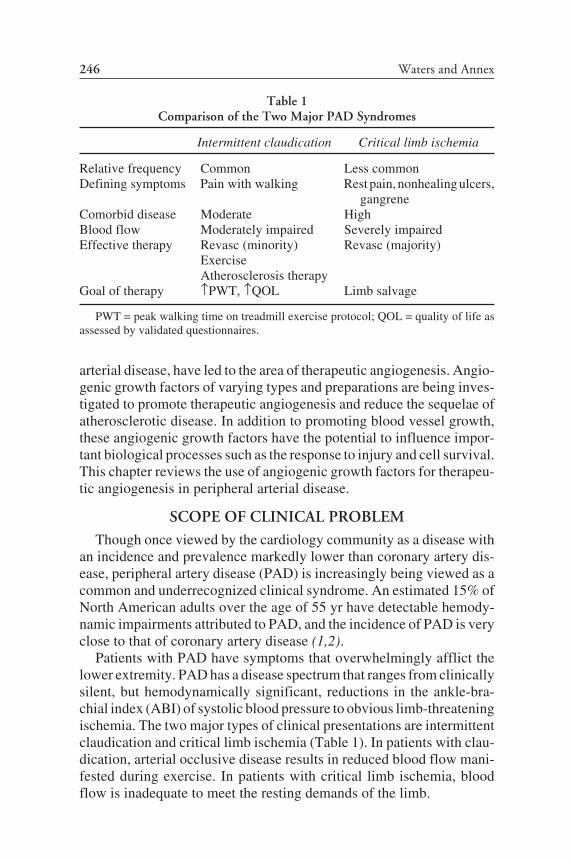
246 Waters and Annex
arterial disease, have led to the area of therapeutic angiogenesis. Angio-genic growth factors of varying types and preparations are being inves-tigated to promote therapeutic angiogenesis and reduce the sequelae ofatherosclerotic disease. In addition to promoting blood vessel growth,these angiogenic growth factors have the potential to influence impor-tant biological processes such as the response to injury and cell survival.This chapter reviews the use of angiogenic growth factors for therapeu-tic angiogenesis in peripheral arterial disease.
SCOPE OF CLINICAL PROBLEM
Though once viewed by the cardiology community as a disease withan incidence and prevalence markedly lower than coronary artery dis-ease, peripheral artery disease (PAD) is increasingly being viewed as acommon and underrecognized clinical syndrome. An estimated 15% ofNorth American adults over the age of 55 yr have detectable hemody-namic impairments attributed to PAD, and the incidence of PAD is veryclose to that of coronary artery disease (1,2).
Patients with PAD have symptoms that overwhelmingly afflict thelower extremity. PAD has a disease spectrum that ranges from clinicallysilent, but hemodynamically significant, reductions in the ankle-bra-chial index (ABI) of systolic blood pressure to obvious limb-threateningischemia. The two major types of clinical presentations are intermittentclaudication and critical limb ischemia (Table 1). In patients with clau-dication, arterial occlusive disease results in reduced blood flow mani-fested during exercise. In patients with critical limb ischemia, bloodflow is inadequate to meet the resting demands of the limb.
Table 1Comparison of the Two Major PAD Syndromes
Intermittent claudication Critical limb ischemia
Relative frequency Common Less commonDefining symptoms Pain with walking Rest pain, nonhealing ulcers,
gangreneComorbid disease Moderate HighBlood flow Moderately impaired Severely impairedEffective therapy Revasc (minority) Revasc (majority)
ExerciseAtherosclerosis therapy
Goal of therapy PWT, QOL Limb salvage
PWT = peak walking time on treadmill exercise protocol; QOL = quality of life asassessed by validated questionnaires.

Chapter 9 / Therapeutic Angiogenesis in Peripheral Arterial Disease 247
Intermittent claudication (IC), muscular leg discomfort provoked byexercise and promptly relieved by rest, is the most common manifesta-tion of PAD. Intermittent claudication, however, is not merely a diseasethat limits walking, as these patients actually have marked reductions inwalking speed, exercise tolerance, functional status, as well as signifi-cant impairments in many quality-of-life measures (3). Critical limbischemia (CLI) is a less common but more profound manifestation ofPAD. It includes a constellation of syndromes: intermittent or unremit-ting metatarsalgia even at rest (probably representing ischemic neuritis),lower extremity ulcers that fail to heal, or frank gangrene. Comparedwith patients with IC, patients with CLI tend to be older and have signifi-cantly more co-morbidity (especially diabetes mellitus) and more dif-fuse and distal obstructive atherosclerosis. Lower extremity blood flowis severely impaired in CLI, and limb loss is common without successfulrevascularization. The mortality rates in this population can approach40% by 6 mo (4).
The site of vascular obstruction to the lower extremity in PAD is oftendivided into inflow vessels (aortic and iliac arteries), conduit vessels(fermoropopliteal arteries), and run-off vessels (tibial and pedal arter-ies). Many patients have more than one level of vascular obstruction. Ingeneral, clinical syndromes do not correlate with the location of theobstruction. Additionally, the extent of atherosclerotic disease also doesnot correlate directly with symptoms. An enormous variability exists inthe severity of clinical symptoms associated with a given lesion, and thisvariability likely can be attributed to one or more of the following: (1)baseline functional status, (2) skeletal muscle metabolic “economy,” (3)adaptive collateral artery formation, and (4) other factors. Furthermore,a large number of patients with hemodynamic abnormalities consistentwith PAD (i.e., a low ABI) have “atypical” or no leg discomfort withwalking but still have marked functional impairment and poor clinicalcourses (5).
Although the primary pathophysiology of PAD is impaired perfusionto the lower extremity, all conventional PAD treatments such asantiplatelet therapy, angiotensin-cascade antagonists, HMG-CoA-reduc-tase inhibitors, and smoking cessation target general atherosclerotic riskfactor reduction (especially smoking cessation). In selected patients,structured exercise (in IC) and limb hygiene (in CLI) are of benefit. Atpresent, pharmacological therapies used in PAD do not improve perfu-sion. Pentoxifylline, although widely prescribed in the United States,has little or no clinical benefit (6). Cilostazol, a phosphodiesteraseinhibitor, offers some clinical efficacy, but its safety in patients withventricular dysfunction remains in question, and the US Food and Drug

248 Waters and Annex
Administration (FDA) has issued a black box warning for patients withany degree of heart failure (7,8). Mechanical revascularization, eithersurgically or percutaneously, can be beneficial in patients with focal or“single-segment” atherosclerotic obstruction confined to aorto-iliac orfemoropopliteal levels. Unfortunately, obstruction at multiple levels iscommon, and it is often difficult or impossible to perform completerevascularization. Even if the anatomy is amenable to surgicalrevascularization, perioperative morbidity and mortality, loss of con-duit, and graft attrition often make this approach largely justifiable onlyin cases of limb-threatening ischemia. Overall, surgical bypass usingautologous or prosthetic conduits is generally offered only topatients with CLI or severe IC. Percutaneous treatment is feasible inmany patients with PAD; however, intermediate-term (6–12 mo) recur-rent obstruction is common, especially in the large proportion of patientswith diffuse or distal disease. For these reasons, new treatment optionssuch as therapeutic angiogenesis have emerged, and such approachesdesigned to improve perfusion to the lower extremity have the potentialto dramatically impact clinical care in this disease.
ANGIOGENESIS VS THERAPEUTIC ANGIOGENESIS
Over the past two decades, tremendous advances have occurred in theunderstanding of the basic mechanisms of blood vessel growth andmaturation (9–11). Two distinct forms of blood vessel growth have beendescribed: vasculogenesis and angiogenesis. Vasculogenesis occursthrough much of embryonic vascular development where there is differ-entiation of endothelial cells from pluripotent stem cells. Once endothe-lial cells have developed, they begin to assemble into a primitive vascularnetwork, called the primary capillary plexus. Vascularization of severalorgans, including the endocardium of the heart and the dorsal aorta,occurs by vasculogenesis. In contrast, the brain, kidneys, and the devel-oping limbs are vascularized by angiogenesis, which is defined as thesprouting of new blood vessels from a pre-existing vascular network.Angiogenesis is likely the primary mechanism for most new blood ves-sel growth in the adult, whether it is due to physiological or pathologicalstimuli (12). Additionally, it appears that the same molecular mecha-nisms that mediate normal embryonic vascular development also play arole in pathological angiogenesis which has led to the use of extremecaution and careful patient selection in human clinical trials (13).
The cellular and molecular mechanisms involved in angiogenesis arequite complex and to this date remain incompletely understood. Even in

Chapter 9 / Therapeutic Angiogenesis in Peripheral Arterial Disease 249
its simplest form, angiogenesis requires a carefully orchestrated se-quence of events, both temporally and spatially, in order to form newblood vessels (14). Conceptually, endothelial and surrounding cells mustundergo a series of characteristic responses to varying stimuli includingproliferation, migration, adhesion and cell-to-cell and cell-to-matrixinteraction, matrix degredation, and capillary lumen formation. Each ofthe events must be viewed within the context of the associated diseaseprocess (e.g., angiogenesis in the process of wound repair and angiogen-esis following episodes of arterial occlusion). Finally, angiogenesis isnot a unidirectional process, but involves a balance between stimulators,inhibitors, and modulators. Therapeutic angiogenesis seeks to exploitthe phenomenon of angiogenesis in order to treat disorders of inadequatetissue perfusion (15). Growth factors known to play a role in angiogen-esis were analyzed initially, and they continue to be used as agents fortherapeutic angiogenesis.
ANGIOGENIC GROWTH FACTORS
Over the past two decades a large number of cytokine growth factorsthat stimulate endothelial cell proliferation in vitro and, either directly orindirectly, induce angiogenesis ex vivo, or modulate angiogenesis invivo, have been identified. A list of some of these angiogenic factors isincluded in Table 2. The vascular endothelial growth factor (VEGF) andfibroblast growth factor (FGF) families have been the most frequentlystudied, and these factors may be the most potent. VEGF itself is not onegene, but consists of a family of five different and distinct genes knownas VEGF-A, VEGF-B (or VEGF-3), VEGF-C (or VEGF-2), VEGF-D,and VEGF-E, as well as the closely related placental growth factor(PIGF) (16). Of these, VEGF-A is thought to be the most physiologicallyrelevant. Even within the VEGF-A gene there are four or more isoforms.These isoforms are produced by alternate splicing of the VEGF mRNAand differ in their extracellular matrix-binding properties, with the 121isoform being the weakest heparin-binding factor and the 206 isoformbeing the strongest. VEGF-A also exists in vivo as dimers—eitherhomodimers (i.e., 121:121) or heterodimers (i.e., 121:165)—and each ofthe single isoform polypeptide chains can bind to other VEGF genes.The VEGF ligands bind to one of three known VEGF receptors. Thecomplexity of the VEGF system is minimal when compared to the com-plexity of the FGF family. The FGF family contains at least 20 members,with the most significant factors being acidic FGF (FGF-1) and basicFGF (FGF-2) (17). Other important angiogenic growth factors includeplatelet-derived growth factor (PDGF), hepatocyte growth factor (HGF),and the angiopoietins.

250 Waters and AnnexT
able
2A
ngio
geni
c Fa
ctor
s, T
heir
Rec
epto
rs, a
nd U
niqu
e P
rope
rtie
s
Gro
wth
fact
orF
ull n
ame
Rec
epto
rP
rope
rtie
sH
uman
cli
nica
l tri
als?
VE
GF
-AV
ascu
lar
endo
thel
ial
VE
GF
R1
(flt
-1),
End
othe
lial
cel
l mit
ogen
;Y
es(V
EG
F,V
EG
F1)
gro
wth
fac
tor-
AV
EG
FR
2 (f
lk-1
, kdr
),re
quir
ed f
or a
ngio
/vas
culo
gene
sis;
VE
GF
R3
mul
tipl
e is
ofor
ms
VE
GF
-BV
ascu
lar
endo
thel
ial
VE
GF
R1
(flt
-1)
For
ms
dim
ers
wit
h V
EG
F-A
;Y
esgr
owth
fac
tor-
Bpr
efer
enti
ally
exp
ress
ed in
hear
t and
ske
leta
l mus
cle
VE
GF
-CV
ascu
lar
endo
thel
ial
VE
GF
R2
(flk
-1, k
dr),
May
pla
y ro
le in
lym
phan
gio-
Yes
grow
th f
acto
r-C
VE
GF
R3
gene
sis;
not
reg
ulat
ed b
y hy
poxi
aaF
GF
(F
GF
-1)
Aci
dic
fibr
obla
stF
GF
rec
epto
rE
ndot
heli
al c
ell,
fibr
obla
st,
Yes
grow
th f
acto
rsm
ooth
mus
cle
cell
mit
ogen
;is
oele
ctri
c po
int 5
.6bF
GF
(F
GF
-2)
Bas
ic f
ibro
blas
tF
GF
rec
epto
rE
ndot
heli
al c
ell,
fibr
obla
st,
Yes
grow
th f
acto
rsm
ooth
mus
cle
cell
mit
ogen
;is
oele
ctri
c po
int 9
.6H
IF1
Hyp
oxia
indu
cibl
eH
ypox
ia r
espo
nse
elem
ents
Con
stit
uiti
vely
exp
ress
ed;
Yes
fact
or-1
alp
haup
stre
am f
rom
VE
GF
bind
s V
EG
F p
rom
oter
indu
cing
and
othe
r ge
nes
VE
GF
tran
scri
ptio
nH
GF
(sc
atte
rH
epat
ocyt
e gr
owth
c-m
et ty
rosi
ne k
inas
eS
tron
gly
extr
acel
lula
rY
esfa
ctor
, SF
)fa
ctor
mat
rix
boun
dP
DG
FP
late
let-
deri
ved
PD
GF
rec
epto
rC
urre
ntly
in to
pica
l for
mY
esgr
owth
fac
tor
PIG
FP
lace
ntal
gro
wth
VE
GF
R1
(flt
-1)
neur
opil
inS
yner
gist
ic w
ith
VE
GF
No
fact
orA
ng-1
Ang
iopo
ieti
n-1
Tie
-2V
ascu
lar
bran
chin
g m
orph
ogen
esis
;N
opo
tent
iate
sef
fect
of
VE
GF
Ang
-2A
ngio
poie
tin-
2T
ie-2
Inhi
biti
on o
f va
scul
arN
ost
abil
izat
ion
250
Chapter 9 / Therapeutic Angiogenesis in Peripheral Arterial Disease 251
ANGIOGENIC GROWTH FACTORS: FORMULATIONSAND DELIVERY STRATEGIES
To date, attempts to achieve therapeutic angiogenesis have exclu-sively involved the exogenous administration of angiogenic growth fac-tors through either direct administration of recombinant proteins or theadministration of genes that encode the proteins. Protein administrationoffers more predictable and controllable delivery than gene therapy, butmany of the proteins have very short half-lives (18,19). Despite this,single-dose administration of recombinantly manufactured growth fac-tors have been shown to be effective, and the short half-life can becircumvented with repeat dosing or through pharmacological alterations(20,21). Systemic hypotension has been reported with recombinant pro-tein administration, but this can usually be managed with fluid hydra-tion (22).
Gene therapy includes the use of viral or plasmid DNA, with or with-out liposomal vectors, to promote transfection efficiency. Persistent geneexpression leads to prolonged local exposure of the angiogenic growthfactor protein, although the precise timing of this expression in humantissues is unknown. Adenoviral vectors are easily produced, have a rea-sonable transfection efficiency, and are expressed in nonproliferatingcells. The deletion of nonessential components of the adenoviral ge-nome prior to administration leads to less host inflammation, whichresults in more efficient gene delivery and expression. More recent ap-proaches to further reduce host inflammation have included the devel-opment of a fully deleted adenoviral vector that uses a helper virus toprovide essential replication and packaging proteins, as well as the de-velopment of a recombinant adeno-associated virus (AAV) (23–25).The use of nonviral techniques, including plasmid DNA and liposomalvectors, is also associated with less host inflammation. The ease of pro-duction of these nonviral vectors is appealing, but they are generallyregarded as less efficient than viral agents (26,27).
Although delivery of angiogenic agents to patients with PAD may beviewed as trivial, alternative administration routes exist. Angiogenicagents have been delivered intra-arterially or intramuscularly inpreclinical and clinical trials. Intra-arterial delivery of protein producesmore systemic growth factor exposure and, in theory, could produceunwanted toxicities. This has yet to be observed clinically. A majoradvantage of therapeutic angiogenesis approaches in the periphery vsthe myocardium is that direct intramuscular delivery to peripheral skel-etal muscle is technically simple, feasible for repeated injection strate-gies, and can be achieved without systemic exposure. However, it

252 Waters and Annex
remains to be determined whether intramuscular injections will lead to“focal” or “patchy” expression that may limit efficacy.
PRECLINICAL MODELS OF THERAPEUTICANGIOGENESIS IN PERIPHERAL ARTERIAL DISEASE
Preclinical models of hindlimb ischemia have been used in an attemptto better understand the angiogenic response in humans with PAD.Before considering the various animal models or approaches, it is criticalto recognize that, for the most part, all preclinical ischemia models as-sess an angiogenic agent’s ability to improve upon the extent of endog-enous recovery that occurs after arterial ligation. Because of this, thecontext in which the model is studied is critical (Fig. 1). For example, theendogenous angiogenic response in normal young animals is very ro-bust, and even a complete occlusion (i.e., simple arterial ligation) oftenresults in normal or near-normal limb perfusion in a relatively shortperiod of time. In this setting, the speed and magnitude of the endog-enous angiogenic response makes quantitative assessment of potentialtherapies challenging and may bring their therapeutic/clinical applica-bility into question. For this reason, preclinical models with a slower and
Fig. 1. Angiogenic response to limb ischemia.

Chapter 9 / Therapeutic Angiogenesis in Peripheral Arterial Disease 253
less complete endogenous angiogenic response to ischemia have beendeveloped. Arterial ligation followed by complete excision of the femo-ral artery produces an ischemic/normal limb flow ratio and a bluntedangiogenic response that more closely mimics PAD, and intermittentclaudication in particular (28,29). Subsequent administration of angio-genic growth factors or angiogenic gene therapy leads to more rapid andcomplete recovery, as manifested by enhanced collateral vessel devel-opment, increased capillary density, and improved calf blood pressureratios (25–27,30–39,51). When arterial ligation and excision is appliedto animal models with hypercholesterolemia, diabetes or hyperhomo-cystemia, an even more profound impairment in the endogenous angio-genic response, results, and this scenario is more analogous to criticallimb ischemia (40–44). Therapeutic angiogenesis in this setting willseek to prevent limb loss (40–44). All of the angiogenic growth factorsthat have entered human trials have been successful in some preclinicalcontext (Table 3).
CLINICAL TRIALS OF THERAPEUTIC ANGIOGENESISIN CRITICAL LIMB ISCHEMIA
The first reports of therapeutic angiogenesis in humans with PAD(CLI) were reported by Isner and colleagues, who described angiographicand histological evidence of angiogenesis after intravascular adminis-tration of VEGF plasmid DNA in a single patient with critical limbischemia (45). This report was followed by a small open-label phase Itrial that suggested efficacy following VEGF plasmid DNA administra-tion in nine patients with critical limb ischemia. The authors reported asignificant improvement in ABI (0.33 SEM ± 0.05 to 0.48 SEM ± 0.03,p = 0.02 at 12 wk) as well as new angiographically visible vessels,improved distal flow by magnetic resonance angiography, and enhancedischemic ulcer healing in the majority of patients (46). Comerota andcolleagues published preliminary results of a phase I trial using acidicFGF-1 in patients with critical limb ischemia (47). In this study, 51patients with unreconstructible peripheral arterial disease and rest painor tissue necrosis received intramuscular naked plasmid DNA encodingFGF-1 (increasing single and repeated doses). The treatment was welltolerated in all patients. Clinical outcomes in the first 15 patients tocomplete the 6-mo follow-up study demonstrated a significant reductionin pain (p < 0.001) and aggregate ulcer size (p < 0.01) that was associatedwith an increased transcutaneous oxygen pressure (p < 0.01) as com-pared with baseline values. Additionally, a significant increase in ABI(p < 0.01) was seen at 8 and 12 wk, but not at 24 wk. Taken together, these

254 Waters and Annex
preliminary trials, although small, provided sufficient evidence for thefollowing studies in intermittent claudication.
CLINICAL TRIALS OF THERAPEUTIC ANGIOGENESISIN INTERMITTENT CLAUDICATION
The first human studies using basic FGF , or FGF-2, were performedat the National Institutes of Health, where Lazarous and colleagues ad-ministered intravascular FGF-2 to 11 patients with claudication and anABI < 0.8 in a phase I, double-blind, placebo-controlled, dose escalationtrial (48). They found FGF-2 to be well tolerated, even at the highest
Table 3Preclinical Studies of Therapeutic Angiogenesis
Factor Route Model Species Outcome Ref.
VEGF im LE Rabbit angio score, flow ratio, CD, 30,31(protein) BF (microspheres)
VEGF ia LE Rabbit angio score, flow ratio, CD 32(protein)
VEGF iv LE Rabbit angio score, flow ratio, CD, 33(protein) max flow reserve
VEGF im LE Rabbit BP ratio, flow (microspheres, 26(plasmid) doppler wire)
VEGF ia LE Rabbit angio score, flow ratio, CD 34(plasmid)
VEGF im La Rat angio score, CD, flow 35(adenovirus) (microspheres)
AAV-VEGF im LE Rat CD, flow (microspheres, 24,25(adeno-asso- infrared thermography)ciated virus)
VEGF im LE Rabbit angio score, flow ratio, CD, 27(plasmid- flow (microspheres)liposome)
FGF-2 im L Rabbit CD, recovery TcPO2, muscle 36(protein) viability
rFGF-2 ia L Rat CD, flow (microspheres) 37(protein)
HIF-1 /VP16 im LE Rabbit angio score, flow ratio, CD, 51(hybird gene flow (microspheres)transfer)
aLigation in setting of recent ischemia.im = intramuscular; ia = intra-arterial; iv = intravenous; LE= ligation and excision; L=
ligation; CD = capillary density; flow ratio = ratio between ischemic and nonischemic limb.

Chapter 9 / Therapeutic Angiogenesis in Peripheral Arterial Disease 255
doses, and showed plethysmographic evidence of improved lower ex-tremity blood flow in the treated group.
The Therapeutic Angiogenesis with Recombinant Fibroblast GrowthFactor-2 for Intermittent Claudication (TRAFFIC) trial was a phase II,double-blind, placebo-controlled study comparing intra-arterial infu-sions of recombinant FGF-2 (rFGF-2) with placebo (49). One hundredand ninety patients with moderate-severe intermittent claudication, in-fra-inguinal obstructive atherosclerosis, and a resting ABI of <0.8 wererandomly assigned to receive bilateral infusions of placebo, single-bolus, or double-bolus rFGF-2. All groups demonstrated an increase inthe primary endpoint, a change in peak walking time at 90 d, which wassignificantly greater in the single-bolus group than in the placebo group(1.77 min vs 0.6 min, p = 0.026). The addition of a second bolus did notprovide additional benefit (1.54 min). The secondary endpoints of qual-ity of life and claudication onset time did not differ significantly betweenthe groups, but there was a trend toward an increase in peak walking timeat 180 d and a small but significant increase in ABI among treated pa-tients that was not seen with placebo. TRAFFIC remains the only phaseII trial of therapeutic angiogenesis to show benefit in its primary efficacymeasure.
The Regional Angiogenesis with Vascular Endothelial Growth Fac-tor in PAD (RAVE) trial was a phase II, double-blind, placebo-con-trolled trial comparing intramuscular injections of VEGF121 adenovirus(AdVEGF121) with placebo (50). One hundred and five patients withchronic, stable, predominately unilateral, intermittent claudication, andresting ABI of <0.8 were randomly assigned to receive intramuscularinjections of low-dose AdVEGF121, high-dose AdVEGF121, or placebo.Although all groups demonstrated an increase in the primary endpoint,a change in peak walking time at 12 wk, this change did not differbetween the placebo (1.8 ± 3.2 min), low-dose (1.6 ± 1.9 min), and high-dose (1.5 ± 3.1 min) groups. Furthermore, the secondary endpoints ofpeak walking time at 26 wk, quality-of-life measures, claudication onsettime, and ABI also did not differ among the groups.
FUTURE DIRECTIONS
Future approaches to achieve therapeutic angiogenesis in humans canbe categorized into four areas: new factors, alternative targets, combina-tions of factors, and the link to cell therapy. The vast majority of initialhuman studies have employed single growth factors delivered as a pro-tein or gene product. These cytokine growth factors have generally tar-geted one receptor or one family of receptors. New targets for therapeutic

256 Waters and Annex
angiogenesis are emerging, and these include transcription factors thathave the potential to modulate a number of downstream gene products;an example of this is hypoxia-inducible factor 1 (51). Angiogenesis isa complex coordinated process that involves multiple growth factors.Asahara and colleagues first demonstrated the synergy between VEGFand basic FGF in models of hindlimb ischemia (52). A recent report byCao et al. (53) demonstrated that PDGF is synergistic with basic FGF,but the synergy did not apply to PDGF and VEGF. Although complexi-ties will exist in translating this into human studies, approaches like thiswill eventually be employed.
Angiogenic growth factors may also have effects aside from simpleproliferation. For example, VEGF is a survival factor for microvascularendothelial cells and can permit the survival of these cells in the face ofinjury (54,55). A recent study from our group demonstrated that hindlimbischemia induces apoptosis in peripheral skeletal muscle and angiogenicgrowth factors may ultimately influence apoptosis and thereby exertbeneficial effects in targeted skeletal muscle (56). Finally, the classicstudy by Asahara and Isner (57) demonstrated that circulating cells con-tribute to the angiogenic response. Currently, it is well established thatangiogenic growth factors have some interplay with circulating or bonemarrow–derived progenitor cells (58). Initial studies have demonstratedthe potential feasibility of cell therapy to mitigate the sequelea of periph-eral arterial disease (59), but definitive studies are still pending.
REFERENCES1. Criqui MH, Fronek A, Barrett-Connor E, et al. The prevalence of peripheral arterial
disease in a defined population. Circulation 1985;71:510–515.2. Hirsch AT, Criqui MH, Treat-Jacobson D, et al. Peripheral arterial disease detec-
tion, awareness, and treatment in primary care. JAMA 2001;286:1317–1324.3. Hiatt WR, Hirsch AT, Regensteiner JG, et al. Clinical trials for claudication. Assess-
ment of exercise performance, functional status, and clinical endpoints. VascularClinical Trialist. Circulation 1995;92:614–621.
4. Management of peripheral arterial disease (PAD). TransAtlantic Inter-Society Con-sensus (TASC). Section D: chronic critical limb ischaemia. Eur J Vasc EndovascSurg 2000;Suppl A:S144–S243.
5. McDermott MM, Greenland P, Liu K, et al. Leg symptoms in peripheral arterialdisease: associated clinical characteristics and functional impairment. JAMA2001;286:1599–1606.
6. Radack K, Wyderski RJ. Conservative management of intermittent claudication.Ann Intern Med 1990;113:135–146.
7. Money SR, Herd JA, Issacsohn JL, et al. Effect of Cilastazol on walking distancesin patients with intermittent claudication caused by peripheral vascular disease. JVasc Surg 1998;27:267–275.
8. Beebe HG, Dawson DL, Cutler BS, et al. A new pharmacological treatment forintermittent claudication: results of a randomized multicenter trial. Arch Intern Med1999;159:2041–2050.

Chapter 9 / Therapeutic Angiogenesis in Peripheral Arterial Disease 257
9. Carmeliet P, Conway EM. Growing better blood vessels. Nat Biotechnol2001;19:1019–1020.
10. Conway EM, Collen D, Carmeliet P. Molecular mechanisms of blood vessel growth.Cardiovasc Res 2001;49:507–521.
11. Folkman J. Clinical applications of research on angiogenesis. N Engl J Med1995;333:1757–1763.
12. Folkman J. Angiogenesis in cancer, vascular, rheumatoid, and other disease. NatMed 1995;1:27–31.
13. Folkman J. Tumor angiogenesis: therapeutic implications. N Engl J Med1971;285:1182–1186.
14. Jain RK, Carfmeliet P. Anti- and pro-angiogenesis therapy. Sci Am 2001;285:38–45.15. Waltenberger J. Modulation of growth factor action: Implications for the treatment
of cardiovascular diseases. Circulation 1997;96:4083–4094.16. Veikkola T, Karkkainen M, Claesson-Welsh L, et al. Regulation of angiogenesis via
vascular endothelial growth factor receptors. Cancer Res 2000;60:203–212.17 Simons M, Annex BH, Laham RJ, et al. Pharmacological treatement of coronary
artery disease with recombinant fibroblast growth factor-2: double-blind, random-ized, controlled, clinical trial. Circulation 2002;105:788–793.
18. Laham RJ, Rezaee M, Post M, et al. Intracoronary and intravenous administrationof basic fibroblast growth factor: myocardial and tissue distribution. Drug MetabDispos 1999;27:821–826.
19. Eppler SM, Combs DL, Henry TD, et al. A target-mediated model to describe thepharmacokinetics and hemodynamic effects of rhVEGF in humans. Clin PharmacolTherap 2002;72:20–32.
20. Lopez JJ, Edelman ER, Stamler A, et al. Angiogenic potential of perivascularydelivered aFGF in a porcine model of chronic myocardial ischemia. Am J Physiol1998;274:H930–H936.
21. East MA, Landis DI, Thompson MA, et al. Effect of single dose of intravenousheparin on plasma levels of angiogenic growth factors. Am J Cardiol 2003;91:1234–1236.
22. Hariawala MD, Horowitz JR, Esakof D, et al. VEGF improves myocardial bloodflow but produces EDRF-mediated hypotension in porcine hearts. J Surg Res1996;63:77–82.
23. Maione D, Della Rocca C, Gianetti P, et al. An improved helper-dependent aden-oviral vector allows persistent gene expression after intramuscular delivery andovercomes preexisting immunity to adenovirus. Proc Natl Acad Sci USA2001;98:5986–5991.
24. Byun J, Heard JM, Huh JE, et al. Efficient expression of the vascular endothelialgrowth factor gene in vitro and in vivo, using an adeno-associated virus vector. J MolCell Cardiol 2001;33:295–305.
25. Shimpo M, Ikeda U, Maeda Y, et al. AAV-mediated VEGF gene transfer intoskeletal muscle stimulates angiogenesis and improves blood flow in a rat hindlimbischemia model. Cardiovasc Res 2002;53:993–1001.
26. Tsurumi Y, Takeshita S, Chen D, et al. Direct intramuscular gene transfer of nakedDNA encoding vascular endothelial growth factor augments collateral developmentand tissue perfusion. Circulation 1996;94:3281–3290.
27. Gowdak LH, Poliakova L, Li Z, et al. Induction of angiogenesis by cationic lipid-mediated VEGF165 gene transfer in the rabbit ischemic hindlimb model. J VascSurg 2000;32:342–352.
28. Couffinhal T, Silver M, Zheng LP, et al. Mouse model of angiogenesis. Am J Pathol1998;152:1667–1679.
258 Waters and Annex
29. Hong JH, Bahk YW, Suh JS, et al. An experimental model of ischemia in rabbithindlimb. J Korean Med Sci 2001;5:630–635.
30. Pu LQ, Sniderman AD, Brassard R, et al. Enhanced revascularization of the is-chemic limb by angiogenic therapy. Circulation 1993;88:208–215
31. Takeshita S, Pu LQ, Stein LA, et al. Intramuscular administration of vascular endot-helial growth factor induces dose-dependent collateral artery augmentation in arabbit model of chronic limb ischemia. Circulation 1994;90:II228–II234.
32. Takeshita S, Zheng LP, Brogi E, et al. Therapeutic angiogenesis. A single intrarterialbolus of vascular endothelial growth factor augments revascularization in a rabbitischemic hind limb model. J Clin Invest 1994;93:662–670.
33. Bauters C, Asahara T, Zheng LP, et al. Site-specific therapeutic angiogenesis aftersystemic administration of vascular endothelial growth factor. J Vasc Surg1995;21:314–324.
34. Takeshita S, Weir L, Chen D, et al. Therapeutic angiogenesis following arterial genetransfer of vascular endothelial growth factor in a rabbit model of hindlimb is-chemia. Biochem Biophys Res Commun 1996;227:628–635.
35. Mack CA, Magovern CJ, Budenbender KT, et al. Salvage angiogenesis induced byadenovirus-mediated gene transfer of vascular endothelial growth factor protectsagainst ischemic vascular occlusion. J Vasc Surg 1998;27:699–709.
36. Baffour R, Berman J, Garb JL, et al. Enhanced angiogenesis and growth of collateralsby in vivo administration of recombinant basic fibroblast growth factor in a rabbitmodel of acute lower limb ischemia: dose-response effect of basic fibroblast growthfactor. J Vasc Surg 1992;16:181–191.
37. Yang HT, Deschenes MR, Ogilvie RW, et al. Basic fibroblast growth factor in-creases collateral blood flow in rats with femoral arterial ligation. Circ Res.1996;79:62–69.
38. Murohara T, Asahara T, Silver M, et al. Nitric oxide synthase modulates angiogen-esis in response to tissue ischemia. J Clin Invest 1998;101:2567–2578.
39. Rissanen TT, Markkanene JE, Arve K, et al. Fibroblast growth factor 4 inducesvascular permeability, angiogenesis and arteriogenesis in a rabbit hindlimb ischemiamodel. FASEB J 2003;1:100–102.
40. Rivard A, Silver M, Chen D, et al. Rescue of diabetes-related impairment of angio-genesis by intramuscular gene therapy with adeno-VEGF. Am J Pathol1999;154:355–363.
41. Van Belle E, Rivard A, Chen D, et al. Hypercholesterolemia attenuates angiogenesisbut does not preclude augmentation by angiogenic cytokines. Circulation1997;96:2667–2674.
42. Duan J, Muohara T, Ikeda H, et al. Hyperhomocysteinemia impairs angiogenesis inresponse to hindlimb ischemia. Atherioscler Thromb Vasc Biol 200;20:2579–2585.
43. Duan J, Murohara T, Ikeda H, et al. Hypercholesterolemia inhibits angiogenesis inresponse to hindlimb ischemia; nitric oxide-dependent mechanism. Circulation2000;102(Suppl III):III370–III376.
44. Couffinhal T, Silver M, Kearney M, et al. Impaired collateral vessel developmentassociated with reduced expression of vascular endothelial growth factor in ApoE-/- mice. Circulation 1999;99:3188–3198.
45. Isner JM, Pieczek A, Schainfeld R, et al. Clinical evidence of angiogenesis follow-ing arterial gene transfer of phVEGF165. Lancet 1996;348:370–374.
46. Baumgartner I, Pieczek A, Manor O, et al. Constitutive expression of phVEGF165after intramuscular gene transfer promotes collateral vessel development in patientswith critical limb ischemia. Circulation 1988;97:1114–1123.

Chapter 9 / Therapeutic Angiogenesis in Peripheral Arterial Disease 259
47. Comerota AJ, Throm RC, Miller KA, et al. Naked plasmid DNA encoding fibroblastgrowth factor type I for the treatment of end-stage unreconstructible lower extrem-ity ischemia: preliminary results of a phase I trial. J Vasc Surg 2002;35:930–936.
48. Lazarous DF, Unger EF, Epstein SE, et al. Basic fibroblast growth factor in patientswith intermittent claudication: results of a phase I trial. JACC 2000;36:1239–1244.
49. Lederman RG, Mendelsohn FO, Anderson RD, et al. Therapeutic angiogenesis withrecombinant fibroblast growth factor-2 for intermittent claudication (the TRAFFICstudy): a randomised trial. Lancet 2002;359:2053–2058.
50. Rajagopalan S, Mohler ER, Lederman RJ, et al. A phase II randomized double blindcontrolled study of adenoviral delivery of VEGF121 in patients with disablingintermittent claudication. Regional angiogenesis with vascular endothelial growthfactor (VEGF) in peripheral arterial disease. Circulation 2003 (in press).
51. Vincent KA, Shyu KG, Luo Y, et al. Angiogenesis is induced in a rabbit model ofhindlimb ischemia by naked DNA encoding an HIF-1(/VP16 hybrid transcriptionfactor. Circulation 2000;102:2266–2261.
52. Asahara T, Bauters C, Zheng OP, et al. Synergistic effect of vascular endothelialgrowth factor and basic fibroblast growth factor on angiogenesis in vivo. Circula-tion 1995;93(9 Suppl):II365–II371.
53. Cao R, Brakenhielm E, Pawliuk R, et al. Angiogenic synergism, vascular stabilityand improvement of hind-limb ischemia by a combination of PDGF BB and FGF-2. Nat Med 2003;5:604–613.
54. Spyridopolous I, Brogi E, Kearney M, et al. Vascular endothelial growth factorinhibits endothelial cell apoptosis induced by tumor necrosis factor-alpha:balancebetween growth and death signals. J Mol Cell Cardiol 1997;29:1321–1330.
55. Dimmeler S, Zeiher AM. Endothelial cell apoptosis in angiogenesis and vesselregression. Circ Res 2000;434–439.
56. Dai Q, Thompson MA, Pippen AM, et al. Alterations in endothelial cell prolifera-tion and apoptosis contribute to vascular remodeling following hindlimb ischemiain rabbits. Vasc Med 2002;7:87–93.
57. Iwaguro H, Yamaguchi J, Kalka C, et al. Endothelial progenitor cell vascular-endothelial growth factor gene transfer for vascular regeneration. Circulation2002;105:732–738.
58. Luttun A, Carmeliet G, Carmeliet P. Vascular progenitors:from biology to treat-ment. Trends Cardiovasc Med 2002;12:84–92.
59. Tateishi-Yuyama E, Matsubara H, Murohara T, et al. Therapeutic angiogenesis forpatients with limb ischaemia by autologous transplantation of bone-marrow cells:a pilot study and a randomised controlled trial. Lancet 2002;360:427–435.

Chapter 10 / Bone Marrow Cell Transplantation 261
261
From: Contemporary Cardiology: Angiogenesis and Direct Myocardial RevasularizationEdited by: R. J. Laham and D. S. Baim © Humana Press Inc., Totowa, NJ
INTRODUCTION
Coronary artery disease remains the leading cause of death in devel-oping countries. In the United States, up to 12 million Americans havea history of myocardial infarction, angina pectoris, or both (1). Althoughrecent advances in medical treatment and interventional procedures havereduced the mortality in patients with coronary artery disease (2), thenumber of patients with refractory myocardial ischemia and congestiveheart failure is rapidly increasing. In a significant proportion of these
10 Bone Marrow CellTransplantation for MyocardialRegeneration and TherapeuticAngiogenesis
Hung-Fat Tse, MD, Pui-Yin Lee, MBBS,and Chu-Pak Lau, MD
CONTENTS
INTRODUCTION
BONE MARROW STEM CELLS
KINETICS OF ADULT BONE MARROW STEM CELLS
BONE MARROW AS A CELL SOURCE FOR MYOCARDIAL
REGENERATION AND THERAPEUTIC ANGIOGENESIS
CLINICAL UTILIZATION OF BONE MARROW CELL
TRANSPLANTATION
CONCLUSIONS

262 Tse et al.
patients, percutaneous coronary intervention or surgical bypass revascu-larization is either not feasible or incomplete as a result of patients’comorbidity, total occlusion, or poor distal vessels. After a myocardialinfarction, some cardiomyocytes are lost and others hibernate because ofinsufficient myocardial perfusion. Therefore, therapeutic approachesaimed at promoting blood vessel formation (angiogenesis) and growingnew heart muscle fibers (myocardial regeneration) are attractive alterna-tives. Accumulating evidence suggests that bone marrow cells have thepotential of contributing to tissue revascularization and cardiac regen-eration after myocardial injury. This chapter summarizes the currentstatus of bone marrow cell transplantation for myocardial regenerationand therapeutic angiogenesis.
BONE MARROW STEM CELLS
With the recent advances in stem cell biology, it has been shown thatadult bone marrow contains several stem cell populations that cantransdifferentiate into various mesenchymal and nonmesenchymal celltypes (3,4). These stem cells can be broadly divided into two main groupsbased on their expression of cell marker CD34: CD34+ hematopoieticstem/progenitor cells and CD34–mesenchymal stem cells (MSCs) (Fig. 1).
The CD34+ hematopoietic stem/progenitor cells are precursors ofblood and endothelial cells in adult bone marrow. The hematopoieticstem cells (HSCs) refer to those stem cell populations that can self-renewand differentiate into more mature progenitor cells in the bone marrowand provide permanent long-term reconstitutionof the entire hematopoi-etic system. In contrast, hematopoietic progenitor cells of bone marrowhave a limited capacity for self-renewal and differentiation and can onlysustain hematopoiesis for a short-term period. A small population ofendothelial progenitor cells (EPCs) has also been identified in the bonemarrow, which can be mobilized to the circulatory system and contributeto the postnatal neovascularization process (5).
The CD34– MSCs are precursors of stromal cells and appear to havemultilineage differentiation capacity in vitro (6). However, there is noadequate cell marker to allow selection of purified cell populations, andthus their phenotypes remain unclear. Cardiomyocytes can be differen-tiated from MSCs after exposure to 5-azacytidine, a cytosine analogcapable of alterating gene expression that regulates differentiation (7).Recently, another subset of bone marrow stromal cells, referred to asmultipotent stem cells, has been described on the basis of their capacityto differentiate into multiple lineages (8).
Recent studies have shown that the adult heart is not a terminallydifferentiated organ, although the rate of myocardial cell turnover is

Chapter 10 / Bone Marrow Cell Transplantation 263
Fig
. 1. O
rigi
n an
d di
ffer
enti
atio
n of
ste
m c
ells
in a
dult
bon
e m
arro
w. R
epre
sent
ativ
e an
tige
nici
ties
to s
tem
/pro
geni
tor
cell
s ar
e sh
own
(+, p
osit
ive;
–, n
egat
ive)
.
263

264 Tse et al.
extremely low (9). The origin of stem cells in regenerating myocardiumremains unclear. Emerging data suggest that bone marrow cells have theability to differentiate into stem and progenitor cells that mature intofunctional cells in a variety of tissues including myocardium (10). Ex-perimental studies have suggested that bone marrow-derived cells cantransdifferentiate into liver, brain, skeletal muscle, heart, lung, kidney,gut, and skin cells (11). Many such studies are based on a sex-mis-matched hematopoietic stem cell transplantation model using the Ychromosome as a marker (3,4). Furthermore, recent clinical studies havealso demonstrated male chimerism in heart allografts from female do-nors (12,13). It has been postulated that in addition to the effect of directmigration of cardiomyocytes from adjacent recipient tissue into the al-lograft, the circulating stem cells from the bone marrow may have afavorable role in ventricular remodeling after transplantation. In addi-tion to transdifferentiation of bone marrow stem cells, cell fusion alsoappears to account for a proportion of the presence of cells that showdonor characteristics in solid organ tissue (14,15). However, the relativeimportance of stem cell fate transition and cell fusion is unknown.
KINETICS OF ADULT BONE MARROW STEM CELLS
It has been postulated that circulating bone marrow-derived stemcells may contribute to tissue regeneration and repair in solid organs (4).Myocardial injury that causes changes in the microenvironment mayplay an important role in stem cell recruitment. Previous studies havedemonstrated that EPCs were mobilized from the hematopoietic systemafter acute myocardial infarction (16). However, the mechanisms bywhich circulating bone marrow-derived stem cells are recruited into theheart and the subsequent cardiomyocyte generation is not fully understood.
Several cytokines may regulate the proliferation, mobilization, andhoming of bone marrow stem cells into the injured solid-organ. Stem cellfactor (SCF) is expressed in HSCs and is a ligand of c-kit (a tyrosinekinase receptor). It may regulate the migration of HSC duringembryonicdevelopment as well as in response to myocardial injury. Recent studieshave demonstrated that SCF, c-kit, and metalloproteinase-9 play animportant role in the mobilization of stem and progenitor cells from thebone marrow after myocardial necrosis (17). Granulocyte-macrophagecolony-stimulating factor (GM-CSF) is a well-known stimulator of he-matopoietic stem/progenitor cells. However, experimental studies alsodemonstrated that GM-CSF mobilizes EPCs to severely ischemic tissueand enhances neovascularization (18). Similar modulation of EPC kinet-ics has been observed in response to other hematopoietic cytokines, such

Chapter 10 / Bone Marrow Cell Transplantation 265
as granulocyte colony-stimulating factor (G-CSF) and stromal-derivedfactor (SDF)-1 (19,20). The roles of vascular endothelial growth factors(VEGFs) and the tyrosine kinase VEGF receptors in endothelial cellproliferation and differentiation during neovascularization are well de-scribed (21). However, evidence is emerging that VEGF-mediatedmobilization of bone marrow-derived EPCs also contributes toneovascularization (22,23).
BONE MARROW AS A CELL SOURCEFOR MYOCARDIAL REGENERATIONAND THERAPEUTIC ANGIOGENESIS
As discussed above, bone marrow contains multiple types of stemcells and progenitor cells, which can potentially be used for cellulartherapy for myocardial regeneration and therapeutic angiogenesis (Fig. 2).
Myocardial RegenerationAcute myocardial infarction or chronic ischemic heart disease results
in the loss of cardiomyocytes and vasculature that is difficult to regen-erate. Furthermore, peri-infarct tissue undergoes apoptosis as a result ofischemia and acts as a barrier to the incorporation of vascular or myocar-dial cells to damaged tissues. Experimental studies have shown that bone
Fig. 2. Therapeutic role of different types of bone marrow stem cells in myocar-dial regeneration and angiogenesis.

266 Tse et al.
marrow-derived stem cells can be transdifferentiated into cardio-myocytes in vitro and in vivo. Treatment of bone marrow cells with 5-azacytidine resulted in the formation of a cell line that, upon furtherdifferentiation, gave rise to cells with spontaneous electrical andcontractile activity and other molecular attributes of differentiatedcardiomyocytes (7). In animal models of myocardial infarction, directintramyocardial injection of c-kit expressing bone marrow stem cellsinto the sites of myocardial infarcts formed myocardium comprised ofnew myocytes as well as endothelial and smooth muscle cells (24,25).Myocardial regeneration was not observed in hearts that were trans-planted with the subpopulation of c-kit– bone marrow cells known to bedevoid of stem cells. A concomitant improvement of cardiac hemody-namic and angiogenesis, and reduction in fibrosis and apoptosis wereobserved in hearts transplantedwith c-kit+ bone marrow stem cells com-pared with negative control mice (25) (Fig. 3). Furthermore, bone mar-row-derived myocytes and endothelial cells were observed in theinfarcted mouse heart following bone marrow reconstitution with CD34–side-population bone marrow hematopoietic stem cells (26). In fact, thissubpopulation of stem cells may represent a more primitive type of stemcell that can differentiate into either mesenchymal stem cells or endot-helial progenitor cells.
Fig. 3. Potential beneficial mechanisms of bone marrow cell therapy aftermyocardial infarction. Angiogenesis, decreased apoptosis, myocardial regen-eration, and reduced fibrosis can preserve the myocardial function and diminishor reverse negative left ventricular remodeling after myocardial infarction.

Chapter 10 / Bone Marrow Cell Transplantation 267
MCSs isolated from bone marrow also appeared to undergo differen-tiation into cardiomyocytes (27). Furthermore, cardiomyocytes derivedfrom mesenchymal stem cells transplanted into ventricular scar tissuehave shown to integrate with the myocardium and improve left-ventricu-lar function (28,29). Several studies also supported the notion that adultbone marrow stem cells can migrate into the heart for myocardial regen-eration. Administration of cytokines can expand and mobilize bonemarrow stem cells into circulation. A 250-fold increase in circulatingbone marrow stem cells was induced in mice after myocardial infarctionwith the combined use of SCF and G-CSF (24). This cytokine therapyresulted in a new brand of myocardium, consisting of myocytes andblood vessels, occupying 70% of the infarcted area, and improving car-diac hemodynamic functions.
These experiments demonstrated the capacity of adult bone marrowstem cells to give rise to new myocytes, endothelial cells, and smoothmuscle cells in ischemic myocardium. However, the population of bonemarrow stem cells responsible for the generation of these several myo-cardial cell types remains undefined. A recent experimental study failedto show any evidence of myocardial regeneration after transplantation ofautologous fresh bone marrow into infarcted myocardium (30). Thishighlighted the potential importance of the timing as well as the compo-sition of the bone marrow stem cells used for myocardial regeneration.The fate of the transplanted bone marrow stem cells may be affected bythe local microenvironment. During the early phase of myocardial inf-arction or in the presence of residual ischemia at the peri-infarct tissue,the local myocardial environment may harbor the appropriate signal fordriving the bone marrow stem cells toward an endothelial and/orcardiomyogenic differentiation pathway. Indeed, experimental studieshave shown evidence of myocardial regeneration after transplantation ofbone marrow cells early after myocardial infarction (24–26). However,the local cues are likely to be lost at the late stage of chronic scarring orin the absence of active ischemia. Currently, there is no evidence of newcardiomyocyte formation after bone marrow transplantation into an oldfibrous postinfarct scar. Furthermore, the composition and/or the con-centration of different types of bone marrow stem cells in the fresh bonemarrow may not be optimal for myocardial regeneration. A sufficientamount of cardiomyocyte/mesenchymal stem cells will also require thepresence of endothelial progenitor cells to provide vascular structuresfor the supply of oxygen and nutrients to both the chronically ischemic,endogenous myocardium and the newly implanted cardiomyocytes (Fig.2). Furthermore, recent studies have shown the existence of adult cardiacstem cells, which are multipotent and contribute to myocardial regenera-tion (31).

268 Tse et al.
Therapeutic AngiogenesisRecently, EPCs were successfully isolated from circulating mono-
nuclear cells (MNCs) using VEGFR2, CD34, and CD133 antigens sharedby both embryonic EPCs and HSCs in adults. EPCs are thought to sharecommon stem/progenitor cells with HSCs and to derive from bonemarrow (Fig. 1). In the presence of sufficient density and appropriategrowth factors, EPCs can differentiate into endothelial-like cells thataggregate into capillary-like structures. Experimental studies haveshown that EPCs functionally contribute to neoangiogenesis during woundhealing, limb ischemia, postmyocardial infarction, endothelialization ofvascular grafts, and atherosclerosis (32). This is consistent withvasculogenesis, a critical paradigm well described for embryonicneovascularization, but recently proposed in adults.
Recent studies with animal bone marrow transplantation models inwhich bone marrow donor-derived EPCs could be distinguished have shownthat the contribution of EPCs to endothelialization and neovascularization(32,33). In chronic hindlimb ischemic models, infusion of EPCs derivedfrom bone marrow, cord blood, or peripheral blood enhanced the forma-tion of new vasculature and improved perfusion (4,32,33). EPCs alsoinduced neovascularization after myocardial infarction. In a rat model ofmyocardial infarction, injection of EPCs isolated from rat or humanperipheral blood (after treatment with G-CSF) induced neovascularization,reduced infarct size, and preserved cardiac function (34,35). Further-more, direct intramyocardial injection of autologous bone marrow cellsin the swine model of chronic ischemic myocardium also inducedneovascularization and improved myocardial function (36,37). Althoughthe mechanism remains unclear, these experimental studies suggest thatautologous bone marrow cells could augment neovascularization in is-chemic myocardium mainly through the production of angiogenicgrowth factors and less through the differentiation of a portion of thecells into EPCs/ECs in situ (Fig. 4). Furthermore, the improvement incardiac function in these studies without evidence of myocardial regen-eration was attributed to the improvement in myocardial perfusion, whichin turn prevented cardiomyocyte apoptosis at the peri-infarct zone anddecreased left ventricular remodeling (Fig. 3).
CLINICAL UTILIZATION OF BONE MARROW CELLTRANSPLANTATION
Clinical Approach of TransplantationThe delivery route of stem cells may have a critical role in the success
of bone marrow cell therapy for myocardial regeneration and therapeutic

Chapter 10 / Bone Marrow Cell Transplantation 269
Fig. 4. Two proposed mechanisms of neovascularization by bone marrow celltransplantation. First, hematopioetic stem cells (HSCs) dfferentiate into endot-helial cells due to the local cue in the ischemic myocardium and secret cytokinesto induce neovascularization (upper panel). Alternatively, both the HSCs andendothelail progenitor cells (EPCs) are transplanted into ischemic myocardium.The EPCs differentiate into endothelial cells as a result of local cue in theischemic myocardium and the HSCs secret cytokines to induce neovascu-larization (lower panel).
angiogenesis. There are two clinical strategies for delivering bone mar-row stem cells into the ischemic and/or infarcted myocardium. Oneapproach is based on direct delivery of bone marrow stem cells to theheart via intramyocardial or intracoronary injection. The other approachis based on the in vivo availability of a pool of systemic and circulatingbone marrow stem cells that can be mobilized by cytokine therapy to theheart. The advantages and disadvantages of the two potential clinicalstrategies for myocardial regeneration and angiogenesis are summa-rized in Table 1.
It is now established that after myocardial infarction, there is sponta-neous mobilization of bone marrow EPCs to the circulation (16). Thisraises the questions of why mobilization of these cells does not automati-cally result in myocardial regeneration and angiogenesis and what is therole of delivering the bone marrow cells into the myocardium. It has beenpostulated that the EPCs mobilized during the acute phase of tissueinjury have not achieved the complete maturation necessary for incor-poration into the ischemic myocardium. Furthermore, the lack of bloodsupply to the ischemic tissue after occlusion of the coronary artery reduces

270 Tse et al.
the number of EPCs that arrive at the sites of myocardial injury. Finally,other bone marrow cells in addition to EPCs may also be required toachieve an optimal response (32). Direct introduction of bone marrowstem cells into the compromised zone by direct intramyocardial injec-tion or by intracoronary infusion may circumvent these hurdles. How-ever, systemic delivery of bone marrow stem cells may contribute totumorigenesis, atherosclerosis, or retinopathy and this strategy shouldbe used with utmost caution (38).
Mobilization of bone marrow stem cells with cytokine therapy isappealing as a potential therapy as it is easy to administer and applies toa larger patient population. In clinical hematological practice, short-
Table 1Advantages and Disadvantage of Potential Clinical Strategies
for Bone Marrow Therapy
Strategy Advantage Disadvantage
Direct Delivery
IntramyocardialOpen heart surgery Applicable to patients who Risk of mortality and mor-
need open heart surgery bidity of open heart surgeryAllows direct visualization of
the site of injectionCatheter-based Avoids the risk of open heart Specialized catheters and
approach surgery imaging technology neededShort-term safety provenClinical trials ongoing
Intracoronary Possible wide use in catheter- Efficacy of cell delivery toization laboratory the myocardium uncertain
Not applicable to patientswith an occluded artery
Risk of systemic administra-tion unclear
Intravenous Avoids the risk of cardiac Low efficacy of cell deliverycatheterization procedure Not applicable to patients
with an occluded arteryRisk of systemic administra-
tion unclear
Mobilization
Cytokine therapy Avoids the cost and risk asso- Uncertainty regarding riskciated with bone marrow cell and timingharvest and preparation
Widespread practicality

Chapter 10 / Bone Marrow Cell Transplantation 271
term administration of G-CSF in healthy humans is not associated withan increased risk of a coronary event (39). On the other hand, there is nosuitable animal model to test the safety and feasibility of this approachin the presence of coronary artery disease. In patients with acute coro-nary syndrome, multiple unstable coronary plaques may exist in thecoronary artery. Theoretically, these plaques might be destabilized by anincreased number of circulating leukocytes after cytokine therapy. In-deed, an elevated white cell count does predict a poorer prognosis inpatients with acute myocardial infarction (40). Furthermore, hypervis-cosity resulting from an increased number of circulating leukocytes mayalso lead to a worsening of the myocardial perfusion in patients withsevere coronary artery disease. Therefore, the short-term safety of thistreatment approach remains a concern.
Early Clinical ExperienceBased on the encouraging results from the experimental models, di-
rect introduction of bone marrow cells into the peri-infarct region hasbeen investigated as a means to facilitate revascularization. This proce-dure, known as cellular cardiomyoplasty, involves either directintramyocardial injection or intracoronary arterial delivery of stem cellsinto the peri-infarct area. Clinical investigation of patients with bothchronic myocardial ischemia and myocardial infarction is currently inthe early stage of nonrandomized testing of safety and feasibility (Table2). In human subjects, autologous bone-marrow mononuclear cells weredelivered either by using direct intramyocardial injection during coro-nary artery bypass surgery (41) or as guided by electromechanical map-ping technique (42–44) or by intracoronary infusion into the coronaryarteries feeding the infarcted and ischemic tissue (45,46).
Recently, our group has reported the first human experience of cath-eter-based direct intramyocardial injection of autologous bone marrowmononuclear cells in eight patients with end-stage ischemic heart disease(42). The procedure was guided by using three-dimensional nonfluoroscopicelectromechanical mapping (NOGA™ system, Biosense-Webster) (47).The use of the electromechanical system mapping can allow us to targetischemic regions for bone marrow cell injection (Fig. 5). No acute com-plication was observed after the procedure. After 3 mo follow-up, thenumber of anginal episodes and nitroglycerin tablet usage decreased.Magnetic resonance imaging (MRI) 90 d after procedure demonstratedimproved wall motion, wall thickness and myocardial perfusion at thetarget region (Figs. 6, 7). During follow-up, none of our patients experi-enced ventricular arrhythmias or developed new tumors or myocardialscar on MRI. Subsequently, reports from other investigators (43,44) usinga similar method have also suggested a beneficial effect.

272 Tse et al.T
able
2C
linic
al T
rial
s of
Aut
olog
ous
Blo
od-
or B
one
Mar
row
(B
M)-
Der
ived
Cel
l Tra
n spl
anta
tion
for
Isch
emic
Hea
rt D
isea
se
No.
of
Aut
hors
(re
f.)pa
tien
tsT
ype
of B
M c
ell
Rou
te o
f adm
inis
trat
ion
Cli
nica
l out
com
e
Myo
card
ial I
sche
mia
Ham
ano
et a
l.,5
BM
mon
onuc
lear
cel
lsD
irec
t int
ram
yoca
rdia
l inj
ecti
on d
urin
gIm
prov
ed in
myo
card
ial p
erfu
sion
2001
(41)
coro
nary
art
ery
bypa
ss g
raft
sur
gery
Tse
et a
l.,8
BM
mon
onuc
lear
cel
lsC
athe
ter-
base
d in
tram
yoca
rdia
l inj
ecti
onIm
prov
ed in
sym
ptom
s, m
yoca
rdia
l20
03 (
42)
guid
ed b
y el
ectr
omec
hani
cal m
appi
ngpe
rfus
ion
and
func
tion
Fuc
hs e
t al.,
10F
resh
asp
irat
ed B
M c
ells
Cat
hete
r-ba
sed
intr
amyo
card
ial i
njec
tion
Impr
oved
in s
ympt
oms
2003
(43
)gu
ided
by
elec
trom
echa
nica
l map
ping
and
myo
card
ial p
erfu
sion
Per
in e
t al.,
14B
M m
onon
ucle
ar c
ells
Cat
hete
r-ba
sed
intr
amyo
card
ial i
njec
tion
Impr
oved
in m
yoca
rdia
l per
fusi
on20
03(4
4)gu
ided
by
elec
trom
echa
nica
l map
ping
and
sym
ptom
s
Myo
card
ial I
nfar
ctio
n
Str
auer
et a
l.,10
BM
mon
onuc
lear
cel
lsIn
trac
oron
ary
inje
ctio
n af
ter
perc
utan
e-D
ecre
ased
infa
rct s
ize,
impr
oved
2002
(45)
ous
coro
nary
inte
rven
tion
myo
card
ial f
unct
ion
and
perf
usio
nA
ssm
us e
t al.,
20B
M m
onon
ucle
ar c
ells
Intr
acor
onar
y in
ject
ion
afte
r pe
rcut
ane-
Impr
oved
myo
card
ial f
unct
ion,
2002
(46
)(n
= 9
) or
per
iphe
ral
ous
coro
nary
inte
rven
tion
impr
oved
reg
iona
l wal
l mot
ion
inbl
ood-
deri
ved
pro-
infa
rct z
one;
sim
ilar
eff
icac
y of
geni
tor
cell
s (n
= 1
1)bl
ood-
deri
ved
and
BM
-der
ived
prog
enit
or c
ells
Sta
mm
et a
l.,6
Pur
ifie
d B
M–d
eriv
edD
irec
t int
ram
yoca
rdia
l inj
ecti
on d
urin
gIm
prov
ed m
yoca
rdia
l fun
ctio
n an
d20
03(4
7)he
mat
opoi
etic
ste
mco
rona
ry a
rter
y by
pass
gra
ft s
urge
ryre
gion
al w
all m
otio
n in
infa
rct z
one
cell
s (A
C13
3+)
272
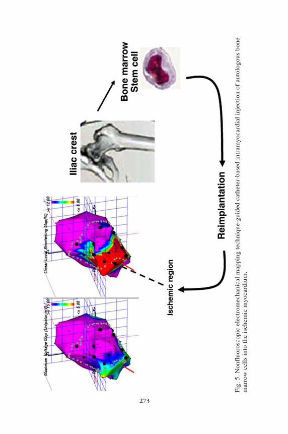
Chapter 10 / Bone Marrow Cell Transplantation 273
Fig
. 5. N
onfl
uoro
scop
ic e
lect
rom
echa
nica
l m
appi
ng t
echn
ique
-gui
ded
cath
eter
-bas
ed i
ntra
myo
card
ial
inje
ctio
n of
aut
olog
ous
bone
mar
row
cel
ls i
nto
the
isch
emic
myo
card
ium
.
273

274 Tse et al.
Fig
. 6. L
eft-
vent
ricu
lar
ejec
tion
fra
ctio
n (L
VE
F) (
A) ,
reg
iona
l wal
l thi
cken
ing
(B),
rad
ical
wal
l mot
ion
(C)
of th
e no
rmal
and
targ
etw
all,
and
per
cent
of
hypo
perf
used
myo
card
ium
(D
) , a
s de
term
ined
by
mag
neti
c re
sona
nce
imag
ing
at b
asel
ine
and
90 d
aft
er b
one
mar
row
cel
l im
plan
tati
on. *
Sta
tist
ical
sig
nifi
canc
e w
ith
a p
< 0
.05.
Fro
m r
ef. 4
1.
274

Chapter 10 / Bone Marrow Cell Transplantation 275
Intracoronary infusion of bone marrow mononuclear cells was per-formed in 10 patients 5–9 d after acute myocardial infarction. At 3 mo,these patients had improvement in myocardial function and viability(45). In the Transplantation of Progenitor Cells and Regeneration En-hancement in Acute Myocardial Infarction (TOPCARE-AMI) study,the efficacy of the ex vivo expanded peripheral blood mononuclearcells was compared to bone marrow-derived EPCs in restoringrevascularization after acute myocardial infarction. In both treatmentarms there was a similar improvement in myocardial function and viabil-ity (46).
In all of these studies there was improved blood flow and left ventricu-lar function, suggesting that infusion of autologous bone marrow cellsappears to be feasible, safe, and may confer short-term therapeutic ben-efit. However, a mixture of bone marrow mononuclear cells was used,and it remains unclear which types of cells are therapeutic and whetherthe injected cells themselves or the cytokines produced by them contrib-ute to the beneficial effects.
The use of purified populations of autologous bone marrow-derivedCD133+ cells were investigated in six patients with acute myocardialinfarction who underwent coronary bypass graft surgery (48). After 9mo, all patients were alive, with improvement in cardiac function in fourpatients. However, reasons for beneficial effects are confounded by thesurgery.
Fig. 7. An example of adenosine stress gadolinium-DTPA enhanced myocardialperfusion imaging at baseline and after 90 d. The amount of hyoperfusionmyocardium at the targeted inferior site (yellow area) reduced after bone mar-row cell implantation. MRI, magnetic resonance imaging.

276 Tse et al.
The effect of intracoronary and systemic administration of GM-CSFwas investigated using 10 patients with coronary artery disease (49). Animprovement in collateral flow was demonstrated in these patients byinvasive measures of collateral artery blood flow (estimatedby coronaryartery pressure distal to balloon occlusion). However, no clinically rel-evant endpoint or assessment on the mobilization of bone marrow stemcells was performed. It is unclear whether the coronary vascular benefitdetermined in this study may have resulted from direct effects of thiscytokine on angiogenesis or on collateral vascular dilator tone withimproved regional blood flow.
Despite enthusiasm for these pioneering clinical trials, none of themare randomized or have sufficient efficacy data to support any conclu-sion. It remains to be determined in randomized clinical trials whetherbone marrow cell transplantation will result in long-standing improve-ment in cardiac function and decrease morbidity and mortality withoutany long-term toxicity.
Potential Clinical ComplicationsIn the preliminary safety and feasibility trials, no authors have re-
ported a significant short-term safety problem. However, late-onset tox-icity may emerge as a result of the use of whole populations of bonemarrow mononuclear cells, which contain different organ-specific stem,progenitor, and hematopoietic cells. These nonessential cells, if incor-porated into regenerating myocardium, may result in generation of non-cardiac tissues as well as life-threatening arrhythmias. In particular, asbone marrow stem cells are also a rich source of different angiogenicfactors, including VEGF-A, this may result in the generation of edemaand aberrant angiomas (50). Therefore, whole bone marrow mono-nuclear cells should be used with caution and close long-term clinicalmonitoring. As many pathophysiological conditions, including tumorgrowth, diabetic- or age-related retinopathies, and atheroma formationare angiogenesis dependent, intravenous introduction of proangiogenicEPCs and hematopoietic cells may have adverse effects by inducingneovascularization in other organs.
Future PerspectiveIn human studies, autologous bone marrow cells are used to avoid the
potential issue of rejection, which necessitates the use of immunosup-pressive therapy. However, this approach would deliver a much lowerconcentration of stem cells than those used in animal studies, especiallyfor myocardial regeneration. Therefore, the fundamental scarcity of bonemarrow stem cells (EPCs, MSCs, and HSCs) in the circulation, com-bined with their possible functional impairment associated with a vari-

Chapter 10 / Bone Marrow Cell Transplantation 277
ety of diseases such as aging and diabetes, constitute major limitationsof autologous bone marrow transplantation for myogenesis. Consider-ing autologous bone marrow stem cell therapy, certain technical im-provements that may help to overcome the primary scarcity of a viableand functional stem cell population should include (1) delivery of opti-mal composition of types of bone marrow stem cells, (2) adjunctivestrategies to promote stem cell mobilization and survival, e.g., combinedwith cytokines, (3) enrichment procedures, i.e., leukapheresis and exvivo expansion, or (4) enhancement of stem cells function by gene trans-duction to improve survival and homing. These more complex isolationand preparation procedures for bone marrow stem cells may be associ-ated with increased cost and regulatory hurdles. However, these proce-dures may be essential to avoid future complications and to substantiatethe true value of bone marrow stem cell therapeutics for myocardialregeneration and angiogenesis.
Recently, bone marrow stem cells have also been applied to the fieldof tissue engineering as a means of improving biocompatibility. Recentstudies have demonstrated that the use of bone marrow stem cells en-ables the establishment of tissue engineered vascular autograft (51,52)and heart valves (53), which might reduce the complications associatedwith incompatible material, such as thrombosis. Alternatively, as previ-ously reported, the cell sheets of cultured cardiomyocytes with EPCsmay be effective for the improvement of cardiac function in damagedhearts, i.e., ischemic heart disease or cardiomyopathy, and induceneovascularization to maintain the viability of the cardiomyocytes (54).
CONCLUSIONS
Recent advances in the understanding of the mechanisms involved inproliferation, recruitment, mobilization, and incorporation of bone mar-row stem cells into the myocardium and blood vessels have prompted thedevelopment of cellular transplantation therapy for heart diseases re-fractory to conventional therapy. To acquire the more optimized qualityand quantity of bone marrow stem cells for myocardial regenerationseveral issues remain to be addressed, such as the development of a moreefficient method of stem cell identification, purification, and expansion.Despite the initial promising studies indicating potential clinical benefitof bone marrow therapy for therapeutic myogenesis and angiogenesis,many obstacles remain, such as long-term safety, optimal timing, andtreatment strategy. Emerging, rationally designed, randomized clinicaltrials assessing the role of bone marrow-derived stem cells in tissuerevascularization and myocardial regeneration will open new avenues ofresearch in stem cell therapeutics.

278 Tse et al.
ACKNOWLEDGMENTS
This project was supported in part by a research grant from the HongKong Research Grants Council (HKU 7357/02M), Sun Chieh Yeh HeartFoundation, and SK Yee Medical Foundation, (Project no. 203217) andthe Research Grant Council of Hong Kong (HKU 7357/02M).
REFERENCES
1. American Heart Association. Heart and Stroke Statistical Update. Dallas, TX:American Heart Association, 2001.
2. Ryan TJ, Antman EM, Brooks NH, Califf RM, Hillis LD, Hiratzka LF, Rapaport E,Riegel B, Russell RO, Smith EE III, Weaver WD, Gibbons RJ, Alpert JS, Eagle KA,Gardner TJ, Garson A Jr, Gregoratos G, Ryan TJ, Smith SC Jr. 1999 update: ACC/AHA guidelines for the management of patients with acute myocardial infarction:report of the American College of Cardiology/American Heart Association TaskForce on Practice Guidelines (Committee on Management of Acute MyocardialInfarction). J Am Coll Cardiol 1999;34:890–911.
3. Rosenthal N. Prometheus’s vulture and the stem-cell promise. N Engl J Med2003;349:267–274.
4. Körbling M, Estrov Z. Adult stem cells for tissue repair-a new therapeutic concept?N Engl J Med 2003;349:570–582.
5. Asahara T, Murohara T, Sullivan A, Silver M, van der Zee R, Li T, WitzenbichlerB, Schatteman G, Isner JM. Isolation of putative progenitor endothelial cells forangiogenesis. Science 1997;275:964–967.
6. Pittenger MF, Mackay AM, Beck SC, Jaiswal RK, Douglas R, Mosca JD, MoormanMA, Simonetti DW, Craig S, Marshak DR. Multilineage potential of adult humanmesenchymal stem cells. Science 1999;284:143–147.
7. Makino S, Fukuda K, Miyoshi S, Konishi F, Kodama H, Pan J, Sano M, TakahashiT, Hori S, Abe H, Hata J, Umezawa A, Ogawa S. Cardiomyocytes can be generatedfrom marrow stromal cells in vitro. J Clin Invest 1999;103:697–705.
8. Jiang Y, Jahagirdar BN, Reinhardt RL, Schwartz RE, Keene CD, Ortiz-GonzalezXR, Reyes M, Lenvik T, Lund T, Blackstad M, Du J, Aldrich S, Lisberg A, Low WC,Largaespada DA, Verfaillie CM. Pluripotency of mesenchymal stem cells derivedfrom adult marrow. Nature 2002;418:41–49.
9. Beltrami AP, Urbanek K, Kajstura J, Yan SM, Finato N, Bussani R, Nadal-GinardB, Silvestri F, Leri A, Beltrami CA, Anversa P. Evidence that human cardiacmyocytes divide after myocardial infarction. N Engl J Med 2001;344:1750–1757.
10. Jackson KA, Majka SM, Wang H, Pocius J, Hartley CJ, Majesky MW, Entman ML,Michael LH, Hirschi KK, Goodell MA. Regeneration of ischemic cardiac muscleand vascular endothelium by adult stem cells. J Clin Invest 2001;107:1395–1402.
11. Burt R, Pearce W, Luo K, Oyama Y, Davidson C, Beohar N, Gheorghiade M.Hematopoietic stem cell transplantation for cardiac and peripheral vascular disease.Bone Marrow Transplant 2003;32:S29–S31.
12. Quaini F, Urbanek K, Beltrami A, Finato N, Beltrami CA, Nadal-Ginard B, KajsturaJ, Leri A, Anversa P. Chimerism of the transplanted heart. N Engl J Med2002;346:5–15.
13. Muller P, Pfeiffer P, Koglin J, Schafers HJ, Seeland U, Janzen I, Urbschat S, BohmM. Cardiomyocytes of noncardiac origin in myocardial biopsies of human trans-planted hearts. Circulation 2002;106:31–35.

Chapter 10 / Bone Marrow Cell Transplantation 279
14. Wang X, Willenbring H, Akkari Y, Torimaru Y, Foster M, Al-Dhalimy M, LagasseE, Finegold M, Olson S, Grompe M. Cell fusion is the principal source of bone-marrow-derived hepatocytes. Nature 2003;422:897–901.
15. Vassilopoulos G, Wang PR, Russell DW. Transplanted bone marrow regeneratesliver by cell fusion. Nature 2003;422:901–904.
16. Shintani S, Murohara T, Ikeda H, Ueno T, Honma T, Katoh A, Sasaki KI, ShimadaT, Oike Y, Imaizumi T. Mobilization of endothelial progenitor cells in patients withacute myocardial infarction. Circulation 2001;103:2776–2779.
17. Heissig B, Hattori K, Dias S, Friedrich M, Ferris B, Hackett NR, Crystal RG,Besmer P, Lyden D, Moore MA, Werb Z, Rafii S. Recruitment of stem and progeni-tor cells from the bone marrow niche requires MMP-9 mediated release of kit-ligand. Cell. 2002;109:625–637.
18. Takahashi T, Kalka c, Masuda H, Chen D, Silver M, Kearney M, Magner M, Isner JM,Asahara T. Ischemia- and cytokine-induced mobilization of bone marrow-derivedendothelial progenitor cells for neovascularization. Nat Med 1999;5:434–438.
19. Moore MA, Hattori K, Heissig B, Shieh JH, Dias S, Crystal RG, Rafii S. Mobiliza-tion of endothelial and hematopoietic stem and progenitor cells by adenovector-mediated elevation of serum levels of SDF-1, VEGF, and angiopoietin-1. Ann NYAcad Sci 2001;938:36–45.
20. Hattori K, Heissig B, Tashiro K, Honjo T, Tateno M, Shieh JH, Hackett NR,Quitoriano MS, Crystal RG, Rafii S, Moore MA. Plasma elevation of stromal cell-derived factor-1 induces mobilization of mature and immature hematopoietic pro-genitor and stem cells. Blood 2001;97:3354–3360.
21. Risau W, Flamme I. Vasculogenesis. Annu Rev Cell Dev Biol 1995;11:73–91.22. Asahara T, Takahashi T, Masuda H, Kalka C, Chen D, Iwaguro H, Inai Y, Silver M,
Isner JM. VEGF contributes to postnatal neovascularization by mobilizing bonemarrow-derived endothelial progenitor cells. EMBO J 1999;8:3964–3972.
23. Kalka C, H. Masuda, T. Takahashi, Gordon R, Tepper O, Gravereaux E, Pieczek A,Iwaguro H, Hayashi SI, Isner JM, Asahara T. Vascular endothelial growth factor(165) gene transfer augments circulating endothelial progenitor cells in humansubjects. Circ Res 2000;86:1198–1202.
24. Orlic D, Kajstura J, Chimenti S, Limana F, Jakoniuk I, Quaini F, Nadal-Ginard B,Bodine DM, Leri A, Anversa P. Mobilized bone marrow cells repair the infarctedheart, improving function and survival. Proc Natl Acad Sci USA 2001;98:10344–10349.
25. Orlic D, Kajstura J, Chimenti S, Jakoniuk I, Anderson SM, Li B, Pickel J, McKayR, Nadal-Ginard B, Bodine DM, Leri A, Anversa P. Bone marrow cells regenerateinfarcted myocardium. Nature 2001;410:701–705.
26. Jackson KA, Majka SM, Wang H, Pocius J, Hartley CJ, Majesky MW, Entman ML,Michael LH, Hirschi KK, Goodell MA. Regeneration of ischemic cardiac muscleand vascular endothelium by adult stem cells. J Clin Invest 2001;107:1395–1402.
27. Jiang Y, Jahagirdar BN, Reinhardt RL, Schwartz RE, Keene CD, Ortiz-GonzalezXR, Reyes M, Lenvik T, Lund T, Blackstad M, Du J, Aldrich S, Lisberg A, Low WC,Largaespada DA, Verfaillie CM. Pluripotency of mesenchymal stem cells derivedfrom adult marrow. Nature 2002;418:41–49.
28. Wang JS, Shum-Tim D, Galipeau J, Chedrawy E, Eliopoulos N, Chiu RC. Marrowstromal cells for cellular cardiomyoplasty: feasibility and potential clinical advan-tages. J Thorac Cardiovasc Surg 2000;120:999–1005.
29. Toma C, Pittenger MF, Cahill KS, Byrne BJ, Kessler PD. Human mesenchymalstem cells differentiate to a cardiomyocyte phenotype in the adult murine heart.Circulation 2002;105:93–98.

280 Tse et al.
30. Bel A, Messas E, Agbulut O, Richard P, Samuel JL, Bruneval P, Hagege AA,Menasche P. Transplantation of autologous fresh bone marrow into infarcted myo-cardium: a word of caution. Circulation 2003;108(Suppl 1):II247–II252.
31. Beltrami AP, Barlucchi L, Torella D, Baker M, Limana F, Chimenti S, Kasahara H,Rota M, Musso E, Urbanek K, Leri A, Kajstura J, Nadal-Ginard B, Anversa P. Adultcardiac stem cells are multipotent and support myocardial regeneration. Cell2003;114:763–776.
32. Rafii S, Lyden D. Therapeutic stem and progenitor cell transplantation for organvascularization and regeneration. Nat Med 2003;9:702–712.
33. Masuda H, Asahara T. Post-natal endothelial progenitor cells for neovascularizationin tissue regeneration. Cardiovasc Res 2003;58:390–398.
34. Kocher AA, Schuster MD, Szabolcs MJ, Takuma S, Burkhoff D, Wang J, HommaS, Edwards NM, Itescu S. Neovascularization of ischemic myocardium by humanbone-marrow-derived angioblasts prevents cardiomyocyte apoptosis, reduces re-modeling and improves cardiac function. Nat Med 2001;7:430–436.
35. Kawamoto A, Gwon HC, Iwaguro H, Yamaguchi JI, Uchida S, Masuda H, SilverM, Ma H, Kearney M, Isner JM, Asahara T. Therapeutic potential of ex vivoexpanded endothelial progenitor cells for myocardial ischemia. Circulation2001;103:634–637.
36. Fuchs S, Baffour R, Zhou YF, Shou M, Pierre A, Tio FO, Weissman NJ, Leon MB,Epstein SE, Kornowski R. Transendocardial delivery of autologous bone marrowenhances collateral perfusion and regional function in pigs with chronic experimen-tal myocardial ischemia. J Am Coll Cardiol 2001;37:1726–1732.
37. Kawamoto A, Tkebuchava T, Yamaguchi J, Nishimura H, Yoon YS, Milliken C,Uchida S, Masuo O, Iwaguro H, Ma H, Hanley A, Silver M, Kearney M, LosordoDW, Isner JM, Asahara T. Intramyocardial transplantation of autologous endothe-lial progenitor cells for therapeutic neovascularization of myocardial ischemia.Circulation 2003;107:461–468.
38. van Royen N, Hoefer I, Bottinger M, Hua J, Grundmann S, Voskuil M, Bode C,Schaper W, Buschmann I, Piek JJ. Local monocyte chemoattractant protein-1therapy increases collateral artery formation in apolipoprotein E-deficient mice butinduces systemic monocytic CD11b expression, neointimal formation, and plaqueprogression. Circ Res 2003;92:218–225.
39. Gutierrez-Delgado F, Bensinger W. Safety of granulocyte colony-stimulating fac-tor in normal donors. Curr Opin Hematol 2001;8:155–60.
40. Sabatine MS, Morrow DA, Cannon CP, Murphy SA, Demopoulos LA, DiBattistePM, McCabe CH, Braunwald E, Gibson CM. Relationship between baseline whiteblood cell count and degree of coronary artery disease and mortality in patients withacute coronary syndromes: a TACTICS-TIMI 18 (Treat Angina with Aggrastat anddetermine Cost of Therapy with an Invasive or Conservative Strategy—Thromboly-sis in Myocardial Infarction 18 trial) substudy. J Am Coll Cardiol 2002;40:1761–1768.
41. Hamano K, Nishida M, Hirata K, Mikamo A, Li TS, Harada M, Miura T, MatsuzakiM, Esato K. Local implantation of autologous bone marrow cells for therapeuticangiogenesis in patients with ischemic heart disease: clinical trial and preliminaryresults. Jpn Circ J 2001;65:845–847.
42. Tse HF, Kwong YL, Lo G, Ho CL, Chan JKF, Yeung DW, Lau CP. Angiogenesisin ischemic myocardium by intramyocardial autologous bone marrow mononuclearcell implantation. Lancet 2003;361:47–49.
43. Fuchs S, Satler LF, Kornowski R, Okubagzi P, Weisz G, Baffour R, Waksman R,Weissman NJ, Cerqueira M, Leon MB, Epstein SE. Catheter-based autologous bonemarrow myocardial injection in no-option patients with advanced coronary arterydisease: a feasibility study. J Am Coll Cardiol 2003;41:1721–1724.

Chapter 10 / Bone Marrow Cell Transplantation 281
44. Perin EC, Dohmann HF, Borojevic R, Silva SA, Sousa AL, Mesquita CT, Rossi MI,Carvalho AC, Dutra HS, Dohmann HJ, Silva GV, Belem L, Vivacqua R, Rangel FO,Esporcatte R, Geng YJ, Vaughn WK, Assad JA, Mesquita ET, Willerson JT.Transendocardial, autologous bone marrow cell transplantation for severe, chronicischemic heart failure. Circulation 2003;107:2294–2302.
45. Strauer BE, Brehm M, Zeus T, Kostering M, Hernandez A, Sorg RV, Kogler G,Wernet P. Repair of infarcted myocardium by autologous intracoronary mono-nuclear bone marrow cell transplantation in humans. Circulation 2002;106:1913–1918.
46. Assmus B, Schachinger V, Teupe C, Britten M, Lehmann R, Dobert N, GrunwaldF, Aicher A, Urbich C, Martin H, Hoelzer D, Dimmeler S, Zeiher AM; Transplan-tation of Progenitor Cells and Regeneration Enhancement in Acute MyocardialInfarction. Transplantation of Progenitor Cells and Regeneration Enhancement inAcute Myocardial Infarction (TOPCARE-AMI). Circulation 2002;106:3009–3017.
47. Stamm C, Westphal B, Kleine HD, Petzsch M, Kittner C, Klinge H, Schumichen C,Nienaber CA, Freund M, Steinhoff G. Autologous bone-marrow stem-cell trans-plantation for myocardial regeneration. Lancet 2003;361:45–46.
48. Gepstein L, Hayam G, Ben-Haim SA. A novel method for nonfluoroscopic catheter-based electroanatomical mapping of the heart: in vitro and in vivo accuracy results.Circulation 1997;95:1611–1622.
49. Seiler C, Pohl T, Wustmann K, Hutter D, Nicolet PA, Windecker S, Eberli FR,Meier B. Promotion of collateral growth by granulocyte-macrophage colony-stimu-lating factor in patients with coronary artery disease: a randomized, double-blind,placebo-controlled study. Circulation 2001;104:2012–2017.
50. Epstein SE, Kornowski R, Fuchs S, Dvorak HF. Angiogenesis therapy: amidst thehype, the neglected potential for serious side effects. Circulation 2001;104:115–119.
51. He H, Shirota T, Yasui H, Matsuda T. Canine endothelial progenitor cell-linedhybrid vascular graft with nonthrombogenic potential. J Thorac Cardiovasc Surg2003;126:455–464.
52. Matsumura G, Miyagawa-Tomita S, Shin’oka T, Ikada Y, Kurosawa H. First evi-dence that bone marrow cells contribute to the construction of tissue-engineeredvascular autografts in vivo. Circulation 2003;108:1729–1734.
53. Perry TE, Kaushal S, Sutherland FW, Guleserian KJ, Bischoff J, Sacks M, MayerJE. Bone marrow as a cell source for tissue engineering heart valves. Ann ThoracSurg 2003;75:761–767.
54. Zimmermann WH, Eschenhagen T. Cardiac tissue engineering for replacementtherapy. Heart Fail Rev 2003;8:259–269.

Chapter 11 / Cellular Cardiomyoplasty With Stem Cells 283
283
From: Contemporary Cardiology: Angiogenesis and Direct Myocardial RevasularizationEdited by: R. J. Laham and D. S. Baim © Humana Press Inc., Totowa, NJ
INTRODUCTION
Congestive heart failure remains the leading cause of death in devel-oped countries. Myocardial infarction (MI) results in the loss of heartmuscle cells, which is the main contributor to the development of heartfailure. Classical medical therapy and mechanical left-ventricular assistdevices are available for physicians to improve the prognosis of patientswith MI and heart failure, but only half of the patients with end-stageheart failure survive the following year (1). At the present time, alloge-neic heart transplantation to extend life span and to improve the qualityof daily life is probably the preferred alternative treatment for patientswith end-stage heart failure, but extreme organ shortage and chronic
11 Transplantation of EmbryonicStem Cells for MyocardialRegeneration and Angiogenesis
Yong-Fu Xiao, MD, PhD,Jiang-Yong Min, MD,and James P. Morgan, MD, PhD
CONTENTS
INTRODUCTION
EMBRYONIC STEM CELLS
TRANSPLANTATION OF EMBRYONIC STEM CELLS
FOR CARDIAC REPAIR
HOMING AND IMMUNE TOLERANCE
FUTURE PERSPECTIVES AND SUMMARY

284 Xiao et al.
cardiac rejection limit the therapy. In recent years, research on stem cellsis leading scientists to investigate the possibility of cell-based therapiesfor cardiac repair, often referred to as regenerative or reparative medi-cine. Stem cell-based cellular cardiomyoplasty (CCM) for cardiomyo-cyte replacement/regeneration has been evaluated in animal settings (2–6) and clinical trials (7–9). Transplantation of exogenous stem cellscould regenerate damaged myocardium and improve cardiac function infailing hearts. Such efforts may offer exciting novel options for treatingpatients with end-stage heart failure.
The feasibility of CCM for repair of failing hearts has been examinedvia transplantation of exogenous stem cells into host myocardium suf-fering from cardiomyocyte loss (6,7,10,11). Formation of intercalateddisks and gap junctions between transplanted cells and the host tissueindicates functional electrical-contraction coupling (2,10,12). In inf-arcted mice transplantation of bone marrow cells regenerated infarctedmyocardium and improved heart function (6). Our previous studyshowed that co-transplantation of human mesenchymal stem cells(hMHCs) and human fetal cardiomyocytes significantly improved car-diac function in postinfarcted pigs (13). Transplantation of mouse em-bryonic stem cells (ESCs) or their derived cardiac-like cells alsoimproved cardiac function in diseased animal models (14–17). Perin andassociates reported that transendocardial injections of autologous mono-nuclear bone marrow cells in patients with end-stage ischemic heartdisease can safely promote neovascularization and improve regional andglobal left-ventricular function (5). Another study shows that 3 mo afterintramyocardial implantation of autologous mononuclear bone marrowcells, improvement in symptoms, myocardial perfusion, and functionwas observed in patients with severe ischemic heart disease (8). Otherclinical studies demonstrate that compared to the standard therapy group,transplantation of bone marrow stem cells significantly improved car-diac function in the cell-treated MI patients (7,18). Generally speaking,tissue engineering is a potential therapy for end-stage organ disease ortissue loss. Therefore, cell therapy holds great promise for cardiac disease.
Stem cells are one of the most fascinating areas of biology today, butthe big question is how they can replace damaged cells in adult organs.Most adult tissues have stem or progenitor cells, and environmentalstimuli can activate and mobilize them. But adult stem cells can onlyform a limited number of cell types (19,20). ESCs in culture have thecapacity for differentiation into all of specialized somatic cell types ofthe body, including cardiomyocytes. Human embryonic stem cells(hESCs) in culture were isolated and cultured in 1998 by Thomson’sgroup (21). Therefore, identification, derivation, and characterization of

Chapter 11 / Cellular Cardiomyoplasty With Stem Cells 285
hESCs may push the rapidly progressing field of therapeutic cell trans-plantation. Because of the proliferation and plasticity of differentiationto various mature tissues, ESCs are a potential valuable resource for celltherapy targeting regeneration of functional myocardium in diseasedhearts. However, many ethical, political, and scientific barriers have tobe overcome before hESCs or their differentiated cells can be used clini-cally. Here we summarize the findings on transplantation of ESCs ortheir derived cells in experimental animals with myocardial injury andhighlight the progress in research on these particular cells.
EMBRYONIC STEM CELLSCharacterization
ESCs are pluripotent cells that are obtained from the blastocyst stageof early mammalian embryos. A blastocyst includes three structures: thetrophoblast, which is the layer of cells that surrounds the blastocyst; theblastocoel, which is the hollow cavity inside the blastocyst; and the innercell mass, which is a group of cells at one end of the blastocoel. Theseunique inner cells are characterized by their capacity for prolongedundifferentiated proliferation in culture and have the capability to derivedescendants of all three embryonic germ layers—endoderm, mesoderm,and ectoderm. Because ESCs are derived prior to implantation and cer-tain immune-related cell surface proteins (e.g., class I products of themajor histocompatibility complex) are not yet expressed, ESCs are apotentially rich source for cell transplantation. An important test to en-sure undifferentiation of ESCs is the presence of a protein called Oct-4,which is expressed in totipotent and pluripotent cells of the early embryoand the germ cell lineage. Oct-4 is a transcription factor that helps turngenes on and off at the right time (22,23). ESCs lack the growth phase1 (G1) checkpoint in the cell cycle and do not require any externalstimulus to initiate DNA replication. Mouse ESCs were successfullyisolated from murine blastocysts by Evans and Kaufman (24) and Martin(25) more than two decades ago. The hESC lines were obtained in 1998(21). The histological profile of the hESCs is (1) positive for alkalinephosphatase, stage-specific embryonic antigen (SSEA)-3, SSEA-4, tu-mor rejection antigen (TRA)-1-60, and TRA-1-81, (2) negative forSSEA-1, and (3) strongly positive staining of SSEA-1 after differentia-tion (21).
Cardiomyocyte DifferentiationIf ESCs in culture are removed from conditions that maintain them in
an undifferentiated state, they clump together to form embryoid bodies(EBs) and begin to differentiate spontaneously. They can form muscle

286 Xiao et al.
cells, nerve cells, and many other cell types. During in vitro differentia-tion, ESCs have been shown to recapitulate many processes of earlyembryonic development. The heart is one of the first organs to developduring embryogenesis. The heart tube with spontaneous contractionsappears at about embryonic d 8 in mice (26). Cardiomyocytes derivedfrom cultured mouse ESCs exhibit cell morphology, sarcomere forma-tion, and cell–cell junctions similar to those observed in cardiomyocytesdeveloping in vivo (27,31). Mouse ESCs in embryoid bodies presenthighly specialized phenotypes of cardiac tissue with several electro-physiological characteristics similar to those in adult cardiomyocytes(32,34). Figure 1A shows the hanging-drops procedure to inducecardiomyocyte differentiation of cultured mouse ESCs. These cellsshowed cardiac action potentials (Fig. 1B) and contractions that re-sponded well to the change of extracellular Ca2+ concentrations (Fig. 1C).
If conditions in vitro allow, hESCs also spontaneously form EBscontaining derivatives of all three germ layers and differentiate intovarious cell types, including cardiomyocytes. The phenotypicpropertiesof cardiomyocytes derived from hESCs have been described (35,36).Immunostaining against cardiac myosin heavy chain, -actinin, desmin,cardiac troponin I, and atrial natriuretic factor was positive in cells fromthe spontaneously contracting areas within EBs. Electron microscopyrevealed varying degrees of myofibrillar organization, consistent withearly-stage cardiomyocytes. Expression of several cardiac-specificgenes and transcription factors was found in human ESC-derived car-diac-like cells, which also showed electrophysiological properties ascardiomyocytes. These contracting cells showed positive and negativechronotropic responses to the stimuli of isoproterenol and carbamyl-choline, respectively (35). In addition, functional cardiomyocytes couldefficiently be differentiated from three parent (H1, H7, and H9) hESClines and two clonal (H9.1 and H9.2) hESC lines (36), even after long-term culture (50 passages, or approx 260 population doublings). Beatingcells were observed in hESCs 1 wk after culture under differentiationconditions and increased in numbers with time. The beating cells ex-pressed several transcription factors and characteristic markers ofcardiomyocytes. The dissociated and enriched population of differenti-ated cells reached 70% cardiomyocytes and was proliferative. There-fore, cardiomyocytes derived from ESCs can be a rich and viable sourceas donor cells for clinical application in heart disease.
Factors Influencing Cardiomyocyte DifferentiationTo generate specific types of differentiated cells, such as heart muscle
cells, scientists try to influence the direction of ESC differentiation viachange in the chemical composition of the surface on which the ESCs are

Chapter 11 / Cellular Cardiomyoplasty With Stem Cells 287
Fig. 1. Action potentials and cell contractions of cardiomyocytes differentiatedfrom mouse embryonic stem cells (ESCs). (A) The protocol is shown to inducedifferentiation of cultured mouse ESCs (ES-D3 cell line) to cardiomyocytes.Left column is a schematic diagram of the hanging-drops method to differenti-ate ESCs to heart cells. Right column shows single ESCs in culture (whitearrows in upper panel, ×200), an embryoid body (red arrow in middle panel,×100), and a beating cluster of cardiac-like cells (yellow arrow in lower panel,×200) corresponding to the time periods (black arrows) in culture after with-drawal of leukemia inhibitory factor (LIF) from the conditioned culture me-dium. (B) Cardiac action potentials were observed in a spontaneously beatingcell approx 10 d after differentiation of mouse ESCs. (C) Cell contractions ofESC-derived cardiomyocytes are shown. Cell shortenings were detected andrecorded by the edge-detection method. The amplitude and beating rate of cellcontraction were increased when the levels of extracellular Ca2+ were raised to4 or 6 mM. However, the amplitude of cell shortening was significantly reducedin the 8 mM Ca2+ solution, and the cells eventually stopped beating in the 10 mMCa2+ solution.

288 Xiao et al.
growing, alteration of contents (growth factors) in culture medium, orinsertion of specific genes. The microenvironment, known as “niche,”greatly affects the fate of stem cells (37). ESCs with adequate environ-ments have a great ability to proliferate and differentiate. During thedifferentiation process, ESCs have the ability to express different recep-tor types and release growth factors. In addition, many growth factorsand cytokines or other signals from adjacent cells affect ESC prolifera-tion and differentiation. Therefore, cross-talks are always there betweenESCs and their environment or neighboring cells.
Co-culture with mouse embryonic fibroblast feeders or addition ofleukemia inhibitory factor (LIF) in culture medium suppresses differen-tiation of mouse ESCs (38). When ESCs spontaneously differentiate,cardiomyocytes of pacemaker-, atrium-, and ventricle-like types can begenerated (27). It has been shown that retinoic acid (RA) treatmentduring ESC differentiation increases the number of cardiomyocytes (39).RA treatment during the first 2 d or between d 2 and 5 of ESC-derivedEB formation significantly inhibited cardiogenesis, whereas treatmentbetween d 5 and 7 resulted in an increased cardiomyocyte differentiation(40). The number of differentiated cardiomyocytes with Purkinje- andventricle-like properties was increased in the presence of RA, whereasthe number of pacemaker- and atrium-like cells was reduced (39). Re-cently, Hidaka et al. (41) also reported that murine ESCs differentiate tochamber-specific Nkx2.5-positive cardiac precursor cells and RA modi-fies the differentiation potential in a time- and dose-dependent fashion.Our results demonstrate that vascular endothelial growth factor (VEGF)did not affect proliferation of mouse ESCs in vitro in the presence of LIF,but stimulated their proliferation and differentiation to cardiomyocytesin the absence of LIF (42). Our data in vivo also show that transplantationof ESCs with overexpression of VEGF produced a better improvementof heart function in MI mice, which might result from stronger stimula-tion of myocardial regeneration and neovascularization by ESC-releasedVEGF (17). A recent study demonstrates that the intact signaling path-way of transforming growth factor (TGF)- was required for engraftedESCs to differentiate into functional cardiomyocytes in infarcted myo-cardium (43). Engrafted ESCs with engineered disruption of TGF-family failed to differentiate to cardiomyocytes after cell transplantationin infarcted animals. Additionally, Schuldiner et al. (44) reported theeffects of eight factors—basic fibroblast growth factor (bFGF), TGF- ,activin-A, bone morphogenic protein (BMP)-4, hepatocyte growth fac-tor (HGF), epidermal growth factor (EGF), nerve growth factor( -NGF), and RA—on the differentiation of human embryonic stem-derived cells in vitro. Human ESCs initiated development as aggregates

Chapter 11 / Cellular Cardiomyoplasty With Stem Cells 289
(EBs) expressed a receptor for each of these factors. Each factor had aunique effect on cell differentiation (44). These results indicate thatseveral factors, cytokines, and growth factors affect ESC differentiation.Table 1 summarizes the factors affecting cardiomyocyte differentiationof ESCs reported in literature.
Understanding the regulation of ESC proliferation and differentiationat the cellular and molecular levels may be make it possible to direct ESCdifferentiation to certain cell types for clinical cell therapy in the future.Specific differentiated cells may be used to treat certain diseases, includ-ing Parkinson’s disease, diabetes, traumatic spinal cord injury, Purkinjecell degeneration, Duchenne’s muscular dystrophy, heart disease, andvision and hearing loss. Alteration of environments of cultured human
Table 1Factors Influencing Differentiation of ESCs to Cardiomyocytes
Factor Result Ref.
Mouse ESCsLIF Prevention of differentiation 38Retinoic acid Increase in number of Purkinje- and ventricle-like 39
cells and decrease in pacemaker- and atrium-like cells
VEGF Increase in percentage of -MHC and cTn-I 42positive cells
TGF- /BMP-2 Significant increase in beating areas and enhance- 43ment of myofibrillogenesis
HBR Increase in the expression of GATA-4 and 91Nkx-2.5 and enhancement of yield of beatingcardiomyocytes
PDGF-BB Promoting cardiogenesis in vitro 92Dynorphin B Increase in gene expression of GATA-4 and 93
Nkx-2.5 and enhancement of appearance of-MHC and MLC-2v
Human ESCsRetinoic acid No effects on cardiomyocyte differentiation 365-aza-2'-dC Increase in percentage of cardiomyocyte 36
differentiationTGF- Enrichment of myocardial-specific cells 44
ESCs, embryonic stem cells; LIF, leukemia inhibitory factor; VEGF, vascularendothelial growth factor; TGF- , transforming growth factor ; BMP-2, bonemorphogenic protein 2; HBR, an ester of hyaluronan linked to both butyric andretinoic acid; PDGF-BB, platelet-derived growth factor-BB; 5-aza-2'-dC, 5-aza-2'-deoxycytidine; MHC, myosin heavy chain.

290 Xiao et al.
ESCs can initiate cardiomyocyte differentiation. The percentage ofhECS-derived cardiomyocytes could be enhanced by treatment of themwith 5-aza-2'-deoxycytidine, but not by dimethylsulfoxide (DMSO) orRA (36). Because of their combined properties of indefinite prolifera-tion and differentiation to heart cells, hESCs can potentially provide alarge number of cardiomyocytes for cell therapy aiming to regeneratefunctional myocardium. However, studies are required to prove the fea-sibility of transplantation of hESCs or their derived cells for cardiacrepair before clinical utilization is possible.
TRANSPLANTATION OF EMBRYONIC STEM CELLSFOR CARDIAC REPAIR
Myocardial InfarctionCell transplantation is a potentially novel approach to deliver viable
myocytes or cells in adult hearts. In 1994, Soonpaa et al. (2) reported thatimplantation of embryonic d 15 transgenic cardiomyocytes into themyocardium of syngeneic hosts formed stable grafts for as long as the 2-mo observation period. Engrafted cardiomyocytes had no significantnegative effects on the host myocardium and formed nascent interca-lated disk connection with the host myocardium (2). One year later,Connold and coworkers (45) transplanted small fragments of embryonichearts into soleus muscles of adult rat hosts. The implanted grafts sur-vived and grew for at least 6 mo after transplantation and established anetwork of blood vessels communicated with the host’s circulation. Thegrafted tissue was rhythmically active with a contraction rate similar tothat of adult rat hearts. Application of acetylcholine caused a reversibleslowing of the beating rate of the grafts (45). Klug et al. recently reportedthat transplantation of cultured cardiomyocytes differentiated frommurine ESCs formed stable intracardiac grafts and gap junctions (30).They genetically selected relatively pure cultures of cardiomyocytesfrom differentiating murine ESCs. The selected cardiomyocyte cultureswere more than 99% pure and highly differentiated. Intracardiac trans-plantation of genetically selected cardiomyocytes formed stable graftsin adult mouse hearts for 7-wk observation after implantation (30). Thesestudies demonstrate that grafts of embryonic cardiac tissue or ESC-derived cardiomyocytes survived well in normal tissues with adequateblood supply from the host’s circulation (Table 2).
In a non-MI animal model, Roell et al. (46) reported that embryonicmurine ventricular cardiomyocytes labeled with enhanced green fluo-rescent protein (GFP) were transplanted into mouse left ventricular walls2 wk after cryoinfarction. GFP-positive cardiomyocytes isolated fromcell-engrafted hearts had postnatal ventricular action potentials and in-

Chapter 11 / Cellular Cardiomyoplasty With Stem Cells 291T
able
2T
rans
plan
tati
on o
f ESC
s or
Em
bryo
n ic
Car
diom
yocy
tes
into
the
Hea
rt in
An i
mal
Mod
els
Cel
lA
nim
alO
bser
vati
onC
ell t
ype
deli
very
mod
elpe
riod
Res
ult
Ref
.
Rat
cel
lsS
mal
l fra
gmen
t of
rE-h
eart
ism
Rat
6 m
oF
orm
ing
gap
junc
tion
CX
-43,
neo
vasc
ular
izat
ion
45,
Sm
all p
iece
s of
rE
-hea
rts
imR
at w
ith
cut-
2 d
to 7
mo
For
min
g ga
p ju
ncti
on,m
igra
tion
to in
jure
d ar
ea47
inju
red
hear
tD
isse
cted
rE
-car
diom
yocy
tes
imM
I ra
t1–
8 w
kG
raft
for
mat
ion,
func
tion
al im
prov
emen
t,48
posi
tive
sta
inin
g ag
inst
-M
HC
and
CX
-43
Mou
se c
ells
Dis
sect
ed m
E-c
ardi
omyo
cyte
sim
Mou
se2
mo
Gra
ft f
orm
atio
n,in
terc
alat
ed d
isks
2D
isse
cted
ES
C b
eati
ng c
ells
imM
I ra
t30
dSu
rviv
al, p
ositi
ve s
tain
ing
gain
st s
arco
mer
ic m
yosi
n15
ES
C-d
eriv
ed c
ardi
omyo
cyte
sim
Mou
se7
wk
For
mat
ion
of s
tabl
e gr
afts
30E
SC
-der
ived
car
diom
yocy
tes
imM
ouse
wit
h1–
8 w
kN
ew c
ardi
omyo
cyte
s,fu
ncti
onal
impr
ovem
ent,
46cr
yoin
jure
dpo
siti
ve s
tain
ing
agai
nst c
Tn-
Ihe
art
ES
C-d
eriv
ed c
ells
imM
I m
ouse
6 w
kN
eova
scul
ariz
atio
n,fu
ncti
onal
impr
ovem
ent,
posi
-17
tive
sta
inin
g ag
ains
t -M
HC
, cT
n-I,
and
CX
-43
ES
Cs
imM
I ra
t6–
32 w
kN
eova
scul
ariz
atio
n an
d ca
rdio
gene
sis,
fun
cion
al14
,16,
43im
prov
emen
t
ES
Cs
ivM
yoca
rdit
ic2–
4 w
kN
ew c
ardi
omyo
cyte
s, r
educ
tion
of
mor
tali
ty52
mou
se
rE, r
at e
mbr
yoni
c; m
E, m
ouse
em
bryo
nic;
im, i
ntra
myo
card
ial i
njec
tion
or
impl
anta
tion
; ism
, int
rask
elet
al m
uscl
e im
plan
tati
on; M
HC
, myo
sin
heav
ych
ain;
cT
n-I,
car
diac
trop
onin
I; C
X-4
3; c
onne
xin-
43; m
ES
C, m
ouse
em
bryo
nic
stem
cel
ls; M
I, m
yoca
rdia
l inf
arct
ion.

292 Xiao et al.
tact -adrenergic modulation. Analysis of echocardiography showed asignificant improvement of force generation and heart function in cell-transplanted hearts. These results show that the grafted embryonic car-diac tissue or myocytes survived in adult host myocardium and expressedcharacteristics typical of heart cells, which improved damaged heartfunction (Table 2).
Similarly, two studies show that transplantation of pieces of embry-onic ventricular myocardium or cardiomyocytes isolated from earlyembryos survived in infarcted myocardium and improved damaged heartfunction. In an early study, Connold et al. (47) transplanted pieces ofembryonic ventricular myocardium (prelabeled with 4',6-diamindino-2-phenylindole) into a damaged area of the host myocardium andexamined the grafts between 2 d and 5–7 mo later. Initially the 4'-6-diamindino-2-phenylindole–labeled cells were localized only at the siteof grafting, but by 2–5 wk they migrated along the ventricular surface ofthe heart. The greatest density of grafted cells was always found in thedamaged area. The 4'-6-diamindino-2-phenylindole-labeled cells con-tained myosin heavy chains and stained positively with antibodiesagainst cardiac gap junction proteins. Recently, Etzion and colleagues(48) transplanted cardiomyocytes isolated from 15-d-old embryos intorat MI hearts. Embryonic cardiac cells were labeled with 5-bromo-2'-deoxyuridine (BrdU) or the reporter gene LacZ. They found that cell-transplanted MI animals had attenuated left ventricular dilatation, infarctthinning, and myocardial dysfunction. Immunostaining for several car-diac proteins revealed grafts in various stages of differentiation in cell-treated hearts.
Transplanted mouse ESCs have survived and differentiated in inf-arcted mouse or rat myocardium (14,16,17). ESCs tagged with GFPwere transplanted locally into injured myocardium in a rat model withligation of the left coronary artery. Compared with the control MI ani-mals, cardiac function was significantly improved in ESC-implanted MIrats 6 wk after cell transplantation. Double immunostaining against GFPand cardiac sarcomeric -actin, -myosin heavy chain, or troponin Iconfirmed the survival and differentiation of engrafted cells in MI heartswith ESC transplantation. Additionally, isolated single cells showedrod-shaped GFP-positive myocytes with clear striations in ESC-trans-planted animals. GFP-positive myocytes were 7.3% of total cells iso-lated from each MI left ventricle. The shape and size of matureGFP-positive myocytes did not significantly differ from those of hostcardiomyocytes. In contrast, no GFP-positive cells were detected in cellsisolated from the sham-operated or MI control hearts (14). Recently,Naito et al. reported similar findings in rats with ligation of the left

Chapter 11 / Cellular Cardiomyoplasty With Stem Cells 293
coronary artery (15). Cells dissected from beating regions of GFP-taggedmouse ESCs were injected into the border zone between the infarctedmyocardium and normal myocardium. Thirty days after transplantation,GFP-expressed cells were detected and stained positively by the anti-body against sarcomeric myosin (15). These results indicate that trans-planted ESCs survived and differentiated in injured myocardium andimproved cardiac function in MI animals (Table 2).
More recently, the long-term effects of ESC transplantation on car-diac function were investigated in postinfarcted rats (16). Mouse ESCswere implanted into the border area and infarcted myocardium. Duringthe observation period of 32 wk, the survival rate was significantly in-creased in MI rats with ESC transplantation. Hemodynamic andechocardiographic data showed significant improvement of cardiac func-tion in the cell-transplanted MI group. GFP-positive tissue was identi-fied in the injured myocardium and stained positively by the antibodiesagainst several specific cardiac proteins (16). The data demonstrate thatESC engrafts exhibit long-term survival in a myocardial infarct modeland contribute to long-term improvement of injured heart function(Table 2).
Genetic manipulation of ESCs may produce better turnout after celltransplantation. The effects of transplantation of mouse ESCs trans-fected with cDNA of VEGF were investigated in a MI mouse model (17).Compared to the MI control mice, left ventricular function was signifi-cantly improved in the MI mice with ESC transplantation. In addition,the improvement of heart function was significantly greater in the MImice engrafted with ESCs overexpressing VEGF. Histological analysisof the sections of infarcted hearts with cell transplantation showed GFP-positive spots which immunostained positively against cardiac troponinI and -myosin heavy chain. Double staining for GFP and connexin-43was positive in injured myocardium with ESC transplantation.
MyocarditisMyocarditis predominantly results from infection by viruses (49),
and has a disease process of viral infection, immunoreaction, and mi-crovascular spasm (50,51). Myocardial necrosis and inflammation oc-cur during the acute stage. Development of myocardial fibrosis,calcification, cardiac dilatation, hypertrophy, and heart failure are char-acterized in the chronic stage. It is a great challenge to prevent or reversethe structural and functional injuries that may cause death or debility ofmyocarditic patients. In a recent study, Wang and colleagues (52) inves-tigated whether infused ESCs could migrate to the injured area ofmyocarditic mouse hearts and, if so, whether migrated ESCs could dif-

294 Xiao et al.
ferentiate and proliferate in the damaged myocardium. Mice were inocu-lated with encephalomyocarditis virus (EMCV) immediately followingan intravenous infusion of mouse ESCs tagged with GFP. The mortalityof mice during the 30-d observation period was significantly reduced,from 37% for the viral control group to 16% for the viral plus ESC-treated group (p < 0.05). Compared with the viral control mice, theanimals with ESC infusion had a significantly lower incidence of in-flammatory cell infiltration and myocardial lesions. In addition,immunostaining against GFP and cardiac -actin showed that part ofinfused ESCs migrated to the hearts and differentiated into myocytesthere in viral inoculated mice. These results demonstrate that infusion ofESCs significantly reduced mortality and global injury of the hearts inviral myocarditis mice (Table 2). The beneficial effect of engrafted ESCsmay have resulted from attenuation of myocardial necrosis and inflam-mation and from regeneration of damaged myocardium in viralmyocarditic animals (52).
Aging HeartBiogerontology has become attractive to biologists in the past decade.
Normal human and animal cells have a finite capacity to replicate andfunction whether they are cultured in vitro or transplanted as grafts invivo. This phenomenon has been interpreted to be aging at the cellularlevel. Alterations in the organization and mobility of cell membraneconstituents of cultured rat heart myocytes (53) appear a general phe-nomenon of aging cells. The aging heart exhibits diastolic dysfunctionand increased stiffness. Evidence shows that aging affects the passivemechanical properties of single cardiac myocytes isolated from the heartsof 4-mo young and 30-mo old rats (54). Progressive loss of cardio-myocytes occurs during the aging process and may cause heart failure.A recent study showed that intramyocardial transplantation of mouseESCs improved cardiac function in aging old rats (55). Mouse ESCslabeled with GFP were injected into myocardium of aged rat hearts.Compared with the young adult rats, cardiac function in aging rats wassignificantly decreased accompanied by a significant reduction in thenumber of left ventricular cardiomyocytes and regional blood flow. Sixweeks after ESC transplantation, the aged hearts partially restored theblood flow and the number of myocytes and improved their function. Inaddition, the aged hearts receiving ESCs showed positive response toisoproterenol stimulation, but not in untreated aged hearts. Histologicaldata showed that GFP-positive cells in myocardium that received celltransplantation were positively stained by the antibody against -myo-sin heavy chain. Therefore, stem cell therapy may slow down the occur-rence of cardiac dysfunction during aging process.

Chapter 11 / Cellular Cardiomyoplasty With Stem Cells 295
Myocardial Regeneration
The studies presented above show that transplantation of ESCs orembryonic cardiac cells improves injured cardiac function and reducesmortality in experimental animal models. However, the underlyingmechanisms of the beneficial effects remain to be delineated. Genera-tion of new functional cardiomyocytes and formation of functional con-nections between engrafts and host cells are critical for restoration ofcardiac function in diseased hearts. ESCs have the ability to differentiateinto cardiomyocytes in vitro. Our recent results demonstrate that com-pared to untreated MI hearts, ESC transplantation significantly reducedthe infarct size in MI rats by 5% at 6 wk (14) and by 7% at 32 wk (16)after MI induction and cell implantation. Calculation of enzymaticallyisolated single cells shows that 7.3% of isolated left-ventricularcardiomyocytes were GFP positive in the post-MI hearts 6 wk after ESCtransplantation (14). The percentage of GFP-positive cells was increasedto 11.9% in MI rat hearts 32 wk after ESC engraftment (16). Theseresults suggest that engrafted cells differentiated to cardiomyocytes inMI animals and generated more heart cells if experiments lasted longer.
TGF- and BMP-2 increased ESC cardiomyocyte differentiation invitro and in vivo (43). Engrafted mouse ESCs differentiated into func-tional cardiomyocytes, integrated with surrounding myocardium, beatin synchrony with host cells, and improved contractile performance ininfarcted rat myocardium (43). This process was significantly enhancedby TGF- or BMP-2. Differentiation of engrafted mouse ESCs intocardiomyocytes also occurred in normal (43), infarcted (17,43), andviral myocarditic (52) myocardium in mice. Therefore, the improve-ment of cardiac function in injured hearts of animal models mainly re-sults from myocardial regeneration of engrafted cells.
It has been reported that mouse ESC-derived cardiomyocytes usedfor cell transplantation in vivo withdrew from the cell cycle by themanifestation of multinucleation, a feature of terminally differentiatedcardiomyocytes (29). However, our studies showed that the number ofGFP-positive cells isolated from the MI hearts at 6 or 32 wk after ESCtransplantation increased by 6- to 38-fold over those of the original ESCsinitially transplanted (14,16). This result indicates that differentiationand proliferation continuously occurred in injured myocardium afterESC transplantation. Continuous proliferation in vitro was also observedin cultured ESCs under differentiation conditions. Additionally, sponta-neously beating cells differentiated from human ESCs increased in num-bers with time and retained contractility for over 70 d (36). More recently,Perez-Terzic et al. (56) reported that compared to postmitotic cardiaccells from heart muscle, cardiomyocytes or contracting cells differenti-

296 Xiao et al.
ated from mouse ESCs were proliferative and underwent structural ad-aptation during commitment to mature cardiac lineage. The differencerelated to proliferative termination of ESC-differentiated cardiomyo-cytes among the above studies may results from using an early or latestage of ES-derived cells or from different cell lines, types of ESCs(human vs murine), and culture conditions. Indeed, one study showedthat the capacity of different mouse ES lines to differentiate tocardiomyocytes is variable (57).
AngiogenesisIt has been shown that ESCs are able to differentiate in vitro to endot-
helial cells, and if injected, they can contribute to the developing vascu-lature in vivo (58–61). The number of capillaries in the scar tissue wassignificantly larger in the bone marrow cell-transplanted group than thecontrol untreated group. Recently, we evaluated the effects of ESC trans-plantation on myocardial angiogenesis in MI rats (16). Rich blood ves-sels were observed in damaged myocardium with engrafted cells.Compared with the control MI hearts, the number of capillaries wasalmost doubled in injured myocardium at 32 wk after ESC transplanta-tion. In another study we used an anti-von Willebrand Factor (vWF, amarker of endothelial cells) antibody to evaluate the effects of engraftedcells on neovascularization in infarcted areas in mice (17). Compared tonormal myocardium, the amount of vWF staining dramatically decreasedin infarct myocardium treated with the cell-free medium. However, trans-plantation of mouse ESCs markedly increased the amount of vWF stain-ing in infarcted myocardium. Additionally, calculation of capillarydensity showed that compared to the control MI hearts, the number ofcapillaries was significantly increased in injured myocardium with ESCtransplantation and even much greater in the MI hearts transplanted withESCs overexpressing VEGF (Fig. 2). The better outcome on cardiacfunction in MI mice transplanted with ESCs overexpressing VEGF mightresult from the stronger angiogenesis effect.
Transplantation with other cell types in injured myocardium alsocauses neovascularization. The growth of new blood vessels in MI pigshas been observed in graft area with transplantation of either humanatrial cardiomyocytes or fetal human ventricular cardiomyocytes(62,63). In addition, Tomita et al. observed that transplanted bone mar-row cells participated the formation of new blood vessels in cryoinjuredrat myocardium (11). Recently, 5-azacytidine-treated bone marrow stro-mal cells were transplanted into injured myocardium in a porcine MImodel. The cell-transplanted sites induced angiogenesis and improvedregional and global contractile function (64). Moreover, direct trans-

Chapter 11 / Cellular Cardiomyoplasty With Stem Cells 297
Fig. 2. Immunostaining for blood vessel endothelial cells by the anti-vonWillebrand Factor (vWF) antibody in mouse myocardial sections. (A) vWFstaining (red) in a normal myocardial section demonstrates normal blood vesseldistribution in a sham-operated mouse heart. (B) The density of positive vWFstaining was significantly reduced in the infarcted myocardium injected withthe cell-free medium. Sporadic vWF staining indicates few blood vessels inmyocardial infarction (MI) region. (C) Transplantation of mouse embryonicstem cells (ESCs) with overexpression of vascular endothelial growh factor(VEGF) significantly increased the amount of positive vWF staining in inf-arcted myocardium. (Magnification is ×400 in panels A, B, and C.) Transplan-tation of ESCs enhanced capillary densities in infarcted myocardium (panel D).The average numbers of blood vessels are shown for sham (n = 8), MI + medium(n = 7), MI + ESCs (n = 8), and MI + ESCs-VEGF (n = 7). **, p < 0.001 vs sham;##, p < 0.001 vs MI + Medium; ✟✟, p < 0.001 vs MI + ESCs.
plantation of aortic endothelial cell into myocardial scars in cryoinjuredrats significantly increased vascular density and regional blood flow at6 wk after cell transplantation (65). Regional myocardial blood flow

298 Xiao et al.
measured by neutron microspheres was significantly increased inpostinfarcted pigs with stem cell transplantation (13).
These results suggest that engrafted stem cells can be a cell source forforming new blood vessels or stimulate angiogenesis in damaged myo-cardium around the engrafted area. The increase in regional blood sup-ply provides more nutrition to implanted cells, helps to remove cellulardebris from infarction, and probably rescues host myocytes injuredduring ischemia. Therefore, neovascularization is critical in the survivalof transplanted cells and plays an important role in the improvement ofcardiac function in MI hearts with cell transplantation.
HOMING AND IMMUNE TOLERANCE
HomingESC migration is very important for tissue repair. Transplanted pieces
of embryonic ventricular myocardium were initially localized only inthe site of grafting, but by 2–5 wk engrafted cells migrated along theventricular surface of the damaged heart (47). A recent report showedthat in the presence of an acute MI, cytokine-mediated translocation ofbone marrow stem cells resulted in a significant degree of tissue regen-eration (6). In addition, multiple injections of the cytokines stem cellfactor (SCF) and granulocyte colony-stimulating factor (G-CSF) in-creased the number of circulating hematopoietic stem cells traffic to theinjured site in ischemic hearts and give rise to cardiomyocytes and bloodvessels (66). In our myocarditis study, we observed that intravenouslyinfused ESCs were able to migrate to damaged myocardium and repairinjured hearts (52). It is known that tumor necrosis factor (TNF)- playsan important role in the pathogenesis of myocardial injury and heartfailure (67). Clinical data show that TNF- generation and release occurin patients with acute MI (68). There is a correlation between infarct sizeand TNF- concentration in infarcted patients (69). High concentrationsof TNF- appear in the circulation of patients with heart failure, and thelevels are directly proportional to a patient’s functional class and prog-nosis. In rat experiments, TNF- production of infarcted myocardiumstarted on d 1 and was sustained for 35 d (70). To test ESC migration tothe heart in vivo, we intravenously infused undifferentiated mouse ESCsin cardiac ischemia-reperfusion mice. Migration was assessed in MImice 3 d after cell infusion. Figure 3 shows that TNF- was highlyexpressed in infarcted myocardium and that GFP-labeled ESCs migratedto the infarcted area of the mouse heart treated with ESC infusion.
Our recent study in vitro also show that TNF- was able to enhancemigration of mouse ESCs in vitro (71). This enhancement of TNF- -

Chapter 11 / Cellular Cardiomyoplasty With Stem Cells 299
induced migration of ESCs was blocked by the antibody against theType II TNF- receptor, but not by the antibody against the Type I TNF-
receptor. Western blot analysis showed that the phosphorylated pro-tein levels of p38 and c-Jun N-terminal kinase (JNK) were significantlyincreased in TNF- -treated ESCs. Inhibition of the activity of p38 orJNK significantly attenuated TNF- -induced ESC migration in vitro(Fig. 4).
To further assess the signaling pathway of TNF- -induced ESC mi-gration, we used high-speed optical sectioning microscopy to record themigratory pattern of ESCs in the presence of TNF- . A direct viewingchemotaxis chamber with two concentric wells (Dunn chamber) allowed
Fig. 3. Antibody immunostaining against tumor necrosis factor (TNF)- (red)and green fluorescent protein (GFP) (green) in infarcted myocardium. Themyocardial sections were made consecutively from a mouse heart on the d 3after myocardial infarction (MI) induction and intravenous infusion of mouseESCs (1 × 10⁷ cells in 0.5 mL) labeled with GFP. (A) myocardial infarction(between two blue arrows, ×10) of a heart 3 d after MI induction. (B–D) PositiveTNF- (B) and GFP (C) immunostaining, and the merge D of B and C (×100).The yellow arrows point out a blood vessel (B–D) with a large cluster of mi-grated GFP-positive cells (C,D).

300 Xiao et al.
us to establish a concentration gradient of TNF- . ESCs migrated to-wards the side with a higher concentration of TNF- . TNF- -inducedformation of proteopodia might play an important role in the migration.An increase in intracellular Ca2+ was observed during TNF- -inducedmigration of ESCs. Raising the extracellular Ca2+ level had a positiveeffect on the migration (72). These results demonstrate that ESCs arehighly motivated for migration in the presence of TNF- and increasesin intracellular Ca2+ levels enhance the migration of ESCs. These data
Fig. 4. Tumor necrosis factor (TNF)- -induced enhancement of migration ofmouse embryonic stem cells (ESCs). Migration of green fluorescent protein(GFP)-positive ESCs was evaluated with flow cytometry assay. ESCs werecultured with the Transwell method. Control: the lower compartments wereplated with control cardiomyocytes and the upper chambers were added ESCs(n = 23); TNF- : the lower compartments plated with cardiomyocytes trans-fected with TNF-a cDNA and the upper chambers were added ESCs (n = 5);TNF- + anti-RII: the lower compartments were plated with TNF- -transfectedcardiomyocytes and the upper chambers were added ESCs with preincubationwith the antibody against TNF-RII (n = 9); TNF- + SB: the lower compart-ments plated with TNF- -transfected cardiomyocytes and the upper chamberswere added ESCs pretreated with the p38 inhibitor SB203580 (10 μM, n = 5);TNF- + SP: the lower compartments plated with TNF- -transfected cardio-myocytes and the upper chambers were added ESCs pretreated with the c-JunN-terminal kinase (JNK) inhibitor SP600125 (10 μM, n = 5); TNF- + SB & SP:the lower compartments plated with TNF- –transfected cardiomyocytes andthe upper chambers were added ESCs pretreated with the inhibitors SB203580and SP600125 (10 μM each, n = 5). ***, p < 0.001 vs the control and inhibitor-treated groups; ##, p < 0.01 vs the control; ✟✟, p < 0.01 vs the single inhibitor–treated groups.

Chapter 11 / Cellular Cardiomyoplasty With Stem Cells 301
provide important information for understanding stem cell homing toinjured myocardium, which is critical for stem cell therapy and tissuerepair. The effects of other cytokines, growth factors, adhesion mol-ecules, or cytoskeleton on stem cell migration also need to be evaluatedin vitro and in vivo.
Immune Tolerance to Cellular XenotransplantationTolerance of xenotransplanted mouse ESCs was evaluated in inf-
arcted animal models. Min and coworkers locally transplanted ESCstagged with GFP into injured myocardium in a rat MI model (14). Com-pared with the control MI animals, cardiac function was significantlyimproved in ESC-grafted MI rats at 6 wk after cell transplantation.Double immunostaining against GFP and cardiac sarcomeric -actin, -myosin heavy chain, or troponin I showed the survival and differentia-tion of xenografts in MI hearts. Additionally, isolated single cells showedrod-shaped GFP-positive myocytes with clear striations in ESC-trans-planted animals. The shape and size of GFP-positive myocytes did notsignificantly differ from those of host cardiomyocytes (14). The long-term survival of mouse ESCs xenotransplanted into injured myocardiumwas also observed in MI rats without treatment of immunosuppression(16). The similar finding was also reported by other groups in rats withligation of the left coronary artery. Cells dissected from beating regionsof EBs formed by GFP-tagged mouse ESCs were injected into the borderzone between the infarcted myocardium and normal myocardium orinfarcted tissue. Thirty days after transplantation, GFP-expressed cellswere detected and stained positively by the antibody against sarcomericmyosin (43,55). Engrafted fluorescent cells displayed a typical cardiacphenotype, including sarcomeric striations, and immunostaining posi-tively for the ventricle-specificmyosin light chain MLC2v and gap junc-tion protein connexin (43). Echocardiography showed significantimprovement of left-ventricular ejection fraction of stem cell-trans-planted hearts (43). These results indicate that xenotransplanted mouseESCs survived in rat host myocardium and expressed the typical char-acteristics of heart cells. The intriguing point of these studies is that therecipients with xenotransplantation of mouse ESCs did not receive anyimmunosuppressive treatment (Table 2).
Successful xenotransplantation of adult stem cells without immuno-suppression (73) raises an interesting possibility whether bone marrowstem cells share the privilege of immune tolerance as ESCs (74). Saitoet al. cited the “danger model” hypothesis (75–77) as part of the expla-nation for their intriguing results (73). However, it is not clear whetherspecies between donor cells and recipients play a role in the immune

302 Xiao et al.
tolerance of xenotransplantation of adult stem cells. The survival ofxenografted hMSCs has been demonstrated in murine animals (78), butthe mice were genetically immunodeficient. Rejection of the xenograftedmouse ESCs in the host rats without immunosuppression was not notedin the experiments mentioned above (14–16,43) (Table 3).This xenograftacceptance presumably resulted from the nature of the stem cells usedand could be due to nonexpression of major histocompatibility complex(MHC) antigen in ESCs (79) or due to the relatively privileged site,myocardium, where presentation and recognition of MHC antigen maynot take place because of the lack of a lymphatic drainage system (80).It is known that ESCs do not express many membrane surface antigens(81) and share immune-privileged features relevant for tolerance induc-tion (74). Embryonic tissue also possesses a range of proteins that effi-ciently counteract maternal T-cell responses (74). Therefore, the survivaland tolerance of cellular xenografts in immunocompetent animals areprobably correlated with the degree of differentiation of the donor cells.ESCs are the most plastic cells and presumably more easily adaptable toa new environment, even in a xeno-host.
Another question is why the recipients do not reject the xenograftedESCs after their differentiation, even if the stem cells are immune-privi-leged. One possibility is that the establishment of chimerism might occurat the early stage after cell transplantation. Cross talks may occur be-tween engrafted cells and host cells, which modulates the expression ofantigens of donor cells and also the immunoresponse of a host. It hasbeen demonstrated that after cell transplantation, stem cells candownregulate the host immune response and induce mixed immunechimerism favoring long-term graft acceptance (79,82,83). Engraftedstem cells can also suppress the function of mature T-cells, either di-rectly or by stimulating suppressor T-cells, and thus are tolerogenic (84).ESCs xenotransplanted in the above studies probably contained a smallerpool size of T-cells, which is critical in the induction of transplant tol-erance (85,86). Experiments show that suppression of T-cells results inlong-term survival of mouse hearts xenografts in C6-deficient rats (87).The other explanation for xenotransplant tolerance of stem cells is thatengrafted ESCs in myocardium undergo cardiomyocyte differentiation.During the differential process, the expression of surface antigens ofgrafted cells is influenced or determined by the local microenvironmentof a host heart. Indeed, it has been observed that differentiating stemcell–derived cardiomyocytes undergo structural adaptation and mobi-lize nuclear transport regulator in support of nucleocytoplasmic commu-nication during commitment to mature cardiac lineage (56). In addition,

Chapter 11 / Cellular Cardiomyoplasty With Stem Cells 303
natural selection of implanted cells may occur after stem cell transplan-tation, which results in the possibility that the surviving cells in a recipi-ent heart are less immunogenic. More careful experiments are clearlyrequired to confirm these possibilities or speculations.
FUTURE PERSPECTIVES AND SUMMARY
PerspectivesResearch on ESCs will eventually derive cells for the regeneration of
diseased or damaged tissue. To date mouse ESC preparations have beengrafted successfully into adult myocardium in different animal models(14–16,43). These transplanted ESCs are able to differentiate into newfunctional cardiomyocytes in injured myocardium and restore damagedheart function. However, there is no study using hESCs or their derivedcells to test cardiac repair in vivo. Besides ethical and political concerns,other obstacles and questions need to be overcome or answered beforehESCs or their derived cells can be studied in clinical trials. One questionis whether undifferentiated or differentiated cells should be used. Ifdifferentiated cells are used for cardiac repair, pure cardiomyocytes ormixtures of cardiomyocytes with other cell types, such as endothelialcells and vascular smooth muscle cells, are better for transplantation.The optimal stage(s) of differentiation may also be critical for the effi-cacy of cell therapy. Undifferentiated ESCs have the greatest plasticity,but they also have tumorigenic potential. Therefore, studies on the fea-sibility of using hESCs in vivo are required, otherwise removing anyundifferentiated hESCs is necessary prior to transplantation. Addition-ally, it is possible to devise a fail-safe mechanism—i.e., to insert intotransplanted ES-derived cells suicide genes that can trigger the death ofthe cells should they become tumorigenic. Another obstacle is immunerejection. Scientists are experimenting with different research strategiesto understand whether ESCs or their derived cells are rejected aftertransplantation. Successful xenotransplantation of stem cells withoutimmunosuppression therapy has been reported in animal models (14–16,43,73,86). The underlying mechanism of less immunorejection ofengrafted stem cells remains to be delineated (88), but ESCs have fewermembrane surface antigens (81) and share immune-privileged featuresrelevant for tolerance induction (74). However, whether the host rejectsdonor hESCs or their differentiated cells has not been examined in hu-man trials. To solve the problem of immune incompatibility and to pro-duce cells or tissues for human autologous transplantation, one study(89) transferred human somatic nuclei into rabbit oocytes and produced

304 Xiao et al.
nuclear transfer ESCs (ntESCs). The derived ntES cells were humanbased on karyotype and retained phenotypes similar to those of conven-tional human ES cells, including the ability to undergo multilineagecellular differentiation (89). More recently, a pluripotent ESC line(SCNT-hES-1) was derived from cloned human blastocysts by somaticcell nuclear transfer (SCNT) technology (90). The SCNT-hES-1 cellsdisplayed typical ESC morphology and cell surface markers. They werecapable of differentiating into embryoid bodies in vitro and formingteratomas in vivo containing cell derivatives from all three embryonicgerm layers. After continuous proliferation for more than 70 passages,SCNT-hES-1 cells maintained normal karyotypes and were geneticallyidentical to the somatic nuclear donor cells. Therefore, SCNT-hESCswould be immunocompatible with a patient if the somatic cell comesfrom that patient. The SCNT-cloned hESCs may greatly advance theprogress of the opportunity using hESC-derived cells in clinical celltherapies. Other approaches, such as genetic manipulation of donor cells,chimerism to induce donor-specific tolerance, and immunosuppressivetherapy, are also options to enhance the success of stem cell transplantation.
At the present time, the timing of cell transplantation for cardiacrepair of injured hearts is unclear. Most cell transplantation experimentsin animals have been carried out at the stage of acute MI. It remains tobe determined whether stem cell transplantation is also feasible in chroni-cally infarcted hearts and whether engrafted cells are able to survive,differentiate, integrate, and communicate with the host tissue and im-prove the function of chronically damaged hearts. Scientists are stillsearching for the optimal number of cells and the best ways to deliverstem cells for cardiac repair. It is critical to know whether implantedstem cells are able to differentiate into the desired cell types and prolif-erate to sufficient quantities of functional cells in damaged myocardiumand whether multiple transplantations are required for a diseased heart.Another big question is how transplanted stem cells integrate into thesurrounding host tissue and function appropriately during the recipient’slife, because engrafted ESCs or their derived cells have the potential toform arrhythmogenic sites or teratomas.
ConclusionsESC transplantation for cardiac repair has demonstrated restoration
of myocardial structure and improvement of cardiac function in experi-mental animal settings. Several previous reports showed that transplan-tation of bone marrow stem cells could regenerate heart tissue in infarctedanimal models (2–7). However, we found that transplantation of humanmesenchymal stem cells alone did not repair infarcted myocardium well.

Chapter 11 / Cellular Cardiomyoplasty With Stem Cells 305
Co-transplantation with human fetal cardiomyocytes significantly im-proved heart function in postinfarcted pigs (13). More recently, tworesearch groups have found that stem cells taken from bone marrowdid not readily become heart cells in injured myocardium afterintramyocardial injection (19,20). Two clinical trials also suggest thatimprovement of heart function in heart failure patients with transplan-tation of bone marrow stem cells might come not from new heart musclecells but from new blood vessel growth (8,9). Therefore, the great plas-ticity and proliferative capacity of ESCs are their most valuable advan-tages and make them one of the best cell sources for cell therapy. Clearly,the use of hESCs or their derived cells is potentially more versatile forcell therapies, but ethical, political, and scientific barriers have to beovercome before clinical trials in patients can be conducted.
ACKNOWLEDGMENTS
This study was supported in part by research grants 9930254N (Y.-F.X) from the American Heart Association, DA11762 and DA12774(J.P.M) from the National Institute of Drug Abuse, and The Cardiovas-cular Division Endorsement Research Fund of Beth Israel DeaconessMedical Center.
REFERENCES
1. Kessler PD, Byrne BJ. Myoblast cell grafting into heart muscle: cellular biology andpotential applications. Annu Rev Physiol 1999;61:219–242.
2. Soonpaa MH, Koh GY, Klug MG, et al. Formation of nascent intercalated disksbetween grafted fetal cardiomyocytes and host myocardium. Science 1994;264:98–101.
3. Chiu RCJ, Zibaitis A, Kao RL. Cellular cardiomyoplasty: myocardial regenerationwith satellite cell implantation. Ann Thorac Surg 1995;60:12–18.
4. Li RK, Jia ZQ, Weisel RD, et al. Cardiomyocyte transplantation improves heartfunction. Ann Thorac Surg 1996;62:654–661.
5. Leor J, Patterson M, Quinones MJ, et al. Transplantation of fetal myocardial tissueinto the infarcted myocardium of rat: a potential method for repair of infarctedmyocardium. Circulation 1996;94(Suppl II):II332–II336.
6. Orlic D, Kajstura J, Chimenti S, et al. Bone marrow cells regenerate infarctedmyocardium. Nature. 2001;410:701-706.
7. Strauer BE, Brehm M, Zeus T, et al. Repair of infarcted myocardium by autologousintracoronary mononuclear bone marrow cell transplantation in humans. Circula-tion 2002;106(15):1913–1918.
8. Tse HF, Kwong YL, Chan JK, et al. Angiogenesis in ischaemic myocardium byintramyocardial autologous bone marrow mononuclear cell implantation. Lancet2003;361(9351):47–49.
9. Perin EC, Dohmann HF, Borojevic R, et al. Transendocardial, autologous bonemarrow cell transplantation for severe, chronic ischemic heart failure. Circulation2003;107(18):2294–2302.

306 Xiao et al.
10. Koh GY, Soonpaa MH, Klug MG, et al. Long-term survival of AT-1 cardiomyocytegrafts in syngeneic myocardium. Am J Physiol 1993;264(5 Pt 2):H1727–H1733.
11. Tomita S, Li RK, Weisel RD, et al. Autologous transplantation of bone marrow cellsimproves damaged heart function. Circulation 1999;100(Suppl II):II247–II256.
12. Chedrawy EG, Wang JS, Nguyen DM, et al. Incorporation and integration of im-planted myogenic and stem cells into native myocardial fibers: anatomic basis forfunctional improvements. J Thorac Cardiovasc Surg 2002;124(3):584–590.
13. Min JY, Sullivan MF, Yang Y, et al. Significant improvement of heart function byco-transplantation of human mesenchymal stem cells and fetal cardiomyocytes inpostinfarcted pigs. Ann Thorac Surg 2002;74(5):1568–1575.
14. Min JY, Yang Y, Converso KL, et al. Transplantation of embryonic stem cellsimproves cardiac function in postinfarcted rats. J Appl Physiol 2002;92:288–296.
15. Naito H, Taniguchi S, Kawata T. Embryonic stem cell-derived cardiomyocyte trans-plantation into the infarcted myocardium. Heart Surg Forum 2002;6(1):1.
16. Min JY, Yang Y, Sullivan MF, et al. Long-term improvement of cardiac functionin rats after infarction by transplantation of embryonic stem cells. J ThoracCardiovasc Surg 2003;25(2):361–369.
17. Yang Y, Min JY, Rana JS, et al. VEGF enhances functional improvement ofpostinfarcted hearts by transplantation of ESC-differentiated cells. J Appl Physiol2002;93(3):1140–1151.
18. Strauer BE, Brehm M, Zeus T, et al. Intracoronary, human autologous stem celltransplantation for myocardial regeneration following myocardial infarction. DtschMed Wochenschr 2001;126(34–35):932-938.
19. Murry CE, Soonpaa MH, Reinecke H, et al. Haematopoietic stem cells donot transdifferentiate into cardiac myocytes in myocardial infarcts. Nature2004;428(6983):664–668.
20. Balsam LB, Wagers AJ, Christensen JL, Kofidis T, Weissman IL, Robbins RC.Haematopoietic stem cells adopt mature haematopoietic fates in ischaemic myocar-dium. Nature 2004;428(6983):668–673.
21. Thomson JA, Itskovitz-Eldor J, Shapiro SS, et al. Embryonic stem cell lines derivedfrom human blastocysts. Science 1998;282(5391):1145–1147.
22. Scholer HR, Ciesiolka T, Gruss P. A nexus between Oct-4 and E1A: implicationsfor gene regulation in embryonic stem cells. Cell 1991;66(2):291–304.
23. Pesce M, Scholer HR. Oct-4: gatekeeper in the beginnings of mammalian develop-ment. Stem Cells 2001;19(4):271–278.
24. Evans MJ, Kaufman MH. Establishment in culture of pluripotential cells frommouse embryos. Nature 1981;292(5819):154–156.
25. Martin GR. Isolation of a pluripotent cell line from early mouse embryos culturedin medium conditioned by teratocarcinoma stem cells. Proc Natl Acad Sci USA1981;78(12):7634–7638.
26. Sissman NJ. Developmental landmarks in cardiac morphogenesis: comparativechronology. Am J Cardiol 1970;25(2):141–148.
27. Wobus AM, Wallukat G, Hescheler J. Pluripotent mouse embryonic stem cells areable to differentiate into cardiomyocytes expressing chronotropic responses to adr-energic and cholinergic agents and Ca2+ channel blockers. Differentiation1991;48:173–182.
28. Maltsev VA, Rohwedel J, Hescheler J, et al. Embryonic stem cells differentiate invitro into cardiomyocytes representing sinusnodal, atrial and ventricular cell types.Mech Dev 1993;44(1):41–50.

Chapter 11 / Cellular Cardiomyoplasty With Stem Cells 307
29. Klug MG, Soonpaa MH, Field LJ. DNA synthesis and multinucleation in embryonicstem cell-derived cardiomyocytes. Am J Physiol 1995;269:H1913–H1921.
30. Klug MG, Soonpaa MH, Koh GY, Field LJ. Genetically selected cardiomyocytesfrom differentiating embryonic stem cells form stable intracardiac grafts. J ClinInvest 1996;98:216–224.
31. Westfall MV, Samuelson LC, Metzger JM. Troponin I isoform expression is de-velopmentally regulated in differentiating embryonic stem cell-derived cardiacmyocytes. Dev Dyn 1996;206:24–38.
32. Westfall MV, Pasyk KA, Yule DI, et al. Ultrastructure and cell-cell coupling ofcardiac myocytes differentiating in embryonic stem cell cultures. Cell Motil Cy-toskeleton 1997;36:43–54.
33. Kilborn MJ, Fedida D. A study of the developmental changes in outward currentsof rat ventricular myocytes. J Physiol (Lond) 1990;430:37–0.
34. Rohwedel J, Maltsev V, Bober E, et al. Muscle cell differentiation of embryonicstem cells reflects myogenesis in vivo: developmentally regulated expression ofmyogenic determination genes and functional expression of ionic currents. DevBiol 1994;164(1):87–101.
35. Kehat I, Kenyagin-Karsenti D, Snir M, et al. Human embryonic stem cellscan differentiate into myocytes with structural and functional properties ofcardiomyocytes. J Clin Invest 2001;108(3):407–414.
36. Xu C, Police S, Rao N, et al. Characterization and enrichment of cardiomyocytesderived from human embryonic stem cells. Circ Res 2002;91(6):501–508.
37. Spradling A, Drummond-Barbosa D, Kai T. Stem cells find their niche. Nature2001;414(6859):98–104.
38. Smith AG. Culture and differentiation of embryonic stem cells. J Tissue CultureMethods 1991;13:89–94.
39. Wobus AM, Kaomei G, Shan J, et al. Retinoic acid accelerates embryonic stemcell-derived cardiac differentiation and enhances development of ventricularcardiomyocytes. J Mol Cell Cardiol 1997;29(6):1525–1539.
40. Boheler KR, Czyz J, Tweedie D, et al. Differentiation of pluripotent embryonic stemcells into cardiomyocytes. Circ Res 2002;91(3):189–201.
41. Hidaka K, Lee JK, Kim HS, et al. Chamber-specific differentiation of Nkx2.5-positive cardiac precursor cells from murine embryonic stem cells. FASEB J2003;17(6):740–742.
42. Chen Y, Yang Y, Rana JS, et al. Effects of vascular endothelial growth factor onproliferation and differentiation of embryonic stem cells. J Am Coll Cardiol2003;41(6, Suppl A):273A.
43. Behfar A, Zingman LV, Hodgson DM, et al. Stem cell differentiation requires aparacrine pathway in the heart. FASEB J 2002;16(12):1558–1566.
44. Schuldiner M, Yanuka O, Itskovitz-Eldor J, et al. From the cover: effects of eightgrowth factors on the differentiation of cells derived from human embryonic stemcells. Proc Natl Acad Sci USA 2000;97(21):11307–11312.
45. Connold AL, Frischknecht R, Vrbova G. Survival of embryonic cardiac myocytestransplanted into host rat soleus muscle. J Muscle Res Cell Motil 1995;16(5):481–489.
46. Roell W, Lu ZJ, Bloch W, et al. Cellular cardiomyoplasty improves survival aftermyocardial injury. Circulation 2002;105(20):2435–2441.
47. Connold AL, Frischknecht R, Dimitrakos M, et al. The survival of embryoniccardiomyocytes transplanted into damaged host rat myocardium. J Muscle RESCMotil 1997;18(1):63–70.

308 Xiao et al.
48. Etzion S, Battler A, Barbash IM, et al. Influence of embryonic cardiomyocyte trans-plantation on the progression of heart failure in rat model of extensive myocardialinfarction. J Mol Cell Cardiol 2001;33:1321–1330.
49. Feldman AM, McNamara D. Myocarditis. N Engl J Med 2000;343:1388–1398.50. Silver MA, Kowalczyk D. Coronary microvascular narrowing in acute murine Cox-
sackie B3 myocarditis. Am Heart J 1989;118:173–174.51. Kawai C. From myocarditis to cardiomyopathy: mechanisms of inflammation and
cell death-learning from the past for the future. Circulation 1999;99:1091–1100.52. Wang JF, Yang Y, Wang G, et al. Embryonic stem cells attenuate viral myocarditis
in murine model. Cell Transplant 2002;11(8):753–758.53. Yechiel E, Barenholz Y, Henis YI. Lateral mobility and organization of phospho-
lipids and proteins in rat myocyte membranes. Effects of aging and manipulation oflipid composition. J Biol Chem 1985;260(16):9132–9136.
54. Lieber S, Pain J, Diaz G, et al. Aging increases stiffness of cardiac myocytes mea-sured by atomic force microscopy. Circulation 2003;108(17):IV-276.
55. Min JY, Malek S, Chen Y, et al. Stem cell therapy in aging hearts: myogenesis vsangiogenesis. Circulation 2003;108(17):IV-276.
56. Perez-Terzic C, Behfar A, Mery A, et al. Structural adaptation of the nuclear porecomplex in stem cell-derived cardiomyocytes. Circ Res 2003;92(4):444–452.
57. Mummery C, Ward D, van den Brink CE, et al. Cardiomyocyte differentiation ofmouse and human embryonic stem cells. J Anat 2002;200(Pt 3):233–242.
58. Vittet D, Prandini MH, Berthier R, et al. Embryonic stem cells differentiate in vitroto endothelial cells through successive maturation steps. Blood 1996;88(9):3424–3431.
59. Hirashima M, Kataoka H, Nishikawa S, et al. Maturation of embryonic stemcells into endothelial cells in an in vitro model of vasculogenesis. Blood1999;93(4):1253–1263.
60. Yamashita J, Itoh H, Hirashima M, et al. Flk1-positive cells derived from embryonicstem cells serve as vascular progenitors. Nature 2000;408(6808):92–96.
61. Yurugi-Kobayashi T, Itoh H, Yamashita J, et al. Effective contribution of trans-planted vascular progenitor cells derived from embryonic stem cells to adultneovascularization in proper differentiation stage. Blood 2003;101(7):2675–2678.
62. Van Meter CH, Claycomb WC Jr, Delcarpio JB, et al. Myoblast transplantation inthe porcine model: a potential technique for myocardial repair. J Thorac CardiovascSurg 1995;110:1142–1148.
63. Watanabe E, Smith DM Jr, Delcarpio JB, et al. Cardiomyocyte transplantation in aporcine myocardial infarction model. Cell Transplant 1998;7:239–246.
64. Tomita S, Mickle DA, Weisel RD, et al. Improved heart function with myogenesisand angiogenesis after autologous porcine bone marrow stromal cell transplanta-tion. J Thorac Cardiovasc Surg 2002;123(6):1132–1140.
65. Kim EJ, Li RK, Weisel RD, et al. Angiogenesis by endothelial cell transplantation.J Thorac Cardiovasc Surg 2001;122(5):963–971.
66. Orlic D, Kajstura J, Chimenti S, et al. Mobilized bone marrow cells repair theinfarcted heart, improving function and survival. Proc Natl Acad Sci USA2001;98(18):10344–10349.
67. Li D, Zhao L, Liu M, et al. Kinetics of tumor necrosis factor alpha in plasma and thecardioprotective effect of a monoclonal antibody to tumor necrosis factor alpha inacute myocardial infarction. Am. Heart J 1999;137:1145–1152.
68. Maury CP, Teppo AM. Circulating tumour necrosis factor-alpha (cachectin) inmyocardial infarction. J Intern Med 1989;225:333–336.
69. Halawa B, Salomon P, Jolda-Mydlowska B, et al. Levels of tumor necrosis factor(TNF-alpha) and interleukin 6 (IL-6) in serum of patients with acute myocardialinfarction. Pol. Arch Med Wewn 1999;101:197–203.

Chapter 11 / Cellular Cardiomyoplasty With Stem Cells 309
70. Irwin MW, Mak S, Mann DL, et al. Tissue expression and immunolocalization oftumor necrosis factor-alpha in postinfarction dysfunctional myocardium. Circula-tion 1999;99:1492–1498.
71. Chen Y, Ke Q, Yang Y, et al. Cardiomyocytes overexpressing TNF-alpha attractmigration of embryonic stem cells via activation of p38 and c-Jun amino-terminalkinase. FASEB J 2003;17(15):2231–2239.
72. Kaplan E, Chen Y, Min JY, et al. Intracellular calcium regulates tumor necrosisfactor-alpha-induced embryonic stem cell migration. Circulation 2004;43(5-SupplA):270A.
73. Saito T, Kuang JQ, Bittira B, et al. Xenotransplant cardiac chimera: immune toler-ance of adult stem cells. Ann Thorac Surg 2002;74(1):19–24.
74. Fandrich F, Dresske B, Bader M, et al. Embryonic stem cells share immune-privi-leged features relevant for tolerance induction. J Mol Med 2002;80(6):343–350.
75. Matzinger P. An innate sense of danger. Ann NY Acad Sci 2002;961:341–342.76. Matzinger P. The danger model: a renewed sense of self. Science
2002;296(5566):301–305.77. Anderson CC, Matzinger P. Danger: the view from the bottom of the cliff. Semin
Immunol 2000;12(3):231–238.78. Toma C, Pittenger MF, Cahill KS, et al. Human mesenchymal stem cells differen-
tiate to a cardiomyocyte phenotype in the adult murine heart. Circulation2002;105:93–98.
79. Fandrich F, Lin X, Chai GX, et al. Preimplantation-stage stem cells induce long-term allogeneic graft acceptance without supplementary host conditioning. NatMed 2002;8:171–178.
80. Morris PJ. The immunobiology of cell transplantation. Cell Transplant 1993;2:7–12.81. O’Shea KS. Embryonic stem cell models of development. Anatom Rec
1999;257:32–41.82. Beschorner WE, Sudan DL, Radio SJ, et al. Heart xenograft survival with chimeric
pig donors and modest immune suppression. Ann Surg 2003;237:265–272.83. Billingham RE, Brent L, Medawar PB. Actively acquired tolerance of foreign cells.
Nature 1953;172:603–606.84. Bartholomew A, Sturgeon C, Siatskas M, et al. Mesenchymal stem cells suppress
lymphocyte proliferation in vitro and prolong skin graft survival in vivo. ExpHematol 2002;30:42–48.
85. Strom TB, Field LJ, Ruediger M. Allogeneic stem cells, clinical transplantation, andthe origins of regenerative medicine. Transplant Proc 2001;33:3044–3049.
86. Ildstad ST, Wren SM, Boggs SS, et al. Cross-species bone marrow transplantation:evidence for tolerance induction, stem cell engraftment, and maturation of Tlymphocytes in a xenogeneic stromal environment (rat–mouse). J Exp Med1991;174:467–478.
87. Wu G, Korsgren O, van Rooijen N, et al. Suppression of T cells results in long-termsurvival of mouse heart xenografts in C6-deficient rats. Xenotransplantation2001;8:303–309.
88. Xiao YF, Min JY, Morgan JP. Immunosuppression and xenotransplantation of cellsfor cardiac repair. Ann Thorac Surg 2004;77(2):737–744.
89. Chen Y, He ZX, Liu A, et al. Embryonic stem cells generated by nuclear transfer ofhuman somatic nuclei into rabbit oocytes. Cell Res 2003;13(4):251–263.
90. Hwang WS, Ryu YJ, Park JH, et al. Evidence of a pluripotent human embryonicstem cell line derived from a cloned blastocyst. Science 2004;303(5664):1669–1674.
91. Ventura C, Maioli M, Asara Y, et al. Butyric and retinoic mixed ester of hyaluronan:A novel differentiating glycoconjugate affording a high-throughput of cardiogenesisin embryonic stem cells. J Biol Chem 2004;279:23574–23579.

310 Xiao et al.
92. Sachinidis A, Gissel C, Nierhoff D, et al. Identification of plateled-derived growthfactor-BB as cardiogenesis-inducing factor in mouse embryonic stem cells underserum-free conditions. Cell Physiol Biochem 2003;13(6):423–429.
93. Ventura C, Zinellu E, Maninchedda E, et al. Dynorphin B is an agonist of nuclearopioid receptors coupling nuclear protein kinase C activation to the transcription ofcardiogenic genes in GTR1 embryonic stem cells. Circ Res 2003;92(6):623–629.

Chapter 12 / Skeletal Myoblast Transplantation 311
311
From: Contemporary Cardiology: Angiogenesis and Direct Myocardial RevasularizationEdited by: R. J. Laham and D. S. Baim © Humana Press Inc., Totowa, NJ
INTRODUCTION
Coronary heart disease remains the single largest killer of Americanmen and women. The American Heart Association statistics show thatapprox 1.1 million Americans suffer a myocardial infarction (MI) annu-ally. Of those who survive, 22% of men and 46% of women are disabledwith heart failure (1). Although cardiomyocytes of infarcted or failinghuman hearts have been shown to undergo mitoses (2,3), this regenera-tive capacity is by far too limited to compensate for the loss of cardiaccells resulting from a large infarct. In areas of ischemia, cell death ensuesand scar forms in the place of myocardium. The remaining myocardialcells respond to mitotic signals by hypertrophy rather than hyperplasia.Necrotized myocardial cells are replaced by fibroblasts. If no viable
12 Skeletal MyoblastTransplantationfor Cardiac Repair
Audrey Rosinberg, MD,Jamal S. Rana, MD,and Roger J. Laham, MD
CONTENTS
INTRODUCTION
SKELETAL MYOBLASTS
PRECLINICAL DATA
CLINICAL DATA
MECHANISMS OF ACTION
FUTURE PERSPECTIVES

312 Rosinberg et al.
myocardium is present, scar formation ensues with ventricular wall thin-ning and dilation of the ventricular cavity. This leads to symptoms ofheart failure in a significant number of patients. For conventional coro-nary revascularization to be beneficial, viable myocardium must bepresent. The prognosis for these patients is poor, with a 1-yr mortalityrate of 20% and a 5-yr mortality rate of close to 50% (1).
Current treatment options for patients with advanced heart failure arelimited. Pharmacological treatment attempts to slow left-ventricular(LV) remodeling. The disease is progressive, and when pharmacologi-cal therapy fails, heart transplantation remains the only definitive treat-ment for end-stage heart failure. However, donor hearts for transplantsare in limited supply, and demand far outweighs the supply. At the endof 2001, there were 4096 registrants on the heart transplant waiting list.Of those, 1897 had been waiting for more than 2 yr, and a total of only2208 heart transplants were performed in 2001 (4). Cardiac transplanta-tion is also fraught with the problem of immunosuppression. Alternativeoptions include LV assist devices and devices aimed at surgically re-shaping the left ventricle. Despite recent improvements in ventricularassist devices, their use as destination therapy remains investigational (5).
Currently, no known treatment exists to replace scar tissue with con-tracting myocardium. Experimental therapeutic strategies for treatmentof heart failure are focusing on the transplantation of cells into regionsof nonviable myocardium. Cell types that have been transplanted in-clude skeletal myoblasts, fetal cardiomyocytes, embryonic stem cells,and mesenchymal stem cells. The goals of cell therapy are to replace scartissue with contractile cells to restore function and to induce angiogen-esis into regions of ischemia. Both skeletal myoblasts and cardiomyocytesare contractile cells. Although initial studies focused on and showedimprovement with fetal cardiomyocyte transplantation in rats (6–8),these studies were useful as initial proof-of-concept studies. Fetalcardiomyocytes have limited clinical value for therapeutic purposes fora variety of reasons. Cardiomyocytes cannot be expanded in culture, andit would likely be difficult to obtain a sufficient number of cells from anautologous donor to repair a myocardial infarct in humans. Additionally,cardiomyocytes are very sensitive to ischemic injury. The use of fetalcardiomyocytes in humans raises ethical issues as well. The remainderof this chapter will focus exclusively on skeletal myoblasts.
SKELETAL MYOBLASTS
Adult cardiomyocytes are terminally differentiated and are incapableof division and proliferation in response to injury. Although some stud-

Chapter 12 / Skeletal Myoblast Transplantation 313
ies have shown that some cardiomyocytes can reenter the cell cycle inresponse to ischemic injury, this number is likely too small to signifi-cantly repair the damaged tissue (9,10). Skeletal muscle, unlike cardiacmuscle, retains the ability to regenerate and repair after injury. Althoughthis capacity is not unlimited, as seen in diseases such as Duchenne’smuscular dystrophy, it far outweighs the regenerative ability of cardiacmuscle. During embryological development skeletal myoblasts prolif-erate by mitosis. Subsequently, the myoblasts fuse to form multinucle-ated myotubes. These mature muscle cells have a limited capacity forregeneration. However, some myoblasts persist and retain their myo-genic potential. First described by Mauro in 1961, these myogenic pre-cursors are known as satellite cells and can be found under the basalmembrane of skeletal muscle fibers (11). Following tissue injury, satel-lite cells are mobilized, proliferate, and fuse, resulting in repair andregeneration of the damaged tissue. Myoblasts have been maintainedproliferating in culture for extended periods of time while maintainingtheir ability to fuse to form muscle cells (14).
The advantages of skeletal myoblasts for cell therapy are summarizedin Table 1. Because skeletal myoblasts retain the ability to regenerate,are easily accessible, and are easily cultured, they are an attractive celltype for transplantation. They are easily harvested and expanded in cul-ture. Because an adequate number of autologous cells can be obtained,there is no need for immunosuppression to prevent rejection. Addition-ally, the cells are already committed to a myogenic lineage, and thustumorigenicity is not a problem. When properly conditioned, skeletalmuscle can adapt a fatigue-resistant slow-twitch phenotype capable ofperforming a cardiac-type workload (12,13). More resistant to ischemia
Table 1Advantages of Using Skeletal Myoblasts for Cardiac Transplantation
No ethical issues and safe transplantationHigh proliferative potential under appropriate culture conditions and the
possibility of undifferentiated myoblast amplification in vitroAutologous origin, which overcomes problems related to availabilityGrafted myoblasts have been shown to develop into functioning muscle fiberCommitment to a well-differentiated myogenic lineage, thus eliminating the
risk of tumoreginicityHigh resistance to ischemiaNo graft rejections, thus no need for immunosuppressionPossibly that grafted myoblasts will make satellite reserves to protect against
subsequent injury

314 Rosinberg et al.
than cardiac muscle, skeletal muscle can withstand many hours of is-chemia, whereas irreversible injury begins in cardiac muscle within 20min (15–17). Finally, satellite cells can be genetically modified in vitroto deliver angiogenic cytokines and growth factors to encourageangiomyogenesis.
Skeletal myoblasts differ from cardiomyocytes in their electrome-chanical properties. Cardiomyocytes have gap junctions consisting ofproteins such as connexin-43 and N-cadherin, which are responsible forthe electrical and mechanical support of the cardiomyocytes, respec-tively (18). The skeletal myoblasts express gap junction proteins earlieron. However, this expression is downregulated during the process ofdifferentiation, and skeletal muscle cells are electrically isolated (19).Hence, grafted mature skeletal muscle should not be expected to developelectromechanical junctions with the host myocardium, resulting in thepossibility of reentry arrhythmias.
PRECLINICAL DATA
Extensive preclinical data in a variety of animal models support thefeasibility, safety, and efficacy of skeletal myoblast implantation intoregions of myocardial infarction. The preclinical trials are summarizedin Table 2. Overall, preclinical trials demonstrate that transplanted cellssurvive and form well differentiated myofibers with a contractile appa-ratus as well as contribute to significant functional improvement in in-jured hearts. However, the ability of skeletal myoblasts to differentiateinto cardiomyocytes or form cell-to-cell junctions with host cardiomyocytesremains controversial.
Cell SurvivalInvestigators have used a variety of techniques to track transplanted
cells, including fluorescent cell-tracking dyes, metabolic labeling suchas tritiated thymidine or bromodeoxyuridine, and transfection of donorcells with reporter genes, among others. Initial reports of skeletal myo-blast transplantation for cardiac repair were published by Marelli et al.in 1991 (20). Although it has since been shown that more than 90% oftransplanted cells die within 24 h after transplantation (21,22), manystudies have shown that myoblasts do survive and continue to divide(13,23–25). Murry et al. evaluated cryoinjured rat hearts at 1 d, 3 d, 2 wk,7 wk, and 3 mo after skeletal myoblast transplantation (13). At 1 and 3d after transplantation, grafted cells were seen to have fused and formmultinucleated myotubes. Moreover, the cells were noted to expressmyosin heavy chain (MHC)-fast, a skeletal muscle marker. Some cellsincorporated BrdU, indicating that some cells were dividing. At 2 wk,

Chapter 12 / Skeletal Myoblast Transplantation 315T
able
2Su
mm
ary
of P
recl
inic
al T
rial
s
Aut
hors
(re
f.)A
nim
alT
ype
of m
odel
Del
iver
yD
urat
ion
Res
ults
Mar
elli
et a
l.,D
ogC
ryoi
njur
yIn
tram
yoca
rdia
l8
wk
Mus
cle
pres
ent i
n im
plan
t are
a an
d sc
ar p
rese
nt in
con
trol
are
as 1
992
(20)
Chi
u et
al.,
Dog
Cry
oinj
ury
Intr
amyo
card
ial
18 w
kS
tria
ted
mus
cle
at im
plan
t sit
es w
ith
hist
olog
ical
app
eara
nce
1995
(25)
sim
ilar
to c
ardi
ac c
ells
Yoo
n et
al.,
Dog
Cry
oinj
ury
Intr
amyo
card
ial
8 w
kM
uscl
e ce
lls
pres
ent i
n th
e im
plan
t cha
nnel
s bu
t not
in19
95(2
4)co
ntro
l cha
nnel
sM
urry
et a
l.,R
atC
ryoi
njur
yIn
tram
yoca
rdia
l3
mo
Cel
ls a
dopt
slo
w f
iber
phe
noty
pe, n
o ca
rdia
c m
arke
rs p
rese
nt19
96(1
3)T
aylo
r et
al.,
Rab
bit
Nor
mal
Intr
acor
onar
y3
wk
Cel
ls p
rese
nt th
roug
hout
the
tiss
ue d
istr
ibut
ion
of th
e co
rona
ry 1
997
(55)
arte
ry in
fuse
dT
aylo
r et
al.,
Rab
bit
Cry
oinj
ury
Intr
amyo
card
ial
6 w
kS
tria
ted
cell
s in
reg
ion
of c
ryoi
nfar
ct, i
mpr
oved
car
diac
1998
(56)
perf
orm
ance
by
sono
mic
rom
etry
Dor
fman
et a
l.,R
atN
orm
alIn
tram
yoca
rdia
l4
wk
Dif
fere
ntia
tion
of m
yobl
asts
to fu
lly
deve
lope
d st
riat
ed m
uscl
e19
98(5
7)A
tkin
s et
al.,
Rab
bit
Cry
oinj
ury
Intr
amyo
card
ial
3 w
kS
ucce
ssfu
l eng
raft
men
t of
cell
s in
9 o
f 15
ani
mal
s, s
kele
tal
1999
(29)
mus
cle
mar
kers
pre
sent
, im
prov
ed d
iast
olic
per
form
ance
Sco
rsin
et a
l.,R
atO
cclu
sion
Intr
amyo
card
ial
1 m
oIm
prov
ed L
VE
F, n
o co
nnex
in-4
320
00(4
3)R
eine
cke
et a
l.,R
atC
ryoi
njur
yIn
tram
yoca
rdia
l3
mo
No
dete
ctab
le N
-cad
heri
n or
con
nexi
n-43
2000
(19)
Hut
ches
on e
t al.,
Rab
bit
Cry
oinj
ury
Intr
amyc
ardi
al3
wk
Myo
blas
t com
pare
d w
ith fi
brob
last
impl
anta
tion;
both
impr
oved
2000
(58)
dias
toli
c fu
ncti
on, b
ut o
nly
skel
etal
myo
blas
ts im
prov
edsy
stol
ic f
unct
ion
cont
inue
d
315

316 Rosinberg et al.T
able
2 (
Con
tinu
ed)
Sum
mar
y of
Pre
clin
ical
Tri
als
Aut
hors
(re
f.)A
nim
alT
ype
of m
odel
Del
iver
yD
urat
ion
Res
ults
Jain
et a
l.,R
atO
cclu
sion
wit
hIn
tram
yoca
rdia
l6
wk
No
chan
ge in
infa
rct s
ize,
impr
oved
exe
rcis
e ca
paci
ty,
2001
(30)
repe
rfus
ion
impr
oved
sys
toli
c pr
essu
res
Raj
noch
et a
l.,S
heep
Sna
keIn
tram
yoca
rdia
l2
mo
Cel
ls s
urvi
ved,
impr
oved
glo
bal a
nd re
gion
al fu
ncti
on b
y E
cho
2001
(58)
card
ioto
xin
Pou
zet e
t al.,
Rat
Lig
atio
nIn
tram
yoca
rdia
l 2
mo
Impr
oved
func
tion
by
echo
, lin
ear r
elat
ion
betw
een
num
ber o
f20
01(4
6)ce
lls
and
impr
ovem
ent i
n L
VE
FS
uzuk
i et a
l.,R
atO
cclu
sion
Intr
amyo
card
ial
1 m
oA
nim
als
impl
ante
d w
ith
cell
s tr
ansf
ecte
d w
ith
VE
GF
had
2001
(54)
redu
ced
infa
rct s
ize
and
impr
oved
car
diac
fun
ctio
nR
eine
cke
et a
l.,R
atN
orm
alIn
tram
yoca
rdia
l12
wk
No
expr
essi
on o
f car
diac
mar
kers
, con
nexi
n-43
or N
-cad
heri
n,20
02(2
)m
atur
e sk
elet
al m
uscl
e pr
esen
tD
ib e
t al.,
Pig
Occ
lusi
onE
ndov
entr
icul
ar10
dM
yotu
bes
pres
ent i
n in
farc
ted
area
2002
(59)
Gho
stin
e et
al.,
She
epO
cclu
sion
Intr
amyo
card
ial
1 yr
Ske
leta
l mus
cle
pres
ent i
n sc
ar r
egio
n, c
o-ex
pres
sion
of
fast
2002
(23)
and
slow
isof
orm
s, im
prov
ed s
ysto
lic
regi
onal
fun
ctio
nL
eobo
n et
al.,
Rat
Lig
atio
nIn
tram
yoca
rdia
l1
mo
No
elet
rom
echa
nica
l cou
plin
g be
twee
n ce
lls
2003
(28
)
LV
EF
, lef
t-ve
ntri
cula
r ej
ecti
on f
ract
ion;
VE
GF
, vas
cula
r en
doth
elia
l gro
wth
fac
tor.
316

Chapter 12 / Skeletal Myoblast Transplantation 317
maturing myofibers with well-formed sarcomeres were noted. How-ever, no nuclei were BrdU positive, indicating that cell division hadceased. At 7 wk, islands of mature skeletal muscle within the scar tissuewere evident. At 3 mo after transplantation, the investigators demon-strated mature skeletal muscle with peripheral nuclei and fiber diametersgreater than that at 7 wk. In a sheep model of myocardial infarction,Ghostine et al. demonstrated the long-term viability of transplantedskeletal myoblast cells. At 1 yr, histological analysis revealed large areasof grafted cells within the scars. These cells had histological features ofwell-differentiated skeletal muscle cells (23).
Myoblast Differentiation Into CardiomyocytesAlthough some authors have reported differentiation into cardiomyocyte-
like cells, the vast preponderance of evidence to date indicates that skel-etal myoblasts injected into ischemic or infracted myocardium do notdifferentiate to cardiomyocytes. None of the cells transplanted by Murryet al. into cryoinjured rat hearts stained for cardiac-specific -MHC,cardiac troponin I, or atrial natriuretic peptide at any time point (13,26).However, cells that were transplanted 1 wk after injury did demonstrateconversion to slow-twitch fibers by staining positive for -MHC. Slow-twitch fibers are similar to cardiac fibers with a high capacity for oxida-tive phosphorylation and fatigue resistance (13). Similarly, Ghostine etal. found that 30% of transplanted skeletal muscle cells expressed theslow and fast MHC isoforms (23).
Cell Integration Into Host MyocardiumCardiac muscle cells are electrically coupled by specialized cell-cell
junctions called intercalated discs. N-Cadherin and connexin-43 are themajor proteins found in these junctions. Skeletal muscle differs in thatthe cells are electrically isolated from one another. In adult skeletalmuscle cells N-cadherin and connexin-43 are downregulated (27). Re-gions of skeletal muscle grafts in cryoinjured myocardium and normalmyocardium of rats did not demonstrate any detectable N-cadherin andconnexin-43 by immunostaining at 3 mo after transplantation (27).Ghostine et al. used electron microscopy to examine the transplantedskeletal myoblasts at 4 mo after transplantation. They observed a well-differentiated skeletal myocyte pattern without any junctions or interca-lated disks (23). Leobon and colleagues harvested the grafts at 1 mo postskeletal myoblast transplantation and used intracellular recordingscoupled to video microscopy to analyze the electrical and contractileactivity of transplanted cells. They did not observe any electromechani-cal coupling of transplanted myoblasts to host cardiomyocytes (28).Integration of transplanted cells into host myocardium with the forma-

318 Rosinberg et al.
tion of electrical connections via gap junctions is important for synchro-nous contraction. Thus far, studies indicate that transplanted skeletalmyoblasts do not form gap junctions with host cardiomyocytes.
Functional ImprovementSeveral groups have demonstrated that skeletal myoblasts trans-
planted into regions of injured myocardium not only form stable graftsbut also improve cardiac functioning and limit post-MI remodeling.Various investigators have used a variety of methods to examine cardiacfunction. Atkins et al. implanted skeletal myoblasts in cryoinjured rabbithearts (29). Using micromanometry and sonomicrometry, they found animprovement in diastolic compliance as well as a reduction in diastoliccreep in animals that received myoblast implantation. Using a coronaryartery ligation model in adult male rats, Jain and colleagues transplanted106 skeletal myoblasts into the infarct region (30). They assessed in vivocardiac function by maximum exercise capacity testing. Control animalsdemonstrated a gradual decline in exercise capacity, whereas animalsthat had received skeletal myoblast transplantation did not. Addition-ally, rats that received cell therapy showed significantly less ventriculardilation. Ghostine et al. used tissue Doppler imaging (TDI) to analyzethe long-term efficacy of myoblast transplantation on cardiac functionafter myocardial infarction in sheep (23). At 4 mo, several indices ofmyocardial function cardiac function showed significant improvementin transplanted animals versus control animals. Animals that receivedskeletal myoblast transplants had significantly less deterioration of ejec-tion fraction (EF) as well as reduced increase in end-diastolic volume.These results persisted at 1 yr.
CLINICAL DATA
Based on the encouraging results of preclinical studies, Menasche andcolleagues reported the first patient to receive autologous skeletal myo-blast implantation (31). The patient was a 72-yr-old man with New YorkHeart Association (NYHA) class III heart failure resulting from an ex-tensive inferior MI and anterolateral ischemia. He had a mean ejectionfraction of 21% with akinesia of the posterior wall and anterior andlateral dyskinesia. Positron emission tomography (PET) scanning wasperformed as well, demonstrating a lack of viability of the posterior wallonly. Bypass to the posterior wall would therefore not be expected toimprove cardiac function. The patient received bypass to the diagonaland left anterior descending arteries only and autologous skeletal myo-blasts injection into the posterior wall. The patient had an uneventfulpostoperative course. At 5-mo follow-up, the patient’s clinical statushad improved to NYHA class II and his mean EF was 30%. Additionally,

Chapter 12 / Skeletal Myoblast Transplantation 319
PET scanning now showed new metabolic activity in the posterior wallwhere previously there was nonviable scar, and echocardiography dem-onstrated contractility of the posterior wall that had previously beenakinetic. The patient did not experience any arrhythmias or complica-tions at 5-mo follow-up. The improvement seen in the posterior wallwould not have been expected from bypass alone. The patient died as aresult of a stroke 1.5 yr after the procedure (32). On postmortem examof the heart, myotubes were found in the region of fibrosis. The contrac-tile apparatus of the cells was preserved and immunohistochemical stain-ing was positive for skeletal muscle markers but negative for cardiacspecific markers. More than half of the cells were noted to express theslow MHC isoform that is characteristic of skeletal muscle. Based onthis, the authors concluded that the grafts maintained long-term viabilityand had adapted to a cardiac workload.
The success of myoblast therapy in this patient laid the groundworkfor phase I clinical trials. Menasche and colleagues reported a series of10 patients in a phase I trial (33). All patients had skeletal myoblasttransplantation into regions of akinetic, nonrevascularizable, nonviablescar in conjunction with coronary artery bypass to remote areas. Thepatients were followed for an average of 10.9 mo postoperatively. Fourpatients experienced delayed episodes of sustained ventricular tachycar-dia (VT) requiring implantation of an internal defibrillator. None ofthese patients exhibited myocardial ischemia with the episodes of VT.All four patients did well after implantation of the defibrillator. Thepatients experienced significant improvement in NYHA class as well asejection fraction. The authors reported that 63% of the cell-implantedscars showed improved systolic thickening by blinded echocardio-graphic analysis. This first phase I trial established the safety and feasi-bility of autologous skeletal myoblast transplantation in patients withsevere ischemic heart disease.
Although demonstrating promising results, the phase I trial reportedby Menasche is confounded by the concomitant coronary artery bypassgrafting (CABG). Pagani and colleagues transplanted autologous skel-etal myoblasts in five patients concurrently with LV device implantation(34). Four patients underwent explantation for heart transplant, thusenabling histological analyisis for evidence of myoblast engraftment,survival, and differentiation. They found that less than 1% of injectedcells survived. However, surviving myofibers were evident in regions offibrosis. These myofibers were confirmed to be of skeletal muscle originby staining for skeletal muscle-specific MHC. Additionally, they foundsignificantly more CD-31-stained vessels present in the area of surviv-ing grafts when compared to adjacent nongrafted scar tissue.

320 Rosinberg et al.
Smits and colleagues were the first to assess the safety and feasibilityof catheter-based transendocardial delivery of autologous skeletal myo-blasts (35). Five patients with a previous anterior wall infarction <4 wkold, left-ventricular ejection fraction (LVEF) of <45%, and symptoms ofheart failure were selected to undergo the procedure. The presence andlocation of the scar was assessed by akinesia or dyskinesia at rest duringechocardiography, LV angiography and magnetic resonance imaging(MRI), no contractile reserve during dobutamine stress echo (DSE), andhyperenhancement by gadolinium on MRI. All of the patients were dis-charged from the hospital without complications within 24 h. One patientrequired implantation of a cardioverter-defibrillator because of asymp-tomatic runs of nonsustained ventricular tachycardia at 6 wk. The patientswere followed for 6 mo with ECG, DSE, and pulsed-wave TDI, LV angiog-raphy, technetium-99m-labeled erythrocyte radionuclide scintigraphy, andMRI. Although no conclusions can be drawn from this pilot study as tothe efficacy of treatment, analysis of these studies showed a trend towardincreased LVEF, increased wall thickness, and increased contractionvelocity at 6-mo follow-up compared with baseline.
Several other pilot and phase I clinical trials have been carried out inthe United States as well as abroad (Table 3) (36–41). The major adverseevent noted in these trials is arrhythmia requiring the implantation of acardioverter-defibrillator. It remains unclear, however, whether sucharrhythmias are truly the result of the myoblast implantation or rather theincreased monitoring of this high-risk patient population. The likelihoodof finding nonsustained ventricular arrhythmia recording on serial Holtermonitoring is 40% in patients with heart failure (42). The patients as awhole in these phase I trials have demonstrated improvement in cardiacfunction as evidenced by improvement in NYHA class, increased LVEF,improved regional wall motion, reduced infarct size, and increased per-fusion. As these phase I trials are not controlled blinded studies and thesample sizes are small, no definite conclusions regarding the efficacy ofmyoblast transplantation can be drawn. However, these promising re-sults have laid the groundwork for large multicenter phase II trials cur-rently underway.
MECHANISMS OF ACTION
Although myoblasts survive in regions of fibrosis, it is debatablewhether they actually contribute to the functional improvement seen inthese studies. Several theories have been proposed as to the mechanismby which skeletal myoblasts improve cardiac function. These mecha-nisms are not mutually exclusive, and several may contribute in concert.The preponderance of evidence suggests that although skeletal myo-

Chapter 12 / Skeletal Myoblast Transplantation 321T
able
3Su
mm
ary
of C
linic
al T
rial
s
Aut
hors
(re
f.)So
urce
Typ
e of
stu
dyN
o. o
fR
esul
tsC
ompl
icat
ions
pati
ents
Men
asch
e et
al.,
Aut
olog
ous
Adj
unct
to C
AB
G1
Impr
oved
LV
EF
, seg
men
tal c
ontr
acti
lity
Non
e20
01(3
1)an
d pe
rfus
ion
Sim
inia
k et
al.,
Aut
olog
ous
Adj
unct
to C
AB
G1
Impr
oved
seg
men
tal c
ontr
acti
lity
Sus
tain
ed V
T20
02(6
0)S
imin
iak
et a
l.,A
utol
ogou
sA
djun
ct to
CA
BG
10Im
prov
ed s
egm
enta
l con
trac
tili
tyS
usta
ined
VT
in 2
pat
ient
s; 1
2002
(36)
deat
h no
t rel
ated
to c
ell
impl
anta
tion
Dib
et a
l.,A
utol
ogou
sA
djun
ct to
CA
BG
11Im
prov
ed L
VE
F, e
vide
nce
of v
iabi
lity
Non
e20
02(3
7)on
MR
I an
d P
ET
sca
nP
agan
i et a
l.,A
utol
ogou
sA
djun
ct to
LV
AD
5In
crea
sed
bloo
d ve
ssel
cou
nt, m
yofi
ber
AF
in 2
pat
ient
s, V
T in
2 p
atie
nts
2003
(34)
stai
ning
for
MH
C p
aral
lel t
o ho
stm
yoca
rdia
l fib
ers
Zha
ng e
t al.,
Aut
olog
ous
Adj
unct
to C
AB
G3
Impr
oved
LV
EF
and
left
ven
tric
ular
wal
lO
ccas
iona
l arr
hyth
mia
s20
03(3
8)th
ickn
ess,
impr
ovem
ent o
n pe
rfus
ion
scan
Men
asch
e et
al.,
Aut
olog
ous
Adj
unct
to C
AB
G10
Impr
oved
LV
EF
and
NY
HA
cla
ssV
T in
4 p
atie
nts
2003
(33)
Cha
chqu
es e
t al.,
Aut
olog
ous
Adj
unct
to C
AB
G5
Impr
ovem
ent i
n N
YH
A c
lass
and
reg
iona
lN
one
2003
(61)
frac
tion
al s
hort
enin
g,S
mit
s et
al.,
Aut
olog
ous
Sol
e th
erap
y5
Impr
oved
LV
EF
, inc
reas
ed w
all t
hick
-N
onsu
stai
ned
VT
in 1
pat
ient
2003
(35)
cath
eter
bas
edne
ss o
n M
RI
Law
et a
l.,A
llog
enic
Adj
unct
to C
AB
G2
Impr
ovem
ent L
VE
F, i
mpr
ovem
ent o
nN
one
2003
(39)
perf
usio
n sc
an
cont
inue
d
321

322 Rosinberg et al.T
able
3 (
Con
tinu
ed)
Sum
mar
y of
Clin
ical
Tri
als
Aut
hors
(re
f.)So
urce
Typ
e of
stu
dyN
o. o
fR
esul
tsC
ompl
icat
ions
pati
ents
Sim
inia
k et
al.,
Aut
olog
ous
Sol
e th
erap
y2
Impr
oved
hea
rt f
unct
ion
Non
e20
03(4
0)ca
thet
er b
ased
Her
rero
s et
al.,
Aut
olog
ous
Adj
unct
to C
AB
G12
Impr
oved
LV
EF
, im
prov
emen
t in
the
Non
e20
03(4
1)vi
abil
ity
of c
ardi
ac ti
ssue
in th
ein
farc
t are
aS
im e
t al.,
Aut
olog
ous
Adj
unct
to C
AB
G1
Impr
ovem
ent L
VE
F, i
mpr
ovem
ent o
nN
one
2004
(62)
on b
eati
ng h
eart
perf
usio
n s
can
CA
BG
, co
rona
ry a
rter
y by
pass
gra
ftin
g; L
VA
D,
left
ven
tric
le a
ssis
t de
vice
; L
VE
F,
left
-ven
tric
ular
eje
ctio
n fr
acti
on;
MR
I, m
agne
tic
reso
nanc
eim
agin
g; P
ET
, pos
itro
n em
issi
on to
mog
raph
y; V
T, v
entr
icul
ar ta
chyc
ardi
a; V
F, v
entr
icul
ar fi
bril
lati
on; M
HC
, myo
sin
heav
y ch
ain;
NY
HA
, New
Yor
kH
eart
Ass
ocia
tion
.
322

Chapter 12 / Skeletal Myoblast Transplantation 323
blasts can undergo phenotypic conversion to slow-twitch myosinisoforms, it is unlikely that they undergo transdifferentiation tocardiomyocytes or that they form gap junctions and electromechanicalcoupling with host cardiomyocytes. Comparison of cardiomyocyte withskeletal myoblast transplantation did not show any difference in func-tional improvement (43). Because propagation of electrical impulsesthrough these specialized intercellular junctions is unlikely, it is possiblethat skeletal myoblasts could respond to mechanical stimulation of con-tracting host cardiomyocytes through which they are bound by the ex-tracellular matrix, thereby contributing to systolic function. The elasticproperty of the implanted cells may have a scaffolding effect limitingpostinfarction remodeling. The cells may also limit remodeling by in-hibiting matrix metalloproteinases (44). Engrafted myoblasts may re-lease growth factors, such as insulin growth factor (IGF)-1, hepatocytegrowth factor (HGF), and fibroblast growth factor (FGF), promotingangiogenesis, preventing fibrosis, or recruiting cardiac stem cells (45).Some studies have demonstrated increased angiogenesis in regions ofmyoblast transplantation as compared to control (34). Hibernating myo-cardium in regions at the periphery of the infarct is therefore rescued.
FUTURE PERSPECTIVES
Although phase II clinical trials are already underway, additionalpreclinical work remains to be done. Optimization of parameters such asdose, cell type, and mode of delivery as well as mechanisms to improvecell survival and maximize the functional effectiveness of cell transplan-tation are important areas of intense investigation.
The dose of cells transplanted varies significantly among variousstudies. The optimal dose is yet to be determined, and dose escalationtrials are required. Several investigators have determined, in rats that arelationship exists between the number of myoblasts injected and theimprovement in EF (46,47). The dose of cells administered in humantrials varies, as does the cell selection process. Clinical dose-escalationtrials are currently underway. In addition, the purity of the cells to beimplanted varies between studies from 43% to 98%. Some studies areimplanting a mixture of myoblasts and fibroblasts that may affect boththe efficacy as well as the complications that result. Beauchamp et al.showed that there are two distinct subpopulations of skeletal myoblastsand that only the minority of cells that are slowly growing in cultureactually survive and proliferate after grafting (48).
Another important consideration is how skeletal myoblasts comparewith other forms of cell therapy. Bone marrow-derived stem cells arealso autologous, easy to harvest, and expandable in culture. The poten-

324 Rosinberg et al.
tial of these cells to differentiate into cardiomyocytes is an area of in-tense debate. A direct comparison of autologous skeletal myoblasts andbone marrow stem cells in a rabbit cryoinjury model demonstrated asimilar degree of improvement in systolic function in both treatmentgroups (49).
To date, the majority of preclinical and clinical trials have used directinjections of skeletal myoblasts via epicardial injections. Smits et al.showed promising results in a pilot study of percutaneous endoven-tricular delivery of skeletal myoblast (35). This is also the single trial todate examining the effects of myoblast therapy without concomitantCABG or ventricular assist device. Extensive work remains to be donein the field of delivery. Improved devices for the delivery of cells to themyocardium with minimal invasiveness to the patient are currently be-ing developed. A significant problem with all current devices is leakageof cells from the implant site (50). Additionally, devices that minimizetrauma to the cells upon implantation may improve cell survival.
Cell death can be attributed to trauma from the manipulation andinjection of cells, inflammation, hypoxia, and incompatibility with thesurrounding host tissue. Alternative strategies to improve cell survivalinclude genetic manipulation of the cells to express a variety of proteins.Qu et al. engineered skeletal myoblasts to express an inhibitor ofinterleukin (IL)-1, an inflammatory cytokine thought to play an impor-tant role in the immune response to transplanted cells (51). Cells ex-pressing the inhibitor of IL-1 had significantly improved survival whencompared to nontransfected cells. Zhang et al. showed that cells thatoverexpressed Akt, a cytoprotective protein, had increased survivalcompared to untreated cells when grafted into acutely injured hearts(52). Another promising strategy used heat shock treatment of the cellsto significantly increase their survival (52). Suzuki et al. transformedmyoblasts with a gene to overexpress connexin-43, the major proteinpresent in gap junctions of cardiomyocytes, with the hopes of improvingthe integration of myoblasts with the host myocardium (53). Theyshowed improved gap junction intercellular communication in vitro.Another strategy to improve cell survival has been to increase angiogen-esis to the transplanted myoblasts. A variety of cytokines have beenshown to improve angiogenesis including vascular endothelial growthfactor (VEGF), FGF, and HIF-1 . Myoblasts could be used as a vehiclefor gene therapy. Suzuki et al. transfected rat skeletal myoblasts with thehuman VEGF165 gene (54). These cells were injected into syngeneic rathearts 1 h after left coronary artery occlusion. Myocardial VEGF expres-sion increased for 2 wk in the VEGF group, resulting in enhanced angio-genesis without the formation of tumors. Additionally, grafted myoblasts

Chapter 12 / Skeletal Myoblast Transplantation 325
had differentiated into multinucleated myotubes within host myocar-dium. Infarct size was significantly reduced with VEGF treatment, andcardiac function improved in the VEGF. Importantly, a gradation ofimprovement was seen, with the combined VEGF and cell therapy groupshowing significant improvement over the cell therapy alone group, andthe cell therapy-alone was significantly better than control injections.
The growing field of cell therapy holds enormous promise for thegrowing number of patients with few options. Early preclinical and clini-cal trials are encouraging; however, this optimism must be temperedwith caution as extensive research remains before the reality of the thera-peutic value of cell therapy is realized. It is likely that the answer forthese patients will not be a simplistic approach, but rather a combinationof cell transplantation and angiogenic and gene therapy.
REFERENCES
1. 2003 Heart and Stroke Statistical Update. American Heart Association, 2003.2. Kajstura J, Leri A, Finato N, Di Loreto C, Beltrami CA. Myocyte proliferation in
end-stage cardiac failure in humans. Proc Natl Acad Sci 1998;95:8801–8805.3. Beltrami AP, Urbanek K, Kajstura J, et al. Evidence that human cardiac myocytes
divide after myocardial infarction. N Engl J Med 2001;344:1750–1757.4. URREA; UNOS. 2002 Annual Report of the U.S. Organ Procurement and Trans-
plantation Network and the Scientific Registry of Transplant Recipients: TransplantData 1992-2001 [Internet]. Rockville (MD): HHS/HRSA/OSP/DOT; 2003 [modi-fied 2003 Feb 18]. Available from: http://www.optn.org/data/annualReport.asp.
5. Rose EA, Gelijns AC, Moskowitz AJ, et al. Long term mechanical left ventricularassist device for endstage heart failure. N Engl J Med 2001;345:1435–1443.
6. Li R-K, Jia Z-Q, Weisel RD, et al. Cardiomyocyte transplantation improves heartfunction. Ann Thorac Surg 1996;62:654–661.
7. Scorsin M, Hagège A.A, Marotte F, et al. Does transplantation of cardiomyocytesimprove function of infarcted myocardium. Circulation 1997;96(Suppl II):188–193.
8. Müller-Ehmsen J, Peterson KL, Kedes L, et al. Long term survival of transplantedneonatal rat cardiomyocytes after myocardial infarction and effect on cardiac func-tion. Circulation 2002;105:1720–1726.
9. Beltrami AP, Barlucchi L, Torella D, et al. Adult cardiac stem cells are multipotentand support myocardial regeneration. Cell 2003;114:763–776.
10. Beltrami AP, Urbanek K, Kajstura J, et al. Evidence that human cardiomyocytesdivide after myocardial infarction. N Engl J Med 2001;344:1750–1757.
11. Mauro A. Satellite cell of skeletal muscle fibers. J Biophys Biochem Cytol1961;9:493–495.
12. Mannion JD, Bitto T, Hammond NA, et al. Histochemical and fatigue characteris-tics of conditioned canine latissimus dorsi muscle. Circ Res 1986;58:298–304.
13. Murry CE, Wiseman RW, Schwartz SM, et al. Skeletal myoblast transplantation forrepair of myocardial necrosis. J Clin Invest 1996:98:2512–2523.
14. Alberts B, Bray D, Lewis J, et al. Differentiated cells and the maintenance of tissues.In: Molecular Biology of the Cell, 3d ed. Garland, New York:1994;1161–1175.
15. Eckert P, Schnackerz K. Ischemic tolerance of human skeletal muscle. Ann PlastSurg 1991;26:77–84.

326 Rosinberg et al.
16. Wolff KD, Stiller D. Ischemic Tolerance of free-muscle flaps: an NMR-spectro-scopic study in the rat. Plast Reconstr Surg 1993;91:485–491.
17. Jennings RB, Reimer KA. Lethal myocardial ischemic injury. Am J Pathol1981;102:241–255.
18. Verheule S, van Kempen MJ, Welscher PH, et al. Characterization of gap junctionchannels in adult rabbit atrial and ventricular myocardium. Circ Res 1997;80:673–681.
19. Reinecke H, Macdonald GH, Hauschka SD, et al. Electromechanical coupling be-tween skeletal and cardiac muscle: Implications for infarct repair. J Cell Biol2000;149:731–740.
20. Marelli D, Desrosiers C, el-Alfy M, et al. Cell Transplantation for myocardialrepair: an experimental approach. Cell Transplant 1992;1(6):383–390.
21. Irintchev A, Zweyer M, Wernig A. Cellular and molecular reactions in mousemuscles after myoblast transplantation. J Neurocytol 1995;24:319–331.
22. Fan Y, Maley M, Beilharz M, et al. Rapid death of injected myoblasts in myoblasttransfer therapy. Muscle Nerve 1996;19:853–860.
23. Ghostine S, Carrion C, Souza LCG, et al. Long-term efficacy of myoblast transplan-tation on regional structure and function after myocardial infarction. Circulation2002;106(Supp 1):I131–I136.
24. Yoon PD, Kao RL, Magovern GJ. Myocardial regeneration: transplanting satellitecells into damaged myocardium. Texas Heart Inst J 1995;22:119–125.
25. Chiu RCJ, Zibaitis A, Kao RL. Cellular cardiomyoplasty: myocardial regenerationwith satellite cell implantation. Ann Thorac Surg 1995;60:12–18.
26. Reinecke H, Poppa V, Murry CE. Skeletal muscle stem cells do not transdifferentiateinto cardiomyocytes after cardiac grafting. J Mol Cell Cardiol 2002;34:241–249.
27. Reinecke H, Macdonald GH, Hauschka SD, et al. Electromechanical coupling be-tween skeletal and cardiac muscle: implications for infarct repair. J Cell Biol2000;149:731–740.
28. Leobon B, Garcin I, Menasche P, et al. Myoblasts transplanted into rat infractedmyocardium are functionally isolated from their host. PNAS 2003;100:7808–7811.
29. Atkins BZ, Hueman MT, Meuchel J, et al. Cellular cardiomyoplasty improves di-astolic properties of injured hearts. J Surg Res 1999;85:234–242.
30. Jain M, DerSimonian H, Brenner DA, et al. Cell therapy attenuates deleteriousventricular remodeling and improves cardiac performance after myocardial infarc-tion. Circulation 2001;103:1920–1927.
31. Menasche P, Hagege AA, Scoesin M, et al. Myoblast transplantation for Heartfailure. Lancet 2001;357:279–280.
32. Hagege AA, Carrion C, Menasche P, et al. Viabiliy and differentiation of autologousskeletal myoblast grafts in ischemic cardiomyopathy. Lancet 2003;361:491–492.
33. Menasche P, Hagege AA, Vilquin JT, et al. Autologous skeletal myoblast transplan-tation for severe postinfarction left ventricular dysfunction. J Am Coll Cardiol2003;41:1078–1083.
34. Pagani FD, DerSimonian H, Zawadzka A, et al. Autologous skeletal myoblast trans-planted to ischemia-damaged myocardium in humans. J Am Coll Cardiol2003;41:879–888.
35. Smits PC, van Geuns RJM, Poldermans D, et al. Catheter-based intramyocardialinjection of autologous skeletal myoblasts as a primary treatment of ischemic heartfailure. J Am Coll Cardiol 2003;42:2063–2069.
36. Siminiak T, et al. Transplantation of autologous skeletal myoblasts in the treatmentof patients with post infarction heart failure. Circulation 2002;106:II636 (Abstract3137).

Chapter 12 / Skeletal Myoblast Transplantation 327
37. Dib N, et al. Safety and feasibility of autologous myoblast transplantation in patientswith ischemic cardiomyopathy: interim results from the United States experience.Circulation 2002;106(Suppl II):II463 (Abstract 2291).
38. Zhang FM, et al. Clinical cellular cardiomyoplasty: technical considerations. JCardiovasc Surg 2003;18:268–273.
39. Law PK, Fang G, Chua F, Kakuchaya T, Bockeria LA. First-in-man myoblast al-lografts for heart degeneration. Int J Med Implants Devices 2003;1:100–155.
40. Siminiak T, Fiszer D, Jerzykowska O, et al. Percutaneous autologous myoblasttransplantation in the treatment of post-infarction myocardial contractility impair-ment—report on two cases. Kardiol Pol 2003;59(12):492–501.
41. Herreros J, Prosper F, Perez A, et al. Autologous intramyocardial injection of cul-tured skeletal muscle-derived stem cells in patients with non-acute myocardial in-farction. Eur Heart J 2003;(22):2012–2020.
42. Cleland JGF, Chattopaddhyay S, Khand A, et al. Prevalence and incidence ofarrhythmias and sudden death in heart failure. Hear Fail Rev 2002;7:229–242.
43. Scorsin M, Hagege AA, Vilquin JT, et al. Comparison of the effects of fetalcardiomyocytes and skeletal myoblast transplantation on post-infarction left ven-tricular function. J Thorac Cardiovasc Surg 2000;119:1169–1175.
44. Spinale FG, Coker ML, Krombach D, et al. Matrix metalloproteinase inhibitionduring the development of congestive heart failure. Circ Res 1999;85:364–376.
45. Tatsumi R, Anderson JE, Nevoret CJ, et al. HGF/SF is present in normal adultskeletal muscle and is capable of activating satellite cells. Dev Biol 1998;194:114–128.
46. Pouzet B, Vilquin JT, Hagege AA, et al. Factors affecting functional outcome afterautologous skeletal myoblast transplantation. Ann Thorac Surg 2001;71:844–851.
47. Tambara K, Sakakibara Y, Sakaguchi G, et al. Transplanted skeletal myoblasts canfully replace the infracted myocardium when they survive in the host in large num-bers. Circulation 2003;108(Suppl II):II259–II263.
48. Beauchamp RJ, Morgan JE, Pagel CN, et al. Dynamics of myoblast transplantationreveal a discrete minority of recursors with stem cell like properties as the myogenicsource. J Cell Biol 1999;144:1113–1121.
49. Thompson RB, Emani SM, Davis BH, et al. Comparison of Intracardiac Cell trans-plantation: Autologous skeletal myoblasts versus bone marrow cells. Circulation2003;108(Suppl II):II264–II271.
50. Grossman PM, Han ZG, Palasis M, Barry JJ, Lederman RJ. Incomplete retentionafter direct myocardial injection. Cath Cardiovasc Intervent 2002;55:392–397.
51. Qu Z, Balkir L, van Deutekom JCT, et al. Development of approaches to improvecell survival in myoblast transfer therapy. J Cell Biol 1998;142:1257–1267.
52. Zhang M, Methot D, Poppa V, et al. Cardiomyocyte grafting for cardiac repair: graftcell death and anti-death strategies. J Mol Cell Cardiol 2001;33:907–921.
53. Suzuki K, Brand NJ, Allen S, et al. Overexpression of connexin 43 in skeletalyoblasts: relevance to cell transplantation to the heart. J Thorac Cardiovasc Surg2001;122:759–766.
54. Suzuki K, Murtuza B, Smolenski RT, et al. Cell transplantation for the treatment ofacute myocardial infarction using vascular endothelial growth factor-expressingskeletal myoblasts. Circulation 2001;104(Suppl 1):I207–I212.
55. Taylor DA, Silvestry SC, Bishop SP, et al. Delivery of primary autologous skeletalmyoblasts into rabbit heart by coronary infusion: a potential approach to myocardialrepair. Proc Assoc Am Phys 1997;109(3):245–253.
56. Taylor DA, Atkins BZ, Hungspreugs P, et al. Regenerating functional myocar-dium: improved performance after skeletal myoblast transplantation. Nat Med1998;4:929–933.

328 Rosinberg et al.
57. Dorfman J, Duong M, Zibaitis A et al. Myocardial tissue engineering with autolo-gous myoblast implantation. J Thorac Cardiovasc Surg 1998;116:744–751.
58. Rajnoch C, Chachques JC, Berrebi A, et al. Cellular therapy reverses myocardialdysfunction. J Thorac Cardiovasc Surg 2001;121:871–878.
59. Dib N, Diethrich EB, Campbell A. Endoventricular transplantation of allogeneicskeletal myoblasts in a porcine model of myocardial infarction. J Endovasc Ther2002;9:313–319.
60. Siminiak T, Kalawski R, Kurpisz M. Myoblast transplantation in the treatment ofpost infarction myocardial contractility impairment––a case report. Kardiol Pol2002;53:131.
61. Chachques JC, Gonzalez JH, Trainini JC. Cardiomioplastia celular. Rev Arg Cardiol2003;71:138–145.
62. Haider HKh, Tan AC, Aziz S, Chachques JC, Sim EK. Myoblast transplantation forcardiac repair: a clinical perspective. Mol Ther 2004;9:14–23.

Chapter 13 / Transmyocardial Laser Revascularization 329
329
From: Contemporary Cardiology: Angiogenesis and Direct Myocardial RevasularizationEdited by: R. J. Laham and D. S. Baim © Humana Press Inc., Totowa, NJ
INTRODUCTION
HistoryNumerous patients with coronary artery disease have been success-
fully treated with conventional methods, such as coronary artery bypassgrafting (CABG) or percutaneous coronary intervention (PCI), but asignificant and increasing number of patients have exhausted the abilityto undergo these procedures repeatedly because of the diffuse nature oftheir coronary artery disease. As a result, they have chronic disablingangina that is often refractory to medical therapy. Transmyocardial laserrevascularization (TMR) was developed to treat these patients. AlthoughMirhoseini et al. (1,2) and Okada et al. (3,4) pioneered the use of a laserto perform this type of revascularization in conjunction with CABG inthe early 1980s, the use of a laser as sole therapy required advancementsin the technology to establish its efficacy. Since then, more than 15,000patients have been treated with TMR around the world and results fromindividual institutions, multicenter studies, and prospective randomizedcontrol trials have been reported (5–18).
13 Transmyocardial LaserRevascularization
Keith A. Horvath, MD
CONTENTS
INTRODUCTION
METHODS
RESULTS
MECHANISMS
SUMMARY
330 Horvath
Clinical ResultsThe significant angina relief seen in such patients enrolled in
nonrandomized trials led to prospective randomized studies to furtherdemonstrate the efficacy of TMR. In these pivotal trials more than 1000patients were enrolled and randomized to receiving TMR or medicalmanagement for their severe angina (12–17). The six trials employed a1:1 randomization in which one-half of the patients were treated withlaser and the other half continued on maximal medical therapy. Allpatients were followed for 12 mo.
METHODSPatients
The entry criteria for these studies are as follows: patients hadrefractory angina that was not amenable to standard methods ofrevascularization as verified by a recent angiogram. They had evidenceof reversible ischemia based on myocardial perfusion scanning, andtheir left-ventricular ejection fractions were greater than 25%.
The typical patient profile of TMR patients is listed in Table 1. Be-cause the patients were equally randomized to the medical managementgroup, there were no significant demographic differences between theTMR and the control groups for any of these trials. Three studies (12–14) employed a holmium:yttrium-aluminum-garnett (Ho:YAG) laser,and three (15–17) used a carbon dioxide (CO2) laser. Two of the trials(12,15) permitted a crossover from the medical management group tolaser treatment for the presence of unstable angina that necessitatedintravenous (iv) anti-anginal therapy for which they were unweanableover a period of at least 48 h. By definition, these crossover patients wereless stable and significantly different from those who had been initiallyrandomized to TMR or medical management alone.
Operative TechniqueFor sole therapy TMR, patients undergo a left anterior thoracotomy
in the fifth intercostal space (Fig. 1). Once the ribs are spread by aretractor, the lung is deflated and the pericardium is opened to expose theepicardial surface of the heart (Fig. 2). Channels are created starting nearthe base of the heart and then serially in a line approx 1 cm apart towardthe apex, starting inferiorly and working superiorly to the anterior sur-face of the heart. The number of channels created depends on the size ofthe heart and the size of the ischemic area.
The handpiece in Fig. 2 is from a CO2 laser and illustrates one ofdifferences between the two lasers employed for TMR. The CO2 laserenergy is delivered via hollow tubes and is reflected by mirrors to reach

Chapter 13 / Transmyocardial Laser Revascularization 331
Table 1Baseline Characteristics of Transmyocardial Revascularization Patients
Average age 62Women 14%CCS Angina Class III 39%CCS Angina Class IV 61%Ejection fraction (mean ±SD) 48 ± 10%Previous myocardial infarction 72%Previous coronary artery bypass grafting 83%Previous percutaneous transluminal coronary angioplasty 37%Insulin-dependent diabetes mellitus 32%
Fig. 1. Sole therapy transmyocardial laser revascularization performed as anopen surgical procedure is typically done through a left anterolateral thorac-otomy in the 5th intercostal space. Exposure of the heart through this incisioncan typically be achieved without division of the ribs or costal cartilages.
the epicardial surface. One-millimeter channels are made with a 20–30-J pulse. The firing of the laser is synchronized to occur on the r wave ofthe electrocardiograph to avoid arrhythmias. The transmural channel iscreated by a signal pulse in 40 ms and can be confirmed by transesophagealechocardiography (TEE). The vaporization of blood by the laser energyas the laser beam enters the ventricle creates an obvious and character-istic acoustic effect as noted on TEE. The Ho:YAG laser achieves asimilar 1-mm channel by manually advancing a fiber through the myo-cardium while the laser fires. Typical pulse energies are 2 J for this laser,with 20–30 pulses being required to traverse the myocardium. Detectionof transmural penetration is primarily by tactile and auditory feedback.

332 Horvath
EndpointsThe principal subjective endpoint for all of the trials was a change in
angina symptoms. This was assessed by the investigator and/or a blindedindependent observer. In addition to assigning an angina class, othertools such as the Seattle Angina Questionnaire, the Short Form Ques-tionnaire 36 (SF-36), and the Duke Activity Status Index were employed.Objective measurements consisted of repeated exercise tolerance test-ing as well as repeat myocardial perfusion scans. Patients were reas-sessed at 3, 6, and 12 mo postrandomization.
RESULTS
MortalityPrior to the randomized studies, mortality rates in the 10–20% (5–11)
range were reported for TMR patients. In the randomized trials, lowerperioperative mortality rates were reported ranging from 1 to 5% (12–17). One of the important lessons learned from these controlled trials that
Fig. 2. Channels are created in a distribution of one per square centimeter,starting inferiorly and then working superiorly to the anterior surface of theheart. The number of channels created depends on the size of the heart and onthe size of the ischemic area.

Chapter 13 / Transmyocardial Laser Revascularization 333
differ from the earlier studies was a decrease in the mortality whenpatients taken to the operating room were not unstable, specifically noton iv heparin or nitroglycerin. When patients were allowed to recoverfrom their most recent episode of unstable angina and were able to beweaned from iv medications such that their operation could be per-formed 2 wk later, the mortality dropped to 1% (15). The 1-yr survivalfor TMR patients was 85–95% and for medical management patientswas 79–95%. Meta-analysis of the 1-yr survival demonstrated no statis-tically significant difference between the patients treated with a laser andthose who continued with medical therapy.
MorbidityUnlike mortality, the exact definition of various complications varied
from one study protocol to the next, and therefore, morbidity data aredifficult to pool. Nevertheless, the typical postoperative course had alower incidence of myocardial infarction, heart failure, and arrhythmiasthan was documented in a similar cohort of patients, those that havereoperative CABG (12–17).
Angina ClassThe principal reason for performing TMR is to reduce the patient’s
anginal symptoms. This can be quantified by assessing the angina classpre- and postprocedure. Angina class assessment was performed by ablinded independent observer in all studies. Significant symptomaticimprovement was seen in all studies for patients treated with the laser.Using a definition of success as a decrease of two or more angina classes,all of the studies demonstrated a significant success rate after TMR withsuccess rates ranging from 25 to 76% (Table 2). Significantly fewerpatients in the medical management group experienced symptomaticimprovement, and the success rate for these patients ranged from 0 to32%. The seemingly broad range of success is due to differences be-tween the baseline characteristics of the studies. It is more difficult toachieve a two angina class improvement if the baseline angina class isIII. Studies that started with most of their patients in angina class III, notsurprisingly, showed the lowest success rate. In contrast, the largestsuccess rate for TMR was seen in the trial in which all of the patients werein class IV at enrollment. Of note, the medical management group in thisstudy also showed the largest success rate (12). This underscores someof the baseline differences between the studies.
Quality of Life and Myocardial FunctionQuality of life indices as assessed by the Seattle Angina Question-
naire, the SF-36, and Duke Activity Status Index demonstrated signifi-

334 Horvath
cant improvement for TMR-treated patients vs medical management inevery study. Global assessment of myocardial function by ejection frac-tion using echocardiography or radionuclide multigated acquisitionscans showed no significant change in the overall ejection fraction forany of the patients, regardless of group assignment or study.
Hospital AdmissionAnother indicator of the efficacy of TMR was a reduction in hospital
admissions for unstable angina or cardiac-related events postprocedure.A meta-analysis of the data provided indicates that the 1-yr hospitaliza-tion rate of patients in the laser-treated group was statistically signifi-cantly less then for those treated medically. Medical managementpatients were admitted four times more frequently than TMR patientsover the year of follow-up (19).
Exercise ToleranceAdditional functional test assessment using exercise tolerance was
also performed in three of the trials (3,16,17). Although the method oftreadmill testing differed between the trials, the results demonstrate animprovement in exercise tolerance for TMR-treated patients. Two stud-ies showed an average of 65- to 70-s improvement in the TMR group at12 mo compared to baseline, while the medical management group hadeither an average of 5-s improvement or a 46-s decrease in exercise timeover the same interval (16,17). One additional trial demonstrated that thetime to chest pain during exercise increased significantly and fewerpatients were limited by chest pain in the TMR group, whereas themedical management group showed no improvement (17).
Table 2One-Year Success Rate for Randomized Trials of Transmyocardial
Revascularization (TMR) vs Medical Management (MM)
Study Laser MM TMR
Aaberge et al. (17) Co2 0% 39%Schofield et al. (16) CO2 4% 25%Burkhoff et al. (13) Ho:YAG 11% 61%Frazier et al. (15) CO2 13% 72%Allen et al. (80) Ho:YAG 32% 76%
Success rate = proportion of patients who experienced a decrease of two ormore angina classes.

Chapter 13 / Transmyocardial Laser Revascularization 335
Medical TreatmentAll of the studies employed protocols that continued all of the patients
on maximal medical therapy. TMR patients, as a result of their symp-tomatic improvement, had a reduction in their medication use over theyear of follow-up. The overall medication use decreased or remainedunchanged in 83% of the TMR patients, and conversely the use of medi-cations increased or remained unchanged in 86% of the medical manage-ment patients (15).
Myocardial PerfusionAs previously stated, myocardial perfusion scans were obtained pre-
operatively to verify the extent and severity of reversible ischemia. Thefour largest randomized trials included follow-up scans as part of theirstudy (12,13,15,16). These results reflect over 800 of the patients ran-domized. The methodology of recording and analyzing these resultsdiffered in each study, so it is difficult to pool the data. Nevertheless,review of the results demonstrated an improvement in perfusion for CO2TMR-treated patients. Fixed (scar) and reversible (ischemic defects)were tallied for both the TMR-treated patients and the medical manage-ment groups. A CO2 study demonstrated a significant decrease in thenumber of reversible defects for both the TMR and the medical manage-ment patients (16). This improvement in the reversible defects in theTMR group was seen without a significant increase in the fixed defectsat the end of the study. However, the number of fixed defects in themedical management group had nearly doubled over the same interval.Similarly, there was a 20% improvement in the perfusion of previouslyischemic areas in the CO2 TMR group of another trial, and in that sametrial there was a 27% worsening of the perfusion of the ischemic areasin the medical management group at 12 mo (15). There was no differencein the number of fixed defects between the groups at 12 mo, nor was therea significant change in the number of fixed defects for each patientcompared with their baseline scans. The remaining two Ho:YAG studiesthat obtained follow-up scans showed no significant difference betweenthe TMR and the medical management groups at 12 mo and no signifi-cant improvement in perfusion in the TMR-treated patients over thesame interval (12,13).
Long-Term ResultsLong-term results of Ho:YAG and CO2 TMR also differ. As noted,
after Ho:YAG TMR, significant short-term angina relief was demon-strated at one year, as the average angina class fell from 3.5 ± 0.5 atbaseline to 1.8 ± 0.8 at 1 yr (p < 0.01). However the average angina class

336 Horvath
Fig. 3. (A) Distribution of holmium:yttrium-aluminum-garnett (Ho:YAG)transmyocardial laser revascularization treated patients by decrease in Cana-dian Cardiovascular Society (CCS) angina class; baseline vs 3 yr. (B) Distribu-tion of CO2 transmyocardial laser revascularization treated patients by decreasein Canadian Cardiovascular Society (CCS) angina class; baseline vs 5 yr.
at 3 yr after Ho:YAG TMR was reported to increase significantly to 2.2± 0.7 (p = 0.003 vs 1 yr) (20,21). Additionally, at 3 yr only 30% of thepatients had a two-class improvement in angina compared to theirbaseline, and 70% had a one-class improvement (Fig. 3A). Long-term

Chapter 13 / Transmyocardial Laser Revascularization 337
results with the CO2 laser were markedly different. As reported, theseresults demonstrate a decrease in angina class from 3.7 ± 0.4 at baselineto 1.6 ± 1.0 at 5 yr (p = 0.0001) (22). This was unchanged from the 1.5± 1.0 average angina class at 1 yr of follow-up (p = ns vs 5 yr). Addition-ally, 68% of the patients at 5 yr had a two or more angina class improve-ment, and 17% had no angina with a length of follow-up out to 7 yr (Fig.3B). As would be expected, the patient’s quality-of-life improvementswere also maintained long-term. Additionally, one report of late clinicalfollow-up of another of the randomized control trials also demonstratedcontinued symptomatic improvement with CO2 TMR (23). This study isnoteworthy in that the medical management arm of the original random-ized trial was also followed long-term. In this study with up to 5 yr offollow-up, the average angina class for CO2 TMR-treated patients de-creased from 3.3 at baseline to 2.0 at follow-up. Over the same interval,the medical management group average angina class increased from 3.2to 3.7. Only 3% of the medical management group showed a two-classangina reduction at �5 yr, whereas 24% of the TMR-treated patientsmaintained a two or greater class reduction in angina. Additionally,medical management patients were hospitalized twice as frequently forunstable angina as those treated with CO2 TMR.
MECHANISMS
Understanding the mechanism of TMR starts with understanding thelaser tissue interaction. While numerous devices (24,25), including ul-trasound (26), cryoablation (27), radio frequency (28,29), heated needles(30,31), as well as the aforementioned hollow and solid needles havebeen used; none have engendered the same response that is seen with alaser. Additionally, numerous wavelengths of laser light have also beenemployed (32,36). Only CO2 and Ho:YAG are used clinically for TMR.The result of any laser tissue interaction is dependent on both laser andtissue variables (35–37). CO2 has a wavelength of 10,600 nm, whereasHo:YAG has a wavelength of 2120 nm. These infrared wavelengths areprimarily absorbed in water and therefore rely on thermal energy toablate tissue. One significant difference, however, is that the Ho:YAGlaser is pulsed and the arrival of two successive pulses must be separatedby time to allow for thermal dissipation, otherwise the accumulated heatwill cause the tissue to explode under pressure. Such explosions createacoustic waves, which travel along the planes of lower resistance be-tween muscle fibers and cause structural trauma as well as thermocoagu-lation (38). The standard operating parameters for the Ho:YAG laser arepulse energies of 1–2 Js and 6–8 W/pulse. The energy is delivered at a

338 Horvath
rate of 5 pulses/s through a flexible 1-mm optical fiber. It takes approx20 pulses to create a transmural channel. Despite the low energy leveland short pulse duration, very high levels of peak power are delivered tothe tissue so that with each pulse there is an explosion (Fig. 4). Addition-ally, the fiber is advanced manually through the myocardium, and it istherefore impossible to know whether the channel is being created by thekinetic energy delivered via the mechanical effects of the fiber or whetherthere has been enough time for thermal dissipation prior to the next pulse.
In contrast, the CO2 was used at an energy level of 20–30 J/pulse witha pulse duration of 25–40 ms. At this level the laser photons do not causeexplosive ablation and the extent of structural damage is limited. Addi-tionally, a transmural channel can be created with a single pulse (Fig. 4).Confirmation of this transmurality is obtained by observing the vapor-ization of blood within the ventricle using TEE.
Finally, the CO2 laser is synchronized to fire on the r wave, and withits short pulse duration arrhythmic complications are minimized. TheHo:YAG device is unsynchronized and, because of the motion of thefiber through the myocardium over several cardiac cycles, is more proneto ventricular arrhythmias.
Patent ChannelsAs noted, the original concept of TMR was to create perfusion via
channels connecting the ventricle with the myocardium. Clinical workdemonstrated some evidence of long-term patency (39,40). Additionalexperimental work showed some evidence of patency as well (41–44).There are also significant reports from autopsy series and laboratoriesthat indicate that the channels do not remain patent (45–49). What evi-dence there is that channel patency may be a mechanism was only fol-lowing CO2 TMR (Fig. 5). There has never been any evidence thatHo:YAG TMR channels stay patent.
DenervationIn contrast to the open channel mechanism, damage to the sympa-
thetic nerve fibers may explain the angina relief noted in clinical trials.The nervous system of the heart can function independent of inputs fromextracardiac neurons to regulate regional cardiac function by reflex ac-tion. This intrinsic system contains afferent neurons, sympatheticefferent, postganglionic neurons, and parasympathetic efferent, post-ganglionic neurons. Because of this complex system, it is difficult todemonstrate true denervation. However, several experimental studieshave demonstrated that denervation may indeed play a role in Ho:YAGTMR (50–52). Experimental evidence to the contrary was reported in a

Chapter 13 / Transmyocardial Laser Revascularization 339
Fig
. 4.
Seq
uent
ial
phot
ogra
phy
of t
he f
irin
g of
a s
ingl
e pu
lse
from
a C
O2
lase
r (l
eft)
and
a h
olm
ium
:ytt
rium
-alu
min
um-g
arne
tt(H
o:Y
AG
) la
ser
(rig
ht)
into
wat
er. T
he p
ulse
dur
atio
n an
d en
ergy
lev
els
are
the
sam
e as
tho
se b
eing
use
d cl
inic
ally
.
339

340 Horvath
Fig. 5. The CO2 laser creates a transmural channel in a single 20-J pulse. Con-ceptually direct perfusion may occur via the channel. Evidence indicates that thelaser stimulates angiogenesis in and around the channel and leads to improvedperfusion.

Chapter 13 / Transmyocardial Laser Revascularization 341
nonischemic animal model (53). Regardless of the methodology em-ployed in the laboratory, there is significant evidence of sympatheticdenervation following positron emission tomography (PET) of Ho:YAGTMR-treated patients (54). Although the studies were carefully carriedout, it is difficult to isolate the sympathetic afferent nerve fibers, and theexperiments were in the acute setting and only address the short-term effects.
AngiogenesisThe likely underlying mechanism for the clinical efficacy of TMR is
the stimulation of angiogenesis. This mechanism fits the clinical pictureof significant improvement in symptoms over time as well as a concomi-tant improvement in perfusion, as seen with the CO2 laser. Numerousreports have demonstrated a histological increase in neovascularizationas a result of TMR channels (46,48,55–61). More molecular evidence ofthis angiogenic phenomenon was derived from work that demonstratedan upregulation of vascular endothelial growth factor (VEGF), messen-ger RNA, expression of fibroblast growth factor (FGF)-2, as well asmatrix metalloproteinases following TMR (62–64). Histologically, simi-lar degrees of neovascularization have been noted after mechanicalinjury of various types. Needle injury has been demonstrated by immu-nohistochemistry to also stimulate growth factor expression and angio-genesis. The conclusion is that TMR-induced angiogenesis is anonspecific response to injury (65–67). Investigation of this using hotand cold needles, radiofrequency energy, and laser energy to performTMR clearly demonstrates a spectrum of tissue response to the injury(30). The results in a model of chronic myocardial ischemia to mimic theclinical scenario indicate that indeed neovascularization can occur aftermechanical TMR, but if these new blood vessels grow in the midst of ascar, there will be little functional contribution from blood flow throughthese new vessels. The recovery of function with laser TMR was theresult of a minimization of scar formation and a maximization of angio-genesis.
If TMR induces angiogenesis, is there an ensuing improvement infunction? Clinically, this has been demonstrated subjectively with qual-ity-of-life assessments, but, more importantly, it has been demonstratedobjectively with multiple techniques, including dobutamine stressechocardiography (68), PET (69), and cardiac magnetic resonance im-aging (70,71). As further evidence of the angiogenic response, experi-mental data have mirrored the clinical perfusion results noted, withimprovements in perfusion in porcine models of chronic ischemia wherethe ischemic zone was treated with CO2 TMR (72–75). This improvedperfusion led to an improvement in myocardial function as well.

342 Horvath
SUMMARY
Myocardial laser revascularization has been performed percutane-ously (76–78), thoracoscopically (79), via thoracotomy (12–18), and viasternotomy (80–82). Aside from the percutaneous approach, the othersurgical approaches have yielded similar symptomatic improvement. Arecent double-blind, randomized, controlled trial showed no benefit tothe percutaneously treated patients compared to the untreated controlgroup (78). As the patients were blinded to their treatment, the possibil-ity of a significant placebo effect for pecutaneous TMR (PMR) has beenraised. Of note, the morbidity and mortality of PMR is reportedly similarto that seen with TMR. As a result, the US Food and Drug Administrationrecently rendered PMR unapprovable. The failure of PMR to achieve thesame clinical results that have been seen with TMR may be a result ofseveral significant limitations, the first of which is the partial thicknesstreatment of the left ventricle. Even at the maximal estimated depth of6 mm reported with PMR, this is significantly less than the full thicknesstreatment of the myocardium achieved with an open TMR approach.Furthermore, fewer of these partial thickness channels are typically cre-ated with PMR. The exact location of the channel and the establishmentof a wide distribution of the channels from inside a moving ventricle isalso problematic. Finally, the limitations of the Ho:YAG TMR—thewavelength of light that has been employed—are also applicable to PMR.
While the results of sole TMR therapy are encouraging and werenecessary to confirm the efficacy of the procedure, the future of TMR isin combination therapy (80–82). Mirhoseini’s description of using TMRwith CABG provides the likely clinical scenario for the future. As PCItechniques improve and evolve, patients who undergo coronary arterybypass grafting will more likely than not have more diffuse disease andmore occluded coronary arteries. As a result, some territories may bebypassable, but others may be better suited for TMR. A combination ofboth of these methods will provide a more complete revascularization.Early results with a randomized trial comparing CABG to CABG plusTMR indicated a mortality benefit to undergoing the combined proce-dure (80). Unfortunately, the mortality rate for the CABG-only patientsin the study was high at 7.5% and may be the key contributing factor inthe results. Additionally, the patients were randomized based on theirangiograms and prior to investigation in the operating room. Neverthe-less, these results indicate that the combined procedure is feasible, andin fact, longer-term outcomes of CABG plus TMR patients indicate thatsignificant angina relief and low morbidity and mortality can be achievedin such high-risk patients (81,82).
Chapter 13 / Transmyocardial Laser Revascularization 343
Other applications include the use of TMR in the treatment of cardiactransplant graft atherosclerosis. Although performed on a small numberof patients, the results have indicated a benefit following TMR (83,84).Finally, the combination of TMR plus other methods of angiogenesismay provide an even more robust response. Experimental work inves-tigating these combinations has verified a synergistic effect with regardto histological evidence of significant angiogenesis and, perhaps moreimportantly, an improvement in myocardial function with a combinationof TMR and gene therapy vs either therapy alone (85,89).
REFERENCES
1. Mirhoseini M, Muckerheide M, Cayton MM. Transventricular revascularization bylaser. Lasers Surg Med 1982;2(2):187–198.
2. Mirhoseini M, Fisher JC, Cayton M. Myocardial revascularization by laser: A clini-cal report. Lasers Surg Med 1983;3(3):241–245.
3. Okada M, Ikuta h, Shimizu OK, Horii H, Nakamura K. Alternative method ofmyocardial revascularization by laser: experimental and clinical study. Kobe J MedSci 1986;32:151–161.
4. Okada M, Shimizu K, Ikuta H, Horii H, Nakamura K. A new method of myocardialrevascularization by laser. Thorac Cardiovasc Surg 1991;39(1):1–4.
5. Horvath KA, Mannting F, Cummings N, Shernan SK, Cohn LH. Transmyocardiallaser revascularization: operative techniques and clinical results at two years. JThorac Cardiovasc Surg 1996;111(5):1047–1053.
6. Cooley DA, Frazier OH, Kadipasaoglu KA, Lindenmeir MH, Pehlivanoglu S, KolffJW, Wilansky S, Moore WH. Transmyocardial laser revascularization:clinical experience with twelve-month follow-up. J Thorac Cardiovasc Surg1996;111(4):791–797.
7. Horvath KA, Cohn LC, Cooley DA, Crew JR Frazier OH, Griffith BP, KadipasagluK, Lansing A, Mannting FR, March R, Mirhoseini MR, Smith C. Transmyocardiallaser revascularization: results of a multi-center trial using TLR as sole therapy forend stage coronary artery disease. J Thorac Cardiovasc Surg 1997;113:645–654.
8. Krabatsch T, Tambeur L, Lieback E, Shaper F, Hetzer R. Transmyocardial laserrevascularization in the treatment of end-stage coronary artery disease. Ann ThoracCardiovasc Surg 1998;4(2):64–71.
9. Hattler BG, Griffith BP, Zenati MA, Crew JR, Mirhoseini M, Cohn LH, Aranki SF,Frazier OH, Cooley DA, Lansing AM, Horvath KA, Fontana GP, Landolfo KP,Lowe JE, Boyce SW. Transmyocardial laser revascularization in the patient withunmanageable unstable angina. Ann Thor Surg 1999;68:1203–1209.
10. Milano A, Pratali S, Tartarini G, Mariotti R, DeCarlo M, Paterni G, Boni G, BortolottiU. Early results of transmyocardial revascularization with a holmium laser. AnnThorac Surg 1998;65:700–704.
11. Dowling RD, Petracek MR, Selinger SL, Allen KB. Transmyocardial revascularizationin patients with refractory, unstable angina. Circulation 1998;98(Suppl II):II73–II75.
12. Allen KB, Dowling RD, Fudge TL, Schoettle GP, Selinger SL, Gangahar DM,Angell WW, Petracek MR, Shaar CJ, O’Neill WW. Comparison of transmyocardialrevascularization with medical therapy in patients with refractory angina. N Engl JMed 1999;341:1029–1036.

344 Horvath
13. Burkhoff D, Schmidt S, Schulman SP, Myers J, Resar J, Becker LC, Weiss J, JonesJW. Transmyocardial laser revascularization compared with continued medicaltherapy for treatment of refractory angina pectoris: a prospective randomized trial.Lancet 1999;354:885–890.
14. Jones JW, Schmidt SE, Richman BW, Miller CC, Sapire KJ, Burkhoff D, BaldwinJC. Holmium: YAG laser transmyocardial revascularization relieves angina andimproves functional status. Ann Thorac Surg 1999;67:1596–1602.
15. Frazier OH, March RJ, Horvath KA. Transmyocardial revascularization with acarbon dioxide laser in patients with end-stage coronary artery disease. N Engl JMed 1999;341:1021–1028.
16. Schofield PM, Sharples LD, Caine N, Burns S, Tait S, Wistow T. Transmyocardiallaser revascularization in patients with refractory angina: a randomized controlledtrial. Lancet 1999;353:519–524.
17. Aaberge L, Nordstrand K, Dragsund M, Saatvedt K, Endresen K, Golf S, Geiran O,Abdelnoor M, Forfang K. Transmyocardial revascularization with CO2 laser inpatients with refractory angina pectoris. Clinical results from the Norwegian ran-domized trial. J Am Coll Cardiol 2000;35(5):1170–1177.
18. Agarwal R, Ajit M, Kurian VM, Rajan S, Arumugam SB, Cherian KM. Transmyo-cardial laser revascularization: early results and 1-year follow-up. Ann Thor Surg2000;69(6):1993–1995.
19. Horvath KA. Results of prospective randomized controlled trials of transmyocardiallaser revascularization. Heart Surg Forum 2002;5(1):33–40.
20. De Carlo M, Milano AD, Pratali S, Levantino M, Mariotti R, Bortolotti U. Symp-tomatic improvement after transmyocardial laser revascularization: how long doesit last? Ann Thorac Surg 2000;70(3):1130–1133.
21. Schneider J, Diegeler A, Krakor R, Walther T, Kluge R, Mohr FW. Transmyocardiallaser revascularization with the holmium:YAG laser: loss of symptomatic improve-ment after 2 years. Eur J Cardiothorac Surg 2001;19(2):164–169.
22. Horvath KA, Aranki SF, Cohn LH, Frazier OH, Kadipasaoglu KA, Boyce SW, LytleBW, Lansing AM. Sustained angina relief 5 years after transmyocardial laserrevascularization with a CO2 laser. Circulation 2001;104(Suppl I):I81–I84.
23. Aaberge L, Rootwelt K, Blomhoff S, Saatvedt K, Abdelnoor M, Forfang K. Con-tinued symptomatic improvement three to five years after transmyocardialrevascularization with CO2 laser. JACC 2002;39(10):1588–1593.
24. Shawl FA, Kaul U, Saadat V. Percutaneous myocardial revascularization using amyocardial channeling device: first human experience using the AngioTrax system.J Am Coll Cardiol 2000;35:61A.
25. Malekah R, Reynolds C, Narula N, Kelley ST, Suzuki Y, Bridges CR. Angiogenesisin transmyocardial laser revascularization—a nonspecific response to injury. Cir-culation 1998;98:II62–II65.
26. Smith NB, Hynynen K. The feasibility of using focused ultrasound for transmyo-cardial revascularization. Ultrasound Med Biol 1998;24:1045–1054.
27. Khairy P, Dubuc M, Gallo R. Cryoapplication induces neovascularization: a novelapproach to percutaneous myocardial revascularization. J Am Coll Cardiol2000;35:5A–6A.
28. Yamamoto N, Gu AG, Derosa CM, Shimizu J, Zwas DR, Smith CR, Burkhoff D.Radio frequency transmyocardial revascularization enhances angiogenesis andcauses myocardial denervation in a canine model. Lasers Surg Med 2000;27:18–28.
29. Dietz U, Darius H, Eick O, Buerke M, Ed Odeh R. Transmyocardial revascular-ization using temperature controlled HF energy creates reproducible intramyocardialchannels. Circulation 1998;98:3770.

Chapter 13 / Transmyocardial Laser Revascularization 345
30. Horvath KA, Belkind N, Wu I, Greene R, Lomasney JW, McPherson DD, FullertonDA. Functional comparison of transmyocardial revascularization by mechanicaland laser means. Ann Thorac Surg 2001;72:1997–2002.
31. Whittaker P, Rakusan K, Kloner RA. Transmural channels can protect ischemictissue. Assessment of long-term myocardial response to laser- and needle-madechannels. Circulation 1996;93:143–152.
32. Hughes GC, Kypson AP, Annex BH, et al. Induction of angiogenesis after TMR: acomparison of holmium: YAG, CO2, and excimer lasers. Ann Thorac Surg2000;70(2):504–509.
33. Martin JS, Sayeed-Shah U, Byrne JG, Danton MH, Flores KQ, Laurence RG, CohnLH. Excimer versus carbon dioxide transmyocardial laser revascularization:effects on regional left ventricular function and perfusion. Ann Thorac Surg2000;69:1811–1816.
34. Whittaker P, Spariosu K, Ho ZZ. Success of transmyocardial laser revascularizationis determined by the amount and organization of scar tissue produced in responseto initial injury: results of ultraviolet laser treatment. Lasers Surg Med1999;24:253–260.
35. Genyk IA, Frenz M, Ott B, Walpoth BH, Schaffner T, Carrel TP. Acute and chroniceffects of transmyocardial laser revascularization in the nonischemic pig myocar-dium by using three laser systems. Lasers Surg Med 2000;27:438–450.
36. Jeevanandam V, Auteri JS, Oz MC, Watkins J, Rose EA, Smith CR. Myocardialrevascularization by laser-induced channels. Surg Forum 1990;41:225–227.
37. Kadipasaoglu K, Frazier OH. Transmyocardial laser revascularization: effect oflaser parameters of tissue ablation and cardiac perfusion. Sem Thor Cardiovasc Surg1999;11:4–11.
38. Kadipasaoglu KA, Sartori M, Masai T, Cihan HB, Clubb FJ Jr, Conger JL, FrazierOH. Intraoperative arrhythmias and tissue damage during transmyocardial laserrevascularization. Ann Thorac Surg 1999;67(2):423–431.
39. Cooley DA, Frazier OH, Kadipasaoglu KA, Pehlivanoglu S, Shannon RL, AngeliniP. Transmyocardial laser revascularization. Anatomic evidence of long-term chan-nel patency. Tex Heart Inst J 1994;21(3):220–224.
40. Mirhoseini M, Shelgikar S, Cayton M. Clinical and histological evaluation of lasermyocardial revascularization. J Clin Laser Med Surg 1990;8(3):73–77.
41. Hardy RI, James FW, Millard RW, Kaplan S. Regional myocardial blood flowand cardiac mechanics in dog hearts with CO2 laser-induced intramyocardialrevascularization. Basic Res Cardiol 1990;85(2):179–197.
42. Horvath KA, Smith WJ, Laurence RG, Schoen FJ, Appleyard RF, Cohn LH. Recov-ery and viability of an acute myocardial infarct after transmyocardial laserrevascularization. J Am Coll Cardiol 1995;25:258–263.
43. Krabatsch T, Schaper F, Leder C, Tulsner J, Thalmann U, Hetzer R. Histo-logic findings after transmyocardial laser revascularization. J Card Surg1996;11(5):326–331.
44. Lutter G, Martin J, Ameer K, Heilmann C, Sarai k, Beyersdorf F. Microperfusionenhancement after TMLR in chronically ischemic porcine hearts. Cardiovasc Surg2001;9(3):281–291.
45. Gassler N, Wintzer HO, Stubbe HM, Wullbrand A, Helmchen U. Transmyocardiallaser revascularization: histological features in human nonresponder myocardium.Circulation 1997;95(2):371–375.
46. Khomoto T, Fisher PE, Gu A, Smith CR, De Rosa C, Burkhoff D. Physiology,histology, and two week morphology of acute myocardial channels made with a CO2laser. Ann Thorac Surg 1997;63:1275–1283.

346 Horvath
47. Kohmoto T, Fisher PE, Gu A, Shu-Ming Z, Yano OJ, Spotnitz HM, Smith CR,Burkhoff D. Does blood flow through holmium:YAG transmyocardial laser chan-nels? Ann Thoracic Surg 1996;61(3):861–868.
48. Burkhoff D, Fisher PE, Apfelbaum M, Kohmoto T, DeRosa CM, Smith CR. Histo-logic appearance of transmyocardial laser channels after 4 1/2 wk. Ann Thor Surg1996;61(5):1532–1535.
49. Sigel JE, Abramovitch CM, Lytle BW, Ratliff NB. Transmyocardial laserrevascularization: three sequential autopsy cases. J Thorac Cardiovasc Surg1998;115:1381–1385.
50. Kwong KF, Kanellopoulos GK, Nikols JC, Sundt TR III. Transmyocardiallaser treatment denervates canine myocardium. J Thorac Cardiovasc Surg1997;114:883–890.
51. Kwong KF, Schuessler RB, Kanellopoulos GK, Saffitz JE, Sundt TM. Nontrans-mural laser treatment incompletely denervates canine myocardium. Circulation1998;98:1167–1171.
52. Hirsch GM, Thompson GW, Arora RC, Hirsch KJ, Sullivan JA, Armour JA.Transmyocardial laser revascularization does not denervate the canine heart. AnnThorac Surg 1999;68(2):460–468.
53. Minisi AJ, Topaz O, Quinn MS, Mohanty LB. Cardiac nociceptive reflexes aftertansmyocardial laser revascularization: implications for the neural hypothesis ofangina relief. J Thorac Cardiovasc Surg 2001;122:712–719.
54. Al-Sheikh T, Allen KB, Straka SP, Heimansohn DA, Fain RL, Hutchins GD,Sawada SG, Zipes DP, Engelstein ED. Cardiac sympathetic denervation aftertransmyocardial laser revascularization. Circulation 1999;100(2):135–140.
55. Yamamoto N, Kohmoto T, Gu A, DeRosa C, Smith CR, Burkhoff D. Angio-genesis is enhanced in ischemic canine myocardium by transmyocardial laserrevascularization. J Am Coll Cardiol 1998;31(6):1426–1433.
56. Fisher PE, Khomoto T, DeRosa CM, Spotnitz HM, Smith CR, Burkhoff D. Histo-logic analysis of transmyocardial channels: comparison of CO2 and Holmium:YAGlasers. Ann Thorac Surg 1997;64:466–472.
57. Zlotnick AY, Ahmad RM, Reul RM. Neovascularization occurs at the site ofclosed laser channels after transmyocardial laser revascularization. Surg Forum1996;48:286–287.
58. Kohmoto T, Fisher PE, DeRosa, C, Smith CR, Burkhoff D. Evidence of angiogen-esis in regions treated with transmyocardial laser revascularization. Circulation1996;94:1294.
59. Spanier T, Smith CR, Burkhoff D. Angiogenesis. A possible mechanism underlyingthe clinical benefits of transmyocardial laser revascularization. J Clin Laser MedSurg 1997;15:269–273.
60. Mueller XM, Tevaearai HT, Chaubert P, Genton CY, von Segesser LK. Does laserinjury induce a different neovascularization pattern from mechanical or ischemicinjuries? Heart 2001;85:697–701.
61. Hughes GC, Lowe JE, Kypson AP, St Louis JD, Pippen AM, Peters KG, ColemanRE, DeGrado TR, Donovan CL, Annex BH, Landolfo KP. Neovascularization aftertransmyocardial laser revascularization in a model of chronic ischemia. Ann ThoracSurg 1998;66:2029–2036.
62. Horvath KA, Chiu E, Maun DC, Lomasney JW, Greene R, Pearce WH, FullertonDA. Up-regulation of VEGF mRNA and angiogenesis after transmyocardial laserrevascularization. Ann Thorac Surg 1999;68:825–859.
63. Li W, Chiba Y, Kimura T, Morioka K, Uesaka T, Ihaya A, Muraoka R. Transmyo-cardial laser revascularization induced angiogenesis correlated with the expression

Chapter 13 / Transmyocardial Laser Revascularization 347
of matrix metalloproteinase and platelet-derived endothelial cell growth factor. EurJ Cardiothorac Surg 2001;19:156–163.
64. Pelletier MP, Giaid A, Sivaraman S, Dorfman J, Li CM, Philip A, Chiu RC.Angiogenesis and growth factor expression in a model of transmyocardialrevascularization. Ann Thorac Surg 1998;66:12–18.
65. Chu V, Kuang J, McGinn A, Giai A, Korkola S, Chiu RC. Angiogenic responseinduced by mechanical transmyocardial revascularization. J Thorac CardiovascSurg 1999;118:849–856.
66. Chu VF, Giaid A, Kuagn JQ, McGinn AN, Li CM, Pelletier MP, Chiu RC. Angio-genesis in transmyocardial revascularization: comparison of laser versus mechani-cal punctures. Ann Thor Surg 1999;68(2):301–307.
67. Malekan R, Reynolds C, Narula N, Kelley ST, Suzuki Y, Bridges CR. Angiogenesisin transmyocardial laser revascularization: a nonspecific response to injury. Circu-lation. 1998;98(Suppl II):II62–II66.
68. Donovan CL, Landolfo KP, Lowe JE, Clements F, Coleman RB, Ryan T. Improve-ment in inducible ischemic during dobutamine stress echocardiography aftertransmyocardial laser revascularization in patients with refractory angina pectoris.J Am Coll Cardiol 1997;30:607–612.
69. Frazier OH, Cooley DA, Kadipasaoglu KA, Pehlivanoglu S, Lindenmeir M, BaraschE, Conger JL, Wilansky S, Moore WH. Myocardial revascularization with laser.Preliminary findings. Circulation 1995;92(Suppl):II58–II65.
70. Laham RJ, Simons M, Pearlman JD, Ho KKL, Baim DS. Magnetic resonance im-aging demonstrates improved regional systolic wall motion and thickening andmyocardial perfusion of myocardial territories treated by laser myocardialrevascularization. J Am Coll Cardiol 2002;39:1–8.
71. Kim RJ, Rafael A, Chen E, Wu E, Parker MA, Horvath KA, Simonetti O, Finn P,Bonow RO, Klocke FJ, Judd RM. Contrast-enhanced MRI predicts wall motionimprovement after coronary revascularization. Circulation 1999;100(Suppl):I-797.
72. Horvath KA, Greene R, Belkind N, Kane B, McPherson D, Fullerton DA. Leftventricular functional improvement after transmyocardial laser revascularization.Ann Thor Surg 1998;66:721–725.
73. Hughes GC, Kypson AP, St Louis JD, Annex BH, Coleman RE, DeGrado TR,Donovan CL, Lowe JE, Landolfo, KP. Improved perfusion and contractile reserveafter transmyocardial laser revascularization in a model of hibernating myocar-dium. Ann Thor Surg 1999;67(6):1714–1720.
74. Krabatsch T, Modersohn D, Konertz W, Hetzer R. Acute changes in functional andmetabolic parameters following transmyocardial laser revascularization: an experi-mental study. Ann Thorac Cardiovasc Surg 2000;6(6):383–388.
75. Lutter G, Martin J, von Samson P, Heilmann C, Sarai K, Beyersdorf F. Micro-perfusion enhancement after TMLR in chronically ischemic porcine hearts.Cardiovasc Surg 2001;9:281–291.
76. Oesterle SN, Sanborn TA, Ali N, Resar J, Ramee SR, Heuser R, Dean L, Knopf W,Schofield P, Schaer GL, Reeder G, Masden R, Yeung AC, Burkoff D. Percutaneoustransmyocardial laser revascularization for severe angina: the PACIFIC random-ized trial. Lancet 2000;356:1705–1710.
77. Stone GW, Teirstein PS, Rubenstein R, Schmidt D, Whitlow PL, Kosinski EJ,Mishkel G, Power JA. A prospective, multicenter, randomized trial of percutaneoustransmyocardial laser revascularization in patients with nonrecanalizable chronictotal occlusions. J Am Coll Cardiol 2002;39:1581–1587.
78. Leon MB, Baim DS, Moses JW, Laham R, Knopf W, Reisman M, McCormick D,Cohen H, Fischell T, Cohen B, Kuntz RE, Kornowski R. A randomized blinded

348 Horvath
clinical trial comparing percutaneous laser myocardial revascularization vs. pla-cebo in patients with refractory coronary ischemia. Circulation 2000;102:II-565.
79. Horvath KA. Thoracoscopic transmyocardial laser tevascularization. Ann ThoracSurg 1998;65:1439–1441.
80. Allen KB, Dowling RD, DelRossi AJ, Realyvasques F, Lefrak EA, Pfeffer TA,Fudge TL, Mostovych M, Schuch D, Szentpetery S, Shaar CJ. Transmyocardiallaser revascularization combined with coronary artery bypass grafting; a multicenter,blinded, prospective, randomized, controlled trial. J Thorac Cardiovasc Surg2000;119:540–549.
81. Trehan N, Mishra Y, Mehta Y, Jangid DR. Transmyocardial laser as an adjunct tominimally invasive CABG for complete myocardial revascularization. Ann ThoracSurg 1998;66:1113–1118.
82. Stamou SC, Boyce SW, Cooke RH, Carlos BD, Sweet LC, Corso PJ. One-yearoutcome after combined coronary artery bypass grafting and transmyocardial laserrevascularization for refractory angina pectoris. Am J Cardiol 2002;89:1365–1368.
83. Mehra MR, Uber PA, Prasad AK, Park MH, Scott RL, McFadden PM, VanMeter CH. Long-term outcome of cardiac allograft vasculopathy treated bytransmyocardial laser revascularization early rewards, late losses. J Heart LungTransplant 2000;19:801–804.
84. Frazier OH, Kadipasaoglu KA, Radovancevic B, Cihan HB, March RJ, MirhoseiniM, Cooley DA. Transmyocardial laser revascularization in allograft coronary arterydisease. Ann Thorac Surg 1998;65:1138–1141.
85. Fleischer KJ, Goldschmidt-Clermont PJ, Fonger JD, Hutchins GM, Hruban RH,Baumgartner WA. One-month histologic response of transmyocardial laser chan-nels with molecular intervention. Ann Thorac Surg 1996;62:1051–1058.
86. Sayeed-Shah U, Mann MJ, Martin J, Grachev S, Reimold S, Laurence R, Dzau V,Cohn LH. Complete reversal of ischemic wall motion abnormalities by combineduse of gene therapy with transmyocardial laser revascularization. J ThoracCardiovasc Surg 1998;116:763–768.
87. Doukas J, Ma CL, Craig D. Therapeutic angiogenesis induced by FGF-2 genedelivery combined with laser transmyocardial revascularization. Circulation2000;102:1214.
88. Lutter G, Dern P, Attmann T, Handke M, Ameer K, Schreiber J, Buerkle M, MartinJ, Marme D, Beyersdorf F. Combined use of transmyocardial laser revascularizationwith basic fibroblastic growth factor in chronically ischemic porcine hearts. Circu-lation 2000;102:3693.
89. Horvath KA, Doukas J, Lu CJ, Belkind N, Greene R, Pierce GF, Fullerton DA.Myocardial functional recovery after FGF2 gene therapy as assessed by echocar-diography and MRI. Ann Thorac Surg 2002;74:481–487.

Index 349
INDEX
349
A
ACE inhibitors, see Angiotensin-converting enzyme inhibitors
Adenosine,coronary microcirculation
effects, 94vasomotor tone regulation, 71, 72
Adrenergic receptors, vasomotortone regulation, 73, 74
Ameroid constrictor models,advantages, 44clinical utility prospects, 59, 60constrictor structure and
mechanism, 40, 42, 43disadvantages, 44electromechanical left ventricular
mapping, 58, 59imaging, 46, 55–57rabbit model, 44, 45
Angiogenesis, see also Therapeuticangiogenesis,
definition, 4, 108, 186development, see
Vasculogenesisembryonic stem cell
transplantation studies,296–298
induction in myocardialischemia, 4
inflammation and induction,23, 24
transcriptionally mediatedhypoxia responses, 22, 23
transmyocardial laserrevascularization, 341
Angiotensin-converting enzyme(ACE) inhibitors, coronarymicrocirculation effects, 94, 95
Animal models,cell therapy studies, 54, 55collateral circulation, 38, 39coronary circulation anatomy, 38delivery systems, see Gene
therapy; Myocardialdelivery
growth factor therapy studies,49–51
hindlimb ischemia model, 48myocardial ischemia,
ameroid constrictor models,advantages, 44constrictor structure and
mechanism, 40,42, 43
disadvantages, 44rabbit model, 44, 45
cryothermia-inducedmyocardial infarction,46, 47
infarction, 40intermittent occlusion model,
45ligation myocardial
infarction, 48microembolization model,
47, 48minimally invasive coronary
stenosis model, 46Vineberg direct arterial
implantation, 45, 46

350 Index
pig vs dog, 38, 39selection criteria, 38skeletal myoblast transplantation
studies, 314–316ARNT, angiogenesis control, 22Arteriogenesis,
collateral vessel formation, 217definition, 4, 6
Atherosclerosis, microcirculationdysfunction, 83
BBone marrow cell transplantation,
clinical trials, 271, 272, 275, 276complications, 276cytokine regulation of cells, 264,
265delivery, 268–271myocardial regeneration, 265–267prospects, 276, 277stem cell features, 262, 264therapeutic angiogenesis, 268
CCABG, see Coronary artery bypass
graftingCAD, see Coronary artery diseaseCalcium channel blockers, coronary
microcirculation effects, 94Calcium flux, myogenic tone
regulation, 68, 69Cardiopulmonary bypass,
microcirculation dysfunction,84, 85
CBP, see CREB-binding proteinCell therapy, see Bone marrow cell
transplantation; Embryonicstem cells; Skeletal myoblasttransplantation
CHF, see Congestive heart failureCilostazol, peripheral artery disease
management, 247, 248Computed tomography (CT),
animal model imaging, 56, 57
comparison of imagingtechniques, 145
Congestive heart failure (CHF),epidemiology, 2heart transplantation, 283, 284,
312Coronary artery bypass grafting
(CABG), no-option patients,3, 185, 186, 329
Coronary artery disease (CAD), seealso Atherosclerosis;Myocardial infarction,
economic impact, 2epidemiology, 261mortality, 1no-option patients, 3, 107, 185,
186, 262Coronary microcirculation,
components, 66coronary resistance circulation
as defined by pressuregradients, 66, 67
endothelial factors in vasculargrowth, development, andinjury response, 77–79
growth factor acute effects, 87,89–91
pathophysiology,atherosclerosis, 83cardiopulmonary bypass,
84, 85collateral vessel development,
85, 87hypercholesterolemia, 82,
83, 87pharmacology, 91–95prospects for study, 95, 96vasomotor tone regulation,
endothelial regulation, 74–76extravascular forces, 72, 73humoral agent control, 74metabolic regulation and
autoregulation, 70–72

Index 351
myogenic tone, 68–70neurohormonal control, 73, 74
venules and overall coronaryvascular responsiveness,76, 77
CREB-binding protein (CBP),angiogenesis control, 23
Critical limb ischemia, seePeripheral artery disease
CT, see Computed tomography
D
Delivery systems, see Genetherapy; Myocardial deliverysystems
dHAND, smooth muscle celldifferentiation role, 27, 28
Direct intramyocardial injection,therapeutic angiogenesis, 52,53, 111–113
Dyslipidemia,endothelial dysfunction, 9microcirculation dysfunction, 82,
83, 87EECP, see Enhanced external
counterpulsationElectromechanical left ventricular
mapping, 58, 59, 229, 271ELF-1,
angiogenesis control, 21hematopoiesis and endothelial
control, 29Embryonic stem cells,
cardiomyocyte differentiation,culture conditions, 285, 286factors influencing, 286,
288–290cellular cardiomyoplasty,
aging heart, 294angiogenesis, 296–298immune tolerance, 301–303myocardial infarction studies,
291–293
myocardial regeneration, 295,296
myocarditis, 293, 294overview, 284prospects, 303–305
characterization, 285homing studies after
transplantation, 298–301plasticity, 284, 285
Endothelin-1 (ET-1), vasomotortone regulation, 74
Endothelium-derivedhyperpolarizing factor(EDHF), vasomotor toneregulation, 76
Enhanced external counterpulsation(EECP), efficacy in no-optionpatients, 11
EPAS1, angiogenesis control, 22ESE-1, angiogenesis control, 24ET-1, see Endothelin-1ETS-1, angiogenesis control, 21
F
FGFs, see Fibroblast growth factorsFibroblast growth factors (FGFs),
clinical studies of myocardialangiogenesis,
intracoronary and intravenousFGF-2, 200, 201
intramyocardial FGF-1, 197periadventitial FGF-2 with
slow-release beads, 197,200
delivery systems, see Genetherapy; Myocardialdelivery systems
endothelial responses, 188FGF-2,
animal model studies, 49–51coronary microcirculation
acute effects, 87, 89–91forms used in clinical trials, 188

352 Index
functional overview, 219gene therapy,
myocardial ischemia trials,232–234
peripheral vascular diseasetrials, 231, 232
sequence modifications, 219preclinical studies of myocardial
angiogenesis, 189–192, 195protein therapy versus gene
therapy, 202–205types, 219, 249
Flk-1,endothelial differentiation role, 25ischemia induction, 79
Flt-1, ischemia induction, 79Fra1,
angiogenesis control, 24endothelial differentiation role, 25
G
Gene therapy,delivery in animal models,
catheter-basedtransendocardialinjection, 52
intracoronary delivery, 54intramyocardial injection,
direct injection, 52, 53thoracotomy, 51, 52
intrapericardial delivery,53, 54
fibroblast growth factors,myocardial ischemia trials,
232–234peripheral vascular disease
trials, 231, 232sequence modifications, 219
prospects, 235–237protein therapy vs gene therapy,
202–205safety concerns, 234, 235
vascular endothelial growthfactor,
advantages, 218, 219myocardial ischemia trials,
224–230peripheral vascular disease
trials, 220–223restenosis prevention after
peripheral angioplasty,223, 224
stent restenosis preventiontrials, 230, 231
viral vs nonviral gene transfersystems, 220, 251
H
HESR1, endothelial tube formationrole, 26
HIF-1 , see Hypoxia-inducedfactor-1
HMG-CoA reductase inhibitors, seeStatins
Hox D3,angiogenesis control, 24endothelial tube formation role,
26Hypoxia-induced factor-1 (HIF-
1 ), angiogenesis control,22, 23
I
Intermittent claudication, seePeripheral artery disease
Intracoronary delivery, therapeuticangiogenesis, 54, 121–123
Intrapericardial delivery,therapeutic angiogenesis, 53,54, 118, 119, 121
K
KATP channels,pharmacological modulation, 94vasomotor tone regulation, 71, 72

Index 353
LLKLF,
angiogenesis control, 24smooth muscle cell
differentiation role, 26, 27
MMagnetic resonance imaging
(MRI),angiogenesis-sensitive imaging,
160–162, 164, 167animal model imaging, 46, 55, 56cell tracking, 148, 149collateral-sensitive imaging in
swine, 56, 57comparison of imaging
techniques, 145molecular imaging, 149perfusion-sensitive imaging, 151,
154, 155, 157, 158, 160small animal angiogenesis
imaging, 174–177MAPK, see Mitogen-activated
protein kinaseMEF2C, smooth muscle cell
differentiation role, 26MI, see Myocardial infarctionMicrocirculation, see Coronary
microcirculationMitogen-activated protein kinase
(MAPK), myogenic toneregulation, 70
MRI, see Magnetic resonanceimaging
Myoblast, see Skeletal myoblasttransplantation
Myocardial delivery systems,bone marrow cell
transplantation, 268–271comparison of techniques,
126–129direct intramyocardial injection,
111–113
gene therapy delivery in animalmodels,
catheter-basedtransendocardialinjection, 52
intracoronary delivery, 54intramyocardial injection,
direct injection, 52, 53thoracotomy, 51, 52
intrapericardial delivery,53, 54
ideal criteria, 108, 109intracoronary delivery, 121–123intrapericardial delivery, 118,
119, 121percutaneous intramyocardial
delivery, 113–116, 118perivascular delivery, 118, 119,
121prospects, 129, 130protein growth factors, 205–207retrograde coronary venous
delivery, 123–126systemic cardiac delivery,
108–111Myocardial infarction (MI),
animal models, 40, 46–48embryonic stem cell
transplantation studies,291–293
epidemiology, 311scarring, 311, 312
Myocardial ischemia,angiogenesis induction, 4animal models,
ameroid constrictor models,advantages, 44constrictor structure and
mechanism, 40,42, 43
disadvantages, 44rabbit model, 44, 45

354 Index
cryothermia-inducedmyocardial infarction,46, 47
infarction, 40intermittent occlusion model,
45ligation myocardial
infarction, 48microembolization model,
47, 48minimally invasive coronary
stenosis model, 46Vineberg direct arterial
implantation, 45, 46transcriptionally mediated
hypoxia responses, 22, 23
N
NERF,hematopoiesis and endothelial
control, 29, 30isoforms, 29, 30
Nitrates, coronary microcirculationeffects, 92
Nitric oxide (NO),angiogenesis role, 79–82synthases, 75vascular growth, development,
and injury response role,77–79
vasomotor tone regulation,75, 76
NO, see Nitric oxide
P
PAD, see Peripheral artery diseasePCI, see Percutaneous coronary
interventionPentoxifylline, peripheral artery
disease management, 247Percutaneous coronary intervention
(PCI), no-option patients, 3,107, 185, 186, 329
Percutaneous intramyocardialdelivery, therapeuticangiogenesis, 113–116, 118
Peripheral artery disease (PAD),anatomy, 247angiogenic response to limb
ischemia, 252epidemiology, 246management, 247, 248pathophysiology, 247syndromes,
comparison, 246critical limb ischemia, 247intermittent claudication,
247therapeutic angiogenesis,
angiogenic agent deliverysystems, 251, 252
critical limb ischemia trials,253, 254
fibroblast growth factor trials,231, 232
intermittent claudicationtrials, 254, 255
preclinical studies, 252–254prospects, 255, 256vascular endothelial growth
factor trials, 220–223Perivascular delivery, therapeutic
angiogenesis, 118, 119, 121PET, see Positron emission
tomographyPositron emission tomography
(PET),comparison of imaging
techniques, 145functional imaging, 173, 174molecular imaging, 149principles, 173, 174
Prostanoids,myogenic tone regulation, 69, 70vasomotor tone regulation, 76

Index 355
R
Ranolazine,efficacy in no-option patients, 13metabolic modulation, 11–13
RAVE trial, findings, 255Retrograde coronary venous
delivery, therapeuticangiogenesis, 123–126
REVASC trial, findings, 228
S
SCS, see Spinal cord stimulationSerial Motion Assessment by
Reference Tracking(SMART), functionalimaging, 169, 178
Shape index (SI), micrographanalysis, 146, 147
SI, see Shape indexSingle photon emission computed
tomography (SPECT),animal model imaging, 46, 55, 56comparison of imaging
techniques, 145principles, 170technium-99m sestamibi scans,
170, 172, 173thallium-201 uptake, 170–173
Skeletal myoblast transplantation,advantages, 313, 314clinical trials, 318–322mechanisms of action, 320, 323myoblast features, 313, 314preclinical studies,
animal models, 314–316cardiomyocyte
differentiation, 317cell integration into host
myocardium, 317, 318cell survival, 314, 317functional improvement
outcomes, 318prospects, 323–325
SMAD5, smooth muscle celldifferentiation role, 26
SMART, see Serial MotionAssessment by ReferenceTracking
Spinal cord stimulation (SCS),efficacy in no-option patients,10, 11
Statins,coronary microcirculation
effects, 95peripheral artery disease
management, 247Stem cells, see Bone marrow cell
transplantation; Embryonicstem cells
T
Technium-99m sestamibi scans,170, 172, 173
Therapeutic angiogenesis, see alsoGene therapy,
cancer concerns, 144cell therapy, see Cell therapydelivery systems, see Gene
therapy; Myocardialdelivery systems
endothelial dysfunction indyslipidemia, 9
growth factors, see specificfactors
ideal criteria for agents, 186, 187peripheral artery disease, see
Peripheral artery disease,rationale, 4
Tie2, transcription factors in generegulation, 31
TMR, see Transmyocardial laserrevascularization
TNF- , see Tumor necrosis factor-TRAFFIC trial, findings, 255Transmyocardial laser
revascularization (TMR),

356 Index
clinical trials, 330historical perspective, 329laser modalities and outcomes,
334–338mechanisms of action,
angiogenesis, 341denervation, 338, 341laser–tissue interaction, 337,
338patent channels, 338
operative technique, 330, 331outcomes,
angina class, 333endpoints in trials, 332exercise tolerance, 334hospital admission, 334long-term results, 335–337medication reduction, 335morbidity, 333mortality, 332, 333myocardial perfusion studies,
335quality of life, 333, 334
patient selection, 330percutaneous treatment and
placebo effect, 342prospects, 342, 343
Trimetazidine,efficacy in no-option patients, 13metabolic modulation, 11–13
Tumor necrosis factor- (TNF- ),embryonic stem cell homing
role, 298–300myocardial injury role, 298
V
Vascular endothelial growth factor(VEGF),
animal model studies, 49–51coronary microcirculation acute
effects, 87, 89–91
delivery systems, see Genetherapy; Myocardialdelivery systems
endothelial responses, 187,188
gene therapy,advantages, 218, 219myocardial ischemia trials,
224–230peripheral vascular disease
trials, 220–223restenosis prevention after
peripheral angioplasty,223, 224
stent restenosis preventiontrials, 230, 231
intracoronary and intravenousVEGF165 clinical studies ofmyocardial angiogenesis,201, 202
isoforms, 187, 218, 249nitric oxide relationship in
angiogenesis, 80, 81preclinical studies of myocardial
angiogenesis, 193–97protein therapy vs gene therapy,
202–205receptors, see Flk-1; Flt-1transcription factors in gene
regulation, 31Vasculogenesis,
definition, 4, 217, 248overview, 217transcription factors,
animal models, 20, 21clinical implications, 31, 32conservation in vascular
development, 21targeted disruption studies, 24temporal and spatial aspects,
29, 31

Index 357
VEGF, see Vascular endothelialgrowth factor
Vezf1, endothelial differentiationrole, 25
VIVA trial, findings, 225
XX-ray angiography (XRA), small
animal angiogenesis imaging,177
XRA, see X-ray angiography
Related Documents