The Coordination of Cell Growth during Fission Yeast Mating Requires Ras1-GTP Hydrolysis Cathryn Weston * , Michael Bond, Wayne Croft, Graham Ladds Division of Biomedical Cell Biology, Warwick Medical School, University of Warwick, Coventry, United Kingdom Abstract The spatial and temporal control of polarity is fundamental to the survival of all organisms. Cells define their polarity using highly conserved mechanisms that frequently rely upon the action of small GTPases, such as Ras and Cdc42. Schizosaccharomyces pombe is an ideal system with which to study the control of cell polarity since it grows from defined tips using Cdc42-mediated actin remodeling. Here we have investigated the importance of Ras1-GTPase activity for the coordination of polarized cell growth during fission yeast mating. Following pheromone stimulation, Ras1 regulates both a MAPK cascade and the activity of Cdc42 to enable uni-directional cell growth towards a potential mating partner. Like all GTPases, when bound to GTP, Ras1 adopts an active conformation returning to an inactive state upon GTP-hydrolysis, a process accelerated through interaction with negative regulators such as GAPs. Here we show that, at low levels of pheromone stimulation, loss of negative regulation of Ras1 increases signal transduction via the MAPK cascade. However, at the higher concentrations observed during mating, hyperactive Ras1 mutations promote cell death. We demonstrate that these cells die due to their failure to coordinate active Cdc42 into a single growth zone resulting in disorganized actin deposition and unsustainable elongation from multiple tips. These results provide a striking demonstration that the deactivation stage of Ras signaling is fundamentally important in modulating cell polarity. Citation: Weston C, Bond M, Croft W, Ladds G (2013) The Coordination of Cell Growth during Fission Yeast Mating Requires Ras1-GTP Hydrolysis. PLoS ONE 8(10): e77487. doi:10.1371/journal.pone.0077487 Editor: Juan Mata, University of Cambridge, United Kingdom Received July 20, 2013; Accepted August 31, 2013; Published October 16, 2013 Copyright: © 2013 Weston et al. This is an open-access article distributed under the terms of the Creative Commons Attribution License, which permits unrestricted use, distribution, and reproduction in any medium, provided the original author and source are credited. Funding: This work was supported by Warwick Postgraduate Research Scholarships, the University Hospitals of Coventry and Warwickshire NHS Trust, the Biotechnology and Biological Sciences Research Council grant number (BB/G01227X/1) and Birmingham Science City Research Alliance. The funders had no role in study design, data collection and analysis, decision to publish, or preparation of the manuscript. Competing interests: The authors have declared that no competing interests exist. * E-mail: [email protected] Introduction The ability of cells to maintain their shape and polarity during growth is an essential prerequisite of life. The mechanisms by which cells achieve this are highly conserved, relying on nucleation and growth of actin filaments to re-organize their cytoskeleton in response to changes in their environment. In many cells the regulation of small GTPases is fundamental to the control of polarized growth. Small GTPases act as molecular switches with an active GTP-bound form that interacts with downstream effector proteins and an inactive GDP state. They exhibit intrinsic GTPase activity that hydrolyses GTP to GDP leading to deactivation, but this rate is slow and is enhanced via interaction with GTPase-activating proteins (GAPs). Once GDP-bound, reactivation occurs through the action of guanine nucleotide-exchange factors (GEFs) that catalyze the release of GDP, allowing the more cellular abundant GTP to bind. In all eukaryotic cells small GTPases play an important role in the establishment and maintenance of cell polarity with highly conserved signaling cascades identified from yeast to mammalian cells. Therefore, simpler, unicellular systems such as the fission yeast, Schizosacharomyces pombe have long been used to investigate the control of cell polarity [1]. S. pombe displays a characteristic rod-shaped morphology growing by polarized tip extension. This polar growth is regulated by progression through the cell cycle and is also influenced by external cues such as nutrient limitation and the presence of mating pheromones [2]. Importantly, fission yeast polarity is controlled by the actions of two conserved GTPases, Cdc42 and Ras1 [3] (Figure 1). Cdc42, a Rho-GTPase homologue, is required to establish growth zones via actin nucleation, a mechanism highly conserved among eukaryotic cells. Studies in both mammalian systems [4] and fission yeast [5,6] have revealed a significant role for both the positive and negative regulation of Cdc42 during polarized cell growth. In S. pombe two GEFs (Scd1 and Gef1) and one GAP (Rga4) have been identified for Cdc42 which are spatially and temporally regulated to generate a gradient of activity ensuring growth occurs only at the cell poles PLOS ONE | www.plosone.org 1 October 2013 | Volume 8 | Issue 10 | e77487

Welcome message from author
This document is posted to help you gain knowledge. Please leave a comment to let me know what you think about it! Share it to your friends and learn new things together.
Transcript
The Coordination of Cell Growth during Fission YeastMating Requires Ras1-GTP HydrolysisCathryn Weston*, Michael Bond, Wayne Croft, Graham Ladds
Division of Biomedical Cell Biology, Warwick Medical School, University of Warwick, Coventry, United Kingdom
Abstract
The spatial and temporal control of polarity is fundamental to the survival of all organisms. Cells define their polarityusing highly conserved mechanisms that frequently rely upon the action of small GTPases, such as Ras and Cdc42.Schizosaccharomyces pombe is an ideal system with which to study the control of cell polarity since it grows fromdefined tips using Cdc42-mediated actin remodeling. Here we have investigated the importance of Ras1-GTPaseactivity for the coordination of polarized cell growth during fission yeast mating. Following pheromone stimulation,Ras1 regulates both a MAPK cascade and the activity of Cdc42 to enable uni-directional cell growth towards apotential mating partner. Like all GTPases, when bound to GTP, Ras1 adopts an active conformation returning to aninactive state upon GTP-hydrolysis, a process accelerated through interaction with negative regulators such asGAPs. Here we show that, at low levels of pheromone stimulation, loss of negative regulation of Ras1 increasessignal transduction via the MAPK cascade. However, at the higher concentrations observed during mating,hyperactive Ras1 mutations promote cell death. We demonstrate that these cells die due to their failure to coordinateactive Cdc42 into a single growth zone resulting in disorganized actin deposition and unsustainable elongation frommultiple tips. These results provide a striking demonstration that the deactivation stage of Ras signaling isfundamentally important in modulating cell polarity.
Citation: Weston C, Bond M, Croft W, Ladds G (2013) The Coordination of Cell Growth during Fission Yeast Mating Requires Ras1-GTP Hydrolysis. PLoSONE 8(10): e77487. doi:10.1371/journal.pone.0077487
Editor: Juan Mata, University of Cambridge, United Kingdom
Received July 20, 2013; Accepted August 31, 2013; Published October 16, 2013
Copyright: © 2013 Weston et al. This is an open-access article distributed under the terms of the Creative Commons Attribution License, which permitsunrestricted use, distribution, and reproduction in any medium, provided the original author and source are credited.
Funding: This work was supported by Warwick Postgraduate Research Scholarships, the University Hospitals of Coventry and Warwickshire NHS Trust,the Biotechnology and Biological Sciences Research Council grant number (BB/G01227X/1) and Birmingham Science City Research Alliance. The fundershad no role in study design, data collection and analysis, decision to publish, or preparation of the manuscript.
Competing interests: The authors have declared that no competing interests exist.
* E-mail: [email protected]
Introduction
The ability of cells to maintain their shape and polarity duringgrowth is an essential prerequisite of life. The mechanisms bywhich cells achieve this are highly conserved, relying onnucleation and growth of actin filaments to re-organize theircytoskeleton in response to changes in their environment. Inmany cells the regulation of small GTPases is fundamental tothe control of polarized growth. Small GTPases act asmolecular switches with an active GTP-bound form thatinteracts with downstream effector proteins and an inactiveGDP state. They exhibit intrinsic GTPase activity thathydrolyses GTP to GDP leading to deactivation, but this rate isslow and is enhanced via interaction with GTPase-activatingproteins (GAPs). Once GDP-bound, reactivation occursthrough the action of guanine nucleotide-exchange factors(GEFs) that catalyze the release of GDP, allowing the morecellular abundant GTP to bind.
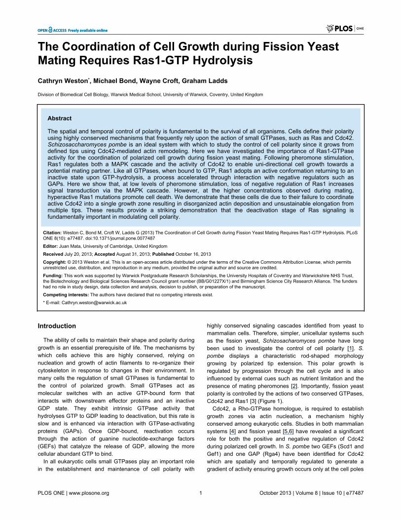
In all eukaryotic cells small GTPases play an important rolein the establishment and maintenance of cell polarity with
highly conserved signaling cascades identified from yeast tomammalian cells. Therefore, simpler, unicellular systems suchas the fission yeast, Schizosacharomyces pombe have longbeen used to investigate the control of cell polarity [1]. S.pombe displays a characteristic rod-shaped morphologygrowing by polarized tip extension. This polar growth isregulated by progression through the cell cycle and is alsoinfluenced by external cues such as nutrient limitation and thepresence of mating pheromones [2]. Importantly, fission yeastpolarity is controlled by the actions of two conserved GTPases,Cdc42 and Ras1 [3] (Figure 1).
Cdc42, a Rho-GTPase homologue, is required to establishgrowth zones via actin nucleation, a mechanism highlyconserved among eukaryotic cells. Studies in both mammaliansystems [4] and fission yeast [5,6] have revealed a significantrole for both the positive and negative regulation of Cdc42during polarized cell growth. In S. pombe two GEFs (Scd1 andGef1) and one GAP (Rga4) have been identified for Cdc42which are spatially and temporally regulated to generate agradient of activity ensuring growth occurs only at the cell poles
PLOS ONE | www.plosone.org 1 October 2013 | Volume 8 | Issue 10 | e77487
[7]. Gef1 and Scd1 share the essential role of Cdc42 activationand a double deletion is not viable however, the two proteinsare not functionally redundant. Gef1 regulates the temporallycontrolled transition from monopolar to bipolar growth ensuringthe correct cell size is achieved before cell division [8]. Scd1 isrequired to concentrate Cdc42 activity at the cell tip focusinggrowth at this location. Deletion of Scd1 or its activator, a
second small GTPase, Ras1 results in a complete loss inpolarity [9] since Cdc42 activity is not as efficiently directed tothe cell tip and as such growth occurs over a wider arearesulting in round cells [7].
In addition to their role in regulating mitotic cell growth, Scd1and Ras1 are also required for mating. Upon nutrient limitationS. pombe cells undergo sexual differentiation resulting in an
Figure 1. Schematic representation of Ras1 signaling cascades. Ras1 signals via two distinct effectors to control of bothpheromone-inducible and routine cell growth pathways. In the absence of pheromone the GEF, Efc25 promotes GDP for GTPexchange on Ras1. Through a complex with a Rho-GEF, Scd1, Ras1-GTP activates the essential G protein, Cdc42 so allowingpolarized cell growth, faithful chromosome segregation and cell division. A second GEF for Cdc42, Gef1, is required to initiatebipolar growth but is not activated by Ras1. Upon pheromone stimulation, Ras1 activation is increased through interaction withanother Ras1-GEF, Ste6. This pool of Ras1-GTP propagates the transcriptional response (acting via a second effector, Byr2 and aMAPK cascade). Pheromone-activated Ras1 also induces further GTP exchange on Cdc42 through activation of Scd1 promotingunidirectional cell growth towards a mating partner. Ras1 and Cdc42 contain a slow intrinsic ability to hydrolyze GTP, but thereaction is accelerated through interaction with GAPs, Gap1 and Rga4 (shown in red). Cross-talk between the two pathways ishighlighted with dashed arrows.doi: 10.1371/journal.pone.0077487.g001
Gap1 Is Essential for Yeast Mating
PLOS ONE | www.plosone.org 2 October 2013 | Volume 8 | Issue 10 | e77487
arrest in G1 and the expression of mating pheromones andtheir receptors [2]. Many of these responses are controlled bythe transcription factor Ste11, which is regulated throughphosphorylation by members of a mitogen-activated proteinkinase (MAPK) cascade that is stimulated by Ras1 [10].Importantly, Ras1 also provides the link between pheromonestimulation and activation of the Scd1-Cdc42 pathway requiredfor polarized (uni-directional) cell growth during the matingresponse (termed shmoo).
Ras1 activity requires similar regulation to Cdc42 having twotemporally restricted GEFs; Efc25 for mitotic growth [11] andSte6, induced upon nitrogen starvation to regulate matingresponses [12]. A single predicted GAP for Ras1, Gap1 hasbeen identified [13]. Deletion of Gap1 or expression ofhyperactive Ras1 mutants (analogous to the GTPase defective,oncogenic transitions observed in human cancer cells) resultsin cells that are hypersensitive to pheromone stimulation butsterile [13]. Interestingly there is no effect on the polarmorphology of mitotically growing cells containing the samehyperactive Ras1 mutations. Given these contrastingobservations for the requirement of negative regulation, wesought to quantitatively investigate the role of Ras1-GTP-hydrolysis on signal propagation.
We confirm, using Förster resonance energy transfer (FRET)that Gap1 interacts with Ras1-GTP. Furthermore, we identify adifference between the mechanisms Ras1 uses to mediatesignal propagation. We demonstrate that, whilst hyperactivatingmutations, predicted to prolong Ras1 activation, increase signaltransduction via the MAPK cascade, a reduction in signalpropagation via Cdc42 was observed. Significantly, we find thatthese cells are unable to initiate polarized cell growth inresponse to mating pheromone. Cells appear unable tocoordinate the localization of Cdc42 and therefore fail toestablish a single growth zone. Pheromone stimulation of thesecells results in attempted growth from multiple sites andultimately cell death. These results offer a novel insight intohow Ras proteins regulate cell polarity and highlight anessential role for negative regulation of G protein activation toensure efficient propagation via downstream signalingcascades.
Results
Gap1 is recruited to Ras1 in a GTP-dependent mannerGenetic analysis suggests that Gap1 acts primarily as a
negative regulator of Ras1 [13] however; it has not beendirectly demonstrated to interact with Ras1-GTP in vivo. Weexpressed CFP-Ras1 and Gap1-YFP fusion constructs in afission yeast strain (JY1618) lacking endogenous copies ofboth the genes. This strain also lacks Cyr1 generating agenotype mimicking nutrient limitation thereby allowingpheromone stimulation but producing cells of a smaller sizewhen grown in minimal media [14-16]. Protein function wasunaffected by expression from inducible plasmids orincorporation of the fluorescent moiety (Figure S1).
Consistent with previous observations [17], CFP-Ras1localized to plasma and endomembrane structures (Figure 2A).In contrast, Gap1-YFP appeared diffuse throughout the cytosol
and nucleus. Following 4 h stimulation with pheromone, Gap1-YFP was observed to translocate to the plasma membranecolocalizing with CFP-Ras1 (Pearson’s correlation coefficient(r) = 0.78) suggesting an interaction with Ras1, which wasconfirmed using FRET by sensitized-emission. Apparent FRETefficiencies were calculated for individual cells 0, 4 and 8 hfollowing stimulation with 10 μM pheromone. A significantincrease in FRET signal was observed 4 h post stimulationwhich returned to basal levels within 8 h (Figure 2B). Toobserve the temporal dynamics of the Gap1-YFP-CFP-Ras1interaction we used fluorescence activate cell sorting (FACS,Figure S1F) to quantify the occurrence of FRET (Figure 2C). Asmall number (8 ± 0.1%) of mitotically growing cells (Time = 0,Figure 2C) co-expressing CFP-Ras1 and Gap1-YFP produceda FRET signal; this percentage increased 2-fold following 30min pheromone stimulation, peaking at 2-4 h before returningto basal by 8 h. These dynamics are in broad agreement withthat of the activation of Ras proteins determined for otherorganisms [18] and demonstrate that in fission yeast, Gap1interacts with GTP-bound, active Ras1.
The requirement for activation of Ras1 through binding ofGTP was confirmed using previously characterized Ras1mutants engineered to contain a CFP-label. Expression ofCFP-Ras1S22N (the mammalian equivalent, RasS17N is unable tobind GTP [19]) prevented pheromone-induced translocation ofGap1-YFP to the plasma membrane (Figure 3A). Similarly, nochange in Gap1-YFP localization (Figure 3B) was observed ina strain lacking the pheromone receptor, Mam2 [20]. Theseresults demonstrate the requirement for GDP for GTPexchange, which is enhanced through pheromone stimulation,to promote interaction between CFP-Ras1 and Gap1-YFP.
Several hyperactivating mutations of human Ras proteinshave been described. The RasG12V transition is predicted toprevent an interaction with GAPs [21]. Expression of theanalogous CFP-Ras1G17V in the fission yeast strains preventedtranslocation of Gap1-YFP to the plasma membrane (Figure3C) and no increase in FRET signal was detected followingpheromone stimulation (Figure 3E). This data is consistent withthe notion that the Gly to Val transition in the P-loop of Rasproteins results in a reduced affinity for the GAP [21] anddemonstrates that Gap1-YFP requires an interaction with CFP-Ras1 to translocate to the plasma membrane in S. pombe.
In contrast, the hyperactive Ras1Q66L mutation (themammalian equivalent, RasQ61L, has been shown to preventGTP hydrolysis) promoted Gap1-YFP association with theplasma and endomembrane structures (Figure 3D, r = 0.8).Pheromone stimulation resulted in an increase in thepercentage of cells co-expressing CFP-Ras1Q66L and Gap1-YFP generating a FRET signal (Figure 3E). Furthermore, theGln66Leu mutation prevented a return to basal levels. Thesedata support the hypothesis that this mutation does not preventGAP-binding but reduces GTP hydrolysis [21] and furtherhighlight that Gap1 interacts with Ras1 in a GTP-dependentmanner in fission yeast.
Gap1 had been predicted to accelerate the hydrolysis ofGTP on Ras1 [13]. The dynamics of the CFP-Ras1 and CFP-Ras1Q66L FRET interactions with Gap1-YFP strongly supportthis prediction: the incorporation of the Gln66Leu transition,
Gap1 Is Essential for Yeast Mating
PLOS ONE | www.plosone.org 3 October 2013 | Volume 8 | Issue 10 | e77487
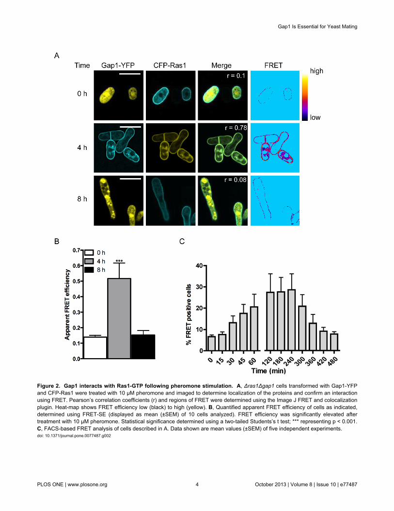
Figure 2. Gap1 interacts with Ras1-GTP following pheromone stimulation. A, Δras1Δgap1 cells transformed with Gap1-YFPand CFP-Ras1 were treated with 10 μM pheromone and imaged to determine localization of the proteins and confirm an interactionusing FRET. Pearson’s correlation coefficients (r) and regions of FRET were determined using the Image J FRET and colocalizationplugin. Heat-map shows FRET efficiency low (black) to high (yellow). B, Quantified apparent FRET efficiency of cells as indicated,determined using FRET-SE (displayed as mean (±SEM) of 10 cells analyzed). FRET efficiency was significantly elevated aftertreatment with 10 μM pheromone. Statistical significance determined using a two-tailed Students’s t test; *** representing p < 0.001.C, FACS-based FRET analysis of cells described in A. Data shown are mean values (±SEM) of five independent experiments.doi: 10.1371/journal.pone.0077487.g002
Gap1 Is Essential for Yeast Mating
PLOS ONE | www.plosone.org 4 October 2013 | Volume 8 | Issue 10 | e77487
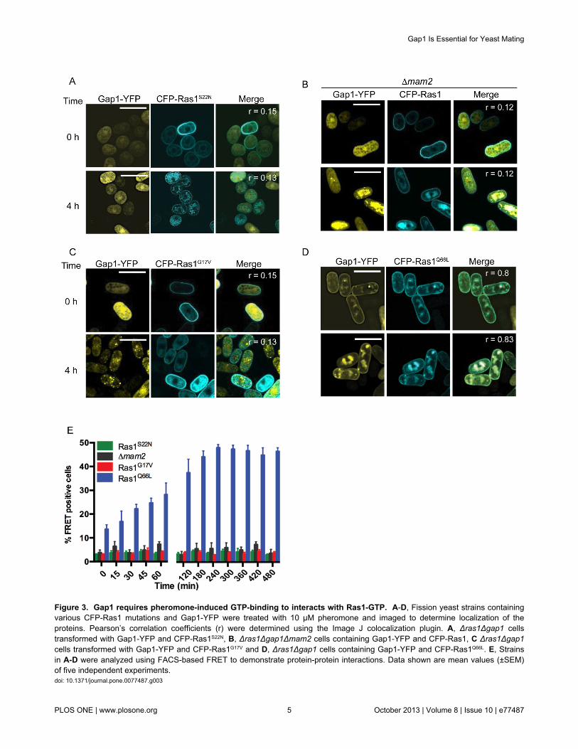
Figure 3. Gap1 requires pheromone-induced GTP-binding to interacts with Ras1-GTP. A-D, Fission yeast strains containingvarious CFP-Ras1 mutations and Gap1-YFP were treated with 10 μM pheromone and imaged to determine localization of theproteins. Pearson’s correlation coefficients (r) were determined using the Image J colocalization plugin. A, Δras1Δgap1 cellstransformed with Gap1-YFP and CFP-Ras1S22N, B, Δras1Δgap1Δmam2 cells containing Gap1-YFP and CFP-Ras1, C Δras1Δgap1cells transformed with Gap1-YFP and CFP-Ras1G17V and D, Δras1Δgap1 cells containing Gap1-YFP and CFP-Ras1Q66L. E, Strainsin A-D were analyzed using FACS-based FRET to demonstrate protein-protein interactions. Data shown are mean values (±SEM)of five independent experiments.doi: 10.1371/journal.pone.0077487.g003
Gap1 Is Essential for Yeast Mating
PLOS ONE | www.plosone.org 5 October 2013 | Volume 8 | Issue 10 | e77487
which prevents GTP-hydrolysis, results in a sustained FRETsignal, indicating that Gap1-YFP is not released until Ras1becomes GDP-bound.
Gap1-mediated GTP hydrolysis is required for anefficient mating response
Ras1-mediated signal transduction is required for fissionyeast mating [22]. Increasing the cellular concentration of Gap1(using the thiamine repressible nmt1 promoter) resulted in cellsthat displayed a reduced mating efficiency (Figure 4A andTable S1). These results, combined with the observeddecrease in the FRET signal (a marker for active Ras1) 4 hpost pheromone stimulation (Figure 2C) suggest that theremoval of Gap1 would prolong the active Ras1-state andtherefore increase signal transduction and mating efficiency.However, counter-intuitively, deletion of gap1 reduced matingefficiency (Figure 4A, [13]). Similar data was obtained for S.pombe cells expressing either the hyperactive Ras1G17V orRas1Q66L mutants suggesting that loss in mating in Δgap1strains results from the absence of Gap1-mediated Ras1-GTPhydrolysis. These data indicate that prolonging the GTP-bound‘active’ state of Ras1 paradoxically reduces signal transduction.
To quantitatively investigate the molecular mechanismsunderlying the reduced mating efficiency, we generatedpheromone-responsive reporter strains either lacking gap1 orwhere the endogenous ras1 gene was replaced with thehyperactive Ras1 mutations. These previously describedreporter strains contain the bacterial gene lacZ, at the sxa2locus [23,24]. Sxa2 is a carboxypeptidase that is onlyexpressed following pheromone stimulation and Ras1-mediated activation of the MAPK cascade [25,26]. Therefore,expressing lacZ under the transcriptional control of the sxa2promoter provides a quantitative readout for Ras1-Byr2-mediated signal transduction, which is required for a matingresponse. All strains lacking gap1 or containing ahyperactivating Ras1 mutation revealed an initial increase inpheromone sensitivity (<0.1 μM) however, when exposed topheromone concentrations exceeding 0.1 μM, signaling wasreduced 5-fold (Figure 4B). These observations are in contrastto the expected outcome from the removal of a negativeregulator and may explain the loss in mating efficiency.
Gap1 is essential during the pheromone responseIn addition to the activation of Byr2 for gene transcription,
efficient mating in S. pombe requires the initiation of uni-directional growth to form a conjugation tube or shmoo (Figure1) [2]. This polarized growth requires Ras1 activation of Scd1[9] and can be studied using cells lacking Sxa2. Uponprolonged exposure to 10 μM pheromone (16 h) Δsxa2 strainsexhibit abnormally long conjugation tubes generated from asingle tip [27] (Figure 4C). Under the same treatmentconditions our hyperactive mutant strains failed to exhibit adefined polarity, often (approximately 33%, n = 103 cells)attempting to elongate from multiple sites (Figure 4C, arrows).In addition, many cells displayed enlarged vacuoles (Figure 4C,cross) and a number of cellular fragments were also observed.
Staining with calcofluor white, a marker for cell wall materialrevealed that many cells within the mutant populations
contained a build up of cell wall material within their cytosol(Figure 4C, arrow heads) appearing to have died post-pheromone treatment. To confirm this observation, we used acommercially available cell viability assay (LIVE/DEAD® FungalightTM yeast viability kit (Invitrogen Ltd)) to quantify thepercentage of cell death within a population followingpheromone stimulation (Figure 4D). All mutant strainsdisplayed a dose-dependent increase in percentage cell death,reaching a maximum at 1 μM pheromone. The pheromone-dependent reduction in the number of viable cellscorresponded to the loss in transcriptional reporter geneactivity observed in the mutant strains. Correcting thetranscriptional reporter gene activity to account for the loss incell viability revealed an increase in maximal response in thehyperactive mutant strains compared to wild type cells (Figure4E). These data are consistent with observations for humanoncogenic Ras mutations, which increase signal transductionvia the Byr2 homologue, Raf1. These data suggest that thedecrease in reporter gene activity observed at high (>0.1 μM)pheromone concentrations in the absence of Ras1-GTPhydrolysis results from a significant reduction in viable cells andnot a loss in Ras1-mediated activation of Byr2. Similar defectsin cell morphology and a loss in cell viability (Figure 4F) wereobserved in the mating mixes of strains lacking gap1 orcontaining hyperactivating Ras1 mutations demonstrating thatGap1-accelerated Ras1-GTP hydrolysis is essential at thephysiological concentrations of pheromone required for mating.
Gap1-mediated Ras1-GTP hydrolysis is required duringcell elongation
Having determined that Ras1-GTP hydrolysis is essentialduring fission yeast mating, we sought to elucidate the timeduring the response when signal termination becomes critical.Following stimulation with 10 μM pheromone, the sxa2>lacZreporter strains elongate from a single tip forming a shmoo(Figure 5A and Movie S1). This change in morphology can bequantified as an increase in cell volume using a Coulterchannelyzer [28], beginning 2–4 h post stimulation, reaching amaximum by 10–12 h (Figure 5B) with similar dynamicsobserved for transcriptional reporter gene activity (Figure 5C).Mutant strains exposed to 10 μM pheromone failed to establisha defined, single growth site during the same time-frame(Movies S2-4). Many cells within the population displayedmultiple projection tips (Figure 5A) after 4 hours coinciding withthe time at which a significant increase in cell death wasobserved (Figure 5D). This cell death resulted in a decrease incell volume as measured using the Coulter channelyzer sincedead S. pombe cells appear small and rounded. Similarly, dueto the large increase in dead cells within the population littlereporter gene activity was observed. Significantly, the time atwhich the mutant cells begin to display aberrant cellmorphologies and a rapid increase in cell death corresponds tothe initiation of shmoo formation in the wild type strain. Theseobservations suggest that Gap1-accelerated Ras1-GTPhydrolysis becomes essential during the activation of Scd1-directed polarized cell growth.
To investigate the temporal dynamics of Ras1-mediatedsignal transduction in the mutant strains we used a sub-lethal
Gap1 Is Essential for Yeast Mating
PLOS ONE | www.plosone.org 6 October 2013 | Volume 8 | Issue 10 | e77487
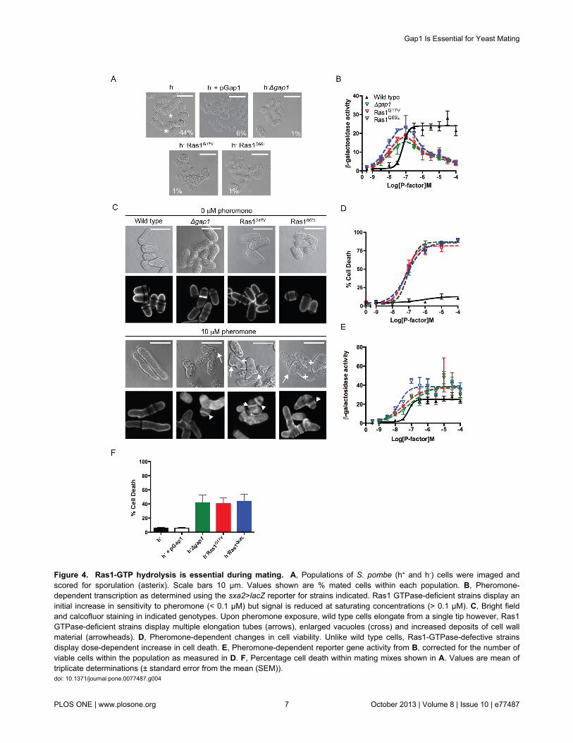
Figure 4. Ras1-GTP hydrolysis is essential during mating. A, Populations of S. pombe (h+ and h-) cells were imaged andscored for sporulation (asterix). Scale bars 10 μm. Values shown are % mated cells within each population. B, Pheromone-dependent transcription as determined using the sxa2>lacZ reporter for strains indicated. Ras1 GTPase-deficient strains display aninitial increase in sensitivity to pheromone (< 0.1 μM) but signal is reduced at saturating concentrations (> 0.1 μM). C, Bright fieldand calcofluor staining in indicated genotypes. Upon pheromone exposure, wild type cells elongate from a single tip however, Ras1GTPase-deficient strains display multiple elongation tubes (arrows), enlarged vacuoles (cross) and increased deposits of cell wallmaterial (arrowheads). D, Pheromone-dependent changes in cell viability. Unlike wild type cells, Ras1-GTPase-defective strainsdisplay dose-dependent increase in cell death. E, Pheromone-dependent reporter gene activity from B, corrected for the number ofviable cells within the population as measured in D. F, Percentage cell death within mating mixes shown in A. Values are mean oftriplicate determinations (± standard error from the mean (SEM)).doi: 10.1371/journal.pone.0077487.g004
Gap1 Is Essential for Yeast Mating
PLOS ONE | www.plosone.org 7 October 2013 | Volume 8 | Issue 10 | e77487
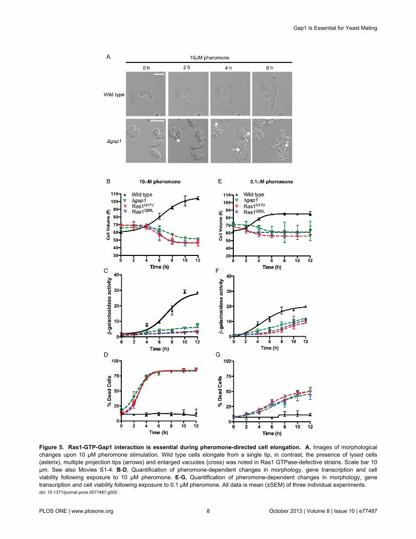
Figure 5. Ras1-GTP-Gap1 interaction is essential during pheromone-directed cell elongation. A, Images of morphologicalchanges upon 10 μM pheromone stimulation. Wild type cells elongate from a single tip, in contrast, the presence of lysed cells(asterix), multiple projection tips (arrows) and enlarged vacuoles (cross) was noted in Ras1 GTPase-defective strains. Scale bar 10μm. See also Movies S1-4. B-D, Quantification of pheromone-dependent changes in morphology, gene transcription and cellviability following exposure to 10 μM pheromone. E-G, Quantification of pheromone-dependent changes in morphology, genetranscription and cell viability following exposure to 0.1 μM pheromone. All data is mean (±SEM) of three individual experiments.doi: 10.1371/journal.pone.0077487.g005
Gap1 Is Essential for Yeast Mating
PLOS ONE | www.plosone.org 8 October 2013 | Volume 8 | Issue 10 | e77487
concentration of pheromone (0.1 μM), observed to cause <50% cell death within a population (Figure 4D). Wild type cellsexposed to 0.1 μM pheromone for 12 h did not obtain the samemaximal level in either the morphological or transcriptionalreporter gene assays as those observed at the highpheromone concentration, although the dynamics were similar(Figure 5E-F). In the mutant population a small increase intranscriptional reporter gene activity was detected however, nosignificant increase in cell volume was observed. Notably areduction, in pheromone-induced cell death in mutant strainstreated with 0.1 μM pheromone for 12 h (Figure 5G) comparedto 10 μM was observed. Previously it has been shown thatgene transcription occurs at a lower concentration ofpheromone than shmoo formation [6]. Therefore these datafurther indicate that Ras1-GTP hydrolysis is essential duringpheromone-induced, polarized cell growth. The lowerconcentration of pheromone induces some gene transcriptionbut this is insufficient to stimulate cell elongation therebyreducing the number of cells that die. Failure to terminate Ras1signaling at the high concentrations of pheromone required formating appears to inhibit formation of a single conjugation tube;instead the mutant cells attempt to elongate from multiple sites.
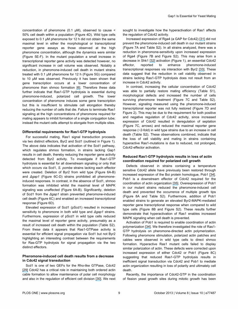
Differential requirements for Ras1-GTP hydrolysisFor successful mating, Ras1 signal transduction proceeds
via two distinct effectors, Byr2 and Scd1 (outlined in Figure 1).The above data indicates that activation of the Scd1 pathway,which regulates shmoo formation, in strains lacking Gap1results in cell death, thereby reducing the reporter gene activitydetected from Byr2 activity. To investigate if Ras1-GTPhydrolysis is essential for all downstream signaling or only thatwhich occurs via Scd1, S. pombe strains lacking each effectorwere created. Deletion of Byr2 from wild type (Figure 6A-B)and Δgap1 (Figure 6C-D) strains prohibited all pheromone-induced responses. In contrast, in the absence of Scd1, shmooformation was inhibited whilst the maximal level of MAPKsignaling was unaffected (Figure 6A-B). Significantly, deletionof Scd1 from the Δgap1 strain prevented pheromone-inducedcell death (Figure 6C) and enabled an increased transcriptionalresponse (Figure 6D).
Elevated expression of Scd1 (pScd1) resulted in increasedsensitivity to pheromone in both wild type and Δgap1 strains.Furthermore, expression of pScd1 in wild type cells reducedthe maximal level of reporter gene activity, presumably as aresult of increased cell death within the population (Table S2).From these data it appears that Ras1-GTPase activity isessential for efficient signal propagation via Scd1 but not Byr2highlighting an interesting contrast between the requirementsfor Ras-GTP hydrolysis for signal propagation via the twodistinct effectors.
Pheromone-induced cell death results from a decreasein Cdc42 signal transduction
Scd1 is one of two GEFs for the Rho-like GTPase, Cdc42[29] Cdc42 has a critical role in maintaining both ordered actincable formation to allow maintenance of polar cell morphologyand also in the regulation of efficient cell division [30]. We next
sought to investigate how the hyperactivation of Ras1 affectsthe regulation of Cdc42 activity.
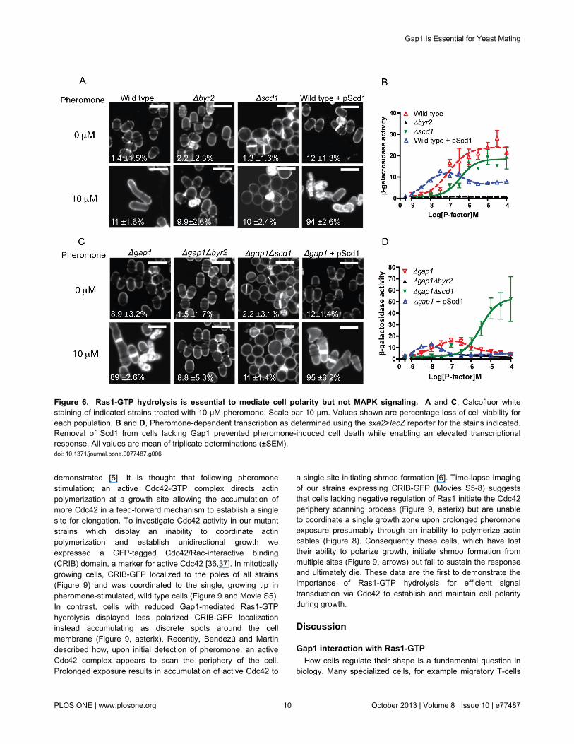
Increased expression of Rga4 (a GAP for Cdc42) [31] did notprevent the pheromone-induced cell death in our mutant strains(Figure 7A and Table S2). In all strains analyzed, there was areduction in pheromone-sensitivity upon increased expressionof Rga4 (Figure 7B and Figure S2). This may arise from adecrease in Shk1 [32] activation (Figure 1), an essential Cdc42effector, reported to enhance pheromone-inducedtranscriptional responses via interaction with Byr2 [33]. Thesedata suggest that the reduction in cell viability observed instrains lacking Ras1-GTP hydrolysis does not result from anincrease in Cdc42 activity.
In contrast, increasing the cellular concentration of Cdc42was able to partially restore mating efficiency (Table S1),morphological defects and increased the number of cellssurviving pheromone treatment (Figure 7C and Table S2).However, signaling measured using the pheromone-inducedtranscriptional reporter was not fully restored (Figure 7D andFigure 2). This may be due to the requirement for both positiveand negative regulation of Cdc42 activity, since increasedexpression of Cdc42 resulted in deregulation of septation(Figure 7C, arrows) and reduced the maximal reporter generesponse (~2-fold) in wild type strains due to an increase in celldeath (Table S2). These observations combined, indicate thatthe loss of cell viability and mating in strains containinghyperactive Ras1-mutations is due to reduced, not prolonged,Cdc42-effector activation.
Reduced Ras1-GTP hydrolysis results in loss of actincoordination required for polarized cell growth
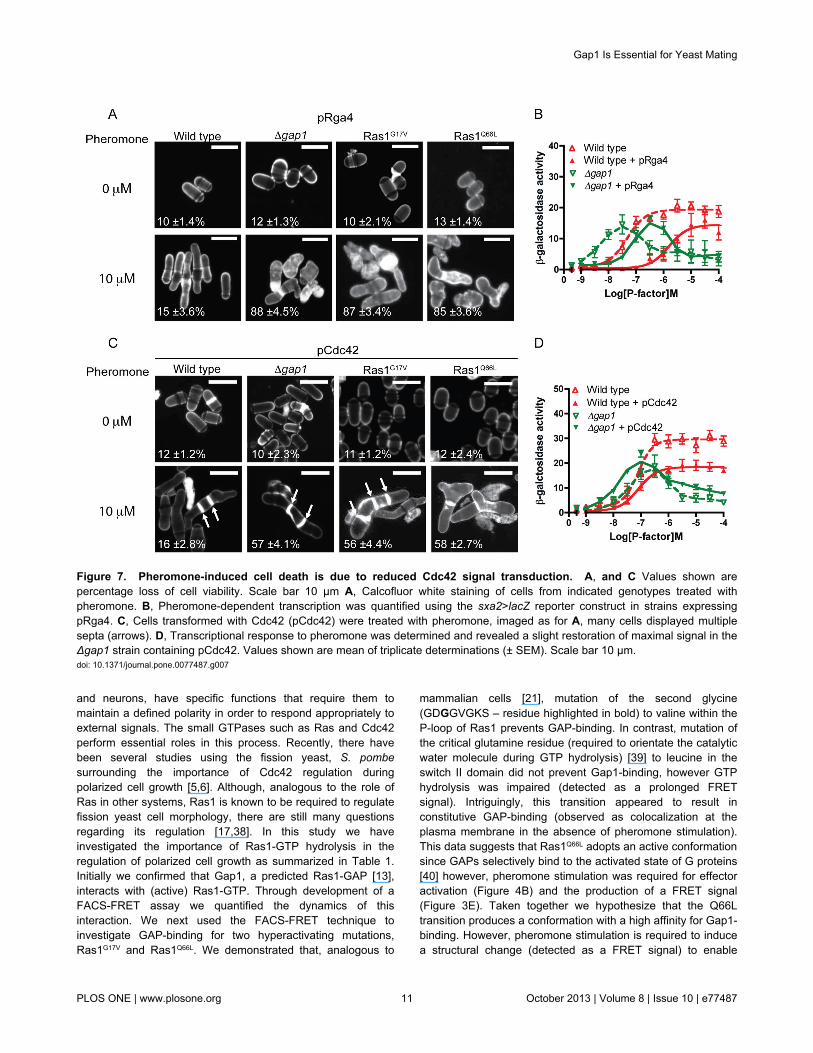
Defects observed in strains containing a temperaturesensitive Cdc42 allele have previously been restored throughincreased expression of the Boi protein homologue, Pob1 [34].Pob1 is a downstream effector of Cdc42 required for thecoordination of actin organization [35]. Overexpression of Pob1in our mutant strains reduced the pheromone-induced celldeath and prevented the occurrence of multiple growth tips(Figure 8A and Table S2). Furthermore, addition of Pob1enabled strains to generate an elevated Byr2-MAPK-mediatedreporter gene transcriptional response when compared to wildtype cells (Figure 8B and Figure S2). These results furtherdemonstrate that hyperactivation of Ras1 enables increasedMAPK signaling when cell death is prevented.
Activation of Pob1 is required to enable coordination of actinpolymerization [34]. We therefore investigated the role of Ras1-GTP hydrolysis on pheromone-directed actin polymerization.Following pheromone stimulation, polarized actin patches andcables were observed in wild type cells to direct shmooformation. Hyperactive Ras1 mutant cells failed to displaysimilar polarization of actin. These defects were corrected uponincreased expression of either Cdc42 or Pob1 (Figure 8C)suggesting that reduced Ras1-GTP hydrolysis results ininefficient signal transduction via Cdc42 and Pob1 to mediateactin organization resulting in loss of polarity and ultimately celldeath.
Recently, the importance of Cdc42-GTP in the coordinationof fission yeast growth sites during mitotic growth has been
Gap1 Is Essential for Yeast Mating
PLOS ONE | www.plosone.org 9 October 2013 | Volume 8 | Issue 10 | e77487
demonstrated [5]. It is thought that following pheromonestimulation; an active Cdc42-GTP complex directs actinpolymerization at a growth site allowing the accumulation ofmore Cdc42 in a feed-forward mechanism to establish a singlesite for elongation. To investigate Cdc42 activity in our mutantstrains which display an inability to coordinate actinpolymerization and establish unidirectional growth weexpressed a GFP-tagged Cdc42/Rac-interactive binding(CRIB) domain, a marker for active Cdc42 [36,37]. In mitoticallygrowing cells, CRIB-GFP localized to the poles of all strains(Figure 9) and was coordinated to the single, growing tip inpheromone-stimulated, wild type cells (Figure 9 and Movie S5).In contrast, cells with reduced Gap1-mediated Ras1-GTPhydrolysis displayed less polarized CRIB-GFP localizationinstead accumulating as discrete spots around the cellmembrane (Figure 9, asterix). Recently, Bendezú and Martindescribed how, upon initial detection of pheromone, an activeCdc42 complex appears to scan the periphery of the cell.Prolonged exposure results in accumulation of active Cdc42 to
a single site initiating shmoo formation [6]. Time-lapse imagingof our strains expressing CRIB-GFP (Movies S5-8) suggeststhat cells lacking negative regulation of Ras1 initiate the Cdc42periphery scanning process (Figure 9, asterix) but are unableto coordinate a single growth zone upon prolonged pheromoneexposure presumably through an inability to polymerize actincables (Figure 8). Consequently these cells, which have losttheir ability to polarize growth, initiate shmoo formation frommultiple sites (Figure 9, arrows) but fail to sustain the responseand ultimately die. These data are the first to demonstrate theimportance of Ras1-GTP hydrolysis for efficient signaltransduction via Cdc42 to establish and maintain cell polarityduring growth.
Discussion
Gap1 interaction with Ras1-GTPHow cells regulate their shape is a fundamental question in
biology. Many specialized cells, for example migratory T-cells
Figure 6. Ras1-GTP hydrolysis is essential to mediate cell polarity but not MAPK signaling. A and C, Calcofluor whitestaining of indicated strains treated with 10 μM pheromone. Scale bar 10 μm. Values shown are percentage loss of cell viability foreach population. B and D, Pheromone-dependent transcription as determined using the sxa2>lacZ reporter for the stains indicated.Removal of Scd1 from cells lacking Gap1 prevented pheromone-induced cell death while enabling an elevated transcriptionalresponse. All values are mean of triplicate determinations (±SEM).doi: 10.1371/journal.pone.0077487.g006
Gap1 Is Essential for Yeast Mating
PLOS ONE | www.plosone.org 10 October 2013 | Volume 8 | Issue 10 | e77487
and neurons, have specific functions that require them tomaintain a defined polarity in order to respond appropriately toexternal signals. The small GTPases such as Ras and Cdc42perform essential roles in this process. Recently, there havebeen several studies using the fission yeast, S. pombesurrounding the importance of Cdc42 regulation duringpolarized cell growth [5,6]. Although, analogous to the role ofRas in other systems, Ras1 is known to be required to regulatefission yeast cell morphology, there are still many questionsregarding its regulation [17,38]. In this study we haveinvestigated the importance of Ras1-GTP hydrolysis in theregulation of polarized cell growth as summarized in Table 1.Initially we confirmed that Gap1, a predicted Ras1-GAP [13],interacts with (active) Ras1-GTP. Through development of aFACS-FRET assay we quantified the dynamics of thisinteraction. We next used the FACS-FRET technique toinvestigate GAP-binding for two hyperactivating mutations,Ras1G17V and Ras1Q66L. We demonstrated that, analogous to
mammalian cells [21], mutation of the second glycine(GDGGVGKS – residue highlighted in bold) to valine within theP-loop of Ras1 prevents GAP-binding. In contrast, mutation ofthe critical glutamine residue (required to orientate the catalyticwater molecule during GTP hydrolysis) [39] to leucine in theswitch II domain did not prevent Gap1-binding, however GTPhydrolysis was impaired (detected as a prolonged FRETsignal). Intriguingly, this transition appeared to result inconstitutive GAP-binding (observed as colocalization at theplasma membrane in the absence of pheromone stimulation).This data suggests that Ras1Q66L adopts an active conformationsince GAPs selectively bind to the activated state of G proteins[40] however, pheromone stimulation was required for effectoractivation (Figure 4B) and the production of a FRET signal(Figure 3E). Taken together we hypothesize that the Q66Ltransition produces a conformation with a high affinity for Gap1-binding. However, pheromone stimulation is required to inducea structural change (detected as a FRET signal) to enable
Figure 7. Pheromone-induced cell death is due to reduced Cdc42 signal transduction. A, and C Values shown arepercentage loss of cell viability. Scale bar 10 μm A, Calcofluor white staining of cells from indicated genotypes treated withpheromone. B, Pheromone-dependent transcription was quantified using the sxa2>lacZ reporter construct in strains expressingpRga4. C, Cells transformed with Cdc42 (pCdc42) were treated with pheromone, imaged as for A, many cells displayed multiplesepta (arrows). D, Transcriptional response to pheromone was determined and revealed a slight restoration of maximal signal in theΔgap1 strain containing pCdc42. Values shown are mean of triplicate determinations (± SEM). Scale bar 10 μm.doi: 10.1371/journal.pone.0077487.g007
Gap1 Is Essential for Yeast Mating
PLOS ONE | www.plosone.org 11 October 2013 | Volume 8 | Issue 10 | e77487
Figure 8. Loss of Ras1-GTP hydrolysis prevents coordination of actin polymerization following pheromone stimulation. A,Cells transformed with Pob1 (pPob1) were treated with pheromone and imaged following staining with calcofluor white. Valuesshown are percentage loss of cell viability. B, Pheromone-dependent reporter gene activity was quantified using the sxa2>lacZreporter construct in strains expressing pPob1. Pob1 expression enabled an increased transcriptional response in strains lackinggap1. All values mean of triplicate determinations (±S.E.M). C, Rhodamine-phalloidin staining of actin wild type and strainscontaining indicated mutations. All mitotically growing cells (0 μM pheromone) display polarized actin at the cell tips (asterix).Following treatment with 10 μM pheromone elongating wild type cells exhibit defined actin patches (asterix) and cables (arrows). Allmutant strains failed to coordinate actin polymerization and a single growth site was not defined. These defects were restored uponincreased expression of pCdc42 or pPob1. Scale bar 10 μm.doi: 10.1371/journal.pone.0077487.g008
Gap1 Is Essential for Yeast Mating
PLOS ONE | www.plosone.org 12 October 2013 | Volume 8 | Issue 10 | e77487
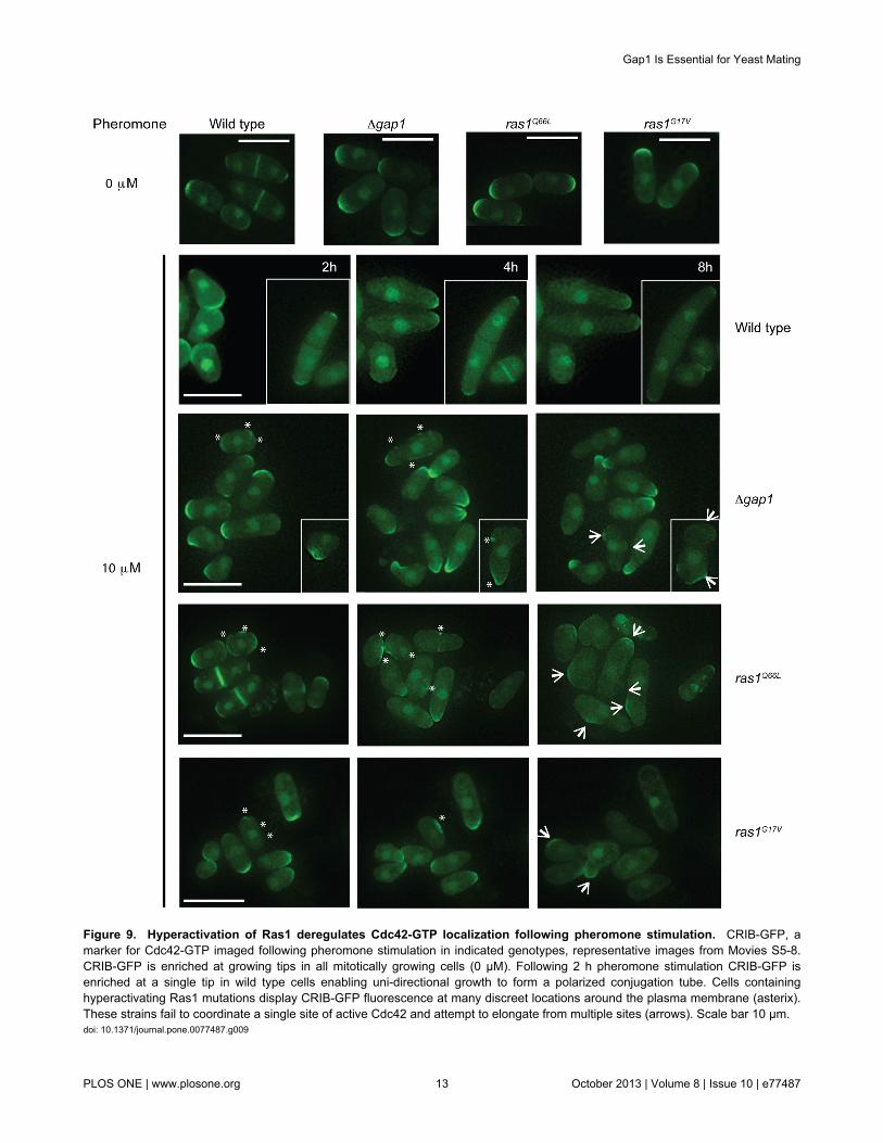
Figure 9. Hyperactivation of Ras1 deregulates Cdc42-GTP localization following pheromone stimulation. CRIB-GFP, amarker for Cdc42-GTP imaged following pheromone stimulation in indicated genotypes, representative images from Movies S5-8.CRIB-GFP is enriched at growing tips in all mitotically growing cells (0 μM). Following 2 h pheromone stimulation CRIB-GFP isenriched at a single tip in wild type cells enabling uni-directional growth to form a polarized conjugation tube. Cells containinghyperactivating Ras1 mutations display CRIB-GFP fluorescence at many discreet locations around the plasma membrane (asterix).These strains fail to coordinate a single site of active Cdc42 and attempt to elongate from multiple sites (arrows). Scale bar 10 μm.doi: 10.1371/journal.pone.0077487.g009
Gap1 Is Essential for Yeast Mating
PLOS ONE | www.plosone.org 13 October 2013 | Volume 8 | Issue 10 | e77487
activation of downstream effectors. Since GAPs cannot bindthe GDP-bound state of G proteins these observations lead toan interesting hypothesis whereby Ras1Q66L adopts anintermediary conformation that is neither GDP- (facilitatinginteraction with Gap1) nor GTP-bound (preventing activation ofByr2).
Ras1-GTP hydrolysis is essential for coordination ofCdc42-GTP
We have shown that upon hyperactivation of Ras1, fissionyeast cells fail to coordinate active Cdc42 to establish a singlegrowth zone following pheromone stimulation, insteadpromoting disorganized actin deposition. S. pombe has a rod-like shape that is regulated by the cell cycle and external cuessuch as mating pheromones. To maintain this polarity, theprecise spatial and temporal regulation of the small GTPases,Cdc42 and Ras1 are essential [3]. Cdc42 regulatescytoskeletal asymmetry and is activated by two GEFs, Gef1and the Ras1-activated Scd1 such that deletion of Scd1 orRas1 generates round, non-polar cells. Polarization of the actincytoskeleton, through localization of active Cdc42, recruitsScd1. Therefore; Cdc42 activity is self-regulated in a feed-forward mechanism, enabling cell growth. During mitoticgrowth, immediately after cell division, Cdc42 activity isdirected to the tip that existed before cytokinesis (the old end).At a defined time within the cell cycle (attainment of a minimalcell size) [41] Gef1 activity mediates the transition to bipolargrowth with active Cdc42 oscillating between both tips [5].Upon pheromone stimulation, the Ras1-Scd1 pathway directs
Table 1. Summary of pheromone-induced defects in Ras1-mediated signaling.
10 μM Pheromone Strains Cell Death MAPK Shmoo MatingWild type Strain alone - ++ ++ ++ Δbyr2 - - - - Δscd1 - ++ - - + pRga4 - ++ ++ ++ + pCdc42 - + ++ + + pScd1 ++ + + - + pPob1 + ++ ++ ++Hyperactive Ras1 mutants Strain alone +++ - - - Δbyr2 - - - - Δscd1 - ++ - - + pRga4 +++ - - - + pCdc42 ++ + ++ + + pScd1 +++ - - - + pPob1 + +++ ++ ++Data has been collated from the assays previously described and conducted in thisstudy. Hyperactive Ras1 mutants include fission yeast strains lacking gap1 orcontaining either Ras1G17V or Ras1Q66L. The extent of the response is
represented as follows: +++ increased level of response, ++normal level of
response, + reduced level of response and - no response.doi: 10.1371/journal.pone.0077487.t001
Cdc42-GTP to a single tip enabling orientation and cellelongation towards a mating partner (Figure 9).
Recently it has been demonstrated that on initial detection ofpheromone, an active Cdc42 complex appears to scan theperiphery of the cell. Following prolonged exposure, a singlesite is specified for growth with Cdc42-GTP accumulating in amechanism similar to that described for mitotic growth [6]. Wesuggest that in the absence of Ras1-GTP hydrolysis, cellsinitiate the membrane exploration phase but Cdc42-GTP isunable to interact with downstream effectors to polarize actinand recruit sufficient activity to define the single, ‘winning’ site.These cells appear to accumulate uncoordinated pockets ofactive Cdc42 around the cell periphery that persist and appearresistant to negative regulation by Rga4 (the Cdc42 GAP). Theresultant ectopic accumulation of Cdc42-GTP initiates growthat multiple sites, which is unsustainable for the cell. Thesepockets appear to sequester Cdc42 and its activator Scd1,thereby preventing it from performing essential roles inmediating routine cellular processes leading to loss in cellviability. We propose that, in the absence of Ras1-GTPhydrolysis, Cdc42-GTP is retained within the Ras1-GTP-Scd1-Cdc42-GTP complex, thereby inhibiting interaction with itsessential downstream effectors and preventing actinorganization. These data reveal an essential requirement forRas1-mediated GTP hydrolysis to transduce signals via Cdc42.
Ras1-GTP hydrolysis differentially regulates signaltransduction
Our data also highlights a striking contrast in themechanisms used by downstream effectors to propagate aresponse. As in higher eukaryotes, Ras1 interacts with multipleeffectors, including Scd1 for actin cytoskeleton remodeling andByr2, for MAPK-mediated gene transcription. When strainsexpressing the activating Ras1 mutations were prevented fromdying (through treatment with a lower concentration ofpheromone or deletion of Scd1) an elevated transcriptionalresponse, compared to strains containing a wild type Ras1protein, was observed. This data is consistent with reports ofoncogenic Ras mutants (deficient in GTPase activity)promoting unregulated cell proliferation via classical growthfactor signaling cascades [42]. These observations suggestthat the downstream effectors of Ras1 differentially requireGTP hydrolysis to propagate a response and could be highlysignificant when we consider the vast array of signaltransduction pathways regulated by Ras proteins inmammalian cells.
The proteins involved in the regulation of cell polarity arehighly conserved among eukaryotic cells; for example, Cdc42regulates actin cytoskeleton remodeling for T-cell migration andneuronal growth. Similarly, in mammalian cells, Ras proteinsregulate both MAPK cascades for gene transcription and Rho-GTPases (such as Cdc42) to maintain polarity. Activatingmutations in Ras proteins have been associated with tumorformation. However, whilst much research into their affects onMAPK signaling has been undertaken [43], little appears to beknown about oncogenic transitions and cell polarity. Theobservations we describe, that Ras1-GTP hydrolysis mayprevent excessive Cdc42-mediated actin organizing, are of
Gap1 Is Essential for Yeast Mating
PLOS ONE | www.plosone.org 14 October 2013 | Volume 8 | Issue 10 | e77487
considerable interest given the many diseases surroundingRas-mediated, uncontrolled cell growth and migration.
Materials and Methods
Strains, reagents and general methodsYeast strains used in this study are listed in Table S3. With
the exception of JY444 and JY1025, all yeast strains werederived from JY546 which contains the sxa2>lacZ construct forquantification of pheromone-dependent transcription [23,44].The strains are also deleted for cyr1 to induce sexualdifferentiation. Deletion of cyr1 results in strains undergoing celldivision at a smaller size although they maintain theircharacteristic barrel morphology [15]. Generation of Ras1mutants integrated at the ras1 locus was performed using atwo-step gene replacement strategy. The endogenous ras1was deleted using the ura4 cassette, which was subsequentlyreplaced by either Ras1G17V or Ras1Q66L. scd1 and byr2 weresingle-step deletions using the kanR cassette. Deletion ofMam2 has been described previously [24,44]. To enableconsistent comparison with the parent strains, which wereura4-, the Ura4 ORF was subsequently deleted from the locusof strains using a BamHI digest of JD437 (pKS+ Bluescriptcontaining the 5’ and 3’ un-translated regions of the ura4cassette separated by an EcoRV site) and selection by growthon minimal media plates containing 5-fluoro-ortic acid (5-FOA).We have used similar techniques for disruption of othermembers of the S. pombe pheromone-response pathway [44].To produce strains expressing the CRIB-3xGFP reporterconstruct we generated strains (JY944, JY1272, JY1386 andJY1538) that contained randomly generated point mutationswithin the Ura4 cassette (contained downstream of Krp1) [23],which were selected by growth on minimal media platescontaining 5-FOA. The shk1 promoter:ScGIC2 CRIB domain:3xGFP (a gift from Kaz Shiozaki) was then integrated byselection on minimal media plates lacking uracil. Colonies werescreened by microscopy to select successful recombinantsexpressing CRIB-GFP. Gene replacements were confirmedusing polymerase chain reaction (PCR). General yeastprocedures were performed as described previously [16,25]using yeast extract (YE) media for routine cell growth andselective, defined minimal media (minimal media) for allassays. Our minimal growth media is a variant of EMM asdescribed by Davey et al. [16], and places a higher nutritionaldemand on the cell resulting in the cells undergoing division ata smaller size [14]. For mild-overexpression of Cdc42, cellscontaining pREP4x-Cdc42 were grown in minimal media withthe addition 5 μg/ml thiamine. Oligonucleotides weresynthesized by Invitrogen (Paisley, Scotland, UK) amplificationby PCR used FastStart high fidelity polymerase blend (RocheDiagnostics Ltd., UK).
PlasmidsThe pREP vectors allow expression of genes under the
control of the thiamine-repressible, nmt1 promoter; pREP3xcontains the nutritional selection marker LEU2 and pREP4x,ura4 [45]. To account for differences in plasmid expressionlevels between individual cells, all yeast strains containing
plasmids were grown for 48 hours in the absence of thiamine toensure full expression from the nmt1 promoter was achieved[45] before assays and imaging were performed. S. pombegenes were amplified from genomic DNA using PCR andcloned into the pREP vectors. All Ras1 mutations weregenerated through inverse PCR and constructs sequenced toensure faithful amplification. Fluorescent constructs weregenerated using a two-step cloning technique as describedpreviously [24]. CFP was amplified using the sense primerJO2518 (ATGGTGAGCAAGGGCGAGGAG) and antisenseprimer JO2590 (atcCTTGTACAGCTCGTCCATGCCG) whichincludes half of an EcoRV site (underlined) in place of the stopanticodon. The PCR product was cloned into the uniqueEcoRV site of a modified pREP vector to generate JD3520(pREP4x-CFP) thereby also re-creating the EcoRV site in placeof stop anticodon. All Ras1 constructs were amplified usingJO1953 (ATGAGGTCTACCTACTTAAGAGAG) and JO1925(CTAACATATAACACAACA) containing the stop anticodon(shown in italics) and were cloned into the EcoRV site ofJD3520. The additional nucleotides (gat) inserted between theras1 ORF and the CFP ORF introduces an aspartate residue.YFP was amplified using the sense primer JD2318(atcATGGTGAGAAAGGGCGAGGAG) which includes half ofan EcoRV site (underlined) and JO2319(TTACTTGTACAGCTCGTCCATGCCG) containing the stopanticodon (shown in italics). This PCR product was cloned intothe unique EcoRV site of a modified pREP vector to generateJD3521 (pREP3x-YFP) thereby also re-creating the EcoRV siteimmediately upstream of the initiator codon of YFP. Gap1 wasamplified using JO2764 (ATGACTAAGCGGCACTCTGG) andJO2828 (CTTTCGTAAAAACAATTGTTCAAATAAAT) removingthe stop anticodon from the ORF and were cloned into theEcoRV site of JD3521. The additional nucleotides (atc) insertedbetween the gap1 ORF and the YFP ORF introduces anisoleucine residue.
β-galactosidase assayAssay of β-galactosidase activity was performed as
previously detailed [23,46]. Activity is displayed as OD420 per106 cells. Cell number and median cell volume weredetermined using Z2 Coulter channelyzer (Beckman Coulter,Luton, UK).
Mating assayTo determine mating efficiency we used a quantitative spore
viability assay. Many of the strains used are Sxa2 null mutantsand are sterile [13,26]. To enable mating assays to beperformed, we generated h- strains lacking Gap1 (JY1641) orexpressing the hyperactivating Ras1 mutations (Ras1G17V
(JY1642) or Ras1Q66L (JY1643)) from JY444 (h-, cyr1-) [47]. Toquantify spore viability yeast cultures were grown to a densityof ~ 5 x10⁶ cells/ml in minimal media and 200 μl of each teststrain was mixed with JY1025 (h+, cyr1-) [48]. Cells wereharvested at 2000 rpm for 3 min, the pellet was re-suspendedin 10 μl of sterile water and spotted on to a minimal media platewith 1 in 100 dilution of nitrogen. After 72 h incubation at 29°Ceach spot was picked into 1 ml of water. 500 μl of this culturewas taken and vegetatively cells were heat inactivated at 55°C
Gap1 Is Essential for Yeast Mating
PLOS ONE | www.plosone.org 15 October 2013 | Volume 8 | Issue 10 | e77487
for 10 min. Heated and unheated samples were plated on toyeast extract plates and incubated for a further 48 h at 29°C toallow spores to form colonies. Spore viability was determinedas:
% recovery = colonies formed after heat treatment / coloniesformed with no heating
Flow cytometryFlow cytometry was performed using a Beckton, Dickinson
and Company (BD) LSRII flow cytometer (BD Biosciences,Oxford, UK). Cell cycle analysis using propidium iodide stainingutilized an adapted protocol from Sazer and Sherwood, [49].Following overnight fixation with 70% ethanol, cells werewashed in 50 mM sodium citrate and digested for 2 h with 0.1mg/ml RNase. DNA was stained with 4 μg/ml propidium iodideand quantified using excitation with 488 nm laser detected with575/26 nm bandpass filter and 550 nm long pass. CFPfluorescence was detected after excitation with 405 nm laserwith 450/50 nm filter and YFP excitation was achieved with a488 nm laser and emission taken by 530/50 nm filter (505 nmlong pass). To determine FRET we excited with 405 nm laserand collected emission with 585/42 nm filter set and 545 nmlong pass. Gating to determine cells displaying a FRET signalis shown in Figure S1F and described previously [50]. Cellviability was calculated after pheromone treatment using theLIVE/DEAD® Funga lightTM yeast viability kit (Invitrogen Ltd)according to manufacturers protocol. Briefly, cells were treatedwith varying concentrations of pheromone, washed three timesin phosphate buffered saline and 0.5 μl of each viability dyereagent (SYTO 9 and propidium iodide) was added. Theproportion of cells containing both dyes was reported aspercentage of cells containing SYTO 9. All analysis wasperformed using FACSDiva v4.1 (BD Biosciences).
MicroscopyBright field microscopy time course experiments were
performed on 2% minimal media agarose plugs at 29°C withimages taken every 15 min. Bright field microscopy andfluorescent images were taken using a True Confocal ScannerLeica TCS SP5 microscope (Leica Microsystems Ltd., MiltonKeynes, UK). Actin patches and cables were stained usingrhodamine-conjugated phalloidin (Universal Biologicals) via aprotocol adapted from Marks et al., [51]. Briefly, 1 ml cells werefixed for 10 min with the addition of 100 μl 37% formaldehyde.Fixed cells were washed 3 times in PEM (0.1 M Na PIPES pH6.8, 1 mM EGTA, 1 mM MgCl2) and extracted for 30 sec inPEM + 1% TritonX-100. 100 μl fixed and extracted cells wereharvested and stained overnight at 4°C with 7 μl phalloidin (6.6μM stock). Actin was visualized using a Personal DeltaVision(Applied Precision, Issaquah, WA) comprising, an OlympusUPlanSApo 100x, N.A. 1.4, oil immersion objective and aPhotometric CoolSNAP HQ camera (Roper Scientific).Captured images were processed by iterative constraineddeconvolution using SoftWoRx (Applied Precession) andanalyzed using ImageJ. To visualize cell wall and septa, cellswere harvested and re-suspended in 0.1 mg/ml calcofluor white(Sigma) and imaged using the Leica TCS SP5 microscope(Leica Microsystems Ltd, UK). For the CRIB-GFP, images were
taken every 10 minutes using the Personal DeltaVision and theimage in the z-stack with the highest intensity chosen.
FRET measurements using confocal microscopeIn all FRET experiments, cells with medium fluorescence of
CFP and YFP fusion constructs were selected. We used thesensitized emission method for FRET measurements withdetection of fluorescence from single donor (CFP-Ras1),FRET, and acceptor (Gap1-YFP) in a line by line sequentialscan acquisition. To correct for excitation and emissioncrosstalk, calibration coefficients were calculated with thedonor-only and acceptor-only references. Parameters were firstadjusted with cells expressing the positive (CFP-YFP fusion)and negative (pCFP and pYFP) controls (Figure S1E) andremained constant for the entire experiment. The calibrationfactor β corrects for donor crosstalk and was calculated fromthe donor only reference by dividing the CFP donor emission(excited 458 nm laser) in the YFP channel by the CFP donoremission (excited 458 nm laser) in the CFP channel. Similarly,the calibration factor γ corrects for acceptor cross-excitationand was calculated from the acceptor only reference bydividing the YFP channel emission excited by the donor(excited 458 nm laser) with the YFP channel emission of YFP(excited 514 nm laser). Once all calibration factors have beencalculated, a region of interest (around a single cell) for theFRET measurement was determined in the FRET image andwas automatically inserted by the software at the identicalposition in the donor and acceptor channels. The apparentFRET efficiency (EA) was calculated by the equation: Ea (i) = B− A * β – C * γ/C [52]. FRET analysis was performed using theLeica Application Suite, Advanced Fluorescence 1.8.0software. In all experiments, CFP was excited by a 458 nmlaser line and detected at 465 to 500 nm. YFP was excited by a514 nm laser line and detected at 524 nm to 600 nm.
Data analysisData were analyzed using linear and non-linear regression
as appropriate using GraphPad Prism v6.0b for Mac OS X(GraphPad Software Inc, San Diego, CA). Statisticalsignificance was determined using a one-way ANOVA with aTukey multiple comparisons post-test or an un-paired Student’st test as appropriate. FRET and colocalization analysis wasperformed using ImageJ 1.46b [53,54].
Supporting Information
Figure S1. Measuring CFP-YFP FRET by FACS. A, Δras1strains transformed with either Ras1 or a CFP-Ras1 fusionconstructs were imaged. Δras1 (unlike wild type cells) have arounded morphology that is complemented upon expression ofplasmid-borne Ras1. Scale bar = 10 μm B, Mating efficiencywas determined using a spore viability assay for h- cells deletedfor Ras1 and expressing either empty vector or plasmid borneRas1 or CFP-Ras1. Both wild type Ras1 and the CFP-fusionwere able to restore wild type mating to the Δras1 cells whenexpressed from a plasmid. C-D, Δgap1 strains transformedwith Gap1-YFP were assayed for pheromone-induced changes
Gap1 Is Essential for Yeast Mating
PLOS ONE | www.plosone.org 16 October 2013 | Volume 8 | Issue 10 | e77487
in gene transcription C, and cell death D. Expression of Gap1-YFP restored signal transduction to near wild type levels. E,Imaging using the FRET-sensitization emission method forcells expressing CFP and YFP (negative control) or a CFP-YFP fusion construct (positive control) from thiamine-inducibleplasmids. Images were analyzed for colocalization and FRET(see Methods). Cells expressing CFP and YFP from individualplasmids gave a high degree of colocalization (r = 0.9) but thisdid not translate to a significant FRET signal. In contrast theCFP-YFP fusion construct displayed both a high colocalization(r = 0.9) and a FRET signal. F, Gating strategy to measureFRET by FACS (see Methods). Cells containing CFP, YFP,CFP and YFP and a CFP-YFP fusion expressed from plasmidswere analyzed as described in the methods section. Gateswere set to select double positive cells (panel 1), remove falsepositive FRET signals (panel 2) and define a positive (red)FRET signal (using CFP and YFP co-transfected population inpanel 3).(TIF)
Figure S2. Increased expression of Pob1 restores genetranscription on Ras1-GTPase defective strains. A, Cellscontaining the Ras1G17V and B, Ras1Q66L mutation weretransformed with various plasmids (see labels) and assayed forpheromone-dependent transcriptional response using thesxa2>lacZ reporter. All data are mean of triplicatedeterminations (±SEM).(TIF)
Movie S1. Wild type cells were grown on minimal mediacontaining agarose and supplemented with 10 μMpheromone. Images were obtained every 15 min, compliedand exported as movies with a frame rate of 5 frames persecond. Wild type cells elongate from a single tip in responseto pheromone displaying characteristic shmoos (original datafor Figure 4A).(AVI)
Movie S2. Cells lacking Gap1 were treated as describedfor Movie S1. Many aberrant responses were observed uponpheromone treatment including multiple projection tips,enlarged vacuoles and cell lysis (original data for Figure 4A).(AVI)
Movie S3. Cells expressing the Ras1G17V mutant weretreated as described for Movie S1. Similar defects inmorphology were observed upon pheromone treatment(including multiple projection tips, enlarged vacuoles and celllysis) to those displayed for cells lacking Gap1.(AVI)
Movie S4. Cells expressing the Ras1Q66L mutant weretreated as described for Movie S1. Similar defects inmorphology were observed upon pheromone treatment(including multiple projection tips, enlarged vacuoles and celllysis) to those displayed for cells lacking Gap1.(AVI)
Movie S5. Wild type cells containing CRIB-GFP weregrown on minimal media containing agarose andsupplemented with 10 μM pheromone. Images wereobtained every 10 min, complied and exported as movies witha frame rate of 5 frames per second. Wild type cells coordinatea single site of active Cdc42 (observed as an accumulation ofCRIB-GFP) enabling polarized elongation from a single tip(original data for Figure 8).(AVI)
Movie S6. Cells lacking Gap1 and containing CRIB-GFPwere treated as described for Movie S5. In the absence ofGap1 cells fail to coordinate a single site of active Cdc42instead displaying discreet dots of CRIB-GFP, which appear toscan the cell membrane. Many cells attempt to elongate frommultiple sites (original data for Figure 8).(AVI)
Movie S7. Cells expressing the Ras1G17V mutation andcontaining CRIB-GFP were treated as described for MovieS5. Similar defects were observed upon pheromone treatmentto those displayed for cells lacking Gap1 (original data forFigure 8).(AVI)
Movie S8. Cells expressing the Ras1Q66L mutation andcontaining CRIB-GFP were treated as described for MovieS5. Similar defects were observed upon pheromone treatmentto those displayed for cells lacking Gap1 (original data forFigure 8).(AVI)
Table S1. Asco-spore viability for strains expressingRas1-GTP hydrolysis mutations.(DOCX)
Table S2. Summary table of cell viability. To determine thenumber of dead cells within a population we utilized a cellviability assay (see Methods). Viability was determined in eachcase before and after treatment with pheromone and valuesare the means (±SEM) of triplicate determinations. Statisticalsignificance was determined using a Student’s t test.Differences were considered significant as follows; * p < 0.05,** p < 0.01, *** p < 0.005 from the same strain expressingvector control.(DOCX)
Table S3. Schizosaccharomyces pombe strains used inthis study.(DOCX)
Acknowledgements
We thank Pilar Pérez for plasmids, Kaz Shiozaki for the CRIB-GFP plasmid, Fulvia Verde for the Rga4 plasmid, and AlanGoddard for FRET discussions. We are indebted to Jonathan
Gap1 Is Essential for Yeast Mating
PLOS ONE | www.plosone.org 17 October 2013 | Volume 8 | Issue 10 | e77487
Miller, Malcolm Begg, Andrew Hall and Dawn Collins forreading and editing of the manuscript.
Author Contributions
Conceived and designed the experiments: CW MB WC GL.Performed the experiments: CW MB WC. Analyzed the data:
CW MB WC GL. Contributed reagents/materials/analysis tools:GL. Wrote the manuscript: CW GL.
References
1. Snell V, Nurse P (1993) Investigations into the control of cell form andpolarity: the use of morphological mutants in fission yeast. Dev Suppl:289-299. PubMed: 8049484.
2. Davey J (1998) Fusion of a fission yeast. Yeast 14: 1529-1566. doi:10.1002/(SICI)1097-0061(199812)14:16. PubMed: 9885154.
3. García P, Tajadura V, García I, Sánchez Y (2006) Role of RhoGTPases and Rho-GEFs in the regulation of cell shape and integrity infission yeast. Yeast 23: 1031–1043. doi:10.1002/yea.1409. PubMed:17072882.
4. Vanni C, Ottaviano C, Guo F, Puppo M, Varesio L et al. (2005)Constitutively active Cdc42 mutant confers growth disadvantage in celltransformation. Cell Cycle 4: 1675-1682. doi:10.4161/cc.4.11.2170.PubMed: 16294011.
5. Das M, Drake T, Wiley DJ, Buchwald P, Vavylonis D et al. (2012)Oscillatory Dynamics of Cdc42 GTPase in the Control of PolarizedGrowth. Science 13: 239-243. PubMed: 22604726.
6. Bendezú FO, Martin SG (2013) Cdc42 explores the cell periphery formate selection in fission yeast. Curr Biol 23: 42-47. doi:10.1016/j.cub.2012.10.042. PubMed: 23200991.
7. Kelly FD, Nurse P (2011) Spatial control of Cdc42 activationdetermines cell width in fission yeast. Mol Cell Biol 22: 3801–3811. doi:10.1091/mbc.E11-01-0057. PubMed: 21849474.
8. Coll PM, Trillo Y, Ametzazurra A, Perez P (2003) Gef1p, a new guaninenucleotide exchange factor for Cdc42p, regulates polarity inSchizosaccharomyces pombe. Mol Cell Biol 14: 313-323. doi:10.1091/mbc.E02-07-0400. PubMed: 12529446.
9. Chang EC, Barr M, Wang Y, Jung V, Xu HP et al. (1994) Cooperativeinteraction of S. pombe proteins required for mating andmorphogenesis. Cell 79: 131-141. doi:10.1016/0092-8674(94)90406-5.PubMed: 7923372.
10. Masuda T, Kariya K, Shinkai M, Okada T, Kataoka T (1995) Proteinkinase Byr2 is a target of Ras1 in the fission yeastSchizosaccharomyces pombe. J Biol Chem 270: 1979-1982. doi:10.1074/jbc.270.5.1979. PubMed: 7836423.
11. Tratner I, Fourticq-Esqueöute A, Tillit J, Baldacci G (1997) Cloning andcharacterization of the S. pombe gene efc25+, a new putative guaninenucleotide exchange factor. Gene 193: 203-210. doi:10.1016/S0378-1119(97)00115-7. PubMed: 9256078.
12. Hughes DA, Yabana N, Yamamoto M (1994) Transcriptional regulationof a Ras nucleotide-exchange factor gene by extracellular signals infission yeast. J Cell Sci 107: 3635-3642. PubMed: 7706412.
13. Imai Y, Miyake S, Hughes DA, Yamamoto M (1991) Identification of aGTPase-activating protein homolog in Schizosaccharomyces pombe.Mol Cell Biol 11: 3088-3094. PubMed: 2038319.
14. Fantes P, Nurse P (1977) Control of cell size at division in fission yeastby a growth-modulated size control over nuclear division. Exp Cell Res107: 377-386. doi:10.1016/0014-4827(77)90359-7. PubMed: 872891.
15. Maeda TT, Mochizuki NN, Yamamoto MM (1990) Adenylyl cyclase isdispensable for vegetative cell growth in the fission yeastSchizosaccharomyces pombe. Proc Natl Acad Sci U_S_A 87: 7814–7818. doi:10.1073/pnas.87.20.7814. PubMed: 2172964.
16. Davey J, Egel R, Nielsen O (1995) Pheromone procedures in fissionyeast. Methods Mol Genet 6: 247-263. doi:10.1016/S1067-2389(06)80016-6.
17. Onken B, Wiener H, Philips MR, Chang EC (2006) Compartmentalizedsignaling of Ras in fission yeast. Proc Natl Acad Sci U_S_A 103:9045-9050. doi:10.1073/pnas.0603318103. PubMed: 16754851.
18. Murakoshi H, Iino R, Kobayashi T, Fujiwara T, Ohshima C et al. (2004)Single-molecule imaging analysis of Ras activation in living cells. ProcNatl Acad Sci U_S_A 101: 7317-7322. doi:10.1073/pnas.0401354101.PubMed: 15123831.
19. Jung V, Wei W, Ballester R, Camonis J, Mi S et al. (1994) Two types ofRAS mutants that dominantly interfere with activators of RAS. Mol CellBiol 14: 3707-3718. PubMed: 8196614.
20. Kitamura K, Shimoda C (1991) The Schizosaccharomyces pombemam2 gene encodes a putative pheromone receptor which has a
significant homology with the Saccharomyces cerevisiae Ste2 protein.EMBO J 10: 3743-3751. PubMed: 1657593.
21. Gideon P, John J, Frech M, Lautwein A, Clark R et al. (1992)Mutational and kinetic analyses of the GTPase-activating protein(GAP)-p21 interaction: the C-terminal domain of GAP is not sufficientfor full activity. Mol Cell Biol 12: 2050-2056. PubMed: 1569940.
22. Nielsen O, Davey J, Egel R (1992) The ras1 function ofSchizosaccharomyces pombe mediates pheromone-inducedtranscription. EMBO J 11: 1391-1395. PubMed: 1563351.
23. Didmon M, Davis K, Watson P, Ladds G, Broad P et al. (2002)Identifying regulators of pheromone signalling in the fission yeastSchizosaccharomyces pombe. Curr Genet 41: 241-253. doi:10.1007/s00294-002-0301-3. PubMed: 12172965.
24. Ladds G, Davis K, Das A, Davey J (2005) A constitutively active GPCRretains its G protein specificity and the ability to form dimers. MolMicrobiol 55: 482-497. PubMed: 15659165.
25. Ladds G, Rasmussen EM, Young T, Nielsen O, Davey J (1996) Thesxa2-dependent inactivation of the P-factor mating pheromone in thefission yeast Schizosaccharomyces pombe. Mol Microbiol 20: 35-42.doi:10.1111/j.1365-2958.1996.tb02486.x. PubMed: 8861202.
26. Ladds G, Davey J (2000a) Sxa2 is a serine carboxypeptidase thatdegrades extracellular P-factor in the fission yeastSchizosaccharomyces pombe. Mol Microbiol 36: 377-390. doi:10.1046/j.1365-2958.2000.01855.x. PubMed: 10792724.
27. Imai Y, Yamamoto M (1992) Schizosaccharomyces pombe sxa1+ andsxa2+ encode putative proteases involved in the mating response. MolCell Biol 12: 1827-1834. PubMed: 1549128.
28. Davey J (1992) Mating pheromones of the fission yeastSchizosaccharomyces pombe: purification and structuralcharacterization of M-factor and isolation and analysis of two genesencoding the pheromone. EMBO J 11: 951-960. PubMed: 1547790.
29. Hirota K, Tanaka K, Ohta K, Yamamoto M (2003) Gef1p and Scd1p,the Two GDP-GTP exchange factors for Cdc42p, form a ring structurethat shrinks during cytokinesis in Schizosaccharomyces pombe. MolCell Biol 14: 3617-3627. doi:10.1091/mbc.E02-10-0665.
30. Merla A, Johnson DI (2000) The Cdc42p GTPase is targeted to the siteof cell division in the fission yeast Schizosaccharomyces pombe. Eur JCell Biol 79: 469-477. doi:10.1078/0171-9335-00073. PubMed:10961446.
31. Nakano K, Mutoh T, Mabuchi I (2001) Characterization of GTPase-activating proteins for the function of the Rho-family small GTPases inthe fission yeast Schizosaccharomyces pombe. Genes Cells 6:1031-1042. doi:10.1046/j.1365-2443.2001.00485.x. PubMed:11737264.
32. Ottilie S, Miller PJ, Johnson DI, Creasy CL, Sells MA et al. (1995)Fission yeast pak1+ encodes a protein kinase that interacts withCdc42p and is involved in the control of cell polarity and mating. EMBOJ 14: 5908-5919. PubMed: 8846783.
33. Tu H, Barr M, Dong DL, Wigler M (1997) Multiple regulatory domainson the Byr2 protein kinase. Mol Cell Biol 10: 5876-5887. PubMed:9315645.
34. Rincón SA, Ye Y, Villar-Tajadura MA, Santos B, Martin SG et al. (2009)Pob1 participates in the Cdc42 regulation of fission yeast actincytoskeleton. Mol Cell Biol 20: 4390-4399. doi:10.1091/mbc.E09-03-0207. PubMed: 19710424.
35. Toya M, Iino Y, Yamamoto M (1999) Fission yeast Pob1p, which ishomologous to budding yeast Boi proteins and exhibits subcellularlocalization close to actin patches, is essential for cell elongation andseparation. Mol Cell Biol 10: 2745-2757. doi:10.1091/mbc.10.8.2745.PubMed: 10436025.
36. Brown JL, Jaquenoud M, Gulli MP, Chant J, Peter M (1997) NovelCdc42-binding proteins Gic1 and Gic2 control cell polarity in yeast.Genes Dev 11: 2972-2982. doi:10.1101/gad.11.22.2972. PubMed:9367980.
37. Tatebe H, Nakano K, Maximo R, Shiozaki K (2008) Pom1 DYRKregulates localization of the Rga4 GAP to ensure bipolar activation of
Gap1 Is Essential for Yeast Mating
PLOS ONE | www.plosone.org 18 October 2013 | Volume 8 | Issue 10 | e77487
Cdc42 in fission yeast. Curr Biol 18: 322-330. doi:10.1016/j.cub.2008.02.005. PubMed: 18328707.
38. Papadaki P, Pizon V, Onken B, Chang EC (2002) Two ras pathways infission yeast are differentially regulated by two ras guanine nucleotideexchange factors. Mol Cell Biol 22: 4598-4606. doi:10.1128/MCB.22.13.4598-4606.2002. PubMed: 12052869.
39. Sprang SR (1997) G protein mechanisms: insights from structuralanalysis. Annu Rev Biochem 66: 639-678. doi:10.1146/annurev.biochem.66.1.639. PubMed: 9242920.
40. Scheffzek K, Lautwein A, Kabsch W, Ahmadian MR, Wittinghofer A(1996) Crystal structure of the GTPase-activating domain of humanp120GAP and implications for the interaction with Ras. Nature 384:591–596. doi:10.1038/384591a0. PubMed: 8955277.
41. Mitchison JM, Nurse P (1985) Growth in cell length in the fission yeastSchizosaccharomyces pombe. J Cell Sci 75: 357-376. PubMed:4044680.
42. Hamilton M, Wolfman A (1998) Ha-ras and N-ras regulate MAPKactivity by distinct mechanisms in vivo. Oncogene 16: 1417–1428. doi:10.1038/sj.onc.1201653. PubMed: 9525741.
43. Malumbres M, Barbacid M (2003) RAS oncogenes: the first 30 years.Nat Rev Cancer 3: 459–465. doi:10.1038/nrc1097. PubMed: 12778136.
44. Ladds G, Goddard A, Hill C, Thornton S, Davey J (2007) Differentialeffects of RGS proteins on G alpha(q) and G alpha (11) activity. CellSignal 19: 103-113. doi:10.1016/j.cellsig.2006.05.027. PubMed:16843638.
45. Maundrell K (1993) Thiamine-repressible expression vectors pREP andpRIP for fission yeast. Gene 123: 127-130. doi:10.1016/0378-1119(93)90551-D. PubMed: 8422996.
46. Ladds G, Davis K, Hillhouse EW, Davey J (2003) Modified yeast cellsto investigate the coupling of G protein-coupled receptors to specific G
proteins. Mol Microbiol 47: 781-792. doi:10.1046/j.1365-2958.2003.03336.x. PubMed: 12535076.
47. Ladds G, Davey J (2000b) Identification of proteases with sharedfunctions to the proprotein processing protease Krp1 in the fissionyeast Schizosaccharomyces pombe. Mol Microbiol 38: 839-853. doi:10.1046/j.1365-2958.2000.02180.x. PubMed: 11115118.
48. Egel R, Willer M, Kjaerulff S, Davey J, Nielsen O (1994) Assessment ofpheromone production and response in fission yeast by a halo test ofinduced sporulation. Yeast 10: 1347–1354. doi:10.1002/yea.320101012. PubMed: 7900424.
49. Sazer S, Sherwood SW (1990) Mitochondrial growth and DNAsynthesis occur in the absence of nuclear DNA replication in fissionyeast. J Cell Sci 97: 509-516. PubMed: 2074269.
50. Banning C, Volleler J, Hoffmann D, Koppensteiner H, Warmer M et al.(2010) A flow cytometry-based FRET assay to identify and analyseprotein-protein interactions in living cells. PLOS ONE 5: e9344. doi:10.1371/journal.pone.0009344. PubMed: 20179761.
51. Marks J, Hagan IM, Hyams JS (1986) Growth polarity and cytokinesisin fission yeast: the role of the cytoskeleton. J Cell Sci Suppl 5:229-241. PubMed: 3477553.
52. Wouters FS, Verveer PJ, Bastiaens PI (2001) Imaging biochemistryinside cells. Trends Cell Biol 11: 203–211. doi:10.1016/S0962-8924(01)01982-1. PubMed: 11316609.
53. Abramoff MD, Magalhães PJ, Ram SJ (2004) Image processing withImageJ. Biophotonics Int 11: 36-42.
54. Hachet-Haas M, Converset N, Marchal O, Matthes H, Gioria S et al.(2006) FRET and colocalization analyzer - A method to validatemeasurements of sensitized emission FRET acquired by confocalmicroscopy and available as an ImageJ Plug-in. Microsc Res Tech 69:941-956. doi:10.1002/jemt.20376. PubMed: 17080432.
Gap1 Is Essential for Yeast Mating
PLOS ONE | www.plosone.org 19 October 2013 | Volume 8 | Issue 10 | e77487
Related Documents