157 J. Exp. Med. The Rockefeller University Press • 0022-1007/98/07/157/11 $2.00 Volume 188, Number 1, July 6, 1998 157–167 http://www.jem.org The Coordinated Action of CC Chemokines in the Lung Orchestrates Allergic Inflammation and Airway Hyperresponsiveness By Jose-Angel Gonzalo,* Clare M. Lloyd,* Danyi Wen,* Juan P. Albar, ‡ Timothy N.C. Wells, § Amanda Proudfoot, § C. Martinez-A, ‡ Martin Dorf, i Torbjörn Bjerke, ¶ Anthony J. Coyle,* and Jose-Carlos Gutierrez-Ramos* From *Millennium Pharmaceuticals, Inc., Cambridge, Massachusetts 02139; the ‡ Centro Nacional Biotecnologia Consejo Superior Investigaciones Cientificas, Madrid, 28049 Spain; the § Glaxo Institute for Molecular Biology, Geneva, CH1228 Switzerland; the i Harvard Medical School, Boston, Massachusetts 02115; and ¶ Astra Draco AB, S-22100 Lund, Sweden Summary The complex pathophysiology of lung allergic inflammation and bronchial hyperresponsiveness (BHR) that characterize asthma is achieved by the regulated accumulation and activation of different leukocyte subsets in the lung. The development and maintenance of these processes correlate with the coordinated production of chemokines. Here, we have assessed the role that different chemokines play in lung allergic inflammation and BHR by blocking their activities in vivo. Our results show that blockage of each one of these chemokines reduces both lung leukocyte infiltration and BHR in a substantially different way. Thus, eotaxin neutralization re- duces specifically BHR and lung eosinophilia transiently after each antigen exposure. Mono- cyte chemoattractant protein (MCP)-5 neutralization abolishes BHR not by affecting the accu- mulation of inflammatory leukocytes in the airways, but rather by altering the trafficking of the eosinophils and other leukocytes through the lung interstitium. Neutralization of RANTES (regulated upon activation, normal T cell expressed and secreted) receptor(s) with a receptor antagonist decreases significantly lymphocyte and eosinophil infiltration as well as mRNA expres- sion of eotaxin and RANTES. In contrast, neutralization of one of the ligands for RANTES re- ceptors, macrophage-inflammatory protein 1a, reduces only slightly lung eosinophilia and BHR. Finally, MCP-1 neutralization diminishes drastically BHR and inflammation, and this correlates with a pronounced decrease in monocyte- and lymphocyte-derived inflammatory mediators. These results suggest that different chemokines activate different cellular and molecular pathways that in a coordinated fashion contribute to the complex pathophysiology of asthma, and that their individual blockage results in intervention at different levels of these processes. Key words: chemokines • allergic inflammation • bronchial hyperresponsiveness • eosinophilia • leukocytes L ung inflammation and bronchial hyperreactivity (BHR) 1 are two distinct characteristics of asthma (1, 2). Eosino- phils are considered to be the central proinflammatory leu- kocyte involved in the asthmatic reaction, due in part to se- creted toxic granular proteins and membrane products that induce pulmonary damage and subsequently intensify BHR (1–3). Moreover, there is a correlation between lev- els of eosinophil-derived cation proteins in the airways of asthmatic patients and the severity of this disease (2). De- spite the fact that the eosinophil is the predominant infil- trating cell type during asthma, other leukocytes may be critical in the initiation and amplification of the inflamma- tory response (4, 5). The prevention of both lung eosino- philia and BHR in T lymphocyte–deficient mice during antigen-induced inflammation supports a critical role for T cell cytokines such as IL-4 and IL-5 (6–9). The role that 1 Abbreviations used in this paper: BAL, bronchoalveolar lavage; BHR, bron- chial hyperresponsiveness; Eot, eotaxin; LT, leukotriene; MCP, mono- cyte chemoattractant protein; Met-RANTES, methionylated RANTES; MIP, macrophage-inflammatory protein; OVA, ovalbumin; RANTES, regulated upon activation, normal T cell expressed and secreted; RPA, RNase protection assay; TX, thromboxane. on January 13, 2015 jem.rupress.org Downloaded from Published July 1, 1998

Welcome message from author
This document is posted to help you gain knowledge. Please leave a comment to let me know what you think about it! Share it to your friends and learn new things together.
Transcript
157
J. Exp. Med.
The Rockefeller University Press • 0022-1007/98/07/157/11 $2.00Volume 188, Number 1, July 6, 1998 157–167http://www.jem.org
The Coordinated Action of CC Chemokines in theLung Orchestrates Allergic Inflammation andAirway Hyperresponsiveness
By Jose-Angel Gonzalo,
*
Clare M. Lloyd,
*
Danyi Wen,
*
Juan P. Albar,
‡
Timothy N.C. Wells,
§
Amanda Proudfoot,
§
C. Martinez-A,
‡
Martin Dorf,
i
Torbjörn Bjerke,
¶
Anthony J. Coyle,
*
and Jose-Carlos Gutierrez-Ramos
*
From
*
Millennium Pharmaceuticals, Inc., Cambridge, Massachusetts 02139; the
‡
Centro Nacional Biotecnologia Consejo Superior Investigaciones Cientificas, Madrid, 28049 Spain; the
§
Glaxo Institute for Molecular Biology, Geneva, CH1228 Switzerland; the
i
Harvard Medical School, Boston, Massachusetts 02115; and
¶
Astra Draco AB, S-22100 Lund, Sweden
Summary
The complex pathophysiology of lung allergic inflammation and bronchial hyperresponsiveness(BHR) that characterize asthma is achieved by the regulated accumulation and activation ofdifferent leukocyte subsets in the lung. The development and maintenance of these processescorrelate with the coordinated production of chemokines. Here, we have assessed the role thatdifferent chemokines play in lung allergic inflammation and BHR by blocking their activitiesin vivo. Our results show that blockage of each one of these chemokines reduces both lungleukocyte infiltration and BHR in a substantially different way. Thus, eotaxin neutralization re-duces specifically BHR and lung eosinophilia transiently after each antigen exposure. Mono-cyte chemoattractant protein (MCP)-5 neutralization abolishes BHR not by affecting the accu-mulation of inflammatory leukocytes in the airways, but rather by altering the trafficking of theeosinophils and other leukocytes through the lung interstitium. Neutralization of RANTES(regulated upon activation, normal T cell expressed and secreted) receptor(s) with a receptorantagonist decreases significantly lymphocyte and eosinophil infiltration as well as mRNA expres-sion of eotaxin and RANTES. In contrast, neutralization of one of the ligands for RANTES re-ceptors, macrophage-inflammatory protein 1
a
, reduces only slightly lung eosinophilia and BHR.Finally, MCP-1 neutralization diminishes drastically BHR and inflammation, and this correlateswith a pronounced decrease in monocyte- and lymphocyte-derived inflammatory mediators.These results suggest that different chemokines activate different cellular and molecular pathwaysthat in a coordinated fashion contribute to the complex pathophysiology of asthma, and that theirindividual blockage results in intervention at different levels of these processes.
Key words: chemokines • allergic inflammation • bronchial hyperresponsiveness • eosinophilia • leukocytes
L
ung inflammation and bronchial hyperreactivity (BHR)
1
are two distinct characteristics of asthma (1, 2). Eosino-phils are considered to be the central proinflammatory leu-kocyte involved in the asthmatic reaction, due in part to se-
creted toxic granular proteins and membrane products thatinduce pulmonary damage and subsequently intensifyBHR (1–3). Moreover, there is a correlation between lev-els of eosinophil-derived cation proteins in the airways ofasthmatic patients and the severity of this disease (2). De-spite the fact that the eosinophil is the predominant infil-trating cell type during asthma, other leukocytes may becritical in the initiation and amplification of the inflamma-tory response (4, 5). The prevention of both lung eosino-philia and BHR in T lymphocyte–deficient mice duringantigen-induced inflammation supports a critical role for Tcell cytokines such as IL-4 and IL-5 (6–9). The role that
1
Abbreviations used in this paper:
BAL, bronchoalveolar lavage; BHR, bron-chial hyperresponsiveness; Eot, eotaxin; LT, leukotriene; MCP, mono-cyte chemoattractant protein; Met-RANTES, methionylated RANTES;MIP, macrophage-inflammatory protein; OVA, ovalbumin; RANTES,regulated upon activation, normal T cell expressed and secreted; RPA,RNase protection assay; TX, thromboxane.
on January 13, 2015jem
.rupress.orgD
ownloaded from
Published July 1, 1998
158
Chemokines in Lung Allergic Responses
monocytes/macrophages play in lung allergic inflammationpertains to their ability to act as APCs and to generate a va-riety of mediators that promote both eosinophil activationand adhesion to endothelium (10). In this regard, macro-phages secrete eosinophilic chemoattractants, including leu-kotriene (LT)B
4
, platelet-activating factor, and C5a, in ad-dition to modulating the generation of some of thesefactors by eosinophils (10–12).
The immunological characterization of lung eosinophiliaimplicates chemokines in the initiation of lung allergic in-flammation and the subsequent development of BHR (5,13–17). Chemokines are a group of cytokines that promoteleukocyte recruitment to inflammatory sites, stimulate leu-kocyte exocytosis, and induce hematopoiesis (18–20).Chemokine expression is high and readily modulated in thelung during the development of an inflammatory response(5, 16, 17). Among these, RANTES (regulated upon acti-vation, normal T cell expressed and secreted), monocytechemoattractant protein (MCP)-5, and eotaxin have beenshown to induce eosinophil migration in vitro and/or to beinvolved in lung eosinophilia in humans, guinea pigs, andmice (5, 16, 13, 21–23). In addition to their activity oneosinophils, RANTES and MCP-5 are strong chemotacticfactors both in vivo and in vitro for T lymphocytes andmonocytes, respectively (16, 24). MCP-1, which is primar-ily involved in the recruitment of mononuclear phagocytes(25), is also strongly expressed in the lung during inflam-mation (5, 26). Signaling mediated by this noneosinophilicchemokine might also be involved in macrophage activa-tion leading to a cascade of proinflammatory and tissuedamage events (27).
In this report, we have used a mouse model of asthma inwhich the expression of the chemokines eotaxin, RANTES,macrophage-inflammatory protein (MIP)-1
a
, MCP-5, andMCP-1 is clearly modulated during disease progression (5).To understand the pathophysiology that results from theexpression of multiple chemokines with partially overlap-ping functions, we have analyzed (
a
) the direct recruitmentof specific cell types to the lung interstitium and airwaysexerted by these chemokines, (
b
) their role in the inductionof BHR, (
c
) the effect they exert on the gene expression ofother chemokines, and (
d
) the modulation by thesechemokines of different components of the pathological re-sponse, such as cell activation or inflammatory mediatorproduction.
Materials and Methods
Mice and In Vivo Procedures.
8–10-wk-old C57BL/6J micewere purchased from The Jackson Laboratory (Bar Harbor, ME)and kept in the specific pathogen–free mouse facility at Millen-nium Pharmaceuticals, Inc. The mouse model of lung inflamma-tion used here consists of a sensitization phase (ovalbumin[OVA], 0.1 mg/mouse intraperitoneally on day 0; Sigma Chemi-cal Co., St. Louis, MO) and an induction of the response phase(2% OVA for 5 min intranasally on day 8, and 1% OVA for 20min intranasally on days 15–21). PBS (intraperitoneal and/or in-tranasal) was administered to mice as a negative control. For the
blocking experiments, mice also received 20
m
g/mouse of neu-tralizing polyclonal Abs against either eotaxin (anti-Eot), MCP-5(anti–MCP-5), MIP-1
a
(anti–MIP-1
a
), or 5
m
g/mouse of mAbagainst MCP-1/JE (anti–MCP-1/JE), or 25
m
g/mouse of theRANTES antagonist, methionylated (Met)-RANTES. Anti–MIP-1
a
(R&D Systems, Inc., Minneapolis, MN) was selected forits ability to neutralize the in vitro bioactivity of recombinantmurine MIP-1
a
and was used at the manufacturer’s recom-mended dose. Anti-Eot Ab blocked the in vitro transmigration ofeosinophils to eotaxin by 97% (5). Similarly, anti–MCP-5 Abblocked MCP-5–induced eosinophil migration in vitro by 99%(16). The anti–MCP-1/JE Ab also showed a dose-dependent in-hibition of MCP-1 bioactivity using in vitro chemotactic assayson monocytes (28). We have determined previously that the doseof antibodies used here blocked 100% of migration to each partic-ular chemokine during in vivo peritoneal migration assays (datanot shown) or during crescentic nephritis (29). Met-RANTEShas been shown to inhibit both in vitro chemotaxis and calciuminflux on the THP-1 cell line and T cells in response toRANTES and MIP-1
a
(30) and in vivo inflammation (29).These antibodies were administered intravenously 30 min beforeOVA provocation on days 8–21. In another series of blocking ex-periments, anti–MCP-1/JE mAb, anti-Eot, or anti–MCP-5 poly-clonal Abs were also administered on days 8–16, days 15–21, days19–21, or day 21. OVA-treated control mice were injected withthe same amount of control Ab at the same time points indicatedduring treatment. Rabbit Ig fraction (DAKO Corp., Santa Bar-bara, CA) was used as control for poly-Eot and poly–MCP-5,normal hamster IgG (Jackson ImmunoResearch Labs, WestGrove, PA) was used as control for anti–MCP-1/JE, and PBS ascontrol for Met-RANTES. 1 or 3 h after OVA administration onday 15 or 21, mice were killed by CO
2
asphyxiation and ana-lyzed.
Bronchoalveolar lavage (BAL) was performed as described(23). In brief, the airways of the mice were lavaged via a tracheacannula with 1 ml of PBS. The resulting BAL fluid was immedi-ately centrifuged (700
g
, 5 min, 4
8
C), and BAL cells were thenwashed and resuspended in 1 ml of PBS.
The degree of bronchoconstriction (BHR) was measured 3 hafter the last antigen challenge by recording respiratory pressurecurves by whole body plethysmography (Buxco Electronics, Inc.,Sharon, CT) in response to inhaled methacholine (MCh; AldrichChemical Co., Milwaukee, WI) at a concentration of 3
3
10
2
2
Mfor 1 min, as described previously (3, 31). BHR was expressed asenhanced pause (
Penh
), a calculated value, which correlates withmeasurement of airway resistance, impedance, and intrapleuralpressure in the same mouse.
Penh
5
(
Te
/
Tr
2
1)
3
(
Pef
/
Pif
),where
Te
is expiration time;
Tr
, relaxation time;
Pef
, peak expira-tory flow; and
Pif
, peak inspiratory flow
3
0.67 coefficient (32).The relaxation time is the time it takes for the box pressure tochange from a maximum to a user-defined percentage of themaximum. Here,
Tr
measurement begins at the maximum boxpressure and ends at 40%.
Immunohistochemical Phenotyping and Quantitation of Leuko-cytes.
Total BAL cell counts were performed, and aliquots (5
3
10
5
cells/slide) were pelleted onto glass slides by cytocentrifugation.To determine the number of eosinophils and neutrophils, slideswere stained with Wright-Giemsa (Fisher Scientific Co., Pitts-burgh, PA). T lymphocytes, B lymphocytes, and mononuclearphagocytes were identified by Thy 1.2 (53-2.1; PharMingen, SanDiego, CA), IgM (II/41; PharMingen), and Moma-2 (BiosourceInternational, Camarillo, CA) staining, respectively, as described(5). Percentage of eosinophils, lymphocytes, neutrophils, and
on January 13, 2015jem
.rupress.orgD
ownloaded from
Published July 1, 1998
159
Gonzalo et al.
macrophages was determined by counting their number in eighthigh power fields (
3
40, total area 0.5 mm
2
) per area randomlyselected, and dividing this number by the total number of cellsper high power field. To obtain the absolute number of each leu-kocyte subtype in the lavage, these percentages were multipliedby the total number of cells recovered from the BAL fluid.
Lung sections from the different experimental groups of micewere prepared as described (5). In brief, lungs were fixed in 10%neutral buffered formalin (NBF; J.T. Baker, Phillipsburg, NJ) andparaffin embedded. Sections (4 microns) were cut onto micro-scope slides and stained with hematoxylin/eosin according tostandard protocols. To determine the sizes of pulmonary infil-trates in the experimental groups of mice, the area of lung tissuecovered by infiltrate was calculated in sections at low power(
3
100) using NIH Image 1.56. At least six fields of at least 0.01mm
2
were scanned, and the mean percentage area was deter-mined for each mouse of each experimental group, relative to thecontrol mice for each experiment.
Determination of Chemokine Protein Expression within Lung Tissue.
The level of expression of eotaxin protein and MCP-5 proteinwas determined in sections from lungs of OVA-treated mice andcontrols. Sections were fixed and stained using a modified avidin/biotin staining method. All incubations were carried out underhumidified conditions, and slides were washed twice betweensteps for 5 min each in 0.1 M PBS supplemented with 0.2% gela-tin. Sections were overlaid with 20% normal rabbit serum in PBSfor 15 min and then incubated overnight at 4
8
C with either mon-oclonal anti-Eot diluted 1:2 in PBS with 0.1% BSA and 0.1% so-dium azide (5) or anti-murine MCP-5 (ZY2A11) culture super-natant (undiluted). Endogenous peroxide was subsequentlyblocked by incubation for 20 min in methanol containing 0.3%hydrogen peroxide. Nonspecific staining due to cross-reactionwith endogenous avidin or biotin was blocked by incubationwith avidin solution followed by biotin solution, both for 20 min.Bound mAb was visualized by incubation with biotinylated rabbitanti–rat Ig diluted in 10% normal mouse serum PBS, and then in-cubated for 1 h each in streptavidin–peroxidase complex preparedaccording to the manufacturer’s instructions (both from DAKOCorp.). Finally, slides were flooded with peroxidase substrate so-lution (20 mg diaminobenzidine in 10 ml PBS, containing 0.01%hydrogen peroxide) for 10 min before counterstaining withhematoxylin. Control slides were either stained with an isotype-matched negative control Ab instead of primary Ab or biotiny-lated anti–rat Ig or streptavidin complex were selectively omitted.
Measurement of Chemokine mRNA Expression.
Total RNA fromthe lungs of OVA-treated mice or control littermates at differenttime points was extracted by the single step method using RNASTAT-60 (Tel-Test, Inc., Friendswood, TX).
Chemokine mRNA expression was determined by multiprobeRNase protection assay (RPA) using the RiboQuant RPA kit(PharMingen) as recommended by the supplier. The identity andquantity of each mRNA species in the original RNA samplewere then determined based on the signal intensities given by theappropriately sized, protected probe fragment bands. The sampleloading was normalized by the housekeeping gene, GAPDH, in-cluded in each template set.
Measurement of Cytokine, Inflammatory Mediator, and Ab Lev-els.
The release of these activating factors during OVA adminis-tration was determined by ELISA. BAL fluid was taken 1 h afterantigen challenge on day 15 or 21. Serial dilutions of BAL fluidsamples were assayed using commercial ELISA kits for IL-4, IL-5,TNF-
a
, and IFN-
g
(Endogen, Inc., Boston, MA) and commer-cial enzyme immunoassay kits for LTB
4
, PGE
2
, thromboxane
(TX)B
2
, and LTC
4
/D
4
/E
4
(Amersham International, Bucking-hamshire, UK). Total IgE titers were also measured by ELISA ac-cording to Ledermann et al. (33). Absorbance values were con-verted to concentrations of each factor in the BAL fluid(picograms or nanograms per milliliter) by interpolation in the re-spective standard curve.
Results
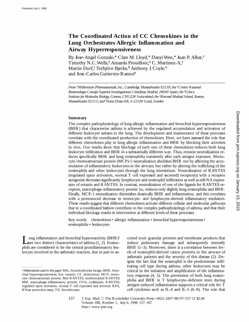
Neutralization of Specific Chemokines Expressed duringOVA-induced Lung Allergic Inflammation.
Mice treated withOVA (see Materials and Methods; Fig. 1
A
,
bottom
) showmaximal lung monocyte/macrophage accumulation atearly stages of the inflammatory response (3 h after OVAchallenge on day 15), whereas eosinophil and T lympho-cyte numbers increase in this organ at late stages of the re-sponse (3 h after OVA challenge on day 21; Fig. 1
A
). Thehighest mRNA expression of MCP-1 (day 15, 3 h), as wellas eotaxin and RANTES (day 21, 3 h) and MCP-5 andMIP-1
a
(day 15–21, 3 h), coincides with the kinetics fol-lowed by the infiltrating leukocytes described above (Fig. 1
A
; reference 5). No MIP-1
b
mRNA expression was de-tected during OVA treatment (Fig. 1
A
). Fig. 1
B
showsthat the production of both eotaxin and MCP-5 protein inresponse to OVA correlates with the mRNA expressionpattern shown by these two chemokines, at least at the timepoints analyzed (days 15 and 21). In addition, 24 h afterOVA administration on day 21, eotaxin and MCP-5 pro-tein expression is clearly diminished compared with thatdetected 3 h after antigen challenge on the same day (datanot shown). This suggests that the kinetics of chemokinemRNA expression parallels that observed, at least, for eo-taxin and MCP-5 protein expression.
To evaluate the specific contribution of eotaxin, an eosi-nophilic chemokine (13, 22, 23, 34), MCP-5, a monocyteand eosinophil chemokine (16), MIP-1
a
, a monocyte,lymphocyte, and eosinophil chemokine (14, 21, 35), andMCP-1, a monocyte chemokine (25, 36), to the develop-ment of lung inflammation in this OVA model, blockingexperiments of these chemokines were performed usingspecific neutralizing Abs that have been characterized ex-tensively in vivo and in vitro (references 5, 16, 28, and 29;and see Materials and Methods for details). Neutralizationof chemokine receptors that are activated by RANTES, a Tlymphocyte and eosinophil chemokine (21), and otherligands was achieved using the antagonist Met-RANTES(see Materials and Methods; reference 30). Neutralizingagents were delivered daily from days 8 to 21 in these firstseries of experiments, and analysis was always performed 3 hafter OVA challenge on day 21. Since the location of infil-trating cells within the lung correlates strongly with the se-verity of the inflammatory response (3), leukocyte enumer-ation was performed in the airways (BAL fluid) and in theinterstitium (lung sections) after OVA treatment.
Eotaxin neutralization during OVA treatment specifi-cally affected eosinophils both in BAL fluid and pulmonaryinterstitium. This effect was more pronounced on day 21(Fig. 2, and Tables 1 and 2). MCP-5 neutralization during
on January 13, 2015jem
.rupress.orgD
ownloaded from
Published July 1, 1998
160
Chemokines in Lung Allergic Responses
OVA treatment strongly affected accumulation of BALmonocytes and eosinophils on day 15, but not on day 21(Fig. 2, and Table 1). Interestingly, minimal OVA-inducedleukocyte accumulation was detected in the lung intersti-tium at any time point after MCP-5 blockage (Table 2, andFig. 3). Met-RANTES administration reduced OVA-induced monocyte accumulation in the BAL by half on day21 (Table 1) and abrogated BAL lymphocyte and eosino-phil accumulation during OVA treatment (Fig. 2, and Ta-ble 1). There was a corresponding decrease in inflammationin the pulmonary interstitium after Met-RANTES admin-istration on day 21 (Table 2). MIP-1
a
neutralization re-duced eosinophil recruitment into the airways and pul-monary interstitium by 20% only at late stages of theinflammatory response, but without affecting monocyte,macrophage, and lymphocyte numbers (day 21, Fig. 2
A
,and Tables 1 and 2).
Neutralization of MCP-1 before each intranasal OVAchallenge from days 8 to 21 reduced all four cell popula-tions that migrate to the lung in response to OVA (Tables 1and 2, and Fig. 3). Mononuclear phagocyte and eosinophilaccumulation in the lung was reduced by 55 and 80%, re-spectively, after MCP-1 blockage (Fig. 2, and Table 1).OVA-induced pulmonary T lymphocyte accumulationduring the inflammatory response was virtually abolished inthe absence of MCP-1–mediated signals (Table 1).
To dissect the windows of action of the chemokines,blockage experiments were performed at different timepoints during OVA treatment, and mice were always ana-lyzed 3 h after OVA challenge on day 21. Thus, when theanti–MCP-1 Abs were administered from days 15 to 21,only a small reduction in the numbers of infiltrating BALleukocytes was detected (Fig. 2
C
), suggesting a critical roleof MCP-1 at early stages of the inflammatory response.However, blockage of MCP-1 neither at early stages (days8–16) nor at late stages of the inflammatory response (days19–21) is sufficient to decrease significantly BAL leukocyte
Figure 1.
Chemokine expression and leukocyte infiltration in the lungof OVA-treated mice. (
A
) Eotaxin, RANTES, MCP-5, MCP-1, MIP-1
a
,and MIP-1
b
mRNA expression (
gray bars
) in the lung of each mousewere normalized to the 28S rRNA expression in the same organ of thesame mice. The quantitation of total RNA was determined by hybridiza-tion of a 28S rRNAcDNA probe. Each bar represents data from five miceat the time points indicated (3 h after OVA challenge on days 15, 18, and21) during treatment (
bottom
). Values are expressed as the mean
6
SEM.The accumulation in the lung of BAL monocytes/macrophages (
M/m
), Tlymphocytes (
L
), and eosinophils (
E
) during OVA treatment (3 h afterantigen challenge on days 15, 18, and 21) is also shown. Although onlyBAL accumulation is presented here, there is correlation between these dataand the accumulation in the interstitium (reference 23). Five data pointsat the different times indicated were collected per day as described previ-ously (reference 23). Values are expressed as the mean
6
SEM.
i.n.
, intra-nasal. (
B
) Immunohistochemical staining of lung sections from OVA-treated mice on day 15 (3 h;
I
and
III
) or day 21 (3 h;
II
and
IV
) after OVAchallenge. Sections were stained with anti–MCP-5 mAb (
I
and
II
; see Ma-terials and Methods) or anti-Eot mAb (
III
and
IV; see Materials andMethods). Note that positive staining (brown precipitate) is comparable be-tween days 15 and 21 for MCP-5, but that there is clearly less eotaxin stain-ing on day 15 compared with day 21. Ig-stained control is also shown (V).
on January 13, 2015jem
.rupress.orgD
ownloaded from
Published July 1, 1998
161 Gonzalo et al.
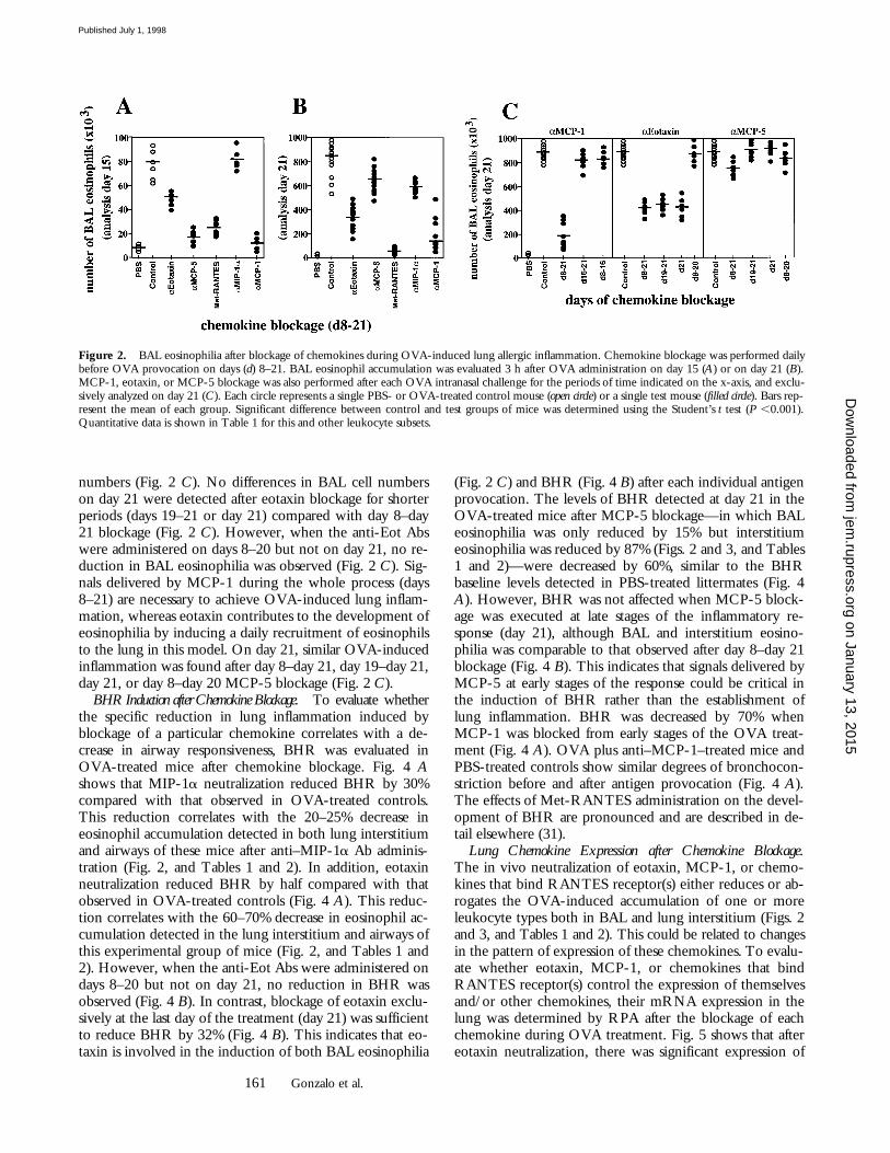
numbers (Fig. 2 C). No differences in BAL cell numberson day 21 were detected after eotaxin blockage for shorterperiods (days 19–21 or day 21) compared with day 8–day21 blockage (Fig. 2 C). However, when the anti-Eot Abswere administered on days 8–20 but not on day 21, no re-duction in BAL eosinophilia was observed (Fig. 2 C). Sig-nals delivered by MCP-1 during the whole process (days8–21) are necessary to achieve OVA-induced lung inflam-mation, whereas eotaxin contributes to the development ofeosinophilia by inducing a daily recruitment of eosinophilsto the lung in this model. On day 21, similar OVA-inducedinflammation was found after day 8–day 21, day 19–day 21,day 21, or day 8–day 20 MCP-5 blockage (Fig. 2 C).
BHR Induction after Chemokine Blockage. To evaluate whetherthe specific reduction in lung inflammation induced byblockage of a particular chemokine correlates with a de-crease in airway responsiveness, BHR was evaluated inOVA-treated mice after chemokine blockage. Fig. 4 Ashows that MIP-1a neutralization reduced BHR by 30%compared with that observed in OVA-treated controls.This reduction correlates with the 20–25% decrease ineosinophil accumulation detected in both lung interstitiumand airways of these mice after anti–MIP-1a Ab adminis-tration (Fig. 2, and Tables 1 and 2). In addition, eotaxinneutralization reduced BHR by half compared with thatobserved in OVA-treated controls (Fig. 4 A). This reduc-tion correlates with the 60–70% decrease in eosinophil ac-cumulation detected in the lung interstitium and airways ofthis experimental group of mice (Fig. 2, and Tables 1 and2). However, when the anti-Eot Abs were administered ondays 8–20 but not on day 21, no reduction in BHR wasobserved (Fig. 4 B). In contrast, blockage of eotaxin exclu-sively at the last day of the treatment (day 21) was sufficientto reduce BHR by 32% (Fig. 4 B). This indicates that eo-taxin is involved in the induction of both BAL eosinophilia
(Fig. 2 C) and BHR (Fig. 4 B) after each individual antigenprovocation. The levels of BHR detected at day 21 in theOVA-treated mice after MCP-5 blockage—in which BALeosinophilia was only reduced by 15% but interstitiumeosinophilia was reduced by 87% (Figs. 2 and 3, and Tables1 and 2)—were decreased by 60%, similar to the BHRbaseline levels detected in PBS-treated littermates (Fig. 4A). However, BHR was not affected when MCP-5 block-age was executed at late stages of the inflammatory re-sponse (day 21), although BAL and interstitium eosino-philia was comparable to that observed after day 8–day 21blockage (Fig. 4 B). This indicates that signals delivered byMCP-5 at early stages of the response could be critical inthe induction of BHR rather than the establishment oflung inflammation. BHR was decreased by 70% whenMCP-1 was blocked from early stages of the OVA treat-ment (Fig. 4 A). OVA plus anti–MCP-1–treated mice andPBS-treated controls show similar degrees of bronchocon-striction before and after antigen provocation (Fig. 4 A).The effects of Met-RANTES administration on the devel-opment of BHR are pronounced and are described in de-tail elsewhere (31).
Lung Chemokine Expression after Chemokine Blockage.The in vivo neutralization of eotaxin, MCP-1, or chemo-kines that bind RANTES receptor(s) either reduces or ab-rogates the OVA-induced accumulation of one or moreleukocyte types both in BAL and lung interstitium (Figs. 2and 3, and Tables 1 and 2). This could be related to changesin the pattern of expression of these chemokines. To evalu-ate whether eotaxin, MCP-1, or chemokines that bindRANTES receptor(s) control the expression of themselvesand/or other chemokines, their mRNA expression in thelung was determined by RPA after the blockage of eachchemokine during OVA treatment. Fig. 5 shows that aftereotaxin neutralization, there was significant expression of
Figure 2. BAL eosinophilia after blockage of chemokines during OVA-induced lung allergic inflammation. Chemokine blockage was performed dailybefore OVA provocation on days (d) 8–21. BAL eosinophil accumulation was evaluated 3 h after OVA administration on day 15 (A) or on day 21 (B).MCP-1, eotaxin, or MCP-5 blockage was also performed after each OVA intranasal challenge for the periods of time indicated on the x-axis, and exclu-sively analyzed on day 21 (C). Each circle represents a single PBS- or OVA-treated control mouse (open circle) or a single test mouse (filled circle). Bars rep-resent the mean of each group. Significant difference between control and test groups of mice was determined using the Student’s t test (P ,0.001).Quantitative data is shown in Table 1 for this and other leukocyte subsets.
on January 13, 2015jem
.rupress.orgD
ownloaded from
Published July 1, 1998
162 Chemokines in Lung Allergic Responses
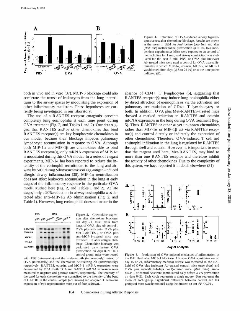
OVA-induced eotaxin, RANTES, and MCP-1 on day 21.The mRNA expression level of these chemokines wascomparable to those found in OVA-treated controls at thesame time point (Fig. 5). In contrast, eotaxin and RANTESmRNA expression was reduced significantly in the lung ofMet-RANTES–treated mice at each time point studied(Fig. 5, and data not shown). MCP-1 expression was notaltered significantly by Met-RANTES in these mice (Fig. 5).Surprisingly, MCP-1 blockage, which prevents lung accu-mulation of eosinophils and lymphocytes in response toOVA, does not prevent the expression of the lymphocyticand/or eosinophilic chemokines RANTES and eotaxin(Fig. 5).
Production of Inflammatory Mediators after MCP-1 Blockage.Since MCP-1 blockage does not affect the mRNA expres-sion of the chemokines studied, the absence of leukocyteinfiltration in the lung of OVA plus anti–MCP-1–treatedmice could be due to the modulation of activating factorsother than chemokines. These activating factors could beproduced directly by monocytes or induced indirectly bythis subset of leukocytes. Fig. 6 A shows that at 1 h afterOVA administration, the production of LTB4 and PGE2
was increased in the BAL fluid of the OVA-treated miceon day 21 but not on day 15. However, blockage of MCP-1completely inhibits OVA-induced production of these twoinflammatory mediators (Fig. 6 A). Similarly, detectablelevels of TXB2 were observed in the BAL fluid of OVA-treated mice, but not in OVA plus anti–MCP-1–treatedlittermates, on day 21 of treatment (Fig. 6 A). No differ-ences between controls and test mice were detected inLTC4/D4/E4 levels at the time point analyzed (Fig. 6 A).The production of IgE in response to OVA was decreasedfivefold in the BAL fluid after MCP-1 blockage at the timepoint indicated (Fig. 6 B). Similarly, the production ofTh2-derived cytokines in the same fluid was decreasedfourfold for IL-5 and threefold for IL-4 both on days 15and 21 (Fig. 6 B). TNF-a production in the BAL fluid wassimilar in both groups of mice at both time points (Fig. 6B). No IFN-g production was detected after OVA orOVA plus anti–MCP-1 administration at the time pointsindicated (Fig. 6 B).
Discussion
We have characterized previously the expression of thechemokines eotaxin, MCP-5, RANTES, and MCP-1(mRNA and/or protein), and correlated this with the leu-kocytes migrating to the lung during a murine model oflung inflammation (5, 16). From these experiments, weconcluded that MCP-1 mRNA expression paralleled theaccumulation of monocytes/macrophages in this organ,both events occurring predominantly at early stages of the
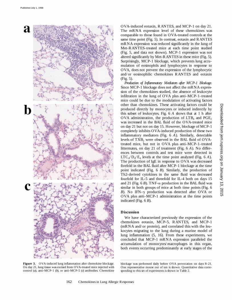
Figure 3. OVA-induced lung inflammation after chemokine blockage.On day 21, lung tissue was excised from OVA-treated mice injected withcontrol (a), anti–MCP-1 (b), or anti–MCP-5 (c) antibodies. Chemokine
blockage was performed daily before OVA provocation on days 8–21.One representative mouse out of ten is shown. Quantitative data corre-sponding to this set of experiments is shown in Table 2.
on January 13, 2015jem
.rupress.orgD
ownloaded from
Published July 1, 1998
163 Gonzalo et al.
response (day 15). Also, eotaxin mRNA expression paral-leled lung eosinophilia predominantly at late stages (day21). In contrast, other chemokines, such as RANTES orMCP-5, were expressed throughout the inflammatory re-action. This underlines the contribution of chemokines atdifferent stages of the response.
From the work presented here, we first conclude thateosinophil recruitment and development of BHR in thismodel system involve the action of both eosinophilic (eo-taxin, RANTES, MCP-5, and MIP-1a) and noneosino-philic chemokines (MCP-1). This indicates the absence ofredundancy, since these chemokines seem to exert a criticalrole at different stages and on different pathways of the de-velopment of OVA-induced lung eosinophilia and BHR.Thus, MCP-5 recruits practically all the eosinophils presentin the lung airways at early stages of the inflammatory re-sponse (day 15), while eotaxin attracts .60 and 80% of theBAL and interstitial eosinophils, respectively, at later stagesof the response (day 21; Fig. 2, A and B, and Tables 1 and2). In addition, MCP-1 mediates its critical influencethroughout during the development of the inflammatoryresponse (days 8–21), whereas eotaxin expression is impor-tant for eosinophil recruitment after each antigen provoca-tion (Fig. 2, and Tables 1 and 2). MCP-5 also delivers sig-nals at early stages of the response that are involved in theestablishment of BHR. In fact, blockage of MCP-5 at latestages does not modify the degree of bronchoconstrictionin response to OVA compared with that observed in theOVA plus Ab control–treated mice (Fig. 4 A). Further-more, we also conclude that MCP-5 seems to affect leuko-cyte traffic through the interstitium, since the accumulationof these leukocytes in the interstitium—but not in the air-ways—was severely impaired after MCP-5 blockage (Figs.2 and 3, and Tables 1 and 2). This could be due to a differ-ential modulation of adhesion receptors by MCP-5 that re-sults in retention of the leukocytes in the interstitium. Infact, it has been demonstrated that chemokines such as eo-taxin affect the activation state of integrins and their ligandsT
able
1.
OV
A-i
nduc
ed L
euko
cyte
Inf
iltra
tion
in th
e A
irway
s af
ter C
hem
okin
e B
lock
age
Che
mok
ine
bloc
kage
Leuk
ocyt
e su
btyp
e
Mac
roph
ages
Mon
ocyt
esT
lym
phoc
ytes
Eos
inop
hils
Day
15
Day
21
Day
15
Day
21
Day
15
Day
21
Day
15
Day
21
Con
trol
(to
tal
(180
6 3
1) 3
103
(122
6 1
2) 3
103
(38
6 2
) 3
103
(25
6 3
) 3
103
(27
6 5
) 3
103
(130
6 2
2) 3
103
(80
6 1
7) 3
103
(870
6 9
2) 3
103
num
bers
)a
Eot
axin
017
6 4
07
6 1
9 6
221
6 4
27 6
360
6 7
aM
CP-
512
6 3
11 6
266
6 8
11 6
322
6 5
076
6 3
21 6
8M
et-R
AN
TE
S9
6 1
17 6
126
6 4
44 6
880
6 2
95 6
460
6 1
091
6 2
aM
IP-1
a0
2 6
0.3
00
00
023
6 2
aM
CP-
145
6 1
058
6 3
78 6
11
53 6
591
6 3
89 6
686
6 8
78 6
12
Perc
enta
ge o
f re
duct
ion
in t
he n
umbe
r of
leu
kocy
tes
in t
he B
AL
fluid
of
OV
A-t
reat
ed m
ice
afte
r ch
emok
ine
bloc
kage
. 30
min
bef
ore
each
int
rana
sal
antig
en a
dmin
istra
tion,
eot
axin
, M
CP-
5,R
AN
TE
S, o
r M
CP-
1 w
as n
eutr
aliz
ed a
s de
scri
bed
in M
ater
ials
and
Met
hods
. BA
L flu
id w
as o
btai
ned
3 h
afte
r O
VA
tre
atm
ent
on d
ay 1
5 or
21,
and
the
num
ber
of t
he d
iffer
ent
cell
type
s w
as d
e-te
rmin
ed. T
otal
num
bers
of m
acro
phag
es, m
onoc
ytes
, T ly
mph
ocyt
es, a
nd e
osin
ophi
ls pr
esen
t in
the
BA
L flu
id o
f OV
A p
lus
Ab
cont
rol–
trea
ted
mic
e ar
e sh
own.
Val
ues
are
give
n as
per
cent
ages
re-
ferr
ed t
o th
e O
VA
plu
s A
b co
ntro
l–tr
eate
d m
ice,
whi
ch a
re c
onsid
ered
to
be 1
00%
.
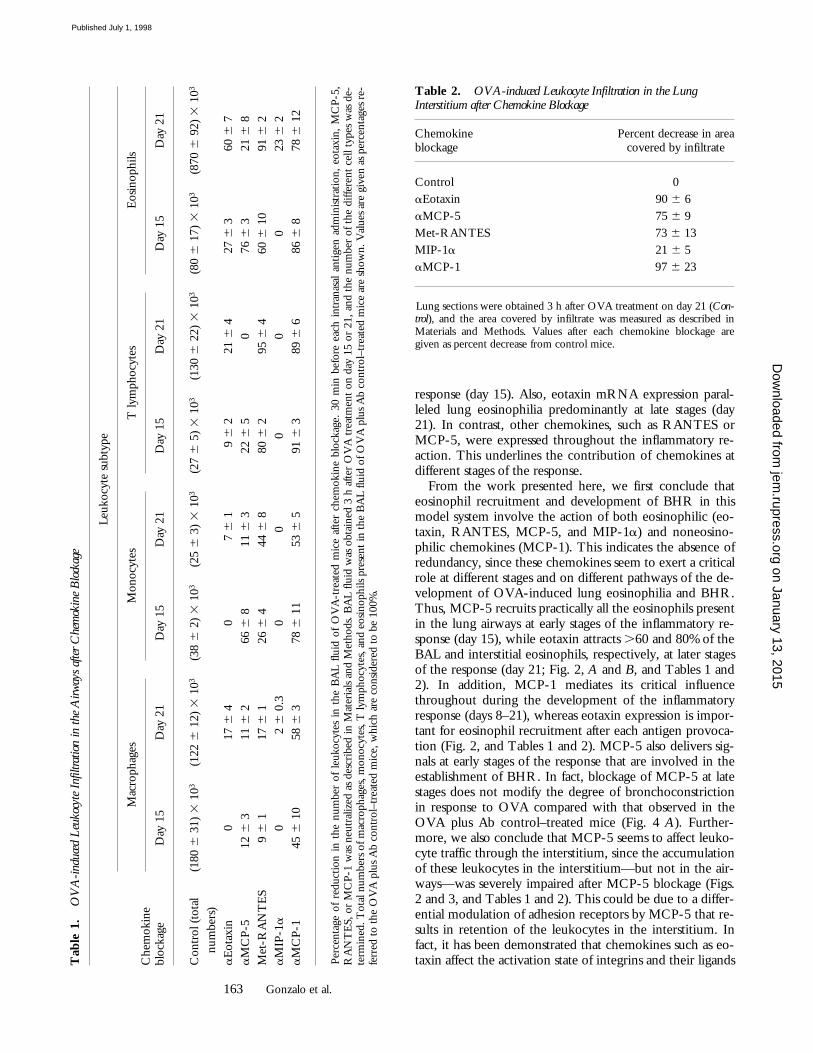
Table 2. OVA-induced Leukocyte Infiltration in the Lung Interstitium after Chemokine Blockage
Chemokineblockage
Percent decrease in areacovered by infiltrate
Control 0aEotaxin 90 6 6aMCP-5 75 6 9Met-RANTES 73 6 13MIP-1a 21 6 5aMCP-1 97 6 23
Lung sections were obtained 3 h after OVA treatment on day 21 (Con-trol), and the area covered by infiltrate was measured as described inMaterials and Methods. Values after each chemokine blockage aregiven as percent decrease from control mice.
on January 13, 2015jem
.rupress.orgD
ownloaded from
Published July 1, 1998
164 Chemokines in Lung Allergic Responses
both in vivo and in vitro (37). MCP-5 blockage could alsoaccelerate the transit of leukocytes from the lung intersti-tium to the airway spaces by modulating the expression ofother inflammatory mediators. These hypotheses are cur-rently being investigated in our laboratory.
The use of a RANTES receptor antagonist preventscompletely lung eosinophilia at each time point duringOVA treatment (Fig. 2, and Tables 1 and 2). Our data sug-gest that RANTES and/or other chemokines that bindRANTES receptor(s) are key lymphocytic chemokines inour model, because their blockage impedes pulmonarylymphocyte accumulation in response to OVA. Althoughboth MIP-1a and MIP-1b are chemokines able to bindRANTES receptor(s), only mRNA expression of MIP-1ais modulated during this OVA model. In a series of elegantexperiments, MIP-1a has been reported to reduce the in-tensity of the eosinophil recruitment to the lung and air-ways by 50% during Schistosoma mansoni egg antigen–inducedallergic airway inflammation (38). MIP-1a neutralizationdoes not affect leukocyte accumulation in the lung at earlystages of the inflammatory response in the particular OVAmodel studied here (Fig. 2, and Tables 1 and 2). At latestages, only a 20% reduction in airway eosinophilia was de-tected after anti–MIP-1a Ab administration (Fig. 2, andTable 1). However, lung eosinophilia does not occur in the
absence of CD41 T lymphocytes (5), suggesting thatRANTES receptor(s) may induce lung eosinophilia eitherby direct attraction of eosinophils or via the activation andpulmonary accumulation of CD41 T lymphocytes, orboth. In addition, OVA plus Met-RANTES–treated miceshowed a marked reduction in RANTES and eotaxinmRNA expression in the lung during OVA treatment (Fig.5). Thus, RANTES or other as yet unknown chemokinesrather than MIP-1a or MIP-1b act via RANTES recep-tor(s) and control directly or indirectly the expression ofother chemokines. Therefore, OVA-induced T cell andeosinophil infiltration in the lung is regulated by RANTESthrough itself and eotaxin. However, it is important to notethat the reagent used here, Met-RANTES, may bind tomore than one RANTES receptor and therefore inhibitthe activity of other chemokines. Due to the complexity ofthis system, we have reported it in detail elsewhere (31).
Figure 4. Inhibition of OVA-induced airway hyperre-sponsiveness after chemokine blockage. Results are shownas the mean 6 SEM for Penh before (gray bars) and after(black bars) methacholine provocation (n 5 10, two inde-pendent experiments). Mice were exposed to an aerosol ofmethacholine for 1 min, and airway constriction was eval-uated for the next 5 min. PBS- or OVA plus irrelevantAb–treated mice were used as control for OVA-treated lit-termates in which MIP-1a, eotaxin, MCP-5, or MCP-1was blocked from days (d) 8 to 21 (A) or at the time pointsindicated (B).
Figure 5. Chemokine expres-sion after chemokine blockage.On day 21, total RNA fromlungs of OVA plus Ab control–,OVA plus anti-Eot–, OVA plusMet-RANTES–, or OVA plusanti–MCP-1–treated mice wasextracted 3 h after antigen chal-lenge. Chemokine blockage wasperformed daily before OVAprovocation on days 8–21. In acontrol group, mice were treated
with PBS (intranasally) and the irrelevant Ab (intravenously) instead ofOVA (intranasally) and the chemokine-neutralizing Ab (intravenously),respectively. RANTES, eotaxin, and MCP-1 mRNA expression weredetermined by RPA. Both TCA and GAPDH mRNA expression weremeasured as negative and positive control, respectively. The intensity ofthe band for each chemokine was normalized to the intensity of the bandof GAPDH in the control sample (not shown) and analyzed. Chemokineexpression of two representative mice out of four is shown.
Figure 6. Production of OVA-induced mediators of inflammation inthe BAL fluid after MCP-1 blockage. 1 h after OVA administration onday 15 or 21, inflammatory mediator release was measured in the BALfluid of OVA plus irrelevant Ab–treated control mice (open circles) andOVA plus anti–MCP-1(days 8–21)–treated mice (filled circles). Anti–MCP-1 or control Abs were administered daily before OVA provocationon days 8–21. Each circle represents a single mouse. Bars represent themean of each group. Significant difference between control and testgroups of mice was determined using the Student’s t test (*P ,0.01).
on January 13, 2015jem
.rupress.orgD
ownloaded from
Published July 1, 1998
165 Gonzalo et al.
Finally, the blockage of the mononuclear phagocytechemokine MCP-1 throughout both the induction andlater phases of the response affects these four leukocytepopulations that migrate to the lung in response to OVA aswell as blocking the induction of BHR (Figs. 2, 3, and 4,and Tables 1 and 2). Monocytes and macrophages are thefirst cell types to accumulate during OVA-induced lung in-flammation in this model (5), and the strongest expressionof MCP-1 occurs at early stages of the inflammatory re-sponse (Fig. 1; reference 5). Moreover, modification of theOVA-induced inflammatory response exclusively occurswhen anti–MCP-1 mAb is administered from very earlystages of the inflammatory process (Fig. 2, and Tables 1 and2). In the absence of MCP-1–mediated signals, there is anz50% reduction in the number of lung monocytes/mac-rophages after OVA administration, but interestingly, thereis an almost complete reduction in lymphocyte and eosino-phil infiltration of the lung (Fig. 2, and Tables 1 and 2).Taken together, these data suggest that MCP-1 acts up-stream of the inflammatory process and is a strong candi-date to regulate the expression of the other chemokinesduring this physiological response. Nevertheless, compara-ble mRNA expression of eotaxin, RANTES, and MCP-1was detected in the lung of OVA plus anti–MCP-1–treatedmice and OVA-treated controls (Fig. 5). This indicates thatMCP-1 is involved in other regulatory pathways ratherthan chemokine expression. Based on our present knowl-edge of MCP-1 function, it can be hypothesized that theregulatory pathways are critically affected by the reductionin monocyte/macrophage numbers after anti–MCP-1treatment and/or by the depletion of a specific subset of thecells that is essential for this inflammatory process.
Both lung-resident macrophages distributed along theairways and circulating macrophages release mediators thatcan modulate the function of other cell types and amplifythe lung inflammatory reactions (10, 39). Macrophages canrelease eosinophil chemoattractants, including LTB4 (10),or modulate the generation of LTC4 by eosinophils (12).They also produce PGE2 and TXB2, which are mediatorsof increased vascular permeability. In fact, levels of LTB4,PGE2, or TXB2 were detectable in the BAL fluid of OVA-treated mice but greatly diminished in the BAL fluid ofOVA plus anti–MCP-1–treated littermates (Fig. 6 A). This
indicates that the observed production of these factors isdue either to infiltrating cells at this point, to increased acti-vation of resident cells at late stages of the response, or to acombination of these two events. LTC4 was not detectablein the BAL fluid of either group of mice (Fig. 6 A). Mac-rophages can act as APCs and drive the proliferation of ef-fector T cells that have been demonstrated to be requiredto achieve a functional response. In this scenario, macro-phage-induced IL-4 production by T cells after antigenpresentation drives the switch in Ig synthesis by B cells toIgE and potentiates the subsequent priming of mast cells byIgE plus antigen (40). MCP-1 blockage correlates with aclear decrease in the BAL levels of IgE and IL-4 after OVAtreatment (Fig. 6 B). Similarly, IL-5 production is also af-fected by the MCP-1 blockage during OVA-induced lungallergic inflammation. These results indicate that MCP-1function(s) (probably through monocyte/macrophage re-cruitment) are also involved in B and T cell activation. NoIFN-g production was detected in the BAL fluid of test orcontrol mice, mainly because this model has a predominantTh2-type response. Macrophages and monocytes also rep-resent the major source of inflammatory cytokines such asTNF (39). Despite the fact that only low levels of TNF-awere detected in the BAL fluid of OVA-treated mice,MCP-1 blockage reduced these levels by half at the timepoint of maximal macrophage and monocyte infiltration inthe lung (day 15; Fig. 6 B).
In conclusion, this paper reports the functional relevanceof chemokines at different stages of a complex inflamma-tory response in vivo. Chemokines not only ensure thephysical presence of specific leukocytes at the site of in-flammation by acting directly on them, but also regulatethe expression of other chemokines and the activation of avariety of inflammatory mediators made by different celltypes. Our results suggest that specific chemokines are in-volved in different cellular and molecular pathways that ina coordinated fashion contribute to the complex patho-physiology of asthma. Understanding all the componentsinvolved in lung allergic inflammation as well as the criticaltiming of their actions could represent a crucial step in theprevention and therapy of this disease. We believe that thein vivo results presented here may contribute to the supportof clinical applications for the improvement of asthma therapy.
We thank Profs. Drazen and Butcher for critical reading of this manuscript, Drs. MacKay, Tepper, and Zlot-nik for their comments and important suggestions, and Dr. L. Kremer, Centro Nacional Biotecnologia, forproviding antibodies against MCP-5. We are also indebted to T. Delaney and N. Bikkal for skilled technicalassistance, and M. Melzer for editorial assistance.
This work was partially funded by Astra Draco AB.
Address correspondence to J.-C. Gutierrez-Ramos, Millennium Pharmaceuticals, Inc., 640 Memorial Dr.,Cambridge, MA 02139. Phone: 617-679-7262; Fax: 617-374-9379; E-mail: [email protected]
Received for publication 11 December 1997 and in revised form 6 April 1998.
on January 13, 2015jem
.rupress.orgD
ownloaded from
Published July 1, 1998
166 Chemokines in Lung Allergic Responses
References1. Gleich, G.J., N.A. Flavahan, T. Fujisawa, and P.M. Van-
houtte. 1988. The eosinophil as a mediator of damage to res-piratory epithelium: a model for bronchial hyperreactivity. J.Allergy Clin. Immunol. 81:776–781.
2. Gleich, G.J. 1990. The eosinophil and bronchial asthma.Current understanding. J. Allergy Clin. Immunol. 85:422–436.
3. Eum, S.-Y., S. Hailé, J. Lefort, M. Huerre, and B.B.Vargaftig. 1995. Eosinophil recruitment into the respiratoryepithelium following antigenic challenge in hyper-IgE miceis accompanied by interleukin 5-dependent bronchial hyper-responsiveness. Proc. Natl. Acad. Sci. USA. 92:12290–12294.
4. Bradley, B.L., M. Azzawi, M. Jacobson, B. Assoufi, J.V. Col-lins, A.-M.A. Irani, L.B. Schwartz, S.R. Durham, P.K. Jef-frey, and A.B. Kay. 1991. Eosinophils, T-lymphocytes, mastcells, neutrophils and macrophages in bronchial biopsy speci-mens from atopic subjects with asthma: comparison with bi-opsy specimens from atopic subjects without asthma and normalcontrol subjects and relationship to bronchial hyperrespon-siveness. J. Allergy Clin. Immunol. 88:661–667.
5. Gonzalo, J.A., C.M. Lloyd, L. Kremer, E. Finger, C. Mar-tinez-A, M.H. Siegelman, M. Cybulski, and J.C. Gutierrez-Ramos. 1996. Eosinophil recruitment to the lung in a murinemodel of allergic inflammation. The role of T cells, chemo-kines, and endothelial adhesion receptors. J. Clin. Invest. 98:2332–2345.
6. Gavett, S.H., X. Chen, F. Kinkelman, and M. Wills-Karp.1994. Depletion of murine CD41 T lymphocytes preventsantigen-induced airway hyperreactivity and pulmonary eosi-nophilia. Am. J. Respir. Cell Mol. Biol. 10:587–593.
7. Coyle, A.J., G. Le Gros, C. Bertrand, S. Tsuyuki, C.H.Heusser, M. Kopf, and G.P. Anderson. 1995. Interleukin-4 isrequired for the induction of lung Th2 mucosal immunity.Am. J. Respir. Cell Mol. Biol. 13:54–59.
8. Corry, D.B., H.G. Folkesson, M.L. Warnock, D.J. Erle,M.A. Matthay, J.P. Wiener-Kronish, and R.M. Locksley.1996. Interleukin 4, but not interleukin 5 or eosinophils, isrequired in a murine model of acute airway hyperreactivity.J. Exp. Med. 183:109–117.
9. Foster, P.S., S.P. Hogan, A.J. Ramsay, K.I. Matthaei, andI.G. Young. 1996. Interleukin 5 deficiency abolishes eosino-philia, airways hyperreactivity, and lung damage in a mouseasthma model. J. Exp. Med. 183:195–201.
10. Fels, A.O.S., N.A. Pawlowski, and E.B. Cramer. 1982. Hu-man alveolar macrophages produce leukotriene B4. Proc.Natl. Acad. Sci. USA. 79:7866–7870.
11. Nathan, C.F. 1987. Secretory products of macrophages. J.Clin. Invest. 79:319–326.
12. Wilkinson, J.R.W., A.E.G. Crea, and T.J.H. Clark. 1989.Identification and characterization of a monocyte-derivedneutrophil-activating peptide in corticosteroid-resistant bron-chial asthma. J. Clin. Invest. 84:1930–1941.
13. Jose, P.J., D.A. Griffiths-Johnson, P.D. Collins, D.T. Walsh,R. Moqbel, N.F. Totty, O. Truong, J.J. Hsuan, and T.J.Williams. 1994. Eotaxin: a potent eosinophil chemoattractantcytokine detected in a guinea pig model of allergic airwaysinflammation. J. Exp. Med. 179:881–887.
14. Lukacs, N.W., R.M. Strieter, C.L. Shaklee, S.W. Chensue,and S.L. Kunkel. 1995. Macrophage inflammatory protein-1a influences eosinophil recruitment in antigen-specific air-way inflammation. Eur. J. Immunol. 25:245–251.
15. Lukacs, N.W., R.M. Strieter, S.W. Chensue, and S.L.Kunkel. 1996. Activation and regulation of chemokines in al-
lergic airway inflammation. J. Leukocyte Biol. 59:13–17.16. Jia, G.Q., J.A. Gonzalo, C. Lloyd, L. Kremer, L. Lu, C. Mar-
tinez, B.K. Wershil, and J.-C. Gutierrez-Ramos. 1996. Dis-tinct expression and function of the novel mouse chemokinemonocyte chemotactic protein-5 in lung allergic inflamma-tion. J. Exp. Med. 184:1939–1951.
17. MacLean, J.A., R. Ownbey, and A.D. Luster. 1996. T cell-dependent regulation of eotaxin in antigen-induced pulmo-nary eosinophilia. J. Exp. Med. 184:1461–1469.
18. Miller, M.D., and M.S. Krangel. 1992. Biology and bio-chemistry of the chemokines: a family of chemotactic and in-flammatory cytokines. Crit. Rev. Immunol. 12:17–46.
19. Baggiolini, M., B. Dewald, and B. Moser. 1994. Interleukin-8and related chemotactic cytokines—CXC and CC chemo-kines. Adv. Immunol. 55:97–179.
20. Peled, A., J.-A. Gonzalo, C. Lloyd, and J.-C. Gutierrez-Ramos. 1998. The chemotactic cytokine eotaxin acts as agranulocyte-macrophage colony-stimulating factor duringlung inflammation. Blood. 91:1909–1916.
21. Rot, A., M. Krieger, T. Brunner, S.C. Bischoff, T.J. Schall,and C.A. Dahinden. 1992. RANTES and macrophage in-flammatory protein 1 alpha induce the migration and activa-tion of normal human eosinophil granulocytes. J. Exp. Med.176:1489–1495.
22. Rothenberg, M.E., A.D. Luster, and P. Leder. 1995. Murineeotaxin: an eosinophil chemoattractant inducible in endothe-lial cells and in interleukin 4-induced tumor suppression.Proc. Natl. Acad. Sci. USA. 92:8960–8964.
23. Gonzalo, J.-A., G.-Q. Jia, V. Aguirre, D. Friend, A.J. Coyle,N.A. Jenkins, G.S. Lin, H. Katz, A. Litchman, N. Copeland,et al. 1996. Mouse eotaxin expression parallels eosinophil ac-cumulation during lung allergic inflammation but it is not re-stricted to a Th2-type response. Immunity. 4:1–14.
24. Ebisawa, M., T. Yamada, C. Bickel, D. Klunk, and R.P.Schleimer. 1994. Eosinophil transendothelial migration in-duced by cytokines. III. Effect of the chemokine RANTES1.J. Immunol. 153:2153–2160.
25. Rollins, B.J., T. Yoshimura, E.J. Leonard, and J. Pober.1990. Cytokine-activated human endothelial cells synthesizeand secrete monocyte chemoattractant protein, MCP-1. Am.J. Pathol. 136:1229–1233.
26. Chensue, S.W., K.S. Warmington, J.H. Ruth, P.S. Sanghi,P. Lincoln, and S. Kunkel. 1996. Role of monocytechemoattractant protein-1 (MCP-1) in Th1 (mycobacterial)and Th2 (schistosomal) antigen-induced granuloma forma-tion: relationship to local inflammation, Th cell expression,and IL-12 production. J. Immunol. 157:4602–4608.
27. Gunn, M.D., N.A. Nelken, X. Liao, and L.T. Williams.1997. Monocyte chemoattractant protein-1 is sufficient forthe chemotaxis of monocytes and lymphocytes in transgenicmice but requires an additional stimulus for inflammatory ac-tivation. J. Immunol. 158:376–383.
28. Luo, Y., J. Laning, M. Hayashi, P.R. Hancock, B. Rollins,and M.E. Dorf. 1994. Serologic analysis of the mouse betachemokine JE/monocyte chemoattractant protein-1. J. Im-munol. 153:3708–3716.
29. Lloyd, C.M., A.W. Minto, M.E. Dorf, A. Proudfoot, T.N.C.Wells, D.J. Salant, and J.-C. Gutierrez-Ramos. 1997.RANTES and monocyte chemoattractant protein-1 (MCP-1)play an important role in the inflammatory phase of crescen-tic nephritis, but only MCP-1 is involved in crescent forma-tion and interstitial fibrosis. J. Exp. Med. 185:1371–1380.
on January 13, 2015jem
.rupress.orgD
ownloaded from
Published July 1, 1998
167 Gonzalo et al.
30. Proudfoot, A.E.I., C.A. Power, A.J. Hoogewerf, M.O.Montjovent, F. Borlat, R.E. Offord, and T.N.C. Wells.1996. Extension of recombinant human RANTES by the re-tention of the initiating methionine produces a potent antag-onist. J. Biol. Chem. 271:2599–2603.
31. Coyle, A.J., J. Kips, A.E.I. Proudfoot, and T.N.C. Wells.1998. Inhibition of lung inflammation and altered airway re-sponsiveness by Met-RANTES, a novel chemokine receptorantagonist. J. Exp. Med. In press.
32. Gelfand, E.W., and C.G. Irvin. 1997. T lymphocyte: settingthe tone of the airways. Nat. Med. 3:382–383.
33. Ledermann, F., C. Schlienger, K. Wagner, and C. Heusser.1991. A sensitive and efficient induction system for murineIgE. Single cell analysis at the clonal level. J. Immunol. Meth-ods. 141:263–275.
34. Ponath, P.D., S. Qin, D.J. Ringler, I. Clark-Lewis, J. Wang,N. Kassam, H. Smith, X. Shi, J.-A. Gonzalo, W. Newman,et al. 1996. Cloning of the human eosinophil chemoattrac-tant, eotaxin. Expression, receptor binding, and functionalproperties suggest a mechanism for the selective recruitmentof eosinophils. J. Clin. Invest. 97:604–612.
35. Uguccioni, M., M. D’Apuzzo, M. Loetscher, B. Dewald, andM. Baggiolini. 1995. Actions of the chemotactic cytokines
MCP-1, MCP-2, MCP-3, RANTES, MIP-1a and MIP-1bon human monocytes. Eur. J. Immunol. 25:64–68.
36. Rollins, B.J., E.D. Morrison, and C.D. Stiles. 1988. Cloningand expression of JE, a gene inducible by platelet-derivedgrowth factor and whose product has cytokine-like proper-ties. Proc. Natl. Acad. Sci. USA. 85:3738–3742.
37. Jia, G.-Q., J.-A. Gonzalo, D. Wagner, A.J. Coyle, M. Cybul-ski, and J.-C. Gutierrez-Ramos. 1997. Selective eosinophiltransendothelial migration triggered by eotaxin via modula-tion of Mac-1/ICAM-1 and VLA-4/VCAM-1 interactions.Eur. J. Immunol. In press.
38. Lukacs, N.W., R.M. Strieter, K. Warmington, P. Lincoln,S.W. Chensue, and S.L. Kunkel. 1997. Differential recruit-ment of leukocyte populations and alteration of airway hy-perreactivity by C-C family chemokines in allergic airway in-flammation. J. Immunol. 158:4398–4404.
39. Elias, J.A., A.D. Schreiber, and K. Gostilo. 1985. Differentialinterleukin-1 elaboration by unfractionated and density frac-tionated human alveolar macrophages and blood monocytes.J. Immunol. 135:3198–3204.
40. Drazen, J.M., J.P. Arm, and K.F. Austen. 1996. Sorting outthe cytokines of asthma. J. Exp. Med. 183:1–5.
on January 13, 2015jem
.rupress.orgD
ownloaded from
Published July 1, 1998
Related Documents