Brain Research Reviews 37 (2001) 38–58 www.elsevier.com / locate / bres Review The control of preoptic aromatase activity by afferent inputs in q Japanese quail a,b a c d Philippe Absil , Michelle Baillien , Gregory F. Ball , Gian Carlo Panzica , a, * Jacques Balthazart a ` Center for Cellular and Molecular Neurobiology, Research Group in Behavioral Neuroendocrinology, University of Liege, 17 place Delcour, ` B-4020 Liege, Belgium b Department of Biology, University of Antwerp, Wilrijk, Belgium c Department of Psychology, Johns Hopkins University, Baltimore, MD, USA d Department of Anatomy, Pharmacology and Forensic Medicine, Laboratory of Neuroendocrinology, University of Torino, Torino, Italy Accepted 12 June 2001 Abstract This review summarizes current knowledge on the mechanisms that control aromatase activity in the quail preoptic area, a brain region that plays a key role in the control of reproduction. Aromatase and aromatase mRNA synthesis in the preoptic area are enhanced by testosterone and its metabolite estradiol, but estradiol receptors of the alpha subtype are not regularly colocalized with aromatase. Estradiol receptors of the beta subtype are present in the preoptic area but it is not yet known whether these receptors are colocalized with aromatase. The regulation by estrogen of aromatase activity may be, in part, trans-synaptically mediated, in a manner that is reminiscent of the ways in which steroids control the activity of gonadotropic hormone releasing hormone neurons. Aromatase-immunoreactive neurons are surrounded by dense networks of vasotocin-immunoreactive and tyrosine hydroxylase-immunoreactive fibers and punctate structures. These inputs are in part steroid-sensitive and could therefore mediate the effects of steroids on aromatase activity. In vivo pharmacological experiments indicate that catecholaminergic depletions significantly affect aromatase activity presumably by modulating aromatase transcription. In addition, in vitro studies on brain homogenates or on preoptic–hypothalamic explants show that aromatase activity can be rapidly modulated by a variety of dopaminergic compounds. These effects do not appear to be mediated by the membrane dopamine receptors and could involve changes in the phosphorylation state of the enzyme. Together, these results provide converging evidence for a direct control of aromatase activity by catecholamines consistent with the anatomical data indicating the presence of a catecholaminergic innervation of aromatase cells. These dopamine-induced changes in aromatase activity are observed after several hours or days and presumably result from changes in aromatase transcription but rapid non-genomic controls have also been identified. The potential significance of these processes for the physiology of reproduction is critically evaluated. 2001 Elsevier Science B.V. All rights reserved. Theme: Endocrine and autonomic regulation Topic: Hypothalamic–pituitary–gonadal regulation Keywords: Estrogen synthase; Dopamine; Rapid estrogen effects; Estrogen receptor; Phosphorylation; Vasotocin Contents 1. Introduction ............................................................................................................................................................................................ 39 2. Aromatase and sexual behavior in quail .................................................................................................................................................... 39 3. Control of aromatase transcription by steroids ........................................................................................................................................... 40 q Proceedings of the International Meeting on Steroids and the Nervous System, Torino, Italy, 11–14 February, 2001. *Corresponding author. Tel.: 132-4-366-5970; fax: 132-4-366-5971 http: / / www.ulg.ac.be / bioneuro /. E-mail address: [email protected] (J. Balthazart). 0165-0173 / 01 / $ – see front matter 2001 Elsevier Science B.V. All rights reserved. PII: S0165-0173(01)00122-9

Welcome message from author
This document is posted to help you gain knowledge. Please leave a comment to let me know what you think about it! Share it to your friends and learn new things together.
Transcript
Brain Research Reviews 37 (2001) 38–58www.elsevier.com/ locate /bres
Review
The control of preoptic aromatase activity by afferent inputs inqJapanese quail
a,b a c dPhilippe Absil , Michelle Baillien , Gregory F. Ball , Gian Carlo Panzica ,a ,*Jacques Balthazart
a `Center for Cellular and Molecular Neurobiology, Research Group in Behavioral Neuroendocrinology, University of Liege, 17 place Delcour,`B-4020 Liege, Belgium
bDepartment of Biology, University of Antwerp, Wilrijk, BelgiumcDepartment of Psychology, Johns Hopkins University, Baltimore, MD, USA
dDepartment of Anatomy, Pharmacology and Forensic Medicine, Laboratory of Neuroendocrinology, University of Torino, Torino, Italy
Accepted 12 June 2001
Abstract
This review summarizes current knowledge on the mechanisms that control aromatase activity in the quail preoptic area, a brain regionthat plays a key role in the control of reproduction. Aromatase and aromatase mRNA synthesis in the preoptic area are enhanced bytestosterone and its metabolite estradiol, but estradiol receptors of the alpha subtype are not regularly colocalized with aromatase.Estradiol receptors of the beta subtype are present in the preoptic area but it is not yet known whether these receptors are colocalized witharomatase. The regulation by estrogen of aromatase activity may be, in part, trans-synaptically mediated, in a manner that is reminiscentof the ways in which steroids control the activity of gonadotropic hormone releasing hormone neurons. Aromatase-immunoreactiveneurons are surrounded by dense networks of vasotocin-immunoreactive and tyrosine hydroxylase-immunoreactive fibers and punctatestructures. These inputs are in part steroid-sensitive and could therefore mediate the effects of steroids on aromatase activity. In vivopharmacological experiments indicate that catecholaminergic depletions significantly affect aromatase activity presumably by modulatingaromatase transcription. In addition, in vitro studies on brain homogenates or on preoptic–hypothalamic explants show that aromataseactivity can be rapidly modulated by a variety of dopaminergic compounds. These effects do not appear to be mediated by the membranedopamine receptors and could involve changes in the phosphorylation state of the enzyme. Together, these results provide convergingevidence for a direct control of aromatase activity by catecholamines consistent with the anatomical data indicating the presence of acatecholaminergic innervation of aromatase cells. These dopamine-induced changes in aromatase activity are observed after several hoursor days and presumably result from changes in aromatase transcription but rapid non-genomic controls have also been identified. Thepotential significance of these processes for the physiology of reproduction is critically evaluated. 2001 Elsevier Science B.V. Allrights reserved.
Theme: Endocrine and autonomic regulation
Topic: Hypothalamic–pituitary–gonadal regulation
Keywords: Estrogen synthase; Dopamine; Rapid estrogen effects; Estrogen receptor; Phosphorylation; Vasotocin
Contents
1. Introduction ............................................................................................................................................................................................ 392. Aromatase and sexual behavior in quail .................................................................................................................................................... 393. Control of aromatase transcription by steroids ........................................................................................................................................... 40
qProceedings of the International Meeting on Steroids and the Nervous System, Torino, Italy, 11–14 February, 2001.*Corresponding author. Tel.: 132-4-366-5970; fax: 132-4-366-5971 http: / /www.ulg.ac.be /bioneuro /.E-mail address: [email protected] (J. Balthazart).
0165-0173/01/$ – see front matter 2001 Elsevier Science B.V. All rights reserved.PI I : S0165-0173( 01 )00122-9
P. Absil et al. / Brain Research Reviews 37 (2001) 38 –58 39
4. Catecholaminergic and peptidergic inputs to aromatase-containing brain areas ............................................................................................. 415. In vivo pharmacological evidence for a control by catecholamines of aromatase activity............................................................................... 46
5.1. Systemic treatments with catecholaminergic neurotoxins.................................................................................................................... 465.2. Central treatment with 6-hydroxydopamine ....................................................................................................................................... 46
6. Cellular mechanisms mediating changes by afferent inputs of aromatase concentration................................................................................. 497. Rapid changes in aromatase activity.......................................................................................................................................................... 508. Inhibition of aromatase activity measured in vitro by dopaminergic agonists and antagonists ........................................................................ 509. Effects of dopamine on aromatase activity expressed by in vitro explants .................................................................................................... 5210. Controls of aromatase activity by protein phosphorylation ........................................................................................................................ 5211. The control of aromatase activity by afferent inputs: physiological effects or experimental artifacts? ........................................................... 53Acknowledgements ...................................................................................................................................................................................... 55References................................................................................................................................................................................................... 55
1. Introduction mechanisms could therefore modify the bioavailability ofestrogens in the brain within minutes or even seconds.
Sex steroid hormones act in the brain to coordinate the Rapid effects of estrogens on neuronal physiology orproduction of a wide variety of behavioral and physiologi- behavior have been described recently. Such changes incal endpoints. The ability of steroids to exert such effects estrogen availability through the modification of the es-can differ between individuals of the same species (e.g. trogen synthase (aromatase) could therefore provide athere are sex differences in the physiological response to physiologically relevant mechanism that would controlsteroids [27]) or within individuals when they are in these rapid non-genomic effects of estrogens. In thisdifferent environments [66]. It was proposed over 15 years context, aromatase could produce locally high concen-ago that one-way for steroid action to be affected by trations of estrogens acting at the membrane level. Thesecontext would be for different environments to influence estrogens would then have to be considered as neuro(ac-the availability of sex steroid receptors [88]. Data have tive)-steroids.now accumulated indicating that ascending catecholamineinputs to diencephalic brain nuclei containing sex steroidreceptor types such as estrogen receptors (ER) and proges- 2. Aromatase and sexual behavior in quailterone receptors (PR) can regulate the availability of ERand PR as measured by in vitro binding assays [45]. Male quail reliably display the full sequence of copulat-
Another important component of the neural substrate ory behavior including grabbing the female’s neck feath-that mediates the response to steroid hormones involves ers, mount and performing cloacal contact movements inthe local metabolism of steroids. For example, in males of strictly controlled laboratory conditions. This entire be-many vertebrate species testosterone (T) exerts its effects havioral sequence is steroid-dependent. Castration elimi-at the cellular level after first being metabolized to an nates these behaviors within a few days and treatment ofandrogenic (via the action of the 5a-reductase) or es- castrated males with exogenous T rapidly restores thetrogenic (via the action of aromatase) metabolite of testo- behaviors in most, if not all, birds in less than 1 weeksterone [16,78]. In some cases this metabolic conversion is [3,109].rate limiting for a particular physiological process. One In quail as in other vertebrate species, T can besuch example is the activation of male sexual behavior in irreversibly metabolized into a variety of compoundsmany species. In Japanese quail (Coturnix japonica) the within the central nervous system and these transforma-aromatization of testosterone to 17b-estradiol is necessary tions play a key role in the activation of male behaviorand sufficient for the activation of male sexual behavior [16,111]. The enzyme 5a-reductase transforms T into 5a-[28]. Thus the regulation of aromatase activity (AA) is dihydrotestosterone, a potent androgen (like T) that ap-another route by which the environmental context could pears to be responsible for the development of maleinfluence the effectiveness of steroids. secondary sexual characteristics such as the growth of the
In this paper, we first review data from our laboratory cloacal gland in quail or the growth of the comb andillustrating the key role played by the aromatization of T in wattles in chickens. In contrast, 5b-reductase, the enzymethe medial preoptic area (POA) in the activation of male that transforms T into 5b-dihydrotestosterone represents ansexual behavior. We shall then describe the mechanisms inactivation pathway for the androgen at least as far asthat regulate the activity of this enzyme. These mecha- reproductive behavior is concerned. To our knowledge,nisms are in part genomic and regulate the concentration of 5b-dihydrotestosterone has no or very little behavioralthe enzyme. In addition, recent studies also indicate that activity and does not bind with high affinity to androgenaromatase activity can be modulated in a rapid manner by receptors. A third enzyme, aromatase (or estrogen synthasemechanisms that do not involve changes in the concen- or P450 ) metabolizes T into estradiol. This enzyme is aaro
tration of the enzymatic protein. These rapid regulatory member of the P450 enzyme family encoded by gene
40 P. Absil et al. / Brain Research Reviews 37 (2001) 38 –58
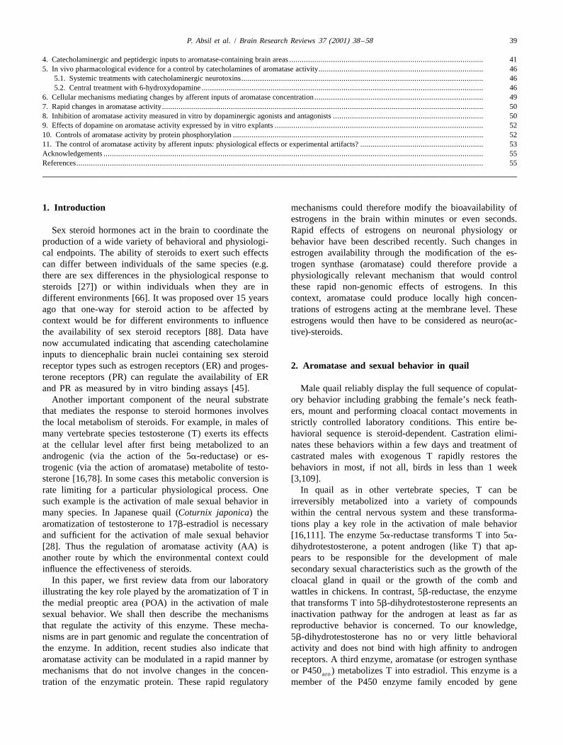
CYP19 [75,119] and in Japanese quail, brain AA plays a vertebrates examined so far. This effect appears to becritical limiting role in the activation of male sexual largely mediated by the interaction of the steroid withbehavior by T. This conclusion has been derived from a androgen receptors in rats [106,107] but by an action ofvariety of experiments in which either the AA was blocked locally produced estrogens in birds [68] and in quail inby specific inhibitors or the access of locally formed particular [28]. There is a synergism between non-estrogens to their receptor was blocked by drugs known as aromatizable androgens and estrogens in the mechanismanti-estrogens (e.g. nitromifene citrate or tamoxifen). In that regulates aromatase and this synergism can be ob-both cases, pharmacological interference with the pro- served at the three different levels at which aromatase hasduction or activity of estrogens almost completely sup- been studied (the mRNA, the protein and the enzymepressed the behavioral effects of T [28]. activity) [36,59,112] (Fig. 1).
Morphometric analyses of the POA, a brain region The most parsimonious model that could explain thisknown to be involved in the control of male-typical control of aromatase synthesis by estrogens assumes thatcopulatory behavior in most, if not all vertebrate species, when entering its target cells, T is aromatized in theidentified the presence of a sexually dimorphic nucleus. cytoplasmic compartment into an estrogen which wouldThis structure, called the medial preoptic nucleus (POM) is then bind to estrogen receptors (ER) and activate thesignificantly larger in volume in males than in females transcription of the aromatase gene. Surprisingly however,[132]. Furthermore the POM volume is sensitive to the immunocytochemical studies revealed that the estrogen-circulating levels of androgens: it regresses in castrated synthesizing enzyme and the ER, although they are presentbirds and is restored to the volume typical of sexuallymature males by a two week treatment with exogenous T[99,100]. These variations nicely parallel the mechanismsthat control copulatory behavior in quail and it washypothesized that the POM may be an important part ofthe neural pathway mediating the activation of male-typical reproductive behavior by T [25,98]. This conclu-sion has been fully supported by a series of behavioralexperiments. Lesions of the POM decrease or abolish theexpression of male copulatory behavior and the behavioraldeficits are proportional to the volume of POM lesionedbut not to the absolute size of the lesion. Furthermore,stereotaxic implantation of T within the cytoarchitectonicboundaries of the POM activates male copulatory behaviorin castrated males but implants located elsewhere in thePOA are behaviorally ineffective. Stereotaxic implants ofan aromatase inhibitor or of an antiestrogen within thePOM substantially decrease or suppress the activatingeffects of a systemic treatment with T in castrated males.
Taken together, these data indicate that T must bearomatized and locally-produced estrogens must act withinthe POM to activate male copulation. The preoptic aromat-ase in quail thus plays a key role in the control of malereproductive behavior and a number of studies weretherefore devoted to the analysis of the neuroanatomicaldistribution of this enzyme and of the mechanisms thatcontrol its activity [25,26,98].
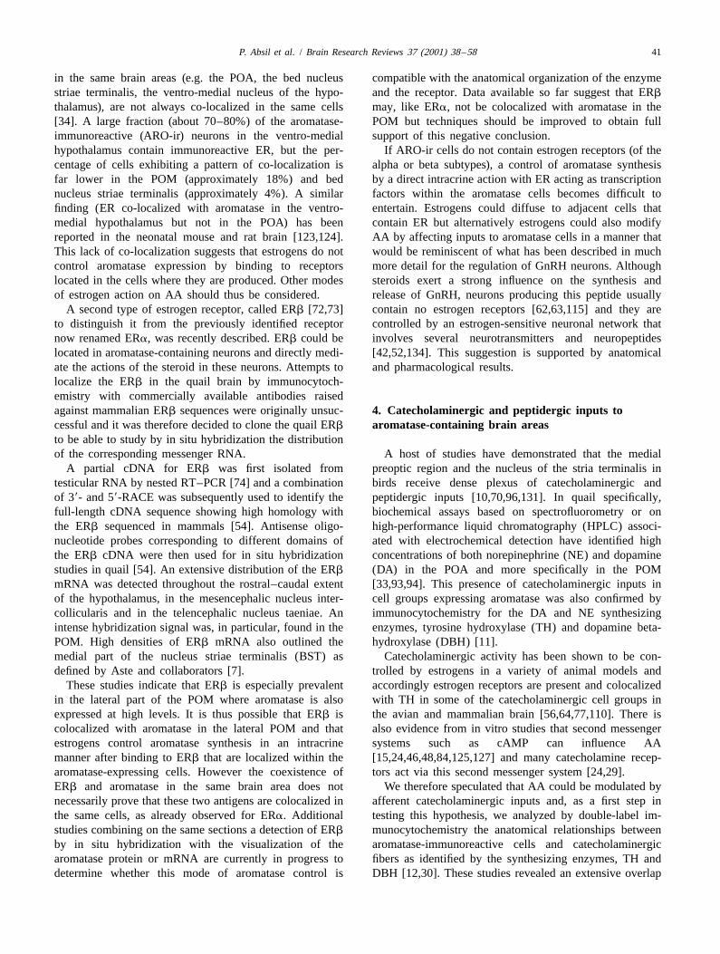
Fig. 1. Effects of testosterone (T), of a non-aromatizable androgen (A)3. Control of aromatase transcription by steroidsand of an estrogen (E) alone or in combination (A1E) on aromataseactivity (top), on the number of aromatase-immunoreactive cells in the
In quail, AA is higher in the POM than in any other POM (middle) and on the aromatase mRNA concentration (bottom) in thebrain region [113]. Accordingly, the boundaries of the POA in male quail. The non aromatizable androgen was
methyltrieneolone in top panel and 5a-dihydrotestosterone in the otherPOM can be delineated by the high density of cellstwo panels. The estrogen used in top panel was diethylstilbestrol.expressing aromatase as identified at the protein or mRNAEstradiol-17b was used in the other panels. All data (means6S.D.) were
levels by immunocytochemistry or in situ hybridization, expressed as a percentage of corresponding values in castrates (CX) torespectively [9,31]. permit direct comparisons. Recalculated and plotted from data in Refs.
T increases AA in the POA of all species of higher [59,112].
P. Absil et al. / Brain Research Reviews 37 (2001) 38 –58 41
in the same brain areas (e.g. the POA, the bed nucleus compatible with the anatomical organization of the enzymestriae terminalis, the ventro-medial nucleus of the hypo- and the receptor. Data available so far suggest that ERb
thalamus), are not always co-localized in the same cells may, like ERa, not be colocalized with aromatase in the[34]. A large fraction (about 70–80%) of the aromatase- POM but techniques should be improved to obtain fullimmunoreactive (ARO-ir) neurons in the ventro-medial support of this negative conclusion.hypothalamus contain immunoreactive ER, but the per- If ARO-ir cells do not contain estrogen receptors (of thecentage of cells exhibiting a pattern of co-localization is alpha or beta subtypes), a control of aromatase synthesisfar lower in the POM (approximately 18%) and bed by a direct intracrine action with ER acting as transcriptionnucleus striae terminalis (approximately 4%). A similar factors within the aromatase cells becomes difficult tofinding (ER co-localized with aromatase in the ventro- entertain. Estrogens could diffuse to adjacent cells thatmedial hypothalamus but not in the POA) has been contain ER but alternatively estrogens could also modifyreported in the neonatal mouse and rat brain [123,124]. AA by affecting inputs to aromatase cells in a manner thatThis lack of co-localization suggests that estrogens do not would be reminiscent of what has been described in muchcontrol aromatase expression by binding to receptors more detail for the regulation of GnRH neurons. Althoughlocated in the cells where they are produced. Other modes steroids exert a strong influence on the synthesis andof estrogen action on AA should thus be considered. release of GnRH, neurons producing this peptide usually
A second type of estrogen receptor, called ERb [72,73] contain no estrogen receptors [62,63,115] and they areto distinguish it from the previously identified receptor controlled by an estrogen-sensitive neuronal network thatnow renamed ERa, was recently described. ERb could be involves several neurotransmitters and neuropeptideslocated in aromatase-containing neurons and directly medi- [42,52,134]. This suggestion is supported by anatomicalate the actions of the steroid in these neurons. Attempts to and pharmacological results.localize the ERb in the quail brain by immunocytoch-emistry with commercially available antibodies raisedagainst mammalian ERb sequences were originally unsuc- 4. Catecholaminergic and peptidergic inputs tocessful and it was therefore decided to clone the quail ERb aromatase-containing brain areasto be able to study by in situ hybridization the distributionof the corresponding messenger RNA. A host of studies have demonstrated that the medial
A partial cDNA for ERb was first isolated from preoptic region and the nucleus of the stria terminalis intesticular RNA by nested RT–PCR [74] and a combination birds receive dense plexus of catecholaminergic andof 39- and 59-RACE was subsequently used to identify the peptidergic inputs [10,70,96,131]. In quail specifically,full-length cDNA sequence showing high homology with biochemical assays based on spectrofluorometry or onthe ERb sequenced in mammals [54]. Antisense oligo- high-performance liquid chromatography (HPLC) associ-nucleotide probes corresponding to different domains of ated with electrochemical detection have identified highthe ERb cDNA were then used for in situ hybridization concentrations of both norepinephrine (NE) and dopaminestudies in quail [54]. An extensive distribution of the ERb (DA) in the POA and more specifically in the POMmRNA was detected throughout the rostral–caudal extent [33,93,94]. This presence of catecholaminergic inputs inof the hypothalamus, in the mesencephalic nucleus inter- cell groups expressing aromatase was also confirmed bycollicularis and in the telencephalic nucleus taeniae. An immunocytochemistry for the DA and NE synthesizingintense hybridization signal was, in particular, found in the enzymes, tyrosine hydroxylase (TH) and dopamine beta-POM. High densities of ERb mRNA also outlined the hydroxylase (DBH) [11].medial part of the nucleus striae terminalis (BST) as Catecholaminergic activity has been shown to be con-defined by Aste and collaborators [7]. trolled by estrogens in a variety of animal models and
These studies indicate that ERb is especially prevalent accordingly estrogen receptors are present and colocalizedin the lateral part of the POM where aromatase is also with TH in some of the catecholaminergic cell groups inexpressed at high levels. It is thus possible that ERb is the avian and mammalian brain [56,64,77,110]. There iscolocalized with aromatase in the lateral POM and that also evidence from in vitro studies that second messengerestrogens control aromatase synthesis in an intracrine systems such as cAMP can influence AAmanner after binding to ERb that are localized within the [15,24,46,48,84,125,127] and many catecholamine recep-aromatase-expressing cells. However the coexistence of tors act via this second messenger system [24,29].ERb and aromatase in the same brain area does not We therefore speculated that AA could be modulated bynecessarily prove that these two antigens are colocalized in afferent catecholaminergic inputs and, as a first step inthe same cells, as already observed for ERa. Additional testing this hypothesis, we analyzed by double-label im-studies combining on the same sections a detection of ERb munocytochemistry the anatomical relationships betweenby in situ hybridization with the visualization of the aromatase-immunoreactive cells and catecholaminergicaromatase protein or mRNA are currently in progress to fibers as identified by the synthesizing enzymes, TH anddetermine whether this mode of aromatase control is DBH [12,30]. These studies revealed an extensive overlap
42 P. Absil et al. / Brain Research Reviews 37 (2001) 38 –58
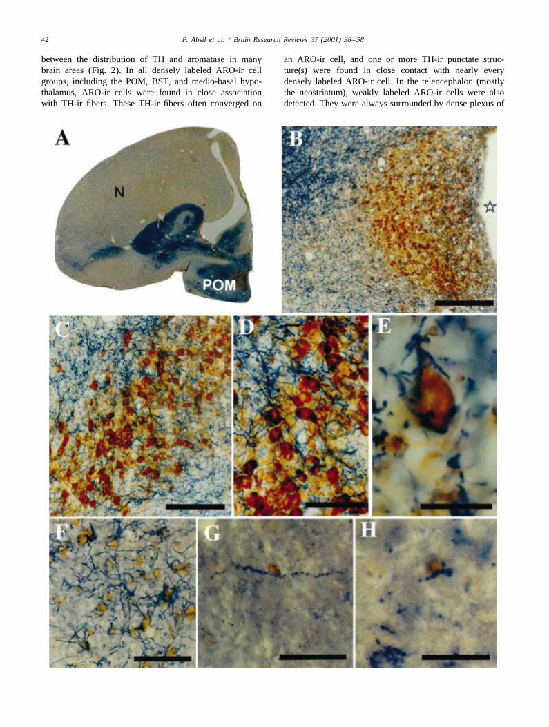
between the distribution of TH and aromatase in many an ARO-ir cell, and one or more TH-ir punctate struc-brain areas (Fig. 2). In all densely labeled ARO-ir cell ture(s) were found in close contact with nearly everygroups, including the POM, BST, and medio-basal hypo- densely labeled ARO-ir cell. In the telencephalon (mostlythalamus, ARO-ir cells were found in close association the neostriatum), weakly labeled ARO-ir cells were alsowith TH-ir fibers. These TH-ir fibers often converged on detected. They were always surrounded by dense plexus of
P. Absil et al. / Brain Research Reviews 37 (2001) 38 –58 43
TH-ir fibers. TH and aromatase were, however, not found tion of ERa and TH has in mammals been mostlyto be colocalized except in a few cells located at the observed in the hypothalamic dopaminergic neuronsrostro-medial pole of the anterior hypothalamus in a TH-ir [56,64,110] as well as in some brain stem and medullarcell cluster that appears to be homologous to the an- nuclei [64]. More recently, similar data have been reportedteroventral periventricular (AVPv) catecholaminergic cell in one avian species, the canary. In a study comparing thegroup described in the rat brain [116–118]. distribution of the estrogen receptor-alpha (ERa) mRNA
Attempts were also made to double label sections expression with that of TH-ir structures in the brainstem ofthrough the quail brain for aromatase and DBH. Because male canaries, it was found that dense ERa mRNAthe only available antibodies that permit visualization of labeling overlaps with TH-ir structures in both the locusthese antigens have been both raised in rabbits, cross- ceruleus and the area ventralis of Tsai [77]. Furthermore,reactions were frequently observed so that meaningful immunocytochemical double label experiments have dem-observations could not be performed. However, in selected onstrated the presence of immunoreactive ERa in thecases where all antigenic sites from the first immuno- TH-ir neurons of the anterior hypothalamus at the level ofcytochemical sequence had been effectively blocked by the the putative homologue of AVPv The presence of ERa
diaminobenzidine precipitate, specific labels could be within these cell groups suggests that sex steroid hormonesobtained as confirmed by the subcellular localization of the may affect catecholaminergic activity at the site of syn-chromogens (in perikarya for aromatase, in fibers and thesis.terminals for DBH). DBH-ir punctate structures and fibers Taken together, these observations are consistent withwere then found in close association with ARO-ir cell the hypothesis that catecholamines regulate the synthesisbodies in all brain regions where these are present namely of aromatase and potentially mediate the effects of es-the POM, BST and the ventro-medial and tuberal hypo- trogens on AA. Pharmacological experiments were there-thalamus. These data thus support the notion that norad- fore carried out to test this notion (see below).renergic fibers also innervate ARO-ir cells. Other afferent inputs have also been described in brain
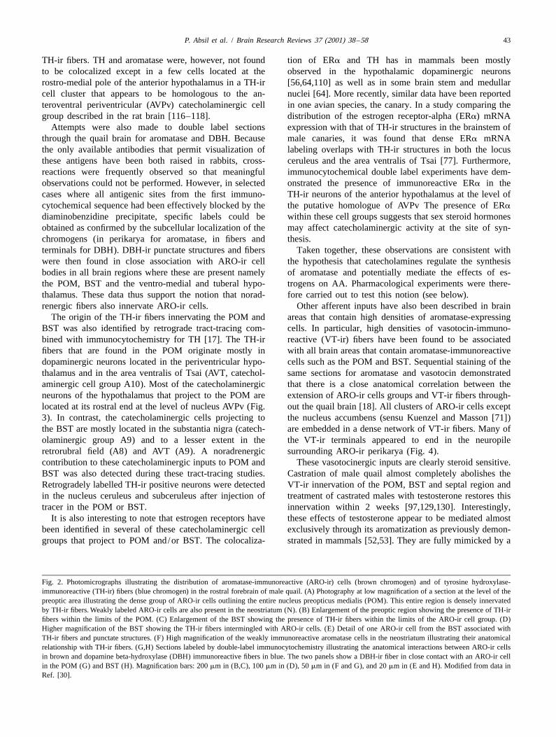
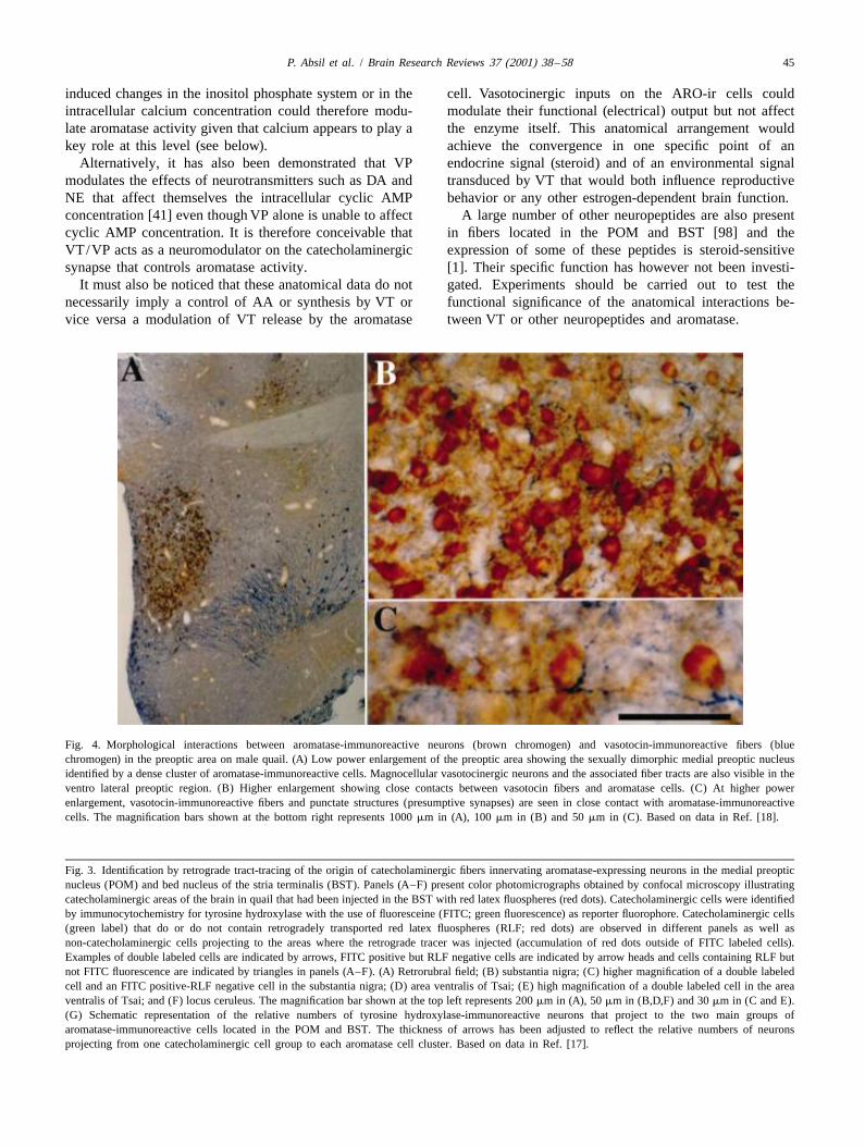
The origin of the TH-ir fibers innervating the POM and areas that contain high densities of aromatase-expressingBST was also identified by retrograde tract-tracing com- cells. In particular, high densities of vasotocin-immuno-bined with immunocytochemistry for TH [17]. The TH-ir reactive (VT-ir) fibers have been found to be associatedfibers that are found in the POM originate mostly in with all brain areas that contain aromatase-immunoreactivedopaminergic neurons located in the periventricular hypo- cells such as the POM and BST. Sequential staining of thethalamus and in the area ventralis of Tsai (AVT, catechol- same sections for aromatase and vasotocin demonstratedaminergic cell group A10). Most of the catecholaminergic that there is a close anatomical correlation between theneurons of the hypothalamus that project to the POM are extension of ARO-ir cells groups and VT-ir fibers through-located at its rostral end at the level of nucleus AVPv (Fig. out the quail brain [18]. All clusters of ARO-ir cells except3). In contrast, the catecholaminergic cells projecting to the nucleus accumbens (sensu Kuenzel and Masson [71])the BST are mostly located in the substantia nigra (catech- are embedded in a dense network of VT-ir fibers. Many ofolaminergic group A9) and to a lesser extent in the the VT-ir terminals appeared to end in the neuropileretrorubral field (A8) and AVT (A9). A noradrenergic surrounding ARO-ir perikarya (Fig. 4).contribution to these catecholaminergic inputs to POM and These vasotocinergic inputs are clearly steroid sensitive.BST was also detected during these tract-tracing studies. Castration of male quail almost completely abolishes theRetrogradely labelled TH-ir positive neurons were detected VT-ir innervation of the POM, BST and septal region andin the nucleus ceruleus and subceruleus after injection of treatment of castrated males with testosterone restores thistracer in the POM or BST. innervation within 2 weeks [97,129,130]. Interestingly,
It is also interesting to note that estrogen receptors have these effects of testosterone appear to be mediated almostbeen identified in several of these catecholaminergic cell exclusively through its aromatization as previously demon-groups that project to POM and/or BST. The colocaliza- strated in mammals [52,53]. They are fully mimicked by a
Fig. 2. Photomicrographs illustrating the distribution of aromatase-immunoreactive (ARO-ir) cells (brown chromogen) and of tyrosine hydroxylase-immunoreactive (TH-ir) fibers (blue chromogen) in the rostral forebrain of male quail. (A) Photography at low magnification of a section at the level of thepreoptic area illustrating the dense group of ARO-ir cells outlining the entire nucleus preopticus medialis (POM). This entire region is densely innervatedby TH-ir fibers. Weakly labeled ARO-ir cells are also present in the neostriatum (N). (B) Enlargement of the preoptic region showing the presence of TH-irfibers within the limits of the POM. (C) Enlargement of the BST showing the presence of TH-ir fibers within the limits of the ARO-ir cell group. (D)Higher magnification of the BST showing the TH-ir fibers intermingled with ARO-ir cells. (E) Detail of one ARO-ir cell from the BST associated withTH-ir fibers and punctate structures. (F) High magnification of the weakly immunoreactive aromatase cells in the neostriatum illustrating their anatomicalrelationship with TH-ir fibers. (G,H) Sections labeled by double-label immunocytochemistry illustrating the anatomical interactions between ARO-ir cellsin brown and dopamine beta-hydroxylase (DBH) immunoreactive fibers in blue. The two panels show a DBH-ir fiber in close contact with an ARO-ir cellin the POM (G) and BST (H). Magnification bars: 200 mm in (B,C), 100 mm in (D), 50 mm in (F and G), and 20 mm in (E and H). Modified from data inRef. [30].

44 P. Absil et al. / Brain Research Reviews 37 (2001) 38 –58
treatment with exogenous estradiol but not by a treatment that could mediate this effect are unclear at present butwith the non-aromatizable androgen 5a-dihydrotestos- several hypothesis can be suggested.terone [128]. Vasotocin is therefore another estrogen-sensi- Vasopressin (VP), the mammalian homologue of VT,tive potential candidate that could mediate effects of acts mainly through its V1 receptors that are coupled to theestrogens on AA. The intracellular transduction systems inositol phosphate second messenger system [41]. VT-
P. Absil et al. / Brain Research Reviews 37 (2001) 38 –58 45
induced changes in the inositol phosphate system or in the cell. Vasotocinergic inputs on the ARO-ir cells couldintracellular calcium concentration could therefore modu- modulate their functional (electrical) output but not affectlate aromatase activity given that calcium appears to play a the enzyme itself. This anatomical arrangement wouldkey role at this level (see below). achieve the convergence in one specific point of an
Alternatively, it has also been demonstrated that VP endocrine signal (steroid) and of an environmental signalmodulates the effects of neurotransmitters such as DA and transduced by VT that would both influence reproductiveNE that affect themselves the intracellular cyclic AMP behavior or any other estrogen-dependent brain function.concentration [41] even though VP alone is unable to affect A large number of other neuropeptides are also presentcyclic AMP concentration. It is therefore conceivable that in fibers located in the POM and BST [98] and theVT/VP acts as a neuromodulator on the catecholaminergic expression of some of these peptides is steroid-sensitivesynapse that controls aromatase activity. [1]. Their specific function has however not been investi-
It must also be noticed that these anatomical data do not gated. Experiments should be carried out to test thenecessarily imply a control of AA or synthesis by VT or functional significance of the anatomical interactions be-vice versa a modulation of VT release by the aromatase tween VT or other neuropeptides and aromatase.
Fig. 4. Morphological interactions between aromatase-immunoreactive neurons (brown chromogen) and vasotocin-immunoreactive fibers (bluechromogen) in the preoptic area on male quail. (A) Low power enlargement of the preoptic area showing the sexually dimorphic medial preoptic nucleusidentified by a dense cluster of aromatase-immunoreactive cells. Magnocellular vasotocinergic neurons and the associated fiber tracts are also visible in theventro lateral preoptic region. (B) Higher enlargement showing close contacts between vasotocin fibers and aromatase cells. (C) At higher powerenlargement, vasotocin-immunoreactive fibers and punctate structures (presumptive synapses) are seen in close contact with aromatase-immunoreactivecells. The magnification bars shown at the bottom right represents 1000 mm in (A), 100 mm in (B) and 50 mm in (C). Based on data in Ref. [18].
Fig. 3. Identification by retrograde tract-tracing of the origin of catecholaminergic fibers innervating aromatase-expressing neurons in the medial preopticnucleus (POM) and bed nucleus of the stria terminalis (BST). Panels (A–F) present color photomicrographs obtained by confocal microscopy illustratingcatecholaminergic areas of the brain in quail that had been injected in the BST with red latex fluospheres (red dots). Catecholaminergic cells were identifiedby immunocytochemistry for tyrosine hydroxylase with the use of fluoresceine (FITC; green fluorescence) as reporter fluorophore. Catecholaminergic cells(green label) that do or do not contain retrogradely transported red latex fluospheres (RLF; red dots) are observed in different panels as well asnon-catecholaminergic cells projecting to the areas where the retrograde tracer was injected (accumulation of red dots outside of FITC labeled cells).Examples of double labeled cells are indicated by arrows, FITC positive but RLF negative cells are indicated by arrow heads and cells containing RLF butnot FITC fluorescence are indicated by triangles in panels (A–F). (A) Retrorubral field; (B) substantia nigra; (C) higher magnification of a double labeledcell and an FITC positive-RLF negative cell in the substantia nigra; (D) area ventralis of Tsai; (E) high magnification of a double labeled cell in the areaventralis of Tsai; and (F) locus ceruleus. The magnification bar shown at the top left represents 200 mm in (A), 50 mm in (B,D,F) and 30 mm in (C and E).(G) Schematic representation of the relative numbers of tyrosine hydroxylase-immunoreactive neurons that project to the two main groups ofaromatase-immunoreactive cells located in the POM and BST. The thickness of arrows has been adjusted to reflect the relative numbers of neuronsprojecting from one catecholaminergic cell group to each aromatase cell cluster. Based on data in Ref. [17].
46 P. Absil et al. / Brain Research Reviews 37 (2001) 38 –58
5. In vivo pharmacological evidence for a control by OHDA). Male quail were castrated and received, 2 weekscatecholamines of aromatase activity later, two 20 mm-long subcutaneous Silastic capsules filled
with crystalline testosterone. The catecholaminergic neuro-toxin, 6-hydroxydopamine hydrochloride (6-OHDA HCl)
5.1. Systemic treatments with catecholaminergicwas dissolved in distilled water containing 9‰ NaCl and
neurotoxins1‰ ascorbic acid at the dose of 50 mg/ml and each birdwas injected stereotaxically in the medial part of the POA
In a series of experiments, castrated male quail that were with 200 nl of this solution (10 mg of the toxin per bird).submitted to a replacement therapy with T were sys- Control animals received the corresponding volume of thetemically injected with the specific noradrenergic neuro- vehicle solution (saline1ascorbic acid). Preliminary ex-toxin, DSP4. HPLC assays confirmed that the single periments had shown that this neurotoxin does not depleteinjection of DSP4 significantly depleted NE concentrations tyrosine hydroxylase activity measured 5 days after thein all brain areas including the POA-hypothalamus, and the injection and does not affect aromatase activity if injectedPOM in particular [22,35]. The preoptic AA was increased at a lower dose (100 nl of a solution at 20 mg/ml, i.e. 2 mgafter the injection of DSP4 indicating that NE may exert a per bird). On another hand injection of 20 mg 6-OHDA perchronic inhibition of this enzymatic activity [22]. The subject leads to a high rate of mortality. The 10 mg doseDBH fibers originating from the locus ceruleus and selected for this experiment can therefore be considered assubceruleus that are in close contact with ARO-ir cells in the highest dose that can be tolerated by adult quail.the POM and BST [12,17] presumably represent the Stereotaxic injections were targeted to the medial preop-morphological signature of this regulation. tic nucleus (POM), with coordinates derived from previous
It has been shown previously that noradrenergic agonists studies [37–39]. Pressure injections were made unilaterallymodulate aromatase activity in the pineal gland [48] or in through a glass micropipette (external tip diameter of aboutthe testes [125] and similar controls might take place in the 50 mm) connected to a 0.5 ml Hamilton syringe and amammalian hypothalamus [47,104,105]. Given that the micropump. The micropipette was introduced verticallynoradrenergic activity in the brain appears to be under the and 200 nl of the toxin solution were delivered in about 7control of estrogens [81], an estrogen-sensitive noradrener- min. The micropipette was removed slowly 5 min later togic transmission might represent the neuroanatomical avoid leakage of the drug through the pipette track. Birdssubstrate underlying the regulation of aromatase activity by were injected on the left or right side of the brain in equalestrogens. numbers. No lateralization could be detected in the abso-
In male quail, a rapid decrease in DA concentration is lute level of the biochemical variables (depletion ofobserved after a single injection of the TH inhibitor, aromatase activity or tyrosine hydroxylase activity, seea-methyl-para tyrosine (aMPT) [33,93]. This DA dis- below) that were measured in the present experiments andappearance is supposed to reflect the turnover of the amine similarly no interaction between the experimental effects(see however Ref. [133] for a critical discussion of this and the injection side was observed. The brain side wasissue). In one experiment, brain DA levels were chronical- therefore ignored in the analysis of results that is presentedly depressed in castrated testosterone-treated male quail by here. A total of 12 birds were injected in this way withdaily injections of aMPT [29]. This treatment produced, 6-OHDA while 11 birds received a control injection.after 3 days, a significant inhibition of aromatase activity One day after the 6-OHDA injection, birds were killedin the POA by comparison with the control T-treated birds by decapitation, their brain was quickly removed from theinjected with the solvent. It is clear that a chronic treatment skull and frozen on powdered dry ice. The POA-hypo-with aMPT decreases both NE (which presumably inhibits thalamic block was dissected from the frozen brains byAA in the quail brain based on the DSP4 experiments two coronal cuts at the level of the tractus septomesence-described above) and DA [33]. It can therefore be expected phalicus (rostral edge of the POA) and of the occulomotorthat a selective dopaminergic inhibition would lead to an nerves (caudal edge of hypothalamus), two parasagittaleven larger decrease in enzymatic activity which would be cuts placed approximately 2 mm lateral to the brainconsistent with the notion that DA up-regulates brain midline and one horizontal cut about 2 mm above the flooraromatase, in particular in the POM (see Ref. [24] for of the brain. This isolated a block of tissue weighingadditional discussion). 70–90 mg that contains the vast majority of the cells that
express aromatase in the quail brain. This block was thenseparated into its left and right parts by a medial cut
5.2. Central treatment with 6-hydroxydopamine passing largely through the third ventricle so that it couldbe very accurately positioned. This isolated the brain site
To specify further the functional interactions between ispilateral to the injection from its contra-lateral counter-catecholamines and preoptic AA, we depleted catechol- part. Finally, the left and right preoptic area-hypothalamicaminergic inputs to the POM by injecting into this nucleus blocks were cut in two equal halves by a coronal cutthe catecholamine neurotoxin 6-hydroxydopamine (6- isolating the rostral and caudal parts corresponding roughly
P. Absil et al. / Brain Research Reviews 37 (2001) 38 –58 47
to the preoptic area (POA, where injection of 6-OHDA/saline had been made) and to the hypothalamus (HYP),respectively.
AA was quantified in these four blocks by measuring thetritiated water assay method, as described by Roselli andResko [108], with minor modifications [13]. Tyrosinehydroxylase activity (TH-A) was also assayed in the samesamples in order to assess the extent of the catechol-aminergic lesion by measuring the tritiated water pro-
3duction from 3,5-[ H]-L-tyrosine, as initially described byNagatsu et al. [87] and later Levine et al. [76], with somefurther modifications [14]. These two methods have beenpreviously validated for use in the quail brain [13,14].
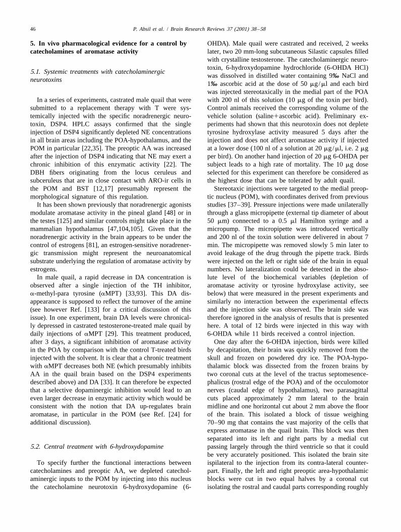
Results described below were expressed as fmol /nmolper brain fraction, representing the total enzymatic ac-tivities by brain area instead of activities per mg protein ormg fresh weight. The reasons for this choice are explainedby the anatomy of the systems under investigation.Aromatase-expressing cells and TH-containing structuresonly form a small fraction (5% or less) of the POA andHYP [8,11,30,40]. The micro-dissection into four blocks ofthe POA–HYP reliably separated the left and right hemi-spheres and isolated in a reliable manner the rostralaromatase cells that are primarily located in the POM andBST from the more caudal cells located in the medio-basalhypothalamus. However, the precise location of the lateralcuts was probably more variable. Thus tissue blockspresumably contained variable amounts of extraneoustissue that does not contain AA, given that all ARO-ir cells Fig. 5. Effects of a unilateral injection of 6-OHDA (10 mg) in the
preoptic area on the aromatase activity and tyrosine hydroxylase activityare located in periventricular position and are necessarilyin the preoptic area. Enzyme activities were measured on the injectionincluded in the dissection independent of the position of(ipsi.) and on the contra-lateral (contra.) side that was used as internalthe lateral cuts. Therefore expressing data per mg proteincontrol. Values obtained in saline-injected birds (SAL) are presented as
or fresh weight introduces unexplained variance in the additional controls. Data are means6S.E.M.; n512 for 6OHDA birds andresults, a problem that is not encountered when enzyme n511 for saline controls. Analysis of these data by two-way ANOVA
with one repeated factor (brain side) indicated the presence of significantactivities are expressed by entire brain area (POA orinteraction between brain side and experimental treatment (6OHDA vs.HYP). These data were analyzed directly by one or two-saline) for aromatase activity and of brain side for tyrosine hydroxylaseway analysis of variance (ANOVA) or as a ratio of theactivity (see text). Further analysis of these data in 6OHDA and saline
enzyme activities (AA or TH-A) measured on the injection birds separately by one-way repeated ANOVA confirmed the decline ofside divided by the control values on the contralateral side aromatase activity (* P,0.05) and of tyrosine hydroxylase activity
(** P,0.01) on the injection side in 6OHDA birds.(percentage (%) AA, %TH-A).Both AA and TH-A were very similar in the two sides
of the brain in birds that had been injected with the controlsaline solution, thus confirming the reliability of the braindissections and accuracy of the enzymatic assays (Fig. 5). catecholaminergic depletion that was similarly small (Fig.A noticeable decrease in AA was observed in the POA on 5).the side receiving the 6-OHDA injection (Fig. 5). Analysis TH-A was also slightly smaller on the injection than onof these results by two-way ANOVA with the two ex- the contralateral sides in saline-treated birds, so that noperimental groups as an independent factor and the two significant treatment effect (F 52.162, P50.1563) and1,21
brain sides as a repeated factor detected no significant no interaction between side and treatment (F 52.445,1,21
effects of the two main factors (Treatment: F 50.138, P50.1328) was detected in the analysis of these results.1,21
P50.7141; Side: F 53.653, P50.0697) but indicated a There was however a significant effect of the brain side on1,21
significant interaction between them, therefore confirming TH-A (F 515.263, P50.0008).1,21
a localized effect of the experimental treatment (F 5 In the HYP, caudal to the injection site, AA was not1,21
4.525, P50.0454). The observed decrease in AA in the affected by the treatment, the side of the brain and theirbrain side injected with 6-OHDA was admittedly of a interaction (respectively: F 50.228, P50.6378; F 51,21 1,21
limited amplitude but the assays of TH activity indicated a 1.151, P50.2955 and F 50.023, P50.8809). Interest-1,21
48 P. Absil et al. / Brain Research Reviews 37 (2001) 38 –58
ingly, TH-A was enhanced specifically on the injection TH-A depletions being associated with the largest deple-side in the HYP of 6-OHDA treated birds. This resulted in tions in aromatase (r50.629, n512, P50.0283). Bya significant interaction of the treatment with brain side contrast there was no relationship between these two(F 56.865, P50.0160) in the absence of effect of the percentages of depletion in birds injected with saline (r51,21
two main factors (Treatment: F 50.429, P50.5196, 0.015, n511, P50.9662).1,21
Side: F 50.739, P50.3396). These percentages of AA and TH-A depletion (by1,21
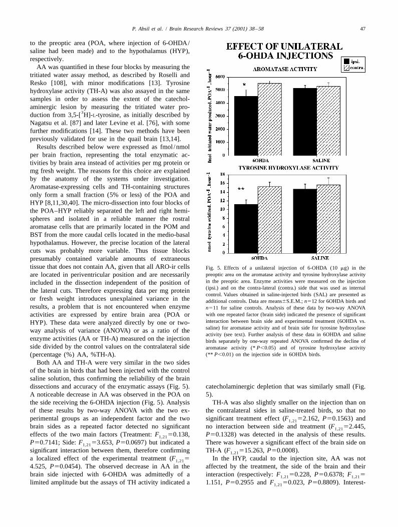
The extent of AA depletion in the POA side injected comparison with the non-injected side) were furtherwith 6-OHDA was found to be quite variable from one studied by a one-way analysis of covariance using thebird to the other and we therefore asked whether this AA experimental treatment (6-OHDA vs. saline) as the in-depletion was related to the degree of catecholaminergic dependent factor, the %AA remaining on the injection sidedepletion measured by the TH-A decrease. Correlations (as a percentage of the non injected side) as dependentwere therefore calculated between the individual percent- variable and the %TH-A remaining on the injection side asage of AA left in the injection side (compared with the a covariate. An extremely significant effect of the ex-contra-lateral side) and the percentage of TH-A computed perimental treatment was detected in this way (F 51,20
in the same way. Interestingly, these two variables were 15.811, P50.0007) as well as a significant effect of thefound to be significantly correlated (Fig. 6) with large interaction between the treatment and the covariate (F 51,20
12.539, P50.0021).These results were replicated in a second experiment
testing in addition the effects of two catecholamine uptakeinhibitors on the 6-OHDA induced depletion of AA, in anattempt to identify the specific nature of the catechol-aminergic effect on AA. In this study, castrated male quailthat had been treated with exogenous testosterone wereagain stereotaxically injected in the POM with 10 mg6-OHDA or with the control solution but in addition, someof the birds were pretreated 50 min before with either thenorepinephrine uptake inhibitor desipramine hydrochloride(DMI; 25 mg/kg ip) or the dopamine uptake inhibitorGBR-12909 hydrochloride (GBR; 40 mg/kg ip). Thecombination of these treatments thus defined six ex-perimental groups: birds injected with 6-OHDA after apre-treatment with saline (6-OHDA, n512), birds injectedwith saline after a pre-treatment with DMI (DMI, n512),birds injected with saline after a pre-treatment with GBR(GBR, n512), birds injected with 6-OHDA after a pre-treatment with DMI (DMI1OHDA, n512), birds injectedwith 6-OHDA after a pre-treatment with GBR (GBR1
OHDA, n512) and finally birds injected with saline after apre-treatment with saline (SAL, n59).
A two-way ANOVA of AA levels with an independent(six treatment groups) and a repeated (two brain sides)factor confirmed the absence of overall effects of thetreatments (F 50.664, P50.6522) but the side and, most5,63
importantly, the interaction of side with treatment had verysignificant effects on the enzyme activity (respectivelyF 57.456, P50.0082 and F 53.635, P50.0059).1,63 5,63
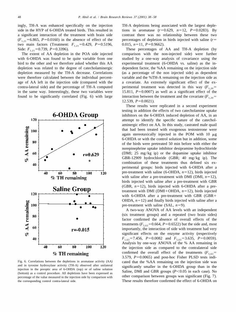
Analysis by one-way ANOVA of the % AA remaining inthe injection side as compared to the contralateral sideconfirmed the overall effect of the treatments (F 55,63
3.579, P50.0065) and post-hoc Fisher PLSD tests indi-Fig. 6. Correlations between the depletions in aromatase activity (AA) cated that the %AA remaining on the injection side wasand in tyrosine hydroxylase activity (TH-A) observed after unilateral significantly smaller in the 6-OHDA group than in theinjection in the preoptic area of 6-OHDA (top) or of saline solution
Saline, DMI and GBR groups (P,0.05 in each case). No(bottom) as a control procedure. All depletions have been expressed asother comparison between groups was significant (Fig. 7).percentage of the value measured in the injection side by comparison with
the corresponding control contra-lateral side. These results therefore confirmed the effect of 6-OHDA on
P. Absil et al. / Brain Research Reviews 37 (2001) 38 –58 49
6. Cellular mechanisms mediating changes byafferent inputs of aromatase concentration
Taken together, these anatomical data and experimentalmanipulations of the catecholaminergic inputs to the POAstrongly suggest that catecholamines control AA in thebrain POA. The single experiment carried out so far inquail, suggests that NE exerts a tonic inhibition on thepreoptic AA. On the contrary, pharmacological depletionsof DA result in a decrease of AA and the decreases of AAand TH-A are significantly correlated within individuals,further supporting the existence of a causal link betweenthese two enzymatic changes. All these data thus point to astimulatory effect of DA on AA. These studies are,however, all based on dopaminergic depletions (lesions)Fig. 7. Effects of a unilateral injection of 6-OHDA in the preoptic areaand it would be desirable to confirm the specificity of thesecombined or not with a pre-treatment by the catecholamine reuptake
inhibitors GBR-12909 hydrochloride (GBR) and desipramine hydrochlo- effects by testing whether a chronic stimulation by D1 orride (DMI) on the aromatase activity (AA) in the preoptic area. Enzyme D2 agonists really leads to an increase in AA. Such a goalactivity measured on the injection side was expressed as a percentage of is however technically difficult to achieve.the control value obtained on the contra-lateral side. The different
The cellular mechanisms that mediate the stimulation oftreatments were compared two by two by Fisher PLSD tests whose resultsAA by DA have not been investigated in the brain. It isare reported by symbols at the top of corresponding columns (* P,0.05
by comparison with the 6-OHDA group). Data presented are clear however that the binding of DA to its membranemeans6S.E.M.; n512 subjects in each group except in the saline group receptors modulates the production of cAMP (respectivelywhere n59. increase and decrease in concentration mediated by the D1
and D2-like receptors). In addition, it has been shown thatAA but failed to determine whether the effect is mediated experimental manipulations of cAMP affect aromataseby noradrenergic or dopaminergic receptors, presumably activity in a variety of tissues including the brainbecause the dose and/or timing selected for the reuptake [46,84,95,125–127,135]. It is conceivable that the stimula-inhibitors were not adequate to block the 6-OHDA induced tion of the adenylyl cyclase activity caused by the bindinglesion on specific receptor types. of DA to its D1 receptors leads to the activation of protein
A correlation was again observed between the AA kinase A resulting in the phosphorylation of the cAMPdepletion and the TH-A depletion (both expressed as a response element binding protein (CREB). Phosphorylatedpercentage of activity found in the injected side by CREB is a transcriptional regulator that could then in-comparison with the contra-lateral side) in the 6-OHDA crease transcription of the aromatase gene and thus theinjected birds but it fell short of significance (r50.523, concentration of the enzyme. In agreement with thisn512, P50.0813). No relationship between these vari- scenario, CREB responsive elements (CRE or SF1) haveables was present in saline-injected subjects (r520.499, been identified on forms of the aromatase gene exon 1 thatn59, P50.1719). are typically found in peripheral tissues but also to a lesser
The pooled data of both experiments expressed as a extent in the brain [55,65,102,120].percentage of activity remaining on the injection vs. It must be stressed that this model of DA actions impliescontralateral side were also submitted to an analysis of the presence of DA receptors at the surface of aromatasecovariance with the six experimental groups as the in- cells and no evidence for this presence has to date beendependent factor, the %AA remaining as a dependent collected due to the lack of suitable antibodies. It hasvariable and the %TH-A remaining as a covariate. This actually been shown that the D1 receptor-associated pro-global analysis confirmed the presence of an overall effect tein DARPP-32 (DA and cAMP-regulated phosphoprotein)of treatments (F 54.804, P50.007), of the covariate, is not colocalized with aromatase in the quail brain [2].5,80
i.e. the TH depletion (F 58.186, P50.0054) and of a This observation thus raises questions concerning the mode1,80
significant interaction between the main effect and the of DA action on AA (see Ref. [2] for further discussion).covariate (F 53.479, P50.0068). Correlation analyses Many other questions are also unanswered at this point.5,80
of these pooled data expressed in percentages also con- They relate to the mechanism of DA action but also to thefirmed the positive relationship in 6-OHDA injected birds real magnitude of the effects of this catecholamine on AA.between AA remaining and TH-A remaining in the in- Significant decreases of AA have been repeatedly observedjection site (r50.499, n524, P50.0131). These correla- after a pharmacological depletion of DA brain concen-tions were not observed in the pooled data of saline tration but a fairly large amount of residual AA wasinjected birds (r50.293, P50.2093). always present. This could reflect the failure to completely
50 P. Absil et al. / Brain Research Reviews 37 (2001) 38 –58
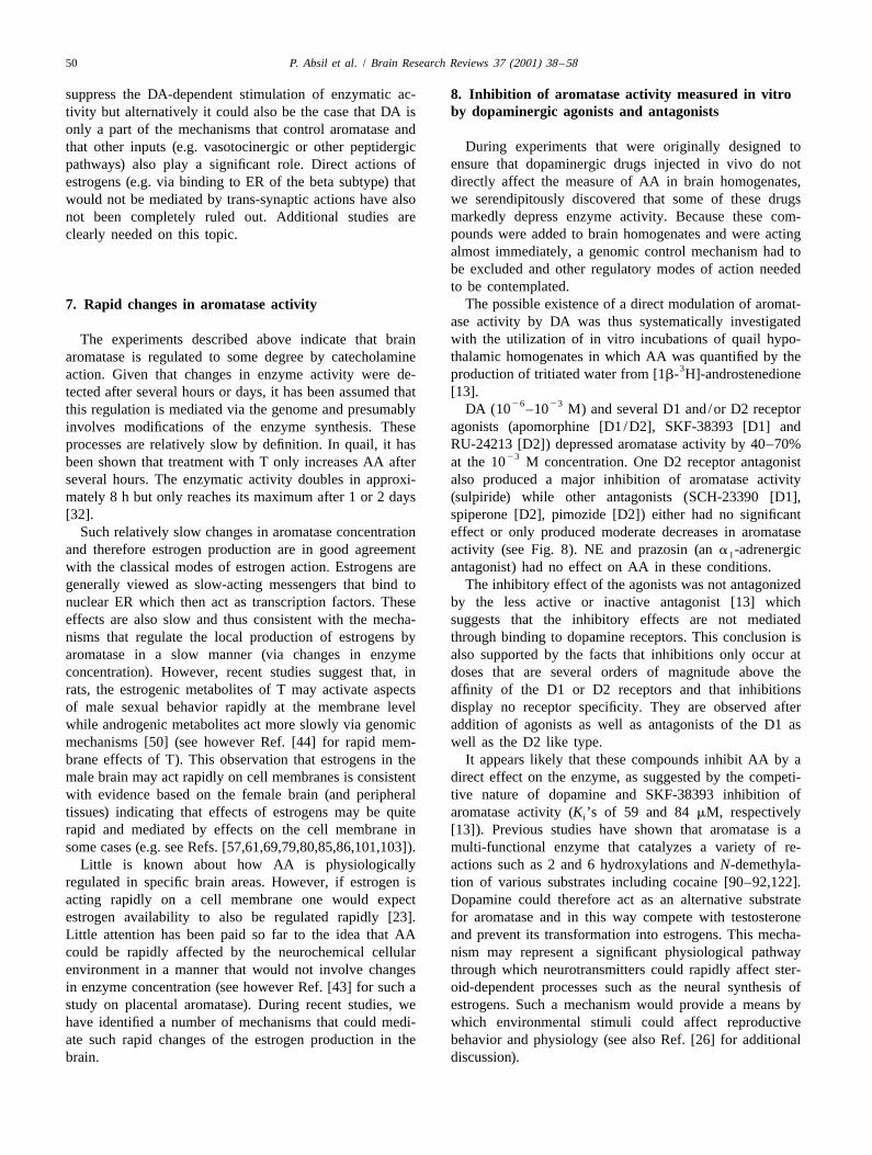
suppress the DA-dependent stimulation of enzymatic ac- 8. Inhibition of aromatase activity measured in vitroby dopaminergic agonists and antagoniststivity but alternatively it could also be the case that DA is
only a part of the mechanisms that control aromatase andDuring experiments that were originally designed tothat other inputs (e.g. vasotocinergic or other peptidergic
ensure that dopaminergic drugs injected in vivo do notpathways) also play a significant role. Direct actions ofdirectly affect the measure of AA in brain homogenates,estrogens (e.g. via binding to ER of the beta subtype) thatwe serendipitously discovered that some of these drugswould not be mediated by trans-synaptic actions have alsomarkedly depress enzyme activity. Because these com-not been completely ruled out. Additional studies arepounds were added to brain homogenates and were actingclearly needed on this topic.almost immediately, a genomic control mechanism had tobe excluded and other regulatory modes of action neededto be contemplated.
The possible existence of a direct modulation of aromat-7. Rapid changes in aromatase activityase activity by DA was thus systematically investigatedwith the utilization of in vitro incubations of quail hypo-The experiments described above indicate that brainthalamic homogenates in which AA was quantified by thearomatase is regulated to some degree by catecholamine
3production of tritiated water from [1b- H]-androstenedioneaction. Given that changes in enzyme activity were de-[13].tected after several hours or days, it has been assumed that
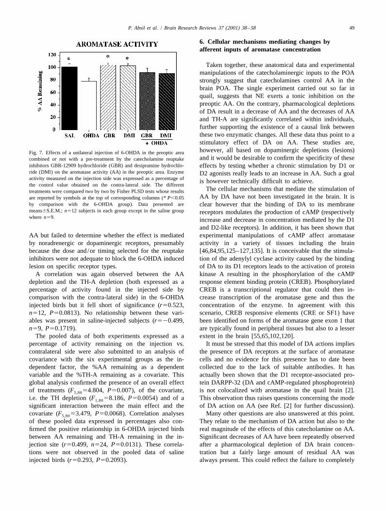
26 23DA (10 –10 M) and several D1 and/or D2 receptorthis regulation is mediated via the genome and presumablyagonists (apomorphine [D1/D2], SKF-38393 [D1] andinvolves modifications of the enzyme synthesis. TheseRU-24213 [D2]) depressed aromatase activity by 40–70%processes are relatively slow by definition. In quail, it has
23been shown that treatment with T only increases AA after at the 10 M concentration. One D2 receptor antagonistseveral hours. The enzymatic activity doubles in approxi- also produced a major inhibition of aromatase activitymately 8 h but only reaches its maximum after 1 or 2 days (sulpiride) while other antagonists (SCH-23390 [D1],[32]. spiperone [D2], pimozide [D2]) either had no significant
Such relatively slow changes in aromatase concentration effect or only produced moderate decreases in aromataseand therefore estrogen production are in good agreement activity (see Fig. 8). NE and prazosin (an a -adrenergic1
with the classical modes of estrogen action. Estrogens are antagonist) had no effect on AA in these conditions.generally viewed as slow-acting messengers that bind to The inhibitory effect of the agonists was not antagonizednuclear ER which then act as transcription factors. These by the less active or inactive antagonist [13] whicheffects are also slow and thus consistent with the mecha- suggests that the inhibitory effects are not mediatednisms that regulate the local production of estrogens by through binding to dopamine receptors. This conclusion isaromatase in a slow manner (via changes in enzyme also supported by the facts that inhibitions only occur atconcentration). However, recent studies suggest that, in doses that are several orders of magnitude above therats, the estrogenic metabolites of T may activate aspects affinity of the D1 or D2 receptors and that inhibitionsof male sexual behavior rapidly at the membrane level display no receptor specificity. They are observed afterwhile androgenic metabolites act more slowly via genomic addition of agonists as well as antagonists of the D1 asmechanisms [50] (see however Ref. [44] for rapid mem- well as the D2 like type.brane effects of T). This observation that estrogens in the It appears likely that these compounds inhibit AA by amale brain may act rapidly on cell membranes is consistent direct effect on the enzyme, as suggested by the competi-with evidence based on the female brain (and peripheral tive nature of dopamine and SKF-38393 inhibition oftissues) indicating that effects of estrogens may be quite aromatase activity (K ’s of 59 and 84 mM, respectivelyi
rapid and mediated by effects on the cell membrane in [13]). Previous studies have shown that aromatase is asome cases (e.g. see Refs. [57,61,69,79,80,85,86,101,103]). multi-functional enzyme that catalyzes a variety of re-
Little is known about how AA is physiologically actions such as 2 and 6 hydroxylations and N-demethyla-regulated in specific brain areas. However, if estrogen is tion of various substrates including cocaine [90–92,122].acting rapidly on a cell membrane one would expect Dopamine could therefore act as an alternative substrateestrogen availability to also be regulated rapidly [23]. for aromatase and in this way compete with testosteroneLittle attention has been paid so far to the idea that AA and prevent its transformation into estrogens. This mecha-could be rapidly affected by the neurochemical cellular nism may represent a significant physiological pathwayenvironment in a manner that would not involve changes through which neurotransmitters could rapidly affect ster-in enzyme concentration (see however Ref. [43] for such a oid-dependent processes such as the neural synthesis ofstudy on placental aromatase). During recent studies, we estrogens. Such a mechanism would provide a means byhave identified a number of mechanisms that could medi- which environmental stimuli could affect reproductiveate such rapid changes of the estrogen production in the behavior and physiology (see also Ref. [26] for additionalbrain. discussion).
P.A
bsilet
al./
Brain
Research
Review
s37
(2001)38
–5851
Fig. 8. Effects of dopamine, general dopamine agonists, specific dopamine agonists and antagonists on the aromatase activity in homogenates of the quail preoptic area-hypothalamus. DA, dopamine;APO, apomorphine (general DA agonist); NOM, nomoifensine (indirect DA agonist: reuptake inhibitor); AMF, amfonelic acid (indirect DA agonist: complex mechanisms including enhanced release);SKF, SKF-38393; PPP, 3-PPP; RU, RU-24213; QUIN, Quinpirole; SCH, SCH-23390; SPI, spiperone; PIM, pimozide; and SUL, sulpiride. The specificity for the D1 or D2-like receptor subtypes isindicated below the name of each compound on the graph. All assays were performed in triplicate, error bars represent the S.D. of the measures that are expressed as percentage of the respective
23 26controls (line at 100%). All compounds were tested at two concentrations (10 and 10 M). Redrawn from data in Ref. [13].
52 P. Absil et al. / Brain Research Reviews 37 (2001) 38 –58
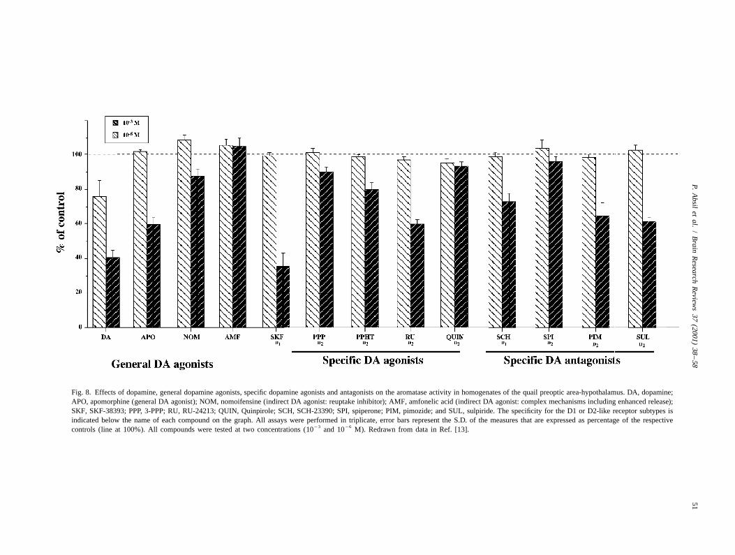
These experiments, however, raise questions concerning inhibitory and reversible effects were observed with otherthe mode of rapid DA action on AA. Anatomical studies dopaminergic agents such as SKF 38393 (a D1 agonist),describing the cellular and subcellular localization of DA SCH 23390 (a D1 antagonist), PPHT (a D2 agonist) andin relation to aromatase have indeed failed to identify sulpiride (a D2 antagonist) [19]. These effects are veryARO-ir cells that also contain DA. No colocalization has reminiscent of those observed on brain homogenates and inbeen discovered between aromatase and tyrosine hydroxy- particular they also do not display a clear receptor spe-lase, the catecholamine synthesizing enzyme except in a cificity: similar effects are observed with agonists andsmall dopaminergic cell group located at the antero-ventral antagonists of the D1-like and D2-like receptors. Thispole of the hypothalamus (putative homologue of the seems to preclude an action that would be mediated vianucleus AVPv; see above). The DA released from the binding to the D1 or D2 receptors. However, the observa-TH-ir positive terminals in the brain and in particular in tion that nomifensine had no effect in homogenates butthe vicinity of ARO-ir cells is thought to act on specific strongly inhibits AA in explants suggests that the accumu-surface receptors that mediate intracellular response in the lation of dopamine within the synaptic cleft is the cause oftarget neurons via a cascade of second and third messenger the AA inhibition. Indications concerning the mecha-systems (e.g. activation of adenylyl cyclase activity, CREB nism(s) that could be involved in this inhibition of estrogenphosphorylation, . . . ). The in vitro data presented above production by dopaminergic compounds come from studiessuggest however that the effects of DA on AA in brain that have analyzed changes in AA that could be mediatedhomogenates are not mediated by such membrane re- by phosphorylations /dephosphorylations of the enzymaticceptors. These effects are observed in spatially disrupted protein.cellular systems in which the coupling between secondmessenger systems activated by membrane receptors andphysiological intracellular responses is unlikely to be 10. Controls of aromatase activity by proteineffective and the responses are not related to the receptor phosphorylationcharacteristics (lack of D1/D2 specificity, similar effectswith agonists and antagonist, range of effective concen- The activity of many enzymes, such as tyrosine hy-trations unrelated to the K of receptors). One could droxylase, the rate limiting enzyme in catecholaminem
therefore wonder whether these inhibitions of AA observed synthesis, is rapidly modified by conformational changesin brain homogenates have any physiological significance. in the enzyme molecule, including phosphorylations, that
are produced in the presence of suitable concentrations of21ATP and the divalent cation, Mg [4,51]. These phos-
9. Effects of dopamine on aromatase activity phorylations are catalyzed by specific kinases that transferexpressed by in vitro explants the terminal phosphate group from ATP to the hydroxyl
moiety of amino acid residues (tyrosine, threonine, serine)21To approach this question, we investigated whether of the enzymatic protein. Mg is required for this reaction
effects of DA and dopaminergic drugs would still be which makes kinase activity critically dependent on the21observed in an in vitro explant system in which the Mg intracellular concentration. Given that previous
anatomical integrity of the aromatase cells would not be studies have implicated divalent cations in the control of21 21disrupted as it was in the homogenates. Paired left and AA (Ca [67,89]; Mg [121]) and because several
right explants of quail preoptic area-hypothalamus were consensus sites of phosphorylation are present in theincubated in vitro in oxygenated glucose-saline in the mammalian and avian aromatase sequences
3 21presence of 25 nM [1b- H]-androstenedione. The incuba- [49,58,60,82,83,114], we investigated whether Ca con-21tion medium was aspirated with a syringe every 5 min and centrations or ATP and Mg concentrations such as those
3replaced by fresh medium containing [1b- H]-andros- used to obtain maximal changes in tyrosine hydroxylasetenedione. Withdrawn samples were immediately cooled in activity [6] would affect AA in male quail brain homoge-an ice bath and further processed to isolate the tritiated nates.water produced by aromatization from the remaining In the presence of suitable physiological concentrations
21 21radioactive steroids as previously described [13,30]. This of ATP, Mg and Ca , a profound (80–90%) inhibitionprovided a continuous measure of AA in the blocks (Fig. of AA is observed in quail brain homogenates. This9). inhibition is reinforced by the addition of a phosphatase
In these conditions, nomifensine, a dopamine reuptake inhibitor such as sodium orthovanadate but prevented byinhibitor that had no effect on AA when added directly in agents that chelate divalent ions such as EGTA or EDTAPOA-hypothalamic homogenates (see Fig. 8), produced an [21]. These data indicate that AA is almost completelyinhibition of approximately 50% of AA within 5 min after suppressed in conditions that promote protein phosphoryla-its addition to the incubation medium (Fig. 9). DA tion. This enzymatic inhibition is actually mediated bysimilarly inhibited AA within 5 min and both effects were phosphorylation processes as indicated by the fact that the
21 21reversible when the compounds were washed out. Similar inhibitory effects of ATP, Mg and Ca are blocked in
P. Absil et al. / Brain Research Reviews 37 (2001) 38 –58 53
Fig. 9. Effects of dopamine (DA; 1 mM), nomifensine (0.5 mM), SKF 38393 (0.5 mM; a D1 agonist), SCH 23390 (0.5 mM; a D1 antagonist), PPHT (0.5mM; a D1 agonist) or Sulpiride (0.5 mM; a D2 antagonist) on the aromatase activity (AA) of paired quail POA-hypothalamus explants. All data aremeans6S.E.M. (n55) of tritiated water production expressed as a percentage of the 15–20 min period. For each brain the experimental treatment wasapplied to one hemi-explant (EXP) at 20 min (up arrow) and normal saline was restored at 30 min (down arrow), the other hemi-explant served as control(CON). Redrawn from data in Ref. [19].
the presence of inhibitors that block the phosphorylation of ty. It is conceivable that DA could interfere in many waysTyrosine and Serine–Threonine residues. This clearly with these phosphorylation processes although a specificpoints to the existence of multiple phosphorylation sites mode of action has not been identified to date.that play a significant regulatory role. The pharmacologicalprofile emerging from the studies carried out so farsuggests however that a protein kinase C (PKC) and one or 11. The control of aromatase activity by afferentseveral calmodulin (CAM) kinase (possibly MLCK) par- inputs: physiological effects or experimental artifacts?ticipate in the control of enzyme activity [20]. Furtherpharmacological characterization is however desirable. The studies reviewed here clearly indicate that ex-
A rapid (within minutes) and reversible regulation of perimental manipulations of dopaminergic activity haveAA was also observed in hypothalamic explants incubated reliable significant effects on AA in the quail brain. In vivoin vitro and exposed to conditions that lead to an increase studies have demonstrated these types of effects after
21 1in the intracellular Ca levels such as a K -induced periods of 1 to a few days which suggest that changes indepolarization, a treatment by thapsigargin or by glutamate enzyme activity are mediated by changes in the con-agonists (kainate, AMPA or NMDA) [21]. The local centration of the enzymatic protein as a consequence ofproduction and availability of estrogens in the brain can increased or decreased transcription of the corresponding
21therefore be rapidly changed by variations in the Ca gene. In vitro studies in contrast demonstrate rapid varia-concentration based on variation in neurotransmitter activi- tions (within minutes) of enzymatic activity by DA and
54 P. Absil et al. / Brain Research Reviews 37 (2001) 38 –58
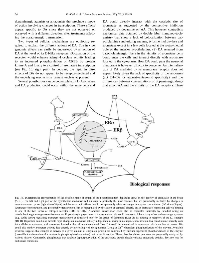
dopaminergic agonists or antagonists that preclude a mode DA could directly interact with the catalytic site ofof action involving changes in transcription. These effects aromatase as suggested by the competitive inhibitionappear specific to DA since they are not observed or produced by dopamine on AA. This however contradictsobserved with a different direction after treatments affect- anatomical data obtained by double label immunocytoch-ing the noradrenergic transmission. emistry that show a lack of colocalization between cat-
Two types of cellular mechanisms are obviously re- echolamine synthesizing enzyme, tyrosine hydroxylase andquired to explain the different actions of DA. The in vivo aromatase except in a few cells located at the rostro-medialgenomic effects can easily be understood by an action of pole of the anterior hypothalamus. (2) DA released fromDA at the level of its D1-like receptors. Occupation of the catecholaminergic fibers in the vicinity of aromatase cellsreceptor would enhance adenylyl cyclase activity leading could enter the cells and interact directly with aromataseto an increased phosphorylation of CREB by protein located in the cytoplasm. How DA could pass the neuronalkinase A and finally to a control of aromatase transcription membrane is however difficult to conceive. An internaliza-(see Fig. 10; right part). In contrast, the rapid in vitro tion of DA mediated by its membrane receptor does noteffects of DA do not appear to be receptor-mediated and appear likely given the lack of specificity of the responsesthe underlying mechanisms remain unclear at present. (not D1–D2 or agonist–antagonist specificity) and the
Several possibilities can be contemplated: (1) Aromatase differences between concentrations of dopaminergic drugsand DA production could occur within the same cells and that affect AA and the affinity of the DA receptors. There
Fig. 10. Diagrammatic representation of the possible mode of action of the neurotransmitter, dopamine (DA) on the activity of aromatase in the brain(ARO). The left and right part of the hypothetical aromatase cell illustrate respectively the slow controls that are presumably mediated by changes inaromatase transcription (right side of figure) and the more rapid effects that do not apparently relate to changes in enzyme concentration (left side of figure).Aromatase concentration, and presumably transcription, can be upregulated by the action of estradiol directly on an aromatase expressing cell via bindingto one of the two forms of estrogen receptor (ERa or ERb). Aromatase transcription could also be controlled indirectly by estradiol acting oncatecholaminergic estrogen-sensitive neurons. Dopaminergic projections on the aromatase cells could then control the activity of second messenger systems(e.g. cyclic AMP) regulating aromatase transcription as illustrated here for the action of dopamine (DA) via its binding to receptors of the D1 subtype(D1-R). Dopamine could also mediate rapid changes in aromatase activity independent of changes in enzyme concentration. DA could interact directly withintracellular aromatase or with aromatase located at the cell membrane level. How DA could be internalized in aromatase cells is unclear at present. DA
21could also modify aromatase activity less directly by interfering with the glutamate (Glut.) or Ca -dependent phosphorylations of the enzyme. Availableevidence suggests that changes in activity of a given amount of enzymatic protein are controlled by calcium-dependent phosphorylations of the enzyme(reversible transformation of aromatase in phosphorylated aromatase) that render it inactive. These phosphorylation processes are presumably catalyzed byvarious kinases. Conversely, phosphatases that catalyze dephosphorylation of the enzymatic protein should enhance enzymatic activity. See also text foradditional comments.

P. Absil et al. / Brain Research Reviews 37 (2001) 38 –58 55
[2] P. Absil, A. Foidart, H.C.J. Hemmings, H.W.M. Steinbusch, G.F.is also at present no evidence for the presence of DABall, J. Balthazart, Distribution of DARPP-32 immunoreactivereceptors on aromatase cells. (3) There is limited evidencestructures in the quail brain: anatomical relationship with dopamine
for the existence of aromatase in the membrane of certain and aromatase, J. Chem. Neuroanat. 21 (2000) 23–39.cell types [5] and DA could then affect the enzyme [3] E.K. Adkins, N.T. Adler, Hormonal control of behavior in the
Japanese quail, J. Comp. Physiol. Psychol. 81 (1972) 27–36.expressed at the cell surface. However membrane aromat-¨[4] K.A. Albert, E. Helmer-Matyjek, A.A. Nairn, T.H. Muller, J.W.ase has not been observed in the brain and examination of
Haycock, L.A. Greene, M. Goldstein, P. Greengard, Calcium/phos-ARO-ir cells by confocal microscopy does not revealpholipid-dependent protein kinase (protein kinase C) phosphorylates
specific immunoreactivity on the cell surface (J. Balthazart, and activates tyrosine hydroxylase, Proc. Natl. Acad. Sci. USA 81unpublished observations). (4) Finally, DA could trigger, (1984) 7713–7717.
[5] B.A. Amarneh, E.R. Simpson, Detection of aromatase cytochromeby some unidentified mechanism, a change in the intracel-P450, 17alpha-hydroxylase cytochrome P450 and NADPH:P450lular level of calcium in the aromatase-containing cells thatreductase on the surface of cells in which they are expressed, Mol.would change the phosphorylation state of the enzyme andCell. Endocrinol. 119 (1996) 69–74.
then its activity. The links between DA and aromatase [6] M.M. Ames, P. Lerner, W. Lovenberg, Tyrosine hydroxylase:phosphorylation are however unknown and it is also activation by protein phosphorylation and end product inhibition, J.
Biol. Chem. 253 (1978) 27–31.unclear how such a mechanism could take place both in¨[7] N. Aste, J. Balthazart, P. Absil, R. Grossmann, E. Mulhbauer, C.brain homogenates and in brain explants.
Viglietti-Panzica, G.C. Panzica, Anatomical and neurochemicalIt is clear though that none of these mechanisms can, indefinition of the nucleus of the stria terminalis in Japanese quail
isolation, explain all characteristics of the effects of DA on (Coturnix japonica), J. Comp. Neurol. 396 (1998) 141–157.aromatase that have been observed. Additional research [8] N. Aste, G.C. Panzica, P. Aimar, C. Viglietti-Panzica, N. Harada, A.
Foidart, J. Balthazart, Morphometric studies demonstrate thatwill be necessary to identify the cellular mechanisms thataromatase-immunoreactive cells are the main target of androgensmediate the inhibition of AA by DA. Although manyand estrogens in the quail medial preoptic nucleus, Exp. Brain Res.questions remain open, it is our contention that these101 (1994) 241–252.
controls observed during in vivo and in vitro experiments [9] N. Aste, G.C. Panzica, C. Viglietti-Panzica, N. Harada, J. Balthazart,do not represent experimental artifacts but reflect mecha- Distribution and effects of testosterone on aromatase mRNA in the
quail forebrain: A non-radioactive in situ hybridization study, J.nisms that are physiologically relevant. This conclusion isChem. Neuroanat. 14 (1998) 103–115.based on the specificity of dopaminergic effects as com-
[10] N. Aste, C. Viglietti-Panzica, J. Balthazart, G.C. Panzica, Testo-pared to the effects of compounds that modify the norad-sterone modulation of peptidergic pathways in the septo-preoptic
renergic activity and on the converging evidence provided region of male Japanese quail, Poultry Avian Biol. Rev. 8 (1997)by both pharmacological and anatomical data. 77–93.
[11] T. Bailhache, J. Balthazart, The catecholaminergic system of theIndependent of the underlying mechanisms(s) involved,quail brain: Immunocytochemical studies of dopamine beta-hy-the studies described above support the notion that rapiddroxylase and tyrosine hydroxylase, J. Comp. Neurol. 329 (1993)changes in the availability of estrogens can take place in230–256.
the brain and be regulated by neurotransmitter (catechol- [12] T. Bailhache, A. Foidart, C. Surlemont, N. Harada, J. Balthazart,aminergic) activity. These data suggest that the local Catecholaminergic innervation of aromatase and estrogen receptor-bioavailability of estrogens in the brain can change more immunoreactive cells in the quail brain, Soc. Neurosci. Abstr. 17
(1991) 269.rapidly than previously assumed and these rapidly chang-[13] M. Baillien, J. Balthazart, A direct dopaminergic control of aromat-ing levels of locally-produced estrogens could control the
ase activity in the quail preoptic area, J. Steroid Biochem. Mol. Biol.rapid non-genomic effects that presumably take place at 63 (1997) 99–113.the membrane level. The functional significance of these [14] M. Baillien, A. Foidart, J. Balthazart, Regional distribution andeffects should now be investigated. control of tyrosine hydroxylase activity in the quail brain, Brain Res.
Bull. 48 (1999) 31–37.[15] J. Balthazart, Steroid control and sexual differentiation of brain
aromatase, J. Steroid Biochem. Mol. Biol. 61 (1997) 323–339.Acknowledgements [16] J. Balthazart, Steroid metabolism and the activation of social
behavior, in: J. Balthazart (Ed.), Advances in Comparative andSupported by grants from the NIMH (MH50388) to G.F. Environmental Physiology, Vol. 3, Springer-Verlag, Berlin, 1989, pp.
105–159.Ball and J. Balthazart, by a grant from the MURST[17] J. Balthazart, P. Absil, Identification of catecholaminergic inputs to(COFIN 2001) to G.C. Panzica, and by grants from the
and outputs from aromatase-containing brain areas of the JapaneseBelgian FRFC (Nbr. 2.4555.01), the French Community of quail by tract tracing combined with tyrosine hydroxylase immuno-
`Belgium (ARC 99/04-241), and the University of Liege cytochemistry, J. Comp. Neurol. 382 (1997) 401–428.´(Fonds Speciaux pour la Recherche) to J. Balthazart. [18] J. Balthazart, P. Absil, C. Viglietti-Panzica, G.C. Panzica, Vas-
otocinergic innervation of areas containing aromatase-immuno-reactive cells in the quail forebrain, J. Neurobiol. 33 (1997) 45–60.
[19] J. Balthazart, M. Baillien, G.F. Ball, Interactions between aromataseReferences (estrogen synthase) and dopamine in the control of male sexual
behavior in quail, Comp. Biochem. Physiol. [B] (2001) in press.[1] P. Absil, J. Balthazart, Testosterone effects on neurotensin-immuno- [20] J. Balthazart, M. Baillien, G.F. Ball, Phosphorylation processes
reactive cells in the quail preoptic area, Neuroreport 5 (1994) 1129– mediate rapid changes of brain aromatase activity, J. Steroid1132. Biochem. Mol. Biol. (2001) in press.

56 P. Absil et al. / Brain Research Reviews 37 (2001) 38 –58
[21] J. Balthazart, M. Baillien, G.F. Ball, Rapid and reversible inhibition [43] F.L. Bellino, L. Holben, Placental estrogen synthetase (aromatase):of brain aromatase activity, J. Neuroendocrinol. 13 (2001) 61–71. Evidence for phosphatase-dependent inactivation, Biochem. Bio-
[22] J. Balthazart, G.F. Ball, Effects of the noradrenergic neurotoxin phys. Res. Commun. 162 (1989) 498–504.DSP-4 on luteinizing hormone levels, catecholamine concentrations, [44] W.P.M. Benten, M. Lieberherr, O. Stamm, C. Wrehlke, Z.Y. Guo, F.alpha2-adrenergic receptor binding, and aromatase activity in the Wunderlich, Testosterone signaling through internalizable surfacebrain of the Japanese quail, Brain Res. 492 (1989) 163–175. receptors in androgen receptor-free macrophages, Mol. Biol. Cell 10
[23] J. Balthazart, G.F. Ball, Fast regulation of steroid biosynthesis: a (1999) 3113–3123.further piece in the neurosteroid puzzle, Trends Neurosci. 23 (2000) [45] J.D. Blaustein, M.J. Tetel, J.M. Meredith, Neurobiological regula-57–58. tion of hormonal response by progestin and estrogen receptors, in:
[24] J. Balthazart, G.F. Ball, Is dopamine interacting with aromatase to P.E. Micevych, R.P.J. Hammer (Eds.), Neurobiological Effects ofcontrol sexual behavior in male quail?, Poultry Sci. Rev. 4 (1992) Sex Steroid Hormones, Cambridge University Press, Cambridge,217–233. 1995, pp. 324–349.
[25] J. Balthazart, G.F. Ball, The Japanese quail as a model system for [46] G.V. Callard, Aromatization is cyclic AMP-dependent in culturedthe investigation of steroid-catecholamine interactions mediating brain cells, Brain Res. 204 (1981) 461–464.appetitive and consummatory aspects of male sexual behavior, Ann. [47] J.A. Canick, S.A. Tobet, M.J. Baum, D.E. Vaccaro, K.J. Ryan, S.E.Rev. Sex Res. 9 (1998) 96–176. Leeman, T.O. Fox, Studies on the role of catecholamines in the
[26] J. Balthazart, G.F. Ball, New insights into the regulation and regulation of the developmental pattern of hypothalamic aromatase,function of brain estrogen synthase (aromatase), Trends Neurosci. 21 Steroids 50 (1987) 510–521.(1998) 243–249. [48] D.P. Cardinali, M.N. Ritta, P.V. Gejman, Norepinephrine stimulates
[27] J. Balthazart, G.F. Ball, Sexual differentiation of brain and behavior testosterone aromatization and inhibits 5a-reduction via b-adreno-in birds, Trends Endocrinol. Metab. 6 (1995) 21–29. receptors in the rat pineal gland, Mol. Cell. Endocrinol. 28 (1982)
[28] J. Balthazart, A. Foidart, Brain aromatase and the control of male 199–209.sexual behavior, J. Steroid Biochem. Mol. Biol. 44 (1993) 521–540. [49] C.J. Corbin, S. Graham-Lorence, M. McPhaul, J.I. Mason, C.R.
[29] J. Balthazart, A. Foidart, P. Absil, N. Harada, Effects of testosterone Mendelson, E.R. Simpson, Isolation of a full-length cDNA insertand its metabolites on aromatase-immunoreactive cells in the quail encoding human aromatase system cytochrome P-450 and itsbrain: relationship with the activation of male reproductive behavior, expression in nonsteroidogenic cells, Proc. Natl. Acad. Sci. USA 85J. Steroid Biochem. Mol. Biol. 56 (1996) 185–200. (1988) 8948–8952.
[30] J. Balthazart, A. Foidart, M. Baillien, N. Harada, G.F. Ball, [50] E. Cross, C.E. Roselli, 17beta-estradiol rapidly facilitates chemoin-Anatomical relationships between aromatase and tyrosine hydroxy- vestigation and mounting in castrated male rats, Am. J. Physiol.lase in the quail brain: double-label immunocytochemical studies, J. Regul. Integr. Comp. Physiol. 276 (1999) R1346–R1350.Comp. Neurol. 391 (1998) 214–226. [51] S.C. Daubner, C. Lauriano, J.W. Haycock, P.F. Fitzpatrick, Site-
[31] J. Balthazart, A. Foidart, N. Harada, Immunocytochemical localiza- directed mutagenesis of serine 40 of rat tyrosine hydroxylase, J.tion of aromatase in the brain, Brain Res. 514 (1990) 327–333. Biol. Chem. 267 (1992) 12639–12646.
[32] J. Balthazart, A. Foidart, J.C. Hendrick, The induction by testo- [52] G.J. de Vries, Sex differences in neurotransmitter systems, J.sterone of aromatase activity in the preoptic area and activation of Neuroendocrinol. 2 (1990) 1–13.copulatory behavior, Physiol. Behav. 47 (1990) 83–94. [53] G.J. de Vries, Z. Wang, N.A. Bullock, S. Numan, Sex differences in
[33] J. Balthazart, A. Foidart, P. Sante, J.C. Hendrick, Effects of alpha- the effects of testosterone and its metabolites on vasopressinmethyl-para-tyrosine on monoamine levels in the Japanese quail: messenger RNA levels in the bed nucleus of the stria terminalis ofSex differences and testosterone effects, Brain Res. Bull. 28 (1992) rats, J. Neurosci. 14 (1994) 1789–1794.275–288. [54] A. Foidart, B. Lakaye, T. Grisar, G.F. Ball, J. Balthazart, Estrogen
[34] J. Balthazart, A. Foidart, C. Surlemont, N. Harada, Neuroanatomical receptor-beta in quail: Cloning, tissue expression and neuroanatomi-specificity in the co-localization of aromatase and estrogen re- cal distribution, J. Neurobiol. 40 (1999) 327–342.ceptors, J. Neurobiol. 22 (1991) 143–157. ¨[55] R. Furbass, C. Kalbe, J. Vanselow, Tissue-specific expression of the
[35] J. Balthazart, J.M. Libioulle, P. Sante, Stimulatory effects of the bovine aromatase-encoding gene uses multiple transcriptional startnoradrenergic neurotoxin DSP4 on sexual behavior in male quail, sites and alternative first exons, Endocrinology 138 (1997) 2813–Behav. Proc. 17 (1988) 27–44. 2819.
[36] J. Balthazart, R. Stoop, A. Foidart, N. Harada, Synergistic control by [56] L.D. Grant, W.E. Stumpf, Hormone uptake sites in CNS biogenicandrogens and estrogens of aromatase in the quail brain, Neurore- amines systems, in: W.E. Stumpf, L.D. Grant (Eds.), Anatomicalport 5 (1994) 1729–1732. Neuroendocrinology, Karger, Basel, 1975, pp. 445–463.
[37] J. Balthazart, C. Surlemont, Androgen and estrogen action in the [57] Q. Gu, K.S. Korach, R.L. Moss, Rapid action of 17beta-estradiol onpreoptic area and activation of copulatory behavior in quail, Physiol. kainate-induced currents in hippocampal neurons lacking intracellu-Behav. 48 (1990) 599–609. lar estrogen receptors, Endocrinology 140 (1999) 660–666.
[38] J. Balthazart, C. Surlemont, Copulatory behavior is controlled by the [58] N. Harada, Novel properties of human placental aromatase assexually dimorphic nucleus of the quail POA, Brain Res. Bull. 25 cytochrome P-450: purification and characterization of a unique(1990) 7–14. form of aromatase, J. Biochem. 103 (1988) 106–113.
[39] J. Balthazart, C. Surlemont, N. Harada, Aromatase as a cellular [59] N. Harada, S. Abe-Dohmae, R. Loeffen, A. Foidart, J. Balthazart,marker of testosterone action in the preoptic area, Physiol. Behav. Synergism between androgens and estrogens in the induction of51 (1992) 395–409. aromatase and its messenger RNA in the brain, Brain Res. 622
[40] J. Balthazart, O. Tlemcani, N. Harada, Localization of testosterone- (1993) 243–256.sensitive and sexually dimorphic aromatase-immunoreactive cells in [60] N. Harada, K. Yamada, A. Foidart, J. Balthazart, Regulation ofthe quail preoptic area, J. Chem. Neuroanat. 11 (1996) 147–171. aromatase cytochrome P-450 (estrogen synthetase) transcripts in the
[41] C. Barberis, S. Audigier, T. Durroux, J. Elands, A. Schmidt, S. Jard, quail brain by testosterone, Mol. Brain Res. 15 (1992) 19–26.Pharmacology of oxytocin and vasopressin receptors in the central [61] A.E. Herbison, Multimodal influence of estrogen upon gonado-and peripheral nervous system, Ann. N.Y. Acad. Sci. 652 (1992) tropin-releasing hormone neurons, Endocr. Rev. 19 (3) (1998)39–45. 302–330.
[42] C.A. Barraclough, P.M. Wise, The role of catecholamines in the [62] A.E. Herbison, J.E. Robinson, D.C. Skinner, Distribution of estrogenregulation of pituitary luteinizing hormone and follicle stimulating- receptor-immunoreactive cells in the preoptic area of the ewe:hormone secretion, Endocr. Rev. 3 (1982) 91–119.

P. Absil et al. / Brain Research Reviews 37 (2001) 38 –58 57
Co-localization with glutamic acid decarboxylase but not luteinizing estrogen from androgen, J. Biol. Chem. 263 (1988) 16358–16363.[83] G.D. Means, M.S. Mahendroo, C.J. Corbin, J.M. Mathis, F.E.hormone-releasing hormone, Neuroendocrinology 57 (1993) 751–
Powell, C.R. Mendelson, E.R. Simpson, Structural analysis of the759.gene encoding human aromatase cytochrome P-450, the enzyme[63] A.E. Herbison, D.T. Theodosis, Localization of oestrogen receptorsresponsible for estrogen biosynthesis, J. Biol. Chem. 264 (1989)in preoptic neurons containing neurotensin but not tyrosine hydroxy-19385–19391.lase, cholecystokinin or luteinizing hormone-releasing hormone in
[84] C.R. Mendelson, M.E. Smith, W.H. Cleland, E.R. Simpson, Regula-the male and female rat, Neuroscience 50 (1992) 283–298.tion of aromatase activity of cultured adipose stromal cells by[64] A.S. Heritage, W.E. Stumpf, M. Sar, L.D. Grant, Brain stemcatecholamines and adrenocorticotropin, Mol. Cell. Endocrinol. 37catecholamine neurons are target sites for sex steroid hormones,(1984) 61–72.Science 204 (1980) 1377–1379.
[85] P.G. Mermelstein, J.B. Becker, D.J. Surmeier, Estradiol reduces[65] G.J. Hickey, J.S. Krasnow, W.G. Beattie, J.S. Richards, Aromatasecalcium currents in rat neostriatal neurons via a membrane receptor,cytochrome P450 in rat ovarian granulosa cells before and afterJ. Neurosci. 16 (1996) 595–604.luteinization: Adenosine 39,59-monophosphate-dependent and inde-
[86] R.L. Moss, Q. Gu, M. Wong, Estrogen: nontranscriptional signalingpendent regulation. Cloning and sequencing of rat aromatase cDNApathway, Rec. Prog. Horm. Res. 52 (1997) 33–69.and 59 genomic DNA, Mol. Endocrinol. 4 (1990) 3–12.
[87] T. Nagatsu, M. Levitt, S. Udenfriend, A rapid and simple radioassay[66] R.A. Hinde, E. Steel, The influence of daylength and male vocaliza-for tyrosine hydroxylase activity, Anal. Biochem. 9 (1964) 122–tions on the estrogen-dependent behavior of female canaries and126.
budgerigars, with discussion of data from other species, Adv. Study[88] B. Nock, H.H. Feder, Neurotransmitter modulation of steroid action
Anim. Behav. 8 (1978) 39–73.in target cells that mediate reproduction and reproductive behavior,
[67] Z. Hochberg, T. Bick, R. Pelman, J.M. Brandes, D. Barzilai, TheNeurosci. Biobehav. Rev. 5 (1981) 437–447.
dual effect of calcium on aromatization by cultured human trophob- [89] O.M. Onagbesan, M.J. Podie, Calcium-dependent stimulation oflast, J. Steroid Biochem. 24 (1986) 1217–1219. estrogen secretion by FSH from theca cells of the domestic hen
[68] J.B. Hutchison, T.H. Steimer, Formation of behaviorally effective (Gallus domesticus), Gen. Comp. Endocrinol. 75 (1989) 177–186.17beta-estradiol in the dove brain: steroid control of preoptic [90] Y. Osawa, T. Higashiyama, Y. Toma, C. Yarborough, Diversearomatase, Endocrinology 118 (1986) 2180–2187. function of aromatase and the N-terminal sequence deleted form, J.
¨[69] M. Joels, Steroid hormones and excitability in the mammalian brain, Steroid Biochem. Mol. Biol. 61 (1997) 117–126.Front. Neuroendocrinol. 18 (1997) 2–48. [91] Y. Osawa, T. Higashiyama, C. Yarborough, Diverse functions of
[70] H.W. Korf, G.C. Panzica, C. Viglietti-Panzica, A. Oksche, Pattern of aromatase cytochrome P-450: catecholestrogen synthesis, cocainepeptidergic neurons in the avian brain: clusters–local circuitries– N-demethylation, and other selective drug metabolisms, in: M.C.projections, Bas. Appl. Histochem. 32 (1988) 55–75. Lechner (Ed.), Cytochrome P450. 8th International Conference,
[71] W.J. Kuenzel, M. Masson, A Stereotaxic Atlas of the Brain of the John Libbey Eurotext, Paris, 1994, pp. 893–896.Chick (Gallus domesticus), The Johns Hopkins University Press, [92] Y. Osawa, T. Higashiyama, C. Yarborough, Formation of 6a-hy-Baltimore, 1988, 1–166, pp. 1–166. droxyestradiol (6a-OHE2) from E2 by aromatase: evidence for the
˚[72] G.G.J.M. Kuiper, E. Enmark, M. Pelto-Huikko, S. Nilsson, J.A. reverse side interaction of E2 at the active site of aromatase, Endocr.Gustafsson, Cloning of a novel estrogen receptor expressed in rat Soc. 77 Abst. (1995) 88.prostate and ovary, Proc. Natl. Acad. Sci. USA 93 (1996) 5925– [93] M.A. Ottinger, J. Balthazart, Brain monoamines in Japanese quail:5930. effects of castration and steroid replacement therapy, Behav. Proc.
˚[73] G.G.J.M. Kuiper, P.J. Shughrue, I. Merchenthaler, J.-A. Gustafsson, 14 (1987) 197–216.The estrogen receptor b subtype: a novel mediator of estrogen action [94] M.A. Ottinger, M. Schumacher, R.N. Clarke, C.S. Duchala, J.in neuroendocrine systems, Front. Neuroendocrinol. 19 (1998) 253– Balthazart, Comparison of monoamine concentrations in the brains286. of adult male and female Japanese quail, Poultry Sci. 65 (1986)
[74] B. Lakaye, A. Foidart, T. Grisar, J. Balthazart, Partial cloning and 1413–1420.distribution of estrogen receptor beta in the avian brain, Neuroreport [95] H.W.T.M. Overes, R. De Leeuw, H.J. Kloosterboer, Regulation of9 (1998) 2743–2748. aromatase activity in FSH-primed rat granulosa cells in vitro by
[75] E.D. Lephart, A review of brain aromatase cytochrome P450, Brain follicle-stimulating hormone and various amounts of humanRes. Rev. 22 (1996) 1–26. chorionic gonadotrophin, Hum. Reprod. 7 (1992) 191–196.
[76] R.A. Levine, H.B. Pollard, D.M. Kuhn, A rapid and simplified assay [96] G.C. Panzica, Sex differences in the avian brain, Sitta 2 (1988)method for tyrosine hydroxylase, Anal. Biochem. 143 (1984) 205– 77–92.208. [97] G.C. Panzica, E. Garcia-Ojeda, C.Viglietti-Panzica, N.E. Thompson,
[77] D.L. Maney, D.J. Bernard, F.W. Turek, G.F. Ball, Localization of M.A. Ottinger, Testosterone effects on vasotocinergic innervation ofandrogen receptor and estrogen receptor alpha mRNA in catechol- sexually dimorphic medial preoptic nucleus and lateral septumaminergic cell groups of the canary brain, Soc. Neurosci. Abstr. 25 during aging in male quail, Brain Res. 712 (1996) 190–198.(1999) 860–866. [98] G.C. Panzica, C. Viglietti-Panzica, J. Balthazart, The sexually
[78] B.S. McEwen, Neural gonadal steroid actions, Science 211 (1981) dimorphic medial preoptic nucleus of quail: a key brain area1303–1311. mediating steroid action on male sexual behavior, Front. Neuroen-
[79] B.S. McEwen, Steroid hormone actions on the brain: When is the docrinol. 17 (1996) 51–125.genome involved?, Horm. Behav. 28 (1994) 396–405. [99] G.C. Panzica, C. Viglietti-Panzica, M. Calcagni, G.C. Anselmetti,
[80] B.S. McEwen, S.E. Alves, Estrogen actions in the central nervous M. Schumacher, J. Balthazart, Sexual differentiation and hormonalsystem, Endocr. Rev. 20 (1999) 279–307. control of the sexually dimorphic preoptic medial nucleus in quail,
[81] B.S. McEwen, L.C. Krey, Properties of estrogen-sensitive neurons: Brain Res. 416 (1987) 59–68.aromatization, progestin receptor induction and neuroendocrine [100] G.C. Panzica, C. Viglietti-Panzica, F. Sanchez, P. Sante, J. Balth-effects, in: F. Celotti, F. Naftolin, L. Martini (Eds.), Metabolism of azart, Effects of testosterone on a selected neuronal populationHormonal Steroids in the Neuroendocrine Structures, Raven Press, within the preoptic sexually dimorphic nucleus of the JapaneseNew York, 1984, pp. 117–128. quail, J. Comp. Neurol. 303 (1991) 443–456.
[82] M.J. McPhaul, J.F. Noble, E.R. Simpson, C.R. Mendelson, J.D. [101] C. Pasqualini, V. Olivier, B. Guibert, O. Frain, V. Leviel, AcuteWilson, The expression of a functional cDNA encoding the chicken stimulatory effect of estradiol on striatal dopamine synthesis, J.cytochrome P-450arom (aromatase) that catalyzes the formation of Neurochem. 65 (1995) 1651–1657.

58 P. Absil et al. / Brain Research Reviews 37 (2001) 38 –58
[102] B. Ramachandran, B.A. Schlinger, A.P. Arnold, A.T. Campagnoni, Hinshelwood, S. Graham-Lorence, B. Amarneh, Y. Ito, C.R. Fisher,Zebra finch aromatase gene expression is regulated in the brain M.D. Michael, C.R. Mendelson, S.E. Bulun, Aromatase cyto-through an alternate promoter, Gene 240 (1999) 209–216. chrome P450, the enzyme responsible for estrogen biosynthesis,
[103] V.D. Ramirez, J.B. Zheng, K.M. Siddique, Membrane receptors for Endocr. Rev. 15 (1994) 342–355.estrogen, progesterone, and testosterone in the rat brain: Fantasy or [120] E.R. Simpson, M.D. Michael, V.R. Agarwal, M.M. Hinshelwood,reality, Cell. Mol. Neurobiol. 16 (1996) 175–198. S.E. Bulun, Y. Zhao, Expression of the CYP19 (aromatase) gene:
[104] W.J. Raum, M. Marcano, R.S. Swerdloff, Nuclear accumulation of An unusual case of alternative promoter usage, FASEB J. 11estradiol derived from the aromatization of testosterone is inhibited (1997) 29–36.by hypothalamic beta-receptor stimulation in the neonatal female [121] T. Steimer, J.B. Hutchison, Micromethods for the in vitro study ofrat, Biol. Reprod. 30 (1984) 388–396. steroid metabolism in the brain using radiolabelled tracers, in: B.
[105] W.J. Raum, R.S. Swerdloff, The role of hypothalamic adrenergic Greenstein (Ed.), Neuroendocrine Research Methods, Vol. 2, Har-receptors in preventing testosterone-induced androgenization in the wood Academic Publishers, Chur, Switzerland, 1991, pp. 875–919.female rat brain, Endocrinology 109 (1981) 273–278. [122] Y. Toma, T. Higashiyama, C. Yarborough, Y. Osawa, Diverse
[106] C.E. Roselli, L.E. Horton, J.A. Resko, Time-course and steroid functions of aromatase: O-deethylation of 7-ethoxycoumarin,specificity of aromatase induction in rat hypothalamus-preoptic Endocrinology 137 (1996) 3791–3796.area, Biol. Reprod. 37 (1987) 628–633. [123] Y. Tsuruo, K. Ishimura, S. Hayashi, Y. Osawa, Immunohistochemi-
[107] C.E. Roselli, J.A. Resko, Androgens regulate brain aromatase cal localization of estrogen receptors within aromatase-immuno-activity in adult male rats through a receptor mechanism, Endo- reactive neurons in the fetal and neonatal rat brain, Anat. Embryol.crinology 114 (1984) 2183–2189. (Berl) 193 (1996) 115–121.
[108] C.E. Roselli, J.A. Resko, In vitro assay of aromatase activity in the [124] Y. Tsuruo, K. Ishimura, Y. Osawa, Presence of estrogen receptorscentral nervous system, in: B. Greenstein (Ed.), Neuroendocrine in aromatase-immunoreactive neurons in the mouse brain, Neuro-Research Methods, Vol. 2, Harwood Academic Publishers, Chur, sci. Lett. 195 (1995) 49–52.Switzerland, 1991, pp. 937–951. [125] G. Verhoeven, Effect of neurotransmitters and follicle-stimulating
[109] B.D. Sachs, Photoperiodic control of reproductive behavior and hormone on the aromatization of androgens and the production ofphysiology of the male Japanese quail (Coturnix coturnix 39,59-monophosphate by cultured testicular cells, J. Steroid Bio-japonica), Horm. Behav. 1 (1969) 7–24. chem. 12 (1980) 315–322.
[110] M. Sar, Estradiol is concentrated in tyrosine hydroxylase-con- [126] G. Verhoeven, J. Cailleau, Testicular peritubular cells secrete ataining neurons of the hypothalamus, Science 223 (1984) 938–940. protein under androgen control that inhibits induction of aromatase
[111] B.A. Schlinger, G.V. Callard, A comparison of aromatase, 5a-and activity in Sertoli cells, Endocrinology 123 (1988) 2100–2110.5b-reductase activities in the brain and pituitary of male and [127] G.P. Verhoeven, P. Dierckx, P. de Moor, Stimulation effect offemale quail (Coturnix c. Japonica, J. Exp. Zool. 242 (1987) neurotransmitters on the aromatization of testosterone by Sertoli171–180. cell-enriched cultures, Mol. Cell. Endocrinol. 13 (1979) 241–253.
[112] M. Schumacher, C. Alexandre, J. Balthazart, Interactions des [128] C. Viglietti-Panzica, J. Balthazart, L. Plumari, S. Fratesi, P. Absil,` ` ˆandrogenes et des oestrogenes dans le controle de la reproduction, G.C. Panzica, Estradiol mediates effects of testosterone on vas-
´C.R. Acad. Sci. Paris, Serie III 305 (1987) 569–574. otocin-immunoreactivity in the adult quail brain, Horm. Behav.[113] M. Schumacher, J. Balthazart, Neuroanatomical distribution of (2001) in press.
testosterone metabolizing enzymes in the Japanese quail, Brain [129] C. Viglietti-Panzica, G.C. Anselmetti, J. Balthazart, N. Aste, G.C.Res. 422 (1987) 137–148. Panzica, Vasotocinergic innervation of the septal region in the
[114] P. Shen, C.W. Campagnoni, K. Kampf, B.A. Schlinger, A.P. Japanese quail: Sexual differences and the influence of testo-Arnold, A.T. Campagnoni, Isolation and characterization of a zebra sterone, Cell Tissue Res. 267 (1992) 261–265.finch aromatase cDNA: In situ hybridization reveals high aromat- [130] C. Viglietti-Panzica, N. Aste, J. Balthazart, G.C. Panzica, Vas-ase expression in brain, Mol. Brain Res. 24 (1994) 227–237. otocinergic innervation of sexually dimorphic medial preoptic
[115] B.D. Shivers, R.E. Harlan, J.I. Morrell, D.W. Pfaff, Absence of nucleus of the male Japanese quail: Influence of testosterone, Brainoestradiol concentration in cell nuclei of LHRH-immunoreactive Res. 657 (1994) 171–184.neurones, Nature 304 (1983) 345–347. [131] C. Viglietti-Panzica, G.C. Panzica, Peptidergic neurons in the avian
[116] R.B. Simerly, Hormonal control of the development and regulation brain, Ann. Sci. Nat., Zool., Paris 12 (1991) 137–155.of tyrosine hydroxylase expression within a sexually dimorphic [132] C. Viglietti-Panzica, G.C. Panzica, M.G. Fiori, M. Calcagni, G.C.population of dopaminergic cells in the hypothalamus, Mol. Brain Anselmetti, J. Balthazart, A sexually dimorphic nucleus in the quailRes. 6 (1989) 297–310. preoptic area, Neurosci. Lett. 64 (1986) 129–134.
[117] R.B. Simerly, L.W. Swanson, The distribution of neurotransmitter- [133] N. Weiner, A critical assessment of methods for the determinationspecific cells and fibers in the anteroventral periventricular nucleus: of monoamine synthesis turnover rates in vivo, Adv. Biochem.implications for the control of gonadotropin secretion in the rat, Pharmacol. 12 (1974) 143–159.Brain Res. 400 (1987) 11–34. [134] R.I. Weiner, P.R. Findell, C. Kordon, Role of classic and peptide
[118] R.B. Simerly, L.W. Swanson, R.A. Gorski, The distribution of neuromediators in the neuroendocrine regulation of LH andmonoaminergic cells and fibers in a periventricular preoptic prolactin, in: E. Knobil, J. Neill et al. (Eds.), The Physiology ofnucleus involved in the control of gonadotropin release: immuno- Reproduction, Raven Press, New York, 1988, pp. 1235–1281.histochemical evidence for a dopaminergic sexual dimorphism, [135] J.-P. Weniger, A. Zeis, Stimulation of aromatase activity in the fetalBrain Res. 330 (1985) 55–64. rat gonads by cAMP and FSH, Acta Endocrinol. (Copenhagen) 119
[119] E.R. Simpson, M.S. Mahendroo, G.D. Means, M.W. Kilgore, M.M. (1988) 381–385.
Related Documents