The complete genome sequence of Francisella tularensis, the causative agent of tularemia Pa ¨r Larsson 1 , Petra C F Oyston 2 , Patrick Chain 3 , May C Chu 4 , Melanie Duffield 2 , Hans-Henrik Fuxelius 5 , Emilio Garcia 3 , Greger Ha ¨lltorp 5 , Daniel Johansson 1 , Karen E Isherwood 2 , Peter D Karp 6 , Eva Larsson 1 , Ying Liu 7 , Stephen Michell 2 , Joann Prior 2 , Richard Prior 2 , Stephanie Malfatti 3 , Anders Sjo ¨stedt 8 , Kerstin Svensson 1 , Nick Thompson 9 , Lisa Vergez 3 , Jonathan K Wagg 6 , Brendan W Wren 10 , Luther E Lindler 7 , Siv G E Andersson 5 , Mats Forsman 1 & Richard W Titball 2,10 Francisella tularensis is one of the most infectious human pathogens known. In the past, both the former Soviet Union and the US had programs to develop weapons containing the bacterium. We report the complete genome sequence of a highly virulent isolate of F. tularensis (1,892,819 bp). The sequence uncovers previously uncharacterized genes encoding type IV pili, a surface polysaccharide and iron-acquisition systems. Several virulence-associated genes were located in a putative pathogenicity island, which was duplicated in the genome. More than 10% of the putative coding sequences contained insertion-deletion or substitution mutations and seemed to be deteriorating. The genome is rich in IS elements, including IS630 Tc-1 mariner family transposons, which are not expected in a prokaryote. We used a computational method for predicting metabolic pathways and found an unexpectedly high proportion of disrupted pathways, explaining the fastidious nutritional requirements of the bacterium. The loss of biosynthetic pathways indicates that F. tularensis is an obligate host-dependent bacterium in its natural life cycle. Our results have implications for our understanding of how highly virulent human pathogens evolve and will expedite strategies to combat them. Francisella tularensis is one of the most infectious pathogens known and is the etiological agent of tularemia, a disease of humans and animals 1 . The vector-borne form of the disease (glandular or ulceroglandular tularemia) is usually contracted from the bite of an arthropod vector that previously fed on an infected animal 1 . Respiratory tularemia is less frequent and is usually contracted during farming activities that generate dust from sites where infected animals have resided. The mortality rate of respiratory tularemia may be as high as 5–30% without antibiotic therapy; even if not fatal, the disease may be severely incapacitating for a period of weeks or even months 1 . One outbreak of respiratory tularemia occurred in Martha’s Vineyard in the US, probably triggered by the mechanical disruption of a rabbit carcass during lawn mowing. The aerosols that were generated infected two individuals, underscoring the highly infectious nature of the organism by the airborne route. The infectious dose of F. tularensis in humans by the airborne route is as low as 10 cells 1 . Although the bacterium is nutritionally fastidious, it was developed as a weapon by Japanese Germ Warfare units during the 1930s and 1940s and later by the former Soviet Union and the US 2 . There is concern that bioweapons containing this bacterium still exist elsewhere in the world. The high level of interest in F. tularensis and concerns over possible misuse contrast with the paucity of knowledge on virulence mechan- isms. The bacterium infects macrophages 1 , and a few virulence determinants have been proposed, including an ill-defined capsule 3 and a 23-kDa protein that seems to have a role in downregulating proinflammatory cytokines 4 . Some other genes required for growth in macrophages have been identified 5–7 , but their roles are uncertain. There is currently no licensed vaccine for the prevention of tularemia. We report here the complete genome sequence and a phylogenetic analysis of a fully virulent human isolate of F. tularensis subspecies tularensis (strain SCHU S4). In the long term, this study will support work to devise improved countermeasures against tularemia. RESULTS General features The genome of the F. tularensis strain SCHU S4 consists of a 1,892,819-bp circular chromosome, with an overall G+C content of 32.9% and 1,804 predicted coding sequences (CDSs; including Published online 9 January 2005; doi:10.1038/ng1499 1 Swedish Defence Research Agency, SE-901 82 Umea ˚, Sweden. 2 Defence Science and Technology Laboratory, Salisbury SP4 0JQ, UK. 3 Biology and Biotechnology Research Program, Lawrence Livermore National Laboratory, California 94550, USA. 4 Division of Vector-Borne Infectious Diseases, Centers for Disease Control and Prevention, Fort Collins, Colorado, USA. 5 Department of Molecular Evolution, University of Uppsala, S-752 36 Uppsala, Sweden. 6 Bioinformatics Research Group, SRI International, Menlo Park, California 94025, USA. 7 Walter Reed Army Institute of Research, Silver Spring, Maryland 20910, USA. 8 Department of Clinical Microbiology, Umea ˚ University, SE-901 85 Umea ˚, Sweden. 9 Welcome Trust Sanger Institute, Cambridge CB10 1SA, UK. 10 Department of Infectious and Tropical Diseases, London School of Hygiene and Tropical Medicine, London WC1E 7HT, UK. Correspondence should be addressed to R.W.T. ([email protected]). NATURE GENETICS VOLUME 37 [ NUMBER 2 [ FEBRUARY 2005 153 ARTICLES © 2005 Nature Publishing Group http://www.nature.com/naturegenetics

Welcome message from author
This document is posted to help you gain knowledge. Please leave a comment to let me know what you think about it! Share it to your friends and learn new things together.
Transcript
The complete genome sequence of Francisella tularensis,the causative agent of tularemiaPar Larsson1, Petra C F Oyston2, Patrick Chain3, May C Chu4, Melanie Duffield2, Hans-Henrik Fuxelius5,Emilio Garcia3, Greger Halltorp5, Daniel Johansson1, Karen E Isherwood2, Peter D Karp6, Eva Larsson1,Ying Liu7, Stephen Michell2, Joann Prior2, Richard Prior2, Stephanie Malfatti3, Anders Sjostedt8,Kerstin Svensson1, Nick Thompson9, Lisa Vergez3, Jonathan K Wagg6, Brendan W Wren10, Luther E Lindler7,Siv G E Andersson5, Mats Forsman1 & Richard W Titball2,10
Francisella tularensis is one of the most infectious human pathogens known. In the past, both the former Soviet Union and theUS had programs to develop weapons containing the bacterium. We report the complete genome sequence of a highly virulentisolate of F. tularensis (1,892,819 bp). The sequence uncovers previously uncharacterized genes encoding type IV pili, a surfacepolysaccharide and iron-acquisition systems. Several virulence-associated genes were located in a putative pathogenicity island,which was duplicated in the genome. More than 10% of the putative coding sequences contained insertion-deletion or substitutionmutations and seemed to be deteriorating. The genome is rich in IS elements, including IS630 Tc-1 mariner family transposons,which are not expected in a prokaryote. We used a computational method for predicting metabolic pathways and found anunexpectedly high proportion of disrupted pathways, explaining the fastidious nutritional requirements of the bacterium. The loss ofbiosynthetic pathways indicates that F. tularensis is an obligate host-dependent bacterium in its natural life cycle. Our results haveimplications for our understanding of how highly virulent human pathogens evolve and will expedite strategies to combat them.
Francisella tularensis is one of the most infectious pathogens knownand is the etiological agent of tularemia, a disease of humansand animals1. The vector-borne form of the disease (glandularor ulceroglandular tularemia) is usually contracted from the biteof an arthropod vector that previously fed on an infected animal1.Respiratory tularemia is less frequent and is usually contractedduring farming activities that generate dust from sites where infectedanimals have resided. The mortality rate of respiratory tularemiamay be as high as 5–30% without antibiotic therapy; even if notfatal, the disease may be severely incapacitating for a period ofweeks or even months1. One outbreak of respiratory tularemiaoccurred in Martha’s Vineyard in the US, probably triggered bythe mechanical disruption of a rabbit carcass during lawn mowing.The aerosols that were generated infected two individuals,underscoring the highly infectious nature of the organism by theairborne route.
The infectious dose of F. tularensis in humans by the airborne routeis as low as 10 cells1. Although the bacterium is nutritionallyfastidious, it was developed as a weapon by Japanese Germ Warfareunits during the 1930s and 1940s and later by the former Soviet Union
and the US2. There is concern that bioweapons containing thisbacterium still exist elsewhere in the world.
The high level of interest in F. tularensis and concerns over possiblemisuse contrast with the paucity of knowledge on virulence mechan-isms. The bacterium infects macrophages1, and a few virulencedeterminants have been proposed, including an ill-defined capsule3
and a 23-kDa protein that seems to have a role in downregulatingproinflammatory cytokines4. Some other genes required for growth inmacrophages have been identified5–7, but their roles are uncertain.There is currently no licensed vaccine for the prevention of tularemia.
We report here the complete genome sequence and a phylogeneticanalysis of a fully virulent human isolate of F. tularensis subspeciestularensis (strain SCHU S4). In the long term, this study will supportwork to devise improved countermeasures against tularemia.
RESULTSGeneral featuresThe genome of the F. tularensis strain SCHU S4 consists of a1,892,819-bp circular chromosome, with an overall G+C content of32.9% and 1,804 predicted coding sequences (CDSs; including
Published online 9 January 2005; doi:10.1038/ng1499
1Swedish Defence Research Agency, SE-901 82 Umea, Sweden. 2Defence Science and Technology Laboratory, Salisbury SP4 0JQ, UK. 3Biology and BiotechnologyResearch Program, Lawrence Livermore National Laboratory, California 94550, USA. 4Division of Vector-Borne Infectious Diseases, Centers for Disease Control andPrevention, Fort Collins, Colorado, USA. 5Department of Molecular Evolution, University of Uppsala, S-752 36 Uppsala, Sweden. 6Bioinformatics Research Group, SRIInternational, Menlo Park, California 94025, USA. 7Walter Reed Army Institute of Research, Silver Spring, Maryland 20910, USA. 8Department of ClinicalMicrobiology, Umea University, SE-901 85 Umea, Sweden. 9Welcome Trust Sanger Institute, Cambridge CB10 1SA, UK. 10Department of Infectious and TropicalDiseases, London School of Hygiene and Tropical Medicine, London WC1E 7HT, UK. Correspondence should be addressed to R.W.T. ([email protected]).
NATURE GENETICS VOLUME 37 [ NUMBER 2 [ FEBRUARY 2005 15 3
A R T I C L E S©
2005
Nat
ure
Pub
lishi
ng G
roup
ht
tp://
ww
w.n
atur
e.co
m/n
atur
egen
etic
s
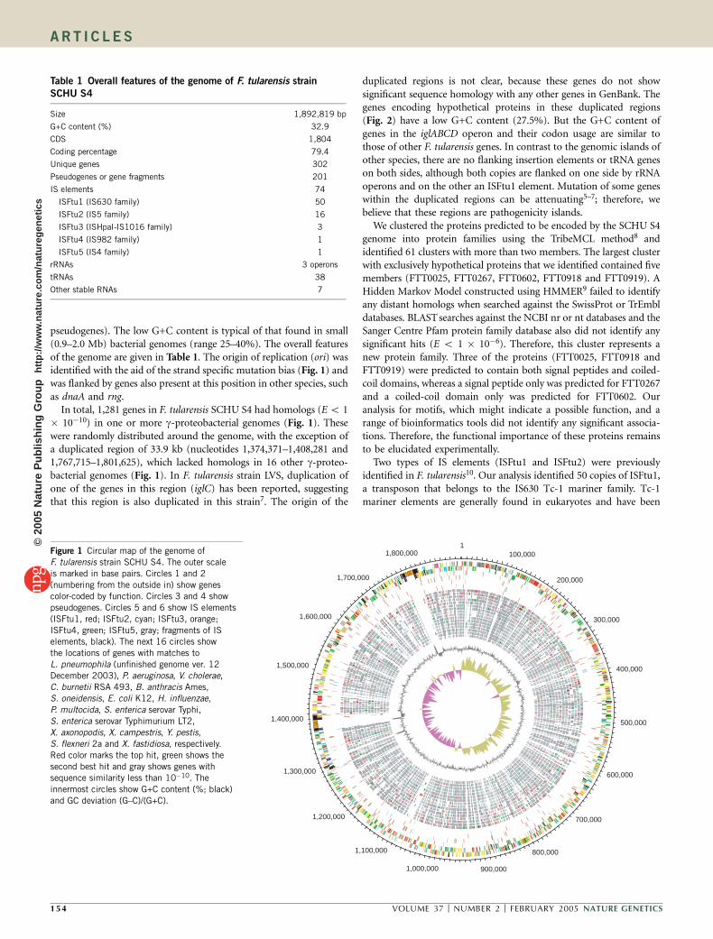
pseudogenes). The low G+C content is typical of that found in small(0.9–2.0 Mb) bacterial genomes (range 25–40%). The overall featuresof the genome are given in Table 1. The origin of replication (ori) wasidentified with the aid of the strand specific mutation bias (Fig. 1) andwas flanked by genes also present at this position in other species, suchas dnaA and rng.
In total, 1,281 genes in F. tularensis SCHU S4 had homologs (E o 1� 10�10) in one or more g-proteobacterial genomes (Fig. 1). Thesewere randomly distributed around the genome, with the exception ofa duplicated region of 33.9 kb (nucleotides 1,374,371–1,408,281 and1,767,715–1,801,625), which lacked homologs in 16 other g-proteo-bacterial genomes (Fig. 1). In F. tularensis strain LVS, duplication ofone of the genes in this region (iglC) has been reported, suggestingthat this region is also duplicated in this strain7. The origin of the
duplicated regions is not clear, because these genes do not showsignificant sequence homology with any other genes in GenBank. Thegenes encoding hypothetical proteins in these duplicated regions(Fig. 2) have a low G+C content (27.5%). But the G+C content ofgenes in the iglABCD operon and their codon usage are similar tothose of other F. tularensis genes. In contrast to the genomic islands ofother species, there are no flanking insertion elements or tRNA geneson both sides, although both copies are flanked on one side by rRNAoperons and on the other an ISFtu1 element. Mutation of some geneswithin the duplicated regions can be attenuating5–7; therefore, webelieve that these regions are pathogenicity islands.
We clustered the proteins predicted to be encoded by the SCHU S4genome into protein families using the TribeMCL method8 andidentified 61 clusters with more than two members. The largest clusterwith exclusively hypothetical proteins that we identified contained fivemembers (FTT0025, FTT0267, FTT0602, FTT0918 and FTT0919). AHidden Markov Model constructed using HMMER9 failed to identifyany distant homologs when searched against the SwissProt or TrEmbldatabases. BLAST searches against the NCBI nr or nt databases and theSanger Centre Pfam protein family database also did not identify anysignificant hits (E o 1 � 10�6). Therefore, this cluster represents anew protein family. Three of the proteins (FTT0025, FTT0918 andFTT0919) were predicted to contain both signal peptides and coiled-coil domains, whereas a signal peptide only was predicted for FTT0267and a coiled-coil domain only was predicted for FTT0602. Ouranalysis for motifs, which might indicate a possible function, and arange of bioinformatics tools did not identify any significant associa-tions. Therefore, the functional importance of these proteins remainsto be elucidated experimentally.
Two types of IS elements (ISFtu1 and ISFtu2) were previouslyidentified in F. tularensis10. Our analysis identified 50 copies of ISFtu1,a transposon that belongs to the IS630 Tc-1 mariner family. Tc-1mariner elements are generally found in eukaryotes and have been
1100,000
200,000
300,000
400,000
500,000
600,000
700,000
800,000
900,0001,000,000
1,100,000
1,200,000
1,300,000
1,400,000
1,500,000
1,600,000
1,700,000
1,800,000
Table 1 Overall features of the genome of F. tularensis strain
SCHU S4
Size 1,892,819 bp
G+C content (%) 32.9
CDS 1,804
Coding percentage 79.4
Unique genes 302
Pseudogenes or gene fragments 201
IS elements 74
ISFtu1 (IS630 family) 50
ISFtu2 (IS5 family) 16
ISFtu3 (ISHpaI-IS1016 family) 3
ISFtu4 (IS982 family) 1
ISFtu5 (IS4 family) 1
rRNAs 3 operons
tRNAs 38
Other stable RNAs 7
Figure 1 Circular map of the genome of
F. tularensis strain SCHU S4. The outer scale
is marked in base pairs. Circles 1 and 2
(numbering from the outside in) show genes
color-coded by function. Circles 3 and 4 show
pseudogenes. Circles 5 and 6 show IS elements
(ISFtu1, red; ISFtu2, cyan; ISFtu3, orange;
ISFtu4, green; ISFtu5, gray; fragments of ISelements, black). The next 16 circles show
the locations of genes with matches to
L. pneumophila (unfinished genome ver. 12
December 2003), P. aeruginosa, V. cholerae,
C. burnetii RSA 493, B. anthracis Ames,
S. oneidensis, E. coli K12, H. influenzae,
P. multocida, S. enterica serovar Typhi,
S. enterica serovar Typhimurium LT2,
X. axonopodis, X. campestris, Y. pestis,
S. flexneri 2a and X. fastidiosa, respectively.
Red color marks the top hit, green shows the
second best hit and gray shows genes with
sequence similarity less than 10�10. The
innermost circles show G+C content (%; black)
and GC deviation (G–C)/(G+C).
1 54 VOLUME 37 [ NUMBER 2 [ FEBRUARY 2005 NATURE GENETICS
A R T I C L E S©
2005
Nat
ure
Pub
lishi
ng G
roup
ht
tp://
ww
w.n
atur
e.co
m/n
atur
egen
etic
s

reported in a range of invertebrates such asnematodes and insects. The presence of thiselement in a bacterium is unusual. F. tular-ensis is often transmitted by infected insectvectors; the IS630 element that we identifiedmay have been acquired originally from aninsect. One copy of ISFTu1 is located in theO-antigen cluster, like the IS630 elementfound in the O-antigen cluster of Shigellasonnei. In S. sonnei, this element has a keyrole in the stable expression of form 1 of theO antigen, which is essential for virulence11.The IS630 element in the F. tularensis O-antigen cluster may have a similar function.
Sixteen copies of ISFtu2, an IS5 familyelement, were present. The genome also con-tained three types of IS element previouslyunreported in F. tularensis: ISFtu3 (two com-plete copies and one fragment), ISFtu4 (onecopy) and ISFtu5 (one copy). ISFtu3 hashomology to ISHpaI-IS1016 elements, andISFtu4 and ISFtu5 belong to the IS familiesIS982 and IS4, respectively. Also present arethree IS element fragments, which share homology with ISHpaI-IS1016 elements. These fragments possess terminal inverted repeatsequences not previously reported and therefore represent a new typeof IS element.
Most members of the IS630 and Tc-1 mariner family of insertionsequences possess a single open reading frame12, but translation of theISFtu1 CDS requires ribosomal frameshifting. In ISFtu1, the firstaspartic acid residue of the DDE triad, which is essential for transpo-sition, is generated only after a frameshift13. The programmedribosomal frameshifting motif in ISFtu1 may be used to control thetransposition rate of this element.
More than 10% of the CDSs in the SCHU S4 genome arepseudogenes or gene fragments that may have become fixed as theresult of a recent evolutionary bottleneck (Supplementary Table 1online). The proportion of pseudogenes due to disruption byIS elements (14%) is broadly similar to the proportion in otherpathogens such as Yersinia pestis (34%), Leifsonia xyli (5.5%) andBordetella pertussis (20.6%). Most of the pseudogenes were foundamong genes uniquely present in F. tularensis, hypothetically con-served or encoding proteins involved in transport, DNA metabolismor amino acid biosynthesis (Fig. 3). In agreement with this observa-tion, pseudogenes in these categories were also overrepresented inF. tularensis compared with other bacteria having more than 50pseudogenes (Supplementary Fig. 1 online).
PhylogenyFrancisella is the only genus of the family Francisellaceae, whichbelongs to the g subclass of proteobacteria. The Francisellaceae haveno close pathogenic relatives, as inferred from sequence similarity or16S rRNA phylogenies14. Instead, 16S rRNA data suggests thatF. tularensis is a sister clade with arthropod endosymbionts likeWolbachia persica14 and only distantly related to the human pathogensCoxiella burnetii and Legionella. This suggestion is supported by aphylogenomic analysis of more than 200 genes with homologs inF. tularensis and 15 other g-proteobacterial genomes, as previouslyundertaken for a smaller set of g-proteobacterial genomes15. Morethan 40% of the single gene trees suggest with strong support (475%)that F. tularensis is the most deeply diverging lineage among the 16 g-
proteobacterial species examined here. This is also shown in a treederived from a concatenated alignment of ten proteins (Fig. 4). TheC. burnetii genome is 1.9 Mbp, with 2,134 predicted CDSs16. Coxiella,Legionella and Francisella are g-proteobacterial pathogens with manylifestyle similarities. But they are not sister clades, and their deepdivergences explain the lack of overall gene-order conservation amongthe three genomes. Therefore, although these pathogens have similarlifestyles and their similar genome sizes seem to reflect this similarity,their positions in the phylogenetic tree suggest that they experiencedindependent, convergent evolution.
Predicted metabolic pathways and growth requirementsWe identified genes encoding 350 enzymes involved in small-moleculemetabolism in the annotated genome. We inferred 429 distinctenzymatic reactions to be catalyzed by these enzymes and predicted155 small-molecule metabolic pathways to be present (SupplementaryTable 2 online). Pathway predictions and information parsed from theannotated genome were output as a pathway-genome database calledFrantCyc. Each predicted pathway, P, was assigned a score X/Y/Z: Pconsists of X reactions; enzymes for Y reactions were identified in thegenome; and Z of the Y reactions are used in other predicted
0
5
10
15
20
25
30
35
Hypoth
etica
l
p
rotei
ns
Cell en
velop
e
Energ
y meta
bolis
m
Protei
n syn
thesis
Trans
port
and
bindin
g pr
oteins
Biosyn
thesis
of co
factor
s, pr
osthe
tic
grou
ps, a
nd ca
rrier
s
Mobile
and
extr
achr
omos
omal
ele
ment fu
nctio
ns
DNA meta
bolis
m
Protei
n fate
Cellula
r pro
cess
es
Centra
l inter
mediar
y meta
bolis
m
Unkno
wn fun
ction
Amino ac
id bio
synth
esis
Purine
s, py
rimidi
nes,
nucle
oside
s,
an
d nuc
leotid
es
Regula
tory f
uncti
ons
Fatty a
cid an
d pho
spho
lipid
meta
bolis
m
Trans
cripti
on
Ope
n re
adin
g fr
ames
(%)
Figure 3 Percentage of total F. tularensis strain SCHU S4 CDSs (black) and
pseudogenes or fragments (gray) attributed by predicted biological function.
1350
5S rRNA
23SrRNA
16S rRNA
pdpA1 pdpB1 1346
1347
1348
1349 1351
1352
1353
pdpC1
1355
iglD1
iglC1
iglB1
iglA1
pdpD1
1361
1362'
ISFtu1
1705
5S rRNA
23S rRNA
16S rRNA
pdpA2 pdpB2 1701
1702
1703
1704 1706
1707
1708
pdpC2
1710
iglD2
iglC2
iglB2
iglA2
pdpD2
1716
1717'
ISFtu1
55.23
32.26
19.89
10 kb
1,374,371
1,767,715
1,408,281
1,801,625
10 kb
Figure 2 The organization of the duplicated region in F. tularensis strain SCHU S4. The leftmost
scale shows G+C content. Blue indicates RNA coding regions; green indicates open reading frames
encoding hypothetical proteins; brown indicates pseudogenes; and pink indicates IS elements.
Open reading frame labels refer to the corresponding annotated gene or FTT number in the genome
sequence of SCHU S4.
NATURE GENETICS VOLUME 37 [ NUMBER 2 [ FEBRUARY 2005 15 5
A R T I C L E S©
2005
Nat
ure
Pub
lishi
ng G
roup
ht
tp://
ww
w.n
atur
e.co
m/n
atur
egen
etic
s

pathways. We inserted all pathways for which Y was nonzero intoFrantCyc. In total 1,105 operons were predicted using the PathwayTools operon predictor and inserted into FrantCyc.
Overall, we identified 390 pathway holes in 137 predicted pathways,corresponding to 54% of the reactions involved in the predictedmetabolic pathway network of F. tularensis. This percentage is higherthan we have observed in other bacteria17 and is consistent with theproposal that the F. tularensis genome is in an advanced state of decay.But we cannot exclude the possibility that because of the relativephylogenetic isolation of this bacterium, some of the pathways holesare filled by divergent orthologs that have not been identified. Pathwayholes were input to a program that identifies candidate genes withfunctions corresponding to each pathway hole17. We evaluated eachcandidate gene manually, and for those deemed sufficiently reliable,we assigned new gene functions to reflect the function postulated bythe program (Supplementary Note online). Application of thisalgorithm to a complete genome has not been published previouslyto our knowledge; in this case, it resulted in the identification of high-probability putative functions for 74 genes whose functions were notidentified by classical sequence analysis. We then rescored pathwaysand removed from the pathway-genome database any pathways forwhich Y/X was less than 1/3. Figure 5 shows one part of the entirepredicted metabolic map of F. tularensis, with the unfilled pathwayholes highlighted.
A growth medium consisting of 14 essential compounds (Table 2)was developed to support the growth of avirulent F. tularensis strain176 (ref. 18) and was also reported to support growth of SCHU S4(ref. 19). F. tularensis strain SCHU S4 also has a requirement for
cysteine20, which seems to be due to a nonfunctional pathway forsulfate assimilation resulting from a pseudogene (missing start codon)encoding adenylylsulfate kinase (EC 2.7.1.25). It remains to bedetermined experimentally how many of the other 13 essentialcompounds18 are absolutely required for growth. Our analysis indi-cated that biosynthetic pathways were present for 7 of these 13compounds. The pathways for sulfate assimilation, threonine bio-synthesis, valine biosynthesis and isoleucine biosynthesis seemed to beincomplete (i.e., they contained pathway holes). The available evidencedoes not indicate whether the enzymes for these pathway holes aretruly missing from the genome, thus inactivating the pathway, orwhether the enzymes are present but the activity of these pathways istoo low to support growth. We found genomic evidence for loss ofbiosynthetic capabilities for valine, isoleucine and threonine, indicat-ing that these amino acids are required for growth. Specifically, weidentified a pseudogene encoding homoserine kinase that mapped tothe one step missing from the predicted pathway for threoninebiosynthesis and a pseudogene encoding the large subunit of aceto-lactate synthase that mapped to the one step missing from thebiosynthetic pathways for both valine and isoleucine. This loss ofbiosynthetic capacity may have followed a change of evolutionaryniche (such as a move from a free-living organism to one or morespecific host cells) that resulted in these amino acids being readilyavailable in the environment. It remains to be determined whether oneor more of the other four compounds for which pathways werepredicted to be present (i.e., serine, aspartic acid, leucine and proline)is absolutely required for growth. Conversely, the other compoundsfor which we found little or no evidence of a biosynthetic pathwayhave probably always been readily available to the organism across adiverse range of evolutionary niches, such that the correspondingpathways were never required by the organism. These compounds areprobably required for growth.
We correctly predicted functional biosynthetic pathways for allseven nonessential amino acids (alanine, asparagine, glutamate, gluta-mine, glycine, phenylalanine and tryptophan; Supplementary Table 3online). Because the genomic evidence for these pathways (fraction ofenzymes present) was comparable to that observed for the ‘falsepositive’ pathways above, we infer that the predominant mechanismof gene-function inactivation rendering biosynthetic pathways inactiveor insufficiently active involved relatively small sequence changes,such as one or more point mutations. Biosynthetic pathways forseveral polyamines, including putrescine and spermine, were alsodisrupted. This is consistent with the observations that F. tularensisis unable to survive under hypotonic conditions21 and that osmoto-lerance can be attained by addition of micromolar amounts ofputrescine and spermine21.
Candidate mechanisms of virulenceLittle is known about the virulence mechanisms of F. tularensis, butgrowth in macrophages is central to the ability of F. tularensis to causedisease. Mutation of the genes iglA, iglC or pdpD in the 33.9-kbduplicated region reduces the ability of F. tularensis to survive inamoebae or macrophages and is attenuating5–7. These genes, andothers in this region, are regulated by the transcriptional regulatorMglA6. The precise functions of these genes are not known; the geneproducts do not show sufficient homology with any other genes inGenBank to infer their functions. Therefore, undiscovered mechan-isms of virulence are probably encoded in the 33.9-kb pathogenicityisland in F. tularensis. Within the macrophage, the bacterium candegrade the phagosomal membrane and escape into the cytosol22. Weidentified genes encoding a phospholipase C acpA (FTT0221) and a
0.1100,100
100,100
100,100
100,100
100,100100,100
100,100
52,97
69,74
100,100
100,100
100,100100,100
100,94
Bacillus anthracis
Francisella tularensis
Legionella pneumophila
Coxiella burnetii
Xylella fastidiosa
Xanthomonas axonopodis
Xanthomonas campestris
Pseudomonas aeruginosa
Shewanella oneidensis
Vibrio cholerae
Haemophilus influenzae
Pasteurella multocida
Yersinia pestis
Salmonella typhimurium
Salmonella enterica
Shigella flexneri
Escherichia coli
Figure 4 Phylogenetic relationship of 16 g-proteobacterial species inferred
from a concatenated alignment of the proteins encoded by dnaA, ftsA, mfd,
mraY, murB, murC, parC, recA, recG and rpoC. B. anthracis was used as the
outgroup. The topology, branch lengths and bootstrap support are according
to the reconstruction with the neighbor-joining method. Values at nodes are
bootstrap support values for the neighbor-joining and maximum parsimony
methods (in that order).
1 56 VOLUME 37 [ NUMBER 2 [ FEBRUARY 2005 NATURE GENETICS
A R T I C L E S©
2005
Nat
ure
Pub
lishi
ng G
roup
ht
tp://
ww
w.n
atur
e.co
m/n
atur
egen
etic
s
phospholipase D family protein (FTT0490), which may have a role inthis process. FTT1043 encodes a macrophage infectivity potentiatorprotein previously found to confer virulence for several pathogens,including Legionella pneumophila23. We also identified a homolog ofmce, involved in entry of Mycobacterium tuberculosis into host cells24,in the SCHU S4 genome sequence.
When F. tularensis is cultured in acidified medium, the pH of themedium increases25, reportedly owing to the generation of ammo-nia18. The generation of ammonia, and subsequent buffering ofthe endosomal compartment, may allow pathogens to survive inmacrophages26. Deaminases such as L-glutaminase, L-asparginaseand citrulline ureidase, which could be responsible for ammoniageneration, have previously been reported in F. tularensis27. In addi-tion, citrulline ureidase activity is used to differentiate strains withhigh virulence (subspecies tularensis) from strains with low virulence(subspecies holarctica)20, and low levels ofglutaminase activity have been associatedwith low virulence27. We identified severalgenes in the SCHU S4 genome that couldhave a role in ammonia production. In addi-tion to genes potentially encoding an L-aspar-ginase (FTT0591) and an L-glutaminase(FTT0195), we also identified an operonpredicted to encode a peptidyl-arginine dea-minase (FTT0434) and a candidate geneencoding citrulline ureidase (FTT0435). Thelatter seems to encode a carbon-nitrogenhydrolase family protein and possesses aPfam (PF00795) motif, indicating that it isan enzyme capable of reducing organic nitro-gen compounds and producing ammonia28.
Type I secretion systems transport sub-stances across the bacterial envelope usingtransporters containing ATP-binding cassettes.The genome of SCHU S4 is predicted tocontain 15 potentially functional ATP-bindingcassette systems (H. Garmory, personal com-munication). We did not identify gene clustersencoding type III, type IV or type V exportsystems, but we did identify some candidatecell surface–located virulence factors. The pre-sence of pili on the surface of F. tularensis hasbeen reported29, and we identified all cur-rently known genes necessary for type IV pilibiosynthesis. The exact role of type IV pili inFrancisella is not yet known, but in otherbacteria, they contribute to virulence. Themakeup of the poorly characterized capsulesurrounding F. tularensis is not known, but weidentified a gene cluster (FTT0789–FTT0801)that could encode a polysaccharide additionalto the lipopolysaccharide O antigen. We alsoidentified homologs of the genes capB(FTT0805) and capC (FTT0806) required forcapsule biosynthesis in Bacillus anthracis.Therefore, the capsule of F. tularensis mightcontain poly-D-glutamic acid.
Virulence and iron acquisitionThe ability of the bacterium to acquire ironin the phagosome seems to be crucial for
virulence of F. tularensis30, and growth under iron-limited conditionsresults in changes in the composition of the cell envelope31. For manymicroorganisms, the ferric uptake regulator (Fur) has a key role inmodulating iron uptake, and the genome of F. tularensis strain SCHUS4 is predicted to encode a Fur protein (FTT0030). We also identifieda number of genes that may be regulated by Fur, including ftnA, fumB,acnA, sodB and an ortholog of iraB (FTT0651), which is associatedwith iron uptake in L. pneumophila32. A gene (frgA; FTT0029)belonging to a family of hydroxamate-siderophore synthetic genes33
was located downstream of fur, and a putative iron-box was found inthe promoter region. Recent papers have described the ability ofF. tularensis to escape the phagosome34,35. In the cytoplasmic envir-onment, iron is highly insoluble, and a TonB-dependent systemfor complex-bound iron uptake would be expected. Although alow–molecular weight iron-binding compound, growth-initiating
Saturated and unsaturatedfatty acid elongationFatty acid biosynthesis – initial steps
Phospholipidbiosynthesis II
PhospholipidbiosynthesisI
O-antigenbiosynthesis
Colanic acidbuilding blocksbiosynthesis
Polyaminebiosynthesis
III
Polyaminebiosynthesis
I
Salvage pathways of purine and pyrimidine nucleotidesDe novo
biosynthesisof purine
nucleotides I
Salvage pathways ofpyrimidinedeoxyribonucleotides
Salvage pathwaysof pyrimidine
ribonucleotides
Peptidoglycanbiosynthesis
KDO2-lipid Abiosynthesis
UDP-N-acetylgalactosaminebiosynthesis
UDP-N-acetylglucosaminebiosynthesis
Polyaminebiosynthesis
II
tRNAchargingpathway
De novo biosynthesisof pyrimidinedeoxyribonucleotides
De novobiosynthesis
of purinenucleotides II
Salvage pathways ofpurine nucleosides
Salvage pathwaysof adenine,
hypoxanthine andtheir nucleosides
ppGppbiosynthesis
PRPPbiosynthesis
I
De novobiosynthesis of
pyrimidineribonucleosides
Figure 5 A portion of the FrantCyc cellular overview for F. tularensis, showing a region of the predictedmetabolic map of the organism. Each line is a single metabolic reaction, and each node is a single
metabolite. Green lines indicate reactions that have no enzyme assigned in the genome (pathway
holes); blue lines indicate reactions that do have an assigned enzyme. Upward triangle, amino acid;
square, carbohydrate; diamond, protein; vertical oval, purine; horizontal oval, pyrimidine; downward
triangle, cofactor; T, tRNA; open circle, other; shaded symbol, phosphorylated.
NATURE GENETICS VOLUME 37 [ NUMBER 2 [ FEBRUARY 2005 15 7
A R T I C L E S©
2005
Nat
ure
Pub
lishi
ng G
roup
ht
tp://
ww
w.n
atur
e.co
m/n
atur
egen
etic
s

substance, has been reported to be secreted by F. tularensis36, theevidence does not indicate growth-initiating substance to be a hydro-xamate siderophore37. No genes encoding TonB; outer membraneuptake receptors for ferric siderophore-complexes; or receptors fortransferrin, lactoferrin, heme, hemoglobin or hemopexin were foundin the genome.
DISCUSSIONPathogens are frequently thought to evolve by acquiring DNA frag-ments encoding virulence determinants. But an emerging theme ingenome biology is that several pathogens that cause severe disease haveevolved by losing genetic information instead. Different genomesequences provide different snapshots of this process of evolution.For example, Y. pestis seems to be at the very early stages of evolutionand has both lost and acquired genes during this process38. Mycobac-terium leprae39 and Rickettsia prowazekii40 seem to have evolved solelyby gene loss from a progenitor species39. The genome sequence ofF. tularensis SCHU S4 shows extensive inactivation of genes and aduplicated region that is strongly implicated in virulence and may be apathogenicity island. The origins of the pathogenicity islands are notknown, and the function of the genes in this region cannot be inferredon the basis of sequence homology with gene products of knownfunctions. This finding raises the possibility that new mechanisms ofvirulence operate in F. tularensis. SCHU S4 lacks coding potential forseveral expected features. The ability to import complexed ferric(Fe3+) iron should be important for Francisella, because it can escapefrom the phagosome22,35, thereby losing its access to soluble ironpresent in the acidic milieu. But no previously known ferric ironuptake systems were found in the genome sequence.
F. tularensis is considered one of the microorganisms most likely tobe used as a biological warfare or bioterrorism agent, but there is apaucity of information on the biochemical makeup of the organismand mechanisms of virulence. In part, this lack of information is a
consequence of the difficulties associated with working with highlyvirulent strains. The complete genome sequence of F. tularensis strainSCHU S4 is a key advances in our understanding of this pathogen andwill fuel future work to devise defensive countermeasures against thispotential biological warfare and bioterrorism agent.
METHODSF. tularensis subspecies tularensis strain SCHU S4 was derived from an
isolate from a case of human tularemia in the US41. A clonal seedstock of
the bacterium has a median lethal dose in the murine model of disease
of less than 1 colony-forming unit42. We isolated DNA from a culture derived
from this seedstock. We constructed plasmid libraries from randomly sheared
DNA in pUC18 or pUC19 with insert sizes of 1–2 kb or 2–4 kb, respectively. We
also constructed five libraries with insert sizes of 1–4 kb using the TOPO
Shotgun subcloning kit (Invitrogen), from nebulized DNA, and one M13
library with insert sizes of 1–2 kb using the double adaptor method43
from nebulized DNA. We carried out DNA sequencing and assembly as
previously described44. We produced a total of 32,743 sequence reads, resulting
in an overall genomic coverage of �12.9. For finishing and gap closure, we
used PCR, multiplexed combinatorial PCR, single primed PCR and pulse field
gel electrophoresis.
Gene prediction was done using Glimmer45. We carried out annotation and
curation, facilitated by Artemis, as described previously39 and checked them
manually. We identified protein motifs using CONSENSUS and SMART. We
used Pathway Tools software to determine metabolic pathways46, operons47 and
pathway hole fillers17. Pathway holes are reactions in a pathway for which no
catalyzing enzymes were identified in the genome annotation. The algorithm
for identifying candidate genes for each pathway hole involves querying the
public protein sequence databases for proteins in other organisms that are
known to catalyze the reaction associated with each pathway hole, BLAST
searching these sequences against all F. tularensis open reading frames and then
scoring each matching gene using a Bayesian network that integrates several
types of evidence48. For example, the Bayesian network will score a given
candidate gene higher if multiple query sequences show similarity to it and if
the candidate is adjacent to, or in the same direction as, another gene in the
same pathway (a direction is a contiguous group of genes transcribed in the
same direction). This algorithm differs from previous work49,50 in that it is
completely automated, it applies a reverse BLAST search with increased
sensitivity compared with other methods and it computes a probability value
for each candidate (validated through cross-validation studies) that allows
ranking of the candidates.
URLs. CONSENSUS is available at http://npsa-pbil.ibcp.fr/. SMART is available
at http://smart.embl-heidelberg.de/. A version of Figure 5 that can be explored
interactively is available at http://biocyc.org/FRANT/new-image?type¼OVER
VIEW. A pathway-genome database that describes the F. tularensis chromo-
some; its genes and their predicted operons; the product of each gene; the
biochemical reaction(s), if any, catalyzed by each gene product; the substrates of
each reaction; and the predicted organization of those reactions into small-
molecule metabolic pathways is available at http//biocyc.org/server.html. Also
available at this site (http://biocyc.org/FRANT/pathologic-index.html) is a
complete listing of all predicted F. tularensis pathways and their corresponding
evidence scores.
GenBank accession numbers. Genome sequences of F. tularensis subspecies
tularensis strain SCHU S4, AJ749949; Pseudomonas aeruginosa, NC_002516;
Vibrio cholerae, NC_002505 and NC_002506; C. burnetii RSA 493, NC_002971;
B. anthracis Ames, NC_003997; Shewanella oneidensis, NC_004347 and
NC_004349; Escherichia coli K12, NC_000913; Haemophilus influenzae,
NC_000907; Pasteurella multocida, NC_002663; Salmonella enterica serovar
Typhi, NC_003198, NC_003384 and NC_003385; Salmonella enterica serovar
Typhimurium LT2, NC_003197 and NC_003277; Xanthomonas axonopodis,
NC_003919; Xanthomonas campestris, NC_003902; Y. pestis, NC_003131,
NC_003134 and NC_003143; Shigella flexneri 2a, NC_004337; and Xylella
fastidiosa, NC_002488, NC_002489 and NC_002490.
Note: Supplementary information is available on the Nature Genetics website.
Table 2 A pathway-genome view of the 14 compounds supporting
growth of F. tularensis
Growth medium requirement Biosynthetic pathway predicted Pathway score
Amino acids
DL-isoleucine Yes 5/4/1
DL-methionine No –
DL-serine Yes 3/2/2
DL-threonine Yes 2/1/0
DL-valine Yes 4/3/2
L-arginine No –
L-aspartic acid Yes 1/1/1
L-cysteine Yes 5/3/3
L-histidine No –
L-leucine Yes 4/2/0
L-lysine No –
L-proline Yes 4/2/1
L-tyrosine No –
Other
Thiamine HCl No –
Evidence was found in the genome for biosynthetic pathways for 8 of these 14compounds. No evidence was found for pathways for the other 6 compounds andno pathway score was assigned. Absent pathways have probably never been presentin this organism, whereas the eight ‘false positive’ pathways almost certainly reflectthe genomic remnants of once-active pathways. The pathway score predicted pathwaysis denoted X/Y/Z: a pathway P consists of X total reactions; enzymes for Y of thereactions were identified in the genome; and Z of these Y reactions are used in otherpredicted pathways.
1 58 VOLUME 37 [ NUMBER 2 [ FEBRUARY 2005 NATURE GENETICS
A R T I C L E S©
2005
Nat
ure
Pub
lishi
ng G
roup
ht
tp://
ww
w.n
atur
e.co
m/n
atur
egen
etic
s

ACKNOWLEDGMENTSWe thank the Pathogen Sequencing Unit at the Sanger Institute for advice on theannotation and analysis of this genome sequence. This work was supported bythe UK Ministry of Defence, Swedish Ministry of Defence, Defense AdvancedResearch Projects Agency and US Department of Energy. Work carried out atLawrence Livermore Laboratory was done under the auspices of the USDepartment of Energy by the University of California.
COMPETING INTERESTS STATEMENTThe authors declare that they have no competing financial interests.
Received 29 September; accepted 8 December 2004
Published online at http://www.nature.com/naturegenetics/
1. Ellis, J., Oyston, P.C.F., Green, M. & Titball, R.W. Tularemia. Clin. Microbiol. Rev. 15,631–646 (2002).
2. Dennis, D.T. et al. Tularemia as a biological weapon - Medical and public healthmanagement. J. Am. Med. Assoc. 285, 2763–2773 (2001).
3. Hood, A.M. Virulence factors of Francisella tularensis. J. Hyg. (Lond.) 79, 47–65(1977).
4. Telepnev, M., Golovliov, I., Grundstrom, T., Tarnvik, A. & Sjostedt, A. Francisellatularensis inhibits Toll-like receptor-mediated activation of intracellular signalling andsecretion of TNF-alpha and IL-1 from murine macrophages. Cell. Microbiol. 5, 41–51(2003).
5. Nano, F.E. et al. A Francisella tularensis pathogenicity island required for intrama-crophage growth. J. Bacteriol. 186, 6430–6436 (2004).
6. Lauriano, C.M. et al. MglA regulates transcription of virulence factors necessary forFrancisella tularensis intraamoebae and intramacrophage survival. Proc. Natl. Acad.Sci. USA 101, 4246–4249 (2004).
7. Golovliov, I., Sjostedt, A., Mokrievich, A.N. & Pavlov, V.M. A method for allelicreplacement in Francisella tularensis. FEMS Microbiol. Lett. 222, 273–280(2003).
8. Enright, A.J., Kunin, V. & Ouzounis, C.A. Protein families and TRIBES in genomesequence space. Nucleic Acids Res. 31, 4632–4638 (2003).
9. Eddy, S.R. Profile hidden Markov models. Bioinformatics 14, 755–763 (1998).10. Thomas, R. et al. Discrimination of human pathogenic subspecies of Francisella
tularensis by using restriction fragment length polymorphism. J. Clin. Microbiol. 41,50–57 (2003).
11. Houng, H.S. & Venkatesan, M.M. Genetic analysis of Shigella sonnei form I antigen:identification of a novel IS630 as an essential element for the form I antigenexpression. Microb. Pathog. 25, 165–173 (1998).
12. Mahillon, J. & Chandler, M. Insertion sequences. Microbiol. Mol. Biol. Rev. 62, 725–774 (1998).
13. Doak, T.G., Doerder, F.P., Jahn, C.L. & Herrick, G. A proposed superfamily oftransposase genes - transposon-like elements in ciliated protozoa and a commonD35E motif. Proc. Natl. Acad. Sci. USA 91, 942–946 (1994).
14. Forsman, M., Sandstrom, G. & Sjostedt, A. Analysis of 16S ribosomal DNA sequencesof Francisella strains and utilization for determination of the phylogeny of the genusand for identification of strains by PCR. Int. J. Syst. Bacteriol. 44, 38–46 (1994).
15. Canback, B., Tamas, I. & Andersson, S.G.E. A phylogenomic study of endosymbioticbacteria. Mol. Biol. Evol. 21, 1110–1122 (2004).
16. Seshadri, R. et al. Complete genome sequence of the Q-fever pathogen Coxiellaburnetii. Proc. Natl. Acad. Sci. USA 100, 5455–5460 (2003).
17. Green, M.L. & Karp, P. A Bayesian method for identifying missing enzymes in predictedmetabolic pathway databases. BMC Bioinformatics 5, 76 (2004).
18. Traub, A., Mager, J. & Grossowicz, N. Studies on the nutrition of Pasteurella tularensis.J. Bacteriol. 70, 60–69 (1955).
19. Nagle, S.C.J., Anderson, R.E. & Gary, N.D. Chemically defined medium for the growthof Pasteurella tularensis. J. Bacteriol. 79, 566–571 (1960).
20. Sjostedt, A. Gram-negative aerobic cocci. Family XVII. Francisellaceae. in Bergey’sManual of Systematic Bacteriology (ed. Brenner, D.J.) 200–210 (Springer, New York,2004).
21. Mager, J. The stabilizing effect of spermine and related polyamines and bacterialprotoplasts. Biochim. Biophys. Acta 36, 529–531 (1959).
22. Clemens, D.L., Lee, B.Y. & Horwitz, M.A. Virulent and avirulent strains ofFrancisella tularensis prevent acidification and maturation of their phagosomes and
escape into the cytoplasm in human macrophages. Infect. Immun. 72, 3204–3217(2004).
23. Cianciotto, N.P., Eisenstein, B.I., Mody, C.H. & Engleberg, N.C. A mutation in themip gene results in an attenuation of Legionella pneumophila virulence. J. Infect. Dis.162, 121–126 (1990).
24. Arruda, S., Bomfim, G., Knights, R., Huima-Byron, T. & Riley, L.W. Cloning of anM. tuberculosis DNA fragment associated with entry and survival inside cells. Science261, 1454–1457 (1993).
25. Chamberlain, R.E. Evaluation of live tularemia vaccine prepared in a chemicallydefined medium. Appl. Microbiol. 13, 232–235 (1965).
26. Gordon, A.H., Hart, P.D. & Young, M.R. Ammonia inhibits phagosome-lysosome fusionin macrophages. Nature 286, 79–80 (1980).
27. Fleming, D.E. & Foshay, L. Studies on the physiology of virulence of Pasteurellatularensis. I. Citrulline ureidase and deamidase activity. J. Bacteriol. 70, 345–349(1955).
28. Bork, P. & Koonin, E.V. A new family of carbon-nitrogen hydrolases. Protein Sci. 3,1344–1346 (1994).
29. Gil, H., Benach, J.L. & Thanassi, D.G. Presence of pili on the surface of Francisellatularensis. Infect. Immun. 72, 3042–3047 (2004).
30. Fortier, A.H. et al. Growth of Francisella tularensis LVS in macrophages: the acidicintracellular compartment provides essential iron required for growth. Infect. Immun.63, 1478–1483 (1995).
31. Bhatnager, N.B., Elkins, K.L. & Fortier, A.H. Heat stress alters the virulence of arifampin-resistant mutant of Francisella tularensis LVS. Infect. Immun. 63, 154–159(1995).
32. Viswanathan, V.K., Edelstein, P.H., Pope, C.D. & Cianciotto, N.P. The Legionellapneumophila iraAB locus is required for iron assimilation, intracellular infection,and virulence. Infect. Immun. 68, 1069–1079 (2000).
33. Hickey, E.K. & Cianciotto, N.P. An iron- and fur-repressed Legionella pneumophilagene that promotes intracellular infection and encodes a protein with similarity to theEscherichia coli aerobactin synthetases. Infect. Immun. 65, 133–143 (1997).
34. Lindgren, H. et al. Factors affecting the escape of Francisella tularensis from thephagolysosome. J. Med. Microbiol. 53, 953–958 (2004).
35. Golovliov, I., Baranov, V., Krocova, Z., Kovarova, H. & Sjostedt, A. An attenuated strainof the facultative intracellular bacterium Francisella tularensis can escape the phago-some of monocytic cells. Infect. Immun. 71, 5940–5950 (2003).
36. Mager, J.A. Factor required for growth initiation of Pasteurella tularensis. Nature 203,898 (1964).
37. Halmann, M. & Mager, J. An endogenously produced substance essential for growthinitiation of Pasteurella tularensis. J. Gen. Microbiol. 49, 461–468 (1967).
38. Parkhill, J. et al. Genome sequence of Yersinia pestis, the causative agent of plague.Nature 413, 523–527 (2001).
39. Cole, S.T. et al. Massive gene decay in the leprosy bacillus. Nature 409, 1007–1011(2001).
40. Andersson, S.G.E. et al. The genome sequence of Rickettsia prowazekii and the originof mitochondria. Nature 396, 133–140 (1998).
41. Eigelsbach, H.T., Braun, W. & Herring, R. Studies on the variation of Bacteriumtularense. J. Bacteriol. 61, 557–570 (1951).
42. Russell, P., Eley, S.M., Fulop, M.J., Bell, D.L. & Titball, R.W. The efficacyof ciprofloxacin and doxycycline against experimental tularemia. J. Antimicrob.Chemother. 41, 461–465 (1998).
43. Andersson, B., Wentland, M.A., Ricafrente, J.Y., Liu, W. & Gibbs, R.A. A ‘‘doubleadaptor’’ method for improved shotgun library construction. Anal. Biochem. 236, 107–113 (1996).
44. Ewing, B. & Green, P. Base-calling of automated sequencer traces using PHRED II.Genome Res. 8, 186–194 (1998).
45. Delcher, A., Harmon, D., Kasif, S., White, O. & Salzberg, S. Improved microbial geneidentification with Glimmer. Nucleic Acid Res. 27, 4636–4641 (1999).
46. Karp, P.D., Paley, S. & Romero, P. The Pathway Tools software. Bioinformatics 18,S225–S232 (2002).
47. Romero, P. & Karp, P.D. Using functional and organizational information to improvegenome-wide computational prediction of transcription units on pathway/genomedatabases. Bioinformatics 20, 709–717 (2004).
48. Krieger, C.J. et al. MetaCyc: a multiorganism database of metabolic pathways andenzymes. Nucleic Acids Res. 32, D438–D442 (2004).
49. Osterman, A. & Overbeek, R. Missing Genes in metabolic pathways: a comparativegenomics approach. Curr. Opin. Chem. Biol. 7, 238–251 (2003).
50. Reed, J.L., Vo, T.D., Schilling, C.H. & Palsson, B.O. An expanded genomescale modelof Escherichia coli K-12 (iJR904 GSM/GPR). Genome Biol. 4, R54 (2003).
NATURE GENETICS VOLUME 37 [ NUMBER 2 [ FEBRUARY 2005 15 9
A R T I C L E S©
2005
Nat
ure
Pub
lishi
ng G
roup
ht
tp://
ww
w.n
atur
e.co
m/n
atur
egen
etic
s
Related Documents