1 23 European Journal of Clinical Microbiology & Infectious Diseases ISSN 0934-9723 Eur J Clin Microbiol Infect Dis DOI 10.1007/s10096-015-2346-5 The clinical features, diagnosis, treatment, and prognosis of neuroinvasive listeriosis: a multinational study F. Arslan, E. Meynet, M. Sunbul, O. R. Sipahi, B. Kurtaran, S. Kaya, A. C. Inkaya, P. Pagliano, G. Sengoz, A. Batirel, B. Kayaaslan, et al.

Welcome message from author
This document is posted to help you gain knowledge. Please leave a comment to let me know what you think about it! Share it to your friends and learn new things together.
Transcript

1 23
European Journal of ClinicalMicrobiology & Infectious Diseases ISSN 0934-9723 Eur J Clin Microbiol Infect DisDOI 10.1007/s10096-015-2346-5
The clinical features, diagnosis, treatment,and prognosis of neuroinvasive listeriosis: amultinational study
F. Arslan, E. Meynet, M. Sunbul,O. R. Sipahi, B. Kurtaran, S. Kaya,A. C. Inkaya, P. Pagliano, G. Sengoz,A. Batirel, B. Kayaaslan, et al.

1 23
Your article is protected by copyright and
all rights are held exclusively by Springer-
Verlag Berlin Heidelberg. This e-offprint is
for personal use only and shall not be self-
archived in electronic repositories. If you wish
to self-archive your article, please use the
accepted manuscript version for posting on
your own website. You may further deposit
the accepted manuscript version in any
repository, provided it is only made publicly
available 12 months after official publication
or later and provided acknowledgement is
given to the original source of publication
and a link is inserted to the published article
on Springer's website. The link must be
accompanied by the following text: "The final
publication is available at link.springer.com”.
ARTICLE
The clinical features, diagnosis, treatment, and prognosisof neuroinvasive listeriosis: a multinational study
F. Arslan & E. Meynet & M. Sunbul & O. R. Sipahi &B. Kurtaran & S. Kaya & A. C. Inkaya & P. Pagliano &
G. Sengoz & A. Batirel & B. Kayaaslan & O. Yıldız &
T. Güven & N. Türker & İ. Midi & E. Parlak & S. Tosun &
S. Erol & A. Inan & N. Oztoprak & I. Balkan & Y. Aksoy &
B. Ceylan & M. Yılmaz & A. Mert
Received: 7 January 2015 /Accepted: 1 February 2015# Springer-Verlag Berlin Heidelberg 2015
Abstract The aim of this study was to determine theindependent risk factors, morbidity, and mortality ofcentral nervous system (CNS) infections caused byListeria monocytogenes. We retrospectively evaluated100 episodes of neuroinvasive listeriosis in a multina-tional study in 21 tertiary care hospitals of Turkey,France, and Italy from 1990 to 2014. The mean ageof the patients was 57 years (range, 19–92 years), and64% were males. The all-cause immunosuppression ratewas 54 % (54/100). Forty-nine (49 %) patients werereferred to a hospital because of the classical triad ofsymptoms (fever, nuchal rigidity, and altered level ofconsciousness). Rhombencephalitis was detected radio-logically in 9 (9 %) cases. Twenty-seven (64 %) ofthe patients who had cranial magnetic resonance imag-ing (MRI) performed had findings of meningeal andparenchymal involvement. The mean delay in the
initiation of specific treatment was 6.8±7 days. Empirictreatment was appropriate in 52 (52 %) patients. Themortality rate was 25 %, while neurologic sequelae oc-curred in 13 % of the patients. In the multivariate anal-ysis, delay in treatment [odds ratio (OR), 1.07 [95 %confidence interval (CI), 1.01–1.16]] and seizures (OR,3.41 [95 % CI, 1.05–11.09]) were significantly associ-ated with mortality. Independent risk factors for neuro-logic sequelae were delay in treatment (OR, 1.07 [95 %CI, 1.006–1.367]) and presence of bacteremia (OR, 45.2[95 % CI, 2.73–748.1]). Delay in the initiation of treat-ment of neuroinvasive listeriosis was a poor risk factorfor unfavorable outcomes. Bacteremia was one of theindependent risk factors for morbidity, while the pres-ence of seizures predicted worse prognosis. Moreover,the addition of aminoglycosides to ampicillin monother-apy did not improve patients’ prognosis.
F. Arslan (*) :A. Mert (*)Department of Infectious Diseases and Clinical Microbiology,Istanbul Medipol University Hospital, Istanbul, Turkeye-mail: [email protected]: [email protected]
E. MeynetPôle thorax et vaisseaux, Unité d’oncologie thoracique, CHUGrenoble, BP217, 38043 Grenoble cedex 9, France
M. SunbulInfectious Diseases and Clinical Microbiology, Ondokuz MayisUniversity Medical School, Samsun, Turkey
O. R. SipahiDepartment of Infectious Diseases and Clinical Microbiology, EgeUniversity Faculty of Medicine, Izmir, Turkey
B. KurtaranDepartment of Infectious Diseases, Faculty of Medicine, CukurovaUniversity, Adana, Turkey
S. KayaDepartment of Infectious Diseases and Clinical Microbiology,Karadeniz Technical University Faculty of Medicine,Trabzon, Turkey
A. C. InkayaDepartment of Infectious Diseases, School of Medicine, HacettepeUniversity, Ankara, Turkey
P. PaglianoDepartment of Infectious Diseases, D. Cotugno Hospital, AORNDeiColli, Naples, Italy
Eur J Clin Microbiol Infect DisDOI 10.1007/s10096-015-2346-5
Author's personal copy
Introduction
Listeria monocytogenesis a Gram-positive, facultatively anaer-obic, intracellular bacterium that causes invasive disease inhumans and animals. An important foodborne pathogen,L. monocytogenes has a specific affinity for the central nervoussystem (CNS), especially in cell-mediated immunodeficientindividuals [1]. This species’ ability to penetrate the intestinal,blood–brain, and fetoplacental barriers is one of its most im-portant virulence factors [2]. Although the exact mechanism isunknown, L. monocytogenes is thought to cause infection viathe neural retrograde (in rhombencephalitis) and hematogenous(in meningitis and encephalitis) routes [2–4]. In one US study,L. monocytogeneswas reportedly the cause of nearly 4% of thecases of bacterial meningitis [5]; it also ranked as the fourthmost common etiologic agent of encephalitis in a study fromFrance [6]. Rhombencephalitis, a progressive dysfunction ofthe brain stem, can also be caused by L. monocytogenes andhas been reported as a case series in the literature [7, 8].
Very few cases of neuroinvasive listeriosis have been report-ed [9–13]. To the best of our knowledge, this study evaluatedthe largest number of cases with neuroinvasive listeriosis todate. We analyzed the epidemiological, clinical, laboratory, ra-diological, and therapeutic characteristics of 100 cases withneuroinvasive listeriosis derived from 21 centers in three differ-ent countries and investigated their impact on neurologic se-quelae and mortality. We also compared our results with thoseof cohort groups previously published in the medical literature.
Methods
This study was designed as a retrospective cohort study in-cluding 100 cases with neuroinvasive listeriosis derived from21 centers in Turkey, Italy, and France. The study criteriaincluded the following: (1) patients with any clinical formsof meningitis, meningoencephalitis, rhombencephalitis, and/or brain abscess; (2) patients whose cerebrospinal fluid(CSF)and/or blood cultures grew L. monocytogenes; and (3)patients who were followed up in the aforementioned three
countries from 1990 to 2014. A standard data-recording formwas prepared and sent to the collaborating centers by email.This form recorded demographic and clinical characteristics(symptoms on admission and their duration), laboratory find-ings (complete blood count, CSF microbiological and bio-chemical results, and blood culture results), radiological fea-tures [computed tomography (CT) and magnetic resonanceimaging (MRI)], and therapeutic data (initial empiric treat-ment, specific treatment, and treatment duration). These pa-rameters were evaluated to determine the independent riskfactors predicting neurologic sequelae and mortality.
Definition of neuroinvasive listeriosis
Cases in which L. monocytogenes was isolated from the CSFand/or blood culture and included signs of meningeal irrita-tion, pleocytosis [white blood cell (WBC) count >10/μL] inthe CSF, hypoglycorrhachia (CSF glucose <40 mg/dL) orCSF glucose/blood glucose <60 %, or increased CSF proteinlevel (>45 mg/dL). Encephalitis was defined as the presenceof an altered level of consciousness, epileptic seizures, focalneurologic deficits, or radiographic parenchymal involve-ment. Rhombencephalitis was defined as the involvement ofthe mesencephalon, pons and/or cerebellum based on cranialMRI, cranial nerve involvement or cerebellar signs (ataxia,tremor), or the development of hemiparesis [14].
Definition of immunosuppression
The presence of hematologic malignancy, solid organ trans-plantation, diabetes mellitus, chronic renal failure, alcoholism,cirrhosis, human immunodeficiency virus (HIV) infection,history of corticosteroid use, chemotherapy, or monoclonalantibody use.
Definition of neurological signs
The classical triad of fever, nuchal rigidity, and altered level ofconsciousness. The presence of focal neurologic signs wasdefined as cranial nerve involvement (diplopia, anisocoria,
G. SengozInfectious Diseases and Clinical Microbiology, Haseki Training andResearch Hospital, Istanbul, Turkey
A. BatirelInfectious Diseases and Clinical Microbiology, Dr. Lutfi KirdarKartal Education and Research Hospital, Istanbul, Turkey
B. KayaaslanClinic of Infectious Diseases and Clinical Microbiology, AnkaraNumune Education and Research Hospital, Ankara, Turkey
O. YıldızDepartment of Infectious Diseases, School of Medicine, ErciyesUniversity, Kayseri, Turkey
T. GüvenInfectious Diseases and Clinical Microbiology Clinic, AtaturkTraining and Research Hospital, Ankara, Turkey
N. TürkerDepartment of Infectious Diseases, İzmir Katip Çelebi UniversityAtatürk Research and Education Hospital, Izmir, Turkey
İ. MidiDepartment of Neurology, Marmara University Hospital,Istanbul, Turkey
E. ParlakDepartment of Infectious Diseases and Clinical Microbiology,Faculty of Medicine, Atatürk University, Erzurum, Turkey
Eur J Clin Microbiol Infect Dis
Author's personal copy
ptosis, dysphagia, or facial hypoesthesia), hemiparesis, andcerebellar involvement (dysarthria, ataxia, or ataxic gait).
Hyponatremia
Serum sodium (Na) level <135 mmol/L.
Epileptic seizures
Convulsions on admission and during patient follow-up.
Radiological signs
The presence of only hydrocephalus on CT scan or the pres-ence of both hydrocephalus and parenchymal involvement onMRI.
Delay in treatment
The period of time from the beginning of symptoms until theday onwhich treatment with an appropriate antibacterial agentat the proper dose was initiated.
Appropriate empiric treatment
The use of any of the following agents: ampicillin, penicillin,meropenem, or ampicillin–sulbactam plus an aminoglycoside.
Adjuvant corticosteroid therapy
A dose of >5 mg/day of prednisolone or an equivalent dose ofanother corticosteroid used for the underlying disease.
Unfavorable clinical outcomes
Neurological sequelae were defined as signs detected in thefirst outpatient visit after discharge from the hospital. Mortal-ity was defined as death within 2 months after the first admis-sion. The neurological sequelae and mortality risk factors aresummarized in Table 1.
Statistical analysis
For the evaluation of the predictors of neurologic sequelae ormortality, the Chi-square test was used for categorical vari-ables, whereas Fisher’s exact test, Student’s t-test, and theMann–Whitney U-test were used for categorical, normallydistributed, and non-normally distributed variables, respec-tively. Multivariate logistic regression analysis was performedto identify independent predictors of neurological sequelaeand mortality in patients with neuroinvasive listeriosis. TheHosmer–Lemeshow test was used to determine the overallfit of the model. All statistical tests were two-tailed. Condi-tions in which the type 1 error rate was <5 % were consideredstatistically significant. Analyses were performed using SPSSsoftware version 16.0 (SPSS Inc., Chicago, IL).
Results
Patients
A total of 100 cases with Listeria meningitis derived from 21centers in Turkey, Italy, and France were involved in ourstudy. The mean age of the patients was 57 years (range,19–92 years), and 64 % of the patients were male. The all-cause immunosuppression rate was 54 %. We detected thefollowing underlying immunosuppressive conditions (in orderof frequency): 43 % (23/54) were prescribed corticosteroids,16 % (9/54) had alcoholism or cirrhosis, 13 % (7/54) had anunderlying hematologic malignancy, 9 % (5/54) had uncon-trolled diabetes mellitus, 9 % (5/54) had undergone chemo-therapy due to a solid organ tumor, 5 % (3/54) had undergonerenal transplantation, 1 % (1) had active HIV infection, and1 % (1) had chronic renal failure.
Clinical findings
The mean duration of symptoms of the patients before admis-sion to the hospital was 5.1 days (range, 1–30 days). Thirty-seven (37 %) patents were admitted to the hospital within the
S. TosunIzmir Bozyaka Research and Education Hospital, Izmir, Turkey
S. Erol :A. InanInfectious Diseases and Clinical Microbiology, Haydarpasa NumuneEducation and Training Hospital, Istanbul, Turkey
N. OztoprakDepartment of Infectious Diseases and Clinical Microbiology, Schoolof Medicine, Karaelmas University, Zonguldak, Turkey
I. BalkanDepartment of Infectious Diseases and Clinical Microbiology, Schoolof CerrahpasaMedicine Faculty, Istanbul University, Istanbul, Turkey
Y. AksoyInfectious Diseases and Clinical Microbiology, Gebze State Hospital,Gebze, Turkey
B. Ceylan :M. YılmazInfectious Diseases and Clinical Microbiology, Istanbul MedipolUniversity, Istanbul, Turkey
A. MertInternal Medicine, Istanbul Medipol University, Istanbul, Turkey
Eur J Clin Microbiol Infect Dis
Author's personal copy

first 2 days of showing symptoms, 50 (50 %) patients in thefirst 2–7 days, and 13 (13 %) patients in the first 7 or moredays. Forty-nine (49 %) patients presented with the classicaltriad of symptoms on admission to the hospital. Neck stiffness
was present in 75 (75 %) of the cases. Thirty-one (31 %) casespresented with focal neurologic signs. Additional findings in-cluded coma at the time of presentation in 7 (7 %) cases,pneumonia in 3 (3 %) cases, diarrhea in 3 (3 %) cases,
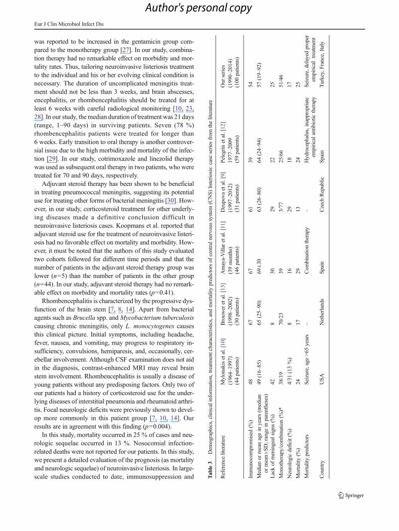
Table 1 Univariate analysis of the risk factors for mortality or severe neurological sequelae due to neurolisteriosis
Death* Neurological sequelae
Variable No Yes p-Value No Yes p-Value
No. of patients 75 25 62 13
Age (years) 51±17 54±14.5 0.73 60 (23–89) 57 (19–79) 0.15
Gender (male) 49 (65 %) 15 (60 %) 0.63 44 (71 %) 5 (39 %) 0.05
Predisposing factorsImmunocompromised conditions*
32 (42.7 %) 18 (72 %) 0.01 24 (38.7 %) 8 (62 %) 0.21
Delay in proper initial treatment (days) 5 (2–26) 8 (4–32) 0.03 4 (1–25) 6 (2–26) 0.014
Symptoms on admission
Classical triad 31 (41 %) 18 (72 %) 0.008 26 (42 %) 5 (39 %) 0.81
Fever 71 (94.7 %) 25 (100 %) 0.56 60 (97 %) 11 (85 %) 0.13
Altered sensorium 47 (62 %) 23 (92 %) 0.06 37 (60 %) 10 (77 %) 0.34
Nuchal rigidity 56 (74.7 %) 19 (74 %) 0.89 49 (79 %) 7 (54 %) 0.08
Headache 73 (97.3 %) 24 (96 % 1.00 61 (98 %) 12 (92 %) 0.31
Focal neurologic findings 19 (25.3 %) 12 (48 %) 0.03 7 (11 %) 12 (92 %) 0.00
Seizures 17 (24.6 %) 14 (58 %) 0.03 10 (18 %) 7 (54 %) 0.012
CSF findings
WBC count 535 (80–4,000) 285 (50–1,000) 0.51 833±1042 730±867 0.22
Pleocytosis (neutrophilic) 35 (76 %) 16 (84.2) 0.74 29 (81 %) 6 (60 %) 0.22
Protein level (g/L) 188±98.2 229±100 0.32 196±110 211±136 0.09
Glucose 44.6±24 54.8±29 0.60 35 (1.7–126) 45 (0–100) 0.25
Gram stain (Gram-positive rods) 14 (27 %) 3 (14 %) 0.36 10 (25 %) 4 (33 %) 0.71
Positive culture 68 (76 %) 88 (58 %) 0.55 59 (97 %) 9 (69 %) 0.007
Blood parameters
WBC count (/mm3) 14,250±7,763 14,762±5,642 0.53 13,377±7,280 11,282±5,800 0.37
Hemoglobin (g/dl) 12.7±1.6 12.1±2.7 0.85 12.8±1.7 11.9±1.8 0.18
Platelet count (/mm3) 182,520±76,452 249,800±100,154 0.21 179,580±74,230 181,270±50,829 0.94
C-reactive protein (mg/L) 111±85 115±82 0.87 102±85 144±80 0.13
Hyponatremia (Na <135 meq/L) 14 (30 %) 13 (68 %) 0.04 7 (18 %) 7 (78 %) 0.001
Positive culture 26 (34 %) 9 (36 %) 0.90 14 (22 %) 12 (92 %) 0.001
Focal neurological findings
Cranial nerve palsies 14 (19 %) 5 (20 %) 0.08 4 (7 %) 10 (77 %) 0.001
Hemiparesis 9 (12 %) 5 (20 %) 0.33 2 (3 %) 7 (54 %) 0.001
Cerebellar dysfunction 11 (12 %) 5 (20 %) 0.53 3 (5 %) 8 (62 %) 0.001
CT and/or MRI parameters
Parenchymal involvement (MRI) 21 (64 %) 7 (78 %) 0.44 11 (50 %) 10 (91 %) 0.04
Hydrocephalus 5 (14 %) 5 (31 %) 0.14 2 (7 %) 3 (38 %) 0.06
Rhombencephalitis 7 (19 %) 16 (20 %) 0.22 1 (2 %) 4 (80 %) 0.03
Treatment
Ampicillin/penicillin monotherapy 38 (51.4 %) 13 (56.5 %) 0.66 37 (61 %) 1 (8 %) –
Ampicillin+gentamicin therapy 36 (47 %) 10 (43 %) 0.41 24 (39 %) 12 (92 %)
Concomitant steroid therapy 34 (45 %) 9 (36 %) 28 (45 %) 6 (46 %) 0.94
CSF cerebrospinal fluid, CT computed tomography, MRI magnetic resonance imaging, WBC white blood cell
*All of the deaths occurred within 1 month except in two patients (times to death were days 47 and 56, respectively)
Eur J Clin Microbiol Infect Dis
Author's personal copy

diverticulitis in 2 (2 %) cases, and maculopapular rash in 1(1 %) case. Nineteen (19 %) patients had at least one cranialnerve paralysis, and 5 (5 %) cases had multiple cranial nerveparalysis. Involvement of the sixth and seventh cranial nerveswas the most common, appearing in 9 (9 %) patients each.Other nerves involved included the third, ninth, and tenthcranial nerves. Additionally, patients presented with focal neu-rological symptoms, including hemiparesis in 13 (13 %) pa-tients, aphasia and dysarthria in 12 (12 %) patients, and ataxiain 6 (6 %) patients. Thirty-one (31 %) patients presented withepileptic seizures. Twenty-nine (29 %) cases required me-chanical ventilation during clinical follow-up, 19 (66 %) ofwhom died. Nine (9%) cases had rhombencephalitis, 8 (89%)of whom were≤60 years of age (p=0.020) and 4 (45 %) ofwhom had neurological sequelae (p=0.003). Nearly 80 %(7/9) of the cases had no underlying disease.
Laboratory findings
Lumbar puncture was performed in 97 % of the cases. Themean leukocyte count was 1,051 cells/mL, and 79 % (51/65)of the cases had neutrophilic pleocytosis. The mean CSF glu-cose was 39 mg/dL (range, 0–130 mg/dL) and the mean CSFprotein count was 234 mg/dL (range, 30–2,196 mg/dL). Gramstaining revealed microorganisms in 23 % (17/73) of the casesin which Gram stain results were recorded. The mean WBCcount was 13,100 cells/mm3 (range, 1,690–36,900 cells/mm3),themean hemoglobin level was 12.6mg/dL (range, 7.9–16mg/dL), the mean platelet count was 188,000 cells/mm3 (range, 29,000–455,000 cells/mm3), the erythrocyte sedimentation rate(ESR) was 60 mm/h (range, 6–136 mm/h), and the C-reactiveprotein (CRP) count was 112 mg/L (range, 5–330 mg/L). Al-though CSF cultures grew L. monocytogenes in 90 % of thecases, only 35 cases had bacteremia. Hyponatremia (serum Na<135 mmol/L) was present in 41 % (27/66) of the cases inwhich serum sodium levels were recorded.
Cranial CT and MRI were performed in 63 (63 %) and 43(43 %) cases, respectively. Whereas no involvement was de-tected by CT imaging in 44 (70%) of the patients who had CTimaging performed, 9 (14 %) had parenchymal involvement,6 (10 %) had hydrocephalus, 3 (5 %) had vascular phenomena(acute/subacute infarct and hemorrhage), and only 1 (2%) hadmeningeal contrast enhancement. Twenty-seven (63 %) pa-tients who underwent cranial MRI had findings of meningealand parenchymal involvement. Hydrocephalus was present in19 % of the cases in whom both types of cranial imaging wereperformed (CT and MRI).
Treatment
The mean delay in the initiation of specific treatment was 6.8±7 days. Empiric treatment was appropriate in 52 (52 %) pa-tients. Ampicillin monotherapy and ampicillin–gentamicincombination therapy were preferred for definitive therapy in51 (51 %) and 46 (46 %) patients, respectively. Three (3 %)patients received meropenem for treatment. Adjuvant cortico-steroid therapy was given in 43 (43 %) cases. The medianduration of treatment was 21 days (range, 1–90 days). Combi-nation therapy was preferred in 87.5 % (7) of the cases withrhombencephalitis (p=0.025). There was no significant differ-ence in treatment modalities (monotherapy or combinationtherapy) in patients with mental status changes or a requirementfor mechanical ventilation (p=0.92 and p=0.063, respectively).
Predictors of mortality and neurologic sequelae
A number of risk factors were statistically significant for bothmortality and the development of neurologic sequelae in theunivariate analysis (Table 1). In the multivariate analysis, de-lay in treatment [odds ratio (OR), 1.07 [95 % confidence in-terval (CI), 1.01–1.16]] and epileptic attack (OR, 3.41 [95 %CI, 1.05–11.09]) were significantly associated with mortality(Table 2). Delay in treatment (OR, 1.07 [95 % CI, 1.006–
Table 2 Binomial logisticregression analysis ofindependent risk factors formortality or severe neurologicalsequelae in patients withneurolisteriosis
CI confidence interval, CSFcerebrospinal fluid, OR odds ratio
Death (n=25) Neurological sequelae (n=13)
Variable OR (95 % CI) p-Value OR (95 % CI) p-Value
Age (years) 1.01 (0.97–1.04) 0.44 0.93 (0.86–1.01) 0.12
Gender (male) 1.286 (0.43–3.80) 0.65 0.60 (0.07–5.23) 0.64
Classical triad 3.22 (0.93–11.07) 0.06 –
Immunocompromised conditions 3.43 (0.92–12.7) 0.06 0.07 (0.004–1.56) 0.09
Delayed initiation of treatment 1.07 (1.01–1.16) 0.04 1.07 (1.006–1.367) 0.042
Focal neurological findings 1.57 (0.47–5.18) 0.45 –
CSF culture positivity – 0.80 (0.08–7.60) 0.85
Bacteremia – 45.2 (2.73–748.1) 0.008
Seizures 3.41 (1.05–11.09) 0.04 –
Treatment (combination) – 5.35 (0.38–74.7) 0.21
Eur J Clin Microbiol Infect Dis
Author's personal copy

1.367]) and the presence of bacteremia (OR, 45.2 [95 % CI,2.73–748.1]) were independent risk factors for neurologic se-quelae. A delay in treatment of 7 or more days significantlyincreased the risk of mortality (p=0.011) and the risk of de-velopment of neurologic sequelae (p=0.042).
Discussion
The finding that the clinical picture of neuroinvasive listeriosismay differ from that of community-acquired purulent menin-gitis is controversial [13, 15]. Studies have emphasized thatneck stiffness occurs less commonly, whereas focal neurolog-ic deficits and epileptic seizures develop more frequently, inpatients with neuroinvasive listeriosis compared to those withcommunity-acquired purulent meningitis [10]. In our study,headache (97 %), fever (96 %), and neck stiffness (75 %) weredetected in most cases, and the classical triad was present innearly half of the cases. Focal neurological deficits and epi-leptic seizures occurred in 31 % of cases. In a German pro-spective cohort study evaluating 696 cases of bacterial men-ingitis, the classical triad was reported in 44 % of the cases[16]. Additionally, the presence of focal neurological signswas reported in 33 % of cases, which was similar to the ratein our study. The frequency of the complete classical triad wasalso similar in immunosuppressed cases and patients olderthan 50 years of age. These data demonstrate that the possi-bility of atypical presentation in neuroinvasive listeriosis (withthe exception of rhombencephalitis) was no different from thatin other bacterial meningitis cases.
Epileptic seizures developing within the first 24 h werefound to be an independent risk factor for mortality in anotherstudy involving 493 cases of acute bacterial meningitis, inwhich 10% were due to L. monocytogenes [17]. The presenceof epileptic seizures was an independent predictor for mortal-ity in two other studies; other risk factors included hydroceph-alus, advanced age, inappropriate antibiotic therapy, and com-bination therapy with aminoglycosides [10–12, 18].
Despite the high yield (67–90 %) of CSF Gram stainingperformed under suitable conditions in bacterial meningitis,the rates are significantly lower (nearly 30 %) in cases ofneuroinvasive listeriosis [19, 20]. In our study, Gram stainingrevealed the causative agent in ~25% (17/73) of the cases. In acase series of neuroinvasive listeriosis, the rate ofhyponatremia ranged between 44 and 73 %. Although thepresence of hyponatremia (41 %; 27/66) was found to be arisk factor for both mortality and neurologic sequelae in theunivariate analysis, we did not include hyponatremia in ourmultivariate analysis because serum sodium levels were notrecorded in the majority of cases. In another series examiningneuroinvasive listeriosis, cranial CTwas performed in 7–91%of the cases, but very few patients were reported to have cra-nial involvement on MRI [10, 11, 13]. Two possible reasons
for this finding may be the inclusion of old cases when MRIwas not widely used or that cranial CT was considered suffi-cient for the diagnosis. In those series, abnormal findings fromcranial CT were present in 23–27 % of the cases, particularlyfocal involvement or hydrocephalus. Newly developed in-farcts and hemorrhage were specifically reported in very fewcases. In our study, no involvement was detected in the cranialCT imaging of 44 patients (70 % of those who had CT imag-ing). However, it was remarkable that significant radiologicinvolvement was detected in 64 % of patients who underwentcranial MRI. Moreover, eight patients whose cranial CT im-aging was reported to be normal had significant radiologicinvolvement on cranial MRI. Therefore, we propose that cra-nial MRI is superior to cranial CT scanning in patients withneuroinvasive listeriosis.
Antimicrobial therapy in community-acquired meningitisis recommended empirically according to the age and immu-nosuppression status of the patient. Ampicillin is recommend-ed if the patient is <1 month or >50 years of age, and partic-ularly if the patient is immunosuppressed [21]. Inappropriateinitial empiric treatment was reported in 30 % of cases in aprospective series of 30 cases with neuroinvasive listeriosis[13]. Interestingly, in another prospective study investigating43 cases of neuroinvasive listeriosis (out of 278 patients withcommunity-acquired acute bacterial meningitis), inappropri-ate initial empiric treatment was only prescribed in 3 % of thelisteriosis cases [11]. In contrast, in the present study, the rateof inappropriate initial treatment was nearly 50 %. Despite theretrospective nature of our study, we believe that these find-ings better describe real-life data. The increased inappropriateantibiotic usage was related to the failure of the physicians toconsider neuroinvasive listeriosis in the differential diagnosis.For this reason, adults >50 years of age should receive anantimicrobial agent with activity against L. monocytogenesas part of the empiric regimen, regardless of their immuno-compromised status.
The optimal therapeutic regimen and its optimal durationfor the t rea tment of CNS infec t ions caused byL. monocytogenes have not been definitively defined.L. monocytogenes is very sensitive to penicillin G and ampi-cillin in vitro. Additionally, increased bactericidal activity insusceptibility tests with streptomycin or gentamicin combina-tions have encouraged physicians to prefer combination treat-ment [22, 23]. Case reports have been published of patientstreated with trimethoprim–sulfamethoxazole, meropenem, orlinezolid [24, 25]. For example, combination regimens (ampi-cillin plus an aminoglycoside versus ampicillin/amoxicillinplus cotrimoxazole) for treatment were compared in a studythat included 22 cases of meningi t i s caused byL. monocytogenes who were admitted to the intensive careunit. Combination regimens including cotrimoxazole havebeen reported to be more effective [26]. In another retrospec-tive study investigating 102 listeriosis cases, the mortality rate
Eur J Clin Microbiol Infect Dis
Author's personal copy
was reported to be increased in the gentamicin group com-pared to the monotherapy group [27]. In our study, combina-tion therapy had no remarkable effect on morbidity and mor-tality rates. Thus, tailoring neuroinvasive listeriosis treatmentto the individual and his or her evolving clinical condition isnecessary. The duration of uncomplicated meningitis treat-ment should not be less than 3 weeks, and brain abscesses,encephalitis, or rhombencephalitis should be treated for atleast 6 weeks with careful radiological monitoring [10, 23,28]. In our study, themedian duration of treatment was 21 days(range, 1–90 days) in surviving patients. Seven (78 %)rhombencephalitis patients were treated for longer than6 weeks. Early transition to oral therapy is another controver-sial issue due to the high morbidity and mortality of the infec-tion [29]. In our study, cotrimoxazole and linezolid therapywas used as subsequent oral therapy in two patients, who weretreated for 70 and 90 days, respectively.
Adjuvant steroid therapy has been shown to be beneficialin treating pneumococcal meningitis, suggesting its potentialuse for treating other forms of bacterial meningitis [30]. How-ever, in our study, corticosteroid treatment for other underly-ing diseases made a definitive conclusion difficult inneuroinvasive listeriosis cases. Koopmans et al. reported thatadjuvant steroid use for the treatment of neuroinvasive listeri-osis had no favorable effect on mortality and morbidity. How-ever, it must be noted that the authors of this study evaluatedtwo cohorts followed for different time periods and that thenumber of patients in the adjuvant steroid therapy group waslower (n=5) than the number of patients in the other group(n=44). In our study, adjuvant steroid therapy had no remark-able effect on morbidity and mortality rates (p=0.41).
Rhombencephalitis is characterized by the progressive dys-function of the brain stem [7, 8, 14]. Apart from bacterialagents such as Brucella spp. andMycobacterium tuberculosiscausing chronic meningitis, only L. monocytogenes causesthis clinical picture. Initial symptoms, including headache,fever, nausea, and vomiting, may progress to respiratory in-sufficiency, convulsions, hemiparesis, and, occasionally, cer-ebellar involvement. Although CSF examination does not aidin the diagnosis, contrast-enhanced MRI may reveal brainstem involvement. Rhombencephalitis is usually a disease ofyoung patients without any predisposing factors. Only two ofour patients had a history of corticosteroid use for the under-lying diseases of interstitial pneumonia and rheumatoid arthri-tis. Focal neurologic deficits were previously shown to devel-op more commonly in this patient group [7, 10, 14]. Ourresults are in agreement with this finding (p=0.004).
In this study, mortality occurred in 25 % of cases and neu-rologic sequelae occurred in 13 %. Nosocomial infection-related deaths were not reported for our patients. In this study,we present a detailed evaluation of the prognosis (as mortalityand neurologic sequelae) of neuroinvasive listeriosis. In large-scale studies conducted to date, immunosuppression and T
able3
Dem
ographics,clinicalinform
ation,treatm
entcharacteristics,andmortalitypredictorsof
centraln
ervous
system
(CNS)
listeriosis:caseseries
from
theliterature
Reference
literature
Mylonakisetal.[10]
Brouw
eretal.[13]
Amaya-Villar
etal.[11]
Dzupova
etal.[9]
Pelegrínetal.[12]
Our
series
(1964–1997)
(1998–2002)
(39months)
(1997–2012)
1977–2009
(1990–2014)
(44patients)
(30patients)
(46patients)
(31patients)
(59patients)
(100
patients)
Immunocom
prom
ised
(%)
4867
6761
3954
Medianor
meanagein
years(m
edian
ormean±SD;range
inparentheses)
49(16–85)
65(25–90)
69±30
63(26–80)
64(24–94)
57(19–92)
Lackof
meningealsigns(%
)42
830
2922
25
Monotherapy/com
binatio
n(%
)*38/19
70/23
393/77
25/66
51/46
Neurologicdeficit(%)
4/31
(13%)
816
2918
17
Mortality(%
)24
1729
1324
25
Mortalitypredictors
Seizure,age>65
years
–Com
binatio
ntherapy
–Hydrocephalus,inappropriate
empiricalantibiotic
therapy
Seizure,delayedproper
empiricaltreatm
ent
Country
USA
Netherlands
Spain
Czech
Republic
Spain
Turkey,France,Italy
Eur J Clin Microbiol Infect Dis
Author's personal copy
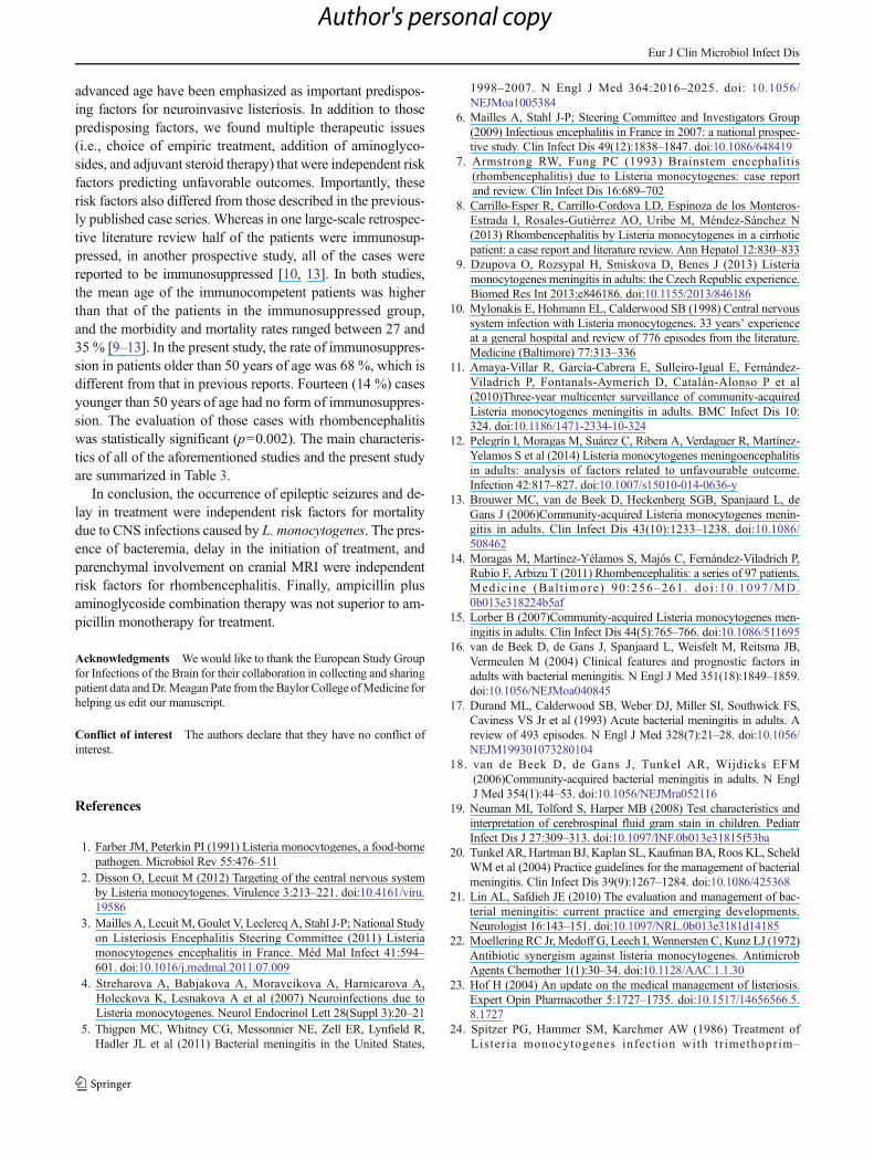
advanced age have been emphasized as important predispos-ing factors for neuroinvasive listeriosis. In addition to thosepredisposing factors, we found multiple therapeutic issues(i.e., choice of empiric treatment, addition of aminoglyco-sides, and adjuvant steroid therapy) that were independent riskfactors predicting unfavorable outcomes. Importantly, theserisk factors also differed from those described in the previous-ly published case series. Whereas in one large-scale retrospec-tive literature review half of the patients were immunosup-pressed, in another prospective study, all of the cases werereported to be immunosuppressed [10, 13]. In both studies,the mean age of the immunocompetent patients was higherthan that of the patients in the immunosuppressed group,and the morbidity and mortality rates ranged between 27 and35 % [9–13]. In the present study, the rate of immunosuppres-sion in patients older than 50 years of age was 68 %, which isdifferent from that in previous reports. Fourteen (14 %) casesyounger than 50 years of age had no form of immunosuppres-sion. The evaluation of those cases with rhombencephalitiswas statistically significant (p=0.002). The main characteris-tics of all of the aforementioned studies and the present studyare summarized in Table 3.
In conclusion, the occurrence of epileptic seizures and de-lay in treatment were independent risk factors for mortalitydue to CNS infections caused by L. monocytogenes. The pres-ence of bacteremia, delay in the initiation of treatment, andparenchymal involvement on cranial MRI were independentrisk factors for rhombencephalitis. Finally, ampicillin plusaminoglycoside combination therapy was not superior to am-picillin monotherapy for treatment.
Acknowledgments We would like to thank the European Study Groupfor Infections of the Brain for their collaboration in collecting and sharingpatient data andDr.Meagan Pate from the Baylor College ofMedicine forhelping us edit our manuscript.
Conflict of interest The authors declare that they have no conflict ofinterest.
References
1. Farber JM, Peterkin PI (1991) Listeria monocytogenes, a food-bornepathogen. Microbiol Rev 55:476–511
2. Disson O, Lecuit M (2012) Targeting of the central nervous systemby Listeria monocytogenes. Virulence 3:213–221. doi:10.4161/viru.19586
3. Mailles A, Lecuit M, Goulet V, Leclercq A, Stahl J-P; National Studyon Listeriosis Encephalitis Steering Committee (2011) Listeriamonocytogenes encephalitis in France. Méd Mal Infect 41:594–601. doi:10.1016/j.medmal.2011.07.009
4. Streharova A, Babjakova A, Moravcikova A, Harnicarova A,Holeckova K, Lesnakova A et al (2007) Neuroinfections due toListeria monocytogenes. Neurol Endocrinol Lett 28(Suppl 3):20–21
5. Thigpen MC, Whitney CG, Messonnier NE, Zell ER, Lynfield R,Hadler JL et al (2011) Bacterial meningitis in the United States,
1998–2007. N Engl J Med 364:2016–2025. doi: 10.1056/NEJMoa1005384
6. Mailles A, Stahl J-P; Steering Committee and Investigators Group(2009) Infectious encephalitis in France in 2007: a national prospec-tive study. Clin Infect Dis 49(12):1838–1847. doi:10.1086/648419
7. Armstrong RW, Fung PC (1993) Brainstem encephalitis(rhombencephalitis) due to Listeria monocytogenes: case reportand review. Clin Infect Dis 16:689–702
8. Carrillo-Esper R, Carrillo-Cordova LD, Espinoza de los Monteros-Estrada I, Rosales-Gutiérrez AO, Uribe M, Méndez-Sánchez N(2013) Rhombencephalitis by Listeria monocytogenes in a cirrhoticpatient: a case report and literature review. Ann Hepatol 12:830–833
9. Dzupova O, Rozsypal H, Smiskova D, Benes J (2013) Listeriamonocytogenes meningitis in adults: the Czech Republic experience.Biomed Res Int 2013:e846186. doi:10.1155/2013/846186
10. Mylonakis E, Hohmann EL, Calderwood SB (1998) Central nervoussystem infection with Listeria monocytogenes. 33 years’ experienceat a general hospital and review of 776 episodes from the literature.Medicine (Baltimore) 77:313–336
11. Amaya-Villar R, García-Cabrera E, Sulleiro-Igual E, Fernández-Viladrich P, Fontanals-Aymerich D, Catalán-Alonso P et al(2010)Three-year multicenter surveillance of community-acquiredListeria monocytogenes meningitis in adults. BMC Infect Dis 10:324. doi:10.1186/1471-2334-10-324
12. Pelegrín I, Moragas M, Suárez C, Ribera A, Verdaguer R, Martínez-Yelamos S et al (2014) Listeria monocytogenes meningoencephalitisin adults: analysis of factors related to unfavourable outcome.Infection 42:817–827. doi:10.1007/s15010-014-0636-y
13. Brouwer MC, van de Beek D, Heckenberg SGB, Spanjaard L, deGans J (2006)Community-acquired Listeria monocytogenes menin-gitis in adults. Clin Infect Dis 43(10):1233–1238. doi:10.1086/508462
14. Moragas M, Martínez-Yélamos S, Majós C, Fernández-Viladrich P,Rubio F, Arbizu T (2011) Rhombencephalitis: a series of 97 patients.Medic ine (Bal t imore) 90:256–261. doi :10 .1097/MD.0b013e318224b5af
15. Lorber B (2007)Community-acquired Listeria monocytogenes men-ingitis in adults. Clin Infect Dis 44(5):765–766. doi:10.1086/511695
16. van de Beek D, de Gans J, Spanjaard L, Weisfelt M, Reitsma JB,Vermeulen M (2004) Clinical features and prognostic factors inadults with bacterial meningitis. N Engl J Med 351(18):1849–1859.doi:10.1056/NEJMoa040845
17. Durand ML, Calderwood SB, Weber DJ, Miller SI, Southwick FS,Caviness VS Jr et al (1993) Acute bacterial meningitis in adults. Areview of 493 episodes. N Engl J Med 328(7):21–28. doi:10.1056/NEJM199301073280104
18. van de Beek D, de Gans J, Tunkel AR, Wijdicks EFM(2006)Community-acquired bacterial meningitis in adults. N EnglJ Med 354(1):44–53. doi:10.1056/NEJMra052116
19. Neuman MI, Tolford S, Harper MB (2008) Test characteristics andinterpretation of cerebrospinal fluid gram stain in children. PediatrInfect Dis J 27:309–313. doi:10.1097/INF.0b013e31815f53ba
20. Tunkel AR, HartmanBJ, Kaplan SL, Kaufman BA, Roos KL, ScheldWM et al (2004) Practice guidelines for the management of bacterialmeningitis. Clin Infect Dis 39(9):1267–1284. doi:10.1086/425368
21. Lin AL, Safdieh JE (2010) The evaluation and management of bac-terial meningitis: current practice and emerging developments.Neurologist 16:143–151. doi:10.1097/NRL.0b013e3181d14185
22. Moellering RC Jr, Medoff G, Leech I, Wennersten C, Kunz LJ (1972)Antibiotic synergism against listeria monocytogenes. AntimicrobAgents Chemother 1(1):30–34. doi:10.1128/AAC.1.1.30
23. Hof H (2004) An update on the medical management of listeriosis.Expert Opin Pharmacother 5:1727–1735. doi:10.1517/14656566.5.8.1727
24. Spitzer PG, Hammer SM, Karchmer AW (1986) Treatment ofLister ia monocytogenes infect ion with tr imethoprim–
Eur J Clin Microbiol Infect Dis
Author's personal copy

sulfamethoxazole: case report and review of the literature. Rev InfectDis 8:427–430
25. Manfredi R (2007) Linezolid activity against disseminatedListeria monocytogenes meningitis and central nervous systemabscesses: focus on early drug myelotoxicity. Curr Drug Saf 2:141–145
26. Merle-Melet M, Dossou-Gbete L, Maurer P, Meyer P, Lozniewski A,Kuntzburger O et al (1996) Is amoxicillin–cotrimoxazole the mostappropriate antibiotic regimen for listeria meningoencephalitis?Review of 22 cases and the literature. J Infect 33:79–85
27. Mitjà O, Pigrau C, Ruiz I, Vidal X, Almirante B, Planes A-M et al(2009) Predictors of mortality and impact of aminoglycosides on
outcome in listeriosis in a retrospective cohort study. J AntimicrobChemother 64:416–423. doi:10.1093/jac/dkp180
28. Clauss HE, Lorber B (2008) Central nervous system infection withListeria monocytogenes. Curr Infect Dis Rep 10(4):300–306. doi:10.1007/s11908-008-0049-0
29. Grant MH, Ravreby H, Lorber B (2010) Cure of Listeriamonocytogenes meningitis after early transition to oral therapy.Antimicrob Agents Chemother 54:2276–2277. doi:10.1128/AAC.01815-09
30. van de Beek D, de Gans J, McIntyre P, Prasad K (2003)Corticosteroids in acute bacterial meningitis. Cochrane DatabaseSyst Rev (3):CD004405. doi:10.1002/14651858.CD004305
Eur J Clin Microbiol Infect Dis
Author's personal copy
Related Documents