The Circulatory System

Welcome message from author
This document is posted to help you gain knowledge. Please leave a comment to let me know what you think about it! Share it to your friends and learn new things together.
Transcript

The Circulatory System

Structures in the Circulatory System
Heart – pumps blood throughout the entire body.
Muscle that is the size of your fist.
Has a left side that pumps blood with oxygen into the body.
Has a right side that pumps deoxygenated blood into the lungs to pick up oxygen.

Heart Chambers
•Receives blood through 2 chambers: Left & Right Atrium
•Blood is pumped out by 2 lower chambers: Left & Right Ventricles.
•Valves control the flow of blood through the chambers so blood never goes back into a chamber once it is pumped out.

Circulation
•The Ventricle’s powerful contractions is what we feel as the heartbeat through the arteries.
•Circulation: One way high speed transport system carrying fuel and oxygen to all cells of the body.
•The heart beats about 70 times per minute
•1 cup of blood equals 1 beat
Arteries, Veins & Capillaries
Arteries: Thick walled tubes through which oxygenated blood travels from the heart to all parts of the body. Pumps blood AWAY from the heart. -Responsible for your “pulse”
Veins: Returns deoxygenated blood to the heart and lungs from capillaries. Brings blood TO the heart. Thin blue lines.
Capillaries: Tiniest blood vessels which connect the arteries and veins.

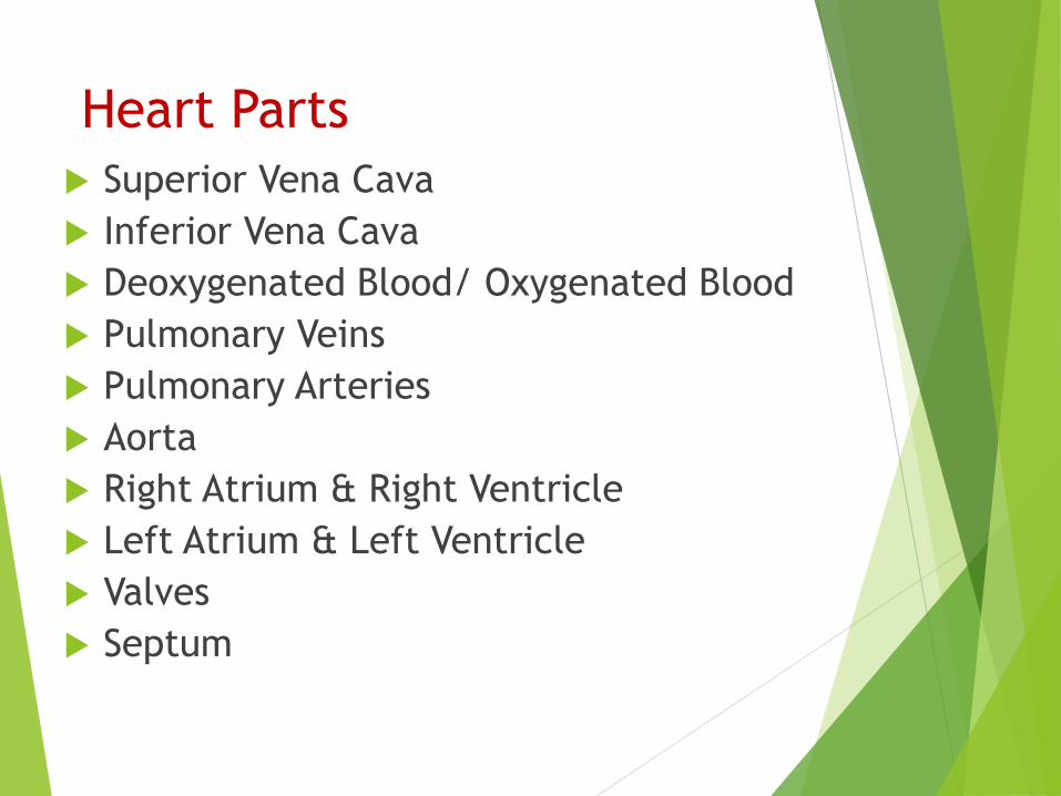
Heart Parts
Superior Vena Cava
Inferior Vena Cava
Deoxygenated Blood/ Oxygenated Blood
Pulmonary Veins
Pulmonary Arteries
Aorta
Right Atrium & Right Ventricle
Left Atrium & Left Ventricle
Valves
Septum

The Parts
Right Atrium – receives deoxygenated blood from all parts of the body.
Right Ventricle– pumps deoxygenated blood to the lungs.
Left atrium – receives oxygen-rich blood from the lungs.
Left ventricle – pumps oxygen –rich blood to all parts of the body.
Atrium- blood enters heart
Ventricle- blood leaves heart
Vena Cava
Superior Vena Cava: Returns blood from upper body to heart
Inferior Vena Cava: Returns blood from lower body to heart
Aorta: Carries blood to the body.

Heart Parts

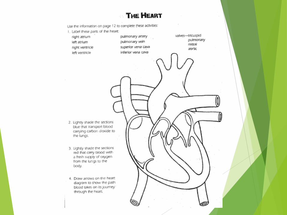
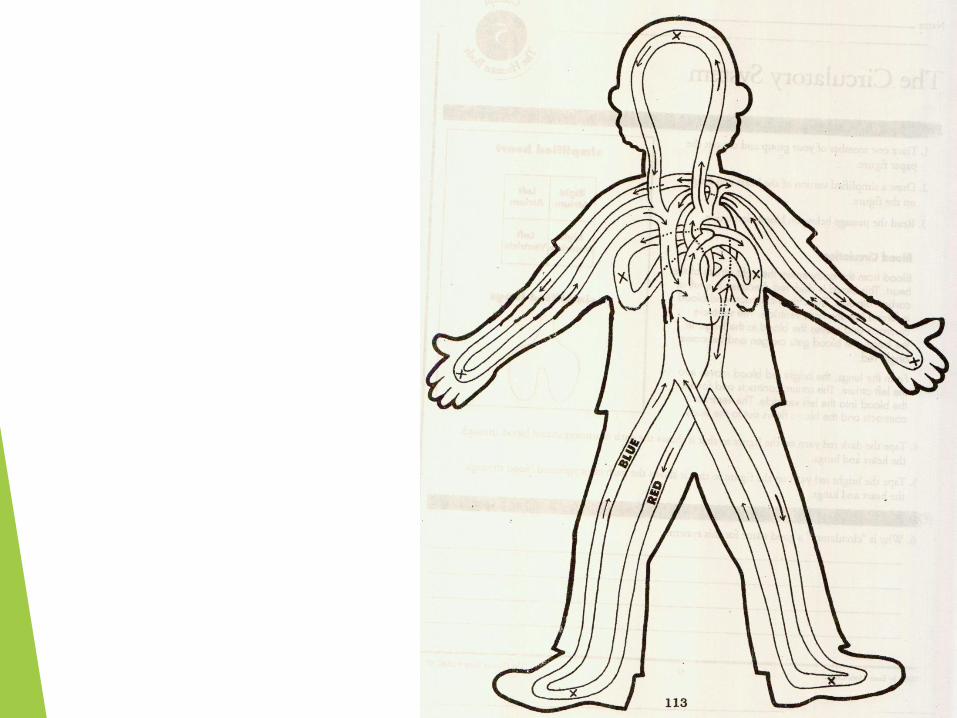
“The Path of Blood”Materials:
• Path of Blood Worksheet
• Blue & red colored pencils,
markers or crayons
• Pencil

“The Path of Blood”1. The Right Atrium (A), which is the upper chamber of
the right side of the heart, receives blood from the
upper body through the Superior Vena Cava (B) and
from the lower body through the Inferior Vena Cava
(C). This blood is blue in color because it is returning
from the body carrying CO2 (waste from the cells)
that was released by body cells as the blood
deposited oxygen.
2. Blood then flows through the Tricuspid Valve (D) into
the Right Ventricle (E), which is the lower chamber
on the right side of the heart.
On your worksheet, label these parts on your heart
in pencil and color them blue.

“The Path of Blood”3. Through contraction of the right ventricle, the blue
blood is forced through the Pulmonary Valve (F) into
the Pulmonary Artery (G). The Pulmonary Artery
(G) branches to both the right and the left lung to pick
up oxygen and release carbon dioxide wastes.
Label these parts on the heart and color them blue
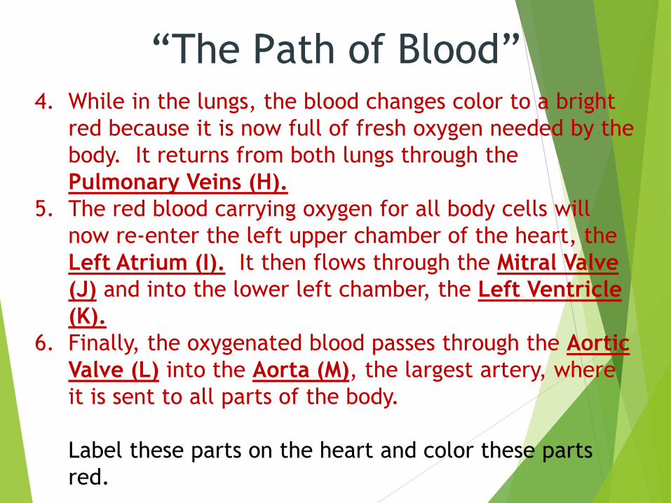
“The Path of Blood”4. While in the lungs, the blood changes color to a bright
red because it is now full of fresh oxygen needed by the
body. It returns from both lungs through the
Pulmonary Veins (H).
5. The red blood carrying oxygen for all body cells will
now re-enter the left upper chamber of the heart, the
Left Atrium (I). It then flows through the Mitral Valve
(J) and into the lower left chamber, the Left Ventricle
(K).
6. Finally, the oxygenated blood passes through the Aortic
Valve (L) into the Aorta (M), the largest artery, where
it is sent to all parts of the body.
Label these parts on the heart and color these parts
red.

Related Documents