UNIVERSIDAD AUTÓNOMA DE MADRID DEPARTMENT OF MOLECULAR BIOLOGY PhD Thesis The chromatin remodeller BPTF is a novel and critical c-MYC co-factor LAIA RICHART GINÉS Madrid, 2015

Welcome message from author
This document is posted to help you gain knowledge. Please leave a comment to let me know what you think about it! Share it to your friends and learn new things together.
Transcript
UNIVERSIDAD AUTÓNOMA DE MADRID
DEPARTMENT OF MOLECULAR BIOLOGY
PhD Thesis
The chromatin remodeller BPTF is a novel and critical c-MYC co-factor
LAIA RICHART GINÉS
Madrid, 2015
UNIVERSIDAD AUTÓNOMA DE MADRID
FACULTY OF SCIENCES
DEPARTMENT OF MOLECULAR BIOLOGY
The chromatin remodeller BPTF is a novel and critical c-MYC co-factor
Doctoral thesis submitted to the Universidad Autónoma
de Madrid for the degree of Doctor of Philosophy by MSc in Biotechnology,
Laia Richart Ginés
Thesis Directors
Prof. Dr. Francisco X. Real Arribas Dr. Victor Javier Sánchez-Arévalo Lobo
EPITHELIAL CARCINOGENESIS GROUP
CELL BIOLOGY PROGRAMME
SPANISH NATIONAL CANCER RESEARCH CENTRE
This thesis, submitted for the degree of Doctor of Philosophy at the Universidad Autónoma de Madrid, has been carried out and completed in
the Epithelial Carcinogenesis Group at the Spanish National Cancer Research Centre (CNIO), under the supervision of
Prof. Dr. Francisco X. Real Arribas and Dr. Victor Javier Sánchez-Arévalo Lobo
The work was supported by grants from Ministerio de Economía y Competitividad, Madrid, Spain (grants Consolíder ONCOBIO, Consolider INESGEN, SAF2010-21517 and SAF2011-15934-E), Instituto de Salud Carlos III (grants G03/174, 00/0745, PI051436, PI061614, G03/174, PI080440, PI120425 and Red Temática de Investigación Cooperativa en Cáncer (RTICC)), Asociación Española Contra el Cáncer, EUFP7-201663 and 201333, and US National Institutes of Health grant RO1 CA089715.

“Quan surts per fer el viatge cap a Ítaca,
has de pregar que el camí sigui llarg,
ple d'aventures, ple de coneixences.
Has de pregar que el camí sigui llarg,
que siguin moltes les matinades
que entraràs en un port que els teus ulls ignoraven,
i vagis a ciutats per aprendre dels que saben.
Tingues sempre al cor la idea d'Ítaca.”
I-Kavafis (adaptació de Lluís Llach)

Dedicat a la meva mare i la meva àvia.

1
Index of Contents
2
INDEX OF CONTENTS
SUMMARY ................................................................................ 6
PRESENTACIÓN ......................................................................... 8
DIRECTORY OF TABLES ............................................................ 10
DIRECTORY OF FIGURES .......................................................... 12
ABBREVIATIONS ................................................................ 14-15
INTRODUCTION ................................................................. 17-36
1. THE ROLE OF CHROMATIN DURING TRANSCRIPTION ............. 17
1.1. Nucleosomes are the basic unit of chromatin ............................. 17
1.2. Histone covalent modifications ................................................... 18
1.3. Chromatin remodelling ................................................................ 19
1.4. Histone variants ........................................................................... 20
1.5. Transcription in the chromatin context ....................................... 21
2. c-MYC .................................................................................... 22
2.1. Protein structure and interaction partners ................................. 22
2.2. c-MYC control of gene transcription ........................................... 23
2.2.1. Widespread binding to chromatin........................................ 24
2.2.2. Transcriptional activation .................................................... 25
2.2.3. Transcriptional repression .................................................... 27
2.3. c-MYC biological roles .................................................................. 28
2.3.1. Cell proliferation and differentiation .................................... 28
2.3.2. Cell growth and metabolism ................................................ 30
2.3.3. Apoptosis .............................................................................. 30
2.3.4. Tumorigenesis....................................................................... 30
2.3.5. Reprogramming .................................................................... 31
3. BPTF ...................................................................................... 32
3.1. Protein structure and interactors ................................................ 33
3.2. Biological function of BPTF .......................................................... 34
3.2.1. Transcriptional activator and repressor ............................... 34
3.2.2. Chromatin structure ............................................................. 35
3.2.3. Developmental regulator ..................................................... 35
3.3. BPTF in human cancer ................................................................. 36
AIMS ...................................................................................... 38
3
OBJETIVOS .............................................................................. 40
MATERIALS AND METHODS ............................................... 42-55
1. CELL CULTURE ........................................................................ 42
1.1. Cell lines and reagents ................................................................. 42
1.2. Plasmids, viral constructs and virus production .......................... 42
1.3. iPS Reprogramming ..................................................................... 43
1.4. FACS analysis of proliferation and apoptosis .............................. 44
2. MOUSE BIOLOGY ................................................................... 44
2.1. Mouse strains ............................................................................... 44
2.2. Histopathology and Immunohistochemistry ............................... 45
2.3. Hematological analysis and characterization of B cell
Compartment ...................................................................................... 46
3. MOLECULAR BIOLOGY ........................................................... 46
3.1. Western blotting .......................................................................... 46
3.2. Co-immunoprecipitation analyses ............................................... 47
3.3. Generation of polyclonal anti-BPTF anti-sera ............................. 47
3.4. Immunofluorescence staining and Proximity Ligation Assay ...... 47
3.5. Quantitative real-time PCR .......................................................... 48
3.6. Chromatin immunoprecipitation ................................................. 50
3.7. DNAse I hypersensitivity assay .................................................... 51
4. GENOME-WIDE STUDIES AND BIOINFORMATICS ANALYSES ... 51
4.1. ChIP-Seq library construction and massive parallel sequencing . 51
4.2. ChIP-Seq data processing............................................................. 51
4.3. Motif enrichment analysis, peak annotation and density
plot analysis ........................................................................................ 52
4.4. Gene set enrichment analysis (GSEA) .......................................... 52
4.5. RNA-seq........................................................................................ 52
4.6. RNA-Seq data processing ............................................................. 53
4.7. RNA-Seq GSEA analysis ................................................................ 53
4.8. Analysis of human tumor genomic data ...................................... 54
5. STATISTICAL ANALYSIS ........................................................... 54
6. INDIVIDUAL CONTRIBUTIONS ................................................ 54
RESULTS............................................................................. 56-84
1. BPTF IS REQUIRED FOR IN VITRO PROLIFERATION
OF TUMOR CELLS ....................................................................... 57
4
2. BPTF IS MODULATED DURING CELL CYCLE PROGRESSION AND IS
REQUIRED FOR G0-G1/S TRANSITION ......................................... 58
3. BPTF IS NECESSARY FOR c-MYC TRANSCRIPTIONAL ACTIVITY 60
4. BPTF AND c-MYC INTERACT IN VITRO .................................... 64
5. GENOME-WIDE ANALYSIS OF c-MYC RECRUITMENT TO DNA
UPON BPTF KNOCK-DOWN ........................................................ 65
6. BPTF IS REQUIRED FOR c-MYC-INDUCED REMODELLING
OF TARGET CHROMATIN ............................................................ 69
7. BPTF IS REQUIRED FOR A SUBSET OF c-MYC BIOLOGICAL
FUNCTIONS................................................................................ 71
8. BPTF IS REQUIRED FOR THE REPROGRAMMING OF MOUSE
EMBRYONIC FIBROBLASTS ......................................................... 73
9. BPTF CORRELATES WITH c-MYC SIGNATURES IN
HUMAN CANCER ....................................................................... 77
10. BPTF IS REQUIRED FOR c-MYC-DRIVEN PANCREATIC
TUMORIGENESIS ....................................................................... 82
DISCUSSION ..................................................................... 85-104
1. BPTF AND CELL PROLIFERATION ............................................. 86
2. BPTF AND c-MYC AXIS ............................................................ 88
2.1. c-MYC recruitment to DNA and/or stability of the complex ....... 88
2.2. Remodelling of c-MYC target chromatin ..................................... 89
2.3. Long-range interactions ............................................................... 90
2.4. Transcription elongation .............................................................. 93
2.5. Repression by c-MYC ................................................................... 93
3. BPTF IN DEVELOPMENT AND DIFFERENTIATION .................... 94
3.1. Early embryonic development ..................................................... 94
3.2. Cell differentiation ....................................................................... 94
4. BPTF AND TUMORIGENESIS ................................................... 97
CONCLUSIONS ...................................................................... 102
CONCLUSIONES .................................................................... 104
REFERENCES ................................................................... 105-125

5
Summary
6
SUMMARY
c-MYC is a major oncogene involved in human cancer. Here, I have identified
BPTF as a novel interactor of c-MYC required for its biological functions. This
interaction is crucial for c-MYC transcriptional activity: BPTF knock-down leads to
a decrease in c-MYC binding to DNA, changes in chromatin accessibility, and
impaired activation of the c-MYC transcriptional program. In murine embryonic
fibroblasts, BPTF is necessary for c-MYC-driven proliferation, G1-S progression,
and replication stress, but not for c-MYC-induced apoptosis. Moreover, BPTF is
critical for reprogramming of somatic cells into induced pluripotent stem cells
using the four Yamanaka factors. In agreement with these findings, BPTF is
required for the proliferation of c-MYC-addicted cancer cells and in human
tumors its expression positively correlates with the activation of c-MYC gene
signatures.
To determine whether BPTF is required for the oncogenic effects of c-MYC, we
used two genetic mouse models: Ela-Myc and E-Myc. Ela-Myc mice develop
aggressive acinar and ductal tumors. While BPTF is dispensable for normal
pancreatic development and differentiation, its embryonic inactivation in Ela-
Myc mice is associated with extensive loss of acinar cells. Moreover, deletion of
BPTF in young Ela-Myc via activation of the Ptf1a-CreERT2 recombinase results in
a significant delay in tumor onset and a corresponding extension in disease-free
survival. c-MYC overexpression in the B cell lineage (E-Myc) leads to the
development of Burkitt-like lymphomas. Inactivation of one Bptf allele does not
impair B cell maturation but completely blocks lymphoma development. These
findings underscore the importance of a more detailed study of BPTF function in
mammals and highlight the potential of exploiting the c-MYC:BPTF axis in cancer
therapy.

7
Presentación
8
PRESENTACIÓN
c-MYC es uno de los principales oncogenes implicados en el cáncer humano. En
el presente trabajo he identificado a BPTF como un nuevo interactor de c-MYC
que además es requerido para sus funciones biológicas. Esta interacción es crucial
para la actividad transcripcional de c-MYC: el knock-down de BPTF se acompaña
de una disminución de la unión de c-MYC al DNA, cambios en la accesibilidad de
la cromatina y de una inadecuada activación del programa transcripcional de c-
MYC. En fibroblastos embrionarios de ratón, BPTF es necesario para la
proliferación, progresión G1-S y estrés replicativo dirigidos por c-MYC, pero no
para la apoptosis instruida por el mismo. Además, BPTF es crítico para la
reprogramación de células somáticas a células madre pluripotentes por medio de
los cuatro factores descritos por Yamanaka. De acuerdo con estas observaciones,
BPTF es necesario para la proliferación de líneas cancerosas adictas a c-MYC, y en
tumores humanos su expresión correlaciona positivamente con la activación de
los programas de expresión génica dirigidos por c-MYC.
Con el objetivo de determinar si BPTF es necesario para los efectos oncogénicos
de c-MYC, hemos usado dos modelos genéticos de ratón: Ela-Myc y E-Myc. Los
ratones Ela-Myc desarrollan tumores acinares y ductales muy agresivos. Aunque
BPTF es dispensable para la diferenciación y desarrollo pancreáticos normales, su
inactivación embrionaria en ratones Ela-Myc se asocia con una extensa pérdida
de células acinares. Además, la depleción de BPTF en ratones Ela-Myc jóvenes por
medio de la recombinasa Ptf1a-CreERT2 resulta en un retraso significativo en la
aparición de los tumores y en una consiguiente extensión de la supervivencia libre
de enfermedad. La sobre-expresión de c-MYC en el compartimento de células B
conduce al desarrollo de linfomas que reproducen la enfermedad del Linfoma de
Burkitt. La inactivación de una sola copia de Bptf no afecta a la maduración de las
células B pero bloquea por completo la formación de tumores. Estas
observaciones destacan la importancia de un estudio más detallado de la función
de BPTF en mamíferos y subrayan el potencial de explotar el eje c-MYC:BPTF
como blanco terapéutico en cáncer.

9
Directory of Figures
10
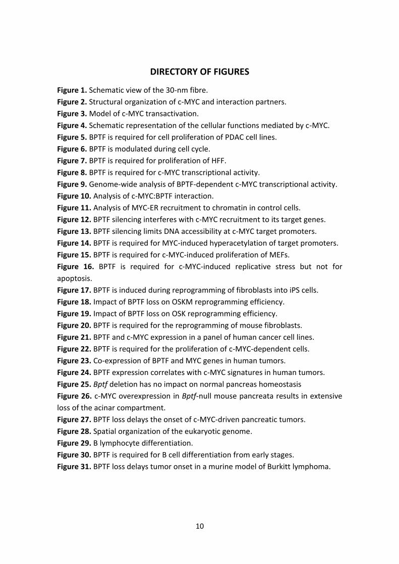
DIRECTORY OF FIGURES
Figure 1. Schematic view of the 30-nm fibre.
Figure 2. Structural organization of c-MYC and interaction partners.
Figure 3. Model of c-MYC transactivation.
Figure 4. Schematic representation of the cellular functions mediated by c-MYC.
Figure 5. BPTF is required for cell proliferation of PDAC cell lines.
Figure 6. BPTF is modulated during cell cycle.
Figure 7. BPTF is required for proliferation of HFF.
Figure 8. BPTF is required for c-MYC transcriptional activity.
Figure 9. Genome-wide analysis of BPTF-dependent c-MYC transcriptional activity.
Figure 10. Analysis of c-MYC:BPTF interaction.
Figure 11. Analysis of MYC-ER recruitment to chromatin in control cells.
Figure 12. BPTF silencing interferes with c-MYC recruitment to its target genes.
Figure 13. BPTF silencing limits DNA accessibility at c-MYC target promoters.
Figure 14. BPTF is required for MYC-induced hyperacetylation of target promoters.
Figure 15. BPTF is required for c-MYC-induced proliferation of MEFs.
Figure 16. BPTF is required for c-MYC-induced replicative stress but not for
apoptosis.
Figure 17. BPTF is induced during reprogramming of fibroblasts into iPS cells.
Figure 18. Impact of BPTF loss on OSKM reprogramming efficiency.
Figure 19. Impact of BPTF loss on OSK reprogramming efficiency.
Figure 20. BPTF is required for the reprogramming of mouse fibroblasts.
Figure 21. BPTF and c-MYC expression in a panel of human cancer cell lines.
Figure 22. BPTF is required for the proliferation of c-MYC-dependent cells.
Figure 23. Co-expression of BPTF and MYC genes in human tumors.
Figure 24. BPTF expression correlates with c-MYC signatures in human tumors.
Figure 25. Bptf deletion has no impact on normal pancreas homeostasis
Figure 26. c-MYC overexpression in Bptf-null mouse pancreata results in extensive
loss of the acinar compartment.
Figure 27. BPTF loss delays the onset of c-MYC-driven pancreatic tumors.
Figure 28. Spatial organization of the eukaryotic genome.
Figure 29. B lymphocyte differentiation.
Figure 30. BPTF is required for B cell differentiation from early stages.
Figure 31. BPTF loss delays tumor onset in a murine model of Burkitt lymphoma.

11
Directory of Tables

12
TABLES
Table 1. Histone modifications, writers, readers and their function.
Table 2. Representative mouse models used to study c-MYC function.
Table 3. Summary of published NURF interactions with transcription factors.
Table 4. List of RT-qPCR primers used in this study
Table 5. List of ChIP-qPCR primers used in this study
Table 6. Top-ranking gene sets enriched in 4-OHT-treated control cells.
Table 7. Summary of human tumor datasets.

13
Abbreviations
14
ABBREVIATIONS
AP: Anterior-Posterior
bHLH-LZ: Basic Helix-Loop-Helix Leucine Zipper
BL: Burkitt Lymphoma
BM: Bone Marrow
BPTF: Bromodomain PHD Transcription Factor
BRCT: BRCA1 C-Terminus domain
BRD: Bromodomain
CDK: Cyclin-Dependent Kinase
CHD: Chromodomain Helicase DNA-binding
ChIP: Chromatin Immunoprecipitation
CLP: Common Lymphoid Progenitors
DDT: DNA binding homeobox and Different Transcription factors
DNA: Deoxyribonucleic Acid
DRD2: Dopamine Receptor D2
DSB: Double Strand Break
ESc: Embryonic Stem cells
FALZ: Fetal Alzheimer Antigen (also known as FAC1)
FDR: False Discovery Rate
GSEA: Gene Set Enrichment Analysis
GSK3: Glycogen Synthase Kinase-3
GTFs: General Transcription Factors
HAT: Histone Acetyl Transferases
HDAC: Histone De-acetylase
HFF: Human Foreskin Fibroblasts
HLH: Helix-Loop-Helix
HSC: Hematopoietic Stem Cells
INO80: INositol requiring 80
isPLA: In situ Proximity Ligation Assay
ISWI: Imitation SWItch
IUIM: Inverted Ubiquitin Interaction Motif
LDHA: Lactate DeHydrogenase A
LZ: Leucine-Zipper
MAPK: Mitogen-Activated Protein Kinase
MBD: Methyl-CpG-binding Domain
MBI-IV: MYC boxes
MBT: Malignant Brain Tumor domain
MEF: Murine Embryonic Fibroblast
mRNA: messenger RNA
15
MSigDB: Molecular Signature DataBase
NES: Normalized Enrichment Score
NLS: Nuclear Localization Sequence
NuRD: Nucleosome Remodelling and Deacetylation
NURF: Nucleosome Remodelling Factor
ORC: Origin Recognition Complex
OSK: Oct4, Sox2 and Klf4
PBZ: Poly ADP-ribose Binding Zinc finger
PHD: Plant Homeodomain
PIC: Pre-Initiation Complex
Pol II: RNA Polymerase II
PSEN1: Presenilin 1
P-TEFb: Positive Transcription Elongation Factor b
PWWP: Proline-tryptophan-tryptophan-Proline domain
rRNA: ribosomal RNA
SEM: Standard Error of Mean
SIM: Sumo Interaction Motif
SWI/SNF: SWItching defective/Sucrose Non-Fermenting
TAD: Transactivation domain (when referred to c-MYC) / Topologically Associated
Domain (when referred to chromatin organization)
TBP: TATA-Binding Protein
tRNA: transfer RNA
TSS: Transcription Start Site
UIM: Ubiquitin Interaction Motif
WT: Wild Type

16
Introduction
17
INTRODUCTION
1. THE ROLE OF CHROMATIN DURING TRANSCRIPTION
1.1. Nucleosomes are the basic unit of chromatin
Chromatin is the complex of DNA, histones, and non-histone proteins from
which eukaryotic chromosomes are formed. The nucleosome is the primary unit
of chromatin and is composed of 147 bp of DNA wrapped 1.65 times around an
octamer of the four core histones (H2A, H2B, H3, and H4). Structurally, core
histones are relatively small proteins with a globular domain (the histone fold)
and two N-terminal “tails”. Consecutive nucleosome core particles are separated
by unwrapped linker DNA of variable length (20-90 bp). In addition, one molecule
of histone H1 associates at the position where the DNA enters and exits the
nucleosome core, thus sealing the two turns of DNA (Laybourn and Kadonaga
1991). The multiple contact points between histones and DNA make the
nucleosome a very stable complex and, for this reason, it is well suited for its
packaging function. Nonetheless, its role extends beyond DNA compaction and
occlusion. Nucleosomes are also dynamic participants in chromatin-directed
processes such as transcription, replication, DNA repair, kinetochore and
centromere construction, and telomere maintenance (Saha et al. 2006). Cells
modulate the way chromatin is packed in order to regulate such processes. This
involves the dynamic competition between nucleosomes and DNA-binding
factors for regulatory sequences in the DNA (Li et al. 2007). This competition is
mainly influenced by three different types of protein complexes. One family
includes ATP-dependent remodelling complexes that weaken DNA-histone
interactions, thereby facilitating nucleosome repositioning, reconfiguration or
ejection (Kingston and Narlikar 1999). Another family includes chromatin-
modifying enzymes that add or remove covalent modifications at particular
residues within histones. The third family is constituted by the DNA
methyltransferases (DNMTs) that methylate cytosines within CpG dinucleotides
and thus regulate transcription, high-order chromatin structures and genome
syability (Espada and Esteller 2010). Of note, histone modifying complexes and
DNMTs work in concert with chromatin-remodelling complexes. Thus, the
chromatin fibre is a dynamic and flexible structure that continuously changes in
response to a wide range of biological inputs (Zhang and Reinberg 2001).
The linear string of nucleosomes (“beads on a string”) is further packed into a
30-nm fibre where nucleosomes are arranged in a spiral or solenoid (Hayes and
18
Hansen 2001). The histone tails, although dispensable for the formation of the
nucleosome, are required for inter-nucleosomal interactions and, together with
histone H1, help condensing the DNA (Luger et al. 1997). Additional levels of
compaction enable these fibres to be packaged into the small volume of the
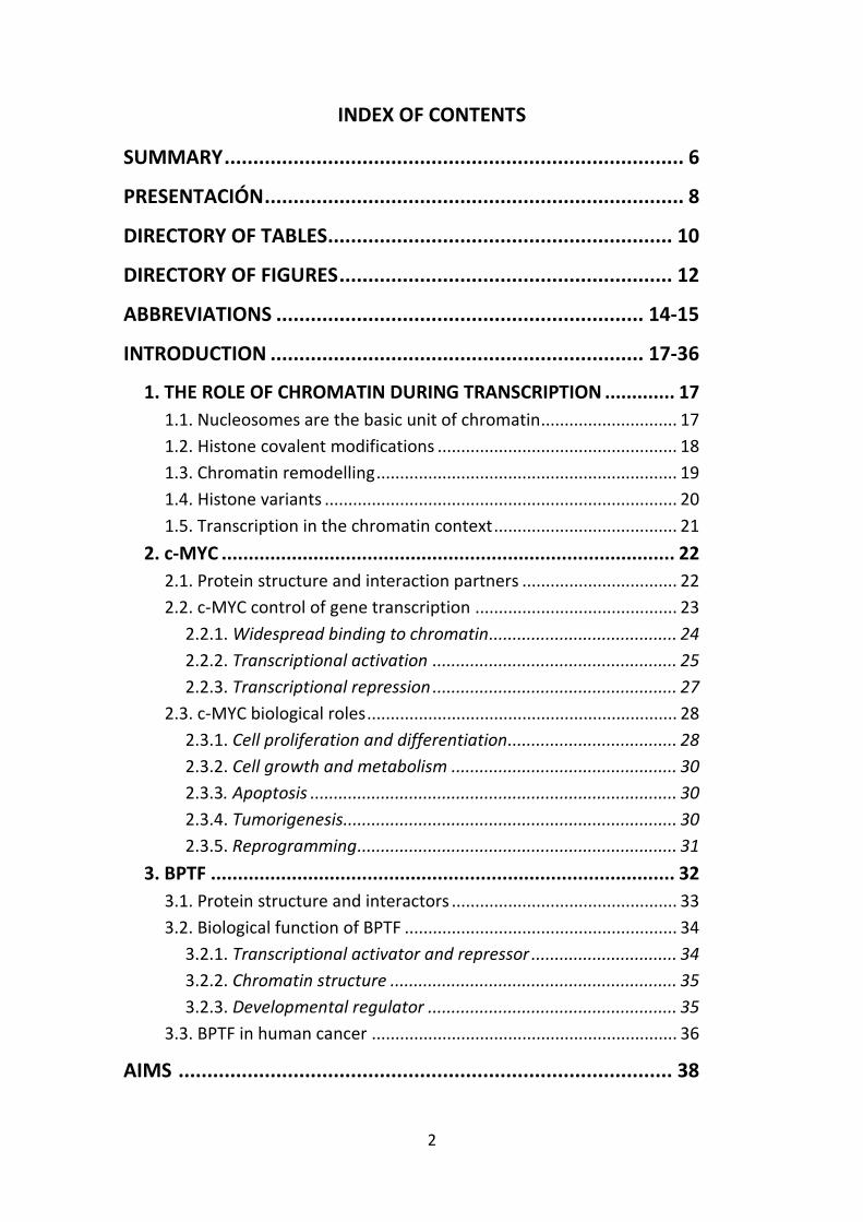
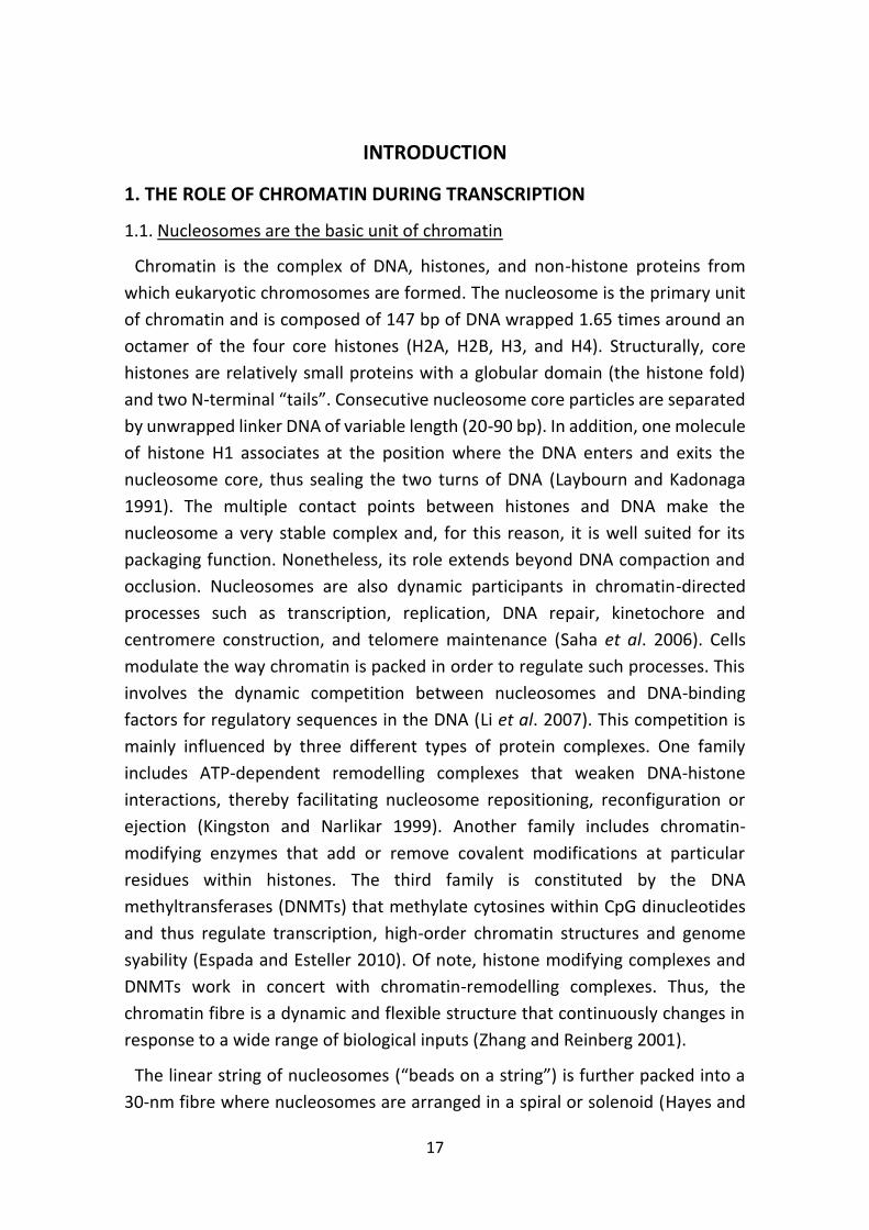
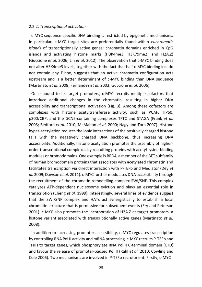
nucleus (Fig. 1).
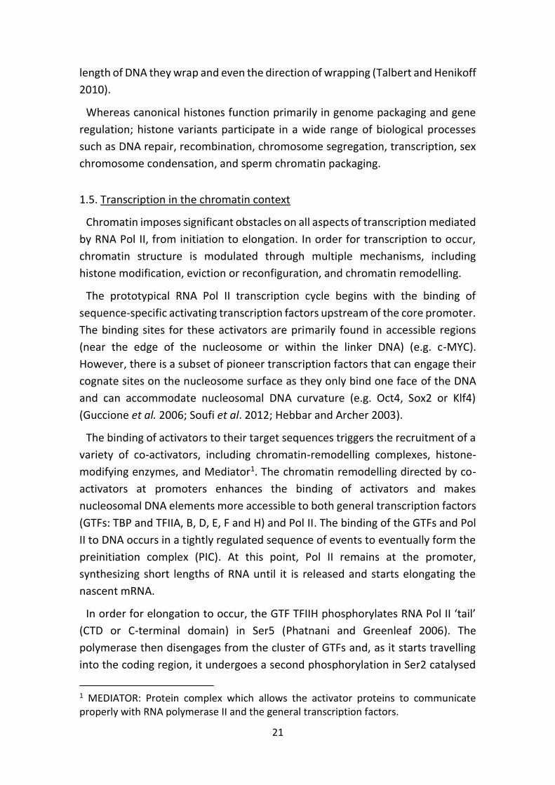
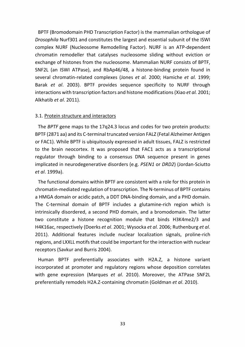
Figure 1. Schematic view of the 30-nm fibre. Sequence-specific DNA-binding factors
bind to accessible regions in the linker DNA, the edge of the nucleosome or in
remodelled nucleosomes. Regions of chromatin that are nucleosome-free or contain
remodelled nucleosomes can often be detected experimentally by the unusually high
susceptibility of their DNA to digestion by nucleases - as compared with the DNA in
nucleosomes. Adapted from Alberts et al 2002.
1.2. Histone covalent modifications
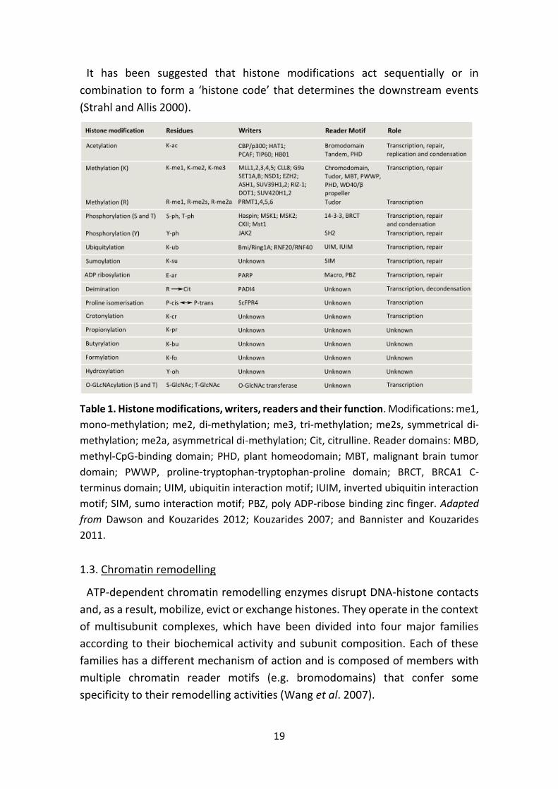
Core histones are susceptible to a wide variety of post-translational
modifications (up to 130), including methylation, acetylation, ubiquitination,
ADP-ribosylation, sumoylation or phosphorylation (Kouzarides 2007; Tan et al.
2011) (Table 1). The majority of modifications take place at the N-terminal tails
of histones, with a few exceptions occurring within the globular regions (e.g.
phosphorylation of H3Y41) (Dawson et al. 2009). The distribution of these
modifications is tightly regulated and is crucial for their functional outcome.
Histone modifications serve two main functions. First (with the exception of
methylation), they alter the net charge of histones and thus enhance or loosen
the non-covalent interactions within and between nucleosomes. Second, they
serve as docking sites for the recruitment of epigenetic readers with unique
domains that specifically recognize these modifications. These chromatin readers
recruit in turn additional chromatin modifiers and remodelling enzymes, which
perform diverse chromatin functions (Dawson and Kouzarides 2012).
19
It has been suggested that histone modifications act sequentially or in
combination to form a ‘histone code’ that determines the downstream events
(Strahl and Allis 2000).
Table 1. Histone modifications, writers, readers and their function. Modifications: me1,
mono-methylation; me2, di-methylation; me3, tri-methylation; me2s, symmetrical di-
methylation; me2a, asymmetrical di-methylation; Cit, citrulline. Reader domains: MBD,
methyl-CpG-binding domain; PHD, plant homeodomain; MBT, malignant brain tumor
domain; PWWP, proline-tryptophan-tryptophan-proline domain; BRCT, BRCA1 C-
terminus domain; UIM, ubiquitin interaction motif; IUIM, inverted ubiquitin interaction
motif; SIM, sumo interaction motif; PBZ, poly ADP-ribose binding zinc finger. Adapted
from Dawson and Kouzarides 2012; Kouzarides 2007; and Bannister and Kouzarides
2011.
1.3. Chromatin remodelling
ATP-dependent chromatin remodelling enzymes disrupt DNA-histone contacts
and, as a result, mobilize, evict or exchange histones. They operate in the context
of multisubunit complexes, which have been divided into four major families
according to their biochemical activity and subunit composition. Each of these
families has a different mechanism of action and is composed of members with
multiple chromatin reader motifs (e.g. bromodomains) that confer some
specificity to their remodelling activities (Wang et al. 2007).
20
1. ISWI (Imitation Switch): With the exception of NURF and Iswi1, ISWI
remodelling complexes slide nucleosomes in an orderly manner to repress gene
transcription (Badenhorst et al. 2002; Morillon et al. 2003). In addition, they play
key roles in chromatin assembly after DNA replication and maintenance of
higher-order chromatin structures (Erdel and Rippe 2011).
2. SWI/SNF (SWItching defective/Sucrose Non-Fermenting): SWI complexes
catalyse the sliding or ejection of nucleosomes (in part or as a whole) with the
help of histone chaperones. Their function correlates with nucleosome
disorganization, increased accessibility for transcription factor binding, and gene
activation (Saha et al. 2006). Members of this family have also been implicated in
DNA repair following DNA damage (Chai et al. 2005; Shim et al. 2007).
3. INO80/SWR1 (INositol requiring 80): INO80 complexes have both activating
and repressive effects on gene transcription. SWR1 complexes promote the
incorporation of the histone variant H2A.Z into nucleosomes in a replication-
independent manner (Mizuguchi et al. 2004). H2A.Z differs from canonical H2A
in its amino acid sequence and stability, which depends on the histone H3
subtype present in the histone octamer. Hybrid nucleosomes containing both
H2A.Z and the histone variant H3.3 are more unstable and prone to movement
or ejection by chromatin remodellers (Jin and Felsenfeld 2007). In human cells,
H2A.Z is preferentially enriched at poised promoters. Upon transcriptional
activation, H2A.Z is rapidly evicted and its loss is required for full transcription
(Zhang et al. 2005).
4. NuRD/Mi-2/CHD (Nucleosome Remodelling and Deacetylation/Mi-2/
Chromodomain Helicase DNA-binding): Members of this family primarily mediate
transcriptional repression.
1.4. Histone variants
Nucleosomes are constructed from the four canonical histones (H2A, H2B, H3,
and H4) or, alternatively, from histone variants with specific expression,
localization, and species-distribution patterns (e.g. H3.3, macroH2A, H2A.Z,
H2ABbd or H2A.X) (Kamakaka and Biggins 2005).
The genes encoding the four canonical histones cluster together in the genome
and are transcribed during S phase. Conversely, genes encoding non-canonical
histones are found singly in the genome and are constitutively expressed. Histone
variants differ in their primary amino acid sequence from their canonical
paralogues. These differences impact on their structure, intrinsic stability, the
21
length of DNA they wrap and even the direction of wrapping (Talbert and Henikoff
2010).
Whereas canonical histones function primarily in genome packaging and gene
regulation; histone variants participate in a wide range of biological processes
such as DNA repair, recombination, chromosome segregation, transcription, sex
chromosome condensation, and sperm chromatin packaging.
1.5. Transcription in the chromatin context
Chromatin imposes significant obstacles on all aspects of transcription mediated
by RNA Pol II, from initiation to elongation. In order for transcription to occur,
chromatin structure is modulated through multiple mechanisms, including
histone modification, eviction or reconfiguration, and chromatin remodelling.
The prototypical RNA Pol II transcription cycle begins with the binding of
sequence-specific activating transcription factors upstream of the core promoter.
The binding sites for these activators are primarily found in accessible regions
(near the edge of the nucleosome or within the linker DNA) (e.g. c-MYC).
However, there is a subset of pioneer transcription factors that can engage their
cognate sites on the nucleosome surface as they only bind one face of the DNA
and can accommodate nucleosomal DNA curvature (e.g. Oct4, Sox2 or Klf4)
(Guccione et al. 2006; Soufi et al. 2012; Hebbar and Archer 2003).
The binding of activators to their target sequences triggers the recruitment of a
variety of co-activators, including chromatin-remodelling complexes, histone-
modifying enzymes, and Mediator1. The chromatin remodelling directed by co-
activators at promoters enhances the binding of activators and makes
nucleosomal DNA elements more accessible to both general transcription factors
(GTFs: TBP and TFIIA, B, D, E, F and H) and Pol II. The binding of the GTFs and Pol
II to DNA occurs in a tightly regulated sequence of events to eventually form the
preinitiation complex (PIC). At this point, Pol II remains at the promoter,
synthesizing short lengths of RNA until it is released and starts elongating the
nascent mRNA.
In order for elongation to occur, the GTF TFIIH phosphorylates RNA Pol II ‘tail’
(CTD or C-terminal domain) in Ser5 (Phatnani and Greenleaf 2006). The
polymerase then disengages from the cluster of GTFs and, as it starts travelling
into the coding region, it undergoes a second phosphorylation in Ser2 catalysed
1 MEDIATOR: Protein complex which allows the activator proteins to communicate properly with RNA polymerase II and the general transcription factors.
22
by the TAK/P-TEFb/CDK9 complex (Marshall et al. 1996). These events signal the
recruitment of the elongation machinery (factors involved in polymerization,
mRNA processing, and export) and couple them with alterations in chromatin
function. One example is PAF, an elongation factor associated with Ser5-
phosphorylated CTD that controls the binding of chromatin regulators, such as
the H3K4 methyltransferase Set1, the histone ubiquitin ligase Rad6 or the
chromatin-remodelling factor CHD1 (Li et al. 2007).
2. c-MYC
MYC genes are key modulators of cell proliferation and their deregulation
contributes to almost every aspect of tumor cell biology (Adhikary and Eilers
2005). In mammals, the main MYC family constituents are c-MYC, N-MYC, and L-
MYC, and they all share significant similarity in their genomic, RNA, and protein
sequences. c-MYC was the first to be discovered as the cellular homolog of the
transforming gene of the avian myelocytomatosis virus (Vennstrom et al. 1982).
Despite the enormous progress done during these past 30 years of research,
many aspects of c-MYC biology remain elusive (Wolf et al. 2014).
2.1. Protein structure and interaction partners
The gene coding for c-MYC is located on the human chromosome 8q24 and is
comprised of three exons. The predominant product is c-MYC (also known as
p64); however, alternative translational initiation gives rise to two additional
naturally-occurring translation products: p67 and S-MYC (Hann et al. 1984;
Sugiyama et al. 1989). A distinct function for p67 is not known, but the shorter S-
MYC appears to play a role in stress response and might act as a dominant-
negative MYC (Spotts et al. 1997).
The N-terminus of c-MYC contains an unstructured transactivation domain
(TAD) which spans two highly conserved sequences known as MYC boxes (MBI
and MBII). The TAD domain is followed by MYC boxes III and IV and a nuclear
localization signal (Sarid et al. 1987; Fladvad et al. 2005; Cowling et al. 2006). MYC
boxes participate in protein-protein interactions with E3 ubiquitin ligases that
regulate c-MYC protein stability (FBW7 and SKP2) (Yada et al. 2004, Kim et al.
2003), together with co-factors that modulate its transcriptional activity. The
latter include histone acetyltransferases or HATs (GCN5/PCAF, TIP60, and
CBP/p300), the histone exchange factor p400, and components of the basal
transcriptional machinery such as Mediator and P-TEFb (McMahon et al. 2000;
23
Frank et al. 2003; Kanazawa et al. 2003; Faiola et al. 2005; Martinato et al. 2008;
Liu et al. 2008a). Two residues within the N-terminal domain of c-MYC control its
stability: Ser62 (S62) and Thr58 (T58). Phosphorylation of S62 by mitogen-
activated kinases stabilizes the protein, whereas phosphorylation of T58 by GSK3
(Glycogen Synthase Kinase-3) marks it for proteosomal degradation (Sears et al.
2000; Gregory et al. 2003). The C-terminus contains a basic DNA-binding domain
tethered to a HLH-LZ motif involved in the dimerization with MAX (Blackwood
and Eisenman 1991). In addition to its classical chromatin-recruitment role, the
C-terminal domain of c-MYC is also involved in transactivation through the
recruitment of the H3K27 acetyltransferase CBP/p300 and the nucleosome
remodeler SWI/SNF (Cheng et al. 1999; Park et al. 2002; Vervoorts et al. 2003)
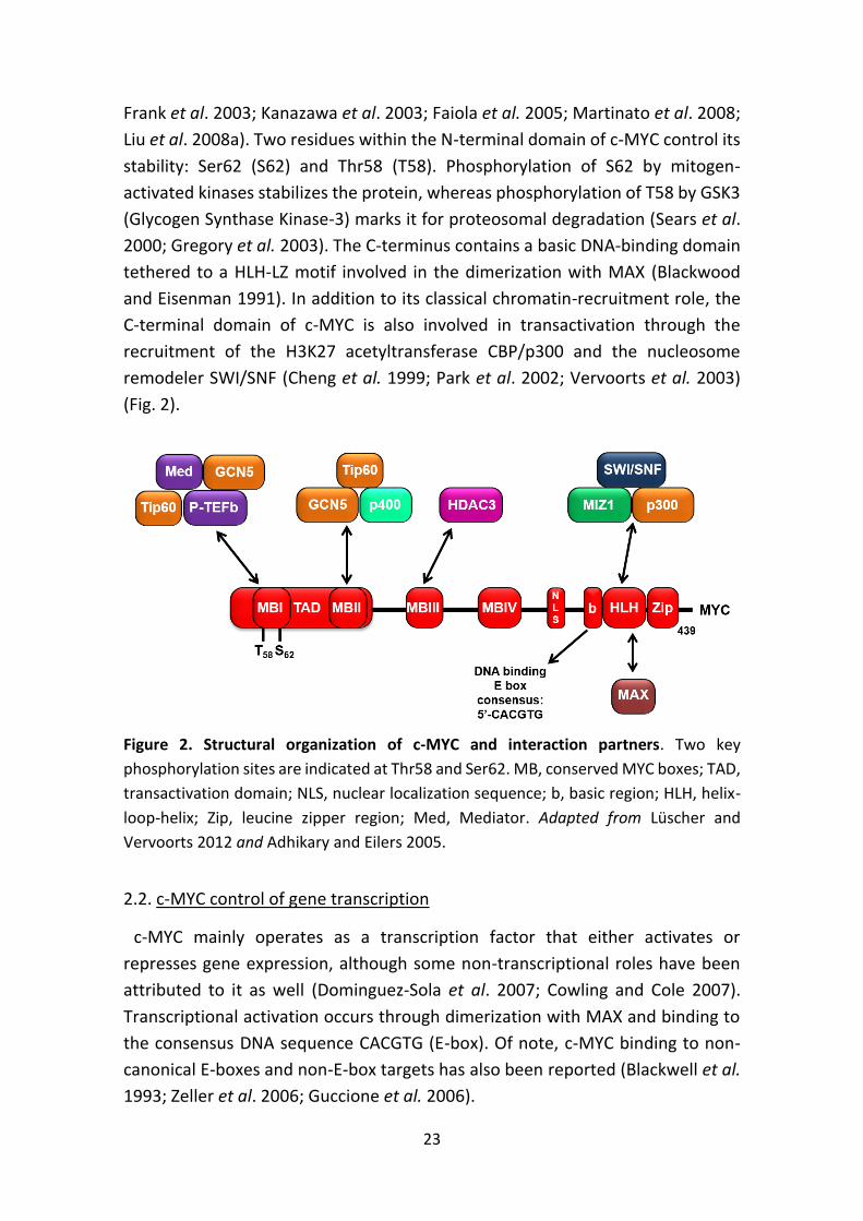
(Fig. 2).
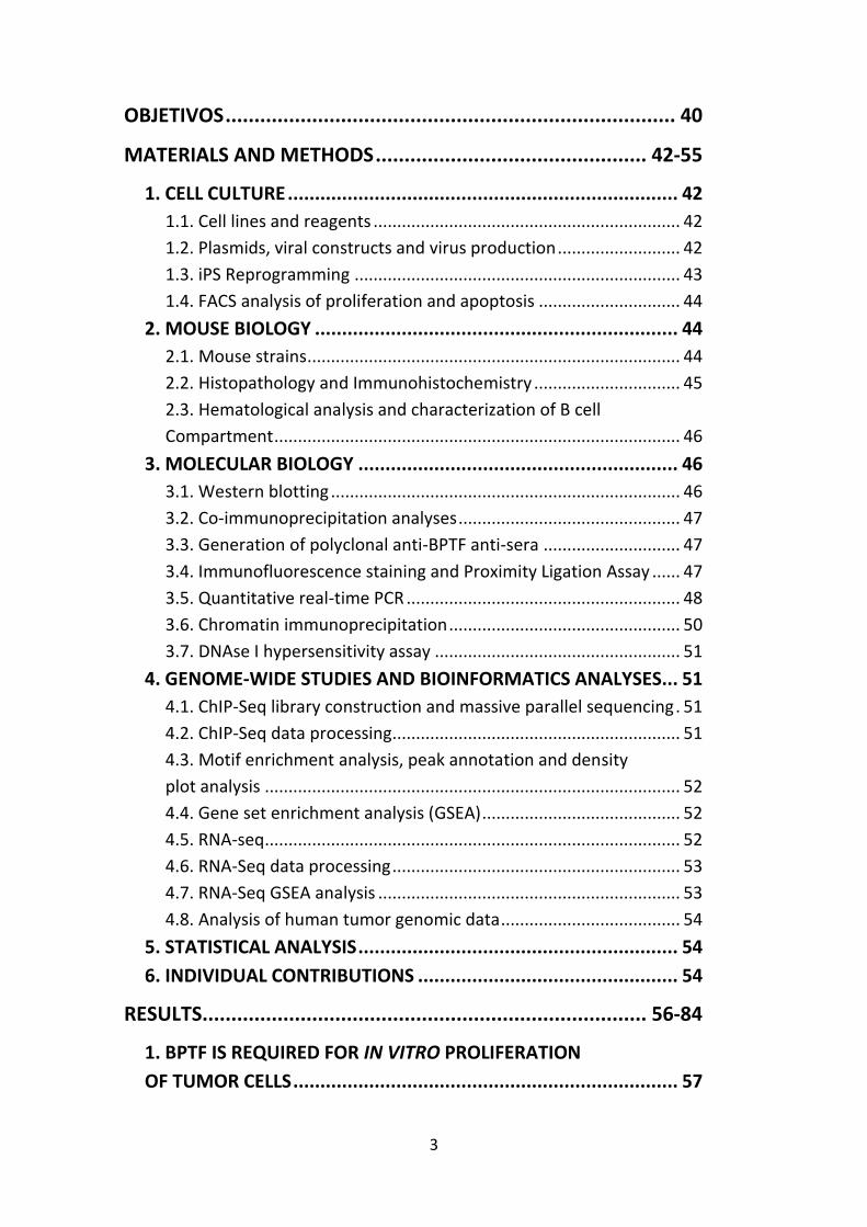
Figure 2. Structural organization of c-MYC and interaction partners. Two key
phosphorylation sites are indicated at Thr58 and Ser62. MB, conserved MYC boxes; TAD,
transactivation domain; NLS, nuclear localization sequence; b, basic region; HLH, helix-
loop-helix; Zip, leucine zipper region; Med, Mediator. Adapted from Lüscher and
Vervoorts 2012 and Adhikary and Eilers 2005.
2.2. c-MYC control of gene transcription
c-MYC mainly operates as a transcription factor that either activates or
represses gene expression, although some non-transcriptional roles have been
attributed to it as well (Dominguez-Sola et al. 2007; Cowling and Cole 2007).
Transcriptional activation occurs through dimerization with MAX and binding to
the consensus DNA sequence CACGTG (E-box). Of note, c-MYC binding to non-
canonical E-boxes and non-E-box targets has also been reported (Blackwell et al.
1993; Zeller et al. 2006; Guccione et al. 2006).
24
The interaction with MAX is required for many of c-MYC biological functions,
although c-MYC appears to function in the absence of a functional MAX protein
in PC12 cells and in Drosophila (Hopewell et al. 1995; Steiger et al. 2008).
MAX also binds bHLH-LZ-containing proteins of the MAD family and the resulting
dimers recognize the same consensus E-boxes as c-MYC:MAX. MAD proteins
antagonize c-MYC function by competing with c-MYC proteins for free MAX,
competing with c-MYC:MAX dimers for available binding sites, and recruiting
repressor complexes such as SIN3 and its associated factors N-COR and HDAC1 at
bound sites (Alland et al. 1997). In contrast to MAX, which is ubiquitously
expressed, MAD proteins levels are tightly regulated and restrict c-MYC’s
functional access to DNA.
2.2.1. Widespread binding to chromatin
Numerous studies based on chromatin immunoprecipitation (ChIP) have shown
that c-MYC associates with a large fraction of cellular genes in a variety of cell
types (Schuhmacher et al. 2001; O’Connell et al. 2003; Fernandez et al. 2003; Li
et al. 2005). These c-MYC target signatures show little overlap (Chandriani et al.
2009). The small set of genes common to all c-MYC signatures is involved in
processes directed towards biomass accumulation or cell growth (ribosome
biogenesis, protein synthesis, and mitochondrial function) (Ji et al. 2011). Not
only c-MYC modulates hundreds of genes, but it also controls genes transcribed
by all three RNA polymerases. Thus, besides protein-coding genes and non-coding
RNAs controlled by Pol II, c-MYC regulates rRNAs and tRNAs transcribed by Pol I
and Pol III, respectively (Arabi et al. 2005; Gomez-Roman et al. 2003).
When expressed at low physiological levels, c-MYC tends to occupy canonical E-
boxes within CpG-rich promoters (CpG islands). These chromatin domains are
H3K4-methylated and constitute high-affinity binding sites common to different
cell lines (Fernandez et al. 2003; Guccione et al. 2006). c-MYC overexpression
results in binding to low-affinity non-canonical E-boxes situated at active
regulatory elements in a process termed ‘invasion’ (Fernandez et al. 2003; Lin et
al. 2012; Sabò et al. 2014).
Genome-wide mapping of c-MYC-binding sites and associated gene expression
studies established that c-MYC is required but not sufficient to drive gene
transcription. c-MYC cooperates with other sequence-specific regulators to
activate the transcription of its targets, such as E2F (Zeller et al. 2006), estrogen
receptor (ER) (Cheng et al. 2006) and the stem cell factors Sox2, Oct4, and Klf4
(Kim et al. 2010).
25
2.2.2. Transcriptional activation
c-MYC sequence-specific DNA binding is restricted by epigenetic mechanisms.
In particular, c-MYC target sites are preferentially found within euchromatic
islands of transcriptionally active genes: chromatin domains enriched in CpG
islands and activating histone marks (H3K4me3, H3K79me2, and H2A.Z)
(Guccione et al. 2006; Lin et al. 2012). The observation that c-MYC binding does
not alter H3K4me3 levels, together with the fact that half c-MYC binding loci do
not contain any E-box, suggests that an active chromatin configuration acts
upstream and is a better determinant of c-MYC binding than DNA sequence
(Martinato et al. 2008; Fernandez et al. 2003; Guccione et al. 2006).
Once bound to its target promoters, c-MYC recruits multiple cofactors that
introduce additional changes in the chromatin, resulting in higher DNA
accessibility and transcriptional activation (Fig. 3). Among these cofactors are
complexes with histone acetyltransferase activity, such as PCAF, TIP60,
p300/CBP, and the GCN5-containing complexes TFTC and STAGA (Frank et al.
2003; Bedford et al. 2010; McMahon et al. 2000; Nagy and Tora 2007). Histone
hyper-acetylation reduces the ionic interactions of the positively charged histone
tails with the negatively charged DNA backbone, thus increasing DNA
accessibility. Additionally, histone acetylation promotes the assembly of higher-
order transcriptional complexes by recruiting proteins with acetyl-lysine-binding
modules or bromodomains. One example is BRD4, a member of the BET subfamily
of human bromodomain proteins that associates with acetylated chromatin and
facilitates transcription via direct interaction with P-TEFb and Mediator (Dey et
al. 2009; Dawson et al. 2011). c-MYC further modulates DNA accessibility through
the recruitment of the chromatin-remodelling complex SWI/SNF. This complex
catalyzes ATP-dependent nucleosome eviction and plays an essential role in
transcription (Cheng et al. 1999). Interestingly, several lines of evidence suggest
that the SWI/SNF complex and HATs act synergistically to establish a local
chromatin structure that is permissive for subsequent events (Fry and Peterson
2001). c-MYC also promotes the incorporation of H2A.Z at target promoters, a
histone variant associated with transcriptionally active genes (Martinato et al.
2008).
In addition to increasing promoter accessibility, c-MYC regulates transcription
by controlling RNA Pol II activity and mRNA processing. c-MYC recruits P-TEFb and
TFIIH to target genes, which phosphorylate RNA Pol II C-terminal domain (CTD)
and favour the release of promoter-paused Pol II (Rahl et al. 2010; Cowling and
Cole 2006). Two mechanisms are involved in P-TEFb recruitment. Firstly, c-MYC
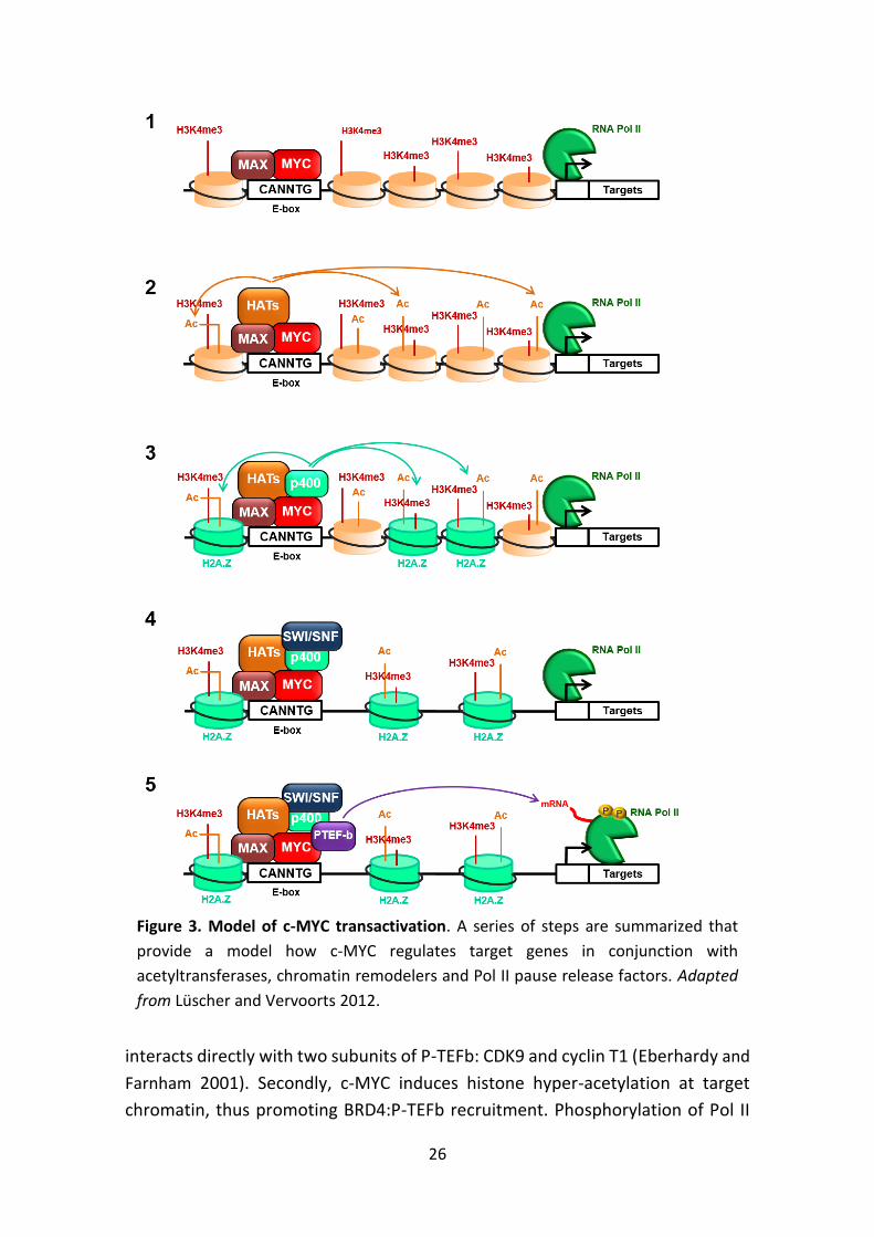
26
interacts directly with two subunits of P-TEFb: CDK9 and cyclin T1 (Eberhardy and
Farnham 2001). Secondly, c-MYC induces histone hyper-acetylation at target
chromatin, thus promoting BRD4:P-TEFb recruitment. Phosphorylation of Pol II
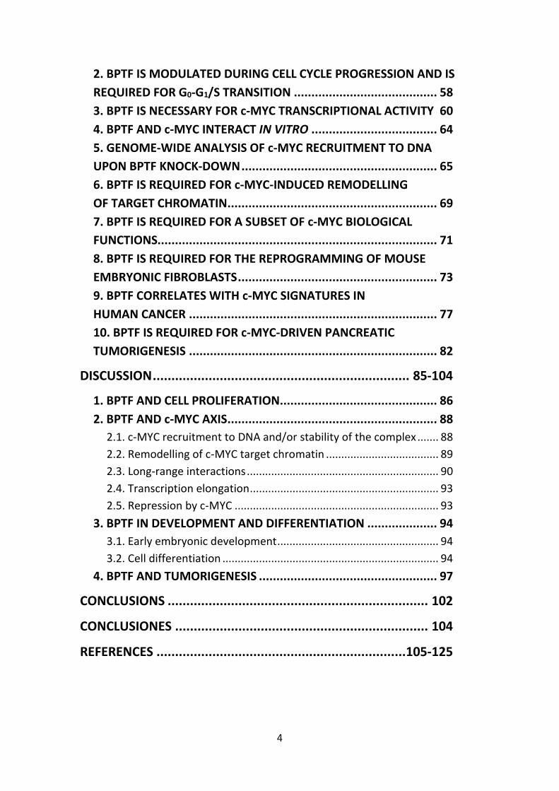
Figure 3. Model of c-MYC transactivation. A series of steps are summarized that
provide a model how c-MYC regulates target genes in conjunction with
acetyltransferases, chromatin remodelers and Pol II pause release factors. Adapted
from Lüscher and Vervoorts 2012.
27
also triggers the recruitment of mRNA capping and splicing factors, which are
essential for the processing of the emerging transcript (Cowling and Cole 2010).
In summary, c-MYC drives transcription by recruiting multiple co-factors to
promoters in a pre-existing transcriptionally active or poised state and further
modulating their activity.
It has been suggested that c-MYC does not have a unique transcriptional
program but, instead, it targets all active promoters and enhancers in the genome
and acts as a non-specific general amplifier of transcription (Lin et al. 2012; Nie
et al. 2012). Conversely, two recent reports offered an alternative to the amplifier
model, showing that c-MYC can actually activate and repress discrete gene sets.
The authors hypothesized that RNA amplification and promoter/enhancer
occupancy by c-MYC are in fact separable events. The increase in global RNA
production would be an indirect effect, explained by the nature of the targets
regulated by c-MYC (e.g. proteins involved in nucleotide synthesis) (Sabò et al.
2014; Walz et al. 2014; Dang 2014).
2.2.3. Transcriptional repression
Ectopic expression of c-MYC leads to down-regulation of specific genes
encoding negative regulators of cell proliferation (e.g. Cdkn2b, Cdkn2c or Cdkn1a)
and proteins involved in cell adhesion (Itgb1) and cell-cell communication. c-MYC
represses transcription by binding to the core promoter of target genes. Some
original studies suggested that this process occurred independently of c-MYC
binding to DNA. However, c-MYC recruitment to Cdkn2b requires dimerization
with MAX, and E-box elements have been found in the core promoter of genes
repressed by c-MYC (Herkert and Eilers 2010).
Mechanistically, c-MYC represses transcription by binding to two transcription
factors: MIZ1 and SP1 (Peukert et al. 1997; Gartel et al. 2001). The interaction
with c-MYC results in the displacement of the co-factors CBP/p300 and
nucleophosmin (NPM), and recruitment of repressors such as the histone
deacetylase HDAC3 and the DNA methyltransferase DNMT3A (Staller et al. 2001;
Brenner et al. 2005; Kurland and Tansey 2008; Wanzel et al. 2008). c-MYC also
modulates MIZ1 through the induction of RPL23, a ribosomal protein that
sequesters NPM to the nucleolus and thus hampers MIZ1 activity (Wanzel et al.
2008). The case of TGF-mediated cell cycle arrest illustrates the MYC-MIZ1
interaction. In the absence of TGF signalling, c-MYC represses Cdkn2b (p15INK4B)
in a complex with MIZ1. Increased levels of TGF lead to phosphorylation and
nuclear translocation of SMAD proteins, which cooperate with MIZ1 in inducing
28
Cdkn2b expression. In parallel, activated SMADs inhibit c-Myc transcription
(Seoane et al. 2001).
2.3. c-MYC biological roles
c-MYC is almost universally present in proliferating normal somatic cells, where
it operates as an integrator of extracellular stimuli transduced by multiple
signaling cascades (e.g. Wnt, Ras/Raf/MAPK, JAK/STAT or TGFb). As a result, it
modulates a wide range of cellular processes such as proliferation, growth,
apoptosis, metabolism, and differentiation. In normal cells, c-MYC is under tight
transcriptional and post-transcriptional control, and its expression is continuously
dependent upon mitogenic signalling.
By contrast, cancer cells typically show a deregulated and elevated c-MYC
expression, which is responsible for changes in chromatin structure, ribosome
biogenesis, metabolic pathways, cell, and angiogenesis among others (Lin et al.
2012) (Fig. 4).
2.3.1. Cell proliferation and differentiation
c-MYC has a crucial role in cell division by controlling the transition from G0/G1
to S phase. It regulates proliferation by transcriptionally activating genes involved
in cell cycle progression (e.g. cyclin D1, cyclin D2 or CDK4) and repressing
checkpoint genes and cyclin-dependent kinase inhibitors (e.g. GADD45, p15INK4B
or p21CIP1). Moreover, c-MYC enhances DNA replication by binding to pre-
replicative complexes and promoting origin firing (Dominguez-Sola et al. 2007).
c-MYC overexpression and/or deregulation is associated with unscheduled firing
of DNA replication origins, DNA damage response, and checkpoint activation
(Murga et al. 2011).
Several experiments document the ability of c-MYC to inhibit differentiation of
various cell types in vitro (e.g. murine ES cells) and in vivo (e.g. B cell lymphomas)
(Cartwright et al. 2005; Langdon et al. 1986). However, c-MYC role is far more
complex. In tissues where commitment to a specific lineage is linked to an
increase in proliferation, c-MYC promotes cell differentiation by controlling the
exit from the stem cell niche. One example is the skin, where ectopic expression
of c-MYC is associated with the depletion of the stem cell compartment and an
accumulation of differentiated layers (Waikel et al. 2001). Part of c-MYC role in
driving differentiation of keratinocytes involves its ability to reduce adhesive
interactions of stem cells with their niche (Gebhardt et al. 2006).
29
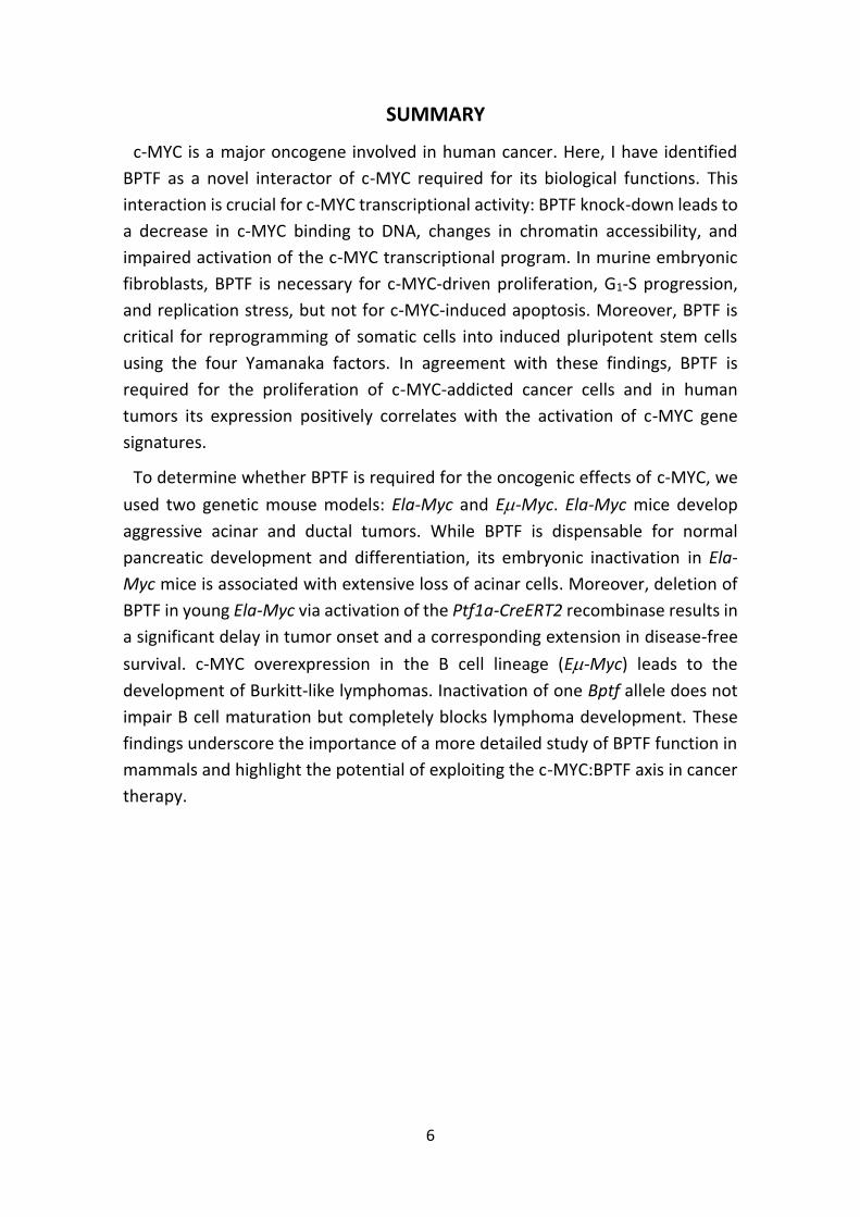
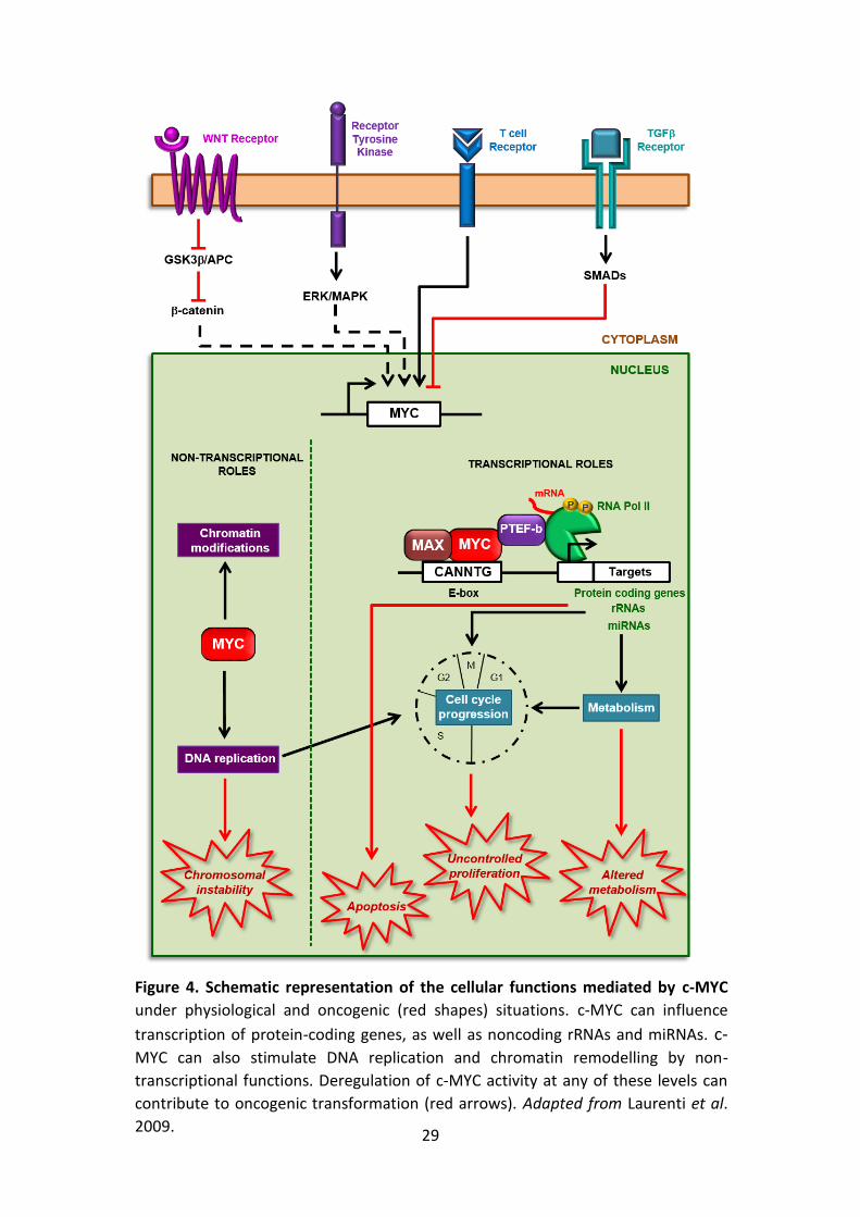
Figure 4. Schematic representation of the cellular functions mediated by c-MYC
under physiological and oncogenic (red shapes) situations. c-MYC can influence
transcription of protein-coding genes, as well as noncoding rRNAs and miRNAs. c-MYC can also stimulate DNA replication and chromatin remodelling by non-
transcriptional functions. Deregulation of c-MYC activity at any of these levels can
contribute to oncogenic transformation (red arrows). Adapted from Laurenti et al.
2009.
30
2.3.2. Cell growth and metabolism
c-MYC promotes cell growth by providing the cell with an abundant supply of
basic building blocks as well as increasing cell metabolism and protein synthesis.
Several c-MYC target genes participate in this activity, including those associated
with metabolism, ribosomal and mitochondrial biogenesis, and protein and
nucleic acid synthesis.
2.3.3. Apoptosis
Ectopic expression of c-MYC in the presence of limiting survival signals or cell
stress sensitizes cells to undergo apoptosis. This phenomenon has been reported
in both cells and transgenic mice in which c-MYC is expressed under the control
of a foreign promoter (Evan et al. 1992; Jacobsen et al. 1994). c-MYC-induced
apoptosis is an example of intrinsic tumor suppression, a defence mechanism
against the tumorigenic potential of oncogenes. In fact, suppression of c-MYC
pro-apoptotic activity is essential to tumorigenesis. Noticeably, it is
overexpression, rather than deregulation, what is required in order for c-MYC to
trigger apoptosis (Murphy et al. 2008).
Several mechanisms are involved in c-MYC-mediated apoptosis. High c-MYC
levels upregulate p19ARF, an inhibitor of the MDM2 E3 ligase, which leads to the
stabilization of p53 (Zindy et al. 1998). p53 regulates a cohort of target genes
involved in apoptosis and growth arrest. FoxO transcription factors have been
shown to mediate c-MYC-induced p19ARF expression through direct binding to the
Ink4a/Arf locus (Bouchard et al. 2007).
c-MYC can also trigger apoptosis by altering the balance between pro- and anti-
apoptotic factors, in parallel with or independent of p53. c-MYC indirectly
suppresses the anti-apoptotic proteins BCL2 and BCL-XL and induces the pro-
apoptotic Bax and BIM (Strasser et al. 1990; Eischen et al. 2001; Mitchel et al.
2000; Egle et al. 2004). These events lead to the release of cytochrome c from the
mitochondria and the subsequent activation of downstream effector caspases.
Moreover, c-MYC overexpression activates apoptosis through the induction of
DNA instability and breaks. This appears to be the consequence of several
mechanisms: inhibition of double-stranded DNA repair and/or increase in
reactive oxygen species (Vafa et al. 2002; Karlsson et al. 2003).
2.3.4. Tumorigenesis
c-MYC is over-expressed and/or deregulated in more than half of human cancers
(Gabay et al. 2014); high levels being associated with aggressive, poorly
31
differentiated tumors. This occurs through multiple mechanisms, including
amplification, chromosomal translocation, single nucleotide polymorphisms in
regulatory regions, constitutive activation of upstream signalling pathways and
mutations that enhance c-MYC protein stability (Eilers and Eisenman 2008; Meyer
and Penn 2008).
Even though c-MYC is one of the most potent oncogenes, its sole activation in
normal cells is not able to induce neoplastic transformation. Moreover, tumors
that arise from c-MYC transgenic mice are clonal, suggesting that additional
mutations are required for tumor formation. c-MYC-induced cell transformation
is restrained by two mechanisms. First, c-MYC half-life and function are
modulated by Ras-dependent signalling pathways. Second, several mechanisms
exist that protect cells from unchecked cell growth: proliferative arrest,
senescence, and/or apoptosis. Therefore, Ras activating mutations and genetic
events that abrogate these checkpoints (e.g. p53 loss) often synergize with c-MYC
to induce tumors (Adhikary and Eilers 2005).
When pathologically activated in a permissive context, c-MYC enforces many of
the "hallmark" features of cancer, including relentless DNA replication, cellular
proliferation and growth, protein synthesis, and altered metabolism. c-MYC
mandates tumor cell fate by inducing stemness and blocking cellular senescence
and differentiation. Additionally, c-MYC orchestrates changes in the tumor
microenvironment, including the activation of angiogenesis and suppression of
the host immune response (Gabay et al. 2014).
c-MYC plays a role both in tumor initiation and maintenance. In transgenic
mouse models with inducible c-MYC, established tumors regress upon
withdrawal of c-MYC ectopic expression (e.g. hematopoietic, epithelial, and
mesenchymal tumors) (Arvanitis and Felsher 2006). Interestingly, brief
suppression of c-MYC using the inducible dominant negative ‘Omomyc’ can result
in restoration of checkpoint mechanisms, resulting in tumor regression,
remodelling of the tumor microenvironment, and shutdown of angiogenesis.
Therefore, tumors appear to be “addicted” to c-MYC (Soucek et al. 2002; Soucek
et al. 2008).
Cellular transformation by c-MYC depends on specific cell cycle and metabolic
pathways. For example, c-MYC enhances glucose uptake and glycolysis through
transcriptional activation of different target genes, including lactate
dehydrogenase A (LDHA). Induction of LDHA might explain the “Warburg effect”;
namely, the observation that tumor cells show enhanced rates of glycolysis even
under aerobic conditions (Shim et al. 1997).
32
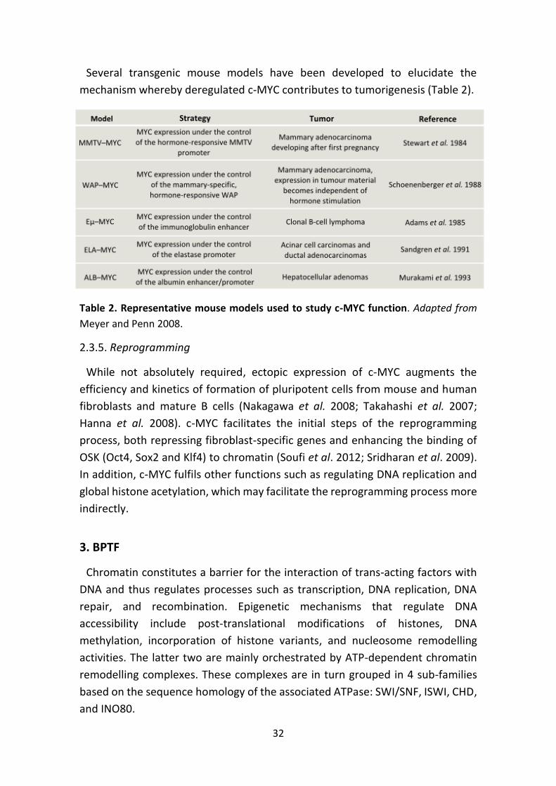
Several transgenic mouse models have been developed to elucidate the
mechanism whereby deregulated c-MYC contributes to tumorigenesis (Table 2).
Table 2. Representative mouse models used to study c-MYC function. Adapted from
Meyer and Penn 2008.
2.3.5. Reprogramming
While not absolutely required, ectopic expression of c-MYC augments the
efficiency and kinetics of formation of pluripotent cells from mouse and human
fibroblasts and mature B cells (Nakagawa et al. 2008; Takahashi et al. 2007;
Hanna et al. 2008). c-MYC facilitates the initial steps of the reprogramming
process, both repressing fibroblast-specific genes and enhancing the binding of
OSK (Oct4, Sox2 and Klf4) to chromatin (Soufi et al. 2012; Sridharan et al. 2009).
In addition, c-MYC fulfils other functions such as regulating DNA replication and
global histone acetylation, which may facilitate the reprogramming process more
indirectly.
3. BPTF
Chromatin constitutes a barrier for the interaction of trans-acting factors with
DNA and thus regulates processes such as transcription, DNA replication, DNA
repair, and recombination. Epigenetic mechanisms that regulate DNA
accessibility include post-translational modifications of histones, DNA
methylation, incorporation of histone variants, and nucleosome remodelling
activities. The latter two are mainly orchestrated by ATP-dependent chromatin
remodelling complexes. These complexes are in turn grouped in 4 sub-families
based on the sequence homology of the associated ATPase: SWI/SNF, ISWI, CHD,
and INO80.
33
BPTF (Bromodomain PHD Transcription Factor) is the mammalian orthologue of
Drosophila Nurf301 and constitutes the largest and essential subunit of the ISWI
complex NURF (Nucleosome Remodelling Factor). NURF is an ATP-dependent
chromatin remodeller that catalyses nucleosome sliding without eviction or
exchange of histones from the nucleosome. Mammalian NURF consists of BPTF,
SNF2L (an ISWI ATPase), and RbAp46/48, a histone-binding protein found in
several chromatin-related complexes (Jones et al. 2000; Hamiche et al. 1999;
Barak et al. 2003). BPTF provides sequence specificity to NURF through
interactions with transcription factors and histone modifications (Xiao et al. 2001;
Alkhatib et al. 2011).
3.1. Protein structure and interactors
The BPTF gene maps to the 17q24.3 locus and codes for two protein products:
BPTF (2871 aa) and its C-terminal truncated version FALZ (Fetal Alzheimer Antigen
or FAC1). While BPTF is ubiquitously expressed in adult tissues, FALZ is restricted
to the brain neocortex. It was proposed that FAC1 acts as a transcriptional
regulator through binding to a consensus DNA sequence present in genes
implicated in neurodegenerative disorders (e.g. PSEN1 or DRD2) (Jordan-Sciutto
et al. 1999a).
The functional domains within BPTF are consistent with a role for this protein in
chromatin-mediated regulation of transcription. The N-terminus of BPTF contains
a HMGA domain or acidic patch, a DDT DNA-binding domain, and a PHD domain.
The C-terminal domain of BPTF includes a glutamine-rich region which is
intrinsically disordered, a second PHD domain, and a bromodomain. The latter
two constitute a histone recognition module that binds H3K4me2/3 and
H4K16ac, respectively (Doerks et al. 2001; Wysocka et al. 2006; Ruthenburg et al.
2011). Additional features include nuclear localization signals, proline-rich
regions, and LXXLL motifs that could be important for the interaction with nuclear
receptors (Savkur and Burris 2004).
Human BPTF preferentially associates with H2A.Z, a histone variant
incorporated at promoter and regulatory regions whose deposition correlates
with gene expression (Marques et al. 2010). Moreover, the ATPase SNF2L
preferentially remodels H2A.Z-containing chromatin (Goldman et al. 2010).
34
3.2. Biological function of BPTF
3.2.1. Transcriptional activator and repressor
Chromatin remodelling machines have been traditionally thought to be required
exclusively during gene activation to expose or “open-up” chromatin. In
agreement with this view, NURF has been shown to facilitate transcription of
chromatin in vitro and in vivo. This effect is not observed with naked DNA
templates, suggesting that it functions to relieve the inhibitory effects of
chromatin on transcription (Mizuguchi et al. 1997; Badenhorst et al. 2002).
However, several lines of evidence indicate that NURF can also repress gene
transcription (Goldmark et al. 2000; Badenhorst et al. 2002; Landry et al. 2008).
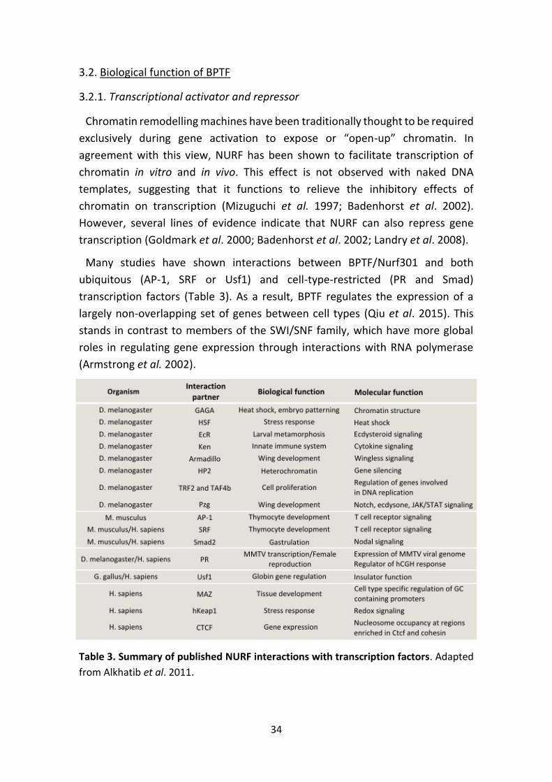
Many studies have shown interactions between BPTF/Nurf301 and both
ubiquitous (AP-1, SRF or Usf1) and cell-type-restricted (PR and Smad)
transcription factors (Table 3). As a result, BPTF regulates the expression of a
largely non-overlapping set of genes between cell types (Qiu et al. 2015). This
stands in contrast to members of the SWI/SNF family, which have more global
roles in regulating gene expression through interactions with RNA polymerase
(Armstrong et al. 2002).
Table 3. Summary of published NURF interactions with transcription factors. Adapted
from Alkhatib et al. 2011.
35
3.2.2. Chromatin structure
In addition to having key roles in transcription, NURF is a general regulator of
chromatin structure.
Inactivation of Drosophila Nurf301 leads to dramatic decondensation of the
male X chromosome (Badenhorst et al. 2002). NURF effects on X chromosome
chromatin architecture could be direct, through nucleosome remodelling, or
indirect, through the transcriptional regulation of genes involved in this process.
One possible mechanism would be through NURF-dependent localization of the
ATAC acetyltransferase (Carré et al. 2008).
NURF has also been characterized as a regulator of insulator elements in a
number of contexts. Drosophila NURF has been proposed to be recruited to
insulators by the GAGA factor, where it repositions nucleosomes to facilitate
insulator function (Xiao et al. 2001; Li et al. 2010). Similarly, human NURF is
critical for the barrier function of the USF-bound insulator 5’HS4, which prevents
erythroid genes from encroachment by heterochromatin (Li et al. 2011).
Interestingly, a recent report identified BPTF as a facilitator of the
interchromosomal interactions that take place between the enhancers of
olfactory receptor genes. These long-range interactions account for the
robustness of olfactory gene expression (Markenscoff-Papadimitriou et al. 2014).
Overall, these data suggest that BPTF:NURF modulates gene expression directly,
through the interaction with transcription factors, and indirectly, through the
regulation of high-order chromatin structures.
3.2.3. Developmental regulator
NURF has been shown to be essential for specific stages of metazoan
development, functioning in pathways signalling to the nucleus, including heat
shock, TGF/Smad, JAK/STAT, WNT/-catenin, and nuclear hormone receptors.
D. melanogaster: Nurf301 is required to maintain homeotic gene expression
during development, represses JAK/STAT signalling in the immune system, and
promotes ecdysone signalling during metamorphosis (Xiao et al. 2001;
Badenhorst et al. 2002; Badenhorst et al. 2005; Kwon et al. 2008). It also plays a
role in the development of larval blood cells and in the maintenance of the germ
stem cells compartment in Drosophila testis (Badenhorst et al. 2002; Cherry and
Matunis 2010).
M. musculus: Bptf knockout mice do not gastrulate due to defects in the
differentiation of extra-embryonic tissue lineages: the distal visceral endoderm
36
and the ectoplacental cone (Goller et al. 2008; Landry et al. 2008). For this reason,
the characterization of NURF function in the adult mammal has been limited. Cre-
LoxP conditional knockout technology revealed that BPTF is essential for adult
thymocyte development (Landry et al. 2011).
H. sapiens: In a competitive epidermal reconstitution assay, BPTF was identified
as a negative regulator of epidermal differentiation (Mulder et al. 2012).
3.3. BPTF in human cancer
Several lines of evidence suggest that BPTF could play a tumor-promoting role
in human cancer. Firstly, primary human cancers and cancer cell lines frequently
duplicate the 17q chromosome arm containing the BPTF gene. In fact, partial gain
of 17q is the most abundant genetic alteration in neuroblastoma (Bown et al.
1999; Alkhatib et al. 2011). Secondly, mutations targeting BPTF have been
reported for several human tumors (e.g. lung, breast, bladder, liver, and uterine
cancer) (Xiao et al. 2014a; Balbás-Martínez et al. 2013; Fujimoto et al. 2012;
González-Pérez et al. 2013). Finally, BPTF was appointed in a recent report as an
independent marker for survival prediction in hepatocellular carcinoma patients;
high BPTF levels being associated with invasiveness, recurrence, and poor
outcome (Xiao et al. 2014b).

37
Aims

38
AIMS
The specific aims for this thesis were:
1. To investigate the role of BPTF in normal mammalian cells by analyzing the
effect of its inactivation in both cell lines and mouse tissues.
2. To assess the function of BPTF in c-MYC transcriptional activity using a
combination of biochemical assays and genome-wide approaches.
3. To study the role of BPTF in c-MYC biological activity using cell cultures
expressing a tamoxifen-inducible form of c-MYC.
4. To determine the relevance of BPTF in tumorigenesis by analyzing publicly
available genomic data on human tumors and also by studying the impact of
its inactivation on mouse genetic models of cancer.

39
Objetivos

40
OBJETIVOS
Los objetivos específicos de esta tesis fueron:
1. Investigar la función de BPTF en células normales de mamífero por medio del
análisis de los efectos de su inactivación en líneas celulares y tejidos de ratón.
2. Evaluar el papel de BPTF en la actividad transcripcional de c-MYC usando una
combinación de ensayos bioquímicos y aproximaciones genómicas globales.
3. Estudiar el papel de BPTF en la actividad biológica de c-MYC usando cultivos
celulares que expresan una forma de c-MYC inducible por tamoxifeno.
4. Determinar la relevancia de BPTF en cáncer por medio del análisis de datos
genómicos públicos de tumores humanos así como del estudio del impacto de
su inactivación en modelos genéticos murinos de cáncer.

41
Materials and Methods
42
MATERIALS AND METHODS
1. CELL CULTURE
1.1. Cell lines and reagents
Primary neonatal human foreskin fibroblasts (HFF), 293T (transformed human
embryonic kidney cells), and human cancer cells - MIA PaCa-2, PK9 (pancreas) and
VM-CUB-3 (bladder) - were cultured in Dulbecco’s modified Eagle’s medium
(DMEM; Sigma-Aldrich, St Louis, MO, USA) supplemented with 10% Foetal Bovine
Serum (FBS; HyClone, Logan, UT, USA), sodium pyruvate (Life Technologies,
Madrid, Spain), and penicillin/streptomycin (Life Technologies). Mouse Bptf+/+
and Bptflox/lox MEFs (Murine Embryonic Fibroblasts) were cultured in DMEM
supplemented with 10% FBS, sodium pyruvate, non-essential amino acids (Life
Technologies), -mercaptoethanol (Sigma-Aldrich), and penicillin/streptomycin.
NAMALWA and RAJI Burkitt lymphoma cells were cultured in suspension in RPMI
medium (Sigma-Aldrich) supplemented with 10% FBS and penicillin/
streptomycin.
MEFs were generated by mechanical disruption and trypsin-digestion of E13.5
embryos from which the foetal liver and the head had been removed.
Recombination efficiency of exon 2 upon Cre recombinase expression was
evaluated by PCR on genomic DNA as reported elsewhere (Landry et al. 2008).
The following primers were used: CTCAGGAATTAAGAGGTAATTGACTATC,
TGATTTAGTTCTGATTGTTAGGTCTAC, and AGACCAGCCTGTTCTACATGGCCAGCC.
Additionally, recombination efficiency was assessed by RT-qPCR using the
following primers:
Amplified region Sequence Species
Exon1-Exon2
Junction
Forward AAGCAGCTTCAGGAGCCATA Mouse
Reverse AGCAAAAAGGGGACAACCT Mouse
Exon1-Exon3
Junction
Forward CAGCAGCACTCCAGAGAAGA Mouse
Reverse CGCTAGGAAGGACTTGTTGC Mouse
Exon1-Exon4
Junction
Forward CAGCAGCACTCCAGAGGAAA Mouse
Reverse GCTCTTCTCAGCATCCTTGG Mouse
1.2. Plasmids, viral constructs and virus production
Mission shRNAs (Sigma-Aldrich) were used to carry out RNA-interference
experiments. Out of 3 BPTF-targeting shRNAs, two were selected because they
provided optimal knockdown (shBPTF-1, clone TRCN0000016819; shBPTF-2,
clone TRCN0000016820) and compared to a control non-targeting shRNA. MYC-
43
ER was expressed from the cDNA cloned into the FG12 plasmid by V.J. Sánchez-
Arévalo (CNIO, Madrid). For lentiviral transduction of Cre recombinase, we used
the lentiviral vector pLVXpuro-iCRE-ORF, a gift from C. Bar and M.A. Blasco (CNIO,
Madrid). The packaging plasmid pCL-Eco and the retroviral constructs expressing
pluripotency factors were generously provided by C.J. Lynch and M. Serrano
(CNIO, Madrid).
Lentiviral production: Infectious lentiviruses were produced in 293T cells by
calcium phosphate-mediated transfection of the lentiviral construct together
with the packaging plasmids psPAX2 and pCMV-VSV-G. Post-transfection (48h),
the medium was harvested twice for an additional 48h. Viral supernatants were
filtered and either frozen down in aliquots or applied on target cells in the
presence of 5 µg/ml polybrene. Cells were used after 48h puromycin selection (2
μg/ml). Human fibroblasts were infected first with lentivirus coding for MYC-ER,
expanded, and then infected with either control or BPTF-targeting shRNAs. MEFs
were infected concomitantly with lentivirus encoding for MYC-ER and Cre
recombinase.
Retroviral production for reprogramming of MEFs into iPSc: Retroviral
supernatants were produced in HEK-293T cells (5×10⁶ cells per 100-mm-diameter
dish) transfected with the packaging plasmid pCL-Eco (4 μg) together with one of
the following retroviral constructs (4 μg): pMXs-Klf4, pMXs-Sox2, pMXs-Oct4 or
pMXs-cMyc. Transfections were performed using Fugene-6 transfection reagent
(Roche, Basel, Switzerland) according to the manufacturer’s protocol. Two days
post-transfection, retroviral supernatants were collected at 12 h intervals, each
time adding fresh medium to the cells.
Infection of BL cell lines: BL cells (3×10⁵ cells/well) were seeded on plastic plates
coated with retronectin (Fisher Scientific, Pittsburgh, PA, USA) and preloaded
with viral supernatants. After 3 additional rounds of infection with viral
supernatants supplemented with polybrene (8 μg/ml), cells were allowed to
recover for 24h, then selected for 48h in puromycin-containing medium (2
g/ml). After selection, cells (sh#1, sh#2 and shNT) were plated (5x10³/well in 96-
well plates) in replicates. Viable cell count was assessed at the indicated time
points by adding WST1 cell proliferation reagent (Roche) to each well and
determining OD450 nm after 2 h, according to the manufacturer's instructions.
1.3. iPS Reprogramming
Early passage (2-3) primary MEFs were reprogrammed following a protocol
described elsewhere (Li et al. 2009). Recipient MEFs were seeded the previous
44
day (150,000 cells on a 6-well plate) and received 1.5 ml of each of the
corresponding retroviral supernatants (3F: 4.5 ml in total; 4F: 6 ml in total). This
procedure was repeated 4 times in total. At 48 h after the first round of infection,
medium was changed to iPSC medium (DMEM high glucose supplemented with
serum replacement (KSR, 15%, Invitrogen), leukemia inhibitory factor (LIF) (1000
U/ml), non-essential amino acids, glutamax and -mercaptoethanol). Cultures
were maintained in the absence of drug selection with daily medium changes. At
day 12-14, colonies with ES-like morphology were scored after staining for AP
activity (BCIP/NBT Colour Development Substrate, Promega, S3771). Colonies
were picked at day 14 and expanded on feeder fibroblasts using standard
procedures.
1.4. FACS analysis of proliferation and apoptosis
For proliferation assays of MEFs, cells were pulse-labelled with 10 M BrdU
(Sigma-Aldrich) for 1 h, harvested by trypsinization and then fixed in 100%
ethanol. Upon DNA denaturation using 2 N HCl, cells were stained with mouse
anti-BrdU primary antibody (Santa Cruz Biotechnology, sc-51514; 1g/106 cells)
and anti-mouse Alexa Fluor 488-conjugated secondary antibody (Life
Technologies, A21202; 1g/106 cells). DNA was stained by resuspension of cells
in 0.1 mg/ml propidium iodide and incubated 30 min at room temperature until
FACS analysis.
In order to measure apoptosis, MEFs were seeded at high density and then
transferred to 0.5% FBS-containing DMEM in the presence of either vehicle
(EtOH) or 2 mM 4-OHT. At the indicated time points, cells and supernatants were
harvested, washed, and resuspended in Annexin V binding buffer containing 5 l
per sample of Annexin V-APC (BD Biosciences, 550474, Franklin Lakes, NJ, USA).
Prior to analysis, DAPI was added.
2. MOUSE BIOLOGY
2.1. Mouse strains
The following mouse strains were used: Bptflox/lox (Landry et al. 2008), Ptf1a-
Cre+/KI (Kawaguchi et al. 2002), Ptf1a-CreERT2+/KI (Kopinke et al. 2011), Ela1-Myc
(Sandgren et al. 1991), Mb1-Cre+/KI (Hobeika et al. 2006) and E-Myc (Harris et al.
1988). Mb1-Cre mice were provided by Dr. A.M. Ramiro (CNIC, Madrid), and E-
Myc mice were supplied by Dr. C. Blanco (CNIO, Madrid). C57BL/6 Bptflox/lox mice
were obtained from Jackson Laboratories (stock number 009367). Other strains
were available at CNIO.
45
To determine the role of BPTF in c-MYC-driven pancreatic tumorigenesis, we
administered 25 mg of tamoxifen (Sigma-Aldrich, T-5648) by gavage over the
course of one week to 5-7 weeks old Bptflox/lox; Ptf1a-CreERT2+/KI; Ela1-Myc mice
and their corresponding controls. Mice were screened for pancreatic tumors once
a week using a small animal ultrasound system.
Mice were housed under specific pathogen-free conditions according to
institutional guidelines. Mice were observed on a daily basis and sacrificed when
they showed signs of morbidity or tumor burden was greater than 10% body
weight in accordance with the Guidelines for Human Endpoints for Animals Used
in Biomedical Research. All experiments were approved by the ISCIII (Instituto de
Salud Carlos III) Ethical Committee and performed in accordance with the
guidelines for Ethical Conduct in the Care and Use of Animals as stated in The
International Guiding Principles for Biomedical Research involving Animals,
developed by the CIOMS.
2.2. Histopathology and Immunohistochemistry
Mouse tissues were fixed in 4% PBS-buffered formaldehyde, embedded in
paraffin and serially sectioned. 4 m sections were deparaffinized and stained
with hematoxylin-eosin or specific antibodies.
For immunohistochemistry, mouse tissue sections were prepared as follows.
After deparaffinization, sections were rehydrated and boiled in 10 mM sodium
citrate buffer (pH 6) for 10 min to retrieve the antigens. Next, sections were
washed in distilled water and incubated for 30 min with 3% hydrogen peroxide in
methanol, after which they were washed again and blocked for 30 min with 2%
BSA in PBS/0.5% Triton X-100. After blocking, the sections were incubated with
primary antibodies in 2% BSA in PBS/0.1% Triton X-100 for 1h at room
temperature. Antibody dilutions used: P-Histone H3 (Abcam ab14955), 1:2000; c-
Myc (Millipore 06-340), 1:300; cleaved caspase 3 (R&D AF835), 1:300. Next,
sections were washed in PBS/0.1% Triton X-100 three times and incubated for 30
min with Envision+ HRP-labelled secondary antibodies (Dako, Glostrup,
Denmark). Sections were washed again and the staining was developed using
DAB Chromogen system (Dako). Sections were rinsed with water, counterstained
with Carazzi's Hematoxylin solution DC (Panreac, Castellar del Vallès, Spain),
dehydrated with increasing concentrations of alcohol and xylol, and mounted
using DePeX mounting medium.
46
2.3. Hematological analysis and characterization of B cell compartment
For the analysis of cellular components of peripheral blood of Bptflox/lox; Mb1-
Cre+/KI mice, samples were collected from 8-10 week old mice and assessed using
an Abacus Junior Hematology Analyzer.
In order to assess the bone marrow (BM) and spleen B cell compartments, single
cell suspensions were prepared according to standard procedures (Iritani et al.
1997). BM cells were harvested by flushing two tibias and two femurs per mouse
with 5 ml of RPMI 10% FBS (HyClone). Splenocytes were prepared by crushing
spleens through 70m filters and into the above media. Next, erythrocytes were
depleted by incubation in ammonium chloride buffer (0.15 M NH4Cl, 10 mM
NaHCO3, 1 mM EDTA, pH 7.4) for 2 min at 37 ºC. Cells were collected by
centrifugation for 5 min at 1,200 rpm and resuspended in 1 ml of FACS buffer (2
mM EDTA in PBS/0.1% BSA) for further analysis. Prior to staining, cell suspensions
were blocked for 20 min in FACS buffer supplemented with 1:200 FC block (BD
Pharmingen, purified rat anti-mouse CD16/CD32, 553142). 3-4 colour flow
cytometry analyses were performed by staining 4×10⁶ cells with 0.25 g of the
following mAbs directed against lineage markers (in various combinations): APC‐
conjugated anti‐CD45R/B220 (ebioscience, 17-0452, San Diego, CA, USA), FITC‐
conjugated anti‐CD43 (ebioscience, 11-0431-81), PE‐conjugated anti‐IgM
(ebioscience, 12-5790-81). Samples were analyzed using a FACS Canto II (BD
Biosciences) flow cytometer. Analyses were performed using FlowJo flow
cytometry analysis software.
3. MOLECULAR BIOLOGY
3.1. Western blotting
Cells were lysed in RIPA buffer supplemented with protease and phosphatase
inhibitors. Following sonication, clearing by centrifugation, and protein
determination, equal amounts of protein per sample were subjected to
electrophoresis in 8% or 10% polyacrylamide SDS gels, or in NuPAGE® 3-8% Tris-
acetate precast polyacrylamide gels (Life Technologies). Samples were run under
reducing conditions and then transferred to nitrocellulose membranes, which
were blocked with TBST, 5% skim milk. Membranes were subsequently incubated
with the following primary antibodies: BPTF (ab72036, Abcam, Cambridge, UK;
1:500) and Vinculin (Sigma-Aldrich, V9131-2ML; 1:2000). This was followed by
incubation with horseradish peroxidase-conjugated secondary antibodies (Dako).
Reactions were detected using the ECL system.
47
3.2. Co-immunoprecipitation analyses
For the analysis of c-MYC:BPTF interaction, the following plasmids were used:
BPTF-Flag (courtesy of O. Barak, Wistar Institute, Philadelphia, USA) and HA-
tagged c-MYC (a gift from V.J. Sánchez-Arévalo, CNIO, Madrid).
293T cells transiently transfected with the corresponding plasmids were washed
twice with ice-cold PBS and lysed for 30 min on ice with NP-40 lysis buffer (150
mM NaCl, 50 mM Tris pH 8.0, 1% NP-40) supplemented with a protease inhibitor
cocktail. Lysates were then centrifuged at 16,000 x g for 20 min at 4 ºC. Total
protein (1 mg) was incubated with primary antibody (2 g) overnight. Protein A/G
agarose beads (Laboratorios Conda, Madrid, Spain) preblocked with BSA were
then added to the lysates. Following 4 h incubation at 4 ºC, beads were washed
3 times with NP-40 lysis buffer and immunoprecipitated proteins were eluted
with SDS sample buffer by boiling at 90 ºC. SDS-PAGE electrophoresis was then
performed on 6% and 10% (w/v) gels and proteins were then transferred onto
nitrocellulose membranes.
3.3. Generation of polyclonal anti-BPTF anti-sera
The RLHRMTSIEREEKEKVKKKEKKQEEETC peptide was chemically synthesized,
coupled to Keyhole limpet hemocyanin (KLH), and used as immunogen for the
generation of polyclonal antibodies against BPTF. Two rabbits were inoculated
subcutaneously with 500 g of peptide-KLH conjugate emulsified in Freund’s
Complete Adjuvant (FCA). Five rounds of 250 g peptide-KLH boosters were
administered together with Freund’s Incomplete Adjuvant (FIA) to each animal in
the interval of three weeks. Test bleeds were taken 10 days after the last boost.
Antibodies raised against BPTF were purified from serum by affinity
chromatography on a HiTrap NHS-activated High Performance column (Sigma-
Aldrich, GE17-0716-01) and tested by ELISA, in HEK293T-BPTF-Flag transfected
cells and in BPTF-silenced VM-CUB-3 cells.
3.4. Immunofluorescence staining and Proximity Ligation Assay
Cells grown on coverslips were fixed with 4% paraformaldehyde for 10 min,
washed, and permeabilized with 0.1% Triton X-100 in PBS for 10 min. Samples
were washed in PBS and blocked with 3% BSA in PBS for 1 h at room temperature.
Primary antibody incubation was performed in blocking solution for 2 h at room
temperature. Mouse anti-MYC (C-33, sc-42, Santa Cruz Biotechnology, Dallas, TX,
USA) was used at a 1:50 dilution and home-made affinity-purified rabbit anti-
BPTF antibodies (residues 913-942) were used at 10 g/ml. After three washes
with PBS, cells were incubated with an appropriate secondary antibody diluted in
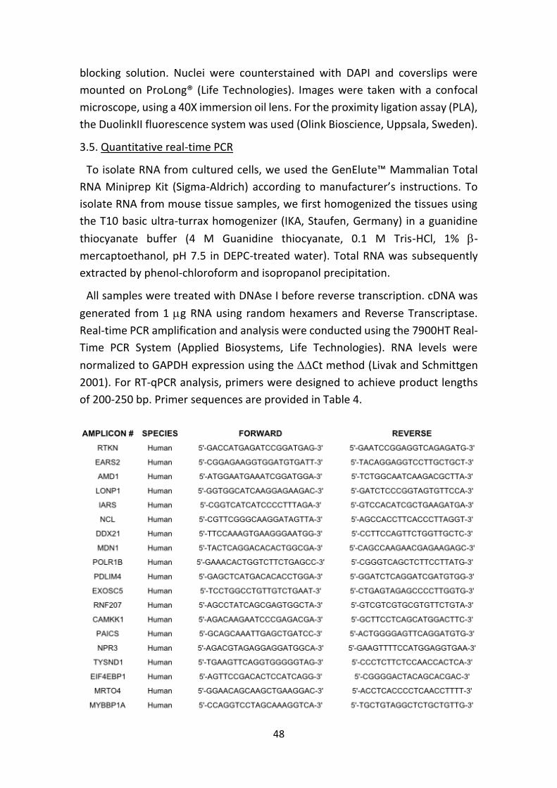
48
blocking solution. Nuclei were counterstained with DAPI and coverslips were
mounted on ProLong® (Life Technologies). Images were taken with a confocal
microscope, using a 40X immersion oil lens. For the proximity ligation assay (PLA),
the DuolinkII fluorescence system was used (Olink Bioscience, Uppsala, Sweden).
3.5. Quantitative real-time PCR
To isolate RNA from cultured cells, we used the GenElute™ Mammalian Total
RNA Miniprep Kit (Sigma-Aldrich) according to manufacturer’s instructions. To
isolate RNA from mouse tissue samples, we first homogenized the tissues using
the T10 basic ultra-turrax homogenizer (IKA, Staufen, Germany) in a guanidine
thiocyanate buffer (4 M Guanidine thiocyanate, 0.1 M Tris-HCl, 1% -
mercaptoethanol, pH 7.5 in DEPC-treated water). Total RNA was subsequently
extracted by phenol-chloroform and isopropanol precipitation.
All samples were treated with DNAse I before reverse transcription. cDNA was
generated from 1 g RNA using random hexamers and Reverse Transcriptase.
Real-time PCR amplification and analysis were conducted using the 7900HT Real-
Time PCR System (Applied Biosystems, Life Technologies). RNA levels were
normalized to GAPDH expression using the Ct method (Livak and Schmittgen
2001). For RT-qPCR analysis, primers were designed to achieve product lengths
of 200-250 bp. Primer sequences are provided in Table 4.
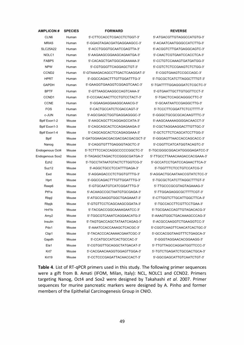
49
Table 4. List of RT-qPCR primers used in this study. The following primer sequences were a gift from B. Amati (IFOM, Milan, Italy): NCL, NOLC1 and CCND2. Primers targeting Nanog, Oct4 and Sox2 were designed by Takahashi et al. 2007. Primer sequences for murine pancreatic markers were designed by A. Pinho and former members of the Epithelial Carcinogenesis Group in CNIO.
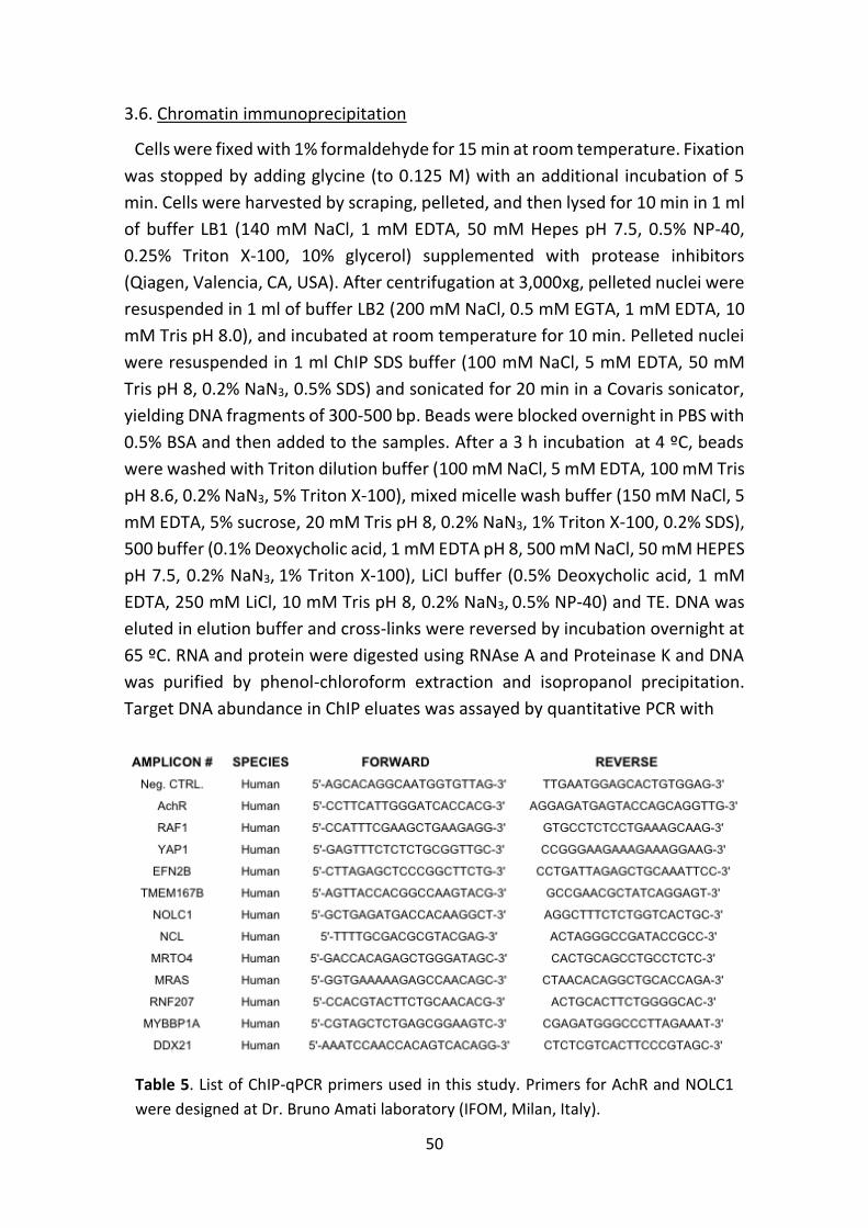
50
3.6. Chromatin immunoprecipitation
Cells were fixed with 1% formaldehyde for 15 min at room temperature. Fixation
was stopped by adding glycine (to 0.125 M) with an additional incubation of 5
min. Cells were harvested by scraping, pelleted, and then lysed for 10 min in 1 ml
of buffer LB1 (140 mM NaCl, 1 mM EDTA, 50 mM Hepes pH 7.5, 0.5% NP-40,
0.25% Triton X-100, 10% glycerol) supplemented with protease inhibitors
(Qiagen, Valencia, CA, USA). After centrifugation at 3,000xg, pelleted nuclei were
resuspended in 1 ml of buffer LB2 (200 mM NaCl, 0.5 mM EGTA, 1 mM EDTA, 10
mM Tris pH 8.0), and incubated at room temperature for 10 min. Pelleted nuclei
were resuspended in 1 ml ChIP SDS buffer (100 mM NaCl, 5 mM EDTA, 50 mM
Tris pH 8, 0.2% NaN3, 0.5% SDS) and sonicated for 20 min in a Covaris sonicator,
yielding DNA fragments of 300-500 bp. Beads were blocked overnight in PBS with
0.5% BSA and then added to the samples. After a 3 h incubation at 4 ºC, beads
were washed with Triton dilution buffer (100 mM NaCl, 5 mM EDTA, 100 mM Tris
pH 8.6, 0.2% NaN3, 5% Triton X-100), mixed micelle wash buffer (150 mM NaCl, 5
mM EDTA, 5% sucrose, 20 mM Tris pH 8, 0.2% NaN3, 1% Triton X-100, 0.2% SDS),
500 buffer (0.1% Deoxycholic acid, 1 mM EDTA pH 8, 500 mM NaCl, 50 mM HEPES
pH 7.5, 0.2% NaN3, 1% Triton X-100), LiCl buffer (0.5% Deoxycholic acid, 1 mM
EDTA, 250 mM LiCl, 10 mM Tris pH 8, 0.2% NaN3, 0.5% NP-40) and TE. DNA was
eluted in elution buffer and cross-links were reversed by incubation overnight at
65 ºC. RNA and protein were digested using RNAse A and Proteinase K and DNA
was purified by phenol-chloroform extraction and isopropanol precipitation.
Target DNA abundance in ChIP eluates was assayed by quantitative PCR with
Table 5. List of ChIP-qPCR primers used in this study. Primers for AchR and NOLC1
were designed at Dr. Bruno Amati laboratory (IFOM, Milan, Italy).
51
primer pairs designed to achieve products of 50-200bp. Primer sequences are
provided in Table 5. The following antibodies were used: anti-MYC N262 (Santa
Cruz Biotechnology, sc-764), anti-H3K4me3 (Abcam, ab8580), anti-panAc Histone
H3 (Merck Millipore, 06-599, Billerica, MA, USA), and anti-total Histone H3
(Abcam, ab1791).
3.7. DNAse I hypersensitivity assay
DNAse I experiments were performed as described previously (Di Stefano et al.
2014). Briefly, chromatin samples were obtained as described above and
subjected to DNAse I digestion. Chromatin (2 g) was treated with 0.5, 1, and 2
units of RQ1 RNase-Free DNAse I (Promega, Fitchburg, WI, USA) for 3 min at 37
ºC in 1X DNAse incubation buffer. Reactions were terminated by adding 2 mM
EGTA and the crosslinking was reversed by incubating samples at 65 ºC. After 6h,
proteinase K (40 mg/ml) was added to each reaction and incubated overnight at
37 ºC. After phenol-chloroform extraction, DNA was quantified and used as
template for q-PCR reactions with the same primer pairs used for ChIP-qPCR.
4. GENOME-WIDE STUDIES AND BIOINFORMATICS ANALYSES
4.1. ChIP-Seq library construction and massive parallel sequencing
ChIP was performed as described above. DNA (20 ng) was quantified by
fluorometry, resolved by electrophoresis, and fractions of 50-250bp were
extracted. Input samples correspond to balanced blends of inputs from selected
samples. Fractions were processed through subsequent enzymatic treatments of
end-repair, dA-tailing, and ligation to adapters following Illumina's "TruSeq DNA
Sample Preparation Guide" (part # 15005180 Rev. C). Adapter-ligated libraries
were amplified by limited-cycle PCR with Illumina PE primers (12 cycles). The
resulting purified DNA library was applied to an Illumina flow cell for cluster
generation (TruSeq cluster generation kit v5) and sequenced on the Genome
Analyzer IIx with SBS TruSeq v5 reagents following manufacturer's protocols.
4.2. ChIP-Seq data processing
Image analysis and per-cycle base-calling was performed with Illumina Real
Time Analysis software (RTA1.13). Conversion to FASTQ read format with the
ELAND algorithm (v2e) was performed with CASAVA-1.8 (Illumina). Quality check
was done via fastqc (v0.9.4, Babraham Bioinformatics). ChIP-seq reads were
aligned to the human reference genome (GRCh37/hg19, Feb 2009) with Burrows-
Wheeler Aligner (bwa,v0.5.9-r16) allowing 0-1 mismatches. Unique aligned reads
were converted to BED format. All ChIP and input samples were normalized
52
randomly to the same number of reads (10,512.988). Furthermore, reads were
directionally extended to 300 bp and, for each base pair in the genome, the
number of overlapping sequence reads was determined and averaged over a 10
bp window to create a wig file to visualize the data in the University of California
Santa Cruz (UCSC) genome browser. The number of significant peaks of MYC
binding sites was 1762 for sh#1+OHT and 1397 for shNt+OHT, using MACS
(version 2.0.9 20111102, tag:alpha) and parameters: -g 2.7e9; -m 10,30; -q 0.05.
4.3. Motif enrichment analysis, peak annotation and density plot analysis
Motifs for the list of peaks in shNt+OHT were identified with the MEME suite
and then TOMTOM was used to compare the identified motifs with known
transcription factor binding motifs. Sequence logos were generated using
WebLogo 2.8.2. Genomic annotation was carried out with Hypergeometric
Optimization of Motif EnRichment (HOMER, software v4.2). The tool
annotatePeaks.pl was used with parameters by default and defined in the help.
A gtf file from UCSC based on GRCh37/hg19 was used for annotations; the latter
included whether a segment is in the TSS, TTS, exon, 5' UTR exon, 3' UTR exon,
intron, or is intergenic. Since some annotations overlap, the following priority was
assigned: TSS (from -1kb to +100bp), TTS (from -100 bp to +1kb), CDS exon, 5'
UTR exon, 3' UTR exon, intron, intergenic. More detailed information is available
in http://homer.salk.edu/homer/ngs/annotation.html. The SeqMINER (v1.3.3e)
platform (Ye et al. 2010) was used to generate the density read plots shown in
Fig. 11b.
4.4. Gene set enrichment analysis (GSEA)
MYC-bound genes were rank-ordered according to the fold-change in FPKM
values (4-OHT vs. vehicle) in HFF MYC-ER control cells and then submitted to
analysis using GSEA software (www.broadinstitute.org/gsea). The list of pre-
ranked genes was analysed with the gene set matrix composed file
c2.all.v4.0.symbols.gmt and c5.all.v4.0.symbols.gmt. Significant gene sets
enriched by 4-OHT-treatment of control cells were identified using an FDR q-
value < 0.25 and a nominal P value < 0.05, as defined by
http://www.broadinstitute.org/gsea/doc/GSEAUserGuideFrame.html?Interpreti
ng_GSEA.
4.5. RNA-seq
Total RNA (1 µg) was spiked with ERCC ExFold RNA spike-In mixes (Life
Technologies). RNA quality was assessed on an Agilent 2100 Bioanalyzer and
samples with a RNA Integrity Number > 8.5 were used. PolyA+ fractions were
53
purified, randomly fragmented, converted to double stranded cDNA, and
processed through subsequent enzymatic treatments of end-repair, dA-tailing,
and ligation to adapters following Illumina's "TruSeq Stranded mRNA Sample
Preparation Part # 15031047 Rev. D" (this kit incorporates dUTP during 2nd
strand cDNA synthesis, which implies that only the cDNA strand generated during
1st strand synthesis is eventually sequenced). Adapter-ligated libraries were
generated by PCR with Illumina PE primers (8 cycles). The resulting purified cDNA
libraries were applied to an Illumina flow cell for cluster generation (TruSeq
cluster generation kit v5) and sequenced on the Genome Analyzer IIx with SBS
TruSeq v5 reagents by following manufacturer's protocols.
4.6. RNA-Seq data processing
Image analysis and per-cycle base-calling was performed with Illumina Real
Time Analysis software (RTA1.13). Conversion to FASTQ read format with the
ELAND algorithm (v2e) was performed with CASAVA-1.8 (Illumina). These files
contain only reads that passed "chastity" filtering (flagged with a ‘N’, for *NOT
filtered* in the sequence identifier line). "Chastity" parameter measures signal
contamination in raw data and allows to flag unreliable reads. Quality check was
done via fastqc (v0.9.4, Babraham Bioinformatics). Raw reads were aligned to the
build version GRCh37/hg19 of the human genome where the sequence of the
ERCC synthetic spike-in RNAs
(http://tools.invitrogen.com/downloads/ERCC92.fa) had been added. Tophat5
(version 2.0.4) was used for alignment with the following parameters: --bowtie1,
--max-multihits 5, --genome-read-mismatches 1 --segment-mismatches 1 --
segment-length 19 --splice-mismatches 0 --library-type fr-firststrand. Gene
expression levels and synthetic spike-in RNA (Fragments Per Kilobase of exon per
Million fragments mapped, FPKM) were quantified with cufflinks (version 2.0.2),
with the following parameters: -N, --library-type fr-firststrand, -u. Further, we
used a loess regression to renormalize the FPKM values by using only the spike-
in values to fit the loess following the strategy described (Lovén et al. 2012). The
affy package in R provides a function, loess.normalize, performing loess
regression on a matrix of values and allowing to specify which subset of data to
use when fitting the loess (see the affy package for further details). The result
was a matrix of FPKM values normalized to the control ERCC spike-ins.
4.7. RNA-Seq GSEA analysis
Genes were rank-ordered according to the fold change in FPKM values (4-OHT
vs Vehicle) in HFF MYC-ER control cells and then submitted to analysis using GSEA
software (www.broadinstitute.org/gsea). The list of pre-ranked genes was
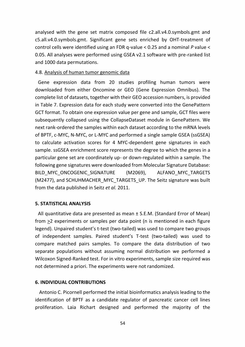
54
analysed with the gene set matrix composed file c2.all.v4.0.symbols.gmt and
c5.all.v4.0.symbols.gmt. Significant gene sets enriched by OHT-treatment of
control cells were identified using an FDR q-value < 0.25 and a nominal P value <
0.05. All analyses were performed using GSEA v2.1 software with pre-ranked list
and 1000 data permutations.
4.8. Analysis of human tumor genomic data
Gene expression data from 20 studies profiling human tumors were
downloaded from either Oncomine or GEO (Gene Expression Omnibus). The
complete list of datasets, together with their GEO accession numbers, is provided
in Table 7. Expression data for each study were converted into the GenePattern
GCT format. To obtain one expression value per gene and sample, GCT files were
subsequently collapsed using the CollapseDataset module in GenePattern. We
next rank-ordered the samples within each dataset according to the mRNA levels
of BPTF, c-MYC, N-MYC, or L-MYC and performed a single sample GSEA (ssGSEA)
to calculate activation scores for 4 MYC-dependent gene signatures in each
sample. ssGSEA enrichment score represents the degree to which the genes in a
particular gene set are coordinately up- or down-regulated within a sample. The
following gene signatures were downloaded from Molecular Signature Database:
BILD_MYC_ONCOGENIC_SIGNATURE (M2069), ALFANO_MYC_TARGETS
(M2477), and SCHUHMACHER_MYC_TARGETS_UP. The Seitz signature was built
from the data published in Seitz et al. 2011.
5. STATISTICAL ANALYSIS
All quantitative data are presented as mean ± S.E.M. (Standard Error of Mean)
from >2 experiments or samples per data point (n is mentioned in each figure
legend). Unpaired student’s t-test (two-tailed) was used to compare two groups
of independent samples. Paired student’s T-test (two-tailed) was used to
compare matched pairs samples. To compare the data distribution of two
separate populations without assuming normal distribution we performed a
Wilcoxon Signed-Ranked test. For in vitro experiments, sample size required was
not determined a priori. The experiments were not randomized.
6. INDIVIDUAL CONTRIBUTIONS
Antonio C. Picornell performed the initial bioinformatics analysis leading to the
identification of BPTF as a candidate regulator of pancreatic cancer cell lines
proliferation. Laia Richart designed and performed the majority of the

55
experiments. Ana Río Machín (CNIO, Madrid) designed and performed the
experiments with Burkitt lymphoma cell lines. Enrique Carrillo-de Santa Pau
(CNIO, Madrid) was responsible for the bioinformatics analysis of the genome-
wide experiments. V.J. Sánchez-Arévalo (CNIO, Madrid) generated the following
constructs: HA-tagged c-MYC and FG12-MycER. Natalia del Pozo (CNIO, Madrid)
and Mónica Pérez de Andrés (Universidad Complutense, Madrid) helped with
mouse experiments. Francisco X. Real and V.J. Sánchez-Arévalo supervised the
overall conduct of the study.

56
Results
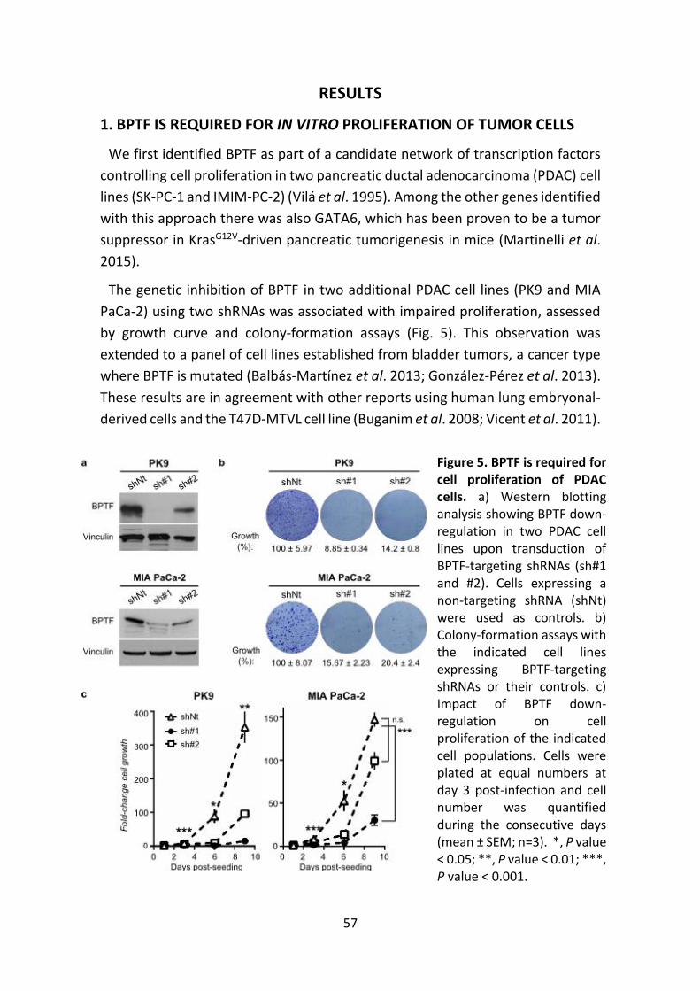
57
RESULTS
1. BPTF IS REQUIRED FOR IN VITRO PROLIFERATION OF TUMOR CELLS
We first identified BPTF as part of a candidate network of transcription factors
controlling cell proliferation in two pancreatic ductal adenocarcinoma (PDAC) cell
lines (SK-PC-1 and IMIM-PC-2) (Vilá et al. 1995). Among the other genes identified
with this approach there was also GATA6, which has been proven to be a tumor
suppressor in KrasG12V-driven pancreatic tumorigenesis in mice (Martinelli et al.
2015).
The genetic inhibition of BPTF in two additional PDAC cell lines (PK9 and MIA
PaCa-2) using two shRNAs was associated with impaired proliferation, assessed
by growth curve and colony-formation assays (Fig. 5). This observation was
extended to a panel of cell lines established from bladder tumors, a cancer type
where BPTF is mutated (Balbás-Martínez et al. 2013; González-Pérez et al. 2013).
These results are in agreement with other reports using human lung embryonal-
derived cells and the T47D-MTVL cell line (Buganim et al. 2008; Vicent et al. 2011).
Figure 5. BPTF is required for cell proliferation of PDAC cells. a) Western blotting analysis showing BPTF down-regulation in two PDAC cell lines upon transduction of BPTF-targeting shRNAs (sh#1 and #2). Cells expressing a non-targeting shRNA (shNt) were used as controls. b) Colony-formation assays with the indicated cell lines expressing BPTF-targeting shRNAs or their controls. c) Impact of BPTF down-regulation on cell proliferation of the indicated cell populations. Cells were plated at equal numbers at day 3 post-infection and cell number was quantified during the consecutive days (mean ± SEM; n=3). *, P value < 0.05; **, P value < 0.01; ***, P value < 0.001.
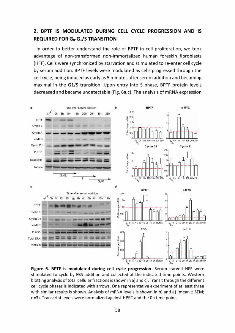
58
2. BPTF IS MODULATED DURING CELL CYCLE PROGRESSION AND IS
REQUIRED FOR G0-G1/S TRANSITION
In order to better understand the role of BPTF in cell proliferation, we took
advantage of non-transformed non-immortalized human foreskin fibroblasts
(HFF). Cells were synchronized by starvation and stimulated to re-enter cell cycle
by serum addition. BPTF levels were modulated as cells progressed through the
cell cycle, being induced as early as 5 minutes after serum addition and becoming
maximal in the G1/S transition. Upon entry into S phase, BPTF protein levels
decreased and became undetectable (Fig. 6a,c). The analysis of mRNA expression
Figure 6. BPTF is modulated during cell cycle progression. Serum-starved HFF were stimulated to cycle by FBS addition and collected at the indicated time points. Western blotting analysis of total cellular fractions is shown in a) and c). Transit through the different cell cycle phases is indicated with arrows. One representative experiment of at least three with similar results is shown. Analysis of mRNA levels is shown in b) and e) (mean ± SEM; n=3). Transcript levels were normalized against HPRT and the 0h time point.
59
revealed that, unlike cyclins (e.g. D1 and E) or transcription factors classically
involved in cell cycle control (e.g. c-MYC and AP-1), BPTF mRNA did not change
significantly throughout the experiment. These data suggest that post-
translational modifications are involved in the regulation of BPTF protein levels
(Fig. 6b,d).
BPTF silencing in HFF using two shRNAs led to a decrease in cell proliferation,
assessed by growth curve (Fig. 7a,b). When BPTF-interfered cells were serum-
starved and challenged to proliferate by FBS stimulation, they expressed lower
levels of c-MYC and Cyclins D1 and A than control cells (Fig. 7c). According to
these results, BPTF is necessary for the G0-G1/S transition.
Figure 7. BPTF is required for proliferation of HFF. a) HFF cells expressing control or BPTF-targeting shRNAs (sh#1 and sh#2) were examined by Western blotting. b) Cells in a) were seeded at similar densities at day 3 post-infection and counted at the indicated time points (mean ± SEM; n=3). *, P < 0.05. c) HFF cells transduced as in a) were synchronized and collected at different time points. Total cellular fractions were assessed by Western blotting. Only the data regarding shBPTF#1 is shown. One representative experiment of at least three with similar results is shown.
60
3. BPTF IS NECESSARY FOR c-MYC TRANSCRIPTIONAL ACTIVITY
BPTF modulates gene expression through the interaction with sequence-specific
transcription factors. The identity of such regulators has been extensively studied
in Drosophila but only a few have been discovered in murine and human cells
(Alkhatib and Landry 2011; Qiu et al. 2015). Since BPTF is necessary for cell
proliferation and, more precisely, for G0-G1/S transition, we hypothesized that its
effects might be mediated, at least in part, by c-MYC.
The hypothesis of BPTF as an interactive partner of c-MYC is attractive for
several reasons. First, c-MYC binding sites are highly enriched in H3K4me3,
H3K79me2 and H3 acetylation (Guccione et al. 2006; Martinato et al. 2008). This
open chromatin configuration operates upstream of sequence-recognition by c-
MYC and, most likely, is ‘read’ by c-MYC binding proteins and complexes with
specialized motifs (bromodomains, PHD fingers or chromodomains). So far,
however, the protein(s) involved in the recognition of these marks by c-MYC have
not been identified. Human BPTF contains two PHD fingers and one
bromodomain that bind to H3K4me2/3 and H4K16ac respectively (Li et al. 2006;
Ruthenburg et al. 2011), thus making it a plausible candidate. Second, c-MYC has
long been considered an undruggable oncogene. However, the disruption of
chromatin-dependent processes that suppress c-MYC activity - such as the
inhibition of the BET bromodomain protein Brd4 by JQ1 - has recently shown
promising results in experimental models of multiple myeloma, Burkitt
Lymphoma (BL), acute myeloid leukemia and acute lymphoblastic leukemia
(Dawson et al. 2011; Delmore et al. 2011; Mertz et al. 2011). BPTF also contains
a potentially druggable bromodomain that, if proven relevant for c-MYC function,
could be exploited in cancer therapy.
To assess whether BPTF is required for the transcriptional activity of c-MYC, we
took advantage of the steroid-activatable construct c-MYC-ER. c-MYC-ER is a
fusion protein in which the ligand-binding domain (ER) of a mutant estrogen
receptor, G525R (Danielian et al. 1993), is fused to the carboxyl terminus of c-
MYC. ER lacks intrinsic transactivation activity; it responds to the synthetic steroid
4-hydroxytamoxifen (4-OHT), but not to estrogens (Littlewood et al. 1995). The
MYC-ER protein is constitutively expressed but it is sequestered in the ctytoplasm
unless 4-OHT is supplied. Upon addition of 4-OHT, MYC-ER induces proliferation
and apoptosis in the same manner as wild-type MYC (Littlewood et al. 1995;
Alarcon et al. 1996).
HFF were stably transduced with the chimeric MYC-ER cDNA (HFF MYC-ER) and
infected with lentiviruses coding for either control (shNt) or BPTF-targeting
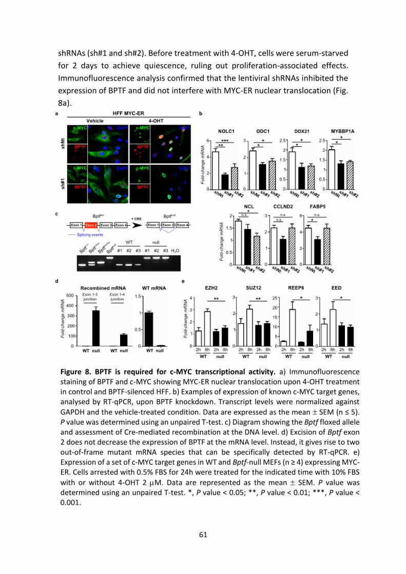
61
shRNAs (sh#1 and sh#2). Before treatment with 4-OHT, cells were serum-starved
for 2 days to achieve quiescence, ruling out proliferation-associated effects.
Immunofluorescence analysis confirmed that the lentiviral shRNAs inhibited the
expression of BPTF and did not interfere with MYC-ER nuclear translocation (Fig.
8a).
Figure 8. BPTF is required for c-MYC transcriptional activity. a) Immunofluorescence staining of BPTF and c-MYC showing MYC-ER nuclear translocation upon 4-OHT treatment in control and BPTF-silenced HFF. b) Examples of expression of known c-MYC target genes, analysed by RT-qPCR, upon BPTF knockdown. Transcript levels were normalized against
GAPDH and the vehicle-treated condition. Data are expressed as the mean SEM (n ≤ 5). P value was determined using an unpaired T-test. c) Diagram showing the Bptf floxed allele and assessment of Cre-mediated recombination at the DNA level. d) Excision of Bptf exon 2 does not decrease the expression of BPTF at the mRNA level. Instead, it gives rise to two out-of-frame mutant mRNA species that can be specifically detected by RT-qPCR. e) Expression of a set of c-MYC target genes in WT and Bptf-null MEFs (n ≥ 4) expressing MYC-ER. Cells arrested with 0.5% FBS for 24h were treated for the indicated time with 10% FBS
with or without 4-OHT 2 M. Data are represented as the mean SEM. P value was determined using an unpaired T-test. *, P value < 0.05; **, P value < 0.01; ***, P value < 0.001.
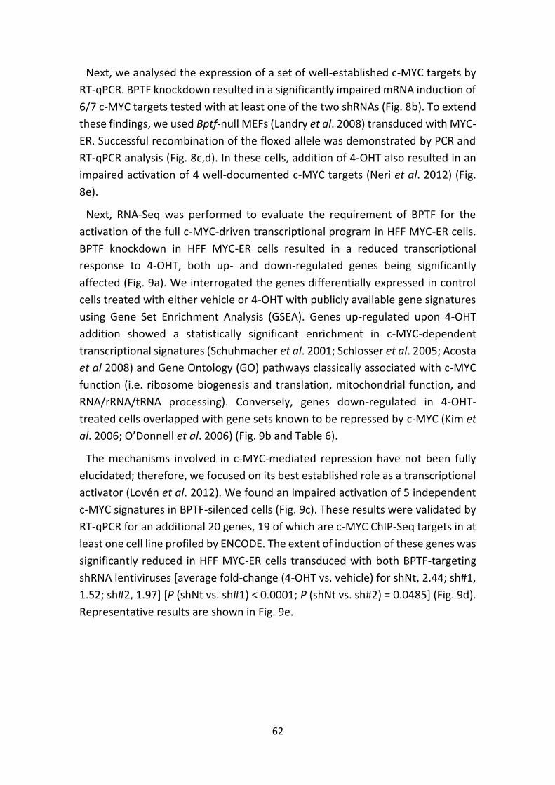
62
Next, we analysed the expression of a set of well-established c-MYC targets by
RT-qPCR. BPTF knockdown resulted in a significantly impaired mRNA induction of
6/7 c-MYC targets tested with at least one of the two shRNAs (Fig. 8b). To extend
these findings, we used Bptf-null MEFs (Landry et al. 2008) transduced with MYC-
ER. Successful recombination of the floxed allele was demonstrated by PCR and
RT-qPCR analysis (Fig. 8c,d). In these cells, addition of 4-OHT also resulted in an
impaired activation of 4 well-documented c-MYC targets (Neri et al. 2012) (Fig.
8e).
Next, RNA-Seq was performed to evaluate the requirement of BPTF for the
activation of the full c-MYC-driven transcriptional program in HFF MYC-ER cells.
BPTF knockdown in HFF MYC-ER cells resulted in a reduced transcriptional
response to 4-OHT, both up- and down-regulated genes being significantly
affected (Fig. 9a). We interrogated the genes differentially expressed in control
cells treated with either vehicle or 4-OHT with publicly available gene signatures
using Gene Set Enrichment Analysis (GSEA). Genes up-regulated upon 4-OHT
addition showed a statistically significant enrichment in c-MYC-dependent
transcriptional signatures (Schuhmacher et al. 2001; Schlosser et al. 2005; Acosta
et al 2008) and Gene Ontology (GO) pathways classically associated with c-MYC
function (i.e. ribosome biogenesis and translation, mitochondrial function, and
RNA/rRNA/tRNA processing). Conversely, genes down-regulated in 4-OHT-
treated cells overlapped with gene sets known to be repressed by c-MYC (Kim et
al. 2006; O’Donnell et al. 2006) (Fig. 9b and Table 6).
The mechanisms involved in c-MYC-mediated repression have not been fully
elucidated; therefore, we focused on its best established role as a transcriptional
activator (Lovén et al. 2012). We found an impaired activation of 5 independent
c-MYC signatures in BPTF-silenced cells (Fig. 9c). These results were validated by
RT-qPCR for an additional 20 genes, 19 of which are c-MYC ChIP-Seq targets in at
least one cell line profiled by ENCODE. The extent of induction of these genes was
significantly reduced in HFF MYC-ER cells transduced with both BPTF-targeting
shRNA lentiviruses [average fold-change (4-OHT vs. vehicle) for shNt, 2.44; sh#1,
1.52; sh#2, 1.97] [P (shNt vs. sh#1) < 0.0001; P (shNt vs. sh#2) = 0.0485] (Fig. 9d).
Representative results are shown in Fig. 9e.
63
Figure 9. Genome-wide analysis of BPTF-dependent c-MYC transcriptional activity. a) Fold-change in FPKM values (vehicle vs. 4-OHT) of up-regulated [Log2 F.c. ≥ +1] and down-regulated [Log2 F.c. ≤ -1] genes in control and BPTF-silenced cells. b) Snapshots of MYC-dependent gene sets displaying a positive or a negative enrichment in 4-OHT-treated control cells. c) Fold-change in FPKM values of c-MYC-dependent gene sets enriched in 4-OHT-treated control cells. Values are displayed for both control and BPTF-silenced HFF MYC-ER cells. Gene sets tested and P values: 1, Schuhmacher et al. ‘MYC Targets Up’ (P = 3.156·10-15); 2, Acosta et al. ‘Proliferation Independent MYC Targets Up’ (P = 2.034·10-08); 3, Schlosser et al. ‘MYC Targets Serum Response Up’ (P = 6.62·10-09); 4, Schlosser et al. ‘MYC Targets Serum Response Dn’ (P = 6.11·10-09); 5, Schlosser et al. ‘MYC Targets Repressed By Serum’ (P = 2.747·10-14). d) Fold-change in mRNA levels for the set of 20 genes used for validation. Left panel: data calculated from FPKM values. Right panel: data calculated from ≥3 independent experiments assessed by RT-qPCR. e) Examples of genes included in the validation. Transcript levels were normalized against GAPDH and the vehicle-treated
condition. Data are expressed as the mean SEM. *, P value < 0.05; **, P value < 0.01; ***, P value < 0.001
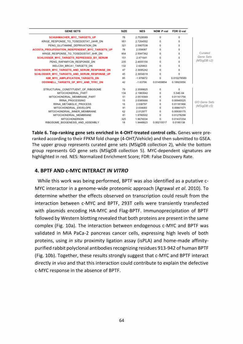
64
4. BPTF AND c-MYC INTERACT IN VITRO
While this work was being performed, BPTF was also identified as a putative c-
MYC interactor in a genome-wide proteomic approach (Agrawal et al. 2010). To
determine whether the effects observed on transcription could result from the
interaction between c-MYC and BPTF, 293T cells were transiently transfected
with plasmids encoding HA-MYC and Flag-BPTF. Immunoprecipitation of BPTF
followed by Western blotting revealed that both proteins are present in the same
complex (Fig. 10a). The interaction between endogenous c-MYC and BPTF was
validated in MIA PaCa-2 pancreas cancer cells, expressing high levels of both
proteins, using in situ proximity ligation assay (isPLA) and home-made affinity-
purified rabbit polyclonal antibodies recognizing residues 913-942 of human BPTF
(Fig. 10b). Together, these results strongly suggest that c-MYC and BPTF interact
directly in vivo and that this interaction could contribute to explain the defective
c-MYC response in the absence of BPTF.
Table 6. Top-ranking gene sets enriched in 4-OHT-treated control cells. Genes were pre-ranked according to their FPKM fold change (4-OHT/Vehicle) and then submitted to GSEA. The upper group represents curated gene sets (MSigDB collection 2), while the bottom group represents GO gene sets (MSigDB collection 5). MYC-dependent signatures are highlighted in red. NES: Normalized Enrichment Score; FDR: False Discovery Rate.
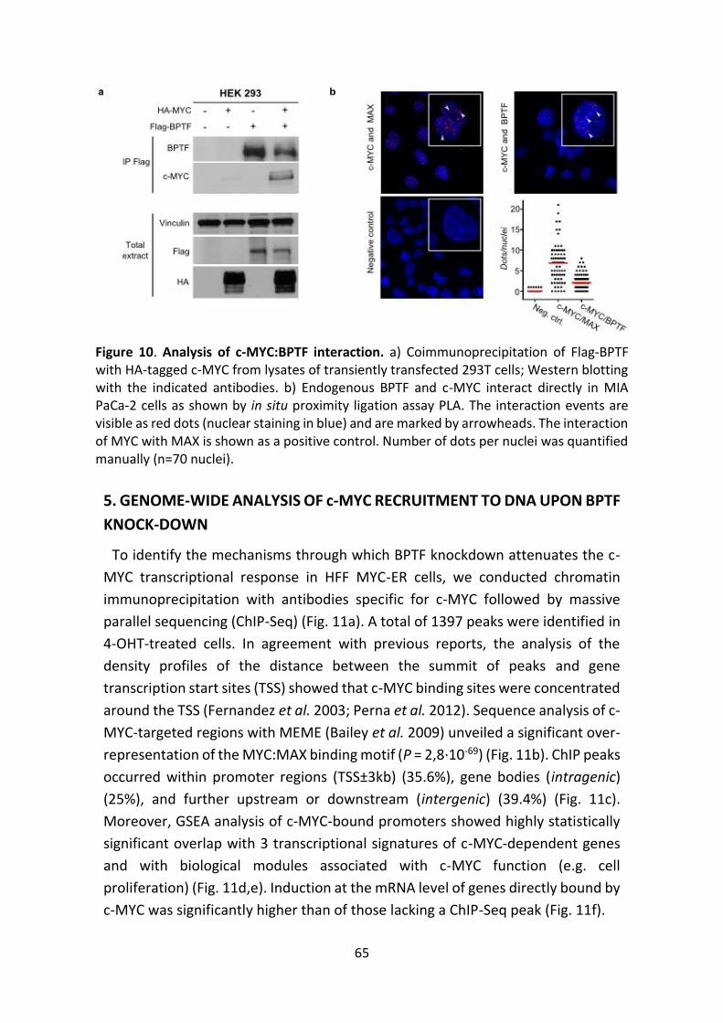
65
5. GENOME-WIDE ANALYSIS OF c-MYC RECRUITMENT TO DNA UPON BPTF
KNOCK-DOWN
To identify the mechanisms through which BPTF knockdown attenuates the c-
MYC transcriptional response in HFF MYC-ER cells, we conducted chromatin
immunoprecipitation with antibodies specific for c-MYC followed by massive
parallel sequencing (ChIP-Seq) (Fig. 11a). A total of 1397 peaks were identified in
4-OHT-treated cells. In agreement with previous reports, the analysis of the
density profiles of the distance between the summit of peaks and gene
transcription start sites (TSS) showed that c-MYC binding sites were concentrated
around the TSS (Fernandez et al. 2003; Perna et al. 2012). Sequence analysis of c-
MYC-targeted regions with MEME (Bailey et al. 2009) unveiled a significant over-
representation of the MYC:MAX binding motif (P = 2,8·10-69) (Fig. 11b). ChIP peaks
occurred within promoter regions (TSS±3kb) (35.6%), gene bodies (intragenic)
(25%), and further upstream or downstream (intergenic) (39.4%) (Fig. 11c).
Moreover, GSEA analysis of c-MYC-bound promoters showed highly statistically
significant overlap with 3 transcriptional signatures of c-MYC-dependent genes
and with biological modules associated with c-MYC function (e.g. cell
proliferation) (Fig. 11d,e). Induction at the mRNA level of genes directly bound by
c-MYC was significantly higher than of those lacking a ChIP-Seq peak (Fig. 11f).
Figure 10. Analysis of c-MYC:BPTF interaction. a) Coimmunoprecipitation of Flag-BPTF with HA-tagged c-MYC from lysates of transiently transfected 293T cells; Western blotting with the indicated antibodies. b) Endogenous BPTF and c-MYC interact directly in MIA PaCa-2 cells as shown by in situ proximity ligation assay PLA. The interaction events are visible as red dots (nuclear staining in blue) and are marked by arrowheads. The interaction of MYC with MAX is shown as a positive control. Number of dots per nuclei was quantified manually (n=70 nuclei).
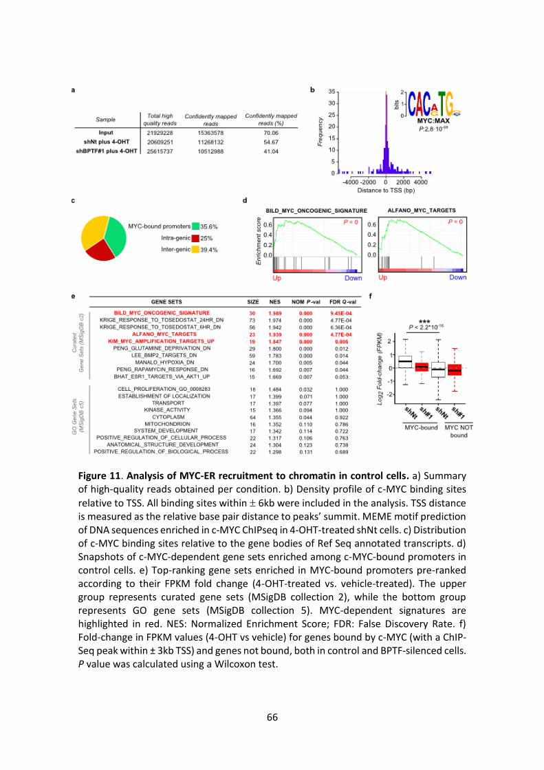
66
Figure 11. Analysis of MYC-ER recruitment to chromatin in control cells. a) Summary of high-quality reads obtained per condition. b) Density profile of c-MYC binding sites
relative to TSS. All binding sites within 6kb were included in the analysis. TSS distance is measured as the relative base pair distance to peaks’ summit. MEME motif prediction of DNA sequences enriched in c-MYC ChIPseq in 4-OHT-treated shNt cells. c) Distribution of c-MYC binding sites relative to the gene bodies of Ref Seq annotated transcripts. d) Snapshots of c-MYC-dependent gene sets enriched among c-MYC-bound promoters in control cells. e) Top-ranking gene sets enriched in MYC-bound promoters pre-ranked according to their FPKM fold change (4-OHT-treated vs. vehicle-treated). The upper group represents curated gene sets (MSigDB collection 2), while the bottom group represents GO gene sets (MSigDB collection 5). MYC-dependent signatures are highlighted in red. NES: Normalized Enrichment Score; FDR: False Discovery Rate. f) Fold-change in FPKM values (4-OHT vs vehicle) for genes bound by c-MYC (with a ChIP-Seq peak within ± 3kb TSS) and genes not bound, both in control and BPTF-silenced cells. P value was calculated using a Wilcoxon test.
67
To determine whether BPTF silencing interfered with c-MYC recruitment to
chromatin, we analysed the magnitude and distribution of c-MYC ChIP-Seq peaks
upon BPTF knockdown in HFF. Globally, c-MYC binding intensity was significantly
lower in shBPTF-expressing cells (P < 2.2·10-16) (Fig. 12a,b). This reduction was not
evenly distributed at the genome wide level, with 50.2% of the peaks showing a
read number fold-change ≥ 2. The selective effect of BPTF silencing on a subset
of c-MYC ChIP-Seq peaks -regardless of their intensity- suggests that the
differences do not result from an inefficient ChIP. We validated these
observations by ChIP-qPCR on gene promoters for which a peak was identified in
the ChIP-Seq experiment ("target"), as well as on a set of “non-target” control
genomic regions, in at least 3 independent experiments. c-MYC was recruited to
target regions and did not show significant binding to non-target promoters. c-
MYC recruitment to target genes was significantly reduced in cells infected with
the BPTF-targeting shRNAs (Fig. 12c,d). High-affinity MYC targets (Fernandez et
al. 2003; Guccione et al. 2006; Perna et al. 2012) were significantly enriched
among the genes for which BPTF silencing had more effect on c-MYC recruitment
(Fig. 12e). The effect of BPTF silencing on the induction of c-MYC target mRNAs
was independent from the extent of reduction in c-MYC binding at their
promoters (Fig. 12f), suggesting that BPTF operates downstream of c-MYC in the
sequence of events resulting in transcriptional activation.
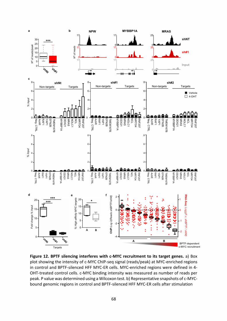
68
Figure 12. BPTF silencing interferes with c-MYC recruitment to its target genes. a) Box plot showing the intensity of c-MYC ChIP-seq signal (reads/peak) at MYC-enriched regions in control and BPTF-silenced HFF MYC-ER cells. MYC-enriched regions were defined in 4-OHT-treated control cells. c-MYC binding intensity was measured as number of reads per peak. P value was determined using a Wilcoxon test. b) Representative snapshots of c-MYC-bound genomic regions in control and BPTF-silenced HFF MYC-ER cells after stimulation
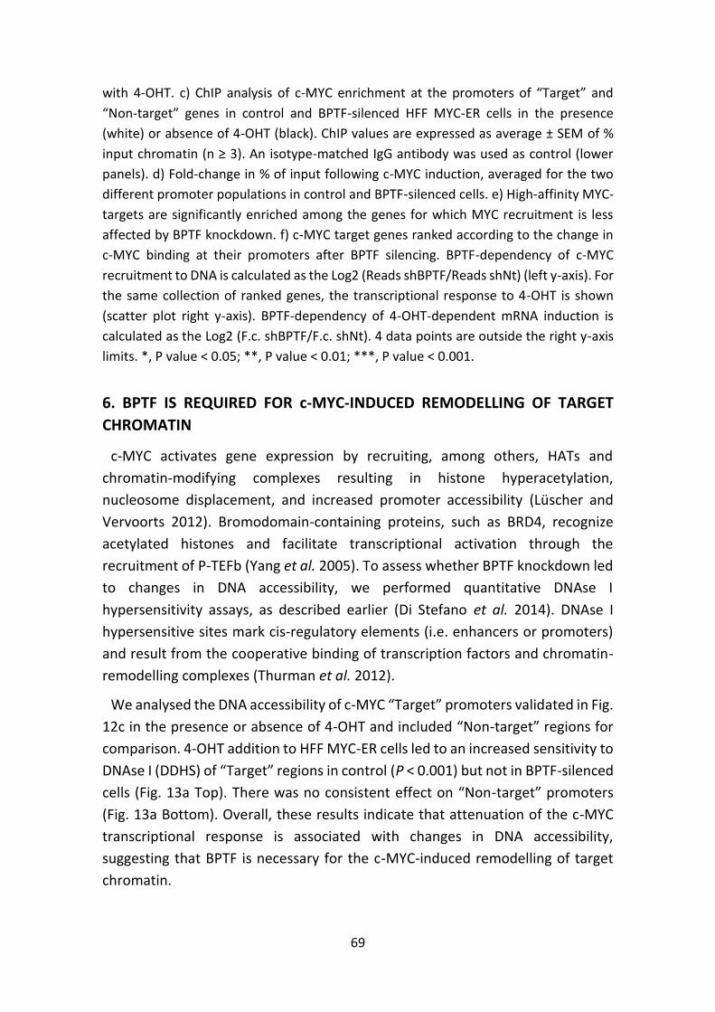
69
with 4-OHT. c) ChIP analysis of c-MYC enrichment at the promoters of “Target” and
“Non-target” genes in control and BPTF-silenced HFF MYC-ER cells in the presence
(white) or absence of 4-OHT (black). ChIP values are expressed as average ± SEM of %
input chromatin (n ≥ 3). An isotype-matched IgG antibody was used as control (lower
panels). d) Fold-change in % of input following c-MYC induction, averaged for the two
different promoter populations in control and BPTF-silenced cells. e) High-affinity MYC-
targets are significantly enriched among the genes for which MYC recruitment is less
affected by BPTF knockdown. f) c-MYC target genes ranked according to the change in
c-MYC binding at their promoters after BPTF silencing. BPTF-dependency of c-MYC
recruitment to DNA is calculated as the Log2 (Reads shBPTF/Reads shNt) (left y-axis). For
the same collection of ranked genes, the transcriptional response to 4-OHT is shown
(scatter plot right y-axis). BPTF-dependency of 4-OHT-dependent mRNA induction is
calculated as the Log2 (F.c. shBPTF/F.c. shNt). 4 data points are outside the right y-axis
limits. *, P value < 0.05; **, P value < 0.01; ***, P value < 0.001.
6. BPTF IS REQUIRED FOR c-MYC-INDUCED REMODELLING OF TARGET
CHROMATIN
c-MYC activates gene expression by recruiting, among others, HATs and
chromatin-modifying complexes resulting in histone hyperacetylation,
nucleosome displacement, and increased promoter accessibility (Lüscher and
Vervoorts 2012). Bromodomain-containing proteins, such as BRD4, recognize
acetylated histones and facilitate transcriptional activation through the
recruitment of P-TEFb (Yang et al. 2005). To assess whether BPTF knockdown led
to changes in DNA accessibility, we performed quantitative DNAse I
hypersensitivity assays, as described earlier (Di Stefano et al. 2014). DNAse I
hypersensitive sites mark cis-regulatory elements (i.e. enhancers or promoters)
and result from the cooperative binding of transcription factors and chromatin-
remodelling complexes (Thurman et al. 2012).
We analysed the DNA accessibility of c-MYC “Target” promoters validated in Fig.
12c in the presence or absence of 4-OHT and included “Non-target” regions for
comparison. 4-OHT addition to HFF MYC-ER cells led to an increased sensitivity to
DNAse I (DDHS) of “Target” regions in control (P < 0.001) but not in BPTF-silenced
cells (Fig. 13a Top). There was no consistent effect on “Non-target” promoters
(Fig. 13a Bottom). Overall, these results indicate that attenuation of the c-MYC
transcriptional response is associated with changes in DNA accessibility,
suggesting that BPTF is necessary for the c-MYC-induced remodelling of target
chromatin.
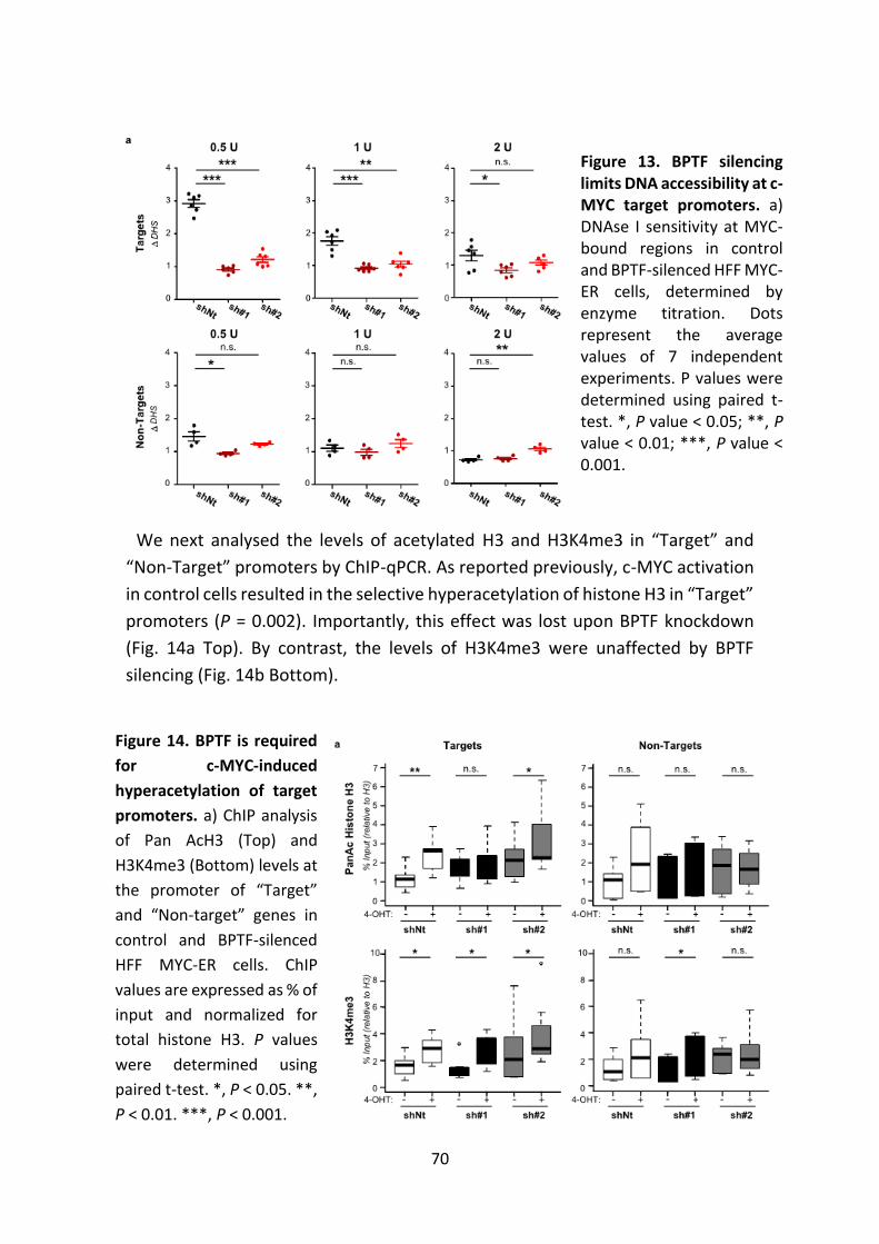
70
We next analysed the levels of acetylated H3 and H3K4me3 in “Target” and
“Non-Target” promoters by ChIP-qPCR. As reported previously, c-MYC activation
in control cells resulted in the selective hyperacetylation of histone H3 in “Target”
promoters (P = 0.002). Importantly, this effect was lost upon BPTF knockdown
(Fig. 14a Top). By contrast, the levels of H3K4me3 were unaffected by BPTF
silencing (Fig. 14b Bottom).
Figure 13. BPTF silencing limits DNA accessibility at c-MYC target promoters. a) DNAse I sensitivity at MYC-bound regions in control and BPTF-silenced HFF MYC-ER cells, determined by enzyme titration. Dots represent the average values of 7 independent experiments. P values were determined using paired t-test. *, P value < 0.05; **, P value < 0.01; ***, P value < 0.001.
Figure 14. BPTF is required
for c-MYC-induced
hyperacetylation of target
promoters. a) ChIP analysis
of Pan AcH3 (Top) and
H3K4me3 (Bottom) levels at
the promoter of “Target”
and “Non-target” genes in
control and BPTF-silenced
HFF MYC-ER cells. ChIP
values are expressed as % of
input and normalized for
total histone H3. P values
were determined using
paired t-test. *, P < 0.05. **,
P < 0.01. ***, P < 0.001.
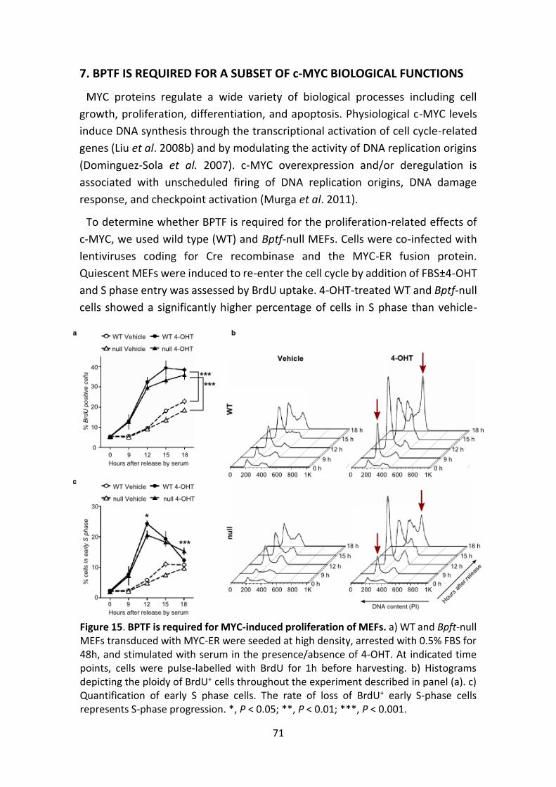
71
7. BPTF IS REQUIRED FOR A SUBSET OF c-MYC BIOLOGICAL FUNCTIONS
MYC proteins regulate a wide variety of biological processes including cell
growth, proliferation, differentiation, and apoptosis. Physiological c-MYC levels
induce DNA synthesis through the transcriptional activation of cell cycle-related
genes (Liu et al. 2008b) and by modulating the activity of DNA replication origins
(Dominguez-Sola et al. 2007). c-MYC overexpression and/or deregulation is
associated with unscheduled firing of DNA replication origins, DNA damage
response, and checkpoint activation (Murga et al. 2011).
To determine whether BPTF is required for the proliferation-related effects of
c-MYC, we used wild type (WT) and Bptf-null MEFs. Cells were co-infected with
lentiviruses coding for Cre recombinase and the MYC-ER fusion protein.
Quiescent MEFs were induced to re-enter the cell cycle by addition of FBS±4-OHT
and S phase entry was assessed by BrdU uptake. 4-OHT-treated WT and Bptf-null
cells showed a significantly higher percentage of cells in S phase than vehicle-
Figure 15. BPTF is required for MYC-induced proliferation of MEFs. a) WT and Bpft-null MEFs transduced with MYC-ER were seeded at high density, arrested with 0.5% FBS for 48h, and stimulated with serum in the presence/absence of 4-OHT. At indicated time points, cells were pulse-labelled with BrdU for 1h before harvesting. b) Histograms depicting the ploidy of BrdU+ cells throughout the experiment described in panel (a). c) Quantification of early S phase cells. The rate of loss of BrdU+ early S-phase cells represents S-phase progression. *, P < 0.05; **, P < 0.01; ***, P < 0.001.
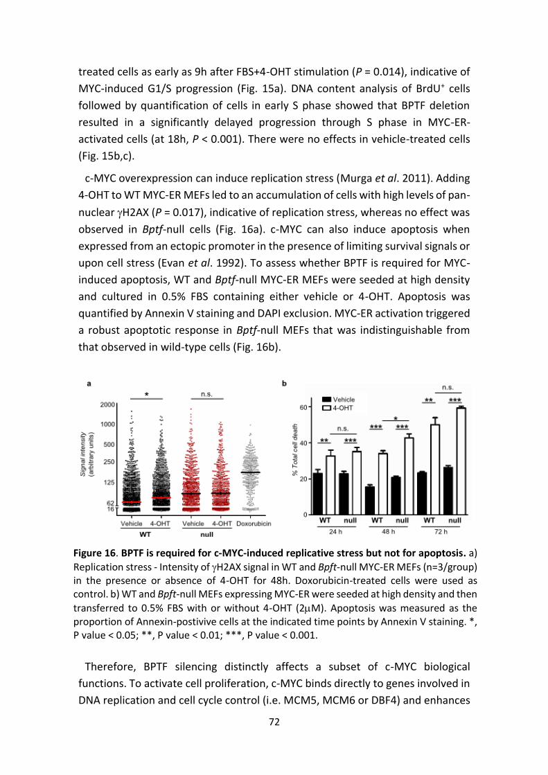
72
treated cells as early as 9h after FBS+4-OHT stimulation (P = 0.014), indicative of
MYC-induced G1/S progression (Fig. 15a). DNA content analysis of BrdU+ cells
followed by quantification of cells in early S phase showed that BPTF deletion
resulted in a significantly delayed progression through S phase in MYC-ER-
activated cells (at 18h, P < 0.001). There were no effects in vehicle-treated cells
(Fig. 15b,c).
c-MYC overexpression can induce replication stress (Murga et al. 2011). Adding
4-OHT to WT MYC-ER MEFs led to an accumulation of cells with high levels of pan-
nuclear H2AX (P = 0.017), indicative of replication stress, whereas no effect was
observed in Bptf-null cells (Fig. 16a). c-MYC can also induce apoptosis when
expressed from an ectopic promoter in the presence of limiting survival signals or
upon cell stress (Evan et al. 1992). To assess whether BPTF is required for MYC-
induced apoptosis, WT and Bptf-null MYC-ER MEFs were seeded at high density
and cultured in 0.5% FBS containing either vehicle or 4-OHT. Apoptosis was
quantified by Annexin V staining and DAPI exclusion. MYC-ER activation triggered
a robust apoptotic response in Bptf-null MEFs that was indistinguishable from
that observed in wild-type cells (Fig. 16b).
Therefore, BPTF silencing distinctly affects a subset of c-MYC biological
functions. To activate cell proliferation, c-MYC binds directly to genes involved in
DNA replication and cell cycle control (i.e. MCM5, MCM6 or DBF4) and enhances
Figure 16. BPTF is required for c-MYC-induced replicative stress but not for apoptosis. a)
Replication stress - Intensity of H2AX signal in WT and Bpft-null MYC-ER MEFs (n=3/group) in the presence or absence of 4-OHT for 48h. Doxorubicin-treated cells were used as control. b) WT and Bpft-null MEFs expressing MYC-ER were seeded at high density and then
transferred to 0.5% FBS with or without 4-OHT (2M). Apoptosis was measured as the proportion of Annexin-postivive cells at the indicated time points by Annexin V staining. *, P value < 0.05; **, P value < 0.01; ***, P value < 0.001.
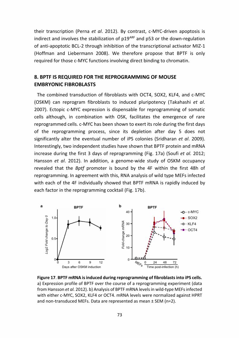
73
their transcription (Perna et al. 2012). By contrast, c-MYC-driven apoptosis is
indirect and involves the stabilization of p19ARF and p53 or the down-regulation
of anti-apoptotic BCL-2 through inhibition of the transcriptional activator MIZ-1
(Hoffman and Liebermann 2008). We therefore propose that BPTF is only
required for those c-MYC functions involving direct binding to chromatin.
8. BPTF IS REQUIRED FOR THE REPROGRAMMING OF MOUSE
EMBRYONIC FIBROBLASTS
The combined transduction of fibroblasts with OCT4, SOX2, KLF4, and c-MYC
(OSKM) can reprogram fibroblasts to induced pluripotency (Takahashi et al.
2007). Ectopic c-MYC expression is dispensable for reprogramming of somatic
cells although, in combination with OSK, facilitates the emergence of rare
reprogrammed cells. c-MYC has been shown to exert its role during the first days
of the reprogramming process, since its depletion after day 5 does not
significantly alter the eventual number of iPS colonies (Sridharan et al. 2009).
Interestingly, two independent studies have shown that BPTF protein and mRNA
increase during the first 3 days of reprogramming (Fig. 17a) (Soufi et al. 2012;
Hansson et al. 2012). In addition, a genome-wide study of OSKM occupancy
revealed that the Bptf promoter is bound by the 4F within the first 48h of
reprogramming. In agreement with this, RNA analysis of wild type MEFs infected
with each of the 4F individually showed that BPTF mRNA is rapidly induced by
each factor in the reprogramming cocktail (Fig. 17b).
Figure 17. BPTF mRNA is induced during reprogramming of fibroblasts into iPS cells. a) Expression profile of BPTF over the course of a reprogramming experiment (data from Hansson et al. 2012). b) Analysis of BPTF mRNA levels in wild-type MEFs infected with either c-MYC, SOX2, KLF4 or OCT4. mRNA levels were normalized against HPRT and non-transduced MEFs. Data are represented as mean ± SEM (n=2).
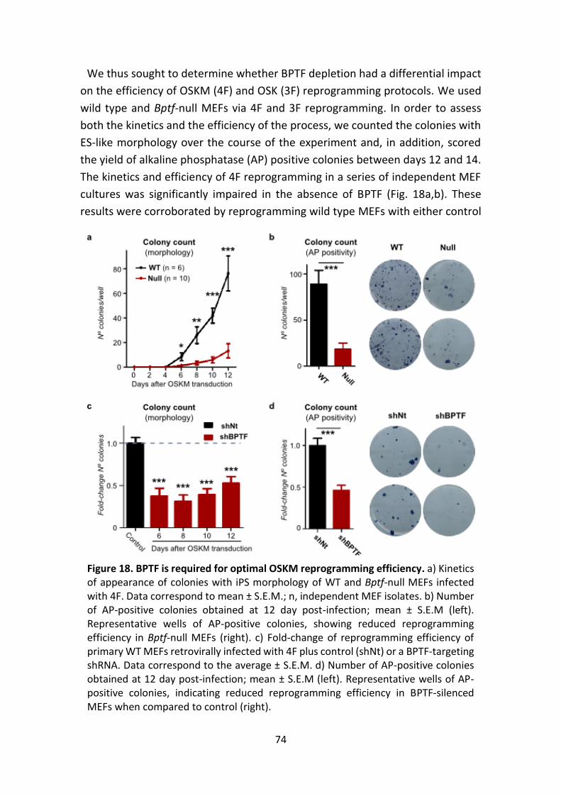
74
We thus sought to determine whether BPTF depletion had a differential impact
on the efficiency of OSKM (4F) and OSK (3F) reprogramming protocols. We used
wild type and Bptf-null MEFs via 4F and 3F reprogramming. In order to assess
both the kinetics and the efficiency of the process, we counted the colonies with
ES-like morphology over the course of the experiment and, in addition, scored
the yield of alkaline phosphatase (AP) positive colonies between days 12 and 14.
The kinetics and efficiency of 4F reprogramming in a series of independent MEF
cultures was significantly impaired in the absence of BPTF (Fig. 18a,b). These
results were corroborated by reprogramming wild type MEFs with either control
Figure 18. BPTF is required for optimal OSKM reprogramming efficiency. a) Kinetics of appearance of colonies with iPS morphology of WT and Bptf-null MEFs infected with 4F. Data correspond to mean ± S.E.M.; n, independent MEF isolates. b) Number of AP-positive colonies obtained at 12 day post-infection; mean ± S.E.M (left). Representative wells of AP-positive colonies, showing reduced reprogramming efficiency in Bptf-null MEFs (right). c) Fold-change of reprogramming efficiency of primary WT MEFs retrovirally infected with 4F plus control (shNt) or a BPTF-targeting shRNA. Data correspond to the average ± S.E.M. d) Number of AP-positive colonies obtained at 12 day post-infection; mean ± S.E.M (left). Representative wells of AP-positive colonies, indicating reduced reprogramming efficiency in BPTF-silenced MEFs when compared to control (right).
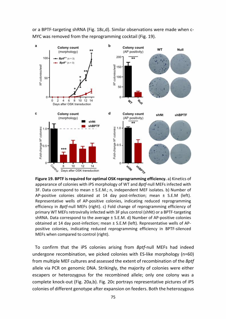
75
or a BPTF-targeting shRNA (Fig. 18c,d). Similar observations were made when c-
MYC was removed from the reprogramming cocktail (Fig. 19).
To confirm that the iPS colonies arising from Bptf-null MEFs had indeed
undergone recombination, we picked colonies with ES-like morphology (n=60)
from multiple MEF cultures and assessed the extent of recombination of the Bptf
allele via PCR on genomic DNA. Strikingly, the majority of colonies were either
escapers or heterozygous for the recombined allele; only one colony was a
complete knock-out (Fig. 20a,b). Fig. 20c portrays representative pictures of iPS
colonies of different genotype after expansion on feeders. Both the heterozygous
Figure 19. BPTF is required for optimal OSK reprogramming efficiency. a) Kinetics of appearance of colonies with iPS morphology of WT and Bptf-null MEFs infected with 3F. Data correspond to mean ± S.E.M.; n, independent MEF isolates. b) Number of AP-positive colonies obtained at 14 day post-infection; mean ± S.E.M (left). Representative wells of AP-positive colonies, indicating reduced reprogramming efficiency in Bptf-null MEFs (right). c) Fold change of reprogramming efficiency of primary WT MEFs retrovirally infected with 3F plus control (shNt) or a BPTF-targeting shRNA. Data correspond to the average ± S.E.M. d) Number of AP-positive colonies obtained at 14 day post-infection; mean ± S.E.M (left). Representative wells of AP-positive colonies, indicating reduced reprogramming efficiency in BPTF-silenced MEFs when compared to control (right).
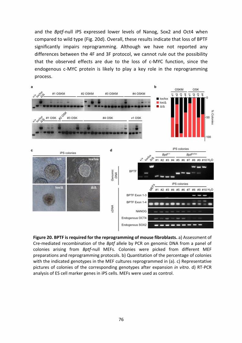
76
and the Bptf-null iPS expressed lower levels of Nanog, Sox2 and Oct4 when
compared to wild type (Fig. 20d). Overall, these results indicate that loss of BPTF
significantly impairs reprogramming. Although we have not reported any
differences between the 4F and 3F protocol, we cannot rule out the possibility
that the observed effects are due to the loss of c-MYC function, since the
endogenous c-MYC protein is likely to play a key role in the reprogramming
process.
Figure 20. BPTF is required for the reprogramming of mouse fibroblasts. a) Assessment of Cre-mediated recombination of the Bptf allele by PCR on genomic DNA from a panel of colonies arising from Bptf-null MEFs. Colonies were picked from different MEF preparations and reprogramming protocols. b) Quantitation of the percentage of colonies with the indicated genotypes in the MEF cultures reprogrammed in (a). c) Representative pictures of colonies of the corresponding genotypes after expansion in vitro. d) RT-PCR analysis of ES cell marker genes in iPS cells. MEFs were used as control.
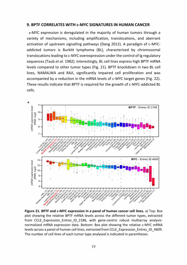
77
9. BPTF CORRELATES WITH c-MYC SIGNATURES IN HUMAN CANCER
c-MYC expression is deregulated in the majority of human tumors through a
variety of mechanisms, including amplification, translocations, and aberrant
activation of upstream signalling pathways (Dang 2012). A paradigm of c-MYC-
addicted tumors is Burkitt lymphoma (BL), characterized by chromosomal
translocations leading to c-MYC overexpression under the control of Ig regulatory
sequences (Taub et al. 1982). Interestingly, BL cell lines express high BPTF mRNA
levels compared to other tumor types (Fig. 21). BPTF knockdown in two BL cell
lines, NAMALWA and RAJI, significantly impaired cell proliferation and was
accompanied by a reduction in the mRNA levels of c-MYC target genes (Fig. 22).
These results indicate that BPTF is required for the growth of c-MYC-addicted BL
cells.
Figure 21. BPTF and c-MYC expression in a panel of human cancer cell lines. a) Top: Box plot showing the relative BPTF mRNA levels across the different tumor types, extracted from CCLE_Expression_Entrez_ID_2186, with gene-centric robust multiarray analysis-normalized mRNA expression data. Bottom: Box plot showing the relative c-MYC mRNA levels across a panel of human cell lines, extracted from CCLE_Expression_Entrez_ID_4609. The number of cell lines of each tumor type analysed is indicated in parentheses.
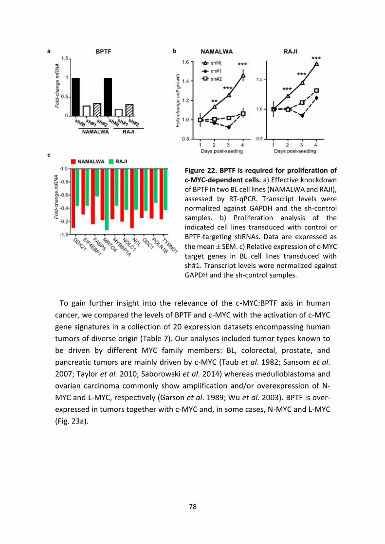
78
To gain further insight into the relevance of the c-MYC:BPTF axis in human
cancer, we compared the levels of BPTF and c-MYC with the activation of c-MYC
gene signatures in a collection of 20 expression datasets encompassing human
tumors of diverse origin (Table 7). Our analyses included tumor types known to
be driven by different MYC family members: BL, colorectal, prostate, and
pancreatic tumors are mainly driven by c-MYC (Taub et al. 1982; Sansom et al.
2007; Taylor et al. 2010; Saborowski et al. 2014) whereas medulloblastoma and
ovarian carcinoma commonly show amplification and/or overexpression of N-
MYC and L-MYC, respectively (Garson et al. 1989; Wu et al. 2003). BPTF is over-
expressed in tumors together with c-MYC and, in some cases, N-MYC and L-MYC
(Fig. 23a).
Figure 22. BPTF is required for proliferation of c-MYC-dependent cells. a) Effective knockdown of BPTF in two BL cell lines (NAMALWA and RAJI), assessed by RT-qPCR. Transcript levels were normalized against GAPDH and the sh-control samples. b) Proliferation analysis of the indicated cell lines transduced with control or BPTF-targeting shRNAs. Data are expressed as
the mean SEM. c) Relative expression of c-MYC target genes in BL cell lines transduced with sh#1. Transcript levels were normalized against GAPDH and the sh-control samples.
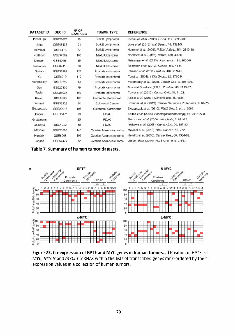
79
Table 7. Summary of human tumor datasets.
Figure 23. Co-expression of BPTF and MYC genes in human tumors. a) Position of BPTF, c-MYC, MYCN and MYCL1 mRNAs within the lists of transcribed genes rank-ordered by their expression values in a collection of human tumors.
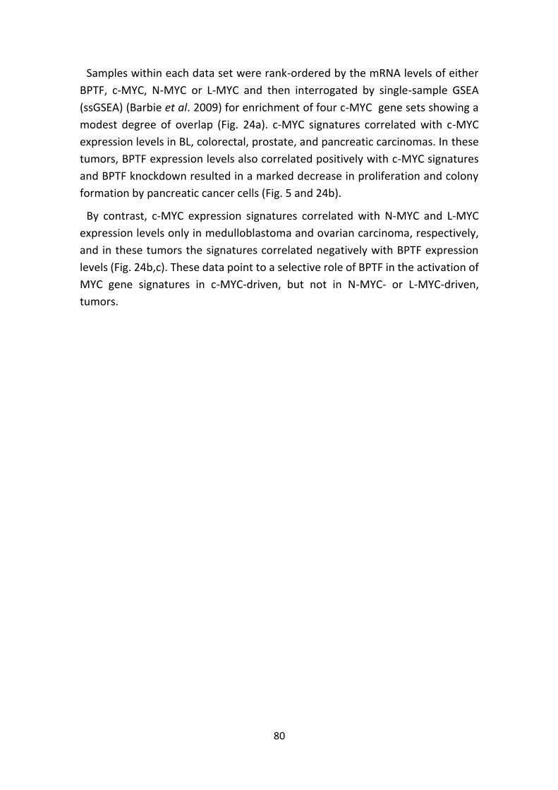
80
Samples within each data set were rank-ordered by the mRNA levels of either
BPTF, c-MYC, N-MYC or L-MYC and then interrogated by single-sample GSEA
(ssGSEA) (Barbie et al. 2009) for enrichment of four c-MYC gene sets showing a
modest degree of overlap (Fig. 24a). c-MYC signatures correlated with c-MYC
expression levels in BL, colorectal, prostate, and pancreatic carcinomas. In these
tumors, BPTF expression levels also correlated positively with c-MYC signatures
and BPTF knockdown resulted in a marked decrease in proliferation and colony
formation by pancreatic cancer cells (Fig. 5 and 24b).
By contrast, c-MYC expression signatures correlated with N-MYC and L-MYC
expression levels only in medulloblastoma and ovarian carcinoma, respectively,
and in these tumors the signatures correlated negatively with BPTF expression
levels (Fig. 24b,c). These data point to a selective role of BPTF in the activation of
MYC gene signatures in c-MYC-driven, but not in N-MYC- or L-MYC-driven,
tumors.
81
Figure 24. BPTF expression correlates with c-MYC signatures in human tumors. a) Venn diagram showing the overlap of the c-MYC signatures used in the following analyses. b) Dot plot of Normalized Enrichment Scores (NES) of the 4 c-MYC signatures based on GSEA. NES values were calculated for each data set previously ranked-ordered by either BPTF or c-MYC levels. c) Volcano plots of NES and enrichment P values of c-MYC signatures based on GSEA. NES values were calculated for each data set previously ranked-ordered by either BPTF, c-MYC, N-MYC or L-MYC mRNA levels. Filled circles represent gene sets with a FDR < 0.25.
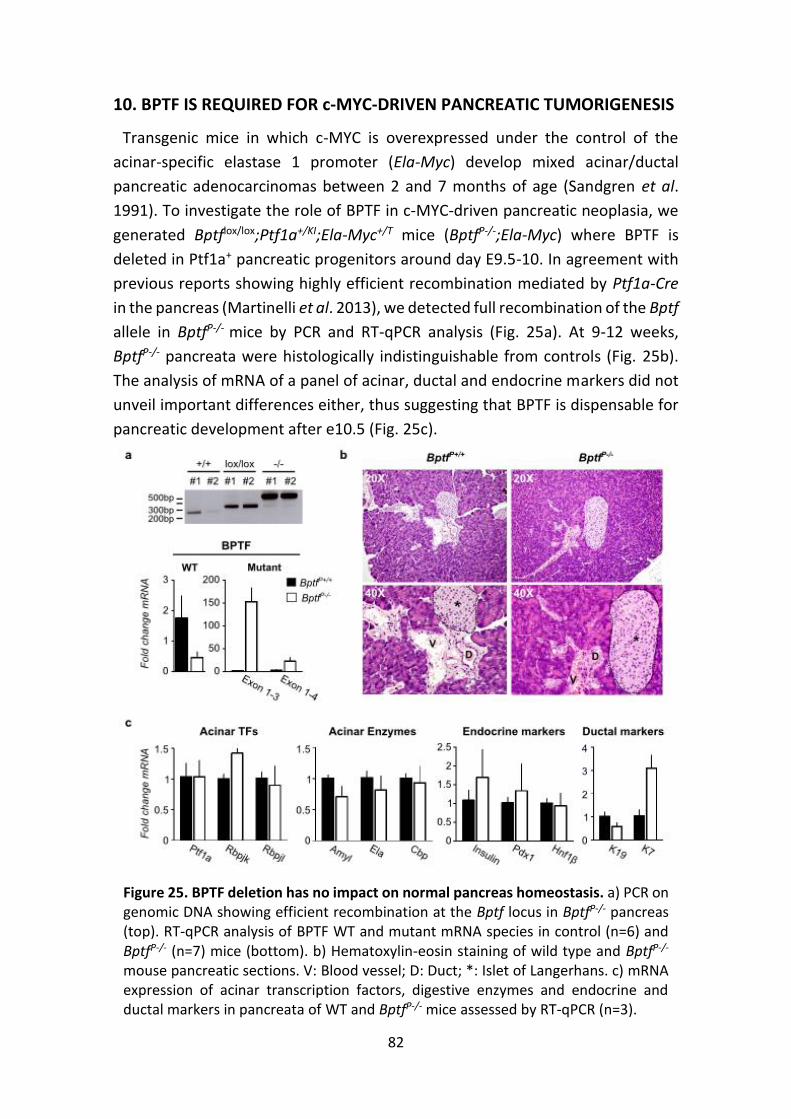
82
10. BPTF IS REQUIRED FOR c-MYC-DRIVEN PANCREATIC TUMORIGENESIS
Transgenic mice in which c-MYC is overexpressed under the control of the
acinar-specific elastase 1 promoter (Ela-Myc) develop mixed acinar/ductal
pancreatic adenocarcinomas between 2 and 7 months of age (Sandgren et al.
1991). To investigate the role of BPTF in c-MYC-driven pancreatic neoplasia, we
generated Bptflox/lox;Ptf1a+/KI;Ela-Myc+/T mice (BptfP-/-;Ela-Myc) where BPTF is
deleted in Ptf1a+ pancreatic progenitors around day E9.5-10. In agreement with
previous reports showing highly efficient recombination mediated by Ptf1a-Cre
in the pancreas (Martinelli et al. 2013), we detected full recombination of the Bptf
allele in BptfP-/- mice by PCR and RT-qPCR analysis (Fig. 25a). At 9-12 weeks,
BptfP-/- pancreata were histologically indistinguishable from controls (Fig. 25b).
The analysis of mRNA of a panel of acinar, ductal and endocrine markers did not
unveil important differences either, thus suggesting that BPTF is dispensable for
pancreatic development after e10.5 (Fig. 25c).
Figure 25. BPTF deletion has no impact on normal pancreas homeostasis. a) PCR on genomic DNA showing efficient recombination at the Bptf locus in BptfP-/- pancreas (top). RT-qPCR analysis of BPTF WT and mutant mRNA species in control (n=6) and BptfP-/- (n=7) mice (bottom). b) Hematoxylin-eosin staining of wild type and BptfP-/- mouse pancreatic sections. V: Blood vessel; D: Duct; *: Islet of Langerhans. c) mRNA expression of acinar transcription factors, digestive enzymes and endocrine and ductal markers in pancreata of WT and BptfP-/- mice assessed by RT-qPCR (n=3).
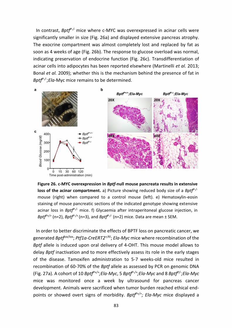
83
In contrast, BptfP-/- mice where c-MYC was overexpressed in acinar cells were
significantly smaller in size (Fig. 26a) and displayed extensive pancreas atrophy.
The exocrine compartment was almost completely lost and replaced by fat as
soon as 4 weeks of age (Fig. 26b). The response to glucose overload was normal,
indicating preservation of endocrine function (Fig. 26c). Transdifferentiation of
acinar cells into adipocytes has been reported elsewhere (Martinelli et al. 2013;
Bonal et al. 2009); whether this is the mechanism behind the presence of fat in
BptfP-/-;Ela-Myc mice remains to be determined.
In order to better discriminate the effects of BPTF loss on pancreatic cancer, we
generated Bptflox/lox; Ptf1a-CreERT2+/KI; Ela-Myc mice where recombination of the
Bptf allele is induced upon oral delivery of 4-OHT. This mouse model allows to
delay Bptf inactivation and to more effectively assess its role in the early stages
of the disease. Tamoxifen administration to 5-7 weeks-old mice resulted in
recombination of 60-70% of the Bptf allele as assessed by PCR on genomic DNA
(Fig. 27a). A cohort of 10 BptfP+/+;Ela-Myc, 5 BptfP-/+;Ela-Myc and 8 BptfP/-;Ela-Myc
mice was monitored once a week by ultrasound for pancreas cancer
development. Animals were sacrificed when tumor burden reached ethical end-
points or showed overt signs of morbidity. BptfP+/+; Ela-Myc mice displayed a
Figure 26. c-MYC overexpression in Bptf-null mouse pancreata results in extensive
loss of the acinar compartment. a) Picture showing reduced body size of a BptfP-/-
mouse (right) when compared to a control mouse (left). e) Hematoxylin-eosin
staining of mouse pancreatic sections of the indicated genotype showing extensive
acinar loss in BptfP-/- mice. f) Glycaemia after intraperitoneal glucose injection, in
BptfP+/+ (n=2), BptfP-/+ (n=3), and BptfP-/- (n=2) mice. Data are mean ± SEM.
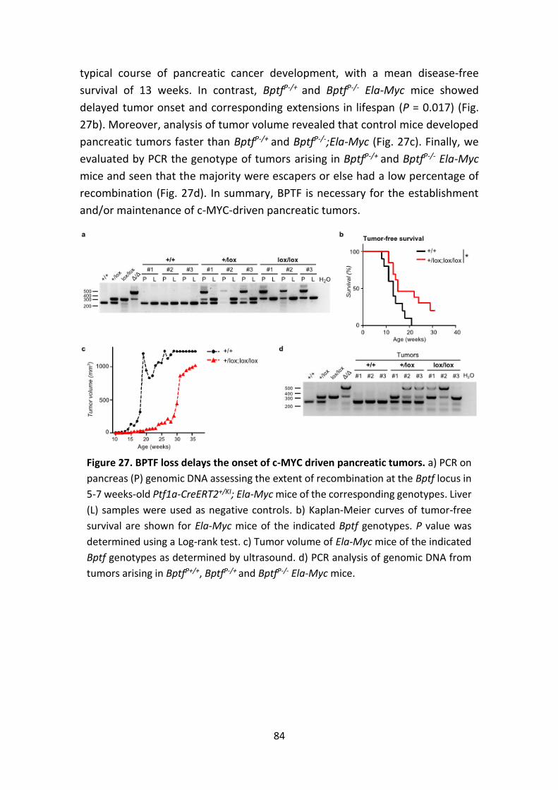
84
typical course of pancreatic cancer development, with a mean disease-free
survival of 13 weeks. In contrast, BptfP-/+ and BptfP-/- Ela-Myc mice showed
delayed tumor onset and corresponding extensions in lifespan (P = 0.017) (Fig.
27b). Moreover, analysis of tumor volume revealed that control mice developed
pancreatic tumors faster than BptfP-/+ and BptfP-/-;Ela-Myc (Fig. 27c). Finally, we
evaluated by PCR the genotype of tumors arising in BptfP-/+ and BptfP-/- Ela-Myc
mice and seen that the majority were escapers or else had a low percentage of
recombination (Fig. 27d). In summary, BPTF is necessary for the establishment
and/or maintenance of c-MYC-driven pancreatic tumors.
Figure 27. BPTF loss delays the onset of c-MYC driven pancreatic tumors. a) PCR on
pancreas (P) genomic DNA assessing the extent of recombination at the Bptf locus in
5-7 weeks-old Ptf1a-CreERT2+/KI; Ela-Myc mice of the corresponding genotypes. Liver
(L) samples were used as negative controls. b) Kaplan-Meier curves of tumor-free
survival are shown for Ela-Myc mice of the indicated Bptf genotypes. P value was
determined using a Log-rank test. c) Tumor volume of Ela-Myc mice of the indicated
Bptf genotypes as determined by ultrasound. d) PCR analysis of genomic DNA from
tumors arising in BptfP+/+, BptfP-/+ and BptfP-/- Ela-Myc mice.

85
Discussion
86
DISCUSSION
We initially identified BPTF as an element of a network of transcription
regulators whose expression was modulated during proliferation/cell cycle arrest
of two pancreatic cancer cell lines. In agreement with our in silico prediction, BPTF
down-regulation in these cells was associated with impaired proliferation. We
have expanded this observation to other human cancer cell lines (bladder and
Burkitt lymphoma) and to non-transformed, non-immortalized human
fibroblasts. Based on these findings, we focused on the study of its biological
functions.
1. BPTF AND CELL PROLIFERATION
In quiescent human fibroblasts, BPTF depletion impedes cells from progressing
into S phase and completing cell cycle. Of note, Bptf-null MEFs do not show
significant proliferation defects when compared to wild type cells, which could
reflect species- and/or cell type-specific BPTF roles. Alternatively, MEFs could
have a distinct ability to adapt and bypass the need of BPTF.
DNA replication requires extensive chromatin rearrangements; it is thus
conceivable that chromatin remodelling by BPTF:NURF might contribute to this
process via multiple mechanisms.
Initiation of DNA synthesis takes place through a series of tightly coordinated
events occurring from early G1 to the G1/S transition. During early G1, ORC (Origin
Recognition Complex) proteins assemble at replication origins throughout the
genome. The mechanisms involved therein are not well understood because
replication origins are widely distributed and do not share a common DNA
sequence, but epigenetic factors such nucleosome phasing and histone
modifications are plausible candidates (McNairn and Gilbert 2003; Cohen et al.
2010). Not all origins initiate replication, but many are licensed when the
replicative helicases MCM2-7 are recruited in a CDC6- and CDT1-dependent
manner (Falbo and Shen 2006). Licensed origins that fire early during S phase tend
to have higher histone acetylation levels than those that fire later (Goren et al.
2008; Vogelauer et al. 2002). The final step in replication initiation is the loading
of the replicative polymerases. NURF-dependent chromatin remodelling could be
critical to multiple steps of this process, from unmasking replication origins by
reconfiguring the nucleosomes around them, to facilitating the loading of MCMs
and replicative polymerases. Alternatively, NURF could interact with and facilitate
the activity of transcription factors involved in G0/G1 or G1/S transition (e.g. E2F,
87
AP-1 or c-MYC). A role in the early steps of DNA replication has been already
established for other chromatin remodellers such as BRG1, the catalytic subunit
of the SWI/SNF complex, which co-localizes with proteins of the replication
machinery (Cohen et al. 2010).
Once replication has been initiated, the replication forks progress along the
genome and the synthesis of new strands of DNA takes place. If DNA polymerases
encounter a lesion, or else nucleotide pools are depleted, replication forks stall.
Similarly to other ISWI family members (e.g. ACF or WICH), NURF could be in
charge of keeping an open chromatin structure around replication forks and thus
facilitate their progression (Collins et al. 2002; Poot et al. 2004). In addition, NURF
might participate in rescuing stalled replication forks, either through their
stabilization or the activation of checkpoint responses (Falbo and Shen 2006).
These mechanisms remain to be experimentally addressed.
Given the essential role of chromatin remodellers during DNA synthesis, both
their levels and activity are subjected to tight mechanisms of control. In
agreement with this, the experiments with human fibroblasts arrested in G0 show
that BPTF protein levels fluctuate during the cell cycle: it is rapidly induced upon
mitogenic stimulation and its levels drop as cells progress into S phase. BPTF re-
expression is only detected 35 hours post-release, after G2/M is completed and
cultures become asynchronous. Additional experiments are required in order to
confirm that BPTF protein is indeed restricted to G0-G1, such as synchronization
of U2OS cells at the G1/S boundary or in metaphase via double-thymidine block
or colcemid, respectively (Marqués et al. 2008).
The changes in BPTF protein levels during cell cycle are not accompanied by
concomitant alterations in its mRNA levels, thus suggesting that post-
translational modifications are involved. In fact, independent large scale
proteomics studies have identified multiple residues in the BPTF sequence that
are susceptible to phosphorylation and acetylation (Olsen et al. 2006; Matsuoka
et al. 2007; Dephoure et al. 2008; Mayya et al. 2009; Rigbolt et al. 2011;
Choudhary et al. 2011; Olsen et al. 2010). One example is the phosphorylation on
S216, which has been uncovered by three studies and appears to be mutated in
cutaneous squamous cell carcinoma (Durinck et al. 2011). In line with these
observations, examples of both phosphorylation and acetylation of chromatin
remodellers have been reported in the literature. Phosphorylation of the
SWI/SNF subunits BRG1 and BAF155 by ERK1 inhibits the remodelling activity of
the complex (Sif et al. 1998), while phosphorylation of FAC1 enhances its DNA
binding activity (Jordan-Sciutto et al. 1999b). Also, acetylation of Drosophila ISWI
88
by the histone acetyltransferase Gcn5 controls NURF function during
chromosome condensation (Ferreira et al. 2007).
The roles of BPTF post-translational modifications are not yet known but they
probably impact the half-life or the DNA-binding activity of the NURF complex.
The generation of cell lines and, eventually, mice carrying point mutations in
these residues using the CRISPR/Cas9 technology would provide key insights into
BPTF function (Inui et al. 2014). Another important issue that needs to be tackled
is the identification of the effectors of such modifications. Taking into account the
amino acid sequence, plausible candidates are the MAPK kinases for P-Ser216
(PRSP), and the CDK kinases for P-Ser2465 (SPVR) (Holmes and Solomon 1996;
Songyang et al. 1996). Furthermore, and considering the expression pattern of
BPTF during cell cycle, we could narrow down the latter to the interphase CDKs
(2, 4 and 6) or the transcriptional CDKs (7-11) (Malumbres 2011).
2. BPTF AND c-MYC AXIS
We have shown that BPTF and c-MYC are found within the same complex in 293
cells and that they interact in cultured cells using the isPLA assay. Unfortunately,
and due to the lack of immunoprecipitation-grade antibodies for BPTF, we have
not been able to conduct the co-IP with the endogenous proteins. Nonetheless,
our experiments confirm a functional interaction between BPTF and c-MYC, since
BPTF down-regulation in human fibroblasts transduced with MYC-ER impairs the
transcriptional response to c-MYC activation. Several mechanisms might account
for the attenuation of c-MYC transcriptional activity in the absence of BPTF, as
hypothesized below:
2.1. c-MYC recruitment to DNA and/or stability of the complex
c-MYC target promoters cluster into ‘high-affinity’ or ‘low-affinity’ sites. High-
affinity targets are bound by c-MYC in different cell lines regardless of c-MYC
levels. Conversely, low-affinity sites vary among cell types and are only engaged
when c-MYC is expressed at high levels (Fernandez et al. 2003). The two groups
of promoters cannot be differentiated on the basis of sequence motifs but can be
discriminated according to their associated epigenetic marks. High-affinity
promoters typically display higher levels of H2A.Z, H3K4/K79me, and global
H3/H4ac. In contrast, low-affinity promoters show enrichment in macroH2A,
H3K27me3, and H4K16ac (Guccione et al. 2006; Martinato et al. 2008).
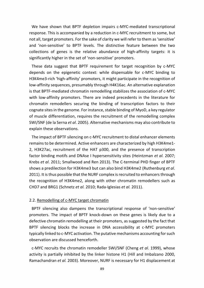
89
We have shown that BPTF depletion impairs c-MYC-mediated transcriptional
response. This is accompanied by a reduction in c-MYC recruitment to some, but
not all, target promoters. For the sake of clarity we will refer to them as ‘sensitive’
and ‘non-sensitive’ to BPTF levels. The distinctive feature between the two
collections of genes is the relative abundance of high-affinity targets: it is
significantly higher in the set of ‘non-sensitive’ promoters.
These data suggest that BPTF requirement for target recognition by c-MYC
depends on the epigenetic context: while dispensable for c-MYC binding to
H3K4me3-rich ‘high-affinity’ promoters, it might participate in the recognition of
low-affinity sequences, presumably through H4K16ac. An alternative explanation
is that BPTF-mediated chromatin remodelling stabilizes the association of c-MYC
with low-affinity promoters. There are indeed precedents in the literature for
chromatin remodellers securing the binding of transcription factors to their
cognate sites in the genome. For instance, stable binding of MyoD, a key regulator
of muscle differentiation, requires the recruitment of the remodelling complex
SWI/SNF (de la Serna et al. 2005). Alternative mechanisms may also contribute to
explain these observations.
The impact of BPTF silencing on c-MYC recruitment to distal enhancer elements
remains to be determined. Active enhancers are characterized by high H3K4me1-
2, H3K27ac, recruitment of the HAT p300, and the presence of transcription
factor binding motifs and DNAse I hypersensitivity sites (Heintzman et al. 2007;
Krebs et al. 2011; Smallwood and Ren 2013). The C-terminal PHD finger of BPTF
shows a predilection for H3K4me3 but can also bind H3K4me2 (Ruthenburg et al.
2011). It is thus possible that the NURF complex is recruited to enhancers through
the recognition of H3K4me2, along with other chromatin remodellers such as
CHD7 and BRG1 (Schnetz et al. 2010; Rada-Iglesias et al. 2011).
2.2. Remodelling of c-MYC target chromatin
BPTF silencing also dampens the transcriptional response of ‘non-sensitive’
promoters. The impact of BPTF knock-down on these genes is likely due to a
defective chromatin remodelling at their promoters, as suggested by the fact that
BPTF silencing blocks the increase in DNA accessibility at c-MYC promoters
typically linked to c-MYC activation. The putative mechanisms accounting for such
observation are discussed henceforth.
c-MYC recruits the chromatin remodeller SWI/SNF (Cheng et al. 1999), whose
activity is partially inhibited by the linker histone H1 (Hill and Imbalzano 2000,
Ramachandran et al. 2003). Moreover, NURF is necessary for H1 displacement at
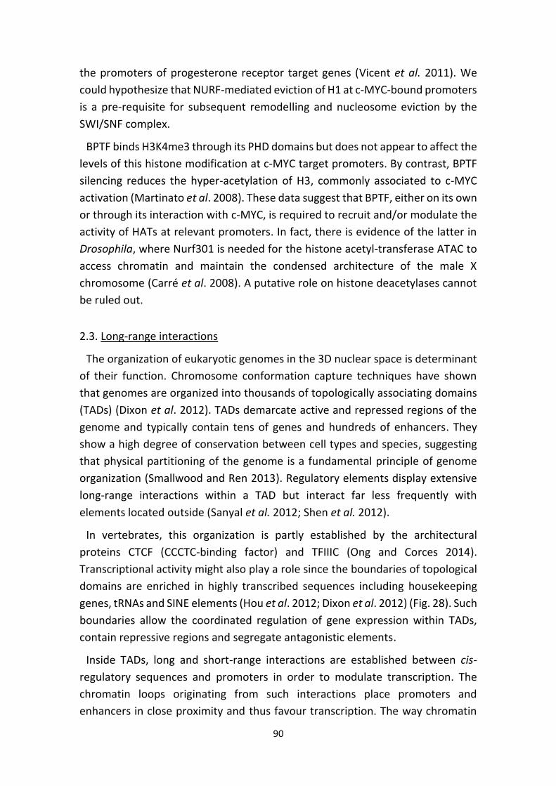
90
the promoters of progesterone receptor target genes (Vicent et al. 2011). We
could hypothesize that NURF-mediated eviction of H1 at c-MYC-bound promoters
is a pre-requisite for subsequent remodelling and nucleosome eviction by the
SWI/SNF complex.
BPTF binds H3K4me3 through its PHD domains but does not appear to affect the
levels of this histone modification at c-MYC target promoters. By contrast, BPTF
silencing reduces the hyper-acetylation of H3, commonly associated to c-MYC
activation (Martinato et al. 2008). These data suggest that BPTF, either on its own
or through its interaction with c-MYC, is required to recruit and/or modulate the
activity of HATs at relevant promoters. In fact, there is evidence of the latter in
Drosophila, where Nurf301 is needed for the histone acetyl-transferase ATAC to
access chromatin and maintain the condensed architecture of the male X
chromosome (Carré et al. 2008). A putative role on histone deacetylases cannot
be ruled out.
2.3. Long-range interactions
The organization of eukaryotic genomes in the 3D nuclear space is determinant
of their function. Chromosome conformation capture techniques have shown
that genomes are organized into thousands of topologically associating domains
(TADs) (Dixon et al. 2012). TADs demarcate active and repressed regions of the
genome and typically contain tens of genes and hundreds of enhancers. They
show a high degree of conservation between cell types and species, suggesting
that physical partitioning of the genome is a fundamental principle of genome
organization (Smallwood and Ren 2013). Regulatory elements display extensive
long-range interactions within a TAD but interact far less frequently with
elements located outside (Sanyal et al. 2012; Shen et al. 2012).
In vertebrates, this organization is partly established by the architectural
proteins CTCF (CCCTC-binding factor) and TFIIIC (Ong and Corces 2014).
Transcriptional activity might also play a role since the boundaries of topological
domains are enriched in highly transcribed sequences including housekeeping
genes, tRNAs and SINE elements (Hou et al. 2012; Dixon et al. 2012) (Fig. 28). Such
boundaries allow the coordinated regulation of gene expression within TADs,
contain repressive regions and segregate antagonistic elements.
Inside TADs, long and short-range interactions are established between cis-
regulatory sequences and promoters in order to modulate transcription. The
chromatin loops originating from such interactions place promoters and
enhancers in close proximity and thus favour transcription. The way chromatin
91
looping alters transcriptional output is not yet understood, but it might enhance
RNA Pol II recycling, mRNA export and recruitment of remodelling and histone
modification complexes (Maksimenko and Georgiev 2014). Moreover, enhancer-
promoter pairs may move to a more favourable transcriptional compartment
within the nucleus (Krivega and Dean 2012). These chromatin loops are
orchestrated by sequence-specific transcription factors, CTCF, Cohesin1, and
Mediator, either alone or in various combinations (Fig. 28).
1 COHESIN: Multiprotein complex that forms a ring-like structure which holds together two DNA helices and is critical for sister chromatid cohesion (Nasmyth and Haering 2009). Strong evidence indicates that, besides its role in mitosis and meiosis, cohesin
regulates transcriptional activity during interphase (Merkenschlager and Odom 2013).
Figure 28. Spatial organization of the eukaryotic genome. (Top) Schematic data
generated by Hi-C representing an interaction heat map of a chromosome segment.
(Bottom) TAD borders are established by the cooperative action of CTCF, TFIIIC and
Cohesin. Within TADs, CTCF facilitates enhancer-promoter looping and plays an
essential role in controlling gene expression (Erokhin et al. 2011). One mechanism is
through the interaction with TAF3, a component of the basal TFIID transcriptional
machinery. TAF3 localizes at promoters and distal sites containing CTCF, and both
sequences form a loop in a TAF3-dependent manner (Liu et al. 2011). CTCF recruits
Cohesin, which stabilizes the enhancer-promoter DNA loops built by CTCF, transcription
factors and mediator (Kagey et al. 2010; Schmidt et al. 2010; Yan et al. 2013; Smallwood
and Ren 2013). Adapted from Ong and Corces 2014.
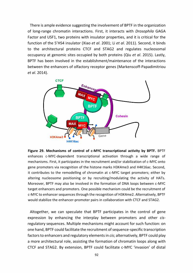
92
There is ample evidence suggesting the involvement of BPTF in the organization
of long-range chromatin interactions. First, it interacts with Drosophila GAGA
Factor and USF1, two proteins with insulator properties, and it is critical for the
function of the 5’HS4 insulator (Xiao et al. 2001; Li et al. 2011). Second, it binds
to the architectural proteins CTCF and STAG2 and regulates nucleosomal
occupancy at genomic sites occupied by both proteins (Qiu et al. 2015). Lastly,
BPTF has been involved in the establishment/maintenance of the interactions
between the enhancers of olfactory receptor genes (Markenscoff-Papadimitriou
et al. 2014).
Altogether, we can speculate that BPTF participates in the control of gene
expression by enhancing the interplay between promoters and other cis-
regulatory sequences. Multiple mechanisms might account for such function: on
one hand, BPTF could facilitate the recruitment of sequence-specific transcription
factors to enhancers and regulatory elements in cis; alternatively, BPTF could play
a more architectural role, assisting the formation of chromatin loops along with
CTCF and STAG2. By extension, BPTF could facilitate c-MYC ‘invasion’ of distal
Figure 29. Mechanisms of control of c-MYC transcriptional activity by BPTF. BPTF
enhances c-MYC-dependent transcriptional activation through a wide range of
mechanisms. First, it participates in the recruitment and/or stabilization of c-MYC onto
gene promoters via recognition of the histone marks H3K4me3 and H4K16ac. Second,
it contributes to the remodelling of chromatin at c-MYC target promoters; either by
altering nucleosome positioning or by recruiting/modulating the activity of HATs.
Moreover, BPTF may also be involved in the formation of DNA loops between c-MYC
target enhancers and promoters. One possible mechanism could be the recruitment of
c-MYC to enhancer sequences through the recognition of H3K4me2. Alternatively, BPTF
would stabilize the enhancer-promoter pairs in collaboration with CTCF and STAG2.
93
enhancer elements (Lin et al. 2012; Sabò et al. 2014) and boost transcription by
helping to build the DNA loops between c-MYC target enhancers and promoters
(Fig. 29).
2.4. Transcription elongation
Among other mechanisms, c-MYC enhances gene transcription by recruiting P-
TEFb and promoting RNA Pol II pause-release (Rahl et al. 2010; Cowling and Cole
2006). We could also speculate that BPTF:NURF plays a role in transcription
elongation, since the yeast chromatin remodelling complex Iswi has been found
in gene bodies, where it coordinates RNA Pol II elongation, termination and pre-
mRNA processing (Morillon et al. 2003; Zentner et al. 2013).
2.5. Repression by c-MYC
The effects of BPTF silencing on the output of c-MYC transcriptional activity
involve not only genes whose expression is up-regulated but also those that are
down-regulated. The mechanisms involved in "repression" by c-MYC are a highly
debated topic (Lovén et al. 2012). Chromatin remodelling complexes can repress
gene transcription by restricting the access to DNA (Morillon et al. 2003) or by
removing DNA-binding factors required for transcriptional activation. In this
regard, the yeast Isw1 complex displaces TBP from the PHO8 promoter and
effectively inhibits basal transcription (Moreau et al. 2003). Alternatively,
chromatin remodellers associate with chromatin modifiers that help enforce
repression. This is the case of Drosophila ISWI, which interacts with the HDAC
RPD3 to inhibit gene expression (Burgio et al. 2008).
Identification of BPTF target sequences will significantly improve our
understanding of the mechanisms whereby it modulates c-MYC transcriptional
activity. So far, the lack of ChIP-grade antibodies has rendered this task
unfeasible. Vicent et al. have published the only ChIP-Sequencing experiment
against endogenous BPTF to date using a home-made antibody that is no longer
available. Interestingly, in their study 40.8% of BPTF peaks were located in introns
and only 5% fell into promoter regions, further supporting the hypothesis that
BPTF function extends beyond remodelling of nucleosomes at promoters.
94
3. BPTF IN DEVELOPMENT AND DIFFERENTIATION
Extensive evidence indicates that chromatin remodellers play decisive roles in
regulating gene expression during development. They not only regulate the
global body plan but they also contribute to tissue specification and maintenance
of stem cell compartments in the adult (Clapier and Cairns 2009).
3.1. Early embryonic development
BPTF is essential for the embryonic development of different animal species.
Mutation of Drosophila Nurf301 results in lethality at the third larval stage and
deregulation of homeobox, heat shock, JAK/STAT and ecdysone pathways
(Badenhorst et al. 2002; Deuring et al. 2000). Similarly, Nurf301 knock-down at
the 2 cell stage of Xenopus is associated with defective axial development, gut
formation, blood cell development and expression of homeobox genes (Wysocka
et al. 2006). In this regard, BPTF-deficient mouse embryos fail to develop the
ectoplacental cone and show defective AP patterning of the epiblast (Landry et
al. 2008; Goller et al. 2008). c-Myc null mouse embryos also exhibit severe
developmental defects and die before midgestation. These abnormalities arise
secondary to placental insufficiency, since specific deletion of c-MYC in the
epiblast using a Sox2-Cre rescues the majority of developmental anomalies.
Epiblast-restricted c-Myc knock-out embryos progress normally but die around
E12 due to a severe anemia (Dubois et al. 2008).
3.2. Cell differentiation
There is little information on NURF function in adult mouse tissues. So far, NURF
has been proven to be required for the full maturation of thymocytes after
positive selection: BPTF deletion impairs differentiation without interfering with
proliferation, apoptosis or co-receptor expression (Landry et al. 2011). There are
some hints, as well, suggesting that BPTF participates in the homeostasis of
epidermis, since its down-regulation in human keratinocytes favours their
differentiation (Mulder et al. 2012).
As a step towards the understanding the role of BPTF in c-MYC-driven
tumorigenesis, we have developed two mouse models to conditionally delete
Bptf in the pancreas and in B cells, both tissues showing a dependence on c-MYC
for their full development. c-MYC inactivation in pancreatic progenitors is
associated with impaired growth, defective acinar cell maturation and
accumulation of adipocytes with time (Bonal et al. 2009). Similarly, depletion of
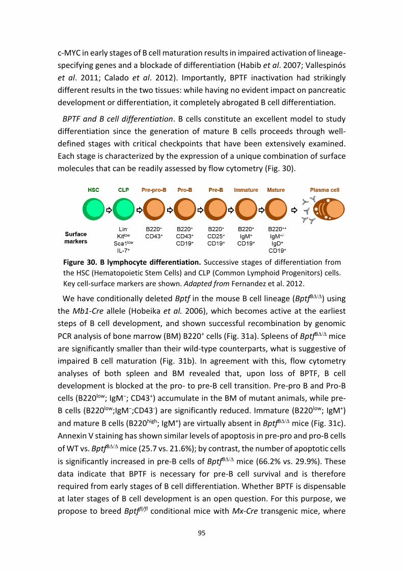
95
c-MYC in early stages of B cell maturation results in impaired activation of lineage-
specifying genes and a blockade of differentiation (Habib et al. 2007; Vallespinós
et al. 2011; Calado et al. 2012). Importantly, BPTF inactivation had strikingly
different results in the two tissues: while having no evident impact on pancreatic
development or differentiation, it completely abrogated B cell differentiation.
BPTF and B cell differentiation. B cells constitute an excellent model to study
differentiation since the generation of mature B cells proceeds through well-
defined stages with critical checkpoints that have been extensively examined.
Each stage is characterized by the expression of a unique combination of surface
molecules that can be readily assessed by flow cytometry (Fig. 30).
We have conditionally deleted Bptf in the mouse B cell lineage (BptfB) using
the Mb1-Cre allele (Hobeika et al. 2006), which becomes active at the earliest
steps of B cell development, and shown successful recombination by genomic
PCR analysis of bone marrow (BM) B220+ cells (Fig. 31a). Spleens of BptfB mice
are significantly smaller than their wild-type counterparts, what is suggestive of
impaired B cell maturation (Fig. 31b). In agreement with this, flow cytometry
analyses of both spleen and BM revealed that, upon loss of BPTF, B cell
development is blocked at the pro- to pre-B cell transition. Pre-pro B and Pro-B
cells (B220low; IgM−; CD43+) accumulate in the BM of mutant animals, while pre-
B cells (B220low;IgM−;CD43-) are significantly reduced. Immature (B220low; IgM+)
and mature B cells (B220high; IgM+) are virtually absent in BptfB mice (Fig. 31c).
Annexin V staining has shown similar levels of apoptosis in pre-pro and pro-B cells
of WT vs. BptfB mice (25.7 vs. 21.6%); by contrast, the number of apoptotic cells
is significantly increased in pre-B cells of BptfB mice (66.2% vs. 29.9%). These
data indicate that BPTF is necessary for pre-B cell survival and is therefore
required from early stages of B cell differentiation. Whether BPTF is dispensable
at later stages of B cell development is an open question. For this purpose, we
propose to breed Bptffl/fl conditional mice with Mx-Cre transgenic mice, where
Figure 30. B lymphocyte differentiation. Successive stages of differentiation from
the HSC (Hematopoietic Stem Cells) and CLP (Common Lymphoid Progenitors) cells.
Key cell-surface markers are shown. Adapted from Fernandez et al. 2012.
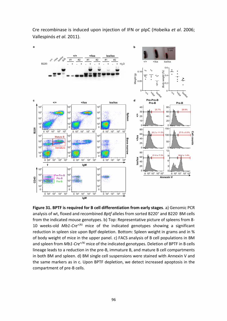
96
Cre recombinase is induced upon injection of IFN or pIpC (Hobeika et al. 2006;
Vallespinós et al. 2011).
Figure 31. BPTF is required for B cell differentiation from early stages. a) Genomic PCR
analysis of wt, floxed and recombined Bptf alleles from sorted B220+ and B220- BM cells
from the indicated mouse genotypes. b) Top: Representative picture of spleens from 8-
10 weeks-old Mb1-Cre+/KI mice of the indicated genotypes showing a significant
reduction in spleen size upon Bptf depletion. Bottom: Spleen weight in grams and in %
of body weight of mice in the upper panel. c) FACS analysis of B cell populations in BM
and spleen from Mb1-Cre+/KI mice of the indicated genotypes. Deletion of BPTF in B cells
lineage leads to a reduction in the pre-B, immature B, and mature B cell compartments
in both BM and spleen. d) BM single cell suspensions were stained with Annexin V and
the same markers as in c. Upon BPTF depletion, we detect increased apoptosis in the
compartment of pre-B cells.
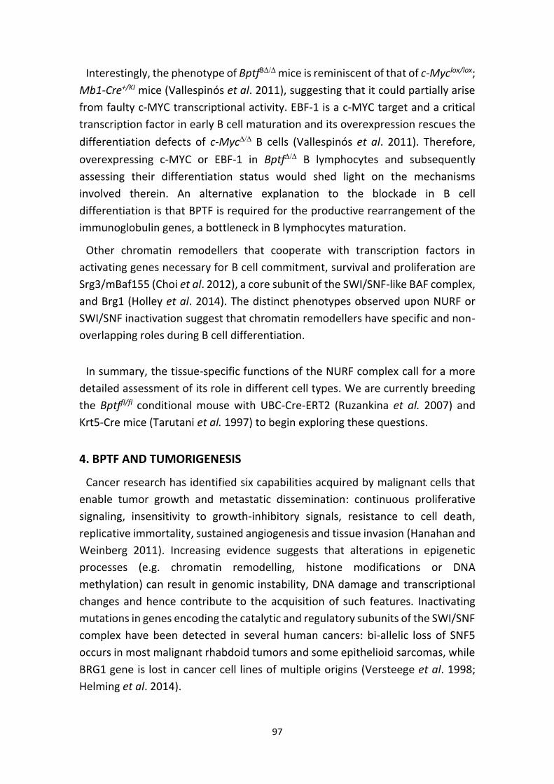
97
Interestingly, the phenotype of BptfB mice is reminiscent of that of c-Myclox/lox;
Mb1-Cre+/KI mice (Vallespinós et al. 2011), suggesting that it could partially arise
from faulty c-MYC transcriptional activity. EBF-1 is a c-MYC target and a critical
transcription factor in early B cell maturation and its overexpression rescues the
differentiation defects of c-Myc B cells (Vallespinós et al. 2011). Therefore,
overexpressing c-MYC or EBF-1 in Bptf B lymphocytes and subsequently
assessing their differentiation status would shed light on the mechanisms
involved therein. An alternative explanation to the blockade in B cell
differentiation is that BPTF is required for the productive rearrangement of the
immunoglobulin genes, a bottleneck in B lymphocytes maturation.
Other chromatin remodellers that cooperate with transcription factors in
activating genes necessary for B cell commitment, survival and proliferation are
Srg3/mBaf155 (Choi et al. 2012), a core subunit of the SWI/SNF-like BAF complex,
and Brg1 (Holley et al. 2014). The distinct phenotypes observed upon NURF or
SWI/SNF inactivation suggest that chromatin remodellers have specific and non-
overlapping roles during B cell differentiation.
In summary, the tissue-specific functions of the NURF complex call for a more
detailed assessment of its role in different cell types. We are currently breeding
the Bptffl/fl conditional mouse with UBC-Cre-ERT2 (Ruzankina et al. 2007) and
Krt5-Cre mice (Tarutani et al. 1997) to begin exploring these questions.
4. BPTF AND TUMORIGENESIS
Cancer research has identified six capabilities acquired by malignant cells that
enable tumor growth and metastatic dissemination: continuous proliferative
signaling, insensitivity to growth-inhibitory signals, resistance to cell death,
replicative immortality, sustained angiogenesis and tissue invasion (Hanahan and
Weinberg 2011). Increasing evidence suggests that alterations in epigenetic
processes (e.g. chromatin remodelling, histone modifications or DNA
methylation) can result in genomic instability, DNA damage and transcriptional
changes and hence contribute to the acquisition of such features. Inactivating
mutations in genes encoding the catalytic and regulatory subunits of the SWI/SNF
complex have been detected in several human cancers: bi-allelic loss of SNF5
occurs in most malignant rhabdoid tumors and some epithelioid sarcomas, while
BRG1 gene is lost in cancer cell lines of multiple origins (Versteege et al. 1998;
Helming et al. 2014).
98
Early work in Drosophila showed that deletion of Nurf301 is associated with
neoplastic transformation of circulating blood cells, leading to the formation of
inflammatory or melanotic tumors (Kwon et al. 2008). In C. elegans, inhibition of
ISWI function suppresses the defects associated with loss of the tumor
suppressor lin-35 Rb or the activation of oncogenic let-60 Ras (Andersen et al.
2006). More recently, mutations in BPTF have been found in several tumor types
(Fujimoto et al. 2012; Balbás-Martínez et al. 2013; González-Pérez et al. 2013;
Xiao et al. 2014a). The impact of such mutations has not been analysed in detail
yet due to the size of the protein and little knowledge of its biological roles.
Nonetheless, and according to the data integrated in IntOGen, 10% of the
mutations reported so far are truncating and cold give rise to BPTF fragments
with dominant negative properties (Gundem et al. 2010). Moreover, high BPTF
levels have been reported to be associated with poor prognosis and invasiveness
in hepatocellular carcinoma, melanoma and colorectal carcinoma (Xiao et al.
2014b; Xiao et al. 2015; Dar et al. 2015). These findings suggest that BPTF may
play an important role in tumorigenesis and therefore it constitutes an attractive
candidate for drug targeting in cancer therapy. There are indeed precedents in
the literature for inhibition of chromatin remodellers as successful cancer
therapeutic strategies. One example is Brd4, whose inhibition has been proven
successful in c-MYC-driven experimental models of hematologic malignancies
(Dawson et al. 2011; Delmore et
al. 2011; Mertz et al. 2011).
Until now, however, the power
of genetic mouse models of
cancer has not been exploited to
assess the role of BPTF in tumor
formation and/or maintenance.
As a proof of concept, we set out
to explore this notion using two
mouse models of highly
aggressive c-MYC-driven tumors:
the Ela-Myc and the E-Myc.
We have preliminary data
suggesting that inactivation of
Bptf in the pancreas of adult Ela-
Myc mice inhibits the formation
of pancreatic tumors. Moreover,
Figure 31. BPTF loss delays tumor onset in a
murine model of Burkitt lymphoma. In E-
Myc mice, c-MYC is overexpressed in the B cell
lineage under the control of the IgH enhancer.
These mice develop spontaneous pre-B and B
cell lymphomas at 15-20 weeks of age. This
graph depicts the Kaplan-Meier curves of
tumor-free survival for E-Myc mice of the
indicated Bptf genotypes. P-value was
calculated with the log-rank test.
99
inactivation of only one Bptf allele has no major impact on B cell maturation but
is sufficient to block the formation of c-MYC-driven B cell lymphomas (Fig. 31).
These promising results highlight the relevance of the c-MYC:BPTF axis as a target
for cancer therapy. A more detailed molecular, structural, and functional
dissection of BPTF will allow the development of therapeutic strategies exploiting
its function in cancer. One strategy could consist of disrupting the interaction
between the two proteins. Alternatively, drugs could be designed specifically
targeting the BPTF bromodomain.
The biochemical properties of the NURF:BPTF complex have been well
characterized, but the importance of its function in mammals has just begun to
emerge. Here we have provided some key insights into BPTF function, although
many important questions remain to be answered (Box 1). First, we have unveiled
the interaction with the oncogene c-MYC. This observation could lead to a new
field of research by itself and raises the possibility of developing new anti-cancer
strategies. We have also assessed BPTF role in adult tissue homeostasis and
shown that, while dispensable for formation of the pancreas, it is crucial for the
maturation of B cells. In addition, we have preliminary data suggesting that it
plays a critical role in tumorigenesis. In conclusion, we provide strong evidence
that BPTF is an important protein involved in chromatin remodelling required for
the action of c-MYC that merits additional study. Unravelling the molecular
function of BPTF may also provide opportunities to develop novel antitumor
drugs.
100
1. Is Nurf301/BPTF exclusive to the NURF complex? It will be essential to
determine if BPTF only works as a component of the NURF complex, or rather, it
has ATPase-independent functions. We find a precedent for this in Brg1, which has
been proven to have gene regulatory functions separate from its ATPase activity
(Jani et al. 2008).
2. How is NURF function regulated? The activity of chromatin remodelling
complexes can be modulated by alterations in their subunit composition (Lessard
et al. 2007) or alternative splicing events. Drosophila NURF is an example of the
latter. The gene nurf301 gives rise to three distinct isoforms by alternative splicing,
one of them lacking the C-terminal domains involved in the recognition of
H3K4me3 and H4K16ac. Interestingly, full-length Nurf301 is not essential for the
correct expression of most NURF targets, with the exception of genes related to
spermatogenesis (Kwon et al. 2009). The existence of an isoform with similar
properties in mammalian cells remains unknown.
3. Which are the direct targets of NURF in vivo? Genome-wide mapping of NURF
localization coupled to high-throughput methods assessing changes in chromatin
structure upon Bptf/Nurf301 knockout will help to identify these sites.
4. What are the transcription factors BPTF interacts with in human cells? The
development of high-quality immunoprecipitating antibody would facilitate the
identification of BPTF interactome using immunoprecipitation and mass-
spectrometry-based analysis.
5. Does BPTF participate in the DNA damage response? Bptf is phosphorylated in
response to activation of the ATM/ATR pathway (Matsuoka et al. 2007), thus
suggesting that it could play a role in the DNA damage response. We can find
examples in the literature of chromatin remodelling complexes that participate in
such pathway. For instance, INO80 is phosphorylated by the Mec1/Tel1 kinases
that coordinate the DNA damage response and is recruited to DSBs (Double Strand
Breaks) marked by -H2AX (Morrison et al. 2007).
Box 1. Some pending questions on NURF biology.

101
Conclusions
102
CONCLUSIONS
1. BPTF is a component of the NURF complex playing a critical role in the
proliferation of normal and cancer cells. Its knock-down in human fibroblasts
blocks S phase entry.
2. BPTF levels are modulated during cell cycle progression: in human fibroblasts,
it is induced upon entry into G1 and is down-regulated in S phase.
3. Using a combination of biochemical assays we demonstrate that BPTF and c-
MYC are found within the same protein complex and that they interact directly.
4. In human fibroblasts transduced with the MYC-ER fusion protein, BPTF is
required for the activation of the full c-MYC transcriptional program through
chromatin remodelling of its target promoters.
5. In MEFs transduced with Myc-ER, BPTF is necessary for c-MYC-driven
proliferation but not for apoptosis, suggesting that BPTF is only required for those
c-MYC functions involving direct binding to chromatin.
6. BPTF is necessary for the reprogramming of murine fibroblasts into induced
pluripotent stem cells.
7. BPTF is expressed at high levels in human tumors of diverse origin. Its
expression positively correlates with the activation of c-MYC, but not N-MYC or
L-MYC, driven gene signatures.
8. BPTF has tissue-specific roles in cell differentiation: while dispensable for the
formation of the mouse pancreas, it is crucial for the generation of mature B cells
in bone marrow and spleen.
9. Preliminary data indicate that, in mice, BPTF plays a critical function in the
formation and/or maintenance of c-MYC-driven tumors.

103
Conclusiones
104
CONCLUSIONES
1. BPTF es un componente del complejo NURF con un papel crítico en la
proliferación de células normales y cancerosas. Su knock-down en fibroblastos
humanos resulta en un bloqueo en la entrada en fase S.
2. Los niveles de BPTF son modulados durante la progresión del ciclo celular: en
fibroblastos humanos, BPTF es inducido al entrar las células en G1 y se down-
regula en fase S.
3. Por medio de una combinación de ensayos bioquímicos hemos demostrado
que BPTF y c-MYC se encuentran en el mismo complejo proteico y que
interaccionan directamente.
4. En fibroblastos humanos transducidos con la proteína de fusión MYC-ER, BPTF
es requerido para la activación completa del programa transcripcional de c-MYC
a través de la remodelación de la cromatina en sus promotores diana.
5. En fibroblastos embrionarios de ratón transducidos con MYC-ER, BPTF es
necesario para la proliferación inducida por c-MYC, pero no para la apoptosis
dirigida por este oncogén, sugiriendo que BPTF solo es necesario para aquellas
funciones de c-MYC que implican la unión directa a la cromatina.
6. BPTF es necesario para la reprogramación de fibroblastos de ratón a células
madre de pluripotencia inducida.
7. BPTF se expresa a niveles altos en diversos tipos de tumores humanos. Su
expresión correlaciona positivamente con la activación de programas de
expresión génica instruidos por c-MYC, pero no por N-MYC o L-MYC.
8. BPTF tiene funciones tejido-específicas en diferenciación celular: mientras que
es dispensable para la formación del páncreas de ratón, es crucial para la
generación de células B maduras en la médula ósea y bazo.
9. Datos preliminares sugieren que, en ratones, BPTF juega un papel crítico en la
formación y/o mantenimiento de tumores dirigidos por c-MYC.

105
References
106
REFERENCES
1. Acosta, J.C., Ferrándiz, N., Bretones, G., Torrano, V., Blanco, R., et al. (2008). Myc
inhibits p27-induced erythroid differentiation of leukemia cells by repressing
erythroid master genes without reversing p27-mediated cell cycle arrest. Mol Cell
Biol., 28, 7286-95.
2. Adams, J.M., Harris, A.W., Pinkert, C.A., Corcoran, L.M., Alexander, W.S. et al.
(1985). The c-myc oncogene driven by immunoglobulin enhancers induces
lymphoid malignancy in transgenic mice. Nature, 318, 533-8.
3. Adhikary, S. and Eilers, M. (2005). Transcriptional regulation and transformation
by Myc proteins. Nat Rev Mol Cell Biol., 6, 635-45.
4. Agrawal, P., Yu, K., Salomon, A.R. and Sedivy, J.M. (2010). Proteomic profiling of
Myc associated proteins. Cell Cycle, 9, 4908-21.
5. Alarcon, R.M., Rupnow, B.A., Graeber, T.G., Knox, S.J., Giaccia, A.J. (1996)
Modulation of c-Myc activity and apoptosis in vivo. Cancer Res., 56, 4315-19.
6. Alberts, B., Johnson, A., Lewis, J., Raff, M., Roberts, K. et al. (2002). Molecular
Biology of the Cell, 4th edition. New York, NY: Garland Science.
7. Alkhatib, S.G. and Landry, J.W. (2011). The nucleosome remodeling factor. FEBS
lett., 585, 3197-207.
8. Alland, L., Muhle, R., Hou, H. Jr., Potes, J., Chin, L. et al. (1997). Role for N-CoR and
histone deacetylase in Sin3-mediated transcriptional repression. Nature, 387, 49-
55.
9. Andersen, E.C., Lu, X., Horvitz, H.R. (2006). C. elegans ISWI and NURF301
antagonize an Rb-like pathway in the determination of multiple cell fates.
Development, 133, 2695-704.
10. Arabi, A., Wu, S., Ridderstråle, K., Bierhoff, H., Shiue, C. et al. (2005). c-Myc
associates with ribosomal DNA and activates RNA polymerase I transcription. Nat.
Cell. Biol., 7, 303-10.
11. Armstrong, J.A., Papoulas, O., Daubresse, G., Sperling, A.S., Lis, J.T. et al. (2002).
The Drosophila BRM complex facilitates global transcription by RNA polymerase II.
EMBO J., 21, 5245-54.
12. Arvanitis, C. and Felsher, D.W. (2006). Conditional transgenic models define how
MYC initiates and maintains tumorigenesis. Semin. Cancer Biol., 16, 313-7.
13. Badenhorst, P., Voas, M., Rebay, I., Wu, C. (2002) Biological functions of the ISWI
chromatin remodeling complex NURF. Genes Dev., 16, 3186-98.
14. Badenhorst, P., Xiao, H., Cherbas, L., Kwon, S.Y., Voas, M. et al. (2005). The
Drosophila nucleosome remodeling factor NURF is required for Ecdysteroid
signaling and metamorphosis. Genes Dev., 19, 2540-45.
107
15. Bailey, T.L., Boden, M., Buske, F.A., Frith, M., Grant, C.E. et al. (2009). MEME
SUITE: tools for motif discovery and searching. Nucleic Acids Res., 37 (Web Server
issue), W202-W208.
16. Balbás-Martínez, C., Sagrera, A., Carrillo-de-Santa-Pau, E., Earl, J., Márquez, M.
et al. (2013). Recurrent inactivation of STAG2 in bladder cancer is not associated
with aneuploidy. Nat Genet., 45, 1464-9.
17. Bannister, A.J., and Kouzarides, T. (2011). Regulation of chromatin by histone
modifications. Cell Res., 21, 381-95.
18. Barak, O., Lazzaro, M.A., Lane, W.S., Speicher, D.W., Picketts, D.J. et al. (2003)
Isolation of human NURF: a regulator of Engrailed gene expression. EMBO J., 22,
6089-100.
19. Barbie, D.A., Tamayo, P., Boehm, J.S., Kim, S.Y., Moody, S.E. et al. (2009).
Systematic RNA interference reveals that oncogenic KRAS-driven cancers require
TBK1. Nature, 462, 108-12.
20. Bedford, D.C., Kasper, L.H., Fukuyama, T. and Brindle, P.K. (2010). Target gene
context influences the transcriptional requirement for the KAT3 family of CBP and
p300 histone acetyltransferases. Epigenetics, 5, 9-15.
21. Blackwell, T.K., Huang, J., Ma, A., Kretzner, L., Alt, F.W. et al. (1993). Binding of
Myc proteins to canonical and noncanonical DNA sequences. Mol Cell Biol., 13,
5216-24.
22. Blackwood, E.M. and Eisenman, R.N. (1991). Max: a helix-loop-helix zipper
protein that forms a sequence-specific DNA-binding complex with Myc. Science,
251, 1211-7.
23. Bonal, C., Thorel, F., Ait-Lounis, A., Reith, W., Trumpp, A. et al. (2009). Pancreatic
inactivation of c-Myc decreases acinar mass and transdifferentiates acinar cells into
adipocytes in mice. Gastroenterology, 136, 309-19.
24. Bouchard, C., Lee, S., Laulus-Hock, V., Loddenkemper, C., Eilers, M. et al. (2007).
FoxO transcription factors suppress Myc-driven lymphomagenesis via direct
activation of Arf. Gene Dev., 21, 2775-87.
25. Bown, N., Cotterill, S., Lastowska, M., O’Neill, S., Pearson, A.D., et al. (1999) Gain
of chromosome arm 17q and adverse outcome in patients with neuroblastoma. N
Engl J Med., 340, 1954-61.
26. Brenner, C., Deplus, R., Didelot, C., Loriot, A., Viré, E. et al. (2005) Myc represses
transcription through recruitment of DNA methyltransferase corepressor. EMBO J.,
24, 336-46.
27. Buganim, Y., Goldstein, I., Lipson, D., Milyavsky, M., Polak-Charcon, S. et al.
(2010). A novel translocation breakpoint within the BPTF gene is associated with a
pre-malignant phenotype. PloS One, 5, e9657.
108
28. Burgio, G., La Rocca, G., Sala, A., Arancio, W., Di Gesu, D., et al. (2008). Genetic
identification of a network of factors that functionally interact with the nucleosome
remodeling ATPase ISWI. PLoS Genet., 4, e1000089.
29. Calado, D.P., Sasaki, Y., Godinho, S.A., Pellerin, A., Köchert, K., et al. (2012). The
cell-cycle regulator c-Myc is essential for the formation and maintenance of
germinal centers. Nat Immunol., 13, 1092-100.
30. Carré, C., Ciurciu, A., Komonyi, O., Jacquier, C., Fagegaltier, D. et al. (2008). The
Drosophila NURF remodelling and the ATAC histone acetylase complexes
functionally interact and are required for global chromosome organization. EMBO
Rep., 9, 187-92.
31. Cartwright, P., McLean, C., Sheppard, A., Rivett, D., Jones, K. et al. (2005).
LIF/STAT3 controls ES cell self-renewal and pluripotency by a Myc-dependent
mechanism. Development, 132, 885-96.
32. Chai, B., Huang, J., Cairns, B.R., Laurent, B.C. (2005). Distinct roles for the RSC
and Swi/Snf ATP-dependent chromatin remodelers in DNA double-strand break
repair. Genes Dev., 19, 1656-61.
33. Chandriani, S., Frengen, E., Cowling, V.H., Pendergrass, S.A., Perou, C.M. et al.
(2009). A core MYC gene expression signature is prominent in basal-like breast
cancer but only partially overlaps the core serum response. PLoS One, 4, e6693.
34. Cheng, S.W., Davies, K.P., Yung, E., Beltran, R.J., Yu, J. et al. (1999). c-MYC
interacts with INI1/hSNF5 and requires the SWI/SNF complex for transactivation
function. Nat Genet., 22, 102-5.
35. Cheng, A.S., Jin, V.X., Fan, M., Smith, L.T., Liyanarachchi, S., et al. (2006).
Combinatorial analysis of transcription factor partners reveals recruitment of c-
MYC to estrogen receptor-alpha responsive promoters. Mol Cell, 21, 393-404.
36. Cherry, C.M. and Matunis, E.L. (2010). Epigenetic regulation of stem cell
maintenance in the Drosophila testis via the nucleosome-remodeling factor NURF.
Cell Stem cell, 6, 557-67.
37. Choi, J., Ko, M., Jeon, S., Jeon, Y., Park, K. et al. (2012). The SWI/SNF-like BAF
complex is essential for early B cell development. J Immunol., 188, 3791-803.
38. Choudhary, C., Kumar, C., Gnad, F., Nielsen, M.L., Rehman, M. et al. (2009).
Lysine acetylation targets protein complexes and co-regulates major cellular
functions. Science, 325, 834-40.
39. Clapier, C.R. and Cairns, B.R. (2009). The biology of chromatin remodeling
complexes. Annu Rev Biochem., 78, 273-304.
40. Cohen, S.M., Chastain, P.D., Rosson, G.B., Groh, B.S., Weissman, B.E. et al.
(2010). BRG1 co-localizes with DNA replication factors and is required for efficient
replication fork progression. Nucleic Acids Res., 38, 6906-19.
109
41. Collins, N., Poot, R.A., Kukimoto, I., Garcia-Jimenez, C., Dellaire, G., et al. (2002).
An ACF1-ISWI chromatin-remodeling complex is required for DNA replication
through hetero- chromatin. Nat Genet., 32, 627-32.
42. Cowling, V.H., Chandriani S., Whitfield M.L., Cole M.D. (2006). A conserved Myc
protein domain, MBIV, regulates DNA binding, apoptosis, transformation and G2
arrest. Mol Cell Biol., 26, 4226-39.
43. Cowling, V.H. and Cole, M.D. (2006). Mechanism of transcriptional activation by
the Myc oncoproteins. Semin Cancer Biol., 16, 242-52.
44. Cowling, V.H. and Cole, M.D. (2007). The Myc transactivation domain promotes
global phosphorylation of the RNA polymerase II carboxy-terminal domain
independently of direct DNA binding. Mol Cell Biol., 27, 2059-73.
45. Cowling, V.H. and Cole, M.D. (2010). Myc Regulation of mRNA Cap Methylation.
Genes Cancer, 1, 576-9.
46. Dang, C.V. (2012). MYC on the path to cancer. Cell, 149, 22-35.
47. Dang, C.V. (2014). Gene regulation: fine-tuned amplification in cells. Nature,
511, 417-8.
48. Danielian, P.S., White, R., Hoare, S.A., Fawell, S.E., Parker, M.G. (1993).
Identification of residues in the estrogen receptor that confer differential sensitivity
to estrogen and hydroxytamoxifen. Mol Endocrinol., 7, 232-40.
49. Dar, A.A., Nosrati, M., Bezrookove, V., de Semir, D., Majid, S. et al. (2015). The
Role of BPTF in Melanoma Progression and in Response to BRAF-Targeted Therapy.
J Natl Cancer Inst., 107, pii: djv034.
50. Dawson, M., Bannister, A.J., Göttgens, B., Foster, S.D., Bartke, T. et al. (2009).
JAK2 phosphorylates histone H3Y41 and excludes HP1alpha from chromatin.
Nature, 461, 819-22.
51. Dawson, M., Prinjha, R., Dittmann, A., Giotopoulos, G., Bantscheff, M. et al.
(2011). Inhibition of BET recruitment to chromatin as an effective treatment for
MLL-fusion leukaemia. Nature, 478, 529-33.
52. Dawson, M., and Kouzarides, T. (2012). Cancer epigenetics: from mechanism to
therapy. Cell, 150, 12-27.
53. de la Serna, I.L., Ohkawa, Y., Berkes, C.A., Bergstrom, D.A., Dacwag, C.S., et al.
(2005). MyoD targets chromatin remodeling complexes to the myogenin locus prior
to forming a stable DNA-bound complex. Mol Cell Biol., 25, 3997-4009.
54. Delmore, J.E., Issa, G.C., Lemieux, M.E., Rahl, P.B., Shi, J. et al. (2011). BET
bromodomain inhibition as a therapeutic strategy to target c-Myc. Cell, 146, 904-
17.
55. Dephoure, N., Zhou, C., Villen, J., Beausoleil, S.A., Bakalarski, C.E. et al. (2008). A
quantitative atlas of mitotic phosphorylation. Proc Natl Acad Sci USA., 105, 10762-
7.
110
56. Deuring, R., Fanti, L., Armstrong, J.A., Sarte, M., Papoulas, O. et al. (2000) The
ISWI chromatin-remodeling protein is required for gene expression and the
maintenance of higher order chromatin structure in vivo. Mol Cell, 5, 355-65.
57. Dey, A., Nishiyama, A., Karpova, T., McNally, J. and Ozato, K. (2009) Brd4 marks
select genes on mitotic chromatin and directs postmitotic transcription. Mol Cell
Biol., 20, 4899-909.
58. Di Stefano, B., Sardina, J.L., van Oevelen, C., Collombet, S., Kallin, E.M. et al.
(2014). C/EBP poises B cells for rapid reprogramming into induced pluripotent
stem cells. Nature, 506, 235-9.
59. Dixon, J.R., Selvaraj, S., Yue, F., Kim, A., Li, Y. et al. (2012). Topological domains
in mammalian genomes identified by analysis of chromatin interactions. Nature,
485, 376-80.
60. Doerks, T., Copley, R., and Bork, P. (2001). DDT -- a novel domain in different
transcription and chromosome remodeling factors. Trends Biochem Sci., 26, 145-6.
61. Dominguez-Sola, D., Ying, C.Y., Grandori, C., Ruggiero, L., Chen, B. et al. (2007).
Non- transcriptional control of DNA replication by c-Myc. Nature, 448, 445-51.
62. Dubois, N.C., Adolphe, C., Ehninger, A., Wang, R.A., Robertson, E.J. et al. (2008).
Placental rescue reveals a sole requirement for c-Myc in embryonic erythroblast
survival and hematopoietic stem cell function. Development, 135, 2455-65.
63. Durinck, S., Ho, C., Wang, N.J., Liao, W., Jakkula, L.R. et al. (2011). Temporal
dissection of tumorigenesis in primary cancers. Cancer Discov., 1, 137-43.
64. Eberhardy, S. and Farnham, P. (2001). c-Myc mediates activation of the cad
promoter via a post-RNA polymerase II recruitment mechanism. J. Biol. Chem., 276,
48562-71.
65. Egle, A., Harris, A.W., Bouillet, P., Cory S. (2004). Bim is a suppressor of Myc-
induced mouse B cell leukemia. Proc Natl Acad Sci USA, 101, 6164-9.
66. Eilers, M. and Eisenman, R.N. (2008). Myc’s broad reach. Genes Dev., 22, 2755-
66.
67. Eischen, C.M., Woo, D., Roussel, M.F., Cleveland, J.L. (2001). Apoptosis triggered
by Myc-induced suppression of Bcl-XL or Bcl-2 is bypassed during
lymphomagenesis. Mol Cell Biol., 21, 5063-70.
68. Erdel, F. and Rippe, K. (2011). Chromatin remodelling in mammalian cells by
ISWI-type complexes--where, when and why? FEBS J., 278, 3608-18.
69. Erokhin, M., Davydova, A., Kyrchanova, O., Parshikov, A., Georgiev, P. (2011).
Insulators form gene loops by interacting with promoters in Drosophila.
Development, 138, 4097-106.
70. Espada, J. and Esteller, M. (2010). DNA methylation and the functional
organization of the nuclear compartment. Semin Cell Dev Biol., 21, 238-46.
111
71. Evan, G.I., Wyllie, A.H., Gilbert, C.S., Littlewood, T.D., Land, H. et al. (1992).
Induction of apoptosis in fibroblasts by c-Myc protein. Cell, 69, 119-28.
72. Faiola, F., Liu, X., Lo, S., Pan, S., Zhang, K. et al. (2005). Dual regulation of c-Myc
by p300 via acetylation-dependent control of Myc protein turnover and
coactivation of Myc-induced transcription. Mol Cell Biol., 25, 10220-34.
73. Falbo, K. B. and Shen, X. (2006). Chromatin remodelling in DNA replication. J Cell
Biochem., 97, 684-–9.
74. Fernandez, P.C., Frank, S.R., Wang, L., Schroeder, M., Liu, S., et al. (2003).
Genomic targets of the human c-Myc protein. Genes Dev., 17, 1115-29.
75. Fernandez, D., Sanchez-Arevalo, V.J. and de Alboran, I.M. (2012). The role of the
proto-oncogene c-myc in B lymphocyte differentiation. Crit Rev Immunol., 32, 321-
34.
76. Ferreira, R., Eberharter, A., Bonaldi, T., Chioda, M., Imhof, A. et al. (2007). Site-
specific acetylation of ISWI by GCN5. BMC Mol Biol., 8, 73.
77. Fladvad, M., Zhou, K., Moshref, A., Pursglove, S., Säfsten, P. et al. (2005). N and
C-terminal sub-regions in the c-Myc transactivation region and their joint role in
creating versatility in folding and binding. J Mol Biol., 346, 175-89.
78. Frank, S.R., Parisi, T., Taubert, S., Fernandez, P., Fuchs, M. et al. (2003). MYC
recruits the TIP60 histone acetyltransferase complex to chromatin. EMBO rep., 4,
575-80.
79. Fry, C.J. and Peterson, C.L. (2001). Chromatin remodeling enzymes: who's on
first? Curr. Biol., 11, R185-97.
80. Fujimoto, A., Totoki, Y., Abe, T., Boroevich, K.A., Hosoda, F. et al. (2012). Whole-
genome sequencing of liver cancers identifies etiological influences on mutation
patterns and recurrent mutations in chromatin regulators. Nat Genet., 44, 760-4.
81. Gabay, M., Li, Y. and Felsher, D.W. (2014). MYC activation is a hallmark of cancer
initiation and maintenance. Cold Spring Harb. Perspect. Med., 4, pii: a014241.
82. Garson, J.A., Pemberton, L.F., Sheppard, P.W., Varndell, I.M., Coakham, H.B. et
al. (1989). N-myc gene expression and oncoprotein characterization in
medulloblastoma. Br J Cancer, 59, 889-94.
83. Gartel, A.L., Ye, X., Goufman, E., Shianov, P., Hay, N. et al. (2001) Myc represses
the p21 (WAF1/CIP1) promoter and interacts with Sp1/Sp3. Proc Natl Acad Sci USA.,
98, 4510-5.
84. Gebhardt, A., Frye, M., Herold, S., Benitah, S.A., Braun, K. (2006). Myc regulates
keratinocyte adhesion and differentiation via complex formation with Miz1. J Cell
Biol., 172, 139-49.
85. Goldman, J.A., Garlick, J.D. and Kingston, R.E. (2010). Chromatin remodeling by
imitation switch (ISWI) class ATP-dependent remodelers is stimulated by histone
variant H2A.Z. J. Biol. Chem., 285, 4645-51.
112
86. Goldmark, J.P., Fazzio, T.G., Estep, P.W., Church, G.M., Tsukiyama, T. (2000). The
Isw2 chromatin remodeling complex represses early meiotic genes upon
recruitment by Ume6p. Cell, 103, 423-33.
87. Goller, T., Vauti, F., Ramasamy, S., Arnold, H.H. (2008) Transcriptional regulator
BPTF/FAC1 is essential for trophoblast differentiation during early mouse
development. Mol Cell Biol., 28, 6819-27.
88. Gomez-Roman, N., Grandori, C., Eisenman, R.N., White, R.J. (2003). Direct
activation of RNA polymerase III transcription by c-Myc. Nature, 421, 290-4.
89. González-Pérez, A., Jene-Sanz, A. and López-Bigas, N. (2013). The mutational
landscape of chromatin regulatory factors across 4,623 tumor samples. Genome
Biol., 14, r106.
90. Goren, A., Tabib, A., Hecht, M., Cedar, H. (2008). DNA replication timing of the
human beta-globin domain is controlled by histone modification at the origin.
Genes Dev., 22, 1319-24.
91. Gregory, M.A., Qi, Y. and Hann, S.R. (2003) Phosphorylation by glycogen
synthase kinase-3 controls c-myc proteolysis and subnuclear localization. J. Biol.
Chem., 278, 51606-12.
92. Guccione, E., Martinato, F., Finocchiaro, G., Luzi, L., Tizzoni, L. et al. (2006). Myc-
binding-site recognition in the human genome is determined by chromatin context.
Nat Cell Biol., 8, 764-70.
93. Gundem, G., Perez-Llamas, C., Jene-Sanz, A., Kedzierska, A., Islam, A. et al.
(2010). IntOGen: Integration and data-mining of multidimensional oncogenomic
data. Nat Methods, 7, 92-3.
94. Habib, T., Park, H., Tsang, M., De Alborán, I. M., Nicks, A. et al. (2007). Myc
stimulates B lymphocyte differentiation and amplifies calcium signalling. J Cell Biol.,
179, 717-31.
95. Hamiche, A., Sandaltzopoulos, R., Gdula, D.A. and Wu, C. (1999). ATP-dependent
histone octamer sliding mediated by the chromatin remodeling complex NURF.
Cell, 97, 833-42.
96. Hanahan, D and Weinberg, R.A. (2011). Hallmarks of cancer: the next generation.
Cell, 144, 646-74.
97. Hann, S.R. and Eisenman, R.N. (1984). Proteins encoded by the human c-myc
oncogene: differential expression in neoplastic cells. Mol Cell Biol., 4, 2486-97.
98. Hanna, J., Markoulaki, S., Schorderet, P., Carey, B.W., Beard, C. et al. (2008).
Direct reprogramming of terminally differentiated mature B lymphocytes to
pluripotency. Cell, 133, 250-64.
99. Hansson, J., Rafiee, M.R., Reiland, S., Polo, J.M., Gehring, J. et al. (2012). Highly
Coordinated Proteome Dynamics during Reprogramming of Somatic Cells to
Pluripotency. Cell Rep., 2, 1579-92.
113
100. Harris, A.W., Pinkert, C.A., Crawford, M., Langdon, W.Y., Brinster, R.L. et al.
(1988). The E mu-myc transgenic mouse. A model for high-incidence spontaneous
lymphoma and leukemia of early B cells. J Exp Med., 167, 353-71.
101. Hayes, J.J. and Hansen, J.C. (2001). Nucleosomes and the chromatin fiber. Curr
Opin Genet Dev., 11, 124-9.
102. Hebbar, P.B. and Archer, T.K. (2003) Chromatin remodeling by nuclear
receptors. Chromosoma, 111, 495-504.
103. Heintzman, N.D., Stuart, R.K., Hon, G., Fu, Y., Ching, C.W. et al. (2007). Distinct
and predictive chromatin signatures of transcriptional promoters and enhancers in
the human genome. Nat Genet., 39, 311-8.
104. Helming, K., Wang, X., Roberts, C. (2014). Vulnerabilities of mutant SWI/SNF
complexes in cancer. Cancer Cell, 4, 309-17.
105. Herkert, B. and Eilers, M. (2010). Transcriptional repression: the dark side of
myc. Genes Cancer, 1, 580-6.
106. Hill, D.A. and Imbalzano, A.N. (2000). Human SWI/SNF nucleosome remodeling
activity is partially inhibited by linker histone H1. Biochemistry, 39, 11649-56.
107. Hobeika, E.S., Thiemann, B., Storch, H. Jumaa, P.J., Nielsen, R. et al. (2006).
Testing gene function early in the B cell lineage in mb1-cre mice. Proc Natl Acad Sci
USA., 103, 13789-94.
108. Hoffman, B. and Liebermann, D.A. (2008). Apoptotic signaling by c-MYC.
Oncogene, 27, 6462-72.
109. Holley, D.W., Groh, B.S., Wozniak, G., Donohoe, D.R., Sun, W. et al. (2014). The
BRG1 chromatin remodeler regulates widespread changes in gene expression and
cell proliferation during B cell activation. J Cell Physiol., 229, 44-52.
110. Holmes, J.K. and Solomon, M.J. (1996). A predictive scale for evaluating cyclin-
dependent kinase substrates. A comparison of p34cdc2 and p33cdk2. J Biol Chem.,
271, 25240-6.
111. Hopewell, R. and Ziff, E.B. (1995). The nerve growth factor-responsive PC12 cell
line does not express the Myc dimerization partner Max. Mol Cell Biol., 15, 3470-8.
112. Hou, C., Li, L., Qin, Z.S., Corces, V.G. (2012). Gene density, transcription, and
insulators contribute to the partition of the Drosophila genome into physical
domains. Mol Cell, 48, 471-84.
113. Inui, M., Miyado, M., Igarashi, M., Tamano, M., Kubo, A. et al. (2014). Rapid
generation of mouse models with defined point mutations by the CRISPR/Cas9
system. Sci Rep., 4, 5396.
114. Iritani, B.M., Forbush, K.A., Farrar, M.A., Perlmutter, R.M. (1997). Control of B
cell development by Ras-mediated activation of Raf. EMBO J., 16, 7019-31.
115. Jacobsen, K.A., Prasad, V.S., Sidman, C.L., Osmond, D.G. (1994). Apoptosis and
macrophage-mediated deletion of precursor B cells in the bone marrow of E mu-
myc transgenic mice. Blood, 84, 2784-94.
114
116. Jani, A., Wan, M., Zhang, J., Cui, K., Wu, J. et al. (2008). A novel genetic strategy
reveals unexpected roles of the Swi-Snf-like chromatin-remodeling BAF complex in
thymocyte development. J Exp Med., 205, 2813-25.
117. Ji, H., Wu, G., Zhan, X., Nolan, A., Koh, C. et al. (2011). Cell-type independent
MYC target genes reveal a primordial signature involved in biomass accumulation.
PLoS One, 6, e26057.
118. Jin, C. and Felsenfeld, G. (2007) Nucleosome stability mediated by histone
variants H3.3 and H2A.Z. Genes Dev., 21, 1519-29.
119. Jones, M.H., Hamana, N. and Shimane, M. (2000). Identification and
characterization of BPTF, a novel bromodomain transcription factor. Genomics, 63,
35-9.
120. Jordan-Sciutto, K.L., Dragich, J.M., Rhodes, J.L., Bowser, R. (1999a). Fetal Alz-50
clone 1, a novel zinc finger protein, binds a specific DNA sequence and acts as a
transcriptional regulator. J. Biol. Chem., 274, 35262-8.
121. Jordan-Sciutto, K.L., Dragich, J.M., Bowser, R. (1999b). DNA binding activity of
the fetal Alz-50 clone 1 (FAC1) protein is enhanced by phosphorylation. Biochem
Biophys Res Commun., 260, 785-9.
122. Kagey, M.H., Newman, J.J., Bilodeau, S., Zhan, Y., Orlando, D.A. et al. (2010).
Mediator and cohesin connect gene expression and chromatin architecture.
Nature, 467, 430-5.
123. Kamakaka, R.T. and Biggins, S. (2005). Histone variants: deviants? Genes Dev.,
19, 295-310.
124. Kanazawa, S., Soucek, L., Evan, G., Okamoto, T. and Peterlin, B.M. (2003). c-
Myc recruits P-TEFb for transcription, cellular proliferation and apoptosis.
Oncogene, 22, 5707-11.
125. Karlsson, A., Deb-Basu, D., Cherry, A., Turner, S., Ford, J. (2003). Defective
double-strand DNA break repair and chromosomal translocations by MYC
overexpression. Proc Natl Acad Sci USA., 100, 9974-9.
126. Kawaguchi, Y., Cooper, B., Gannon, M., Ray, M., MacDonald, R.J. et al. (2002).
The role of the transcriptional regulator Ptf1a in converting intestinal to pancreatic
progenitors. Nat Genet., 32, 128-34.
127. Kim, S.Y., Herbst, A., Tworkowski, K.A., Salghetti, S.E. and Tansey, W.P. (2003).
Skp2 regulates Myc protein stability and activity. Mol. Cell., 11, 1177-88.
128. Kim, Y.H., Girard, L., Giacomini, C.P., Wang, P., Hernandez-Boussard, T. et al.
(2006). Combined microarray analysis of small cell lung cancer reveals altered
apoptotic balance and distinct expression signatures of MYC family gene
amplification. Oncogene, 25, 130-8.
129. Kim, J., Woo, A.J., Chu, J., Snow, J.W., Fujiwara, Y. et al. (2010). A Myc network
accounts for similarities between embryonic stem and cancer cell transcription
programs. Cell, 143, 313-24.
115
130. Kingston, R.E. and Narlikar, G.J. (1999). ATP-dependent remodeling and
acetylation as regulators of chromatin fluidity. Genes Dev., 13, 2339-52.
131. Kopinke, D., Brailsford, M., Pan, F.C., Magnuson, M.A., Wright, C.V. et al.
(2011). Ongoing Notch signaling maintains phenotypic fidelity in the adult exocrine
pancreas. Dev Biol., 362, 57-64.
132. Kouzarides, T. (2007). Chromatin modifications and their function. Cell, 128,
693-705.
133. Krebs, A.R., Karmodiya, K., Lindahl-Allen, M., Struhl, K., Tora, L. (2011). SAGA
and ATAC histone acetyl transferase complexes regulate distinct sets of genes and
ATAC defines a class of p300-independent enhancers. Mol Cell, 44, 410-23.
134. Krivega, I. and Dean, A. (2012). Enhancer and promoter interactions-long
distance calls. Curr Opin Genet Dev., 22, 79-85.
135. Kurland, J.F. and Tansey, W.P. (2008) Myc-mediated transcriptional repression
by recruitment of histone deacetylase. Cancer Res., 68, 3624-9.
136. Kwon, S.Y., Xiao, H., Glover, B.P., Tjian, R., Wu, C. et al. (2008). The nucleosome
remodeling factor (NURF) regulates genes involved in Drosophila innate immunity.
Dev Biol., 316, 538-47.
137. Kwon, S.Y., Xiao, H., Wu, C., Badenhorst, P. (2009). Alternative splicing of
NURF301 generates distinct NURF chromatin remodeling complexes with altered
modified histone binding specificities. PLoS Genet., 5, e1000574.
138. Landry, J., Sharov, A.A., Piao, Y., Sharova, L.V., Xiao, H. et al. (2008). Essential
role of chromatin remodeling protein Bptf in early mouse embryos and embryonic
stem cells. PLoS Genet., 4, e1000241.
139. Landry, J., Banerjee, S., Taylor, B., Aplan, P.D., Singer, A. et al. (2011).
Chromatin remodeling complex NURF regulates thymocyte maturation. Genes
Dev., 25, 275-86.
140. Langdon, W.Y., Harris, A.W., Cory, S. and Adams, J.M. (1986). The c-myc
oncogene perturbs B lymphocyte development in E-myc transgenic mice. Cell, 47,
11-8.
141. Laurenti, E., Wilson, A., Trumpp, A. (2009). Myc’s other life: stem cells and
beyond. Curr. Opin. Cell. Biol., 21, 844-54.
142. Laybourn, P.J., Kadonaga, J.T. (1991). Role of nucleosomal cores and histone
H1 in regulation of transcription by RNA polymerase II. Science, 254, 238-45.
143. Lessard, J., Wu, J.I., Ranish, J.A., Wan, M., Winslow, M.M., et al. (2007). An
essential switch in subunit composition of a chromatin remodelling complex during
neural development. Neuron, 55, 201-15.
144. Li, F., Wang, Y., Zeller, K.I., Potter, J.J., Wonsey, D.R. et al. (2005). Myc
stimulates nuclearly encoded mitochondrial genes and mitochondrial biogenesis.
Mol Cell Biol., 25, 6225-34.
116
145. Li, H., Ilin, S., Wang, W., Duncan, E.M., Wysocka, J. et al. (2006) Molecular basis
for site-specific read-out of histone H3K4me3 by the BPTF PHD finger of NURF.
Nature, 442, 91-5.
146. Li, B., Carey, M., Workman, J.L. (2007). The role of chromatin during
transcription. Cell, 128, 707-19.
147. Li, H., Collado, M., Villasante, A., Strati, K., Sagrario, O. et al. (2009). The
Ink4/Arf locus is a barrier for iPS cell reprogramming. Nature, 460, 1136-9.
148. Li, M., Belozerov, V.E., Cai, H.N. (2010). Modulation of chromatin boundary
activities by nucleosome-remodeling activities in Drosophila melanogaster. Mol
Cell Biol., 30, 1067-76.
149. Li, X., Wang, S., Li, Y., Deng, C., Steiner, L.A. et al. (2011). Chromatin boundaries
require functional collaboration between the hSET1 and NURF complexes. Blood,
118, 1386-94.
150. Lin, C.Y., Lovén, J., Rahl, P.B., Paranal, R.M., Burge, C.B. et al. (2012).
Transcriptional amplification in tumor cells with elevated c-Myc. Cell, 151, 56-67.
151. Littlewood, T.D., Hancock, D.C., Danielian, P.S., Parker, M.G., Evan, G.I. (1995).
A modified oestrogen receptor ligand-binding domain as an improved switch for
the regulation of heterologous proteins. Nucleic Acids Res., 23, 1686-90.
152. Liu, X., Vorontchikhina, M., Wang, Y.L., Faiola, F. and Martinez, E. (2008a).
STAGA recruits mediator to the MYC oncoprotein to stimulate transcription and cell
proliferation. Mol Cell Biol., 28, 108-21.
153. Liu, Y.C., Li, F., Handler, J., Huang, C.R., Xiang, Y. et al. (2008b). Global regulation
of nucleotide biosynthetic genes by c-Myc. PLoS One, 3, e2722 (2008).
154. Liu, Z., Scannell, D.R., Eisen, M.B., Tjian, R. (2011). Control of embryonic stem
cell lineage commitment by core promoter factor, TAF3. Cell, 146, 720-31.
155. Livak, K.J. and Schmittgen, T.D. (2001). Analysis of relative gene expression data
using real-time quantitative PCR and the 2(-Delta Delta C(T)) Method. Methods, 25,
402.
156. Lovén, J., Orlando, D.A., Sigova, A.A., Lin, C.Y., Rahl, P.B. et al. (2012). Revisiting
global gene expression analysis. Cell, 151, 476-82.
157. Luger, K., Mader, A.W., Richmond, R.K., Sargent, D.F. and Richmond, T.J.
(1997). Crystal structure of the nucleosome core particle at 2.8 Å resolution.
Nature, 389, 251-60.
158. Lüscher, B. and Vervoorts, J. (2012). Regulation of gene transcription by the
oncoprotein MYC. Gene, 494, 145-60.
159. Maksimenko, O. and Georgiev, P. (2014). Mechanisms and proteins involved in
long-distance interactions. Front Genet., 5, 28.
160. Malumbres, M. (2011). Physiological relevance of cell cycle kinases. Physiol
Rev., 91, 973-1007.
117
161. Markenscoff-Papadimitriou, E., Allen, W.E., Colquitt, B.M., Goh, T., Murphy,
K.K. et al. (2014). Enhancer interaction networks as a means for singular olfactory
receptor expression. Cell, 159, 543-57.
162. Marqués, M., Kumar, A., Cortés, I., Gonzalez-García, A., Hernández, C. et al.
(2008). Phosphoinositide 3-kinases p110alpha and p110beta regulate cell cycle
entry, exhibiting distinct activation kinetics in G1 phase. Mol Cell Biol., 28, 2803-14.
163. Marques, M., Laflamme, L., Gervais, A.L. and Gaudreau, L. (2010). Reconciling
the positive and negative roles of histone H2A.Z in gene transcription. Epigenetics,
5, 267-72.
164. Marshall, N.F., Peng, J., Xie, Z. and Price, D.H. (1996). Control of RNA
polymerase II elongation potential by a novel carboxyl-terminal domain kinase. J
Biol Chem., 271, 27176-83.
165. Martinato, F., Cesaroni, M., Amati, B. and Guccione, E. (2008). Analysis of Myc-
induced histone modifications on target chromatin. PloS One, 3, e3650.
166. Martinelli, P., Cañamero, M., Del Pozo, N., Madriles, F., Zapata, A. et al. (2013).
Gata6 is required for complete acinar differentiation and maintenance of the
exocrine pancreas in adult mice. Gut, 62, 1481-8.
167. Martinelli, P., Madriles, F., Cañamero, M., Pau, E.C., Pozo, N.D. et al. (2015).
The acinar regulator Gata6 suppresses KrasG12V-driven pancreatic tumorigenesis
in mice. Gut, pii: gutjnl-2014-308042. [Epub ahead of print]
168. Matsuoka, S., Ballif, B.A., Smogorzewska, A., McDonald, E.R. III, Hurov, K.E. et
al. (2007). ATM and ATR substrate analysis reveals extensive protein networks
responsive to DNA damage. Science, 316, 1160-6.
169. Mayya, V., Lundgren, D.H., Hwang, S.-I., Rezaul, K., Wu, L. et al. (2009)
Quantitative phosphoproteomic analysis of T cell receptor signaling reveals system-
wide modulation of protein-protein interactions. Sci. Signal., 2, RA46.
170. McMahon, S.B., Wood, M.A. and Cole, M.D. (2000). The essential cofactor
TRRAP recruits the histone acetyltransferase hGCN5 to c‐Myc. Mol. Cell. Biol., 20,
556-62.
171. McNairn, A.J., Gilbert, D.M. (2003). Epigenomic replication: Linking epigenetics
to DNA replication. Bioessays, 25, 647-56.
172. Merkenschlager, M. and Odom, D.T. (2013). CTCF and cohesin: linking gene
regulatory elements with their targets. Cell, 152, 1285-97.
173. Mertz, J.A., Conery, A.R., Bryant, B.M., Sandy, P., Balasubramanian, S. et al.
(2011). Targeting MYC dependence in cancer by inhibiting BET bromodomains. Proc
Natl Acad Sci USA., 108, 16669-74.
174. Meyer, N. and Penn, L. (2008). Reflecting on 25 years with MYC. Nat Rev
Cancer, 8, 976-90.
118
175. Mitchell, K.O., Ricci, M.S., Miyashita, T., Dicker, D.T., Jin, Z. et al. (2000). Bax is
a transcriptional target and mediator of c-Myc-induced apoptosis. Cancer Res., 60,
6318-25.
176. Mizuguchi, G., Tsukiyama, T., Wisniewski, J., Wu, C. (1997). Role of nucleosome
remodeling factor NURF in transcriptional activation of chromatin. Mol Cell, 1, 141-
50.
177. Mizuguchi, G., Shen, X., Landry, J., Wu, W.H., Sen, S. et al. (2004). ATP-driven
exchange of histone H2AZ variant catalyzed by SWR1 chromatin remodeling
complex. Science, 303, 343-8.
178. Moreau, J.L., Lee, M., Mahachi, N., Vary, J., Mellor, J. et al. (2003). Regulated
displacement of TBP from the PHO8 promoter in vivo requires Cbf1 and the Isw1
chromatin remodeling complex. Mol Cell, 11, 1609-20.
179. Morillon, A., Karabetsou, N., O'Sullivan, J., Kent, N., Proudfoot, N. et al. (2003).
Isw1 chromatin remodeling ATPase coordinates transcription elongation and
termination by RNA polymerase II. Cell, 115, 425-35.
180. Morrison, A.J., Kim, J.A., Person, M.D., Highland, J., Xiao, J. et al. (2007).
Mec1/Tel1 phosphorylation of the INO80 chromatin remodeling complex
influences DNA damage checkpoint responses. Cell, 130, 499-511.
181. Mulder, K.W., Wang, X., Escriu, C., Ito, Y., Schwarz, R.F., Gillis, J. et al. (2012).
Diverse epigenetic strategies interact to control epidermal differentiation. Nat Cell
Biol., 14, 753-63.
182. Murakami, H., Sanderson, N.D., Nagy, P., Marino, P.A., Merlino, G. (1993)
Transgenic mouse model for synergistic effects of nuclear oncogenes and growth
factors in tumorigenesis: interaction of c-myc and transforming growth factor alpha
in hepatic oncogenesis. Cancer Res., 53, 1719-23.
183. Murga, M., Campaner, S., Lopez-Contreras, A.J., Toledo, L.I., Soria, R. et al.
(2011) Exploiting oncogene-induced replicative stress for the selective killing of
Myc-driven tumors. Nat Struct Biol., 18, 1331-5.
184. Murphy, D.J., Junttila, M.R., Pouyet, L., Karnezis, A., Shchors, K. et al. (2008)
Distinct Thresholds Govern Myc’s Biological Output In Vivo. Cancer Cell, 14, 447-
57.
185. Nagy, Z. and Tora, L. (2007). Distinct GCN5/PCAF-containing complexes
function as co-activators and are involved in transcription factor and global histone
acetylation. Oncogene, 26, 5341-57.
186. Nakagawa, M., Koyanagi, M., Tanabe, K., Takahashi, K., Ichisaka, T. et al. (2008)
Generation of induced pluripotent stem cells without Myc from mouse and human
fibroblasts. Nat Biotechnol., 26, 101-6.
187. Nasmyth, K. and Haering, C.H. (2009). Cohesin: its roles and mechanisms. Annu
Rev Genet., 43, 525-58.
119
188. Neri, F., Zippo, A., Krepelova, A., Cherubini, A., Rocchigiani, M. et al. (2012).
Myc regulates the transcription of the PRC2 gene to control the expression of
developmental genes in embryonic stem cells. Mol Cell Biol., 32, 840-51.
189. Nie, Z., Hu, G., Wei, G., Cui, K., Yamane, A. et al. (2012). c-Myc is a universal
amplifier of expressed genes in lymphocytes and embryonic stem cells. Cell, 151,
68-79.
190. O’Connell, B.C., Cheung, A.F., Simkevich, C.P., Tam, W., Ren, X. et al. (2003). A
large scale genetic analysis of c-Myc-regulated gene expression patterns. J Biol
Chem., 278, 12563-73.
191. O'Donnell, K.A., Yu, D., Zeller, K.I., Kim, J.W., Racke, F. et al. (2006). Activation
of transferrin receptor 1 by c-Myc enhances cellular proliferation and
tumorigenesis. Mol Cell Biol., 26, 2373-86.
192. Olsen, J.V., Blagoev, B., Gnad, F., Macek, B., Kumar, C. et al. (2006). Global, in
vivo, and site-specific phosphorylation dynamics in signaling networks. Cell, 127,
635-48.
193. Olsen, J.V., Vermeulen, M., Santamaria, A., Kumar, C., Miller, M.L. et al. (2010).
Quantitative phosphoproteomics reveals widespread full phosphorylation site
occupancy during mitosis. Sci Signal., 3, RA3.
194. Ong, C.T. and Corces, V.G. (2014). CTCF: an architectural protein bridging
genome topology and function. Nat Rev Genet., 15, 234-46.
195. Park, J., Wood, M.A. and Cole, M.D. (2002). BAF53 forms distinct nuclear
complexes and functions as a critical c-Myc-interacting nuclear cofactor for
oncogenic transformation. Mol Cell Biol., 22, 1307-16.
196. Perna, D., Fagà, G., Verrecchia, A., Gorski, M.M., Barozzi, I. et al. (2012).
Genome-wide mapping of Myc binding and gene regulation in serum-stimulated
fibroblast. Oncogene, 31, 1695-1709.
197. Peukert, K., Staller, P., Schneider, A., Carmichael, G., Hanel, F. et al. (1997) An
alternative pathway for gene regulation by Myc. EMBO J., 16, 5672-86.
198. Phatnani, H.P. and Greenleaf, A.L. (2006). Phosphorylation and functions of the
RNA polymerase II CTD. Genes Dev., 20, 2922-36.
199. Poot, R.A., Bozhenok, L., van den Berg, D.L., Steffensen, S., Ferreira, F. et al.
(2004). The Williams syndrome transcription factor interacts with PCNA to target
chromatin remodelling by ISWI to replication foci. Nat Cell Biol., 6, 1236-44.
200. Qiu, Z., Song, C., Malakouti, N., Murray, D., Hariz, A. et al. (2015). Functional
Interactions between NURF and Ctcf Regulate Gene Expression. Mol Cell Biol., 35,
224-37.
201. Rada-Iglesias, A., Bajpai, R., Swigut, T., Brugmann, S.A., Flynn, R.A. et al. (2011).
A unique chromatin signature uncovers early developmental enhancers in humans.
Nature, 470, 279-83.
120
202. Rahl, P.B., Lin, C.Y., Seila, A.C., Flynn, R.A., Mccuine, S. et al. (2010). c-Myc
Regulates Transcriptional Pause Release. Cell, 141, 432-45.
203. Ramachandran A., Omar M., Cheslock P. and Schnitzler G.R. (2003). Linker
histone H1 modulates nucleosome remodeling by human SWI/SNF. J Biol Chem.,
278, 48590-601.
204. Rigbolt, K.T., Prokhorova, T.A., Akimov, V., Henningsen, J., Johansen, P.T. et al.
(2011). System-wide temporal characterization of the proteome and
phosphoproteome of human embryonic stem cell differentiation. Sci Signal., 4, RS3.
205. Ruthenburg, A.J., Li, H., Milne, T.A., Dewell, S., McGinty, R.K. et al. (2011).
Recognition of a mononucleosomal histone modification pattern by BPTF via
multivalent interactions. Cell, 145, 692-706.
206. Ruzankina, Y., Pinzon-Guzman, C., Asare, A., Ong, T., Pontano, L. et al. (2007).
Deletion of the developmentally essential gene ATR in adult mice leads to age-
related phenotypes and stem cell loss. Cell Stem Cell, 1, 113-26.
207. Sabò, A., Kress, T.R., Pelizzola, M., de Pretis, S., Gorski, M.M. et al. (2014).
Selective transcriptional regulation by Myc in cellular growth control and
lymphomagenesis. Nature, 511, 488-92.
208. Saborowski, M., Saborowski, A., Morris, J.P. 4th, Bosbach, B., Dow, L.E. et al.
(2014). A modular and flexible ESC-based mouse model of pancreatic cancer. Genes
Dev., 28, 85-97.
209. Saha, A., Wittmeyer, J. and Cairns, B. R. (2006). Chromatin remodelling: the
industrial revolution of DNA around histones. Nat Rev Mol Cell Biol., 7, 437-47.
210. Sandgren, E.P., Quaife, C.J., Paulovich, A.G., Palmiter, R.D., Brinster, R.L. (1991).
Pancreatic tumor pathogenesis reflects the causative genetic lesion. Proc Natl Acad
Sci USA., 88, 93-7.
211. Sansom, O.J., Meniel, V.S., Muncan, V., Phesse, T.J., Wilkins, J.A. et al. (2007).
Myc deletion rescues Apc deficiency in the small intestine. Nature, 446, 676-9.
212. Sanyal, A., Lajoie, B.R., Jain, G., Dekker, J. (2012). The long-range interaction
landscape of gene promoters. Nature, 489, 109-13.
213. Sarid, J., Halazonetis, T.D., Murphy, W. and Leder, P. (1987). Evolutionarily
conserved regions of the human c-myc protein can be uncoupled from
transforming activity. Proc Natl Acad Sci USA., 84, 170-3.
214. Savkur, R.S. and Burris, T.P. (2004). The coactivator LXXLL nuclear receptor
recognition motif. J. Peptide Res., 63, 207-12.
215. Schlosser, I., Hölzel, M., Hoffmann, R., Burtscher, H., Kohlhuber, F. et al. (2005).
Dissection of transcriptional programmes in response to serum and c-Myc in a
human B-cell line. Oncogene, 24, 520-4.
216. Schmidt, D., Schwalie, P.C., Ross-Innes, C.S., Hurtado, A., Brown, G.D. et al.
(2010). A CTCF-independent role for cohesion in tissue-specific transcription.
Genome Res., 20, 578-88.
121
217. Schnetz, M.P., Handoko, L., Akhtar-Zaidi, B., Bartels, C.F., Pereira, C.F., et al.
(2010). CHD7 targets active gene enhancer elements to modulate ES cell-specific
gene expression. PLoS Genet., 6, pe1001023.
218. Schoenenberger, C.A., Andres, A.C., Groner, B., van der Valk, M., LeMeur, M.
et al. (1988) Targeted c-myc gene expression in mammary glands of transgenic mice
induces mammary tumours with constitutive milk protein gene transcription.
EMBO J., 7, 169-75.
219. Schuhmacher, M., Kohlhuber, F., Hölzel, M., Kaiser, C., Burtscher, H. et al.
(2001). The transcriptional program of a human B cell line in response to Myc.
Nucleic Acids Res., 29, 397-406.
220. Sears, R., Nuckolls, F., Haura, E., Taya, Y., Tamai, K. et al. (2000). Multiple Ras-
dependent phosphorylation pathways regulate Myc protein stability. Genes Dev.
14, 2501-14.
221. Seitz, V., Butzhammer, P., Hirsch, B., Hecht, J., Gütgemann, I., et al. (2011).
Deep sequencing of MYC DNA-binding sites in Burkitt lymphoma. PLoS One, 6,
e26837.
222. Seoane, J., Pouponnot, C., Staller, P., Schader, M., Eilers, M. et al. (2001).
TGFbeta influences Myc, Miz-1 and Smad to control the CDK inhibitor p15INK4b.
Nat Cell Biol., 3, 400-8.
223. Shen, Y., Yue, F., McCleary, D. F., Ye, Z., Edsall, L. et al. (2012). A map of the cis-
regulatory sequences in the mouse genome. Nature, 488, 116-20.
224. Shim, H., Dolde, C., Lewis, B.C., Wu, C.S., Dang, G. et al. (1997). c-Myc
transactivation of LDH-A: implications for tumor metabolism and growth. Proc Natl
Acad Sci USA., 94, 6658-63.
225. Shim, E.Y., Hong, S.J., Oum, J.H., Yanez, Y., Zhang, Y., Lee, S.E. (2007). RSC
mobilizes nucleosomes to improve accessibility of repair machinery to the
damaged chromatin. Mol Cell Biol., 27, 1602-13.
226. Sif, S., Stukenberg, P.T., Kirschner, M.W., Kingston, R.E. (1998). Mitotic
inactivation of a human SWI/SNF chromatin remodeling complex. Genes Dev., 12,
2842-51.
227. Smallwood, A. and Ren, B. (2013). Genome organization and long-range
regulation of gene expression by enhancers. Curr Opin Cell Biol., 25, 387-94.
228. Songyang, Z., Lu, K.P., Kwon, Y.T., Tsai, L.H., Filhol, O. et al. (1996). A structural
basis for substrate specificities of protein Ser/Thr kinases: primary sequence
preference of casein kinases I and II, NIMA, phosphorylase kinase, calmodulin-
dependent kinase II, CDK5, and Erk1. Mol Cell Biol., 16, 6486-93.
229. Soucek, L., Jucker, R., Panacchia, L., Ricordy, R., Tato, F. et al. (2002). Omomyc,
a potential Myc dominant negative, enhances Myc-induced apoptosis. Cancer Res,
62, 3507-10.
122
230. Soucek, L., Whitfield, J., Martins, C.P., Finch, A.J., Murphy, D.J. et al. (2008).
Modelling Myc inhibition as a cancer therapy. Nature, 455, 679-83.
231. Soufi, A., Donahue, G. and Zaret, K. S. (2012). Facilitators and Impediments of
the Pluripotency Reprogramming Factors’ Initial Engagement with the Genome.
Cell, 151, 994-1004.
232. Spotts, G.D., Patel, S.V., Xiao, Q. and Hann, S.R. (1997). Identification of
downstream-initiated c-Myc proteins which are dominant-negative inhibitors of
transactivation by full-length c-Myc proteins. Mol. Cell. Biol., 17, 1459-68.
233. Sridharan, R., Tchieu, J., Mason, M. J., Yachechko, R., Kuoy, E. et al. (2009). Role
of the murine reprogramming factors in the induction of pluripotency. Cell, 136,
364-77.
234. Staller, P., Peukert, K., Kiermaier, A., Seoane, J., Lukas, J. et al. (2001)
Repression of p15INK4b expression by Myc through association with Miz-1. Nat.
Cell. Biol., 3, 392-9.
235. Steiger, D., Furrer, M., Schwinkendorf, D. and Gallant, P. (2008). Max-
independent functions of Myc in Drosophila melanogaster. Nat. Genet., 40, 1084-
91.
236. Stewart, T.A., Pattengale, P.K. and Leder P. (1984). Spontaneous mammary
adenocarcinomas in transgenic mice that carry and express MTV/myc fusion genes.
Cell, 38, 627-37.
237. Strahl, B.D. and Allis, C.D. (2000). The language of covalent histone
modifications. Nature, 403, 41-5.
238. Strasser, A., Harris, A.W., Bath, M.L. and Cory, S. (1990) Novel primitive
lymphoid tumours induced in transgenic mice by cooperation between myc and
bcl-2. Nature, 348, 331-3.
239. Sugiyama, A. and Kume, A. (1989). Isolation and characterization of s-myc, a
member of the rat myc gene family. Proc Natl Acad Sci USA., 86, 9144-8.
240. Takahashi, K., Tanabe, K., Ohnuki, M., Narita, M., Ichisaka, T. et al. (2007).
Induction of pluripotent stem cells from adult human fibroblasts by defined factors.
Cell, 131, 861-72.
241. Talbert, P.B. and Henikoff, S. (2010). Histone variants--ancient wrap artists of
the epigenome. Nat Rev Mol Cell Biol., 11, 264-75.
242. Tan, M., Luo, H., Lee, S., Jin, F., Yang, J.S., et al. (2011). Identification of 67
histone marks and histone lysine crotonylation as a new type of histone
modification. Cell, 146, 1016-28.
243. Tarutani, M., Itami, S., Okabe, M., Ikawa, M., Tezuka, T. et al. (1997). Tissue-
specific knockout of the mouse Pig-a gene reveals important roles for GPI-anchored
proteins in skin development. Proc Natl Acad Sci USA., 94, 7400-5.
123
244. Taub, R., Kirsch, I., Morton, C., Lenoir, G., Swan, D. et al. (1982). Translocation
of the c-myc gene into the immunoglobulin heavy chain locus in human Burkitt
lymphoma and murine plasmacytoma cells. Proc Natl Acad Sci USA. , 79, 7837-41.
245. Taylor, B.S., Schultz, N., Hieronymus, H., Gopalan, A., Xiao, Y. et al. (2010).
Integrative genomic profiling of human prostate cancer. Cancer Cell, 18, 11-22.
246. Thurman, R.E., Rynes, E., Humbert, R., Vierstra, J., Maurano, M.T. et al. (2012).
The accessible chromatin landscape of the human genome. Nature, 489, 75-82.
247. Vafa, O., Wade, M., Kern, S., Beeche, M., Pandita, T.K. et al. (2002) c-Myc can
induce DNA damage, increase reactive oxygen species, and mitigate p53 function:
A mechanism for oncogene-induced genetic instability. Mol Cell, 9, 1031-44.
248. Vallespinós, M., Fernández, D., Rodríguez, L., Alvaro-Blanco, J., Baena, E. et al.
(2011). B Lymphocyte commitment program is driven by the proto-oncogene c-
Myc. J Immunol., 186, 6726-36.
249. Vennstrom, B., Sheiness, D., Zabielski, J. and Bishop, J.M. (1982). Isolation and
characterization of c-myc, a cellular homolog of the oncogene (v-myc) of avian
myelocytomatosis virus strain 29. J Virol., 42, 773-9.
250. Versteege, I., Sevenet, N., Lange, J., Rousseau-Merck, M.F., Ambros, P. et al.
(1998). Truncating mutations of hSNF5/INI1 in aggressive paediatric cancer.
Nature, 394, 203-6.
251. Vervoorts, J., Luscher-Firzlaff, J.M., Rottmann, S., Lilischkis, R., Walsemann, G.
et al. (2003). Stimulation of c-MYC transcriptional activity and acetylation by
recruitment of the cofactor CBP. EMBO Rep., 4, 484-90.
252. Vicent, G.P., Nacht, A.S., Font-Mateu, J., Castellano, G., Gaveglia, L. et al.
(2011). Four enzymes cooperate to displace histone H1 during the first minute of
hormonal gene activation. Genes Dev., 25, 845-62.
253. Vilá, M.R., Lloreta, J., Schüssler, M.H., Berrozpe, G., Welt, S., Real, F.X. (1995).
New pancreas cancers cell lines that represent distinct stages of ductal
differentiation. Lab Invest., 72, 395-404.
254. Vogelauer, M., Rubbi, L., Lucas, I., Brewer, B.J., Grunstein, M. (2002). Histone
acetylation regulates the time of replication origin firing. Mol Cell, 10, 1223-33.
255. Waikel, R.L., Kawachi, Y., Waikel, P.A., Wang, X.J. and Roop, D.R. (2001).
Deregulated expression of c-Myc depletes epidermal stem cells. Nat. Genet., 28,
165-8.
256. Walz, S., Lorenzin, F., Morton, J., Wiese, K.E., von Eyss, B., et al. (2014).
Activation and repression by oncogenic MYC shape tumour-specific gene
expression profiles. Nature, 511, 483-7.
257. Wang, G.G., Allis, C.D., Chi, P. (2007). Chromatin remodeling and cancer, Part
II: ATP-dependent chromatin remodeling. Trends Mol Med., 13, 373-80.
124
258. Wanzel, M., Russ, A.C., Kleine-Kohlbrecher, D., Colombo, E., Pelicci, P.G. et al.
(2008). A ribosomal protein L23-nucleophosmin circuit coordinates Miz1 function
with cell growth. Nat. Cell. Biol., 10, 1051-61.
259. Wolf, E., Lin, C.Y., Eilers, M., Levens, D.L. (2014). Taming of the beast: shaping
Myc-dependent amplification. Trends Cell Biol., pii: S0962-8924(14)00193-7.
260. Wu, R., Lin, L., Beer, D.G., Ellenson, L.H., Lamb, B.J. et al. (2003). Amplification
and overexpression of the L-MYC proto-oncogene in ovarian carcinomas. Am. J.
Pathol., 162, 1603-10.
261. Wysocka, J., Swigut, T., Xiao, H., Milne, T.A., Kwon, S.Y. et al. (2006). A PHD
finger of NURF couples histone H3 lysine 4 trimethylation with chromatin
remodelling. Nature, 442, 86-90.
262. Xiao, H., Sandaltzopoulos, R., Wang, H.M., Hamiche, A., Ranallo, R. et al. (2001).
Dual functions of largest NURF subunit NURF301 in nucleosome sliding and
transcription factor interactions. Mol Cell, 8, 531-43.
263. Xiao, F., Kim, Y.C., Snyder, C., Wen, H., Chen, P.X. et al. (2014a). Genome
instability in blood cells of a BRCA1+ breast cancer family. BMC Cancer, 14, 342.
264. Xiao, S., Liu, L., Fang, M., Zhou, X., Peng, X. et al. (2014b). BPTF Associated with
EMT Indicates Negative Prognosis in Patients with Hepatocellular Carcinoma. Dig
Dis Sci. [Epub ahead of print]
265. Xiao, S., Liu, L., Lu, X., Long, J., Zhou, X. et al. (2015). The prognostic significance
of bromodomain PHD-finger transcription factor in colorectal carcinoma and
association with vimentin and E-cadherin. J Cancer Res Clin Oncol. [Epub ahead of
print]
266. Yada, M., Hatakeyama, S., Kamura, T., Nishiyama, M., Tsunematsu, R. et al
(2004). Phosphorylation‐dependent degradation of c‐Myc is mediated by the F‐box
protein Fbw7. EMBO J., 23, 2116-25.
267. Yan, J., Enge, M., Whitington, T., Dave, K., Liu, J., Sur, I., et al. (2013).
Transcription factor binding in human cells occurs in dense clusters formed around
cohesin anchor cites. Cell, 154, 801-13.
268. Yang, Z., Yik, J.H., Chen, R., He, N., Jang, M.K. et al. (2005). Recruitment of P-
TEFb for stimulation of transcriptional elongation by the bromodomain protein
Brd4. Mol Cell., 19, 535-45.
269. Ye, T., Krebs, A.R., Choukrallah, M.A., Keime, C., Plewniak, F. et al. (2010).
SeqMINER: an integrated ChIP-seq data interpretation platform. Nucleic Acids Res.,
39, e35.
270. Zeller, K.I., Zhao, X., Lee, C.W., Chiu, K.P., Yao, F. et al. (2006). Global mapping
of c-Myc binding sites and target gene networks in human B cells. Proc Natl Acad
Sci USA., 103, 17834-9.
271. Zentner, G.E., Tsukiyama, T., Henikoff, S. (2013). ISWI and CHD chromatin
remodelers bind promoters but act in gene bodies. PLoS Genet., 9, e1003317.

125
272. Zhang, Y. and Reinberg, D. (2001). Transcription regulation by histone
methylation: interplay between different covalent modifications of the core
histone tails. Genes Dev., 15, 2343-60.
273. Zhang, H., Roberts, D.N., Cairns, B.R. (2005). Genome-wide dynamics of Htz1, a
histone H2A variant that poises repressed/basal promoters for activation through
histone loss. Cell, 123, 219-31.
274. Zindy, F., Eischen, C.M., Randle, D.H., Kamijo, T., Cleveland, J.L. et al. (1998)
Myc signaling via the ARF tumor suppressor regulates p53-dependent apoptosis
and immortalization. Genes Dev., 12, 2424-33.
Related Documents


![Research Paper JNK/AP1 Pathway Regulates MYC ...Chromatin immunoprecipitation assays (ChIP) ChIP analysis was performed as previously described [11]. Chromatin solutions were precipitated](https://static.cupdf.com/doc/110x72/608625bcea8a6a2e9165f1fb/research-paper-jnkap1-pathway-regulates-myc-chromatin-immunoprecipitation-assays.jpg)