Biochem. J. (1994) 301, 211-216 (Printed in Great Britain) 211 The chondrodystrophy, nanomelia: biosynthesis and processing of the defective aggrecan precursor Barbara M. VERTEL,* Bonnie L. GRIER,* Hao Lit and Nancy B. SCHWARTZt *Department of Cell Biology and Anatomy, Chicago Medical School, North Chicago, IL 60064, U.S.A., and tDepartments of Pediatrics, Biochemistry and Molecular Biology, University of Chicago, Chicago, IL 60637, U.S.A. The lethal chicken mutation nanomelia leads to severe skeletal was not further processed; this is consistent with the conclusion defects because of a deficiency of aggrecan, which is the large that it moves no further along the secretory pathway than the aggregating chondroitin sulphate proteoglycan of cartilage. In ER. Using brefeldin A we demonstrated that the defective previous work, we have demonstrated that nanomelic chondro- precursor can function as a substrate for Golgi-mediated glycos- cytes produce a truncated aggrecan precursor that fails to be aminoglycan chains, but does not do so in the nanomelic secreted, and is apparently arrested in the endoplasmic reticulum chondrocyte because it fails to be translocated to the appropriate (ER). In this study, we investigated the biosynthesis and extent membrane compartment. These studies illustrate how combined of processing of the abnormal aggrecan precursor. The truncated cell biological/biochemical and molecular investigations may precursor was translated directly in cell-free reactions, indicating contribute to our understanding of the biological consequences that it does not arise post-translationally. Further studies addres- and molecular basis of genetic diseases, particularly those sed the processing capabilities of the defective precursor. We involving errors in large, highly modified molecules such as found that the mutant precursor was modified by N-linked, proteoglycans. mannose-rich oligosaccharides and by the addition of xylose, but INTRODUCTION the level of hydration and thereby provide an expanded tissue volume for bone replacement during long-bone development. Recent studies highlight how important sequential processing Without aggrecan, the intercellular, matrix-filled spaces are and assembly events and movement through specific compart- greatly reduced, the cartilage model fails to expand,and conse- ments of the secretory pathway are for normal protein function quently, the growth of long bones is seriously impaired. Aggrecan (Rose and Doms, 1988; Hurtley and Helenius, 1989; Farquhar, function principally reflects the more than 100 covalently at- 1991). By extrapolation, diseases may arise as a consequence of tached chondroitin sulphate (CS) and keratan sulphate (KS) errors in these processes. For example, problems of synthesis, glycosaminoglycan chains that are the result of complex biosyn- assembly and intracellular trafficking are implicated in the basic thetic processes involving thousands of co- and post-translational mechanisms underlying lysosomal storage diseases, familial reactions (reviewed recently by Wight et al., 1991; Hardingham hypercholesterolaemia, and cystic fibrosis (Amara et al., 1992). and Fosang, 1992). The large (> 200 kDa) aggrecan core protein In connective-tissue disorders such as osteogenesis imperfecta is only 10 % of the molecular mass of the fully processed and chondrodystrophies, defects may involve the abnormal proteoglycan, which is modified as well by N- and 0-linked synthesis, processing, translocation and assembly of extracellular oligosaccharides. The chondrocyte is characterized by an ex- matrix (ECM) molecules (Goetinck, 1983; Stanescu et al., 1984; tensive endoplasmic reticulum (ER) and Golgi, as expected for a Johnson, 1986; Prockop, 1990; Spranger and Martoux, 1990; cell heavily committed to the production of highly modified Byers et al., 1991). These types of errors might be expected for ECM molecules in large quantities. The synthetic, processing ECM molecules, as they are mostly large, highly modified and and assembly events are accomplished in several subcompart- strongly interactive, and are characteristically assembled into ments of the secretory pathway that have been described in extensive macromolecular arrays. Although defects in the col- previous studies (Pacifici et al., 1984; Vertel and Barkman, 1984; lagens have frequently been found (Prockop, 1990; Byers et al., Vertel and Hitti, 1987; Campbell and Schwartz, 1988; Vertel et 1991), proteoglycans and other matrix molecules are also al., 1985a,b, 1989, 1993a; Kearns et al., 1993), although some candidates for these genetic diseases (Goetinck, 1983; Stanescu features of intracellular processing and assembly are not yet well et al., 1984; Johnson, 1986; Spranger and Martoux, 1990). defined. Structural studies of aggrecan from several species Nanomelia is one such genetic mutation that causes shortened (including chicken) have provided the complete coding sequence limbs, other skeletal abnormalities and death in chicken embryos and a clear definition of several domains (see Figure 6); these as a result of the absence of aggrecan in cartilage ECM include three highly conserved globular domains, GI and G2 at (Landauer, 1965; Pennypacker and Goetinck, 1976; Vertel et al., the N-terminus, and G3 at the C-terminus, and the domains 1993b). involved in glycosaminoglycan chain substitution for KS and CS Aggrecan is associated with a network of type-IT collagen- (reviewed by Wight et al., 1991; Hardingham and Fosang, 1992; containing fibrils in normal cartilage ECM where it contributes for chicken aggrecan, see Li et al., 1993; Chandrasekaran and localized concentrations of negative charges that serve to increase Tanzer, 1992). Abbreviations used: BFA, brefeldin A; CS, chondroitin sulphate; ECM, extracellular matrix; ER, endoplasmic reticulum; FITC, fluorescein isothiocyanate; GAPDH, glyceraldehyde-3-phosphate dehydrogenase; KS, keratan sulphate; FCS, fetal-calf serum. $ To whom correspondence should be sent.

Welcome message from author
This document is posted to help you gain knowledge. Please leave a comment to let me know what you think about it! Share it to your friends and learn new things together.
Transcript
Biochem. J. (1994) 301, 211-216 (Printed in Great Britain) 211
The chondrodystrophy, nanomelia: biosynthesis and processing of thedefective aggrecan precursorBarbara M. VERTEL,* Bonnie L. GRIER,* Hao Lit and Nancy B. SCHWARTZt*Department of Cell Biology and Anatomy, Chicago Medical School, North Chicago, IL 60064, U.S.A.,and tDepartments of Pediatrics, Biochemistry and Molecular Biology, University of Chicago, Chicago, IL 60637, U.S.A.
The lethal chicken mutation nanomelia leads to severe skeletal was not further processed; this is consistent with the conclusiondefects because of a deficiency of aggrecan, which is the large that it moves no further along the secretory pathway than theaggregating chondroitin sulphate proteoglycan of cartilage. In ER. Using brefeldin A we demonstrated that the defectiveprevious work, we have demonstrated that nanomelic chondro- precursor can function as a substrate for Golgi-mediated glycos-cytes produce a truncated aggrecan precursor that fails to be aminoglycan chains, but does not do so in the nanomelicsecreted, and is apparently arrested in the endoplasmic reticulum chondrocyte because it fails to be translocated to the appropriate(ER). In this study, we investigated the biosynthesis and extent membrane compartment. These studies illustrate how combinedof processing of the abnormal aggrecan precursor. The truncated cell biological/biochemical and molecular investigations mayprecursor was translated directly in cell-free reactions, indicating contribute to our understanding of the biological consequencesthat it does not arise post-translationally. Further studies addres- and molecular basis of genetic diseases, particularly thosesed the processing capabilities of the defective precursor. We involving errors in large, highly modified molecules such asfound that the mutant precursor was modified by N-linked, proteoglycans.mannose-rich oligosaccharides and by the addition of xylose, but
INTRODUCTION the level of hydration and thereby provide an expanded tissuevolume for bone replacement during long-bone development.
Recent studies highlight how important sequential processing Without aggrecan, the intercellular, matrix-filled spaces areand assembly events and movement through specific compart- greatly reduced, the cartilage model fails to expand,and conse-ments of the secretory pathway are for normal protein function quently, the growth oflong bones is seriously impaired. Aggrecan(Rose and Doms, 1988; Hurtley and Helenius, 1989; Farquhar, function principally reflects the more than 100 covalently at-1991). By extrapolation, diseases may arise as a consequence of tached chondroitin sulphate (CS) and keratan sulphate (KS)errors in these processes. For example, problems of synthesis, glycosaminoglycan chains that are the result of complex biosyn-assembly and intracellular trafficking are implicated in the basic thetic processes involving thousands ofco- and post-translationalmechanisms underlying lysosomal storage diseases, familial reactions (reviewed recently by Wight et al., 1991; Hardinghamhypercholesterolaemia, and cystic fibrosis (Amara et al., 1992). and Fosang, 1992). The large (> 200 kDa) aggrecan core proteinIn connective-tissue disorders such as osteogenesis imperfecta is only 10 % of the molecular mass of the fully processedand chondrodystrophies, defects may involve the abnormal proteoglycan, which is modified as well by N- and 0-linkedsynthesis, processing, translocation and assembly of extracellular oligosaccharides. The chondrocyte is characterized by an ex-matrix (ECM) molecules (Goetinck, 1983; Stanescu et al., 1984; tensive endoplasmic reticulum (ER) and Golgi, as expected for aJohnson, 1986; Prockop, 1990; Spranger and Martoux, 1990; cell heavily committed to the production of highly modifiedByers et al., 1991). These types of errors might be expected for ECM molecules in large quantities. The synthetic, processingECM molecules, as they are mostly large, highly modified and and assembly events are accomplished in several subcompart-strongly interactive, and are characteristically assembled into ments of the secretory pathway that have been described inextensive macromolecular arrays. Although defects in the col- previous studies (Pacifici et al., 1984; Vertel and Barkman, 1984;lagens have frequently been found (Prockop, 1990; Byers et al., Vertel and Hitti, 1987; Campbell and Schwartz, 1988; Vertel et1991), proteoglycans and other matrix molecules are also al., 1985a,b, 1989, 1993a; Kearns et al., 1993), although somecandidates for these genetic diseases (Goetinck, 1983; Stanescu features of intracellular processing and assembly are not yet wellet al., 1984; Johnson, 1986; Spranger and Martoux, 1990). defined. Structural studies of aggrecan from several speciesNanomelia is one such genetic mutation that causes shortened (including chicken) have provided the complete coding sequencelimbs, other skeletal abnormalities and death in chicken embryos and a clear definition of several domains (see Figure 6); theseas a result of the absence of aggrecan in cartilage ECM include three highly conserved globular domains, GI and G2 at(Landauer, 1965; Pennypacker and Goetinck, 1976; Vertel et al., the N-terminus, and G3 at the C-terminus, and the domains1993b). involved in glycosaminoglycan chain substitution for KS and CSAggrecan is associated with a network of type-IT collagen- (reviewed by Wight et al., 1991; Hardingham and Fosang, 1992;
containing fibrils in normal cartilage ECM where it contributes for chicken aggrecan, see Li et al., 1993; Chandrasekaran andlocalized concentrations of negative charges that serve to increase Tanzer, 1992).
Abbreviations used: BFA, brefeldin A; CS, chondroitin sulphate; ECM, extracellular matrix; ER, endoplasmic reticulum; FITC, fluoresceinisothiocyanate; GAPDH, glyceraldehyde-3-phosphate dehydrogenase; KS, keratan sulphate; FCS, fetal-calf serum.
$ To whom correspondence should be sent.
212 B. M. Vertel and others
In this article, we explore the biological consequences of thenanomelia defect. On the basis of previous work that reportedthe arrest of a truncated aggrecan precursor in the ER of thenanomelic chondrocyte (Vertel et al., 1993b), we have investi-gated the origin of the truncated product, the extent of itscompartment-specific processing and its potential for additionalprocessing in studies that utilize cell-biological, biochemical andmolecular techniques. In concomitant studies reported elsewhere,we identify the defect as a premature stop codon (Li et al., 1993).
MATERIALS AND METHODSMaterialsFertile White Leghorn chicken eggs were purchased from SharpSales (West Chicago, IL, U.S.A.). Fertile eggs with the nanomeliatrait were supplied by the Department of Animal Genetics,University of Connecticut (Storrs, CT, U.S.A.). Trypsin, Ham'sF-12 medium, fetal-calf serum (FCS), antibiotic-antimycoticmixture and Hank's balanced salt solution were obtained fromGIBCO-BRL (Grand Island, NY, U.S.A.). En3Hance,[35S]methionine, UDP-[14C]xylose, and GeneScreen Plus mem-branes were products of DuPont-New England Nuclear(Wilmington, DE, U.S.A.). H235SO4 was purchased from ICNBiochemicals (Irvine, CA, U.S.A.). Testicular hyaluronidase wasa product of Leo (Helsingborg, Sweden) and chondroitinaseABC was from Miles (Elkhart, IN, U.S.A.). Rabbit reticulocytelysate translation kits were purchased from Promega (Madison,WI, U.S.A.). Brefeldin A (BFA) was a product of EpicentreTechnologies (Madison, WI, U.S.A.). Goat anti-(rabbit IgG)coupled to fluorescein isothiocyanate (FITC) was obtained fromOrganon Teknika (Durham, SC, U.S.A.). Rabbit polyclonalantibodies directed against aggrecan and its precursors, and theS103L monoclonal antibody have been described previously(Vertel and Dorfman, 1979; Vertel et al., 1993b). FITC-coupledBandeiraea simplicifolia lectin I was obtained from Vector(Burlingame, CA, U.S.A.).
Cell cultureChondrocytes were prepared from the sterna of 16-day-oldnormal White Leghorn chicken embryos and embryos carryingthe genetic mutation, nanomelia, as described previously(O'Donnell et al., 1988). Embryos with the trait nanomelia wereselected from crosses between chickens known to be hetero-geneous for the mutation. Cell suspensions, plated at a density of2 x 106 cells per 100-mm-diam. Petri plate in 9 ml of Ham's F-12medium containing 10% (v/v) FCS and 1 % antibiotic-antimycotic mix, were used for the isolation of RNA andbiosynthetic labelling experiments. Permeabilized cells for xylo-sylation studies were prepared from cells cultured on gelatinized60-mm-diam. tissue-culture dishes at a density of 2 x 10⁶ cells perdish in 3 ml of medium. For immunofluorescence studies, cellswere cultured on gelatinized, carbon-coated coverslips under thesame conditions. Cultures were incubated at 37 °C in a humidifiedatmosphere of 95 % air/5 % CO2. When indicated, BFA wasused at a concentration of 5 itg/ml.
Isolation of RNATotal RNA was prepared from day 7 cultures of normal andnanomelic chondrocytes grown in suspension. After harvesting,normal chondrocytes were digested for 12 min at 37 °C withhyaluronidase to remove extracellular aggrecan. Harvested cellswere washed with PBS, quick frozen in liquid nitrogen and
stored at -70 'C. RNA was extracted in acidic guanidiniumisothiocyanate according to the procedure of Chomczynski andSacchi (1987) with an initial Polytron homogenization.
Cell-free translationProtein synthesis in vitro was performed with rabbit reticulocytelysate translation kits purchased from Promega. Individualreactions contained 16,Ci of [35S]methionine and 5-10 ,tg oftotal RNA per 20 1l of reaction mix, and were run undermodified conditions that included incubation at 30 'C for 2 h inthe presence of 0.4 mM phenylmethanesulphonyl fluoride,1.2 mM magnesium acetate and 120 mM potassium acetate asdescribed previously (Vertel and Hitti, 1987).
Biosynthetic labelling of intact cells, immunoprecipitation and gelelectrophoresisBiosynthetic labelling of normal and nanomelic chondrocytesfrom 4-day-old suspension cultures was accomplished as de-scribed previously (O'Donnell et al., 1988). Cells were pulse-labelled for 5 min at 37 'C with 100,uCi of [35S]methionine andchased in 4 ml of medium containing an excess of unlabelledmethionine or labelled for 2 h with 100,Ci of Na235SO . Cellaliquots were removed at the indicated times, collected bycentrifugation in a microfuge, washed with cold Hanks' balancedsalt solution by repeated suspension and centrifugation, andtreated for immunoprecipitation or for gel electrophoresis.Immunoprecipitations of cell-synthesized and cell-free translatedproducts with S103L, and SDS/PAGE were as described pre-viously (Vertel and Hitti, 1987; O'Donnell et al., 1988). Fordeterminations ofchondroitinase sensitivity, immunoprecipitatesof radiolabelled products were resuspended in 40,tl of 0.06 Msodium acetate/0.05 M Tris, pH 7.5 containing 0.020% azide,divided into two aliquots and incubated for 3 h at 37 'C in thepresence or absence of 0.25 units/ml chondroitinase. Sampleswere then treated for SDS/PAGE.
XylosylationSemi-intact cells were prepared from chondrocytes grown inmonolayer culture using described permeabilization proceduresand conditions for labelling (Beckers et al., 1987; Kearns et al.,1993; Vertel et al., 1993a). Briefly, cells were swollen in hypotonicbuffer (15 mM KCl, 50 mM Hepes, pH 7.2) for 10 min on iceand permeabilized by scraping from the dish with a rubberpoliceman in 'breaking buffer' (90 mM KCl, 50 mM Hepes,pH 7.2). On the basis of the uptake of Trypan Blue, > 95 %permeabilization was achieved. Semi-intact chondrocytes werelabelled with 30,Ci of UDP-[14C]xylose for 15 min at 37 'C.
Immunofluorescence and lectin localizationAt day 5 of culture, chondrocytes in monolayer culture werefixed with 75 % (v/v) ethanol and processed for immuno-fluorescence staining with rabbit polyclonal antibodies raised toaggrecan as previously described (Vertel and Dorfman, 1979;Vertel et al., 1993b). Immunolocalization was detected withFITC-coupled goat anti-(rabbit IgG). For lectin localization,ethanol-fixed chondrocytes were incubated with FITC-coupledBandeiraea simplicifolia lectin I, which recognizes a-galactosylresidues (Hayes and Goldstein, 1974). Immuno- and lectin-labelled chondrocytes were observed and photographed under aLeitz Ortholux microscope equipped with phase and epifluor-escence optics.
Defective aggrecan biosynthesis and processing in nanomelia
RESULTS
Nanomelic aggrecan core-protein-like precursor is translated
directly in a truncated formOur previous work demonstrated the synthesis of a smaller core-
protein-like precursor by nanomelic chicken chondrocytes. How-ever, those experiments did not allow us to determine whetherthe abnormal precursor was truly a shorter protein product or
whether it resulted from an aberrant post-translational mech-anism. In order to distinguish between these alternatives, we
performed cell-free translation experiments with RNA preparedfrom normal and nanomelic chondrocytes. Northern-blot analy-sis confirmed the presence of low levels of normal-sized aggrecan
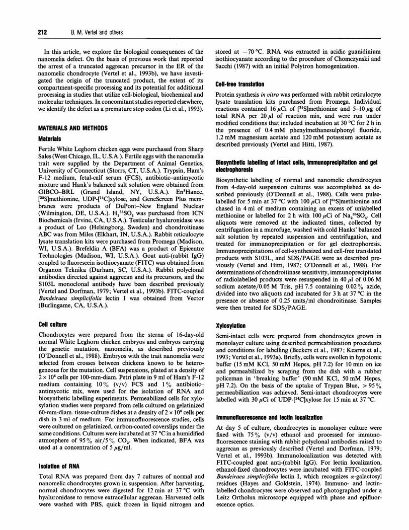
mRNA in nanomelic chondrocytes, as reported by Stirpe et al.(1987). When RNA was used to direct protein synthesis in vitro,immunoprecipitable aggrecan-related precursors were detectedamong the products (Figure 1). The core-protein precursor,
migrating as a protein of 340 kDa, was translated from RNA ofnormal chondrocytes (Figure 1, lanes 1 and 3). In contrast, RNA
No Na
:. l :. '' .......- .*w.. c.n.oo..
........
~~~~~~~~~~~~~~~~~~~~~~~.
-
280 kDa 10
*. ..
~~~~~~~. <...
1 2 3 4 5 6
Figure 1 Both cell-synthesized and cell-free-translated aggrecan pre-
cursors are truncated in nanomelic chondrocytes
Aggrecan-related precursors were immunoprecipitated from [35S]methionine-labelled products
of cell-free-translation reactions directed by RNA from normal (lanes 1 and 3) and nanomelic
(lanes 4 and 6) chondrocytes, and from [35S]methionine pulse-labelled normal (lane 2) and
nanomelic (lane 5) chondrocytes. Note that the aggrecan-related precursors of nanomelic cells
(Na) (lanes 4-6) are truncated compared with the aggrecan core protein of normal chondrocytes
(No) (lanes 1-3), and that the cell-synthesized precursors (lanes 2 and 5, indicated by
asterisks) exhibit slightly slower electrophoretic mobilities compared with cell-free translated
precursors (lanes 1, 3, 4 and 6). Products are displayed on SDS/3-5% polyacrylamide gradient
gels. Positions of the cell-free-translated (arrowhead) and cell-synthesized (asterisks) aggrecan
core protein precursors are indicated. Apparent molecular masses (kDa), based on the
electrophoretic mobilities of protein standards, are given on the left.
xyl met
.:..... ..:..::
1 2
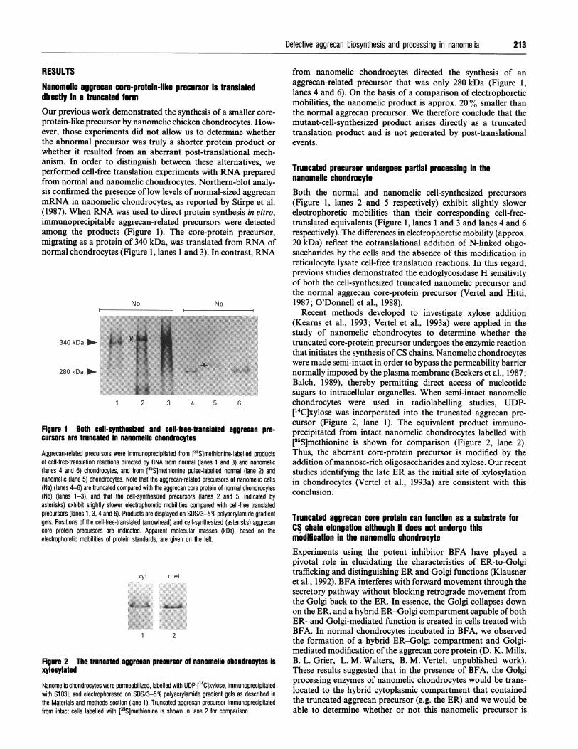
Figure 2 The truncated aggrecan precursor of nanomelic chondrocytes Is
xylosylated
Nanomelic chondrocytes were permeabilized, labelled with UDP [14]ixylose, immunoprecipitatedwith S103L and electrophoresed on SDS/3-5% polyacrylamide gradient gels as described in
the Materials and methods section (lane 1). Truncated aggrecan precursor immunoprecipitated
from nanomelic chondrocytes directed the synthesis of anaggrecan-related precursor that was only 280 kDa (Figure 1,lanes 4 and 6). On the basis of a comparison of electrophoreticmobilities, the nanomelic product is approx. 20% smaller thanthe normal aggrecan precursor. We therefore conclude that themutant-cell-synthesized product arises directly as a truncatedtranslation product and is not generated by post-translationalevents.
Truncated precursor undergoes partial processing in thenanomelic chondrocyteBoth the normal and nanomelic cell-synthesized precursors(Figure 1, lanes 2 and 5 respectively) exhibit slightly slowerelectrophoretic mobilities than their corresponding cell-free-translated equivalents (Figure 1, lanes 1 and 3 and lanes 4 and 6respectively). The differences in electrophoretic mobility (approx.20 kDa) reflect the cotranslational addition of N-linked oligo-saccharides by the cells and the absence of this modification inreticulocyte lysate cell-free translation reactions. In this regard,previous studies demonstrated the endoglycosidase H sensitivityof both the cell-synthesized truncated nanomelic precursor andthe normal aggrecan core-protein precursor (Vertel and Hitti,1987; O'Donnell et al., 1988).Recent methods developed to investigate xylose addition
(Kearns et al., 1993; Vertel et al., 1993a) were applied in thestudy of nanomelic chondrocytes to determine whether thetruncated core-protein precursor undergoes the enzymic reactionthat initiates the synthesis ofCS chains. Nanomelic chondrocyteswere made semi-intact in order to bypass the permeability barriernormally imposed by the plasma membrane (Beckers et al., 1987;Balch, 1989), thereby permitting direct access of nucleotidesugars to intracellular organelles. When semi-intact nanomelicchondrocytes were used in radiolabelling studies, UDP-[14C]xylose was incorporated into the truncated aggrecan pre-cursor (Figure 2, lane 1). The equivalent product immuno-precipitated from intact nanomelic chondrocytes labelled with[35S]methionine is shown for comparison (Figure 2, lane 2).Thus, the aberrant core-protein precursor is modified by theaddition ofmannose-rich oligosaccharides and xylose. Our recentstudies identifying the late ER as the initial site of xylosylationin chondrocytes (Vertel et al., 1993a) are consistent with thisconclusion.
Truncated aggrecan core protein can function as a substrate forCS chain elongation although it does not undergo thismodffication in the nanomelic chondrocyteExperiments using the potent inhibitor BFA have played apivotal role in elucidating the characteristics of ER-to-Golgitrafficking and distinguishing ER and Golgi functions (Klausneret al., 1992). BFA interferes with forward movement through thesecretory pathway without blocking retrograde movement fromthe Golgi back to the ER. In essence, the Golgi collapses downon the ER, and a hybrid ER-Golgi compartment capable of bothER- and Golgi-mediated function is created in cells treated withBFA. In normal chondrocytes incubated in BFA, we observedthe formation of a hybrid ER-Golgi compartment and Golgi-mediated modification of the aggrecan core protein (D. K. Mills,B. L. Grier, L. M. Walters, B. M. Vertel, unpublished work).These results suggested that in the presence of BFA, the Golgiprocessing enzymes of nanomelic chondrocytes would be trans-located to the hybrid cytoplasmic compartment that containedthe truncated aggrecan precursor (e.g. the ER) and we would beable to determine whether or not this nanomelic precursor is
213
from intact cells labelled with [35S]methionine is shown in lane 2 for comparison.
214 B. M. Vertel and others
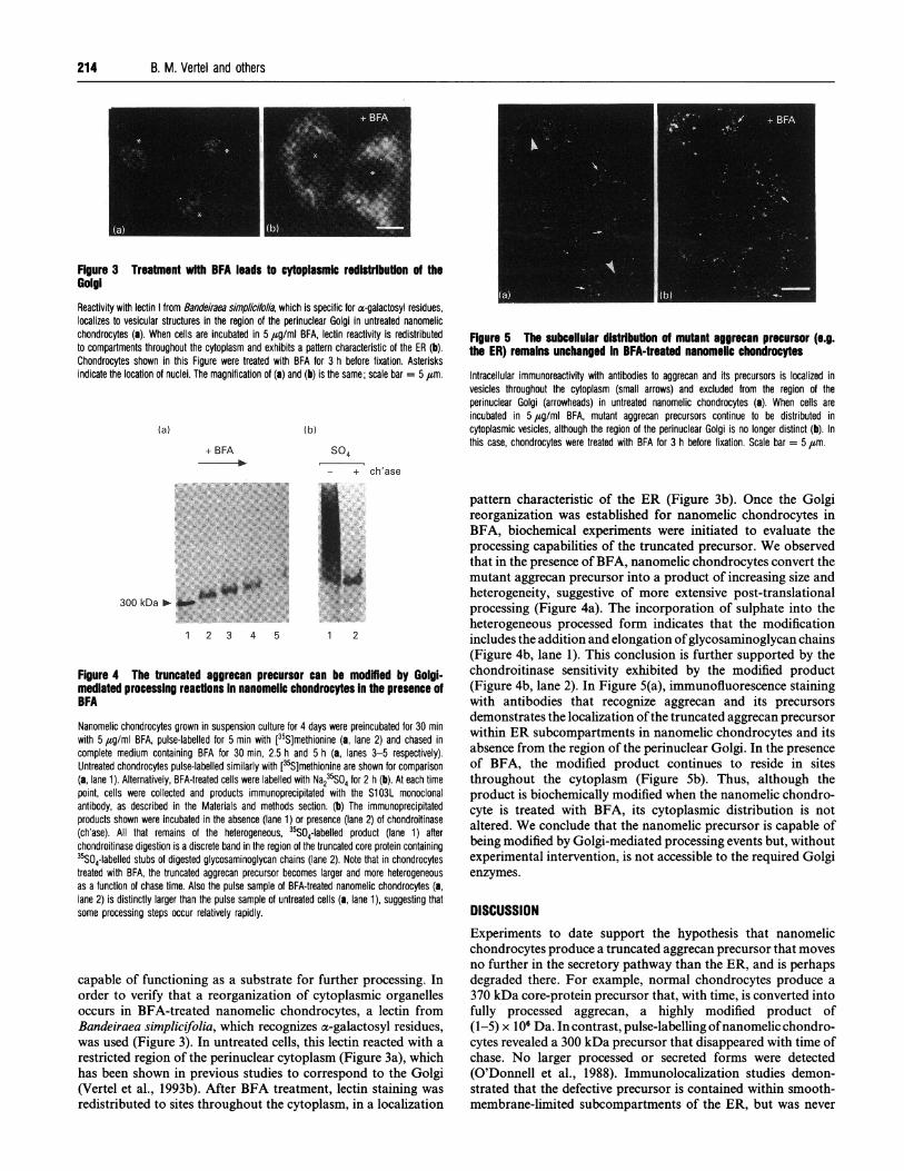
FIgure 3 Treatment with BFA leads to cytoplasmic redistrbution of theGolgiReactivity with lectin from Bandeiraea simplicifolia, which is specific for a-galactosyl residues,localizes to vesicular structures in the region of the perinuclear Golgi in untreated nanomelicchondrocytes (a). When cells are incubated in 5,g/ml BFA, lectin reactivity is redistributedto compartments throughout the cytoplasm and exhibits a pattern characteristic of the ER (b).Chondrocytes shown in this Figure were treated with BFA for 3 h before fixation. Asterisksindicate the location of nuclei. The magnification of (a) and (b) is the same; scale bar = 5 ,m.
(a)
+ BFA
(b)
S04
- + ch'ase
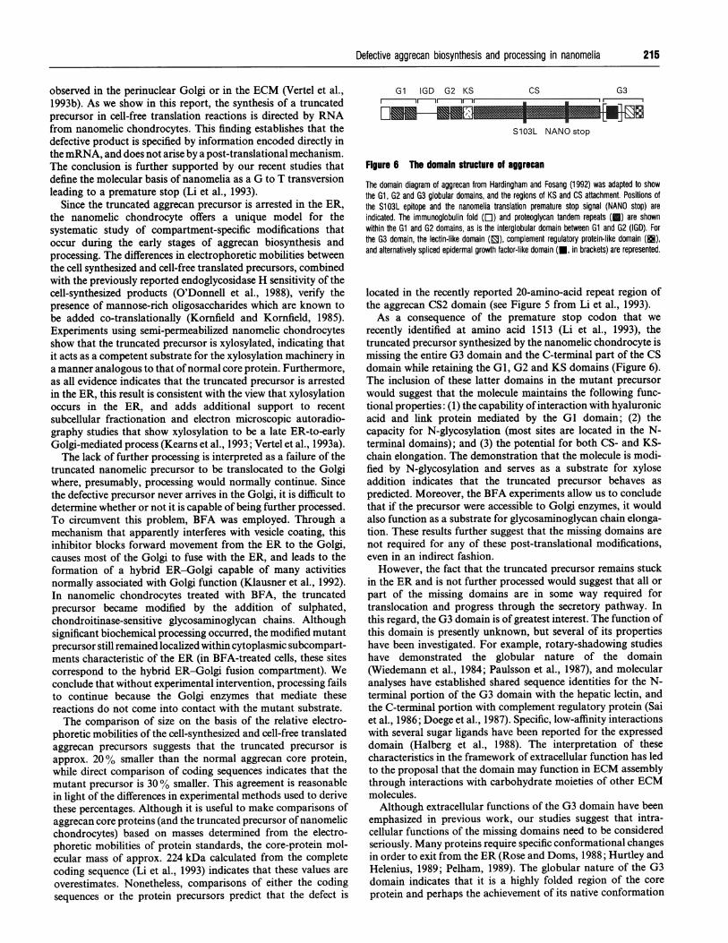
Figure 5 The subkeliular distribution of mutant aggrecan precursor (e.g.the ER) remains unchanged in BFA-treated nanomelic chondrocytes
Intracellular immunoreactivity with antibodies to aggrecan and its precursors is localized invesicles throughout the cytoplasm (small arrows) and excluded from the region of theperinuclear Golgi (arrowheads) in untreated nanomelic chondrocytes (a). When cells areincubated in 5,ug/ml BFA, mutant aggrecan precursors continue to be distributed incytoplasmic vesicles, although the region of the perinuclear Golgi is no longer distinct (b). Inthis case, chondrocytes were treated with BFA for 3 h before fixation. Scale bar = 5 Fim.
300 kDa -
1 2 3 4 5 1 2
Figure 4 The truncated aggrecan precursor can be modffled by Golgi-mediated processing reactlons In nanomelic chondrocytes In the presence ofBFA
Nanomelic chondrocytes grown in suspension culture for 4 days were preincubated for 30 minwith 5 pg/mI BFA, pulse-labelled for 5 min with [35S]methionine (a, lane 2) and chased incomplete medium containing BFA for 30 min, 2.5 h and 5 h (a, lanes 3-5 respectively).Untreated chondrocytes pulse-labelled similarly with [35S]methionine are shown for comparison(a, lane 1). Alternatively, BFA-treated cells were labelled with Na235SO4 for 2 h (b). At each timepoint, cells were collected and products immunoprecipitated with the S103L monoclonalantibody, as described in the Materials and methods section. (b) The immunoprecipitatedproducts shown were incubated in the absence (lane 1) or presence (lane 2) of chondroitinase(ch'ase). All that remains of the heterogeneous, 35S04-labelled product (lane 1) afterchondroitinase digestion is a discrete band in the region of the truncated core protein containing35S04-labelled stubs of digested glycosaminoglycan chains (lane 2). Note that in chondrocytestreated with BFA, the truncated aggrecan precursor becomes larger and more heterogeneousas a function of chase time. Also the pulse sample of BFA-treated nanomelic chondrocytes (a,lane 2) is distinctly larger than the pulse sample of untreated cells (a, lane 1), suggesting thatsome processing steps occur relatively rapidly.
capable of functioning as a substrate for further processing. Inorder to verify that a reorganization of cytoplasmic organellesoccurs in BFA-treated nanomelic chondrocytes, a lectin fromBandeiraea simplicifolia, which recognizes oc-galactosyl residues,was used (Figure 3). In untreated cells, this lectin reacted with a
restricted region of the perinuclear cytoplasm (Figure 3a), whichhas been shown in previous studies to correspond to the Golgi(Vertel et al., 1993b). After BFA treatment, lectin staining was
redistributed to sites throughout the cytoplasm, in a localization
pattern characteristic of the ER (Figure 3b). Once the Golgireorganization was established for nanomelic chondrocytes inBFA, biochemical experiments were initiated to evaluate theprocessing capabilities of the truncated precursor. We observedthat in the presence ofBFA, nanomelic chondrocytes convert themutant aggrecan precursor into a product of increasing size andheterogeneity, suggestive of more extensive post-translationalprocessing (Figure 4a). The incorporation of sulphate into theheterogeneous processed form indicates that the modificationincludes the addition and elongation ofglycosaminoglycan chains(Figure 4b, lane 1). This conclusion is further supported by thechondroitinase sensitivity exhibited by the modified product(Figure 4b, lane 2). In Figure 5(a), immunofluorescence stainingwith antibodies that recognize aggrecan and its precursorsdemonstrates the localization ofthe truncated aggrecan precursorwithin ER subcompartments in nanomelic chondrocytes and itsabsence from the region of the perinuclear Golgi. In the presenceof BFA, the modified product continues to reside in sitesthroughout the cytoplasm (Figure Sb). Thus, although theproduct is biochemically modified when the nanomelic chondro-cyte is treated with BFA, its cytoplasmic distribution is notaltered. We conclude that the nanomelic precursor is capable ofbeing modified by Golgi-mediated processing events but, withoutexperimental intervention, is not accessible to the required Golgienzymes.
DISCUSSIONExperiments to date support the hypothesis that nanomelicchondrocytes produce a truncated aggrecan precursor that movesno further in the secretory pathway than the ER, and is perhapsdegraded there. For example, normal chondrocytes produce a370 kDa core-protein precursor that, with time, is converted intofully processed aggrecan, a highly modified product of(1-5) x 106 Da. In contrast, pulse-labelling ofnanomelic chondro-cytes revealed a 300 kDa precursor that disappeared with time ofchase. No larger processed or secreted forms were detected(O'Donnell et al., 1988). Immunolocalization studies demon-strated that the defective precursor is contained within smooth-membrane-limited subcompartments of the ER, but was never
Defective aggrecan biosynthesis and processing in nanomelia
observed in the perinuclear Golgi or in the ECM (Vertel et al.,1993b). As we show in this report, the synthesis of a truncatedprecursor in cell-free translation reactions is directed by RNAfrom nanomelic chondrocytes. This finding establishes that thedefective product is specified by information encoded directly inthe mRNA, and does not arise by a post-translational mechanism.The conclusion is further supported by our recent studies thatdefine the molecular basis of nanomelia as a G to T transversionleading to a premature stop (Li et al., 1993).
Since the truncated aggrecan precursor is arrested in the ER,the nanomelic chondrocyte offers a unique model for thesystematic study of compartment-specific modifications thatoccur during the early stages of aggrecan biosynthesis andprocessing. The differences in electrophoretic mobilities betweenthe cell synthesized and cell-free translated precursors, combinedwith the previously reported endoglycosidase H sensitivity of thecell-synthesized products (O'Donnell et al., 1988), verify thepresence of mannose-rich oligosaccharides which are known tobe added co-translationally (Kornfield and Kornfield, 1985).Experiments using semi-permeabilized nanomelic chondrocytesshow that the truncated precursor is xylosylated, indicating thatit acts as a competent substrate for the xylosylation machinery ina manner analogous to that of normal core protein. Furthermore,as all evidence indicates that the truncated precursor is arrestedin the ER, this result is consistent with the view that xylosylationoccurs in the ER, and adds additional support to recentsubcellular fractionation and electron microscopic autoradio-graphy studies that show xylosylation to be a late ER-to-earlyGolgi-mediated process (Kearns et al., 1993; Vertel et al., 1993a).The lack of further processing is interpreted as a failure of the
truncated nanomelic precursor to be translocated to the Golgiwhere, presumably, processing would normally continue. Sincethe defective precursor never arrives in the Golgi, it is difficult todetermine whether or not it is capable of being further processed.To circumvent this problem, BFA was employed. Through amechanism that apparently interferes with vesicle coating, thisinhibitor blocks forward movement from the ER to the Golgi,causes most of the Golgi to fuse with the ER, and leads to theformation of a hybrid ER-Golgi capable of many activitiesnormally associated with Golgi function (Klausner et al., 1992).In nanomelic chondrocytes treated with BFA, the truncatedprecursor became modified by the addition of sulphated,chondroitinase-sensitive glycosaminoglycan chains. Althoughsignificant biochemical processing occurred, the modified mutantprecursor still remained localized within cytoplasmic subcompart-ments characteristic of the ER (in BFA-treated cells, these sitescorrespond to the hybrid ER-Golgi fusion compartment). Weconclude that without experimental intervention, processing failsto continue because the Golgi enzymes that mediate thesereactions do not come into contact with the mutant substrate.The comparison of size on the basis of the relative electro-
phoretic mobilities of the cell-synthesized and cell-free translatedaggrecan precursors suggests that the truncated precursor isapprox. 20 % smaller than the normal aggrecan core protein,while direct comparison of coding sequences indicates that themutant precursor is 30% smaller. This agreement is reasonablein light of the differences in experimental methods used to derivethese percentages. Although it is useful to make comparisons ofaggrecan core proteins (and the truncated precursor of nanomelicchondrocytes) based on masses determined from the electro-phoretic mobilities of protein standards, the core-protein mol-ecular mass of approx. 224 kDa calculated from the completecoding sequence (Li et al., 1993) indicates that these values areoverestimates. Nonetheless, comparisons of either the codingsequences or the protein precursors predict that the defect is
Gl IGD G2 KS Cs G3
S103L NANO stop
Figure 6 The domain structure of aggrecan
The domain diagram of aggrecan from Hardingham and Fosang (1992) was adapted to showthe Gl, G2 and G3 globular domains, and the regions of KS and CS attachment. Positions ofthe S103L epitope and the nanomelia translation premature stop signal (NANO stop) areindicated. The immunoglobulin fold ([E) and proteoglycan tandem repeats (c) are shownwithin the Gl and G2 domains, as is the interglobular domain between Gl and G2 (IGD). Forthe G3 domain, the lectin-like domain (n), complement regulatory protein-like domain (EE),and alternatively spliced epidermal growth factor-like domain (-, in brackets) are represented.
located in the recently reported 20-amino-acid repeat region ofthe aggrecan CS2 domain (see Figure 5 from Li et al., 1993).As a consequence of the premature stop codon that we
recently identified at amino acid 1513 (Li et al., 1993), thetruncated precursor synthesized by the nanomelic chondrocyte ismissing the entire G3 domain and the C-terminal part of the CSdomain while retaining the GI, G2 and KS domains (Figure 6).The inclusion of these latter domains in the mutant precursorwould suggest that the molecule maintains the following func-tional properties: (1) the capability of interaction with hyaluronicacid and link protein mediated by the GI domain; (2) thecapacity for N-glycosylation (most sites are located in the N-terminal domains); and (3) the potential for both CS- and KS-chain elongation. The demonstration that the molecule is modi-fied by N-glycosylation and serves as a substrate for xyloseaddition indicates that the truncated precursor behaves aspredicted. Moreover, the BFA experiments allow us to concludethat if the precursor were accessible to Golgi enzymes, it wouldalso function as a substrate for glycosaminoglycan chain elonga-tion. These results further suggest that the missing domains arenot required for any of these post-translational modifications,even in an indirect fashion.However, the fact that the truncated precursor remains stuck
in the ER and is not further processed would suggest that all orpart of the missing domains are in some way required fortranslocation and progress through the secretory pathway. Inthis regard, the G3 domain is of greatest interest. The function ofthis domain is presently unknown, but several of its propertieshave been investigated. For example, rotary-shadowing studieshave demonstrated the globular nature of the domain(Wiedemann et al., 1984; Paulsson et al., 1987), and molecularanalyses have established shared sequence identities for the N-terminal portion of the G3 domain with the hepatic lectin, andthe C-terminal portion with complement regulatory protein (Saiet al., 1986; Doege et al., 1987). Specific, low-affinity interactionswith several sugar ligands have been reported for the expresseddomain (Halberg et al., 1988). The interpretation of thesecharacteristics in the framework of extracellular function has ledto the proposal that the domain may function in ECM assemblythrough interactions with carbohydrate moieties of other ECMmolecules.
Although extracellular functions of the G3 domain have beenemphasized in previous work, our studies suggest that intra-cellular functions of the missing domains need to be consideredseriously. Many proteins require specific conformational changesin order to exit from the ER (Rose and Doms, 1988; Hurtley andHelenius, 1989; Pelham, 1989). The globular nature of the G3domain indicates that it is a highly folded region of the coreprotein and perhaps the achievement of its native conformation
215
216 B. M. Vertel and others
is required for movement out of the ER. Alternatively, themissing region may be involved in interactions (either with otherdomains or with different molecules) that must be completedbefore the precursor can leave the ER. As another possibility,some aspect of the missing region may function as a signal fortranslocation. We previously suggested that the premature stopcodon might result in the exposure of an ER retention signal, butan examination of the newly created C-terminal sequence on thetruncated precursor, VHETSG, shows that it bears no resemb-lance to the standard KDEL ER retention signal (Pelham, 1989).The possibility of the existence of other, as yet unidentified,retention or recognition signals, however, is not ruled out. It isunlikely that the epidermal growth factor-like domain is required,as this domain exists as an alternatively spliced form in the chick(Li et al., 1993) and other aggrecan mRNAs (Fulop et al., 1993).
It has been established that retention in the ER and targetingfor degradation can be the consequence of a general cellularmechanism for quality control that operates if specific confor-mational and assembly events fail to occur (Rose and Doms,1988; Hurtley and Helenius, 1989; Bonifacino and Lippincott-Schwartz, 1991). Studies of viral glycoproteins and membranereceptor complexes have been particularly informative in ad-vancing our understanding of this process. We can regard errorsin the trafficking ofdefective cell products, such as those describedfor -the mutant nanomelic precursor, in this context. Relatedmechanisms have been implicated for several other geneticdiseases, including familial hypercholesterolaemia, some formsofTay-Sachs disease, and cystic fibrosis (Amara et al., 1992). Forexample, one patient with Tay-Sachs disease accumulated ganglio-sides in lysosomes as a result of the production of a truncatedprecursor of the a-subunit of lysosomal /I-hexosaminidase thatwas retained and degraded in an early biosynthetic compartment(presumably the ER) rather than being delivered to the lysosome(Lau and Neufeld, 1989).
Connective-tissue disorders may be considered similarly, asECMs are composed of large, highly modified, strongly inter-active macromolecules organized into characteristic assemblies.Many collagen defects have been associated with osteogenesisimperfecta, a number of chondrodystrophies and other diseases,and some proteoglycan defects have been suggested as well(Goetinck, 1983; Stanescu et al., 1984; Johnson, 1986; Prockop,1990; Spranger and Martoux, 1990; Byers et al., 1991). In manycases, the swollen appearance ofthe ER and biochemical evidenceof delayed secretion and impaired ECM deposition reflect errorsin synthesis, processing and assembly. A detailed analysis of theintracellular mechanisms involved in the ER retention of ab-normal type-I procollagen was reported in a recent study offibroblasts from a patient with osteogenesis imperfecta (Chesslerand Byers, 1992). Our studies demonstrate the first mutation ofan aggrecan gene that exhibits similar characteristics. It is likelythat further investigations of this nature will reveal more aboutthe underlying defects and consequences of genetic diseases, andabout basic biological processes.
We thank Dr. Louis Pierro and Ms. Jean Haines for graciously supplying eggscarrying the genetic mutation, nanomelia, and Mr. Jonathan Liang for his assistance
in the preparation of Figure 6. Research support was provided by USPHS grants DK-28433 (B. M.V.), HD-17332 (N. B. S.), AR-19266 (N. B. S.), and HD-09402 (N. B. S.).
REFERENCESAmara, J. F., Cheng, S. H. and Smith, A. E. (1992) Trends Cell Biol. 2,145-149Balch, W. E. (1989) J. Biol. Chem. 264,16965-16968Beckers, C. J. M., Keller, D. S. and Balch, W. E. (1987) Cell 50, 523-534Bonifacino, J. S. and Lippincott-Schwartz, J. (1991) Curr. Opin. Cell Biol. 3, 592-600Byers, P. H., Wallis, G. A. and Willing, M. C. (1991) J. Med. Genet. 28, 433-442Campbell, S. C. and Schwartz, N. B. (1988) J. Cell Biol. 28, 2191-2202Chandrasekaran, L. and Tanzer, M. L. (1992) Biochem. J. 288, 903-910Chessler, S. D. and Byers, P. H. (1992) J. Biol. Chem. 267, 7751-7757Chomczynski, P. and Sacchi, N. (1987) Anal. Biochem. 162, 156-159Doege, K., Sasakai, M., Horigan, E., Hassell, J. R. and Yamada, Y. (1987) J. Biol. Chem.
262, 17757-17767Farquhar, M. G. (1991) Intracellular Trafficking of Proteins, pp. 431-471, Cambridge
University Press, CambridgeFUlop, C., Walcz, E., Valyon, M. and Glant, T. T. (1993) J. Biol. Chem. 268, 17377-17383Goetinck, P. F. (1983) Cartilage: Biomedical Aspects, pp. 165-189, Academic Press,
New YorkHalberg, D. F., Proulx, G., Doege, K., Yamada, Y. and Drickamer, K. (1988) J. Biol. Chem.
263, 9486-9490Hardingham, T. E. and Fosang, A. J. (1992) FASEB J. 6, 861-870Hayes, C. E. and Goldstein, I. J. (1974) J. Biol. Chem. 249, 1904-1914Hurtley, S. M. and Helenius, A. (1989) Annu. Rev. Cell Biol. 5, 277-307Johnson, D. R. (1986) The Genetics of the Skeleton, Clarendon Press, OxfordKearns, A. E., Vertel, B. M. and Schwartz, N. B. (1993) J. Biol. Chem. 268, 11097-11104Klausner, R. D., Donaldson, J. G. and Lippincott-Schwartz, J. (1992) J. Cell Biol. 116,
1071-1 080Kornfield, R. and Kornfeld, S. (1985) Annu. Rev. Biochem. 54, 631-664Landauer, W. (1965) J. Hered. 56,131-138Lau, M. M. H. and Neufeld, E. F. (1989) J. Biol. Chem. 264, 21376-21380Li, H., Vertel, B. M. and Schwartz, N. B. (1993) J. Biol. Chem. 268, 23504-23511O'Donnell, C. M., Kaczman-Daniel, K., Goetinck, P. F. and Vertel, B. M. (1988) J. Biol.
Chem. 263, 17749-17754Pacifici, M., Soltesz, R., Tahl, G., Shanley, D. J., Boettiger, D. and Holtzer, H. (1984)
J. Cell Biol. 97, 1724-1735Paulsson, M., Morgelin, M., Wiedemann, H., Beardmore-Gray, M., Dunham, D.,
Hardingham, T., Heinegard, D., Timpl, R. and Engel, J. (1987)Biochem. J. 245, 763-772
Pelham, H. R. B. (1989) Annu. Rev. Cell Biol. 5,1-23Pennypacker, J. P. and Goetinck, P. F. (1976) Dev. Biol. 50, 35-47Prockop, D. J. (1990) J. Biol. Chem. 265, 15349-15352Rose, J. K. and Doms, R. W. (1988) Ann. Rev. Cell Biol. 4, 257-288Sai, S., Tanaka, T., Kosher, R. and Tanzer, M. L. (1986) Proc. NatI. Acad. Sci. U.S.A. 83,
5081-5085Spranger, J. and Martoux, P. (1990) Adv. Human Genet. 19, 1-103Stanescu, V., Stanescu, R. and Maroteaux, P. (1984) J. Bone Joint Surg. 66-A, 817-836Stirpe, N. S., Argraves, W. S. and Goetinck, P. F. (1987) Dev. Biol. 124, 77-81Vertel, B. M. and Barkman, L. L. (1984) Collagen Rel. Res. 4,1-20Vertel, B. M. and Dortman, A. (1979) Proc. NatI. Acad. Sci. U.S.A. 76, 4847-4851Vertel, B. M. and Hitti, Y. (1987) Collagen Rel. Res. 7, 57-75Vertel, B. M., Barkman, L. L. and Morrell, J. J. (1985a) J. Cell. Biochem. 27, 215-229Vertel, B. M., Morrell, J. J. and Barkman, L. L. (1985b) Exp. Cell Res. 158, 423-432Vertel, B. M., Velasco, A., La France, S., Walters, L. and Kaczman-Daniel, K. (1989)
J. Cell Biol. 109,1827-1836Vertel, B. M., Walters, L. M., Flay, N., Kearns, A. E. and Schwartz, N. B. (1993a) J. Biol.
Chem. 268, 11105-11112Vertel, B. M., Walters, L. M., Grier, B., Maine, N. and Goetinck, P. F. (1993b) J. Cell Sci.
104, 939-948Wiedemann, H., Paulsson, M., Timpl, R., Engel, J. and Heinegard, D. (1984) Biochem. J.
224, 331-333Wight, T. N., Heinegard, D. K. and Hascall, V. C. (1991) Cell Biology of Extracellular Matrix
(Hay, E. D., ed.), pp. 45-78, Plenum, New York
Received 19 July 1993/11 January 1994; accepted 26 January 1994
Related Documents