The chemokine receptors CXCR3 and CCR5 mark subsets of T cells associated with certain inflammatory reactions. S Qin, … , B Moser, C R Mackay J Clin Invest. 1998;101(4):746-754. https://doi.org/10.1172/JCI1422. T cells infiltrating inflammatory sites are usually of the activated/memory type. The precise mechanism for the positioning of these cells within tissues is unclear. Adhesion molecules certainly play a role; however, the intricate control of cell migration appears to be mediated by numerous chemokines and their receptors. Particularly important chemokines for activated/memory T cells are the CXCR3 ligands IP-10 and Mig and the CCR5 ligands RANTES, macrophage inflammatory protein-1alpha, and macrophage inflammatory protein-1beta. We raised anti-CXCR3 mAbs and were able to detect high levels of CXCR3 expression on activated T cells. Surprisingly, a proportion of circulating blood T cells, B cells, and natural killer cells also expressed CXCR3. CCR5 showed a similar expression pattern as CXCR3, but was expressed on fewer circulating T cells. Blood T cells expressing CXCR3 (and CCR5) were mostly CD45RO+, and generally expressed high levels of beta1 integrins. This phenotype resembled that of T cells infiltrating inflammatory lesions. Immunostaining of T cells in rheumatoid arthritis synovial fluid confirmed that virtually all such T cells expressed CXCR3 and approximately 80% expressed CCR5, representing high enrichment over levels of CXCR3+ and CCR5+ T cells in blood, 35 and 15%, respectively. Analysis by immunohistochemistry of various inflamed tissues gave comparable findings in that virtually all T cells within the lesions expressed CXCR3, particularly in perivascular regions, whereas far fewer […] Research Article Find the latest version: https://jci.me/1422/pdf

Welcome message from author
This document is posted to help you gain knowledge. Please leave a comment to let me know what you think about it! Share it to your friends and learn new things together.
Transcript

The chemokine receptors CXCR3 and CCR5 mark subsets of Tcells associated with certain inflammatory reactions.
S Qin, … , B Moser, C R Mackay
J Clin Invest. 1998;101(4):746-754. https://doi.org/10.1172/JCI1422.
T cells infiltrating inflammatory sites are usually of the activated/memory type. The precise mechanism for the positioningof these cells within tissues is unclear. Adhesion molecules certainly play a role; however, the intricate control of cellmigration appears to be mediated by numerous chemokines and their receptors. Particularly important chemokines foractivated/memory T cells are the CXCR3 ligands IP-10 and Mig and the CCR5 ligands RANTES, macrophageinflammatory protein-1alpha, and macrophage inflammatory protein-1beta. We raised anti-CXCR3 mAbs and were able todetect high levels of CXCR3 expression on activated T cells. Surprisingly, a proportion of circulating blood T cells, B cells,and natural killer cells also expressed CXCR3. CCR5 showed a similar expression pattern as CXCR3, but was expressedon fewer circulating T cells. Blood T cells expressing CXCR3 (and CCR5) were mostly CD45RO+, and generallyexpressed high levels of beta1 integrins. This phenotype resembled that of T cells infiltrating inflammatory lesions.Immunostaining of T cells in rheumatoid arthritis synovial fluid confirmed that virtually all such T cells expressed CXCR3and approximately 80% expressed CCR5, representing high enrichment over levels of CXCR3+ and CCR5+ T cells inblood, 35 and 15%, respectively. Analysis by immunohistochemistry of various inflamed tissues gave comparable findingsin that virtually all T cells within the lesions expressed CXCR3, particularly in perivascular regions, whereas far fewer […]
Research Article
Find the latest version:
https://jci.me/1422/pdf

746
Qin et al.
Rapid Publication
J. Clin. Invest.© The American Society for Clinical Investigation, Inc.0021-9738/98/02/0746/09 $2.00Volume 101, Number 4, February 1998, 746–754http://www.jci.org
The Chemokine Receptors CXCR3 and CCR5 Mark Subsets of T Cells Associated with Certain Inflammatory Reactions
Shixin Qin,* James B. Rottman,* Paul Myers,* Nasim Kassam,* Michael Weinblatt,
‡
Marcel Loetscher,
i
Alisa E. Koch,
§
Bernhard Moser,
i
and Charles R. Mackay*
*
LeukoSite, Inc., Cambridge, Massachusetts 02142;
‡
Brigham and Women’s Hospital, Boston, Massachusetts 02150;
§
Northwestern University Medical School and Veterans’ Administration Chicago Healthcare System, Lakeside Division, Chicago, Illinois 60611;
i
Theodor-Kocher Institute, University of Bern, CH-3000-Bern-9, Switzerland
Abstract
T cells infiltrating inflammatory sites are usually of the acti-vated/memory type. The precise mechanism for the posi-tioning of these cells within tissues is unclear. Adhesion mole-cules certainly play a role; however, the intricate control ofcell migration appears to be mediated by numerous chemo-kines and their receptors. Particularly important chemo-kines for activated/memory T cells are the CXCR3 ligandsIP-10 and Mig and the CCR5 ligands RANTES, mac-rophage inflammatory protein-1
a
, and macrophage inflam-matory protein-1
b
. We raised anti-CXCR3 mAbs and wereable to detect high levels of CXCR3 expression on activatedT cells. Surprisingly, a proportion of circulating blood Tcells, B cells, and natural killer cells also expressed CXCR3.CCR5 showed a similar expression pattern as CXCR3, butwas expressed on fewer circulating T cells. Blood T cells ex-pressing CXCR3 (and CCR5) were mostly CD45RO
1
, andgenerally expressed high levels of
b
1 integrins. This pheno-type resembled that of T cells infiltrating inflammatory le-sions. Immunostaining of T cells in rheumatoid arthritissynovial fluid confirmed that virtually all such T cells ex-
pressed CXCR3 and
z
80% expressed CCR5, representinghigh enrichment over levels of CXCR3
1
and CCR5
1
T cellsin blood, 35 and 15%, respectively. Analysis by immuno-histochemistry of various inflamed tissues gave comparablefindings in that virtually all T cells within the lesions expressedCXCR3, particularly in perivascular regions, whereas farfewer T cells within normal lymph nodes expressed CXCR3or CCR5. These results demonstrate that the chemokine re-ceptor CXCR3 and CCR5 are markers for T cells associatedwith certain inflammatory reactions, particularly TH-1 type
reactions. Moreover, CXCR3 and CCR5 appear to identifysubsets of T cells in blood with a predilection for homing tothese sites. (
J. Clin. Invest.
1998. 101:746–754.) Key words:chemokines
•
inflammation
•
cell -migration
•
antibodies-monoclonal
•
lymphocytes
Introduction
Leukocyte migration is essential for immune surveillance ofthe body’s tissues, and for focusing immune cells to sites ofantigenic challenge. The control of leukocyte migration de-pends on the combined actions of various adhesion molecules,as well as a vast array of chemotactic cytokines (chemokines)and their receptors. The role of adhesion molecules in leuko-cyte migration is well appreciated (1, 2), whereas that of dif-ferent chemokines and their receptors is less certain. Thechemokine receptors comprise two groups, the CC receptors 1–8(CCR1–8)
1
that bind CC chemokines, and the CXC receptors1–4 (CXCR1–4), which bind CXC chemokines (3–5). In gen-eral, the CC chemokines and their receptors effect the migra-tion of monocytes, eosinophils, basophils, and T cells (6–8),whereas CXCR1 and CXCR2, which are the two IL-8 recep-tors, effect the migration of neutrophils (3).
The notion that CXC chemokines are generally poor che-moattractants for T cells has been challenged recently. Thusthe CXC chemokine SDF-1 is a potent chemoattractant forblood T cells (9), and binds to CXCR4 (10, 11), a broadly ex-pressed chemokine receptor (12). The CXC chemokines IP-10and Mig, both inducible by interferon-
g
during inflammation,also are effective T cell chemoattractants (13–15). IP-10 andMig attract activated T cells, but not resting T cells. These twochemokines induce an increase in intracellular calcium by cellstransfected with the cDNA for the chemokine receptorCXCR3, indicating that this is the likely IP-10/Mig receptor onT cells (14). By Northern blot analysis, CXCR3 is highly re-stricted to activated T cells and natural killer (NK) cells, andnot to other leukocytes (14). Therefore IP-10 or Mig signalingappears to be an important mechanism for selective homing ofactivated/effector cells, which are known to accumulate prefer-entially at inflammatory sites (16), as well as in many tumors.IP-10 is expressed abundantly in various inflammatory lesions,
S. Qin and J.B. Rottman contributed equally to this work.Address correspondence to Dr. Shixin Qin, LeukoSite, Inc., 215
First Street, Cambridge, MA 02142. Phone: 617-621-9350; FAX: 617-621-9349; E-mail: [email protected] Charles R. Mackay’scurrent address is Millennium Biotherapeutics, Cambridge, MA 02139.
Received for publication 6 August 1997 and accepted in revisedform 5 December 1997.
1.
Abbreviations used in this paper:
CCR, CC chemokine receptor;CXCR, CXC chemokine receptor; MCP, monocyte chemotactic pro-tein; MIP, macrophage inflammatory protein; NK, natural killer;[Ca2
1
]i, intracellular calcium concentration.

CXCR3, CCR5, and Inflammation
747
particularly those characterized by T cell infiltration, such asdelayed type hypersensitivity responses in skin (17), in EAE(18), and in transplants undergoing rejection (19).
Another chemokine receptor expressed by T cells is CCR5,the receptor for RANTES, macrophage inflammatory protein-1
a
(MIP-1
a
), and MIP-1
b
. CCR5 is expressed on activatedand memory (CD45RO
1
) T cells (12, 20), which correlateswith the finding that memory and activated T cells, but not na-ive T cells, respond to RANTES, MIP-1
a
, and MIP-1
b
inchemotaxis assays (21, 22). In addition, RANTES, MIP-1
a
,and MIP-1
b
are expressed in many inflammatory lesions (23).The assumption has been that different chemokines will attractparticular types of leukocytes based on the ligand specificityand expression patterns of the relevant receptors. For instance,CCR5 and CXCR4 are expressed on T cells in a largely recip-rocal fashion (12), and probably facilitate the positioning oftwo types of T cells in different tissues.
We now report on the distribution of CXCR3 and CCR5,particularly their association with inflammation. A strikingfinding was the high proportion of CXCR3
1
and CCR5
1
T cellsin certain inflammatory lesions, compared with relatively lowlevels of CXCR3
1
and CCR5
1
T cells in blood or lymph node.In the blood, CXCR3, as well as CCR5, was expressed on asubset of circulating CD45RO
1
and
b
1 integrin
hi
T cells, a phe-notype consistent with previous activation. Moreover, theCCR5
1
subset was contained entirely within the CXCR3
1
sub-set. We conclude that these two chemokine receptors mark themajority of T cells within inflamed tissues, as well as peripheralblood T cells with a predilection for homing to these sites.
Methods
Cells, cell lines, and tissue culture.
Normal human blood leukocyteswere isolated as described (24). To generate CD3 blasts, 2
3
106PBMC/ml in RPMI 1640 plus 10% FCS were added to tissue cultureplates first coated with the anti-CD3 antibody TR66. After 4–6 d,blasts were removed to fresh media and supplemented with IL-2(kindly provided by Antonio Lanzavecchia, Basel Institute for Immu-nology, Basel, Switzerland) at 50 U/ml. Other cell lines used includedtransfectants of the L1.2 murine pre B cell lymphoma, expressinghigh levels of either CXCR3 (this report), CXCR1 (25), CXCR2 (25),CCR2b, CCR4, CCR5 (26), or CCR1 (27). CXCR3 cDNA was ob-tained by PCR using a 5
9
-oligonucleotide primer and 3
9
-oligonucleo-tide primer which contained flanking XhoI and XbaI sites respec-tively. The PCR fragment was subcloned into the XhoI–XbaI sites ofpCDNA3 (Invitrogen Corp., San Diego, CA), and the inserted genewas driven by a CMV promoter. Stable transfection of the DNA intoa murine pre B lymphoma cell line (L1.2) was obtained as described(24). The cell surface expression of CXCR3 was monitored by ligandbinding and Scatchard analysis. Transfectants were maintained in RPMI1640 supplemented with 10% bovine serum and 800
m
g/ml G418.
mAb production and flow cytometry.
mAbs reactive with CXCR3were generated by immunizing BALB/C mice with 10
m
g of a 37-mersynthetic peptide corresponding to the first 37 NH
2
-terminal aminoacids of CXCR3 (14), five times over a period of 10 wk. This peptidewas synthesized and coupled to purified protein derivative of tuber-culin by the manufacturer (Severn Biotech Ltd., Kidderminster,United Kingdom). The first immunization was intraperitoneal withCFA, the second, third, and fourth were intraperitoneal with IFA,and the final immunization was intravenous with protein alone. 4 dafter the last immunization, the spleen was taken and cell fusion per-formed using the cell line SP2/O, as described (28). Specific mAbs re-active with CXCR3 were identified using untransfected and CXCR3transfected L1.2 cells, and immunofluorescent staining and analysis
using a FACScan
®
(Becton Dickinson, Mountain View, CA). The mainmAb used in this study termed 1C6, is of isotype IgG1. Anti-CXCR3mAbs were also generated by immunizing mice with CXCR3/L1-2transfectant, in a similar manner to that described for anti-CCR3 andanti-CCR5 (20, 29).
mAbs to CCR5 have been described (20). PE-conjugated mAbsto CD4, CD8, CD14, CD19, CD25, CD26, CD29, CD69, CD45RO,CD45RA, and CD95 were supplied by PharMingen (San Diego, CA).To assess reactivity of mAbs against transfected cells or leukocytes,indirect immunofluorescence and flow cytometry were used. Cellswere washed once with PBS, and resuspended in 100
m
l PBS contain-ing 2% human serum and 0.1% sodium azide (staining buffer), 5
m
g/ml purified antibody, 5
m
g/ml isotype matched control mAb (SigmaChemical Co., St. Louis, MO) or 50
m
l hybridoma culture superna-tant. After 20 min at 4
8
C, cells were washed twice with stainingbuffer, and resuspended in 50
m
l FITC-conjugated affinity purifiedF(ab
9
)2 goat anti–mouse IgG (Jackson ImmunoResearch Labs., Inc.,West Grove, PA). After 20 min, cells were washed twice in stainingbuffer and analyzed on the FACScan
®
to determine the level of sur-face expression. Propidium iodide was used to exclude dead cells. Formulticolor analysis, PE- or FITC-conjugated mAbs were used to-gether with biotinylated anti-CXCR3 mAb 1C6 to stain cells. Afterwashing, the cells were incubated with Streptavidin-Red 670 (GIBCOBRL, Gaithersburg, MD). The results were analyzed by FACScan
®
using electronic gating and compensation.
Chemokines, chemotaxis assays, and ligand-binding assay.
Recom-binant human chemokines were obtained from PeproTech, Inc.(Rocky Hill, NJ). Chemotaxis of CD3 blasts was assessed using a mod-ification of a transendothelial assay (7), using the cell line ECV304 asdescribed (24). Cells that had migrated to the bottom chamber wereplaced in a tube, and relative cell counts were obtained using theFACScan
®
.
125
I-labeled chemokines were obtained from DuPont-NEN (Bos-ton, MA). The functional activity of radiolabeled IP-10 was tested inchemotaxis assays and was found to have 80% activity of unlabeledIP-10 (data not shown). Chemokine binding to target cells was car-ried out as described previously (24). Briefly, cells were resuspendedin binding buffer (50 mM Hepes, 1 mM CaCl
2
, 5 mM MgCl
2
, 0.5%BSA) and incubated with radiolabeled ligand in the presence or ab-sence of inhibitors. After 60 min at 37
8
C, cells were washed threetimes in binding buffer supplemented with 0.5 N NaCl and pelletswere counted. All experiments were repeated at least three times.Curve fit and concentrations that inhibit 50% specific binding (IC50)were calculated by KaleidaGraph software (Synergy Software, Inc.,Reading, PA). Scatchard Plot analysis was carried out using Mi-crosoft Excel.
Tissues and immunohistochemistry.
Human tissues (normal andinflamed colon and vagina) were obtained from the National DiseaseResearch Institute, a service organization funded by the National In-stitutes of Health. Synovial fluid was obtained from patients withrheumatoid arthritis.
Immunohistochemical analysis for CXCR3 was performed on fro-zen tissue samples. Briefly, tissue was sectioned at a thickness of 4
m
m,desiccated, and then fixed in 2% paraformaldehyde/0.5 X PBS for 10min at 4
8
C. After PBS washing, nonspecific antibody binding siteswere blocked with 10% normal goat serum/5% human AB serum/PBS for 30 min at room temperature. Next, the purified CXCR3 mono-clonal antibody 1C6 was diluted to a concentration of 1
m
g/ml in 0.3%Triton X-100/0.2% Tween 20/1% FCS/5% human AB serum/0.1%sodium azide and applied to tissue sections overnight at 4
8
C. An iso-type-matched irrelevant monoclonal antibody was used as a negativecontrol on step sections of tissues (IgG1, MOPC21; Sigma ChemicalCo.). Subsequently, biotinylated goat anti–mouse IgG (Vector Labo-ratories, Inc., Burlingame, CA) and avidin-biotin-alkaline phos-phatase complexes (BioGenex Labs, San Ramon, CA) were added insequence. Fast Red (BioGenex Labs), containing 2% levamisol toblock endogenous alkaline phosphatase activity, was used as thechromogen and Mayers hematoxylin as the counterstain.

748
Qin et al.
Results
CXCR3 is expressed by lymphocytes but not by other leukocytetypes.
mAbs have proven to be powerful tools for assessingthe biology of various chemokine receptors (6, 25, 29), particu-larly CCR5 (12, 20). To study the functions of one of the im-portant T cell chemokine receptors, CXCR3, specific mAbswere generated by immunizing mice either with synthetic pep-tides or with CXCR3 transfected cells (see Methods). ThesemAbs reacted specifically with CXCR3, as judged by FACS
®
staining of CXCR3 transfected L1.2 cells, and not wild-typeL1.2 cells or L1.2 cells transfected with numerous other recep-tors. (Fig. 1
A
). In peripheral blood, anti-CXCR3 mAbs wereunreactive on neutrophils, monocytes, and eosinophils (notshown), as expected from previous analyses of CXCR3 expres-sion by Northern blot, as well as functional responsiveness ofcells to IP-10 (14). However the phenotypic analysis did revealthe expression of CXCR3 on a proportion of circulating lym-phocytes (Fig. 1
B
, day 0), observed in all (
.
20) normal indi-viduals examined. A feature of CXCR3, determined from pre-vious studies on mRNA expression, is its expression onactivated T cells (14). Staining of T cells activated by anti-CD3confirmed that CXCR3 was expressed at high levels on thesecells, but only after 5–8 d of activation, and in fact CXCR3 ex-pression was downregulated immediately after activation (Fig.1
B
, day 3). This pattern of expression and regulation was sim-ilar to that observed for CCR5 (Fig. 1
B
). Both CCR5 andCXCR3 were upregulated on long-term–activated IL-2–stimu-lated cells, and usually required 2–3 wk for peak expression(Fig. 1
B
, day 21).
CXCR3 expression is skewed to previously activated/mem-ory lymphocytes, but shows a broader pattern of expressionthan CCR5.
A two color immunofluorescence analysis of pe-ripheral blood lymphocytes showed that it was mostly CD3
1
cells that expressed CXCR3, although a small proportion ofB cells (CD19
1
) and NK cells (CD56
1
) also expressed thisreceptor (Fig. 2
A
). A three color analysis of T cells, per-formed using anti-CD3–FITC to label T cells, showed that a
subset of both CD4
1
cells and CD8
1
cells expressed CXCR3.An analysis using markers of acute activation, such as CD25and CD69, revealed that acutely activated T cells generally ex-pressed this receptor, in contrast to CCR5 which is absentfrom such cells (12). CXCR3
1
T cells were CD95
1
, CD45RO
1
,and CD45RA
low
, a phenotype consistent with previous activa-tion. We next compared the expression of CXCR3 with thatof CCR5, since in peripheral blood, CCR5 is also expressedpredominantly on previously activated T cells (20). In all in-stances, CCR5
1
cells were contained entirely within theCXCR3
1
subset, with CXCR3 being always expressed onmore cells than CCR5 (Fig. 2
B
). The subset of B cells express-ing CXCR3 was assessed by gating on CD19
1
lymphocytes. Ingeneral, CXCR3
1
B cells expressed higher levels of
b
1 integ-rins, however there was no strong correlation with IgD orCD11a expression (Fig. 2
C
).
Activated T cell binding of IP-10 and chemotaxis is blockedby anti-CXCR3 mAb.
In a previous study, IP-10 and Mig werefound to signal through the chemokine receptor CXCR3 (14).To confirm that these two chemokines did in fact bind toCXCR3, and to gather information on the binding affinity andexpression of CXCR3, studies were performed using radiola-beled IP-10. Fig. 3
A
shows that
125
I-labeled IP-10 bound toanti-CD3–activated, IL-2–stimulated T cells, and this bindingcould be inhibited with increasing concentrations of unlabeledIP-10. Scatchard analysis revealed a K
d
of 69 pM, and 11,000receptors per cell (Fig. 3
A
,
insert
).
125
I-labeled IP-10 bindingto activated T cells could also be totally inhibited by unlabeledMig, with a K
d
of 90 pM (Fig. 3
A
). To verify IP-10 indeedbinds CXCR3, we carried out ligand binding assays using re-ceptor transfectants. Only the CXCR3 transfected, but not pa-rental L1.2 cells or other chemokine receptor transfected cellsdemonstrated significant binding activity to
125
I-labeled IP-10,with a similar affinity to that observed on activated T cells(data not shown). Another evidence to support IP-10 bindingspecifically to CXCR3 was obtained by blocking IP-10 bindingwith anti-CXCR3 mAbs. The ability of mAb 1C6 to inhibit
125
I-labeled IP-10 binding to activated T cells is shown in Fig. 3
B
.
Figure 1. Identification of a CXCR3-specific mAb, expression of CXCR3 on activated T cells, and comparison with expression of CCR5. (A) mAb 1C6 staining of various L1.2 transfectants. Stable L1.2 transfectants expressing either CCR1, CCR2b, CCR3, CCR4, CCR5, CXCR1, CXCR2, CXCR3, and CXCR4 were stained with the anti-CXCR3 mAb 1C6. Negative control staining for all the L1.2 transfectants (not shown) resembled the staining shown for 1C6 on wt L1.2 cells. (B) Expression of CXCR3 and CCR5 on resting and activated T cells. Leukocyte subsets were identified in whole blood, by their forward angle and side scatter, and were gated accordingly. To gen-
erate CD3 blasts, PBMC were activated with anti-CD3 mAb, and were then maintained in media containing IL-2 for the indicated time periods. In each plot, the fainter profile represents staining with the anti-CXCR3 mAb 1C6, and the sharper profile staining with an isotype matched con-trol mAb.

CXCR3, CCR5, and Inflammation
749
Figure 2. CXCR3 expression on various populations of blood lymphocytes. (A) Two color staining protocol was used to assess for expression of CXCR3 (horizontal axis) on T cells (CD31), B cells (CD191), and NK cells (CD561) as displayed on the vertical axis. (B) CXCR3 expression (horizontal axis) versus various markers (vertical axis), on the CD31 subset of blood lymphocytes, analyzed by three color immunofluorescence. Anti-CD3 FITC was used to stain T cells, and these cells were gated electronically for analysis. (C) CXCR3 expression on B cells (CD191). Quadrants were set according to the staining of control mAbs. The staining shown was representative of five donors analyzed.
750
Qin et al.
In this study, 5
m
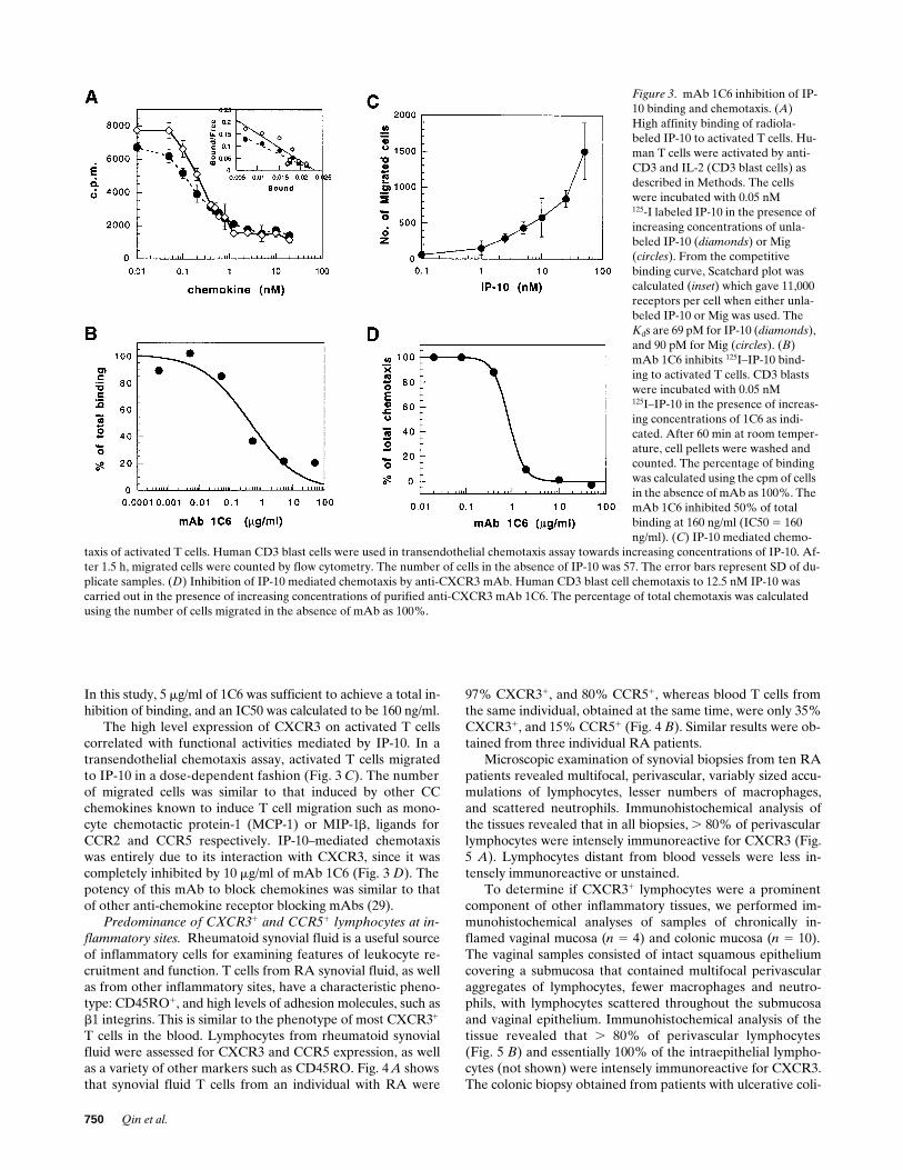
g/ml of 1C6 was sufficient to achieve a total in-hibition of binding, and an IC50 was calculated to be 160 ng/ml.
The high level expression of CXCR3 on activated T cellscorrelated with functional activities mediated by IP-10. In atransendothelial chemotaxis assay, activated T cells migratedto IP-10 in a dose-dependent fashion (Fig. 3
C
). The numberof migrated cells was similar to that induced by other CCchemokines known to induce T cell migration such as mono-cyte chemotactic protein-1 (MCP-1) or MIP-1
b
, ligands forCCR2 and CCR5 respectively. IP-10–mediated chemotaxiswas entirely due to its interaction with CXCR3, since it wascompletely inhibited by 10
m
g/ml of mAb 1C6 (Fig. 3
D
). Thepotency of this mAb to block chemokines was similar to thatof other anti-chemokine receptor blocking mAbs (29).
Predominance of CXCR31 and CCR51 lymphocytes at in-flammatory sites. Rheumatoid synovial fluid is a useful sourceof inflammatory cells for examining features of leukocyte re-cruitment and function. T cells from RA synovial fluid, as wellas from other inflammatory sites, have a characteristic pheno-type: CD45RO1, and high levels of adhesion molecules, such asb1 integrins. This is similar to the phenotype of most CXCR31
T cells in the blood. Lymphocytes from rheumatoid synovialfluid were assessed for CXCR3 and CCR5 expression, as wellas a variety of other markers such as CD45RO. Fig. 4 A showsthat synovial fluid T cells from an individual with RA were
97% CXCR31, and 80% CCR51, whereas blood T cells fromthe same individual, obtained at the same time, were only 35%CXCR31, and 15% CCR51 (Fig. 4 B). Similar results were ob-tained from three individual RA patients.
Microscopic examination of synovial biopsies from ten RApatients revealed multifocal, perivascular, variably sized accu-mulations of lymphocytes, lesser numbers of macrophages,and scattered neutrophils. Immunohistochemical analysis ofthe tissues revealed that in all biopsies, . 80% of perivascularlymphocytes were intensely immunoreactive for CXCR3 (Fig.5 A). Lymphocytes distant from blood vessels were less in-tensely immunoreactive or unstained.
To determine if CXCR31 lymphocytes were a prominentcomponent of other inflammatory tissues, we performed im-munohistochemical analyses of samples of chronically in-flamed vaginal mucosa (n 5 4) and colonic mucosa (n 5 10).The vaginal samples consisted of intact squamous epitheliumcovering a submucosa that contained multifocal perivascularaggregates of lymphocytes, fewer macrophages and neutro-phils, with lymphocytes scattered throughout the submucosaand vaginal epithelium. Immunohistochemical analysis of thetissue revealed that . 80% of perivascular lymphocytes(Fig. 5 B) and essentially 100% of the intraepithelial lympho-cytes (not shown) were intensely immunoreactive for CXCR3.The colonic biopsy obtained from patients with ulcerative coli-
Figure 3. mAb 1C6 inhibition of IP-10 binding and chemotaxis. (A) High affinity binding of radiola-beled IP-10 to activated T cells. Hu-man T cells were activated by anti-CD3 and IL-2 (CD3 blast cells) as described in Methods. The cells were incubated with 0.05 nM125-I labeled IP-10 in the presence of increasing concentrations of unla-beled IP-10 (diamonds) or Mig (circles). From the competitive binding curve, Scatchard plot was calculated (inset) which gave 11,000 receptors per cell when either unla-beled IP-10 or Mig was used. The Kds are 69 pM for IP-10 (diamonds), and 90 pM for Mig (circles). (B) mAb 1C6 inhibits 125I–IP-10 bind-ing to activated T cells. CD3 blasts were incubated with 0.05 nM125I–IP-10 in the presence of increas-ing concentrations of 1C6 as indi-cated. After 60 min at room temper-ature, cell pellets were washed and counted. The percentage of binding was calculated using the cpm of cells in the absence of mAb as 100%. The mAb 1C6 inhibited 50% of total binding at 160 ng/ml (IC50 5 160 ng/ml). (C) IP-10 mediated chemo-
taxis of activated T cells. Human CD3 blast cells were used in transendothelial chemotaxis assay towards increasing concentrations of IP-10. Af-ter 1.5 h, migrated cells were counted by flow cytometry. The number of cells in the absence of IP-10 was 57. The error bars represent SD of du-plicate samples. (D) Inhibition of IP-10 mediated chemotaxis by anti-CXCR3 mAb. Human CD3 blast cell chemotaxis to 12.5 nM IP-10 was carried out in the presence of increasing concentrations of purified anti-CXCR3 mAb 1C6. The percentage of total chemotaxis was calculated using the number of cells migrated in the absence of mAb as 100%.

CXCR3, CCR5, and Inflammation 751
tis, was characterized by focal ulceration of the colonic epithe-lium, and expansion of the lamina propria by large numbers oflymphocytes, fewer macrophages and neutrophils. Within thelamina propria infiltrate, . 70% of lymphocytes were immu-noreactive for CXCR3, particularly below the area of epithe-lial ulceration. The actual proportion of cells expressingCXCR3 may be higher than this, since immunohistochemistryis not sensitive enough to detect low level receptor expressionon the cell surface.
In contrast to inflamed tissues, a much lower percentage ofT cells in normal lymph node expressed CXCR3 (Fig. 5 D).Approximately 20–30% of lymphocytes in the paracortex andmedullary rays were immunoreactive for CXCR3, but onlyrare cells within cortical follicles were immunoreactive. Scat-tered CXCR3 immunoreactive lymphocytes were also identi-fied within the subcapsular and medullary sinusoids. Other celltypes such as smooth muscle, endothelium, and fibroblastswere uniformly nonimmunoreactive. CCR5 immunoreactivelymphocytes were fewer in number but similarly distributed.In contrast to CXCR3, CCR5 was expressed on medullarymacrophages, as reported previously (20), endothelium and
vascular smooth muscle (Rottman, J.B., unpublished observa-tions).
Discussion
Lymphocytes migrate through the body in a nonrandom fash-ion (2, 16). One of the best examples of this is the selective mi-gration of activated/memory T cells to certain tissues and in-flammatory lesions (30, 31). Activated and memory T cellsexpress higher levels of some adhesion molecules that facili-tate their binding to inflamed endothelium. Nevertheless, inmany cases, the expression of adhesion molecules, per se, failsto explain the selectivity of a leukocyte for a particular tissueor microenvironment. The chemokines and their receptorsrepresent another layer where selectivity can operate, possiblyin combination with adhesion mechanisms. Some chemokinereceptors are expressed on leukocytes in a highly restrictedmanner, notably CCR3, the eotaxin receptor, on eosinophils,basophils, TH2 cells (29, 32, 33), and the IL-8 receptors onneutrophils (6, 34). Just how important the chemokine systemis for the selective localization of leukocyte subsets is uncer-tain, although inhibition of chemokines has profound effectson certain inflammatory reactions (23).
Given that chemokine receptors play a fundamental rolefor leukocyte migration, attention has focused on identifyingrelevant receptors on T cells. Until now, analyses of the preciserole of various chemokine receptors on T cells has been diffi-cult, since specific reagents have not been available. In thisstudy, we identified, at the protein level, one of the importantchemokine receptors for T cells, CXCR3. In earlier studies,Northern blot analysis indicated that CXCR3 was restricted toactivated T cells (14). Staining of blood leukocytes with mAbsto CXCR3 revealed, however, that CXCR3 is much more widelyexpressed, for example on blood T cells, and a small propor-tion of B cells and NK cells. It is possible that the CXCR3 pro-tein was produced during previous activation and remains dis-played on, but no longer actively synthesized by peripheralblood lymphocytes. T cells activated in vitro express particu-larly high levels of CXCR3, which fits with our findings thatIP-10 is one of the most potent chemoattractants for thesecells, together with MCP-1 and the CCR5 ligands RANTES,MIP-1a, and MIP-1b. In fact, expression and regulation ofCXCR3 were similar to that of CCR5, since both were foundto be expressed by activated/memory T cells, and both showedthe same pattern of downregulation and upregulation after Tcell activation and IL-2 stimulation.
CXCR3 and CCR5 appear to mark subsets of lymphocyteswith a capacity for migration to inflammatory sites. Identifyingthe nature of this lymphocyte subset is important for under-standing the cellular and molecular mechanisms of inflamma-tion, and for designing strategies for immunosuppression.CXCR31 and CCR51 T cells in blood are generally b1 integ-rinhi, CD45RO1, CD45RAlow, a phenotype consistent withprevious activation. The T cells that infiltrate the inflamed syn-ovium are also b1 integrinhi, CD45RO1, CD45RAlow (30, 35),and also CXCR31 and mostly CCR51 (this study). We stillneed to determine whether the actions of IP-10 or Mig, result-ing in the recruitment of CXCR31 lymphocytes, is the reasonfor the distinctive phenotype of migrating cells, or whether an-other chemokine such as a CCR5 ligand is responsible. An al-ternative explanation for the high expression of CXCR3 orCCR5 on inflammatory cells is upregulation by inflammatory
Figure 4. Expression of CXCR3 and CCR5 on synovial fluid T cells (A) and blood T cells (B) from rheumatoid arthritis patients. Synovial fluid lymphocytes and blood lymphocytes were stained for CXCR3, CCR5, and various other markers as described in Methods. Shown in the figure are CXCR3 staining versus CD3; and CXCR3 versus CCR5 gated on CD31 cells. Quadrants were set based on control mAb staining. The staining shown was representative of three indi-viduals analyzed.

752
Qin et al.
Figure 5. Enrichment of CXCR31 lymphocytes in inflamed tissues. (A) Immunohistochemical detection of CXCR3 in a synovial biopsy from a patient with rheumatoid arthritis. A small blood vessel is surrounded by a thick aggregate of CXCR3 immunoreactive lymphocytes; 3200. (B) Immunohistochemical detection of CXCR3 in chronic vaginitis. Numerous CXCR3 immunoreactive lymphocytes are present in the perivascular space (arrow) and adjacent stroma; 3100. (C) Immunohistochemical detection of CXCR3 in ulcerative colitis. The superficial lamina propria contains numerous CXCR31 lymphocytes immediately beneath an area of discontinuous colonic epithelium (arrow); 3200. (D) CXCR3 Expres-sion in a normal human lymph node. CXCR31 cells are scattered throughout the paracortex. Rare CXCR31 cells are present within lymphoid follicles (arrowheads).

CXCR3, CCR5, and Inflammation 753
cytokines, after extravasation. However, perivascular expres-sion of CXCR3 (Fig. 5) and CCR5 (not shown) at the sites ofinflammation and our finding that upregulation of CXCR3and CCR5 in vitro takes many days would argue against thesereceptors being upregulated at the site of inflammation.
Blocking interaction of chemokines and their receptorsshould be useful therapeutically in autoimmune diseases ortransplant rejection, in which particular receptors and theirligands may be responsible for the recruitment of cells. Wehave generated mAbs that effectively block CCR3 (29), CXCR1(25), CXCR2 (25), and CCR5 (36). There is as yet no clarity onhow chemokines bind to their 7-transmembrane receptors, al-though RANTES, MIP-1a, and MIP-1b have been found tobind to CCR5 through the second extracellular loop, and thiscan be blocked totally with a mAb specific for this loop (36).The anti-CXCR3 mAb 1C6 was able to inhibit completely thebinding and chemotactic activity of IP-10 on activated T cells.mAb 1C6 is directed to the first 37 residuals of the NH2 termi-nus, and so this region is of particular importance for ligandbinding. Reactivity of 1C6 with shorter synthetic peptides nar-rowed the epitope to the first 15 amino acids (Qin, S., unpub-lished observations). The NH2-terminal regions of CXCR1and CXCR2 are also critical for IL-8 binding (25, 37), and theNH2-terminal region of CCR2b is sufficient in itself to bindMCP-1 (38).
In conclusion, we have established that CXCR3 is ex-pressed abundantly on human T cells, and particularly on thoseT cells associated with activation and inflammation, and theexpression of CXCR3 closely resembled that of CCR5. We an-ticipate that an understanding of the role of these two recep-tors in inflammatory diseases, particularly those with a strongT cell involvement should help in the design of therapies to in-hibit leukocyte recruitment and suppress adverse inflamma-tory reactions.
Acknowledgments
We thank Dr. Walter Newman and Dr. Craig Gerard for advice dur-ing the course of these experiments, Dr. Ian Clark-Lewis for the sup-ply of certain chemokines, and Dr. Jim Campbell, Dr. Paul Ponath,Dr. Lijun Wu, and Dr. Greg LaRosa for various L1.2 chemokine re-ceptor transfectants.
References
1. Springer, T.A. 1994. Traffic signals for lymphocyte recirculation and leu-kocyte emigration: the multistep paradigm. Cell. 76:301–314.
2. Butcher, E.C., and L.J. Picker. 1996. Lymphocyte homing and homeosta-sis. Science. 272:60–66.
3. Baggiolini, M., B. Dewald, and B. Moser. 1997. Human chemokines: anupdate. Annu. Rev. Immunol. 15:675–705.
4. Mackay, C.R. 1997. Chemokines: what chemokine is that? Curr. Biol. 7:R384–R386.
5. Moser, B., M. Loetscher, L. Piali, and P. Loetscher. 1997. Lymphocyte re-sponses to chemokines. Int. Rev. Immunol. In press.
6. Qin, S., G. LaRosa, J.J. Campbell, H. Smith-Heath, N. Kassam, X. Shi, L.Zeng, E.C. Butcher, and C.R. Mackay. 1996. Expression of monocyte chemoat-tractant protein-1 and interleukin-8 receptors on subsets of T cells: correlationwith transendothelial chemotactic potential. Eur. J. Immunol. 26:640–647.
7. Carr, M.W., S.J. Roth, E. Luther, S.S. Rose, and T.A. Springer. 1994.Monocyte chemoattractant protein 1 acts as a T-lymphocyte chemoattractant.Proc. Natl. Acad. Sci. USA. 91:3652–3656.
8. Taub, D.D., P. Proost, W.J. Murphy, M. Anver, D.L. Longo, J. vanDamme, and J.J. Oppenheim. 1995. Monocyte chemotactic protein-1 (MCP-1),-2, and -3 are chemotactic for human T lymphocytes. J. Clin. Invest. 95:1370–1376.
9. Bleul, C.C., R.C. Fuhlbrigge, J.M. Casasnovas, A. Aiuti, and T.A.
Springer. 1996. A highly efficacious lymphocyte chemoattractant, stromal cell-derived factor 1 (SDF-1). J. Exp. Med. 184:1101–1110.
10. Bleul, C.C., M. Farzan, H. Choe, C. Parolin, I. Clark-Lewis, J. Sodroski,and T.A. Springer. 1996. The lymphocyte chemoattractant SDF-1 is a ligand forLESTRE/fusin and blocks HIV-1 entry. Nature. 382:829–833.
11. Oberlin, E., A. Amara, F. Bachelerie, C. Bessia, J.-L. Virelizier, F.Arenzana-Seisdedos, O. Schwartz, J.-M. Heard, I. Clark-Lewis, D.F. Legler, M.Loetscher, M. Baggiolini, and B. Moser. 1996. The CXC chemokine SDF-1 isthe ligand for LESTR/fusin and prevents infection by T cell line–adapted HIV-1.Nature. 832:833–835.
12. Bleul, C.C., L. Wu, J.A. Hoxie, T.A. Springer, and C.R. Mackay. 1997.The HIV coreceptors CXCR4 and CCR5 are differentially expressed and regu-lated on human T lymphocytes. Proc. Natl. Acad. Sci. USA. 94:1925–1930.
13. Taub, D.D., A.R. Lloyd, K. Conlon, J.M. Wang, J.R. Ortaldo, A.Harada, K. Matsushima, D.J. Kelvin, and J.J. Oppenheim. 1993. Recombinanthuman interferon–inducible protein 10 is a chemoattractant for human mono-cytes and T lymphocytes and promotes T cell adhesion to endothelial cells. J.Exp. Med. 177:1809–1814.
14. Loetscher, M.L., B. Gerber, P. Loetscher, S.A. Jones, L. Piali, I.C.Lewis, M. Baggiolini, and B. Moser. 1996. Chemokine receptor specific for IP-10 and Mig: structure, function and expression in activated T lymphocytes. J.Exp. Med. 184:963–969.
15. Liao, F., R.L. Rabin, J.R. Yannelli, L.G. Koniaris, P. Vanguri, and J.M.Farber. 1995. Human Mig chemokine: biochemical and functional characteriza-tion. J. Exp. Med. 182:1301–1314.
16. Mackay, C.R. 1993. Homing of naive, memory and effector lympho-cytes. Curr. Opin. Immunol. 5:423–427.
17. Kaplan, G., A.D. Luster, G. Hancock, and Z.A. Cohn. 1987. The ex-pression of a g interferon–induced protein (IP-10) in delayed immune re-sponses in human skin. J. Exp. Med. 166:1098–1108.
18. Ransohoff, R.M., T.A. Hamilton, M. Tani, M.H. Stoler, H.E. Shick, J.A.Major, M.L. Estes, D.M. Thomas, and V.K. Tuohy. 1993. Astrocyte expressionof mRNA encoding cytokines IP-10 and JE/MCP-1 in experimental autoim-mune encephalomyelitis. FASEB (Fed. Am. Soc. Exp. Biol.) J. 7:592–600.
19. Kondo, T., A.C. Novick, H. Toma, and R.L. Fairchild. 1996. Inductionof chemokine gene expression during allogeneic skin graft rejection. Transplan-tation (Baltimore). 61:1750–1757.
20. Wu, L., W.A. Paxton, N. Kassam, N. Ruffing, J.B. Rottman, N. Sullivan,H. Choe, J. Sodroski, W. Newman, R.A. Koup, and R.C. Mackay. 1997. CCR5levels and expression pattern correlate with infectability by macrophage-tropicHIV-1 in vitro. J. Exp. Med. 185:1681–1691.
21. Schall, T.J., K. Bacon, K.J. Toy, and D.V. Goeddel. 1990. Selective at-traction of monocytes and T lymphocytes of the memory phenotype by cyto-kine RANTES. Nature. 347:669–671.
22. Taub, D.D., K. Conlon, A.R. Lloyd, J.J. Oppenheim, and D.J. Kelvin.1993. Preferential migration of activated CD41 and CD81 T cells in responseto MIP-1 alpha and MIP-1 beta. Science. 260:355–358.
23. Strieter, R.M., T.J. Standiford, G.B. Huffnagle, L.M. Colletti, N.W.Lukacs, and S.L. Kunkel. 1996. “The good, the bad, and the ugly.” The role ofchemokines in models of human disease. J. Immunol. 156:3583–3586.
24. Ponath, P.D., S. Qin, D.J. Ringler, I. Clark-Lewis, J. Wang, N. Kassam,H. Smith, X. Shi, J.-A. Gonzalo, W. Newman, et al. 1996. Cloning of the humaneosinophil chemoattractant, eotaxin: expression, receptor binding and func-tional properties provide a mechanism for the selective recruitment of eosino-phils. J. Clin. Invest. 97:604–612.
25. Wu, L., N. Ruffing, X. Shi, W. Newman, D. Soler, C.R. Mackay, and S.Qin. 1996. Discrete steps in binding and signaling of interleukin-8 with its re-ceptor. J. Biol. Chem. 271:31202–31209.
26. Wu, L., N. Gerard, R. Wyatt, H. Choe, C. Parolin, N. Ruffing, A.Borsetti, A.A. Cardoso, E. Desjardin, W. Newman, et al. 1996. CD4-induced in-teraction of primary HIV-1 gp120 glycoproteins with the chemokine receptorCCR-5. Nature. 384:179–183.
27. Campbell, J.J., S. Qin, K.B. Bacon, C.R. Mackay, and E.C. Butcher.1996. The biology of chemokine and classical chemoattractant receptors: differ-ential requirements for adhesion-triggering versus chemotactic responses inlymphoid cells. J. Cell Biol. 134:255–266.
28. Coligan, J.E., A.M. Kruisbeek, D.H. Margulies, E.M. Shevach, and W.Strober. 1992. Current Protocols in Immunology. John Wiley and Sons, NewYork. 2.5.1–2.5.12.
29. Heath, H., S. Qin, P. Rao, L. Wu, G. LaRosa, N. Kassam, P. Ponath, andC.R. Mackay. 1997. Chemokine receptor usage by human eosinophils. The im-portance of CCR3 demonstrated using an antagonistic monoclonal antibody. J.Clin. Invest. 99:178–184.
30. Pitzalis, C., G. Kingsley, D. Haskard, and G. Panayi. 1988. The prefer-ential accumulation of helper-inducer T lymphocytes in inflammatory lesions:evidence for regulation by selective endothelial and homotypic adhesion. Eur.J. Immunol. 18:1397–1404.
31. Salmi, M., D.P. Adrew, E.C. Butcher, and S. Jalkanen. 1995. Dual bind-ing capacity of mucosal immunoblasts to mucosal and synovial endothlium inhumans: dissection of the molecular mechanisms. J. Exp. Med. 181:137–149.
32. Uguccioni, M., C.R. Mackay, B. Ochensberger, P. Loetscher, S. Rhis, G.LaRosa, P. Rao, P. Ponath, M. Baggiolini, and C.A. Dahinden. 1997. High ex-

754 Qin et al.
pression of the chemokine receptor CCR3 in human blood basophils. Role inactivation by eotaxis, MCP-4 and other chemokines. J. Clin. Invest. 100:1137–1143.
33. Sallusto, F., C.R. Mackay, and A. Lanzavecchia. 1997. Selective expres-sion of the eotaxin receptor CCR3 by human T helper 2 cells. Science. 277:2005–2007.
34. Chuntharapai, A., J. Lee, C.A. Hebert, and K.J. Kim. 1994. Monoclonalantibodies detect different distribution patterns of IL-8 receptor A and IL-8 re-ceptor B on human peripheral blood leukocytes. J. Immunol. 153:5682–5688.
35. Oppenheinmer-Marks, N., and P.E. Lipsky. 1997. Migration of naiveand memory T cells. Immunol. Today. 18:456–457.
36. Wu, L., G. LaRosa, N. Kassam, C.J. Gordon, H. Heath, N. Ruffing, H.
Chen, J. Humblias, M. Samson, M. Parmentier, et al. 1997. Interaction ofchemokine receptor CCR5 with its ligands: multiple domains for HIV-1 gp120binding and a single domain for chemokine binding. J. Exp. Med. 186:1373–1381.
37. Chuntharapai, A., J. Lee, J. Burnier, W.I. Wood, C. Hebert, and K.J.Kim. 1994. Neutralizing monoclonal antibodies to human IL-8 receptor A mapto the NH2-terminal region of the receptor. J. Immunol. 152:1783–1789.
38. Monteclaro, F.S., and I. Charo. 1996. The amino-terminal extracellulardomain of the MCP-1 receptor, but not the RANTES/MIP-1alpha receptor,confers chemokine selectivity. Evidence for a two-step mechanism for MCP-1receptor activation. J. Biol. Chem. 271:19084–19092.
Related Documents


![Moderate Restriction of Macrophage-Tropic Human ......9], and this observation led to the identification of the CCR5 and CXCR4 chemokine receptors as HIV-1 co-receptors for viral fusion](https://static.cupdf.com/doc/110x72/606645ddad14062d597e7589/moderate-restriction-of-macrophage-tropic-human-9-and-this-observation.jpg)


