See discussions, stats, and author profiles for this publication at: https://www.researchgate.net/publication/315673999 The Challenges and Advances in Diagnosis of Vector-Borne Diseases: Where Do We Stand? Article in Vector borne and zoonotic diseases (Larchmont, N.Y.) · March 2017 DOI: 10.1089/vbz.2016.2074 CITATIONS 0 READS 40 11 authors, including: Some of the authors of this publication are also working on these related projects: Upgrading the research performance in molecular medicine - ERA Chair FP7 View project Identification of SNP markers of fertility in dairy cows by genetical genomics View project Josipa Kuleš University of Zagreb 23 PUBLICATIONS 35 CITATIONS SEE PROFILE Anita Horvatić University of Zagreb 23 PUBLICATIONS 40 CITATIONS SEE PROFILE Vladimir Mrljak University of Zagreb 95 PUBLICATIONS 473 CITATIONS SEE PROFILE Mangesh Bhide University of Veterinary Medicine in Kosice - Un… 165 PUBLICATIONS 682 CITATIONS SEE PROFILE All content following this page was uploaded by Nicolas Guillemin on 30 March 2017. The user has requested enhancement of the downloaded file. All in-text references underlined in blue are added to the original document and are linked to publications on ResearchGate, letting you access and read them immediately.

Welcome message from author
This document is posted to help you gain knowledge. Please leave a comment to let me know what you think about it! Share it to your friends and learn new things together.
Transcript
Seediscussions,stats,andauthorprofilesforthispublicationat:https://www.researchgate.net/publication/315673999
TheChallengesandAdvancesinDiagnosisofVector-BorneDiseases:WhereDoWeStand?
ArticleinVectorborneandzoonoticdiseases(Larchmont,N.Y.)·March2017
DOI:10.1089/vbz.2016.2074
CITATIONS
0
READS
40
11authors,including:
Someoftheauthorsofthispublicationarealsoworkingontheserelatedprojects:
Upgradingtheresearchperformanceinmolecularmedicine-ERAChairFP7Viewproject
IdentificationofSNPmarkersoffertilityindairycowsbygeneticalgenomicsViewproject
JosipaKulešUniversityofZagreb
23PUBLICATIONS35CITATIONS
SEEPROFILE
AnitaHorvatićUniversityofZagreb
23PUBLICATIONS40CITATIONS
SEEPROFILE
VladimirMrljak
UniversityofZagreb
95PUBLICATIONS473CITATIONS
SEEPROFILE
MangeshBhide
UniversityofVeterinaryMedicineinKosice-Un…
165PUBLICATIONS682CITATIONS
SEEPROFILE
AllcontentfollowingthispagewasuploadedbyNicolasGuilleminon30March2017.
Theuserhasrequestedenhancementofthedownloadedfile.Allin-textreferencesunderlinedinblueareaddedtotheoriginaldocument
andarelinkedtopublicationsonResearchGate,lettingyouaccessandreadthemimmediately.

REVIEW
The Challenges and Advancesin Diagnosis of Vector-Borne Diseases:
Where Do We Stand?
Josipa Kules,1 Lenka Potocnakova,2 Katarina Bhide,2 Laura Tomassone,3 Hans-Peter Fuehrer,4
Anita Horvatic,1 Asier Galan,1 Nicolas Guillemin,1 Petra Nizic,5 Vladimir Mrljak,5 and Mangesh Bhide1,2,6
Abstract
Vector-borne diseases (VBD) are of major importance to human and animal health. In recent years, VBD havebeen emerging or re-emerging in many geographical areas, alarming new disease threats and economic losses.The precise diagnosis of many of these diseases still remains a major challenge because of the lack ofcomprehensive data available on accurate and reliable diagnostic methods. Here, we conducted a systematic andin-depth review of the former, current, and upcoming techniques employed for the diagnosis of VBD.
Keywords: chip-based technology, diagnostics, mass spectrometry, molecular biology, serology, vector-bornediseases
Introduction
Vector-borne diseases (VBD) are of major importanceto human and animal health. Hematophagous arthropod
vectors such as mosquitoes, ticks, and sand flies are respon-sible for transmitting bacteria, viruses, and protozoa betweenvertebrate hosts, causing diseases such as malaria, denguefever, and Crimean-Congo hemorrhagic fever. Historically,diseases such as leishmaniasis and malaria have had a greatimpact on health and they are still posing a huge burden onpublic health in many countries (Hotez et al. 2006). Until theearly 20th century, VBD were responsible for more deaths inhumans than all other causes combined (Kalluri et al. 2007).
In recent years, VBD have emerged or re-emerged in manygeographical regions, causing global health issues for humans,livestock, companion animals, and wildlife (Harrus andBaneth 2005). The eco-epidemiology of VBD is affected byan interplay between major factors such as the pathogen, thehost (human, animal), the vector, and the environment. Majorcontributors in the spreading of VBD include eco-climaticchanges, development of insecticide and drug resistance,globalization, and the significant increase in internationaltrade and travel (Harrus and Baneth 2005). Altogether, these
factors govern disease epidemiology and infection patterns inmany geographic regions.
For example, invasive mosquitoes have become widelyestablished across Europe, resulting in the emergence ofmalaria in Greece and West Nile virus (WNV) throughoutparts of south-eastern Europe (Medlock and Leach 2015).Similarly, the prevalence of Lyme disease continues to in-crease throughout Europe, and tick-borne encephalitis andCrimean-Congo hemorrhagic fever viruses have changedtheir geographical distribution. Recently, the World HealthOrganization has declared that the Zika virus constitutes apublic health emergency of international concern, and itsspreading can also be expected in Europe. From a veterinaryperspective, the emergence of Bluetongue and Schmallenbergviruses has shown that northern Europe is equally susceptibleto transmission of VBD, despite the longer winter periodcompared with southern Europe (Medlock and Vaux 2015).
Lack of comprehensive information on existing and upcom-ing diagnostic methods for VBD makes the diagnosis and dis-ease control complicated. In general, laboratory diagnosis ofVBD can be divided into two categories: direct methods (mi-croscopy, culturing of the causative agent, nucleic acid detection,etc.) and indirect methods (e.g., detection of organism-specific
1ERA Chair Team, Faculty of Veterinary Medicine, University of Zagreb, Zagreb, Croatia.2Laboratory of Biomedical Microbiology and Immunology of University of Veterinary Medicine and Pharmacy, Kosice, Slovakia.3Department of Veterinary Science, University of Torino, Grugliasco, Italy.4Department of Pathobiology, Institute of Parasitology, University of Veterinary Medicine, Vienna, Austria.5Faculty of Veterinary Medicine, Internal Diseases Clinic, University of Zagreb, Zagreb, Croatia.6Institute of Neuroimmunology, Slovak Academy of Sciences, Bratislava, Slovakia.
VECTOR-BORNE AND ZOONOTIC DISEASESVolume XX, Number XX, 2017ª Mary Ann Liebert, Inc.DOI: 10.1089/vbz.2016.2074
1

immune responses). Acute infections are usually identifiedby direct tools, whereas indirect tools are useful for thecharacterization of secondary or convalescent phases.
The majority of VBD have common issues in rapid andsufficient diagnostics (Table 1). Many of the diagnostic toolsbased on molecular techniques are applied for the finest de-tection and characterization of vector-borne infections; nev-ertheless, VBD may still be misdiagnosed. On the other hand,the advances in molecular techniques have brought to thediscovery of previously unknown pathogens, distinguishedclosely related species and revealed new possible transmis-sion mechanisms of vector-borne infections (Harrus andBaneth 2005, Colwell et al. 2011). Continuous developmentof the diagnostic technologies and their deployment arecrucial to identify, control, and treat the VBD. High-throughput technologies such as whole-genome sequencing,omics approaches (metagenomics and metaproteomics) andhigh-resolution analytical methods such as mass spectrome-try (MS), open new opportunities for diagnostics of VBD.
Here, we conducted a systematic review of the former,current, and upcoming techniques employed for the diagnosisof VBD.
Routine and Advanced Platforms for the Diagnosisof Vector-Borne Pathogens
Conventional techniques
Microscopy is still one of the well-established, low-costdirect methods for identification of vector-borne pathogens.It is still the method of choice for blood parasites such asPlasmodium or Babesia, although its sensitivity is lower thanthat of molecular diagnosis (Solano-Gallego et al. 2016).However, in some cases, sample preparation can be timeconsuming and labor intensive, and in many cases, diagnosismay be hampered by sparseness of organisms. The methodis subjective, especially in cases when differentiation ofsimilar-appearing organisms is required. Thus, morpho-logic interpretation needs significant expertise (Ndao 2009).
Table 1. Overview of General Advantages and Disadvantages
of Diagnostic Methods for Vector-Borne Diseases
Methodsa Advantages Disadvantages
Conventional(classical)methods
Microscopy—blood/tissuesmear examination
Pathogen isolationCell culture
Simple equipment andreagents
Suitable for resource-limitedsettings
Labor and time intensiveLower sensitivityExpertise required
Serology-basedmethods
IFAELISAImmunoblottingPRNTSNHICFT
Easy sample collectionLow cost and easy application
Potential cross-reactivityLower sensitivityHistory of exposure (lag time)
Nucleic acid-basedmethods
PCRRFLP-PCRRT-PCRFISHLAMP
Increased sensitivity,specificity, and speed ofdiagnosis
Differentiation ofmorphologically similarparasites
Equipment and reagent costLack of standardization and
variability in performanceFalse-positive and -negative
results
Omics-basedmethods
NGSMS-based assays (LC-MS/
MS, MALDI-TOF, iTRAQ,ICAT, SILAC);
Metagenomics (NGS)Metabolomics (GC-MS,
LC-MS, NMR)
High-throughput analysisIncreased sensitivity and
specificity
Equipment and reagent costLabor intensity of data
analysisLack of sequenced genomes
Multiplexmethods
Multiplex PCRMicroarraysPCR-ESI/MSiTRAQMRM
Detection of coinfectionHigh-throughput analysisLess sample volume
requirementsEfficiency in terms of time
and costs
Competition betweencoinfecting pathogens
Diagnostic tools are divided as a part of conventional (classical) methods, serology-based methods, nucleic acid-based methods, omics-based methods, and multiplex-based methods. Note that performance and requirements of individual assays may vary.
aNot an exhaustive list.IFA, immunofluorescence assay; ELISA, enzyme-linked immunosorbent assay; PRNT, plaque reduction neutralization test; SN, serum
neutralization test; HI, hemagglutination inhibition test; CFT, complement fixation test; RFLP-PCR, restriction fragment lengthpolymorphism PCR; FISH, fluorescence in situ hybridization; LAMP, loop-mediated isothermal amplification; PCR-ESI/MS, PCR-electrospray mass spectrometry; iTRAQ, isobaric tags for relative and absolute quantitation; MRM, multiple-reaction monitoring; NGS,next-generation sequencing; MS, mass spectrometry; LC-MS/MS, liquid chromatography-tandem mass spectrometry; MALDI-TOF,matrix-assisted laser desorption/ionization time-of-flight mass spectrometry; ICAT, isotope coded affinity tag; SILAC, stable isotopelabeling by amino acids in cell culture; GC-MS, gas chromatography–mass spectrometry; NMR, nuclear magnetic resonance.
2 KULES ET AL.

Despite these limitations, the microscopy represents a usefulinitial test for the patient with general signs of infectionwithout diagnostic hallmarks, especially in endemic areas(Dumler et al. 2007).
Electron microscopy (EM) is still on the forefront ofclinical diagnosis of viral and rickettsial diseases, study ofultrastructure, and unfolding of the basic principles of path-ogenesis. In the diagnostic setting, it is particularly valuablein the surveillance of emerging diseases and potential bio-terrorism viruses (Goldsmith and Miller 2009). Traditionally,negative staining for transmission electron microscopy hasbeen the ‘‘gold standard’’ for imaging of microbial samples,for example, in diagnostic virology; on the other hand, im-provements in the specimen preparation have enabled scan-ning electron microscopy (SEM) as an efficient diagnostictool (Golding et al. 2016). Diagnostic EM has two advantagesover enzyme-linked immunosorbent assay (ELISA) and nucleicacid amplification tests: after a simple and fast negative stain-ing, EM (mainly SEM) enables fast morphological identifica-tion and differential diagnosis of infectious agents contained inthe specimen without the need for special considerations and/orreagents. Nevertheless, EM has the disadvantage of being un-suitable as a screening method (Schramlova et al. 2010).
Cell culture is another gold-standard diagnostic method(Portillo 2015). These procedures are time consuming, andthe isolation of pathogens is not always successful. Cultiva-tion of slow-growing and fastidious bacteria, or even non-cultivable bacteria, has always been a limitation. Bacteriatransmitted by ticks are fastidious and difficult to grow inaxenic media; many of them are obligate intracellular path-ogens, as well as viruses, rickettsiae, and some protozoalpathogens. However, cultivation methods have improvedconsiderably over the past several decades, with advances inthe scope and diversity of media components, control ofenvironmental conditions, use of heterologous host cells, anduse of growth-promoting factors (Mukamolova et al., 1998).
Xenodiagnosis, the feeding of a natural arthropod vectoron a patient to detect evidence of infection, has provideddefinitive evidence for the etiology of diverse vector-borneinfections, including malaria, yellow fever, epidemic typhus,and Lyme disease (Telford et al. 2014). The obvious infec-tions for which xenodiagnosis may be useful would be thosethat are known to be vector borne, maintain a chronic state,and are characterized by sparse organisms that are very dif-ficult to detect.
As molecular diagnostic techniques progress in scope andmagnitude, it is critical to retain and use classical techniques,including cell culture and EM, that complement the advancesin molecular methods. There is a continuing need to trainyoung scientists in these traditional methods to maintain anunderlying expertise.
Serology-based assays
Serological tests are widely employed to diagnose humanand animal VBD, at the screening level, for both surveillanceand research. They have a wide diagnostic time window, asantibodies for a pathogen may persist for months or evenyears. That makes these assays valuable to investigate pastexposure to vector-borne pathogens. Advantages of serologyinclude the ease of sample collection, mainly blood, and anability to detect current infections by sero-conversion. They
are often used as confirmatory tests in conjunction withmolecular methods or cytology (Otranto et al. 2010).
A poor sensitivity of serological tests is observed in theearly stage of infection in the majority of vector-borne in-fections. The demonstration of sero-conversion is also pos-sible by retrospective confirmation of recent infectionbetween antibody titers of acute and convalescent samples(Nilsson et al. 2005). Another limitation of serologic assays isthe potential cross-reactivity within or between genera ofviral or bacterial pathogens (Costa et al. 2005, Rawlins et al.2005, Vermeulen et al. 2010). Thus, there is a need of testingwith other related microorganisms to ensure no cross-reactions and to avoid false positives. To improve the diag-nostic specificity, various recombinant or purified antigensare being widely used (Maggi et al. 2014).
Fast development of serological assays often relies onculture for antigen production. The use of purified antigens islimited by the inability of producing proteins in a sufficientamount. Therefore, to overcome this limitation in the caseswhere cultures are not available, the production of recombi-nant proteins or synthetic peptides offers an alternative if ge-netic information of potentially antigenic proteins is known.
The choice whether to use monoclonal or polyclonal an-tibodies depends on the context in which the application isbeing used. The principal advantages of monoclonal anti-bodies are their homogeneity and consistency, whereaspolyclonal antibodies can be generated much more rapidly, atless expense, and with less technical skill than is required toproduce monoclonal antibodies. Monoclonal antibodies arenot generally useful for assays that depend on antigen cross-linking (e.g., hemagglutination) unless dimeric or multimericantigens or antigens bound to a solid phase are used. In ad-dition, they may not activate complements readily, becauseactivation requires the close proximity of Fc receptors (Lip-man et al. 2005). Modification of antibodies by covalentlylinking a fluorochrome or radionuclide may also alter anti-body binding. This potential is of less concern when usingpolyclonal antibodies, which recognize a host of epitopes.
ELISA, immunofluorescence assays (IFA), and immuno-blotting are the most widely used serological tests. Results ofsuch assays must be interpreted with caution since a positiveserological test could be associated with a past infection, andunrelated with current clinical signs. Moreover, cliniciansshould consider limitations of each commercially availabletest due to different specificity and sensitivity that may beaffected by various factors, including antigen use.
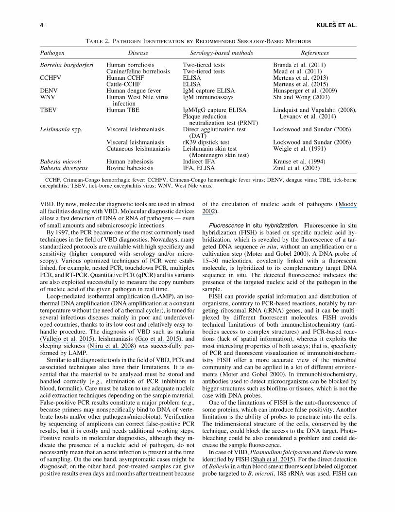
Besides conventional single-pathogen assays, some serolog-ical tests detect multiple VBD (Volgina et al. 2013). MultiplexedLuminex-based immunoassay demonstrates a prospective de-tection of viral hemorrhagic fever-associated immunoglobulinG antibodies by using a panel of recombinant antigens ofpathogens; for example, Crimean-Congo hemorrhagic fevervirus, dengue virus (DENV), Rift Valley fever virus, andHantaan virus (Wu et al. 2014). Luminex xMAP technologyseems to be more sensitive and time saving than widely em-ployed ELISA or IFA. A short overview of the recommendedserology-based methods for VBD is given in Table 2.
Nucleic acid-based assays
With the upcoming of molecular diagnostic techniques inthe 1990s, a new era of diagnostics was initiated in the field of
VECTOR-BORNE DISEASES: WHERE DO WE STAND? 3
VBD. By now, molecular diagnostic tools are used in almostall facilities dealing with VBD. Molecular diagnostic devicesallow a fast detection of DNA or RNA of pathogens — evenof small amounts and submicroscopic infections.
By 1997, the PCR became one of the most commonly usedtechniques in the field of VBD diagnostics. Nowadays, manystandardized protocols are available with high specificity andsensitivity (higher compared with serology and/or micro-scopy). Various optimized techniques of PCR were estab-lished, for example, nested PCR, touchdown PCR, multiplexPCR, and RT-PCR. Quantitative PCR (qPCR) and its variantsare also exploited successfully to measure the copy numbersof nucleic acid of the given pathogen in real time.
Loop-mediated isothermal amplification (LAMP), an iso-thermal DNA amplification (DNA amplification at a constanttemperature without the need of a thermal cycler), is tuned forseveral infectious diseases mainly in poor and underdevel-oped countries, thanks to its low cost and relatively easy-to-handle procedure. The diagnosis of VBD such as malaria(Vallejo et al. 2015), leishmaniasis (Gao et al. 2015), andsleeping sickness (Njiru et al. 2008) was successfully per-formed by LAMP.
Similar to all diagnostic tools in the field of VBD, PCR andassociated techniques also have their limitations. It is es-sential that the material to be analyzed must be stored andhandled correctly (e.g., elimination of PCR inhibitors inblood, formalin). Care must be taken to use adequate nucleicacid extraction techniques depending on the sample material.False-positive PCR results constitute a major problem (e.g.,because primers may nonspecifically bind to DNA of verte-brate hosts and/or other pathogens/microbiota). Verificationby sequencing of amplicons can correct false-positive PCRresults, but it is costly and needs additional working steps.Positive results in molecular diagnostics, although they in-dicate the presence of a nucleic acid of pathogen, do notnecessarily mean that an acute infection is present at the timeof sampling. On the one hand, asymptomatic cases might bediagnosed; on the other hand, post-treated samples can givepositive results even days and months after treatment because
of the circulation of nucleic acids of pathogens (Moody2002).
Fluorescence in situ hybridization. Fluorescence in situhybridization (FISH) is based on specific nucleic acid hy-bridization, which is revealed by the fluorescence of a tar-geted DNA sequence in situ, without an amplification or acultivation step (Moter and Gobel 2000). A DNA probe of15–30 nucleotides, covalently linked with a fluorescentmolecule, is hybridized to its complementary target DNAsequence in situ. The detected fluorescence indicates thepresence of the targeted nucleic acid of the pathogen in thesample.
FISH can provide spatial information and distribution oforganisms, contrary to PCR-based reactions, notably by tar-geting ribosomal RNA (rRNA) genes, and it can be multi-plexed by different fluorescent molecules. FISH avoidstechnical limitations of both immunohistochemistry (anti-bodies access to complex structures) and PCR-based reac-tions (lack of spatial information), whereas it exploits themost interesting properties of both assays; that is, specificityof PCR and fluorescent visualization of immunohistochem-istry FISH offer a more accurate view of the microbialcommunity and can be applied in a lot of different environ-ments (Moter and Gobel 2000). In immunohistochemistry,antibodies used to detect microorganisms can be blocked bybigger structures such as biofilms or tissues, which is not thecase with DNA probes.
One of the limitations of FISH is the auto-fluorescence ofsome proteins, which can introduce false positivity. Anotherlimitation is the ability of probes to penetrate into the cells.The tridimensional structure of the cells, conserved by thetechnique, could block the access to the DNA target. Photo-bleaching could be also considered a problem and could de-crease the sample fluorescence.
In case of VBD, Plasmodium falciparum and Babesia wereidentified by FISH (Shah et al. 2015). For the direct detectionof Babesia in a thin blood smear fluorescent labeled oligomerprobe targeted to B. microti, 18S rRNA was used. FISH can
Table 2. Pathogen Identification by Recommended Serology-Based Methods
Pathogen Disease Serology-based methods References
Borrelia burgdorferi Human borreliosis Two-tiered tests Branda et al. (2011)Canine/feline borreliosis Two-tiered tests Mead et al. (2011)
CCHFV Human CCHF ELISA Mertens et al. (2013)Cattle-CCHF ELISA Mertens et al. (2015)
DENV Human dengue fever IgM capture ELISA Hunsperger et al. (2009)WNV Human West Nile virus
infectionIgM immunoassays Shi and Wong (2003)
TBEV Human TBE IgM/IgG capture ELISA Lindquist and Vapalahti (2008),Levanov et al. (2014)Plaque reduction
neutralization test (PRNT)Leishmania spp. Visceral leishmaniasis Direct agglutination test
(DAT)Lockwood and Sundar (2006)
Visceral leishmaniasis rK39 dipstick test Lockwood and Sundar (2006)Cutaneous leishmaniasis Leishmanin skin test
(Montenegro skin test)Weigle et al. (1991)
Babesia microti Human babesiosis Indirect IFA Krause et al. (1994)Babesia divergens Bovine babesiosis IFA, ELISA Zintl et al. (2003)
CCHF, Crimean-Congo hemorrhagic fever; CCHFV, Crimean-Congo hemorrhagic fever virus; DENV, dengue virus; TBE, tick-borneencephalitis; TBEV, tick-borne encephalitis virus; WNV, West Nile virus.
4 KULES ET AL.
also be applied to detect viruses, as demonstrated by the studyof Raquin et al. (2012), who successfully detected DENVs inmosquitoes. Their technique is also applicable to detect bothdengue and chikungunya viruses. In situ nucleic acid ampli-fication is being developed to increase FISH sensitivity, no-tably to make FISH able to address the physiological state ofthe cell, by targeting all the RNAs (Porter and Pickup 2000).
Wire-guided droplet manipulation. The wire-guideddroplet manipulation is a new technique in which a wiremanipulates a microliter-sized droplet in a hydrophobic mi-lieu (Harshman et al. 2014). It allows a molecular partition-ing, that is, the separation of complex sample components.This technique can be useful for blood screening, because thedifferent cells form no aggregate and can be targeted by aPCR reaction for the identification of microorganisms in situ,such as Klebsiella pneumoniae. Although this technique isnot used extensively for VBD, evolution of the technique andmaterials could enhance the diagnostic possibilities of thistool in the coming years.
Next-generation sequencing. Next-generation sequenc-ing (NGS) provides fast and reliable DNA sequencing,without a priori knowledge of the targeted genome (VanBorm et al. 2015). Alignment tools are a crucial componentof NGS. Using NGS, it is possible to rapidly sequence DNAdirectly from the vectors to generate a broad overview of themicrobial community inside it, without the bias introduced bystandard identification methods based on cultivable patho-gens. By targeting different regions of the parasite genomes(from less to more conserved), NGS can perform identifica-tions at different levels, from strains to families of parasites.NGS can also detect unknown parasites. Moreover, by se-quencing some mRNA, NGS can provide a quantitative andqualitative evaluation of the microbial community. The an-alytical sensitivity of NGS approaches that of standardqPCR assays, enabling the detection of pathogen genomes atconcentrations as low as 1 · 104 genome copies/mL (Freyet al. 2014).
Some studies have used NGS to identify microbial com-position of the tick Ixodes ricinus, by sequencing the 16SrRNA gene (Vayssier-Taussat et al. 2013), and for identifi-cation of tick Nuttalliella namaqua (Mans et al. 2015). NGScan provide a global survey of the microbial composition ofvectors, and it is, therefore, useful in epidemiology (Carpiet al. 2011). Tissue samples of animals can also be investi-gated directly, to detect their pathogens and survey herds orwildlife (Wittekindt et al. 2010). Detection of a pathogenthat is present in low copy numbers can be hampered by thebackground of the host’s genomic material. To circumventthis issue, a recent study (Carpi et al. 2015) used an enrich-ment system to enhance the efficiency of NGS with Borreliaburgdorferi, using ticks directly harvested on the field, andprovided a cheap and fast technique for pathogen identification.
Because of its advantages (fast, reliable, and circum-vent culture bias), NGS are considered as a valuable tool tomonitor pathogens in ticks and animals, both quantitativelyand qualitatively. As the required equipment could still be tooexpensive for many laboratories, biotechnology companiesare continuously improving their technologies to offercheaper, faster, and easier NGS techniques.
Multiplex assays
Multiplex assays have been developed based on nucleicacid or protein detection technologies to enable the simulta-neous detection in a high-throughput manner. Several newstrategies for multiplex PCR including incorporating tags toamplicons, suspension microsphere arrays, and pyrosequen-cing have increased both multiplexing and high-throughputcapabilities of detection techniques. On the other hand,multiplexing for protein detection has evolved from tradi-tional ELISA assays, with the purpose of measuring multipleanalytes in the same sample at the same time. Protein mul-tiplex assays are available in several different formats basedon the utilization of flow cytometry, chemiluminescence, andarray-based technology.
Compared with traditional ELISA and PCR, multiplexarrays have a number of advantages, including (1) high-throughput multiplex analysis, (2) less sample volume re-quirements, (3) efficiency in terms of time and cost, (4)ability to evaluate the levels of the given analyte in thecontext of multiple others, and (5) ability to perform repeatedmeasures of the multiplex panels in the same experimentalassay conditions (Leng et al. 2008).
Multiple pathogens or species can be detected by usingmultiplex PCR assays, but coinfecting pathogens may causecompetition in the PCR reaction. Significantly higher con-centrations of one pathogen compared with the others canresult in the detection of only one organism. However, arecent study reported a real-time multiplex PCR assay todetect the presence of B. burgdorferi, B. microti, and Ana-plasma phagocytophilum simultaneously even when they arepresent in very low copy numbers (Chan et al. 2013). Else-where, for the detection of B. burgdorferi, A. phagocyto-philum, and a protozoan pathogen B. microti, two real-timemultiplex PCR assays were reported as a fast and cost-effective method for pathogen detection (Hojgaard et al.2014). Another study used multiplexing for the detection ofAnaplasma spp., B. burgdorferi sensu lato, and Bartonellaspp. (Hegarty et al. 2014). The detection of Ehrlichia canisand A. platys based on 16S rDNA by nested PCR was suc-cessfully achieved by using generic primers for Anaplas-mataceae in the first round of PCR, followed by a secondround of PCR using species-specific primers (Rufino et al.2013).
For the detection of arthropod-borne viruses, a RT-PCRwas designed to target S genomic segments of 47 viruses,including 29 arthropod-borne human pathogens, of the fam-ily RNA viruses Bunyaviridae (Lambert and Lanciotti 2009).After amplification, DNAs were subjected to a novel multi-plex nucleotide sequencing for further species identificationwithin the Bunyaviridae. Similarly, a multiplex qRT-PCRhas been developed for the rapid detection and identificationof eight medically important Flaviviruses (yellow fever virus,Japanese encephalitis virus, WNV, St. Louis encephalitisvirus, and DENV serotypes 1–4 [DENV-1 to DENV-4], re-spectively) from mosquitoes, by using consensus amplimerslocated at the RNA-dependent RNA polymerase domain ofnonstructural protein 5 (Chao et al. 2007).
Recently, new platforms for the rapid diagnosis of patho-gens from direct clinical specimens have been introduced,such as RT-PCR-electrospray ionization-MS (ESI-MS).Here, ESI-MS is used to measure mass of the amplicons
VECTOR-BORNE DISEASES: WHERE DO WE STAND? 5

amplified by PCR to determine the base composition( Jordana-Lluch et al. 2014). This approach has been suc-cessfully used for detecting flaviviruses in biological samples(Grant-Klein et al. 2010, De Filette et al. 2012), Rickettsiaricketsii, Babesia spp., and Borrelia spp. (Eshoo et al. 2012,Jordana-Lluch et al. 2014).
Mass spectrometry based assays
MS has recently emerged as a diagnostic tool in clinicallaboratories (Grebe and Singh 2011). As a diagnostic tool,MS is used for pathogen protein profiling (Sauer and Kliem2010), protein identification (Kules et al. 2014), charac-terization of post-translational modifications (Bag et al.2014), and relative/absolute protein quantification (Angelet al. 2012). There are two most important MS platformsused in pathogen diagnostics based on ionization type:matrix-assisted laser desorption/ionization (MALDI) andESI. Although MALDI is used predominantly in gel-basedapproaches, ESI can be used in both gel-based and liquidchromatography (LC)-based proteomic approaches.
The gel-based proteomic approach is mostly employed forproteome mapping of circulating serum proteins that are in-dicative for a specific pathogen infection and/or identificationof altered expression of proteins with a potential role as di-agnostic tools (biomarkers) (Kules et al. 2014), developmentof therapeutics (vaccines) (Renesto et al. 2005, Kules et al.2016), and antigen-based detection tests (Ndao 2009). De-spite some limitations (time consuming, protein propertiesconsiderations), the gel-based approach gives the insight ofdifferentially expressed proteins. ESI, for example, LC-ESI-MS/MS is mostly used for large-scale pathogen profiling byusing the bottom-up shotgun proteomic approach (Paapeet al. 2010) or peptide mass fingerprinting (Brinkworth et al.2015), and for quantification purposes rather than for directmicrobial identification (Ho and Reddy 2010).
Since the introduction of ‘‘biotyping’’ pathogen identifi-cation based on protein profiling directly from clinical sam-ples or colonies made MALDI-time-of-flight (TOF) MS apowerful high-throughput diagnostic method (Seng et al.2013). MALDI-TOF MS combined with a reference databasesearch (Table 3) has also been used for the rapid identificationof vectors and vector-borne pathogens from ticks (FotsoFotso et al. 2014). However, in case of viral and parasiticdiseases, a limitation of the method remains the cultivation ofpathogens that are necessary to reduce the confoundingbackground by increasing specific protein concentration inthe sample (Mouri et al. 2014).
Using MALDI-TOF MS, different pathogens were iden-tified directly from the ticks, such as Borrelia spp. andRickettsia spp. (Calderaro et al. 2014, Yssouf et al. 2015).MALDI-TOF MS has also been employed for the identifi-cation of Leishmania promastigotes at the species level,consistent with the existing reference molecular method(PCR), but there are still some limitations of the MS methoddue to principal component analysis (PCA) cluster analysis,which is unable to properly identify subgenera species(Mouri et al. 2014).
For the detection and diagnosis of the malaria Plasmodiumparasite both in vitro and in vivo (directly from the blood), aroutine, rapid, and high-throughput MS method has beendeveloped. The method is based on the detection of intact
ferriprotoporphirin IX (heme) sequestered by parasites inmolecular crystal hemozoin by direct ultraviolet laser de-sorption MS. The intensity of MS signal of the pigment he-mozoin is correlated with the number of parasites per unitvolume of blood (limit of detection is 10 parasites/lL ofblood), making the hemozoin a qualitative and quantitativebiomarker for malaria (Demirev et al. 2002, Demirev 2004).
For pathogen diagnostics, chip-based MS platforms, suchas surface-enhanced laser desorption/ionization time-of-flight (SELDI-TOF) MS, can be applied to detect biomarkerpatterns (Ndao et al. 2010). An advantage of the SELDI isthat target proteins can be retained, purified, and character-ized on an affinity chip surface depending on protein prop-erties (Protein Chip Array) (Ndao 2009). The limitation ofSELDI MS compared with MALDI MS is that SELDI haslower resolution and accuracy, and it is unsuitable for high-molecular-weight proteins (above 100 kDa). The SELDItechnique has been used in the diagnostics of African try-panosomiasis (Papadopoulos et al. 2004) and DENV (Poole-Smith et al. 2014).
Apart from proteomic methods, diagnostic MS platforms forvolatile organic compounds have emerged, based on odormodification in hosts and vectors due to parasite infection.Head-space solid-phase microextraction/gas chromatography-mass spectrometry (GC-MS) combined with multivariate PCAhas been used for leishmaniasis diagnosis from canine hair as anoninvasive and painless method that is acceptable for dogowners (de Oliveira et al. 2008). Volatile biomarkers dis-criminated by this method could be classified as aldehydes,ketones, and hydrocarbons (benzaldehyde, 2-hexanone, and2,4-nonadienal).
Chip-based technologies and point-of-care diagnostics
The desired characteristics of chip technology-basedPoint-of-Care (POC) diagnostic technologies include (1)disposability, (2) cost-effectiveness, (3) ease of use, and (4)portability (Huckle 2008). POC diagnostics is able to analyzesmall volumes of body fluids. The cost of diagnostics is alsoone of the important parameters for global health applica-tions. Systems that can be automated and miniaturized offerenormous advantage over others, as they may be used in fieldsituations requiring less complicated protocols.
For example, disposable dipstick tests seem to indicate themost promising advancement for POC, rapid, sensitive, andcost-effective microbial detection (Doria et al. 2012). Adisposable plastic chip and a low-cost portable device havebeen reported for Plasmodium-specific PCR (Taylor et al.2014). These chips are run on a custom-built instrumentcontaining a Peltier element for thermal cycling and a laser/camera setup for amplicon detection of SYBR Green fluo-rescence, representing an important step toward POC de-velopment for malaria control.
For many VBD, the chip-based techniques based on nu-cleic acid biosensors (NABs) and antibody-/aptamer-basedsensing designs are developed (Table 4) (Foudeh et al. 2012).Nucleic acid-based biosensors primarily use DNA, RNA,peptide nucleic acid, and aptamers (both DNA and RNA) asoligonucleotide probes (Bora and Sett 2013). Depending onthe transduction platform used, NABs can be optical, elec-trochemical (either label-based indirect or label-free direct),or piezoelectric. The major advantage of DNA-based probes
6 KULES ET AL.

is their ability to amplify specifically a targeted DNA ofpathogen from the host genomic DNA.
An optical sensing-based microfluidic technology wascombined with RT, PCR amplification, and microarrayhybridization to develop a silicon-based micro-electro-mechanical system integrated lab-on-chip that can simulta-neously detect and differentiate between 26 pathogen species(including bacteria, parasites, and viruses, most of which arevector borne) that cause 14 tropical diseases (Tan et al. 2014).Elsewhere, an oligonucleotide DNA microarray design formulti-gene detection and identification of mosquito-borne
RNA viruses was recently developed based on an amplifi-cation of three genes from different viral genera for electro-chemical detection on a portable, field-tested microarrayplatform (Grubaugh et al. 2013).
Metagenomics and metabolomics
Metagenomics applied to VBD research aimed at charac-terizing the microorganisms present in the environment (e.g.,vectors, media) and at understanding the association of themicrobiota with pathogens. Metagenomic analyses are most
Table 3. Pathogen Identification by Mass Spectrometry-Based Diagnostic Methods
Pathogen SpeciesDiagnostic MS-based
method Sample type References
Flavivirus Mosquito-borne viruses:Dengue virus 1, Denguevirus 2, Dengue virus 3,Dengue virus 4, Japaneseencephalitis virus, St. Louisencephalitis virus, Tembusuvirus, West Nile virus,Yellow fever virus
RT PCR-ESI-MS Infected vectors(Mosquito/Tick),blood, braintissue, spikedsamples
Grant-Klein et al.(2010), De Filetteet al. (2012)
Tick-borne viruses: CentralEuropean subtype TBEV,Far Eastern subtype TBEVKarshi virus, KyasanurForest disease virus,Langat, Omsk hemorrhagicfever virus, Powassan
Rickettsia Rickettsia c. conoriiRickettsia ricketsii
Protein profiling byMALDI TOF MSRT PCR-ESI-MS
Tick hemolymphCulture, plasma
Yssouf et al. (2015)Wolk et al. (2009)
Borrelia B. afzelii,B. crocidurae,B. burgdorferi,B. recurrentis,B. crocidurae,B. duttonii,B. lusitaniae,B. japonica,Borrelia sp.,B. andersonii,B. garinii,B. californiensis,B. valaisiana,B. hermsii,B. turcica
Borrelia
Protein profiling byMALDI TOF MS
Protein profiling byMALDI TOF MSRT PCR-ESI-MS
PlasmaTick legsCultureWhole blood
Fotso Fotso et al.(2014)
Eshoo et al. (2012),Jordana-Lluch et al.(2014)
Leishmania L. majorL. tropicaL. killickiL. guyanensis/
L. Panamensisa
L. braziliensis/L. Peruvianaa
Protein profiling byMALDI TOF MS
Promastigotescultured fromskin lesions
Mouri et al. (2014)Fotso Fotso et al.
(2014)
Plasmodiumspecies
Plasmodium falciparumPlasmodium yoelii
Hemozonin assay byLDMS
Blood Demirev et al. (2002)
Babesia Babesia canis MALDI TOF, RT PCR-ESI-MS
Plasma Adaszek et al. (2014)
aNot differentiated by dendrogram built from principal component analysis.LDMS, laser desorption MS; MALDI-TOF MS, matrix-assisted laser desorption/ionization time-of-flight mass spectrometry; RT PCR-
ESI/MS, reverse transcription PCR-electrospray mass spectrometry.
VECTOR-BORNE DISEASES: WHERE DO WE STAND? 7

often undertaken by sequencing the bacterial 16S rRNAsubunit or by whole-metagenome shotgun sequencing, typi-cally on a massively parallel pyrosequencing platform (Pre-idis and Hotez 2015). High-throughput pyrosequencing hasbeen suggested as an improved means of detecting arthropod-borne viruses among entire populations of vectors, such asDENV detection in mosquitoes. Sequencing the metagenomeof multiple tick vectors revealed known tick-borne patho-gens, including Anaplasma, Bartonella, Borrelia, Ehrlichia,Francisella, and Rickettsia (Nakao et al. 2013).
Large-scale analyses of metabolites produced during thecourse of infection, by both the parasite and the vertebratehost, may represent a gold mine for the identification of noveldiagnostic biomarkers. Metabolites produced by microbialand host cells contain an extraordinary array of physico-chemical properties, may be present in virtually any tissue orbody fluid, and are found in concentrations differing bymultiple orders of magnitude (Preidis and Hotez 2015). MS
coupled to GC (GC-MS) can easily detect volatile, thermallystable metabolites with less than micromolar sensitivity,whereas LC (LC-MS) is used to detect nonvolatile polar andnonpolar compounds with nanomolar resolution.
The major drawback of all metabolomics approaches iscost, in terms of both data acquisition and labor intensity ofdata analysis. Moreover, a fully annotated, comprehensivemetabolite library, especially for microbial-derived com-pounds, is still not complete. Metabolomics has recently re-sulted in the discovery of biosignatures for several VBD,including diagnostic approaches for malaria and Lyme dis-ease (Tritten et al. 2013, Molins et al. 2015).
Multiple steps along the ‘‘omics’’ approaches (metage-nomics, transcriptomics, proteomics, and metabolomics) hadto be employed simultaneously to increase the yield of ex-ploratory studies. Just as an example, the initiative launchedto compile L. major genes evolved enormously and nowprovides a freely accessible LeishCyc database that houses a
Table 4. Examples of Chip-Based Methods in Vector-Borne Disease Diagnostics
Technique Species Chip type Sample type References
Nucleic acid-basedbiosensors (NABs)
Trypanosoma bruceigambiense, T. bruceirhodesiense
Trypanosoma cruziPlasmodium falciparum
(P. falciparum),P. knowlesi, P. malariae,P. ovale, and P. vivax
Chikungunya virus (CHIKV)Dengue virus (DENV)
(serotype 1 to 4)Japanese Encephalitis virus
( JEV)West Nile virus (WNV)Rift Valley virus (RVV)
Integrated lab-on-chipmicroelectromechanicalsystems (MEMS)
Plasma Tan et al. (2014)
Plasmodium falciparum Microfluidic (nucleic acidbased)
Blood Warkiani et al.(2015)
Flavivirus (Flaviviridae),Alphavirus (Togaviridae),Orthobunyavirus(Bunyaviridae), andPhlebovirus (Bunyaviridae)
ArboChip5.1DNA microarray
Mosquitoes Grubaugh et al.(2013)
Leishmania donovani ITO-coated glass plateDNA biosensor (18SrRNA)
Blood Mohan et al.(2011)
Antibody/aptamerbasedbiosensors
Leishmania spp. rK39 dipstick test, ELISA-based dipstick
Blood Bern et al. (2000)
Dengue virus Paper-based stacking flowto detect dengue-specificimmunoglobulins
Saliva Zhang et al.(2015)
Dengue virus Magnetic bead-basedassay to detect Denguevirion and antibodies
Serum Lee et al. (2009)
P. falciparum, P. vivax(variants 210 and 247)
VecTest� MalariaELISA-based dipstick
Groundmosquitoes
Ryan et al. (2002)
Anaplasma phagocytophilum,A. platys, Borreliaburgdorferi, Ehrlichiacanis, Ehrlichiachaffeensis, and Ehrlichiaewingii
SNAP� Multi-Analyteassay
Serum Hegarty et al.(2015)
8 KULES ET AL.
comprehensive bank of gene products, metabolites, and bio-chemical pathways from transcript, protein, and metabolomeprofiling studies in an integrated format (Doyle et al. 2009).
Gold Standards in the Diagnostics—Time for Revising?
Identification of pathogens in biological samples hasbeen dominated by the use of culture-dependent methods,conventional molecular approaches, and serological tests.However, these methodologies suffer from major limitations.Microscopy remains the important part of laboratory testingfor the diagnosis of most VBD, especially in resource-limitedsettings, but it is highly subjective and dependent on expe-rience and training. Cell culture procedures are time con-suming, and isolation of pathogens is not always successful.The specificity and the sensitivity of serological tests are notalways optimal, and cross-reactions are a common problem.
The upcoming of molecular diagnostic techniques resultedin several paradigm shifts: viability of the organism is nolonger necessary, assays can take hours instead of days, andthe prevalence of some diseases is shown to be much higherthan previously believed based on culture results alone(Baron 2011).
Molecular assays have become widely available for mostVBD, including infections with significant worldwide mor-bidity and mortality, such as malaria and leishmaniasis. It isnoteworthy, however, that most of the molecular tests arebased on nonstandardized, laboratory-developed methods,requiring significant maintenance demands and quality con-trol measures to ensure optimal assay performance. Testingsystems are often complex and expensive, requiring sophis-ticated instrumentation, molecular-grade reagents, highlyskilled operators, consistent electricity sources, temperatureand humidity controls, and highly regulated transportationand storage capabilities for patient specimens and reagents(Vasoo and Pritt 2013). As a result, the use of laboratory-developed tests is generally limited to centralized referencelaboratories and specialized research facilities.
With new applications for molecular assays, new questionsarise. Can these tests have enough sensitivity and reproduc-ibility to replace traditional methods? What kind of changesin sample collection, transport, and storage are necessary toconduct? Can laboratory personnel, physicians, and veteri-narians perform some of these highly technically complexassays, interpret results correctly, and apply them in clinicalpractice? Are the decreased turnaround time and improvedsensitivity and specificity worth the additional cost? Is it thetime to replace old-fashioned techniques with modern andhigh-throughput ones?
New molecular test applications such as LAMP andnucleic acid amplification show promise for future wide-spread implementation in resource-poor settings, because theneed for a thermocycler is obviated, although additionalchallenges remain, such as specificity of LAMP. Chiptechnology-based diagnostic technologies also deliver majorbreakthrough, with the main advantages of disposability,cost-effectiveness, ease of use, portability, and possibility ofmultiplexing. Hitherto, much progress has been made withmolecular multiplexing enabling simultaneous identificationand discrimination of a large number of pathogens. It is,undoubtedly, a potential turning point in the molecular di-agnostics of VBD.
Over the past decade, advances in genomics and tran-scriptomics have contributed toward considerably enhancingour knowledge of the host–parasite–vector triangle. As thefirst vector genome to be sequenced, Anopheles gambiaegenome heralded the ‘‘genomics era’’ for VBD research.Developments of omics methods, including genomics, tran-scriptomics, proteomics, metabolomics, and metagenomics,have shown promise for the in-depth research in VBD. Thehuge amount of omics data needs to be digested by the use ofpowerful bioinformatics tools. As innovative technologies arebeing more incorporated into the workflow of the VBD labo-ratory diagnosis, traditional diagnostics will slowly step asideand serve as a supplemental or confirmatory methodology.
Authors’ Contributions
Performed literature searches, wrote and revised the arti-cle: J.K., L.P., K.B., L.T., H.-P.F., A.H., A.G., N.G., P.N.,V.M., and M.B. All authors read and approved the finalversion of the article.
Acknowledgments
The authors acknowledge the European commission forfunding the ERA Chair team VetMedZg (ERA Chair In-iciative 621394). P.N. is supported by the Croatian ScienceFoundation under the project 4135 (BioDog). L.P. and K.B.are supported by APVV-14-218, VEGA1/0258/15, andVEGA 1/0261/15. The authors would like to acknowledgeCOST action TD1303 EurNegVec, which booted the col-laboration and initiated the article presentation.
Author Disclosure Statement
No competing financial interests exist.
References
Adaszek L, Banach T, Bartnicki M, Winiarczyk D, et al. Ap-plication the mass spectrometry MALDI-TOF technique fordetection of Babesia canis canis infection in dogs. ParasitolRes 2014; 113:4293–4295.
Angel TE, Jacobs JM, Smith RP, Pasternack MS, et al. Cere-brospinal fluid proteome of patients with acute Lyme disease.J Proteome Res 2012; 11:4814–4822.
Bag AK, Saha S, Sundar S, Saha B, et al. Comparative pro-teomics and glycoproteomics of plasma proteins in Indianvisceral leishmaniasis. Proteome Sci 2014; 12:48.
Baron EJ. Conventional versus molecular methods for pathogendetection and the role of clinical microbiology in infectioncontrol. J Clin Microbiol 2011; 49:S43.
Bern C, Jha SN, Joshi AB, Thakur GD, et al. Use of the re-combinant K39 dipstick test and the direct agglutination testin a setting endemic for visceral leishmaniasis in Nepal. Am JTrop Med Hyg 2000; 63:153–157.
Bora U, Sett A. Nucleic acid based biosensors for clinical ap-plications. Biosens J 2013; 2:8.
Branda JA, Linskey K, Kim YA, Steere AC, et al. Two-tieredantibody testing for Lyme disease with use of 2 enzymeimmunoassays, a whole-cell sonicate enzyme immunoassayfollowed by a VlsE C6 peptide enzyme immunoassay. ClinInfect Dis 2011; 53:541–547.
Brinkworth AJ, Hammer CH, Olano LR, Kobayashi SD,et al. Identification of outer membrane and exoproteins of
VECTOR-BORNE DISEASES: WHERE DO WE STAND? 9

carbapenem-resistant multilocus sequence type 258 Kleb-siella pneumoniae. PLoS One 2015; 10:e0123219.
Calderaro A, Gorrini C, Piccolo G, Montecchini S, et al.Identification of Borrelia species after creation of an in-houseMALDI-TOF MS database. PLoS One 2014; 9:e88895.
Carpi G, Cagnacci F, Wittekindt NE, Zhao F, et al. Metage-nomic profile of the bacterial communities associated withIxodes ricinus ticks. PLoS One 2011; 6:e25604.
Carpi G, Walter KS, Bent SJ, Hoen AG, et al. Whole genomecapture of vector-borne pathogens from mixed DNA samples:A case study of Borrelia burgdorferi. BMC Genomics 2015;16:434.
Chan K, Marras SA, Parveen N. Sensitive multiplex PCR assayto differentiate Lyme spirochetes and emerging pathogensAnaplasma phagocytophilum and Babesia microti. BMCMicrobiol 2013; 13:295.
Chao DY, Davis BS, Chang GJ. Development of multiplexreal-time reverse transcriptase PCR assays for detecting eightmedically important flaviviruses in mosquitoes. J Clin Mi-crobiol 2007; 45:584–589.
Colwell DD, Dantas-Torres F, Otranto D. Vector-borne para-sitic zoonoses: Emerging scenarios and new perspectives. VetParasitol 2011; 182:14–21.
Costa PS, Brigatte ME, Greco DB. Antibodies to Rickettsiarickettsii, Rickettsia typhi, Coxiella burnetii, Bartonellahenselae, Bartonella quintana, and Ehrlichia chaffeensisamong healthy population in Minas Gerais, Brazil. Mem InstOswaldo Cruz 2005; 100:853–859.
De Filette M, Ulbert S, Diamond M, Sanders NN. Recentprogress in West Nile virus diagnosis and vaccination. VetRes 2012; 43:16.
de Oliveira LS, Rodrigues FdeM, de Oliveira FS, Mesquita PR, et al.Headspace solid phase microextraction/gas chromatography-mass spectrometry combined to chemometric analysis forvolatile organic compounds determination in canine hair: Anew tool to detect dog contamination by visceral leishmaniasis.J Chromatogr B Analyt Technol Biomed Life Sci 2008; 875:392–398.
Demirev PA. Mass spectrometry for malaria diagnosis. ExpertRev Mol Diagn 2004; 4:821–829.
Demirev PA, Feldman AB, Kongkasuriyachai D, Scholl P, et al.Detection of malaria parasites in blood by laser desorptionmass spectrometry. Anal Chem 2002; 74:3262–3266.
Doria G, Conde J, Veigas B, Giestas L, et al. Noble metalnanoparticles for biosensing applications. Sensors (Basel)2012; 12:1657–1687.
Doyle MA, MacRae JI, De Souza DP, Saunders EC, et al.LeishCyc: A biochemical pathways database for Leishmaniamajor. BMC Syst Biol 2009; 3:57.
Dumler JS, Madigan JE, Pusterla N, Bakken JS. Ehrlichioses inhumans: Epidemiology, clinical presentation, diagnosis, andtreatment. Clin Infect Dis 2007; 45 Suppl 1:S45–S51.
Eshoo MW, Crowder CC, Rebman AW, Rounds MA, et al.Direct molecular detection and genotyping of Borreliaburgdorferi from whole blood of patients with early Lymedisease. PLoS One 2012; 7:e36825.
Fotso Fotso A, Mediannikov O, Diatta G, Almeras L, et al.MALDI-TOF mass spectrometry detection of pathogens invectors: The Borrelia crocidurae/Ornithodoros sonrai para-digm. PLoS Negl Trop Dis 2014; 8:e2984.
Foudeh AM, Fatanat Didar T, Veres T, Tabrizian M. Micro-fluidic designs and techniques using lab-on-a-chip devices forpathogen detection for point-of-care diagnostics. Lab Chip2012; 12:3249–3266.
Frey KG, Herrera-Galeano JE, Redden CL, Luu TV, et al.Comparison of three next-generation sequencing platformsfor metagenomic sequencing and identification of pathogensin blood. BMC Genomics 2014; 15:96.
Gao CH, Ding D, Wang JY, Steverding D, et al. Developmentof a LAMP assay for detection of Leishmania infantum in-fection in dogs using conjunctival swab samples. ParasitVectors 2015; 8:370.
Golding CG, Lamboo LL, Beniac DR, Booth TF. The scanningelectron microscope in microbiology and diagnosis of infec-tious disease. Sci Rep 2016; 6:26516.
Goldsmith CS, Miller SE. Modern Uses of electron microscopyfor detection of viruses. Clin Microbiol Rev 2009; 22:552–563.
Grant-Klein RJ, Baldwin CD, Turell MJ, Rossi CA, et al. Rapididentification of vector-borne flaviviruses by mass spec-trometry. Mol Cell Probes 2010; 24:219–228.
Grebe SKG, Singh RJ. LC-MS/MS in the clinical laborato-ry—where to from here? Clin Biochem Rev 2011; 32:5–31.
Grubaugh ND, McMenamy SS, Turell MJ, Lee JS. Multi-genedetection and identification of mosquito-borne RNA virusesusing an oligonucleotide microarray. PLoS Negl Trop Dis2013; 7:e2349.
Harrus S, Baneth G. Drivers for the emergence and re-emergence of vector-borne protozoal and bacterial diseases.Int J Parasitol 2005; 35:1309–1318.
Harshman DK, Reyes R, Park TS, You DJ, et al. Enhancednucleic acid amplification with blood in situ by wire-guideddroplet manipulation (WDM). Biosens Bioelectron 2014; 53:167–174.
Hegarty BC, Bradley JM, Lappin MR, Balakrishnan N, et al.Analysis of seroreactivity against cell culture-derived Bar-tonella spp. antigens in dogs. J Vet Intern Med 2014; 28:38–41.
Hegarty BC, Qurollo BA, Thomas B, Park K, et al. Serologicaland molecular analysis of feline vector-borne anaplasmosisand ehrlichiosis using species-specific peptides and PCR.Parasit Vectors 2015; 8:320.
Ho YP, Reddy PM. Identification of pathogens by mass spec-trometry. Clin Chem 2010; 56:525–536.
Hojgaard A, Lukacik G, Piesman J. Detection of Borreliaburgdorferi, Anaplasma phagocytophilum and Babesia mi-croti, with two different multiplex PCR assays. Ticks TickBorne Dis 2014; 5:349–351.
Hotez P, Ottesen E, Fenwick A, Molyneux D. The neglectedtropical diseases: The ancient afflictions of stigma and pov-erty and the prospects for their control and elimination. AdvExp Med Biol 2006; 582:23–33.
Huckle D. Point-of-care diagnostics: An advancing sector withnontechnical issues. Expert Rev Mol Diagn 2008; 8:679–688.
Hunsperger EA, Yoksan S, Buchy P, Nguyen VC, et al. Eva-luation of commercially available anti-dengue virus immu-noglobulin M tests. Emerg Infect Dis 2009; 15:436–440.
Jordana-Lluch E, Gimenez M, Quesada MD, Ausina V, et al.Improving the diagnosis of bloodstream infections: PCRcoupled with mass spectrometry. BioMed Res Int 2014; 2014:501214.
Kalluri S, Gilruth P, Rogers D, Szczur M. Surveillance of ar-thropod vector-borne infectious diseases using remote sensingtechniques: A review. PLoS Pathog 2007; 3:1361–1371.
Krause PJ, Telford SR, 3rd, Ryan R, Conrad PA, et al. Diag-nosis of babesiosis: Evaluation of a serologic test for thedetection of Babesia microti antibody. J Infect Dis 1994; 169:923–926.
10 KULES ET AL.

Kules J, Horvatic A, Guillemin N, Galan A, et al. New approachesand omics tools for mining of vaccine candidates againstvector-borne diseases. Mol Biosyst 2016; 12:2680–2694.
Kules J, Mrljak V, Baric Rafaj R, Selanec J, et al. Identificationof serum biomarkers in dogs naturally infected with Babesiacanis canis using a proteomic approach. BMC Vet Res 2014;10:111.
Lambert AJ, Lanciotti RS. Consensus amplification and novelmultiplex sequencing method for S segment species identi-fication of 47 viruses of the Orthobunyavirus, Phlebovirus,and Nairovirus genera of the family Bunyaviridae. J ClinMicrobiol 2009; 47:2398–2404.
Lee YF, Lien KY, Lei HY, Lee GB. An integrated microfluidicsystem for rapid diagnosis of dengue virus infection. BiosensBioelectron 2009; 25:745–752.
Leng SX, McElhaney JE, Walston JD, Xie D, et al. ELISA andmultiplex technologies for cytokine measurement in inflam-mation and aging research. J Gerontol A Biol Sci Med Sci2008; 63:879–884.
Levanov L, Kuivanen S, Matveev A, Swaminathan S, et al.Diagnostic potential and antigenic properties of recombinanttick-borne encephalitis virus subviral particles expressed inmammalian cells from Semliki Forest virus replicons. J ClinMicrobiol 2014; 52:814–822.
Lindquist L, Vapalahti O. Tick-borne encephalitis. Lancet2008; 371:1861–1871.
Lipman NS, Jackson LR, Trudel LJ, Weis-Garcia F. Mono-clonal versus polyclonal antibodies: Distinguishing charac-teristics, applications, and information resources. ILAR J2005; 46:258–268.
Lockwood DN, Sundar S. Serological tests for visceral leish-maniasis. BMJ 2006; 333:711–712.
Maggi RG, Birkenheuer AJ, Hegarty BC, Bradley JM, et al.Comparison of serological and molecular panels for diagnosisof vector-borne diseases in dogs. Parasit Vectors 2014; 7:127.
Mans BJ, de Klerk D, Pienaar R, de Castro MH, et al. Next-generation sequencing as means to retrieve tick systematicmarkers, with the focus on Nuttalliella namaqua (Ixodoidea:Nuttalliellidae). Ticks Tick Borne Dis 2015; 6:450–462.
Mead P, Goel R, Kugeler K. Canine serology as adjunct tohuman lyme disease surveillance. Emerg Infect Dis 2011; 17:1710–1712.
Medlock JM, Leach SA. Effect of climate change on vector-borne disease risk in the UK. Lancet Infect Dis 2015; 15:721–730.
Medlock JM, Vaux AG. Impacts of the creation, expansion andmanagement of English wetlands on mosquito presence andabundance—developing strategies for future disease mitiga-tion. Parasit Vectors 2015; 8:142.
Mertens M, Schmidt K, Ozkul A, Groschup MH. The impact ofCrimean-Congo hemorrhagic fever virus on public health.Antiviral Res 2013; 98:248–260.
Mertens M, Vatansever Z, Mrenoshki S, Krstevski K, et al.Circulation of Crimean-Congo hemorrhagic fever virus in theformer Yugoslav Republic of Macedonia revealed byscreening of cattle sera using a novel enzyme-linked immu-nosorbent assay. PLoS Negl Trop Dis 2015; 9:e0003519.
Mohan S, Srivastava P, Maheshwari SN, Sundar S, et al.Nano-structured nickel oxide based DNA biosensor for de-tection of visceral leishmaniasis (Kala-azar). Analyst 2011;136:2845–2851.
Molins CR, Ashton LV, Wormser GP, Hess AM, et al. Devel-opment of a metabolic biosignature for detection of earlyLyme disease. Clin Infect Dis 2015; 60:1767–1775.
Moody A. Rapid diagnostic tests for malaria parasites. ClinMicrobiol Rev 2002; 15:66–78.
Moter A, Gobel UB. Fluorescence in situ hybridization (FISH)for direct visualization of microorganisms. J MicrobiolMethods 2000; 41:85–112.
Mouri O, Morizot G, Van der Auwera G, Ravel C, et al. Easyidentification of leishmania species by mass spectrometry.PLoS Negl Trop Dis 2014; 8:e2841.
Mukamolova GV, Kaprelyants AS, Young DI, Young M, KellDB. A bacterial cytokine. Proc Natl Acad Sci USA 1998; 95:8916–8921.
Nakao R, Abe T, Nijhof AM, Yamamoto S, et al. A novelapproach, based on BLSOMs (Batch Learning Self-Organizing Maps), to the microbiome analysis of ticks. ISMEJ 2013; 7:1003–1015.
Ndao M. Diagnosis of parasitic diseases: Old and new ap-proaches. Interdiscip Perspect Infect Dis 2009; 2009:278246.
Ndao M, Rainczuk A, Rioux MC, Spithill TW, et al. IsSELDI-TOF a valid tool for diagnostic biomarkers? TrendsParasitol 2010; 26:561–567.
Nilsson K, Lukinius A, Pahlson C, Moron C, et al. Evidence ofRickettsia spp. infection in Sweden: A clinical, ultrastructuraland serological study. APMIS 2005; 113:126–134.
Njiru ZK, Mikosza AS, Armstrong T, Enyaru JC, et al.Loop-mediated isothermal amplification (LAMP) method forrapid detection of Trypanosoma brucei rhodesiense. PLoSNegl Trop Dis 2008; 2:e147.
Otranto D, Testini G, Dantas-Torres F, Latrofa MS, et al.Diagnosis of canine vector-borne diseases in young dogs:A longitudinal study. J Clin Microbiol 2010; 48:3316–3324.
Paape D, Barrios-Llerena ME, Le Bihan T, Mackay L, et al. Gelfree analysis of the proteome of intracellular Leishmaniamexicana. Mol Biochem Parasitol 2010; 169:108–114.
Papadopoulos MC, Abel PM, Agranoff D, Stich A, et al. Anovel and accurate diagnostic test for human African try-panosomiasis. Lancet 2004; 363:1358–1363.
Poole-Smith BK, Gilbert A, Gonzalez AL, Beltran M, et al.Discovery and characterization of potential prognostic bio-markers for dengue hemorrhagic fever. Am J Trop Med Hyg2014; 91:1218–1226.
Porter J, Pickup RW. Nucleic acid-based fluorescent probes inmicrobial ecology: Application of flow cytometry. J Micro-biol Methods 2000; 42:75–79.
Portillo A. New tools, new tick-borne diseases? World J ClinInfect Dis 2015; 5:51.
Preidis GA, Hotez PJ. The newest ‘‘omics’’—metagenomicsand metabolomics—enter the battle against the neglectedtropical diseases. PLoS Negl Trop Dis 2015; 9:e0003382.
Raquin V, Wannagat M, Zouache K, Legras-Lachuer C, et al.Detection of dengue group viruses by fluorescence in situhybridization. Parasit Vectors 2012; 5:243.
Rawlins ML, Gerstner C, Hill HR, Litwin CM. Evaluation of awestern blot method for the detection of Yersinia antibodies:Evidence of serological cross-reactivity between Yersiniaouter membrane proteins and Borrelia burgdorferi. ClinDiagn Lab Immunol 2005; 12:1269–1274.
Renesto P, Azza S, Dolla A, Fourquet P, et al. Proteomeanalysis of Rickettsia conorii by two-dimensional gel elec-trophoresis coupled with mass spectrometry. FEMS Micro-biol Lett 2005; 245:231–238.
Rufino CP, Moraes PH, Reis T, Campos R, et al. Detection ofEhrlichia canis and Anaplasma platys DNA using multiplexPCR. Vector Borne Zoonotic Dis 2013; 13:846–850.
VECTOR-BORNE DISEASES: WHERE DO WE STAND? 11

Ryan JR, Dave K, Collins KM, Hochberg L, et al. Extensivemultiple test centre evaluation of the VecTestTM malariaantigen panel assay. Med Vet Entomol 2002; 16:321–327.
Sauer S, Kliem M. Mass spectrometry tools for the classifica-tion and identification of bacteria. Nat Rev Microbiol 2010; 8:74–82.
Schramlova J, Arientova S, Hulınska D. The role of electronmicroscopy in the rapid diagnosis of viral infections—review.Folia Microbiol (Praha) 2010; 55:88–101.
Seng P, Abat C, Rolain JM, Colson P, et al. Identification ofrare pathogenic bacteria in a clinical microbiology labora-tory: Impact of matrix-assisted laser desorption ionization-time of flight mass spectrometry. J Clin Microbiol 2013; 51:2182–2194.
Shah J, Mark O, Weltman H, Barcelo N, et al. Fluorescencein situ hybridization (FISH) assays for diagnosing malaria inendemic areas. PLoS One 2015; 10:e0136726.
Shi PY, Wong SJ. Serologic diagnosis of West Nile virus in-fection. Expert Rev Mol Diagn 2003; 3:733–741.
Solano-Gallego L, Sainz A, Roura X, Estrada-Pena A, et al. Areview of canine babesiosis: The European perspective.Parasit Vectors 2016; 9:336.
Tan JJ, Capozzoli M, Sato M, Watthanaworawit W, et al. Anintegrated lab-on-chip for rapid identification and simulta-neous differentiation of tropical pathogens. PLoS Negl TropDis 2014; 8:e3043.
Taylor BJ, Howell A, Martin KA, Manage DP, et al. A lab--on-chip for malaria diagnosis and surveillance. Malar J 2014;13:179.
Telford SR, 3rd, Hu LT, Marques A. Is there a place for xe-nodiagnosis in the clinic? Expert Rev Anti Infect Ther 2014;12:1307–1310.
Tritten L, Keiser J, Godejohann M, Utzinger J, et al. Metabolicprofiling framework for discovery of candidate diagnosticmarkers of malaria. Sci Rep 2013; 3:2769.
Vallejo AF, Martinez NL, Gonzalez IJ, Arevalo-Herrera M,et al. Evaluation of the loop mediated isothermal DNA am-plification (LAMP) kit for malaria diagnosis in P. vivax en-demic settings of Colombia. PLoS Negl Trop Dis 2015; 9:e3453.
Van Borm S, Belak S, Freimanis G, Fusaro A, et al.Next-generation sequencing in veterinary medicine: Howcan the massive amount of information arising from high-throughput technologies improve diagnosis, control, andmanagement of infectious diseases? In: Cunha MV, Inacio J,eds. Veterinary Infection Biology: Molecular Diagnosticsand High-Throughput Strategies. New York: Springer, 2015:415–436.
Vasoo S, Pritt BS. Molecular diagnostics and parasitic disease.Clin Lab Med 2013; 33:461–503.
Vayssier-Taussat M, Moutailler S, Michelet L, Devillers E,et al. Next generation sequencing uncovers unexpected bac-
terial pathogens in ticks in western Europe. PLoS One 2013;8:e81439.
Vermeulen MJ, Verbakel H, Notermans DW, Reimerink JH,et al. Evaluation of sensitivity, specificity and cross-reactivityin Bartonella henselae serology. J Med Microbiol 2010; 59:743–745.
Volgina NS, Romashov BV, Romashova NB, Shtannikov AV.Prevalence of borreliosis, anaplasmosis, ehrlichiosis andDirofilaria immitis in dogs and vectors in Voronezh Reserve(Russia). Comp Immunol Microbiol Infect Dis 2013; 36:567–574.
Warkiani ME, Tay AK, Khoo BL, Xiaofeng X, et al. Malariadetection using inertial microfluidics. Lab Chip 2015; 15:1101–1109.
Weigle KA, Valderrama L, Arias AL, Santrich C, et al.Leishmanin skin test standardization and evaluation of safety,dose, storage, longevity of reaction and sensitization. Am JTrop Med Hyg 1991; 44:260–271.
Wittekindt NE, Padhi A, Schuster SC, Qi J, et al. Nodeomics:Pathogen detection in vertebrate lymph nodes using meta-transcriptomics. PLoS One 2010; 5:e13432.
Wolk DM, Blyn LB, Hall TA, Sampath R, et al. Pathogenprofiling: Rapid molecular characterization of Staphylococcusaureus by PCR/electrospray ionization-mass spectrometryand correlation with phenotype. J Clin Microbiol 2009; 47:3129–3137.
Wu W, Zhang S, Qu J, Zhang Q, et al. Simultaneous detectionof IgG antibodies associated with viral hemorrhagic fever bya multiplexed Luminex-based immunoassay. Virus Res 2014;187:84–90.
Yssouf A, Almeras L, Berenger JM, Laroche M, et al. Identi-fication of tick species and disseminate pathogen using he-molymph by MALDI-TOF MS. Ticks Tick Borne Dis 2015;6:579–586.
Zhang Y, Bai J, Ying JY. A stacking flow immunoassay for thedetection of dengue-specific immunoglobulins in salivaryfluid. Lab Chip 2015; 15:1465–1471.
Zintl A, Mulcahy G, Skerrett HE, Taylor SM, et al. Babesiadivergens, a bovine blood parasite of veterinary and zoonoticimportance. Clin Microbiol Rev 2003; 16:622–636.
Address correspondence to:Josipa Kules
ERA Chair TeamFaculty of Veterinary Medicine
University of ZagrebHeinzelova 55Zagreb 10 000
Croatia
E-mail: [email protected]
12 KULES ET AL.
View publication statsView publication stats
Related Documents