rstb.royalsocietypublishing.org Review Cite this article: Rios RM. 2014 The centrosome– Golgi apparatus nexus. Phil. Trans. R. Soc. B 369: 20130462. http://dx.doi.org/10.1098/rstb.2013.0462 One contribution of 18 to a Theme Issue ‘The centrosome renaissance’. Subject Areas: cellular biology Keywords: centrosome, Golgi apparatus, microtubules, AKAP450 Author for correspondence: Rosa M. Rios e-mail: [email protected] The centrosome–Golgi apparatus nexus Rosa M. Rios Cell Signalling Department, CABIMER-CSIC, Seville 41092, Spain A shared feature among all microtubule (MT)-dependent processes is the requirement for MTs to be organized in arrays of defined geometry. At a fun- damental level, this is achieved by precisely controlling the timing and localization of the nucleation events that give rise to new MTs. To this end, MT nucleation is restricted to specific subcellular sites called MT-organizing centres. The primary MT-organizing centre in proliferating animal cells is the centrosome. However, the discovery of MT nucleation capacity of the Golgi apparatus (GA) has substantially changed our understanding of MT net- work organization in interphase cells. Interestingly, MT nucleation at the Golgi apparently relies on multiprotein complexes, similar to those present at the centrosome, that assemble at the cis-face of the organelle. In this process, AKAP450 plays a central role, acting as a scaffold to recruit other centrosomal proteins important for MT generation. MT arrays derived from either the cen- trosome or the GA differ in their geometry, probably reflecting their different, yet complementary, functions. Here, I review our current understanding of the molecular mechanisms involved in MT nucleation at the GA and how Golgi- and centrosome-based MT arrays work in concert to ensure the formation of a pericentrosomal polarized continuous Golgi ribbon structure, a critical feature for cell polarity in mammalian cells. In addition, I comment on the important role of the Golgi-nucleated MTs in organizing specialized MT arrays that serve specific functions in terminally differentiated cells. 1. Introduction The microtubule (MT) cytoskeleton is critically important for the organization of eukaryotic cells and plays a central role in the regulation of a wide variety of cellular processes. The organization and nucleation of MTs must be highly regulated in order to generate and maintain MT complex arrays. In most model systems studied so far, MT nucleation relies on g-tubulin complexes that control MT formation spatio- temporally [1]. g-Tubulin complexes are necessary because spontaneous nucleation of new tubulin polymers is kinetically limiting both in vivo and in vitro. Surprisingly, the majority of g-tubulin-containing complexes are found in the cytoplasm, where they are devoid of significant MT nucleation activity [2]. This raises the question as to how g-tubulin nucleating complexes are recruited and then activated at specific intracellular locations and how this recruitment is regulated. An answer to this question is found in the activity of MT-organizing centres. The major MT-organizing centre in animal cells is the centrosome that consists of a pair of centrioles surrounded by a pericentriolar matrix (PCM) [3]. It orches- trates MT organization by stimulating MT nucleation and anchoring. These activities mostly reside on the PCM which is highly enriched in g-tubulin nucle- ating complexes. Several PCM components are known to serve for g-tubulin recruitment, and their roles in MT nucleation during the cell cycle have been widely studied [4]. Interestingly, some of these centrosomal proteins also localize at the Golgi apparatus (GA), which has been shown to act as an important MT- nucleating centre [5]. In this review, I will focus on the roles of PCM proteins at the GA as compared to those they perform at the centrosome. I will briefly intro- duce our current knowledge about MT nucleation at the centrosome. Then I will analyse the mechanisms and functions of MT nucleation at the GA and its regu- lation during mitosis. Finally, I will provide an overview on recent advances on our understanding of the potential mechanisms by which the GA contributes to generate specialized MT arrays in differentiated cells. & 2014 The Authors. Published by the Royal Society under the terms of the Creative Commons Attribution License http://creativecommons.org/licenses/by/3.0/, which permits unrestricted use, provided the original author and source are credited.

Welcome message from author
This document is posted to help you gain knowledge. Please leave a comment to let me know what you think about it! Share it to your friends and learn new things together.
Transcript
rstb.royalsocietypublishing.org
ReviewCite this article: Rios RM. 2014 The
centrosome – Golgi apparatus nexus. Phil.
Trans. R. Soc. B 369: 20130462.
http://dx.doi.org/10.1098/rstb.2013.0462
One contribution of 18 to a Theme Issue
‘The centrosome renaissance’.
Subject Areas:cellular biology
Keywords:centrosome, Golgi apparatus, microtubules,
AKAP450
Author for correspondence:Rosa M. Rios
e-mail: [email protected]
& 2014 The Authors. Published by the Royal Society under the terms of the Creative Commons AttributionLicense http://creativecommons.org/licenses/by/3.0/, which permits unrestricted use, provided the originalauthor and source are credited.
The centrosome – Golgi apparatus nexus
Rosa M. Rios
Cell Signalling Department, CABIMER-CSIC, Seville 41092, Spain
A shared feature among all microtubule (MT)-dependent processes is the
requirement for MTs to be organized in arrays of defined geometry. At a fun-
damental level, this is achieved by precisely controlling the timing and
localization of the nucleation events that give rise to new MTs. To this end,
MT nucleation is restricted to specific subcellular sites called MT-organizing
centres. The primary MT-organizing centre in proliferating animal cells is
the centrosome. However, the discovery of MT nucleation capacity of the
Golgi apparatus (GA) has substantially changed our understanding of MT net-
work organization in interphase cells. Interestingly, MT nucleation at the Golgi
apparently relies on multiprotein complexes, similar to those present at the
centrosome, that assemble at the cis-face of the organelle. In this process,
AKAP450 plays a central role, acting as a scaffold to recruit other centrosomal
proteins important for MT generation. MT arrays derived from either the cen-
trosome or the GA differ in their geometry, probably reflecting their different,
yet complementary, functions. Here, I review our current understanding of the
molecular mechanisms involved in MT nucleation at the GA and how Golgi-
and centrosome-based MT arrays work in concert to ensure the formation of a
pericentrosomal polarized continuous Golgi ribbon structure, a critical feature
for cell polarity in mammalian cells. In addition, I comment on the important
role of the Golgi-nucleated MTs in organizing specialized MT arrays that serve
specific functions in terminally differentiated cells.
1. IntroductionThe microtubule (MT) cytoskeleton is critically important for the organization of
eukaryotic cells and plays a central role in the regulation of a wide variety of cellular
processes. The organization and nucleation of MTs must be highly regulated in order
to generate and maintain MT complex arrays. In most model systems studied so far,
MT nucleation relies on g-tubulin complexes that control MT formation spatio-
temporally [1]. g-Tubulin complexes are necessary because spontaneous nucleation
of new tubulin polymers is kinetically limiting both in vivo and in vitro. Surprisingly,
the majority of g-tubulin-containing complexes are found in the cytoplasm, where
they are devoid of significant MT nucleation activity [2]. This raises the question
as to howg-tubulin nucleating complexes are recruited and then activated at specific
intracellular locations and how this recruitment is regulated. An answer to this
question is found in the activity of MT-organizing centres.
The major MT-organizing centre in animal cells is the centrosome that consists
of a pair of centrioles surrounded by a pericentriolar matrix (PCM) [3]. It orches-
trates MT organization by stimulating MT nucleation and anchoring. These
activities mostly reside on the PCM which is highly enriched in g-tubulin nucle-
ating complexes. Several PCM components are known to serve for g-tubulin
recruitment, and their roles in MT nucleation during the cell cycle have been
widely studied [4]. Interestingly, some of these centrosomal proteins also localize
at the Golgi apparatus (GA), which has been shown to act as an important MT-
nucleating centre [5]. In this review, I will focus on the roles of PCM proteins at
the GA as compared to those they perform at the centrosome. I will briefly intro-
duce our current knowledge about MT nucleation at the centrosome. Then I will
analyse the mechanisms and functions of MT nucleation at the GA and its regu-
lation during mitosis. Finally, I will provide an overview on recent advances on
our understanding of the potential mechanisms by which the GA contributes
to generate specialized MT arrays in differentiated cells.

rstb.royalsocietypublishing.orgPhil.Trans.R.Soc.B
369:20130462
2
2. Microtubule nucleation at the centrosome(a) Overview of pericentriolar matrix organizationThe PCM consists of a meshwork of fibrous proteins [3]. Bothits size and ability to nucleate MTs are tightly regulated
during the cell cycle and during cell differentiation. As cells
enter mitosis, the PCM undergoes a drastic increase in size.
This process, referred to as centrosome maturation, occurs
during the G2/prophase transition and is driven by the
accumulation and activation of g-tubulin and other PCM pro-
teins [6]. In contrast, the centrosome loses its function as an
MT-organizing centre during differentiation of some tissues
such as epithelia, muscles and neurons. In these cases,
the amount of g-tubulin at the centrosome decreases and the
majority of MTs arise from acentrosomal sites [7,8].
The PCM has been traditionally considered an amorphous
structure, probably due to its rather homogeneous density in
electron microscopy images. Recently, the application of
subdiffraction-resolution fluorescence microscopy combined
with site-specific antibody analyses has unveiled a high-order
spatial organization of the PCM [9–12]. During interphase,
the PCM is arranged as concentric layers with distinct molecu-
lar composition and architecture. Some PCM components
adopt ring-like distributions located at specific distances from
the centriole walls, whereas other PCM components show an
elongated orientation and extend radially from the centriole
wall towards the periphery, thus spanning several layers.
During mitosis, this concentric organization is less defined
and PCM proteins appear organized as extended networks
[10–12]. To elucidate how this highly ordered organization con-
tributes to the primary PCM functional role, which is to
nucleate and anchor MTs, is a major task for future studies. It
would also be interesting to evaluate the significance of the
PCM architecture in generating radial arrays of MTs.
(b) Mechanisms of microtubule nucleationCentrosomal MT nucleation is mediated by a large protein
complex named the g-tubulin ring complex (g-TuRC) due to
its striking ring shape in electron micrographs [1]. In addition
to g-tubulin, the g-TuRC contains five homologous g-tubulin
complex proteins (GCPs; GCP2 to GCP6). The conserved
essential core of the MT nucleating machinery is the g-TuSC
that consists of two copies of g-tubulin bound to GCP2 and
GCP3. Multiple copies of the g-TuSC associate with GCP4,
GCP5 and GCP6 and this association contributes to formation
of the characteristic structure of the g-TuRC. Several other pro-
teins including MOZART1, MOZART2 (or GCP8) and
NEDD1 (also called GCP-WD or GCP7) have also been
described as components of the human g-TuRC but they
might have a regulatory rather than a structural role [13–15].
Although recent structural work has shed light on the
mechanism of g-TuRC-based MT nucleation [16], the molecu-
lar details of g-TuRC recruitment to the centrosome are still
not completely understood. Centrosomal attachment seems
to occur through interaction with g-TuSC components since
in Saccharomyces cerevisiae, naturally lacking GCP4 and GCP6
proteins, g-TuSC components are still found at the spindle-
pole body, the functional analogue of the centrosome. And in
other organisms, although present, GCP4 to GCP6 are dispen-
sable for g-TuSC centrosomal localization [1,4]. However, in
humans centrosomal targeting of g-tubulin requires an intact
g-TuRC [15,17]. Recruitment of g-TuRC to centrosomes at
different cell cycle stages involves several centrosomal pro-
teins. Among them, Cep192 was shown to be required for the
recruitment of NEDD1, one of major g-tubulin recruiting fac-
tors [17,18]. Cep192 and NEDD1 silencing resulted in the loss
of functional centrosomes in mitotic but not interphasic cells,
suggesting that they are involved in the centrosome maturation
process and in bipolar spindle assembly [19,20]. On the con-
trary, ninein-like protein recruits g-TuRCs to the centrosome
and stimulates MT nucleation specifically during interphase
[21]. At the onset of mitosis, these three proteins (Cep192,
NEDD1 and ninein-like protein) are phosphorylated by
PLK1, the main protein kinase responsible for centrosome
maturation at G2/M transition. However, while PLK1 acti-
vation and subsequent phosphorylation of Cep192 and
NEDD1 result in their accumulation at the centrosome,
PLK1-mediated phosphorylation of ninein-like protein trig-
gers its displacement from the centrosome and inhibits its
dynein–dynactin-dependent intracellular transport towards
the centrosome [19–22].
AKAP450 (also known as AKAP350 or CG-NAP) and
both pericentrin isoforms (A and B, also known as kendrin
[23]) interact with GCP2/GCP3 components of g-TuRCs
[24,25]. These two proteins share a common C-terminal
domain called PACT domain that targets them at the centro-
some, whereas the N-terminal domain mediates their binding
to GCP2/GCP3 [25,26]. Finally, CDK5Rap2 directly binds to
GCP4 through a motif called g-TuNA that has been described
as a strong activator of MT nucleation [27,28]. The g-TuNA
motif is located at the conserved N-terminal CNN1 domain
and is also present in myomegalin, a CDK5Rap2 paralogue
[29]. Depletion of each of these proteins, their release from
the centrosome or disruption of their interactions with
g-TuRCs leads to defects in MT nucleation either in interphase
or in mitosis or in both [24,27,30,31].
Interestingly, several studies have reported mutual inter-
actions among these proteins. Thus, AKAP450 interacts with
pericentrin and both of these proteins bind to CDK5Rap2
[25,32,33]. These proteins are also interdependent for their
localization at the centrosome. Pericentrin drives CDK5Rap2
recruitment to the centrosome [11,31,32]. CDK5Rap2 mediates
AKAP450 centrosomal targeting [33] and AKAP450, in turn,
recruits myomegalin [29]. Since these are all large structural
proteins that form coiled-coil interactions, they all are putative
scaffolding components of the PCM. These structural proper-
ties together with their ability to recruit g-tubulin and their
interdependence for centrosomal targeting point to the
possibility that they form multiprotein complexes essential
for both PCM organization and MT generation at different
phases of the cell cycle. Further studies, including three-
dimensional super-resolution microscopy, are required to
define their contribution to PCM architecture as well as their
precise function and regulation during the cell cycle.
(c) Microtubule anchoring at the centrosomeIn addition to MT nucleation, MT growth and dynamics require
other centrosome-associated activities, most importantly
MT capping and MT anchoring. A link between all these centro-
some-dependent processes is found in the role carried out by
ninein [34]. Ninein localizes at the subdistal appendages of
mother centrioles that are thought to be a major site for
MT anchoring. Ninein targets the centriole via its C-terminus
and recruits g-tubulin-containing complexes via its N-terminus
AKAP450 GMAP210
(a)
(b) CDK5Rap2 AKAP450 CAP350 AKAP450
AKAP450 CDK5Rap2 CAP350
CTRCTR
CTR
CTRCTR
CTR
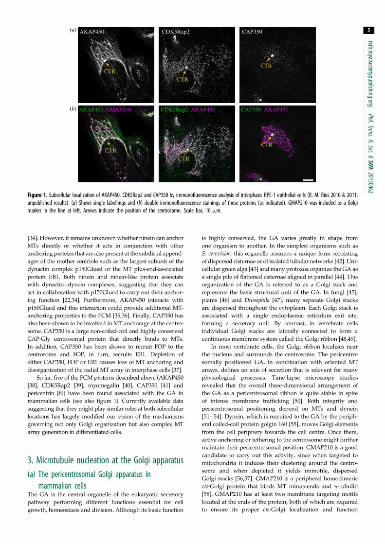
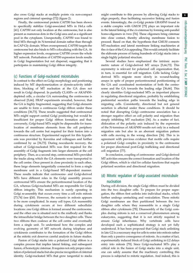
Figure 1. Subcellular localization of AKAP450, CDK5Rap2 and CAP350 by immunofluorescence analysis of interphasic RPE-1 epithelial cells (R. M. Rios 2010 & 2011,unpublished results). (a) Shows single labellings and (b) double immunofluorescence stainings of these proteins (as indicated). GMAP210 was included as a Golgimarker in the line at left. Arrows indicate the position of the centrosome. Scale bar, 10 mm.
rstb.royalsocietypublishing.orgPhil.Trans.R.Soc.B
369:20130462
3
[34]. However, it remains unknown whether ninein can anchor
MTs directly or whether it acts in conjunction with other
anchoring proteins that are also present at the subdistal append-
ages of the mother centriole such as the largest subunit of the
dynactin complex p150Glued or the MT plus-end-associated
protein EB1. Both ninein and ninein-like protein associate
with dynactin–dynein complexes, suggesting that they can
act in collaboration with p150Glued to carry out their anchor-
ing function [22,34]. Furthermore, AKAP450 interacts with
p150Glued and this interaction could provide additional MT-
anchoring properties to the PCM [35,36]. Finally, CAP350 has
also been shown to be involved in MT anchorage at the centro-
some. CAP350 is a large non-coiled-coil and highly conserved
CAP-Gly centrosomal protein that directly binds to MTs.
In addition, CAP350 has been shown to recruit FOP to the
centrosome and FOP, in turn, recruits EB1. Depletion of
either CAP350, FOP or EB1 causes loss of MT anchoring and
disorganization of the radial MT array in interphase cells [37].
So far, five of the PCM proteins described above (AKAP450
[38], CDK5Rap2 [39], myomegalin [40], CAP350 [41] and
pericentrin [8]) have been found associated with the GA in
mammalian cells (see also figure 1). Currently available data
suggesting that they might play similar roles at both subcellular
locations has largely modified our vision of the mechanisms
governing not only Golgi organization but also complex MT
array generation in differentiated cells.
3. Microtubule nucleation at the Golgi apparatus(a) The pericentrosomal Golgi apparatus in
mammalian cellsThe GA is the central organelle of the eukaryotic secretory
pathway performing different functions essential for cell
growth, homeostasis and division. Although its basic function
is highly conserved, the GA varies greatly in shape from
one organism to another. In the simplest organisms such as
S. cerevisiae, this organelle assumes a unique form consisting
of dispersed cisternae or of isolated tubular networks [42]. Uni-
cellular green alga [43] and many protozoa organize the GA as
a single pile of flattened cisternae aligned in parallel [44]. This
organization of the GA is referred to as a Golgi stack and
represents the basic structural unit of the GA. In fungi [45],
plants [46] and Drosophila [47], many separate Golgi stacks
are dispersed throughout the cytoplasm. Each Golgi stack is
associated with a single endoplasmic reticulum exit site,
forming a secretory unit. By contrast, in vertebrate cells
individual Golgi stacks are laterally connected to form a
continuous membrane system called the Golgi ribbon [48,49].
In most vertebrate cells, the Golgi ribbon localizes near
the nucleus and surrounds the centrosome. The pericentro-
somally positioned GA, in combination with oriented MT
arrays, defines an axis of secretion that is relevant for many
physiological processes. Time-lapse microscopy studies
revealed that the overall three-dimensional arrangement of
the GA as a pericentrosomal ribbon is quite stable in spite
of intense membrane trafficking [50]. Both integrity and
pericentrosomal positioning depend on MTs and dynein
[51–54]. Dynein, which is recruited to the GA by the periph-
eral coiled-coil protein golgin 160 [55], moves Golgi elements
from the cell periphery towards the cell centre. Once there,
active anchoring or tethering to the centrosome might further
maintain their pericentrosomal position. GMAP210 is a good
candidate to carry out this activity, since when targeted to
mitochondria it induces their clustering around the centro-
some and when depleted it yields immotile, dispersed
Golgi stacks [56,57]. GMAP210 is a peripheral homodimeric
cis-Golgi protein that binds MT minus-ends and g-tubulin
[58]. GMAP210 has at least two membrane targeting motifs
located at the ends of the protein, both of which are required
to ensure its proper cis-Golgi localization and function
rstb.royalsocietypublishing.orgPhil.Trans.R.Soc.B
369:20130462
4
[59,60]. These results reveal a role for GMAP210 in maintain-ing Golgi ribbon positioning and integrity, probably by
participating in the formation of connecting cis-cisternae
[57]. Dispersed Golgi stacks in GMAP210-depleted cells
are competent for general protein transport to the cell sur-
face excluding an essential role for GMAP210 in membrane
trafficking [56].
Support for a role of GMAP210 in Golgi architecture came
from the analysis of mutagenized mice dying from an auto-
somal recessive neonatal lethal skeletal dysplasia [61]. This
dysplasia shares common phenotypic features with achondro-
genesis type 1A in humans. Both affected mice and patients
have nonsense mutations in the Trip11 gene, which encodes
GMAP210. Loss of GMAP210 altered the Golgi structure in
many (but not all) tissues and compromised normal glycosyla-
tion in the Golgi as well as the transport of certain proteins that
would normally be destined for the extracellular matrix [61].
Follit and colleagues, using an alternative approach, also
engineered a GMAP210 deficient mouse. Embryonic kidney
cells derived from this knockout mouse exhibited an appar-
ently normal Golgi complex, although the structure of the
GA in other tissues was not examined [62]. GMAP210 is also
the receptor of IFT20 protein at the GA [62]. IFT20 is a critical
component of the intraflagellar transport machinery required
for the formation and extension of the primary cilium. In
mouse embryonic kidney cells lacking GMAP210, primary
cilia are shorter than normal and contain reduced amounts
of the membrane protein polycystin-2, suggesting that
GMAP210 and IFT20 function together at the Golgi in the
sorting or the transport of proteins destined for the ciliary
membrane [62]. Accordingly, the homologue of GMAP210
in Caenorhabditis elegans is involved in maintaining Golgi
organization and in the regulation of cilium length [63].
(b) Mechanisms of microtubule nucleation at theGolgi apparatus
In the past few years, a new concept about the role of the GA in
MT dynamics has emerged: the GA acting as an MT-organiz-
ing centre. In a pioneering study, Christian Pous’s group in
2001 [64] reported that Golgi membranes were able to assemble
and stabilize MTs in hepatic cells after nocodazole treatment. In
addition, purified Golgi membranes were shown to contain g-
tubulin and to promote MT assembly, a finding that has
recently been confirmed in neurons [64,65]. By tracking poly-
merizing MTs, Kaverina’s group then unambiguously
demonstrated that an MT subset grows directly from Golgi
membranes [5]. Two other important findings from this
study were that siRNA-mediated depletion of g-tubulin inhib-
its both Golgi and centrosome MT generation, and that laser
ablation of the centrosome does not affect the number of MTs
formed at the GA. It was estimated that up to 50% of MTs
in RPE-1 interphasic cells are produced by the GA. MT nuclea-
tion at the GA was shown to require the MT-stabilizing activity
provided by CLASPs, MT plus-end binding proteins that
are recruited to the GA through the interaction with the
TGN-associated protein GCC185 [5].
This study did not identify, however, the machinery respon-
sible for MT nucleation at the GA. We further demonstrated that
MT nucleation at the GA depends on the PCM protein
AKAP450 that specifically associates with the cis-GA [66].
Depletion of Golgi-associated AKAP450 or expression of a
dominant negative mutant, which dissociated AKAP450 from
the GA but not from the centrosome, completely abolished
MT nucleation at the GA [36,66]. We then proposed that the
cis-GA becomes a major site for MT nucleation by acting as a
preferential g-TuRC docking site through a mechanism similar
to that operating at the centrosome. This hypothesis has received
additional support with the finding that other g-TuRC recruiting
PCM proteins such as CDK5Rap2 (figure 1) and myomegalin
localize at the cis-GA [29,39].
Indeed, recent data suggest that Golgi capacity to nucleate
MTs might rely on multiprotein complexes similar to those
present at the PCM (see figure 2 for a model). These data also
point out a hierarchy in the association of these proteins to the
Golgi. Thus, the peripheral cis-Golgi protein GM130 recruits
AKAP450 [66] that, in turn, recruits both CDK5Rap2 and
myomegalin [29,39]. Accordingly, GM130 depletion caused
the dissociation of both AKAP450 and myomegalin from the
GA [29], whereas brefeldin A treatment induced redistribution
of GM130, AKAP450 and myomegalin to endoplasmic reticu-
lum exit sites that concomitantly acquired MT nucleation
capacity [29,66]. It is worth noting that although significant cyto-
plasmic pools of AKAP450, CDK5Rap2, myomegalin and
g-tubulin exist, MTs do not normally form in the cytoplasm.
This suggests that multiprotein complexes become competent
for MT nucleation only after being assembled at the cis-Golgi
membrane surface. Interestingly, a cytoplasmic pool of GM130
is lacking, and GM130 is exclusively present at the cis-GA
[57,66]. Thus, it appears as a critical factor for MT nucleation
at the GA by controlling both localization and rate of the process.
Despite these data, the specific mechanism whereby
AKAP450 induces MT formation at the GA has not been
fully elucidated. AKAP450 could recruit g-TuRC directly
and/or indirectly through CDK5Rap2 (see model in figure 2).
AKAP450, CDK5Rap2 and their respective orthologues in
other species have been reported to bind g-tubulin-containing
complexes [25,27,67,68]. Takahashi et al. [25] showed that
the N-terminal region of CG-NAP indirectly associates with
g-tubulin through interaction with GCP2/GCP3 components
of g-TuRC. We have not detected any interaction between
g-tubulin and the most N-terminal part of the protein in spite
of careful examination [36], although the truncated mutants
used in both studies were not identical, which could explain
the discrepancies. However, CDK5Rap2 directly binds g-TuRC
and works as a strong activator of MT nucleation through its
g-TuNA motif [28].
Based on the capacity of AKAP450 to bind p150Glued and
the finding that blocking dynein/dynactin interferes with GA-
based MT nucleation at the GA [66], it has been proposed that
AKAP450 might also support MT formation via a dynein/
dynactin-dependent mechanism [69]. The binding site for
p150Glued is localized at the N-terminus of AKAP450, close to
the GM130-interacting motif. An AKAP450-truncated mutant
containing both p150Glued and GM130-binding motifs targets
the GA and MTs, and yet GA membranes are unable to nucleate
MTs [36]. Therefore, although a direct proof is still lacking, all
evidence supports the idea that the main mechanism for MT
nucleation at the GA is based on g-TuRC recruitment. Interest-
ingly, the PTTG1/securin protein has been found in a complex
with AKAP450, GM130 and g-tubulin [70]. PTTG1/securin
localizes at both the centrosome and the GA, and when depleted,
MT nucleation is delayed at both subcellular localizations. Based
on described PTTG1/securin functions, the authors propo-
sed that PTTG1 could act as a chaperone contributing to the
formation and stability of MT-nucleating complexes.
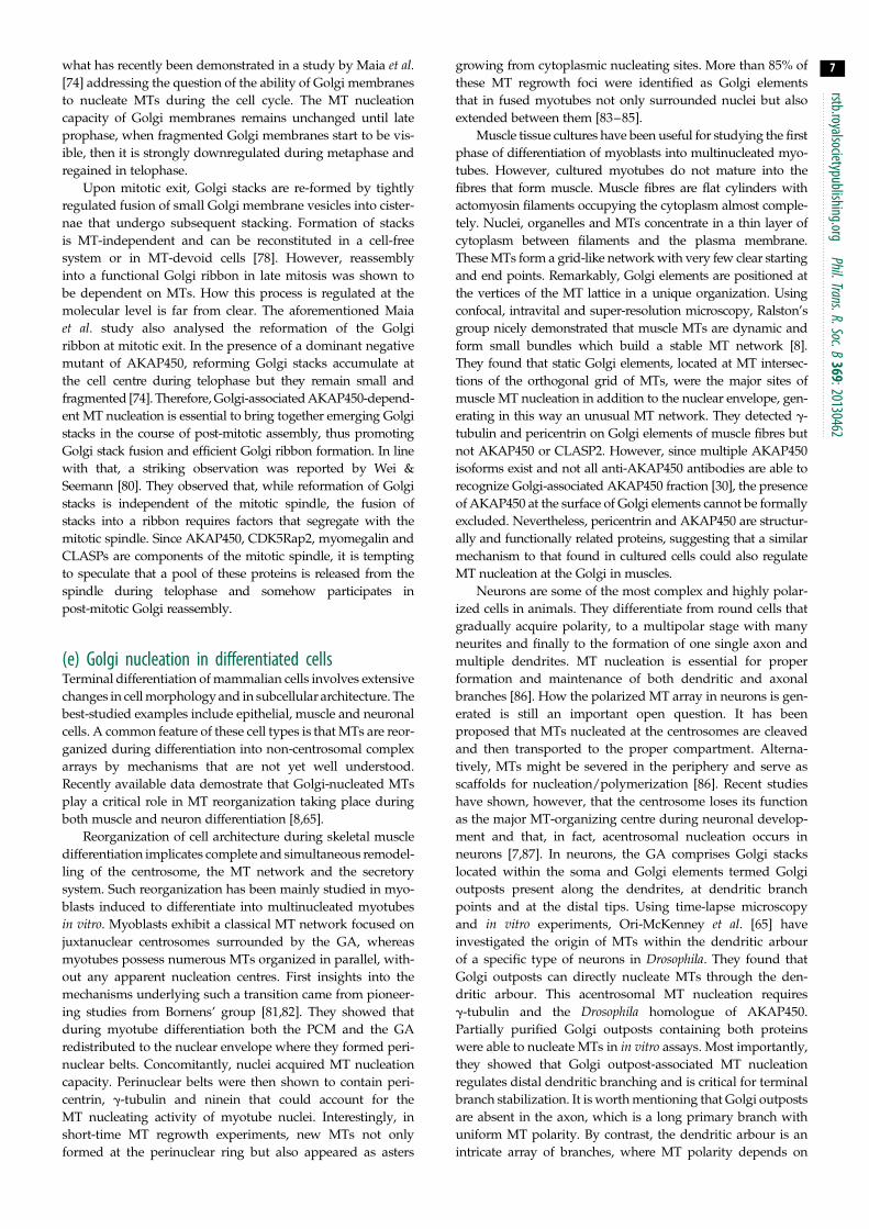
centrosome
AKAP450
GM130
CDK5Rap2
myomegalin
g -TuRC
MTs
p150Glued
other MT -nucleating complexes
GCC185
CLASPs
Golgi apparatus(a) (c)
(b)
mechanisms of MT nucleation
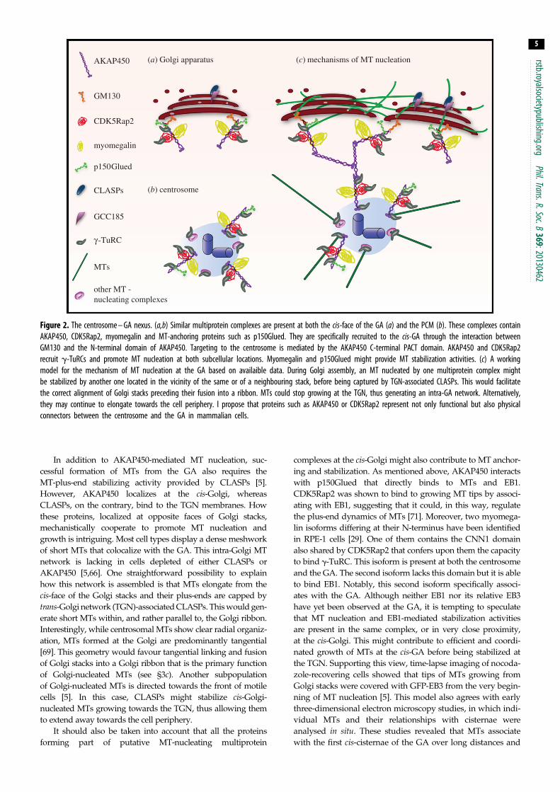
Figure 2. The centrosome – GA nexus. (a,b) Similar multiprotein complexes are present at both the cis-face of the GA (a) and the PCM (b). These complexes containAKAP450, CDK5Rap2, myomegalin and MT-anchoring proteins such as p150Glued. They are specifically recruited to the cis-GA through the interaction betweenGM130 and the N-terminal domain of AKAP450. Targeting to the centrosome is mediated by the AKAP450 C-terminal PACT domain. AKAP450 and CDK5Rap2recruit g-TuRCs and promote MT nucleation at both subcellular locations. Myomegalin and p150Glued might provide MT stabilization activities. (c) A workingmodel for the mechanism of MT nucleation at the GA based on availaible data. During Golgi assembly, an MT nucleated by one multiprotein complex mightbe stabilized by another one located in the vicinity of the same or of a neighbouring stack, before being captured by TGN-associated CLASPs. This would facilitatethe correct alignment of Golgi stacks preceding their fusion into a ribbon. MTs could stop growing at the TGN, thus generating an intra-GA network. Alternatively,they may continue to elongate towards the cell periphery. I propose that proteins such as AKAP450 or CDK5Rap2 represent not only functional but also physicalconnectors between the centrosome and the GA in mammalian cells.
rstb.royalsocietypublishing.orgPhil.Trans.R.Soc.B
369:20130462
5
In addition to AKAP450-mediated MT nucleation, suc-
cessful formation of MTs from the GA also requires the
MT-plus-end stabilizing activity provided by CLASPs [5].
However, AKAP450 localizes at the cis-Golgi, whereas
CLASPs, on the contrary, bind to the TGN membranes. How
these proteins, localized at opposite faces of Golgi stacks,
mechanistically cooperate to promote MT nucleation and
growth is intriguing. Most cell types display a dense meshwork
of short MTs that colocalize with the GA. This intra-Golgi MT
network is lacking in cells depleted of either CLASPs or
AKAP450 [5,66]. One straightforward possibility to explain
how this network is assembled is that MTs elongate from the
cis-face of the Golgi stacks and their plus-ends are capped by
trans-Golgi network (TGN)-associated CLASPs. This would gen-
erate short MTs within, and rather parallel to, the Golgi ribbon.
Interestingly, while centrosomal MTs show clear radial organiz-
ation, MTs formed at the Golgi are predominantly tangential
[69]. This geometry would favour tangential linking and fusion
of Golgi stacks into a Golgi ribbon that is the primary function
of Golgi-nucleated MTs (see §3c). Another subpopulation
of Golgi-nucleated MTs is directed towards the front of motile
cells [5]. In this case, CLASPs might stabilize cis-Golgi-
nucleated MTs growing towards the TGN, thus allowing them
to extend away towards the cell periphery.
It should also be taken into account that all the proteins
forming part of putative MT-nucleating multiprotein
complexes at the cis-Golgi might also contribute to MT anchor-
ing and stabilization. As mentioned above, AKAP450 interacts
with p150Glued that directly binds to MTs and EB1.
CDK5Rap2 was shown to bind to growing MT tips by associ-
ating with EB1, suggesting that it could, in this way, regulate
the plus-end dynamics of MTs [71]. Moreover, two myomega-
lin isoforms differing at their N-terminus have been identified
in RPE-1 cells [29]. One of them contains the CNN1 domain
also shared by CDK5Rap2 that confers upon them the capacity
to bind g-TuRC. This isoform is present at both the centrosome
and the GA. The second isoform lacks this domain but it is able
to bind EB1. Notably, this second isoform specifically associ-
ates with the GA. Although neither EB1 nor its relative EB3
have yet been observed at the GA, it is tempting to speculate
that MT nucleation and EB1-mediated stabilization activities
are present in the same complex, or in very close proximity,
at the cis-Golgi. This might contribute to efficient and coordi-
nated growth of MTs at the cis-GA before being stabilized at
the TGN. Supporting this view, time-lapse imaging of nocoda-
zole-recovering cells showed that tips of MTs growing from
Golgi stacks were covered with GFP-EB3 from the very begin-
ning of MT nucleation [5]. This model also agrees with early
three-dimensional electron microscopy studies, in which indi-
vidual MTs and their relationships with cisternae were
analysed in situ. These studies revealed that MTs associate
with the first cis-cisternae of the GA over long distances and
rstb.royalsocietypublishing.orgPhil.Trans
6
also cross Golgi stacks at multiple points via non-compactregions and cisternal openings [72] (figure 2).
Finally, the centrosomal protein CAP350 has been shown
to specifically stabilize Golgi-associated MTs in HeLa cells
[41]. CAP350 not only localizes at the centrosome, but is also
present as numerous dots in the Golgi area and as a significant
pool in the cytoplasm. Unexpectedly, CAP350 was found to
bind MTs through its N-terminal domain rather than through
its CAP-Gly domain. When overexpressed, CAP350 targets the
centrosome but also binds to MTs colocalizing with the GA. At
higher expression levels, exogenous CAP350 covers the whole
MT network. Perturbation of CAP350 expression levels results
in Golgi fragmentation but not dispersal, suggesting that it
participates in maintaining Golgi ribbon integrity.
.R.Soc.B369:20130462
(c) Functions of Golgi-nucleated microtubulesIn contrast to the effect on Golgi morphology and positioning
induced by MT depolymerization or dynein-activity inhib-
ition, blocking of MT nucleation at the GA does not
result in Golgi dispersal. In partially CLASPs- or AKAP450-
depleted cells, a circular GA surrounding the centrosome is
observed [5,66]. FRAP experiments revealed, however, that
the GA is highly fragmented, suggesting that Golgi elements
are unable to form a continuous Golgi ribbon under these
conditions [36,73]. These results suggested that centrosomal
MTs might support central Golgi positioning but would be
insufficient for proper Golgi ribbon formation and that,
conversely, Golgi-based MTs might be dispensable for trans-
location of membrane elements from the cell periphery
towards the cell centre but required for their fusion into a
continuous structure. Experimental support for this hypoth-
esis was provided by Kaverina and colleagues and further
confirmed by us [36,73]. During nocodazole recovery, the
subset of Golgi-nucleated MTs was first required for the
assembly of Golgi fragments into larger elements at the cell
periphery. Then, as a second step, centrosomal MTs provided
the tracks along which the GA elements were transported to
the cell centre. Once present in close proximity to each other,
these large elements tangentially connected to form a single
membrane unit in a Golgi-based MT-dependent manner.
These results indicate that centrosome- and Golgi-derived
MTs have different roles in the Golgi assembly process:
centrosomal MTs ensure the pericentrosomal location of the
GA, whereas Golgi-nucleated MTs are responsible for Golgi
ribbon integrity. This mechanism is surely operating in
Golgi re-assembly that occurs every time a cell exits mitosis
[73,74] (see §3d). However, the scenario in mitosis appears
to be more complicated. In many cell types, GA reassembly
during cytokinesis occurs at two different subcellular
locations: one Golgi ribbon is formed around the centrosome,
and the other one is situated next to the midbody and flanks
the intracellular bridge between the two daughter cells. These
two ribbons then coalesce at the cell centre and eventually
form a single unit. To understand how the complex and
evolving geometry of MT network during telophase and
cytokinesis contributes to the formation of the Golgi ribbon
at the mitotic exit deserves careful analysis in future studies.
Fusion of Golgi stacks into a polarized Golgi ribbon is a
complex process that implies lateral linking, and subsequent
fusion, of homotypic cisternae. It requires not only proper orien-
tation of polarized stacks but also precise recognition of cisternal
identity. Golgi-nucleated MTs that grow tangential to stacks
might contribute to this process by allowing Golgi stacks to
align properly, thus facilitating successive linking and fusion
events. Interestingly, the cis-Golgi protein GRASP65 found in
a stable complex with GM130 [75] plays an important role
in the lateral linking of the cis-cisternae by forming anti-parallel
homo-oligomers in trans [76]. These oligomers bring cisternae
into close contact, thereby allowing membrane fusion to
proceed. Based on that, the hypothesis that GM130 connects
MT-nucleation and lateral membrane linking machineries at
the cis-face of the GA is appealing. This would certainly facilitate
the elaborate process of fusing hundreds of Golgi stacks into a
single highly polarized structure.
Several studies have emphasized the intrinsic asym-
metric nature of Golgi-derived MT arrays [5,66,73]. This
asymmetry is relevant for polarized cell organization that,
in turn, is essential for cell migration. Cells lacking Golgi-
derived MTs migrate more slowly in wound-healing
assays in spite of proper pericentrosomal positionioning of
the GA and coordinated reorientation of both the centro-
some and the GA towards the leading edge [36,66]. This
clearly identifies Golgi-nucleated MTs as important players
in regulating directional migration, probably by establishing
preferential secretion paths towards the leading edge in
migrating cells. Consistently, directional but not general
secretion is affected under these conditions. It should be
noted that disrupting Golgi–centrosome association has a
stronger negative effect on cell polarity and migration than
simply inhibiting MT nucleation [36]. As a matter of fact,
dislocation of the polarity axis induced by the expression
of the N-terminus of AKAP450 results not only in a reduced
migration rate but also in an aberrant migration pattern
with cells moving in the wrong direction [36]. This is in
agreement with previous evidence that motile cells require
a polarized Golgi complex in proximity to the centrosome
for proper directional post-Golgi trafficking and directional
cell migration [77].
In conclusion, coupling of centrosome- and Golgi-derived
MT activities ensures the correct formation and location of the
Golgi ribbon, which is vital for cellular functions that require
polarized secretion and directional migration.
(d) Mitotic regulation of Golgi-associated microtubulenucleation
During cell division, the single Golgi ribbon must be divided
into the two daughter cells. To prepare for proper segre-
gation, the ribbon first unlinks into individual stacks, which
further undergo unstacking and vesiculation. These mitotic
Golgi membranes are then partitioned between the two
daughter cells where they reassemble in a single Golgi
ribbon after cytokinesis [78]. Disassembly of the Golgi com-
plex during mitosis is not a conserved phenomenon among
eukaryotes, suggesting that it is not strictly required to
ensure Golgi inheritance. Why vertebrate cells have
developed such a sophisticated strategy is not yet fully
understood. It has been proposed that Golgi stack unlinking
in late G2 is a necessary step for cells to enter into mitosis rather
than only a passive consequence of mitosis. As a matter of fact,
experimentally induced block of Golgi unlinking in G2 delays
entry into mitosis [79]. Since Golgi-nucleated MTs play a
critical role in the fusion of Golgi stacks into a single unit,
one can safely assume that the machinery controlling this
process is subjected to mitotic regulation. And indeed, this is
rstb.royalsocietypublishing.orgPhil.Trans.R.Soc.B
369:20130462
7
what has recently been demonstrated in a study by Maia et al.[74] addressing the question of the ability of Golgi membranesto nucleate MTs during the cell cycle. The MT nucleation
capacity of Golgi membranes remains unchanged until late
prophase, when fragmented Golgi membranes start to be vis-
ible, then it is strongly downregulated during metaphase and
regained in telophase.
Upon mitotic exit, Golgi stacks are re-formed by tightly
regulated fusion of small Golgi membrane vesicles into cister-
nae that undergo subsequent stacking. Formation of stacks
is MT-independent and can be reconstituted in a cell-free
system or in MT-devoid cells [78]. However, reassembly
into a functional Golgi ribbon in late mitosis was shown to
be dependent on MTs. How this process is regulated at the
molecular level is far from clear. The aforementioned Maia
et al. study also analysed the reformation of the Golgi
ribbon at mitotic exit. In the presence of a dominant negative
mutant of AKAP450, reforming Golgi stacks accumulate at
the cell centre during telophase but they remain small and
fragmented [74]. Therefore, Golgi-associated AKAP450-depend-
ent MT nucleation is essential to bring together emerging Golgi
stacks in the course of post-mitotic assembly, thus promoting
Golgi stack fusion and efficient Golgi ribbon formation. In line
with that, a striking observation was reported by Wei &
Seemann [80]. They observed that, while reformation of Golgi
stacks is independent of the mitotic spindle, the fusion of
stacks into a ribbon requires factors that segregate with the
mitotic spindle. Since AKAP450, CDK5Rap2, myomegalin and
CLASPs are components of the mitotic spindle, it is tempting
to speculate that a pool of these proteins is released from the
spindle during telophase and somehow participates in
post-mitotic Golgi reassembly.
(e) Golgi nucleation in differentiated cellsTerminal differentiation of mammalian cells involves extensive
changes in cell morphology and in subcellular architecture. The
best-studied examples include epithelial, muscle and neuronal
cells. A common feature of these cell types is that MTs are reor-
ganized during differentiation into non-centrosomal complex
arrays by mechanisms that are not yet well understood.
Recently available data demostrate that Golgi-nucleated MTs
play a critical role in MT reorganization taking place during
both muscle and neuron differentiation [8,65].
Reorganization of cell architecture during skeletal muscle
differentiation implicates complete and simultaneous remodel-
ling of the centrosome, the MT network and the secretory
system. Such reorganization has been mainly studied in myo-
blasts induced to differentiate into multinucleated myotubes
in vitro. Myoblasts exhibit a classical MT network focused on
juxtanuclear centrosomes surrounded by the GA, whereas
myotubes possess numerous MTs organized in parallel, with-
out any apparent nucleation centres. First insights into the
mechanisms underlying such a transition came from pioneer-
ing studies from Bornens’ group [81,82]. They showed that
during myotube differentiation both the PCM and the GA
redistributed to the nuclear envelope where they formed peri-
nuclear belts. Concomitantly, nuclei acquired MT nucleation
capacity. Perinuclear belts were then shown to contain peri-
centrin, g-tubulin and ninein that could account for the
MT nucleating activity of myotube nuclei. Interestingly, in
short-time MT regrowth experiments, new MTs not only
formed at the perinuclear ring but also appeared as asters
growing from cytoplasmic nucleating sites. More than 85% of
these MT regrowth foci were identified as Golgi elements
that in fused myotubes not only surrounded nuclei but also
extended between them [83–85].
Muscle tissue cultures have been useful for studying the first
phase of differentiation of myoblasts into multinucleated myo-
tubes. However, cultured myotubes do not mature into the
fibres that form muscle. Muscle fibres are flat cylinders with
actomyosin filaments occupying the cytoplasm almost comple-
tely. Nuclei, organelles and MTs concentrate in a thin layer of
cytoplasm between filaments and the plasma membrane.
These MTs form a grid-like network with very few clear starting
and end points. Remarkably, Golgi elements are positioned at
the vertices of the MT lattice in a unique organization. Using
confocal, intravital and super-resolution microscopy, Ralston’s
group nicely demonstrated that muscle MTs are dynamic and
form small bundles which build a stable MT network [8].
They found that static Golgi elements, located at MT intersec-
tions of the orthogonal grid of MTs, were the major sites of
muscle MT nucleation in addition to the nuclear envelope, gen-
erating in this way an unusual MT network. They detected g-
tubulin and pericentrin on Golgi elements of muscle fibres but
not AKAP450 or CLASP2. However, since multiple AKAP450
isoforms exist and not all anti-AKAP450 antibodies are able to
recognize Golgi-associated AKAP450 fraction [30], the presence
of AKAP450 at the surface of Golgi elements cannot be formally
excluded. Nevertheless, pericentrin and AKAP450 are structur-
ally and functionally related proteins, suggesting that a similar
mechanism to that found in cultured cells could also regulate
MT nucleation at the Golgi in muscles.
Neurons are some of the most complex and highly polar-
ized cells in animals. They differentiate from round cells that
gradually acquire polarity, to a multipolar stage with many
neurites and finally to the formation of one single axon and
multiple dendrites. MT nucleation is essential for proper
formation and maintenance of both dendritic and axonal
branches [86]. How the polarized MT array in neurons is gen-
erated is still an important open question. It has been
proposed that MTs nucleated at the centrosomes are cleaved
and then transported to the proper compartment. Alterna-
tively, MTs might be severed in the periphery and serve as
scaffolds for nucleation/polymerization [86]. Recent studies
have shown, however, that the centrosome loses its function
as the major MT-organizing centre during neuronal develop-
ment and that, in fact, acentrosomal nucleation occurs in
neurons [7,87]. In neurons, the GA comprises Golgi stacks
located within the soma and Golgi elements termed Golgi
outposts present along the dendrites, at dendritic branch
points and at the distal tips. Using time-lapse microscopy
and in vitro experiments, Ori-McKenney et al. [65] have
investigated the origin of MTs within the dendritic arbour
of a specific type of neurons in Drosophila. They found that
Golgi outposts can directly nucleate MTs through the den-
dritic arbour. This acentrosomal MT nucleation requires
g-tubulin and the Drosophila homologue of AKAP450.
Partially purified Golgi outposts containing both proteins
were able to nucleate MTs in in vitro assays. Most importantly,
they showed that Golgi outpost-associated MT nucleation
regulates distal dendritic branching and is critical for terminal
branch stabilization. It is worth mentioning that Golgi outposts
are absent in the axon, which is a long primary branch with
uniform MT polarity. By contrast, the dendritic arbour is an
intricate array of branches, where MT polarity depends on
rstb.royalsocietypublis
8
the branch length. In neurons lacking cytoplasmic dynein,Golgi outposts mislocalize to the axon which appears branched
and contains MTs of mixed polarity [88]. Interestingly, small
MT bundles growing from Golgi elements in muscle fibres
also contain MTs of mixed polarity. Therefore, it can be specu-
lated that generation of MT arrays with mixed MT orientation
might be a property of Golgi-associated MT nucleation in
complex morphogenetic processes.
hing.orgPhil.Trans.R.Soc.B369:20130462
4. Concluding remarks and perspectivesA comparison between the MT nucleation process at the cen-
trosome and at the GA highlights some common features
and interesting differences. Indeed, data presented in this
review show that the GA uses classical centrosomal proteins
for its MT nucleation activity. These data further suggest that
centrosome-associated proteins can function fully indepen-
dently of the centrosome. While this assumption is still valid,
the network-like distribution of proteins such as AKAP450 or
CDK5Rap2 extending from the centrosome towards the GA,
suggests the existence of direct connections between these
two organelles (see figure 2 for a working model). Admittedly,
this connection would facilitate the well-known coordinated
behaviour of both organelles in physiological processes that
require MTs to be dynamic, such as cell migration, Golgi reas-
sembly after mitosis, and the formation of the immunological
synapse [49]. Additionally, the existence of PCM protein net-
works connecting the centrosome and the GA raises the
interesting question of how precisely to define the limit of the
centrosome in mammalian cells. In vivo analysis and super
high-resolution imaging techniques will certainly help to
refine our knowledge of the organization of this crucial
subcellular region. Despite the recent steps forward in our
understanding of the MT nucleation process at the GA, a coher-
ent view about how Golgi-associated PCM proteins interact
with each other and with CLASPs in order to orchestrate this
process is still lacking. The mechanism by which Golgi-based
MTs cooperate with membrane tethering and fusion machin-
eries to generate a single membrane unit also remains
unknown. Thus, integrative studies will be useful to assemble
in common networks proteins involved in controlling MT for-
mation and those regulating Golgi ribbon assembly and
membrane trafficking.
The major differences between centrosome-nucleated and
Golgi-nucleated MTs stem from their geometry and nature. It
has been known for long time that MTs colocalizing with the
GA are highly enriched in post-translationally modified tubu-
lins, in particular detyrosinated and acetylated a-tubulin [54].
In this regard, the most obvious questions are how and why
the molecular machinery responsible for such modifications
specifically targets the Golgi subpopulation of MTs.
Data examined in this review also reveal an important
role of the GA in organizing complex and specialized MT
arrays that carry out specific functions in differentiated
cells. Hopefully, the recent discoveries in muscles and neur-
ons will be soon extended to other cell types with equally
complex MT arrays. Particularly relevant will be a thorough
understanding of how MT nucleation at the GA contributes
to MT remodelling during the establishment of apico-basal
polarity in epithelial cells.
Finally, since MT nucleation at the centrosome and the GA
is probably differently, yet coordinatively regulated in a
cell cycle- and cell type-dependent manner, deciphering the
signalling pathways underlying such regulation will no
doubt deserve more attention in years to come. Further efforts
should also be made to understand this regulation better
in different biological contexts, for example, during animal
development and disease pathogenesis.
Acknowledgements. I am grateful to M. P. Gavilan and C. Marcozzi forvaluable discussions and support.
Funding statement. R.M.R. team is supported by the Consejo Superior deInvestigaciones Cientı́ficas and grants from the Ministerio de Econo-mia y Competitividad (BFU2012-36717 and CSD2009-00016) and theJunta de Andalucı́a, Spain.
References
1. Kollman JM, Merdes A, Mourey L, Agard DA. 2011Microtubule nucleation by gamma-tubulincomplexes. Nat. Rev. Mol. Cell Biol. 12, 709 – 721.(doi:10.1038/nrm3209)
2. Moudjou M, Bordes N, Paintrand M, Bornens M.1996 gamma-Tubulin in mammalian cells: thecentrosomal and the cytosolic forms. J. Cell Sci. 109,875 – 887.
3. Bornens M. 2002 Centrosome composition andmicrotubule anchoring mechanisms. Curr. Opin. CellBiol. 14, 25 – 34. (doi:10.1016/S0955-0674(01)00290-3)
4. Teixido-Travesa N, Roig J, Luders J. 2012 The where,when and how of microtubule nucleation: one ringto rule them all. J. Cell Sci. 125, 4445 – 4456.(doi:10.1242/jcs.106971)
5. Efimov A et al. 2007 Asymmetric CLASP-dependentnucleation of noncentrosomal microtubules at thetrans-Golgi network. Dev. Cell 12, 917 – 930.(doi:10.1016/j.devcel.2007.04.002)
6. Khodjakov A, Rieder CL. 1999 The suddenrecruitment of gamma-tubulin to the centrosome atthe onset of mitosis and its dynamic exchangethroughout the cell cycle, do not requiremicrotubules. J. Cell Biol. 146, 585 – 596. (doi:10.1083/jcb.146.3.585)
7. Stiess M, Maghelli N, Kapitein LC, Gomis-Ruth S,Wilsch-Brauninger M, Hoogenraad CC, Tolic-Norrelykke IM, Bradke F. 2010 Axon extensionoccurs independently of centrosomal microtubulenucleation. Science 327, 704 – 707. (doi:10.1126/science.1182179)
8. Oddoux S, Zaal KJ, Tate V, Kenea A, Nandkeolyar SA,Reid E, Liu W, Ralston E. 2013 Microtubules thatform the stationary lattice of muscle fibers aredynamic and nucleated at Golgi elements. J. CellBiol. 203, 205 – 213. (doi:10.1083/jcb.201304063)
9. Mennella V, Keszthelyi B, McDonald KL, Chhun B,Kan F, Rogers GC, Huang B, Agard DA. 2012Subdiffraction-resolution fluorescence microscopy
reveals a domain of the centrosome critical forpericentriolar material organization. Nat. Cell Biol.14, 1159 – 1168. (doi:10.1038/ncb2597)
10. Sonnen KF, Schermelleh L, Leonhardt H, Nigg EA.2012 3D-structured illumination microscopyprovides novel insight into architecture of humancentrosomes. Biol. Open 1, 965 – 976. (doi:10.1242/bio.20122337)
11. Lawo S, Hasegan M, Gupta GD, Pelletier L. 2012Subdiffraction imaging of centrosomes revealshigher-order organizational features of pericentriolarmaterial. Nat. Cell Biol. 14, 1148 – 1158. (doi:10.1038/ncb2591)
12. Fu J, Glover DM. 2012 Structured illumination of theinterface between centriole and peri-centriolar material.Open Biol. 2, 120104. (doi:10.1098/rsob.120104)
13. Hutchins JR et al. 2010 Systematic analysis ofhuman protein complexes identifies chromosomesegregation proteins. Science 328, 593 – 599.(doi:10.1126/science.1181348)
rstb.royalsocietypublishing.orgPhil.Trans.R.Soc.B
369:20130462
9
14. Teixido-Travesa N, Villen J, Lacasa C, Bertran MT,Archinti M, Gygi SP, Caelles C, Roig J, Luders J. 2010The gammaTuRC revisited: a comparative analysis ofinterphase and mitotic human gammaTuRCredefines the set of core components and identifiesthe novel subunit GCP8. Mol. Biol. Cell 21,3963 – 3972. (doi:10.1091/mbc.E10-05-0408)15. Luders J, Patel UK, Stearns T. 2006 GCP-WD is agamma-tubulin targeting factor required forcentrosomal and chromatin-mediated microtubulenucleation. Nat. Cell Biol. 8, 137 – 147. (doi:10.1038/ncb1349)
16. Kollman JM, Polka JK, Zelter A, Davis TN, Agard DA.2010 Microtubule nucleating gamma-TuSCassembles structures with 13-fold microtubule-likesymmetry. Nature 466, 879 – 882. (doi:10.1038/nature09207)
17. Manning JA, Shalini S, Risk JM, Day CL, Kumar S.2010 A direct interaction with NEDD1 regulatesgamma-tubulin recruitment to the centrosome.PLoS ONE 5, e9618. (doi:10.1371/journal.pone.0009618)
18. Zhu F et al. 2008 The mammalian SPD-2 orthologCep192 regulates centrosome biogenesis. Curr. Biol.18, 136 – 141. (doi:10.1016/j.cub.2007.12.055)
19. Gomez-Ferreria MA, Bashkurov M, Helbig AO,Larsen B, Pawson T, Gingras AC, Pelletier L. 2012Novel NEDD1 phosphorylation sites regulategamma-tubulin binding and mitotic spindleassembly. J. Cell Sci. 125, 3745 – 3751. (doi:10.1242/jcs.105130)
20. Haren L, Stearns T, Luders J. 2009 Plk1-dependentrecruitment of gamma-tubulin complexes to mitoticcentrosomes involves multiple PCM components.PLoS ONE 4, e5976. (doi:10.1371/journal.pone.0005976)
21. Casenghi M, Meraldi P, Weinhart U, Duncan PI,Korner R, Nigg EA. 2003 Polo-like kinase 1 regulatesNlp, a centrosome protein involved in microtubulenucleation. Dev. Cell 5, 113 – 125. (doi:10.1016/S1534-5807(03)00193-X)
22. Casenghi M, Barr FA, Nigg EA. 2005 Phosphorylationof Nlp by Plk1 negatively regulates its dynein –dynactin-dependent targeting to the centrosome.J. Cell Sci. 118, 5101 – 5108. (doi:10.1242/jcs.02622)
23. Flory MR, Davis TN. 2003 The centrosomal proteinspericentrin and kendrin are encoded by alternativelyspliced products of one gene. Genomics 82,401 – 405. (doi:10.1016/S0888-7543(03)00119-8)
24. Zimmerman WC, Sillibourne J, Rosa J, Doxsey SJ.2004 Mitosis-specific anchoring of gamma tubulincomplexes by pericentrin controls spindleorganization and mitotic entry. Mol. Biol. Cell 15,3642 – 3657. (doi:10.1091/mbc.E03-11-0796)
25. Takahashi M, Yamagiwa A, Nishimura T, Mukai H,Ono Y. 2002 Centrosomal proteins CG-NAP andkendrin provide microtubule nucleation sites byanchoring gamma-tubulin ring complex. Mol. Biol.Cell 13, 3235 – 3245. (doi:10.1091/mbc.E02-02-0112)
26. Gillingham AK, Munro S. 2000 The PACT domain, aconserved centrosomal targeting motif in thecoiled-coil proteins AKAP450 and pericentrin.
EMBO Rep. 1, 524 – 529. (doi:10.1093/embo-reports/kvd105)
27. Fong KW, Choi YK, Rattner JB, Qi RZ. 2008CDK5RAP2 is a pericentriolar protein that functionsin centrosomal attachment of the gamma-tubulinring complex. Mol. Biol. Cell 19, 115 – 125. (doi:10.1091/mbc.E07-04-0371)
28. Choi YK, Liu P, Sze SK, Dai C, Qi RZ. 2010 CDK5RAP2stimulates microtubule nucleation by the gamma-tubulin ring complex. J. Cell Biol. 191, 1089 – 1095.(doi:10.1083/jcb.201007030)
29. Roubin R, Acquaviva C, Chevrier V, Sedjai F, Zyss D,Birnbaum D, Rosnet O. 2013 Myomegalin isnecessary for the formation of centrosomal andGolgi-derived microtubules. Biol. Open 2, 238 – 250.(doi:10.1242/bio.20123392)
30. Keryer G, Witczak O, Delouvee A, Kemmner WA,Rouillard D, Tasken K, Bornens M. 2003 Dissociatingthe centrosomal matrix protein AKAP450 fromcentrioles impairs centriole duplication and cellcycle progression. Mol. Biol. Cell 14, 2436 – 2446.(doi:10.1091/mbc.E02-09-0614)
31. Lee K, Rhee K. 2011 PLK1 phosphorylation ofpericentrin initiates centrosome maturation at theonset of mitosis. J. Cell Biol. 195, 1093 – 1101.(doi:10.1083/jcb.201106093)
32. Buchman JJ, Tseng HC, Zhou Y, Frank CL, Xie Z, TsaiLH. 2010 Cdk5rap2 interacts with pericentrin tomaintain the neural progenitor pool in thedeveloping neocortex. Neuron 66, 386 – 402.(doi:10.1016/j.neuron.2010.03.036)
33. Barr AR, Kilmartin JV, Gergely F. 2010 CDK5RAP2functions in centrosome to spindle pole attachmentand DNA damage response. J. Cell Biol. 189,23 – 39. (doi:10.1083/jcb.200912163)
34. Delgehyr N, Sillibourne J, Bornens M. 2005Microtubule nucleation and anchoring at thecentrosome are independent processes linked byninein function. J. Cell Sci. 118, 1565 – 1575.(doi:10.1242/jcs.02302)
35. Kim HS, Takahashi M, Matsuo K, Ono Y. 2007Recruitment of CG-NAP to the Golgi apparatusthrough interaction with dynein – dynactin complex.Genes Cells 12, 421 – 434. (doi:10.1111/j.1365-2443.2007.01055.x)
36. Hurtado L, Caballero C, Gavilan MP, Cardenas J,Bornens M, Rios RM. 2011 Disconnecting the Golgiribbon from the centrosome prevents directionalcell migration and ciliogenesis. J. Cell Biol. 193,917 – 933. (doi:10.1083/jcb.201011014)
37. Yan X, Habedanck R, Nigg EA. 2006 A complex of twocentrosomal proteins, CAP350 and FOP, cooperateswith EB1 in microtubule anchoring. Mol. Biol. Cell 17,634 – 644. (doi:10.1091/mbc.E05-08-0810)
38. Takahashi M, Shibata H, Shimakawa M, MiyamotoM, Mukai H, Ono Y. 1999 Characterization of a novelgiant scaffolding protein, CG-NAP, that anchorsmultiple signaling enzymes to centrosome and theGolgi apparatus. J. Biol. Chem. 274, 17 267 –17 274. (doi:10.1074/jbc.274.24.17267)
39. Wang Z, Wu T, Shi L, Zhang L, Zheng W, Qu JY, NiuR, Qi RZ. 2010 Conserved motif of CDK5RAP2mediates its localization to centrosomes and the
Golgi complex. J. Biol. Chem. 285, 22 658 – 22 665.(doi:10.1074/jbc.M110.105965)
40. Verde I, Pahlke G, Salanova M, Zhang G, Wang S,Coletti D, Onuffer J, Jin SL, Conti M. 2001Myomegalin is a novel protein of the Golgi/centrosome that interacts with a cyclic nucleotidephosphodiesterase. J. Biol. Chem. 276, 11 189 –11 198. (doi:10.1074/jbc.M006546200)
41. Hoppeler-Lebel A, Celati C, Bellett G, MogensenMM, Klein-Hitpass L, Bornens M, Tassin AM. 2007Centrosomal CAP350 protein stabilises microtubulesassociated with the Golgi complex. J. Cell Sci. 120,3299 – 3308. (doi:10.1242/jcs.013102)
42. Preuss D, Mulholland J, Franzusoff A, Segev N,Botstein D. 1992 Characterization of theSaccharomyces Golgi complex through the cell cycleby immunoelectron microscopy. Mol. Biol. Cell 3,789 – 803. (doi:10.1091/mbc.3.7.789)
43. Henderson GP, Gan L, Jensen GJ. 2007 3-Dultrastructure of O. tauri: electron cryotomographyof an entire eukaryotic cell. PLoS ONE 2, e749.(doi:10.1371/journal.pone.0000749)
44. He CY, Ho HH, Malsam J, Chalouni C, West CM, UlluE, Toomre D, Warren G. 2004 Golgi duplication inTrypanosoma brucei. J. Cell Biol. 165, 313 – 321.(doi:10.1083/jcb.200311076)
45. Mogelsvang S, Gomez-Ospina N, Soderholm J, GlickBS, Staehelin LA. 2003 Tomographic evidence forcontinuous turnover of Golgi cisternae in Pichiapastoris. Mol. Biol. Cell 14, 2277 – 2291. (doi:10.1091/mbc.E02-10-0697)
46. daSilva LL, Snapp EL, Denecke J, Lippincott-Schwartz J, Hawes C, Brandizzi F. 2004 Endoplasmicreticulum export sites and Golgi bodies behave assingle mobile secretory units in plant cells. PlantCell 16, 1753 – 1771. (doi:10.1105/tpc.022673)
47. Kondylis V, Rabouille C. 2009 The Golgi apparatus:lessons from Drosophila. FEBS Lett. 583, 3827 –3838. (doi:10.1016/j.febslet.2009.09.048)
48. Rios RM, Bornens M. 2003 The Golgi apparatus atthe cell centre. Curr. Opin. Cell Biol. 15, 60 – 66.(doi:10.1016/S0955-0674(02)00013-3)
49. Yadav S, Linstedt AD. 2011 Golgi positioning. ColdSpring Harbor Perspect. Biol. 3, a000521. (doi:10.1101/cshperspect.a005322)
50. Presley JF, Cole NB, Schroer TA, Hirschberg K, Zaal KJ,Lippincott-Schwartz J. 1997 ER-to-Golgi transportvisualized in living cells. Nature 389, 81 – 85.(doi:10.1038/38891)
51. Corthesy-Theulaz I, Pauloin A, Pfeffer SR. 1992Cytoplasmic dynein participates in the centrosomallocalization of the Golgi complex. J. Cell Biol. 118,1333 – 1345. (doi:10.1083/jcb.118.6.1333)
52. Harada A, Takei Y, Kanai Y, Tanaka Y, Nonaka S,Hirokawa N. 1998 Golgi vesiculation andlysosome dispersion in cells lacking cytoplasmicdynein. J. Cell Biol. 141, 51 – 59. (doi:10.1083/jcb.141.1.51)
53. Cole NB, Sciaky N, Marotta A, Song J, Lippincott-Schwartz J. 1996 Golgi dispersal during microtubuledisruption: regeneration of Golgi stacks at peripheralendoplasmic reticulum exit sites. Mol. Biol. Cell 7,631 – 650. (doi:10.1091/mbc.7.4.631)
rstb.royalsocietypublishing.orgPhil.Trans.R.Soc.B
369:20130462
10
54. Thyberg J, Moskalewski S. 1999 Role ofmicrotubules in the organization of the Golgicomplex. Exp. Cell Res. 246, 263 – 279. (doi:10.1006/excr.1998.4326)55. Yadav S, Puthenveedu MA, Linstedt AD. 2012Golgin160 recruits the dynein motor to position theGolgi apparatus. Dev. Cell 23, 153 – 165. (doi:10.1016/j.devcel.2012.05.023)
56. Yadav S, Puri S, Linstedt AD. 2009 A primaryrole for Golgi positioning in directed secretion, cellpolarity, wound healing. Mol. Biol. Cell 20,1728 – 1736. (doi:10.1091/mbc.E08-10-1077)
57. Rios RM, Sanchis A, Tassin AM, Fedriani C, BornensM. 2004 GMAP-210 recruits gamma-tubulincomplexes to cis-Golgi membranes and is requiredfor Golgi ribbon formation. Cell 118, 323 – 335.(doi:10.1016/j.cell.2004.07.012)
58. Infante C, Ramos-Morales F, Fedriani C, Bornens M,Rios RM. 1999 GMAP-210, A cis-Golgi network-associated protein, is a minus end microtubule-binding protein. J. Cell Biol. 145, 83 – 98. (doi:10.1083/jcb.145.1.83)
59. Drin G, Morello V, Casella JF, Gounon P, Antonny B.2008 Asymmetric tethering of flat and curved lipidmembranes by a golgin. Science 320, 670 – 673.(doi:10.1126/science.1155821)
60. Cardenas J, Rivero S, Goud B, Bornens M,Rios RM. 2009 Golgi localisation of GMAP210requires two distinct cis-membrane bindingmechanisms. BMC Biol. 7, 56. (doi:10.1186/1741-7007-7-56)
61. Smits P et al. 2010 Lethal skeletal dysplasia in miceand humans lacking the golgin GMAP-210. NewEngl. J. Med. 362, 206 – 216. (doi:10.1056/NEJMoa0900158)
62. Follit JA, San Agustin JT, Xu F, Jonassen JA, SamtaniR, Lo CW, Pazour GJ. 2008 The golgin GMAP210/TRIP11 anchors IFT20 to the Golgi complex. PLoSGenet. 4, e1000315. (doi:10.1371/journal.pgen.1000315)
63. Broekhuis JR, Rademakers S, Burghoorn J, Jansen G.2013 SQL-1, homologue of the Golgi proteinGMAP210, modulates intraflagellar transport in C.elegans. J. Cell Sci. 126, 1785 – 1795. (doi:10.1242/jcs.116640)
64. Chabin-Brion K, Marceiller J, Perez F, Settegrana C,Drechou A, Durand G, Pous C. 2001 The Golgicomplex is a microtubule-organizing organelle. Mol.Biol. Cell 12, 2047 – 2060. (doi:10.1091/mbc.12.7.2047)
65. Ori-McKenney KM, Jan LY, Jan YN. 2012 Golgioutposts shape dendrite morphology by functioningas sites of acentrosomal microtubule nucleation in
neurons. Neuron 76, 921 – 930. (doi:10.1016/j.neuron.2012.10.008)
66. Rivero S, Cardenas J, Bornens M, Rios RM. 2009Microtubule nucleation at the cis-side of the Golgiapparatus requires AKAP450 and GM130. EMBO J.28, 1016 – 1028. (doi:10.1038/emboj.2009.47)
67. Sawin KE, Lourenco PC, Snaith HA. 2004Microtubule nucleation at non-spindle pole bodymicrotubule-organizing centers requires fission yeastcentrosomin-related protein mod20p. Curr. Biol. 14,763 – 775. (doi:10.1016/j.cub.2004.03.042)
68. Kawaguchi S, Zheng Y. 2004 Characterization of aDrosophila centrosome protein CP309 that shareshomology with kendrin and CG-NAP. Mol. Biol. Cell15, 37 – 45. (doi:10.1091/mbc.E03-03-0191)
69. Zhu X, Kaverina I. 2013 Golgi as an MTOC: makingmicrotubules for its own good. Histochem. Cell Biol.140, 361 – 367. (doi:10.1007/s00418-013-1119-4)
70. Moreno-Mateos MA, Espina AG, Torres B, Gamez delEstal MM, Romero-Franco A, Rios RM, Pintor-Toro JA.2011 PTTG1/securin modulates microtubulenucleation and cell migration. Mol. Biol. Cell 22,4302 – 4311. (doi:10.1091/mbc.E10-10-0838)
71. Fong KW, Hau SY, Kho YS, Jia Y, He L, Qi RZ. 2009Interaction of CDK5RAP2 with EB1 to track growingmicrotubule tips and to regulate microtubuledynamics. Mol. Biol. Cell 20, 3660 – 3670. (doi:10.1091/mbc.E09-01-0009)
72. Marsh BJ, Mastronarde DN, Buttle KF, Howell KE,McIntosh JR. 2001 Organellar relationships in theGolgi region of the pancreatic beta cell line,HIT-T15, visualized by high resolution electrontomography. Proc. Natl Acad. Sci. USA 98,2399 – 2406. (doi:10.1073/pnas.051631998)
73. Miller PM, Folkmann AW, Maia AR, Efimova N,Efimov A, Kaverina I. 2009 Golgi-derived CLASP-dependent microtubules control Golgi organizationand polarized trafficking in motile cells. Nat. CellBiol. 11, 1069 – 1080. (doi:10.1038/ncb1920)
74. Maia AR, Zhu X, Miller P, Gu G, Maiato H, Kaverina I.2013 Modulation of Golgi-associated microtubulenucleation throughout the cell cycle. Cytoskeleton(Hoboken) 70, 32 – 43. (doi:10.1002/cm.21079)
75. Puthenveedu MA, Bachert C, Puri S, Lanni F,Linstedt AD. 2006 GM130 and GRASP65-dependentlateral cisternal fusion allows uniform Golgi-enzymedistribution. Nat. Cell Biol. 8, 238 – 248. (doi:10.1038/ncb1366)
76. Sengupta D, Truschel S, Bachert C, Linstedt AD.2009 Organelle tethering by a homotypic PDZinteraction underlies formation of the Golgimembrane network. J. Cell Biol. 186, 41 – 55.(doi:10.1083/jcb.200902110)
77. Millarte V, Farhan H. 2012 The Golgi in cellmigration: regulation by signal transduction and itsimplications for cancer cell metastasis. Sci. World J.2012, 498278. (doi:10.1100/2012/498278)
78. Wei JH, Seemann J. 2010 Unraveling the Golgiribbon. Traffic 11, 1391 – 1400. (doi:10.1111/j.1600-0854.2010.01114.x)
79. Sutterlin C, Hsu P, Mallabiabarrena A, Malhotra V.2002 Fragmentation and dispersal of thepericentriolar Golgi complex is required for entryinto mitosis in mammalian cells. Cell 109,359 – 369. (doi:10.1016/S0092-8674(02)00720-1)
80. Wei JH, Seemann J. 2009 The mitotic spindlemediates inheritance of the Golgi ribbon structure.J. Cell Biol. 184, 391 – 397. (doi:10.1083/jcb.200809090)
81. Tassin AM, Maro B, Bornens M. 1985 Fate ofmicrotubule-organizing centers during myogenesisin vitro. J. Cell Biol. 100, 35 – 46. (doi:10.1083/jcb.100.1.35)
82. Tassin AM, Paintrand M, Berger EG, Bornens M.1985 The Golgi apparatus remains associated withmicrotubule organizing centers during myogenesis.J. Cell Biol. 101, 630 – 638. (doi:10.1083/jcb.101.2.630)
83. Bugnard E, Zaal KJ, Ralston E. 2005 Reorganizationof microtubule nucleation during muscledifferentiation. Cell Motil. Cytoskeleton 60, 1 – 13.(doi:10.1002/cm.20042)
84. Lu Z, Joseph D, Bugnard E, Zaal KJ, Ralston E. 2001Golgi complex reorganization during muscledifferentiation: visualization in living cells andmechanism. Mol. Biol. Cell 12, 795 – 808. (doi:10.1091/mbc.12.4.795)
85. Zaal KJ, Reid E, Mousavi K, Zhang T, Mehta A,Bugnard E, Sartorelli V, Ralston E. 2011 Who needsmicrotubules? Myogenic reorganization of MTOC,Golgi complex and ER exit sites persists despite lackof normal microtubule tracks. PLoS ONE 6, e29057.(doi:10.1371/journal.pone.0029057)
86. Kuijpers M, Hoogenraad CC. 2011 Centrosomes,microtubules and neuronal development. Mol. Cell.Neurosci. 48, 349 – 358. (doi:10.1016/j.mcn.2011.05.004)
87. Nguyen MM, Stone MC, Rolls MM. 2011Microtubules are organized independently of thecentrosome in Drosophila neurons. Neural Dev. 6,38. (doi:10.1186/1749-8104-6-38)
88. Zheng Y, Wildonger J, Ye B, Zhang Y, Kita A,Younger SH, Zimmerman S, Jan LY, Jan YN. 2008Dynein is required for polarized dendritic transportand uniform microtubule orientation in axons. Nat.Cell Biol. 10, 1172 – 1180. (doi:10.1038/ncb1777)
Related Documents