The Cell Membrane The Cell Membrane (Plasma Membrane) (Plasma Membrane) The fluid mosaic model

The Cell Membrane (Plasma Membrane) The fluid mosaic model.
Jan 01, 2016
Welcome message from author
This document is posted to help you gain knowledge. Please leave a comment to let me know what you think about it! Share it to your friends and learn new things together.
Transcript

The Cell Membrane The Cell Membrane (Plasma Membrane)(Plasma Membrane)
The fluid mosaic model

Phospholipid bilayerPhospholipid bilayer


Functions of the Functions of the phospholipidsphospholipids• Lipid soluble substances can easily Lipid soluble substances can easily
enter and leaveenter and leave
• Water soluble substances cannot Water soluble substances cannot enter – the cell can differentiateenter – the cell can differentiate
• The membrane is flexible (fluid The membrane is flexible (fluid mosaic model)mosaic model)

Proteins in the membraneProteins in the membrane
• Proteins arranged in the membrane Proteins arranged in the membrane (mosaic)(mosaic)
• Can be Extrinsic – on either surfaceCan be Extrinsic – on either surface
• Or Intrinsic – Completely span Or Intrinsic – Completely span membranemembrane

•Support
•Carrier Proteins for water soluble products
•Ion channels – active transport
•Recognition
•Adhesion
•Receptors

How do we investigate Cell How do we investigate Cell Organelles?Organelles?
First we have to remove the Cell Organelles…
We call this CELL FRACTIONATION and it takes place in two stages
1.Homogenation2.Ultracentrifugation

Cell FractionationCell Fractionation
• Cells are broken apart and the Cells are broken apart and the organelles are isolatedorganelles are isolated
• The cells The cells mustmust be placed in COLD, be placed in COLD, ISOTONIC, BUFFERED SOLUTION…ISOTONIC, BUFFERED SOLUTION…why?why?

HomogenationHomogenation
• This is where the cells are broken This is where the cells are broken apartapart
• A blender or high frequency sound A blender or high frequency sound waves can be usedwaves can be used
• This produces a fluid known as This produces a fluid known as HOMOGENATE.HOMOGENATE.
• This is filtered to remove unbroken This is filtered to remove unbroken cells are large pieces of cell debriscells are large pieces of cell debris

The Homogenate is then The Homogenate is then centrifugedcentrifuged
•The Cell Online: Chapter 1 – Animations
• Watch this link to see what happens.Watch this link to see what happens.
• Then read pages 41 and 42 of your Then read pages 41 and 42 of your text book and answer questions 5 + text book and answer questions 5 + 7.7.

Viewing OrganellesViewing Organelles
• We can use 1 of 3 types of We can use 1 of 3 types of microscopemicroscope
1 – Light Microscope1 – Light Microscope
2 – Scanning Electron Microscope2 – Scanning Electron Microscope
3 – Transmission Electron Microscope3 – Transmission Electron Microscope
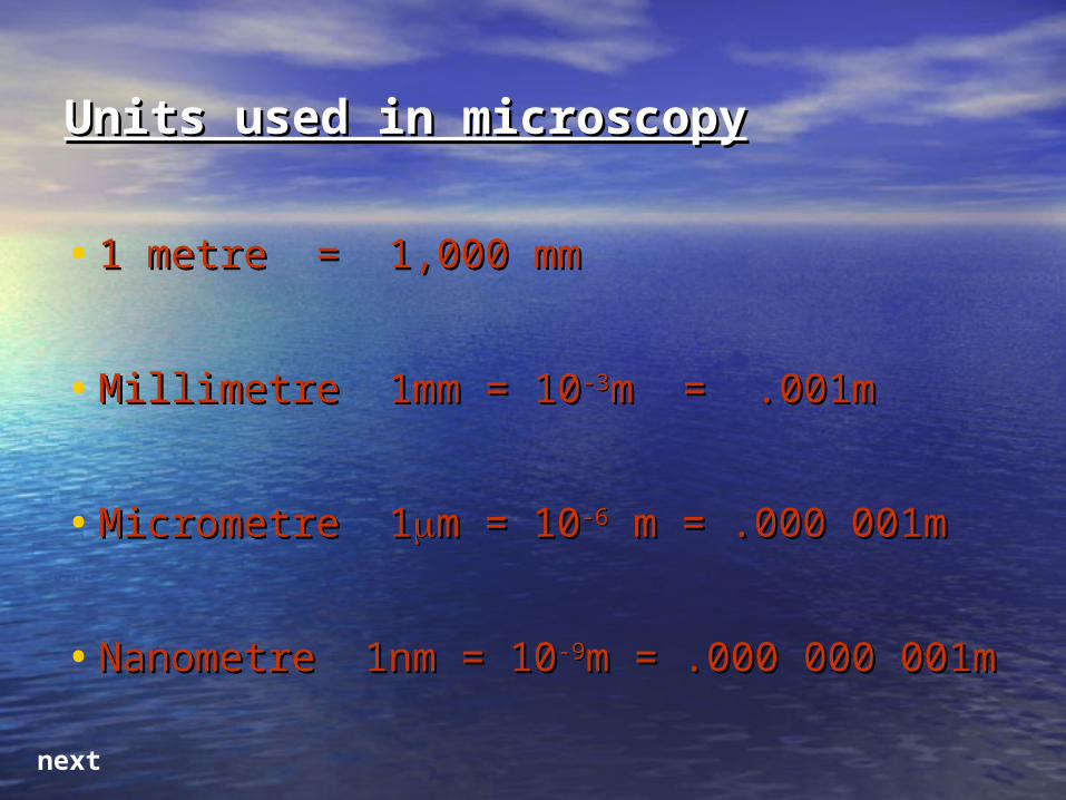
Units used in microscopyUnits used in microscopy
• 1 metre = 1,000 mm1 metre = 1,000 mm
• Millimetre 1mm = 10Millimetre 1mm = 10-3-3m = .001mm = .001m
• Micrometre 1Micrometre 1m = 10m = 10-6-6 m = .000 001m m = .000 001m
• Nanometre 1nm = 10Nanometre 1nm = 10-9-9m = .000 000 m = .000 000 001m001m
next

Light MicroscopeLight Microscope
We will make Temporary Mounts and use Light Microscopes to view our slides

Disadvantages of the Light Disadvantages of the Light MicroscopeMicroscope
Poor Resolution-Due to the long wavelength of light.
Relatively low Magnification

POWER of MICROSCOPESPOWER of MICROSCOPES
•MAGNIFICATION: how many times an image is enlarged.
•RESOLUTION (resolving power): the ability to distinguish two points as being separate.
(resolving power of the naked eye is about 0.2mm).

Electron MicroscopeElectron Microscope
Advantages – Higher Resolution as short wavelength electrons are used instead of long wavelength light.
Higher Magnification.

Transmission E.M.Transmission E.M.
• The specimen is very thinThe specimen is very thin
• May contain ‘artefacts’May contain ‘artefacts’
• Must be viewed in a Must be viewed in a vacuumvacuum
• So samples must be deadSo samples must be dead
• A slow process to build anA slow process to build an
overall 3D viewoverall 3D view
• Complex staining methodComplex staining method
A beam of electrons is focused onto the specimenParts of the specimen absorb electronsThey appear darkOther parts allow electrons to passSo appear light

Scanning E.M.Scanning E.M.
Samples do not need to be as thin as TEMSamples do not need to be as thin as TEM
Beam of electrons does not penetrateBeam of electrons does not penetrate
Same negative points as TEMSame negative points as TEM
- VacuumVacuum
- Dead SamplesDead Samples
- Complex staining usedComplex staining used
Lower resolution than TEMLower resolution than TEM

An Electron Micrograph of An Electron Micrograph of an animal cell.an animal cell.

A TEM image of a plant cellA TEM image of a plant cell

A TEM image of a A TEM image of a ChloroplastChloroplast

A Plant Cell – which type of A Plant Cell – which type of microscope?microscope?

A White Blood Cell – which A White Blood Cell – which Microscope?Microscope?

Which Organelle? Which Which Organelle? Which Microscope?Microscope?

MagnificationMagnification
• How many times bigger the object is How many times bigger the object is made.made.
• The magnification can be worked out The magnification can be worked out by…by…
Size of imageSize of image
Size of objectSize of object

This equation can be This equation can be rearrangedrearranged
Size of Object = Size of Object = Size of ImageSize of Image
MagnificationMagnification
REMEMBER – ALL UNITS MUST BE THE REMEMBER – ALL UNITS MUST BE THE SAME!!SAME!!

UnitsUnits
• 1 millimetre = mm 1 millimetre = mm
• 1 micrometer = 1 micrometer = µm = 1000 times µm = 1000 times smallersmaller
• 1 nanometer = nm = 1 000 000 1 nanometer = nm = 1 000 000 times times smallersmaller

ResolutionResolution
• How clear an image isHow clear an image is
• Resolution depends on whether the Resolution depends on whether the light or electrons can pass between light or electrons can pass between two objects.two objects.

Difference between light and Difference between light and electron microscopeselectron microscopes
• Light microscopes have a low resolution Light microscopes have a low resolution as light has a long wavelengthas light has a long wavelength
• Electron microscopes have a high Electron microscopes have a high resolution as the beam of electrons resolution as the beam of electrons have a short wavelength.have a short wavelength.

The Plasma MembraneThe Plasma Membrane
The fluid mosaic model

Transport Across Transport Across MembranesMembranes
• DiffusionDiffusion
• OsmosisOsmosis
• Active TransportActive Transport

DiffusionDiffusion• Animation: How Diffusion WorksAnimation: How Diffusion Works
• Particles in constant motion – kinetic Particles in constant motion – kinetic energyenergy
• Random motionRandom motion
• Particles bounce off one another and Particles bounce off one another and other particlesother particles
• Passive – the energy is just kineticPassive – the energy is just kinetic

NET movement of particles NET movement of particles down a concentration down a concentration gradient until DYNAMIC gradient until DYNAMIC EQULIBRIUM is reachedEQULIBRIUM is reached

Rate of DiffusionRate of Diffusion
• Fick’s Law.Fick’s Law.
Rate = Rate = surface area x difference in surface area x difference in conc.conc.
thickness of diffusion pathwaythickness of diffusion pathway
QuestionQuestion
• Use Fick’s Law to explain why the Use Fick’s Law to explain why the lungs are an efficient gas exchange lungs are an efficient gas exchange surface.surface.
Facilitated DiffusionFacilitated Diffusion
• Diffusion which happens through Diffusion which happens through specificspecific protein channels.protein channels.
• Water soluble substances can diffuse in Water soluble substances can diffuse in this waythis way
• Read page 55 and 56 of your textbook, Read page 55 and 56 of your textbook, then answer the questions in the green then answer the questions in the green box.box.
Click

OsmosisOsmosis
•The passage of water molecules The passage of water molecules from a region of from a region of higher water higher water potentialpotential to a region of to a region of lower lower water potentialwater potential through a through a partially permeable membranepartially permeable membrane. .
• A more specialised form of diffusion.A more specialised form of diffusion.
Click

Water PotentialWater Potential ΨΨ
• Pure water = water potential of 0 Pure water = water potential of 0 ((ΨΨ = 0) = 0)
• Less water = more concentratedLess water = more concentrated
ΨΨ 0 is the MOST WATER
More concentrated = more negative ΨΨ

Which way will water move?Which way will water move?
Cell A
ΨΨ = -167 = -167
Cell B
ΨΨ = -98 = -98

Which way will water move?Which way will water move?
Cell A
ΨΨ = 0 = 0
Cell B
ΨΨ = -26 = -26

Isotonic = a solution that is the same concentration as the cytoplasm
Now explain these diagrams in terms of water potential.

PlasmolysisPlasmolysis
• photosynthesis elodea animation - photosynthesis elodea animation - Google VideosGoogle Videos
• Look at the tables on page 59 and 60 Look at the tables on page 59 and 60 of your textbook.of your textbook.
• Complete the practical on osmosis.Complete the practical on osmosis.
Related Documents


