The Cell Cycle Picture of animal cell -- http:// www.cellsalive.com/cells/cell_model .htm Picture of plant cell -- http:// www.cellsalive.com/cells/cell_model .htm OR How cells make new cells (Sullivan, J. 2010

The Cell Cycle Picture of animal cell -- Picture of plant.
Dec 28, 2015
Welcome message from author
This document is posted to help you gain knowledge. Please leave a comment to let me know what you think about it! Share it to your friends and learn new things together.
Transcript
The Cell Cycle
Picture of animal cell -- http://www.cellsalive.com/cells/cell_model.htm
Picture of plant cell -- http://www.cellsalive.com/cells/cell_model.htm
OR
How cells make new cells
(Sullivan, J. 2010)
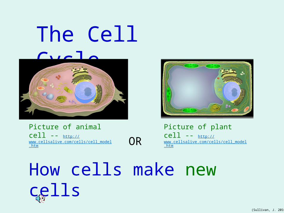
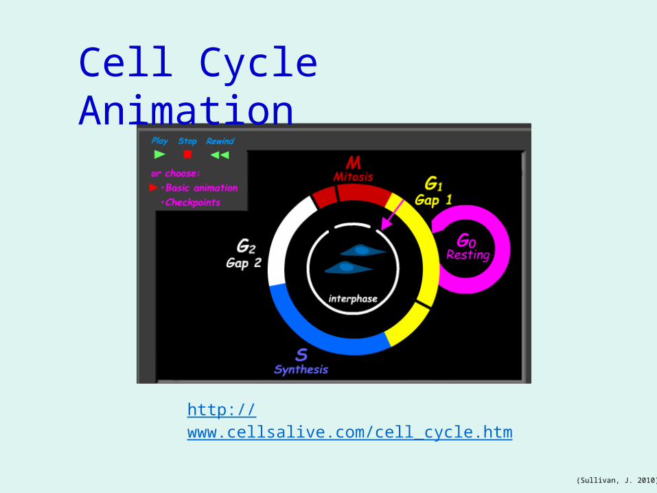
Two major parts of the cell cycle:
Interphase – resting and growing
Mitosis – dividing http://www.biology.arizona.edu/cell_bio/
activities/cell_cycle/cell_cycle.html
http://micro.magnet.fsu.edu/micro/gallery/mitosis/mitosis.html
(University of Arizona, 2005)
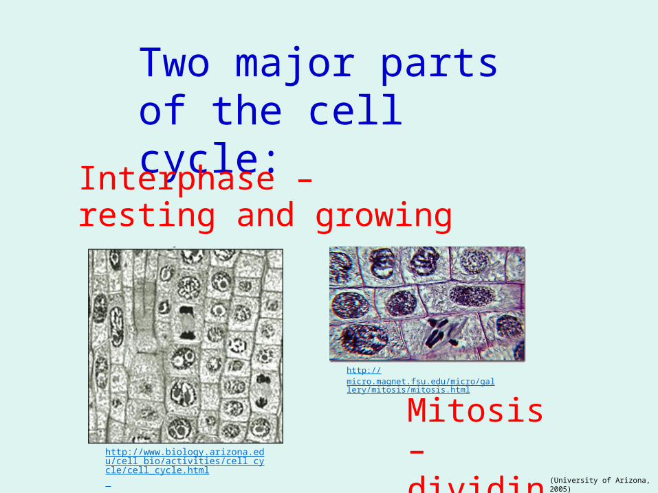
Interphase usually lasts 10-30 hours in mammals
Three stages: GAP 1 (G1) – growing Cells increase in size, produce RNA and synthesize proteins,
S Phase (S) – synthesizing Duplication of Chromosomes or DNA, forming chromatids connected with centromere
GAP 2 (G2) – checkingCell continues to grow and synthesize proteins at the end it double checks before starting mitosis
http://education.kings.edu/dsmith/Lesson%206.html
http://education.kings.edu/dsmith/Lesson%206.html
(Sullivan, J. 2010;
University of Arizona, 2005)
http://www.cellsalive.com/cell_cycle.htm
Cell Cycle Animation
(Sullivan, J. 2010)
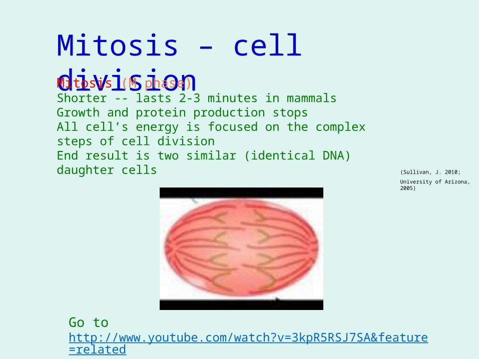
Mitosis – cell division Mitosis (M phase) Shorter -- lasts 2-3 minutes in mammalsGrowth and protein production stopsAll cell’s energy is focused on the complex steps of cell division End result is two similar (identical DNA) daughter cells
Go to http://www.youtube.com/watch?v=3kpR5RSJ7SA&feature=related
(Sullivan, J. 2010;
University of Arizona, 2005)
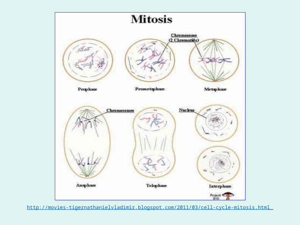
http://movies-tigernathanielvladimir.blogspot.com/2011/03/cell-cycle-mitosis.html
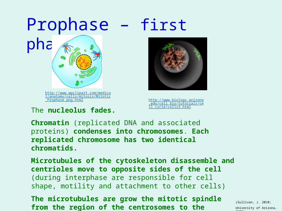
Prophase – first phase
The nucleolus fades.
Chromatin (replicated DNA and associated proteins) condenses into chromosomes. Each replicated chromosome has two identical chromatids.
Microtubules of the cytoskeleton disassemble and centrioles move to opposite sides of the cell(during interphase are responsible for cell shape, motility and attachment to other cells)
The microtubules are grow the mitotic spindle from the region of the centrosomes to the centrioles.
http://www.biology.arizona.edu/cell_bio/tutorials/cell_cycle/cells3.html
http://www.wpclipart.com/medical/anatomy/cells/mitosis/Mitotic_Prophase.png.html
(Sullivan, J. 2010;
University of Arizona, 2005)
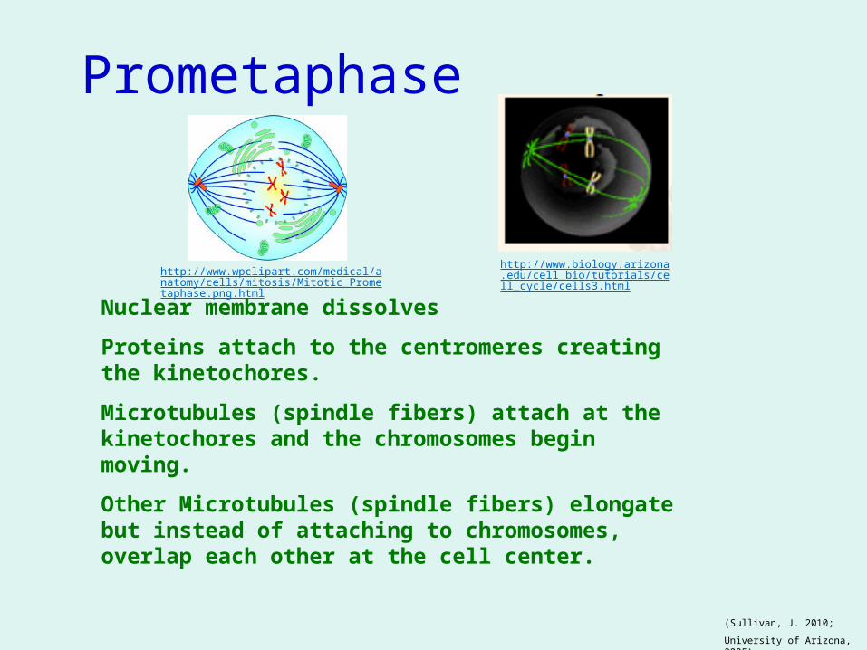
Nuclear membrane dissolves
Proteins attach to the centromeres creating the kinetochores.
Microtubules (spindle fibers) attach at the kinetochores and the chromosomes begin moving.
Other Microtubules (spindle fibers) elongate but instead of attaching to chromosomes, overlap each other at the cell center.
Prometaphase
http://www.biology.arizona.edu/cell_bio/tutorials/cell_cycle/cells3.htmlhttp://www.wpclipart.com/medical/anatomy/ce
lls/mitosis/Mitotic_Prometaphase.png.html
(Sullivan, J. 2010;
University of Arizona, 2005)
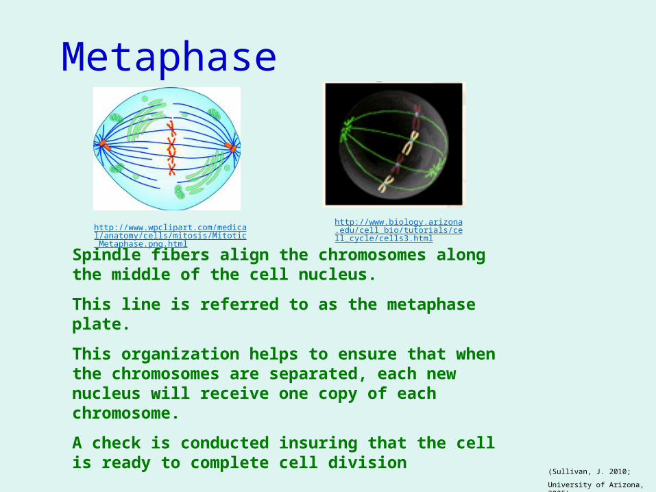
Spindle fibers align the chromosomes along the middle of the cell nucleus.
This line is referred to as the metaphase plate.
This organization helps to ensure that when the chromosomes are separated, each new nucleus will receive one copy of each chromosome.
A check is conducted insuring that the cell is ready to complete cell division
Metaphase
http://www.wpclipart.com/medical/anatomy/cells/mitosis/Mitotic_Metaphase.png.html
http://www.biology.arizona.edu/cell_bio/tutorials/cell_cycle/cells3.html
(Sullivan, J. 2010;
University of Arizona, 2005)
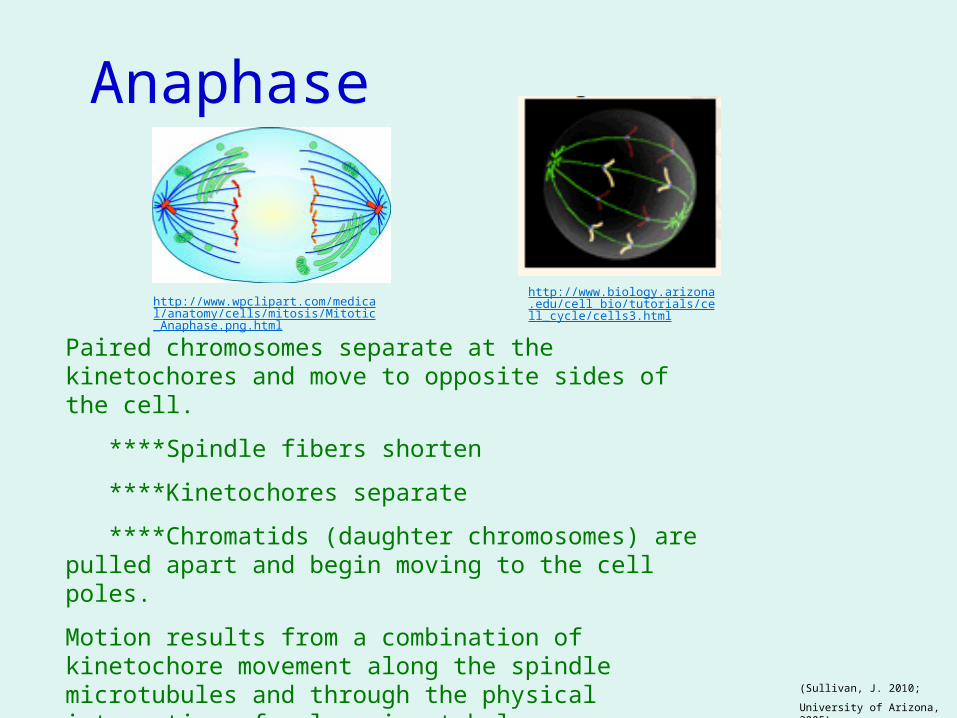
Paired chromosomes separate at the kinetochores and move to opposite sides of the cell.
****Spindle fibers shorten
****Kinetochores separate
****Chromatids (daughter chromosomes) are pulled apart and begin moving to the cell poles.
Motion results from a combination of kinetochore movement along the spindle microtubules and through the physical interaction of polar microtubules.
Anaphase
http://www.wpclipart.com/medical/anatomy/cells/mitosis/Mitotic_Anaphase.png.html
http://www.biology.arizona.edu/cell_bio/tutorials/cell_cycle/cells3.html
(Sullivan, J. 2010;
University of Arizona, 2005)
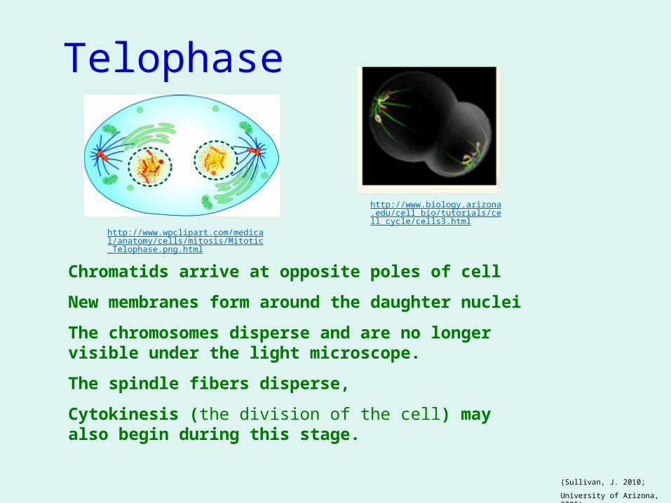
Chromatids arrive at opposite poles of cell
New membranes form around the daughter nuclei
The chromosomes disperse and are no longer visible under the light microscope.
The spindle fibers disperse,
Cytokinesis (the division of the cell) may also begin during this stage.
Telophase
http://www.wpclipart.com/medical/anatomy/cells/mitosis/Mitotic_Telophase.png.html
http://www.biology.arizona.edu/cell_bio/tutorials/cell_cycle/cells3.html
(Sullivan, J. 2010;
University of Arizona, 2005)
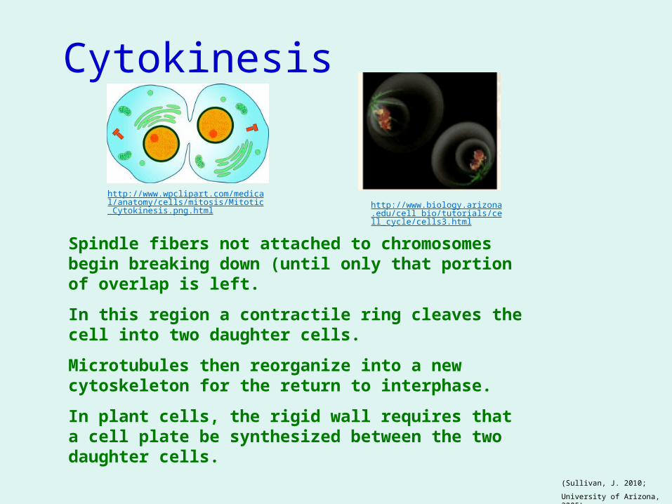
Spindle fibers not attached to chromosomes begin breaking down (until only that portion of overlap is left.
In this region a contractile ring cleaves the cell into two daughter cells.
Microtubules then reorganize into a new cytoskeleton for the return to interphase.
In plant cells, the rigid wall requires that a cell plate be synthesized between the two daughter cells.
Cytokinesis
http://www.wpclipart.com/medical/anatomy/cells/mitosis/Mitotic_Cytokinesis.png.html http://www.biology.arizona.edu/cell
_bio/tutorials/cell_cycle/cells3.html
(Sullivan, J. 2010;
University of Arizona, 2005)
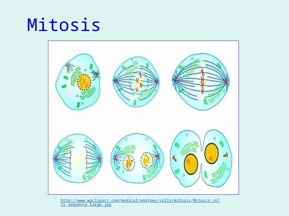
http://www.wpclipart.com/medical/anatomy/cells/mitosis/Mitosis_cells_sequence_large.jpg
Mitosis
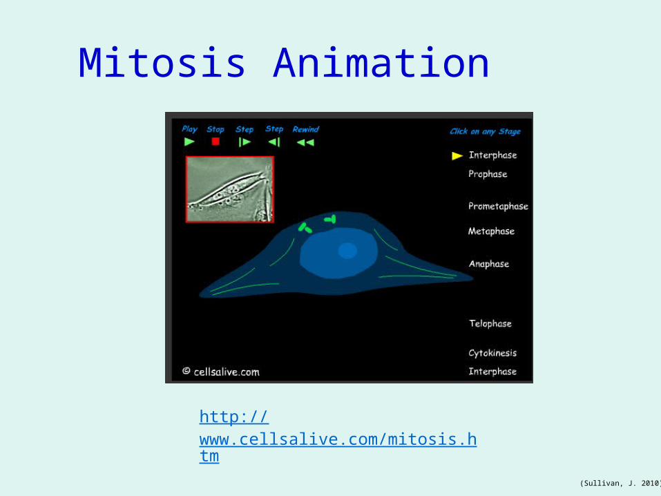
Mitosis Animation
http://www.cellsalive.com/mitosis.htm
(Sullivan, J. 2010)
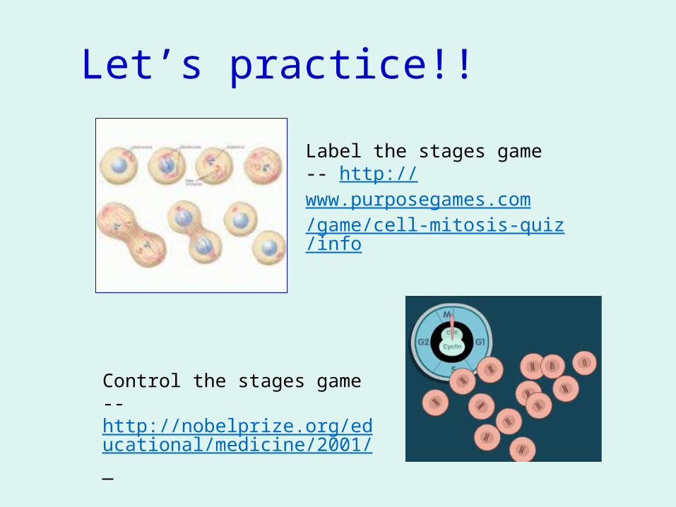
Let’s practice!!
Control the stages game -- http://nobelprize.org/educational/medicine/2001/
Label the stages game -- http://www.purposegames.com/game/cell-mitosis-quiz/info
For your viewing pleasure!The Cell Rap-- here are the lyrics, you add the tunes!! http://bizzarroworld.homestead.com/songs.html
These gals sing their hearts out -- www.youtube.com/watch?v=sa0uWNlqKyc&feature=related%20%20
Follow Bob on his most epic life journey-- http://www.youtube.com/watch?v=2xeEdn8EXyk&feature=related%20%20
Grading!
your grade will also be based on the application of this information in the Onion Root Tip Lab, to be completed in the future. http://www.biology.arizona.edu/cell_bio/activities/cell_cycle/cell_cycle.html(You will need to identify the different stages of the cell cycle.)
From the Website: Cellsalive -- http://www.cellsalive.com
Cells Alive Mitosis Crossword Puzzle -- http://www.cellsalive.com/puzzles/mitosis/index.html
Complete Online quiz: http://quizstar.4teachers.org/ CRA Biology classes -- The Cell Cycle Posttest
References
Angier, Natalie (2002). Lesson 6: mitosis. Retrieved February 26, 2011 from: http://education.kings.edu/dsmith/Lesson%206.html Davidson, Michael M. (2004) Molecular expressions. Retrieved February 26, 2011 from: The University of Florida http://micro.magnet.fsu.edu/micro/gallery/mitosis/mitosis.html Nobel Prize.Org. Games (2011) Control of the cell cycle. Retrieved February 26, 2011 from: http://nobelprize.org/educational/medicine/2001/ RierroTheCoolest (2008) Cell mitosis game. Retrieved February 26, 2011 from: http://www.purposegames.com/game/cell-mitosis-quiz/info Srokes, Ms. (2007) The cell cycle featuring: mitosis. Retrieved February 26, 2011 from: http://www.youtube.com/watch?v=3kpR5RSJ7SA&feature=related
References
Sullivan, Jim. (2010) Cells alive. Retrieved February 26, 2011 from: http://www.cellsalive.com/cells/cell_model.htm The University of Arizona (2005). The Biology project. Retrieved February 26, 2011 from: http://www.biology.arizona.edu/cell_bio/activities/cell_cycle/cell_cycle.html
Valdimir, Nathanial (2011) The cell cycle mitosis. (Retrieved February 26, 2011 from: http://movies-tigernathanielvladimir.blogspot.com/2011/03/cell-cycle-mitosis.html
WP Clipart (n.d.). Medical section: mitosis. Retrieved February 26, 2011 from: http://www.wpclipart.com/medical/anatomy/cells/mitosis/
Related Documents