© 2013. Published by The Company of Biologists Ltd | Development (2013) 140, 4471-4479 doi:10.1242/dev.090613 4471 ABSTRACT Fertilization is the process by which eggs and spermatozoa interact, achieve mutual recognition, and fuse to create a zygote, which then develops to form a new individual, thus allowing for the continuity of a species. Despite numerous studies on mammalian fertilization, the molecular mechanisms underpinning the fertilization event remain largely unknown. However, as I summarize here, recent work using both gene-manipulated animals and in vitro studies has begun to elucidate essential sperm and egg molecules and to establish predictive models of successful fertilization. KEY WORDS: Fertilization, Izumo1, Acrosome reaction, Eggs, Hyperactivation, Spermatozoa Introduction Our individual body has a limited lifetime. However, through fertilization we are able to continue life as a species. The role of spermatozoa is to fertilize eggs. However, mammalian spermatozoa cannot accomplish this task when ejaculated. They must first undergo a physiological change called capacitation and a subsequent morphological change known as the acrosome reaction in the female reproductive tract. Spermatozoa also harbor the ability to migrate into the oviduct, where they interact with and subsequently fuse with the egg. A number of factors that contribute to sperm-egg interactions have been identified, based on observations using enzyme inhibitors and antibodies in in vitro fertilization systems (Box 1). This research led to the conclusion that various sperm enzymes within the acrosome dissolved the egg components and that various membrane proteins were used for binding with eggs. However, recent experiments using gene disruption of these factors did not result in an infertile phenotype, suggesting that they are not essential for fertilization, although they may indeed play a role during the fertilization event. By contrast, using in vivo gene- targeting experiments, a number of proteins have unexpectedly emerged as being essential factors for fertilization. In this Primer, I discuss the factors that have been implicated in the various stages of fertilization, ranging from sperm capacitation and migration to sperm-egg fusion. Newly arising views of mammalian fertilization are reviewed and compared with previously postulated models. The nature of eggs Ovaries are endowed at birth with a fixed number of oocytes enclosed in primordial follicles. This number declines as a result of ovulation and atresia during the reproductive life of the female (Faddy, 2000). Oocytes are arrested in the dictyate stage of first meiotic division. Over time, cohorts of oocytes enter into a growth phase and become among the largest cells in the body, their diameters reaching about 80 μm and 100 μm in mouse and human, PRIMER Center for Genetic Analysis for Biological Responses Research Institute for Microbial Diseases Osaka University, Yamadaoka 3-1, Suita, Osaka 565-0871, Japan. *Author for correspondence ([email protected]) respectively. During growth, eggs form an extracellular matrix called the zona pellucida (ZP) by secreting glycoproteins (Fig. 1A). The ZP of human eggs consists of four ZP glycoproteins (ZP1 to ZP4), whereas that of the mouse egg consists of three ZP proteins (ZP1 to ZP3; mouse Zp4 is a pseudogene). Full oocyte growth must be supported by surrounding granulosa cells, which proliferate and form multiple layers of cumulus cells that surround the ovulated egg (Fig. 1A). Cumulus cells support fertilization, and in vitro fertilization can be achieved more efficiently with them than without them (Jin et al., 2011; Tokuhiro et al., 2012). The molecular basis of this observation remains obscure; however, a few studies have addressed the function of cumulus cells (Oren-Benaroya et al., 2008; Shimada et al., 2008). Following meiotic maturation, eggs are ovulated in preparation for fertilization. After ovulation, the eggs are picked up by adhesion between the extracellular matrix of the cumulus cells and oviductal cells of the oviductal fimbriae and are subsequently transferred into the oviduct (Talbot et al., 2003). The eggs are then transported to the ampulla portion of the oviduct, where they await spermatozoa for fertilization. However, there is only a short ‘fertile window’, which is less than a day after ovulation in humans (Wilcox et al., 1995) and a few hours in the mouse, during which fertilization can successfully occur. The nature of spermatozoa In human testes, ~1000 spermatozoa are produced per second (Amann and Howards, 1980), although the reason why mammalian males produce so many spermatozoa to fertilize so few eggs is not understood. Spermatozoa produced in the testes are transferred to epididymides, where they receive various proteins (Busso et al., 2007), probably in part through a structure called the epididymosome (Frenette et al., 2010). The spermatozoa are Box 1. Studying fertilization in vitro In vitro fertilization (IVF) requires different elements for different species. In mice, eggs are usually collected from the oviduct after treatment with hormones that induce super ovulation. The eggs are introduced into a tiny drop of IVF medium on a dish covered by paraffin oil and cultivated under 5% CO 2 in air. Spermatozoa from mice are normally prepared by squeezing them out from an opening made in the epididymis, followed by suspension in IVF medium; alternatively, ejaculated spermatozoa are utilized in larger animals after washing with medium. The spermatozoa are introduced into the egg culture drop at a final concentration of ~1×10 5 spermatozoa/ml (note that IVF requires a large number of spermatozoa compared with fertilization in vivo, in which only a few spermatozoa are required per egg). The success of fertilization can be assessed by observing: (1) spermatozoa inside the ZP; (2) the formation of pronuclei; or (3) the formation of two-cell embryos. The eggs fertilized by IVF can also be transferred into oviducts of pseudo-pregnant females to assess the developmental outcome of these embryos. When antibodies or inhibitors added to the IVF medium successfully inhibit fertilization, the factors that these antibodies and inhibitors target have traditionally have been viewed as fertilization-related factors. The cell biology of mammalian fertilization Masaru Okabe* Development

Welcome message from author
This document is posted to help you gain knowledge. Please leave a comment to let me know what you think about it! Share it to your friends and learn new things together.
Transcript

© 2013. Published by The Company of Biologists Ltd | Development (2013) 140, 4471-4479 doi:10.1242/dev.090613
4471
ABSTRACTFertilization is the process by which eggs and spermatozoa interact,achieve mutual recognition, and fuse to create a zygote, which thendevelops to form a new individual, thus allowing for the continuity ofa species. Despite numerous studies on mammalian fertilization, themolecular mechanisms underpinning the fertilization event remainlargely unknown. However, as I summarize here, recent work usingboth gene-manipulated animals and in vitro studies has begun toelucidate essential sperm and egg molecules and to establishpredictive models of successful fertilization.
KEY WORDS: Fertilization, Izumo1, Acrosome reaction, Eggs,Hyperactivation, Spermatozoa
IntroductionOur individual body has a limited lifetime. However, throughfertilization we are able to continue life as a species. The role ofspermatozoa is to fertilize eggs. However, mammalian spermatozoacannot accomplish this task when ejaculated. They must firstundergo a physiological change called capacitation and a subsequentmorphological change known as the acrosome reaction in the femalereproductive tract. Spermatozoa also harbor the ability to migrateinto the oviduct, where they interact with and subsequently fuse withthe egg. A number of factors that contribute to sperm-egginteractions have been identified, based on observations usingenzyme inhibitors and antibodies in in vitro fertilization systems(Box 1). This research led to the conclusion that various spermenzymes within the acrosome dissolved the egg components andthat various membrane proteins were used for binding with eggs.However, recent experiments using gene disruption of these factorsdid not result in an infertile phenotype, suggesting that they are notessential for fertilization, although they may indeed play a roleduring the fertilization event. By contrast, using in vivo gene-targeting experiments, a number of proteins have unexpectedlyemerged as being essential factors for fertilization. In this Primer, Idiscuss the factors that have been implicated in the various stages offertilization, ranging from sperm capacitation and migration tosperm-egg fusion. Newly arising views of mammalian fertilizationare reviewed and compared with previously postulated models.
The nature of eggsOvaries are endowed at birth with a fixed number of oocytesenclosed in primordial follicles. This number declines as a result ofovulation and atresia during the reproductive life of the female(Faddy, 2000). Oocytes are arrested in the dictyate stage of firstmeiotic division. Over time, cohorts of oocytes enter into a growthphase and become among the largest cells in the body, theirdiameters reaching about 80 μm and 100 μm in mouse and human,
PRIMER
Center for Genetic Analysis for Biological Responses Research Institute forMicrobial Diseases Osaka University, Yamadaoka 3-1, Suita, Osaka 565-0871,Japan.
*Author for correspondence ([email protected])
respectively. During growth, eggs form an extracellular matrixcalled the zona pellucida (ZP) by secreting glycoproteins (Fig. 1A).The ZP of human eggs consists of four ZP glycoproteins (ZP1 toZP4), whereas that of the mouse egg consists of three ZP proteins(ZP1 to ZP3; mouse Zp4 is a pseudogene).
Full oocyte growth must be supported by surrounding granulosacells, which proliferate and form multiple layers of cumulus cellsthat surround the ovulated egg (Fig. 1A). Cumulus cells supportfertilization, and in vitro fertilization can be achieved moreefficiently with them than without them (Jin et al., 2011; Tokuhiroet al., 2012). The molecular basis of this observation remainsobscure; however, a few studies have addressed the function ofcumulus cells (Oren-Benaroya et al., 2008; Shimada et al., 2008).
Following meiotic maturation, eggs are ovulated in preparationfor fertilization. After ovulation, the eggs are picked up by adhesionbetween the extracellular matrix of the cumulus cells and oviductalcells of the oviductal fimbriae and are subsequently transferred intothe oviduct (Talbot et al., 2003). The eggs are then transported to theampulla portion of the oviduct, where they await spermatozoa forfertilization. However, there is only a short ‘fertile window’, whichis less than a day after ovulation in humans (Wilcox et al., 1995) anda few hours in the mouse, during which fertilization can successfullyoccur.
The nature of spermatozoaIn human testes, ~1000 spermatozoa are produced per second(Amann and Howards, 1980), although the reason whymammalian males produce so many spermatozoa to fertilize sofew eggs is not understood. Spermatozoa produced in the testes aretransferred to epididymides, where they receive various proteins(Busso et al., 2007), probably in part through a structure called theepididymosome (Frenette et al., 2010). The spermatozoa are
Box1. Studying fertilization in vitroIn vitro fertilization (IVF) requires different elements for different species.In mice, eggs are usually collected from the oviduct after treatment withhormones that induce super ovulation. The eggs are introduced into atiny drop of IVF medium on a dish covered by paraffin oil and cultivatedunder 5% CO2 in air. Spermatozoa from mice are normally prepared bysqueezing them out from an opening made in the epididymis, followedby suspension in IVF medium; alternatively, ejaculated spermatozoa areutilized in larger animals after washing with medium. The spermatozoaare introduced into the egg culture drop at a final concentration of ~1×10⁵
spermatozoa/ml (note that IVF requires a large number of spermatozoacompared with fertilization in vivo, in which only a few spermatozoa arerequired per egg).
The success of fertilization can be assessed by observing: (1)spermatozoa inside the ZP; (2) the formation of pronuclei; or (3) theformation of two-cell embryos. The eggs fertilized by IVF can also betransferred into oviducts of pseudo-pregnant females to assess thedevelopmental outcome of these embryos. When antibodies or inhibitorsadded to the IVF medium successfully inhibit fertilization, the factors thatthese antibodies and inhibitors target have traditionally have beenviewed as fertilization-related factors.
The cell biology of mammalian fertilizationMasaru Okabe*
Dev
elop
men
t

4472
mature (Fig. 1B), yet they remain unable to fertilize eggs.According to Yanagimachi, the sperm plasma membrane remains‘biologically frozen’ until spermatozoa leave the male’s body andbegin the ‘defrosting’ process, known as capacitation, which isnecessary for spermatozoa to be fertilization competent(Yanagimachi, 1994).
During capacitation, various physiological changes occur in thehead, acrosome and tail of spermatozoa, which biologically ‘defrost’spermatozoa and prepare them for fertilization (Fig. 1B). It is knownthat spermatozoa in suspension are heterogeneous and, as such,when a sperm suspension is judged to be capacitated, a certainpercentage of spermatozoa in the same suspension are alreadydegrading. Numerous papers discuss the phosphorylation of spermproteins during capacitation, but no clear mechanisms leading tocapacitation have been elucidated (Bailey, 2010; Visconti et al.,2011). This may be due, at least in part, to the heterogeneity ofspermatozoa.
Glucose is known to be essential for successful capacitation. It notonly serves as an energy source allowing spermatozoa to swim, butalso functions to enable spermatozoa to fertilize eggs (Goodson etal., 2012; Okabe et al., 1986). Cholesterol, bicarbonate, intracellularCa2+ and many other factors are also involved in capacitation(Bailey, 2010; Evans, 2012; Florman and Ducibella, 2006;Yanagimachi, 1994); however, the precise molecular mechanismsunderlying their action are yet to be clarified.
Once capacitated, spermatozoa demonstrate a vigorous swimmingpattern called hyperactivation, a movement considered to givespermatozoa the strong thrusting power that allows them topenetrate the ZP. It is characterized by asymmetrical flagellarbeating consisting of pro- and anti-hook bends accompanied by anincrease in cytoplasmic Ca2+. The sperm-specific CatSper ionchannels have been suggested to control the intracellular Ca2+
concentration and, thereby, the swimming behavior of spermatozoa
(Chang and Suarez, 2011). CatSper1 to 4 are essential forfertilization in mouse (Qi et al., 2007). Furthermore, the CatSperchannel seems to be activated by progesterone in humanspermatozoa (Brenker et al., 2012; Lishko et al., 2011), which isinteresting from the sperm-egg interaction viewpoint becausecumulus cells are known to produce progesterone. However,progesterone is not reported to activate mouse spermatozoa.
After capacitation, the final step in sperm development prior tofertilization is the acrosome reaction (Fig. 1B). The acrosome, whichis a subcellular organelle found at the apical tip of the sperm head,is filled with a variety of lytic enzymes and ZP-binding proteins.Through poorly understood mechanisms, which may involveSNARE proteins (De Blas et al., 2005), the plasma membrane andouter acrosomal membrane fuse and acrosomal contents areexocytosed into the environment (Yanagimachi, 1994). This process,referred to as the acrosome reaction, renders spermatozoa able topenetrate the ZP.
Factors regulating fertilizationFertilization is a complicated multi-step event, involving maturationand development of the spermatozoa and eggs, followed by spermmigration into the oviduct, and ending with sperm-egg interactionand then fusion. As such, various factors that play a role in each ofthese events are emerging.
The classical models of fertilization were mainly focused onsperm-zona binding and the zona-induced acrosome reaction, basedon in vitro experiments, and many factors were postulated to berequired for fertilization. With time, however, the genes encodingthese factors were cloned and their roles were investigated by genedisruption experiments in mice (Box 2). Surprisingly, almost all ofthem showed no or a very minor phenotype in fertilization (Ikawaet al., 2010); although they may play a role, the knockout of theseindividual factors did not affect fertilization. In addition, various
PRIMER Development (2013) doi:10.1242/dev.090613
Egg
Acrosomalcap region
PM
Acrosome
� Phospholipase Cδ4, SNARE proteins etc.
OAMIAM
Nucleus
Acrosome reaction
Equatorialsegment
A B Capacitation
� Albumin-mediated cholesterol efflux� Removal of decapacitation factor?� Interaction with cumulus cells through cytokines?� Ca2+ influx FF� Acrosomal pH F� Soluble adenylyl cyclase F� Protein kinase A and C F� Tyrosine phosphorylation etc. F Hyaluronic acid
matrix
Zona pellucida Cumuluscells
Perivitellinespace
Polar body
Fig.1. The features of mammalian eggs and spermatozoa. (A)Ovulated eggs (yellow) are surrounded by small cumulus cells embedded in extracellularmaterial (blue) that consists mainly of hyaluronic acid. The cumulus layer is separated from the egg by the zona pellucida (gray), which is made up of zonapellucida proteins (ZP1-3 in mouse and ZP1-4 in human). (B)Physiological and morphological changes in spermatozoa required for fertilization. Whenspermatozoa are deposited in the female reproductive tract or suspended in in vitro fertilization medium, a number of changes start to occur that involve thephosphorylation of various proteins, activation of PKA and PKC, removal of cholesterol from the membrane and elevation of the intracellular Ca2+ level. It is notknown whether interaction with cumulus cells is beneficial for some of these changes or if the removal of an inhibitory or ‘decapacitation’ factor is involved.Sperm also undergo a morphological change called the acrosome reaction, during which various enzymes and proteins are released from the acrosome. Onlyacrosome-reacted spermatozoa can penetrate and fuse with eggs. The acrosomal vesicle (green) is surrounded by the outer acrosomal membrane (OAM) andthe inner acrosomal membrane (IAM). The acrosome contains both instantly and slowly released proteins and its contents are exocytosed during the acrosomereaction in which the sperm plasma membrane (PM) and the OAM fuse in multiple places within the acrosomal cap area. The acrosome reaction is thought toinvolve phospholipase C (PLCδ4) and SNARE proteins and is generally considered to be a good indicator for the completion of capacitation. Once capacitated,spermatozoa demonstrate a vigorous swimming pattern called hyperactivation, which involves CatSper ion channels.
Dev
elop
men
t

genes essential for successful fertilization began emerging from suchgene disruption experiments. Most of these gene-disrupted mouselines shared common phenotypes, showing: (1) no migration into theoviduct in vivo with aberrant zona-binding ability in vitro; or (2) nofusing ability (Table 1, Class I and II). Interestingly, all thespermatozoa from class I mouse lines lacked ADAM3 (Shamsadinet al., 1999) without exception, suggesting that ADAM3 plays a keyrole in fertilization in the mouse.
Below, I highlight the various factors that have been implicatedin the fertilization process, placing an emphasis on those for whichan essential role has been identified based on gene disruptionapproaches.
Factors regulating sperm migrationAll of the 11 Class I lines of gene-disrupted males shown in Table1 exhibited the same defect of sperm migration into oviduct, a defectwhich would not have been identified by in vitro studies. In mice,the uterus and oviduct meet in a structure called the uterotubaljunction (UTJ), which significantly reduces the number ofspermatozoa reaching eggs. The flow in the UTJ is bidirectional.After coitus, spermatozoa must move up to reach the eggs and, aftergamete fusion, embryos resulting from fertilized eggs must movedown the UTJ for implantation into the uterus. How the UTJregulates this bidirectional flow is not known. It is clear, however,that sperm migration is not regulated by simple opening and closingof the UTJ entrance in mouse. A testis-specific molecular chaperone,calmegin, is one of the essential genes required for spermatozoa tomigrate into oviduct (Ikawa et al., 1997). Using chimeric mice thatejaculate both wild-type and GFP-tagged, calmegin-disruptedspermatozoa, it was observed that only wild-type spermatozoamigrated into the oviduct, whereas the equally motile calmegin-disrupted spermatozoa remained in the uterus (Nakanishi et al.,2004). These observations indicated that entry into the oviduct isregulated by a recognition system between an individualspermatozoon and the UTJ, but the molecular mechanismunderpinning this system remains to be determined.
The zona-binding ability of spermatozoaThe sperm-zona interaction is regulated almost in an all or nonemanner, as illustrated in Fig. 2. In vitro, many spermatozoa can bind(or attach) to the ZP of unfertilized eggs but they cannot bind to two-cell-stage eggs, suggesting that sperm-zona binding might be acontrolled event. The loss of zona-binding ability, which wascommonly observed in Class I knockout mouse lines, could easilybe assessed by mixing spermatozoa with cumulus-free eggs, andinitially no one doubted that the infertility was caused by the loss ofzona-binding ability. However, this assumption was not correct asinitially thought.
Mice deficient for the testis-specific molecular chaperone PDILTproduce spermatozoa that, based on in vitro experiments, exhibitimpaired zona-binding ability (Tokuhiro et al., 2012). After crossingwith a transgenic fluorescently tagged sperm line (Hasuwa et al.,2010), the failure of sperm migration into the oviduct was indicatedby live imaging. However, when the spermatozoa were depositeddirectly into the ampulla by bypassing the UTJ, the eggs could befertilized (Fig. 2). Likewise, spermatozoa from infertile Adam1a−/−
mice, which also show impaired zona-binding ability in vitro, couldfertilize eggs that were covered by cumulus cells (Nishimura et al.,2004). It is not known what the true sperm-zona interaction entailsbut these studies suggest that the so-called ‘zona-binding ability’ asmeasured by binding to cumulus-free eggs in vitro appears to bedispensable for fertilization in vivo (Fig. 2).
Many studies have also postulated the involvement ofcarbohydrates in sperm-zona binding. It must be noted, however,that many of these postulations were based on in vitro studies, andan essential role for such carbohydrate residues in vivo has not yetbeen confirmed. The enzymatic removal of terminal Gal (Bleil andWassarman, 1988) or GlcNAc (Shur and Hall, 1982) residues fromeggs, for example, abolished the ability of ZP3 to inhibit spermbinding. However, gene-disrupted mice lacking these residues ontheir zonae were fertile (Asano et al., 1997; Thall et al., 1995),suggesting that these residues are not essential per se forfertilization. Fucose in Lewis X- and A-containing glycans wasalso indicated to play a role in sperm-zona binding (Kerr et al.,2004), but mice lacking the corresponding fucosyltransferase(Fut9) were fertile (Kudo et al., 2004). A role for mannose presenton glycans has also been proposed (Cornwall et al., 1991) but hasbeen negated by N-glycanase treatment (Florman and Wassarman,1985). In vitro experiments have also ascribed sperm bindingactivity to O-linked oligosaccharide side chains attached to Ser332and Ser334 of ZP3 (Chen et al., 1998). However, transgenic miceharboring mutations in Ser332 and 334 were fertile (Liu et al.,1995). Moreover, glycosylation was not observed on these residueson native ZP proteins (Boja et al., 2003). Furthermore, disruptionof the ZP3-binding protein SP56 (ZP3R – Mouse GenomeInformatics) on spermatozoa (Bookbinder et al., 1995) resulted inalmost no phenotype in fertilization (Muro et al., 2012). Finally,enzymes that make core glycans have also been disrupted, andeggs lacking complex and hybrid N-glycans as well as core-1-derived O-glycans were fertilized normally (Shi et al., 2004;Williams et al., 2007). The hypothesis that carbohydrates play animportant role in sperm-egg interaction thus requires furthersupport.
4473
PRIMER Development (2013) doi:10.1242/dev.090613
Box2. Potential limitations of knockout approachesThere are two main drawbacks to gene-targeting approaches. The firstoccurs when the knockout mice show a minor or no phenotype. As amatter of fact, not all genes are functionally indispensable. If the functionof a gene is minor, the knockout mice may not show a strong phenotype,therefore this gene would not be considered a good candidate forknockout-based experimentation. Furthermore, the issue of redundancycan also complicate interpretation of gene-targeting experiments, as theknockout of one factor could be compensated for by other similar factors.It is also known that some genes require the disruption of other gene(s)as a prerequisite to show a phenotype (Rudnicki et al., 1993); some geneproducts may interact together and knockout of both/all interacting factorsmight be needed to observe a phenotype. The factors listed in Table2,which showed almost no phenotype after knockout, might fall into theabove category. A second potential drawback relates to the interpretationof the phenotype. For example, fertilin (an ADAM1b/ADAM2 heterodimer)has been depicted as a sperm-egg fusion protein in many textbooks(Blobel et al., 1992) and this was confirmed by the disruption of Adam2(Cho et al., 1998), resulting in infertile males. However, when fertilinknockout mice were produced by eliminating Adam1b (the second subunitof the fertilin heterodimer) the males were fertile (Nishimura et al., 2004).Infertility in the Adam2-deficient/fertilin knockout mice was subsequentlyattributed to the loss of the testicular ADAM1a/ADAM2 protein dimer,which led to the absence of ADAM3 (a protein that does play an essentialrole in fertilization) from the spermatozoa.
Thus, the interpretation of a knockout phenotype is not always simple.Moreover, the effect of gene disruption might originate from anunexpected route. This includes the unintentional elimination ofmicroRNAs along with the gene of interest (Osokine et al., 2008). Inother cases, the gene manipulation may affect other genes located nearthe intended target and confound the interpretation of phenotypes fromalleles designed to be simple null mutations (Olson et al., 1996).Therefore, caution is required when interpreting knockout results.
Dev
elop
men
t

4474
A recent view that has been verified by gene-manipulated animalsis that the zona loses its affinity for spermatozoa when ovastacin(also known as astacin-like metalloendopeptidase), an enzymecontained in egg cortical granules (Burkart et al., 2012), cleaves ZP2after fertilization (Bleil et al., 1981; Gahlay et al., 2010). In addition,it has been suggested that liver-originated circulating glycoproteinfetuin-B is essentially required to inhibit premature cleavage of ZP2by inhibiting ovastacin (Dietzel et al., 2013).
Together, these studies have shown that various sperm-zonainteractions can occur but that many of these may not be absolutelyrequired for successful fertilization. We also must remember that the‘sperm-zona binding’ described above may not reflect the realsperm-egg interaction required for fertilization in vivo, as shown inFig. 2.
Anatomical or behavioral differences between species guaranteethat a male and a female from two different species do not normallymate. Nevertheless, taxon-specific zona penetration has beendemonstrated (Rankin et al., 2003). However, if spermatozoa do notrequire a zona-binding ability for fertilization, how can theydistinguish the ZP of the same species? One can anticipate that there
is an as yet unclarified affinity between the cumulus-covered eggsand spermatozoa. We can further speculate that the same moleculeresponsible for zona binding is utilized in sperm-UTJ recognition,which could explain why the defects in zona-binding and UTJpenetration take place inseparably (Table 1).
The sperm protein zonadhesin (ZAN) is reported to contribute tospecies-specific zona binding (Tardif et al., 2010), but this has notyet been proven by rescue of Zan−/− by other species’ Zan.Furthermore, when mouse ZP proteins were replaced with humanhomologs by septuple gene manipulation (mZP1 to mZP3 triplegene disruption and hZP1 to hZP4 quadruple transgenic insertion),it was shown that human spermatozoa could bind to and penetratethe humanized ZP (Baibakov et al., 2012). These data may indicatetaxon-specific zona penetration. However, Gahlay et al. alsomentioned that mouse spermatozoa could also penetrate thehumanized ZP, which, in a way, indicated that taxon non-specificzona penetration is possible for mouse spermatozoa. It should alsobe noted that field vole spermatozoa have been shown to penetratemouse and hamster zonae pellucidaes without the need for anacrosome reaction (Wakayama et al., 1996).
PRIMER Development (2013) doi:10.1242/dev.090613
Table 1. Factors that are essential for fertilization as indicated by gene-disruption experiments Class I. Impaired sperm migration into oviduct and aberrant zona binding Gene Localization of protein ADAM3 on spermatozoa Zona-binding ability References Clgn (calmegin) ER membrane Disappeared Impaired (Ikawa et al., 1997) Calr3 (calsperin) ER lumen Disappeared Impaired (Ikawa et al., 2011) Pdilt ER membrane Disappeared Impaired (Tokuhiro et al., 2012) Pmis-2 Sperm surface Disappeared Impaired (Yamaguchi et al., 2012) Adam1a Sperm surface Disappeared Impaired (Nishimura et al., 2004) Adam2 Sperm surface Disappeared Impaired (Cho et al., 1998) Adam3 Sperm surface Disappeared Impaired (Shamsadin et al., 1999;
Yamaguchi et al., 2009) Ace (angiotensin converting
enzyme) Sperm surface Aberrantly localized Impaired (Hagaman et al., 1998;
Yamaguchi et al., 2006) Tpst2 Acrosomal cap equatorial
segment Disappeared Impaired (Marcello et al., 2011)
RNase10 Epididymis Disappeared Impaired (Krutskikh et al., 2012) Tex101 Spermatid Disappeared Impaired (Fujihara et al., 2013) Prss37 Spermatid/sperm Disappeared Impaired (Shen et al., 2013) ER, endoplasmic reticulum
Class II. Sperm-egg fusion incompetent
Gene Localization of protein Phenotype
Average number of pups/litter (wild type versus knockout) References
Cd9 Egg membrane Fusion incompetent 8.3 versus <0.10 (Kaji et al., 2000; Le Naour et al., 2000; Miyado et al., 2000)
Izumo1 Acrosomal membrane plasma membrane
Fusion incompetent 8 versus 0 (Inoue et al., 2005)
Table 2. Sperm factors involved in sperm-egg interaction indicated by biochemical means but negated by knockout experiments
Gene Localization of protein Predicted role
Number of pups/litter (wild type versus knockout) References
Acr (acrosin) Acrosome Zona penetration 10.0 versus 12..5 (Baba et al., 1994) 4galt1 (GalTase) Acrosome/sperm surface Sperm-zona binding Fertile in vivo (Lu and Shur, 1997)
Sed1 (Mfge8) Sperm surface Sperm-zona binding 9.3 versus 3.3 Fertile in vivo
(Ensslin and Shur, 2003) (Hanayama et al., 2004)
Cd46 Inner acrosomal membrane plasma membrane
Sperm-egg fusion 9.0 versus 8.9 (Inoue et al., 2003)
Crisp1 Sperm surface Sperm-egg fusion 7.3 versus 6.5 (Da Ros et al., 2008) Zan (zonadhesin) Sperm surface Sperm-zona binding 7.4 versus 6.0 (Tardif et al., 2010) Pkdrej Sperm surface Sperm-zona binding 8.8 versus 7.1 (Sutton et al., 2008) Spam1 (Ph-20) Acrosome Sperm-zona binding 13.8 versus 12.2 (Baba et al., 2002)
Dev
elop
men
t

Regulation and importance of the zona-induced acrosome reactionVarious enzymes considered to aid spermatozoa in penetrating thecumulus cell layers and the ZP are released by acrosomal exocytosis(Florman and Ducibella, 2006). The timing of the acrosome reactionwas thus believed to be important for spermatozoa as they approacheggs. Therefore, many researchers believed that the acrosomereaction of fertilizing spermatozoa was induced upon contact withthe ZP and that spermatozoa that have undergone the acrosomereaction before contact with the ZP have no fertilizing ability (Bleiland Wassarman, 1983). More recently, it was shown that theaddition of solubilized ZP can induce the acrosome reaction(Florman and Ducibella, 2006), and a partial ZP3 sequence canaccomplish the same effect (Hinsch et al., 2005). In this context,
zona-binding proteins were assumed to induce a signaling cascadein spermatozoa (Gong et al., 1995). In fact, various zona-bindingproteins were sought and purified from spermatozoa. However, thegene disruption of these factors resulted in no significant phenotypein fertilization [e.g. GalTase (Lu and Shur, 1997), Sp56 (Muro et al.,2012), zonadhesin (Tardif et al., 2010), Crisp1 (Da Ros et al., 2008),acrosin (Baba et al., 1994)]. Thus, although these factors may playa role in inducing the acrosome reaction, the importance of thesefactors, and a zona-induced acrosome reaction, during fertilizationis unclear.
A zona-independent acrosome reactionEarly studies using transgenic spermatozoa with GFP in theiracrosomes were unsuccessful in observing an acrosome reaction onthe ZP, and addition of the calcium ionophore A23187 to zona-bound spermatozoa was required to induce an acrosome reaction(Nakanishi et al., 1999). Another group suggested that a penetratingaction through the mesh structure rather than by surface bindingcould induce acrosome exocytosis, because they too could notobserve an acrosome reaction on ZP-binding spermatozoa(Baibakov et al., 2007). Recent in vitro studies also suggested thatspermatozoa did not require contact with the ZP to cause theacrosome reaction. Rather, most of the fertilizing spermatozoa werein fact acrosome-reacted before reaching the ZP in vitro (Jin et al.,2011). This, together with the report that spermatozoa can fertilizeeggs without zona-binding ability under defined experimentalconditions (Nishimura et al., 2004; Tokuhiro et al., 2012), questionsthe importance of the zona-induced acrosome reaction and whetherit is physiologically relevant.
Interestingly, hamster spermatozoa complete the acrosomereaction around the time they pass through the cumulus, or shortlybefore, or after contacting the surface of the ZP in vivo (Cumminsand Yanagimachi, 1982). Likewise, guinea pig spermatozoaparticipating in fertilization appear to undergo the acrosome reactionafter reaching the proximal part of the oviduct or when they are verynear the eggs (Yanagimachi and Mahi, 1976). In addition, rabbitspermatozoa recovered from the perivitelline space of fertilized eggscan still penetrate and fertilize fresh oocytes (Kuzan et al., 1984).These results suggest that the fertilizing ability of spermatozoa ismaintained for a while after the acrosome reaction. Consistent withthis notion, a compartmentalized structure within the acrosome (Kimet al., 2001) and asynchronous release of acrosomal proteins duringacrosomal exocytosis have been reported (Hardy et al., 1991; Kimand Gerton, 2003; Nakanishi et al., 2001).
More recently, acrosome-reacted mouse spermatozoa were shownto penetrate fresh, cumulus-enclosed, zona-intact eggs, and theresultant fertilized eggs developed to term after uterinetransplantation (Inoue et al., 2011). These results suggest that thetiming of acrosome reaction can be flexible. It also suggests that thecrucial sperm-zona interaction occurs between the ZP and acrosome-reacted, rather than acrosome-intact, spermatozoa.
Sperm-egg fusionUpon fusion, spermatozoa activate the egg thereby inducingcalcium oscillations and completion of the second meiotic celldivision (Miyazaki and Ito, 2006) PLCζ from spermatozoa isconsidered to be the responsible activator during this process(Nomikos et al., 2012), but other factors may also be involved(Harada et al., 2011). Activation of the egg leads to exocytosisfrom peripherally located cortical granules. Ovastacin is anenzyme that accumulates in cortical granules as described earlier,and is reported to result in cleavage of ZP2, which is considered
4475
PRIMER Development (2013) doi:10.1242/dev.090613
A Wild-type spermatozoa
B Aberrant zona binding of Class I spermatozoa
C Migration defects of Class I spermatozoa
UTJ
Uterus
Oviduct
WT
Fertilizedegg
Unfertilizedegg
Class I
UTJ
Uterus
Oviduct
D Rescue of Class I spermatozoa
Egg withoutcumulus cells
Egg withcumulus cells
Fig.2. Journey of spermatozoa to encounter the egg. (A)Wild-typespermatozoa are not able to bind to the zona pellucida (ZP) of fertilized eggs(left) but easily bind to the ZP of unfertilized eggs (right) in vitro. (B)Bycontrast, spermatozoa from the gene-disrupted mice shown in Class I ofTable1 cannot bind to the ZP of the unfertilized egg in vitro (left). However,eggs can be fertilized by these spermatozoa if the eggs are covered incumulus cell layers (right). (C)Class I spermatozoa share the samephenotype: spermatozoa can be observed in the uterus but cannot migrateinto the oviduct (top arrow), whereas wild-type (WT) spermatozoa can(bottom arrow). (D)This infertility can be rescued by injecting the gene-disrupted spermatozoa directly into oviduct.
Dev
elop
men
t
4476
to decrease the affinity to spermatozoa (Burkart et al., 2012). Thisphenomenon, which prevents polyspermy, is called the zonareaction.
To identify factors involved in sperm-egg fusion, the antigens ofmonoclonal antibodies that inhibit fusion in vitro (Blobel et al.,1992; Okabe et al., 1987; Okabe et al., 1992) were characterized,and this led to the identification of molecules such as CD46 (Tayloret al., 1994), IZUMO1 (Inoue et al., 2005) and fertilin(ADAM1b/ADAM2 heterodimer) (Blobel et al., 1992). To assessthe roles of these molecules in vivo, researchers turned to gene-disruption experiments in mice. Initially, fertilin gathered attentionbut turned out not to be required for fertilization in vivo (Box 2). ACd46-disrupted mouse line was also produced but, even though thetestis is the only place in which CD46 is expressed in mouse, noeffects on the fertilizing ability of spermatozoa from these mice wasobserved (Inoue et al., 2003). It is possible that these proteins mayplay redundant roles and, when knocked out, their function can becompensated for by other proteins. Alternatively, it is possible thatthese factors interact with or rely on other factors, and knockout ofadditional interacting factors is required to observe an effect in vivo(see Box 2 for a further discussion of the limitations of suchknockout approaches).
Despite their potential limitations, gene disruptions sometimesbring us serendipitous findings. For example, the gene encoding the
tetraspanin CD9 was initially disrupted in order to clarify its role inimmunology, but it became the first known essential factor forsperm-egg fusion (Kaji et al., 2000; Le Naour et al., 2000; Miyadoet al., 2000). More recently, we disrupted yet another gene, Izumo1,that was shown to be essential for fertilization; Izumo1−/− miceproduce normal-looking spermatozoa but are completely infertile.The Izumo1-disrupted spermatozoa are able to penetrate cumuluslayers and the ZP normally but they fail to fuse with eggs (Inoue etal., 2005), thus highlighting the importance of IZUMO1 in thesperm-egg fusion event.
Based on such gene-disruption studies, CD9 on the egg andIZUMO1 on spermatozoa are the only two essential factors forfusion described to date. However, interaction between the twofactors has not been observed and, structurally, neither factor has afusogenic domain, which suggests that they might act via additionalinteracting proteins. However, the recent identification anddisruption of an IZUMO1-associating protein (angiotensinconverting enzyme 3; ACE3) resulted in fertile males (Inoue et al.,2010), implying that there must be other additional factors that areinvolved in fusion.
To assess the role of IZUMO1 during fertilization, the localizationof IZUMO1 at the time of fusion was investigated using mCherry-tagged IZUMO1. IZUMO1 is initially hidden under the plasmamembrane before the acrosome reaction but then moves out to the
PRIMER Development (2013) doi:10.1242/dev.090613
B C D EA
F
C DD E
First fusion Second fusion
Sperm-egg fusion
AC EQ H ACdim AC EQ ACdim
Marginal phase Separating phase
Fig.3. Two distinct fusion steps in sperm-egg fusion. (A)When zona-free eggs are mixed with spermatozoa expressing mCherry-tagged IZUMO1 (red),various patterns of IZUMO1 localization are observed: acrosomal cap (AC), equatorial (EQ) or head (H), and dim AC (ACdim). Note that the color in the ACappears yellow in A owing to the existence of acrosomal GFP. EQ or H patterns are evident pre-fusion, whereas the ACdim pattern is seen in the fusedspermatozoa, indicated by Hoechst staining (blue). (B-F)The dynamic movement of IZUMO1 (red circles and red fill) at the time of sperm-egg fusion, asdetermined by live imaging (Satouh et al., 2012), is illustrated. CD9 is represented by blue circles. The top panels represent cross sections and the bottompanels show top views of spermatozoa. Intact spermatozoa (B) show an AC pattern. Fusion-competent spermatozoa (C) exhibit an EQ pattern of IZUMO1 andinitiate interaction with eggs (yellow). Once fusion starts (D), the IZUMO1 on spermatozoa spreads out from the spermatozoa to the egg plasma membrane.However, IZUMO1 on the inner acrosomal membrane remains on spermatozoa. Acrosome-reacted spermatozoa (E) have a complicatedly invaginatedstructure, but after fusion the sperm plasma membrane unites with that of the egg and forms a single plane including the tail part of the membrane. However,the inner acrosomal membrane area containing IZUMO1 is detached from the original fusion plane (E) and forms an independent membrane structure, givingrise to the ACdim pattern. (F)The fertilization process can be separated into two phases: (1) a marginal phase (C to D); and (2) a separating phase (D to E). Theformer can be described as the first fusion event, shown here as a standard cell-cell fusion event using blue stars as fusion focal points. The latter, which canbe separated from the first fusion event, has an endocytotic aspect and a different topological characteristic from the first fusion (fusion sites are indicated withred stars).
Dev
elop
men
t

plasma membrane from the outer acrosomal membrane during theacrosome reaction in live spermatozoa (Satouh et al., 2012). Thissuggests that an additional function of the acrosome reaction is torender spermatozoa capable of fusing with the egg by transferringIZUMO1, and possibly other proteins, onto the sperm surface (Fig.3A). Of note, three IZUMO1 localization patterns after acrosomereaction were observed: (1) an equatorial (EQ) fluorescence pattern;(2) a head (H) fluorescence pattern; or (3) a dim acrosomal cap (AC)fluorescence pattern (ACdim) (Fig. 3C-E). The formation of theACdim pattern was noteworthy, because this pattern was consideredto be produced by the phagocytosis of the inner acrosomalmembrane after the first (marginal) fusion event is accomplished,with part of the sperm membrane thereby detaching from the fusionplane (dividing fusion) (Fig. 3D,E). The formation of an ACdimpattern is the product of fusion step 2 (Fig. 3F). As each fusion event
has a characteristic nature, each might require individual fusionfactors. In any case, we need to identify more factors involved insperm-egg fusion.
PerspectivesA new view (Fig. 4) on the mechanism of fertilization is emerging.This view includes diminished importance of the so-called ‘zona-binding ability’ of sperm, a zona-independent acrosome reaction,and the zona-penetrating ability of spermatozoa recovered from theperivitelline space. This updated view is largely based on in vivoobservations of gene-manipulated animals, whereas classical modelswere mainly centered on observations of in vitro fertilization (IVF).In IVF conditions, eggs must be exposed to 105 spermatozoa per ml(comprising a mixture of acrosome-intact, acrosome-reacted, anddegrading spermatozoa). However, only a few (presumablyacrosome-reacted) spermatozoa approach eggs for fertilization invivo. Furthermore, recent studies have shown that many factorsreported to be important for fertilization in vitro can be geneticallydisrupted with little or no perturbation of fertility (although see Box2 for further discussion of potential reasons for this). These studiesemphasize that, to avoid misplaced enthusiasm that could misleadthe field, it will be important to validate candidate molecules ingene-manipulated animals in order to focus on those essential forfertility. Fertilization might be one of the most suitable researchfields to require the use of gene-manipulated animals to perform invivo studies, as the mysterious behavior of gametes is seeminglydifficult to reproduce in vitro.
Competing interestsThe author declares no competing financial interests.
ReferencesAmann, R. P. and Howards, S. S. (1980). Daily spermatozoal production and
epididymal spermatozoal reserves of the human male. J. Urol. 124, 211-215.Asano, M., Furukawa, K., Kido, M., Matsumoto, S., Umesaki, Y., Kochibe, N. and
Iwakura, Y. (1997). Growth retardation and early death of beta-1,4-galactosyltransferase knockout mice with augmented proliferation and abnormaldifferentiation of epithelial cells. EMBO J. 16, 1850-1857.
Baba, T., Azuma, S., Kashiwabara, S. and Toyoda, Y. (1994). Sperm from micecarrying a targeted mutation of the acrosin gene can penetrate the oocyte zonapellucida and effect fertilization. J. Biol. Chem. 269, 31845-31849.
Baba, D., Kashiwabara, S., Honda, A., Yamagata, K., Wu, Q., Ikawa, M., Okabe, M.and Baba, T. (2002). Mouse sperm lacking cell surface hyaluronidase PH-20 canpass through the layer of cumulus cells and fertilize the egg. J. Biol. Chem. 277,30310-30314.
Baibakov, B., Gauthier, L., Talbot, P., Rankin, T. L. and Dean, J. (2007). Spermbinding to the zona pellucida is not sufficient to induce acrosome exocytosis.Development 134, 933-943.
Baibakov, B., Boggs, N. A., Yauger, B., Baibakov, G. and Dean, J. (2012). Humansperm bind to the N-terminal domain of ZP2 in humanized zonae pellucidae intransgenic mice. J. Cell Biol. 197, 897-905.
Bailey, J. L. (2010). Factors regulating sperm capacitation. Syst Biol Reprod Med 56,334-348.
Bleil, J. D. and Wassarman, P. M. (1983). Sperm-egg interactions in the mouse:sequence of events and induction of the acrosome reaction by a zona pellucidaglycoprotein. Dev. Biol. 95, 317-324.
Bleil, J. D. and Wassarman, P. M. (1988). Galactose at the nonreducing terminus ofO-linked oligosaccharides of mouse egg zona pellucida glycoprotein ZP3 is essentialfor the glycoprotein’s sperm receptor activity. Proc. Natl. Acad. Sci. USA 85, 6778-6782.
Bleil, J. D., Beall, C. F. and Wassarman, P. M. (1981). Mammalian sperm-egginteraction: fertilization of mouse eggs triggers modification of the major zonapellucida glycoprotein, ZP2. Dev. Biol. 86, 189-197.
Blobel, C. P., Wolfsberg, T. G., Turck, C. W., Myles, D. G., Primakoff, P. and White,J. M. (1992). A potential fusion peptide and an integrin ligand domain in a proteinactive in sperm-egg fusion. Nature 356, 248-252.
Boja, E. S., Hoodbhoy, T., Fales, H. M. and Dean, J. (2003). Structuralcharacterization of native mouse zona pellucida proteins using mass spectrometry.J. Biol. Chem. 278, 34189-34202.
Bookbinder, L. H., Cheng, A. and Bleil, J. D. (1995). Tissue- and species-specificexpression of sp56, a mouse sperm fertilization protein. Science 269, 86-89.
Brenker, C., Goodwin, N., Weyand, I., Kashikar, N. D., Naruse, M., Krähling, M.,Müller, A., Kaupp, U. B. and Strünker, T. (2012). The CatSper channel: apolymodal chemosensor in human sperm. EMBO J. 31, 1654-1665.
4477
PRIMER Development (2013) doi:10.1242/dev.090613
Classical model
Modern model
Integrin α6β1
Fertilin
Sperm-egg fusion Sperm-egg fusion
Hyaluronic acidmatrix
CD9
IZUMO1
Zonapellucida
ZP3(attaching)
ADAM3
PH-20
ZP-in
duce
d A
R
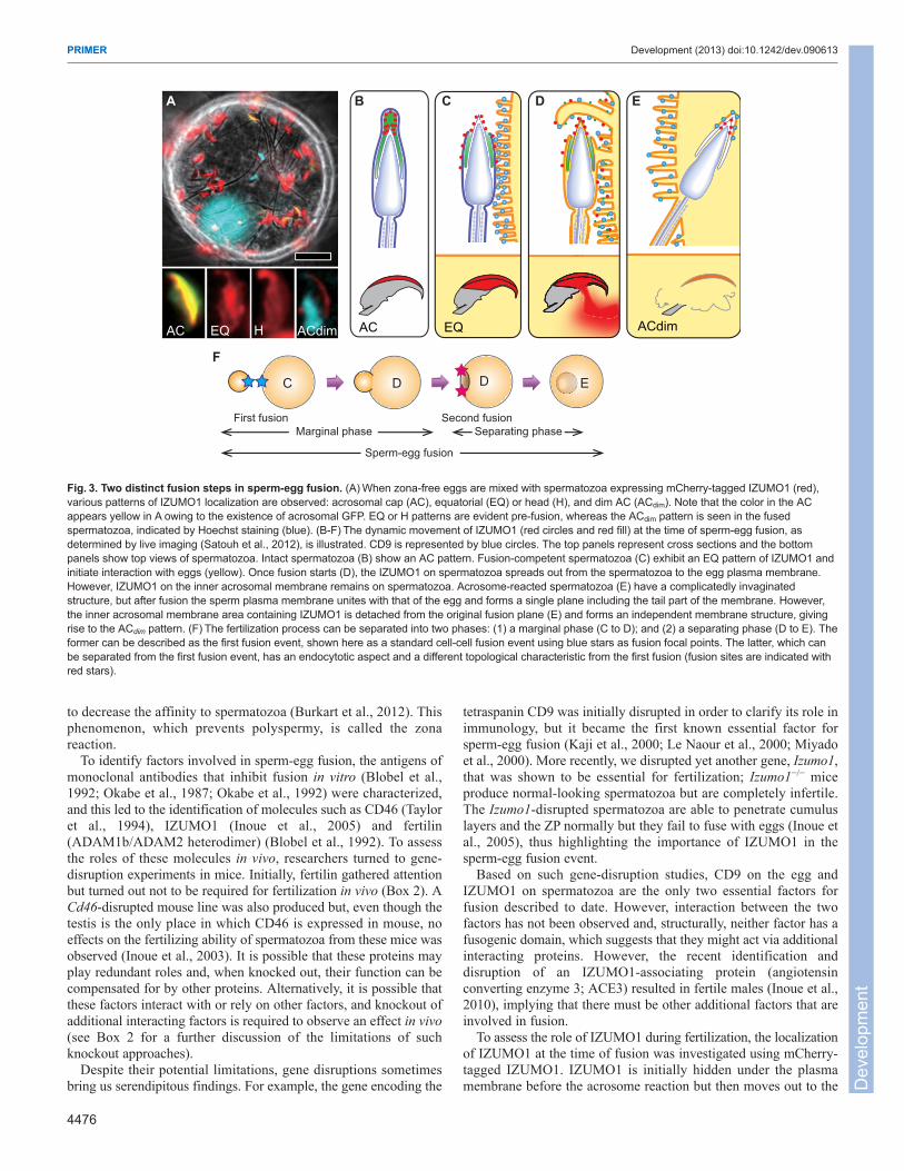
Fig.4. Mechanism of fertilization: old and new models. In the classicalscheme (left), the spermatozoa have hyaluronidase (PH-20; also known asSPAM1) (pink triangles) on their surface and penetrate the cumulus layer andattach to the zona pellucida (ZP). Many zona-binding proteins onspermatozoa have been postulated (Table2). ZP3 was reported to be themolecule to which spermatozoa bind (or attach) and had acrosome reaction-inducing ability. The timing of the acrosome reaction was also proposed to beimportant for zona penetration because it was believed that the acrosomalenzymes (such as acrosin) must be released upon binding to zona. Spermfertilin was also thought to function during egg fusion, with eggs usingintegrin α6β1 for its counterpart. However, none of these gene-disruptedmice became infertile (He et al., 2003). In the modern scheme (right), thespermatozoa penetrate the cumulus layer with or without the acrosomereaction. Acrosome-reacted spermatozoa can even penetrate the cumulusand zona for a second time (Inoue et al., 2011). Although the so-called ‘zona-binding’ ability is not required for spermatozoa to fertilize eggs, it is knownthat the ‘zona-binding’ ability is attributable to ADAM3 (pink dots) on thesurface of sperm. If ADAM3 became aberrant, spermatozoa could notmigrate into the oviduct and hence failed to approach eggs. If ADAM3became aberrant, spermatozoa could not migrate into the oviduct and hencefailed to approach eggs. However, ADAM3 is a pseudogene in human(Grzmil et al., 2001). Therefore, if a general mechanism does exist amongmammalian species, we might still not have found the key factor forfertilization. IZUMO1 on spermatozoa and CD9 on eggs were shown to beessential for fusion but the precise mechanism of fusion is yet to be clarified.
Dev
elop
men
t

4478
Burkart, A. D., Xiong, B., Baibakov, B., Jiménez-Movilla, M. and Dean, J. (2012).Ovastacin, a cortical granule protease, cleaves ZP2 in the zona pellucida to preventpolyspermy. J. Cell Biol. 197, 37-44.
Busso, D., Cohen, D. J., Maldera, J. A., Dematteis, A. and Cuasnicu, P. S. (2007).A novel function for CRISP1 in rodent fertilization: involvement in sperm-zonapellucida interaction. Biol. Reprod. 77, 848-854.
Chang, H. and Suarez, S. S. (2011). Two distinct Ca(2+) signaling pathways modulatesperm flagellar beating patterns in mice. Biol. Reprod. 85, 296-305.
Chen, J., Litscher, E. S. and Wassarman, P. M. (1998). Inactivation of the mousesperm receptor, mZP3, by site-directed mutagenesis of individual serine residueslocated at the combining site for sperm. Proc. Natl. Acad. Sci. USA 95, 6193-6197.
Cho, C., Bunch, D. O., Faure, J. E., Goulding, E. H., Eddy, E. M., Primakoff, P. andMyles, D. G. (1998). Fertilization defects in sperm from mice lacking fertilin beta.Science 281, 1857-1859.
Cornwall, G. A., Tulsiani, D. R. and Orgebin-Crist, M. C. (1991). Inhibition of themouse sperm surface alpha-D-mannosidase inhibits sperm-egg binding in vitro. Biol.Reprod. 44, 913-921.
Cummins, J. M. and Yanagimachi, R. (1982). Sperm-egg ratios and the site of theacrosome reaction during in vivo fertilization in the hamster. Gamete Res. 5, 239-256.
Da Ros, V. G., Maldera, J. A., Willis, W. D., Cohen, D. J., Goulding, E. H., Gelman,D. M., Rubinstein, M., Eddy, E. M. and Cuasnicu, P. S. (2008). Impaired spermfertilizing ability in mice lacking Cysteine-RIch Secretory Protein 1 (CRISP1). Dev.Biol. 320, 12-18.
De Blas, G. A., Roggero, C. M., Tomes, C. N. and Mayorga, L. S. (2005). Dynamicsof SNARE assembly and disassembly during sperm acrosomal exocytosis. PLoSBiol. 3, e323.
Dietzel, E., Wessling, J., Floehr, J., Schäfer, C., Ensslen, S., Denecke, B., Rösing,B., Neulen, J., Veitinger, T., Spehr, M. et al. (2013). Fetuin-B, a liver-derivedplasma protein is essential for fertilization. Dev. Cell 25, 106-112.
Ensslin, M. A. and Shur, B. D. (2003). Identification of mouse sperm SED1, a bimotifEGF repeat and discoidin-domain protein involved in sperm-egg binding. Cell 114,405-417.
Evans, J. P. (2012). Sperm-egg interaction. Annu. Rev. Physiol. 74, 477-502. Faddy, M. J. (2000). Follicle dynamics during ovarian ageing. Mol. Cell. Endocrinol.
163, 43-48. Florman, H. M. and Ducibella, T. (2006). Fertilization in Mammals, 3rd edn. New York,
NY: Elsevier Academic Press.Florman, H. M. and Wassarman, P. M. (1985). O-linked oligosaccharides of mouse
egg ZP3 account for its sperm receptor activity. Cell 41, 313-324. Frenette, G., Girouard, J., D’Amours, O., Allard, N., Tessier, L. and Sullivan, R.
(2010). Characterization of two distinct populations of epididymosomes collected inthe intraluminal compartment of the bovine cauda epididymis. Biol. Reprod. 83, 473-480.
Fujihara, Y., Tokuhiro, K., Muro, Y., Kondoh, G., Araki, Y., Ikawa, M. and Okabe, M.(2013). Expression of TEX101, regulated by ACE, is essential for the production offertile mouse spermatozoa. Proc. Natl. Acad. Sci. USA 110, 8111-8116.
Gahlay, G., Gauthier, L., Baibakov, B., Epifano, O. and Dean, J. (2010). Gameterecognition in mice depends on the cleavage status of an egg’s zona pellucidaprotein. Science 329, 216-219.
Gong, X., Dubois, D. H., Miller, D. J. and Shur, B. D. (1995). Activation of a G proteincomplex by aggregation of beta-1,4-galactosyltransferase on the surface of sperm.Science 269, 1718-1721.
Goodson, S. G., Qiu, Y., Sutton, K. A., Xie, G., Jia, W. and O’Brien, D. A. (2012).Metabolic substrates exhibit differential effects on functional parameters of mousesperm capacitation. Biol. Reprod. 87, 75.
Grzmil, P., Kim, Y., Shamsadin, R., Neesen, J., Adham, I. M., Heinlein, U. A.,Schwarzer, U. J. and Engel, W. (2001). Human cyritestin genes (CYRN1 andCYRN2) are non-functional. Biochem. J. 357, 551-556.
Hagaman, J. R., Moyer, J. S., Bachman, E. S., Sibony, M., Magyar, P. L., Welch, J.E., Smithies, O., Krege, J. H. and O’Brien, D. A. (1998). Angiotensin-convertingenzyme and male fertility. Proc. Natl. Acad. Sci. USA 95, 2552-2557.
Hanayama, R., Tanaka, M., Miyasaka, K., Aozasa, K., Koike, M., Uchiyama, Y. andNagata, S. (2004). Autoimmune disease and impaired uptake of apoptotic cells inMFG-E8-deficient mice. Science 304, 1147-1150.
Harada, Y., Kawazoe, M., Eto, Y., Ueno, S. and Iwao, Y. (2011). The Ca2+ increaseby the sperm factor in physiologically polyspermic newt fertilization: its signalingmechanism in egg cytoplasm and the species-specificity. Dev. Biol. 351, 266-276.
Hardy, D. M., Oda, M. N., Friend, D. S. and Huang, T. T., Jr (1991). A mechanism fordifferential release of acrosomal enzymes during the acrosome reaction. Biochem. J.275, 759-766.
Hasuwa, H., Muro, Y., Ikawa, M., Kato, N., Tsujimoto, Y. and Okabe, M. (2010).Transgenic mouse sperm that have green acrosome and red mitochondria allowvisualization of sperm and their acrosome reaction in vivo. Exp. Anim. 59, 105-107.
He, Z. Y., Brakebusch, C., Fässler, R., Kreidberg, J. A., Primakoff, P. and Myles, D.G. (2003). None of the integrins known to be present on the mouse egg or to beADAM receptors are essential for sperm-egg binding and fusion. Dev. Biol. 254, 226-237.
Hinsch, E., Aires, V. A., Hedrich, F., Oehninger, S. and Hinsch, K. D. (2005). Asynthetic decapeptide from a conserved ZP3 protein domain induces the G protein-regulated acrosome reaction in bovine spermatozoa. Theriogenology 63, 1682-1694.
Ikawa, M., Wada, I., Kominami, K., Watanabe, D., Toshimori, K., Nishimune, Y. andOkabe, M. (1997). The putative chaperone calmegin is required for sperm fertility.Nature 387, 607-611.
Ikawa, M., Inoue, N., Benham, A. M. and Okabe, M. (2010). Fertilization: a sperm’sjourney to and interaction with the oocyte. J. Clin. Invest. 120, 984-994.
Ikawa, M., Tokuhiro, K., Yamaguchi, R., Benham, A. M., Tamura, T., Wada, I.,Satouh, Y., Inoue, N. and Okabe, M. (2011). Calsperin is a testis-specificchaperone required for sperm fertility. J. Biol. Chem. 286, 5639-5646.
Inoue, N., Ikawa, M., Nakanishi, T., Matsumoto, M., Nomura, M., Seya, T. andOkabe, M. (2003). Disruption of mouse CD46 causes an accelerated spontaneousacrosome reaction in sperm. Mol. Cell. Biol. 23, 2614-2622.
Inoue, N., Ikawa, M., Isotani, A. and Okabe, M. (2005). The immunoglobulinsuperfamily protein Izumo is required for sperm to fuse with eggs. Nature 434, 234-238.
Inoue, N., Kasahara, T., Ikawa, M. and Okabe, M. (2010). Identification and disruptionof sperm-specific angiotensin converting enzyme-3 (ACE3) in mouse. PLoS ONE 5,e10301.
Inoue, N., Satouh, Y., Ikawa, M., Okabe, M. and Yanagimachi, R. (2011). Acrosome-reacted mouse spermatozoa recovered from the perivitelline space can fertilize othereggs. Proc. Natl. Acad. Sci. USA 108, 20008-20011.
Jin, M., Fujiwara, E., Kakiuchi, Y., Okabe, M., Satouh, Y., Baba, S. A., Chiba, K.and Hirohashi, N. (2011). Most fertilizing mouse spermatozoa begin their acrosomereaction before contact with the zona pellucida during in vitro fertilization. Proc. Natl.Acad. Sci. USA 108, 4892-4896.
Kaji, K., Oda, S., Shikano, T., Ohnuki, T., Uematsu, Y., Sakagami, J., Tada, N.,Miyazaki, S. and Kudo, A. (2000). The gamete fusion process is defective in eggsof Cd9-deficient mice. Nat. Genet. 24, 279-282.
Kerr, C. L., Hanna, W. F., Shaper, J. H. and Wright, W. W. (2004). Lewis X-containingglycans are specific and potent competitive inhibitors of the binding of ZP3 tocomplementary sites on capacitated, acrosome-intact mouse sperm. Biol. Reprod.71, 770-777.
Kim, K. S. and Gerton, G. L. (2003). Differential release of soluble and matrixcomponents: evidence for intermediate states of secretion during spontaneousacrosomal exocytosis in mouse sperm. Dev. Biol. 264, 141-152.
Kim, K. S., Cha, M. C. and Gerton, G. L. (2001). Mouse sperm protein sp56 is acomponent of the acrosomal matrix. Biol. Reprod. 64, 36-43.
Krutskikh, A., Poliandri, A., Cabrera-Sharp, V., Dacheux, J. L., Poutanen, M. andHuhtaniemi, I. (2012). Epididymal protein Rnase10 is required for post-testicularsperm maturation and male fertility. FASEB J. 26, 4198-4209.
Kudo, T., Kaneko, M., Iwasaki, H., Togayachi, A., Nishihara, S., Abe, K. andNarimatsu, H. (2004). Normal embryonic and germ cell development in mice lackingalpha 1,3-fucosyltransferase IX (Fut9) which show disappearance of stage-specificembryonic antigen 1. Mol. Cell. Biol. 24, 4221-4228.
Kuzan, F. B., Fleming, A. D. and Seidel, G. E., Jr (1984). Successful fertilization invitro of fresh intact oocytes by perivitelline (acrosome-reacted) spermatozoa of therabbit. Fertil. Steril. 41, 766-770.
Le Naour, F., Rubinstein, E., Jasmin, C., Prenant, M. and Boucheix, C. (2000).Severely reduced female fertility in CD9-deficient mice. Science 287, 319-321.
Lishko, P. V., Botchkina, I. L. and Kirichok, Y. (2011). Progesterone activates theprincipal Ca2+ channel of human sperm. Nature 471, 387-391.
Liu, C., Litscher, E. S. and Wassarman, P. M. (1995). Transgenic mice with reducednumbers of functional sperm receptors on their eggs reproduce normally. Mol. Biol.Cell 6, 577-585.
Lu, Q. and Shur, B. D. (1997). Sperm from beta 1,4-galactosyltransferase-null miceare refractory to ZP3-induced acrosome reactions and penetrate the zona pellucidapoorly. Development 124, 4121-4131.
Marcello, M. R., Jia, W., Leary, J. A., Moore, K. L. and Evans, J. P. (2011). Lack oftyrosylprotein sulfotransferase-2 activity results in altered sperm-egg interactionsand loss of ADAM3 and ADAM6 in epididymal sperm. J. Biol. Chem. 286, 13060-13070.
Miyado, K., Yamada, G., Yamada, S., Hasuwa, H., Nakamura, Y., Ryu, F., Suzuki,K., Kosai, K., Inoue, K., Ogura, A. et al. (2000). Requirement of CD9 on the eggplasma membrane for fertilization. Science 287, 321-324.
Miyazaki, S. and Ito, M. (2006). Calcium signals for egg activation in mammals. J.Pharmacol. Sci. 100, 545-552.
Muro, Y., Buffone, M. G., Okabe, M. and Gerton, G. L. (2012). Function of theacrosomal matrix: zona pellucida 3 receptor (ZP3R/sp56) is not essential for mousefertilization. Biol. Reprod. 86, 1-6.
Nakanishi, T., Ikawa, M., Yamada, S., Parvinen, M., Baba, T., Nishimune, Y. andOkabe, M. (1999). Real-time observation of acrosomal dispersal from mouse spermusing GFP as a marker protein. FEBS Lett. 449, 277-283.
Nakanishi, T., Ikawa, M., Yamada, S., Toshimori, K. and Okabe, M. (2001).Alkalinization of acrosome measured by GFP as a pH indicator and its relation tosperm capacitation. Dev. Biol. 237, 222-231.
Nakanishi, T., Isotani, A., Yamaguchi, R., Ikawa, M., Baba, T., Suarez, S. S. andOkabe, M. (2004). Selective passage through the uterotubal junction of sperm froma mixed population produced by chimeras of calmegin-knockout and wild-type malemice. Biol. Reprod. 71, 959-965.
Nishimura, H., Kim, E., Nakanishi, T. and Baba, T. (2004). Possible function of theADAM1a/ADAM2 Fertilin complex in the appearance of ADAM3 on the spermsurface. J. Biol. Chem. 279, 34957-34962.
Nomikos, M., Swann, K. and Lai, F. A. (2012). Starting a new life: sperm PLC-zetamobilizes the Ca2+ signal that induces egg activation and embryo development: anessential phospholipase C with implications for male infertility. BioEssays 34, 126-134.
Okabe, M., Adachi, T., Kohama, Y. and Mimura, T. (1986). Effect of glucose andphloretin-2′-beta-D-glucose (phloridzin) on in vitro fertilization of mouse ova.Experientia 42, 398-399.
Okabe, M., Adachi, T., Takada, K., Oda, H., Yagasaki, M., Kohama, Y. and Mimura,T. (1987). Capacitation-related changes in antigen distribution on mouse spermheads and its relation to fertilization rate in vitro. J. Reprod. Immunol. 11, 91-100.
PRIMER Development (2013) doi:10.1242/dev.090613
Dev
elop
men
t

Okabe, M., Ying, X., Nagira, M., Ikawa, M., Kohama, Y., Mimura, T. and Tanaka, K.(1992). Homology of an acrosome-reacted sperm-specific antigen to CD46. J.Pharmacobiodyn. 15, 455-459.
Olson, E. N., Arnold, H. H., Rigby, P. W. and Wold, B. J. (1996). Know yourneighbors: three phenotypes in null mutants of the myogenic bHLH gene MRF4. Cell85, 1-4.
Oren-Benaroya, R., Orvieto, R., Gakamsky, A., Pinchasov, M. and Eisenbach, M.(2008). The sperm chemoattractant secreted from human cumulus cells isprogesterone. Hum. Reprod. 23, 2339-2345.
Osokine, I., Hsu, R., Loeb, G. B. and McManus, M. T. (2008). Unintentional miRNAablation is a risk factor in gene knockout studies: a short report. PLoS Genet. 4, e34.
Qi, H., Moran, M. M., Navarro, B., Chong, J. A., Krapivinsky, G., Krapivinsky, L.,Kirichok, Y., Ramsey, I. S., Quill, T. A. and Clapham, D. E. (2007). All fourCatSper ion channel proteins are required for male fertility and sperm cellhyperactivated motility. Proc. Natl. Acad. Sci. USA 104, 1219-1223.
Rankin, T. L., Coleman, J. S., Epifano, O., Hoodbhoy, T., Turner, S. G., Castle, P.E., Lee, E., Gore-Langton, R. and Dean, J. (2003). Fertility and taxon-specificsperm binding persist after replacement of mouse sperm receptors with humanhomologs. Dev. Cell 5, 33-43.
Rudnicki, M. A., Schnegelsberg, P. N., Stead, R. H., Braun, T., Arnold, H. H. andJaenisch, R. (1993). MyoD or Myf-5 is required for the formation of skeletal muscle.Cell 75, 1351-1359.
Satouh, Y., Inoue, N., Ikawa, M. and Okabe, M. (2012). Visualization of the momentof mouse sperm-egg fusion and dynamic localization of IZUMO1. J. Cell Sci. 125,4985-4990.
Shamsadin, R., Adham, I. M., Nayernia, K., Heinlein, U. A., Oberwinkler, H. andEngel, W. (1999). Male mice deficient for germ-cell cyritestin are infertile. Biol.Reprod. 61, 1445-1451.
Shen, C., Kuang, Y., Liu, J., Feng, J., Chen, X., Wu, W., Chi, J., Tang, L., Wang, Y.,Fei, J., et al. (2013). Prss37 is required for male fertility in the mouse. Biol. Reprod.88, 123.
Shi, S., Williams, S. A., Seppo, A., Kurniawan, H., Chen, W., Ye, Z., Marth, J. D.and Stanley, P. (2004). Inactivation of the Mgat1 gene in oocytes impairs oogenesis,but embryos lacking complex and hybrid N-glycans develop and implant. Mol. Cell.Biol. 24, 9920-9929.
Shimada, M., Yanai, Y., Okazaki, T., Noma, N., Kawashima, I., Mori, T. andRichards, J. S. (2008). Hyaluronan fragments generated by sperm-secretedhyaluronidase stimulate cytokine/chemokine production via the TLR2 and TLR4pathway in cumulus cells of ovulated COCs, which may enhance fertilization.Development 135, 2001-2011.
Shur, B. D. and Hall, N. G. (1982). A role for mouse sperm surfacegalactosyltransferase in sperm binding to the egg zona pellucida. J. Cell Biol. 95,574-579.
Sutton, K. A., Jungnickel, M. K. and Florman, H. M. (2008). A polycystin-1 controlspostcopulatory reproductive selection in mice. Proc. Natl. Acad. Sci. USA 105, 8661-8666.
Talbot, P., Shur, B. D. and Myles, D. G. (2003). Cell adhesion and fertilization: stepsin oocyte transport, sperm-zona pellucida interactions, and sperm-egg fusion. Biol.Reprod. 68, 1-9.
Tardif, S., Wilson, M. D., Wagner, R., Hunt, P., Gertsenstein, M., Nagy, A., Lobe, C.,Koop, B. F. and Hardy, D. M. (2010). Zonadhesin is essential for species specificityof sperm adhesion to the egg zona pellucida. J. Biol. Chem. 285, 24863-24870.
Taylor, C. T., Biljan, M. M., Kingsland, C. R. and Johnson, P. M. (1994). Inhibition ofhuman spermatozoon-oocyte interaction in vitro by monoclonal antibodies to CD46(membrane cofactor protein). Hum. Reprod. 9, 907-911.
Thall, A. D., Malý, P. and Lowe, J. B. (1995). Oocyte Gal alpha 1,3Gal epitopesimplicated in sperm adhesion to the zona pellucida glycoprotein ZP3 are not requiredfor fertilization in the mouse. J. Biol. Chem. 270, 21437-21440.
Tokuhiro, K., Ikawa, M., Benham, A. M. and Okabe, M. (2012). Protein disulfideisomerase homolog PDILT is required for quality control of sperm membrane proteinADAM3 and male fertility [corrected]. Proc. Natl. Acad. Sci. USA 109, 3850-3855.
Visconti, P. E., Krapf, D., de la Vega-Beltrán, J. L., Acevedo, J. J. and Darszon, A.(2011). Ion channels, phosphorylation and mammalian sperm capacitation. Asian J.Androl. 13, 395-405.
Wakayama, T., Ogura, A., Suto, J., Matsubara, Y., Kurohmaru, M., Hayashi, Y. andYanagimachi, R. (1996). Penetration by field vole spermatozoa of mouse andhamster zonae pellucidae without acrosome reaction. J. Reprod. Fertil. 107, 97-102.
Wilcox, A. J., Weinberg, C. R. and Baird, D. D. (1995). Timing of sexual intercoursein relation to ovulation. Effects on the probability of conception, survival of thepregnancy, and sex of the baby. N. Engl. J. Med. 333, 1517-1521.
Williams, S. A., Xia, L., Cummings, R. D., McEver, R. P. and Stanley, P. (2007).Fertilization in mouse does not require terminal galactose or N-acetylglucosamine onthe zona pellucida glycans. J. Cell Sci. 120, 1341-1349.
Yamaguchi, R., Yamagata, K., Ikawa, M., Moss, S. B. and Okabe, M. (2006).Aberrant distribution of ADAM3 in sperm from both angiotensin-converting enzyme(Ace)- and calmegin (Clgn)-deficient mice. Biol. Reprod. 75, 760-766.
Yamaguchi, R., Muro, Y., Isotani, A., Tokuhiro, K., Takumi, K., Adham, I., Ikawa, M.and Okabe, M. (2009). Disruption of ADAM3 impairs the migration of sperm intooviduct in mouse. Biol. Reprod. 81, 142-146.
Yamaguchi, R., Fujihara, Y., Ikawa, M. and Okabe, M. (2012). Mice expressingaberrant sperm-specific protein PMIS2 produce normal-looking but fertilization-incompetent spermatozoa. Mol. Biol. Cell 23, 2671-2679.
Yanagimachi, R. (1994). Mammalian Fertilization, 2nd edn. New York, NY: RavenPress.
Yanagimachi, R. and Mahi, C. A. (1976). The sperm acrosome reaction andfertilization in the guinea-pig: a study in vivo. J. Reprod. Fertil. 46, 49-54.
4479
PRIMER Development (2013) doi:10.1242/dev.090613
Dev
elop
men
t
Related Documents