Bayerische Julius-Maximilian Universität Würzburg THE CD23 RECEPTOR- REGULATION OF EXPRESSION AND SIGNAL TRANSDUCTION Dissertation zur Erlangung des naturwissenschaftlichen Doktorgrades der Bayerische Julius-Maximilian Universität Würzburg vorgelegt von Ioana Andreea Visan (Ploiesti, Romania) Würzburg, 2003

Welcome message from author
This document is posted to help you gain knowledge. Please leave a comment to let me know what you think about it! Share it to your friends and learn new things together.
Transcript

Bayerische Julius-Maximilian Universität Würzburg
THE CD23 RECEPTOR-
REGULATION OF EXPRESSION AND SIGNAL
TRANSDUCTION
Dissertation zur Erlangung des
naturwissenschaftlichen Doktorgrades
der Bayerische Julius-Maximilian Universität Würzburg
vorgelegt von
Ioana Andreea Visan
(Ploiesti, Romania)
Würzburg, 2003

Eingereicht am: 24 Feb. 2003
Betreuer der Promotion: Prof. Dr. Rainer Hedrich
Medizinische Poliklinik: Prof. Dr. Hans-Peter Tony
Fakultät für Biologie: Prof. Dr. Jörg Hacker
Mitglieder der Promotionskommission:
Vorsitzender:
Gutachter: Prof. Dr. Hans-Peter Tony
Gutachter: Prof. Dr. Jörg Hacker
Tag des Promotionskolloquiums:
Doktorurkunde ausgehäandigt am:

SUMMARY
INTRODUCTION……………………………………………………..……….…………… 5
1. General principles of transmembrane signalling……….……………….…...………… 5
1.1 The Ras/ MAPK pathway…………………………….………...……...…..……… 6
1.2 Second messengers……………………………………………….…...………….. 7
1.3 Transcription factors with important role in B-cell signalling……………………..7
1.3.1 The JAK-STAT pathway…………………………………………….………. 7
1.3.2 The NF-kB family of transcription factors………………...………………… 8
2. Pax-5 - the B-cell-specific activator protein…………………………...……...……….. 8
2.1 Expression pattern………………………………..……………..….…………….. 9
2.2 Molecular structure and DNA binding site………...……...…....……………...….. 9
2.3 Role of Pax-5 in B-cell lineage commitment…………….......……….……….….. 10
2.4 Role of Pax-5 in late B-cell development ……...………….….………….……….. 11
3. CD23- the low affinity receptor for IgE……..……………………...….……...………. 12
3.1 Cellular expression and its regulation………...………………….…...…...……..... 13
3.2 Structure of the molecule…………...………...…….…….……….…………..….. 13
3.3 Ligands for CD23……………...……………………….………..………......……. 15
3.4 Biologic activity………………………………….……….…….…...……...…….. 15
3.5 Isoforms of human CD23……………………….…….………....…….…....…….. 16
3.6 Genomic structure of the human CD23 gene and analyses
of its transcriptional regulation……..….….….……………………….………….. 17
3.7 The murine CD23 receptor……..……………………………….…...……………. 19
3.8 Signal transduction through the CD23 receptor…………...…….……….………...20
4. Two-hybrid systems…………………………………………………………………….. 21
4.1 CytoTrap Two Hybrid System…………………………………………………….. 21
4.2 MATCHMAKER GAL-4 Two-Hybrid System 3……….……………….………... 23
AIMS OF THE PROJECT…………………………… ....………………………………….. 26
MATERIALS AND METHODS………………………..………………………...………….. 27
1. Gene cloning…….…………………………...…..………………...………………….. 27
1.1 Plasmid constructs………………………….…...………….…….……………….. 27
1.2 Oligonucleotides……………...………………………………..….……………….. 28
1

1.3 Annealing reaction……………………………………………..………………….. 28
1.4 RT-PCR…...…………………………………...…………....…...………………… 29
1.5 Site directed mutagenesis……….………………………....…….…………………. 30
1.6 Nucleic acid cleaning and purification procedures………....…….………………... 31
1.7 Polishing of PCR products………………………………...….….………………… 31
1.8 Subcloning of PCR products…….…………………………….……………...……. 31
1.9 DNA digestion with restriction enzymes…………….………..…………………… 32
1.10 Klenow Fill-in reaction………….………………………………………………… 32
1.11 Dephosphorylation of DNA……….…...…….……………………………………. 32
1.12 Ligation……………………………………………………………………...…….. 33
1.13 Transformation of bacteria………..…………….…………………………………. 33
1.14 Plasmid extraction…...……………………..…….…………………………….….. 34
1.15 Visualisation of DNA…………………………….……………………………….. 34
1.16 Sequencing………………………………...……….……………………………… 34
2. Protein extraction……………………………………….……………….….…………… 35
2.1 Protein extraction from mammalian cells…………...…………………………….. 35
2.2 Protein extraction from yeasts……………………….…….…………………...….. 35
3. Western Blot Analyses………………………………………..…………………………. 36
4. Electrophoretic Mobility Shift Assays (EMSAs)………………….……………………. 37
4.1 Oligonucleotides………………………………………...……………………….… 37
4.2 Radioactive labelling of annealed oligonucleotides………….…………..……….. 39
4.3 In vitro Transcription and Translation……………………………………………... 39
4.4 Direct Binding and Competition Assays…………………….………….………..... 39
5. Ribonuclease Protection Assay…………………………………………………...…….. 40
5.1 Linearization of template DNA……………………………….………………….... 41
5.2 In vitro Transcription………………………………………....………...………….. 41
5.3 Hybridization reaction……………...………………..…………………………….. 41
6. Transfection and reporter gene assay…………………..……………………………….. 41
6.1 Transfection ..…………………………………..………….…………...………….. 42
6.2 Cell Lyses and Luciferase Assay……………...………………..…………..……… 42
7. Transfection/ Infection Assays……………..………………….………………..………. 42
8. Nucleic acid extraction………………………………………….…...……..…………… 43
8.1 DNA extraction………...………………………………….……...……………….. 43
8.2 RNA extraction…………………………..………………….……….…………….. 43
2

9. Spectrophotometric measurements……………………………………………………… 44
9.1 Nucleic acids……………...……………………………………………………….. 44
9.2 Proteins……………………………………………………….….….....………….. 44
10. Two Hybrid Systems………………...……………………………...…………………. 44
10.1 CytotrapTM Two Hybrid System (Stratagene)…….………………………………..44
10.2 MATCHMACKER GAL 4 Two-hybrid System 3 (Clontech)……….…………… 47
11. Materials…………………………………………………………..…….…....……….. 52
11.1 Buffers………………………………………………….…..……….……...…….. 52
11.2 Gels……………………………………………………………………………….. 53
11.3 Media for bacteria and yeasts……………..………….……………………...…… 54
11.4 E. coli competent cell strains……………..……………………………...…...….. 55
11.5 Cell lines………………..………………….………….……………...………….. 55
11.6 Software and websites……………..………………………………………………55
11.7 Lab devices……………………..…………..……………….………...………….. 56
RESULTS………………………………………..…………………...….………………….. 57
1. The CD23a promoter- role of Pax-5 in the B-cell specific expression
of CD23a isoform…..……………………………………..…..………………………… 57
1.1 The CD23a core promoter contains three putative binding
sites for Pax-5…...……………………………………………….………….…….. 57
1.2 Pax-5 protein interacts with the CD23-1 binding site from the
CD23a core promoter in vitro………….…...………………………………....…… 59
1.2.1 CD23-1 binding site competes a high affinity Pax-5 binding site….…........ 59
1.2.2 CD23-1 binding site interacts with Pax-5 protein directly…...……………. 59
1.3 Mutations of the CD23-1 binding site prevent Pax-5 binding…………………….. 59
1.4 CD23-1 is the only site in the CD23a core promoter which directly
binds Pax-5 protein………………………………………………………………… 63
1.5 Pax-5 mediates activation of the CD23a promoter in vitro……………….…...…... 63
1.6 Pax-5 mediates CD23a expression in vivo …….………………………….……….. 65
2. Using a Two Hybrid System to find a cytoplasmic interaction partner for the CD23
receptor ……………………..……..……………………...………………….…………. 68 2.1 Establishing the system………………………………….………...……………… 68
2.1.1 Bait constructs………………………………………………...………....….. 68
2.1.2 Phenotype control………………………………………………...……….…. 69
3
2.1.3 Control reactions……………………….. …………………………………… 70
2.1.4 Establishing a high efficiency transformation protocol……………………… 70
2.2 Screening results with pSosCD23a and pSosCD23b…………………...………….. 70
2.3 Using the MATCHMAKER GAL4 Two-Hybrid System to verify the
screening results……….………...…………………...……...……………………... 72
2.3.1 Establishing the system ………………………...……………………………. 72
2.3.2 Results of the testing…………………………..…………………………….. 75
2.4 New constructs for the CytoTrap Two Hybrid System bait vectors……………….. 76
2.5 Screening results with pSosCD23a+Linker and pSosCD23b+Linker….………..... 79
2.6 New construct for the CytoTrap Two Hybrid System CD23a bait vector………… 79
2.7 Verifying interactions between two known protein…………...……...……………. 80
2.8 Perspectives…………………………………………….…………...………...….. 81
DISCUSSION….………………….……………………...…….……………...…....….... 82
ABSTRACT/ZUSAMMENFASSUNG……….……………………………..……………..... 92
1. English ………………………………………………………………..……………...… 92
2. Deutsch………...………...…………………………………………………..………… 94
REFERENCES.……………………..……..………………………………………….…….... 96
ABBREVIATIONS……………………………...………………………..…………….……. 107
ACKNOWLEDGMENTS……………………………………………………………….…… 112
CURRICULUM VITAE…………………………....………………………………….…….. 113
PUBLICATIONS………………..………………........………………………………..……. 114
EIDESSTATTLICHE ERKLÄRUNGEN…………..……………………………..……...……. 116
4

INTRODUCTION
1. General principles of transmembrane signalling
The B-cell represents one of the two major types of lymphocytes in the immune system,
responsible for the humoral immune response. The antigen receptor on B-cells is a cell-
surface immunoglobulin. After encountering the antigen B-cells differentiate into cells
producing antibody molecules of the same antigen-specificity as their receptor.
B-cells communicate with the environment through a variety of cell-surface receptors that
recognize and bind molecules present in the extracellular environment. Beside the antigen
receptor, which is the most important in the response to antigens, a variety of other surface
molecules contribute to coordinate growth, differentiation, metabolism and survival of the B-
lymphocyte. These receptors convert extracellular ligand binding into an intracellular signal
and activate intracellular pathways, which transmit the signal. This process is known as
signal transduction. The final destination of receptor signalling is the nucleus, where the
activation of transcription factors modifies gene expression.
Cell-surface receptors are transmembrane proteins that undergo conformational changes after
ligand binding. This change can enable them for instance to associate with and activate a
trimeric G protein (G protein-coupled receptor) or a protein-tyrosine kinase (tyrosine kinase
linked receptors), or to activate their own intrinsic protein kinase activity (receptor tyrosine
kinases). The cytosolic signal may activate a cascade of protein kinases or act through
increasing the concentration of intracellular signalling molecules named second messengers
(cAMP, cGMP, small lipid molecules and Ca2+). These signal transduction pathways do not
only transmit the signal, but also provide means for its amplification. Another important
feature of signal transduction pathways is that they both converge- several receptors can
activate the same signalling cascade or transcription factor- and diverge- one receptor or
given transduction protein can have more than one effector. This provides means for specific,
fine–tuned or complex responses to a variety of stimuli.
5

In B-cell signalling, tyrosine phosphorylation of membrane receptors is an important way to
activate receptors. Amino acid motifs called ITAMs (immunoreceptor tyrosine based
activation motifs) or ITIMs (immunoreceptor tyrosine based inhibitory motifs) are found in
the cytoplasmic tails of Fc receptors, accessory chains of the B-cell receptor and other
receptors. Phosphorylated tyrosine residues recruit intracellular signalling molecules to an
activated receptor by binding a protein domain known as SH2 domain (Src homology
domain 2). SH2 domains are phosphotyrosine binding motifs implicated in the regulation of
protein-protein interactions and are thought to function as molecular adhesives facilitating
the formation of protein complexes. SH2 domain binding to specific phosphotyrosine
containing sequences may transmit intracellular signals by inducing conformational changes
that alter an enzyme's catalytic activity or by altering the subcellular localization of a protein.
Another protein domain involved in protein-protein interactions is the SH3 domain (Src
homology domain 3), which binds proline-rich regions in diverse proteins to recruit them to
signalling pathways. These binding domains can be found alone or in various combinations
in proteins containing catalytic domains. These combinations provide great potential for
complex interplay and cross-talk between different signalling pathways.
1.1 The Ras / MAPK pathway
The Ras/MAPK pathway is one of the best characterized pathway initiated by receptor
tyrosine kinases. Ras is a small monomeric G protein with a central role in cell growth. In the
active state it is bound to GTP while in the inactive state it is bound to GDP. Ras possesses
an intrinsic GTPase activity that renders it inactive. In mammalian cells SOS, a guanine
nucleotide exchange factor, controls the conversion of Ras from the normal, inactive state to
the active state. SOS is brought to the membrane and activated by Grb2, an adaptor protein,
in response to the phosphorylation of the receptor. Ras activates Raf, a serine/threonine
kinase, which in turn activates MEK. In the MAP kinase pathway, this enzyme provides a
convergence point, as it can be also activated by signals coming from G protein-coupled
receptors. MEK is an enzyme with dual specificity, which can phosphorylate both threonine
and tyrosine. Its target is Erk MAP kinase, which than activates target transcription factors
directly into the cytoplasm or after its translocation to the nucleus. In the B-cell, MAP kinase
pathways from the B-cell receptor or co-receptor combine to regulate the expression of many
genes involved in cell growth [92,93].
6
1.2 Second messengers
Cyclic AMP is a classic second messenger. The initial step in the pathway is the activation of
adenylate cyclase at the plasma membrane by an activated G protein associated with the
receptor. cAMP binds to the regulatory subunit of PKA (protein kinase A) and releases the
catalytic subunit of this enzyme, which is than free to translocate to the nucleus or to
phosphorylate targets in the cytosol. One of the major substrates for PKA is CREB (cAMP
response element binding protein). Phosphorylated CREB binds to CRE elements in the
promoter of genes that are sensible to cAMP [94].
The inositol-lipid pathway is a common pathway for many types of receptors and involves
second messengers derived from phosphatidylinositol. The enzyme phospholipase C-γ can be
recruited through a SH-2 domain to the site of receptor-associated tyrosine kinase activity at
the cell membrane. Phosphorylation of a tyrosine residue in PLC-γ activates the enzyme,
which then cleaves the membrane phosphatidylinositol biphosphate (PIP2) into inositol
triphosphate (IP3) and diacylglycerol (DAG). Diffusion of IP3 away from the membrane
causes the release of Ca2+ from intracellular storage sites into the cytosol, raising the
intracellular Ca2+ level several times. The signal is sustained by the opening of Ca2+ channels
into the plasma membrane. Ca2+ binds and activates a small cytosolic enzyme called
calmodulin, which in turn binds to and regulates other enzymes or transcription factors like
NF-AT. The signal eventually reaches the nucleus. DAG remains associated to the inner
surface of the plasma membrane where it activates protein kinase C. Increased Ca2+ levels
further activate this enzyme, which also initiate pathways leading to the nucleus [92,94].
1.3 Transcription factors with an important role in B-cell signalling
1.3.1 The JAK-STAT pathway
In contrast to signal transduction pathways that use a large number of components, like the
MAPK and the inositol-lipid pathways, JAK-STAT pathway is much simpler. It is often
activated by cytokine receptors that do not possess tyrosine activity. Binding of the ligand
causes the receptor to dimerize and associate with and activate a JAK kinase. These are
tyrosine kinases that phosphorylate transcription factors named STATs (signal transducer
and activator of transcription). There are several JAK kinases and more than 7 STATs. Each
STAT is phosphorylated by a particular set of JAK kinases. STAT phosphorylation leads to
7
the formation of homo- and heterodimers, which translocate to the nucleus and bind specific
recognition elements in target genes. The activation of STATs is transient and can be
terminated by the action of a phosphatase. STATs play important roles in numerous cellular
processes including immune responses, cell growth and differentiation, cell survival and
apoptosis [92,94].
1.3.2 The NF-kB family of transcription factors
The NF-kB transcription factors are important for B-cell activation. NFkB exists in the
cytoplasm mainly as homo- or heterodimers with a family of structurally related proteins,
called the Rel or Rel/NF-kB proteins. In non-stimulated cells, NF-kB complexes are
sequestered in the cytoplasm in an inactive form via interaction with an inhibitory protein
called IkB, which itself belongs to a structurally- and functionally-related family of proteins.
When cells are stimulated by a variety of stimuli, like lipopolysaccharide (LPS) or CD40L, a
kinase cascade leads to the phophorylation of two kinases, named Ikkα and Ikkβ, which form
a dimer that in turn phosphorylates IkB. When phosphorylated, IkB dissociates from the
complex and is rapidly degraded by proteosomes. NF-kB migrates to the nucleus where it is
involved in regulating many aspects of the immune cell function, like cell survival,
processing and presentation of antigen, responses to antigen recognition, aspects of the
inflammatory response as well as responses against bacterial and viral infections [51]. The
NF-kB transcription factor family consists of heterodimers or homodimers of the subunits
NF-kB1 (p50), NF-kB2 (p52), c-REL, RELA (p65) and RELB. Different pairs of these
subunits function at different stages of B cell development [57].
2. Pax-5 - the B-cell-specific activator protein
BSAP/Pax-5 is a member of the Pax (paired box) family of transcription factors, which
constitute a small group of conserved developmental control genes. BSAP (B-cell-specific
activator protein) was identified as the mammalian homologue of a sea urchin protein
(TSAP), which is involved in the developmental regulation of two pairs of nonallelic histones
H2A-2 and H2B-2 [3]. It is the only member of the family that is expressed in B-
8
lymphocytes. The other nine members of the Pax family identified so far have been
associated with mouse developmental mutants and human syndromes. Deletion of the paired
domain of Pax-3 is associated with the Splotch mutation in mice, characterized by spina
bifida and exencephaly and with the Waardenburg's syndrome in humans, an autosomal
dominant combination of deafness and pigmentary disturbances [20,80]; mutations in the
Pax-6 gene are associated with congenital aniridia (lack of iris) in humans and the small eye
(Sey) phenotype in mice- a semidominant mutation that in the homozygous condition results
in the complete lack of eyes and nasal primordial [29].
2.1 Expression pattern
BSAP is encoded by the pax-5 gene and is expressed at all stages of B-cell development
except in terminally differentiated plasma cells [3]. In addition to all B-lymphoid organs,
Pax-5 can also be found in the developing midbrain and adult testis of the mouse [1]. In
accordance to this expression pattern, gene inactivation in the mouse germline revealed that
Pax-5 plays an important role in B-lymphopoiesis and midbrain development. [84].
2.2 Molecular structure and DNA binding site
All members of the Pax protein family have a C-terminal transactivation domain and a N-
terminal paired domain (the DNA binding domain). The transactivation domain is located in
the 55 C-terminal amino acids of the molecule and contains a distinct
serine/threonine/proline-rich sequence. This domain exerts its activating function from a
promoter as well as an enhancer position and is subject of a strong negative regulation by
adjacent sequences from the extreme C-terminus [16].
The paired domain consists of a stretch of 128 amino acids that has been well conserved in
evolution and shows no obvious resemblance to other known DNA-binding motifs. Detailed
mutational analysis of Pax-5 revealed the bipartite structure of a paired domain and lead to
the identification of a nonpalindromic consensus recognition sequence [12].
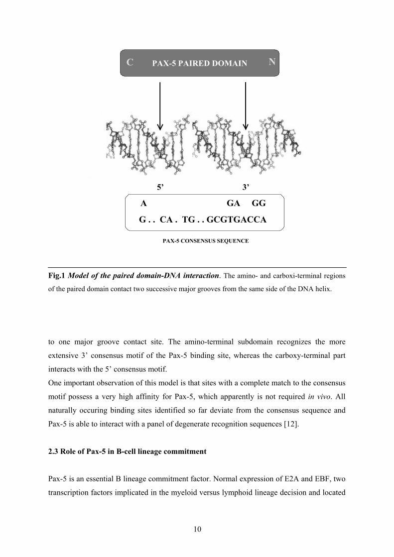
In the model for the paired domain-DNA interaction (Fig. 1) the paired domain binds to its
recognition sequence from one side of the DNA helix and interacts with two successive
major grooves. The Pax-5 recognition sequence is divided in two halves, each corresponding
9
5’ 3’
A GA GG
G . . CA . TG . . GCGTGACCA
PAX-5 PAIRED DOMAINC N
PAX-5 CONSENSUS SEQUENCE
Fig.1 Model of the paired domain-DNA interaction. The amino- and carboxi-terminal regions
of the paired domain contact two successive major grooves from the same side of the DNA helix.
to one major groove contact site. The amino-terminal subdomain recognizes the more
extensive 3’ consensus motif of the Pax-5 binding site, whereas the carboxy-terminal part
interacts with the 5’ consensus motif.
One important observation of this model is that sites with a complete match to the consensus
motif possess a very high affinity for Pax-5, which apparently is not required in vivo. All
naturally occuring binding sites identified so far deviate from the consensus sequence and
Pax-5 is able to interact with a panel of degenerate recognition sequences [12].
2.3 Role of Pax-5 in B-cell lineage commitment
Pax-5 is an essential B lineage commitment factor. Normal expression of E2A and EBF, two
transcription factors implicated in the myeloid versus lymphoid lineage decision and located
10

upstream of Pax-5 in the B-cell developmental process, is not sufficient to commit B-cell
progenitors to the B-lymphoid lineage in the absence of Pax-5. In Pax-5-/- mice B-cell
development is arrested at early pro-B-cell stage in the bone marrow. These Pax-5-/- pro-B-
cells still retain a broad lymphomyeloid development potential characteristic of uncommited
hematopoiectic progenitors [60,67]. Upon appropriate cytokine stimulation, Pax-5-/- pro-B-
cells are able to differentiate in vitro into functional NK cells, dendritic cells, macrophages,
osteoclasts and granulocytes [60]. In addition, Pax-5-/- pro-B-cells possess extensive in vivo
self-renewal potential and long-term reconstitution potential, which are features of
hematopoietic stem cells (HSC), yet they fail to reconstitute the hematopoietic system of
lethally irradiated mice [72].
Binding sites for Pax-5 have been identified in promoters of several genes. While activating
CD19, mb-1, RAG-2 and BLNK [43,59,39] Pax-5 acts as a repressor for the XBP-1, the M-
CSF-R, the immunoglobulin heavy-chain 3’C∝ enhancer and the J-chain gene [64,66,75].
The conversion to a repressor function appears to be possible by recruitment of corepressors
of the Groucho family to selected target genes [19].
At lineage commitment, Pax-5 has a dual role by repressing “lineage-inappropriate” genes
and simultaneously activating B-cell-specific genes, which leads to the consolidation of the
B-lymphoid gene expression program. This role is best illustrated by the regulation of M-
CSF-R and BLNK genes. By repressing M-CSF-R gene, Pax-5 renders B-cell precursors
unresponsive to M-CSF, and prevents them to differentiate to monocytes under the influence
of this cytokine. On the other hand, by activating the BLNK promoter, Pax-5 enables the
expression of a central adaptor protein in BCR signaling [73].
2.4 Role of Pax-5 in late B-cell development
Pax-5 functions also at later stages of B cell development. The generation of a mouse strain
in which the Pax-5 gene can be conditionally inactivated enabled the analysis of Pax-5
function in mature B cells [31]. Loss of Pax-5 resulted in a change of B cell subpopulations
in the periphery with downregulation of several mature cell surface B cell markers.
Considering that Pax-5 is also repressing XBP-1, a transcription factor essential for plasma-
cell differentiation, it proves its important role for maintaining the identity and function of
mature B cells.
11

Furthermore, Pax-5 might play a role in isotype class switching by regulating germline
transcription from the downstream constant region gene, which appears to be a prerequisite
for subsequent class switching. IgH gene expression and rearrangement are regulated by
multiple cis-acting elements within the IgH locus, including the intronic enhancer (Eµ), the I
regions of the constant region genes and the complex regulatory region 3’ of Cα. Pax-5 was
reported to bind at multiple sites in the IgH gene cluster, including regions located upstream
of switch regions, like Sγ2a [46] and Sµ [86] and at sites within the 3’ control region [52].
Repression or activation of these regulatory sites appears to require a concerted effort
involving additional factors, such as octamer binding proteins and NF-kB-like complexes.
3. CD23- the low affinity receptor for IgE
CD23 was described as a low-affinity receptor for IgE (FcεRII) expressed on mature
peripheric B-cells. The same molecule, expressed at high levels on Epstein-Barr virus-
transformed cells was independently described as a B-cell activation marker.
The CD23 molecule is a type II membrane glycoprotein exhibiting substantial homology
with several Ca2+-dependent animal lectins. In humans it is expressed in two isoforms
(CD23a and CD23b). CD23 has a functional role as a transmembrane receptor as well as a
soluble receptor derived from the cell-bound form. CD23 is an important player in allergic,
autoimmune and lymphoproliferative diseases.
In humans, the expression of CD23 is increased in allergic disorders, in terms of membrane
expression on B-cells and monocytes but also in terms of sCD23 production [24]. The
number of circulating CD23-bearing B-cells is also increased in patients with rheumatoid
arthritis. In type II collagen-induced arthritis in mice, a model for human rheumatoid
arthritis, antibody neutralisation of CD23 significantly ameliorated the disease, proving the
involvement of the molecule in inflammatory processes [63]. In B-CLL patients, high levels
of sCD23 in the serum are correlated with the clinical stage of the disease and can be used as
prognostic marker. The accumulation of sCD23 results from an increased number of CD23-
bearing B-cells but also from the overexpression of CD23 on the surface of malignant cells
[69].
12
3.1 Cellular expression and its regulation
On normal human B-cells CD23 expression is restricted to mature peripheric B-cells co-
expressing IgM and IgD. After switching to IgG, IgA and IgE B-lymphocytes cannot be
induced to reexpress CD23. The receptor is comparably expressed on CD5+ and CD5-
circulating or tonsilar B-cells.
CD23 is also found on T-cells, monocytes, macrophages, platelets, eosinophils, Langerhans
cells and follicular dendritic cells [13].
Normal human B-lymphocytes from peripheral blood express both CD23 antigen and CD23
mRNA. Still the molecule is not constitutively expressed since after 48 hours of incubation in
the absence of a stimulant, highly purified B-cells loose both CD23 antigen and CD23
mRNA.
The major inducer of CD23 on B-cells is IL-4, which triggers the expression of both
isoforms. The peak effect is observed after 36-48 hours. Signals delivered via CD40
synergize with IL-4 for the induction of CD23 on mature peripheral B-cells and interactions
between B and T cells (presumably dependent on CD40) result in upregulation of CD23.
EBV-transformation of B-cells leads to CD23 expression, which plays a role in the
immortalization of the cell [8].
IFN-γ and IFN-α inhibit IL-4 induced expression of CD23 on normal B-cells at the protein
and mRNA level [15].
IL-4 is also the main CD23 inducer on all the other CD23 bearing cell types.
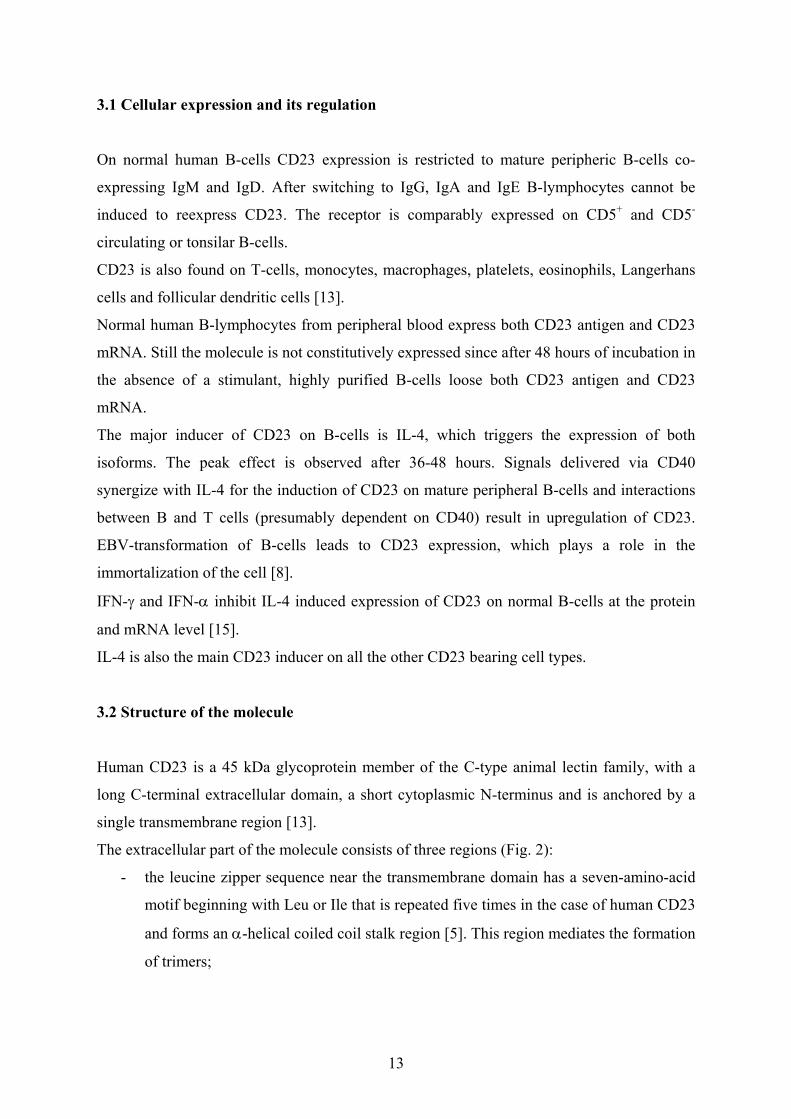
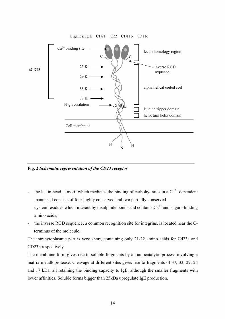
3.2 Structure of the molecule
Human CD23 is a 45 kDa glycoprotein member of the C-type animal lectin family, with a
long C-terminal extracellular domain, a short cytoplasmic N-terminus and is anchored by a
single transmembrane region [13].
The extracellular part of the molecule consists of three regions (Fig. 2):
- the leucine zipper sequence near the transmembrane domain has a seven-amino-acid
motif beginning with Leu or Ile that is repeated five times in the case of human CD23
and forms an α-helical coiled coil stalk region [5]. This region mediates the formation
of trimers;
13
CC
N N
lectin homology region
inverse RGD sequence
alpha helical coiled coil
N-glycosilation
Ca2+ binding site
25 K
29 K
33 K
37 K
leucine zipper domainhelix turn helix domain
Cell membrane
N
Ligands: Ig E CD21 CR2 CD11b CD11c
sCD23
Fig. 2 Schematic representation of the CD23 receptor
- the lectin head, a motif which mediates the binding of carbohydrates in a Ca2+ dependent
manner. It consists of four highly conserved and two partially conserved
cystein residues which interact by disulphide bonds and contains Ca2+ and sugar –binding
amino acids;
- the inverse RGD sequence, a common recognition site for integrins, is located near the C-
terminus of the molecule.
The intracytoplasmic part is very short, containing only 21-22 amino acids for Cd23a and
CD23b respectively.
The membrane form gives rise to soluble fragments by an autocatalytic process involving a
matrix metalloprotease. Cleavage at different sites gives rise to fragments of 37, 33, 29, 25
and 17 kDa, all retaining the binding capacity to IgE, although the smaller fragments with
lower affinities. Soluble forms bigger than 25kDa upregulate IgE production.
14
Binding of IgE and IgE-IC protects and stabilizes the stalk from proteolyses in this way
inhibiting the release of soluble fragments from the membrane form.
3.3 Ligands for CD23
The first ligand to be described for CD23 was IgE. Although IgE is highly glycosilated, the
lectin homology region of CD23 appears to bind the protein moiety of the molecule,
independently of carbohydrates. However, the binding is Ca2+ dependent and the correct
folding of the lectin domain is critical, since deletion of conserved cysteins has a deletorious
effect on IgE binding [6]. The CD23-binding site was mapped in the Cε3 constant region
domain of IgE, in close proximity of the high-affinity receptor (FcεRI) binding site. The
oligomerization of CD23 is an important factor in enabling high affinity binding to IgE.
The other ligands described for CD23 are CD21 (CR2), CD11b/CD18 (CR3) and
CD11c/CD18 (CR4) - two members of the LFA-1 family.
Fucose-1-phosphate has been described as a competitive inhibitor of both IgE and CD21
binding for CD23 [26].
3.4 Biologic activity
Membrane CD23 and its soluble forms have been implicated in different functions, ranging
from cellular adhesion, antigen presentation, growth and differentiation of B and T cells,
rescue from apoptosis, release of cytotoxic mediators and regulation of IgE synthesis.
A well-characterized function of membrane-bound CD23 in B-cells is the enhancement of
IgE-dependent antigen presentation to T-cells [23,27,35]. This requires the binding of
antigen-IgE immune complexes to CD23, internalization of the complexes and transport to
compartements of the endosomal network containing proteolytic enzymes and major
histocompatibility complex class II antigens. CD23 is spatially associated with MHC class II
DR on B-cells [34].
CD23 functions also as an adhesion molecule. Antigen presentation involves interaction
between CD23 and CD21 at points of contact between B and T cells [2,7].
Human CD23 plays also a regulatory role in the IgE production with positive and negative
effects. Crosslinking CD23 at the cell surface by IgE inhibits the release of sCD23 and
delivers a negative feedback for IgE production. In contrast, sCD23 fragments larger than 25
15
kDa that retain part of the stalk region promote IgE production. Two possible mechanisms
are discussed: (1) sCD23 possibly stimulates IgE production through CD21 triggering; (2)
sCD23 traps IgE in the medium and prevents the negative feedback through membrane-
bound CD23 [83].
There is evidence that soluble CD23 fragments exert other important roles except the
regulation of IgE synthesis. In synergy with IL-1, sCD23 acts as a differentiating factor for
early thymocytes [55] and induces proliferation of human bone marrow derived myeloid
precursors [56]. It is also involved in the rescue of germinal center B-cells. In the presence of
recombinant 25 kDa sCD23 and IL-1α centrocytes are rescued from apoptosis and can
differentiate into plasmocytoid cells [49]. This is supported by the high density of CD23 on
the surface of follicular dendritic cells in the light zone of the germinal center.
On monocytes, eosinophils and platelets, CD23 is involved in IgE dependent cytotoxicity
against some parasites and in the IgE induced release of different mediators of inflammation
[10,17]. CD23 also mediates the phagocytosis of IgE coated particles. Finally, sCD23
ligation of CD11b/CD11c on monocytes is able to promote release of inflammatory
mediators such as IL-1β, IL-6 and TNF [45].
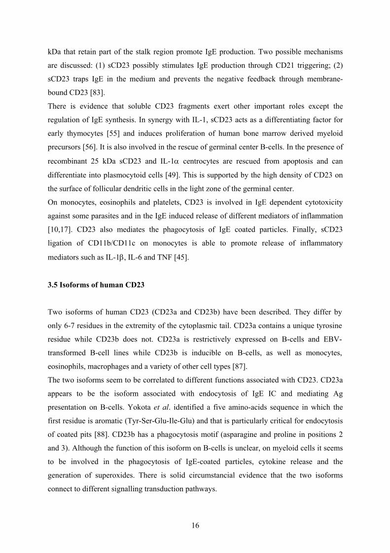
3.5 Isoforms of human CD23
Two isoforms of human CD23 (CD23a and CD23b) have been described. They differ by
only 6-7 residues in the extremity of the cytoplasmic tail. CD23a contains a unique tyrosine
residue while CD23b does not. CD23a is restrictively expressed on B-cells and EBV-
transformed B-cell lines while CD23b is inducible on B-cells, as well as monocytes,
eosinophils, macrophages and a variety of other cell types [87].
The two isoforms seem to be correlated to different functions associated with CD23. CD23a
appears to be the isoform associated with endocytosis of IgE IC and mediating Ag
presentation on B-cells. Yokota et al. identified a five amino-acids sequence in which the
first residue is aromatic (Tyr-Ser-Glu-Ile-Glu) and that is particularly critical for endocytosis
of coated pits [88]. CD23b has a phagocytosis motif (asparagine and proline in positions 2
and 3). Although the function of this isoform on B-cells is unclear, on myeloid cells it seems
to be involved in the phagocytosis of IgE-coated particles, cytokine release and the
generation of superoxides. There is solid circumstancial evidence that the two isoforms
connect to different signalling transduction pathways.
16

CD23 expressing cells CD23a CD23b
B cell ++ ++T cell -- ++Follicular dendritic cells -- ++Langerhans cells -- ++Monocytes -- ++Macrophages -- ++Eosinophils -- ++Platelets -- ++Thymic epithelial cells -- ++
Fig.3 The two isoforms of human CD23 are differentially expressed on cells of the
hematopoietic lineage.
3.6 Genomic structure of the human CD23 gene and analyses of its transcriptional
regulation
CD23 is encoded within the human genome by a single gene located on chromosome 19. It
consists of 11 exons, with a good correlation between the exon/intron structure and the
corresponding domains of the protein. Exons 5-7 seem to have arisen by a triplication of an
exon coding for an exact number of heptads and encode the stalk region. Exons 9-11, which
are separated by a large intron from the rest of the gene, encode for the soluble forms of
CD23.
The two isoforms, which differ by six aminoacids at the cytoplasmic amino terminus, are
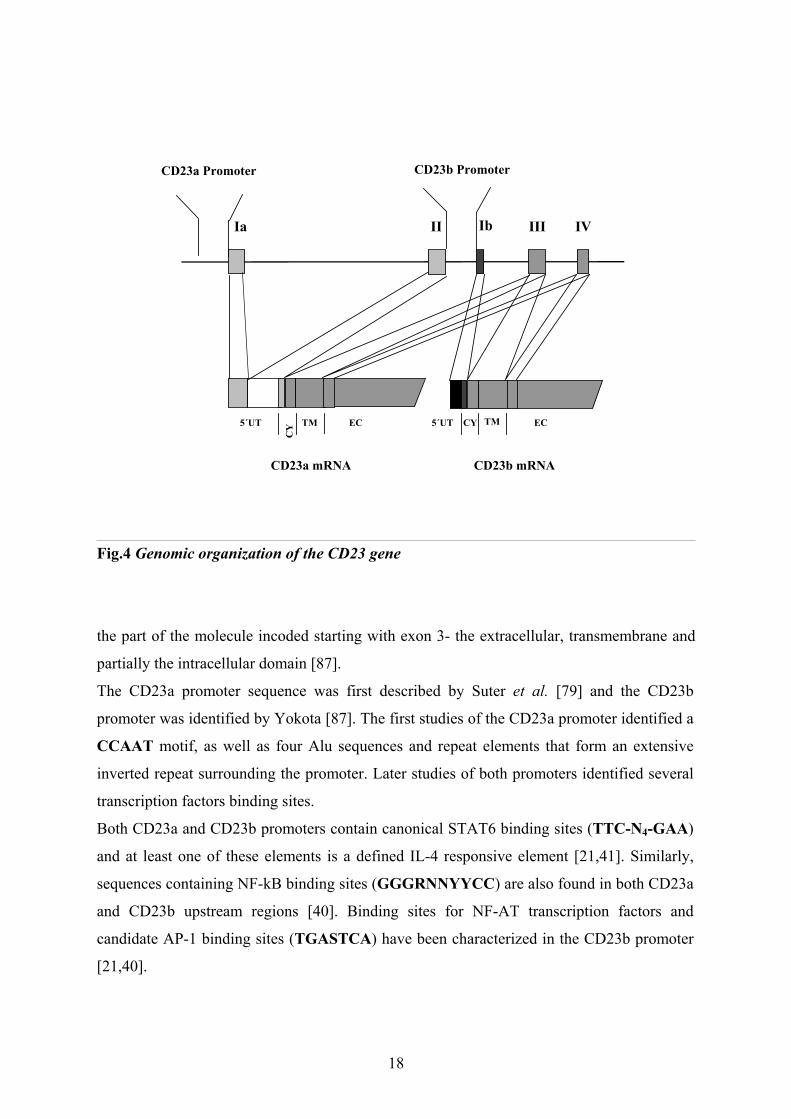
generated by using different transcriptional start sites and alternative RNA splicing (Fig. 4).
Given the genomic sequence of CD23a as a reference, CD23b mRNA is lacking the first two
exons and starts with an optional exon that is located within intron II. The two mRNAs share
17
Ia II III IV
5´UT
CY TM EC 5´UT CY TM EC
CD23a mRNA CD23b mRNA
CD23b PromoterCD23a Promoter
Ib
Fig.4 Genomic organization of the CD23 gene
the part of the molecule incoded starting with exon 3- the extracellular, transmembrane and
partially the intracellular domain [87].
The CD23a promoter sequence was first described by Suter et al. [79] and the CD23b
promoter was identified by Yokota [87]. The first studies of the CD23a promoter identified a
CCAAT motif, as well as four Alu sequences and repeat elements that form an extensive
inverted repeat surrounding the promoter. Later studies of both promoters identified several
transcription factors binding sites. Both CD23a and CD23b promoters contain canonical STAT6 binding sites (TTC-N4-GAA)
and at least one of these elements is a defined IL-4 responsive element [21,41]. Similarly,
sequences containing NF-kB binding sites (GGGRNNYYCC) are also found in both CD23a
and CD23b upstream regions [40]. Binding sites for NF-AT transcription factors and
candidate AP-1 binding sites (TGASTCA) have been characterized in the CD23b promoter
[21,40].
18
EBNA-2 targets the CD23a promoter through a DNA-binding protein, CBF-1, which binds a
recognition sequence with the common core motif GTGGGAA [48,85]. EBNA-2 is a
transcriptional activator that modulates Epstein-Barr virus latency gene expression as well as
the expression of cellular genes. CD23 expression is upregulated by EBNA-2 along with
CD21 [11]. Activation of CD23 might be particularly important, since only EBV-infected B-
cells expressing this marker become immortalized. Notch-2 also can regulate the CD23a
promoter by binding to CBF1 sites. The Notch family genes encode transmembane receptors
that modulate differentiation and proliferation. Notch-2 activation of the CD23a promoter
through CBF1 responsive elements may play a role in the immortalisation of the cell and the
pathogenesis of B-CLL [32].
Emerging data seem to lead to the conclusion of differential regulation of the CD23a and
CD23b promoters, with CD23a showing less sensitivity to external stimuli than the CD23b
promoter, at least in B-cells [21]. This would be in agreement with the idea of the two
isoforms having different functions.
3.7 The murine CD23 receptor
The mouse CD23 shares only 57% aminoacid sequence homology with the human molecule.
The protein is lacking the RGD motif by a naturally occuring truncation and the sCD23
fragments bind IgE with a much lower affinity [4]. There are also differences in the cellular
distribution between species, with the mouse CD23 expressed only on B-cells, follicular
dendritic cells and some T-cells. All these differences in structure and cellular expression
may account for the differences in functions between mice and humans. From the study of
CD23-/- mice, murine CD23 may not have the regulatory effects ascribed to human sCD23.
CD23-/- mice display normal lymphocyte development, normal B-cell proliferation and
germinal center formation. However, antigen-specific IgE-mediated enhancement of
antibody responces was severely impaired, suggesting the role of murine CD23 in antigen-
presentation [23,27]. Regarding the role of mouse CD23 in IgE production, some studies did
not find modifications of IgE levels in the serum of CD23-/- mice [23,77], while other studies
of heterozygous and transgenic mice suggest that the murine CD23, in particular the
membrane-bound form, exerts an inhibitory role on IgE production [81,89]. There are no
expressed isoforms described yet and the mouse CD23 seems to be more related to the
CD23a human isoform, by distribution and functions: the cytoplasmic tail of the mouse
CD23 contains a Tyr residue, part of an YSGT sequence and the mouse promoter also
19
displays homologies to the CD23a promoter. IL-4 is also the main inducer of mouse CD23.
STAT6 and NF-kB binding sites have been described [82].
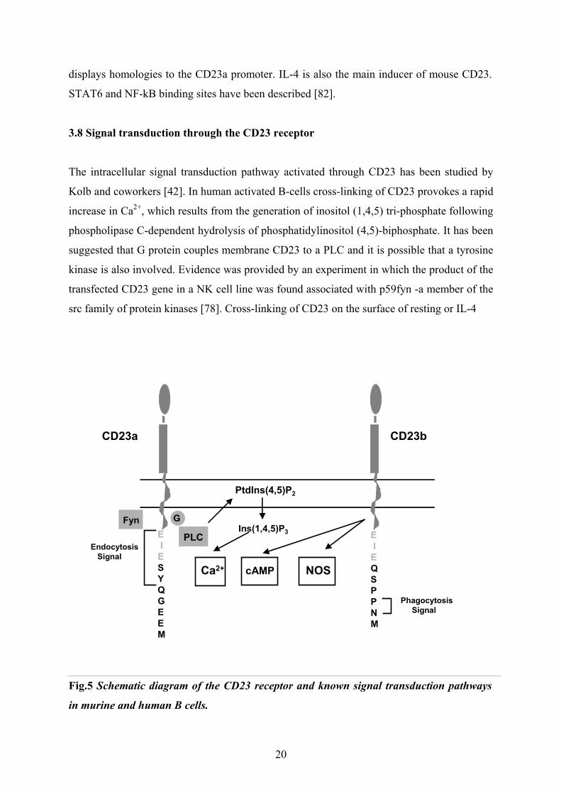
3.8 Signal transduction through the CD23 receptor
The intracellular signal transduction pathway activated through CD23 has been studied by
Kolb and coworkers [42]. In human activated B-cells cross-linking of CD23 provokes a rapid
increase in Ca2+, which results from the generation of inositol (1,4,5) tri-phosphate following
phospholipase C-dependent hydrolysis of phosphatidylinositol (4,5)-biphosphate. It has been
suggested that G protein couples membrane CD23 to a PLC and it is possible that a tyrosine
kinase is also involved. Evidence was provided by an experiment in which the product of the
transfected CD23 gene in a NK cell line was found associated with p59fyn -a member of the
src family of protein kinases [78]. Cross-linking of CD23 on the surface of resting or IL-4
PhagocytosisSignal
EI
ESYQGEEM
EI
EQSPPNM
EndocytosisSignal
CD23a CD23b
cAMP
Fyn
PLC
PtdIns(4,5)P2
Ins(1,4,5)P3
Ca2+
G
NOS
Fig.5 Schematic diagram of the CD23 receptor and known signal transduction pathways
in murine and human B cells.
20
stimulated B-cells resulted in a slow accumulation of intracellular cAMP, although it was
unclear to what extent increased Ca2+ levels were dependent upon the prior activation of
PLC. However, since this change could be observed in resting B-cells, where neither Ca2+
mobilization nor inositol (1,4,5) tri-phosphate production are significantly altered on
engaging CD23, suggests that the elevation in cAMP levels may proceed independently of
PLC activation. Moreover, cAMP accumulation was observed in monocytes, where the
phosphoinositide pathway is clearly not involved [26]. In human monocytes CD23 is
additionally coupled with the activation of inducible nitric oxide synthase (iNOS) pathway
[18].
This would indicate that the B-cell specific, CD23a isoform and the non-lineage restricted
CD23b isoform have distinct signalling mechanisms. The divergence in the signalling
pathways must relate to the first 6-7 aminoacids of their cytoplasmic N-termini.
4. Two-hybrid systems
Two-hybrid systems provide a powerful technique to screen large libraries of genes and to
identify new protein-protein interactions within the cell. A certain number of yeast, bacterial
and mammalian two-hybrid systems have been developed in the last years.
4.1 CytoTrap Two Hybrid System
The CytoTrap Two Hybrid System (Stratagene) is based on generating fusion proteins
whose interaction in the yeast cytoplasm induces cell growth by activating the Ras signaling
pathway.
The yeast strain used by the system- called cdc25H- contains a point mutation in the cdc25
gene, which is the homologue of the human SOS. The gene encodes a guanyl nucleotide
exchange factor for Ras. The mutation prevents host growth at 37°C, with a permissive
21
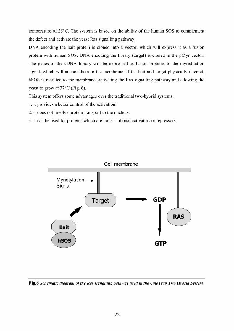
temperature of 25°C. The system is based on the ability of the human SOS to complement
the defect and activate the yeast Ras signalling pathway.
DNA encoding the bait protein is cloned into a vector, which will express it as a fusion
protein with human SOS. DNA encoding the library (target) is cloned in the pMyr vector.
The genes of the cDNA library will be expressed as fusion proteins to the myristilation
signal, which will anchor them to the membrane. If the bait and target physically interact,
hSOS is recruted to the membrane, activating the Ras signalling pathway and allowing the
yeast to grow at 37°C (Fig. 6).
This system offers some advantages over the traditional two-hybrid systems:
1. it provides a better control of the activation;
2. it does not involve protein transport to the nucleus;
3. it can be used for proteins which are transcriptional activators or repressors.
GDP
GTP
Cell membrane
Target
RAS
MyristylationSignal
Bait
hSOS
Fig.6 Schematic diagram of the Ras signalling pathway used in the CytoTrap Two Hybrid System
22

In order to express fusion proteins in the yeast cytoplasm, the system provides two
expression vectors.
The pSos vector (Fig. 18) contains the hSOS gene cloned upstream of the multiple cloning
site and under the control of the ADH1 promoter which is constitutively active. It is
designed with replication origins for propagation in yeast and bacterial strains and
selectivity markers for transforming in yeasts (the auxotrophic marker LEU2) and bacteria
(the ampicillin resistance gene).
The pMyr vector (Fig. 18) contains the gene coding for the myristilation signal cloned
upstream of the target gene and under the control of the GAL1 promoter, which is inducible
by adding galactose into the medium. It is designed with replication origins for propagation
in yeast and bacterial strains and selectivity markers for transforming in yeasts (the
auxotrophic marker URA3) and bacteria (the chloramphenicol resistance gene).
The different antibiotic resistance genes used by the two vectors allow to rapidly distinguish
between the bait and the target vector when recovering plasmids from yeasts.
As controls, four different expression vectors are provided by the system: pSosMAFB,
pMyrMAFB, pSosColl and pMyrLamin C. pSosMAFB and pMyrMAFB express the SOS
protein or the myristilation signal as fusion proteins with full-lengh MAFB, a transcription
factor that can form homodimers via its leucine zipper domain. pSosColl expresses the SOS
protein as a fusion protein with the murine 72 kDa type IV collagenase (aa 148-357) and
pMyrLamin C expresses the myristilation signal as fusion a protein with human lamin C (aa
67-230). These plasmids are used in pairwise combinations as positive and negative controls
for the rescue of the temparature-sensitive phenotype of cdc25H. pSosMAFB and
pMyrMAFB protein products interact in vivo. Co-transformation of these two control
plasmids permits growth of the cdc25H mutants at the restrictive temperature of 37°C. The
pSosMAFB + pMyrLamin C plasmid pair and the pSosColl + pMyrMAFB plasmid pair are
negative controls whose protein products do not interact in vivo. These proteins do not allow
the growth of cdc25H mutants at 37°C.
4.2 MATCHMAKER GAL4 Two-Hybrid System 3
This system (provided by Clontech) is a GAL4- based two-hybrid that provides a
transcriptional assay for detecting protein interactions in vivo in yeasts. The bait gene is
expressed as a fusion protein with the GAL4 DNA-binding domain (DNA-BD), while
another gene or cDNA is expressed as a fusion protein to the GAL4 activation domain (AD).
23

GAL UAS
Library protein
minimal promoterreporter gene
Bait protein
DNA-BD
AD
transcription
Fig.7 Schematic diagram of the principle employed by the MATCHMAKER3 GAL4
Two Hybrid System
When bait and library fusion protein interact, the DNA-BD and AD are brought into
proximity, thus activating transcription of four reporter genes: ADE2, HIS3, MEL1 and lacZ
(Fig. 7). The system can be used to identify novel protein interaction as well as to confirm or
define suspected interacting domains.
The system uses four different reporter genes under the control of distinct GAL4 upstream
activating sequences (UASs) and TATA boxes. ADE2 and HIS3 reporters allow strong
nutritional selection and the control of the stringency of selection. MEL1 and lacZ, which
incode for α-galactosidase and β-galactosidase respectively, allow the employment of
blue/white screening.
The vectors of the system- pGBKT7 and pGADT7 are designed to express different
bacterial transformation markers- kanamycin and ampicillin, different yeast selection
markers- -TRP1 and LEU2, c-Myc and hemagglutinin (HA) epitope tags for convenient
identification of the fusion proteins and T7 promoters to allow in vitro transcription and
translation of epitope-tagged fusion proteins.
The positive controls of the system are pGBKT7-53 and pGADT7-T vectors, which incode
fusion proteins between GAL-4 DNA-BD and AD and murine p53 and SV40 large T-
antigen. p53 and large T-antigen interact in a two-hybrid assay. Additionally, pCL1 encodes
the full-length wild-type GAL4 protein and provides a positive control for α-galactosidase
assays.
24

The negative control of the system is pGBKT7-Lam vector, which encodes for a fusion of
the DNA-BD with human lamin C. This protein neither forms complexes nor interacts with
most other proteins.
The yeast strains provided by the system- AH109 and Y187 are gal4- and gal80- in order to
prevent the interference of native regulatory proteins with the regulatory elements of the
two-hybrid system. AH109 usage is recommended for library screens using HIS3, ADE2
and MEL1. Y187 usage is recommended for testing interactions between two known
proteins using the lacZ reporter only.
25
AIMS OF THE PROJECT
Two isoforms of human CD23 (CD23a and CD23b) have been described. They differ by
only 6-7 residues in the extremity of the cytoplasmic tail. CD23a is restrictively expressed on
B-cells while CD23b is inducible on B-cells, as well as monocytes, eosinophils,
macrophages and a variety of other cell types after IL-4 stimulation.
The two isoforms seems to have different functions. CD23a appears to be the isoform
associated with endocytosis of IgE IC and mediating antigen presentation on B-cells. CD23b
has a phagocytosis motif and seems to be involved in the phagocytosis of IgE-coated
particles, cytokine release and the generation of superoxides.
Previous studies indicate that the two isoforms connect to different signalling transduction
pathways. The comparison of events taking place in cells that express only one or both CD23
isoforms would suggest that CD23b is involved in upregulating cAMP and iNOS, whereas
CD23a mediates an increase in intracellular calcium. Additionally, recent observations show
that there is distinct regulation of the two promoters.
Two questions regarding the biology of the CD23 receptor were addressed in this study:
1) How is the B-cell specific expression of CD23a isoform regulated? In particular, is the B-
cell specific activator protein BSAP/Pax-5 implicated in the control of CD23a expression?
2) Who are the direct interaction partners of the two CD23 isoforms? In particular, can yeast
two-hybrid systems be used in order to look for cytoplasmic interaction partners for the
CD23 receptor?
26

MATERIALS AND METHODS
1.Gene cloning
1.1 Plasmid constructs
The following plasmids were used for in vitro transcription:
pBS-23A contains a 299 bp cDNA fragment of CD23a cloned into the SmaI site of the
pBlueScriptSK+ vector (Stratagene).
pBSβ−actin contains a 540 bp cDNA fragment cloned in the pBlueScriptSK+ vector
(Stratagene).
The following plasmids were used for in vitro transcription and translation:
pCR-Pax-5 –contains the full length human Pax-5 gene cloned in the MCS of the pCR-Zero
Blunt vector (Invitrogen).
The following plasmids were used in mammalian cell transfections/reporter gene assays:
pcDNA3-Pax-5 contains the human Pax-5 gene cloned in the EcoRI site of the pcDNA3
vector (Stratagene).
pEGZ –Pax-5 contains the human Pax-5 gene inserted between the EcoRI and SmiI sites of
the pEGZ vector; pEGZ vector was provided by Dr. I. Berberich (Institute for Virology,
Würzburg).
pLuc+ACP and pLuc+AP contain the CD23a core promoter (-203 to +83) and the CD23a
promoter (-1216 to +211) cloned in the SalI site of the pLuc+ vector; pLuc+ vector was
provided by Dr. J. Altschmied and all the pLuc+ constructs have been previously made in our
lab.
pXM-STAT6 was kindly provided by Dr. E. Pfitzner (Frankfurt).
The following plasmids were used in two-hybrid systems:
pSosCD23a and pSosCD23b contain the intracytoplasmic part of the CD23 isoforms cloned
in the MCS of the pSos vector. The constructs have been previously made in our lab.
pSosCD23a+Linker and pSosCD23b+Linker contain the intracytoplasmic part of the CD23
isoforms and a linker region cloned in the HindIII site of the pSos vector, upstream of the
human SOS gene.
27

pSosCD23a-Glu represents the construct pSosCD23a+Linker in which the tyrosine in
position 6 of the cytoplasmic tail of CD23a has been replaced with a glutamic acid using site
directed mutagenesis.
pMyr-fyn contains the fyn gene cloned in the MCS of the pMyr vector.
pGBKCD23a and pGBKCD23b contain the intracytoplasmic part of the CD23 isoforms
cloned between the EcoRI and XhoI sites of the pGBK vector.
pGAD1/12 constructs contain different clones (spleen library genes) transferred from the
pMyr library and cloned between the NcoI and BamHI site of the pGAD vector.
1.2 Oligonucleotides
The following annealed oligonucleotides (MWG-Biotech) were cloned inside the MCS of
the pSos vector:
CD23a –intracytoplasmic part
M191- 5’-GGC CAA GCT TCC ACC ATG GAG GAA GGT CAA TAT TCA GAG ATC
GAG GAG CTT CCC AGG AGG CGG TGT TGC AGG CGT GGG GGA TCC CG-3’
M192- 5’-CGG GAT CCC CCA CGC CTG CAA CAC CGC CTC CTG GGA AGC TCC TCG
ATC TCT GAA TAT TGA CCT TCC TCC ATG GTG GAA GCT TGG CC-3’
CD23b- intracytoplasmic part
M193- 5’-GGC CAA GCT TCC ACC ATG AAT CCT CCA AGC CAG GAG ATC GAG GAG
CTT CCC AGG AGG CGG TGT TGC AGG CGT GGG GGA TCC CG-3’
M194- 5’-CGG GAT CCC CCA CGC CTG CAA CAC CGC CTC CTG GGA AGC TCC TCG
ATC TCC TGG CTT GGA GGA TTC ATG GTG GAA GCT TGG CC-3’
Linker region
M195-5’-CGG GAT CCG GCG GTG GCG GTT CTG GTG GCG GTG GCT CCG GCG GTG
GCG GTT CTG AAG CTT CGG G-3’
M196-5’-CCC GAA GCT TCA GAA CCG CCA CCG CCG GAG CCA CCG CCA CCA GAA
CCG CCA CCG CCG GAT CCC G-3’
1.3 Annealing reaction
The standard reaction by which two complementary oligonucleotides were annealed in a
double stranded DNA fragment was:
2 nmol oligonucleotide 1
2 nmol oligonucleotide 2
H2O up to 50 µl
28
The reaction was incubated for 5 min at 95°C and left in a 100°C water bath to cool till it
reached room temperature.
1.4 RT-PCR
The reaction was performed using Titan One Tube RT-PCR System (Roche) a sensitive
technique that allows cloning of RNA messages in one step reaction.
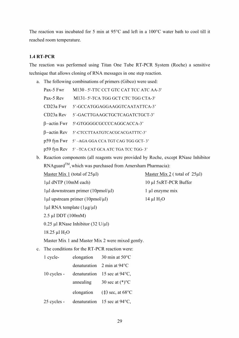
a. The following combinations of primers (Gibco) were used:
Pax-5 Fwr M130 - 5'-TTC CCT GTC CAT TCC ATC AA-3'
Pax-5 Rev M131- 5'-TCA TGG GCT CTC TGG CTA-3'
CD23a Fwr 5’-GCCATGGAGGAAGGTCAATATTCA-3’
CD23a Rev 5´-GACTTGAAGCTGCTCAGATCTGCT-3’
β−actin Fwr 5'-GTGGGGCGCCCCAGGCACCA-3’
β−actin Rev 5’-CTCCTTAATGTCACGCACGATTTC-3’
p59 fyn Fwr 5’ –AGA GGA CCA TGT CAG TGG GCT- 3’
p59 fyn Rev 5’ –TCA CAT GCA ATC TGA TCC TGG- 3’
b. Reaction components (all reagents were provided by Roche, except RNase Inhibitor
RNAguardTM, which was purchased from Amersham Pharmacia):
Master Mix 1 (total of 25µl) Master Mix 2 ( total of 25µl)
1µl dNTP (10mM each) 10 µl 5xRT-PCR Buffer
1µl downstream primer (10pmol/µl) 1 µl enzyme mix
1µl upstream primer (10pmol/µl) 14 µl H2O
1µl RNA template (1µg/µl)
2.5 µl DDT (100mM)
0.25 µl RNase Inhibitor (32 U/µl)
18.25 µl H2O
Master Mix 1 and Master Mix 2 were mixed gently.
c. The conditions for the RT-PCR reaction were:
1 cycle- elongation 30 min at 50°C
denaturation 2 min at 94°C
10 cycles - denaturation 15 sec at 94°C,
annealing 30 sec at (*)°C
elongation (‡) sec, at 68°C
25 cycles - denaturation 15 sec at 94°C,
29

annealing 30 sec at (*)°C
elongation (‡) sec at 68°C + cycle elongation of 5 sec for each
cycle
1 cycle - elongation 7 min at 68°C
Amplification product (*) Annealing temperature (‡) Elongation time
CD23a 61°C 45 sec Pax-5 66°C 60 sec β-actin 66°C 45 sec p59 fyn 53°C 80 sec
1.5 Site directed mutagenesis
The site-specific mutagenesis by overlap extension [28,30] uses two sets of mutagenic
primers (R2/FM and RM/F2) and two successive PCR reactions in order to introduce a
mutation into a desired location. The mutagenesis product is designed to contain restriction
enzyme sites, which allow it to be cloned back into the vector.
a. The mutagenic primers used were (MWG-Biotech):
◦ Set of mutagenic primers for introducing mutations into the CD23-1 site within the
CD23 promoter (pLuc+ACPmu)
M256-R2 – 5’-TGT ATC TTA TCA TGT CTG GAT CTC GAA GCT TGC-3’
M257-FM – 5’-CAC GCA CAA CTT ATA CTGGCACTTCCCACACCC-3’
M258-RM – 5’-GTG TGG GAA GTG CCA GTA TAA GTT GTG CGT GTA AT-3’
M259-F2 –5’- TTT ACC AAC AGT ACC GGA ATG CCA AGC TCA G-3’.
◦ Set of mutagenic primers for Tyr to Glu residues replacement in the
pSosCD23a+Linker
M320-FM- 5’-GGA GGA AGG TCA AGA ATC AGA GAT CGA GGA GCT T-3’
M321–R2 - 5’-CCC AAC CAG CTT TAA AAT GTC TGC AGA AAT GTA TTC-3’
M321–F2 - 5’-AAC GAG TTT ACG CAA TTG CAC AAT CAT GCT GAC-3’
M323–RM -5’-AAG CTC CTC GAT CTC TGA TTC TTG ACC TTC CTC C-3’
b. Reaction components (total of 100 µl):
2 µl template pLuc+ACP (50ng/µl)
3 µl downstream primer (10pmol/ml)
3 µl upstream primer (10pmol/ml)
30
2 µl dNTP (10 mM each)
1 µl Pfu DNA Polymerase (Stratagene)
10 µl 10xPfu Buffer (Stratagene)
79 µl H2O
c. The conditions for all PCR reactions were:
25 cycles - denaturation 1 min at 94°C,
annealing 1 min at 67°C (CD23-1 mutant); 68°C ( Tyr to Glu mutant)
elongation 1 min at 72°C
1 cycle - denaturation 1 min at 94°C
annealing 10 min at 72°C
1.6 Nucleic acid cleaning and purification procedures
PCR and RT-PCR products were either directly purified using QIAquick® PCR Purification
Kit (Qiagen) or the bands were extracted from the agarose gel using QIA®quick Gel
Extraction Kit (Qiagen) and MinEluteTM Gel Extraction Kit (Qiagen).
DNA fragments resulted from enzymatic restriction were purified either by using QIAquick®
Nucleotide Removal Kit (Qiagen) and MinEluteTM Nucleotide Removal Kit (Qiagen) or by
gel extraction.
1.7 Polishing of PCR products
In order to increase the efficiency of blunt-ended cloning reactions, PCR generated fragments
were polished using PCR Polishing Kit (Stratagene) according to manufacturer’s
recommendations.
1.8 Subcloning of PCR products
Purified PCR and RT-PCR products have been cloned using the Zero BluntTM PCR Cloning
Kit (Invitrogen). This kit is designed to clone blunt PCR fragments (or any blunt DNA
fragment) with a low background of recombinants. The pCR®-Blunt vector allows direct
selection of recombinants via desruption of a lethal E. coli gene, ccdB. Ligation of the insert
into the linearized pCR-ZeroBlunt vector and transformation into the One ShotTM Top 10
competent cells was done according to manufacturer’s instructions. The efficiency of ligation
was assessed by restriction enzyme digestion with EcoRI or by a control PCR reaction
following the protocol of the kit, except that bacterial colonies picked directly from the plate
31

were used instead of template DNA. Taq polymerase and Buffer Y (PeqLab) were used in
this specific PCR reaction.
1.9 DNA digestion with restriction enzymes
The procedure was generally used to cut DNA fragments from one plasmid in order to insert
it in the multiple cloning site of another plasmid or to analyse insertion or orientation of a
DNA fragments into a vector.
Reaction components:
1 µg plasmid DNA
2 µl 10x restriction enzyme Buffer
5 U restriction enzyme
H2O up to 20 µl
The enzymatic reaction was incubated at 37°C for 1-4 hours. Enzymatic activity was stopped
by heat inactivation, addition of 1xLoading dye or by freezing at –20°C.
The following restriction enzymes (New England Biolabs) were used in different application:
EcoRI, SalI, BamHI, HindIII, XhoI, XbaI, HpaII, AvrII, NaeI, etc.
1.10 Klenow Fill-in reaction
DNA Polymerase I, Large (Klenow) Fragment is a proteolytic product of E. coli DNA
Polymerase I, which retains polymerization and 3’to 5’ exonuclease activity, but has lost 5’
to 3’ exonuclease activity. It was used for 3’-end labeling of DNA (described elsewhere), fill-
in of 5’ overhangs and removal of 3’ overhangs to form blunt ends.
Reaction:
0.1-4 µg DNA in 1x Klenow Reaction Buffer or 1x NEBuffer (NE Biolabs)
1 µl dNTPs 0.5 mM each
1µl (5U) Klenow (USB Corporation)
The reaction was incubated for 60 minutes at 37°C.
1.11 Dephosphorylation of DNA
Alkaline phosphatase or Calf Intestinal Phosphatase (CIP) catalyses the removal of 5’
phosphate groups from DNA. Since 5’ phosphoryl termini are required by ligases, CIP
treatment was used to prevent recircularization of vectors and thus to decrease the vector
background in cloning strategies.
32

Reaction:
DNA suspended in 1xNEBuffer
CIP (NE Biolabs) 0.5 unit / 1 µg DNA
The reaction was incubated for 60 minutes at 37°C.
1.12 Ligation
T4 DNA Ligase joins blunt and cohesive ends by catalyzing the formation of phosphodiester
bonds between 5’ phosphate and 3’ hydroxyl termini. It was used for cloning restriction
fragments into vectors.
In all ligation reactions, an optimum insert: vector ratio of 5:1, expressed in pmol ends, was
used. The following formula was used in order to assess the number of pmol ends in each
case.
pmol ends/ µg DNA=2x10⁶ / bp x 660
The typical ligation reaction (where x:y respect the above described ratio) was:
x µl insert DNA
y µl vector
1 µl 10x T4 DNA Ligase Reaction Buffer (NE Biolabs)
1 µl T4 DNA Ligase (NE Biolabs)
H2O up to 10 µl
The reaction was incubated at 16°C overnight.
1.13 Transformation of bacteria
Several Escherichia coli strains were used for transformation of different cloning vectors, in
order to propagate, multiply or store different constructs.
A standard transformation reaction is presented here:
- competent cells were thawed on ice; 100 µl competent cells were aliquated in prechilled
15 ml polypropylene tubes;
- 1-50 ng of DNA per trasformation reaction was added;
- the reaction was incubated on ice for 30 minutes;
- the reaction was heat-pulsed in a 42°C water bath for 20-90 sec, depending on the strain
used; the duration of the heat pulse is critical for transformation efficiency;
- the reaction was incubated on ice for 2 minutes;
- 900 µl SOC medium or LB medium per trasformation reaction were added;
- the reaction was incubated at 37°C for 1 hour with shacking at 225-250 rpm;
33
- the cells were plated on LB agar plates containing the appropriate antibiotic;
- the plates were incubated overnight at 37°C.
1.14 Plasmid extraction
Plasmid extraction was performed using NucleobondR AX (Macherey-Nagel), GenElute
Plasmid Mini-prep Kit (Sigma) and WizardR Plus SV Minipreps DNA Purification Systems
(Promega).
1.15 Visualisation of DNA
DNA fragments were visualised by running 1-1.5% agarose gels, depending on the size of
the expected bands. 50 Base-Pair Ladder (Amersham Pharmacia) and peqGold 1 kb DNA-
Ladder (PeqLab) were used as markers.
1.16 Sequencing
Sequencing was mainly used to check the correct insertion of constructs or mutagenesis.
a. The following sequencing primers (Gibco BRL) were generally used:
M13F (universal primer)– 5’ –GTA AAA CGA CGG CCA G-3’
M13R (universal primer) – 5’-CAG GAA ACA GCT ATG AC-3’
M163 (pLuc+ reverse primer) –5’-CTT TAT GTT TTT GGC GTC TTC C-3’
M216 (pLuc+ forward primer) –5’-GCA TTC TAG TTG TGG TTT GTC C-3’
M78 (pSos 5’ primer)- 5’ –CCA AGACCA GGT ACC ATG-3’
M79 (pSos 3’ primer)- 5’ –CGC AGG GTT TTC CCA GT-3’
M215 (upstream of human Sos 5’ primer)- 5’ –CGT TCC CTT TCT TCC TTG-3’
M110 (pMyr 5’primer)- 5’ –ACT ACT AGC AGC TGT AAT AC-3’
M111 (pMyr 3’primer)- 5’ –CGT GAA TGT AAG CGT GAC AT-3’
b. Reaction components:
4 µl ABI Prism®BigDyeTM Terminator (Applied Biosystems)
0.5 µl forward or reverse primer (10nmol/µl)
500 ng DNA
H2O up to 20 µl
c. PCR conditions:
1 cycle- denaturation 3 min at 95°C
25 cycles - denaturation 30 sec at 94°C,
annealing 1 min at 50°C
34
elongation 3 min 60°C
1 cycle - elongation 5 min at 72°C
d. Purification of PCR products was performed using Auto-SeqTMG-50 columns
(Amersham Pharmacia) according to manufacturer’s instructions.
e. DNA precipitation: 20µl PCR product were precipitated with 80µl isopropanol 75%,
vortexed and incubated for 15 min at room temperature, followed by centrifugation at
top speed at room temperature for 20 minutes. After the removal of the supernatant
the DNA pellet was washed with 250µl isopropanol 75%, mixed gently and
centrifuged for another 5 minutes, top speed, at room temperature. The pellet was left
to dry for 10-15 minutes at room temperature.
f. Resuspention of DNA: the DNA pellet was resuspended in 18µl Template
Suppression Reagent (Applied Biosystems)
g. Denaturation of DNA: 3 minutes at 95°C in a heating block.
2. Protein extraction
2.1 Protein extraction from mammalian cells
1x10⁸ cells were resuspended in 200µl Roti®-Load1 (Roth) and heated at 80°C for 5 min.
The lysate was then sonicated 4 x 10 sec, 50% power, followed by another 3 min at 80°C. 3
µl of the lysate were used in Western Blot analyses.
2.2 Protein extraction from yeasts
5 ml overnight yeast culture with the O.D. >1 were pelleted at 4000xg, 10 min, 4°C and
resuspended in 200 µl Cell Lysis Buffer for Protein Isolation with freshly added proteases.
The cells were mixed with an equal volume of 0.5 mm glass beads (Roth) and vortexed for 5
min at 4°C. After centrifuging for 5 min at 12000xg and 4°C the supernatant was transferred
to a new 1.5 ml Eppendorf tube and kept on ice. The procedure was repeated with 100 µl of
Cell Lysis Buffer for Protein Isolation and the supernatants were combined. 15-20 µl were
used in Western Blot analyses.
35
3. Western Blot Analyses
Western Blot analyses were performed in order to detect and quantify proteins by using
polyclonal or monoclonal antibodies.
a. Denaturing SDS-Polyacrylamide Gel Electrophoresis
SDS-PAGE under denaturing conditions (0.1% SDS) separates protein based on molecular
size as they move through a polyacrylamide gel matrix toward the anode. The
polyacrylamide gel is cast as a separating gel topped by a stacking gel and secured in an
electrophoresis apparatus.
The final acrylamide concentration in the stacking gel is 4%, while the acrylamide
concentration in the separating gel had to be adjusted according to the protein size. A 10%
gel was used for Pax-5 (50 kDa) and a 6% gel for human SOS (170 kDa). The samples were
solubilized in Roti®-Load1 (Roth) and loaded on the gel together with a prestained SDS-
PAGE Standard marker (Bio-Rad Laboratories). The electrophoresis was performed at 200V
in an electrophoresis chamber (Hoefer) filled with Running Buffer.
b. Immunoblotting
After being separated by SDS-PAGE, the proteins were transferred using a semidry system
(Panther Semidry Electroblotter- OWL) to a transfer nitrocellulose membrane (HybondTM
ECLTM- Amersham Pharmacia) and stained by polyclonal or monoclonal antibodies.
After disassembling the PAGE gel and discarding the stacking gel, the separating gel was
equilibrated for 15 min in Cathode Buffer on an orbital shaker. The nitrocellulose membrane
was prewet on distilled water and equilibrated for 15 minutes in Anode buffer II on an orbital
shaker.
The transfer stack was assembled as follows:
Cathode electrode plate
3 sheets of Whatman 3MM filter paper saturated with Cathode Buffer
Gel
Transfer membrane
1 sheet of Whatman 3MM filter paper saturated with Anode Buffer II
2 shees of Whatman 3MM filter paper saturated with Anode Buffer I
Anode electrode plate
The proteins were transferred for maximum 2 h at 0.8 mA/cm2.
36

c. Immunodetection
Immunodetection was performed using the following protocol:
Blocking of the membrane- non-specific binding sites were blocked by incubating the
membrane 1 hour at room temperature or overnight at 4°C in 5% dried milk, 0.1% Tween
20 in PBS (Blocking Buffer) on an orbital shaker;
Rincing of the membrane- 3 times for 5 minutes with Wash Buffer on an orbital shaker;
Incubation with the primary antibody- the membrane was incubated with the primary
antibody diluted in Blocking Buffer for 1 hour at room temperature on an orbital shaker.
Anti Pax-5 and the anti SOS antibodies (BD Biosciences) were diluted 1:250;
Rincing of the membrane- 1x 15 minutes and 3x 5 minutes with Wash Buffer;
Incubation with the second antibody- the membrane was incubated with the secondary
antibody –goat anti-Mouse IgG (BD Biosciences) diluted 1:2000 in Blocking Buffer for 1
hour at room temperature on an orbital shaker;
Rincing of the membrane- 1x 15 minutes and 3x 5 minutes with Wash Buffer;
Visualisation of proteins –the presence of proteins was detected using ECL PlusTM
Detection Kit (Amersham Pharmacia) and HyperfilmTM ECLTM chemiluminescence film
(Amersham Pharmacia).
4. Electrophoretic Mobility Shift Assays (EMSAs).
This is a rapid and sensitive method for the detection of interaction between DNA-binding
proteins and specific sequences of DNA. Proteins that bind specifically to an end-labeled
DNA fragment retard the mobility of the fragment during electrophoresis, resulting in
discrete bands corresponding to the protein-DNA complexes.
4.1 Oligonucleotides
The following annealed oligonucleotides (MWG-Biotech) were used as probes or as
unlabeled competitors in direct binding or competition assays:
Pax-5 high affinity binding site from the sea urchin H2A-2.2 gene
M118- 5' -CAG GGT TGT GAC GCA GCG GTG GGT GAC GAC TGT-3’
M119- 5’ -GCC ACA GTC GTC ACC CAC CGC TGC GTC ACA ACC-3’
putative Pax-5 binding site CD23-1 (-87 to -47)
M304 - 5' -GGG TGT GGG GAG CAC CAG GAG AGG CCA TGC GTG TAA TGT TA-3’
37

M305 - 5’ -GGA TAA CAT TAC ACG CAT GGC CTC TCC TGG TGC TCC-3’
putative Pax-5 binding site CD23-2 (-112 to -71)
M306 - 5’–CGG ACT TCA CCC GGG TGT GGG GAG CA-3’
M307 - 5’ –GGT GCT CCC CAC ACC CGG GTG AAG T-3’
putative Pax-5 binding site CD23-3 (-238 to -209)
M308 - 5’–GTG GTA TGA TTC AGT GTG CAG TAA CAG TGG TTC-3’
M309 - 5’-GTG AAC CAC TGT TAC TGC ACA CTG AAT CAT A-3’
• CD23-1mu1 -mutated nucleotides are underlined
M310 - 5’- GGG TGT GGG AAG TGC CAG TAT AAG TTG TG-3’
M311- 5’-ACG CAC AAC TTA TAC TGG CAC TTC CCA C-3’)
CD23-1mu2 -mutated nucleotides are underlined
M265 - 5’- GTG TGG GGA GAA CCA GTA GAG GCC ATG CGT G-3’
M266 - 5’- CAC GCA TGG CCT CTA CTG GTT CTC CCC A-3’
A-1- CD23a promoter (-212 to –173)
M37 - 5’- GGT TCA CAT CTT GAC GCT ACC ACT CAC CTC CTT CAG CCC-3’
M38 - 5’- AGG GCT GAA GGA GGT GAG TGG TAG CGT CAA GAT GTG-3’
• A-2- CD23a promoter (-178 to -140)
M39 - 5’- AGC CCT GTG GGA ACT TGC TGC TTA ACA TCT CTA GT-3’
M40 - 5’- GAG AAC TAG AGA TGT TAA GCA GCA AGT TCC CAC AGG-3’
• A-3- CD23a promoter (-147 to -107)
M41 - 5’- TAG TTC TCA CCC AAT TCT CTT ACC TGA GAA ATG GAG A-3’
M42 - 5’- GTT ATC TCC ATT TCT CAG GTA AGA GAA TTG GGT GAG AA-3’
• A-4- CD23a promoter (-115 to -75)
M43 - 5’- GGA GAT AAT AAT AAC ACG GAC TTC ACC CGG GTG TGG G-3’
M44 - 5’- GCT CCC CAC ACC CGG GTG AAG TCC GTG TTA TTA TTA CT-3’
• A-5- CD23a promoter (-83 to -44)
M45 - 5’- GTG GGG AGC ACC AGG AGA GGC CAT GCG TGT AAT GTT A-3’
M46 - 5’- GGA TAA CAT TAC ACG CAT GGC CTC TCC TGG TGC TCC-3’
• A-6- CD23a promoter (-51 to -9)
M47 - 5’- TGT TAT CCG GGT GGC AAG CCC ATA TTT AGG TCT ATG AAA-3’
M48 - 5’- GTA TTT TCA TAG ACC TAA ATA TGG GCT TGC CAC CCG GAT A-3’
A-7- CD23a promoter (-17 to +25)
M49 - 5’- TGA AAA TAG AAG CTG TCA GTG GCT CTA CTT TCA GAA GA-3’
M50 - 5’- GCT TTC TTC TGA AAG TAG AGC CAC TGA CAG CTT CTA TTT-3’
38

• A-8- CD23a promoter (+16 to +56)
M51 - 5’- GAA GAA AGT GTC TCT CTT CCT GCT TAA ACC TCT GTC TC-3’
M52 - 5’- GTC AGA GAC AGA GGT TTA AGC AGG AAG AGA GAC ACT TT-3’
• A-9- CD23a promoter (+49 to +85)
M53 - 5’- GTC TCT GAC GGT CCC TGC CAA TCG CTC TGG TCG AC-3’
M54 - 5’- GGG GTC GAC CAG AGC GAT TGG CAG GGA CCG TCA GA-3’
4.2 Radioactive labelling of annealed oligonucleotides
Double stranded DNA fragments were radioactively labelled at the 3’-end by a typical
Klenow Fill-in reaction in which dCTP was replaced by 32P-dCTP (Amersham Pharmacia
and Hartmann Analytics).
Reaction components (for a total of 25 µl):
1 µl DNA (100ng/µl)
1µl d(TGA)TP, 5 mM each
5 µl α-32P-dCTP (10µCi/µl)
1 µl (5 U) Klenow Fragments (USB Corporation)
2.5µl 10x Klenow Fill-in Buffer (USB Corporation)
13.5 µl H2O
The reaction was incubated at 37°C for 30 min. Non-incorporated nucleotides were washed
with the QIAquick® Nucleotide Removal Kit (Qiagen). The product was eluted in 80µl
elution buffer and the incorporation of radioactive nucleotides was assessed using a
scintillation counter.
4.3 In vitro Transcription and Translation
Human Pax-5 protein was obtained by in vitro transcription and translation using TNT®Quick
Coupled Transcription/ Translation Systems (Promega) appropriate for vectors containing a
promoter for T7 RNA polymerase. pCR-Pax-5 was used as template.
4.4 Direct Binding and Competition Assays
The method can be used to visualize protein-DNA interaction by direct binding assay, in
which the protein binds a labeled oligonucleotide, or by competition assay, in which cold
competitors are able to inhibit the formation of a complex between the protein and a labeled
oligonucleotide. The specificity of the complexes formed is determined by supershifts, in
39
which antibodies added to the reaction bind to the protein–DNA complexes and retard the
migration of the specific band.
The basic reaction in direct binding assays was:
2 µl 10x Pax-5 Binding Buffer
2 µl in vitro translated Pax-5 protein
1 µg poly[d(I-C)]
40000 cpm 32P-labelled oligonucleotide (approximatively 0,5-1 ng)
1 µg anti-Pax-5 antibody (BD Biosciences) – only in supeshift samples
H2O up to 20 µl
The reaction was incubated for 15 min at room temperature before adding:
2 µl of 10x Loading Buffer.
The basic reaction in competition assays was:
2 µl 10x Pax-5 Binding Buffer
2 µl in vitro translated Pax-5 protein
1 µg poly[d(I-C)]
2 µl of unlabeled oligonucleotides (1, 10 and 100ng/µl)
H2O up to 20 µl
The reaction was incubated for 10 min at room temperature before adding:
40000 cpm 32P-labelled oligonucleotide (approximatively 0,5-1 ng)
The reaction was incubated for 15 min at room temperature before adding:
2 µl of 10x Loading Buffer.
In all case the samples were run on nondenaturating 5% polyacrylamide gel at 15V/cm for 2
hours. The gels were covered with plastic wrap, dried under vacuum and then exposed on
BioMaxTM MS (Kodak scientific imagining film) at –80°C overnight.
5. Ribonuclease Protection Assay
The RPA is an extremely sensitive procedure for the detection and quantitation of mRNA in
a complex sample mixture of total cellular RNA.
In our case 5x10⁶ U-937 cells where stimulated with IL-4 (50ng/ml), PMA (3ng/ml) or both
for 48h. Total RNA was prepared in 1 ml TRIZOL reagent (Gibco) following manufacturer’s
instructions.
40
5.1. Linearization of template DNA
Plasmid DNA must be linearized with a restriction enzyme downstream of the insert to be
transcribed, as circular plasmids will generate extremely long, heterogenous RNA transcripts.
pBS-CD23a, the template plasmid for CD23 transcripts was linearized with EcoRI restriction
enzyme; pBS-β-actin, the template plasmid for β-actin, which was used as an internal
control, was linearized with XbaI restriction enzyme. The linearized plasmids were observed
on a 1 % agarose gel and the bands were extracted with QIAquick® Gel Extraction Kit
(Qiagen).
5.2. In vitro Transcription
In vitro transcription was performed using [α-32P] UTP (Amersham Pharmacia) as
radiolabeled nucleotide and MAXIscript TM T7/T3 (Ambion) In vitro Transcription Kit
following manufacturer’s instructions. Transcription products were purified using
MicroSpinTMS-200 HR Columns (Amersham Pharmacia) and run on a denaturing 5 %
polyacrylamide/urea gel at 200V for 1 hour. The disassembled gel was kept on one plate,
covered with plastic wrap and exposed for 4 min on Sterling film (Diagnostic Imaging).
Using the film as a template, the full length RNA band was excised from the gel. The RNA
transcripts were eluted overnight, at 37°C in elution buffer from the HybSpeed TM RPA Kit
(Ambion). The incorporation of radioactive nucleotides was assessed using a scintillation
counter. The probes gave rise to a 299 bp band for CD23a, a 184 bp band for CD23b and a
double band around 130 bp for β-actin.
5.3. Hybridization reaction
Hybridization reaction was carried on using HybSpeed TM RPA Kit (Ambion) according to
manufacturer’s instructions except hybridization was extended overnight. Results were
assessed by autoradiography on BioMaxTM MS (Kodak Scientific Imaging Film) at –80°C
for 24-72 hours.
6. Transfection and reporter gene assay
Transient transfection assays are used for analysing mammalian gene expression in vivo.
Fusion genes constructs consisting of promoter or enhancer sequences under study attached
41
to a gene directing the synthesis of a reporter molecule are used to assess gene expression
within 48 hours after introduction of the DNA. Luciferase assay use the luc gene of the
firefly as a reporter gene and yields light signals that can be detected using an automated
injection luminometer.
6.1 Transfection
3x10⁵ 293 cells were transfected with 100 ng of the pLuc+ vectors and 500 ng of the
pcDNA3-Pax-5 and pXM-STAT6 vectors. DNA concentration was brought to a total of 2µg
DNA / transfection using salmon sperm DNA (Amersham Pharmacia). Transfection was
performed in 1 ml trasfection volume using GenePORTERTM Transfection Reagent (PeqLab)
according to manufacturer’s instructions, except that growth medium was completely
renewed 24 hours post transfection. Four hours after transfection cells were stimulated with
IL-4 to a final concentration of 50ng/ml (rIL-4 provided by Prof. Sebald, Biozentrum,
University of Würzburg) and PMA to a final concentration of 3ng/ml (Sigma).
6.2 Cell Lyses and Luciferase Assay
Cells were removed by pipetting up and down, collected in 15 ml tubes and washed 2 times
with cold PBS followed by centrifugation at 1200xg, 10 min, 4°C. Pellets were left to dry at
room temperature and then resuspended in 1x Reporter Lysis Buffer (Promega). The
luciferase activity was assessed following manufacturer instructions- Luciferase Assay
System (Promega) using a luminometer (Berthold). The results were normalized for equal
concentration of total protein.
7. Transfection/ Infection Assays
Transfection of U-937 cells with the recombinant vector pEGZ-Pax-5 has been performed by
Dr. I. Berberich (Institute for Virology and Immunobiology, Würzburg). Recombinant
retroviral particles were generated using the pHIT packaging system as described by Soneoka
et al [47].
Briefly, 293T cells were transiently cotransfected using the standard calcium phosphate
method with 5 µg of each of the packaging vectors pHIT456 (which codes for the
amphotropic env protein) and 5-7 µg of the retroviral construct pEGZ/MCS and EGZ/Pax-5.
Sixteen hours later the transfection solution was replaced by DMEM. Viral supernatants were
42

harvested 48h later and filtered (0.45µm). Polybrene (Sigma) was added to a final
concentration of 10µg/ml. U-937 cells were mixed with the retroviral supernatant and
centrifuged for 3 hours at 1000 x g. Thereafter cells were replated in RPMI. Transduced cells
were selected with Zeocin (250 µg/ ml).
The cell line selected for experiments stained positive for EGZ in 97% of the cells, as
revealed by FACS analysis.
8. Nucleic acid extraction
8.1 DNA extraction
DNA was extracted from 1x 10⁸ cells using QIAampR DNA Blood Kit (Qiagen) following
manufacturer’s instructions.
8.2 RNA extraction
Homogenisation- 1x10⁷ mammalian cells were pelleted by centrifugation and lysed in 1.5
ml Trizol Reagent by repetitive pipetting.
Phase separation- The homogenized samples were incubated for 5 minutes at room
temperature to permit the complete dissociation of nucleoprotein complexes. 0.3 ml
chloroform was added and tubes were shacked vigorously by hand for 15 seconds and
incubated at room temperature for 2 to 3 minutes. Then samples were centrifuged at
12,000 x g for 15 minutes at 4°C. Following centrifugation, the mixtures separated into a
lower red, phenol-chloroform phase, an interphase and a colourless upper aqueous phase.
RNA remains exclusively in the aqueous phase.
RNA precipitation- The aqueous phase was transferred to a new tube and the RNA
precipitated by mixing with 0.75ml isopropanol. Then samples were incubated at room
temperature for 10 minutes and centrifuged at 12000x g for 10 minutes at 4°C. The RNA
precipitate was visible as a gel-like pellet.
RNA wash- After removal of the supernatant the pellet was washed once with 1.5ml of
75% ethanol. The samples were mixed by vortexing and centrifuged at 7,500x g for 5
minutes at 4°C.
Redissolving the RNA- After discarding the ethanol the RNA pellet was air-dried for 5-
10 minutes and dissolved in RNase-free water by passing the solution a few times
through a pipette tip and incubating for 10 minutes at 55 °C.
43
9. Spectrophotometric measurements
9.1 Nucleic acids
To assess the concentration of nucleic acids in a sample absorbtion is measured at 260
wavelenght. Absorbance at 280 wavelengths is measured to assess the purity of the sample.
The A260/A280 ratio is used to estimate the purity of the sample and should be between 1.6
and 2. To determine the nucleic acid concentration (in µg/ml) the A260 reading has to be
multiplied with a factor of 50 for double stranded DNA, 33 for single stranded DNA and 40
for RNA.
9.2 Proteins
For measuring the protein concentration 10 µl protein solution were diluted in 190 µl H2O
and 800 µl 1x RotiR- Nanoquant (Roth). The absorbtion of the sample is measured at 590 and
450 wavelenghts. The A590/A450 ratio was introduced into a linear regression program in
order to determine the total protein concentration in the sample.
10. Two Hybrid Systems
10.1 CytotrapTM Two Hybrid System (Stratagene)
This is a novel method for detecting in vivo protein-protein interaction, which is based on
generating fusion proteins whose interaction in the yeast cytoplasm induces cell growth by
activating the Ras signaling pathway.
The system provides:
- the host strain cdc25H
- the vectors: the bait vector (pSos), the target vector (pMyr) and control vectors (pSos
MAFB, pSos Coll, pMyr MAFB, pMyr Lamin C)
The human spleen cDNA library cloned in the pMyr vector was purchesed from Stratagene
and amplified following manufacturer’s instructions.
All protocols and procedures for the CytoTrap Two Hybrid System were carried on
following manufacturer instruction, with a few modifications, as noted:
Preparation and Trasformation of Yeast Competent Cells- was performed using the
following protocol [91]:
44

- 5 ml of liquid YPAD were inoculated with one colony of yeast and shacked at 250
rpm, overnight, at 25°C.
- The overnight culture was used to inoculate 50 ml of YPAD and incubated up to a
cell density of 0.7 OD.
- The culture was harvested in a sterile 50 ml tube at 3000xg for 5 minutes, at room
temperature.
- The cells were resuspended very gently in 25 ml sterile water and centrifuged again.
- The pellet was resuspended in 1 ml 100mM lithium acetate (LiAc) and transferred to
a 1.5 ml centrifuge tube.
- The cells were centrifuged for 5 sec at top speed in a table-top centrifuge and the
LiAc was removed with a pipette.
- The cell were resuspended in a final volume of 500 µl 100mM LiAc
- 50 µl of cell suspension were pipetted in labeled microfuge tubes and centrifuged for
5 sec at top speed; LiAc was removed with a pipette.
- A “transformation mix” was assembled as follows, strictly respecting this order:
240 µl PEG (50%w/v)
36 µl 1.0 M LiAc
25 µl single-stranded carrier DNA (2mg/ml) previously boiled for 5 min
and placed directly on ice
50 µl H2O and plasmid DNA (1-3 µg)
- Each tube was vortexed vigorously until the cell pellet was completely mixed and
incubated at room temperature for 30 minutes.
- Tubes were heat-shocked for 20 minutes in a water bath at 42°C.
- Tubes were centrifuged at 6000-8000 rpm for 15 sec and the transformation mix with
a pipette.
- The pellets were resuspended in 250µl sterile water by gently pipetting up and down
and plated onto selective plates.
Isolation of pMyr cDNA plasmids from Yeasts –was performed using a RPM Yeast
Plasmid Isolation Kit (Bio101).
The chart flow of a screening episode is schematically presented in Figures 8-11.
Briefly, the screening steps were as follows:
- pSosCD23a/b and pMyr spleen library were co-transformed in the cdc25H yeast strain;
45
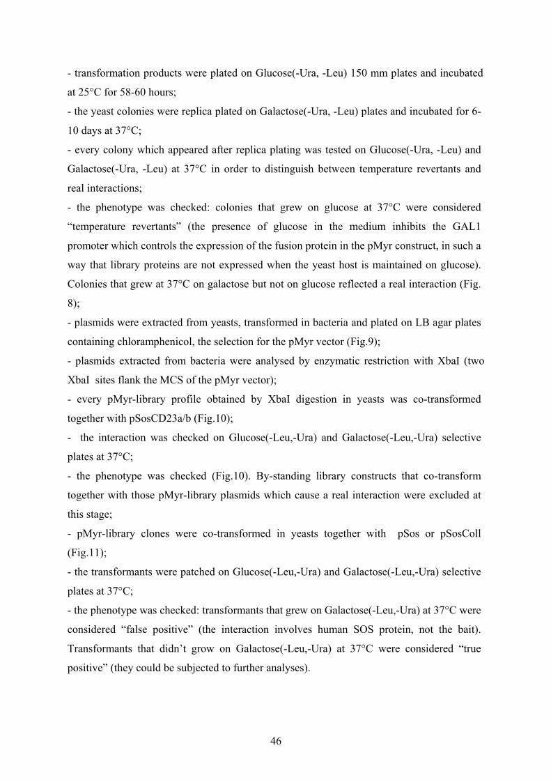
- transformation products were plated on Glucose(-Ura, -Leu) 150 mm plates and incubated
at 25°C for 58-60 hours;
- the yeast colonies were replica plated on Galactose(-Ura, -Leu) plates and incubated for 6-
10 days at 37°C;
- every colony which appeared after replica plating was tested on Glucose(-Ura, -Leu) and
Galactose(-Ura, -Leu) at 37°C in order to distinguish between temperature revertants and
real interactions;
- the phenotype was checked: colonies that grew on glucose at 37°C were considered
“temperature revertants” (the presence of glucose in the medium inhibits the GAL1
promoter which controls the expression of the fusion protein in the pMyr construct, in such a
way that library proteins are not expressed when the yeast host is maintained on glucose).
Colonies that grew at 37°C on galactose but not on glucose reflected a real interaction (Fig.
8);
- plasmids were extracted from yeasts, transformed in bacteria and plated on LB agar plates
containing chloramphenicol, the selection for the pMyr vector (Fig.9);
- plasmids extracted from bacteria were analysed by enzymatic restriction with XbaI (two
XbaI sites flank the MCS of the pMyr vector);
- every pMyr-library profile obtained by XbaI digestion in yeasts was co-transformed
together with pSosCD23a/b (Fig.10);
- the interaction was checked on Glucose(-Leu,-Ura) and Galactose(-Leu,-Ura) selective
plates at 37°C;
- the phenotype was checked (Fig.10). By-standing library constructs that co-transform
together with those pMyr-library plasmids which cause a real interaction were excluded at
this stage;
- pMyr-library clones were co-transformed in yeasts together with pSos or pSosColl
(Fig.11);
- the transformants were patched on Glucose(-Leu,-Ura) and Galactose(-Leu,-Ura) selective
plates at 37°C;
- the phenotype was checked: transformants that grew on Galactose(-Leu,-Ura) at 37°C were
considered “false positive” (the interaction involves human SOS protein, not the bait).
Transformants that didn’t grow on Galactose(-Leu,-Ura) at 37°C were considered “true
positive” (they could be subjected to further analyses).
46

10.2 MATCHMACKER GAL 4 Two-hybrid System 3 (Clontech)
This system is an advanced GAL4-based two-hybrid system that provides a transcriptional
assay for detecting protein interactions in vivo in yeasts.
The system provides:
- the host strains: AH109 and Y187
- the vectors: the bait vector (pGBKT7), the target vector (pGADT7) and control
vectors (pGBKT7-T, pGBKT7-53, pGBKT7-Lam, pCL1)
All protocols and procedures for the MATCHMACKER GAL 4 Two-hybrid System 3 were
carried on following manufacturer instruction.
47

Co-transform pSosCD23 a/b+ pMyr target library
37°C, 10 days
SD Galactose(-Ura,-Leu)
Replica plate on galactose plates
SD Glucose(-Ura,-Leu) 25°C, 48h
SD Glucose(-Ura,-Leu) 25°C
SD Glucose(-Ura,-Leu) 37°C
SD Galactose(-Ura,-Leu) 37°C
_
“real interaction” „temperature revertants“
+ +
+ + +
Fig.8 The chart flow of the CytoTrap screening: eliminating the temperature revertants
48
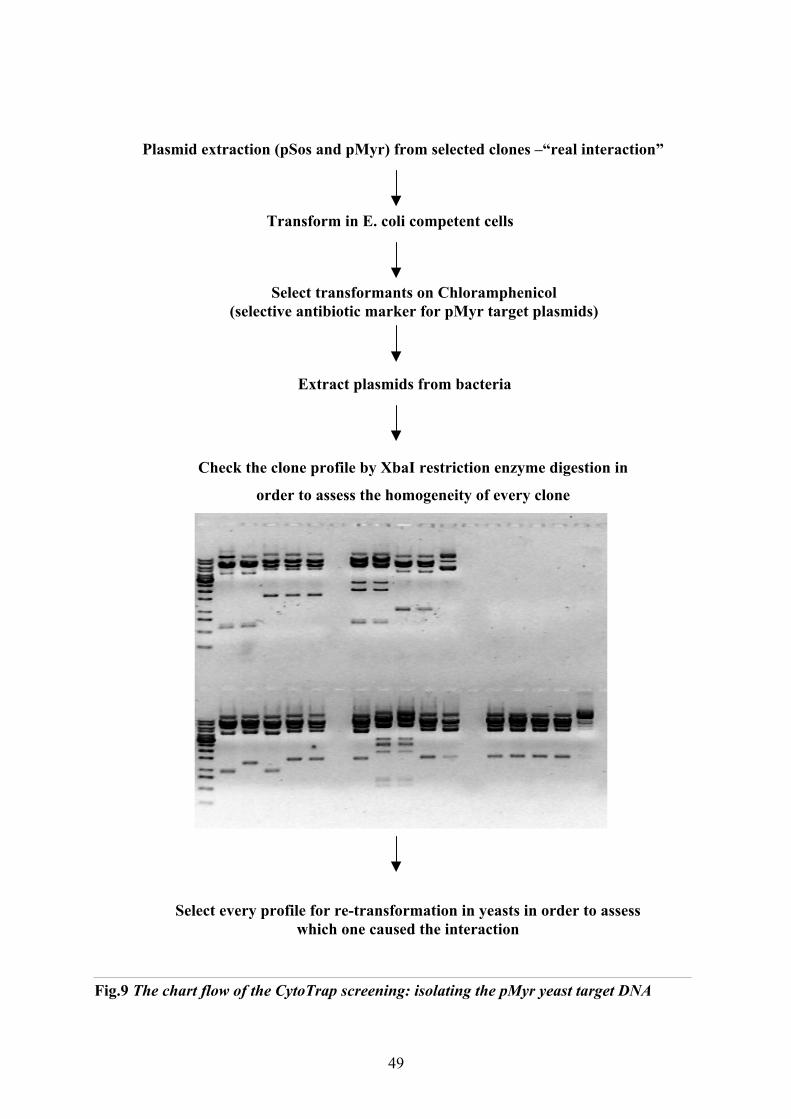
Select transformants on Chloramphenicol (selective antibiotic marker for pMyr target plasmids)
Transform in E. coli competent cells
Plasmid extraction (pSos and pMyr) from selected clones –“real interaction”
Se
Fig.9 The char
Extract plasmids from bacteria
Check the clone profile by XbaI restriction enzyme digestion in
order to assess the homogeneity of every clone
lect every profile for re-transformation in yeasts in order to assess which one caused the interaction
t flow of the CytoTrap screening: isolating the pMyr yeast target DNA
49
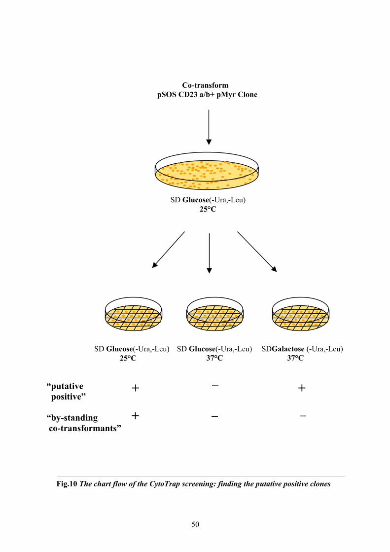
Co-transform
pSOS CD23 a/b+ pMyr Clone
__+
++“putative positive” “by-standing co-transformants”
SDGalactose (-Ura,-Leu) 37°C
SD Glucose(-Ura,-Leu) 37°C
_
SD Glucose(-Ura,-Leu) 25°C
SD Glucose(-Ura,-Leu)25°C
Fig.10 The chart flow of the CytoTrap screening: finding the putative positive clones
50

Co-transform pSos or pSosColl+ pMyr Clone
Fig.11 The chart flow of the CytoTrap screening: eliminating the false positive clones
+-+
-+“true positive” „false positive“
SD Galactose(-Ura,-Leu) 37°C
SD Glucose(-Ura,-Leu) 37°C
-
SD Glucose(-Ura,-Leu) 25°C
SD Glucose(-Ura,-Leu) 25°C
51
11. Materials
11.1 Buffers
10x Pax-5 Binding Buffer 100mM HEPES pH 7,9
1M KCl
40% Ficoll
10mM EDTA
10mM DTT
Cell Lysis Buffer for Protein Isolation 140 mM NaCl
2.7 mM KCl
10mM Na2HPO4
1.8 mM KH2PO4
1% Triton X
+ Protease inhibitors 1mM PMSF
10µg/ml aprotinin
1µM pepstatin A
100µM leupeptin
1µg/ml chymostatin
Running Buffer for SDS-PAGE 3.03 g Tris
14.4 g glycin
1 g SDS
H2O up to 1 liter
Anode Buffer I 0.3 M Tris
10% methanol
Anode Buffer II 25 mM Tris
10 % methanol
Cathode Buffer 25mM Tris
40mM 6- amino n- caproic acid
10 % methanol
Wash Buffer 100 ml PBS (10x)
1 ml Tween 20 (0,1%)
900 ml H2O
52

Blocking Buffer 1x PBS
0.1% Tween 20
5% dry milk
10 x Loading Dye (EMSA) 20% Ficoll
0,1 M Na2EDTA pH 8,0
1 % SDS
0.25 % bromphenolblue
0.25 % xylene cyanol
6 x Loading Dye (Agarose gel) 0.25 % bromphenol blue
0.25 % xylen cyanol
30 % glycerol in water
TAE electrophoresis buffer (50x) 242 g Tris base
57.1 ml glacial acetic acid
37.2 g Na2EDTA⋅2H2O
H2O up to 1 liter
TBE electrophoresis buffer (10x) 108 g Tris base
55 g boric acid
40 ml 0.5 M EDTA, pH 8.0
H2O up to 1 liter
11.2 Gels
Laemmli Gel (PAGE)
Separating gels Stacking gels
6% 10% 4%
Acrylamyde 30% 1.99 ml 3.32 ml 0.67 ml
Tris 1M, pH 8,8 3.75 ml 3.75 ml
Tris 1M, pH 6,8 0.625 ml
10% SDS 0.1 ml 0.1 ml 0.05 ml
H2O 4.12 ml 2.78 ml 3.625 ml
10% ammonium persulphate 50 µl 50 µl 25 µl
TEMED 3.3 µl 3.3 µl 2.5 µl
53
Nondenaturing 5 % polyacrylamide gel (for EMSA)
Acrylamide 30 % 6.67 ml
10 x TBE 2 ml
H2O 31.33 ml
TEMED 40 µl
10% ammonium persulphate 400 µl
Denaturing 5 % polyacrylamide/urea gel (for RPA)
Acrylamide 40 % 6.3 ml
10 x TBE 5 ml
Urea 24 g
H2O up to 50 ml
10 % ammonium persulphate 300 µl
TEMED 34 µl
Agarose gel
Agarose UltraPure (Gibco BRL) 1-1.5 %
1 x TAE
Ethidium bromide ( 0.5 µg /ml)
11.3 Media for bacteria and yeasts
LB Broth Base(Gibco BRL)
LB Agar (Gibco BRL)
Ampicillin (USB Corporation)
Chloramphenicol (Roche)
Kanamycin (USB Corporation)
DOB-Dropout Base (BIO 101)
DOBA-Dropout Agar base (BIO 101)
DOBA, 2% Gal, 1% Raf (BIO 101)
YPD Broth (BIO 101)
Agar-Agar (Roth)
CSM-Leu-Ura (BIO 101)
CSM-His-Leu-Trp (BIO 101)
CSM-His-Leu-Trp-Ura (BIO 101)
54
Adenine (Sigma)
L-Histidine (Sigma)
L-Leucine (Sigma)
L-Tryptophan (Sigma)
Uracil (Sigma)
X-α-Gal (Clontech)
11.4 E.coli competent cell strains
DH5α
Epicurian Coli® BL21-Codon PlusTM (Stratagene)
One ShotTM Top 10 (Invitrogen)
11.5 Cell lines
• The 293 cell line is a permanent line of primary human embryonal kidney transformed
by sheared human adenovirus type 5 (Ad 5) DNA. The cells were maintained in DMEM
(Gibco) supplemented with L-Glutamine (2mM), antibiotics (Penicillin 100U /
Streptomycin 100µg/ml) and 10% FCS.
• The U-937 cell line is a monoblastoid cell line. The cells were maintained in RPMI 1640
(Gibco) supplemented with L-Glutamine (2mM), antibiotics (Penicillin 100U /
Streptomycin 100µg/ml) and 10% FCS.
• The WIL2 cell line is an Epstein-Barr virus infected B cell line. The cells were
maintained in RPMI 1640 (Gibco) supplemented with L-Glutamine (2mM), antibiotics
(Penicillin 100U / Streptomycin 100µg/ml) and 10% FCS.
11.6 Software and websites
DNAsis
JellyFish
pDRAW
Kodak Imager
Miscrosoft Office
WebPrimer- http://genome-www2.stanford.edu/cgi-bin/SGD/web-primer
The Web Primer provides tools for the purpose to design primers. Current choices are
limited to sequencing and PCR.
55
NCBI Entrez Nucleotide- http://www.ncbi.nlm.nih.gov/entrez/query.fcgi?db=Nucleotide
The Entrez Nucleotides database is a collection of sequences from several sources, including
GenBank, RefSeq, and PDB.
NCBI BLAST- http://www.ncbi.nlm.nih.gov/BLAST/
BLAST® (Basic Local Alignment Search Tool) is a set of similarity search programs
designed to explore all of the available sequence databases regardless of whether the query is
protein or DNA. The scores assigned in a BLAST search have a well-defined statistical
interpretation, making real matches easier to distinguish from random background hits.
11.7 Lab devices
GeneAmp PCR System 2400 (Perkin Elmer)
ABI PRISMTM 310 Genetic Analyser (Applied Biosystems)
Digital camera
Ultrospec 1000 spectrophotometer (Amersham Pharmacia)
MicroLumat LB 96P luminometer (EG&G Berthold)
Optimax Developer (Protec)
Scintillation counter (Beckman LS 1801)
Vacuum dryer
Sonicator
Incubators
Shackers
Autoclaves
Centrifuges
Heating blocks
Water bath
56

RESULTS
1. The CD23a promoter- role of Pax-5 in the B-cell specific
expression of CD23a isoform
1.1 The CD23a core promoter contains three putatitive binding sites for Pax-5
The consensus motif for Pax-5 has been characterised by Czerny et al. in an extended
comparison between all known natural ligands of Pax-5, including the recognition sequences
which originates from the sea urchin H2A-2.2 gene and the human CD19 gene [12].
Our approach to test the hypothesis of Pax-5 regulating the expression of CD23a isoform
was to identify putative binding sites for Pax-5 in the CD23a promoter. We performed
multiple sequence alignments between the CD23a core promoter (Fig. 12A) and the Pax-5
consensus sequence. Three putative Pax-5 binding sites were identified in positions -61 to -
79, -78 to -96 and -215 to -233 of the CD23a promoter (Fig. 12) and named CD23-1, CD23-
2 and CD23-3 respectively.
All naturally occurring Pax-5 binding sites identified so far deviate from the consensus
sequence and Pax-5 is able to interact with a panel of seemingly degenerate recognition
sequences. This makes the prediction of Pax-5 binding sites quite difficult, when only
relating to the sequence of the promoter. When chosing the putative Pax-5 binding sites, we
considered only the sites with at least 50% identity to the consensus sequence. Fig. 12B
shows the sequence alignment of these three putative binding sites with the consensus motif
and other previously identified Pax-5 binding sites from the sea urchin H2A-2.2 and CD19
genes. The CD23-3 putative binding site displayed the highest homology with the consensus
motif (89%). A gap had to be introduced in the putative binding site CD23-1 in order to
reconstitute the symmetry of the recognition sequence.
57
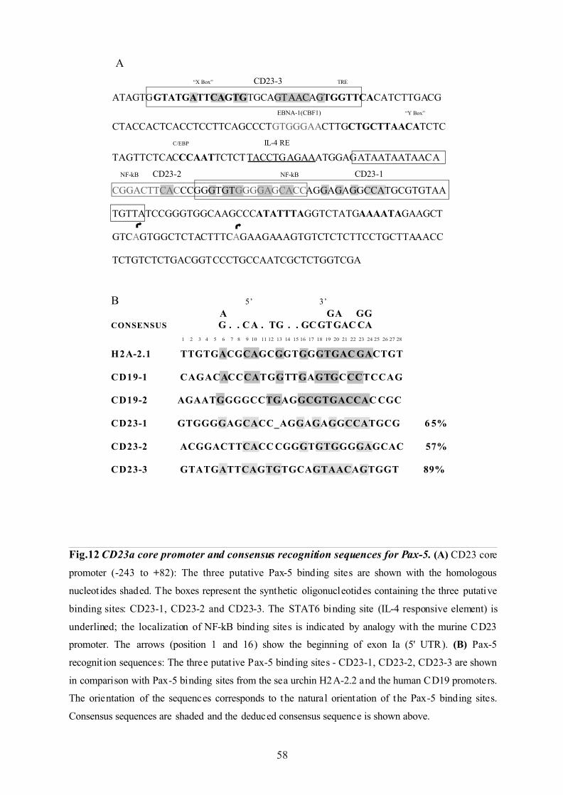
B 5’ 3’
A GA GG CONSENSUS G . . C A . TG . . GC GTGAC CA
H2A-2.1 TTGTGACGCAGCGGTGGGTGAC GACTGT CD19-1 CAGACACCCATGGTTGAGTGCCCTCCAG CD19-2 AGAATGGGGCCTGAGGCGTGACCAC CGC CD23-1 GTGGGGAGCACC_AGGAGAGGCCATGCG 6 5% CD23-2 ACGGACTTCACC CGGGTGTGGGGAGCAC 57% CD23-3 GTATGATTCAGTGTGCAGTAACAGTGGT 89%
1 2 3 4 5 6 7 8 9 10 11 12 13 14 15 16 17 18 19 20 21 22 23 24 25 26 27 28
Fig.12 CD23a core promoter and consensus recognition sequences for Pax-5. (A) CD23 core
promoter (-243 to +82): The three putative Pax-5 binding sites are shown with the homologous
nucleotides shaded. The boxes represent the synthetic oligonucleotides containing the three putative
binding sites: CD23-1, CD23-2 and CD23-3. The STAT6 binding site (IL-4 responsive element) is
underlined; the localization of NF-kB binding sites is indicated by analogy with the murine CD23
promoter. The arrows (position 1 and 16) show the beginning of exon Ia (5' UTR). (B) Pax-5
recognit ion sequences: The three putative Pax-5 binding sites - CD23-1, CD23-2, CD23-3 are shown
in comparison with Pax-5 binding sites from the sea urchin H2A-2.2 and the human CD19 promoters.
The orientation of the sequences corresponds to the natural orientation of the Pax-5 binding sites.
Consensus sequences are shaded and the deduced consensus sequence is shown above.
A
“X Box” CD23-3 TRE
ATAGTGGTATGATTCAGTGTGCAGTAACAGTGGTTCACATCTTGACG
EBNA-1(CBF1) “Y Box”
CTACCACTCACCTCCTTCAGCCCTGTGGGAACTTGCTGCTTAACATCTC
C/EBP IL-4 RE
TAGTTCTCACCCAATTCTCTTACCTGAGAAATGGAGATAATAATAACA
NF-kB CD23-2 NF-kB CD23-1
CGGACTTCACCCGGGTGTGGGGAGCACCAGGAGAGGCCATGCGTGTAA
TGTTATCCGGGTGGCAAGCCCATATTTAGGTCTATGAAAATAGAAGCT
GTCAGTGGCTCTACTTTCAGAAGAAAGTGTCTCTCTTCCTGCTTAAACC
TCTGTCTCTGACGGTCCCTGCCAATCGCTCTGGTCGA
58
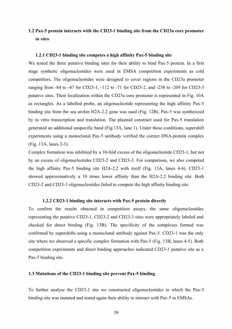
1.2 Pax-5 protein interacts with the CD23-1 binding site from the CD23a core promoter
in vitro
1.2.1 CD23-1 binding site competes a high affinity Pax-5 binding site
We tested the three putative binding sites for their ability to bind Pax-5 protein. In a first
stage synthetic oligonucleotides were used in EMSA competition experiments as cold
competitors. The oligonucleotides were designed to cover regions in the CD23a promoter
ranging from -84 to -47 for CD23-1, -112 to -71 for CD23-2, and -238 to -209 for CD23-3
putative sites. Their localization within the CD23a core promoter is represented in Fig. 10A
as rectangles. As a labelled probe, an oligonucleotide representing the high affinity Pax-5
binding site from the sea urchin H2A-2.2 gene was used (Fig. 12B). Pax-5 was synthesized
by in vitro transcription and translation. The plasmid construct used for Pax-5 translation
generated an additional unspecific band (Fig.13A, lane 1). Under these conditions, supershift
experiments using a monoclonal Pax-5 antibody verified the correct DNA-protein complex
(Fig. 13A, lanes 2-3).
Complex formation was inhibited by a 10-fold excess of the oligonucleotide CD23-1, but not
by an excess of oligonucleotides CD23-2 and CD23-3. For comparison, we also competed
the high affinity Pax-5 binding site H2A-2.2 with itself (Fig. 13A, lanes 4-6). CD23-1
showed approximatively a 10 times lower affinity than the H2A-2.2 binding site. Both
CD23-2 and CD23-3 oligonucleotides failed to compete the high affinity binding site.
1.2.2 CD23-1 binding site interacts with Pax-5 protein directly
To confirm the results obtained in competition assays, the same oligonucleotides
representing the putative CD23-1, CD23-2 and CD23-3 sites were appropriately labeled and
checked for direct binding (Fig. 13B). The specificity of the complexes formed was
confirmed by supershifts using a monoclonal antibody against Pax-5. CD23-1 was the only
site where we observed a specific complex formation with Pax-5 (Fig. 13B, lanes 4-5). Both
competition experiments and direct binding approaches indicated CD23-1 putative site as a
Pax-5 binding site.
1.3 Mutations of the CD23-1 binding site prevent Pax-5 binding
To further analyse the CD23-1 site we constructed oligonucleotides in which the Pax-5
binding site was mutated and tested again their ability to interact with Pax-5 in EMSAs.
59

1 2 3 4 5 6 7 8 9
pCR
Pax
-5
pCR
Pax
-5
+ Pa
x-5
Ab
+ Pa
x-5
Ab
+ Pa
x-5
Ab
+ Pa
x-5
Ab
high affinityPax-5 BS (H2A-2.2)
CD23-1 CD23-2 CD23-3
pCR
Pax
-5
CR
pCR
Pax
-5
P
B
Pax-5supershift
Pax-5
pCR
Pax
-5+
H2A
-2.2
pCR
+ H
2A-2
.2
+ an
ti P
ax-5
Ab
1 1 10 1010 100
100
100
1 110 100
high affinityPax-5 BS(H2A-2.2)
CD23-1 CD23-2 CD23-3
1 2 3 4 5 6 7 8 9 10 11 12 13 14 15
Pax-5 supershift
Pax-5
A
Fig.13 CD23-1 binding site interacts with Pax-5. (A) An oligonucleotide containing the optimal Pax-5 binding site from the sea urchin H2A-2.2 gene was radioactively labeled and incubated with Pax-5 protein obtained by in vi tro transcription and translation. Unlabeled ol igos representing the optimal Pax-5 binding site H2A-2.2 (lanes 4-6) or containing the three putative Pax-5 binding sites from the CD23a promoter (lanes 7-15) were used as competitors (ratios of 1:1,1:10 and 1:100 labeled H2A-2.2 to unlabeled competi tor). Lane 1 shows the empty vector (pCR) used for in vi tro transcription and translation of the Pax-5 protein incubated with the labeled oligonucleotide, which forms an unspecific complex ( ). Lane 2: specific Pax-5 complex. Lane 3: Pax-5 supershift with anti Pax-5 antibody. (B) Oligonucleotides containing the optimal Pax-5 binding site from the sea urchin H2A-2.2 gene (lanes 2-3) and the three putative Pax-5 binding sites from the CD23a promoter (lanes 4-9) were radioactively labeled and incubated with Pax-5 protein obtained by in vitro transcription and translation. Pax-5 antibody was added to check the specificity of the bands (lanes 3, 5, 7, 9). Lane 1 shows the empty vector (pCR) used for in vi tro transcription and translation of the Pax-5 protein incubated with the labeled high affinity site; ( )-unspecific complex.
60

CD23-1 GTGGGGAGCACCAGGAGAGGCCATGCGTGTAATGTTA
CD23-1 mu1 GTGGGAAGTGCCAGTATAAGTTGTGCGTGTAATGTTA
CD23-1 mu2 GTGGGGAGAACCAGTAGAGGCCATGCGTGTAATGTTA
A
CR
+H2A
-2.2
pCR
Pax
-5+H
2A-2
.2
+ an
ti Pa
x-5
Ab
1 110 10100
100
CD23-1 mu1 CD23-1 mu2
Pax-5
Pax-5supershift
B
1 2 3 4 5 6 7 8 9
P
C
CR
pCR
Pax
-5
pCR
Pax
-5
pCR
Pax
-5
+ Pa
x-5
Ab
+ Pa
x-5
Ab
+ Pa
x-5
Ab
CD23-1 mu1 CD23-1 mu2
P
1 2 3 4 5 6 7
CD23-1
Pax-5supershift
Pax-5
Fig. 14 Mutations in the CD23-1 binding site prevent Pax-5 binding. (A) CD23-1mu1 and CD23-1 mu2 oligonucleotides contain mutated variants of the CD23-1 putative Pax-5 binding site. Mutated nucleotides are underlined. (B) An oligonuclotide containing the high affinity Pax-5 binding site from the sea urchin H2A-2.2 gene was radioactively labeled and incubated with Pax-5 protein obtained by in vitro transcription and translation. Unlabeled oligos containing mutated variants of the CD23-1 putative binding sites (lanes 4-9) were used as competitors (ratios of 1:1,1:10 and 1:100 labeled H2A-2.2 to unlabeled competitor). Lane 1 shows the empty vector (pCR) used for in vitro transcription and translation of the Pax-5 protein incubated with the labeled oligonucleotide, which forms an unspecific complex ( ). Lane 2: specific Pax-5 complex. Lane 3: Pax-5 supershift. (C) Oligonucleotides containing the putative CD23-1 binding site (lanes 2-3) and mutated sites CD23-1mu1 and CD23-1mu2 (lanes 4-7) were radioactively labeled and incubated with Pax-5 protein obtained by in vitro transcription and translation. Pax-5 antibody was added to check the specificity of the bands (lanes 3, 5, 7). Lane 1 shows the empty vector (pCR) used for in vitro transcription and translation of the Pax-5 protein incubated with the labeled CD23-1 site; ( )-unspecific complex.
61
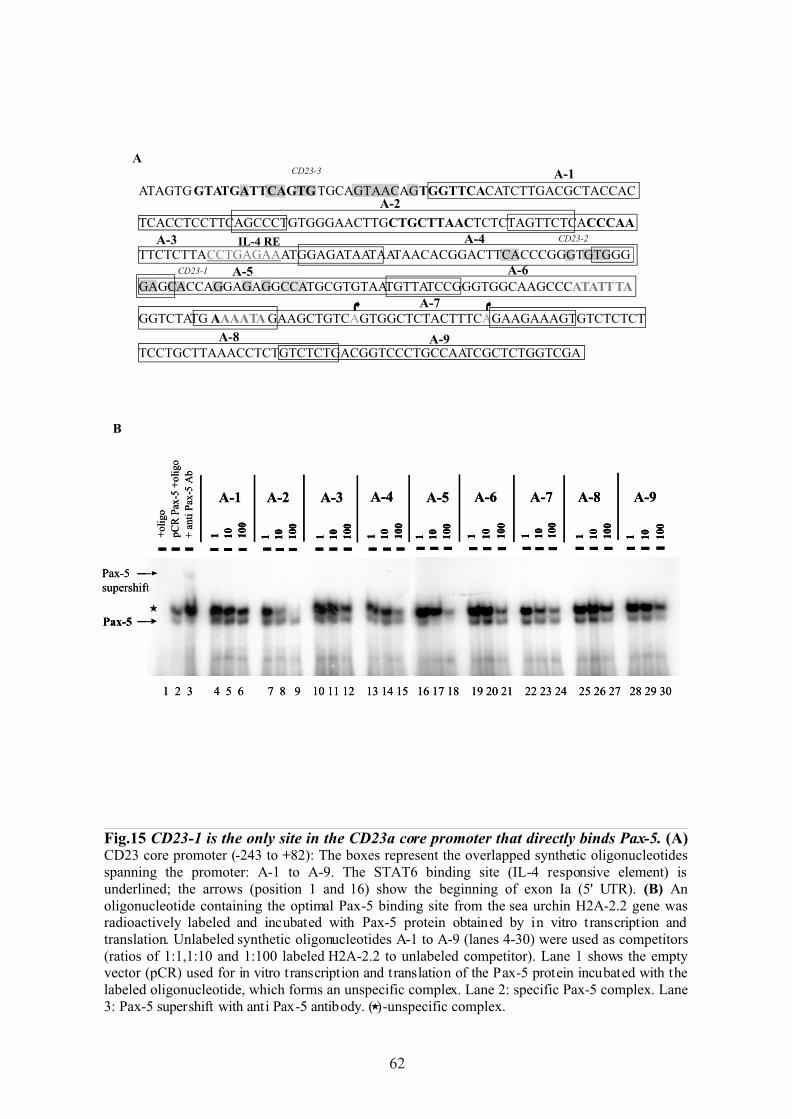
+ol
igo
pCR
Pax
-5+
oli
go
+an
tiP
ax-5
Ab
1 10 10
0
A-1 A-2 A-3
1 10 100
1 10 100
1 10 100
1 10 100
1 10 100
1 10 10
0
A-4 A-5 A-6 A-7 A-8 A-9
1 10 100
1 10 10
0
1 2 3 4 5 6 7 8 9 10 11 12 13 14 15 16 17 18 19 20 21 22 23 24 25 26 27 28 29 30
Pax-5
Pax-5supershift
+ol
igo
pCR
Pax
-5+
oli
go
+an
tiP
ax-5
Ab
1 10 10
0
1 10 10
0
A-1 A-2 A-3
1 10 100
1 10 100
1 10 100
1 10 100
1 10 100
1 10 100
1 10 100
1 10 100
1 10 100
1 10 100
1 10 10
0
1 10 10
0
A-4 A-5 A-6 A-7 A-8 A-9
1 10 100
1 10 100
1 10 10
0
1 10 10
0
1 2 3 4 5 6 7 8 9 10 11 12 13 14 15 16 17 18 19 20 21 22 23 24 25 26 27 28 29 30
Pax-5
Pax-5supershift
ATAGTG GTATGATTCAGTGTGCAGTAACAGTGGTTCACATCTTGACGCTACCAC
TCACCTCCTTCAGCCCTGTGGGAACTTGCTGCTTAACTCTCTAGTTCTCACCCAA
TTCTCTTACCTGAGAAATGGAGATAATAATAACACGGACTTCACCCGGGTGTGGG
GAGCACCAGGAGAGGCCATGCGTGTAATGTTATCCGGGTGGCAAGCCCATATTTA
GGTCTATG AAAATA GAAGCTGTCAGTGGCTCTACTTTCAGAAGAAAGTGTCTCTCT
TCCTGCTTAAACCTCTGTCTCTGACGGTCCCTGCCAATCGCTCTGGTCGA
A-1
A-2
A-3 A-4
A-5 A-6
A-7
A-8 A-9
IL-4 RE
A
B
Fig.15 CD23-1 is the only site in the CD23a core promoter that directly binds Pax-5. (A) CD23 core promoter (-243 to +82): The boxes represent the overlapped synthetic oligonucleotides spanning the promoter: A-1 to A-9. The STAT6 binding site (IL-4 responsive element) is underlined; the arrows (position 1 and 16) show the beginning of exon Ia (5' UTR). (B) An oligonucleotide containing the optimal Pax-5 binding site from the sea urchin H2A-2.2 gene was radioactively labeled and incubated with Pax-5 protein obtained by in vitro transcription and translation. Unlabeled synthetic oligonucleotides A-1 to A-9 (lanes 4-30) were used as competitors (ratios of 1:1,1:10 and 1:100 labeled H2A-2.2 to unlabeled competitor). Lane 1 shows the empty vector (pCR) used for in vitro transcription and translation of the Pax-5 protein incubated with the labeled oligonucleotide, which forms an unspecific complex. Lane 2: specific Pax-5 complex. Lane 3: Pax-5 supershift with anti Pax-5 antibody. ( )-unspecific complex.
62
CD23-3
CD23-1
CD23-2
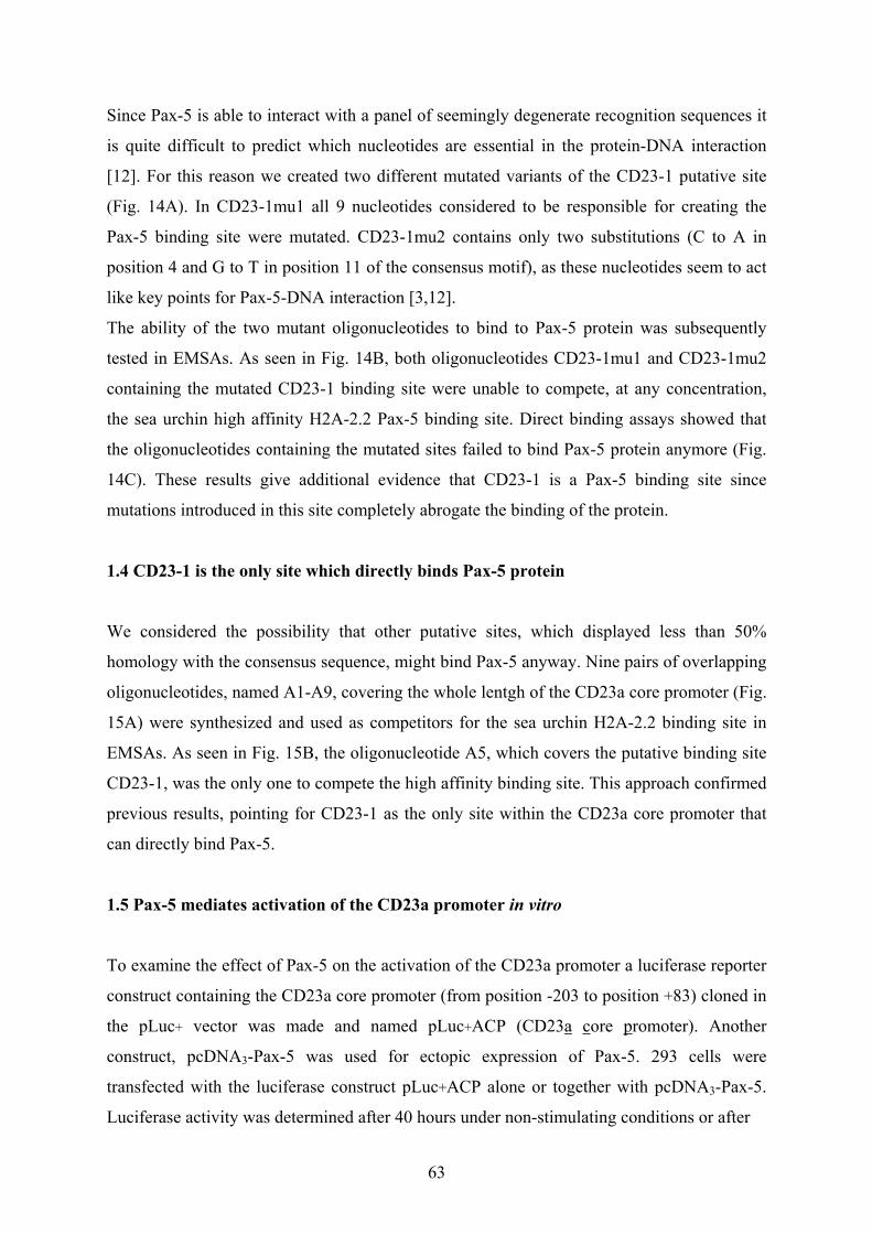
Since Pax-5 is able to interact with a panel of seemingly degenerate recognition sequences it
is quite difficult to predict which nucleotides are essential in the protein-DNA interaction
[12]. For this reason we created two different mutated variants of the CD23-1 putative site
(Fig. 14A). In CD23-1mu1 all 9 nucleotides considered to be responsible for creating the
Pax-5 binding site were mutated. CD23-1mu2 contains only two substitutions (C to A in
position 4 and G to T in position 11 of the consensus motif), as these nucleotides seem to act
like key points for Pax-5-DNA interaction [3,12].
The ability of the two mutant oligonucleotides to bind to Pax-5 protein was subsequently
tested in EMSAs. As seen in Fig. 14B, both oligonucleotides CD23-1mu1 and CD23-1mu2
containing the mutated CD23-1 binding site were unable to compete, at any concentration,
the sea urchin high affinity H2A-2.2 Pax-5 binding site. Direct binding assays showed that
the oligonucleotides containing the mutated sites failed to bind Pax-5 protein anymore (Fig.
14C). These results give additional evidence that CD23-1 is a Pax-5 binding site since
mutations introduced in this site completely abrogate the binding of the protein.
1.4 CD23-1 is the only site which directly binds Pax-5 protein
We considered the possibility that other putative sites, which displayed less than 50%
homology with the consensus sequence, might bind Pax-5 anyway. Nine pairs of overlapping
oligonucleotides, named A1-A9, covering the whole lentgh of the CD23a core promoter (Fig.
15A) were synthesized and used as competitors for the sea urchin H2A-2.2 binding site in
EMSAs. As seen in Fig. 15B, the oligonucleotide A5, which covers the putative binding site
CD23-1, was the only one to compete the high affinity binding site. This approach confirmed
previous results, pointing for CD23-1 as the only site within the CD23a core promoter that
can directly bind Pax-5.
1.5 Pax-5 mediates activation of the CD23a promoter in vitro
To examine the effect of Pax-5 on the activation of the CD23a promoter a luciferase reporter
construct containing the CD23a core promoter (from position -203 to position +83) cloned in
the pLuc+ vector was made and named pLuc+ACP (CD23a core promoter). Another
construct, pcDNA3-Pax-5 was used for ectopic expression of Pax-5. 293 cells were
transfected with the luciferase construct pLuc+ACP alone or together with pcDNA3-Pax-5.
Luciferase activity was determined after 40 hours under non-stimulating conditions or after
63
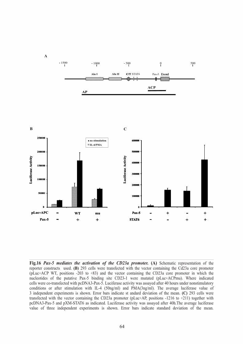
- 1500 - 1000 - 500 0 500
C/TAlu IIAlu I ExonI STAT6 Pax-5
APACP
- - -
C/T ExonI -
A
B C
0
50000
100000
150000
200000
250000no stimulation
IL-4/PMA
pLuc+APC
Pax-5
WT
- +
mu
+
-
Lu
cife
rase
Act
ivit
y
0
50000
100000
150000
200000
250000no stimulation
IL-4/PMA
pLuc+APC
Pax-5
WT
- +
mu
+
-
Lu
cife
rase
Act
ivit
y
Pax-5
STAT6
-- -
+
+
-+
+
0
100000
200000
300000
400000
500000
600000
Pax-5
STAT6
-- -
+
+
-+
+
0
100000
200000
300000
400000
500000
600000
Pax-5
STAT6
-- -
+
+
-+
+
0
100000
200000
300000
400000
500000
600000
Lu
cife
rase
Act
ivit
yL
uci
fera
seA
ctiv
ity
Fig.16 Pax-5 mediates the activation of the CD23a promoter. (A) Schematic representation of thereporter constructs used. (B) 293 cells were transfected with the vector containing the Cd23a core promoter(pLuc+ACP WT, positions -203 to +83) and the vector containing the CD23a core promoter in which thenucleotides of the putative Pax-5 binding site CD23-1 were mutated (pLuc+ACPmu). Where indicated cells were co-transfected with pcDNA3-Pax-5. Luciferase activity was assayed after 40 hours under nonstimulatoryconditions or after stimulation with IL-4 (50ng/ml) and PMA(3ng/ml). The average luciferase value of 3 independent experiments is shown. Error bars indicate st andard deviation of the mean. (C) 293 cells were transfected with the vector containing the CD23a promoter (pLuc+AP, positions -1216 to +211) together with pcDNA3-Pax-5 and pXM-STAT6 as indicated. Luciferase activity was assayed after 40h.The average luciferase value of three independent experiments is shown. Error bars indicate standard deviation of the mean.
64

stimulation with PMA and IL-4. As shown in Fig. 16B, Pax-5 activates the CD23a core
promoter (pLuc+ACP) about 7 fold when compared with the activity of the promoter without
Pax-5. This is in line with our hypothesis that the CD23-1 binding site behaves as a
functional Pax-5 site.
By site directed mutagenesis, we have mutated the CD23-1 putative binding site within the
pLuc+ACP vector. The mutant construct, named pLuc+ACPmu contains the same nucleotide
substitutions as the oligonucleotide CD23-1mu1 (Fig. 14A). The pLuc+ACPmu construct
containing the CD23a core promoter with the mutated Pax-5 binding site failed to activate as
strongly as the pLuc+ACP contruct containing the wild-type promoter. The remaining
activation may be due to the recruitment of Pax-5 into protein complexes that can bind with
lower efficiency but can still lead to a slight activation of the promoter.
A recent study [21] identified IL-4 as the main activator of the CD23a promoter, whereas the
CD23b promoter shows a much wider range of responsiveness to extracellular stimuli. The
authors described two STAT6 binding sites located close to each other in the –500 to -350
region of the CD23a promoter. Therefore we investigated if Pax-5, besides controlling the
specificity of CD23a expression in B-cells, also cooperates with STAT6 in inducing a strong
expression of the isoform.
For this purpose, another luciferase reporter construct (pLuc+AP) that includes these STAT6
binding sites was made by cloninig the whole CD23a promoter (from position –1216 to
position +211) into the pLuc+ vector (Fig. 16A). pcDNA3-Pax-5 and pXM-STAT6 were used
for ectopic expression of Pax-5 and STAT6 respectively in 293 cells. As shown in Fig. 16B,
Pax-5 and STAT6 each induced an 11-14-fold activation of the CD23a promoter
respectively. When the cells are co-transfected with both transcription factors, however, a 40-
fold activation of the CD23a promoter is induced. These results suggest that Pax-5 not only
stimulates CD23a expression, but also cooperates with STAT6 in enhancing the level of
CD23a transcription.
1.6 Pax-5 mediates CD23a expression in vivo
In order to further investigate whether the B-cell restricted expression pattern of CD23a is
determined by lineage specific Pax-5 expression in vivo we used the monocytic cell line U-
937 to determine the effects of ectopic Pax-5 expression on CD23a induction. U-937 cells,
like other myeloid cells, regularly express only CD23b after appropriate stimulation with IL-
4.
65

Human Pax-5 cDNA was stably inserted into the genome of U-937 cells by means of the
recombinant retrovirus pEGZ [47]. The Pax-5 protein was coordinately expressed with the
chimeric selection marker composed of the enhanced green fluorescent protein (EGFP) and
the Zeocin (Zeo) resistance protein. We isolated a Pax-5 expressing population (U-937/EGZ
Pax-5) using Zeo selection. As a control, we generated populations expressing only the
selection marker at equivalent levels (U-937/EGZ). Western Blot analysis of protein extracts
from Pax-5 transfected U-937 cells showed positive Pax-5 protein expression with a band
located around 40 kDa (Fig. 17B). In addition we verified the correct transduction of the Pax-
5 gene by sequencing. The full length Pax-5 gene was successfully integrated in the U-937
genome without deletions or point mutations (data not shown). The smaller size of the Pax-5
expressed in U-937 cells might be due to splicing differences.
Since available anti-CD23 antibodies are directed against the extracellular region of the
receptor, which is shared between the two isoforms, the expression of CD23a cannot be
analysed by FACS staining or Western Blot analyses. Therefore we performed RNase
protection assays using a single RNA probe that is fully homologue to the CD23a mRNA
and 2/3 homologue to CD23b mRNA. The analysis within one sample allows a reliable
quantification of CD23a and CD23b isoforms on the transcriptional level.
Fig. 17A shows the results of the RNase protection assay performed with RNA extracts from
U-937 cells, U-937 expressing only the selection marker EGZ and U-937 transfected with
Pax-5. RNA extracts from WIL2 cells (B-cell line) were used as control for CD23a
expression. CD23a/b isoforms expression was assessed under non-stimulating conditions, as
well as after 48 hours stimulation with IL-4, PMA and IL-4+PMA.
As expected, IL-4 and PMA stimulations alone induce readily detectable amounts of CD23b
mRNA in U-937, U-937/EGZ and U-937/ Pax-5 cells (Fig. 17A, lanes 2-3, 6-7 and 10-11).
CD23b expression was further increased when cells were stimulated with a combination of
the two stimuli (PMA and IL-4).
However, most importantly, U-937 cells transduced with Pax-5 exibited a strong and
reproducible expression of CD23a mRNA when stimulated with PMA plus IL-4 (Fig. 17A,
lane 12). IL-4 alone, as well as PMA stimulation alone were unable to induce CD23a
expression in this monocytic cell line, providing evidence that other factors are required
together with Pax-5. This experiment provides strong evidence that Pax-5 may be a limiting
factor for enabling CD23a expression in vivo.
66

1 2 3 4 5 6 7 8 9 10 11 12 13 14
IL-4 +
+ + + + + +
+ + + + + +
U-937 WIL2U-937/EGZ U-937/Pax-5
Pax-5
CD23a
C D23b
PMA(3ng/ml)
A
B
Fig.17 CD23a is expressed in U-937 cells transduced with Pax-5. (A) RNase protection assayswere performed with RNA extracts from wild-type U-937 cells (lanes 1-4), U-937 transfected with the selection marker EGZ (lanes 5-8) and U-937 transfected with Pax-5 (lanes 9-12). Cellswere stimulated with 50ng/ml IL-4 and 3ng/ml PMA for 48h as indicated. WIL2 cells (B cell line) express CD23a and CD23b constitutively and were used as controls (lanes 13-14). The extracted RNA was hybridized with a single CD23 mRNA probe for both CD23a and CD23bi s o f o r m s . ( B ) We s t e r n B l o t a n a l y s i s t o a s s e s s P a x - 5 e x p r e s s i o n .
67
2. Using a Two Hybrid System to find a cytoplasmic interaction partner
for the CD23 receptor
The question of the transduction pathways involved in CD23 signalling is one of great
importance since CD23 plays important roles in several diseases. No direct interaction
partners for the cytoplasmic domain of membrane receptor CD23 have been found till now.
We used a yeast two-hybrid system to address this question since it provides a powerful
technique to screen large libraries of genes and identify new protein-protein interactions
within the cell.
2.1 Establishing the system
The CytoTrap Two Hybrid System (Stratagene) is based on generating fusion proteins
whose interaction in the yeast cytoplasm induces cell growth by activating the Ras signaling
pathway.
2.1.1 Bait constructs
pSos functions as bait vector by cloning the gene of interest -here CD23- in the MCS of the
plasmid (Fig.18). 60-63 bp long synthetic oligonucleotides incoding for the 21-20 amino
acids of the CD23a and CD23b intracytoplasmic tails were cloned in the MCS of the pSos
vector. These constructs, named pSosCD23a and pSosCD23b, express CD23a and CD23b
cytoplasmic domains as fusion proteins to the C-terminal part of human SOS protein.
pMyr functions as target vector. In order to screen for an interaction partner for CD23 a
human spleen library cloned in the pMyr vector was purchased from Stratagene (Fig.18).
68
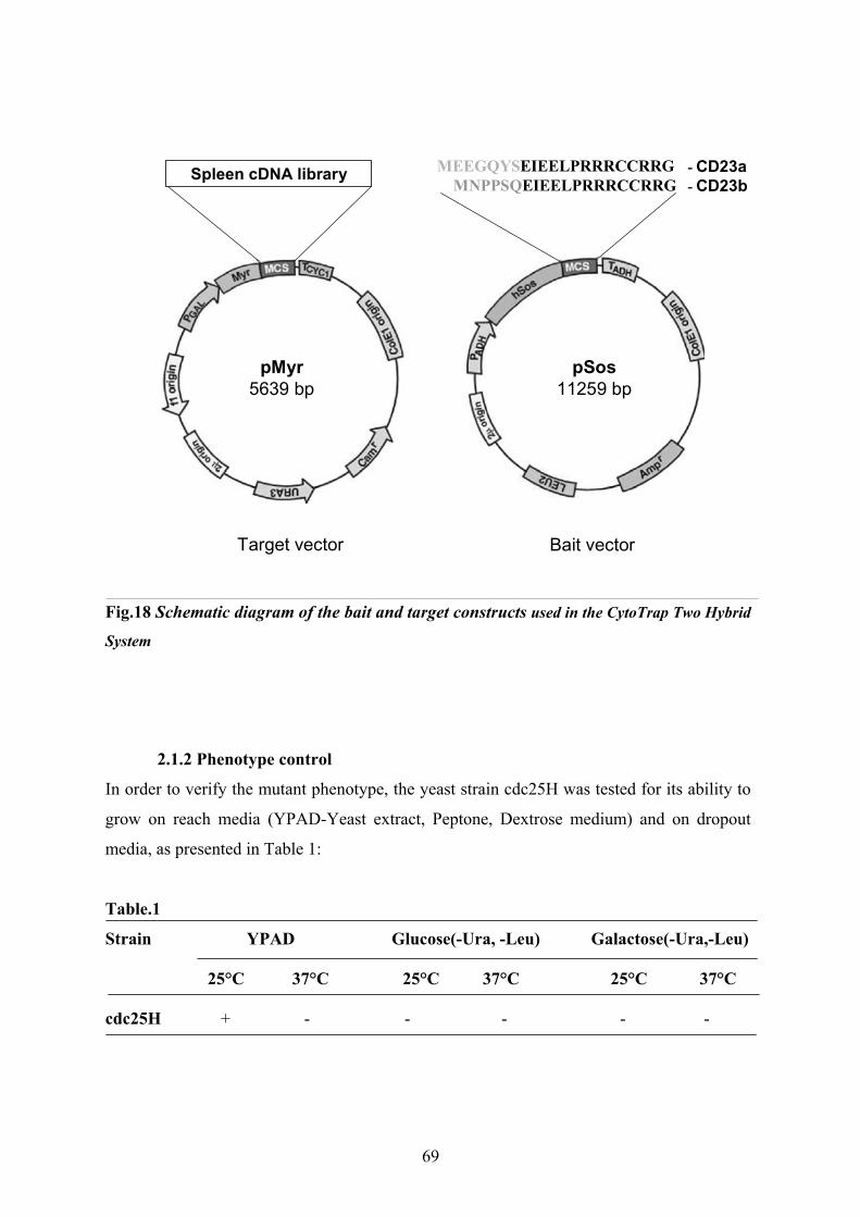
- CD23a- CD23b
MEEGQYSEIEELPRRRCCRRGMNPPSQEIEELPRRRCCRRG
Spleen cDNA library
pMyr5639 bp
pSos11259 bp
Target vector Bait vector
Fig.18 Schematic diagram of the bait and target constructs used in the CytoTrap Two Hybrid
System
2.1.2 Phenotype control
In order to verify the mutant phenotype, the yeast strain cdc25H was tested for its ability to
grow on reach media (YPAD-Yeast extract, Peptone, Dextrose medium) and on dropout
media, as presented in Table 1:
Table.1
Strain YPAD Glucose(-Ura, -Leu) Galactose(-Ura,-Leu)
25°C 37°C 25°C 37°C 25°C 37°C
cdc25H + - - - - -
69
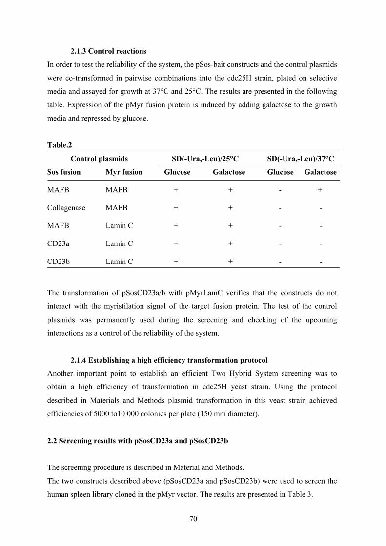
2.1.3 Control reactions
In order to test the reliability of the system, the pSos-bait constructs and the control plasmids
were co-transformed in pairwise combinations into the cdc25H strain, plated on selective
media and assayed for growth at 37°C and 25°C. The results are presented in the following
table. Expression of the pMyr fusion protein is induced by adding galactose to the growth
media and repressed by glucose.
Table.2
Control plasmids SD(-Ura,-Leu)/25°C SD(-Ura,-Leu)/37°C
Sos fusion Myr fusion Glucose Galactose Glucose Galactose
MAFB MAFB + + - +
Collagenase MAFB + + - -
MAFB Lamin C + + - -
CD23a Lamin C + + - -
CD23b Lamin C + + - -
The transformation of pSosCD23a/b with pMyrLamC verifies that the constructs do not
interact with the myristilation signal of the target fusion protein. The test of the control
plasmids was permanently used during the screening and checking of the upcoming
interactions as a control of the reliability of the system.
2.1.4 Establishing a high efficiency transformation protocol
Another important point to establish an efficient Two Hybrid System screening was to
obtain a high efficiency of transformation in cdc25H yeast strain. Using the protocol
described in Materials and Methods plasmid transformation in this yeast strain achieved
efficiencies of 5000 to10 000 colonies per plate (150 mm diameter).
2.2 Screening results with pSosCD23a and pSosCD23b
The screening procedure is described in Material and Methods.
The two constructs described above (pSosCD23a and pSosCD23b) were used to screen the
human spleen library cloned in the pMyr vector. The results are presented in Table 3.
70
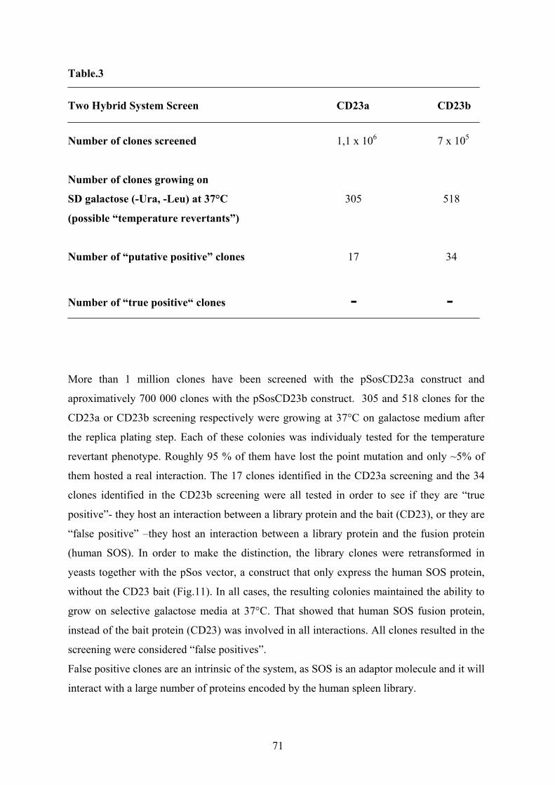
Table.3
Two Hybrid System Screen CD23a CD23b
Number of clones screened 1,1 x 10⁶ 7 x 10⁵
Number of clones growing on
SD galactose (-Ura, -Leu) at 37°C 305 518
(possible “temperature revertants”)
Number of “putative positive” clones 17 34
Number of “true positive“ clones - -
More than 1 million clones have been screened with the pSosCD23a construct and
aproximatively 700 000 clones with the pSosCD23b construct. 305 and 518 clones for the
CD23a or CD23b screening respectively were growing at 37°C on galactose medium after
the replica plating step. Each of these colonies was individualy tested for the temperature
revertant phenotype. Roughly 95 % of them have lost the point mutation and only ~5% of
them hosted a real interaction. The 17 clones identified in the CD23a screening and the 34
clones identified in the CD23b screening were all tested in order to see if they are “true
positive”- they host an interaction between a library protein and the bait (CD23), or they are
“false positive” –they host an interaction between a library protein and the fusion protein
(human SOS). In order to make the distinction, the library clones were retransformed in
yeasts together with the pSos vector, a construct that only express the human SOS protein,
without the CD23 bait (Fig.11). In all cases, the resulting colonies maintained the ability to
grow on selective galactose media at 37°C. That showed that human SOS fusion protein,
instead of the bait protein (CD23) was involved in all interactions. All clones resulted in the
screening were considered “false positives”.
False positive clones are an intrinsic of the system, as SOS is an adaptor molecule and it will
interact with a large number of proteins encoded by the human spleen library.
71
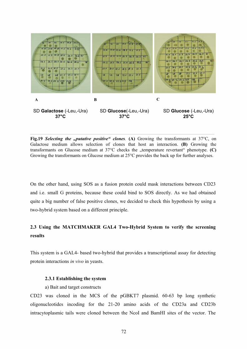
SD Glucose (-Leu,-Ura)25°C
SD Galactose (-Leu,-Ura)37°C
SD Glucose(-Leu,-Ura)37°C
Fig.19 Selecting the „putative positive“ clones. (A) Growing the transformants at 37°C, on Galactose medium allows selection of clones that host an interaction. (B) Growing thetransformants on Glucose medium at 37°C checks the „temperature revertant“ phenotype. (C) Growing the transformants on Glucose medium at 25°C provides the back up for further analyses.
A B C
On the other hand, using SOS as a fusion protein could mask interactions between CD23
and i.e. small G proteins, because these could bind to SOS directly. As we had obtained
quite a big number of false positive clones, we decided to check this hypothesis by using a
two-hybrid system based on a different principle.
2.3 Using the MATCHMAKER GAL4 Two-Hybrid System to verify the screening
results
This system is a GAL4- based two-hybrid that provides a transcriptional assay for detecting
protein interactions in vivo in yeasts.
2.3.1 Establishing the system
a) Bait and target constructs
CD23 was cloned in the MCS of the pGBKT7 plasmid. 60-63 bp long synthetic
oligonucleotides incoding for the 21-20 amino acids of the CD23a and CD23b
intracytoplasmic tails were cloned between the NcoI and BamHI sites of the vector. The
72

constructs would express CD23a and CD23b cytoplasmic domains as fusion proteins with
GAL4 DNA-BD.
Screening with the CytoTrap Two-Hybrid System resulted in a total of 17 clones when
CD23a was used as bait and 34 clones when CD23b was used as bait. There was a high
probability that certain genes of the library came up more than once in the screening process
and that a certain number of duplicates would be found in the total of 51 clones.
In consequence, they were subjected to restriction enzyme analyses with HpaII, an enzyme
that cuts CCGG motifs. The digest profile was used to compare and clasify the clones
(Fig.20). The 51 clones resulted in 12 distinct profiles representing distinct genes. These
genes were then cloned between the EcoRI and XhoI sites of the pGADT7 plasmid. The
constructs, named pGADT7-1/12, would express them as fusion proteins with GAL4 DNA-
AD.
31b
78b
14a
33a
164b
170b
161b
384.
1b19
a21
a28
9.6b
50 b
p m
arke
r
Fig.20 HpaII digest of false positive clones resulted from the Cytotrap Two Hybrid System
screen
73

b) Control of the yeast phenotype
In order to verify the phenotypes, the two yeast strains provided by the system were tested
on dropout media as follows.
Table.4
Strain SD/-Ade SD/-Met SD/-Trp SD/-Leu SD/-His SD/-Ura YPDA
AH109 - + - - - - +
Y187 - - - - - - +
c) Control reactions
In order to test the general reliability of the system and if DNA-BD and AD fusion
constructs do not autonomously activate reporter genes, the pGBKT7-CD23a/b constructs,
pGADT7-1/12 and the control plasmids were co-transformed in various combinations into
the AH109 strain, plated on selective media and assayed for growth and blue/white
phenotypes. The results are presented in the Table 5.
These results also show how one can modulate the stringency of the screening by plating the
transformants on different selective media. The use of high-stringency media, SD/-Ade/-
His-Leu/-Trp/X-α-Gal, virtually eliminates false positive interaction. The use of low-
stringency media, SD/-Leu/-Trp, favours the detection of weak or transient interactions.
Also, the independent transformation of pGBKT7-CD23a/b and pGADT7-1/12 into AH109
verifies that the constructs do not activate reporter genes by themselves.
74
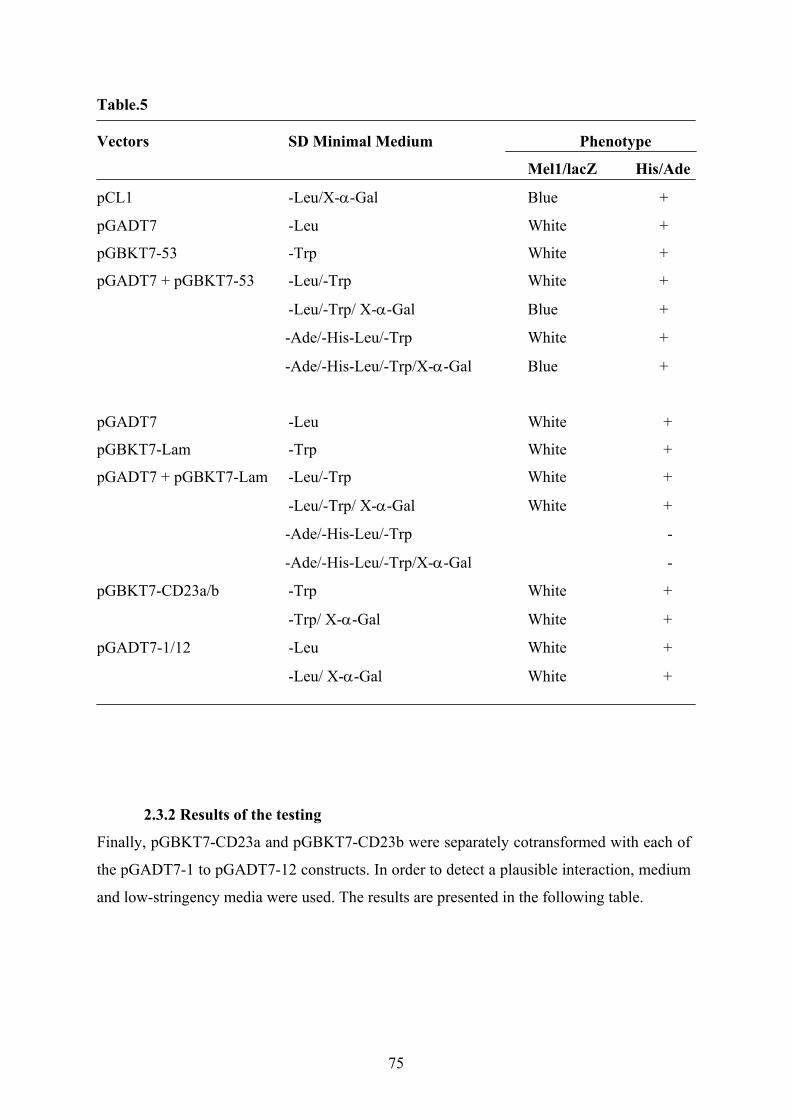
Table.5 Vectors SD Minimal Medium Phenotype
Mel1/lacZ His/Ade
pCL1 -Leu/X-α-Gal Blue +
pGADT7 -Leu White +
pGBKT7-53 -Trp White + pGADT7 + pGBKT7-53 -Leu/-Trp White + -Leu/-Trp/ X-α-Gal Blue +
-Ade/-His-Leu/-Trp White +
-Ade/-His-Leu/-Trp/X-α-Gal Blue +
pGADT7 -Leu White +
pGBKT7-Lam -Trp White + pGADT7 + pGBKT7-Lam -Leu/-Trp White +
-Leu/-Trp/ X-α-Gal White +
-Ade/-His-Leu/-Trp -
-Ade/-His-Leu/-Trp/X-α-Gal -
pGBKT7-CD23a/b -Trp White +
-Trp/ X-α-Gal White +
pGADT7-1/12 -Leu White +
-Leu/ X-α-Gal White +
2.3.2 Results of the testing
Finally, pGBKT7-CD23a and pGBKT7-CD23b were separately cotransformed with each of
the pGADT7-1 to pGADT7-12 constructs. In order to detect a plausible interaction, medium
and low-stringency media were used. The results are presented in the following table.
75

Table.6 Vectors SD Minimal Medium Phenotype
Mel1/lacZ His/Ade
pGBKT7-CD23a + pGADT7-1/12 -Leu/-Trp White +
-Leu/-Trp/ X-α-Gal White +
-His/-Leu/-Trp -
-His/-Leu/-Trp/ X-α-Gal -
pGBKT7-CD23b + pGADT7-1/12 -Leu/-Trp White +
-Leu/-Trp/ X-α-Gal White +
-His/-Leu/-Trp -
-His/-Leu/-Trp/ X-α-Gal -
Testing the interaction into the MATCHMAKER system verified that there was no true
interaction between CD23 cytoplasmic tail and the clones resulted in the CytoTrap
screening.
2.4 New constructs for the CytoTrap Two Hybrid System bait vectors
The fact that CD23 was cloned in the MCS of the pSos vector caused the intracytoplasmic
part of CD23 to be expressed at the carboxyterminal end of SOS, implying also that the free
end of CD23 was the C-terminal end. However, CD23 is a type II receptor, with the N-
terminal end into the cell.
In order to change the orientation of the bait in the fusion protein we decided to clone
CD23a/b upstream of the hSOS gene. This new constructs would express the bait at the N
terminal end of human SOS protein implying the same orientation for the cytoplasmic tails
of CD23a and CD23b as it occurs in vivo (Fig.21).
In addition, there is also a considerable difference in size between the human SOS proteins,
which is a multidomain adaptor protein of 170 kDa and the short cytoplasmic tail of CD23,
which has a total of 20-21 amino acids. In order to make this small amino acid tail more
obvious into the fusion protein, we considered using a linker region. A 15 amino acids long
synthetic hinge region containing a glycine and serine repetitive motif was used as linker
between CD23a/b and the SOS protein. The amino acid sequence of this flexible
oligopeptide is presented in the Fig. 21B.
76
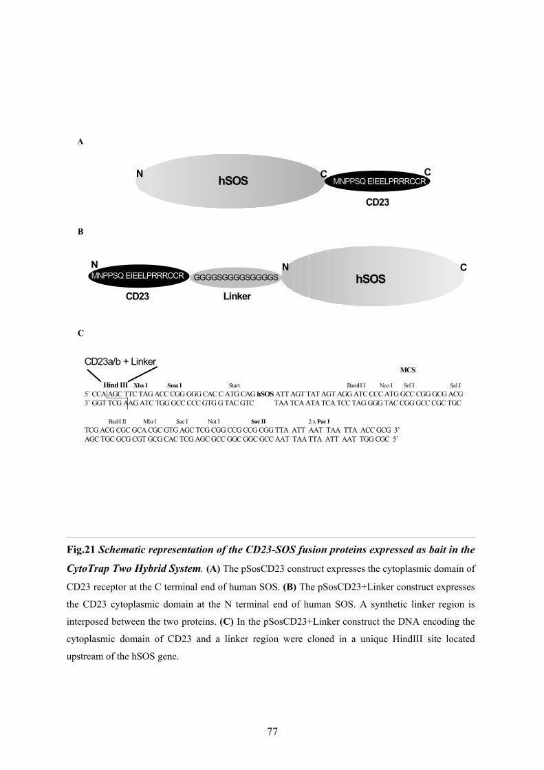
MNPPSQ EIEELPRRRCCRhSOShSOSN C C
CD23
A
GGGGSGGGGSGGGGSMNPPSQ EIEELPRRRCCR hSOShSOSN CN
LinkerCD23
B
Hind III Xba I Sma I Start BamH I Nco I Srf I Sal I 5’ CCA AGC TTC TAG ACC CGG GGG CAC C ATG CAG hSOS ATT AGT TAT AGT AGG ATC CCC ATG GCC CGG GCG ACG3’ GGT TCG AAG ATC TGG GCC CCC GTG G TAC GTC TAA TCA ATA TCA TCC TAG GGG TAC CGG GCC CGC TGC
BssH II Mlu I Sac I Not I Sac II 2 x Pac ITCG ACG CGC GCA CGC GTG AGC TCG CGG CCG CCG CGG TTA ATT AAT TAA TTA ACC GCG 3’AGC TGC GCG CGT GCG CAC TCG AGC GCC GGC GGC GCC AAT TAA TTA ATT AAT TGG CGC 5’
CD23a/b + LinkerMCS
C
Fig.21 Schematic representation of the CD23-SOS fusion proteins expressed as bait in the
CytoTrap Two Hybrid System. (A) The pSosCD23 construct expresses the cytoplasmic domain of
CD23 receptor at the C terminal end of human SOS. (B) The pSosCD23+Linker construct expresses
the CD23 cytoplasmic domain at the N terminal end of human SOS. A synthetic linker region is
interposed between the two proteins. (C) In the pSosCD23+Linker construct the DNA encoding the
cytoplasmic domain of CD23 and a linker region were cloned in a unique HindIII site located
upstream of the hSOS gene.
77
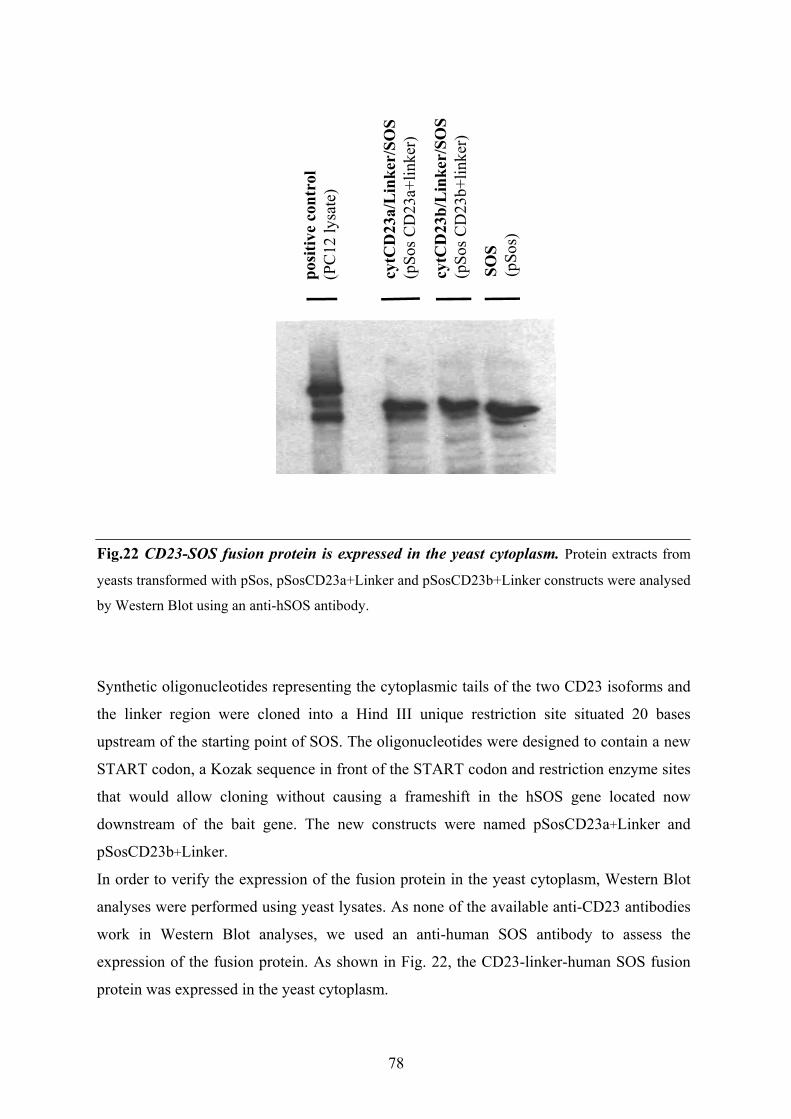
cytC
D23
a/L
inke
r/SO
S(p
SosC
D23
a+lin
ker)
SOS
(pSo
s)
cytC
D23
b/L
inke
r/SO
S(p
SosC
D23
b+lin
ker)
p osi
tive
cont
r ol
( PC
12 ly
sate
)
Fig.22 CD23-SOS fusion protein is expressed in the yeast cytoplasm. Protein extracts from
yeasts transformed with pSos, pSosCD23a+Linker and pSosCD23b+Linker constructs were analysed
by Western Blot using an anti-hSOS antibody.
Synthetic oligonucleotides representing the cytoplasmic tails of the two CD23 isoforms and
the linker region were cloned into a Hind III unique restriction site situated 20 bases
upstream of the starting point of SOS. The oligonucleotides were designed to contain a new
START codon, a Kozak sequence in front of the START codon and restriction enzyme sites
that would allow cloning without causing a frameshift in the hSOS gene located now
downstream of the bait gene. The new constructs were named pSosCD23a+Linker and
pSosCD23b+Linker.
In order to verify the expression of the fusion protein in the yeast cytoplasm, Western Blot
analyses were performed using yeast lysates. As none of the available anti-CD23 antibodies
work in Western Blot analyses, we used an anti-human SOS antibody to assess the
expression of the fusion protein. As shown in Fig. 22, the CD23-linker-human SOS fusion
protein was expressed in the yeast cytoplasm.
78
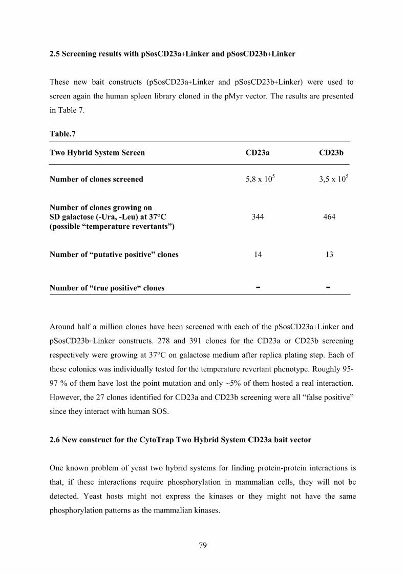
2.5 Screening results with pSosCD23a+Linker and pSosCD23b+Linker
These new bait constructs (pSosCD23a+Linker and pSosCD23b+Linker) were used to
screen again the human spleen library cloned in the pMyr vector. The results are presented
in Table 7.
Table.7 Two Hybrid System Screen CD23a CD23b Number of clones screened 5,8 x 10⁵ 3,5 x 10⁵
Number of clones growing on SD galactose (-Ura, -Leu) at 37°C 344 464 (possible “temperature revertants”) Number of “putative positive” clones 14 13
Number of “true positive“ clones - -
Around half a million clones have been screened with each of the pSosCD23a+Linker and
pSosCD23b+Linker constructs. 278 and 391 clones for the CD23a or CD23b screening
respectively were growing at 37°C on galactose medium after replica plating step. Each of
these colonies was individually tested for the temperature revertant phenotype. Roughly 95-
97 % of them have lost the point mutation and only ~5% of them hosted a real interaction.
However, the 27 clones identified for CD23a and CD23b screening were all “false positive”
since they interact with human SOS.
2.6 New construct for the CytoTrap Two Hybrid System CD23a bait vector
One known problem of yeast two hybrid systems for finding protein-protein interactions is
that, if these interactions require phosphorylation in mammalian cells, they will not be
detected. Yeast hosts might not express the kinases or they might not have the same
phosphorylation patterns as the mammalian kinases.
79
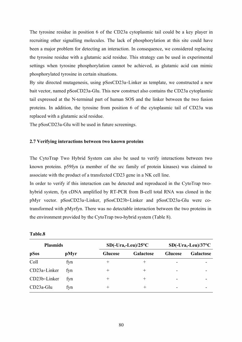
The tyrosine residue in position 6 of the CD23a cytoplasmic tail could be a key player in
recruiting other signalling molecules. The lack of phosphorylation at this site could have
been a major problem for detecting an interaction. In consequence, we considered replacing
the tyrosine residue with a glutamic acid residue. This strategy can be used in experimental
settings when tyrosine phosphorylation cannot be achieved, as glutamic acid can mimic
phosphorylated tyrosine in certain situations.
By site directed mutagenesis, using pSosCD23a+Linker as template, we constructed a new
bait vector, named pSosCD23a-Glu. This new construct also contains the CD23a cytoplasmic
tail expressed at the N-terminal part of human SOS and the linker between the two fusion
proteins. In addition, the tyrosine from position 6 of the cytoplasmic tail of CD23a was
replaced with a glutamic acid residue.
The pSosCD23a-Glu will be used in future screenings.
2.7 Verifying interactions between two known proteins
The CytoTrap Two Hybrid System can also be used to verify interactions between two
known proteins. p59fyn (a member of the src family of protein kinases) was claimed to
associate with the product of a transfected CD23 gene in a NK cell line.
In order to verify if this interaction can be detected and reproduced in the CytoTrap two-
hybrid system, fyn cDNA amplified by RT-PCR from B-cell total RNA was cloned in the
pMyr vector. pSosCD23a+Linker, pSosCD23b+Linker and pSosCD23a-Glu were co-
transformed with pMyrfyn. There was no detectable interaction between the two proteins in
the environment provided by the CytoTrap two-hybrid system (Table 8).
Table.8 Plasmids SD(-Ura,-Leu)/25°C SD(-Ura,-Leu)/37°C
pSos pMyr Glucose Galactose Glucose Galactose
Coll fyn + + - -
CD23a+Linker fyn + + - -
CD23b+Linker fyn + + - -
CD23a-Glu fyn + + - -
80

2.8 Perspectives
pSosCD23a-Glu represents a bait construct substantially improved. It has the advantage to
express CD23 cytoplasmic tail as it occurs in vivo, with the N-terminus free as bait and
contains a glutamic acid residue that can substitute a phosphorylated tyrosine in position 6.
Using this construct for screening an improved potential to find an interaction partner for the
CD23 receptor is to be expected.
81
DISCUSSION
High CD23 expression is associated with various chronic diseases such as B-CLL,
rheumatoid arthritis and lupus erythematosus. It is assumed that signalling through CD23
contributes to the pathogenesis of these diseases. In humans there are two CD23 isoforms
that are differentially expressed. Whereas CD23b is widely detected on lymphocytes and
myeloid cells, CD23a is restricted to B-lymphocytes. The isoforms differ only in the short
amino terminal intracytoplasmic part [87] and seem to be connected to different signal
transduction pathways. The CD23a isoform specifically mediates endocytosis of bound
ligands and can therefore influence B-cell mediated antigen presentation [41]. CD23b was
shown to be associated with phagocytosis of IgE coated-particles [88].
Distinct promoters regulate the two CD23 isoforms expressed in humans. So far, IL-4 has
been described as being the most important activator of CD23a and CD23b expression and
binding sites for STAT6 have been characterized in both promoters. [21,41,87]. In addition
to the IL-4 responsive element, NF-AT binding sites have been characterized in the CD23b
promoter [40]. There are very limited data concerning the regulation of the human CD23a
promoter. Studies of the mouse CD23 promoter, which is closely related in sequence to the
human CD23a promoter, revealed functional STAT6, NF-kB and C/EBP binding sites [82].
Since the mouse and human CD23a promoter are 72% homologous in the core region, most
of the binding motifs in the human promoter were characterized by comparison with the
mouse promoter. Richards et al [65] observed that the homologous nucleotides appear in
clusters, suggesting that common response elements have been conserved in the two
promoters. As shown in Fig. 10A, STAT6, NF-kB and C/EBP binding sites are localized in
the –70 to –140 region of the CD23a core promoter. Additional STAT6 binding sites were
characterized in the extended CD23a promoter, in the –300 to –500 region. Multimerized
STAT6 binding sites, as well as their association with NF-kB sites, seem to be a common
trait of IL-4 regulated promoters [14, 54]. In addition, EBNA2 and Notch-2 can regulate the
CD23a promoter by binding to CBF1 sites [32,48]. Both activatory elements may play a role
in the immortalisation of the B-cell by enhancing CD23a expression.
82
The question of how the B-cell specific expression of CD23a isoform is regulated has not
been experimentally addressed so far. Among B-cell specific transcription factors, Pax-
5/BSAP binding sites have been very recently predicted in the CD23a core promoter [21,32].
Pax-5 is a critical modulator of early B-cell differentiation. Its expression is restricted to B
cells, embryonic brain and testis. Binding sites for Pax-5 have been identified in promoters of
several genes. While being a positive regulator of CD19, mb-1 and RAG-2 [39,43,59] Pax-5
acts as a repressor for the immunoglobulin heavy-chain 3’C∝ enhancer and the J-chain
[66,75]. The importance of Pax-5 for development was demonstrated by knock-out
experiments. Pax-5 is important in early and late B-cell development. Most of Pax-5-/- mice
die within 3 weeks and B-cell development is blocked at the pro-B-cell stage [84]. The loss
of Pax-5 in mature B-cells severely impairs B-cell identity [31].
Here we demonstrate the presence of a functional Pax-5 binding site in the CD23a core
promoter that is able to induce and enhance CD23a expression. We also advance the
hypothesis that Pax-5, being a B-cell specific protein, plays a central role in regulating the
specific expression of CD23a on B-cells. CD23 is expressed later in the B-cell development
than Pax-5. Additional transcription factors must be involved in modulating CD23a
expression. The association of Pax-5 with other transcription factors in regulatory complexes
has been documented so far. For example, Pax-5 associates with Oct-1 (octamer binding
proteins) and NF-kB in modulating the 3’α-hs4, a distal 3’ enhancer that regulates the IgH
gene cluster at multiple stages of B-cell development. While Oct-1 and kB act as positive
regulators at all stages of differentiation, Pax-5 acts as a repressor at the pre-B-cell stage and
as a positive regulator in mature B-cells [52, 53]. Furthermore, both CD23 and Pax-5
expression are lost when differentiation to plasma cells occurs. This is in line with Pax-5
being a prerequisite factor for CD23a expression.
Another interesting observation comes from the comparison of the CD23 promoter with the
germline immunoglobulin Cε and Cγ1 promoters, which are also regulated by IL-4. The
transcription of these germline immunoglobulin genes is necessary for class switching to IgE
and IgG1. Different combinations of cytokines and mitogens have been shown to stimulate
class switching to particular isotypes. IL-4 promotes a class switch from IgM to IgE and
IgG1 and STAT6 binding sites have been identified in both ε and γ1 germline genes. In
addition, NF-kB and C/EBP binding sites have been described [14], while Pax-5 was shown
to bind close to the STAT6 site in the germline ε promoter. Studies by Liao et al. show that
Pax-5 is essential for germline ε transcription and therefore essential for class switching and
83
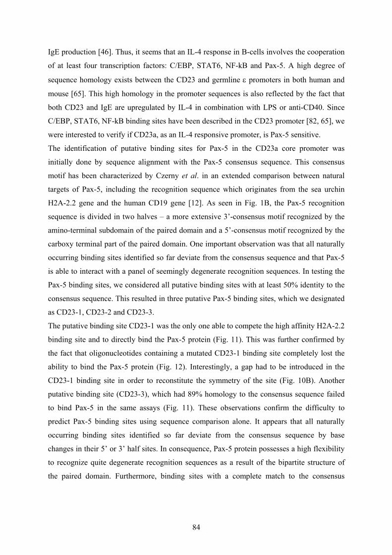
IgE production [46]. Thus, it seems that an IL-4 response in B-cells involves the cooperation
of at least four transcription factors: C/EBP, STAT6, NF-kB and Pax-5. A high degree of
sequence homology exists between the CD23 and germline ε promoters in both human and
mouse [65]. This high homology in the promoter sequences is also reflected by the fact that
both CD23 and IgE are upregulated by IL-4 in combination with LPS or anti-CD40. Since
C/EBP, STAT6, NF-kB binding sites have been described in the CD23 promoter [82, 65], we
were interested to verify if CD23a, as an IL-4 responsive promoter, is Pax-5 sensitive.
The identification of putative binding sites for Pax-5 in the CD23a core promoter was
initially done by sequence alignment with the Pax-5 consensus sequence. This consensus
motif has been characterized by Czerny et al. in an extended comparison between natural
targets of Pax-5, including the recognition sequence which originates from the sea urchin
H2A-2.2 gene and the human CD19 gene [12]. As seen in Fig. 1B, the Pax-5 recognition
sequence is divided in two halves – a more extensive 3’-consensus motif recognized by the
amino-terminal subdomain of the paired domain and a 5’-consensus motif recognized by the
carboxy terminal part of the paired domain. One important observation was that all naturally
occurring binding sites identified so far deviate from the consensus sequence and that Pax-5
is able to interact with a panel of seemingly degenerate recognition sequences. In testing the
Pax-5 binding sites, we considered all putative binding sites with at least 50% identity to the
consensus sequence. This resulted in three putative Pax-5 binding sites, which we designated
as CD23-1, CD23-2 and CD23-3.
The putative binding site CD23-1 was the only one able to compete the high affinity H2A-2.2
binding site and to directly bind the Pax-5 protein (Fig. 11). This was further confirmed by
the fact that oligonucleotides containing a mutated CD23-1 binding site completely lost the
ability to bind the Pax-5 protein (Fig. 12). Interestingly, a gap had to be introduced in the
CD23-1 binding site in order to reconstitute the symmetry of the site (Fig. 10B). Another
putative binding site (CD23-3), which had 89% homology to the consensus sequence failed
to bind Pax-5 in the same assays (Fig. 11). These observations confirm the difficulty to
predict Pax-5 binding sites using sequence comparison alone. It appears that all naturally
occurring binding sites identified so far deviate from the consensus sequence by base
changes in their 5’ or 3’ half sites. In consequence, Pax-5 protein possesses a high flexibility
to recognize quite degenerate recognition sequences as a result of the bipartite structure of
the paired domain. Furthermore, binding sites with a complete match to the consensus
84

sequence would possess an exceptionally high affinity for Pax-5, which apparently is not
required in vivo.
Another approach to identify Pax-5 binding sites in the CD23a core promoter was to
construct overlapping oligonucleotides covering the whole length of the promoter and to test
their ability to compete the sea urchin H2A-2.2 binding site in EMSAs. Both strategies
revealed CD23-1 as the only site in the CD23a core promoter able to bind directly the Pax-5
protein (Fig. 13).
It is interesting to note that in our hands none of the two Pax-5 sites predicted in a recent
study by Hubmann et al. [32] interacts directly with the Pax-5 protein. As this study used
nuclear extract from B-CLL cells, it is possible that Pax-5 was recruited by another factor
into a protein complex, which interacts with these putative sites.
To test the function of the identified Pax-5 binding site in vitro we performed luciferase
assays in 293 cells linking the CD23a core promoter or an extended CD23a promoter to a
luciferase reporter gene. We chose this approach since previous work has shown that
promoters of Pax-5 regulated genes, like CD19, are weakly active in transiently transfected
B-cells and can only be stimulated by ectopic Pax-5 expression in heterologous cell types [9].
Overexpression of Pax-5 in 293 cells led to a 7-10 fold activation of both CD23a promoter
constructs (Fig. 14). However, reporter constructs with the mutated CD23-1 site were still
slightly active. The remaining activation may be due to the recruitment of Pax-5 into protein
complexes that can bind with lower efficiency but can still lead to a slight activation of the
promoter. This may be indeed another mechanism by which Pax-5 regulates the CD23a
promoter and which needs to be approached in future studies. Furthermore, in experiments
using the extended promoter which includes two reported STAT6 sites, Pax-5 acted together
with STAT6 in further enhancing CD23a expression above the level of the induction
provided by the two factors alone. These results suggest that Pax-5 not only determines the
B-cell specificity of CD23a, but also potentiates the STAT6 mediated stimulatory effect.
Previous studies seem to lead to the scenario that STAT6, even when multimerized, is not
sufficient for the induction of IL-4 regulated genes [14, 54]. The cooperation of other
transcriptional activators, like NF-kB, appears to be necessary, for example, in order to
activate the transcription of the germline Cε promoter. The interaction with NF-kB appears to
enhance the DNA binding affinity and the transactivating affinity of STAT6 [74]. It may also
be possible that Pax-5 synergically acts and perhaps also directly associates with STAT6
(and NF-kB) in order to activate the CD23a promoter.
85
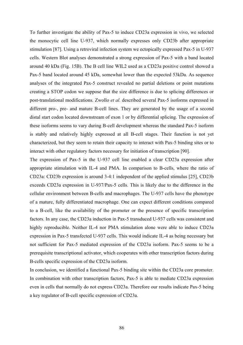
To further investigate the ability of Pax-5 to induce CD23a expression in vivo, we selected
the monocytic cell line U-937, which normally expresses only CD23b after appropriate
stimulation [87]. Using a retroviral infection system we ectopically expressed Pax-5 in U-937
cells. Western Blot analyses demonstrated a strong expression of Pax-5 with a band located
around 40 kDa (Fig. 15B). The B cell line WIL2 used as a CD23a positive control showed a
Pax-5 band located around 45 kDa, somewhat lower than the expected 53kDa. As sequence
analyses of the integrated Pax-5 construct revealed no partial deletions or point mutations
creating a STOP codon we suppose that the size difference is due to splicing differences or
post-translational modifications. Zwollo et al. described several Pax-5 isoforms expressed in
different pro-, pre- and mature B-cell lines. They are generated by the usage of a second
distal start codon located downstream of exon 1 or by differential splicing. The expression of
these isoforms seems to vary during B-cell development whereas the standard Pax-5 isoform
is stably and relatively highly expressed at all B-cell stages. Their function is not yet
characterized, but they seem to retain their capacity to interact with Pax-5 binding sites or to
interact with other regulatory factors necessary for initiation of transcription [90].
The expression of Pax-5 in the U-937 cell line enabled a clear CD23a expression after
appropriate stimulation with IL-4 and PMA. In comparison to B-cells, where the ratio of
CD23a: CD23b expression is around 3-4:1 independent of the applied stimulus [25], CD23b
exceeds CD23a expression in U-937/Pax-5 cells. This is likely due to the difference in the
cellular environment between B-cells and macrophages. The U-937 cells have the phenotype
of a mature, fully differentiated macrophage. One can expect different conditions compared
to a B-cell, like the availability of the promoter or the presence of specific transcription
factors. In any case, the CD23a induction in Pax-5 transduced U-937 cells was consistent and
highly reproducible. Neither IL-4 nor PMA stimulation alone were able to induce CD23a
expression in Pax-5 transfected U-937 cells. This would indicate IL-4 as being necessary but
not sufficient for Pax-5 mediated expression of the CD23a isoform. Pax-5 seems to be a
prerequisite transcriptional activator, which cooperates with other transcription factors during
B-cells specific expression of the CD23a isoform.
In conclusion, we identified a functional Pax-5 binding site within the CD23a core promoter.
In combination with other transcription factors, Pax-5 is able to mediate CD23a expression
even in cells that normally do not express CD23a. Therefore our results indicate Pax-5 being
a key regulator of B-cell specific expression of CD23a.
86

The question of the transduction pathways involved in CD23 signaling is one of great
interest as CD23 plays important roles in several diseases. In B-CLL, for example, CD23a
expression is associated with a state of cell survival. In contrast to normal B-cells where after
24 hours of incubation CD23a transcription is completely lost, accompanied also by a down-
modulation of the antiapoptotic gene bcl-2, the levels of CD23a and bcl-2 expression remain
high in B-CLL cells [32]. Overexpression of CD23a is associated with enhanced cell
viability.
The two isoforms have different expression patterns and apparently quite distinct functions.
Comparison between the events taking place in cells that express only one or both isoforms
seem also to lead to the idea of different signal transduction pathways for CD23a and CD23b.
Studies done by Kolb and co-workers show that cross-linking of CD23 on B-cells provokes a
rapid increase in Ca2+ resulted from inositol (1,4,5) triphosphate generation and a slow
accumulation of cAMP. Ligation of CD23 on the surface of monocytes results in cAMP
generation and is additionally coupled with activation of iNOS. The difference is most likely
due to the 6-7 amino acids at the N-terminal end of the cytoplasmic tail.
CD23a only contains a Tyr residue in position 6 and a Ser residue in position 7 that are
predicted sites for phosphorylation. The tyrosine in CD23a cytoplasmic tail exists as a
member of the YSEI sequence. The YXXL sequence is known as an ITIM motif, and if Leu
can substitute for Ile, than theYSEI in the human CD23a could be seen as a variant of an
ITIM motif [36]. As the suppression of IgE production was released in CD23-knockout mice,
it would be reasonable to suppose that CD23 contains an inhibitory signal within the
molecule. The meaning of this sequence remains unclear, although it may provide a base for
the difference in signalling between the two isoforms.
Our approach to address the question of the transduction pathways involved was to look for
direct interaction partners for CD23. Preliminary data suggested that a pertussis toxin-
insensitive G protein couples membrane CD23 to the PLC. It is also possible that a protein
tyrosine kinase is involved, since the product of the transfected CD23 gene in a NK cell line
was found associated with p59fyn [78].
We used a yeast two-hybrid system to look for interaction partners for the CD23 receptor.
These systems provide a powerful technique to screen large libraries of genes and to identify
new protein-protein interactions within the cell. When compared with more traditional
methods for studying protein-protein interactions, the two-hybrid system seems to offer
several advantages. As the interaction occurs in vivo, the need for the detailed and laborious
manipulation of conditions necessary for in vitro biochemical binding assays is abrogated.
87

Also, in at least some cases, the two-hybrid system is more sensitive than co-
immunoprecipitation in detecting weak interactions. The system we employed –the
CytoTrap Two Hybrid System from Stratagene- offers additional advantages over the
traditional two hybrid systems. As the interaction takes place in the cytoplasm, it does not
involve protein transport to the nucleus. It also provides a better control of the activation.
There are, of course, several limitation and disadvantages, which are common to all yeast
two-hybrids: some hybrid proteins may not be stably expressed in yeast or bacteria; fusion
proteins may occlude the normal site of interaction or impair the proper folding; conditions in
yeast may not allow the proper folding or posttranscriptional modifications.
One problem that is commonly encountered with library screens is achieving optimal
transformation efficiency. Most protocols are a variation of standard lithium acetate
transformation protocols. After testing several of these protocols we achieved good co-
transformation efficiencies allowing us to screen approximately 3 million colonies. The
quality of the carrier DNA and the freshness of the plated yeast streak are two critical factors
that influence greatly the efficiency of the transformation.
Another major problem with two-hybrid screens is the appearance of false-positive clones. In
the classic two-hybrid system, they are inherent in any transcriptional readout. In the
CytoTrap system, the human SOS, as an adaptor protein, will interact with a number of other
proteins. Any candidates from the primary screen must be retested in several configurations
before they can be considered ‘true’ positives. First, the library plasmid must be separated
from the bait plasmid. This is usually done by recovering the plasmid from yeast and
transforming into bacteria, which are then grown on medium containing the same antibiotic
for which the plasmid encoding the candidate gene contains a resistance gene. Once a
plasmid has been recovered, it should be tested in the absence of the bait. Normally, a false
positive will produce an interaction with the human SOS protein alone. The collagenase I
expressed by the pSos Coll negative control plasmid can also be a useful control, as this
protein does not generate interactions with most of the proteins.
As we obtained quite a high number of false positives, we also considered the possibility that
SOS would mask an interaction between the bait (CD23) and a potential target. If the
interaction partner would be a small G protein, for example, SOS could also interact with it
in the absence of the bait. We tested some of the clones we obtained in the screening with
CytoTrap system in another two-hybrid, the MATCHMAKER GAL4 (Clontech), which is
88
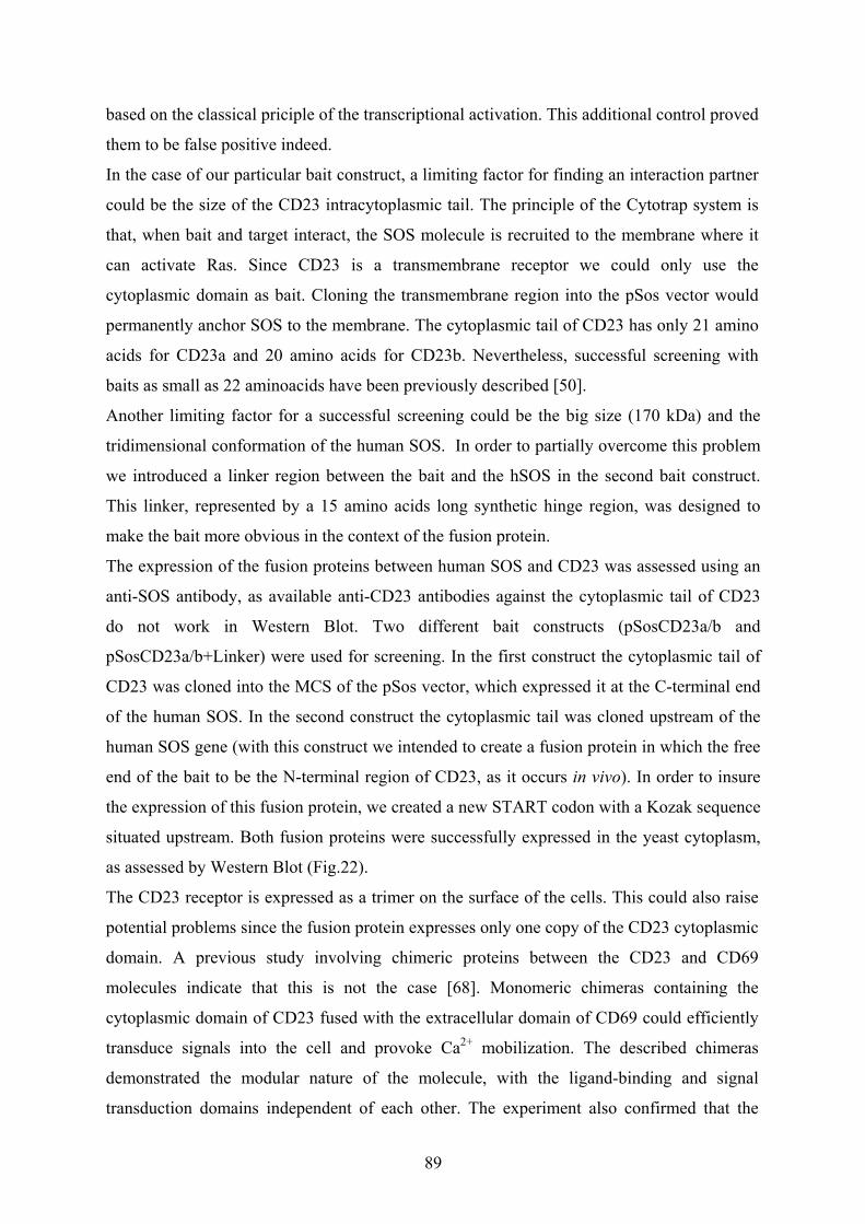
based on the classical priciple of the transcriptional activation. This additional control proved
them to be false positive indeed.
In the case of our particular bait construct, a limiting factor for finding an interaction partner
could be the size of the CD23 intracytoplasmic tail. The principle of the Cytotrap system is
that, when bait and target interact, the SOS molecule is recruited to the membrane where it
can activate Ras. Since CD23 is a transmembrane receptor we could only use the
cytoplasmic domain as bait. Cloning the transmembrane region into the pSos vector would
permanently anchor SOS to the membrane. The cytoplasmic tail of CD23 has only 21 amino
acids for CD23a and 20 amino acids for CD23b. Nevertheless, successful screening with
baits as small as 22 aminoacids have been previously described [50].
Another limiting factor for a successful screening could be the big size (170 kDa) and the
tridimensional conformation of the human SOS. In order to partially overcome this problem
we introduced a linker region between the bait and the hSOS in the second bait construct.
This linker, represented by a 15 amino acids long synthetic hinge region, was designed to
make the bait more obvious in the context of the fusion protein.
The expression of the fusion proteins between human SOS and CD23 was assessed using an
anti-SOS antibody, as available anti-CD23 antibodies against the cytoplasmic tail of CD23
do not work in Western Blot. Two different bait constructs (pSosCD23a/b and
pSosCD23a/b+Linker) were used for screening. In the first construct the cytoplasmic tail of
CD23 was cloned into the MCS of the pSos vector, which expressed it at the C-terminal end
of the human SOS. In the second construct the cytoplasmic tail was cloned upstream of the
human SOS gene (with this construct we intended to create a fusion protein in which the free
end of the bait to be the N-terminal region of CD23, as it occurs in vivo). In order to insure
the expression of this fusion protein, we created a new START codon with a Kozak sequence
situated upstream. Both fusion proteins were successfully expressed in the yeast cytoplasm,
as assessed by Western Blot (Fig.22).
The CD23 receptor is expressed as a trimer on the surface of the cells. This could also raise
potential problems since the fusion protein expresses only one copy of the CD23 cytoplasmic
domain. A previous study involving chimeric proteins between the CD23 and CD69
molecules indicate that this is not the case [68]. Monomeric chimeras containing the
cytoplasmic domain of CD23 fused with the extracellular domain of CD69 could efficiently
transduce signals into the cell and provoke Ca2+ mobilization. The described chimeras
demonstrated the modular nature of the molecule, with the ligand-binding and signal
transduction domains independent of each other. The experiment also confirmed that the
89

cytoplasmic domain is fully responsible for signal transduction. On the other hand,
oligomerization seems to be quite an important factor for ligand binding. CD23 affinity for
IgE decreases in monomeric CD23 or in the case of small soluble CD23 fragments, which
don’t retain the helical coiled coil [38].
Another way in which a two-hybrid system can be used is to test an interaction between two
known proteins. The analysis of the amino acid sequence in the cytoplasmic tail of CD23 and
previous experiments pointed out several potential targets for this membrane receptor. As
mentioned already, the product of a transfected CD23 gene in a NK cell line was found
associated with fyn, a member of the src-family of protein kinases [78]. Also, if the tyrosine
residue in the CD23a isoform is part of a functional ITIM motif, phosphatases like SHP and
SHIP could be recuited through their SH2 domains. The gene for fyn has been cloned as
cDNA into the pMyr vector and the ability to interact with CD23a and CD23b was tested in
the system. Similar experiments were performed for SHP-1 and SHP-2 (results not shown).
Anyway, as all these interaction would normally involve the phosphorylation of the tyrosine
residue, yeast two-hybrid may not be the appropriate system to assess them. Yeast hosts
might not express the kinases or they might not have the same phosphorylation patterns as
the mammalian kinases. To overcome this problem one can, for example, express the
required kinases in the yeast cytoplasm.
Another approach to overcome this problem would be to replace the tyrosine residue with a
glutamic acid residue. This strategy can be used in experimental settings when tyrosine
phosphorylation cannot be achieved. By site directed mutagenesis, we constructed a new bait
vector, named pSosCD23a-Glu, in which the tyrosine from position 6 of the cytoplasmic tail
of CD23a was replaced with a glutamic acid residue. We used this new construct in order to
test the interaction between CD23a and fyn, SHP-1 and SHP-2. With this construct also no
interaction was found. We also plan to use this new bait construct for future screening of the
spleen library.
In conclusion, we used a yeast two-hybrid system in order to identify a direct interaction
partner for CD23. This involved first the direct testing of interactions between the bait and
possible or predicted interaction partners and secondly the intensive screening of a spleen
library using different bait constructs. Other experimental approaches – like
immunoprecipitation assays- were also tested (results not shown). These strategies did not
result in the identification of an interaction partner for CD23 up to now.
90

To summarize, we demonstrate that Pax-5 regulates the CD23a promoter, proving to be a
key regulator of B-cell specific expression of CD23a isoform. The establishment of a two-
hybrid system represents a solid base for future investigations of the signaling mechanisms
through the CD23 receptor.
91
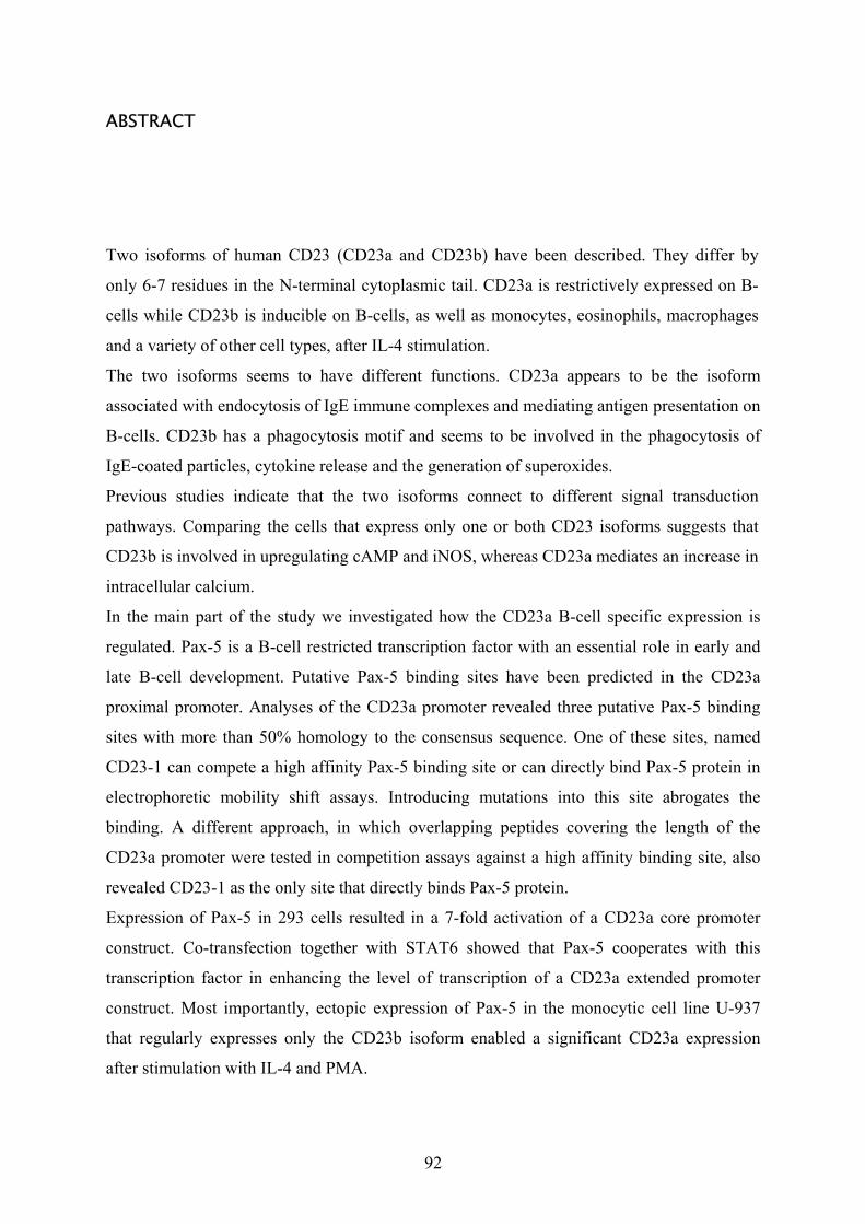
ABSTRACT
Two isoforms of human CD23 (CD23a and CD23b) have been described. They differ by
only 6-7 residues in the N-terminal cytoplasmic tail. CD23a is restrictively expressed on B-
cells while CD23b is inducible on B-cells, as well as monocytes, eosinophils, macrophages
and a variety of other cell types, after IL-4 stimulation.
The two isoforms seems to have different functions. CD23a appears to be the isoform
associated with endocytosis of IgE immune complexes and mediating antigen presentation on
B-cells. CD23b has a phagocytosis motif and seems to be involved in the phagocytosis of
IgE-coated particles, cytokine release and the generation of superoxides.
Previous studies indicate that the two isoforms connect to different signal transduction
pathways. Comparing the cells that express only one or both CD23 isoforms suggests that
CD23b is involved in upregulating cAMP and iNOS, whereas CD23a mediates an increase in
intracellular calcium.
In the main part of the study we investigated how the CD23a B-cell specific expression is
regulated. Pax-5 is a B-cell restricted transcription factor with an essential role in early and
late B-cell development. Putative Pax-5 binding sites have been predicted in the CD23a
proximal promoter. Analyses of the CD23a promoter revealed three putative Pax-5 binding
sites with more than 50% homology to the consensus sequence. One of these sites, named
CD23-1 can compete a high affinity Pax-5 binding site or can directly bind Pax-5 protein in
electrophoretic mobility shift assays. Introducing mutations into this site abrogates the
binding. A different approach, in which overlapping peptides covering the length of the
CD23a promoter were tested in competition assays against a high affinity binding site, also
revealed CD23-1 as the only site that directly binds Pax-5 protein.
Expression of Pax-5 in 293 cells resulted in a 7-fold activation of a CD23a core promoter
construct. Co-transfection together with STAT6 showed that Pax-5 cooperates with this
transcription factor in enhancing the level of transcription of a CD23a extended promoter
construct. Most importantly, ectopic expression of Pax-5 in the monocytic cell line U-937
that regularly expresses only the CD23b isoform enabled a significant CD23a expression
after stimulation with IL-4 and PMA.
92

Our results suggest that Pax-5 is a key regulator of the B-cell restricted expression of the
CD23a isoform.
In the second part of the project, we used a yeast two-hybrid system (CytoTrapTM from
Stratagene) in order to look for cytoplasmic interaction partners for the CD23 receptor. The
system was established in order to reach a high efficiency of transformation and different bait
vector constructs were made. The screening was performed using a human spleen library
cloned in the target vector of the system. The first bait constructs used (pSosCD23a and
pSosCD23b) expressed the very short (22 amino acids) cytoplasmic tails of the isoforms at
the C-terminal end of the fusion protein (human SOS). Improved bait constructs,
(pSosCD23a+Linker and pSos CD23b+Linker) expressed the cytoplasmic tail of CD23a/b at
the N-terminal side of the human SOS and had in consequence the N-terminal part free as a
bait, as it occurs in vivo. A flexible linker region separated the fusion proteins in order to
make the small amino acid bait chain more obvious. Approximately three million library
clones were screened with these various constructs. No “true positive” interaction was
detected. A relatively high number of “false positive” clones were obtained and checked in
another two-hybrid system. A new bait construct, in which the tyrosine residue in the
cytoplasmic tail of CD23a was replaced by a glutamic acid residue will be used for future
screening.
The system was also used in order to test the interaction between CD23 and p59fyn, a
member of the Src family of protein kinases that was mentioned to associate with CD23a. No
interaction was detected by using the CytoTrap two-hybrid system.
In conclusion, the key result of the study demonstrates that Pax-5 is a main regulator of the
B-cell specific expression of the CD23a isoform. In addition, a two-hybrid system was
established and employed in order to look for cytoplasmic interaction partners for CD23.
93
ZUSAMMENFASSUNG
Bisher sind zwei Isoformen des humanen CD23 (CD23a und CD23b) beschrieben. Beide
unterscheiden sich lediglich in 6-7 Resten im N-terminalen, zytoplasmatischen Anteil.
CD23a wird ausschließlich auf B-Zellen exprimiert, während CD23b sowohl auf B-Zellen als
auch auf Monozyten, eosinophilen Granulozyten, Makrophagen und zahlreichen anderen
Zelltypen durch Stimulation mit IL-4 induziert werden kann.
Die beiden Isoformen vermitteln wahrscheinlich unterschiedliche Funktionen. CD23a gilt als
Isoform, welche vornehmlich mit der Endozytose von IgE-Immunkomplexen und der
Vermittlung von Antigen-Präsentation auf B-Zellen assoziiert ist. CD23b besitzt ein
Phagozytose-Motiv und scheint bei der Phagozytose IgE besetzter Partikel, der Freisetzung
von Zytokinen und der Bildung von Peroxiden eine Rolle zu spielen.
Frühere Untersuchungen legen die Vermutung nahe, dass die beiden Isoformen zwei
getrennte Signalübertragungswege miteinander verbinden. Die Gegenüberstellung von
Ereignissen, welche in Zellen, die nur eine einer oder beide Isoformen von CD23 besitzen,
stattfinden, legt die Vermutung nahe, dass CD23b cAMP und iNOS hochreguliert,
wohingegen CD23a einen Anstieg des intrazellulären Kalziums vermittelt.
Im ersten Teil unserer Untersuchungen haben wir die Regulation der B-Zell-spezifischen
Expression von CD23a analysiert. Pax-5 ist ein auf B-Zellen beschränkter
Transkriptionsfaktor, welcher für die frühe und späte B-Zellentwicklung von entscheidender
Bedeutung ist. Mögliche Pax-5 Bindungsstellen wurden in den proximalen Abschnitten des
CD23a Promotors vermutet. Die Analyse des CD23a Promotors ergab drei mutmaßliche Pax-
5 Bindungsstellen mit mehr als 50% Homologie zur Konsensus-Sequenz. Eine dieser
Bindungsstellen, namens CD23-1, kann mit einer hochaffinen Pax-5 Bindungsstelle
konkurrieren oder direkt das Pax-5 Protein in Elektromobilitäts Experimenten (EMSA)
binden. Das Einfügen von Mutationen an dieser Stelle verhindert die Bindung. Ein weiterer
Versuch, bei dem die gesamte Länge des CD23a Promotors durch überlappende Peptide in
einem kompetitiven Verfahren gegenüber hoch affinen Bindungsstellen getestet wurde, zeigt
ebenso CD23-1 als die einzige Stelle, welche direkt Pax-5 binden kann.
In weiteren Experimenten führte die Expression von Pax-5 in 293 Zellen zu einer 7fachen
Aktivierung eines CD23a Kernpromotor Konstrukts. Die Kotransfektion zusammen mit
94

STAT6 zeigte, dass Pax-5 mit diesem Transkriptionsfaktor kooperiert, indem es die
Transkriptionsrate eines vergrößerten CD23a Promotorkonstrukts erhöht. Von besonderer
Bedeutung ist die Tatsache, dass die ektope Expression von Pax-5 in der monozytären
Zelllinie U-937, die normalerweise nur die CD23b Isoform exprimiert, dann zu einer
Expression von CD23a nach Stimulation mit IL-4 und PMA führte. Unsere Ergebnisse legen
nahe, dass Pax-5 in der auf B-Zellen beschränkten Expression der CD23 Isoform eine
Schlüsselrolle zukommt.
Im zweiten Teil des Projekts haben wir ein “Zwei-Hefen-Hybrid-System“ (Cyto-Trap von
Stratagene) verwendet, um nach zytoplasmatischen Interaktionspartnern für den CD23
Rezeptor zu suchen. Das System wurde modifiziert um eine hohe Effizienz an
Transformation zu erzielen. Unterschiedliche „Köder“-Vektorkonstrukte wurden hergestellt.
Das Screening wurde mittels einer humanen Milzbibliothek mit dem Zielvektor des Systems
durchgeführt. Die anfangs benutzten Konstrukte –pSosCD23a und pSosCD23b –
exprimierten sehr kurze (22 Aminosäuren) zytoplasmatischen Reste der Isoformen am C-
terminalen Ende des Fusionsproteins (humanes SOS). Verbesserte Konstrukte (pSos
CD23a+Linker und pSosCD23b+Linker) exprimierten den zytoplasmatischen Anteil von
CD23a/b am N-terminalen Ende des humanen SOS und hatten folglich den N-terminalen
Anteil als Andockstelle frei, entsprechend den Bedingungen in vivo. Eine flexible
Verbindungsregion trennte die Fusionsproteine, um auf diese Weise die kurze
Aminosäurekette deutlich „sichtbar“ werden zu lassen. Annähernd drei Millionen Klone
wurden mittels der verschiedenen Konstrukte untersucht. Dabei konnte keine tatsächlich
positive Interaktion gefunden werden. Stattdessen fand sich eine vergleichsweise hohe Zahl
falsch-positiver Klone. Diese wiederum wurden in einem zweiten “Zwei-Hefen-Hybrid-
System“ getestet.
In Zukunft wird ein neues Konstrukt als Köder verwendet werden. Hierbei wurde ein
Tyrosin-Rest im zytoplasmatischen Anteil von CD23a durch Glutamat ersetzt. Das System
wurde bereits dazu verwendet, die Interaktion zwischen CD23 und p59fyn - einem Mitglied
der Src-Familie von Proteinkinasen, welches mit CD23a assoziiert sein soll – zu testen.
Jedoch konnte im CytoTrap “Zwei-Hefen-Hybrid-System“ keine Wechselwirkung
nachgewiesen werden.
Zusammenfassend zeigt das zentrale Ergebnis der Arbeit, dass Pax-5 der Schlüsselregulator
ist, der die B-Zell-spezifische Expression von CD23a ermöglicht. Zusätzlich wurde ein
“Zwei-Hefen-Hybrid-System“ etabliert, mit dem zytoplasmatische Interaktionspartner für die
CD23 Isoformen gefunden werden können.
95
REFERENCES
1. Adams, B., P. Dorfler, A. Aguzzi, Z. Kozmik, P. Urbanek, I. Maurer-Fogy, and M.
Busslinger. 1992. Pax-5 encodes the transcription factor BSAP and is expressed in B
lymphocytes, the developing CNS, and adult testis. Genes Dev. 6:1589-1607.
2. Aubry, J. P., S. Pochon, P. Graber, K. U. Jansen, and J. Y. Bonnefoy. 1992. CD21
is a ligand for CD23 and regulates IgE production. Nature 358:505-507.
3. Barberis, A., K. Widenhorn, L. Vitelli, and M. Busslinger. 1990. A novel B-cell
lineage-specific transcription factor present at early but not late stages of
differentiation. Genes Dev. 4:849-859.
4. Bartlett, W. C., A. E. Kelly, C. M. Johnson, and D. H. Conrad. 1995. Analysis of
murine soluble Fc epsilon RII sites of cleavage and requirements for dual-affinity
interaction with IgE. J.Immunol. 154:4240-4246.
5. Beavil, A. J., R. L. Edmeades, H. J. Gould, and B. J. Sutton. 1992. Alpha-helical
coiled-coil stalks in the low-affinity receptor for IgE (Fc epsilon RII/CD23) and related
C-type lectins. Proc.Natl.Acad.Sci.U.S.A 89:753-757.
6. Bettler, B., R. Maier, D. Ruegg, and H. Hofstetter. 1989. Binding site for IgE of the
human lymphocyte low-affinity Fc epsilon receptor (Fc epsilon RII/CD23) is confined
to the domain homologous with animal lectins. Proc.Natl.Acad.Sci.U.S.A 86:7118-
7122.
7. Bjorck, P., C. Elenstrom-Magnusson, A. Rosen, E. Severinson, and S. Paulie. 1993.
CD23 and CD21 function as adhesion molecules in homotypic aggregation of human B
lymphocytes. Eur.J.Immunol. 23:1771-1775.
8. Bonnefoy, J. Y., S. Lecoanet-Henchoz, J. P. Aubry, J. F. Gauchat, and P. Graber.
1995. CD23 and B-cell activation. Curr.Opin.Immunol. 7:355-359.
9. Busslinger, M. and P. Urbanek. 1995. The role of BSAP (Pax-5) in B-cell
development. Curr.Opin.Genet.Dev. 5:595-601.
96
10. Capron, M., M. J. Truong, D. Aldebert, V. Gruart, M. Suemura, G. Delespesse, B.
Tourvieille, and A. Capron. 1991. Heterogeneous expression of CD23 epitopes by
eosinophils from patients. Relationships with IgE-mediated functions. Eur.J.Immunol.
21:2423-2429.
11. Cordier, M., A. Calender, M. Billaud, U. Zimber, G. Rousselet, O. Pavlish, J.
Banchereau, T. Tursz, G. Bornkamm, and G. M. Lenoir. Stable transfection of
Epstein-Barr virus (EBV) nuclear antigen 2 in lymphoma cells containing the EBV
P3HR1 genome induces expression of B- cell activation molecules CD21 and CD23.
12. Czerny, T., G. Schaffner, and M. Busslinger. 1993. DNA sequence recognition by
Pax proteins: bipartite structure of the paired domain and its binding site. Genes Dev.
7:2048-2061.
13. Delespesse, G., U. Suter, D. Mossalayi, B. Bettler, M. Sarfati, H. Hofstetter, E.
Kilcherr, P. Debre, and A. Dalloul. 1991. Expression, structure, and function of the
CD23 antigen. Adv.Immunol. 49:149-191.
14. Delphin, S. and J. Stavnezer. 1995. Characterization of an interleukin 4 (IL-4)
responsive region in the immunoglobulin heavy chain germline epsilon promoter:
regulation by NF- IL-4, a C/EBP family member and NF-kappa B/p50. J.Exp.Med.
181:181-192.
15. Denoroy, M. C., J. Yodoi, and J. Banchereau. 1990. Interleukin 4 and interferons
alpha and gamma regulate Fc epsilon R2/CD23 mRNA expression on normal human B
cells. Mol.Immunol. 27 AB:129-134.
16. Dorfler, P. and M. Busslinger. 1996. C-terminal activating and inhibitory domains
determine the transactivation potential of BSAP (Pax-5), Pax-2 and Pax-8. EMBO J.
15:1971-1982.
17. Dugas, B., N. Paul-Eugene, O. Coqueret, J. M. Mencia-Huerta, and P. Braquet.
1992. Possible role of CD23 in allergic diseases. Res.Immunol. 143:448-451.
18. Dugas, B., M. D. Mossalayi, C. Damais, and J. P. Kolb. 1995. Nitric oxide
production by human monocytes: evidence for a role of CD23. Immunol.Today 16:574-
580.
97
19. Eberhard, D., G. Jimenez, B. Heavey, and M. Busslinger. 2000. Transcriptional
repression by Pax5 (BSAP) through interaction with corepressors of the Groucho
family. EMBO J. 19:2292-2303.
20. Epstein, D. J., M. Vekemans, and P. Gros. 1991. Splotch (Sp2H), a mutation
affecting development of the mouse neural tube, shows a deletion within the paired
homeodomain of Pax-3. Cell 67:767-774.
21. Ewart, M. A., B. W. Ozanne, and W. Cushley. 2002. The CD23a and CD23b
proximal promoters display different sensitivities to exogenous stimuli in B
lymphocytes. Genes Immun. 3:158-164.
22. Fournier, S., L. P. Yang, G. Delespesse, M. Rubio, G. Biron, and M. Sarfati. 1995.
The two CD23 isoforms display differential regulation in chronic lymphocytic
leukaemia. Br.J.Haematol. 89:373-379.
23. Fujiwara, H., H. Kikutani, S. Suematsu, T. Naka, K. Yoshida, K. Yoshida, T.
Tanaka, M. Suemura, N. Matsumoto, S. Kojima, and . 1994. The absence of IgE
antibody-mediated augmentation of immune responses in CD23-deficient mice.
Proc.Natl.Acad.Sci.U.S.A 91:6835-6839.
24. Gagro, A. and S. Rabatic. 1994. Allergen-induced CD23 on CD4+ T lymphocytes and
CD21 on B lymphocytes in patients with allergic asthma: evidence and regulation.
Eur.J.Immunol. 24:1109-1114.
25. Goller, M. E., C. Kneitz, C. Mehringer, K. Muller, D. M. Jelley-Gibbs, E. J.
Gosselin, M. Wilhelm, and H. P. Tony. 2002. Regulation of CD23 isoforms on B-
chronic lymphocytic leukemia. Leuk.Res. 26:795-802.
26. Gordon, J. 1994. B-cell signalling via the C-type lectins CD23 and CD72.
Immunol.Today 15:411-417.
27. Gustavsson, S., S. Hjulstrom, T. Liu, and B. Heyman. 1994. CD23/IgE-mediated
regulation of the specific antibody response in vivo. J.Immunol. 152:4793-4800.
98
28. Higuchi, R., B. Krummel, and R. K. Saiki. 1988. A general method of in vitro
preparation and specific mutagenesis of DNA fragments: study of protein and DNA
interactions. Nucleic Acids Res. 16:7351-7367.
29. Hill, R. E., J. Favor, B. L. Hogan, C. C. Ton, G. F. Saunders, I. M. Hanson, J.
Prosser, T. Jordan, N. D. Hastie, and H. van, V. 1991. Mouse small eye results from
mutations in a paired-like homeobox- containing gene. Nature 354:522-525.
30. Ho, S. N., H. D. Hunt, R. M. Horton, J. K. Pullen, and L. R. Pease. 1989. Site-
directed mutagenesis by overlap extension using the polymerase chain reaction. Gene
77:51-59.
31. Horcher, M., A. Souabni, and M. Busslinger. 2001. Pax5/BSAP maintains the
identity of B cells in late B lymphopoiesis. Immunity. 14:779-790.
32. Hubmann, R., J. D. Schwarzmeier, M. Shehata, M. Hilgarth, M. Duechler, M.
Dettke, and R. Berger. 2002. Notch2 is involved in the overexpression of CD23 in B-
cell chronic lymphocytic leukemia. Blood 99:3742-3747.
33. Ikuta, K., M. Takami, C. W. Kim, T. Honjo, T. Miyoshi, Y. Tagaya, T. Kawabe,
and J. Yodoi. 1987. Human lymphocyte Fc receptor for IgE: sequence homology of its
cloned cDNA with animal lectins. Proc.Natl.Acad.Sci.U.S.A 84:819-823.
34. Karagiannis, S. N., J. K. Warrack, K. H. Jennings, P. R. Murdock, G. Christie, K.
Moulder, B. J. Sutton, and H. J. Gould. 2001. Endocytosis and recycling of the
complex between CD23 and HLA-DR in human B cells. Immunology 103:319-331.
35. Kehry, M. R. and L. C. Yamashita. 1989. Low-affinity IgE receptor (CD23) function
on mouse B cells: role in IgE- dependent antigen focusing. Proc.Natl.Acad.Sci.U.S.A
86:7556-7560.
36. Kijimoto-Ochiai, S. 2002. CD23 (the low-affinity IgE receptor) as a C-type lectin: a
multidomain and multifunctional molecule. Cell Mol.Life Sci. 59:648-664.
37. Kikutani, H., M. Suemura, H. Owaki, H. Nakamura, R. Sato, K. Yamasaki, E. L.
Barsumian, R. R. Hardy, and T. Kishimoto. 1986. Fc epsilon receptor, a specific
99
differentiation marker transiently expressed on mature B cells before isotype switching.
J.Exp.Med. 164:1455-1469.
38. Kilmon, M. A., R. Ghirlando, M. P. Strub, R. L. Beavil, H. J. Gould, and D. H.
Conrad. 2001. Regulation of IgE production requires oligomerization of CD23.
J.Immunol. 167:3139-3145.
39. Kishi, H., X. C. Wei, Z. X. Jin, Y. Fujishiro, T. Nagata, T. Matsuda, and A.
Muraguchi. 2000. Lineage-specific regulation of the murine RAG-2 promoter: GATA-
3 in T cells and Pax-5 in B cells. Blood 95:3845-3852.
40. Kneitz, C., M. Goller, H. P. Tony, A. Simon, C. Stibbe, T. König, E. Serfling, and
A. Avots. 2002. The CD23b promoter is a target for NF-AT transcription factors. BBA
in press.
41. Kohler, I. and E. P. Rieber. 1993. Allergy-associated I epsilon and Ec epsilon
receptor II (CD23b) genes activated via binding of an interleukin-4-induced
transcription factor to a novel responsive element. Eur.J.Immunol. 23:3066-3071.
42. Kolb, J. P., A. Abadie, A. Proschnicka-Chalufour, A. de Gramont, and J. Poggioli.
1994. CD23-mediated cell signalling. J.Lipid Mediat.Cell Signal. 9:27-35.
43. Kozmik, Z., S. Wang, P. Dorfler, B. Adams, and M. Busslinger. 1992. The promoter
of the CD19 gene is a target for the B-cell-specific transcription factor BSAP. Mol.Cell
Biol. 12:2662-2672.
44. Krenacs, L., A. W. Himmelmann, L. Quintanilla-Martinez, T. Fest, A. Riva, A.
Wellmann, E. Bagdi, J. H. Kehrl, E. S. Jaffe, and M. Raffeld. 1998. Transcription
factor B-cell-specific activator protein (BSAP) is differentially expressed in B cells and
in subsets of B-cell lymphomas. Blood 92:1308-1316.
45. Lecoanet-Henchoz, S., J. F. Gauchat, J. P. Aubry, P. Graber, P. Life, N. Paul-
Eugene, B. Ferrua, A. L. Corbi, B. Dugas, C. Plater-Zyberk, and . 1995. CD23
regulates monocyte activation through a novel interaction with the adhesion molecules
CD11b-CD18 and CD11c-CD18. Immunity. 3:119-125.
100
46. Liao, F., B. K. Birshtein, M. Busslinger, and P. Rothman. 1994. The transcription
factor BSAP (NF-HB) is essential for immunoglobulin germ-line epsilon transcription.
J.Immunol.2904-2911.
47. Lindemann, D., E. Patriquin, S. Feng, and R. C. Mulligan. 1997. Versatile
retrovirus vector systems for regulated gene expression in vitro and in vivo. Mol.Med.
3:466-476.
48. Ling, P. D., J. J. Hsieh, I. K. Ruf, D. R. Rawlins, and S. D. Hayward. 1994. EBNA-
2 upregulation of Epstein-Barr virus latency promoters and the cellular CD23 promoter
utilizes a common targeting intermediate, CBF1. J.Virol. 68:5375-5383.
49. Liu, Y. J., J. A. Cairns, M. J. Holder, S. D. Abbot, K. U. Jansen, J. Y. Bonnefoy, J.
Gordon, and I. C. MacLennan. 1991. Recombinant 25-kDa CD23 and interleukin 1
alpha promote the survival of germinal center B cells: evidence for bifurcation in the
development of centrocytes rescued from apoptosis. Eur.J.Immunol. 21:1107-1114.
50. Luban, J. and S. P. Goff. 1995. The yeast two-hybrid system for studying protein-
protein interactions. Curr.Opin.Biotechnol. 6:59-64.
51. May, M. J. and S. Ghosh. 1997. Rel/NF-kappa B and I kappa B proteins: an overview.
Semin.Cancer Biol. 8:63-73.
52. Michaelson, J. S., M. Singh, C. M. Snapper, W. C. Sha, D. Baltimore, and B. K.
Birshtein. 1996. Regulation of 3' IgH enhancers by a common set of factors, including
kappa B-binding proteins. J.Immunol. 156:2828-2839.
53. Michaelson, J. S., M. Singh, and B. K. Birshtein. 1996. B cell lineage-specific
activator protein (BSAP). A player at multiple stages of B cell development.
J.Immunol. 156:2349-2351.
54. Mikita, T., D. Campbell, P. Wu, K. Williamson, and U. Schindler. 1996.
Requirements for interleukin-4-induced gene expression and functional characterization
of Stat6. Mol.Cell Biol. 16 AB:5811-5820.
55. Mossalayi, M. D., J. C. Lecron, A. H. Dalloul, M. Sarfati, J. M. Bertho, H.
Hofstetter, G. Delespesse, and P. Debre. 1990. Soluble CD23 (Fc epsilon RII) and
101
interleukin 1 synergistically induce early human thymocyte maturation. J.Exp.Med.
171:959-964.
56. Mossalayi, M. D., M. Arock, J. M. Bertho, C. Blanc, A. H. Dalloul, H. Hofstetter,
M. Sarfati, G. Delespesse, and P. Debre. 1990. Proliferation of early human myeloid
precursors induced by interleukin- 1 and recombinant soluble CD23. Blood 75:1924-
1927.
57. Niiro, H. and E. A. Clark. 2002. Regulation of B-cell fate by antigen-receptor signals.
Nat.Rev.Immunol. 2:945-956.
58. Nutt, S. L., P. Urbanek, A. Rolink, and M. Busslinger. 1997. Essential functions of
Pax5 (BSAP) in pro-B cell development: difference between fetal and adult B
lymphopoiesis and reduced V-to-DJ recombination at the IgH locus. Genes Dev.
11:476-491.
59. Nutt, S. L., A. M. Morrison, P. Dorfler, A. Rolink, and M. Busslinger. 1998.
Identification of BSAP (Pax-5) target genes in early B-cell development by loss- and
gain-of-function experiments. EMBO J. 17:2319-2333.
60. Nutt, S. L., B. Heavey, A. G. Rolink, and M. Busslinger. 1999. Commitment to the
B-lymphoid lineage depends on the transcription factor Pax5. Nature 401:556-562.
61. Nutt, S. L., D. Eberhard, M. Horcher, A. G. Rolink, and M. Busslinger. 2001. Pax5
determines the identity of B cells from the beginning to the end of B-lymphopoiesis.
Int.Rev.Immunol. 20:65-82.
62. Pirron, U., T. Schlunck, J. C. Prinz, and E. P. Rieber. 1990. IgE-dependent antigen
focusing by human B lymphocytes is mediated by the low-affinity receptor for IgE.
Eur.J.Immunol. 20:1547-1551.
63. Plater-Zyberk, C. and J. Y. Bonnefoy. 1995. Marked amelioration of established
collagen-induced arthritis by treatment with antibodies to CD23 in vivo. Nat.Med.
1:781-785.
64. Reimold, A. M., P. D. Ponath, Y. S. Li, R. R. Hardy, C. S. David, J. L. Strominger,
and L. H. Glimcher. 1996. Transcription factor B cell lineage-specific activator
102

protein regulates the gene for human X-box binding protein 1. J.Exp.Med. 183:393-
401.
65. Richards, M. L. and D. H. Katz . 1997. Analysis of the promoter elements necessary
for IL-4 and anti-CD40 antibody induction of murine Fc epsilon RII (CD23):
comparison with the germline epsilon promoter. J.Immunol. 158:263-272.
66. Rinkenberger, J. L., J. J. Wallin, K. W. Johnson, and M. E. Koshland. 1996. An
interleukin-2 signal relieves BSAP (Pax5)-mediated repression of the immunoglobulin
J chain gene. Immunity. 5:377-386.
67. Rolink, A. G., S. L. Nutt, F. Melchers, and M. Busslinger. 1999. Long-term in vivo
reconstitution of T-cell development by Pax5- deficient B-cell progenitors. Nature
401:603-606.
68. Sancho, D., A. G. Santis, J. L. Alonso-Lebrero, F. Viedma, R. Tejedor, and F.
Sanchez-Madrid. Functional analysis of ligand-binding and signal transduction
domains of CD69 and CD23 C-type lectin leukocyte receptors.
69. Sarfati, M., D. Bron, L. Lagneaux, C. Fonteyn, H. Frost, and G. Delespesse. 1988.
Elevation of IgE-binding factors in serum of patients with B cell- derived chronic
lymphocytic leukemia. Blood 71:94-98.
70. Sarfati, M., S. Fournier, C. Y. Wu, and G. Delespesse. 1992. Expression, regulation
and function of human Fc epsilon RII (CD23) antigen. Immunol.Res. 11:260-272.
71. Sarfati, M., B. Bettler, M. Letellier, S. Fournier, M. Rubio-Trujillo, H. Hofstetter,
and G. Delespesse. 1992. Native and recombinant soluble CD23 fragments with IgE
suppressive activity. Immunology 76:662-667.
72. Schaniel, C., M. Gottar, E. Roosnek, F. Melchers, and A. G. Rolink. 2002.
Extensive in vivo self-renewal, long-term reconstitution capacity, and hematopoietic
multipotency of Pax5-deficient precursor B-cell clones. Blood 99:2760-2766.
73. Schebesta, M., B. Heavey, and M. Busslinger. 2002. Transcriptional control of B-cell
development. Curr.Opin.Immunol. 14:216-223.
103

74. Shen, C. H. and J. Stavnezer. 1998. Interaction of stat6 and NF-kappaB: direct
association and synergistic activation of interleukin-4-induced transcription. Mol.Cell
Biol. 18:3395-3404.
75. Singh, M. and B. K. Birshtein. 1993. NF-HB (BSAP) is a repressor of the murine
immunoglobulin heavy-chain 3' alpha enhancer at early stages of B-cell differentiation.
Mol.Cell Biol. 13:3611-3622.
76. Soneoka, Y., P. M. Cannon, E. E. Ramsdale, J. C. Griffiths, G. Romano, S. M.
Kingsman, and A. J. Kingsman. 1995. A transient three-plasmid expression system
for the production of high titer retroviral vectors. Nucleic Acids Res. 23:628-633.
77. Stief, A., G. Texido, G. Sansig, H. Eibel, G. Le Gros, and P. H. van der. 1994. Mice
deficient in CD23 reveal its modulatory role in IgE production but no role in T and B
cell development. J.Immunol. 152:3378-3390.
78. Sugie, K., T. Kawakami, Y. Maeda, T. Kawabe, A. Uchida, and J. Yodoi. 1991.
Fyn tyrosine kinase associated with Fc epsilon RII/CD23: possible multiple roles in
lymphocyte activation. Proc.Natl.Acad.Sci.U.S.A 88:9132-9135.
79. Suter, U., G. Texido, and H. Hofstetter. 1989. Expression of human lymphocyte IgE
receptor (Fc epsilon RII/CD23). Identification of the Fc epsilon RIIa promoter and its
functional analysis in B lymphocytes. J.Immunol. 143:3087-3092.
80. Tassabehji, M., A. P. Read, V. E. Newton, R. Harris, R. Balling, P. Gruss, and T.
Strachan. 1992. Waardenburg's syndrome patients have mutations in the human
homologue of the Pax-3 paired box gene. Nature 355:635-636.
81. Texido, G., H. Eibel, G. Le Gros, and P. H. van der. 1994. Transgene CD23
expression on lymphoid cells modulates IgE and IgG1 responses. J.Immunol.
153:3028-3042.
82. Tinnell, S. B., S. M. Jacobs-Helber, E. Sterneck, S. T. Sawyer, and D. H. Conrad.
1998. STAT6, NF-kappaB and C/EBP in CD23 expression and IgE production.
Int.Immunol. 10:1529-1538.
104
83. Tsicopoulos, A. and M. Joseph. 2000. The role of CD23 in allergic disease.
Clin.Exp.Allergy 30:602-605.
84. Urbanek, P., Z. Q. Wang, I. Fetka, E. F. Wagner, and M. Busslinger. 1994.
Complete block of early B cell differentiation and altered patterning of the posterior
midbrain in mice lacking Pax5/BSAP. Cell 79:901-912.
85. Wang, F., H. Kikutani, S. F. Tsang, T. Kishimoto, and E. Kieff. 1991. Epstein-Barr
virus nuclear protein 2 transactivates a cis-acting CD23 DNA element. J.Virol. 65
:4101-4106.
86. Xu, L., M. G. Kim, and K. B. Marcu. 1992. Properties of B cell stage specific and
ubiquitous nuclear factors binding to immunoglobulin heavy chain gene switch regions.
Int.Immunol. 4:875-887.
87. Yokota, A., H. Kikutani, T. Tanaka, R. Sato, E. L. Barsumian, M. Suemura, and
T. Kishimoto. 1988. Two species of human Fc epsilon receptor II (Fc epsilon
RII/CD23): tissue-specific and IL-4-specific regulation of gene expression. Cell
55:611-618.
88. Yokota, A., K. Yukawa, A. Yamamoto, K. Sugiyama, M. Suemura, Y. Tashiro, T.
Kishimoto, and H. Kikutani. 1992. Two forms of the low-affinity Fc receptor for IgE
differentially mediate endocytosis and phagocytosis: identification of the critical
cytoplasmic domains. Proc.Natl.Acad.Sci.U.S.A 89:5030-5034.
89. Yu, P., M. Kosco-Vilbois, M. Richards, G. Kohler, and M. C. Lamers. 1994.
Negative feedback regulation of IgE synthesis by murine CD23. Nature 369:753-756.
90. Zwollo, P., H. Arrieta, K. Ede, K. Molinder, S. Desiderio, and R. Pollock. 1997.
The Pax-5 gene is alternatively spliced during B-cell development. J.Biol.Chem. 272
:10160-10168.
105

Books
91. Adams, A., Gottschling, D., Kaiser, C., Stearns, T. (Eds.) Methods in yeast genetics.
Cold Spring Harbor Laboratory Press, 1997.
92. Janeway, C., Travers, P., Walport, M., Donald Capra, J. (Eds.) Immunobiology-The
immune system in health and disease. Elsevier Science Ltd/Garland Publishing, 1999.
93. Lewin, B. (Ed.) Genes VII. Oxford University Press, 2000.
94. Lodish, H., Berk, A., Lawrance Zipursky, S., Matsudaira, P., Baltimore, D.,
Darnell, J. (Eds.) Molecular cell biology. W.H. Freemen and Company, 2000.
106
ABREVIATIONS
A260 absorbance at 260 nm
aa amino acids
AD activation domain
Ag antigen
B-CLL chronic lymphocytic leukemia
BCR B cell receptor
BLNK B cell Linker
BSA bovine serum albumine
BSAP B-cell-specific activator protein
bp base pairs
3’C∝ immunoglobulin heavy-chain enhancer
Ca2+ calcium
cAMP adenosine 3’,5’-cyclic-monophosphate
CBF1 C promoter binding factor 1
C/EBP CCAAT/ enhancer binding protein
CD cluster of differentiation
CD40L CD40 ligand
cDNA complementary DNA
CIP calf intestinal phosphatase
cpm counts per minute
CR2/CR3/CR4 complement receptor 2/3/4
CRE cAMP response element
CREB cAMP response element binding protein
CY cytoplasmic domain
Cε immunoglobulin ε chain constant region exon
Cγ1 immunoglobulin γ1 chain constant region exon
DAG diacylglycerol
dCTP deoxycytidine triphosphate
DMEM Dulbeccos minimum essential medium
107
DNA deoxyribonucleic acid
DNA-BD DNA-binding domain
dNTP deooxynucleoside triphosphate
d(TGA)TP deoxy(thymidine/guanosine/adenosine/)
triphosphate
DTT dithiothreinol
EBF early B-cell factor
EBNA-2 Epstein-Barr virus nuclear antigen-2
EBV Epstein-Barr virus
EC extracellular domain
EDTA ethylenediaminetetraacetic acid
EMSA Electrophoretic Mobility Shift Assay
Erk extracellular-regulated kinase
Eµ Ig heavy chain intronic enhancer
FACS fluorescence-activated cell sorter
FCS fetal calf serum
FcεRI high affinity IgE receptor
FcεRII CD23, low affinity IgE receptor
Fwr forward primer
Gal(-Ura,-Leu) galactose medium without uracil and leucine
GDP guanosine 5’-diphosphate
Glu glutamic acid
Glu(-Ura,-Leu) glucose medium without uracil and leucine
GTP guanosine 5’-triphosphate
H2A-2/ H2B-2 histone genes
HA hemagglutinin
HEPES N-2-hydroxyethylpiperazine-N’-2-
ethanesulfonic acid
3’α-hs4 DNase hypersensitivity site 4 in the α gene
enhancer
HSC hematopoietic stem cell
hSOS human SOS
IC immune complexes
108

IFN-α interferon-α
IFN-γ interferon-γ
IgA/ IgD/ IgE/ IgG/ IgM immunoglobulin A / D / E / G / M
Ikkα/β inhibitor of NF-kB kinase α/β
IkB inhibitor of NF-kB
IL-1/ IL-4/ IL-6 interleukins
Ile isoleucine
INOS inducible nitric oxide synthase
IP3 inositol 1,4,5-triphosphate
JAK Janus Kinase
ITAM immunoreceptor tyrosine based activation
motifs
ITIM immunoreceptor tyrosine based inhibitory
motifs
LB luria broth
Leu leucine
LFA-1 lymphocyte-function-associated antigen-1
LiAc lithium acetate
LPS lipopolysaccharide
MAPK mitogen-activated protein kinase
MCS multiple cloning site
M-CSF-R macrophage colony-stimulating factor receptor
Met methionine
MHC major histocompatibility complex
mRNA messenger RNA
N (nucleotide) any nucleotide
NF-AT nuclear factor of activated T cells
NK natural killer cells
NOS nitric oxide synthase
OD optical density
Oct-1 octamer binding protein-1
PAGE Polyacrylamide Gel Electrophoresis
PBS phosphate-buffered saline
109
PCR polymerase chain reaction
PEG polyethylene glycol
PIP2 phosphatidylinositol biphosphate
PKA protein kinase A
PKC protein kinase C
PLC /PLC-γ phospholipase C-γ
PMA phorbolemyristatacetate
PMSF phynilmethylsulfonyl fluoride
poly[d(I-C)] poly-deoxy-inosinic-deoxy-cytidylic acid
R (nucleotide) A or G
RAG-2 recombination-activating genes-2
RGD arginine-glutamine-aspartic acid
rIL-4 recombinant IL-4
RNA ribonucleic acid
RNase ribonuclease
RPA RNase protection assay
rpm rotations per minute
RPMI
RT-PCR reverse-transcriptase polymerase chain reaction
S (nucleotide) C or G
Sγ2a switch region for immunoglobulin γ2a chain
constant region
Sµ switch region for immunoglobulin µ chain
constant region
Ser serine
sCD23 soluble CD23
SDS sodium dodecyl sulphate
SH2 Src homology domain 2
SH3 Src homology domain 3
SHP-1 SH2-domain-containing protein tyrosine
phosphatase 1
SHP-2 SH2-domain-containing protein tyrosine
phosphatase 1
110
SHIP SH2-domain-containing inositol polyphosphate
5’ phosphatase
SOS Son of Sevenless
Src/ src Rous sarcoma
STAT6 signal transducer and activator of transcription
SV40 simian virus 40
TAE Tris acetate electrophoresis buffer
TBE Tris borate electrophoresis buffer
TEMED N,N,N’,N’-tetramethylethylenediamine
TM transmembrane domain
TNF tumor necrosis factor
Tris tris(hydroxymethyl)aminomethane
TSAP tissue specific activator protein
Tyr tyrosine
UAS upstream activatory sequence
Ura uracil
UTP uridine-5’-triphosphate
XBP-1 X-box-binding protein 1
Y (nucleotide) C or T
YPAD Yeast extract, Peptone, Dextrose medium
111

ACKNOWLEDGMENTS
To Prof. Dr. H.-P.Tony, for giving me the opportunity to do my doctorate work in his lab; for
his trust and support during these years and for his help when I had to make important
decisions.
To Prof Dr J. Hacker for accepting to be my tutor at the Faculty of Biology and his support
with all formalities concerning the doctoral thesis.
To Dr. M. Goller, for all his help during my first days in the lab, for guiding and teaching me
not only experimental procedures but also how to think and work independently; for keeping
in touch and for his interest even after he left our lab. To Dr. I. Berberich, for the Pax-5
retroviral construct in U-937 cells.
To Prof. Hünig, for a very scientifically stimulating and creative atmosphere in the Graduate
College and very instructive (and mandatory) seminars every Thursday; for his prompt help
in all the administrative problems. To all my collegues in the Graduate College 520
“Immunomodulation”, for their stimulative discussions, for their support, for being original
and international.
To Anne Sophie Rouziere, my special friend and lab collegue, just for being there- her
presence in the lab in the last three years made things a lot easier in many circumstances. To
the people in my lab: K. Zehe for her technical assistance especially in the Luciferase Assay
experiments and for her optimistic working presence and M. Feuchtenberger for his general
help and good will.
To Prof. A. Schimpl, for critically reviewing the manuscript of the article and for interesting
suggestions and advice to our work To Dr. S.Klein-Hessling, for helpful discussion, advice
and critical observations to my work.
A special thought to my family and to my husband, Lucian. We went together through the
demanding but great experience of working and studying to achieve a certain level of
knowledge in science.
112

CURRICULUM VITAE
Name: Ioana Andreea Visan
Date of birth: 5th of January 1975
Place of birth: Ploiesti, Romania
Education:
1993 Highschool Diploma at the “I.L. Caragiale” National College, Ploiesti
1993-1997 Studies at the Faculty of Biology, “Al.I. Cuza” University, Iasi
(Romania)
1996 Marine Ecology Summer Application with the University Paris 7
(France)
1997 Bachelor Degree at the Faculty of Biology, “Al.I. Cuza” University,
Iasi (Romania)
1997-1999 Master Degree in Molecular Genetics at the Faculty of Biology, “Al.I.
Cuza” University, Iasi (Romania)
1999 courses of Molecular Ecology in Marine Biology at RijkUniversiteit
Groningen (Netherlands)
Since January 2000
- PhD student in the Molecular Immunology Lab at the Medizinische
Poliklinik , Würzburg University, coordinator Prof. H.P. Tony
- member of the Graduate College “Immunomodulation” Würzburg,
coordinator Prof. Th. Hünig
113
PUBLICATIONS
1. Original articles
• Visan I., Goller M., Berberich I., Kneitz Ch. and Tony H.-P. Pax-5 is a key
regulator of the B cell restricted expression of the CD23a isoform. Eur. J. Immunol.
In press.
2. Published abstracts
• Visan I., Goller M., Berberich I., Kneitz Ch., Avots A., Serfling E., Tony H.-P.
2001. Pax-5 and NF-ATp are modulators of high CD23 expression. Scand. J.
Immunol. 54 (Suppl.1)
• Kneitz Ch., Goller M., Visan I., Simon A., Stibbe C., Avots A., Serfling E., Tony
H.-P. 2001. The CD23b promoter is a target of NF-AT transcription factors. Arthritis
and Rheumatism 44, S9.
• Visan I., Goller M., Berberich I., Kneitz Ch. and Tony H.-P. 2002. Pax-5 is a key
regulator of the B cell restricted expression of the CD23a isoform. Arthritis and
Rheumatism 46, S9.
3. Poster and oral presentations at Congresses and Symposia
• Visan I., Goller M., Kneitz Ch., Chang C., Tony H.-P. Looking for cytoplasmic
interaction partners of CD23. 12th Protein Kinase Symposium: NO/cGMP and Protein
Kinase Signaling, 8/2000, Bad Bruckenau
• Visan I. Using two hybrid Systems to find interaction partners of CD23. Joint Retreat
of the Graduate Colleges 520 and 592, "Immunomodulation meets Lymphocyte
Activation" 7/2001, Zeilitzheim
• Visan I., Goller M., Berberich I., Kneitz Ch., Avots A., Serfling E., Tony H.-P.
Pax-5 and NF-ATp are modulators of high CD23 expression. 11th International
Congress of Immunobiology, 7/2001, Stockholm, Sweden
114

• Visan I., Goller M., Berberich I., Kneitz Ch. and Tony H.-P. Role of Pax-5 in the
B cell restricted expression of the CD23a isoform. Joint Retreat of the Graduate
Colleges 520 and 592, "Immunomodulation meets Lymphocyte Activation" 5/2002,
Zeilitzheim
• Visan I. Role of Pax-5 in the B cell restricted expression of the CD23a isoform.
Graduate College 520 Evaluation by the DFG, 5/2002
• Visan I., Goller M., Berberich I., Kneitz Ch. and Tony H.-P. Role of Pax-5 in the
B cell restricted expression of the CD23a isoform. Euroconference- “Interactions
between innate and adaptive immunity in mammalian defence against bacterial
infections” , 6/2002, Goehren-Lebbin
• Visan I., Goller M., Berberich I., Kneitz Ch. and Tony H.-P. Pax-5 is a key
regulator of the B cell restricted expression of the CD23a isoform. 66th Annual
Scientific Meeting of the American College of Rheumatology, 9/2002, New Orleans
115

EIDESSTATTLICHE ERKLÄRUNGEN
Hiermit erkläre ich ehrenwörtlich, dass die vorliegende Dissertation „The CD23 receptor-
regulation of expression and signal transduction“ selbständig an der Medizinischen
Poliklinik der Universität Würzburg angefertigt wurde und dass ich keine anderen als die
angegebenen Quellen und Hilfsmittel benutzt habe.
Würzburg, 31 Januar 2003
(Dipl Biologin Ioana Andreea Visan)
Hiermit erkläre ich ehrenwörtlich, dass die vorliegende Dissertation „The CD23 receptor-
regulation of expression and signal transduction“ in gleicher oder ähnlicher Form noch
nicht in einem anderen Prüfungsverfahren vorgelegen hat.
Würzburg, 31 Januar 2003
(Dipl Biologin Ioana Andreea Visan)
Hiermit erkläre ich ehrenwörtlich, dass ich bisher noch keine akademische Grade erworben
oder zu erwerben versucht habe.
Würzburg, 31 Januar 2003
(Dipl Biologin Ioana Andreea Visan)
116
Related Documents


