THE CARDIAC EXAM Caroline Ball, MD, FACC Stritch School of Medicine Medicine Clerkship

Welcome message from author
This document is posted to help you gain knowledge. Please leave a comment to let me know what you think about it! Share it to your friends and learn new things together.
Transcript

THE CARDIAC EXAM
Caroline Ball, MD, FACC
Stritch School of Medicine
Medicine Clerkship

LEARNING OBJECTIVES
• Define the Mechanism of Generation, Clinical Significance and Best Listening Areas for:
• S1/S2
• S2 Splitting Patterns
• S3/S4
• Ejection Clicks (Early and Mid)
• Opening Snap
• Describe the Grading System for Murmurs
• Compare and contrast the location, pattern of radiation, timing, pitch, shape, quality, and
response to common physiologic maneuvers of several murmurs

KEYS TO THE CARDIOVASCULAR EXAM
• Stand on the right side of the patient
• Only auscultate over skin, never over clothing
• Quiet room

RULES OF THUMB
• “Benign Murmurs” = S’s: short, soft, systolic
• Diastolic Murmurs = Bad
• ARMS (Aortic Regurgitation, Mitral Stenosis)
• Left-Sided Sounds are Louder than right-sided murmurs
• Right-Sided Sounds increase with inspiration (except PS)
• Intensity does not = severity of valve lesions (ex VSD), but timing does!
• Remember the company the murmur keeps

GENERATING HEART SOUNDS

GENERATING HEART SOUNDS
• S1 = T1+M1
• Closure of tricuspid and mitral valves
• S2 = P2+A2
• Closure of pulmonic and aortic valves.

GENERATING HEART SOUNDS, CONTINUED
• Gallops can be right or left-sided
• Best heard with the bell
• Auscultate over PMI for L-sided gallops
• Auscultate over Xiphoid process for R-sided gallops
• “Summation” Gallop = in tachycardia when S3/S4 are
heard together, may be louder than S2.

S3
• Blood being “sucked” into the ventricle
• Heard in Sys-Tol-Ic dysfunction
• Acute MI
• Dilated Cardiomyopathy
• Increased volume of ventricular flow (ex MR, AI, VSD, PDA)
• May be normal in young people, athletes, third trimester of pregnancy
• “Sloshing In”
• “Kentucky”
• May be heard every few beats
• Cannot be heard with mitral stenosis (due to tight mitral valve)

S4
• Atrial Kick
• Heard in Di-A-Stol-Ic dysfunction
• “A Stiff Wall”
• “Tennessee”
• Heard post-MI
• May be heard in acute MR (not chronic, with LA enlargement)
• May be heard with first degree AV block

EJECTION SOUNDS
• Rarely the valves can be heard opening (onset of ventricular systole)
• Sounds close to physiologic splitting of S1
• Due to “doming” of the aortic valve
• Aortic Opening Sound:
• Best heard over the apex or over the aortic area
• Pulmonic Opening Sound:
• Best heard over L second intercostal area
• Respiratory variation (decrease with inspiration)

MITRAL VALVE PROLAPSE
• Mid-systolic prolapse of the mitral (+/- tricuspid) valve leaflets
into the left atrium
• Often accompanied with mid-late systolic murmur
• Systolic Click can also be heard with:
• Marfan Syndrome
• Ostium secundum ASD
• Papillary Muscle dysfunction

OPENING SNAP OF MITRAL STENOSIS
• Similar to aortic ejection sounds: occurs due to “doming”
of the MV leaflets
• The closer the opening snap is to S2 = delayed opening of
the MV = more severe mitral stenosis.

DETERMINING S1
• Usually louder over the apex

S1 IS LOUDER WHEN
• Increased ventricular contraction
• Increased rate of pressure generation in the ventricle
• Heart is closer to the chest wall.
• Hyperdynamic states
• Rheumatic Mitral Stenosis = Loud S1
• Due to elevated LAP
• Short PR interval on ECG = Loud S1
• Due to the MV leaflets being wide apart when the QRS hits
• Mitral Valve Prolapse

S1 IS SOFTER WHEN
• Prolonged PR
• Leaflets are already almost closed when QRS hits
• Acute, Severe AI
• LV diastolic pressure rises quickly -> premature closure of the mitral valve
leaflets
• MR due to lack of leaflet mobility
• Chest configuration reduces auscultation:
• Emphysema
• Obesity
• Large breasts
• Pericardial Effusion

VARIABLE S1: AV DISSOCIATION
•Atrial Fibrillation
•Complete Heart Block
•VT

PHYSIOLOGIC SPLIT HEART SOUNDS
• With Expiration A2 and P2 occur at the same time
• With Inspiration there is increased filling on the right side
of the heart, and thus a delay in closure of P2.
• In patients over age 50 the split sound may be
indistinguishable due to LV dysfunction and some degree of
AS

FIXED SPLIT HEART SOUNDS
• ASD: Right to left shunting
• Higher cardiac output through the R sided valves
• Persistent delay in P2


PARADOXICAL SPLITTING
• A2 is slow to occur, P2 remains stable
• LV is slower to contract (as in LBBB)
• Increased resistance for the LV (AS, HTN)


PERSISTENT SPLITTING
• P2 is always delayed, but more delayed with
inspiration
• RBBB (LV is delayed)
• Pulmonic Stenosis


LOUD P2
• Pulmonary Hypertension

LOUD A2
• Systemic Hypertension

FAINT A2
• Aortic Stenosis
• Faint P2 with pulmonic stenosis

GRADING MURMURS
Grade Description
1 Faintest murmur, heard only with special effort
2 Faint murmur, but heard immediately
3 Moderately loud murmur
4 Loud murmur associated with palpable thrill
5 Very loud murmur heard with part of stethoscope touching
chest wall
6 Loudest murmur heard with stethoscope removed from chest
wall.

PHYSICAL EXAM MANEUVERS
Maneuver Physiologic Effect Murmurs Affected
Passive Leg Raise Increase venous return -> Increase Preload Decreases HCM murmur
Squatting Compresses leg veins -> increased venous
return, increased afterload
Delays MVP click and shortens murmur
Decreases HCM murmur (“Squashes MVP”)
Increases AS murmur intensity
Valsalva Increases intrathoracic pressure ->
decreased preload
Moves MVP earlier in systole
Increases HCM
Standing Blood pools in legs -> Decreases preload Increases HCM
Decreases AS murmur intensity
Isometric Hand
Grip
Increases peripheral vascular resistance Increases MR murmur
Increases VSD murmur
Increases AI murmur

SYSTOLIC MURMURS

SYSTOLIC EJECTION MURMUR
• Early-peaking

AORTIC STENOSIS
• Systolic
• Radiates to carotids (especially R Supraclavicular Region)
• Louder with increased flow:
• Following a pause after a premature beat (“post-extrasystolic”)
• Passive leg raise
• Squatting
• Associated with “pulsus parvus et tardus” (Slow Rising, Late Peaking)
• Gallavardin Phenomenon = musical quality radiation of the AS murmur to the apex.

GRADING AS SEVERITY
• Mild AS: systolic murmur ends before
S2
• Moderate AS: Murmur peaks LATER in
systole. Delay in A2
• Severe AS: Paradoxical splitting of S2
(versus absent S2).
Mild Aortic Stenosis. C = opening click
Severe Aortic Stenosis

HYPERTROPHIC CARDIOMYOPATHY
• Maneuvers:
• Lounder with standing
• Louder during Valsalva
• Softer with squatting
• Softer with passive leg raise
• Associated with “Triple Ripple” on palpation over the apex
• “Spike and dome” pulse (Quick rising, twin-peaking)

HOLOSYSTOLIC MURMUR?
• Chronic MR
• TR
• VSD
• And it is usually chronic MR…

MITRAL REGURGITATION
• Holosystolic, heard best at the apex
• Louder with squatting or isometric handgrip
• Chronic MR: The left atrium has enlarged to accommodate
• Acute MR:
• Decreasing intensity later in systole
• Expect some degree of pulmonary edema
• Mild MR: systolic murmur
• Moderate MR: Add an S3 gallop
• Severe MR: Diastolic flow rumble.
• May radiate to the back (if there is prolapse of anterior MV leaflet)
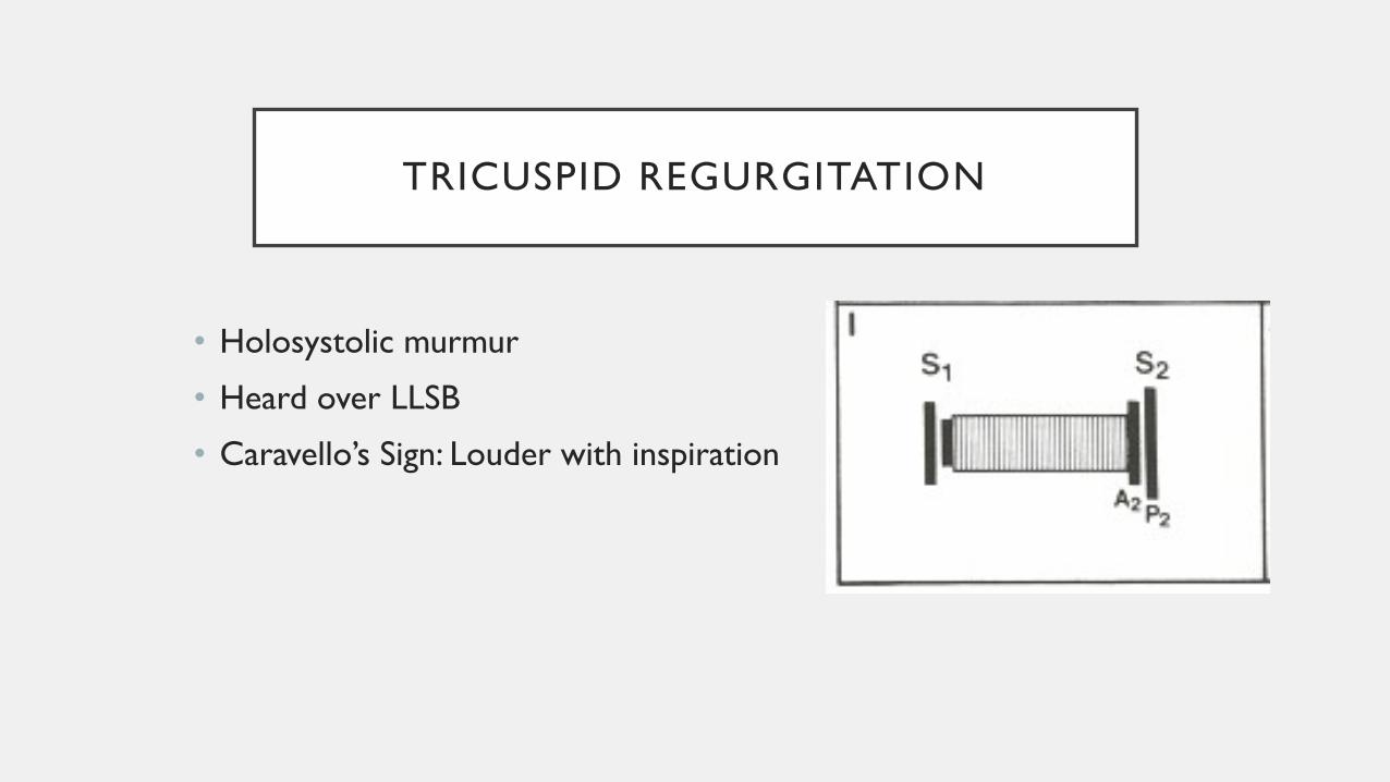
TRICUSPID REGURGITATION
• Holosystolic murmur
• Heard over LLSB
• Caravello’s Sign: Louder with inspiration

VENTRICULAR SEPTAL DEFECT
• Holosystolic murmur
• Heard best along left sternal border
• Louder with isometric handgrip.
Slightly delayed P2

MITRAL VALVE PROLAPSE
• Midsystolic Click moves towards
S1 and late systolic murmur
starts earlier with standing
• Click moves earlier on
inspiration
• Murmur starts later and click
moves towards S2 with squatting

DIASTOLIC MURMURS
Always bad.

ARMS
• Early Diastolic Murmurs
• Aortic and pulmonic regurgitation
• Mid/Late Diastolic Murmurs
• Mitral and tricuspid stenosis
• Combined Systolic and Diastolic Murmurs:
• PDA
• Coronary AV fistula
• Pulmonary AV fistula
• Ruptured Sinus of Valsalva aneurysm

AORTIC REGURGITATION
• High frequency
• Decrescendo
• “Blowing” in character
• Easily disguised by ambient noise
• Press the stethoscope in tight to the patient’s skin
• Louder with sitting upright and leaning forward
• Louder with sudden squatting and isometric handgrip

AUSTIN-FLINT MURMUR
• Severe AI
• “Low-pitched rumble at the apex in mid-late diastole”
• Occurs due to the severe AI jet affecting the anterior
mitral valve leaflet.
• Severe AI causing mitral stenosis
• Board question favorite!!!!

ACUTE AORTIC DISSECTION
• Unlike acute valvular disease, this AI murmur is heard best
over the third right intercostal space (vaLvuLar AI is heard
over the L sternal border)
• Alternatively: AI due to the aortic Root occurs on the R
sternal border

PULMONARY REGURGITATION
• Usually seen in the setting of congenital
heart disease vs pulmonary hypertension

MITRAL STENOSIS
• Louder with inspiration
• May require patient being in L lateral decubitus position.
• A2 -> P2 -> Opening Snap
• Earlier opening snap = higher left atrial pressure

CONTINUOUS MURMURS

PDA
• “Machine-like”
• Best heard at the firsta nd second L intercostal
space
• Diastolic phase is louder with isometric handgrip

VENOUS HUM
• Heard over R IJ
• Heard in:
• Children
• Young adults
• Pregnancy
• Thyrotoxicosis
• Due to low RA pressure

NECK VEIN ASSESSMENT
• Internal Jugular Vein does not have valves
• Officially: RAP (in cm water) = 5 + the
height from sternal angle to top of water
column.
• Patients with very high RAP need to be
more upright
• Patients with low RAP need to be more
supine

NECK VEIN ASSESSMENT
• Venous and Arterial Pulsations will be next to each
other.
• Venous will be lateral
• In sinus rhythm the venous pulsation will be triphasic
• The “A” wave will occur before carotid palpation
• The “V” wave will occur with carotid palpation

VENOUS WAVEFORMS
• Normal = A > V wave
• “Giant A” = Pulmonary Hypertension from any
cause
** ABSENT IN AF **
“Cannon” A waves with AV dissociation
• “CV” = “Large V Waves” = SeVere TR.
May also represent ASD
• Rapid X/Y descent = constriction

ASSESSMENT OF CHF
• Peripheral Edema and Rales are the least sensitive markers
of CHF:
• Peripheral edema can also be due to lymphatic dysfunction, renal,
hepatic dysfunction
• Chronic CHF Increased pulmonary lymphatics fewer rales
• Elevated JVP and the presence of an S3 = the most specific
signs for heart failure

HEPATOJUGULAR REFLEX
• Press over the upper abdomen for 10 seconds and watch
JVP
• Normal = brief rise and fall in the JVP.
• Abnormal = sustained rise in JVP Indicates R heart
failure, most commonly 2/2 elevated LV filling pressures,
but could also be RV infarct.

KUSSMAUL’S SIGN
• Normally: Inspiration Increased RV filling, but the RV can
accommodate
• Constriction: The RV cannot accommodate the increased
RV filling, and therefore there is a rise in JVP with
inspiration
• May also be seen in decreased RV compliance

REFERENCES
• ”Systolic Murmurs.” Chapter 26. Clinical Methods: The
History, Physical, and Laboratory Examinations. 3rd Edition.
Walker HK, Hall WD, Hurst JW, editors. Boston:
Butterworths; 1990.
• Chapter 12: The History and Physical Exam. Braunwald’s
Heart Disease. Bonnow RW, Mann DL, Zipes DP, and Libby
P, editors. 9th edition. Elsevier.
Related Documents


