The Calculution of Accurate Electronic Properties of Bidogical Radica Cs Stacey D. Wetmore Subrnitîed in partial fûlfillment of the requirements for the degree of Doctor of Philosophy Dalhousie University Halifax, Nova Scotia July, 1999 O Copyright by Stacey D. Wetrnore, 1999

Welcome message from author
This document is posted to help you gain knowledge. Please leave a comment to let me know what you think about it! Share it to your friends and learn new things together.
Transcript

The Calculution of Accurate Electronic Properties
of Bidogical Radica Cs
Stacey D. Wetmore
Subrnitîed in partial fûlfillment of the requirements for the degree of Doctor of Philosophy
Dalhousie University Halifax, Nova Scotia
July, 1999
O Copyright by Stacey D. Wetrnore, 1999

National Library I*1 of Canada Bibliothèque nationale du Canada
Acquisitions and Acquisitions et Bibliographie Services seruices bibliographiques
The author has granted a non- exclusive licence aliowing the National Library of Canada to reproduce, loan, distribute or seli copies of this thesis in rnicroform, paper or electronic formats.
The author retauis ownership of the copyright in this thesis. Neither the thesis nor substantial extracts fiom it may be printed or otherwise reproduced without the author's permission.
L'auteur a accordé une iicence non exclusive permettant à la Bibliothèque nationale du Canada de reproduire, prêter, distribuer ou vendre des copies de cette thèse sous la forme de microfiche/film, de reproduction sur papier ou sur format électronique.
L'auteur conserve la propriété du droit d'auteur qui protège cette thèse. Ni la thèse ni des extraits substantiels de celle-ci ne doivent être imprimés ou autrement reproduits sans son autorisation.

To my grandmother.

Table of Contents
CHAPTER ONE.. 1n*odu&n... ................................................................................... I
1 - 1 General Background.. .. .. . . . . . . . . . .. . . . . . . . . . . . . . . . . . . . . . . . . . . . . . . . . . . . . . . . . . . . . . . . . . . . . . . . . . . . . . . . . . . . . . . . . . . . 1
1 -2 Overview ..... ...... .... .. .......... .. .... ... . . . . . . . . . . . . . . . . . . . . . . 2
1.2.1 Peroxyl and Hydroxyl Radicals .. . .. . . . . .. .. . .. . . . . .. . . ... . . . . . . . . . .. . . . . . . . . .. . . . . . . .. . -. . . . . .. 2
1.2.2 Radicals Formed in irradiateci DNA ......... . ... . . .. . .. . . . .. . ... . . .. . . . . . . . . . . . . . . . .. . . . . . . . . . . . .. . 4
1 -3 References .......... ... .... .... . ......... ............ . . . . . . . ...... . . . . . . . . . . ......... . . . . . 9
CHAPTER TWO: Theoretrkal Background .....~................m.....a...............................m... I I
2.1 Introduction ..................................................................................................... 1 1
2.2 The Schfidinger Equation.. . . . . . . . . . . . . . . . . . . . . . . . . . . . . . . . . . . . . . . . . . . . . . . . . . . . . . . . . . . . . . . . . . . . . . . . . . . . . . . . . . . 1 2
2.3 The Electronic Problem.. .... . . . .. . .. . . .. . . . . . . . .. . . . . . . . . . . . . . . . . . . . . . . . . . . . . . . . . . . . .. . . . . . . . . . . . . . 1 3
2.4 The Variational Principle . .. . .. .... . . ..... . .. .... .. ... ........ .. . . . . . . . . . . . . . . . . . . . . . . . . . . . . . . . .. . . . 14
2.5 The Hartree-Fock Approximation .. . . . . . . . . . . . . . . . . . .. . . . . . .. ., .. . .. . . . . . . . . . . . . . . . . . . . . . . . . .. . . . . . . 14
2.6 Restricted Closed-Shell Hartree-Fock. ........ .. .... ... ... .. ... . . . . . . . . . . . . . 1 5
2.7 Open-Shell Hartree-Fock Methods .... .... .... ... ... ... ... ..... ... ... ..... . . . . . . . . . . . . . 16
2.8 Beyond Hartree-Fock ..... . . . . . . . . . . . . . . . . . . . . . . . . . . . . . . . . . . . . . . . . . . . . . . . . . . . . . . . . . . . . . . . . . . . . . . . . . . . . . . . . . . 1 8
2.8.1 Configuration Interaction .. . ... .... ... . . . ... . . . .. ... . . . . . . . . . . . . . . . . . . . . . . . . . . . . . . . . . . . . . . . . 1 8

Table of Contents
................................................... 2.8.2 Multi-Reference Configuration Interaction 19
...................................................... ................... 2.8.3 Coupled-Cluster Methods ... 21
.............................................................. 2.8.4 Quadratic Configuration Interaction 21
................................................................... 2.8.5 Many-Body Perturbation Theory 22
.......................................................................... 2.8.6 Density-Functional Theory 24
.................................................................................................... 2.9 Basis Functions 27
.............................................................. 2.1 0 Determination of Electronic Properties 30
......................................................................... 2.1 1 Hyperfïne Coupling Constants 31
............................................................................... 2.1 1.1 Experimentai Prediction 31
.................................................... 2.1 1.2 More Detailed Experimental Techniques 33 . . ................................................................................ 2.1 1.3 Theoretical Description 36
2.1 1.4 Survey of Computational Methods .............................................................. 38
................................................................................ 2.1 1.5 Basis Set Requirements 43
2.1 1.6 Additional Computational Considerations ................................................ 43
2.12 Conclusions ........................................................................................................ 45
2.13 References .......................................................................................................... 45
CIiAPTER TUREE: Hyperfne Structures of Peroxyf and Hydroql Radierrs ........ 49
3.1 Introduction .......................................................................................................... 49
3.2 Examination of Density-Functional Methods ..................................................... 49
................................................ ............................. 3.2.1 Computational Details .. 50
.................................................................................. 3.2.2 Alkyl Peroxyl Radicals 50
........................................................................................ 3.2.2.1 Basis Set Smdy 50
3.2.2.2 Functional Study ...................................................................................... 54
............................................................................................ 3.2.2.3 Spin Density 55
................................................................................... 3 . 2.3 Fluoroperoxyl Radical 56
...................................................... 3.2.3.1 Evaluation of Calculated Geometries 56
............................................................... 3 .2.3.2 Geomeûy Effects on the HFCC 58
3.2.4 Summary of DFT Study ................................................................................. 59
........................................................................ 3.3 Evaluation of Ab Initio Methods 60

Table of Contents
................................................................................... 3.3.1 Computational Details 61
3.3.2 Multi-Reference Configuration Interaction Study ......................................... 61
........*......................*...... ............................................... 3.3.2.1 Basis Set Study .... 61
3.3.2.2 Attempts to Improve CI Convergence ..................................................... 64
3.3.3 Cornparison of MRCI, DFT and QCISD Hyperfhe S tmctures ..................... 69
3.3 -4 Cornparison of UHF and ROHF Based Methods .......................................... 71
3.3.5 Summary of MRCI Study ................................................ 74
3 -4 The Combined Quantum Mechanics and Molecular Dynamics Technique ....... - 7 5
3.4.1 The Methodology of QM/MD ............................ .. ..................................... 75
3 .4.2 Computational Details ............................. ... ................................................ 79
.......................................................................................... 3.4.3 The HO0 Radical 80
........................................................................................... 3 .4.4 The FOO Radical 82
3.4.5 The C l 0 0 Radical .......................................................................................... 84
3 .4.6 Surnmary of QMIMD S tudy .......................................................................... 85
3.5 Conclusions .......................................................................................................... 86
............................................................................................................ 3 -6 References 88
CHAPTER FOUR: Elucidation of the Main Radiation Products in
Pyrimidine Components ......... .......................... ...................... 93
4.1 Introduction .......................................................................................................... 93
4.2 Computational Details .......................................................................................... 94
............................................................................................................... 4.3 Thymine 95
4.3.1 Previous Experimental Work ....................................................................... 95
........................................................................................... 4.3 -2 Anion and Cation 96
4.3.3 Net Hydmgen Atom Addition Radicals ......................................................... 97
.................................................. 4.3.4 Net H ydrogen Atom Abstraction Radicals 102
........................................................... 4.3.5 Hydroxyl Radical Addition Products 103
..................................................................... 4.3.6 Summary of Thymine Results 105
.............................................................................................................. 4.4 Cytosine 106
....................................................................... 4.4.1 Previous Experimental Work 106

Table of Contents
......................................................................................... 4.4.2 Anion and Cation 107
...................................................... 4.4.3 Net Hydrogen Atom Addition Products 108
.................................................. 4.4.4 Net Hydrogen Atom Abstraction Radicals 111
........................................................... 4.4.5 Hydroxyl Radical Addition Products 112
...................................................................... 4.4.6 Surnmary of Cytosine Results 113
.................................................................................................................. 4.5 Uracil 116
....................................................................... 4.5.1 Previous Experimentd Work 116
.......................................................................... 4.5.2 RadicaI Product Energetics 116
........................................................................ 4.5.3 Discussion of Uracil Results 117
........................................................................................................ 4.6 Conclusions 120
................................ ................................................................... 4.7 Re ferences .. : 122
CHAPTER F M : CAiaracîerization of Purine Radiation Products .................... .... 126
5.1 Introduction ........................................................................................................ 126
.............................................................................................................. 5.2 Adenine 127
....................................................................... 5.2.1 Previous Expenmental Work 127
......................................................................................... 5 .2.2 Anion and Cation 129
....................................................... 5.2.3 Net Hydrogen Atom Addition Radicals 130
5.2.3.1 Nitrogen Hydrogenated Radicals ........................................................... 131
5 .2.3.2 Carbon Hydrogenated Radicals ......................................................... 133
5.2.4 Net Hydrogen Atom Abstraction Radicals .............................................. 136
........................................................... 5.2.5 Hydroxyl Radical Addition Products 138
............................................................................... 5 -2 -6 N 1 -Protonated Radicals 140
........................................... 5 .2.7 Protonated C2 and C8-Hydrogenated Radicals 144
...................................................................... 5 .2.8 Swnmary of Adenine Results 147
.............................................................................................................. 5 -3 Guanine 149
....................................................................... 5 .3.1 Previous Experirnental Work 149
......................................................................................... 5.3.2 Anion and Cation 151
................................................................. 5.3.3 Net Hydrogen Addition Radicals 153
............................................................ 5.3.4 Net Hydrogen Abstraction Radicals 156
viii

Table of Contents
.................................................... 5.3 -5 Net Hyciroxy 1 Radical Addition Products 157
............................................................................... 5.3.6 N7-Protonated Radicals 158
............................................. 5.3.7 Experimentall y Unassigned Guanine Radical 166
5.3.8 Summary of Guanine Results .................................................................. 167
........................................................................................................ 5.4 Conclusions 169
.......................................................................................................... 5 . 5 Re ferences 171
CHAPTER SIX: Sugar Rudicuis in Imadiated DNA Compnents .......................... 173
........................................................................................................ 6.1 Introduction 173
6.2 Background Discussion of Sugar Radical Properties ......................................... 175
................................... 6.3 Energetics and Geometrical Parameters ...................... .. 176
........................................................................... 6.4 Hyperfine Coupling Constants 178
6.4.1 Dehydrogenated Carbon Centered Radicals ............................................ 178
6 .4.2 Alkoxy 1 Radicals .......................................................................................... 185
6.4.3 Radicals Fomed Through Breakage of a Phosphoester Bond .................... 187
6.4.4 Ring-Breaking Radicals ............................................................................... 189
6.5 Conclusions ........................................................................................................ 193
6.6 References .......................................................................................................... 195
CttAPTER SE VEM Reaetions Beîween Wafer and the DNA Bases ...................... 197
7.1 Introduction ........................................................................................................ 197
7.2 Reactions Between Cytosine and Water ...................... ... ............................... 198
7.2.1 The Reaction Profile for Hydroxyl Radical Addition to CS in Cytosine ..... 198
7.2.1 . 1 Computational Details ................................... .... .................................... 198
............................................................................................. 7.2.1.2 Geometries 200
......................................................................... 7.2.1 -3 Reaction Banier Height 202
...... 7.2.2 Mechanism for Radiation Damage in Cytosine Monohydrate Crystals 205
................................................. 7.2.2.1 Water Addition to the Cytosine Cation 206
..... 7.2.3 The Reaction Profile for Hydroxyl Radical Addition to C6 in Cytosine 209
............................................. 7.2.3.1 Geometries and Reaction Barrier Heights 210
Table of Contents
.............. 7.2.3.3 Cornparison of Hydroxyl Addition to CS and C6 in Cytosine 212
.................................................................. 7.2.4 S w n m q of Cytosine Reactions 213
............................. ..........-.........-. 7.3 Hydroxyl Radical Addition to Uracil ... 214
................................................................................................... 7.3.1 Geometries 214
.............................................................................. 7.3.2 Reaction Barrier Heights 217
7.3.3 Summary of Uracil Reactions .................................................................. 219
7.4 Hydroxyl Radical Addition to Thymine ............................................................. 220
................................................................................................... 7.4.1 Geomeûies 220
7.4.2 Reaction Barrier Heights .............................................................................. 223
7.4.3 Summary of Thymine Reactions ................................................................. 225
........................................................................................................ 7.5 Conclusions 225
.......................................................................................................... 7.6 References -227
CHAPTER EIGHT: DNA Radiation Products ............................................................ 229
8.1 Introduction .................................. .. .................................................................. 229
8.2 Experimental Methods Available to Study DNA .......................................... 229
8.3 Initial Characterization of Radicals Generated in DNA ..................................... 231
................................................................... 8.3.1 Electron Gain and Loss Centers 231
8.3.2 Theoretical Predictions of Electron Gain and Loss Centers ........................ 235
......................................................... 8.3.3 The Formation of Secondary Radicals 237
8.4 A Closer Look at DNA Radiation Products ....................................................... 238
................................................................... 8.4.1 Results From Onentateci Fibers 239
8.4.2 Results fiom Randomly Orientated DNA Samples ..................................... 241
8.5 Effects Of Water On Radical Formation In DNA ........................................... 243
8.6 Formation of Sugar or Phosphate Radicals in DNA ........................................ 249
.......................................... 8.7 Major Radical Products Formed in Irradiated DNA 252
........................................................ 8.7.1 DNA Cations and Secondary Radicals 253
......................................................... 8.7 -2 DNA Anions and Secondary Radicals 259
....................................................... 8.7.3 Summary of DNA Radiation Damage 263
8.8 Conclusions ........................................................................................................ 265

Table of Contents
.......................................................................................................... 8.9 References 265
CHAPTER NmE: GIobai Conclushns and Future Work ....................................... 270
9.1 Peroxyl and Hydroxyl Radicals ................................ 270
9.1.1 Conclusions .............................................................................................. 270
................................................................................................. 9.1.2 Future Work 272
............................................................. 9.2 DNA Radiation Products .............. ., ... , 272
.................................................................................. 9.2.1 Conclusions ............ .. 272
................................................................................................. 9.2.2 Future Work 275
9.3 References .......................................................................................................... 279

List of Figures
Figure 1.1 :
Figure 1.2:
Figure 1.3:
Figure 1.4:
Figure 2.1 :
Figure 2.2:
Figure 2.3:
Figure 2.4:
Figure 3.1 :
Figure 3.2:
Figure 3.3:
Figure 3.4:
Figure 4.1 :
Figure 5.1:
Figure 5.2:
Figure 5.3:
Figure 6.1 :
Figure 6.2:
Resonance structures of peroxyl radicals. ........ ..... ....... . . .. .............. . . . . . 2
Chernical structure of pyrimidine O and purine (II), the parent compounds of the nucleobases. ....................... .......... .. . . . . . . . .--........- 5
Chernicd structure of ribose (I) and deoxyribose 0. ................................... 5
The hydrogen-bonded DNA base pairs: deoxythymidine: deoxyadenosine O and deoxycytidine:deoxyguanosine 0. ........................ 6
Depiction of the RHF, ROHF and UHF formalisms. .... -............. ...- .... -.-. ..... 1 7
The interactions and aiiowed transitions which occur in the proton spectnun of the methyl radical, assuming al1 protons are equivalent O. A mode1 proton ESR spectnun depicting relative peak intensities and hyperfine coupling constant of approximately 23 G (II) .............................. 32
Depiction of the ENDOR experiment, where the interactions between one proton and one electron have been considered. ..................... . ............... 34
Description of the ESEEM technique ............................ .. .......................... 35
Oxygen isotropic HFCC in the hydroxyl radical versus log(energy selection threshold . . . . . . . . . . . . . . . . . . . . . . . . . . . . . .. . . . . . . . . . . . . . . . . . . . . . . . . . . . . . . . . . . . . . . . . . . . . . . . . . . . . . . . 6 2
Oxygen isotropic HFCC in the hydroxyl radical versus the size of the references space ......................................................................................... 63
Oxygen isotropic HFCC in the hydroxyl radical versus the sum of the squares of the CI coefficients ................................................................. 68
Division of the QM/MD systern ..... .. .................................. ........ 7 6
The chemical structure and numbering of thymine (1, 5-methyl-2,4- dioxypyrimidine), cytosine (II, 2-oxy-4-aminopyrimidine) and uracil (III, 2,4-dioxyp yrllnidine). . . . . . .. . . . . . . . . . . . . . . . . . . . . . . . . . . . . . . . . . . . . . . . . . . . . . . . . . . . . . . . . . 93
Structure and chernical numbering of adenine O, 6-aminopurine), singly protonated adenine @) and doubly protonated adenine (III). . . . . . . . . . 1 27
Structure and chemical numbering of guanine (I,2-amino- 6-oxypurine) and singly protonated guanine (II). .... . ....... . .. . ... ..... .. ...... .. . . 149
Structure of ring-opened, N7-protonated guanine radical cation. . .. ...... .... . 166
Structure and numbering of the sugar group present in DNA (1) and the mode1 system used for the calculations presented wi thin (IT). . . . . . . 1 74
The pseudorotation cycle for deoxyribose depicting the pseudorotational phase angle, the puckering modes and the location of the north and south conformers . . . . . . . . . . . , . . . . . . . . . . . . . .. . . . . . . . . . . . . . . . . . . . . . . . . . . . . . . . . . . 1 75
xii


List of Figures
Figure 8.2:
Figure 8.3:
Figure 8.4:
Figure 8.5:
Figure 8.6:
Figure 8.7:
Figure 8.8:
Figure 8.9:
The primary radical products generated according to the two-component model for DNA radiation damage. .............,...............-..... 232
The third radical identified as a major radiation damage product: the cytosine anion ..... ............................... .. ........................................ .. . 233
The adenine cation, which may also be a product in inadiated DNA.. ...... 235
The secondary radicals identified in ESR studies on DNA in addition to T(C6H). ..........,...................................................................... 238
Radicals prodicteci to be formed in orientateci samples of DNA. ............... 239
Radiation products speculated to be fomed in randomly onentateci samples of DNA ..... ............................................. . . . . . 242
The first phosphate radicals observed in DNA .......................,............ 25 1
A model for radiation darnage to DNA which includes damage to the bases, the sugar moiety, the phosphate group and the surrounding water molecules.. . . .. .. . . . . . . . . . . . . . . . . . . . . . . . . . . . . -. . . . . . .. . . . . . . . . . . . - -. -. . . . . . . . . . -264
xiv

L i s t of Tables
Table 2.1 :
Table 2.2:
Table 2.3:
Table 2.4:
Table 3.1 :
Table 3.2:
Table 3.3:
Table 3.4:
Table 3.5:
Table 3.6:
Table 3.7:
Table 3.8:
Table 3.9:
Values of the nitrogen isotropic HFCCs (G) calculated for the ............ .......... NO molecule with a modified form of a triple-zeta basis set .. 40
Comparison of HFCCs (G) obtained with the MRCI and MRCItBk ............ ....................*.................................... methods for the CH radical ... 41
Comparison of isotropic HFCCs (G) obtained for CN and HCN- ........................................... molecules with a variety of density functionals. 42
HFCCs (G) calculated for the ethane radical cation with the QM/MD method implernenting the B3LYP functional as the QM method and the 6-3 1 lG(d,p) ba is set. ....................................................................... 45
Isotropic HFCCs (G) in t-buty 1 peroxyl radical calculated with ............... .................... the B3LYP functional and a varïety of basis sets .. 5 1
Absolute mean deviation in isotropic HFCC (G) between experimental and B3LYP results for the allcyl peroxyl radicals and the hydroxyl radical. ..................................................................................... 54
Absolute mean deviation in experimental and calculated isotropic HFCCs (G) obtained with various functionals and the IGLO-III
............................. b a i s set for the akyl peroxyl and the hydroxyl radicals. 55
Spin densities obtained for i-butyl peroxyl radical with a variety of methods. .................................................................................................... 56
Isotropic HFCCs (G) for FOO calculated at the B3LYP/6-3 1 l+G(d,p) geometry with various methods and basis sets. ............................................. 57
The bond lengths (A) and bond angle (degrees) for FOO calculated with various methods. ..................... .., ............................................................ 5 8
Cornparison of FOO hyperfine coupling constants (G) calculated using ....... various optimized geometries, fiinctionals and the IGLO-III basis set. -59
The effects of natural orbitals and the inclusion of important single excitations fiom the spin density matrix on the oxygen isotropic HFCCs (G) in the hydroxyl radical. ......................................................... 66
The effects of bond length (A) on isotropic HFCCs (G) in the .............................. hydroxy 1 radical. .. ........................................................ 67
Table 3.10: Comparison of MRCI, QCISD and B3LYP results for the HFCCs (G) in the hydroxyl radical. .............................................................. 70
Table 3.1 1 : Cornparison of the isotropic HFCCs (G) in the hydroxyl radical ............. ......................... obtained with UHF and ROHF based methods. .. 72

List of Tables
Table 3.12: The geometry and HFCCs obtained for the HO0 radical fiom static and molecdar dynamics (Ar. 4K) calculations at various levels
................. of theory .. .................................................................................... 81
Table 3.1 3 : The geometry and HFCCs obtained for the FOO molecule f?om
Table 4.1 :
Table 4.2:
Table 4-3:
Table 4.4:
Table 4.5:
Table 4.6:
Table 4.7:
Table 4.8:
Table 4.9:
static and molecular dynamics (Ar. 4K) calculations at various levels of theory .............................................................................................. 84
Experimental HFCCs (G) obtained in thymine derivatives .......................... 95
Calculated electron afnnity. ionkation potential and HFCCs (G) for the thymine anion and cation ..................................... .... ..-..................... 97
Caiculated relative energies (kcaYmol) and HFCCs (G) for thymine hydrogenated radicals ...................................................................... 98
The relative energy (kcaVmo1) and change in the 04H HFCCs (G) upon rotation of the Hû4C4C5 dihedral angle (deg.) and the
............................................................................. ................ methyl group .. 99
Calculated relative energies (kcaVmol) and HFCCs (G) for thymine dehydrogenated radicals .............................................................................. 102
Calculated relative energies (kcaVmo1) and HFCCs (G) for thymine hydroxyl radical addition products ........................................................... 104
........... Experirnental HFCCs (G) obtained in various cytosine derivatives 106
Calculated electron affinity, ionization potential and HFCCs (G) for the cytosine cation and anion ................................................................. 107
Calculated relative energies (kcaYmo1) and HFCCs (G) for cytosine hydrogenated radicals .................................................................................. 110
Table 4.10: CaIculated relative energies (kcaUmo1) and HFCCs (G) for cytosine dehydrogenated radicais .............................................................................. 111
Table 4.1 1 : Calculated relative energies (IccaVmol) and HFCCs (G) for cytosine hydroxyl radical addition products .............................................................. 113
.................... Table 4.12. Calculated results for the wacil anion and cation HFCCs (G) 117
Table 4.1 3 : Calculated results for uraciI dehydrogenated and hydrogenated radical HFCCs (G) ....................................................................................... 118
Table 4.14. Calculated results for the HFCCs (G) in uracil hydroxylated radicals ........ 120
Table 5.1 : Expenmental HFCCs (G) in adenine radicals ............................................ 128
Table 5.2. Calculated HFCCs (G) in the adenine anion and cation radicals ................ 130
Table 5.3 : Calculated HFCCs (G) in adenine hydrogenated radicals ........................... 131
Table 5.4. Calculated HFCCs (G) in adeniae dehydrogenated radicals ....................... 136

List of Tables
Table 5.5. Calculated HFCCs (G) in adenine hydroxylated radicals .......................... 139
Table 5.6. Experimental HFCCs (G) for Nl-pmtonated adenine radicals ................... 140
.............. Table 5.7. Calculated HFCCs (G) in adenine N1 -protonated radical cations 142
Table 5.8: Calculated and experimental isotropie HFCCs (G) and calculated dipole ...... moments @) in protonated C2 and C8-hydrogenated adenine radicals 146
Table 5 -9: Experimental HFCCs (G) in guanine radicals ............................................. 150
................ Table 5.10. Calculated HFCCs (G) in the guanine anion and cation radicals 151
Table 5.1 1 : Calculated HFCCs (G) in hydrogenated guanine radicals .......................... 154
....................... Table 5.12. Cdculated HFCCs (G) in dehydrogenated guanine radicals 156
Table 5.13. Calculated HFCCs (G) in guanine hydroxylated radicals ........................... 157
Table 5.14: Experhental HFCCs (G) of N7-protonated guanine radicals ..................... 159
Table 5.15. Calculated HFCCs (G) in various guanine N7-protonated radicals ............ 160
Table 5.16: Variation in the planar, N7-protonated 06-hydrogenated guanine
Table 6.1 :
Table 6.2:
Table 6.3:
Table 6.4:
Table 6.5:
Table 6.6:
Table 6.7:
Table 6.8:
Table 7.1 :
radicai's C8H and N7H HFCCs (G) with respect to the N7H bond length . (A) .................................................................................. 162
Relative energies (kcaVmol). puckering mode. pseudorotational phase angle (deg.) and puckering amplitude (T, ) of hydrogen and hydroxyl abstraction sugar radicals ............................................................. 177
Experimental HFCCs (G) for sugar radicals generated through hydrogen abstraction fiom a ring carbon ............... .. ............................... 179
Calculated m C C s (G) for dehydrogenated sugar radicals ......................... 180
Experimental HFCCs (G) for sugar alkoxyl radicals .................................. 185
Experimental HFCCs (G) for the radical formed thmugh breakage of the CSIOPO~-* bond in expenmental crystals ........................................ 188
Calculated HFCCs (G) for sugar radicals resulting from a breakage of a phosphoester bond ............................................................................ 188
................. Experimental HFCCs (G) for a variety of ring altenng radicals 190
........................................ Calculated HFCCs (G) for ring-altering radicals 191
Relative energies (kcaVmol) with respect to the energy of the separated products obtained for hydroxyl radical addition to CS in cytosine with a variety of methods, the 6-3 1 lG(Zdf, p) basis set and the W/6-3 1 G(d, p) geometries ............................................................. -204
xvii

List of Tables
Table 7.2: Barrier heights (kcallmol) for the reaction of cytosine with the hydroxyl radical obtained with a variety of DFT fùnctionals, the 6-3 1 1G(2df,p) basis set and the HF/6-31G(d,p) geometries. ....................... 205
Table 8.1 : The adiabatic IPs and EAs (kcal/mol) of the DNA bases obtained at various levels of theory and experimentally. ........................................... 236
xviii

Perhaps the most important application of theoretical chemistry is the study of radicals or molecules with one or more unpaired electrons. It is difficult to obtain information about these systems experimentally since radicals are highly reactive, and therefore short-lived, species. Experiniental information about radicals can be obtained by measuring the hyperfine coupling constants (HFCCs) of various atoms within the molecule of interest. However, experimental HFCCs yield very little information about the nature of the radical. Through comparîson of theoretically calculated HFCCs to those obtained experimentally, the radical structure can be revealed and other electronic properties of the system can be obtained. This thesis concentrates on studies involving accurate calculation of HFCCs and their application to specific chemical and biochemical problems-
The fïrst component of the thesis reports a study of peroxyl radicals, which are of interest due to their involvement in biological and industrial processes. Emphasis was placed on the calculation of accurate oxygen HFCCs. The larger peroxyl radicals were investigated with density-fùnctional theory (DFT), a relatively new theoretical method that allows for the study of large molecules using reduced cornputahonal resources (computer tirne, mernory and disk space). The work revealed important information about the electronic structure of these radicals. The smaller peroxyl radicals were investigated via high-level calculations, which require large computer resources. The peroxyl studies elucidated the best method for the calculation of accurate oxygen HFCCs. Since poor agreement was observed with DFT for small inorganic peroxyl radicals, a subset of these species was examined through the use of a combined quantum mechanics and molecular dynamics technique. This method, which accounts for matrix and vibrational effects, cannot correct for the failure of DFT to sufficiently describe the geometry of these radicals.
The accurate methods for the calculation of HFCCs were then applied to an investigation of the radicals formed upon irradiation of DNA, and this study comprises the second component of the thesis. DNA radicals are of interest due to the decrease in the ozone layer and the increase in the use of radiation therapy. Theoretical studies are important since many experimental unknowns exist regarding which radicals are the main radiation products. Studies were perforrned on al1 four DNA bases, as well as the sugar moiety. The results for some of the bases (thymine, adenine and guanine) are in good agreement with experiment indicating that a sufficient level of theory was implemented. For cytosine, however, differences were found between the theoretical and expenmental results and a new mechanism was proposed for radiation damage to this base. This new mechanism indicates that the surrounding water molecules play an important role in the radiation darnage. Based on the good agreement observed for the other DNA bases, this new mechanism seems reasonable, and was tested through an investigation of the various possible reaction mechanisms. Al1 of the calculated data for the DNA bases and the sugar group were then used to generate a model for radiation damage in DNA which encompasses the bases, the sugar-phosphate backbone and the surrounding water molecules. This model provides the bai s for fûture experimental and theoretical studies on DNA since it outlines the main radical products formed upon irradiation.
xix

List of Symbols
wave h c t i o n
energy
potential energy operator
trial wave function
ith molecular orbital
one electron Hamiltoaian
exchange operator
element of the Fock matrix
element of the overlap matrix
two-electron integral
zero-order Hamiltonian
electron density
effective potential
contraction coefficients
electronic g-value
field strength
isotropie hyperfine coupling constant
distance between an electron and nuclei (or electron)
total Hamiltonian
kinetic energy operator
electronic Hamiltonian
Fock operator
ith orbital energy
Coulomb operator
pth atomic orbital
element of the density matrix
expansion coefficient
configuration selection energy threshold
perturbation
exchange-correlation hctionai
Planck's constant
electronic Bohr magneton
nuclear charge
ijth component of the anisotropic hyperfine coupling tensor
nuclear-nuclear distance

List of Abbreviations
MM
HF
LCAO
ROHF
AUHF
CI
IEPA
QCI
RSPT
LSDA
VWN
GGA
LYP
B
G96
GTO
PES
EA
EPR
molecular mechanics
Hartree-Fock
linear combination of atornic otbitals
restricted open-shell Hartree- Fock
annihilated UHF
configuration interaction
independent electron pair approximation
quadratic CI
Rayleigh- Schrodinger perturbation theory
local spin density approximation
Vosko, Wilk and Nusair's correlation functional
generalized gradient approximation
Lee, Yang and Parr's correlation functional
Becke's 1988 exchange fùnctional
Gill's 1996 exchange functional
Gaussian-type orbital
potential energy surface
elec tron affini ty
electron paramagnetic resonance
Dm
SCF
RHF
UHF
PUHF
MRCI
CC
MP
MBPT
S
P86
PW91
PW86
B3
STO
CGT0
IP
ESR
HFCC
density-fûnctional theory
self-consistent field
restricted Hartree-Fock
unrestric ted Hartree-Fock
projected UHF
multi-reference CI
coupled-cluster
Mdler-Plesset
many-body perturbation theory
Slater's exchange fùnctional
Perdew's correlation h c tional
Perdew and Wang's 199 1 exchange functional
Perdew and Wang's 1986 exchange hctional
Becke's hybrid exchange hctional
Slater-type orbital
contracted GTO
ionization potential
electron spin resonance
hyperfine coupling constant
ENDOR electron-nuclear double ESEEM electron spin-echo envelope resonance modulation
QMMD combined quantum mechanics P(C) product (complex) and molecular dynarnics
R(C) reactant (cornplex) TS transition state
xxi

List of DNA Abbreviations
arihydrous thymine
deoxythymidine CO-crystais of l MeT and 9-methy ladenine
cytosine monohydrate
cytidine 3'-monophosphate
m i l
uridine
deoxycytidine 5'- monophosphate
CO-crystals of 1 MeU and 9-ethyladenine
anhydrous deoxyadenosine
deoxyadenosine monohydrate
adenosine CO-crystals of adenosine and 5-bromowacil
inosine cytosine 3'-monophosphate
2'-deoxyguanosine 5'- monophosphate
adenine dihydrochloride guanine hydrobromide
guanine hydrobromide mono hydrate
adenine hydrochloride hemi hydrate
anhydrous adenosine h ydrochloride
guanosine 3',5'-cyclic monophosphate
guanine hydroc hloride monohydrate
uridine 5'-monophosphate
fkee acid of guanosine 5'-monophosphate
xxii

~p
1 would Iike to take this opportunity to thank Dr. R. J. Boyd for introducing me to
theoretical chemistry. It has been a great honor to start my career under his carefùl
supervision. His encouragement and guidance in both research and life is greatly
appreciated. 1 would also like to thank Dr. L. A. Eriksson who assisted me at great
lengths, both through e-mail and in person. The momentous opporhuiities that he
provided to me through visits to Stockholm and Uppsala will not be forgotten. A thanks
also goes to Dr. Aatto Laaksonea for allowing me to visit his laboratory and for teaching
me about his combined quantum mechanics and molecular dynamics program.
My work in the lab could not have been started without the patience of many
people. Dr. Kent Worsnop taught me about the dreaded computers, Dr. Jing Kong
introduced me to the dreaded MELDF-X program and Dr. George Heard always provided
comic relief 1 am also thankfûl for the company of more recent members of the group.
Kathryn Rankin's helpfbl "conversations" and encouragement over the past couple of
years are greatly cherished. Fuqiang Ban's interesting conversations about biological
systems, HFCCs and Chapter Seven were extremely helpfùl. Nelaine Mora-Diez
provided encouragement while 1 was writing with never ending smiles. Sandra Rafai's
company was also greatly appreciated, as was her immense patience with me. A
distinguished thanks also goes to Dr. Susan Boyd for helptiil criticisms on my writing.
Outside the lab, 1 will not forget the company provided by other members of the
department over the years, including the Friday "lunch meetings" with Stephanie
Mehlman, Mitch Lohnes and Brent Jewett. Jill Hollis also provided much needed
support by listening to my cornplaints day after day.
The financial assistance of the Natural Science and Engineering Research Council
(NSERC), the Killam Trust Fund and the Dalhousie Graduate Fellowship Fund was
greatly appreciated.
Without the support of my fmily and fkiends my work would not have been
possible. My farnily (Mom, Dad and Krista) have always been there for me and provided
continuous encouragement. A special thanks goes to Steven who stood by my side
during the last four years with unconditional love, encouragement and support.

CEUPTER ONE Introduction
2.1 Genertü Background
Wiîh the development of cornputer hardware and new theoretical algorithrns, the
use of quantum mechanical methods to solve chemical problems is increasing. One of
the most important applications of quantum chemistry is the study of species that are
extremely reactive and therefore difficult to examine experimentaily. These species can
include ions, reaction intermediates and radicals. Radicals are especially interesting shce
they contain one or more unpaired electron(s) despite the fact that electrons prefer to exist
as pairs of opposite spin. An unpaired electron has a spin angular momentum that results
in unique magnetic properties.
Experimentally, radicals are studied via spectroscopie techniques that utilize their
magnetic character, including electron spin resonance (ESR), electron-nuclear double
resonance (ENDOR) and electron spin-echo envelope modulation (ESEEM). From these
procedures, a property known as the hyperfhe coupiing constant (HFCC) can be obtained
for each atom within the molecule. A HFCC arises h m the interaction between the
unpaired electron and the magnetic nuclei in the radical. This property leads to
information about the distribution of the unpaired spin in the radical, which in tum may
lead to dues about the radical's reactivity. However, these experiments yield no direct
information about the radical's geometry, charge and atornic composition. In addition,
radicals are relatively short-lived species and, hence, experimental conditions required to
isolate them are often unattainable. Thus, experirnental information about these systems
can be difficult to obtain and theoretical calculations provide an attractive alternative
approach.
While theoretical calculations on radicals are desirable, the application of
quantum chemical methods to these systems is not always straightfonvard. Very high-
levels of theory are required to obtain meaninghil infornation. In addition, an extremely
accurate description of the molecular orbitab within the molecule is required and can be
achieved only with a large basis set. This thesis is primarily concerned with the
calculation of accurate hypexfine coupling constants and the use of these calculations to

Introduction 2
obtain information about biochemical systems. A brief description of available quantum
chernical methodologies and basis sets used for the detennination of molecular structure
and other electronic properties w i l be given in Chapter Two. A detaileà discussion of
hypernne coupling constants, including how they are determineci experimentally and the
theoretical requirernents for their calculation, will also be presented. The remainder of
this chapter will focus on background information pertaining to the biochemicai problems
to which these methods and basis sets were applied.
1.2 Oveniew
2.2.1 Peroxyl and Hydroxyl Rdicals
Peroxyl radicals comprise the first class of radicals to be discussed in the present
thesis (Chapter Three). Peroxyl radicals have been invatigated both experimentallyl'-'
and theoretically? Attention has been given to these radicals because they are involved
in many common processes, such as respiration, combustion and even the drymg of paint.
Recent interest in peroxyl radicals has also arisen because their lifetime is long enough to
enable them to travel long distances in solution and in biological systems. Thus, research
has turned to investigating the effects of peroxyl radicals on lipid biomembranes, such as
ce11 membranes. The geometries, electron distributions and various other properties of
allcy 1 perox y1 radicals have been examined through theore tical techniques, including bot h
ab inifio and semi-empirical rneth~ds.~
I II Figure 1.1 : Resonance structures of peroxyl radicals.
Two main resonance structures can be written for peroxyl radicals: structure 1
(Figure 1. l), which involves no formal charges with the unpaired electron located on the
terminal oxygen, and structure II, which involves charges with the unpaired electron
found on the inner oxygen. The charged resonance structure (iI) has previously been
used to explain the behavior of peroxyl radicals.* These arguments were later questioned
in a theoretical sîudy, which concluded that there is a larger negative charge on the inner
oxygen and that the spin density is associated aùnost exclusively with the terminal

Introduction 3
oxygen. These properties imply that the behavior of peroxyl radicais caa be accounted
for without involving charged structure^.^ Experimentall y, the HFCCs in peroxy 1
radicals have been detennined in numerous studies and confîicting results were obtained
for the unpaired spin distribution.' Due to these discrepancies in past research,
theoretical calculations would be usehl to determine the relative magnitude of the
HFCCs on the terminal and inner oxygen atoms, thus revealing idornation about the
location of the unpaired electron, the relative importance of resonance structures and the
distribution of spin density.
Oxygen's most abundant isotope, 160, does not possess a magnetic moment and,
thus, 160 must be replaceci by "0 (natural abundance of 0.037%) if the hyperfine
structure of oxygen is to be examined. Experimentally, this technique is known as spin
labeling and has been performed with relative ease for a number of years. The most
accurate "0 experimental data exists for the hydroxyl radical.' Arnong peroxyl radicals
that have been examined experimentaily, the most complete set of accurate HFCCs exists
for I-butyl pemxyl radical, which includes a I3c coupiing for the carbon attached to the
inner oxygen. 3d-f
Hyperfme coupling constants have been studied with a wide variety of theoretical
methods and basis sets. Accurate data can now be calculated with a great deal of
confidence for hydrogen ('H) and carbon (I3c) nuclei. However, the best method for
calculating "0 hyperfine coupling constants was unknown pnor to the work presented
within. The calculations of "0 hyperhe coupling constants to be presented in Chapter
Three will be discussed according to the size of the radical. First, the WCCs in large
aikyl peroxyl radicals obtained using density-huictional theory (DFT) will be compared
to accurate experimental results. DFT has been used in the past with varying degrees of
success. It is desirable to study large oxygen centered radicals with DFT since this
method possesses many of the important theoretical requirements when HFCCs are to be
investigated. In addition, DFT requires less cornputer resources (time, memory, disk
space) than other methods, which allows for the study of large species. The oxygen
HFCCs obtained with DFT via a systematic study, w h m several variables in the DFT
method (geometry, functional form, basis set) were varied, will be discussed.

Inrroduct ion 4
The density-fimctional study of '70 hyperfhe coupling constants gave results of
sufficient accuracy to allow meaningfd cornparison to experiment. However, the
deviation between experimental and theoretical results for oxygen is larger than that
observed for other nuclei ('H, 13c). Thus following the DFT study, calculatecl oxygen
HFCCs fiom very high-level theoretical techniques, such as multi-reference configuration
interaction (MRCI), quadratic configuration interaction (QCT) and coupled-cluster (CC)
algorithrns, will be discussed. These methods al1 require greater cornputer resources than
DFT, which dramatically increase with the sue of the molecule. Hence, the accuracy of
these meth& will be considered relative to the experimental HFCCs of the hydroxyl
radical, the srnallest oxygen centered radical.
Differences between theoretical and experimental hypef ie structures can arise
for reasons other than the quantum mechanical method ernployed. Calculations are
generally performed on static, gas phase structures in a vacuum at O K. Experiments, on
the other hand, are performed at a variety of temperatures and the radicals may exhibit
vibrational motion that can lead to averaged spectra. In addition, radicals are often
trapped in matrices, such as argon, neon, chlorofluorocarbons (CFCs) or zeolites, in order
to reduce their reactivity. These ciifferences can be accounted for through the use of a
combined quantum mechanics and molecular dynamics approach (QM/MD).~ In this
technique, part of the system (the radical) is treated with highly accurate quantum
mechanical methods and the rest of the system (the experimental matrix) is treated
classically (methods based on the laws of classical physics). Thus, the radical's motion
in terms of the stretching of bonds and bending of angles is simulateci and the property of
interest is calcuiated at each tirne step. This method will be discussed in Chapter Three
where it will be implemented in attempts to improve the agreement between theoretical
and experimental "0 hyperfhe coupling constants in small peroxyl radicals.
1.2.2 Rudicals Formed in Irradiated DNA
Radicals formed through the exposure of deoxyribonucleic acid @NA) to
radiation form the second class of radicals to be discussed. Within its double-helical
structure, DNA stores and transrnits genetic idionnation. Nucleotides are the building
blocks of DNA, where each nucleotide is composed of a base, a sugar and one or more
phosphate groups. The DNA bases are denvatives of either pyrimidine or purine (Figure

Introduction 5
Figure 1.2: Chernical structure of pyrimidine (I) and purine 0, the parcnt compounds of the nucleobases.
1.2). The most common pyrimidines in DNA are thymine (T) and cytosine (C), and the
purines are adenine (A) and guanine (G).
The DNA sugar group, deoxyribose (dR), is a denvative of ribose (R) where the
hydroxyl group at the C2' position is removed (Figure 1.3). A nucleoside is formed when
a bond is created between the Cl' position in the sugar group and the N1 or N9 position
1 II Figure 1 -3: Chernical stmcturt of n i s c (1) and deoxyri~se (II).
in a pyrimidine or purine, respectively. The four DNA nucleosides are denoted
deoxyadenosine @A), deoxyguanosine (dG), deoxythyrnidine (dT) and deoxycytidine
(dC). Nucleotides are phosphate esters of nucleosides, where esterfication occurs at the
CS and C3' positions. Examples of nucleotides include deoxycytidine 5'-monophosphate
(5'dCMP) and deoxyguanosine 5'-monophosphate (S'dGMP). The sugar and phosphate
groups provide the structural features of DNA and the bases store genetic information.
The bases occur in unique hydrogen-bonded pairs, where due to the molecular structure
A and T are always paired and similarly C and G are base paired (Figure 1.4).
DNA also plays an important role in protein synthesis dong with ribonucleic acid
(RNA). RNA has a similar structure to DNA although it is usually present as a single
strand. The main differences between RNA and DNA arise in the sugar group and the
bases present. The DNA pyrimidine thymine is replaced by uracil (U) in RNA, although
the rest of the bases remain unaltered. The main RNA nucleosides, formed through the
addition of ribose to one of the four bases, are cytidine (rC), uridine ( r u , adenosine (rA)

Introduction 6
1 II
Figure t -4: The hydrogen-bonded DNA base pairs: deoxythymidine:deoxyadenosine (T) and deoxycytidine:deoxyguanosine 0.
and guanosine (rG), where the r indicates that the sugar present is ribose rather than
deoxyribose. Exarnples of RNA nucleotides include adenosine 5'-monophosphate
(S'AMP) and cytidine 3'-monophosphate (3'CMP).
The effects of radiation on DNA, as well as RNA, have become increasingly
popular topics in the Literature. Understanding the radiation chemistry of DNA is
important due to increasing radiation exposw to the population as a result of the
decrease in the ozone layer, the increase in the number of space flights and the demand
for radiation therapy to treat aihnents such as cancer. It is accepteci that darnage to DNA
occurs via direct and indirect processes. Direct radiation darnage generates base anions
and cations which subsequently undergo protonation and deprotonation to form radical
products. The primary indirect radiation damage pathway involves reactions of DNA
with products from water radiolysis (hydrogen atoms, hydroxyl radicals and e-(,,). The
primary products of DNA radiation darnage are base and sugar radicals, which react to
form lesions such as DNA-protein cross-links, single-strand breaks and, inevitably, ce11
death. Numerous experimental studies have been performed to identify these primary
base radicals with the hope of preventing the drastic darnage that occurs in cells due to
these radicals. Experimental studies on full DNA samples are extremely difficult since
the spectra of the radiation products are highly similar. Thus, the most reliable
experimental information has been obtained through single-crystal ENDOR studies
performed on dezivatives of the four DNA bases in numerous environments at very low
temperatures.' In addition, some recent work has examined radiation effects on the DNA
base pairs.8 However, even the spectra of the individual bases are elaborate due to
significant hydrogen bonding in the crystal structures. Thus, assignment of these spectra

Introduction 7
often requires simulations, assurnptions of possible mechanisms a d o r other additional
arguments. These assumptions can cause debate over the identity of radical p d u c r .
Due to the difficulties encountered during experimental studies of DNA radiation
products, theoretical caiculations may be able to provide important information. In
particular, calculation of accurate HFCCs in possible radiation products and comparison
to the experimental spectra cm elucidate the prirnary damage sites. It was not until the
development of density-functional theory that the study of biological molecules at a
meaningful level of theory becarne feasible. Discussions in Chapters Two and Three
show that this technique allows for the detennination of accurate HFCCs at a reasonable
computational cost. Previous theoretical work has concentrated on the structure of
possible radiation products, properties such as ionization potentials and electron
affinities, and solvation effects on these pr~perties.~ Two studies have appeared in the
literature that have examineci properties of sugar radicals, including the hyperfine
c ~ u p l i n ~ s . ' ~ However, these HFCCs were obtained at a theoretical level too low to
render any valuable insight.
In order to examine the extent of radiation darnage in DNA thoroughly, an initial
investigation must be performed to determine the most important reaction products.
Chapters Four and Five will present calculations performed on DNA and RNA bases with
density-functional theory. Focus will be placed on the HFCCs calculated for al1 possible
radiation products for each base, as well as the relative energetics and geometrical
distortions arising due to radical formation. These radiation products include al1
hydrogenated (net hydrogen atom addition), dehydrogenated (net hydrogen atom
removal) and hydroxylated (net hydroxyl radical addition) products, as well as the anion
and cation. A discussion cornparing calculated hyperfine couplings to ENDOR results
obtained fkom single-crystai studies of base denvatives will be given. Chapter Four will
focus on the DNA pyrimidines, thymine and cytosine, as well as the RNA base uracil.
The succeeding chapter will offer a similar comparison for the purines, adenine and
guanine.
Sugar radicals are also of interest since it is now widely accepted that single-
strand breaks in DNA occur via t h se intermediates." Sugar radicals have not been
observed directly in the spectra of full DNA,'~ although many products believed to arise

Introduction 8
Corn mechanisms involving these radicals have been observed. Hole et a l L 3 were the
first to note a large variety of sugar radicals in their study of deoxyguanosine 5'-
monophosphate, although numerous sugar radicals were previously observed in studies of
different nucleotides and nucleosides. In their ENDOR study, Hole et al. characterized
nine sugar radicals, indicating that almost every carbon site in the sugar is affected by
radiation. A subsequent ENDOR study of deoxyadenosine ~ r ~ s t a l s ' ~ support ed the
hypothesis of the formation of sugar radicals upon application of small radiation doses.
These studies indicate that the DNA sugar, in addition to the DNA bases, may be the site
of significant radiation damage in DNA even though detection of sugar radicals in full
DNA is difficult.12 Chapter Six will report on a comprehensive study of sugar radicals
generated in M a t e d DNA components. This study focused on the HFCCs of sugar
radicals f o d through hydrogen atom and hydroxyl radical abstraction fkom a model
sugar group, as well as energetics of the various products.
The discussions in Chapters Four through Six will give a complete picture of the
main radiation proâucts in i d i a t e d DNA components. These discussions will be
centered only on the relative energies of radiation products and the HFCCs. As will be
discussed, good agreement between theoretical and experimental results can be obtained
for a wide variety of base and sugar radicals. However, some discrepancies arise, which
lead to the proposal of alternative radiation products and damage mechanisms. These
mechanisms can be tested through a theoretical investigation of the reaction potentiaf
energy surfaces. A more detailed picture of the relative importance of radiation products
c m thereby be obtained. Chapter Seven will present an investigation of the reactions of
DNA bases with water in order to justiQ proposed radiation reaction mechanisms and
dari@ reasons why certain products are favored in some bases, but not in others.
The radiation damage model developed in Chapters Four through Seven can be
extended by comparing the calculated properties for the prirnary DNA radiation products
to those properties observed experimentally in studies on full DNA. The most accurate
results on hi11 DNA have been obtained fkom studies on onentated fi ber^.'^ Additional
experimental work has considered the effects of the hydration layer on DNA and
reactions of the hydration layer with DNA? Consideration of the calculated results,
together with the expehental results on single crystals and on full DNA, allow a model

Introduction 9 - - --
of the radiation damage in DNA to be developd. This mode1 encompasses the damage
to water, the bases and the sugar group. Chapter Eight will present this discussion of
radiation damage in DNA in order to correlate the work presented in the previous
chapters and create a picture of full DNA radiation darnage.
Chapter Nine will present global conclusions drawn fiom the work presented
within. The discussion will include potential research topics arising directly fiom the
research presented on hypertine coupling constants in peroxyl radicals and radicals
fonned in irradiated DNA.
1.3 References
(a) Ingold, K. U. Acc. Chem. Res. 1969, 2, 1; (b) Barclay, L. R. C. In Peroxyl Radicals, Alfossi, 2. B., Ed.; John Wiley & Sons Ltd.: New York, 1997.
(a) Pryor, W. A. Ann. Rev. Physiol. 1986, 48,657; (b) Halliwell, B.; Gutteridge, J. M. C. Free Radicals in Biology and Medicine; Clarendon Press: Oxford, 1985; (c) Free Radicals in Biology; Pryor, W. A., Ed.; Academic Press: New York, 1976; (d) Barclay, L. R. C.; Baskin, K. A.; Locke, S. J.; Schaefer, T. D. Can. J. Chem. 1987, 65,2529.
(a) Fessenden, R. W.; Schuler, R. H . J. Chem. Phys. 1966, 44,434; (b) Melamud, E.; Silver, B. L. J. Phys. Chem. 1973, 77, 1896; (c) Bower, H. J.; Symons, M. C. K.; Tinling, D. J. A. Radical Ions; Kaiser, E. T.; Kevan, L., Eds.; Interscience: New York, 1968; (d) Adamic, K.; Ingold, K. U.; Morton, J. R. J. Am. Chem. Soc. 1970,92 922; (e) Howard, J. A. Can. J. Chem. 1972, 50, 1981; (f) Howard, J. A. Can. J. Chem. 1979,57, 253.
(a) Boyd, S. L.; Boyd, R. J.; Barclay, L. R. C. J. Am. Chem. Soc. 1990,112,5724; (b) Liskow, D. H.; Schaefer, H. F., III; Bender, C. F. J Am. Chem. Soc. 1971,93,6734; (c) Ohkubo, K.; Fujita, T.; Sato, H. J. Mol. Struc. 1977, 36, 101 ; (d) Bair, R. A.; Goddard, W. A., III J: Am. Chem. Soc. 1982, 104,2719; (e) Besler, B. H.; Sevilla, M. D.; MacNeille, P. J. Phys. Chem. 1986,90,6446.
Leopold, K. R.; Evenson, K. M.; Comben, E. R.; Brown, J. M . J Mol. Spectr. 1987, 122,440.
(a) Field, M. J.; Bash, P. A; Karplus, M . J. Comp. Chem. 1990, 11, 700; (b) Aqvist, J.; Warshel, A. Chem. Rev. 1993, 93, 2523; (c) Stanton, R. V.; Hartsough, D. S.; Men, K. M. J. Comp. C h . 1995,16,113.
Close, D. M. Radiat. Res. 1993,135, 1 .

Introduction 10
8. (a) Sagstuen, E.; Hole, E. O.; Nelson, W. H.; Close, D. M. Radiat. Res. 1998, 149, 120; (b) Sagstuen, E.; Hole, E. O.; Nelson, W. H.; Close, O. M. Radiat. Res. 1996, 146,425.
9. Colson, A. -O.; Sevilla, M. D. Int- J. Radiat. Biol. 1995, 67,627.
10. (a) Miaskiewicz, K.; Osman, R. J. Am. C h . Soc. 1994,116,232; (b) Colson, A.-O.; Sevilla, M. D. J. Phys. Chem. 1995,99,3867.
1 1 . (a) von Somtag, C . In The Chernical Bas& of Radiation Biology; Taylor and Francis: New York, 1987; (ô) Becker, D.; Sevilla, M. D. In Advances in Radiation Biology; Academic Press: New York, 1993.
12. Close, D. M. Radiat. Res. 1997,147,663.
13. Hole, E. O.; Nelson, W. H.; Sagstuen, E.; Close, D. M. Rudzut. R a . 1992,129, 1 19.
14. Close, D. M.; Nelson, W. H.; Sagstuen, E.; Hole, E. O. Rndiat. R a . 1994, 137,300.
15. Gatzweiler, W.; Hüttennann, J.; Rupprecht, A. Radiat. Res. 1994, 138, 15 1 .
16. (a) Hüttermann, J.; Rohrig, M.; Kohnlein, W. Int. J Radiat. Biol. 1992, 61, 299; (b) La Vere, T.; Becker, D.; Sevilla, M. D. Radiat. Res. 1996, 145,673.

C . T E R ïW0 Theoretical Background
2.1 Introduction
Many theoretical methods are available for computational chemists to investigate
chernical systems. These techniques fa11 into two main categories: molecular mechanics
and electronic structure methods. Molecular mechanics (MM) techniques are based on
classical physics and require few computer resources. The disadvantages of these
methods include the fact that they rely on the interactions between nuclei rather than
explicitly treating electrons. This implies that properties depending on electronic effects,
such as the formation or breaking of bonds, will be poorly descnbed. Altematively,
electronic structure methods are based on the laws of quantum mechanics. From
quantum rnechanics, it is known that observable properties can be obtained fiom the
wave function. Classes of electronic structure methods differ through the approximations
implemented. One class of electronic structure techniques, semi-empirical methods,
implements empirical parameters to reduce the number of integrals that must be solved to
obtain the wave fiinction. Thus, similar to MM methods, semi-empirical techniques are
computationally efficient. However, these electronic structure methods are reliable only
for systems similar to those included in the data set used to fit the empirical parameter.
The majority of the techniques irnplemented in the present thesis fa11 into two
main categories of electronic structure methods: ab initio and density-fùnctional theory
(DFT). A b initio methods use a srnail number of physical constants in their derivation,
but do not employ ernpirical parameters. These methods provide quantitative results Tor a
variety of systems and the size of the systems to which these methods can be applied is
const antl y increasing with developments in computer hardware. Ab initio methods di ffer
fkom one another through the approximations used to obtain the wave function.
Altematively, density-functional methods are based on the electron density and do not
explicitly solve for the wave bc t ioa . These methods possess the accwacy of ab initio
techniques at a reduced computationd cost.
Since a predominant portion of this thesis is conccmed with the calculation of
hyperfine coupling constants, the present chapter will describe a b initio and density-

23eoretical Background 12
functional methods that can be used to calculate this property. The goal of this chapter is
to leave the non-expert with a concrete idea of how different quantum chemical methods
are developed and their relative level of accuracy. In addition, a complete discussion of
hyperfine coupling constants will be presented, including a description of experimentai
techniques and suitable theoretical methods for the calculation of this property.
2.2 The Sehrddinger Equdon
The energy and many other important properties of a particle are explicitly
defined in quantum mechanics by the wave nin~tion. '~ The wave function ( Y ( f J ) ) can
be obtained fkom the tirne-dependent Schrodlliger equation
- H,,, is the Hamiltonian expresse- as
where m represents the mass of the particle, h is Planck's constant, and Vis the potential
field in which the particle is moving. The tirnedependent Schrodinger equation can be
simplified by writing the wave huiction as the product of a spatial and a time function.
This separation results in one equation dependent only on the position of the particle and
another equation dependent only on time. The tirne-independent Schrodinger equation
can be written as an eigenvalue equation,
fi,od~ (F) = E Y ( F ) . (2.3)
The wave function (now dependent only on spatial coordinates) is the eigenfùnction and
the energy (E) is the eigenvalue. Solutions to the above eigenvalue equation correspond
to stationary states, where the lowest energy solution is the ground state.
Quantum chemise involves the application of the time-independent Schrodinger
equation to atoms and molecules in order to obtain knowledge about their properties.
The total Hamiltonian can be expressed as
&al = T, + tee + î: + t e + Fm (2.4)

where f and f are the kinetic and potentiai energy operaton, the subscripts n and e
represent the nuclea. and electronic contributions to these operators, respectively, and Y
becomes the N-electron wave hction. Since an exact solution to the tirne-independent
Schr6dinger equation cannot be obtained except for a few simple cases, approximate
solutions are sought through the application of various assumptions. The approximations
implemented in ab initio and DFT methods wili now be discussed.
2.3 The Electronic ProbCern
The f h t approximation applied to the time-independent Schrodinger equation is
the Born-Oppenheimer appro~imation.~" This assumption suggests that since the nuclei
are more massive and, thus, move more slowly than the electrons, electronic and nuclear
motion can be separated. Hence, solving the Schrodinger equation is reduced to solving
the electronic eigenvalue equation where the electronic Harniltonian replaces the to ta1
Harniltonian, A L. n n
f4,n =Tc +vi? +v.,e* (2.5)
The electronic energy can be obtained h m the electronic Schrodinger equation,
fieleCf 'f' (FI = Eeiect ( r ) (2.6)
The total energy is evaluated by adding the classical nuclear energy expression to Eefec-.
In order to solve the electronic Schrodinger equation, more information about Y
is required. For a system of 2N noninteracting electrons, the simplest form of Y is a
Hartree product of W spin orbitals (the product of the spin hinction a or P with a spatial
one-electrori wave hction). Mathematically,
v(k2,--,2N) = V ~ ~ ( ~ ) V I ~ ( ~ ) W ~ ~ ( ~ ) - * - C ~ N P ( ~ N ) - (2.7)
This simple description of the wave fiinction is not adequate since experiment indicates
that electrons are fermions, which possess half-integral spin and antisymmetric wave
funciions described by
V(1,2 ,..., i, j ,..., 2N) = -V(l,2,.-, j,i,.*.,2N) (2.8)
Since the Hartree product wave fiinction does not satisfy the antisymmet'ry principle, a
Slater determinant must be useà to express a ZN-electron wave huiction as

Theoretical Background 14
or W,2,...,2W = lyl,a(W,P(2)..~,~(2N)( (2.1 0)
A Slater detenninant guarantees that the antisymmetry principle is satisfied since
interchanging hKo rows (electrons) changes the sign of the wave function. Also, if two
colurnns (orbitals) are identical the determinant vanishes, implying that the Pauli
principle in orbital theary is satisfied.
The next problem to be addressed is how ta obtain the atomic or molecular
orbitals (w) used to create the total wave fùnction. Methods commonly used to obtain
wave functions will be discussed in subsequent sections. However, before discussing
these methods, it is irnperative to introduce one of the main theorems of quantum
rnechanics.
2.4 TIie Variational Rànciple
The variational principle States that for any trial wave fùnction (a), the energy
obtained with 0 will be greater than the true ground state energy,12
E* 2 E,. (2.1 1)
The equality in Equation 2.1 1 holds only when the trial fiinction is the exact ground state
wave hct ion Cf?, implying E* is an upper bounâ to the true energy. The closer is to
Y, the lower the energy. Trial wave functions are written in terms of parameters that are
altered to achieve the lowest energy. In particular, a Slater detenninant has flexibility
through the spin orbitals, indicating that the spin orbitals can be altered until the lowest
energy is achieved. The variational principle thereby provides a means to judge the
relative quality of wave fiinctions.
2.5 The Ha-FocR Approxirndn
The simplest ab inifio method to obtain the wave function, or more specifically
the atomic or molecular orbitals, is the Hartree-Fock (HF) method? The HF equations

Theoretical Background 15
are the basis of many higher order approximations. From the variationai principle, the
best wave function, represented by a single Slater determinant, can be obtained by
finding the lowest energy through optimizing the spin orbitals. The Hartree-Fock
equations were generated by considering this fact and can be written as
where fl is the Fock operator defincd by
n are the HF orbitals and 4 are the orbital energies. H , represents the one-electron
Hamiltonian and corresponds to the motion of electron i in the field of the bare nuclei.
The second term in Equation 2.13 represents the average potential expenenced by the ith
electron due to the presencc of the other electrons where j j and k,. are the Coulomb
and the exchange operators, respectively, defined by
Since the Fock operator is a fiuiction of the spin orbitals, the HF equations rnust
be solved iteratively until the no longer change appreciably. At this point, the orbitals
are said to be self-consistent with the field that they generate and the iterative technique
is known as the self-consistent field (SCF) method.
2.6 Restricîed Closeddhell Hartree-Fock
For systems larger than atoms or diatomic molecules, the Hartree-Fock equations
are too complicated to solve numerically. Rootthaan and Hall extended the applicability
of the HF method to larger, closed-shell systems. The Roothaan-Hall method,2' or the
LCAO method, proposed that molecular orbitals (w) c m be expanded as a linear
combination of atomic orbitals (pp ),

The variational principle leads to the Roothaan-Hall equations
where 6 is the one-electron orbital energy of the molecular orbital w;: and S,, is an
element of the overlap matrix which describes the overlap between the orbitals. The
elements of the Fock matrix (Fpv) are defineci as foilows
where HP, is an element of the matrix representing the energy of a single electron in the
OCC
field of the bare nuclei, P, = 2Xc,cY is an element of the density matrix and (pu 1 lo) i=l
is a two-electron repulsion integral defmed by
These equations must be solved by the SCF method since the Fock matrix depends on the
expansion coefficients (c,,).
The Roothaan-Hall equations provide solutions for closed-shell molecules whose
pairs of electrons occupy the same spatial molecular orbital. This method is called
resûicted HF (RHF) and even for closed-shell molecules it cannot accurately reproduce
al1 molecular properties. For example, RHF dissociation of the hydrogen molecule does
not result in two hydrogen atoms since the wave function forces the two electrons to
occupy the same region in space. Since not dl molecules or States of closed-shell
systems c m be described by RHF methods, altemate techniques with greater flexibility
must be considered.
2.7 Open-Shefl Hartree-Foock Merirods
There are two main methods, both based on the RHF method, used for open-shell
molecules. Open-shell molecules include those possessing one or more unpaireci

Theoretical Background 17
electron(s). This does not mean that open-shell molecules are necessady systems with
an odd number of electrons, but these systems can also include, for example, a triplet
state such as the -und state of oxygen. Tbe f h t technique to be discussed is called
restricted open-shell HF (ROHF). In this method, the orbitals are separated into two
classes: those that are doubly occupied and those that are singly occupied. The doubly
occupied orbitals are treated under the RHF fomalism and the open-shell orbitals are
treated separately through more complicated expressions.
The ROHF method is not sut6ciently flexible since it does not account for
interactions between unpaired and paired electrons. For example, if the unpaired electron
has a spin then paired electrons with a spin will have additional repulsion interactions
with the unpaired electron that the paired electrons with f l spin will not have. These
interactions between the paired and unpaired electrons imply that a and P electrons will
occupy orbitals with different spatial components. The unrestricted-HF (UHF) method3"
accounts for these alterations by irnplementing two molecular orbital expansions,
M B
M via = and wi = XC$P, i=l i=l
Thus, two Fock matrices are required and double the number of equations relative to
those examined for the Roothaan-Hall method must be solved. These equations are
called the Pople-Nesbet equations and the convergence of these equations is slow relative
to the closed-shell problem. A pictorial description of the RHF, ROHF and UKF
formalisms is given in Figure 2.1 in order to illustrate the difference between these
methods.
RHF ROHF üHF
Figure 2.1 : Depiction of the RHF, ROHF and UHF formalisms.

Theoretical Background 18
For reasons outlined above, the UHF methxi yields a superior description of
open-shell systems over the ROHF method. In addition, the ROHF equations are more
complicated and the resulting variational energy is hi& due to the restrictions placed on
pairs of electrons. The major drawback of the UHF method over the ROHF method is
that solutions to the UHF equaîions may not be pure spin states, but are ofien
contaminateci by higher states. For example, the wave fiinction for a radical with one
unpaireci electron should be a pure doublet. However, it is possible that the UHF wave
function contains higher states such that it could be expressed as
%yFMe' = y + C ~ V @ ' - ~ +cs'PSM* +... (2.2 1)
The problem of spin contamination can be resolved by either partial or full annihilation of
the major contaminating quartet spin state (AUIIF)' or by cornplete projection (PUHF)~
which eliminates al1 contaminating spin states.
2.8 Beyond Hawee-Fock
The main deficiency of HF theory is that it assumes the probability of finding two
electrons in the sanie region of space is equal to the product of the individual
probabilities. This clearly does not hold for electrons of parallel spin since it is
energetically favorable for these electrons to be far apart. Thus, in attempts to stay a
reasonable distance away fiom each other, the motion of the electrons in a molecule is
correlated. The energy associatecl with this pmperty is cailed the correlation mer&-'
and is defined as
E , = E - , - E , . (2.22)
Although the cotrelation energy yields only a small contribution to the total energy, it is
very important for the calculation of molecular properties. Methods that account for
electron correlation are classifiecl as pst-HF (pst-SCF) techniques. The pst-HF
methods implemented in the present thesis will be discussed in the following sections.
2.8.1 ConfiguMtiOn Interactiin
Hartree-Fock uses only a single determinant to describe the exact wave hc t ion ,
which proves to be inadquate for the calculation of many electronic properties.

Theoretical Background 19
Configuration interaction3*' (CI) is a pst-HF method which expresses the total wave
fùnction (Y) as a linear combination of numerous Slater deterrninants (a,-)
where ci represents the expansion coefficient for the ith detemiinant. The first
determinant (a,) is taken to be the HF determinant and additional detenninants are
created by moving electrons h m occupied orbitals in the HF determinant to virtual
(unoccupied) orbitals. This is equivalent to exciting electrons to higher energy orbitals.
Thus, the CI wave function could also be written as
where represents a single excitation generated by moving an electron fiom the
occupied orbital a to the virtuai orbital r. Similarly, and represent double and
triple excitations, respectively.
Full CI includes al1 possible configurations or excitations and represents the most
complete non-relativistic treatment of a molecular systern. However, hl1 CI is very
expensive and time consuming for al1 but the smallest systems. To overcome this
problem, the CI expansion is usually truncated at some level by allowing only certain
excitations. For exarnple, CIS includes only single excitations, CISD includes single and
double excitations, CISDT includes single, double and triple excitations, and so on. The
disadvantage of truncating the CI expansion arises since the resulting wave function is
not size consistent. This means the result obtained with a tnincated CI wave fiuiction for
a system of molecules infïnitely separated f h m each other is not equal to the sum of the
resuIts calculated for each individual molecule. Size consistency is important for the
cornparison of results obtained for different systems with the same levei of theory.
2.8.2 Muiti-Reference Configuraîion In!eracîion
Due to demands on computationd resources, it is usually impossible to include al1
quadruple excitations in a CI calculation (CISDTQ), aithough these excitations have been
determined to be important for some molecular properties. The method used to go
beyond the inclusion of triple excitations is multi-reference configuration interaction

Irneoretical Background 20
(MRCI).' in this methoci, al1 single and double excitations are included h m a set of
reference configurations. Including more reference configutattions than solely the HF
determinant guarantees that more of the CI space is covered. More specifically, MRCI is
advantageous over CISD since some triple and quadruple excitations are included
through single and double excitations with respect to the additional reference
configurations.
The problem with MRCI, baides its great demand for cornputer resources, is
developing a method to choose the most important reference configurations and the
important excitations with respect to these configurations. A typical MRCI calculation
involves the following steps?
Transfomi the virtual orbital vace to ~ o r b i t a l s ~ to improve CI convergence.
Select a List of reference configurations, starting with the ROHF detenninant.
Generate al1 single and double excitations nom each configuration in the reference
space and extract the lowest energy eigenvector from the Hamihonian matrix.
Order the CI wave fùnction based on the magnitude of the expansion coefficients or
the energy contribution.
From the ordered lis& choose some set of the most important confjgwations outside
the curent reference space to augment the reference space fot the next calculation.
Repeat the pmcess until convergence has been reached.
The number of single and double excitations generated through this method is very large
and thus double excitations with energy contributions less than some threshold (TE) are
discarded. Al1 single excitations are included since they have been shown to be essential
for the calculation of some molecular propertïes, in particular spin densities."
Hence, the two main assumptions of this method are that many of the
configurations in full CI are not important and that it is possible to detennine the
important configurations. These are very difficult criteria to investigate and thus the
implementation of the MRCI technique cm be more intricate than other quantum
mechanical methods. In addition, MRCI techniques which choose configurations based
on the magnitude of their energy contributions are often criticized since a configuration
which contributes little to the energy may be a large contributor to the property of
interest. "

Theoretical Background 21
2.8.3 Coupled-Cluster Methods
The problem of size consistency discussed for truncated CI techniques can be
overcome by coupled-cluster rneth~ds.~" These techniques extend upon the independent
electron pair approximation (IEPA). Within the IEPA, the correlation energy is estimatecl
as the sum of the correlation energy calculated for pairs of electrons,
The correlation energy for each pair (Q,) is obtained by assuming that other electrons in
the systern can be ignored and ody allowing the two electrons of interest to correlate.
This is accomplished by exciting the electron pair of interest to virtual orbitals and
allowing the HF detenninant to interact with determinants fomed through excitations of
only this pair.
The IEPA c m be improved upon by accountïng for the correlation between pairs
of electrons, in other words accounting for coupling of the pairs. This approach leads to
methods based on the coupledtluster (CC) approximation. In addition to including the
correlation energy between pairs, this formalism approximates the coefficients of higher
order excitations by those of lower order excitations. For exarnple, CCD includes the
correlation between pairs of electrons as does the CID method, but extends upon CID by
approximating quadruple excitations. The quadruple excitations are included in CCD by
approximating the coefficients of these excitations with the coefficients of the double
excitations
It is important to note that Equation 2.26 is not a simple product, but a complex
expression that includes al1 possible products of the coefficients of the double excitations
leading to a particular quadruple excitation. Approximation of the quadruple excitations
through the CCD technique results in a size consistent methoâ, but this method requires
more cornputer tirne than CID.
2.8.4 Quadrutic Configurarion Interaction
The size consistency downfall of truncated CI methods c m also be overcome
simply by adding supplementary terms to these tnincated equations. Examination of the
equations describing CID or CISD truncated CI methods indicates that these equations

Theoretical Background 22
are not size consistent since they are not purely quadratic. Addition of quadratic terms
results in methods known as quadratic CI (QCI)." Essentially, QCI methods expand
upon truncated CI methods by adding terms to make thern size consistent and to
approximate higher order excitations, while at the same t h e neglecting çome of the
terms included in CC techniques. k u g h this fonnaiism, QCID and CCD are identical
techniques, which improve upon CID by approximating the effixts of quadruple
excitations. Alternatively, QCISD ad& tems to CISD to form a size consistent method.
The resulting QCISD method is missing terms included in CCSD and c m be considered
to be an approximation or simplification of CCSD. Thus, QCISD methods are slightly
more computationaIly efficient than CCSD and therefore are a popular computational
technique.
2.8.5 Many-Body Perturbation Tkeory
Al1 of the methods discussed thus far are based on the variational principle.
However, there exists another systematic method for the inclusion of correlation cdled
perturbation theory2' In this approach, the totai Hamiltonian for the systern is divided
into two parts
where fi, is a zero-order Hamiltonian which has known eigenfunctions and eigenvalues
and V is a perturbation. The exact eigenfunctions and eigenvalues are then expanded as a
series in A
EIOIa1 = E ~ O ) + @) + + . - (2.28)
1 , O l a f ) = 1 Y I / ( ) ) ) + R I Y:')) + A* 1 + . . . (2.29)
where ~ , ( " ) i s the nth order energy. Equations 2.28 and 2.29 are subsequently substituted
back into the electronic Schrtldinger equation, the products expanded and the coefficients
of equal powers of  are equated. These steps result in a senes of equations representing
progressively higher orden of perturbation. If &, is chosen wisely, V is small and the
perturbation expansion will converge quickiy, implying that only lower order corrections
to the energy must be considered. This type of perturbation theory is most commonly

Theorefical Background 23
associated with the names Rayleigh and Schrodiinger (RSPT) and is also often referred to
as rnany-body perturbation theory (MBPT).
The most common calculation based on perturbation theory implemented in
computational chemisûy is Meiier-Plesset perturbation theory (MP),.' which uses the HF
Hamiltonian as the zero-order Hamiltonian. Calculation of the total energy to the second
order is called MP2, to the third order MP3 and so on. It should be noted that the energy
obtained fiom the tustader equations is equivalent to the HF energy and thus the first-
order correction to HF occurs in the second-order expansion (MP2). Since MP
techniques are size consistent, they overcome the major disadvantage of variational
techniques, such as truncated CI methods. However, since they are not variational
techniques, these methods may overestimate the correlation energy.
The most familiar form of Meller-Plesset techniques used in the literature
includes only the secondorder energy contribution (MP2). In general, including third-
order corrections l ads to little enhancement in calculated results and of'ten worse
agreement with experiment is obtained at an uicreased computational cost. Thus, MP4 is
usually implemented to improve upon the second-order correction. Similarly, MP5 leads
to little improvement over results obtained with MP4, which repnsents the oscillating
behavior of the MP series. MP2 has been used to obtain a variety of electronic properties
since it includes electron correlation at a reduced computational cost relative to other ab
initio techniques.
It should be noted that although MP techniques are the most cornmon form of
perturbation theory used in the literature, more than one perturbation might be necessary
to describe a molecular system. For exarnple, the molecular Hamiltonian may inciude the
HF determinant as the zerocorder Hamiltonian, a perturbation to account for electron
correlation (sirnila. to MP methods) and a second perturbation to describe the effects of
an extemal electric field.

Theoretical Background 24
2.8.6 &ns@?-F.ncîiond Tlteory
Another class of theoretical techniques that includes the effects of electron
correlation is based on density-fiinctionai theory (DFT).'~ These methods differ fkom
those discussed thus far since they are based on the electron density @) dehed as
and avoid the direct cdculation of the W-electron wave fùnction.
Hohenberg and ICohn14 devised the two main theorems of DFT. The i h t theorem
states that the energy can be written as a funcional of the density, where a fùnctional is a
mathematical fùnction whose variable is also a fiinction. In particular, Hohenberg and
Kohn provided that the energy can be written as
where u(r) is the external potential, which is usually described by the nuclear potential.
F,,[p] is a universal functional since it does not depend on the external potential and
can be expressed as
FM [pl = T[p] + Y&] = T[p] + J[p] + nonclassical (2.32)
where J[p] is the classicai electron repulsion energy. The second Hohmberg-Kohn
theorern is quivalent to the HF variational theorem and states that the energy obtained
with any trial density ( P ) is an upper bound to the exact energy of the ground state,
Kohn and sharnl' made the implementation of these equations practical by
introducing orbitals such that the major portion of the kinetic energy can be evaluated
exactIy, leaving oniy a small contribution to be approximated. The universal functional
introduced by Kohn and Sham can be written as
TCp] is the kinetic energy functional of 2N noninteracting electrons and can be
evaluated exactly. E&] is the exchange-correlation energy huictional, which contains
the difference between the exact kinetic energy and TJp], as well as the non-classical

neoretical Background 25
contributions to Y&] . The density cm be evaluated by solving the following Kohn-
Sham equations
where the effective potential is detïned as
Shce v@(P) depends on the density, the Kohn-Sham equations must be solved self-
consistently and the energy is subsequently evaluated via Equation 2.3 1.
If E J p ] is neglectad in the above equations, a solution anaiogous to the HF
solution would be obtained. An exact expression for EJp] would make the above
equations an exact method to detennine molecdar properties. However, such an
expression is not available at this tirne and DFT methods cumntly used by computational
chemists differ by the Ex&] expression employed. The simplest expression for the
exchange-correlation energy functional is provideà by the local spin density
approximation (LSD A) l 3
E F ~ [ ~ ~ , p f l ] = IspA[pa (l), PB (F)]d(F) . (2.37)
LTDA E, represents the exchange-correlation energy per paxticle of a unifonn electron gas of
density p and can be separated into its individual exchange and correlation components.
The most common LSDA hinctional implemented in the literature consists of the Slater
exchangei6 functional (S) in combination with the correlation functional of Vosko, Wiik
and Nusair (VWN). "
The LSDA is applicable to slowly varying densities but not to atoms or
molecules, which are highly inhomogeneous. The nonuni fomity of the electron densi ty
can be accounted for by including gradients of the density in the exchange and correlation
fbnctionals,

Theoretical Background 26
Functionals of this form are dependent on the genefaiized gradient approximation
(GGA).'~ GGA hctionais are r e f e d to as gradient-correctecl or nonlocal. The
development of E&] is broken into the development of an exchange and a correlation
functional. The most popuiar correlation fûnctionals used in the literature include that of
Perdew ( ~ 8 6 ~ " Perdew and Wang ( ~ ~ 9 1 ) " and Lee, Yang and Parr (LYP).~' The most
commonly used exchange fiinctionals are those derived by Perdew and Wang ( ~~86)~'
and Becke (B or ~ 8 8 ) . ~
In order to improve the GGA functionals, Becke beiieved that pari of the exact
exchange must be taken into account? Through this realization, Becke developed a
hybrid fimctional, where hybrid implies that these functionals combine DFT and HF
methods. The hybrid fûnctional developed by Becke can be expressed as a linear
combination of HF, LSDA and B exchange contributions, together with LSDA and non-
local correlation contributions (usually P86, PW91 or LYP). For exarnple, the B3PW91
fùnctional can be expressed as
PW9I E, = E , " ~ ~ + =O (E?' - EYD') + (I,AE;~* + aCAEc (2.3 9 )
where a,, a, and a, are coefficients whose values are determineci by fitting E&] to
experimental data (atomization energies, ionization potenbals and proton affinities). Al1
hybnd functionals are denoteû B3C. where B3 represents Becke's three parameter
functional and C represents the correlation fiinctionai.
The major advantage of DFT over the other methods diçcussed to this point is that
it includes electron correlation (even at the lowest levels), but it is computationally
efficient (requires few cornputer resources). The disadvantages of DFT include the fact
that there is no systematic way to improve upon a calculation. For exarnple, we can
improve upon CIS by including double excitations (CISD) and this can be further
improved by including approximate quadruple excitations through CCSD or QCISD.
Thus, a lower energy and more reliable properties are expected h m CCSD or QCISD.
Alternatively, there is no systematic way to improve upon DFT methods. In addition, a
lower energy by one DFT methoci does not guarantee that the functional used leads to
more accurate molecular properties. Thus, al1 hctional combinations must be tested to
detennine the best DFT method for a particular property. Due to the advantages of DFT

Theoretical Background 27
and the fact that it would be an exact rnethod if an exact expression for the exchange-
correlation fùnctional was known, many books24u and re,ew articles2"' have appeared
which discuss various aspects of DFT.
2.9 Basis Funcrions
During the discussion of the Rmthaan-Hall equations, it was mentioned that
molecular orbitals are best described through a tinear combination of atomic orbitals
(LCAO). Thus, the problem of describing molecu~ar orbitals ( i y , ) reduces to finding an
accurate description of atornic orbitais (p). Similarly, atomic orbitals cm be expresseci
as a linear combination of a set of mathematical fhctions known as basis functions (&,
Basis sets, a finite group of basis fùnctions, should contain enough fùnctions to provide
an accurate description of the atomic orbitais, while at the same time the number of
functions should be small enough to maintain the feasibility of molecular calculations. It
should be mentioned that expressing the atomic orbitals in terms of a basis set implies
that a larger number of two-electron repulsion integrals, used to solve the Roothaan-Hall
equations, must be evaluated since these are calculateci with basis functions rather than
atomic orbitals,
Ideally, basis fùnctions should closely resemble atomic orbitals and thus functions
of the following form are favorable,
( m m e - 4 ~ 4 ~ 1 (2 -42)
These bais functions, known as Slater-type orbitals (STOS),).~~ duplicate the properties
of atomic orbitals with great accuracy. However, evaluation of the two-electron
repulsion integrals using STOs is complicated. Thus, Gaussian functions or Gaussian-
type orbitals (GTOs) are more cornmonly implemented,328

The advantage of ushg GTOs is that a product of Gaussians on two diffefent centers is a
Gaussian on a third center, implying that integral evaluation is greatly simplified. The
disadvantage of GTOs is that they do not accurately describe atomic orbitals at r = O and
at large r they decay too rapidly.
Ln order to maintain the acclua~y of STOs and the computational advantage of
GTOs, STOs are commonly represented as a linear combination of GTOs. Pople and
coworkers were the first to use this approximation through the following equation3"
where L is the number of Gaussians in the contraction and the di;s are the contraction
coefficients. The atomic orbitals can now be expressed as
where each is a contracteci GTO wïth k e d di> and the c,'s are optimized during
the calculations. In Pople's basis sets, the GTOs, also known as primitives, are fitted
through the optirnization of the d&s to best imitate the behavior of Slater orbitals. For
exarnple, the STO-3G basis set uses three GTOs (L = 3) in a fixed contraction scherne to
mirnic one STO. Other Gaussian basis sets differ h m these by the number of GTOs
used and the way they are linearly combined (contracted). In the search of greater
accuracy, basis sets are of€en decontracted. Decontracting a basis set implies that each
@GTo in Equation 2.44 is used directly in Equation 2.45 and individual coefficients are
optimized for each hction.
The STO-3G basis set discussed above is an exarnple of the smallest basis set
used in molecular calculations, a minimal basis set. Minimal basis sets use the l e s t
number of functions possible to descnbe the occupied atomic orbitals. For example, a
minimal basis set on oxygen would consist of oniy 5 contracted GTOs (ls, h, Zp,, 2py,
tp,) . Due to the small number of contracted GTOs and thus the small number of
coefficients (cip) that can be optimized durllig a calculation, the variational flexibility of
minimal basis sets must be irnpmved upon. This can be acwmplished by using a double-
zeta split-valence basis set, which divides the description of orbitals hto core and valence

Theoretical Background 29
orbitals and uses twice the number of fimctions as a minimal bais set to describe the
valence orbitals. An example of this type of basis set commonly used is 6-31G. For
atoms Li to F, this basis set uses 6 contracted GTOs to form one basis fiuiction to
describe the core orbitals (ls), 3 contracted GTOs to form the first set of basis fùnctions
to descnbe the valence orbitals (a, 2px, 2p, 2pz) and a single GTO to fom each
additional basis bc t ion to describe the valence orbitais. Thus, a double-zeta split-
valence basis set of this fom for oxygen would consist of 9 fùnctions and 9 variational
parameters, which is an improvement over the 5 used in a minimal bais set. Through
using two sets of hinctions to de& the valence region, a double-zeta split-vaience basis
set allows the orbitals to change shape depending on the molecular environment.
Additional flexibility in a basis set can be gained by M e r dividing the valence
region into three (for example, the 6-3 1 1G basis set) or more partitions, but this leads to
an unbaianced basis set since only the s and p space is described. The effects of an
unbalanced basis set can be drastic. For example, an unbalanced bais set can predict
ammonia to be planar. Additional Gaussians can be added to a basis set to extend its
accuracy beyond that of a double-zeta split-valence basis set (6-31G). Polarization
functions, or fiinctions with a high angular momentum, can be added to account for
distortion of the atomic orbitals in the molecular environment. For example, d or higher
hctions can be added to second row atoms (for exarnple, 6-31G*, 6-31G(2df), etc.).
Sirnilarly, p or higher functions can be aâded to hydrogen basis sets (for example, 6-
3 1 G**, 6-3 1 G(2df,pd), etc.). Altematively, diffuse fiinctions, or bctions with smdl
exponents, can be added to heavy atoms (6-31+G) or hydrogen (6-31++G). These
functions account for large electron clouds by allowing the orbitals to occupy larger
regions in space. This is particularly useiùl to describe systerns where electrons are
loosely bound, such as anions.
The above discussion shows that choosing an appropriate basis set can be
challenging. Many research papers have exarnined the effocts of different bais sets on a
variety of molecular properties. in order to obtain resdts that can be compared to
accurate experimental data, both the bais set and the theoretical method must be
carefully considered. A large portion of the work to be presented within involves a

Theoretical Background 30
systematic study of methods and basis sets to determine which combination can provide
an accurate predîction of oxygen hyperfine coupling constants.
2.10 Dderminathn of EIecttonic Ropertics
The primary goal of quantum chemistry is to use the aforementioned techniques
to obtain information about electronic properties such as dipole moments, bond energies
and hyperfine couplings to name but a few. In order to calculate these properties, an
accurate description of the molecular geometry must first be acquired. Geometry
optimizations involve searching the potential energy surface (PES) that describes the
energy of a system as a function of its geometrical parameters.4 Stationary points on this
surface are identified by the first derivatives of the energy with respect to nuclear
coordinates (the energy gradients) which must al1 equal zero. These stationary points are
in turn characterized through the second derivatives of the energy (the force constants)
which are proportional to the square of the vibrational fiequencies. A minimum is
defhed as a point on the PES fiam which motion in any direction dong the surface will
lead to higher energy. Thus, at a minimum the surface possesses al1 positive force
constants and consequently al1 positive fiequencies. A transition state occurs at a point
with maximum energy on the PES dong the path comecting two minima and minimum
energy for motion in any other direction on the surface. A transition structure can be
identified through one negative force constant or, equivalently, one imaginary kpency.
Higher order saddle points are also characterized through the number of imaginary
fkequencies they possess, however, these species are generally not of chemical interest.
Geometries calculated at low levels of theory are ofien comparable to those
obtained with larger basis sets or more involved computational methods mgh level of
theory). Thus, geometries are commonl y optimized ("best" arrangement of atoms
determined) and characterizeà through a fiequency analysis at low theoretical levels.
Subsequently, these geometrïes are held h e d and electronic properties are calculated at a
higher level of theory than that used to obtain the geometry. These calculations are called
single-point caiculations since a single geometry is used rather than optimizîng al1 of the
geometrical parameters. Through this technique accurate properties can be obtained at a

reduced computational cost since searching the PES for an optimum geometry is a time
consumuig process.
2.11 Hyperfine Coupling Constanîs
Radicals provide one of the best examples of a practical application of the
methods discussed in the present chapter since experimental identification of radicals is
sometimes difficult. Theoretical difficultia lie in choosing the most appropnate method
and basis set. This section will describe important features of experimental techniques
used to identifj. radicals and theoretical methods suitable for the caiculation of the
property elucidated h m experiment. The discussion of experïmental methods will
include some more detailed techniques used to identify species when interpretation of
experimental spectra is complicated. The discussion of theoretical considerations will
include the computational requirements for accurate prediction of radical properties in
terms of both the theoretical method and the basis set. Additional concerns when
comparing experimental and theoretical results will also be considered.
2.11.1 Ekpenhental Prediction
The key experimental techniques implemented to identiQ radicals make use of
the fact that radicals contain one or more unpaired electron(s) and therefore have a net
spin angular momentum associateci with them. The most cornmon experimental method
is referred to as electron spin resonance (ESR) or electron paramagnetic resonance (EPR)
spectroscopy. 29.30
To illustrate the concept of an ESR experiment the proton spectra of a methyl
radical with three equivalent hydrogens will be discwed (Figure 2.2). An electron can
possess one of two possible spin states comsponding to a (up or ' 12 ) or f l (down or -'12)
spin. In the absence of a magnetic field these states are degenerate. However, upon
application of a magnetic field many interactions arise and the degeneracy is removed.
The first interaction to consida is the interaction betwem the unpaired electron and the
magnetic field (electronic Zeeman interaction). This interaction splits the degenerate
energy level of the electron into two Ievels. Next, any magnetic nuclei in the radicaI can
also interact with tbe magnetic field (nuclear Zeeman interaction). In the proton spectra

Zero Elcct. Nuclcar Hypcrfinc ~ l l o w c d Field Zeeman Zccman Intciaction Transitions
Figure 2.2: The iateractions and aiiowed transitions wbich occur in the proton spectnim of the methyl radical. assuming al1 protons arc equivalcnt O. A mode1 proton ESR spcctrum depicthg relative pcak intensities and hyperfme coupiing constant of approxirnatcly 23 G 0.
of the methyl radical, each hydrogen can possess spin '/2 or spin -'/2 and thus four
possible states arise comsponding to al1 negative, one positive, two positive and three
positive spins. Thus, this interaction splits each electronic level into four levels. The
I s a l modification of the electronic energy levels occurs due to the interaction between
the unpaired electron and the magnetic nuclei (hyperfine interaction). This interaction
slightly modifies each of the eight energy levels. Thus, four allowed transitions (those
that change the orientation of the electron spin) exist for the methyl radical. The resulting
ESR spectnim contains four peaks with relative intensities of 1:3:3: 1, which correspond
to the ratio of the degeneracy of each level. The hyperfine coupling constant (WCC) can
be obtained from the ESR spectra. The proton HFCC in the methyl radical is
approximately 23 G. If it was instead assumed that al1 protons were inequivalent, then
the degeneracy of the electronic levels would be lifteci and the spectra would contain
eight peaks of equal intensity.
In addition to protons, any nuclei possessing a net spin angular momentum will
give rise to a hyperfhe interaction. These nuclei Uiclude those with an odd mass nurnber
or those with an even m a s number and odd nuclear charge. Examples of magnetic
nuclei include 'k, '%J, "F and 170. Each magnetic nucleus will split the electronic
Zeeman levels into various sublevels depmding on its spin. For example, 7~ possesses a

Theoretical Background 33
3 spin of % and therefore will spüt each electronic energy level into six levels (-%, - 12,
1 - 12, 3/2, %). A typical ESR experiment uses two magnetic fields: one static field and one
oscillating field, which is appiied perpendicular to the f h t . The static field splits the
electronic energy levels and the oscillating field induces transitions between the levels.
The radical will absorb energy h m the oscillating magnetic field once the fiequency (v)
satis fies the following resonance condition
h v = & A B (2.46)
where h is Planck's constant, g, is the electronic g-value (2.00232), p. is the electronic
Bohr magneton and B is the strength of the applied magnetic field. Typically, the
fkquency is fixed and the field strength is scanneci until resonance occurs.
In addition to ESR, hyperhe coupling constants can also be obtained h m a
rotational ~ ~ e c t i u m . ~ ' HFCCs arise in rotational spectra since the rotational angular
momentum of an electron can generate a magnetic moment sixnila. to the spin angular
momentum giving nse to the magnetic moment considered in ESR. The magnetic
moment cm subsequently interact with magnetic nuclei and coupling models are applied
to the experimental data to obtain HFCCs. Most of the experimental data to be discussed
in this thesis have been obtained through ESR or related methods. Units of gauss (G),
which are related to megahertz (MHz) through the conversion factor 2.8025, wiil be used
throughout for the HFCCs.
2. Il.2 More Detaiïed Experimenrcil Techniques
Since radicals are short-lived, extreme experimental conditions are oflen required
to observe these species. For example, radicals are fiequently isolated at low
temperatures and in an extemal rnatri~.)~ Matrices commonly employed include rare
gases (Ar, Ne), zeolites, SF6 and chlorofluorocarbons (CFCs). The compound used to
generate the radical of interest is mixed in low concentrations with a matrix substance.
This mixture is subsequently cooleâ and the sample irradiated (usually y- or X-rays).
Upon irradiation, mahix molecules are the pnmary radiation targets since they are more
abundant and the radical site is subsequently transferred to form the desired radical. For
example, if a radical cation is desired, a matrix with a higher ionization potential than the

Theoretical Background 34
QcaN Nuclear - < & (48. O Spin
P C P N
d-. Saturateci Hyperfine
P&N Figure 2.3: Depiction of the ENDOR cxperimcnî, whcrc the interactions betwecn one proton and one electron have been considered
rnolecule under study is used, which alfows the radical site to propagate until the radical
cation of interest is generated.
The ESR spectra of solid samples can be quite complicated and hence more
elaborate techniques must be used to identify radicals. Electron-nuclear double
resonance (ENDOR) is a commonly emptoyed method. 'OJ3 TO illustrate this technique
the interactions between one proton (spin '/2) and an electron will be considered. From
the discussion of ESR, the interactions between the electron and the proton will result in
four modified energy levels (Figure 2.3). During the ENDOR experiment, the population
of the PeaN and 4 a ~ levels is made equivalent by applying a strong field that induces
transitions between these two levels. Therefore, the electron resonance signal becomes
weak and very broad (saturated hyperfine he). Subsequently, a magnetic field of an
appropnate fiequency is applied to induce transitions between the a$' and &QN levels
correspondhg to a change in the orientation of the nuclear spin. At this instant the
populations of the cqa~ and &b levels interchange. Hence, the populations of the & a ~
and tzëa~ levels are no longer equivalent and a peak in the ENDOR spectnun will appear
until saturation is once again achieved at which point the peak falis back to its low value.
Similarly, as the frequency is M e r increased, transitions between the haN and PeP*r levels will occur resulting in an additional ENDOR peak separateci fiom the first by a
value proportional to the hyperfine coupling constant.
The ENDOR technique has the advantage over traditional ESR that very small
hyperfine couplings can be measured in conditions where many spectral lines overlap.
Through ENDOR it is possible to observe each radical species independently and the

spectrai lines are sharper, closely resembling nuclear resonance lines. In addition, direct
idonnation about the nucleus leading to each coupling can be obtained. Thus, this
method is favorable if many iines appear in the ESR spectnim, more accurate HFCCs are
required or the identity of a magnetic nucleus is desüed. Since the radicals generated in
biological systems oAen possess similar characteristics, the resulting ESR spectra are
very complicated and the ENDOR technique can be usehl to characterize radical sites.
For example, this technique c m be used to determine the protonation state of a radical.
Another use f i l technique is cailed electron spin-ec ho envelope modulation
(ESEEM)." During an ESEEM experiment, a magnetic field (BI) is applied
perpendicular to the static field (Bo) for a short time period and the net magnetization (M)
of the system is redirected onto the plane perpendicular to the direction of the original
orientation (Figure 2.4, I and il). Mer the field BI has been tumed off for some time
Figure 2.4: Description of the ESEEM technique. A ficld (BI) is appiied perpendicular to the static field (1). As a result of BI, the magnetization is oricntated in a plane perpendicular to the original orientation 0. Aftcr some tirne At, the magnetization is dephased (m. The field BI is rcapplicd to reverse the orientation of the rnagnetization (W. A signal (echo) grows and decays due to the rcalignment of the spins followed by dephasing .
interval (At), the spins resulting in the net magnetization dephase or spread out in the
plane (Figure 2.4, III). The field BI is applied again for a short tirne, which has the effect
of reversing the orientation of the spins (Figure 2.4, Iv). This causes the dephasing to
reverse and a net magnetization grows and then dephases again after a time interval At.
The growth and decay of the net magnetization rzsults in ESR signal p w t h and decay
(an echo). The amplitude of the echo versus time interval between the applied fields (At)
can be plotted as a decay cwe. ùi some systems, complex feahires are observed on this

Theoretical Backwound 36
decay curve (the envelope), which represent fluctuations (modulations) in the curve.
Mathematical manipulation (Fourier transformation) converts this time domain curve to a
fkquency domain curve. Examination of the frequency domain curve reveals that the
fluctuations are a direct tesuit of hyperfïne interactions. Tbrough ESEEM experiments,
data for nuclei weakly coupled to that possessing spin density can be obtained, thus
providing another very powerfiil experïmentai tool. ESEEM results are usually more
accurate than those obtained from ESR.
2.11.3 TICeoreltkaî Descr@tion
From experimental ESR spectra, information about the radical such as the
multiplicity or the number of quivalent atoms can be obtained. However, a lot of
properties are leb undetermined such as the geometry, atomic composition, charge
distribution, effécts of hydrogen bondin& protonation state and reaction mechanisms.
Since there exist many experimental unknowns, theory may be able to play an important
role. In particular, the hyperfine coupling constant can be detemhed h m theoretical
calculations. Through cornparison of experimental and theoretical HFCCs more
information about the nature of the species detected experimentdly can be obtained.
The hyperfine coupiing constant is a tensor composed of two main contributions.
The first contribution is called the isotropie hyperfine coupling constant ( A ~ ~ ) . ~ ~ This
component can be obtained h m theoretical calculations through the following equation
where g and f l are the g-factor and Bohr magneton, the subscripts e and N represent the
electronic and nuclear constants, (s,) is the expectation value of the S, operator (% for
fiee radicals) and (O) is the unpaired spin density at the nucleus. The unpaired spin
density is defined by convention to be the difference between the a and P spin densities
nonnalized to unity. Thus if Na and NB are the number of a and f l electrons,
respectively, thm the spin density can be defined as

Theoretical Background 37
Thus, the isotropic HFCC yields a description of the unpaireci spin distribution in the
molecule. Since this contribution depends only on the electron density at the nucIeus, it
is often refmed to as the Fermi contact term. This component has no classical
cornterpart and idormation about the sign of the isotropic coupling constant is
sometirnes di fficult to obtain experimentally . The second contribution to the HFCC measutes the anisotropy of the spin
distribution in a molecule and the t j th component of this tensor for nuclei N can be
calcdated fiom
where P,P;~ is an element of the spin density matrix and the other variables have been
defined previously. This contribution, refemd to as the anisotropic HFCC, arises due to
the interaction between two ~ i i ~ o l e s ? ~ Experimentally, the anisotropic couplings will
average to zero in a spherically symmetric environment or in a situation where molecules
cm tumble fkeely, for example in solution.
Experimentally, three basic numerical parameters ( A n Am. Aa), the principal
components of the HFCC matrix, are obtained. These can be obtained in a special set of
coordinate axes (the principal-axis system). The principal components arise simply as
the sum of the isotropic and the anisotropic coupling tensors,
Sornetimes, it is also useful to define the components of the HFCCs perpendicular (AL)
and parallel (hl) to a particular bond, which is often assumed to be in the direction of the
z-axis. The relation of these parameters to those previously defined is
AL = Abo + %(Tm + Tm), (2.5 1 )
hi =Arro + Tn. (2.52)
Theoretically, the quation descnbing the isotropic component is easy to evaluate,
but A , is difficult to calculate accurately since it depends on the spin density at only one
point in space and thus an accurate description of this point is required. The anisotropic

ïïteoretical Background 38
HFCCs are more tirne consuming to evaluate, but can be obtained to a p a t e r degree of
accuracy since the integrals are calculated over al1 space rather than at only one point.
Even at the lowest levels of theory, the anisotropic components can be calculated with a
great degree of accuracy. Thus, interest lies in the accurate calculation of the isotropic
HFCC. The main contributions to the spin density upon which the isotropic coupling is
based i n ~ l u d e : ~ ~
1. a zero-order (direct) effect arising from the orbital occupied by the unpaired electron;
2. a first-order (indirect or spin polarization) efféct arising h m interactions between the
uapaired electrun and the paired electron(s), which lads to a propagation of the spin
throughout the molecule;
3. second or higher-order e ffects arising due to electron correlation.
Considering that the isotropic HFCC depends on the unpaired spin density at the
nucleus and interactions between electrons, a very good description of the core and inner-
valence regions will be required to calculate this property accurately. This would
indicate that basis sets must descnbe the core region precisely. In addition, since
correlation effects become important near the nucleus, it would be expected that the
isotropic KFCCs require electron correlation in order to be predicted with any accuracy.
Thus, it appears that both the compubtional method and the basis set should be chosen
carefirlly for the calculation of this property. These computational requirments have
been the topic of several review and will be discussed in more detail in the
following sections.
2.11.4 Survey of Computaiiond Methods
The simplest ab initio techniques that can be used to examine open-shell
molecules are the ROHF and UHF methods. ROHF is not a suitable methad since it
incorrectly predicts the isotropic HFCC in z-radicals to be zero. This can be understood
through consideration of the effects leading to A,. In particular, the unpaired electron is
located in a p orbital that has a node at the nucleus and thcrefore no direct effects will
contribute to the HFCC. Indirect effects wiil also lead to a zero contribution siace under
the ROHF formalism the paired electrons are forced to occupy the same spatial orbital
and hence the contrîbutions h m these electrons will cancel. In addition, second or

Theoretical Background 39
higher-order effects arising h m electron correlation make no contribution since the main
portion of electron correlation is not accounted for in ROHF. Thus, A, is predicted to
have a value of zero although experimentaily many n-radicals possess large isotropic
couplings.
The failure of the ROHF method can be overcome through the WHF fornalism.
In this technique, a different spatial arrangement is aiiowed for spin pairs. Thus,
interactions between the unpaireâ electron and the paired electrons can lead to spin
polarization and a net unpaired spin density. However, the dowtlfall of the UHF method
is that large spin contamination occws for many systems. This leads to an overestimation
of the isotropic HFCC. The AUHF and PUHF methods can be used in order to elirninate
a large portion of the spin contamination and hence lead to improved HFCCs. However,
these methods have been shown to be unreliable and there is no theoretical explanation
for why these methods perform better than UKF." 7 ppariicular, PUHF does not always
reproduce the correct experiroental trends in the magnitude of the couplings." In order to
ensure that results obtained h m theoretical calculations are tnistworthy, higher levels of
theory must be used. Specifically, as mentioued earlier, electron correlation is expected
to be important when examining isotropic HFCCs.
The simplest method to include electron comlation is through low orders of
Mdler-Plesset perturbation theory. MP2 up to estirnatecl MPS techniques have been
investigated as possible methods for the calculation of accurate HFCCs. Cdculations
estimating an infinite order of perturbation have proven adequate for the determination of
atomic WCCs as well as those for the Bz rnole~ule.~* However, calculations that use
lower orders of perturbation are unreliable and do not converge fast enough to make these
methods feasible for the calculation of HFCCS.' Since MP methods are based on the
UHF wave function, spin contamination can occur. Spin-projection of the quartet and
sextet contaminants reduces the variation in the MP results, but satisfactory results are
difficult to obtain at low orders of perturbation.
Another method to account for electron correlation âiscussed in the present
chapter is through the addition of important configurations to the wave fùnction. As
previously discussed, fùll CI is much too expensive and truncated CI methods must be

Theoretical Background 40
used, such as CIS or CISD. In addition, higher-order excitations can be accounted for
thmugh coupled-cluster or quadratic CI methods. Studies on these techniques36 have
determined that the inclusion of single excitations is very important and methods that
account for only double excitations (CID, CCD, QCID) prove ta be inadequate to
calculate the HFCCs accurately. It has also been noted that methods which account for
only single excitations, such as CIS, yield couplings in very go& agreement with
experiment. Unfortunately, this good agreement is due to fortuitous cancellation of errors
and these methods should not be relied upon if accurate data is desired. In general, it has
been concluded that QCISD out performs both CISD and CCSD and results within 10 to
20% of the experimental values c m be obtained. Nitrogen HFCCs in the NO molecule
have been calculated with the techniques discussed above8 and the results are compared
to experiment in Table 2.1. These results illustrate the quality of HFCCs that can be
expected fiom each method.
Table 2.1 : Values of the nitmgen isotropic HFCCs (G) calculated for the NO molccule with a modifiai form of a triple-zeta basis set.
Method AbAt% OHF 21.0 PUHF 42.8 MP2 -19.4 P m 2 2.2 MP3 6.0 PMP3 12.2 MP4 - 1 1.6 PMP4 6.8 Qcm -6.9 QCISD 9.1 QCISD(T) 8.0 CCD -6.8 CCSD 8.6 ccsD(T) 6.7 EXpenmcntal 8.0
for the calculation of accurate
CI, CC and QCI techniques is
Triple excitations are a h h o w n to be important
isotropic HFCCs. Duectly including triple excitations in l
very expensive. Alternate techniques have appeared in the literature that approximate the
effects of triples through the use of pemirbation theory (QCISD(T) and CCSD(T)) and
good results for the HFCCs can be obtained." The relative accuracy of these methods to
those discussed above is illustrateci in Table 2.1. Multi-reference configuration

Theoretical Background 41
interaction has dso been implernented to calculate HFCCs. This rnethod was fht
applied to atomsfg and subsequently to mal1 rnolec~les."~ MRCI is attractive since
excitations most important to the calcdation of HFCCs can be included with additional
reference configurations. For example, triple excitations are directly included through
double excitations with respect to some of the additional reference configurations. Thus,
this method is expected to yield more reliable resuits for HFCCs than QCISD(T) or
CCSD(T) which only approximate the inclusion of the triple excitations. However, due
to difficulties in determinhg which configurations to include in a calculation (reference
space and configuration selection thresholds), the convergence of MRCI resuits to the
experimental values is sometimes slow.
An improvement over traditional MRCI methods can be obtained through
methods that correct for excitations not included in the MRCI calculation. One such
technique is the MRCI-Bk rnethod? Configurations can e t the wave hct ion directly
(through their inclusion into the wave fbction) or indirectly (through interactions
between the additional conf&prations and configurations already present in the wave
function). The MRCI-Bk method uses the fact that indirect effects of triple and quadruple
excitations are more important than direct effects. Thus, including the indirect effet of
these excitations through second-order perturbation theory should lead to an improved
wave function without explicitly including al1 configurations in the wave bction. In the
MRCI-Bk me-, corrections are made to the MRCI wave function by accounting for
important configurations or those with coefficients greater than some threshold value
(Bk). The improvement upon the MRCI resutts obtained with this method for the CH
radical36 is displayeâ in Table 2.2. Both MRCI and MRCI-Bk methods are time
consuming and have only beem appüed to small radica~s.~~
Table 2.2: Cornparison of HFCCs (G) obtained with the MRCI and MRCI-Bt mcthods for the CH radical.
&SW 42.1 -57.2 MRCI 37.2 -56.3 MRCI-Bk 45.8 -58.5 Exocrimental 46.8 -57.7

The use of density-hctional theory to cdculate electronic properties, including
WCCs, bas increased since the early 1990's. Despite the fact that the lowest level of
DFT (LSDA) accounts for electmn correlation, unacceptable isotmpic couplings are
obtained since the density is not localized. GGA fiinctionals lead to improved HFCCs.
This arises mainly because these fùnctionals move density h m the outer-core and
valence regions to the core, the region upon which isoîmpic HFCCs are most
dependent. 37 Since al1 GGAs were developed independently and thus provide a di fferent
description of the density, isotropic HFCCs are very dependent on the fiinctional form.
Although DFT methods are bas& on an unresûicted fonnalism, spincontamination is
less a concern than it is with WHF based techniques. Due to the design of better DFT
functionals and the advantage of small cornputer requirements, DFT has been applied to
numerous systerns in the literature. The relative accuracy of hctional combinations for
two small molecules is displayed in Table 2.3.42 The B3LYP and PWP86 fûnctionai
combinations have been shown to calculate HFCCs most accurately,"' especially for
carbon and hydrogen couplings. On a series of hydrocarbons, the PWP86 fûnctional
underestimates hydrogen couplings by approximately 5- i S%, while the B3LYP
funcrional slightly overestimates some hydrogen c o u p l i r ~ ~ s ~ ~ Since results for biological
systems are primarily obtained fiom proton spectra, these hinctionals are increasingly
used to study biological radical^.^ Two main deficiencies of DFT are &adicals and
transition metd complexes or clusters. Poor HFCCs arise in these systems due to poorly
described geometries and inadequate bais setd7
Table 2.3: Comparison of isotropic HFCCs (G) obtained for CN and H m molecules with a variety of dtnsity fûnctionals. Molccule Atom SVWN BLYP B3LYP BPS6 B3P86 Exp. CN "C 177.1 181.2 196.9 174.5 201.4 210.0
'+N -1.9 -2.0 -6.0 -4.1 -7.9 -4.5 HCN- I3c 96.7 105.0 102.3 99.3 97.9 75.4
I'N 2.4 4.6 6.0 3.1 4.6 7.1 ' H 109.9 127.3 131.0 117.8 124.6 137.2
in summary, the above discussion provides a clear picture of the difficulty in
obtaining a wave fiuiction accurate enough for reliable calculations of the isotropic
HFCCs. The strict demands placed on cornputational techniques by this property also
extend to the basis set requirernents.

neoretical Background 43
2.ll.5 Ba i s Set Requirernents
The basis sets required to obtain accurate HFCCs are more complicated than those
necessary to calculate many electronic properties (for example, geometries or reac tion
mechanisms). Basis sets no smaller than triple-zeta quality can be used to obtah accurate
c ~ u ~ l i n ~ s . ' ~ For isotmpic HFCCs, the region around the nucleus m u t be descnbed very
accuratel y. Unfortunatel y, Gaussian huic tio ns fail to properly describe this area
indicating that many fiinctions should be linearly combined to descnbe the core region.
Specifically, s-functions with very large exponents are offen added to basis sets or s-
functions in the outer-core region are decontractecl to describe the area close to the
nucleus more accurately. Since a delicate portraya1 of the core is required, STOs can be
considered to be the "best" basis functions for the calculation of isotropie HFCCs.
However, due to computational costs these basis sets have mostly been used in
conjunction with semi-empirical techniques.37 Density-fùnctional methods have also
implemented STOs to study sa11 m~lecules.'~
In addition to describing the core region, a basis set for the calculation of HFCCs
must be well balmced. This means that the valence space must also be well represented.
Polarization functions are also essential for reliable HFCCs. Difiùse fûnctions can lead
to improved results in some cases although these fùnctions drastically increase the
computational cost. These demands indicate that appropriate basis sets for the
calculation of HFCCs include many functions.
2.11.6 Additionai Computationai Considerations
One of the main assumptions of electronic structure calculations is that results
calculated in the gas phase at zero Kelvin can be compared to experimental results
obtained at a varïety of temperatures in solution or in crystals. This assumption is
generally supporteci by the good agreement observeci between theory and experiment.
However, even in experiments performed at low temperatures with radicals isolated in a
matrix, vibrational motion and crystal effects can influence the hyperfke coupling
constants.
Vibrational effects have been included in electronic structure calculations through
a variety of methods. MRCI has been used to obtain a vibronically corrected wave
hinction and WCCS.~' Accurate HFCCs can be obtained h m this method, but the

process involves searching the entire potential energy surface of a molecule and is
therefore very tune consuming. A more common approach is to use a Boltzmaan
population analysis to approximate the relative population of vibrational levels." The
HFCCs obtained at the optimized geometry are subsequmtly adjusted using the values
calculateci at each vibrationai levef, wbere the magnitude of the adjustment depends on
the relative population of each level. This method has implemented less demanding
methods such as DFT or CIS.
The effects of the local environment have also been accounted for in some
calculations. One technique is to use a "supermolecule" approach where, for example,
the fkst shell of rare gas atoms is directly included in every aspect of the calculation
(geometry optimization and single-point calc~lation).~' Altematively, the effects of a
solvent can be exarnined with various solvation models. The Oasager model, which
estimates interactions with the solvent by descnbing the molecule of interest as a sphere
with a set dipole moment and the solvent through a dielectric constant, has been used on
occa~ioa.~' More compiicated solvation moàels have also been used to estimate the
effects of a solvent on HFCCs.
A combined quantum mechanics and molecular dynamics (QM/MD) technique
has also been used to investigate both vibrational and rnatrix effects." This technique
uses rnolecular mechanics to describe the matrix environment and quantum mechanics to
describe the molecule of interest. Essentially, a molecular dynamics simulation is
perforrned where a quantum mechanical calculation is carried out at each tirne step and
the temperature is held fixed. b u g h QM/MD the motion of the molecule and the
resulting changes in the HFCCs cm be monitored as a fimction of tirne. Some studies
have implemented MP2 as the QM method, but if HFCCs are desired then DFT is a more
promising method since MP techniques are known to be unreliable for this property.
This combined technique is favorable over the aforementioned methods since both
vibrational and rnatrix effects are taken into account in the same calculation. The
disadvantage is this method is computationally expensive since many tirne steps are
required for averaged results and each tirne step involves a QM calculation. The
temperature and maûk effects on the HFCCs obtained h m QM/MD calculations on the
ethane radical cationa are displayad in Table 2.4. The results show that for the ethane

Table 2.4: HFCCs (G) calcuiatcd for the ethane radical cation with the QM/MD methoci implcmcnting the B3LYP fiinctional as the QM mctbod and the 6-3 1 1Gid.n) b i s set.
radical cation, vibrational effects are important even at 4 K where improved results are
obtained f?om simulations performed at this temperature relative to the static, gas phase
results at O K. In addition, experimental temperature effects on the HFCCs are well
repmduced.
2.12 Conclusions
The present chapter outlined many of the approaches (ab initio and density-
fûnctional) commonly used in theoretical calculations. In addition, experimental and
theoretical methods applied to radicals were considered. From these discussions, it is
apparent that many theoretical rnethods and basis sets are available to examine molecular
systems. However, not al1 methods are suitable to investigate radicals. An understanding
of the method and basis set requirements to calculate accurate hydrogen, carbon and
nitrogen couplings is available h m the literature. The âirect extension of these results to
the calculation of oxygen HFCCs is not apparent. Thus, the present thesis can be divided
into two parts. First, a comprehensive survey of many of the methods discussed in this
chapter will be presented in order to determine which methods and basis sets yield
accurate oxygen hyperfine couphg constants (Chapter Three). Secondly, methods
known to yield accurate hydrogen couplings will be used to investigate radicals generated
upon irradiation of DNA components (Chapters Four through Six). Consideration of
available computational resowces, the desired level of accuracy and appropnate methods
outlined in the present chapter, indicate that density-functional theory is the most suitable
method to examine DNA radicals.
2.23 References
1. McQuarrie, D. A. Quantum Chemism; University Science Bwks: California, 1983.
2. Levine, 1. N.; Quuntum Chemistry; Prentice Hall: New Jersey, 199 1.

Theoretical Background 46
Szabo, A.; Ostlund, N. S. Moden Quantum Chemisîry: Introduction to Advonced EZectronic Structure neory; MacMillan Publishing Co., Inc. : New York, 1982.
Hehre, W. J.; Radom, L.; Schleyer, P. v. R.; Pople, J. A. Ab Initio Molenrlor Orbital Theory; John Wiley & Sons, Inc.: New York, 1986.
(a) Amos, A. T.; Hall, G. G. Proc. Roy. Soc. A 1961,263,483; (b) Amos, A. T.; Hall, G. G. J. Chem. Phys. 1964,41,1773.
(a) Harriman, J. E. J. Chem. Phys. 1964,40,2827; (b) Phillips, D. H.; Schug, J. C . 1 Chem. Phys. 1974,61, 103 1.
Methods of Electronic Structure neory,. Schaeffer, H. F., III, Ed.; Plenum Press: New York, 1977.
Feller, D.; Glendening, E. D.; McCullough, E. A., Jr.; Miller, R. J. J. Chem. Phys. 1993,99,2829.
Feller, D.; Davidson, E. R. J Chem. Phys. 1981, 74,3977.
Chipman, D. M. J; Chem. Phys. 1989,91,5455.
Chipman, D. M. n e o r . Chim. Acta 1989, 76,73.
Pople, J. A.; Head-Gordon, M.; Raghavachari, K. J Chem. Phys. 1987,87,5968.
13. P m , R. G.; Yang, W . Density-Functional Tneory of Atom and Moledes; Oxford University Press: New York, 1 989.
14. Hohenberg, P.; Kohn, W. Phys. Rev. B 1964, 136,864
15. Kohn, W.; Sham, L. J. Phys. Ra? A 1965,140,1133.
16. Dirac, P. A. M. Proc. Cambridge Phil. Soc. l930,36,376.
17. Vosko, S. H.; Wilk, L.; Nusair, M. Can. J. Phys. 1980,58, 1200.
18. (a) Perdew, J. P. Phys. Rev. B 1986,33,8822; (b) Perdew, J. P. Phys. Rev. B 1986, 34, 7406.
1 9. Perdew, J. P.; Wang, Y. Phys. Rev. B 1992, 45, 13244.
20. Lee, C.; Yang, W.; Pan; R. G. Phys. Rev. B 1988,37,785.
21. Perdew, J. P.; Wang, Y. Phys. Rev. B 1986,33,8800.

27aeoretical Background 47
22. Becke, A. D. Phys. Rev. A 1988,38,3098.
23. Becke, A. D. J. Chem. Phys. 1993,98,1372.
24. Chemical Applications of Density-Functional Theory,. Laird, B. B.; Ross, R. B.; Ziegler, T., W.; American Chernical Society: Washington, 1 996.
25. Modern Density Functional Theory. A Tool For Chemistry; Seminario, J . M.; Politzer, P.; Eds.; Elsevier: Amsterdam, 1995.
26. Ziegler, T. Chem. Rev. 1991,91,651.
27. Kohn, W.; Becke, A. D.; Pm, R. G. J Phys. Chem. 1996,100,12974.
28. Davidson, E. R.; Feller, D. Chem. Rev. 1986,86,681.
29. Weltner, W., Jr. Magnetic A tom and Molecules; Van Nostrand Reinhold Company Inc.: New York, 1983.
30. Weil, J. A.; Bolton, I. R.; Wertz, J. E. Electmn Paramagnetic Resonance. Elementury Theov and Practical Applications; John Wiley & Sons, Inc.: New York, 1994.
3 1 . Townes, C. H.; Schawlow, A. L. Microwave Spectroscopy; McGraw-Hill: New York, 1955.
32. Chemistry and Physicî of Matrix-Isolated Species; Andrews, L.; Moskovits, M., Eds.; Elsevier Science Publishers B.V. : Amsterdam, 1989.
33. Carrington, A.; McLachlan, A. D. Introduction to Magnetic Resonance with Applications to Chemistry and Chemical Physics; Harper & Row: New York, 1967.
34. Chipman, D. M. neor . Chim. Acta 1992,82,93.
3 5. Feller, D.; Davidson, E. In Theoretical Models of Chemical Bonding, Pan 3, MoZecular Spectroscopy, Electronic Stlrrcrure and Intramolecular Interactions; Maksic, 2. B., Ed.; Springer-Verlag: Berlin, 199 1.
36. Engels, B.; Eriksson, L. A.; Lunell, S. A h . Quantum Chem. 19%, 27,297.
37. Eriksson, L. A. In Encyclopedia of Computational Chemistry, Schleyer, P. v. R., Ed.; Wiley and Sons: New York, 1998.
38. Carmichael, 1. J. Phys. Chem. 1989,93,93.
39. Feller, D. ; Davidson, E. R. J. Chem. Phys. 1988,88,7580.

Theoretical Buckwound 48
40. Kong, J.; Boyd, R. J.; Eriksson, L. A. J . C'hem. Phys. 11995,102,3674.
41. (a) Engels, B. Chem. Phys. Le#. 1991,179,398; (b) Engels, B. J. Chem. Phys. 11994, 100, 1380.
42. Gauld, J. W.; Eriksson, L. A.; Radom, L. J . Phys. Chem. A 1997,101, 1352.
43. Eriksson, L. A. Mol. Phys. 1997, 91,827.
44. Eriksson, L. A.; Himo, F. Trendr in Phiysical C h e m i s t ~ 1997, 6, 153.
45. Eriksson, L. A.; Laaksonen, A. Rec. D m Phys. Chem. 1998,2,369.
46. Weîmore, S. D.; Boyd, R. J.; Eriksson, L. A.; Laakmnen, A. J Chem. Phys. 1999, 1 IO, 12059.

CIiAPTER THME Hyperflne Structures of Peroxyl and Hydroxyl Radicals
3.1 Introducrion
The techniques discussed in Chapter Two for the calculation of accurate hyperfïne
coupling constants will now be appiied to the specific problern of oxygen centered
radicals. This chapter focuses on the calculation of accurate oxygen HFCCs in peroxyl
radicals, as well as the hydroxyt radical. Calculations on peroxyl radicals will include the
determination of the KFCCs in large molecules, where accurate experimental data exists,
with density-Wctional theory. As will be shown, satisfactory agreement can be obtained
with DFT for alkyl peroxyl radicals. However, DFT results for an inorganic peroxyl
radical (fluoroperoxyl radical) do not coincide with experiment. Attempts will be made
to improve upon DFT results using higher levels of theory. A systematic study of a
variety of methods will be perfomed on the hydroxyl radical to elucidate the most
accurate method for the calculation of 170 HFCCs. Additionally, a combined quantum
mechanics and molecular dynarnics technique will be discussed. This unique approach
will be introduced and applied to the problem of calculating accurate coupling constants
in small, inorganic peroxyl radicals.
3.2 Examination of Density-Functrbnrrl Metîiods
Expenmentally, Fessenden and Schuler obtained the h t 170 HFCCs for akyl
peroxyl radicals. ' Later, Melarnud et al. obtained a ratio of 0.56:0.44 for the terminal to
inner oxygen atom spin densities. Alternatively, Bower et al.' concluded that there exists
an equal spin distribution in peroxyl radicals, which is highly unlikely. Adamic et ale4
were the next to examine oxygen labeled peroxyl radicals and the ratio of the spin
densities was determineci to be 2: 1. in these experimental studies, it was speculated that
the larger HFCC should be associated with the texminal oxygen. However, it was not
until a study that specifically labeled the terminal oxygen in t-butyl peroxyl radical with 17 O was perfomed that this assignmemt could be made with confidence.' The oxygen
HFCC obtained h m the "0 labeling experiment was in good agreement with the
HFCCs assigned to the terminal oxygen in other alkyl peroxyl radicals. Hence, it was

Hypeifine Shvctures of Peroxyl and HydroxyI Radcals 50
concluded that the terminal oxygen possesses the larger HFCC. As mentioned above and
in Chapter One, experimental studies have arrived at different conclusions about the spin
distribution in peroxyl radicals and theoretical calculations would be usehl to clariQ
these discrepancies and reveal important information about this class of radicals.
3.2. 2 Cornpututionai Details
The B3LYP functional combined with the 6-311+G(d,p) basis set was used for
the geornetry optimizations. Single-point calculations were performed on these
geornetries with a variety of basis sets including Pople's 6-3 1G and 6-3 1 1 G series, up to
6-311+~(2df,p)p),6 the IGLO-III basis set of Kutzehigg et ai.,' and the correlation-
consistent polarized-valence triple-zeta basis sets of Dunning et ai.* (CC-PVTZ and aug-
CC-PVTZ). Once satisfactory results were obtained fiom the bais set study, other
functionals discussed in Chapter Two were examined including BLYP, BP86, BPW91,
B3P86 and B3PW91. These calculations were perfonned with GAUSSIAN 94.9 The
d e ~ o n " program was used for the calculation of the anisotropic HFCCs with Perdew and
Wang's non-local exchange (PW86) and Perdew's non-local correlation fimctional (P86),
together with the IGLO-III orbital basis set. The H: (5,1;5,1), C-F: (5,2;5,2), and Cl:
(5,4;5,4) auxiliary basis sets were used to fit the spin density and the exchange-
correlation potential. The anisotropic results deviate fiom experiment by less than +2 G.
This irnplies that any discrepancies in the HFCCs reside almost exclusively in the
isotropic component and, hence, the discussion within will be concerned only with this
component.
The mdicals investigated include F m , OH, (CH3)3CO0, ClH2CO0,
HO(CH2)3OO, C02HCHÎO0, and HOCHCH300, with emphasis placed on the peroxyl
functional group. The molecules FOO and OH were chosen since they are two of the
smallest oxygen centered radicals for which accurate experirnental HFCCs exist. 11.12,13
3.2.2 Aiùyf Peroxyl Radiicds
3.2.2.1 Basis Set Smdy
The results for the basis set study will be discussed in texms of the results
obtained for al1 radicals examined, excluding FOO. The results for t-butyl peroxyl
radical (Table 3.1) were chosen to illustrate the typical HFCCs obtained for al1 species

Hyperjine Smctures of Peroxyl and Hydroxyl Radicais 51
~tudied.'~ For the alkyl peroxyl radicals, the HFCCs obtained using the 6-3lG(d,p) basis
set are in good agreement with experimmt. Expanding this basis set h m double-zeta
valence (6-3 1 G(d,p)) to triple-zeta valence (6-3 1 1 G(d,p)) le& to a uni fonn deterioration
in the results. Irnprovement upon triple-zeta valence HFCCs is found by adding a set of
diffuse hc t ions (6-31 l+G(d,p)) and fwther impmvement is obtained by including
additional polarization hct ions (6-3 1 l+G(2dCp)). Results using the largest triple-zeta
valence basis set implemented in this study, 6-31 1+G(2df,p), are still on average
approximately 8.8 G (terminal oxygen) and 3.2 G (inner oxygen) smaller in magnitude
than the experimental results.
Table 3.1: Isotropie HFCCs (G) in t-butyl peroxyl radical calculated with the B3LYP fuoctional and a variety of basis sets.
Basis Set A d l ' O d Airo(llOiaaa) &o(13C) 6-3 1 G(&P) - 17.9 -13.0 -3 -4
6-3 1 +G(4p) -20.9 - 14.1 -3.4 6-3 1 +Wdf,p) -21.6 - 14.4 -3.4 6-3 1 1 ad@) -1 1.0 -8.4 -3.7
6-3 1 1 +G(4p) -1 1.6 -8.7 -3.6 6-3 1 1 +G(2df,p) -12.7 -9.6 -3.5
cc-PVTZ -9.3 -7.5 -3 -2 aug-CC-PVTZ -6.9 -6.3 -3.4
CC-PCVTZ - 16.9 -10.2 -3 -3 aug-CC-PCVTZ -17.0 -10.1 -3.3
IGLO-III -17.5 - 10.3 -3.5 US-6-3 1 G(d,p) -14.5 -10.8 -3 -7
KS-6-3 1 1 G(d,p) -15.3 -1 1.5 -3.7 US-6-3 1 1 ffi(Zdf,p) - 16.7 -12.1 -3.6
US-CC-PVTZ - 16.1 -1 1.9 -3 .f ur-aug-CC-PVTZ -16.6 -12.2 -3.7
W-CC-PCVTZ -15.9 -11.9 -3.6 us-aug-CC-PCVTZ -16.5 -12.1 -3.6
US-IGLO-In -16.6 -12.4 -3.8 ~x~er imen ta l*~~ -2 1.8 -16.4 -3 -9
-23.5 - 17.6' b e r oxygen coupling obtained fiom private correspondence with K. U. Ingoid, since the ratio between the two HFCCs (1.33) is cxpectcd to remain the same as in a previous experimental study.
Considering the size of the 6-3 lG(d,p) basis set and the fact that it does not satisS
many of the criteria for basis sets to be used in HFCC calculations discussed in Chapter
Two (triple-zeta, diffuse and polarization functions), this good agreement is likely due to
fortuitous cancellation of erroa. Single-point calculations were performed using the 6-
3 1+G(d,p) and 6-3 l+G(2dCp) basis sets. The addition of d i f ise functions to 6-3 lG(d,p)

Hyperjine Shuctures of PeroxyI and HydroxyI Ruàicah 52
increases the magnitude of the HFCC h m -17.9 G and -13.0 G to -20-9 G and -14.1 G
on the terminal and inner oxygen atoms, respectively (experhental values: -20.4 G and
-14.2 G). Inclusion of additional polarization hinctions also increases the magnitude of
the HFCCs (-21.6 G and -14.4 G). The trend of increasing magnitude of the HFCCs upon
improving the 6-3 1G series is very similar to that observed for the 6-3 11G series, thus
supporting the hypothesis that g d results obtained with the double-zeta basis set are
fortuitous. This is in agreement with work by Cohen and chong15 who determined that
this bais set does not extend over the orbital space between the 1s and 2s shells. It was
also suggested that cancellation of errors occurs since correlation effects cancel spin
density introduced by larger basis sets.
The IGLO-III basis set appears to yield results that are closest to the experimentd
values. Alternatively, Dunning's correlationconsistent polarized-valence basis set of
triple-zeta quality does not perform well. Augmentation of this basis set is expected to
hprove the results, however worse agreement with experiment is obtained. As
previously noted, the contraction scherne of this basis set is not well designed for DFT
cdculations of WCCS. '~~"*~**~~ A more recent basis set designed by Dunning (CC-
PCVTZ)'~ improves upon CC-PVTZ by accounting for core and core-valence correlation.
Additional basis fûnctions were added to the original CC-PVTZ basis set, where the
exponents were determined by minimizing the diffaence between all-electron and
valence-only correlation energies. The results for both CC-PCVTZ and its augrnented
form show improvement over the regular CC-PVTZ basis set and are comparable to those
obtained with IGLO-III.
Results obtained for 'H and I3c in OH and (cH,)~"coo, respectively, were not
affected to the same extent by the basis sets exarnined herein.I4 This shows the difficulty
in calculating accurate oxygen hyperfine couplings relative to the HFCCs of other atoms.
The carbon couplings in t-butyl peroxyl radical are displayed in Table 3.1.
The basis set study was M e r extended by examining the e&ts of full
decontraction of the s-shell on the heavy atoms (denoted as us- in the tables) for IGLO-
III, aug-CC-PVTZ, 6-3 1 1+G(2dfYp) and 6-3 1 1 G(d,p). Decontraction should lead to an
improvement in the results through a better description of the core region. Accounting
for spin polarization of the 1s shell generally provides a large negative contribution to the

Hyperfne Stmctuts of Peroxyt and Hydrovt Radicafs 53
spin density. As can be seen h m the data (Table 3. l), the basis set decontraction has a
positive effect on the HFCCs. The decontraction improves the result obtained with 6-
3 1 1 G(d,p) by on average 3.7 G for al1 radicals studied. A slightly mialler improvement
(on average 3.4 G) is exhibited for the 6-3 1 l+G(Ldf,p) basis set.
The greatest improvement in results upon decontraction occurs for Dunning's
augrnented correlation-consistent triple-zeta basis set with an average improvement of 8.3
G for al1 radicals studied. Evidently, the standard contraction schemes used in the aug-
CC-PVTZ basis set an unsuitable for HFCC calculations, which is expected since this
basis set was designed to recover only valence correlation energy, but HFCCs require a
good description of core correlation. Many other studies have also shown the importance
of decontracting this basis set in order to calculate accurate HFCCs. 16,17.18.19
Alternatively, decontraction of the CC-PCVTZ and aug-CC-PCVTZ basis sets led to little
improvement over the contracted forms. This is not surprishg since the CC-PCVTZ
series was designed to account for core and core-valence correlation, an important
contribution to HFCCs. This m e r supports the hypothesis that the original CC-PVTZ
basis set is not well designed for HFCC calculations. Decontraction of 6-31G(d,p) leads
to HFCCs in far worse agreement with expenrnental results indicating that good results
obtained with this basis set are due to its contraction scheme.
Minor changes of less than one gauss resulting fiom the decontraction of IGLO-
III indicate that this basis set is well suited for HFCC calculations on peroxyl radicals.
Upon decontraction of the s-shell, al1 bases exarnined are of comparable accuracy.
Analogous results for 'H in the hydroxyl radical were obtained for al1 basis sets ïmplying
that decontraction of basis functions on the neighboring atom has negligible effects.I4 As
well, the results h m (cH~)~'~coo show that the "C HFCC is not affiected by
decontraction of the s-shell on oxygen or carbon. Decontraction of the p-shell was not
examined in this study since it has previously been shown that even with a poorly
behaved basis set pnor to s-shell decontraction, the decontraction o f p fûnctions leads to
little or no improvement in the HFCC with increased computational res~urces. '~*'~
Examination of the absolute mean deviation between experimental and B3LYP
results (Table 3 -2) indi cates that IGLO-III, us-IGLO-LIT, us-6-3 1 1 +G(2df,p) and us-aug-
CC-PVTZ yield similar results. The mean deviations for the "C and 'H HFCCs in t-

Hyper$ke Structures of Peroxyl and Hydroxyl Radicals 54
butyl peroxyl and the hydroxyl radical were extremely small and on average the basis sets
employed recover 92 and 88 percent of the experimental value, r e ~ ~ e c t i v e l ~ . ' ~ Since it
was already stated that changes in HFCCs obtained with IGLO-III upon decontraction
were mail and IGLO-III is the smallest, most computationally time efficient basis set of
those which gave pmmising results, it was chosen as the basis set to be used in a
functional study. The success of IGLO-III for HFCC calculations has also been observed
in several other studies.l8 It should be noted that even though the 6-31G series gave
results comparable to experiment it was not used in the fiinctional study since the reawn
for its success remains unclear.
Table 3.2: Absolute mean deviation bctween experimerital and B3LYP isotropie HFCCs (G) for tht akyl peroxyl radicals and the hvdmxvl radical.
6-3 1 W 4 p ) 6-3 1 1 +w#) 6-3 1 1 +G(2df,p) CC-PVTZ aug-CC-PVTZ IGLO-III W-63 1 ~G(CI,P) U-6-3 1 1 +G(Zdf,p) us-aug-CC-PVTZ US-IGLO-III
- - - -
3.2.2.2 Fundionai Study
The results obtained using six functional combinations with the IGLO-III basis set
are displayed in terms of absolute mean deviations and the spread of the deviation in
Table 3.3 for al1 akyl peroxyl radicals and the hydroxyl radical. Examination of the
results indicates that Becke's hybrid exchange fiuictional (83) is superior to the "pure"
gradient-corrected DFT exchange fiinc tional (B) for both detennining results in
agreement with experiment and d e t e d n g results with a greater certainty. This is
reasonable due to the added degree of flexibility in the hybrid fiinctional and the results
support previous findings.'* The P86 and PW91 correlation comctions gave highly
similar results which were inferior to those obtained with the LYP functional. The spread
in the deviations in the terminal oxygen HFCC for al1 hinctionals is nearly equal, while
the spread in the deviations in the inner oxygen HFCC is smallest for the B3LYP

Hyperfine Stnrctures of Peroxyl and Hydroxyl Radicais 55
functional. The 13c and 'H HFCCs in (cH~)~'~coo and OH, respectively, are not as
sensitive to changes in the fhctional form.
Table 3.3: Absolute mean deviation ia cxpcrimentai and cdculated isotropic HFCCs (G) obtained with various fbnctionals and the IGLO-III basis set for the akyl m x v l and the hvdroxvl radicalS.
From Table 3.3, it is clear that the B3LYP functional predicts HFCCs in peroxyl
radicals with the greatest degree of accuracy and precision, which is in accord with
previous studies. 15,l7,18.21 Hence, it appears that the B3LYP/ IGLO-III approach provides
one of the best methods to determine the HFCCs in large peroxyl radicals. Similar results
could be obtained with WIGLO-III, us-6-31 l+G(2dtp) and us-aug-CC-PVTZ, but
considering the size of these basis sets relative to IGLO-III and the size of the molecules
being examineà, B3LYPflGLO-III would be the most reasonable choice for the
calculation of "0 WCCs in large molecules.
3.2.2.3 Spin Density
New information about the location of the unpaired electron in peroxyl radicals
can be obtained directly fiom the examination of HFCCs, since the isotropic component
provides a direct measure of the unpaired spin density at the nucleus. The results show
that the unpaired electron is primarily located on the terminal oxygen. From the
B3LYWIGLO-lIi hypertine splittïngs, the average predicted ratio for terninal to inner
oxygen atom spin density is 0.6 1 :O.39.
The spin densities obtained fiom the Mulliken population analysis, calculated at
various levels of theory, yield very consistent results. The values obtained indicate that
there is a net spin density of 0.7 electrons on the terminal oxygen and 0.3 electrons on the
inner oxygen for al1 of the a w l peroxyl radicals. Table 3.4 displays the results for r-
butyl peroxyl radical, which are representative of those obtained for al1 alkyl peroxyl
radicals.

Hyperjine Sîructur~ of Peroxyf and Hydroxyf Radicals 56
Table 3.4: Spin densitics obtained for t-butyl peroxyl radical witb a varicty of methods.
Spin Density Spin Density Functional Basis Set (170naut) ( 1 7 ~ ~ )
B3LYP 6-3 ~ W & P ) 0.690 0.306 B3LYP B3LYP B3LYP B3LYP B3LYP B3LYP B3LYP B3LYP B3LYP B3LYP
B3PW91 B3P86 BLYP
BPW91 BP86
6-3 1 1 a & p ) 6-3 1 1 +G(4p)
6-3 1 l+G(2df$) CC-PVTZ
aug-CC-PVTZ US-6-3 1 lG(4p)
W-6-3 1 1 ffi(2df,p) us-aug-CC-PVTZ
US-IGLO-HI IGLO-III IGLO-III IGLO-III IGLO-III IGLO-III IGLO-III
- -
Thus, it is evident that whether the isotropic HFCCs or the spin densities fiom the
Mulliken population analysis are exarnined, the valence bond structure in which the
terminal oxygen possesses the lone electron is favored. However, some unpaired spin
density is also located on the inner oxygen. It should be noted that the values estimated
for the spin density distribution in the pz orbitals by Sevilla et al? (0.3-0.39 for the imer
oxygen and 0.70-0.61 for the outer oxygen) are in excellent agreement with both of o u .
predicted values.
3.2.3 Fluoroperoxy f Rad-
The isotropic HFCCs in the fluoroperoxyl radical (Table 3.5) are more sensitive
to the basis set and the hctional foxm than the HFCCs in akyl peroxyl radicals. The
isotropic component for the inner oxygen is predicted with the wrong sign in al1
calculations. In addition, the ' 9 ~ HFCC is highly erroneous, whereas the value of the
isotropic HFCC on the terminal oxygen displays the same hctional and basis set
dependence observed for alkyl peroxyl radicals. These factors indicate significant
problems in the description of the F-Oi,,, part of the molecule. It should be noted that
the spin contamination for al1 but the pure DFT functionals is very high.
3.2.3.1 EYQIuation of Cuiculated Geometries
Results for the calculated geometry of FOO obtained in this study are compared

Hyperjhe Structures of Peroxyl and Hydroxyl Rudicals 57
Table 3.5: Isotropie HFCCs (G) for FOO calculated at the B3LYPl6-3 1 l+G(d,p) geomeay with various methods and basis sets.
B3L-3 1 1 ~ ( d $ ) B3LYPI6-3 1 l+G(d,p)
B3LYFV6-3 1 1 +G(2df,p) B3LYPKC-PVTZ
B3 LYPIaug-CC-PVTZ B3LYPlu~-6-3 1 1 G(d,p)
B3LYPhs-6-3 1 1 +G(Zdf,p) B3 LYPIus-aug-CC-PVTZ
B~LYP/w-IGLO-ID BLYPIIGLO-III BP86llGLO-III
BPW91 AGLO-III B3LYP/IGLCbiïI B3P86AGLO-LII
B3PW9 1 AGLO-III Experimental -22.2a - 14.5' 110.81-1 17.61~ 0.75
' Referaces (1) and (12). " Reference (13).
to other calculated 23J42425'6 and experimental" results in Table 3.6. The wide range of
values obtained for the geometrical parameters indicates that complications occur when
the geometry of this molecule is calculated. Even high-level perturbation methods have
great difficulty descnbing the molecular geometry, which is predominantly shown by a
drastically underestimateci FO bond length. DFT methods using the B3LYP fùnctional
compensate for this error and yield results closer to experiment, however these
geometries are dependent upon the basis set used for the calculation.
The geometry obtained with B3LYP/6-3 1 l+G(d,p) overestimates the FO bond by
approxirnately 0.2 A. Inclusion of additional polarization functions (6-3 1 1 ++G(3df,3pd))
leads ta a reduction in the FO bond ~ e n g l h ~ ~ and better agreement with experiment. This
information would lead to the conclusion that a large basis set is requireâ to describe the
FOO geometry with DFT. However, the geometry obtained h m a smaller bais set (6-
31G(d)) was also detemiined to be in excellent agreement with ex~erirnent.~~ It is
tempting once again to blame this on fortuitous cancellation of errors, but finther
optiinizations were perfonned at the B3LYP/6-3 1 +G(2df,p) and the B3LYP6-
31 l+G(2df,p) levels (Table 3.6) to achieve a greater understanding of the basis set
dependence of this property. The geornetries obtained with both bais sets are
comparable to those obtained with the 6-3 1 G(d) and 6-3 1 1 +G(3df,3pd) bases. Similar

Hyperfine Stnrctures of Peroxyl and Hydroxyl Radicals 58
Table 3.6: The bond lengths (A) and bond mgle (degrces) for FOO caicuiated with various mcthods-
' ~ c f e r n c e (26). ' Reference (25). ' Refermce (23). ' This work. Refercnce (24). ' Refercnce (27).
results were also obtained with the B3LYPAGLO-ïII combination. Thus, the reason for
the poor agreement with experiment when the 6311+G(d,p) basis set is implemented
remains to be resolved. A possible explanation is spin contamination. The eigenvalues
of (s2) calculated with various methods for FOO range fkom 0.752 to 1.207. where the
value of a pure doublet is 0.75. For al1 basis sets that yield an FO bond length of
approximately 1.62 A, the eigenvalue of (s2) is 0.8 1. However, the eigenvalue of (s2) obtained with the implementation of the 6-3 1 l+G(d,p) basis set is 1.17. Altematively, it
could be speculated that the state of the radical with the long FO bond length is different
fiom that in the other calculations. This arises since it is known that peroxyl radicals c m
be in two states: 'A" or h', where the latter results in longer bond lengths due to a
decrease in the rr character. This fact does not explain the long FO bond length
calculated with B3LYP/6-3 1 1 +G(d), however, since al1 calculations were performed on
the 'A " state.
3.2.3.2 Geomeîty Effects on the HFCCs
The dependence of the HFCCs in FOO on the geometry (Table 3.7) was examined
through single-point calculations performed using a representative set of geometries
(Table 3.6). the IGLO-III basis set and a variety of fùnctionals. The HFCCs calculated
for FOO Vary drastically with geometry and the results are not logical. Concentrathg on
only the B3LW results, the HFCC of the tenninal oxygen is calculated to the greatest

Hjpeene Shuciures of Peroxyl and Hydroxyf Radiecals 59
degree of accuracy with the B3LYP/6-31 l+G(d,p) geometry. However, this geometry
fails to evm reproduce the correct sign for the b e r oxygen coupling. The same
conclusions can be reached when the experimental geometry is implemented in a single-
point calculation. On the other hand, the MP2/6-3 1G(d) geometry gives a much better
description of the HFCC for the inner oxygen despite the fact that the FOO bond length
diflers from the experimental value by approximately 0.27 A. The temiinal oxygen
HFCCs obtained using the MP2 geometry display greater deviations fkom experirnent.
Once again, the degree of spin contamination must be examineci. The largest
eigenvalue of (s2) occurs for the calculation using the B3LYP/6-3 1 l+G(d,p) geometry.
(s2) eigenvalues closest to the value for a pure doublet state were obtained using the
MP216-3 lG(d) geometry, the geornetry with the greatest deviations h m experiment.
Explanations for the cause of hi& contamination or the resulting poor HFCCs are not
available at this tirne.
Table 3.7: Cornparison of FOO hyperfine coupling constants (G) calculatcd using various optimized geornetries. fîinctionals and the IGLO-III basis set. -
Func tionai Geomctry A ' 0 ) A ,A ' '0i-r) Am( 'm <S+ B3LYP MPUd-3 lG(d) -16.3 -10.5 -4.9 0.755
B3PW91
B3P86
BLYP
BPW91
BP86
Experïmental -22.2' - 14.5' l10.8l - 117.61~ 0.75 ' ~eflrences (1) and (12). ' Rcfecence (13).
3.2.4 Sunrmary of DFT SluGy
In this section7 the geometries and hyperfhe coupling constants of a varïety of
oxygen centered radicals were determine through the use of DFT. For the alkyl peroxyl

Hypeifme Stmctures of Peroxyl and Wydroxyl Radicals 60
radicals, the IGLO-III basis set proves to be superior for DFT calculation of oxygen
HFCCs as it does not require decontraction of the s-shell for convergeci results.
Satisfactory results were aiso obtained with decontracteci forms of Pople's 6-
311<<2df,p) and Dunning's aug-CC-PVTZ basis sets. A fùnctional study was
subsequently performed using the IGLO-III basis set and it was concluded that the
B3LYP functional yields "0 hyperfine couplings in best agreement with experiment.
Through the caiculated HFCCs, it was concluded that the terminal oxygen possesses the
main fiaction of the lone electron. This conclusion is supported by the Mulliken spin
densities where the ratio of the spin distribution on the terminai and inner oxygen atoms
is predicted to be 0.7:0.3.
The results for the alkyl peroxyl radicals and the hydroxyl radical follow a clear
pattern. The results for FOO did not conform and an incorrect sign for the inner oxygen
HFCC was ofkn predicted. It was concluded that spin contamination could be leading to
poor results for this molecule. Other possible explanations for the apparent failure of
DFT include vibrational, multi-reference and matrix effects. Multi-reference effects cm
be exarnined through the use of additional detenninants (MRCI). This avenue will be
discussed in more detail in the subsequent section. Furthemore, the matrix used in ESR
experiments could be leading to the discrepancy between theory and experiment since the
geometry may change, even in the presence of rare gas atoms, and drastic effects on the
coupling constants would be obsewed. Investigations of matrix, as well as vibrational,
effects can be achieved through the implementation of a combineci quantum mechanics
and molecular dynamics technique, which was briefly discussed in Chapter Two.
3.3 Evduation of Ab Initio Methods
As mentioned in Chapter Two, multi-reference CI has been used with great
success for the calculation of HFCCs in atoms and small molecules. Since this method
provides a greater degree of fiexibility through the use of additional determinants, an
improvement over DFT results for "0 HFCCs is expected. The hydroxyl radical was
chosen for preliminary investigations of the limitations of this method (rather than
fluoroperoxyl radical) since MRCI techniques are very time consuming. The hydroxyl
radical has been investigated extensively with theoreticai t e c t ~ n i q u e s ~ ' " ~ ~ ~ ~ ~ ~ due to its

H ~ p e f i e Structures of Peroxyl and Hydroxyl Radicnls 61
size relative to other molecules for which accurate experimental "0 couplings exist."
Previous theoretical studies have shown that the calculation of accurate "0 HFCCs in the
hydroxyl radical is extremely difficult. In particular, calculated oxygen HFCCs Vary
between -9.8 and -23.5 G (experimental value: -18.3 G). The calculated hydrogen
couplings also Vary between -16.2 and -31.9 G (experirnental value: -26.1 G). Since
hydrogen couplings can be calculated accurately, the range of hydrogen HFCCs reflects
the variety of basis sets and methods previously tested. Within this section, MRCI
results will be presented, where the basis set, the number of configurations included in
the reference space and the selection threshold (T3 for including configurations will be
systematically improved. These results will be compared to those obtained h m DFT
(discussed previously), as well as values obtained fiom QCI and CC methods. These
calculations will provide a systematic study of methods suitable for the calculation of
oxygen couplings.
3.3.1 Computationai Details
The difficulty of MRCI is detemiining how to select the most important reference
configurations and the important excitations fiom these configurations. The method used
for the calculations to be presented was discussed in detail in Chapter Two (Section
2.8.2). In the work to be discussed, the CI wave function was ordered according to the
magnitude of the expansion coefficients. A variety of basis sets and alternate
modifications to the configuration selection scheme were implemented. Additional
details will be presented in the discussion section. Al1 MRCI calculations were carried
out with the MELDF-X program? The MPU6-3 1 G(d) bond length of 0.979 A was used
throughout (experimental bond length: 0.969 A)."
3.3.2 Mufti-Reference Configuration Interaction Study
3.3.2. I Bais Seî Study
The k t basis set to be examinai is based on Huzinaga's Gaussian basis set
( ~ S S ~ / S S ) ' ~ augmenteci with dimise sp fùnctions on oxygen:3 dimse s fùnctions on
hydrogen34 and polarkation functions." A contraction scheme of the resulting
(10s6pld/6s lp) basis set, suggested by Chipman for the accurate determination of spin
densities," was tested. In the contraction scheme, only the innemost primitives are
contracted resulting in a basis set of the forrn (5 1 1 1 1 1,411,113 1 1 1,l) which indicates the

Hypeene Structures of Peroxyf and Hydroxyl Radicais 62
number of Gaussians used in each contraction. The uncontracted and contracted basis
sets will be referreû to as (1 Os6pl d16slp) and [6s3pld/4slp J, respectively.
The best value of the oxygen isatmpic HFCC obtained with the contracted basis
set is - 16.8 G (experimental value: - 1 8.3 G). In addition, converged results are obtained
for calculations with TE smaller than IO-' hartrees and more than 59 reference
configurations with this basis set. These convergence trends are not observed for the
(1 Os6p ld/6slp) basis set. For the uncontracteci basis set, the convergence of A ~ ~ ( " o ) is
much slower and the result closest to experiment is -14.6 G. Conversely, A,('H) is
overestimated with the [6s3pld/4slp) basis set (-28.2 G compared to the experimental
value of -26.1 G), but a value in good agreement with experiment is obtained when the
uncontracted basis set is used (-25.8 G). In addition, overail converged results were
obtained for TE = lod hartrees within a given size of the reference space through the use
of the uncontracted basis set. The contracted and uncontracted fonns of the modified
Huzinaga's basis set provide a good exarnple of the great difficulty of calculaîing HFCCs
and the sensitivity of this property to different variables, such as the form of the basis set.
The bais set study was M e r extended by using the basis set applied by Kong et
al. ,)' which consists of a set of diffise s and p fùnctions added to the (1 0s6p/6s) basis set
Figure 3.1 : Oxygen isotropie HFCC in the hydroxyl radical versus log(cnergy sclection threshold).

Hyperfine Simcîures of Peroxyi and HydroxyI Radicals 63
of van ~uijneveldt," plus two additional polarization fùnctions (d-type on oxygen and p-
type on hydrogen) h m ~ u n n i n ~ . & This basis set will be denoted as (1 ls7p2d/7s2p).
The calculated A ~ ~ ( " O ) values are consistent with changes in the selection energy
threshold (Figure 3. l), but change more rapidly with the size of the reference space
(Figure 3.2). However, for the sizes of the reference space and the energy selection
thresholds examine& the calculated results are not in agreement with experimental data
The (1 ls7p2d7s2p) basis set was M e r improved upon by the addition of one f
function to oxygen and calculations were done with energy thresholds of 104 and 1 0 ~
hartrees (represented as 1 f -6 and 1 f -8, respective1 y, in the legwd of Figures 3.1 and 3 -2).
The addition of one f function does not improve the convergence of the property at band
to the experimental results.
Results for A,(%) (graphs not shown, but can be found in refermce 37) indicate
that convergence is faster for this property, but results are not as good as expected (the
best value for the (1 ls7p2d/7s2p) basis set is -25.0 G). The addition of one f huiction to
the oxygen basis set causes only a slight alteration in A&I), resulting in a shift away
fiom the experimental value by approximately 0.5 G.
-20 1 22 52 80 104 136
Size of nfenace space
Figure 3.2: Oxygen isotropie HFCC in the hydroxyl radical vcrsus the size of the rcfmnce space.

Hyperfine Structures of Peroxyi and Hydroxyl Radicals 64
Additional modifications of the (1 ls7p2dî7sZp) basis set were also investigated in
attempts to improve the agreement of the calculated ~ ~ ( " 0 ) values with experiment. In
addition to the f hct ion, one more d fiinction was also implemented and was shown to
have little effect on the results. An s fiuiction with a very high exponent was added to the
oxygen basis set. This did not change the results either since cusp iùnctions are not
expected to be important uniess the singly-occupied molecular orbital directly contributes
to the HFCC. In the hydroxyl radical the unpaired elecîmn occupies a p orbital with a
node at the nucleus and therefore does not directiy contribute to A,. A (13s8p2d/8s2p)
basis set, created fiom the (1 ls7p/7s) basis set of van ~ u i j n e v e l d t ~ ~ in a marner
analogous to that used to obtain the previously discussed (1 ls7p2d7s2p) basis set, was
also examined. Although this basis set is larger than (1 1 s7p2d/7s2p), they are very
similar in structure and Little to no improvement was obtained for the oxygen HFCCs
(results not shown since they deviate only slightly from those obtained with
(1 1 s7p2d7s2p)).
The excellent agreement with experùnent obtained for the NH2 molecule by Kong
et a/.'* compared to the poor results obtained for the hydroxyl radical indicate that an
extra degree of difficulty is present when calculating m C C s for oxygen nuclei. Since
the various basis sets failed to yield converged MRCI results, despite the efforts put forth
to improve upon such basis sets, other methods m u t be examineci to irnprove CI
convergence.
3.3.2.2 Atlempts to Improve CI Convergence
One solution to the problem of poor convergence of the MRCI results is the
transformation of the MOs to natural orbital^.'^ Natural orbitals are defined as the
orbitals that diagonalize the first-order reduced density matrix. The K-orbitals, which
were used to obtain the MRCI results discussed thus far, are chosen only to mimic the
frozen natural orbital^.'^ Thus, the use of the hue nahual orbitals may lead to some
improvement in convergence, since in previous studies they have been shown to irnprove
CI convergence.38
The (1 ls7p2d/7s2p) basis set was useâ to investigate the effects of natural orbitals
on the convergence of the oxygen isotropie HFCC. This basis set was chosen since it
gave nice results in previous studies on small molecules and, as seen in the preceding

Hypeflne Stmciures of Peroxyf and Hydroxyl Radicals 65
section, modifications to this basis set are unlikely to irnprove the resuits for the hydroxyl
radical. As discussed, results for hydrogen converge relative1 y quickly and consistent1 y
and, thus, the results and discussion presented within will be confined to the 170 isotropic
HFCCs. Table 3.8 displays the results for the "0 HFCCs obtained using K-orbitals and
natural orbitals with the (1 ls7pZd/7s2p) basis set and a variety of configuration selection
thresholds and reference spaces. From the results, it can be seen that the naturai orbitals
improve the convergence of A&,,('~O). For example, at a configuration energy selection
threshold of 104 hartrees, 136 reference configurations are repuired when K-orbitals are
used, while only 114 reference configurations are required if natural orbitals are
implemented. This reduction increases upon implementation of smaller configuration
energy selection thresholds. For TE = 10-~ or lod hartrees, only 75 reference
configurations are required to obtain convergence with natural orbitals, compared to the
136 reference configurations required when K-orbitals are used. Although the use of
natural orbitals improves the convergence rate of the "0 HFCCs, the calculated results
are not convergeci to the experimental value. The best value obtained for ~d'~0) is
-15.2 G compared to the experirnental value of -18.3 G. Thus, other methods to obtain
better convergence mut be examined.
The effects of various excitation classes on the isotropic hyperfine coupling
constants obtained fiom CI calculations have been previously investigated.'* As
discussed in Chapter Two, it has been concluded that the indirect influences of triple and
quadruple excitations on the isotropic hyperfine coupling constants are more important
than their direct contribution." This arises since single excitations contribute the most to
the isotropic HFCCs. In tum, the double excitations with respect to these single
excitations, or the triple excitations with respect to the main reference configuration,
significantly influence the HFCCs. Inclusion of important triples is most easily
accomplished through the addition of single configurations that contribute to large off-
diagonal elements in the spin density matrix to the reference

Hrperjfbe Structura of Peroxyf and Hydroxyi Radicaii 66
Table 3.8: The effects of naniral orbitals and the inclusion of importaat single excitations fiom the spin density matrix on the oxygea isotropie HFCCs (G) in the hydroxyl radical.-
TE K-Orbitais Naturat Orbitals Spin Density (hart-) No.Rcfs. Ab0 No-Rcfs. A, No.Rcfs. Aiio
1 O" 1 -7.0 1 -7.6 1 -7.6 22 - 10.8 29 -10.8 35 - 13.3 52 -1 1.6 44 - 12.3 50 -13.8 80 -12.3 75 -13.3 85 -13.8 104 -13.4 110 -13.3 118 -1 3.8 136 -13.8 114 -13.8 180 -14.3 149 -14.0
IO-' 1 -6.8 1 -7.1 1 -7.1 22 - 10.8 26 -1 t .7 35 - 14.8 52 -12.2 44 - 12.9 50 - 15.2 80 -12.5 75 -14.5 85 -15.1 104 -13.5 110 -14.5 136 -14.6 114 -14.8
149 -15.1 1 o5 1 -7.0 1 -7.6 1 -7.6
22 -1 1.6 29 -12.2 35 -15.2 52 -13.0 44 -13.8 80 -13.3 75 -15.2 104 -14.3 136 -15.2
EX^." -18.3 -18.3 -18.3 'The number of refmnce coofiguratiom conesponds to the number of spin- adaptai configurations.
The A,("o) obtained by selecting additional single excitations based on the spin
density matrix were obtained through the use of natural orbitais and the (1 ls7p2d/7s2p)
basis set (Table 3 3). The results for hydrogen did not change substantially fkom those
previously discussed and therefore are not uicluded in the table or the present discussion.
Faster convergence for Ai, is once again obtained with this selection scheme. For
example, at a configuration selection energy threshold of 104 hartrees, only 50 reference
configurations are required under the new selection scherne. Once again, as the
configuration selection energy threshold is decreased, even smaller nurnbers of reference
configurations are required. For exampie, at a selection energy threshold of IO-' hartrees,
only 35 reference configurations are required compared to 75 when the spin density
matrix is not examined. Thus, a h i c decrease in the requued number of reference
configurations is obtained, compared to the 136 requind if K-orbitals are used and the
spin density matrix is not analyzed to select important reference configurations.
Although faster convergencr is obtained for the "0 HFCCs by examining the spin

Hype$ne Structures of Peroxyl and Hydroxyl Rudicals 67
density matrix, the values are still not converging to the experimental result. The value in
best agreement with experiment is - 15.2 G (experimental value: -1 8.3 G).
One M e r consideration is the bond length used in the single-point calculations.
Examination of the effects of bond length on the HFCCs was accomplished through
calculations perforrned with a variety of bond lengths, natural orbitals, a configuration
energy selection threshold of IO-' harirees and 35 reference configurations (Table 3.9).
This set of restrictions was chosen based on the results displayed in Table 3.8. The
isotropic 1 7 0 HFCCs do not change appreciably upon alteration of the OH bond length.
Since a large basis set, a large nurnber of reference configurations, a small
configuration selection threshold and an accurate MP2/6-31G(d) geometry were
implemented in the present study, the poor results obtained with MRCI remaui puzzling.
The quality of the reference space used in these calculations can be roughly judged
through the examination of the sum of the squares of the CI coefficients. If the
calculations were canied out to the limit of full CI, the sum of the squares of the
coefficients would be 1.0. A plot of ~~~(''0) venus the sum of the squares of the
reference coefficients is given in Figure 3.3 for the results obtaùied using the
(1 ls7p2&7s2p) basis set and natural orbitals. From the graph, it can be seen that a large
part of the reference space is covered, where the largest sum of the coefficients is 0.9910,
0.9891 and 0.9810 for calculations perfomed with TE qua1 to 104, 10-~ and IO-'
harûees, respectively. It appears that convergence is almost linear over the range studied.
Table 3.9: nie effects o f bond lcngth (A) on isotropic HFCCs (G) in the hydroxyl radical..
Bond Abd: 'H) Abd ' 'O) Lcngth 0.929 -24.8 -13.9 0.969 -25.1 - 14.7 0.979 -25.3 -14.8 0.989 -25.4 -14.5 1 .O09 -25.9 -14.2 1 .O29 -26.4 -14.3 1 .O69 -27.2 -14.0 1.109 -27.9 -13.7 ~ x p . ' ' -26.1 - 18.3
*~csults were obtained using natural orbitals, TE = IO-' hartrces and 35 rcferencc configurations.

Hyperjine Sîrucîures of Peroxyl and Hydroxyl Radicals 68
Similar graphs obtained by Feller et a1.16 for the NO moleçule indicate that convergence
is exponential at very tight selection thresholds ( 1 0 ~ or 1 o - ~ hartrees). Due to the number
of points obtained in the present study for TE = 10 hartrees, it is difficult to determine
whether convergence is linear or exponential. If convergence is linear, then a Iinear
regression can be perfomed for each set of data and the results are displayed as open
symbols for each data set in Figure 3.3. From the regression results, ~~(~'0) at the full
CI limit is approximately -20 G. If however the c iwe should be more exponential, then
~ ~ ( ' ~ 0 ) is approximately - 18 G. On either account, it is apparent that the full CI limit is
approximately -19 f 1 G, which is in good agreement with the experimental value (-18.3
G).
The sum of the squares of the CI coefficients
Figure 3.3: Oxygen isotropie HFCCs (G) in the hydroxyl radical versus the sum of the squares of the CI coefficients with TE = 104 (O), 10-~ (i) and 10" (A).
Since full CI is only limited by the basis set implemented, Figure 3.3 indicates
that a large enough basis set was used in the present study. Thus, the failure of the MRCI
approach to accurately describe the HFCCs in the hydroxyl radical must be due to the
expansion of the reference space. In particular, the failure of this method lies in the slow

Hjperfhe Stnrciures of Peroxyl and Hydroxyi Radicals 69
convergence of the isotropic "0 HFCCs. Feller and ~av idson~ '* '~ noteci that for the
H~cO' radical, there exists a single excitation whose importance was not established
until a very large scale calculation was performed. However, similar to the NO radical
studied by Feller et no single configuration could be identifiai as solely resulting in
the slow convergence of the HFCCs in the hydroxyi radical. Thus, discrepancies
between theory and experiment must be due to the negloet of many different excitations.
Since converged results cannot be obtained for ~ ~ ~ ( " 0 ) with MRCI using reasonable
computational resources, even for a small molecule such as the hydroxyl radical, other
theoretical techniques must be considered.
3.3.3 Conparison of MRCI, DFT and QCISD Hyperfirre Strucfures
Despite the efforts put forth, the best value obtained for the isotropic oxygen
HFCC in the hydroxyl radical with high-level MRCI (-15.2 G) is not in any better
agreement with experiment than the value obtained with B3LYP and the IGLO-III basis
set (-15.3 G). This is rather curious considenng the levels of theory used in each study.
Previously, results in the best agreement with experiment were obtained by ~ a r m i c h a e l ~ ~
through extensive QCISD(T) calculations in which the effects of up to the f i f i order
triples were included and a very large, contractai basis set was implemented
((1 4s9p4dl U9s3pld)/[8s5p4d 1 U6s3pldl). This indicates that a closer look at the QCI
method may be required. Full QCISD calculations were implernented in which al1
electrons were correlated. The results fiom these caIculations are compareci to those
obtained fiom DFT (analogous to calculations discussed in the previous section for t-
butyl peroxyi radical) and MRCI calculations in Table 3.10. Al1 QCI and DFT
calculations were perfomied using the GAUSSIAN 94 program.9
The QCISD isotropic HFCCs display a typical basis set dependence. The "best"
value observed in the current study through the implementation of QCISD was obtained
with s-shell decontraction of Dunning's augmented correlation consistent core and
valence polarization triple zeta basis set (-18.5 G versus -18.3 G for the calculateci and
experimental ' '0 HFCCs, respectively). However, very reliable resuits are also obtained
with Pople's 6-311+G(2df,p) basis set (-17.3 G) which does not requin s-shell
decontraction for improved results. This is promising since this basis set is much smaller

Hyperfne Shuctures of Perovf und HydroxyZ Rudîcafs 70
Table 3.10: Cornparison of MRCI, QCISD and B3LYP results for the HFCCs (G) in the hydmxyl radical.
QCISD B3LYP MRCI QCISD B3LYP MRCI Basis Set A&"o) A ~ " o ) A&"o) A ~ A ' H ) AUA'H) 6-3 1 1 C ~ P ) -15.0 - 10.0 -28.7 -24.3
in magnitude than Dunning's basis set or the basis set used by Cannichael, indicating a
reduction in computational cost. The effects of triple excitations on the QCISD HFCCs
in the hydroxyl radical will be discussed in the following section.
The B3LYP results display a similar basis set dependence to QCISD. An
exception is s-shell decontraction of Pople's basis sets where QCISD values are
decreased in magnitude while DFT HFCCs are increased in magnitude. The "best" value
observed for the "0 HFCC calculated with DFT is -16.1 G (experimental: -18.3 G),
which was obtained using Pople's s-shell decontracted 6-31 l+G(Zdf,p) basis set. This
result is approximately 1 G m e r away fiom the experimental value than the resuIts
obtauied with the identical basis set and QCISD. The values obtained with the same
basis set for the hydrogen HFCCs are -26.6 G and -22.6 G for QCISD and DFT methods,
respectively (experimental value: -26.1 G).
In order to ensure observed differences in MRCI and QCISD methods are not due
to differences in the basis sets, calculations were performed on a subset of basis sets with
each method (Table 3.10). QCISD indicates that the good results obtained with the
contracted [6s3pld/4slpJ basis set for "0 HFCCs are due to the contraction scheme as
the results deteriorate upon decontraction. In addition, the A,('H) QCISD results
obtained with the contracted basis set are overestimated as previously discussed for

Hperjine Structures of Peroxyl and Hydroxyl Radicah 71
MRCI. Overall poor results were obtained with B3LYP for both the contracteci and
uncontracted (1 Os6p 1 d/6s 1 p) basis sets. The (1 1 s7p2d/7s2p) basis set, which yields the
most reliable A,("o) HFCCs for MRCI (-15.2 G), gave very similar results when used
in combination with the B3LYP fûnctional (-15.6 G). Results in much better agreement
with experiment were obtained with QCISD (-17.0 G). As for the oxygen couplings,
QCISD gave better results (-26.7 G) than either DFT (-22.6 G) or MRCI (-25.0 G) for the
hydrogen HFCCs with this basis set (experimental coupling: -26.1 G). Similarly,
comparison of the results obtained with al1 three methods and the basis sets previously
used for QCISD and B3LYP uidicates that QCISD outperforms B3LYP and MRCI for
both the oxygen and the hydrogen isotropic HFCCs.
Thus, more evidence appears to exist to support the previous statements that the
poor results obtained with MRCI are not due to the basis set implemented, since the same
basis sets yield acceptable results with QCISD. It is also evident that QCISD is the best
rnethod discussed thus far for the calculation of the ''0 HFCC in the hydroxyl radical. A
possible explanation for the failure of the MRCI technique, but the success of the QCISD
technique is that the importance of the HF configuration in the hydroxyI radical
outweighs the importance of the other configurations. Hence, when implementing
MRCI, convergence of the HFCCs is very slow since each additional configuration yields
only a small contribution, whereas QCISD appears to represent the effects of subsequent
contributions more accurately.
3.3.4 Cornparison of UHF and ROHF Based Mèîhods
One problem with the comparison of QCISD and MRCI results arises due to
differences in reference detenninants. QCISD uses UHF as the reference determinant
whereas MRCI uses ROHF as the reference detenninant. This poses a problem since, as
discussed in Chapter Two, the unpaired electron in the hydroxyl radical is located in a p-
orbital and therefore ROHF predicts a value of zero for the isotropic HFCCs. Thus,
electron correlation methods implernenting ROHF as the re ference detenninant must
increase the magnitude of Abo. Alternatively, UHF overesthates isotropic HFCCs. In
the hydroxyl radical on average values of -33.2 G and -37.9 G are obtained for the
oxygen and hydrogen HFCCs with UHF (Table 3.11). respectively, through the
implementation of those basis sets previously discussed for QCISD and MRCI. Thus,

Hyperfîne Smclwes of Peroxyl and Hyàkoxyf Radicais 72
electron correlation must decrease the magnitude of the isotmpic HFCC for UHF based
methods. From this discussion it is evident that ciifferences may arise in the basis set
requirernents for methods based on the UHF wave function rather than the ROHF wave
This difficulty can be tested through the implementation of the coupled-cluster
(CC) method. In particular, Bartlett and coworkers have devised a computational scheme
in which either the UKF or ROHF reference detemiinants can be used for the coupled-
duster singles and doubles (CCSD) method. "*" In addition, analytical energy
derivatives to approximate triple excitations (CCSD(T)) have been irnplernented. 45.46 ~h~
effects of the nature of the reference determinant on HFCCs have been investigated
through the use of CCSD and CCSD(T) for the second row elements and the BH2
radical:' as well as for a nurnber of organic ra~licals .~~ However, "0 HFCCs in maIl
molecules have not been investigated. Calculations were petformeci using a variety of
basis sets prwiously discussed for QCI and MRCI with the ACES II program."
Results obtained with the QCISD and CCSD methods based on the CMF wave
fûnction are in very good agreement with one another (Table 3.1 1). In addition, the
MRCI (Table 3.10) and ROHF-CCSD isotmpic HFCCs are in good agreement. This
indicates that perhaps the ROHF based methods do not sufficiently compensate for the
neglect of spin polarization in the ground state wave function. If one considers the
Table 3.1 1 : Cornparison of the isotropic HFCCs (G) in the hydroxyl radical obtained with UHF and ROHF based methods.
UHF- UHF- UHF- UHF- ROHF- ROHF- Basis Set WHF QCISD QCISD(T) CCSD CCSD(T) ROHF CCSD CCSD(T) A d ' '0)

Hvpe$ne Sîructures of Peroxyl and Hydroxyl Radicals 73
inclusion of triples through the CCSD(T) method, then the UHF and ROHF based
methods yield identical results (Table 3.11). This indicates that the HFCCs are
independent of the reference determinant once enough electron correlation has been taken
into account. The effects of triple excitations have been examined by Feller et ai! and
determined to be less than 1 G for both atoms in the NO molecule. The effect of triples
on the QCISD HFCCs in the hydmxyl radical (Table 3.1 1) indicate that HFCCs slightly
smaller in absolute magnitude than the QCISD results are obtained with QCISD(T). In
addition, the QCISD(T) HFCCs are in excellent agreement with those obtained with
CCSD(T) using both UHF and ROHF reference determinants. Thus, convergeci results
are obtained if a high enough level of electron correlation is included.
Excluding the [6s3p 1 d/4s 1 pl basis set due to its de ficiencies discussed earlier, the
UHF-CCSD(T), ROHF-CCSD(T) and QCISD(T) methods recover, on average,
approximately 92% of the experimental oxygen isotropic HFCC for the basis sets
examined (Table 3.1 1). UHF based CCSD and QCISD also recover a large amount of
the experimental value (approximately 93%). The slightly better results obtained with
these methods, cornpared to those which include noniterative triples, indicates that a
cancellation of errors may prevail in methods which do not account for tripie excitations.
in particular, UHF-CCSD and QCISD do not sufficiently compensate for the
overestimation of the HFCCs by UHF, thus leading to values larger in magnitude than
those obtained through the corresponding methods accounting for triple excitations.
ROHF-CCSD and MRCI, on the other hand, recover on average only 88% and 82% of
the oxygen coupiing, respectively. It should be noted that dl methods recover
approximately 98% of the experimental hydrogen isotropic HFCC.
Once again, the results indicate that it is not the basis sets implemented which are
leading to the poor results obtained with MRCI. It appears that the MRCI wave fûnction
inadequately accounts for the additional polarkation required when the ROHF reference
determinant is implemented. Although the ROHF-CCSD method recovers only 88% of
the experimental oxygen coupling, this rault is improved upon through the inclusion of
the effects of triple excitations. These effects are very difficult to descnbe through the
use of MRCI. Thus, even though a great number of reference configurations and a small
configuration energy selection threshold were implemented in the present study, the

Hyperfine Shucr~res of Peroxyi and HydroxyI Radicals 74
MRCI wave fiulction is not easily adjusted to accurately predict 170 HFCCs in the
hydroxyl radical.
3.3.5 Swmmary of MRCI Study
In the present section, the hypef ie coupling constants in the hydroxyl radical
were investigated through comparison of results obtained with MRCI, DFT, QCI and CC
methods. The results obtained fiom MRCI were studied through variations in the basis
set, the configuration selection energy threshold and the size of the reference space. The
results obtained h m the basis set study converged well within themselves, but not to the
experimental value for the isotropie "0 HFCC. Alternatively, the calculated hydrogen
HFCCs agree well with experiment.
The use of natural orbitais increasa the rate of convergence, although results do
not converge with respect to the experimental value. Additional attempts to improve
convergence of "0 HFCCs were made by augmenthg the reference space with
additional configurations chosen thmugh examination of the spin density matrix. The rate
of convergence within the results was improved, but deviations h m expenmental data
were still obsened. Variations in the bond length used for single-point calculations Ied
to no improvement in the results, implying vibrational effects on the HFCCs are small.
Results obtained with QCISD are in better agreement with experiment than results
obtained with MRCI and DFT (B3LYP functional), despite the extreme computational
demands of the former. UHF and ROHF based CCSD and CCSD(T) methods were
examined to ensure that QCISD did not yield improved results over MRCI due to
differences in the reference detenninant. It was concluded that once a high enough level
of correlation is implemented, oxygen HFCCs are independent of the nature of the
reference detemiinants and results in good agreement with experiment are obtained. The
UHF and ROHF-CCSD(T) methods, with a variety of basis sets, recover on average 92%
of the expenmental oxygen HFCC, whereas MRCI recovers on average only 82%.
Approximately 98% of the experimental hydrogen HFCC is recovered by al1 methods.
It was concluded that the basis sets implemented are not responsible for the poor
results obtained with MRCI. Rather, the MRCI method implementd, which included a
large number of reference configurations and a small configuration selection energy
threshold, is not easily adjusted to account for the inadequacies of the ROHF reference

Hypewe Structures of Peroxyl and Hydroxyl Radicals 75
detexminant to describe couplings in the hydroxyl radical. Thus, additional
configurations cannot be accounted for by using MRCI. If DFT "0 HFCCs require
improvernmt through the use of techniques which implement multi-detenninants, then
either the QCISD(T) or CCSD(T) methods are recommended.
3.4 The Combined Quantum Mechanies and Molecular Dynamics Technique
As mention4 in Chapters One and Two, combined quantum mechanics and
molecular dynarnics techniques (QM/MD) allow for the inclusion of vibrational and
ma& effects in calculations. The hybnd QM/MD method consists of treating part of the
system (the "solute") through highly accurate QM methods and treaiing the rest of the
system (the "solvent") classically with MD techniques. The solute could be a fiaction of
a macromolecule, a cluster or a chemically interesting target molecule. The QMh4.D
method has been used previously on numemus occasions for computational problems
such as the study of reaction schemes," solvation phmomena,5' the simulation of enzyme
reactions and various other biochemical problems,52 and the calculation of radical
properties such as HFCCS,'~ to name but a few. In the case of calculathg radical
properties, the molecule whose HFCCs are desired is treated quantum mechanically and
the surroundhg matrix environment is treated classically. in this section, the QM/MD
method will be descnbed and subsequently applied to small inorganic peroxyl radicals.
This study will attempt to improve upon the HFCCs calculated with DFT for FOO in the
gas phase at O K.
3.41 Tke M&hodology of QMXWD
The molecular system to be examined can be separated hto three parts: a QM
region, an MD region and a boundary region (Figure 3.4)."52*" The QM particles are
represented as nuclei and electrons and the potential energy surface for these atoms is
obtained under the Born-Oppenheimer approximation. In the MD region, the particles
are represented as atoms and their interactions are determineci h m empirical potential
energy hctions, a variety of which are available in the literatwe. The boundary region
is included to account for the surroundings that are neglected in the other two regions.

Hyperjine Simctures of Peroxyl and Hydroxyl Radicals 76
Figure 3.4: Division of the QMIMD system. D u ~ g a simulation, the molecular trajectones, which describe the position and
molecular momenta, are obtained. The trajectories are calculated by solving Newton's
equations of motion
i;l: ( t ) = miai ( t ) i = 1, 2, ..., IV (3.1)
where N represents the number of atoms under consideration. htegration of Equations
3.1 once yields velocity and twice yields the position of the atoms. According to
Newton's laws, the forces acting on the body are required. In the QM/MD method, the
forces are obtained by differentiating the system's energy expression (the expectation
value of an e fk t ive Hamiltonian) with respect to the nuclear (a) or atomic (M)
coordinates,
z Fa =-- &E and FM =--.
The energy and forces of the entire system are obtained by sblving the time-
independent Schrodinger equation with an effective Harniltonian and a wave function.
Hence, the problern is reduced to writing an expression for the Harniltonian of the entire
system. Figure 3.4 indicates that an effective Hamiltonian can be written as the sum of
four tenns
The wave function is thus a hinction of the position of the electmns and parametrically
depends on the positions of the nuclei of the QM and MD atoms.

HypeTfine Shuctures of Peroxyi and Hydroxyi Radicak 77
The fïrst terni in Equation 3.3 represents the Hamiltonian descnbing the
interactions between the electrons and nuclei of the QM particles. Mathematically,
where i, j and a. #9 are the elecbonic and nuclear coordinates respectively, r is the
distance between an electron and either another electron or a nucleus, R is a nuclear-
nuclear distance and 2, is the nuclear charge.
The long-range interactions of the MD particles are obtained by rrpresentuig the
atoms by partial charges and van der Waals spheres centerd at the atoms. The short-
range interactions linking the atoms are represented by harmonic bonds and other interna1
coordinate terms. The Hamiltonian for the MD particles can be represented as
where P, and m, are the momentum and m a s of the MD particles and V M ~ is the
potential energy function between the MD particles (the force field). A typical MD force
field can be express4 as
One of the simplest representations of the interactions between the QM and MD
regions is as follows
where the subscripts i and a correspond to the QM electrons and nuclei respectively and
M corresponds to the MD atoms. The f h t two t ems in Equation 3.7 represent the
interactions between the MD atoms and the QM elechons or nuclei, respectively. The

Hype$ne Shucîures of Peroxyl and Hydroxyf Radicafs 78
third terni represents the van der Waals interactions between the QM nuclei and the MD
atoms. This tenn must be included since if a molecule has no charge, then the first two
terms will not account for its infiuence on the QM atoms. Additionally, the first terms
are equivalent for atoms with the same charge and, thus, the third term provides a
distinction between different atom types. The electrostatic and van der Waals
interactions are truncated at some point to Save computational time. For example, a
typical cutoff includes only the interactions between atoms within 8 to 15 A of each
other.
It is also necessary to account for the boundary, since not al1 of the atoms in the
real system are included in a simulation. The most popular method to account for the
neglected area is called the periodic boundary condition. In this method, the systern is
surrounded in three dimensions by exact duplicates (images) of itself. The energy and
forces of the atoms in the image are summed into the total energy and forces for the
system. The MD atoms in the images are treated the same as the MD atoms in the box
and they have similar interactions with the QM electrons and nuclei. One problem is that
the images also contain QM atoms which must be taken into account. However, these
atoms cannot be treated similarly to the MD particles since their charges change as the
simulation proceeds. The preferred method to deal with this problem is to keep the QM
atoms in the duplicate boxes far enough away fiom the original QM atoms so that they do
not interact and, therefore, do not need to be included in the calculation. Altematively,
the image QM atoms can be treated as point charges, where the charge on each atom
must be determined by a population analysis at each tirne step during the simulation.
Many different QM methods have been used including semi-empirical, DFT,
valence bond and HF methods." In particular, DFT is attractive since it includes electron
correlation at a lower computational cost than ab initio methods. Computational speed is
a very important consideration for the QW method since the time requued for each
simulation step is slightly more than the time required to pcrform a single-point
calculation at the sarne level of theory. This implies that it is not practical to use, for
example, fùll CI as the QM method.
When DFT is chosen to calculate the forces for the QMMD technique, Equation
3.4 for each electron becomes

Hypet$ne Shttcfures ofperoxyf and Hydroxyf RadicaLs 79
The first term in Equation 3.8 represents the kinetic electronic energy, the second temi
accounts for the nuclear-electron interactions, the third tenn is the interelectronic
repulsion and the fourth terni is the exchangetorrelation potential, which is defïned by
the chosen DFT fiinctional. Thus, the components of the energy can be obtained by
solving the Kohn-Sham equations, and the remainder of the procedure is as discussed.
As with any theoretical method, the QM/MD technique possesses some
shortcomings. Limitations are clearly imposed by the QM method implemented, which is
constrained by available computer resources and time. The simulations descnbe the
expehental matrix through an assigned value for its density. The density value used in
the calculations can have an effect on the geometry of the radical under consideration and
thus the chosen density can lead to differences between the experimental and modeled
environment. Fractional atomic charges for the QM atoms are obtained fkom the QM
calculation through a Mulliken population analysis. This is controversial since the charge
in the molecule would be distributeci differently if an altemative method for a population
analysis was implemented. Treating the matrix atoms classically is also a downfall since
it is well known that quantum mechanics must be employed to accurately descnbe atoms.
The description of bond stretching and bending through harmonic wells is also a
disadvantage since although this is a good approximation at bond lengths close to the
equilibrium value, it deteriorates when larger deviations are considered. Finally, due to
computer constraints, short-tirne spans (1 ps) are usually considered in the simulation,
which may not be sufficient to obtain reliable averaged properties. Despite the downfalls
discussed within, the QMMD method provides a means to examine matrix, vibrational
and temperature effects on the HFCCs of smail radicals.
3.4.2 Computotionaf Details
Results fiom the QM/MD method were obtained with a modified version of the
McMoldyn simulation package.56 The B3LYP fuactional was implemented due to good
results obsewed in the past.53 The basis set used was Pople's 6-31 lG(d,p). In order to
obtain irnprovements on the results obtained h m the B3LYPI6-31 lG(dyp) methoci,

Hjpe$ne Stmciures of Peroxyl and Hydroxyf Radicals 80
larger b a i s sets and more involved cornputational techniques such as QClSD must be
implemented. Since a single-point cdculation is performed at each MD time step and
improvements in either the basis set or method drasticaiiy increase the computaîional
time requüed, the B3LYP/6-31 lG(d,p) combination is the most feasible QM method.
Additionaily, the MP2/6-3 lG(â,p) combination was used to obtained the QM forces. A
smaller basis set was implemented for the MP2 simulations due to the increased
computational time required over DFT. MP2 was used in addition to the B3LYP
functional to examine differences in geometrical fluctuations calculated with these two
methods. The QM calculations were carrieci out using GAUSSIAN 94?
The geometry of each radical under investigation was optimized and the force
constants calculated at the B3LYP/6-31 IG(d,p) level. A classical simulation was then
performed including both the radical and the matrix. The radicais were embedded in an
matrix consisting of 255 argon atoms. A rare gas matnx was implemented in order to
concentrate on temperature and steric hindrance effects imposed by a rigid matrix system
rather than effects imposed by, for example, a more polar ma&. The temperature was
held constant at 4 K throughout the simulation. A time step of 10*16 s was implemented
and a classical simulation was pdonned to allow for equilibration. The geometrical
variables in the molecule of interest are used as a gauge for equilibration. Next, the
quantum mechanical forces were applied and the system was allowed to re-establish
equilibrium. Once equilibration occurred, the systern was monitored and the data
collected for an additional few thousand t h e steps.
3.4.3 The HU0 Radical
The f%t radical to be discussed in terms of results obtained fiom the DFT/MD
method is HOO. This species was not discussed in the previous sections since no
experimental data is available for the oxygen nuclei. However, it is the smallest
inorganic peroxyl radical, which is a benefit when using a computationally demanding
method such as QM/MD. Additionally, this species is the main radical involved in
biological processes and therefore a complete understanding about this system is
desirable. The results h m static MP2 and B3LYP calculations, dong with those
obtained using the respective force fields in an MD simulation, are displayed in Table
3.12.

Table 3.12: The geomctry and HFCCs obtained for the HOO radical h m static and molccular dynamics (Ar, 4K) calculationsat vario& Ieveis of theory.
IWO) r(m1 L(HO0) A d 1 AiA1'O) &d''O) B3LYPl6-3 1 lG(d,p) Static 0.975 t .328 105.5 -9.1 -7.2 -1 1.4 B3 LYP/6-3 1 1 @4i>) Ar, 4K 0.977 1.330 105.5 MP2/6-3 IG(d,p) Static 0.975 1.326 104.4 h4P2/6-3 1 û(d,p) Ar, 4K 0.976 1.327 104.4 QCISD/6-3 1 1 G(d,p) Static 0.969 1.333 104.3 QCISDl6-3 1 1 +G(2d,p)' Static B3LYPl6-3 1 1 +G(24p)" Static - - - ~x~er imenta l~ -10.2 'Results obtaincd fiom a single-point calculation at the QCISD16-3 1 lG(&p) geomctry. k e ference (57).
The geometries calculated at both the MP2 and B3LYP levels in the gas phase are
very similar (Static, Table 3.12). However, the HFCCs calculated at the respective levels
are very difTerent. This reflects the fact that MP2 overestirnates the isotropie HFCC as
discussed in Chapter Two. The oxygen HFCCs calculated at the B3LYP level (-7.2 and
-1 1.4 G) do not resemble those obtained experimentally for other small peroxyl radicals.
For example, the inner and outer couplings obtained experimentally for FOO (CiH2COO)
are - 1 4.5 (- 1 1.1) and -22.2 (-22.3) G, respectively. The tirne-averaged geometries
obtained fiom the MD simulations (Ar, 4K; Table 3.12) are very similar to the static
results. The fluctuations in the HO and 00 bond lengths observed with both the MP2
and B3LYP force fields are relatively small. More precisely, deviations h m the average
results were approximately M.02 and M.025 A, respectively. Deviations in the HO0
bond angle h m the average value obtained with the MP2 force fields (eO) are much
smaller than those obtained with the B3LYP forces ( S . S 0 ) , despite the fact that the
kequency of the oscillations is similar. This supports data in the literature indicating that
DFT transition barriers are much lower than those predicted by MP2. Similar to the
geometries, the averaged HFCCs obtained £kom the simulations are nearly identical to
those obtained at the same level of th- in static calculations. Larger changes in the
HFCCs have been observed in many other radicals once rnolecular vibration is taken into
account (for exarnple, see Table 2.4, Chapter Two).
Since the calculated HFCCs do not change upon inclusion of matrix and
vibrational effects, it is of interest to examine the HFCCs in HO0 with a hi&-level of
theory. From the work presented on the hydroxyl radical, it is known that either QCI or

H y p e ~ n e Structures of Peroxyf and Hydroxyf Radicak 82
CC methoch are required to improve upon DFT results for oxygen HFCCs. Thus, the
geometry of HO0 was calculated at the QCISD/6-3 1 lG(d,p) level and HFCCs obtained
through a single-point calculation with a larger basis set (6-31 1+G(2d9p)). From the
results (Table 3.12) it can be seen that the geumetry calculated with QCISD is vev
similar to those obtained h m B3LYP and MP2. However, the QCISD HFCCs are much
di fferent fiom those previously obtained. Additionally , the hydrogen isotropie coupling
is in excellent agreement with available experixnental data and the oxygen couplings
closely resernble those obtained for other small peroxyl radicals. The HFCCs calculated
with B3LYP at the QCISD geometry with a slightly larger basis set (6-3 1 lG+(2d,p)) are
not much different fiom those obtained h m the simulations.
These results indicate that the inclusion of matrix and vibrational effects are not
sufficient to yield HFCCs for the HO0 radical in agreement with a value expected nom
experimental studies on other peroxyl radicals. Additionally, couplings that are more
reasonable c m be obtained for this species if QCISD is examineci. This reemphasizes
one of the main sources of error outlined in the previous section. More specifically, the
QM/MD method cannot overcome the main deficiencies of the QM method employed.
3.4.4 The FOO Radical
The uncertainty of the FO bond length in POO has been discussed in Section
3.2.3.1. This peroxyl radical, as well as its peroxide analogue POOF), is well known to
be a difficult test for current computational rne thod~.~~*~* From the data presented earlier,
the experimental FO bond length in FOO (1.649 A) is drastically underestimated with
MP2 (1.38 A), while many DFT methods yield a value (1.62 A) close to that observed
from experimental gas phase studies. However, if a high degree of spin contamination is
observed in the DFT calculation, then the predicted bond length (1.82 A) is much longer
than the experimental value. Regardless of the geometry employed, the calculated
HFCCs are in poor agreement with experimental data.
In a recent CCSD(T) study of FOO, the HFCCs were investigated as a fimction of
increasing FO bond length." Nine single-point calculations were performed and it was
determined that the best agreement with experimental data could be obtained with an FO
bond length of approxirnately 1.58 which is shorter than predicted experimentally
(1.649 A). The great dependence of the HFCCs in FOO on the FO bond length, and the

Hypeijine Stmctutes of Peroxyi and Hydroxyi Radicais 83
differences between the experimental bond length and that required to caicuiate accurate
couplings, indicate that the FO bond length is in reality shorter than reported
experimentdly. Thus, a reexamination of the experimental results was suggested. An
alternative explanation is that the matrix used to study the HFCCs experimentally
influences the radical's geometry. For example, the matrix couid cornpress the elongated
FO bond length measured in gas phase experiments to yield HFCCs in good agreement
with those calculated at shorter bond lengths. This seems unlikely, however, since
identical isobopic coupiings were obtained in both Ar and CF4 matrices.
Alternatively, the experimental WCCs could be modified through a vibrational
averaging of the long and short FO bond lengths.
The possibility that the FO bond length is shortened in the experimentd matrix or
that the HFCCs are dependent upon vibrational effects can be investigated with the
QMMD method. The geometries and HFCCs obtained with both the MP2 and B3LYP
force fields are displayed in Table 3.13. As discussed above, differences exist between
the MP2 and B3LYP geometries for this radical. However, the results obtained fiom
static, gas phase calculations for a particular me- and the averaged data obtained with
the same QM method fiom simulations in Ar at 4 K are very similar. The deviations
from the averaged @O), r(O0) and L(FO0) values at the MP2 level are M.01, M.06
and eO, while the deviations in the B3LYP results are M.15, M.04 and kg0,
respectively. As opposed to the HO0 radical, a great deal of motion is exhibiteci for FOO
under the B3LYP force field. Despite these large oscillations, the HFCCs obtained with
the B3LYP/MD method are nearly identical to those obtained from static calculations.
Neither the B3LYP nor the MP2 HFCCs are in gooà agreement with experimental data.
Since no difference was observed between DFT HFCCs in the gas phase and in an Ar
matrix, the inconsistencies between experimental and theoretical geometries must not be
due to rnatrix effects, but perhaps the theoretical method employed.
To investigate HFCCs obtained fkom hi&-level calculations, the geometty of
FOO was optimized at the QCISD16-3 1 lG(d,p) level (Table 3.13). The QCISD @O)
and L(FOO) parameters are in good agreement with the experimental data. However, the
calculated FO bond length (1.588 A) is shorter than the experimental value (1.649 A).

H j p e ~ n e Stnrctures of Peroxyl and Hydroxyl Rudicals 84
Table 3.13: The geornetry and HFCCs obtained for the FOO molecule fiom static and molccular dynamics (Ar, 4K) calculations at various levels of theow.
B3LYPi6-3 1 lCi(d,p) Ar, 4K 1.788 1.188 111.4 -27.0 21.6 -15.1 m D 6 - 3 1 G(4p) Static 1.383 1.251 109.6 13.3 -18.1 -44.6 MPz6-3 f G(&P) At, 4K 1.382 1.253 109.5 12.6 -8.2 4 . 4 QCISD/6-3 1 1G(d,p) Static 1.588 1.203 1 10.6 QCISD/6-3 1 1 tG(2~i.p)~ Static -13.5 -13.5 -2 1.5 B3LYP/6-3 1 1 +G(2d,p)' Static - 17.9 -6.2 -13.0 - -
~xperimental~ 1.649 1.200 111.2 -12.8 -14.4 -22.1 aResults obtained ftom a singlc-point calculation at the QCISDI6-3 1 lû(d,p) geometry. keferences ( 1 ), ( I 2), (1 3) and (27).
The HFCCs calculated by a single-point calculation with QCISD/6-3 1 1 +G(2d,p) are in
very good agreement with the experimental couplings (Table 3.13). Additionally,
HFCCs calculated at the QCISD geometry with B3LYP/6-311+G(2d,p) are in much
better agreement with the experimental values than those obtained at the B3LYP
geometry or the tirne-averaged geometry h m the simulations. However, the B3LYP
HFCCs are still far h m the experimental values. It should be noted that the optimized
QCISD FO bond length is close to the value predicted by scanning the FO bond length
and comparing CCSD(T) KFCCs to experimental results.
In summary, poor agreement is observed between experimental HFCCs and
geometries and those calculated at O K in the gas phase. Theoretical data in better
agreement with experiment are not obtained upon inclusion of vibrational and matrix
effects, which indicates that these effects are small and not responsible for the
discrepancies. However, when geometries with FO bond lengths shorter than the
experimental value are used to calculate the HFCCs, good agreement between theoretical
and expehental HFCCs is obtained. Since HFCCs are highly dependent on geometry,
deviations between experimental and theoretical FO bond lengths must be explallied by
possible errors in the experimental data. Thus, a reexamination of the experirnental
geometry for FOO is necessary to help clarim these inconsistencies.
3.4.5 The CIO0 Radical
A molecule closely related to FOO is the chloroperoxyl radical (C100). This
species has also been under both theoretical and experimentai investigation. It is
primarily of interest due to its long CIO bond Iength (similar to that observed for FOO).

Hype@ne Stmctures of PeroxyL and Hydroxyi Radicals 85
This radical has been identified as an important radical in ozone depletion and its lifetirne
is too short to allow for detailed studies of its pmperties. The most accurate geometry
calculated for this radical has been obtained with MRCI? The R(C10). R(OO) and
L(C100) values determined with th is rnethod are 2.139 A 1.201 A and 115.T,
respectively. These values were verified through cornparison with experimental data
obtained in both argon and neon matrice^.^' Experimental studies of Cl00 embedded in
a KClO4 matrix," concluded that the matrix cavity into which the radical must be
embedded is small. Therefore. it was predicted that a compressed geometry is present in
this matrix. The r(C10), r(O0) and L(C100) values were estimated to be qua1 to 2.0 A, 1.20 A and 1 1 2 O , respectively.
Despite the geometricai differences predicted when C l 0 0 is placed in argon
versus KClO4, the chlorine isotropie hyperfine coupling constant has been measured in
both matrices and detemiined to be nearly identical. This implies that either the HFCCs
are insensitive to the geometry or that the geometries observed in Ar and KC104 mairices
are very similar despite the available experimental data. In a previous theoretical study,
the HFCCs in C l 0 0 were calculated at both experimental geometries through single-
point calculations at the CCSD(T) level. Naturaily, it was detennined that the HFCCs
depend on the geometry employed. This implies that the latter explanation for the
experimental discrepancies must be me. More specifically, the geometry in both argon
and KClO4 matrices must be the same in order to obtain similar couplings in both
experiments. Thus, once again it appean that more detailed experimental work must be
perfomed on tbis radical.
3-46 Summary of Q W D Study
The hyperfine coupling constants in small inorganic peroxyl radicals have been
discussed in the present section. It has been show that DFT (the B3LYP fùnctionai)
cannot adequately calculate the HFCCs in these systerns. Through the investigation of
time-averaged properties it was hoped that data in better agreement with experiment
would be obtained. This was examined thmugh the combined QM/MD technique, where
the radical of interest was placed in an argon matrix and simulations were perfomed at a
temperature of 4 K. Neither the the-averaged geometrical properties nor the HFCCs

Hyperfme Structures of Peroxyi and Hydroxyi RadicaLr 86
obtained fkom the simulations were drastically different h m those obtained from static,
gas phase calculations, despite the fact that large oscillations were observed in some
instances. The results indicate that neither the Ar matrix nor the vibrational averaging
affects the HFCCs in HOO and FOO. Thus, differences between experiment and theory
must lie entirely in the quantum mechanical method employd. This illustrates one of the
main sources of error for the combined QMMD rnethod, narnely the QM method
implemented.
Hi&-level ab initio methoâs (QCISD) must be used to improve upon the results
obtained fiom DIT for both HO0 and FOO. Once QCISD was implementeâ, HFCCs in
good agreement with experimental data for FOO were obtained. Additionally, the
couplings calculated for HO0 with QCISD were in g w â agreement with the
experimental hydrogen coupling and oxygen couplings observed for other mal1 peroxyl
radicals. The geometry obtained with QCISD for FOO consists of a shorter FO bond
length than the experimental value, even though the experimental and calculated HFCCs
are in good agreement and calculated HFCCs have been previously shown to be very
dependent on the geometry. This provides evidence that the experimentally reported
geometry may not be reliable and M e r experimental work would be very usefiil. The
discussion presented for C l 0 0 lends more credibility to this conclusion. In particular, it
c m clearly be understood that more detailed experimental work must be performed on
C l 0 0 to detennine an accurate geometry. Due to the sirnilar nature between these two
species, it is not surprising that a reinvestigation of both radicds is desirable.
3.5 Conclusions
An in-depth investigation of oxygen hyperfine coupling constants was undertaken
in the present chapter. Large alkyl peroxyl radicals were investigated through the use of
density-functional theory. The calculated couplings were in fair agreement with
experimental results. However, it was noted that couplings which agree much better with
experiment have been obtained for nuclei different than oxygen in other theoretical
investigations. Despite the disagreements between theory and experiment for the oxygen
centered couplings, information about the location of the unpaired spin density in alkyl

Hyperjine Structures of Peroxyl and Hydroxyi RadicuZs 87
peroxyl radicals was obtained. The fluoroperoxyl radical was also investigated with DFT
and the couplings for this species are in v q p r agreement with expenmental results.
in order to improve upon the DFT results for oxygen couplings, the MRCI
technique was investigated by examining the couplings in the hydroxyl radical. It was
determined, after great cornputational efforts, that this method is not adequate to calculate
the property at hand for the hydroxyl radical. The faults in this method lie mainly in the
difficulties encountered when chwsing the reference space. It was concluded that
additional reference configurations provide only small contributions to the oxygen
isotropie hyperfine coupling constant and therefore convergence of the MRCI results is
slow. It was also detennined that the MRCI wave fùnction for the hydroxyl radical
cannot be easily adjusteci to recover effects neglected by the ROHF reference determinant
(spin polarization). Other ab initio methods were also examineci includhg QCI and CC
based techniques. It was detemiined that once enough electron correlation is included in
a calculation, it does not matter whether UHF or ROHF is used as a reference
deterrninant. Additionally, it was concluded that either QCISD(T) or CCSD(T) is
required to accurately calculate oxygen couplings in small molecules. Thus, these
methods can account for the deficiencies in the UHF and ROHF reference determinants
more efficiently than MRCI. These methods were able to recover approximately 92% of
the experimentai "0 HFCCs. wbereas MRCI recovers only 82%.
Besides the direct faults of the DFT method, the extremely poor results observed
for fluoroperoxyl radical may be due to geometrical effects imposed by the experimental
matrix or molecular vibration. These two possibilities were investigated for the HO0 and
FOO molecules through a combined QM/MD method. Both MP2 and DFT were used as
the QM method. It was detennined that neither the geometries nor the HFCCs in HO0
and FOO changed drastically upon inclusion of matrix or vibrational effects. This
indicates that DFT is to blame for the poor agreement between theory and expenment.
Through the use of QCISD, accurate couplings were obtained for both molecules. The
FO bond distance calculated with QCISD is shorter than that determined experimentally,
despite the fact that the HFCCs are in good agreement with expenmental data. This
information, in addition to expenmental and theoretical discrepancies observed for the
C l 0 0 radical, was used to speculate that the available experimentai geometries for both

Wype$ne Shuctures of Peroxyf and Hydroxyf Rudicals 88
fluom and chloroperoxyl radicals are hadequate and reexarnination of this pmperty is
necessm.
Thus, the present chapter clearly shows the difficulty encountered when
caiculating accurate hyperfine coupling constants. It was illustrated that DFT will yield
couplings in faû agreement with experirnemt for large molecules, however a deviation as
large as 80% h m the experimental value must be accepted when implementing this
method for the calculation of oxygen coupling constants. For small molecules, QCI
appears to be a wise choice to determine accurate couplings.
3.6 References
Fessenden, R. W.; Schuler, R. H. J Chem. Phys. 1966,44,434.
Melamud, E.; Silver, B. L.J. Phys. Chem. 1973, 77, 1896.
Bower, H. J.; Syrnons, M. C. K.; Tinling, D. J. A. In Radicalllons; Kaiser, E. T.; Kevan, L.; Eds.; Interscience: New York, 1968.
Adarnic, K.; Ingold, K. U.; Morton, R. J. J. Am. Chem. Soc. 1970,92,922.
(a) Howard, J. A. Can. J. Chem. 1972,50, 198 1; @) Howard, I. A. Can. J. Chem. 1979, 57,253.
Ditchfield, R.; Hehre, W. J.; Pople, J. A. J. Chem. Phys. 1971,54,724; Hehre, W . J.; Ditchfield, R.; Pople, I. A. J Chem. Phys. 1972,56,2225; Hariharan, P. C.; Pople, J. A. Mol. Phys. 1974,27,209; Gordon, M . S. Chem. Phys. Lett. 1980, 76, 163; Hariharan, P. C.; Pople, J. A. n e o r . Chim. Acta 1973,28,213; McLean, A. D.; Chandler, G. S. J. Chem. Phys. 1980, 72,5639; Krishnan, R.; Binkley, J. S.; Seeger, R.; Pople, J. A. J. Chem. Phys. 1980, 72,650; Clark, T.; Chandrasekhar, J.; Spitznagel, G. W.; Schleyer, P. v. R. J. Comput. Chem. 1983,4,294; Frisch, M . J.; Pople, J. A.; Binkley, J. S. J. Chern. Phys. 1984,80,3265.
Kutzelnigg, W.; Fleischer, U.; Schindler, M. in NMR - Basic Principles and Progress; Springer-Verlag: Heidelberg, 1990; Vol. 23. The IGLO-III basis set consists of an (1 1 s7p2dhs2p) primitive set contracted to [7s6p2d/4s2p].
(a) Woon, D. E.; Dunning, T . H . J Chem. Phys. 1993,98,1358; (b) Kendall, R. E.; Dunning, T. H.; Harrison, R. J. J. Chem. Phys. 1992,96,6796; (c) Dunning, T. H. J. Chem. Phys. 1989,90,1007.

Hyperjine Structures of Peroxyl and HydroxyL Radicais 89
9. Frisch, M. J.; Trucks, G. W.; Schlegel, H. B.; Gill, P. M. W.; Johnson, B. G.; Robb, M. A.; Cheeseman, J. R.; Keith, T. A.; Petersson, G. A.; Montgomery, J. A.; hghavachari, EL; Ai-Laham, M. A.; Zakrzewske, V. G.; Ortiz, J. V.; Foresman, J. B.; Cioslowski, J.; Stefanov, B. B.; Nanayakkara, A.; Challacombe, M.; Peng, C. Y.; Ayala, P. Y.; Chen, W.; Wong, M. W.; Andres, J. L.; Replogle, E. S.; Gomperts, R.; Martin, R. L.; Fox, D. J.; Binkley, J. S.; Defiees, D. J.; Baker, J.; Stewart, J. P.; Head- Gordon, M.; Gonzalez, C.; Pople, J. A. Gaussian 94 (Revision B.2); Gaussian, hc. : Pittsburgh, PA, 1995.
10. St-Amant, A.; Salahub, D. R. Chem. Phys. Lett. 1990,169,387; St-Amant, A. PhD. thesis, Université de Montréal, 1991; Salahub, D. R.; Fournier, R.; Mlynarski, P.; Papai, 1.; St-Amant, A.; Ushio, J. In Densi@ Funetional Methodr in Chemisrry: Labanowski, J., Andzelm, J., Eds.; Springer: New York, 1991.
11. Leopold, K. R.; Evenson, K. M.; Comben, E. R.; Brown, J. M. J. Mol. Spectr. 1987, 122,440.
1 2. Adrian, F. J. J . Chem. Phys. 1967,46, 1 543.
13. Morton, J. R.; Preston, K. F. Landolt-Bomstein, New Series. Vol. 9.; Fisher, H.; Hellwege, K.-H.; Eds.; Springer-Verlag, 1977; Part a.
14. Wetmore, S. D.; Boyd, R. J.; Eriksson, L. A. J Chem. Phys. 1997,106,7738.
15. Cohen, M. J.; Chong, D. P. Chent. Phys. Lett. 1995,234,405.
16. Feller, D.; Glendenuig, E. D.; McCullough, E. A., Jr.; Miller, R. J. J . Chem. Phys. 1993,99,2829.
1 7. (a) Barone, V. Theor. Chim. Acta 1995,9 1, 1 13; @) Barone, V. J. Chem. Phys. 1994, 101,6834; (c) Barone, V.; Adamo, C.; Russo, N. Chem. Phys. Lett. 1993,212,s; (d) Adarno, C.; Barone, V.; FortuneIli, A. J. Chem. Phys. 1995,102,384.
18. (a) Gauld, J. W.; Eriksson, L. A.; Radom, L. J Phys Chem. A 1997,101, 1352; (b) Eriksson, L. A.; Malkina, O. L.; Malkin, V. G.; Salahub, D. R. J. Chem. Phys. 1994, 100,5066; (c) Austen, M. A.; Eriksson, L. A.; Boyd, R. J. Con. J Chem. 1994, 72, 695; (d) Kong, J.; Eriksson, L. A.; Boyd, R. J. Chem. Phys. Lett. 1994,21 7,24; (e) Enksson, L. A.; Wang, J.; Boyd, R. J.; Lunell, S. J . Phys. Chem. 1994,98,792; (0 Martell, J. M.; Boyd, R. J.; Eriksson, L. A. J. Phys. Chem. 1995,99,623.
1 9. (a) Chipman, D. M.; Cannichael, 1.; FelIer, D. J. Phys. Chem. 1991,95,4702; (b) Feller, D. J. Chem. Phys. 1990, 93, 579; (c) Beck, S. N.; McCullough, E. A., Jr.; Feller, D. Chem. Phys. Lett. 1990,175,629; (d) Femindez, B.; Jmgensen, P.; Sirnons, J. J Chem. Phys. 1993,98,7021.
20. Woon, D. E.; Dunning, D. H., Jr. J. Chem. Phys. 1995,103,4572.

Hype$ne Structures of Peroxyl and Hydroxyl Radicals 90
2 1. Barone, V.; Adamo, C.; Mele, F. Chem. Phys. Len. 1996,249,290.
22. Sevilla, M. D.; Becker, D.; Yan, M . J . Chem. Soc. Faraday Tram. 1990,86,3279. The numbm reported in Table 3.1 w m obtained by converiing the 4 values to A, through the use of the caiculated anisotropic data. The values for al1 of the alkyl peroxyl raâicals were very similar and on average can be reported as (-74.5 0.9, 36.9 * 0.5, 37.6 * 0.4 G) for the terminal oxygen and (-44.5 * 0.3,21.2 * 0.3,23.3 * 0.5 G) for the inner oxygen representing the anisotropic Ta, Tr/ and Tm cornponents, respectively.
23. McKee, M. L.; Webb, T. R. J . Phys. Chem. 1996,100,11292-
24. Venîura, O. N.; Kieninger, M. Chem. Phys. LeZr. 1995,245,488.
25. Francisco, J. S.; Zhao, Y.; Lester, W. A., Jr.; Williams, 1. H. J. Chem. Phys. 1992,96, 2861.
26. Francisco, J. S.; Goldstein, A. N.; Lin, 2.; Zhao, Y.; Williams, 1. H. J . Phys. Chem. 1990,94,4791.
27. Yamada, C.; Hirota, E. J . Chem. Phys. 1984,80,4694.
28. (a) Bender, C. F.; Davidson, E. R Phys. Rev. 1969,183,23; @) Rodgers, J. E.; Lee, T.; Das, T. P.; Ikenberry, D. Phys. Rev. A 1973, 7,51; (c) Kristiansen, P.; Veseth, L. J. Chem. Phys. 1986,84,27 1 1 ; (d) Knstiansen, P.; Veseth, L. J. Chem. Phys. 1986, 84,6336; (e) Chipman, D. M . J . Chem. Phys. 1989,91,5455; (f) Chong, D. P.; Langhoff, S. R.; Bauschlicher, C. W., Jr. J . Chem. Phys. 1991,94,3700; (g) Momose, T.; Yamaguchi, M.; Shida, T. J. Chem. Phys. 1990,93,7284; (h) Nakatsuji, H.; Ehara, M.; Momose, T. J. Chem. Phys. 1994,100,582 1 ; (i) Suter, H. U.; PleB, V.; Emzerhof, M.; Engels, B. Chern. Phys- Lett. 1994,230,398; (j) Barone, V. Chem. Phys. Lett. 1994,226,392; (k) Ishii, N.; Shimizu, T. Chem. Phys. Lett. 1995,235, 614.
29. Carmichael, 1. J. Phys. Chem. 1990,94,5734.
30. MELDF-X, McMurchie, L.; Elbert, S.; Langhoff, S.; Davidson, E. R.; Feller, D. The University of Washington, Indiana University, and the Molecular Science Researc h Center, 1 993.
3 1. Herzberg, G. Spectra of Diatomic Molecules; Van Nostrand: Princeton, 1950.
32. Huzinaga, S. J . Chem. Phys. 1965,42, 1293.
33. Chipman, D. M. Theor. Chim. Acta 1989, 76,73.

Hyperjhe Stmctures of Peroxyl and Hydroxyl Radicaii 91
34. Chipman, D. M. J. Chem. Phys. 1989,91,5455.
35. Kong, J.; Boyd, R. J.; Eriksson, L. A. J. Phys. Chem. 1995,102,3674.
36. van Duijneveldt, F. B. IBM Research Report RJ 945, 197 1.
37. Wetmore, S. D.; Boyd, R. J. ; Eriksson, L. A. J. Chem. Phys. 1998,109,945 1 .
3 8. Engels, B.; Eriksson, L. A.; Lunell, S- Adv- Quantum Chem. 1997,27,297.
39. Feller, D.; Davidson, E. R J. Chem. P hys. 1981, 74,3977.
40. Engels, B. Theor. Chim. Acta 1993,86,429.
41. Feller, D.; Davidson, E. R. J: Chem. Phys. 1984,80, 1006.
42. Feller, D.; Davidson, E. R. Theor. Chim. Acta 1985,68,57.
43. Gauss, J.; Stanton, J. F.; Bartlett, R. J. J; Chem. Phys. 1991,95,2639.
44. Gauss, J.; Lauderdale, W. J.; Stanton, J. F.; Watts, J. D.; Bartlett, R. J. Chem. Phys. Lett. 1991,182,207.
45. Watts, J. D.; Gauss, J.; Bartlett, R. J. Chem. P hys. Lett. 1992,200, 1 .
46. Watts, J. D.; Gauss, J.; Bartlett, R. J. J. Chem. Phys. 1993, 98,W 18.
47. Perera, S. A.; Watts, J. D.; Bartlett, R. J. J. Chem. Phys. 1994,100, 1425.
48. Perera, S. A.; Salemi, L. M.; Bartlett, R. J. J. Chem. Phys. 1997, 106,4061.
49. Aces II program is a product of the Quantum Theory Project, University of Flordia, Stanton, J. F.; Gauss, J.; Watts, J. D.; Nooijen, M.; Oliphant, N.; Perera, S. A.; Szalay, P. G.; Lauderdale, W. J.; Gwaltney, S. R.; Beck, S.; Balkovi, A.; Bernholdt, D. E.; Baeck, K. K.; Sekino, H.; Rozyczko, P.; Huber, C.; Bartlett, R. J.; integral packages included are VMOL (Ahlof, J.; Tayler, P. R.); VPROPS (Taylor, P. R.); A modified version of ABAWS intergral derivative package (Helgaker, T. U.; Aa, H. J.; Jensen; Olsen, J-; Jsrgensen, P.; Taylor, P. R.).
50. Field, M. J.; Bash, P. A.; Karplus, M. J. Comp. Chem. 1990,11, 700.
5 1. Wang, J.; Boyd, R. J.; Laaksonen, A. J. Chem. Phys. 1996,104,7261.
52. Aqvist, J.; Warshel, A. Chem. Rev. 1993,93,2523.

Hypeene Structures of Peroxyl and Hydmxyl Radieais 92
53. Eriksson, L. A.; Laaksonen, A. Rec. Da? Phys. Chem. 1998,2,369.
54. Stanton, R V.; Hartsough, D. S.; Men, K. M., Jr. J Comp. Chem. 1995, 16, 1 13.
55. Bakowies, D.; Thiel, W. J. Phys. Chem. 1996,100, 1 0580 and references therein.
56. Laaksonen, A. Comp. Phys. Commun. 1986,42,271.
57. Adrain, F. J.; Cochran, E. L.; Bowers, V . A. J. Chem. Phys. 1967,47,5441.
58. Lee, T. J.; Rice, J. E.; Dateo, C. E. Mol. Phys. 1996,89, 1359.
59. Fernindez, B.; Christiansen, O.; Jsrgensen, P.; Byberg, J.; Gauss, J.; Ruud, K. J. Chem. Phys. 1997,106,1847.
60. Peterson, K. A.; Werner, H. J. J Chem. Phys. 1992,96, 8948.
61. Müller, H. S. P.; Willner, H . J. Phys. Chem. 1993,97, 10589.
62. Byberg, J . R. J . Phys. Chern. 1995,99,13392.

CCIAPTER FOUR Elucidation of the Main Radiation Products in Prymidine Components
The structure and chemical numbering of the three major pyrimidine bases are
displayed in Figure 4.1. Thymine is an important target for radiation damage and, thus, is
the DNA base for which the most experimental literature exists? An assortment of
electron spin resonance (ESR) work has been done on this base and many debates exist in
the literature over possible radiation products, such as the protonation state of the
anion.'"' A lot of experimental work on cytosine has also appeared in the literature and
it has been under dispute whether thymine or cytosine is the primary site of electron gain
upon irradiation of full DNA.~ It is also of interest to investigate uracil, since it replaces
thymine whm RNA is investigated rather than DNA, although M t e d experimental work
is available. Many theoretical calculations have been performed previously to obtain a
variety of properties, excluding the HFCCs, of the
1 II III
Figure 4.1 : The chemical structure and n u m b e ~ g of thymine (I, S-methyl-2,4-dioxyp~dine), cytosine (II, 2-oxy4aminopyrimidinc) and uracil (III, 2,4-dioxypynmidinc).
In order to examine the extent of radiation darnage in DNA thoroughly, an initial
investigation must be perfomed to detemine the most important reaction products.
Some of the mechanisms that give rise to the various forms of radiation darnage will be
the subject of a subsequent chapter. Thus, in the present chapter, the possible
hydrogenation (net -H addition), dehydrogenation (net -H removal) and hydroxylation
products (net -OH addition), as well as the anion and the cation, of thymine, cytosine and
uracil will be discussed. In particular, density-hinctional theory (DFT) has been uscd to
calculate the HFCCs in potential radical radiation products and these results will be

Elucidation of the Main Radiation Producrs in Prymidine Components 94
compared to those obtained fiom single-crystal ENDOR studies on base derivatives It
should be noteâ that the calculation of accurate isotropic HFCCs requires both a good
description of electron correlation and a well denned basis set, as discussed in Chapter
Two. On the other hand, accurate anisotropic HFCCs can be calculated more easily.
Thus, cornparison of anisotropic hyperfine tenson can be used as an accurate guide to
identify radical sites even when less satisfactory agreement is obtauied for the isotropic
component.
4.2 Computatioraal Details
The potential energy sunaces for possible radiation products were explored using
Becke's three-parameter exchange functional ( ~ 3 ) ' in combination with Lee, Yang and
Parr's correlation expression and Pople's 6-31G(â,p) basis set.'' It should be
noted that for thymine at least two main corirormers were obtained corresponding to
eclipsed and staggered methyl conformations relative to the CSC6 double bond as
determined in an investigation of thymine tautomm." The minimum energy
conformations were located for each potential radiation product and fiequency analyses
were performed to ensure these to be local minima. The zero-point vibrational energy
can be accounted for through the use of a scde factor of 0.9804.'~
Two sets of single-point calculations were performed on the global minima. First,
the B3LYP hybrid fùnctional and Pople's 6-31 lG(2dEp) basis set" were used to obtain
relative energies and spin densities. The geometry optimizations and this set of single
point calculations were carried out using GAUSSIAN 94.13 Secondly, HFCCs were
obtained ushg Perdew and Wang's nonlocal exchange (PW)," Perdew's nonlocal
correlation functional (~86)'~ and Pople's 6-3 1 lG(2d-p) basis set.'' In some cases, the
isotropic HFCCs were obtained using the B3LYP single-point calculations described
above, but it should be noted that nearly identical results were obtallied with both
functional forms. The present combination of methods has previously been ernployed in
studies of mode1 z- radical^.'^ These calculations were carried out with the deMon
program,'' using the (5,4;5,4) family of auxiliary basis sets for the fitting of the charge
density and the exchange correlation potential.

Elucidation of the Man Radiation Products in Prymidine Components 95
4.3.1 Previous Enpen'mental Work
Perhaps the most accurate data available for the hyperfine coupling constants
(HFCCs) in thymine (Table 4.1) are h m Sagstuen et al. who performed careful ENDOR
studies on anhydrous thymine (T), l 8 1 -methylthymine (1 M~T)" and deoxythymidine
( d ~ ) . ' " ~ The major components identi fied in irradiated crystals of de~x~th~mid ine '~
include the 04 and C6-hydrogenated radicals, the radical fomied through hydrogen
abstraction f h n the methyl group and a sugar group aikoxyl radical. Minor products
Table 4.1 : Experimental HFCCs (G) obtained ni thymine derivatives. Radical Molecule Abo Tm Tn- cz 04-hydrogeaated T'" "C6Hn -14.2 -8.2 1.0 7.2
"04H" "CS-CH" (3) "N3H" "C6H" W3H" "C5-CH" (3) "C6H" "04H" "CS-CH" (3) 'W3Hn "C6H "CS-CH" "CS-CH" "N3H "C6H" "CS-CH" "CS-CH" "C6H" "CS-CH" "CS-CH" "CSH" "C6H" "C5H" "C6H" "CSH" "C6i-Y "C6Hn "CS-CH" (3) "C6HW "C6HW 'Wl-CH" "1-CH" "NI-CH" "NI-CH"

Elucidotion of the Main Radiation Roducts in Prynxidine Compnenîs 96
observed in deoxythymidine include the CS-hydrogenated radical and a sugar radical
formed through abstraction of the Cl' hydmgen. The main products detamined to be
formed in ~-meth~l thy~nine '~ include the 04 and CS-hydrogenated and the CS-methyl
dehydrogenated radicals. One coupling was lefi unassignecl in these crystals.
Examination of the CO-crystals of 1 -methylthymine and 9-methyladenine (1 M ~ T : ~ M ~ A ) ~ '
led to the elucidation of the products fomed through abstraction of hydrogm h m the CS
and NI-methyl gmups as the major products and the 04, CS and C6-hydrogenated
radicals as the minor products. An investigation of anhydrous thymine'' identined the
radicals formed through hydrogen oddition to the 04, CS and C6 positions and those
formed through hydrogen abstraction at the CS-methyl group as the major radiation
products. The Nl-dehydrogenated species was also observeci as a minor product. In
addition, a radical pair formed by linking two CS-methyl dehydrogenated radicals was
also identified. Since calculations were perfonned on T rather than substituted
analogues, the calculated results within this chapter will be discussed predominantly
through cornparison to the experimental work on anhydrous thymine.
4.3.2 Anion and Carion
Base anion and cation radicals are of interest since theories of direct radiation
effects are centered on the formation of these radicals, which are thought to subsequently
lose or gain a proton to become neutral radicals. The calculated data for the thymine
anion and cation are displayed in Table 4.2. The calculated values for the adiabatic
ionization potential (IP) and the adiabatic electron amnity @A) are 196.0 kcdmol and
-14.8 kcal/mol, respectively. The IP is slightly lower than the value detennined
experimentall 9 ' J2 (204.6 kcal/mol) and that calculated at the MP2 levelZ3 (204%. 1
kcaVmol). Unlike the IP, experimental gas phase electron affinities have not been
reported for the DNA bases." The adiabatic electron affhity of thymine in dimethyl
sulfoxide was reported to be 18.2 kcal/mol," which is slightly larger in magnitude and
opposite in sign than our computed value. Sevilla et al.'' computed the adiabatic EA at
the SCF level (with scaling) to be 7.2 kcal/mol. Cornparison of the computed HFCCs
and the experimental values obtained fiom the various thymine derivatives indicates that
even at low temperatures, the cationic species is not observed.

Ehcidation of the Main Radiation Produc& in Pynidine Componenrs 97
Table 4.2: Calculated electron affmity, ionizatioa potential and HFCCs (G) for the thvmine anion and cation- Radical Atom & Tm TW Ti2 Cf Anion NIH 2.3 -1.6 -1.3 2.9
Cs Anion
(EA = -14.8 kcai/mol) N3H C6H CS-CH (1) CS-CH (1) CS-CH (1) N1H N3H cm CSCH (2) CSCH (1)
Cation NIH (IP = 196.0 kcal/moI) C6H
CS-CH (2 )
Many confiicting reports regarding the protonation state of the thymine anion
exist in the literature. For example, Sagstuen et aL's ENDOR measurements on single
crystals at low ternperat~res~*'~''~ and pulse radiolysis studies on aqueous solutions2
indicate that the anion is protonated. However, Bernhard and Patrzalek's ESR work on
oligomers in aqueous low temperature glasses suggests that the anion is not protonated.'
n ie geometry of the thymine anion was calculated to be severely puckend and the
resulting HFCCs (Table 4.2) are quite different nom those assigned experimentally to the
04-protonated radical. If it is assumed that a planar geometcy exists experimentally due
to crystal effects, a different set of isotropic coupling constants is obtained (Table 4.2, Cs
anion). The isotropic HFCCs obtained for the planar thymine anion are much closer to
those assigned expenmentally to the 04-protonateâ fom and B3LYP/6-311G(2df',p)
single-point calculations indicate that the planar geometry is only 1.9 kcaVmol higher in
energy than the nonplanar anion. Despite the fair agreement with experiment for the
planar anion, the calculated results for the OCprotonated radical confirm the
experimental assignment to the 04-hydrogenated product in T. The hypothesis that the
anion exists in an 04-protonated form in single-crystals will be discussed in more detail
in the following seciion.
4.3.3 Nd Hydrogen Atom Addition Radicals
From the relative energies displayed in Table 4.3, it can be observed that the C6-
hydrogen addition product is the lowest lying species in this radical class. The CS-

Elucidation of the Main Radiation Proàucts in Prymidine Components 98
Table 4.3: CaIculated relative energies (lccaVmo1) and HFCCs (G) for thymine hydrogenated radicals.
Relative Radical Encrgy Atom Aiso Tm Tw Tn C6-hydrogenated 0.0 NlH 0.6 -0.6 -0.5 1.1
N3H -1.6 -1.4 -0.8 C6H 33.9 5 -1.1 C6H 33.9 -1.5 -1.1 CS-CH (2) 28.8 -1.5 -1.1 CS-CH (1) 0.8 -1.4 -1.1 N1H -3.08 -2.6 -1.5 C6H -15.9 -11.2 -0.2 CS-CH (1) 0.2 -0.7 -0.5 CS-CH (1) 0.7 -1.4 -0.7 CS-CH (1) 0.6 -0.9 -0.9 C5H 41.9 -1.5 -1.1 N1H -3.4 -2.9 -1.3 N3H -3.4 -2.9 -1.0 C6H -15.1 -8.5 -0.4 CS-CH (2) -4.5 -0.7 -0.3 CS-CH (1) -0.9 -0.7 -0.7 W H -1.6 -1.7 -1.6 NlH -1.8 -2.3 -1.9 N3H -2.3 -3.2 -2.1 C6H 1-2 -0.9 -0.2 CS-CH (2) 5.8 -0.5 -0.3 CS-CH (1) 0.1 -0.4 -0.4 02H 7.6 -4.9 -2.4
-- -
hydrogen addition product lies only 2.9 kcaVmol above the C6H product, while the
products formed by H-addition to the 0 4 and 0 2 positions lie 12.6 and 28.2 kcaVmol
higher in energy, respectively. From the energetics, it can be concluded that the product
fomed by H-addition to the 0 2 position is a minor species, which is confirmecl by the
absence of its assignment experirnentally. It should be noted that conclusions based on
these energetics regarding which radiation products are most predorninant are solely
dependent upon thermodynamics. Kinetic effects and reaction intermediates may also be
important. Such information can be obtained through careful investigations of the
reaction mechanisms for the formation of the various products.
Cornparison of the experimental and calculated HFCCs in the Whydrogenated
product indicates that good agreement between the two sets of data is obtained for al1 of
the HFCCs except for the 04H coupling. The spin density in this molecule was
concluded fkom experimental data to exist predominantly on C6 (0.50) and C4 (0.40),
with a small amount on CS (0.08). This is in good agreement with calculated results,
obtained fiom a Mulliken population analysis (0.56, 0.36 and -0.12 on C6, C4 and CS,

Elucidation of the Main Radiation Producfi in Prymidine Componenis 99
respectively) indicating that an accurate description of the spin distribution in this radical
is obtained with the level of theory irnplemented. The question remains as to why the
04H couplings do not correspond.
Experimentally, the relatively Iarge coupiing (12.3 G) assignecl to the 04-
hydrogen was speculated to be due to an out-of-plane position for this atorn. Semi-
empincal calculations performed by Sagstuen et al. '' support the initial predictioas of an
out-of-plane hydrogen configuration. The present study indicates that at a higher level of
theory the Whydrogen moves back into the molecular plane resulting in a very small
HFCC (-1.6 G). Effects of an out-of-plane position on the 04H HFCCs were
investigated through single-point calculations performed by fixing the ring geometry, as
this is expected not to change considerably, and varying the H04C4C5 dihedrai angle (9 in steps of ten degrees out of the molecular plane. These single-point calculations (Table
4.4, left columns) indicate that the isotropic 04H HFCC is very dependent on the
dihedral angle and a maximum HFCC (= 22 G) is obtained at an angle of 90" out of the
molecular plane. The rotational banier is very small, approximately a 2 kcaVmol
difference between the in-plane position and the position 90" out of the plane, and a 5
kcavmol difference when the hydrogen is cLF relative to the C4N3 bond. Comparing
experimental and theoretical HFCCs, it can be predicted that the hydrogen is located at an
angle of approximately 5û" out of the molecular plane (8 = 50 or 130") in the
experimental environment. The rotation of this dihedral aagle does not modify the spin
distribution in the radical which as previously mentioned is primarily located on C6
(0.55+o.02) and C4 (0.36M.06).
Table 4.4: The relative energy (kcalfmol) and change in the 04H HFCCs (G) upoa rotation of the Hû4C4CS dihedral angle (deg.) and the methyl group.
Dihcdral Methvl nrouv o~timized Methvl mouri rotatcd Angle A,A04H) Relative Encrgies AkJ04H) Relative Energics O -1.6 0.0 -1.7 1.6

Elucidation of the Main Radiation R d u c t s in Prymidine Components 100
The difference between theory and experiment for the molecular geometry of this
radical arises due to the rapid rotation of the methyl p u p in the experimental
environment, which is characterized by the presence of three equivalent methyl group
protons in the ENDOR spectra. Accounting for the rotation of the methyl group, the 04-
hydrogen and the in-plane methyl hyârogen positions are only separated by 1.62 A in the
calculated geometry. The effects of this unfavorable interaction1* and the unfavorable
interaction with the N3-hydrogen are expected to result in an out-of-plane position for the
04H in the crystals. This hypothesis is confïrmed by additional single-point calculations
performed by rotating the H04C4CS dihedral angle as before, but fixing the methyl
group in a staggered orientation with respect to the C5C6 double bond (Table 4.4, right
columns). in this case, the lowest energy orientation for the 04H is at an angle of
approximately 50-600 out of the molecular plane (8 = 120-130°), the same position that
yields the experimentaily determinecl 04H HFCC.
Sagstuen et 01.' suggested that the anion and its Wpmtonated form could be
distinguished through anisotropic data. Comparison of the couplings calculated for the
anion (both CI and Cs forms) and the Whydrogenated radical to those obtained
experimentally indicates that this may be true. In particular, the anion and its 04-
protonated fonn could be identified by differences in the anisotropic data for the
nitrogen-bonded protons provided sufficient experhental resolution is achieved.
The results obtained for the thymine Whydrogenated radical c m be extended to
1-methylthymine and deoxythymidine since geometric and electronic changes are
expected to be small upon substitution at the NI position. Comparison of calculated and
experimental HFCCs indicates that the 04-hydrogen remains in the molecular plane and
at an angle of approximately 60" out of the molecular plane in lMeT and dT crystals,
respectively. The differences in these systems relative to unsubstituted thymine aise due
to the characteristic h ydrogen bonding patterns in the crystals.
The radical generated through net H atom addition to CS displays interesthg
geometrical effects. When a hydrogen atom is added to this position, the molecule
becomes distorted at CS while the rest of the ring remains planar leading to two possible
orientations - pseudo-axial and pseudo-equatorid. The radical with hydrogen in the

Elucidation of the Main Radiation Products in Prvmidine Commnenb 101
pseudo-axial position, ahos t perpendicular to the molecular plane, is lower in energy by
0.7 kcavmol. This agrees with the most stable conformation obsmed for 5,6-
~iih~drothymine.~~ A spin density of 0.79 at C6 leads to a large isotmpic coupling for the
out-of-plane C5H (41.9 G) and a smaller coupling for C6H (-15.9 G). These calculated
couplings (Table 4.3) match well with the experimental predictions (Table 4.1) where a
large coupling was assigned to a Phydrogen orientated in a position perpendicular to the
thymine base." A second coupling was experimentally assigned to the hydrogen at the
C6 position, and the spin density at C6 was predicted to be 0.75.
It was previousl y concluded that C6-hydrogen addition predominates over CS-
hydrogen addition (59.5% versus 37%).' The calculated energy difference between these
two products (2.9 kcavmol) accounts for the slight preference of hydrogen addition to the
C6 site. The HFCCs for the C6-hydrogenated product do not match as well as those for
the CS product. Experimentaily, the hyperfhe coupling tensors for two fimethylene
protons could be clearly extracted in anhydrous thymine.'' The isotropie coupling
constants for the two protons are remarkably different fiom one another (45.3 versus 32.0
G) indicating a locally distorted structure. An additional coupling (20.0 G), assigned to
the CS-methyl protons, was also observed. Theoretically, the geometry for this radical
was optimized to a near-planar structure resulting in equivalent C6H couplings (33.9 G)
and a rotationally averaged CS-methyl proton coupling of approximately 19 G. Since the
calculated and expenmental anisotropic data for the C6-hydrogenated radical are in good
agreement and no other set of couplings calculated for al1 possible radiation products are
closer to those assigned experimentally to the C6-hydrogenated radical, it can be
concluded that the calculations support the experimentd assignment of this radical.
It can be speculated that some extemal influence causes the C6 position to be
slightly puckered in the experimental setting, hence rendering two different experimental
HFCCs. In ENDOR studies of deoxythyrnidine, the Cd-hydrogenated radical was also
observed. In these crystals, the two C6H HFCCs differ by only 2.2 G rather than 13.3 G
as in thymine. This supports our hypothesis that crystal e&ts lead to great puckering.
This proposal does not agree with the work of Dulcic and ~ e r a k 2 ~ who examined both
anhydrous and monohydrate thymine crystals. In their study, both crystals exhibited

Elucidation of the Main Radiation Products in Prymidine Compnents 102
different coupIings for the C6-hydrogens and they concluded that it is an intemal
property of the molecule that leads to inquivalent Cd-hydrogens.
4.3.4 Net Hydrogen Atom Abstraction Ra&&
The CS-rnethyl dehydrogenated product is the lowest lying species for the radicals
forrned by net hydrogen atom abstraction. The N1, C6 and N3-dehydrogenated radicals
lie 8.9, 24.4 and 34.3 kcaVmol higher in energy, respectively, than the lowest lying
species (Table 4.5). The C6 and N3 dehydrogenated radicais are expected to be minor
species based on the energetics which is confirmeci since neither of these radicais were
observed in the ENDOR spectmm of anhydrous thyrnine.I8
Table 4.5: Calculated relative energies (kcaUmo1) and HFCCs (G) for thymine dehydrogenated radicals.
Relative Radical Energy Atom AL, Tm TW TZZ CH3-dehydrogenated 0.0 NIH -2.5 -1.8 -1.1 2.9
N3H 0.1 -0.4 0.1 0.4 C6H -1 1.4 -5.4 -0.7 6.1 CS-CH (1) -15.1 -8.9 -0.1 9.0 CS-CH (1) -14.1 -8.1 -0.5 8.6
N 1 -de hydrogenated 8.9 C6H 2.5 -1.5 0.0 1.5 CS-CH (2) 26.0 -1.3 -0.7 1.9 CS-CH (1) 0.4 -0.9 -0.8 1.7
C6-dehydrogenated 24.4 N1H 13.4 -2.7 -2.0 4.7 N3H 0.7 -0.7 -0.6 1.3 CS-CH (2) 2.2 -0.7 -0.6 1.3 CS-CH (1) 0.7 -0.6 -0.9 2.5
N3-dehydrogenated 34.3 NlH 0.6 -1.0 -0.2 1.2 C6H 1.0 -0.9 -0.4 1.3 CS-CH (2) 1.3 -0.8 0.0 0.8 CS-CH ( 1 ) 0.4 -0.9 -0.8 1.7
The CS-methyl dehydrogenated product has been observed in almost every ESR
study on thymine to date.2 ~ x ~ e r i m e n t a l l ~ , ' ~ this radical is characterized in thymine
crystals by two methyl hydrogen isotropic HFCCs (-15.7 and -1 6.4 G) and a small C6H
isotropic coupling (-10.7 G). The corresponding theoretical isotropic couplings are -14.1,
-1 5.1 and -1 1.4 G, respectively. In addition, the anisotropic HFCCs agree closely for al1
three allylic protons. An additional weak coupling (A, = -1 .O G) was assigned to N3H
and an estirnateci spin density of 0.04 was assigned to N3. The caiculated spin densities
indicate a smaller amount of spin on N3 (-0.01) and a greater amount on NI (0.08).
Although the calculated isotmpic couplings are srnail in both cases, it can be suggated
that the experimental coupling *ses due to the hydrogen at N1 (-2.5 G) rather than at N3

Efucidation of the Main Radiation Products in m i d i n e Componen~ 103
(0.1 G). The experimentally derived anisotropic HFCCs fdl in-between those calculated
for NlH and N3H, and thus assignment to either of these atoms is not facilitated through
the examination of the anisotropic HFCCs.
The Nl-dehydrogenated radical is important since most of the cation chemistry of
thymine is expected to be dorninated by the removal of this hydrogen atom. 27JB ENDOR
spectra revealed that the N1 dehydrogenated species is present in small concentrations in
anhydrous thymine, but no m e r information about the couplings in this radical could
be obtained. From the calcuiated values, it can be seen that this radical's spectnun would
be composed of a relatively large isotropie coupling (17.3 G) due to the rotating methyl
group, obtained simply as the average of the three calculated values, and a smaller
coupling (2.5 G) due to C6H. An ESR study by Dulcic and ~erakz' observed what was
thought to be the NI-dehydrogenated radical. They reporteci a coupling assigned to a
rotating methyl group (Ab,,= 19.1, hi = 20.1 and AL = 18.6 G) and a nitrogen coupling
assigned to the N1 position (Abo= 6.4, hl = 14.0 and AL = 2.6 G). This is in good
agreement with the calculated results for the methyl (Allo= 17.3 G, hl = 19.1 and Al =
16.4 G) and NI (A,,= 4.2 G, bI = 14.8 and Al = -1.1 G) HFCCs in the N1-
dehyàrogenated radical. This cornparison will facilitate the elucidation of the N1-
dehydrogenated product in subsequent solid state studies.
4.3.5 Hydroxyi Radical Addr'toa Products
The reaction between hydroxyl radicals, generated through water radiolysis, and
DNA bases has been speculated to be a predominant pathway for indirect radiation
damage. Although hydroxyl radicals will be absent in anhydrous thymine crystds, the
CS and C6-hydroxylated radicals were examined in the present work and the results
displayed in Table 4.6. The geometries obtained for both of these radiation products
were distorted at the site of OH radical addition. Colson and Sevilla also observed
nonplanar structures for these radicals at a lower level of the~ry.~'
There exists some disagreement in the literature concedng the favored product
of OH radical addition. Many papers 4'1" inincate that the C6 position in thymine is
preferred over the CS position for hydroxyl radical addition due to the methyl group. On
the other han& studies have shown that the CS-OH addition product is favored in a 2:l

Elucid4tion of the Main Radiation Produck~ in Prymidine Components 104
ratio.' The calculated energies of these two products indicate that the C6-hydroxylated
product is lower in energy by approximateIy 6 kcailmol.
Table 4.6: Calcuiated relative energics (kcaVmol) and HFCCs (G) for thymine hydroxyl radical addition products.
Relative Radical Energy Atom A&O Txx Tw C6-hydroxylated 0.0 N3H -1.1 -0.9 -0.8 1.7
C6H 9.8 -1.7 -1.0 2.7 CS-CH (1) 34.1 -1.6 -1.1 2.7 CS-CH(1) 28.6 -1.6 -1.0 2.6 CS-CH (1) 1.0 -1.4 -0.9 2.3 C6-OH 1.5 -2.7 0.1 2.7
CS-hydroxy Iatcd 6.2 N1H -2.5 -2.4 -1.5 3 9 C6H -17.0 -11.2 -0.2 11.4 CS-CH ( 1) 6.0 -1.0 -0.4 1.4 CS-CH (1) -1.0 -1.5 -0.5 2.0 CS-CH (1 ) -0.8 -1.5 -0.3 1.8 CS-OH -0.4 -0.7 -0.7 1.5
Two different groups have studied reactions of hydroxyl radicals with pyrimidines
in solution.3334 T and lMeT C6-hydmxylated radicals were characterized by a C6H
coupling of 15.3 and 15.1 G and a CS-methyl coupling of 22.3 and 22.6 G, respectively.
The corresponding deoxythymidine radical was characterized by a slightly smaller C6H
coupling of approximately 11 G and a CS-methyl coupling of 23 G. The calculated CS-
methyl hydrogen coupling averaged over al1 three hydrogens is 21.2 G, which agrees well
with the experimental values. The calculated C6H coupling (9.8 G) is smaller than that
assigned to T and lMeT, but in agreement with dT resuits. Like the C6-hydrogenated
radical, these results indicate that theory inadequately descnbes the puckenng at the C6
position relative to that observed experimentally for T and 1-MeT. Experimentally, a
large C6H coupling (approximately 18.7 G) was assigned to the T and dT CS-
hydroxylated radicals. The calculated value for this coupling is similar in magnitude, but
opposite in sign.
The calculated results indicate that a distinction between the two hydroxylated
radicals c m be made based on the C6H couplings, which possess a value of
approximately - 1 7 G and 10 G in the CS and C6-hydroxylated radicals, respectively. in
addition, there is a considerable difference in a and Shydrogen anisotropic HFCCs that
would facilitate the identification of these radicals.
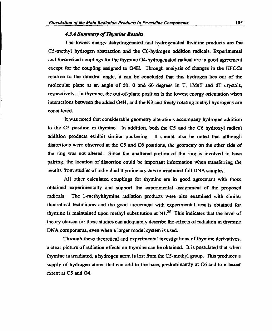
Elucidation of the Main Radiation Produc& in Pryrnidine Compnents 105
4.3.6 Sunrmary of Tliymine Resrrlrs
The lowest energy dehydrogenated and hydrogenated thymine products are the
CS-methyl hydrogen abstraction and the Cdhydrogen addition radicals. Experimental
and theoretical couplings for the thymine 04-hyârogenated radical are in good agreement
except for the coupling assigned to WH. Through analysis of changes in the HFCCs
relative to the dihedrai angle, it c m be concluded that this hydrogen lies out of the
molecular plane at an angle of 50, O and 60 degrees in T, lMeT and dT crystals,
respectively. In thymine, the out-of-plane position is the lowest energy orientation when
interactions between the added WH, and the N3 and k e l y rotating methy1 hydrogens are
considered.
It was noted that considerable geometry alterations accompany hydmgen addition
to the CS position in thymine. in addition, both the CS and the C6 hydroxyl radical
addition products exhibit similar puckering. It should also be noted that although
distortions were observed at the CS and Cd positions, the geometry on the other side of
the ring was not altered. Since the unaltered portion of the ring is involved in base
pairing, the location of distotion could be important information when t r a n s f b g the
results firom studies of individual thymine crystals to irradiated full DNA samples.
Al1 other calculated couplings for thymine are in good agreement with those
obtained experirnentally and support the experimental assignment of the proposed
radicals. The 1-methylthymine radiation products were also examineed with sirnilar
theoretical techniques and the good agreement with experimental results obtained for
thymine is maintained upon methyl substitution at Nl? This indicates that the level of
theory chosen for these studies can adequately describe the effects of radiation in thymine
DNA components, even when a larger mode1 system is used.
Through these theoretical and experimental investigations of thymine derivatives,
a clear picture of radiation effects on thymine can be obtained. It is postulateci that when
thymine is irradiated, a hydrogen atom is lost from the CS-methyl group. This produces a
supply of hydmgen atoms that can add to the base, predominantly at C6 and to a lesser
extent at CS and 04.

Elucidation of the Main Radiation Products in Prymidine Componenfs 106
4.4 C'osine
4.4.1 P r e v h s Experimentaî Work
The most complete experimentai study on cytosine derivatives has been
perfomed on cytosine monohydrate (Cm) crystals by Sagstuen et The crystal
structure of this denvative is composeci of an extensive hydrogen bonding network."
Sagstuen et cil." concluded that two major radical products are formed upon irradiation,
namely the N3-hydrogenated and N1 -dehydrogenated radicals. Minor products include
the CS and Cd hydrogen addition radicais. In addition, one large coupling was Ieft
unassigneci. The CS, C6 and N3 net hydrogen addition radicals were observed in crystals
of 1-methylcytosine ( I M ~ c ) . ~ * The cytosine anion was assigned to a spectra observed
îrom cytidine 3'-monophosphate (3'CMP) ~ r ~ s t a l s . ' ~ This assignrnent was questioned by
close2 who proposeci that the net N3-hydrogenated radical would be more likely to yield
the observed spectnim. In monohydrate crystais of deoxycytidine 5'-monophosphate
(StdCMP) the radical cation and the net N3 hydrogm addition product were assignedm
Table 4.7: Expctimcntal HFCCs (G) obtaincd in various cytosine derivatives. Radical Molecule &a T', Tw Tz Anion 3 ' W Y "C6HW -12.8 -8.3 0.7 7.6 Cation ~ ' ~ c M P ~
N 1 -dehydrogenated c m M
"C5H" "CI'HW "N4H" 'W4Hn "CSH" "N3H" W4H" "C6H" "C6Hw "C6H" "CSH" "C5H "C6H" "CSHw "C5Hw " C m " "CSH" "CSH" "C6H" " C m "
Not assigned cmM "C6Hw -18.2 -9.6 8.6
The caicuiated results presented within will be compared foremost to the
experhental work on cytosine monohydrate (Table 4.7). The suggested mechanism for

Ehcidation of the Main Radiation Produc& in Prymidine Cornponenfs 107
radical formation in cytosine monohydrate involves net hydrogen removal h m the N1
position of one cytosine and hydrogen addition to the N3 position of a neighboring
cytosine. The couplings assigned to the cation in monohydrate crystals of 5'dCMP are
similar to those assigned to the NI-dehydmgenated radical in cytosine monohydrate.
However, the N1 -dehydrogenated product is not possible in S'dCMP since a sugar p u p
replaces hydrogen at this position. This is the first indication that the assignment of the
couplings in cytosine monohydrate may be incorrect and theoretical calculations should
prove ftuitfùl.
4.4.2 Anion und Cation
The calculated adiabatic EA and IP of cytosine equal - 1 3.8 and 194.2 kcal/mol,
respectively (Table 4.8). The IP is in gooâ agreement with the results obtained
expenmentall#' (200.1 kcavmol) and those calculated at the MP2 leve17 (194.4
kcavmol). Both the EA and IP are slightly smaller in magnitude than the values
calculated for thymine. The largest components of the calculated spin distribution in the
cation are located on 02 (0.45), N3 (0.24) and CS (0.33), whereas in the anion over haif
of the spin is concentrated on C6 (0.55). This spin distribution reflects the calculated
planar cation and distorted anion geometries and agrees well with experimental spectra
where the largest HFCCs were obtained for CS and C6 in the anion and cation,
respectively . However, experimental (Table 4.7) and theoretical HFCCs (Table 4.8) for
Table 4.8: Calculated electron aff i ty , ionization potential and HFCCs (G) for the cvtosine cation and anion. Radical Atom Ah T h p Tw TzZ CI Anion NlH 2.1 -3.4 -1.6 5-1 (ËA = - 13 -8 kcavmol) N4
N4 CS C6
Cs Anion NI N4 N4 C6
Cation N1 (IP = 194.2 kcallmol) N4
N4 CS C6

EIucidation of the Main Radiation Products in Prytnîdine Commnents 1 O8
these two species are in poor agreement. A caiculated isotropic C6H coupling in better
agreement with the experimental value assigned to the cytosine anion is obtained if a
planar geometry for this radical is considered (Table 4.8, Cs anion). Fmm B3LYP/6-
3 1 lG(ldf,p) single-point calculations, this planar anion is only 4.4 kcaVmol higher in
energy than the nonplanar foxm. However, the anisotropic couplings are M e r h m the
experimental results. Hence, it can be concluded that the cytosine anion and cation were
more than likely not observed directly in the experimental studies.
4.4.3 Net Hydrogen A t m Addition Products
The N3-hydrogenated radical is the lowest energy radical in this c l w (Table 4.9),
which is in agreement with experimental observations that this tadicd is fonned in the
highest yield. The CS, C6 and 02-hydrogenated radicals lie 8.2, 10.2 and 13.8 kcaYmol
higher in energy than the N3 radical, respectively. The calculated spin density in the N3-
hydrogenated radical indicates that significant spin resides on C6 (0.53) and 0 2 (0.37)
with lesser arnounts on N3 (0.09) and N4 (0.03). This agrees well with the experimental
spin distribution (0.52,0.07 and 0.06 on C6, N3 and N4). The calculated C6H HFCCs in
this radical (Table 4.9) are in very good agreement with those obtained experimentally in
cytosine monohydrate. One of the N4H couplings is also well reproduced thmugh the
calculations. On the contrary, the N3H coupling was calculated to be smaller than that
determineci experirnentally, while a large coupling (19.6 G) was obtained fiom the
calculations for the second amino hydrogen.
Differences in the expenmentd and calculated couplings for the N3-hydrogenated
radical could arise due to a rotation about the C4N4 bond in the optimized geometry
relative to that present experirnentally, where hydrogeu-bonding effects may be
important. More specifically, due to crystal interactions a planar radical may
predominate over one with a distorted amino group. This is confirmed through the
optimization of a radical constrained to Cs symmetry. From B3LYP/6-3 1 lG(2dEp)
single-point calculations, the planar radical is ody 3.6 kcaVmol higher in energy than the
nonplanar fom. The two small N4H, the anisotropic C6H and the isotropic N3H
couplings obtained for the planar radical (Table 4.9, Cs N3-hydrogenated) are in much
better agreement with experimmt than those discussed above for the nonplanar form.

Efucidation of the Main hdiat ion Products in Ptymidine Components 109
The calculated geometry of the CS-hydrogenated radical displays significant ring
puckering resulting in the aforernentioned pseudo-axial and equatoriai positions for
hydrogen. The Cd adduct retaim a planar geometry with the two hydrogens distributed
equally on opposte sides of the molecular plane. The spin density in these systems is
confïned to the carbon adjacent to the hydrogen addition center (0.74 and 0.75 on C6 and
C5 in the CS and Cd-hydrogenated radicals, respectively). The couplings calculated for
the C6 adduct are in good agreement with the experimental results obtained in and
l ~ e ~ , ~ ~ the largest deviation existing for the two C6H isotropic couplings. It should be
noted that although the absolute magnitude of the calculated results is smaller than those
obsewed experirnentally, the difference between the two C6H couplings (approxirnately
3 G) is well reproduced by the calculations. The failure to reproâuce the ciifference in
these couplings in the corresponding T radical was previously discussed. On the other
hand, the experimental anisotropic tensors for the two hydrogens are different in
magnitude, a trait not reproduced by the calculated results which tie between the two
experimental values. Despite this small deviation fiom experiment, the assignrnent of the
C6-hydrogenated radical is supported by the calculations.
The anisotropic CSH couplings obtained in Cm and lMeC crystals and assigned
to the CS-hydrogenated radical agree well with calculated results, but the isotropic
components do not concur. In particular, the two calculated isotropic CSH couplings
(44.6 and 14.0 G) deviate substantially fiom those observed in Cm (47.1 and 3 1 .O G) and
lMeC (45.1 and 30.8 G). One possible explanation is that the calculations inadequately
descnbe the ring puckering, as observed for thymine radicals. Since the two
experimental HFCCs are more similar in magnitude (deviate by 15 G) than the calculated
values for this radical (deviate by 30.6 G), the effects of ring puckering on the HFCCs
m u t be investigated. If a planar geometry (obtained through a constrained optimization)
is considered, the HFCCs of the two CS hydrogens possess equivalent values (35.3 and
35.4 G nom Table 4.9, Cs CS-hydrogenated). The anisotropic tenson calculated for the
planar radical are in better agreement with the experimental couplhgs than those
calculated for the distorted radical. Through cornparison of the calculated and
experimental couplings for the CS-hydrogenated radical, it can be concluded that the
geometrical distortion in the crystal environment must lead to a nonplanar radical with

Elucidation of the Main Radiation Products in Ptymidine Compnents 110
Table 4.9: Calcuiated relative encrgies (IrcaYmoi) arrd HFCCs (G) for cytosine hydronenated radicals. - -
Relative Radical Encrgy Atom &O Txx Tw Ta N3-hydrogenated 0.0 NIH -3.0 -2.8 -1.2 4.1
CS- hydrogena ted
C, CS-hydrogenated
C6-hydrogenated
N3H N4H N4H CSH cm NIH N3H N4H N4H cm NlH C5H C5H cm N1H C5H C5H C6H NIH N4H N4H CSH cm C6H N1H O2H N4H N4H C5H C6H
Iess puckering than initially discussed in the present shidy. The energy ciifference
between the planar and nonplanar forms for thïs radical is only 0.4 kcaVm01, which
indicates that interconversion between conformations is energetically feasible. The
cornparison of calculated and experimental anisotropic tensors confinns the experimental
assignment to the CS-hydrogenated radical.
The 02-hydrogenated radical has not been assigned in any recent ENDOR studies
of cytosine derivatives. However, Herak et aL4' wigned a mm-temperature coupling
(A, = -9.6 G; Tii = -5.4,0.7, 5.0 G) to this radical. This set of experimental couplings is
quite different h m that calculated for the 02-hyhgenated radical. The only HFCCs
resembling these experimental values are those calculated for CSH in the cation, but it is

Elucidation of the Main Radiation PToàucts in Prymidine Components 111
unlikely that the cation would remain stable at this temperature. The calculated C6H
couphg for the 02-hydrogenated radical is similar to that assigned to the anion in
3'CMP crystals, as well as the main component assigned to the N3-hydrogenated radical
in a variety of cytosine derivatives. Since the assignment of the N3-hydrogenated radical
in Cm crystals was supporteci by the calculated couplings in this radical, it can be
concluded that the 0 2 adduct was not generated in these irradiated crystals.
4.4.4 Net Hydrogen Atom Abstraction Radicals
The N1-dehydrogenated radical is the lowest lying d c a l in this class (Table
4.10), which is in agreement with experimental results for cytosine monohydrate where
this radical was determineû to be the major dehydrogenated species. The N4, CS and C6-
dehydrogenated radicals lie 2.3, 8.2 and 12.6 kcdmol above the N1 adduct. The
calculated spin density in the NI-dehydrogenated radical displays an altemathg pattern
with the main components situated on CS (0.49), 0 2 (0.35) and N1 (0.29). This
distribution is quite different nom that obtained experimentally (0.57 and 0.17 at C5 and
N4, respectively). In addition, the calculated and experimental HFCCs deviate
substantially. In particular, the calculated anisotropic couplings for the amino hydrogens
are extremely smail compared to experimental values. Since it is hown that the
anisotropic component can be calculated with a great degree of accuracy using many
theoretical techniques, the deviations observed for this radical are too great to be ascribed
to the method ernployed-
Table 4.10: Calculated relative energies (kcaYmo1) and HFCCs (G) for cytosine dehydrogenated radicals.
Relative Radical Energy Atom Ù rxu Tm k? NI -dehydrogenated 0.0 N4H -0.7 -0.5 -0.4 0.9
N4H -0.5 -0.7 -0.4 1.1 C5H -1 1.2 -6.9 -0.4 7.2 C6H 1.6 4 0.2 1.2
N4dehydrogenated 2.3 N1H -2.1 -1.9 -0.6 2.4 N4H -15.5 -12.5 -2.8 15.3 CSH -2.2 -1.0 -0.5 1.5
One possible explanation for deviations fiom experimental couplings could be
that a rotation occurs about the C4N4 bond in the experimental environment. This could
lead to significant N4H couplings compared to those calculated for the nearly planar

Elucidation ofthe Main Radiation Products in Ptymidine Compnents 112
structure. Variation in the HFCCs with rotation about the C4N4 bond was examineci and
the agreement between the experimental and theoretical HFCCs was not i ~ n ~ r o v e d . ~ ~
Anothet rationalization of the results could be that a net hydrogen atom is lost at N4
rather than N1. The calculated isotropic coupling for the remaining amino hydrogen in
the NMehydmgenated species is similar to the coupling assigned experimentally to C5H
in the Nl-dehydrogenated radical. However, the anisotropic couplings are in poor
agreement. in attempts to improve the agreement between these data sets, a study of the
dependence of the N4H coupling on rotation about the C4N4 bond was performed.
However, the couplings in the NQdehydrogenated radical did not change appreciably and
the possibility that the observed couplings assigned to the NI-dehydrogenated radical
arise due to the N4-dehydrogenated radical cm also be dismisseci. The HFCCs for the CS
and C6-dehydrogenated radicals (not shown) were also in poor agreement with
experiment.
4.4.5 Wydroxyl Radical Addition Producis
Two radicals formed through addition of hydroxyl radicals to the CSC6 double
bond were investigated. The radical formed thmugh addition to the CS position is lower
in energy than the C6 adduct by 2.4 kcaVmoI. An additional conformation of the CS-
hydroxylated radical (CSOH-2) involving an alternative orientation of the amino group
was also obtained, which lies 0.6 kcaWmol above the C6 product. Analogous to the
thymine residues, these hydroxylated radicals exhibit a great degree of ring puckering
resulting in most of the unpaired spin being located on the site neighboring that of
hydroxyl addition (0.70 and 0.78 on C6 and CS in the CS and C6-hydroxylated radicals,
respectively).
The differences in the couplings obtained for the CSOH-1 and CSOH-2
conformations are quite large (Table 4.1 1) given the maII geometrical discrepancies.
Among the entire set of computed couplings, the NlH couplings obtained in each
conformation of the CS-hydroxylated product are in best agreement with the
experimental couplings assigned to the amino hydrogens in the N1-dehydrogenated
radical. One large, negative isotropic coupling, obtained for C6H in these radicals, is not
unlike that assigned to C5H in the NI-dehydrogenated radical, although the anisotropic
results deviate more substantially. In addition, a coupling left unassigned in cytosine

EIucidation of the Main Radiation Proàucts in Prymidine Components 113
Table 4.1 1 : Calculatcd relative energies (kcai/mol) and HFCCs (G) for cytosine hydroxyl radical addition products.
Relative Radical Energy Atom A, TXX Tw T z ~ C5-hydroxylated 0.0 NIH -4.2 -3.5 -1.7 5.3 (CSOH- 1) CSH 33.0 -1.6 -0.5 2.1
C6H -10.6 -9.6 -0.3 9.9 C6-hydmxylated 2.4 N4H -1.3 -0.8 -0.6 1.4
N4H -1.0 -1.4 -0.6 2.0 C5H -17.6 -10.7 -0.5 11.2 C6H 13.1 -1.6 -1.0 2.7 C6-OH 4.8 -1.1 -0.6 1.7
CS-hydroxylated 3 .O NlH -3.8 -3.2 -1.7 4.9 (CSOH-2) CSH 37.4 -1.5 -0.8 2.3
C6H -13.3 -10.2 -0.3 10.6
monohydrate resembles those calculated for C5H and C6H in the C6 and C5-
hydroxylated raâicals, respectively. The large isotropie coupling (33.0 or 37.4 G)
calculated for C5H in the CS-hydroxylated radical could be used as a fingerpnnt for the
identification of this radical in fùture studies. Altematively, this coupling may have gone
undetected in the experiments due to its similarity to the coupling assigned to the CS-
h y drogenated radical.
4.4.6 Summary of Cyiosine Results
Cornparison of expenmental and theoreticai HFCCs indicates that the cytosine
anion and cation were more than likely observed in a protonated or deprotonated state
rather than directly in the experimental studies. The calculated energetics agree with the
experimental results for cytosine monohydrate. In particdar, the N3-hydrogenated and
N1-dehydrogenated radicals were calculated to be the lowest energy radicals in their
respective classes and were determined expenmentally to be present in the highest yield.
The calculated HFCCs for the N3-hydrogenated radical supported the expenmental
assignment to this product, as did the computed couplings for the CS and C6-
hydrogenated radicals.
The calculated couplings for the NI -dehydrogenated radical did not correspond to
those expenmentally assigned to this species. Thus it appears that the suggested
mechanism for radiation darnage in cytosine monohydrate encompassing hydrogen
migration fiom one cytosine to another is unlikely. This statement was M e r verified
through the calculation of the couplings of the NI-dehydrogenated radical surrounded

Elucidation of the Main Radiation Products in Prymidine Components 114
with up to four water molecules or additional neigtiboring cytosine h p e n t s to simulate
the experimental hydrogen-bonding scheme." Even a cytosine dimer was studied to
mode1 the NI-dehydrogenated, N3-hydrogenated diradical pair. None of these
investigations lead to a clear theoretical description of the experimental results.
Ionization of cytosine, followed by electron capture by another cytosine, was calculated
to cost 207 kcaümoi. Subsequent deprotonation of the cation and protonation of the
anion leading to the suggested major products is exothennic by 139 kcdmol. Hence,
this proposed mechanism (Equation 4.1) is overall endothermic by 68 kcal/moI.
C + C + C* + CL -* C(N1-dehydrogenated) + C(N3-hydrogenated) (4.1)
The C6H couplings in the two conformers of the CS-hydroxylated product match
those experimentally assigned to C5H in the Nldehydrogenated radical and a coupling
lefi unassigned. In addition, the experimental N4H couplings can be attributed to the
N1H couplings in these two conforniers. Thus, through cornparison of expeiimental and
calculated h y p d e data, it appears that the hvo major products in irradiateci cytosine
monohydrate are the N3-hydrogenated and CS-hydroxylated products.
At least two different mechanisms can be considered which yield the N3-
hydrogenated and CS-hydroxy lated products and both involve water molecules. in the
fïrst postulated mechanism (Equation 4.2), ionization and electron uptake are initially
assumed to occw on cytosine. This step, which leads to the fomation of the cytosine
anion and cation, costs 207 kcal/mol. Next, water can add to the cation which is followed
by deprotonation and proton transfer to N3 of a second cytosine. This second step leads
to an energy gain of 149 kcdmol and, hence, the net energy cost for this reaction is 58
kcal/mol.
HzO + C + C -+ H20 + C' + C' + C(C.5-hydroxylated) + C(N3-hydrogenated) (4.2) The second postulated reaction mechanism (Equation 4.3) involves ionkation of a
water molecule followed by electron uptake at cytosine, resulting in a water cation and a
cytosine anion. This reaction costs 298 kcaVmol. The water cation subsequently
decomposes into a proton and a hydroxyl radical, which add to the anion and neutral
cytosine units, respectively. Since identical products are obtained in the two
mechanisms, the net energy cost of this reaction is the same as that mentioncd above and

Ehcidation of the Main Radiation pro duc^ in Prvmidine Coni~onenb 115
it can be concluded that the second step releases 240 kcahol.
HzO + C + C -B H20LC + C + CL + C(C5-hydmxyZated) + C(N3-hydrogenuted) (4.3)
Of the mechanisms discussed, the path involving cytosine ionization and water
addition is rnost likely to occur. Reasons for this include the fact that approximately 85%
of al1 ionization processes will occu. on cytosine since it possesses a greater number of
electrons relative to water. In addition, this reaction has lower energy costs for the initial
step (relative to the mechanism involving ionization of water) and the overall pmcess
(relative to the proposeci mechanism involving hydrogen addition and abstraction
products). However, the reaction mechanism involving radiolysis of water to produce
hydroxyl radicals and hydroxyl radical adducts is a commonly used ESR technique. 43.44
In addition, Sevilla and coworken have investigated the presence of hydroxyl radicals in
the DNA hydration layer? Hydroxyl radicals were found in the intemediate hydration
shell, but not in the closest hydration layer. This was speculated to occur due to reactions
of the hydroxyl radicals with DNA. The present work indicates that this option should be
examineci more closely. in addition, Wala et ald5 have reported that strand-breaks in
DNA occur due to hydroxyl radical addition to the DNA bases. Reactions of DNA and
hydroxyl radicals have also been reported to lead to 5-hydroxycytosine.'6
The proposal that water is also involved in the radiation damage mechanism in 47,48 cytosine monohydrate crystals is controversial. Critisms raised against this proposal
include the fact that ENDOR studies predict that the CS hydrogen rernains in the
molecular plane and that the low temperatures of the experiments may prevent the
hydroxyl radical fkom migrating to the CS position in cytosine. However, more recent
experiments indicate that radical yield in monohydrate crystals is greater than the yield in
anhydrous crystals of cytosine de ri vat ive^:^ which provides more evidence that water
may be involved in the darnage mechanism.
Through these theoretical and expenmental investigations of cytosine denvatives,
a picture of radiation damage in cytosine monohydrate crystals can be obtained. It is
postulated that when these crystais are irradiatecl. a net supply of hyhgen atoms and
hydroxyl radicals are generated, most probably h m water. Thmugh one of two

Elucidation of the Main Radiation Roducts in Prymidine Components 116
proposed mechanisms, net hydrogen atom addition occurs predominantly at N3, and to a
lesser extent at CS and C6, and net hydroxyl radical addition occurs predominantly at CS.
4.5 Uracü
4.5.1 Revious Experimental Wurk
The RNA base uracil (U) is of interest since it resembles thymine, where the
methyl group in thymine is replaced by hydrogen. Since the thymine methyl group is one
of the main sites of net hydrogen removal, the radicai chemistry of uracil will di* h m
that discussed for thymine. Relatively few single-crystal ENDOR studies on uracil
derivatives have been perfomed recently. Heralc and McDowell studied single-crystals
of 1 -methyluraci150 (1 MeU) and identified the N1 -methyl dehydrogenated and CS-
hydrogenated radicals. Zehner and co-workerss' examined irradiated single-crystals of
uracil. In their study, the Nldehydrogenated and 04, CS and C6-hydrogenated radicals
were identified, atthough the C6 adduct was thought to be protonated at 04. More
recently, Sagstuen et aLS2 studied the CO-crystals of 1-rnethyluracil and 9-ethyladenosine
(1 MeU:9EA). In addition to adenine radicals, the uracil anion, NI -methyl
dehydrogenated and CS-hydrogenated radicals were assigned.
4.5.2 Radical Pruduct Ene-etics
From the relative energies of uracil radicals, it can be speculated that hydrogen
removal occurs primarily at N1 in U and at C6 in uridine and U in hl1 RNA. The CS-
dehydrogenated species is also possible in U but not in T derivatives and this radical lies
18.6 kcaVmol above the N1 -dehydrogenated species. The relative energy of the hydroxyl
radical addition products is reversed from that predicted h m solely the expenmental
radical yield' (4:l for C5:C6). The relative stability of the hydroxylated uracil radicals is
similar to that previously discwed for thymine, although the energy difference is 1
kcaVmol smaller in uracil than in thymine. Thus, t h u g h sole consideration of
thermodynamics, the hypothesis that the methyl group is leading to a favored C6
hydroxyl radical addition product in thymine (compared to m i l ) cannot be supportad.
The kinetics of these reactions will be discussed in Chapter Seven.
The primary difference in the stability of U and T products is the relative energy
of the hydrogen addition products. The uracil CS-hydrogenated radical is 2.2 kcdmol

Elucidation of the Main Radiation Products in Prvmidine Com~onents 117
lower in energy than the corresponding C6 radical. Ln thymine, the C6 adduct is 2.9
kcal/mol lower in energy than the CS radical. The uracil energetics agree with
experirnentai results which indicate a 2: 1 ratio for the CS to C6 addition products.'
4.5.3 Discussion of Uracil &sui&
Sagstuen et akn speculated that the uracil anion is fonned upon irradiation of
1MeU:9EA, however, the ESRENDOR spectra was weak and the protonation state could
not be determined. Significant isotropic (-14.0 G) and anisotropic (-7.4, 1.4, 6.0 G)
hyperfine couplings were assigned to C6H. The anisotropic couplings are mialler than
those obtained for C6H in the u r a d anion, while the isotropie component is much larger
in magnitude (Table 4.12). Arnong the uracil radicals investigated in the present study,
the only radical with comparable C6H couplings is the 04-hydrogenated radical.
However, it should be noted that a nonplanar geometry was calculated for the anion and it
is possible that better agreement would be obtained with a planar anion as discussed for
thymine and cytosine. This avenue was not investigated in the present work since the
experimentd signal was weak and hence the extracted couplings are prone to erron.
Table 4.12: Calculated results for the uracil anion and cation HFCCs (G).
Radical Atom Aiso TXY TW T22 Anion NIH 2.3 -1.6 -1.4 3.0
N3H -1.6 -1.6 -0.7 2.4 C6H 5.9 -7.8 -0.2 8.0 C5H -1.8 -2.0 -1.1 3.1
Cation N1H -2.4 -5.7 -1.6 7.3 C m -0.3 -1.5 -1.1 2.6 C5H -13.4 -7.3 -1.2 8.6
Zehner and CO-workerssl obsewed a radical, with the unpaireci electmn density
located mainly on a nitrogen atom (0.30) and CS (0.65), suggested to be the N1-
dehydrogenated radical. A set of C5H isotropic (-16.2 G) and anisotropic (-9.8, 1.2, 8.7
G) couplings was obtained, as well as a '% coupling of 15.0 G. The assignment to the
Nl-dehydrogenated radical is supporteci by the calculated C5H HFCCs (Table 4.13) and
the N1 nitrogen isotropic coupling (calculated value: 15.5 G).
Unlike the CS-hydrogenated radical in thymine, the geometry of the uracil radical
optimized to a nearly planar structure which supports the hypothesis that the large
distortion in T is a result of the methyl group. Zehner et al.'' assigned a C6H coupling

Elucidation of the Main Radiation Products in Prymiàine Cornponenrs 118
consisting of large isotropic (-1 8.5 G) and anisotropic components (-1 1.0,0.5 10.5 G) and
two CSH couplings of 35.5 G to the CS-hydrogenated radical. Herak and ~ c ~ o w e l l ~
obtained similar results h m single-crystals of 1 MeU (C6H: Ab0 = -1 9.4 G; -1 1 A, 0.8,
10.6 G and CSH: A, = 35.3/35.7 G). Sagstuen et also speculated that the C5-
hydrogenated radical was formed in irradiated lMeU:.9EA, but accurate coupbg tenson
could not be isolated. These couplings are in excellent agreement with the calculated
values (Table 4.13).
Similar to T and C, the optunized geometry of the C6-hydrogenated U radical
shows no signs of distortion due to the additional hydrogen. This radical product
possesses an experimenta15' C5H coupling of - 18.0 G, with an anisotropic tensor of
(- 10.0, 0.0, 9.9 G), which is supporteci by the calculated results. The two C6H couplings
Table 4.13: Catculated results for uracil dehydrogenated and hydrogenated radical HFCCs (G). . ,
Relative Radical Energy Atom AL, Txn T w & NI-dehydrogenated 0.0 C6H 2.7 -1.6 0.1 1.5
C5H NlH N3H CSH N1H N3H C6H N1H cm C5H NIH C6H CSH C5H NIH N3H C6H C6H C5H N1H N3H C6H C5H 04H NlH N3H C6H C5H

Elucidation of the Main Radiation Proàuc~s in PNmidine Cornnonentir 119
have different experimental isotropic cornponents (45 and 5 1 G), whereas the calculations
render two identical couplings (40.5 G) due to the planar geometry. Zehner et al?'
suggested that in order to obtain these large coupüngs, the C6H product must be
protonated at the 0 4 position. This avenue was not investigated in this work, as it seems
unlikely that a charged radical would predominate aad large couplings were calculated
for the nonprotonated radical. Thus, the calculations support the conclusion that the
observed radical was not protonated. It is interesting to note that the difference between
the two experimental C6H couplings in uracil (6 G) and in cytosine (3.6) is srnaller than
the difference observed in thymine (13 G). This M e r indicates that the CS-methyl
group causes greater geometric distortions in T crystals than in either U or C crystals.
Uracil hydroxylated radicals have been identified upon studying the reaction of
hydroxyl radicals with pyrimidines in solution. 33" Couplings in the CS-hydroxylated
radical were assigned in uracil, uridine (ru and 2'-deoxyuridine (2'dU). The CSH and
C6H couplings obtained in these molecules are very sirnilar (appmximately 2 1.5 and i d.6
G, respectively). The predicted C6H couplhg is not unlike the calculated value (-16.1
G). However, the caiculated CSH coupling (39.9 G) is much larger than that observed
experirnentally.
The assignment of these couplings to the CS-hydmxylated radical can be
rationalized by considenng the radical geometry. Upon hydroxyl radical addition,
considerable distortion occurs at the damaged site leading to two local minima (Table
4.14). The radical with hydrogen in an axial position (CSOH-1) is 2.9 kcal/mol more
stable than the radical with hydrogen in an equatoriai position (CSOH-2). The CSH
couplings in the axial and equatorial positions are 39.9 G and 8.7 G, respectively. The
agreement between theory and experiment becomes evident once vibrational averaging
between these two conformers is considered. The average couplings for the axial and
equatorial conformers are -16.7 G and 24.3 G for C6H and CSH, respectively. These
couplings are very similar to those obtained experimentally and it can be concluded that
in solution considerable molecular motion occurs within this radical.
Experimental couplings have also been assigned to the C6-hydroxylated radical in
uridine and 2'-deo~~uridine."~ The values for the C5H couplings in rU and 2'dU are
20.0 and 10.4 Gy respectively. The C6H couplings assignesi in these two compounds also

Elucidation of the Main Radiation Prducis in Prvmidine Comwnents 120
Table 4.14: Calculated results for the HFCCs (G) in uracil hydroxylated radicals. Relative
Radical Energy Atom Aim Txx Tm Tkz Cbhydroxylated 0.0 N3H O 4.9 -0.8 1.6 (C60H- 1 ) C6H 12.6 -1.9 -0.9 2.8
C5H -17.9 -10.9 -0.8 11.8 C6-OH -1.8 -3.3 0.2 3.2
C6-hydroxylated 2.5 C6H 9.9 -1.8 -1.2 3.0 (C60H-2) CSH -18.9 -11.3 -0.8 12.2
C6OH 5.5 -1.3 -0.7 2.0 CS-hydroxy lated 4.8 N1H -2.6 -2.4 -1.5 3.9 (C50H- 1 ) C6H -16.1 -11.2 -0.3 11.5
CSH 39.9 -0.9 0.4 0.5 CS-OH 0.5 -1.7 -0.7 2.4
CS-hydroxy Iated 7.7 NIH -3.2 -2.8 -1.6 4.4 (CSOH-2) C6H -16.6 -10.5 -0.3 10.8
C5H 8.7 -1.9 -0.9 2.8 C5-OH -2.1 -3.2 -0.4 3.2
differ (26.1 and 13.3 G in r u and î'dU, respectively). The calculated CSH and C6H
couplings for the lowest energy conformer of this radical (C6OH- 1) are - 17.9 and 12.6 G,
respectively. An alternative arrangement of the hydrogen and hydroxyl group (C60H-2)
at C6, which is 2.5 kcaVmol higher in energy than the C6OH-1 conformer, leads to a
C5H and C6H coupling of -1 8.9 and 9.9 G, respectively. Thus, vibrational arguments
used in the discussion of the CS-hydroxylated radical cannot be used here. Due to the
disagreement between al1 data sets, further investigation of these couplings is mandatory.
The HFCCs calculated in additional radical products obtained through irradiation
of uracil are displayed in Tables 4.12 to 4.14. These have bem included to prompt a
more detailed experimental study of radiation products in uracil which may lead to a
clearer picture of radiation damage in the crystals of uracil derivatives.
4.6 Conclusions
Ln this chapter, the net hydmgenated, dehydrogenated and hydroxylated products
fonned in single-crystal studies of thymine, cytosine and uracil derivatives were
investigateâ. The thymine results show that overall good agreement with experimental
KFCCs can be obtained for al1 observed radicals. In cases where this agreement is
initially poor, various arguments can be made to clarify the discrepancies. For example,
al1 couplings in the 04-hydrogenated radical were in agreement with experiment except

Elucidation of the Main Radiation Products in Prymidine Components 121
that due to the additional hydrogen at the O4 position. Coherence between experiment
and theory was obtained through studying the effects of rotation about the C404 bond on
the 04H HFCCs. The reason for the failure of the calculations to reproduce the
experimental results for this radical was concluded to be due to the rapidly rotating
methyl group not explicitly accounted for in the calculations. Additionally, the poor
isotropic couplings obtained for the C6-hyârogenated radical were justified through
crystal effects, where experimentally a geometry exhibithg greater distortion is expected.
The general conclusim of gwd agreement between theory and experhent observed for
thymine can be extended to 1-methylthymine and uracil.
The calculateâ couplings for cytosine, on the other hand, were in overall poor
agreement with those assigned in the spectrum of cytosine monohydrate. Once crystal
interactions were taken into account and a planar radical was considered, experimental
assignment to the N3-hydrogenated radical was supported by the calculations. Good
agreement between theory and experirnent was also observed for the C6-hydrogenated
radical. Unlike the correspondhg T and U radical, the ciifference in the two C6H
coupling in C was well reproduced by the calculations. Additional arguments
conceming the degree of puckering observed in the cytosine CS-hydrogenated radical
were required to support assignment to this product. Deviations between experimental
and calculated results were not observed for the analogous T and U radicals. The poorest
agreement with experirnent was obtained for the NI-dehydrogenated radical, where
calculations could not reconstruct the anisotropic tenson. Agreement between theory and
experiment could not even be achieved through investigations of bond rotation and
crystal effects. in addition, the assigned couplings could not be linked to an alternative
dehydrogenated product.
From the discussion within, it was concluded that no dehydrogenated products
could be assigned to the spectra in cytosine monohydrate. Thus, the question "Where do
the hydrogens used to generate net hydrogenated products corne ikom?" must be
addressed. Through cornparison of expenmental and calculated HFCCs, it was
concluded that the only set of calculated couplings among possible cytosine raâiation
products close in magnitude to those assigned experimentally to the N1-dehydrogenated
radical arise h m the CS-hydroxylated radical. This result indicates that water must also

Elucidafion of the Main Radiation P roducrs in Prymidine Componenrs 122
play an important role in the radiation damage to DNA. In particular, it stands to reason
that net hydnigenated products could obtain hydrogen h m the water molecules. Thus, it
was concluded that the major radiation products in cytosine monohydrate are the N3-
hydrogenated and CS-hydroxylated products.
Assigning the N3-hydrogenated and CS-hyàroxylated radicals as the major
radiation products in cytosine monohydrate crystals would also explain the absence of the
Cm couplings assigned to the NI-dehydrogenated radical in the larger cytosine systerns.
Previously it was assumed that these couplings were not observed since a methyl or sugar
group replaces the hydrogen at N1 preventing the N1-dehydrogenated radical from
fonning. A new explanation uses the fact that water was not present in previous crystal
studies and, thus, the CS-hydroxylated product was not possible. Monohydrate crystals
of S'dCMP were studied, however, and the similarity of the couplings observed in these
crystals (assigned to the cation) to those experimentally assigned to the N1-
dehydrogenated radical in Cm was previously discussed. Since expenmentd evidence
exists that hydroxyl radicals will react with cytosine in full DNA,* it seems reasonable
that the CS-hydroxylated product is formed in crystalline cytosine monohydrate. It
should be noted that couplings have been assigned to hydroxylated products in aqueous
crystals of de~x~adenosine" and crystals of guanine hydrobrornide rn~noh~dra te?~
The good agreement between experimental and theoretical couplings in thymine
makes the newly proposed assignment of the observed radiation products in cytosine
monohydrate tmstworthy. However, no accurate studies have been performed on
monohydrate single-crystals of thymine denvatives. In attempts to gain a greater
understanding of the radiation effects on DNA components and the mle water plays in
this damage, the following chapter will discuss the main radiation products in the purines,
adenine and guanine. The credibility of the mechanisms for radiation damage in cytosine
monohydrate crystals proposed herein will be discussed in more detail in Chapter Seven.
4.7 References
1. von Sonntag, C; Schuchrnann, H.-P. Int. J. Radiat. Biol. 1986,49, 1.
2. Close, D. M. Radiat. Res. 1993,135, 1 .

Ehcidution of the Main Radiation Products in Prymidine Components 123
3. Sagstuen, E.; Hole, E. O.; Nelson, W. H.; Close, D. M. J. Phys. Chem. 1989,93, 5974.
4. Fujita, S.; Steenken, S. J. Am. Chem. Soc. 1981,103,2540.
5. Betahard, W. A.; Patrzdek, A. 2. Radiat. Res. 1989,117,379.
6. Cullis, P. M.; Evans, P.; Maione, M. E. Chem. Commun. 1996,985.
7. Colson, A.-O., Sevilla, M. D. Inf. J. Radiat. Biol. 1995, 67, 627.
8. Becke, A. D. J: Chem. Phys. 1993,98,1372.
9. Lee, C.; Yang, W.; Parr, R. G. Phys. Rev. B 1988,37,785.
10. Ditchfield, R.; Hehre, W. J.; Pople, J. A. J. Chem. Phys. 1971,54, 724; Hehre, W. J.; Ditchfield, R.; Pople, J. A. J. Chem. Phys. 1972,56,2257; Hariharan, P. C.; Pople, J. A. Mol. Phys. 1974,27,209; Gordon, M . S. Chem. Phys. Lett. 1980, 76,163; Hariharan, P. C.; Pople, J. A. Theor. Chim. Acta 1973,28,2 13; McLean, A. D.; Chandler, G. S. J. Chem. Phys. 1980, 72,5639; Krishnan, R.; Binlûey, J. S.; Seeger, R.; Pople, J. A. J. Chem. Phys. 1980, 72,650; Clark, T.; Chandrasekhar, J.; Spitznagel, G. W.; Schleyer, P. v. R J. Comput. Chem. 1983,4,294; Frisch, M . J.; Pople, J. A.; Binkley, J. S. J. Chem. Phys. 1984,80,3265.
11. Ha, T.-K.; Gunthard, H. H . J . Am. Chem. Soc. 1993,ll5,11939.
12. Bauxhlicher, Jr., C. W.; Partridge, H. J. Chem. Phys. 1995,103, 1 788.
13. Frisch, M. J.; Trucks, G. W.; Schlegel, H. B.; Gill, P. M. W.; Johnson, B. G.; Robb, M. A.; Cheeseman, J. R.; Keith, T. A.; Petenson, G. A.; Montgomery, J. A.; Raghavachari, K.; Al-Laham, M. A.; Zakrzewske, V. G.; Ortiz, J. V.; Foresman, J. B.; Cioslowski, J.; Stefanov, B. B.; Nanayakkara, A.; Challacombe, M.; Peng, C. Y.; Ayala, P. Y.; Chen, W.; Wong, M. W.; Andres, J. L.; Replogle, E. S.; Gomperts, R.; Martin, R. L.; Fox, D. J.; Binkley, J. S.; Defrees, D. J.; Baker, J.; Stewart, J. P.; Head- Gordon, M.; Gonzalez, C.; Pople, J. A. Gaussian 94 (Revision B.2); Gaussian, inc.: Pittsburgh, PA, 1995.
14. Perdew, J. P.; Wang, Y. Phys. Rev. B 1986,33,8800.
15. (a) Perdew, J. P. Phys. Rev. B 1986,33 8822; (b) Perdew, J. P. Phys. Rev. B 1986,34, 7406.
16. Eriksson, L. A. Mol. Phys. 1997,91,827.
17. St-Amant, A.; Salahub, D. R.; Chem. Phys. Lert. 1990,169,387; St-Amant, A. PhD.

Etucidation of the Main Radiation Products in Prymidine Compnents 124
thesis, Université de Montréal, 199 1 ; Salahub, D. R.; Fournier, R.; Mlynarski, P.; Papai, 1.; St-Amant, A.; Ushio, J. In Denrity Funetional Methodr in Chemishy: Labanowski, J., Andzelm, J., Eds.; Springer: New York, 1991.
18. Sagstuen, E.; Hole, E. O.; Nelson, W. H.; Close, D. M. J. Phys. Chem. 1992,96, 1121.
19. Hole, E. O.; Sagstuen, E.; Nelson, W. H.; Close, D. M. J. Phys. Chem. 1991,95, 1494.
20. Sagstuen, E.; Hole, E. O.; Nelson, W. H.; Close, D. M. Radiat. Res. 1996,146,425.
2 1. Orlov, V. M.; Smimov, A. N.; Varshavsky, Y. M. Ter. Lett. 1976,48,4377.
22. Hush, N. S.; Cheung, A. S. Chem. Phys. Lert. 1975,34, 11.
23. Sevilla, M. D.; Besler, B.; Colson, A.-O. J. Phys. C h . 1995,99, 1060.
24. Wiley, J. R.; Robinson, J. M.; Ehdaie, S.; Chen, E. C. M.; Chen, E. S. D.; Wentworth, W. E. Biochem. Biophys. Res. Commun. 1991,180,841.
25. Miaskiewicz, K.; Miller, J.; Osman, R Int. J . Radiat. Biol. 1993,63,677.
26. Dulcic, A.; Herak, J. N. Radiat. Res. 1971,47,573.
27. Bansal, K. M.; Fessenden, R. W. Radiat. Res. 1978, 75,497.
28. Novais, H. M.; Steenken, S. J. Phys. Chem. 1987,91,426.
29. Dulcic, A.; Herak, J. N. J. Chern. Phys. 1972,57,2537.
30. Colson, A.-O.; Sevilla, M. D. J. Phys. Chem. 1995,99, 13033.
3 1. Deeble, D. J.; Das, S.; von Sonntag, C. J. Phys. Chem. 1985,89, 5784.
32. Wagner, J. R.; Cadet, J.; Fisher, G. J. Photochem. Photobiol. 1990,52,333.
33. Hildenbrand, K.; Behrens, G.; Schulte-Frohlinde, D.; Herak, J. N. J. Chem. Soc. Perkin. Trans. 1989,2,283.
34. Catterall, H.; Davies, M. J.; Gilbert, B. C. J. Chem. Soc. Perkin Trans. l992,t , 1379.
3 5. Wetmore, S. D.; Boyd, R. J.; Eriksson, L. A. J. Phys. Chem. B 1998,102,5369.
36. Sagstuen, E.; Hole, E. O.; Nelson, W. H.; Close, D. M. J. Phys. Chem. 1992,96, 8269.

Elrrcihtion of the Main Radiation Producfi in Prymidine Components 125
37. Weber, H. P.; Craven, B. M.; McMullan, R K. Acta C'st. B 1980,36, 645; McClure, R. J.; Craven, B. M. Acta Cvsr. B 1973,29, 1234.
38. (a) Rustgi, S. N.; Box, H. C. J. Chern. Phys. 1974,60,3343; (b) Close, D. M.; Bernhard, W. A. Bull. Am. Phys. Soc. 1980,25, 41 6.
39. Box, H. C.; Potter, W. R.; Budzinski, E. E. J. Chern. Phys. 1975,62,3476.
40. Close, D. M.; Bernhard, W . A. J. Chern. Phys. 1979, 70,210.
41. Herak, I. N.; Lenard, D. R.; McDowell, C. A. J . Magn. Reson- 1977,26, 189.
42. Wetmore, S. D.; Himo, F.; Boyd, R. J.; Eriksson, L. A. J. Phys. Chem. B 1998, 102, 7484.
43. Hiraoka, W.; Kuwabara, M.; Sato, F.; Matsuda, A.; Ueda, T . Nucl. Aci& Res. 1990, 18, 1217.
44. (a) Becker, D.; La Vere, T.; Sevilla, M. D. Radiat. Res. 1994, 140, 123; (b) LaVere, T.; Becker, D., Sevilla, M. D. RadiPt. Res. 1996,145,673.
45. Wala, M.; Bothe, E.; Gomer, H.; Shulte-Frohlinde, D. J . Phorochem. Photobiol. A. Chemistry 1990,53,87.
46. (a) Chapman, D.; Gillespie, C. J. Adv. Radiat. Biol. 1981,9, 143; (b) Téoule, R. Int. J. Radiat. Biol. 1987, 51,s 73.
47. Close, D. M.; Sagstuen, E.; Hole, E. O.; Nelson, W. H. J. Phys. C'hem. B 1999,103, 3049.
48. Wetmore, S. D.; Boyd, R. J.; Himo, F.; Eriksson, L. A. J. Phys. Chem. 1999,103, 3051.
49. Information obtained through private correspondence between D. M. Close and L. A. Eriksson.
50. Herak, J. N.; McDowell, C. A. J. Chem. Phys. 1974,61,1129.
5 1. Zehner, H.; Flosmann, W.; Westhof, E.; Muller, A. Mol. Phys. 1976.32.869.
52. Sagstuen, E.; Hole, E. O.; Nelson, W. H.; Close, D. M. Radiat. Res. 1998,249, 120.
53. Gregoli, S.; Olast, M.; Bertinchamps, A. Radiat. Ra. 1974,60,388.
54. Hole, E. O.; Sagstuen, E.; Nelson, W. H.; Close, D. M. Radiat. Res. 1991, 125, 119.

CHAPTER FIVE Characterization of Purine Radiation Products
The results obtained for thymine presented in Chapter Four were very promising.
Conversely, the results discussed for cytosine were puzzling. Through comparison of
experimental and theoretical HFCCs a new mechanism was proposed for radiation
damage in cytosine monohydrate crystals. A similar mechanism for thymine could not be
investigated due to the lack of accurate experimental data on thymine monohydrate
crystals. The newly proposed mechanism for radiation darnage probes an important
question regarding the significance of water in radiation damage. Several experimental
studies have appeared in the literature which investigate monohydrate crystals of
derivatives of the purines, adenine and guanine. Cornparison of theoretical and
experimental HFCCs in these bases is important to understand the role water plays in
DNA radiation darnage. The present chapter will discuss experimental and theoretical
(DFT) resul ts obtained for adenine and guanine. Net hydrogenation, dehydrogenation
and iîydroxylation products will be considered. The computational techniqses applied to
these systems are identical to those previously employed for the pyrimidinrs in Chapter
Four and the discussion will not be repeated in the present chapter.
It is important to study both radicals generated fiom neutral base crystals as well
as those forrned in protonated crystals to better understand the dependence of radical
formation on the environment. Environmental effects are of interest when transferring
results obtained fiom single-crystal studies to full DNA sarnples. In particular, the
importance of understanding proton transfer in DNA has been discussed. Nelson et al.'
have suggested that expenmental studies on different crystalline environments will aid in
gaining a better understanding of the environmental effects on protonation and
deprotonation behaviors in adenine molecules. In addition, detailed ESRENDOR studies
on single crystals of guanine hydrobromide monohydrate2 were used to render
information about the importance of "bound" water to radical formation. Similarily, the
examination of 2kieoxyguanosine 5'-monophosphate crystals3 supplied information
about the influences of the sugar moiety and the phosphate group on radical formation.

Characterîzation of Purine Radiation Products 127
The present chapter primarily focuses on radicals generated fiom the neutral adenine and
guanine molecules. However, sorne of the radicals proposed to be generated in
protonated base crystals will be discussed when experimental data is available.
5.2 Adenine
5.2.1 Previous Experimental Wurk
Even though new experimental data is constantly appearing in the literature, the
various adenine denvatives have been investigated experirnentally to a lesser extent than
the derivatives of any other base. This is due to the fact that early ESR investigations
indicated that thymine and guanine are affected by radiation to a greater extent than the
other bases, fomiing thymine (or cytosine) anions and guanine cations. In addition, due
to solubility problems, few single-crystal studies on adenine derivatives have been
perfonned since they are extremely difficult to ~ r e p a r e . ~ The chemical numbering of
adenine used throughout this study is indicated in Figure 5.1, structure 1.
I II III
Figure 5.1 : Structure and chernical nwnbering of adenine (I,6-aminopurine), singly protonated adenine (II) and doubly protonated adenine (III).
Various adenine radicals have been identified and the HFCCs extracteci (Table
5.1). Crystals examined include 9-rnethyladenine5 (9MeA), anhydrous deoxyadenosine'
(dA), deoxyadeonsine rnon~h~dra te~? '*~ (ciAm) and adenosinegVi0 (rA). In addition, co-
crystals of adenosine and 5-bromouracil have also been investigated (~A:sB~LJ')."
Radicals charactenzed in crystals of 9MeA include the C8 and N3-hydrogenated adducts.
in addition to these two radicals, the C2-hydrogenated and N6-dehydrogenated products
were identified in crystals of dA. The N6-dehydrogenated radical was not determined to
be fonned upon irradiation of dAm although three hydrogenated radicals were identified

Characterization of Purine Radiation Products 128
Table 5.1: Exberimental HFCCs (G) in adenine radicals. . T
Radical Molecuie Atom A, Tm Tw Tzz Cation A:HCI: Y3-i20" W6H -7.0 -4.5 -1.3 5.8
d ~ '
9 ~ e ~ '
CS- hydrogenated d&n6
including those formed via net hydrogen addition to C2, N3 and C8. Since no net
hydrogen removal radical was observed, but hydrogen addition products were identified,
water may also be playing an important role in the radiation damage of these crystals
through supplying hydrogen atoms. In two studies of adenosine crystals, the C2 and C8-
hydrogenated, as well as the No-dehydrogenated, radicals were observed. Studies on co-

Characrerizarion of Purine Radiation Products 129
crystals of adenosine and 5-bromouracil identified the N3 and C2-hydrogenated and the
N6-dehydrogenated radicals. The formation of the N3-hydrogenated and the N6-
dehydrogenated species in these crystals indicates that these radicals can be fonned
regardless of the hydrogen-bonding scheme in the crystals.
As discussed in the introduction, various crystalline samples have been used to
investigate the effects of radiation on adenine. in some crystals, the parent adenine
molecule is protonated at NI (Figure 5.1, II) or doubly protonated at NI and N7 (Figure
5.1, III). Upon irradiation of adenine hydrochlorïde hernihydrate crystals
(A:HCI: ' /~I~O), '~ which are protonated at NI, a radical was observai which was
postulated to be formed via removal of a hydrogen atom Eiom NI. This radical is
structurally equivalent to the cation of the neutral adenine molecule. Additional radicals
identified in protonated crystals will be discussed in a later section.
5.2.2 Anion and Cation
In a review of ab inifio studies on DNA bases, Colson and sevilla" report a
negative value for the EA of adenine (-7.2 kcaVmol), which was obtained by scaling the
vertical EAs to expenmental data on related systems.I4 Direct (DFT) calculation of the
adiabatic EA yields a value of -1 7.7 kcaVmol (Table 5.2). The calculated geometry of the
adenine anion indicates that considerable distortion occurs upon addition of an electron.
The pyrimidine (six-membered) ring remains planar with the amino group located out of
the molecular plane, while the imidazole (five-membered) ring is puckered at C8 and
N9H is also located out of the molecular plane. This puckering leads to a concentration
of the spin density on C8 (0.43), C6 (0.25), C2 (O. 18) and N3 (0.09). The adenine anion
has not been proposed to exist as a radiation product in experiments to date. Calculations
indicate that this radical could be identified through a large C8H isotropie coupling (10.0
G), as well as substantial couplings for C2H (-5.3 G) and the two amino hydrogens (4.1
G ) - The puckered geometry obtained for the anion in the present study may not be
possible in crystals due to hydrogen bonding. For this reason, a planar geomeûy for the
anion, which lies 4.7 kcaVmol higher in energy than the non-planar radical anion, was
obtained through a constrained optimization. The magnitude of the spin density on C2
(0.28), N3 (0.13) and Cd (0.30) in the planar radical is larger than in the non-planar form,

Characrert'tation of Purine Radiation Products 130
Table 5.2: Cdculated HFCCs (G) in the adenine anion and cation radicals. Radical Atom A, Tw T h T z ~ Anion C2H -5.3 -3.0 -0.2 3.2 (EA = - 17.7 kcaYmol) N6H
N6H CSH N9H
Cs Anion C2H N6H N6H CSH N9H
Cation N6H (IP = 182.3 kcal/mol) N6H
CSH
whereas the spin density on CS (0.39) is slightly smaller. The major difference between
the couplings in the two foms of the anion is the sign of the C8H isotropic component
(Table 5.2). Cornparison of the calculated KFCCs of the planar and non-planar radical
anion with future experimental spectra will be useful to eliminate the possibility of the
anion being fonned but its spectra lefi undetected.
The adiabatic IP was calculated to be 182.3 kcailmol in the present study (Table
5.2), which is srnaller than the experimental value (190.4 kca~mol)" and the value
obtained with MP2 (199.6 kcal/mol)." Unlike the anion, the adenine cation remains
planar, and the major components of the spin density reside on N3 (0.19), C5 (0.20), N6
(0.27) and C8 (0.18). As previously mentioned, a radical equivalent to the adenine cation
was assigned in a spectra of A : H c ~ : ~ / ~ H ~ o . ' ~ The spin density distribution in this radical
was detennined to be located primarily on N6 (0.25) and CS (0.17/0.21) which is in
excellent agreement with the calculated values. The calculated and experimental HFCCs
are also in excellent agreement. in particular, the calculated and experimental anisotropic
HFCCs for al1 hydrogens are in extraordinary agreement and the isotropic HFCCs differ
by less than 1 G. Thus, Our results strongly support the assignment of the experimental
couplings in A:HCl:'/2H20 to the net radical cation.
5.2.3 Net Hydrogen A t m Addition Radieais
Relative energies of hydrogenated adenine radicals indicate that the radical
formed by addition of a hydrogen atom to CS is the Lowest energy radical of this form
(Table 5.3). The C2-hydrogenated radical lies 8.7 kcaVmol higher in energy than the

Characreritution o f Purine Radiation Products 13 1
Table 5.3: Calculated HFCCs (G) in adenine hydrogenated radicals. Relative
Radical Energy Atom Au, Tcu Tw Ta C8-hydrogenated 0.0 C2H -5.6 -3.0 -0.1 3.1
. -3.2 -1.2 ~ . .
corresponding C8 radical. Radicals fonned through addition of hydrogen to any of the
nitrogens are much higher in energy, on average 15.3 kcaVmol above the C8-
hydrogenated radical. The C4 and CS hydrogen addition radicals are the highest energy
products in this class.
S. 2.3.2 Nitrogen Hydrogenated Radicals
The radicals fomed through addition of hydrogen to NI or N7 have noi been
reported in the experimental spectra of nonprotonated adenine crystals. Upon formation
of the N1-hydrogenated radical, C6 is displaced slightly to one side of the molecular
plane and the amino group is rotated, resulting in the nitrogen and one hydrogen being

Characterizution of Purine Radiation Products 132
located on one side of the plane and the second hydrogen on the opposite side. These
distortions force a large amount of the spin density to be localized on C6 (0.60). Due to
the non-planar amino group, the calculated HFCCs consist of two large, N6H isotropic
couplings (29.0 and 16.4 G). Upon hydrogen addition to N7, the parent adenine molecule
significantly distorts at C8 and N6. The majority of the spin density resides on CS (0.64)
and the remaining spin density is distributed between N7 (O. 1 1 ) and N9 (0.1 0). The
HFCCs reflect this spin density distribution in that there exists a large isotropic C8H
coupling (22.5 G) that has considerable anisotropy (largest component of the tensor: 9.5
G). A smaller, yet significant, coupling was also obtained for N7 (1 3.4 G).
The radical formed by net hydrogen addition to N3 undergoes significant
geometrical alterations upon fonnation. The N3 hydrogen is located out of the molecular
plane and the amino group is puckered with both hydrogens displaced out of the plane.
Roughly ha1 f of the spin density is located on C2 (0.49) with the rest distributed about the
pyrimidine ring (N3 (0.1 1) and C6 (0.32)). The calculated HFCCs (Table 5.3) indicate
that the large spin density at C2 leads to a significant isotropic C2H coupling (-12.9 G)
which has considerable anisotropy, (Tn = 6.9 G). Substantial couplings were also
calculated for N3H (1 5.2 G), as well as for both of the hydrogens at N6 (1.31- 1.5 G) and
C8H (-3.0 G). Al1 the latter couplings have relatively small anisotropic tensors.
The N3-hydrogenated radical has been observed experimentally in various
adenine crystals, such as d ~ , ' r ~ , l ' d ~ m , ~ ~ A : s B ~ u ' ~ and 9 ~ e A . ' Experimentally, the
spin was determined to be located mainly on C2 (0.4), N3 (0.1) and C8 (0.1), which
agrees with the calculations discussed above, although significant spin was detennined to
reside on C6 rather than C8. The hyperfine coupling constants elucidated fiom al1
experimental studies are similar. A major difference is the anisotropic coupIings in
earlier studies on rA and rA:SBrU are much smaller in magnitude than those determined
more recently. In addition, cornparison of the experimental results indicates that the sign
of the C2H expenmental isotropic couplings in rA should be negative. The experimental
results reveal significant C2H and C8H couplings (approximately -10 and -4 G,
respectively). These isotropic cornponents, as well as the anisotropic tensors, are in good
agreement with the calculated results for this radical (-12.9 and -3.0 for C2H and CgH,
respectively.)

Characrerizution of Purine Radiation Products 133
It should be noted that N3H HFCCs are not observed in al1 of the expenmental
studies of adenine crystals. This has been speculated to occur since very strong signals
are required for the detection of this coupling. The major difference between theoretical
and expenmental HFCCs in the N3-hydrogenated radical occurs in the magnitude of the
N3H isotropic coupling. Experimentally, a small coupling was observed for this radical
(-3.9 G in the most recent study). Alternatively, a large HFCC (15.2 G) was calculated
due to distortions at N3. It is possible that hydrogen bonding in the crystal structure
forces the N3 hydrogen to remain in the molecular plane, thus leading to a small isotropic
HFCC. This hypothesis cm be tested through examination of a fùlly optimized Cs
structure, which lies only 1.7 kcaVmol above the non-planar arrangement and possesses
two imaginary fkequencies. The spin distribution in the planar radical is very similar to
that calculated for its puckered form. The calculated C2 and CS hydrogen HFCCs (Table
5.3) are also very similar for both radical forrns with an average deviation of 1.6 and 0.8
G in the isotropic and anisotropic components, respectively. The main difference in the
computed couplings is in the magnitude of the N3H isotropic HFCC. In the Cs N3-
hydrogenated radical, the N3H isotropic component was calculated to be -3.6 G
compared to 15.2 G in the puckered form. Expenmentally this coupling was determined
to be on average -3.7 G. Hence, it can be concluded that in crystals where the N3H
coupling was detected, the N3-hydrogenated radical is likely to remain in a planar form.
Through cornparison of the couplings in the planar radical with the remainder of the
experimental results, it is difficult to determine whether or not the observed radicals were
planar. In particular, the C8H and C2H couplings are in better agreement with those
values calculated in the distorted radical.
5.2.3.2 Carbon Hydrogenated Radicafs
The radical formed through addition of hydrogen to C2 has been detected on
numerous occasions. It has been proposed that the couplings in this radical depend on the
protonation state of the parent molecule. in dArn, Lichter and coworkers7 determined that
the C2-hydragenated radical was present raîher than the corresponding C8 radical. The
spin density was determined to reside mainly on NI (0.17) and N3 (0.37), which is in
agreement with the calculated results (NI (0.20) and N3 (0.43)). In crystals of d ~ r n , ' ~ ~
r ~ ' and rA:SBrU," two equivalent C2H couplings were recorded (on average

Characterizution of Purine Radiation Products 134
approximately 42 G). In two of these studies, a C8H coupling was also detected
(approximately 10 G). These couplings are in fair agreement with the calculated results
(Table 5.3), although the C8H coupling was calculated to be negative and anisotropic
couplings were not elucidated expenmentally.
In a recent ESRENDOR study of deoxyadenosine monohydrate by Close et a ~ - , ~
a very accurate set of full couplings was assigned to the C2-hydrogenated radical. The
experimental isotropic (-6.4 G) and anisotropic (-3.5, -0.1, 3.6 G) C8H couplings are in
excellent agreement with those calculated in the present study (Aiso = -6.7 G; C, = -3.9,
-0.2, 4.1 G), as well as with the values obtained by the same group in a recent study of
anhydrous deoxyadenosinet ( A , = -6.4 G; fii = -3.4, 0, 3.4 G). However, these
expenmental studiesIp6 and the theoretical results differ in the magnitude of the C2H
isotropic HFCCs. The molecular geometry was determined to remain planar upon radical
formation and the C2 hydrogens distributed equally on either side of the molecular plane.
This arrangement results in two nearly equivalent (43.3 and 45.5 G) isotropic couplings.
Alternatively, in the carefùl ESRENDOR s tudie~l*~ the difference between these
couplings is larger [32.8 (38.9) and 54.3 (47.5) G in deoxyadenosine monohydrate
(anhydrous deoxyadenosine)] .
The experimental results indicate that the distribution of the hydrogens at C2 is
more unsymmetric than modeled by gas phase DFT calculations. Difficulties describing
ring puckering resulting fiom the addition of hydrogen to thymine was discussed in
Chapter Four. It is possible that insufficient ring puckering is also responsible for the
disagreement between theory and experiment in the adenine C2-hydrogenated radical.
The disagreement between the most recent experimental studies and those which
appeared in the earlier literature indicating equal hydrogen couplings can be understood
through the use of a more detailed experirnental technique or a reduced vibrational
averaging in the later studies. Note that the average value of the huo unequal C2H
couplings generated tiom the most recent experiments (-43.6 G) is equal to the couplings
obtained in earlier experirnents and highly similar to the computed average (44.4 G).
Despite discrepancies in isotropic couplings, the anisotropic HFCCs support the
experimental assignments to the C2-hydrogenated radical.

Characterizarion of Purine Radiation Products 135
The C4-hydrogenated radical has not been assigned in spectra of nonprotonated
adenine crystals, although it has been detected in protonated crystals (discussed in a
succeeding section). The addition of hydrogen to C4 leads to considerable geometrical
distortions, as previously observed by Colson and sevilla? The geometry in these types
of radicals is descnbed as a "butterfly" conformation, where the pyrimidine and
imidazole rings remain planar, but are tilted about the C4C5 bond towards each other. A
higher energy conformer, not examined herein, has been discussed in the literature in
which the rings are tilted to opposite sides of the C4CS bond. Even though the molecule
is distorted upon radical formation, the calculated spin density is distributed throughout
both rings with the majority of the spin density located at CS (0.44), N1 (0.14), N3 (0.1 O),
C8 (0.20) and N9 (0.12). The calcufated HFCCs consist of a very large isotropic C4H
coupling (62.9 G) and significant couplings for C8H (-6.0 G) and N9H (-3.3 G). The C5-
hydrogenated radical also displays a "butterfly" conformation. A large part of the spin
density was calculated to be shared between C2 (0.44) and C4 (0.31), with considerable
spin density also located on C6 (0.23), N3 (-0.10) and N6 (0.10). The coupling constants
calculated for this radical include a large C5H coupling (51.2 G) and a smaller C2H
coupling (-1 1.6 G). No expenmental couplings have been isolated for this radical.
The final carbon hydrogenated radical to be discussed is the radical formed
through addition of hydrogen to CS. This radicai has been observed in numerous studies
in the li terature and the couplings in this radical, similar to the C2-hydrogenated radical,
have been shown to depend on the protonation state of the parent molecule. The HFCCs
in the C8 and C2-hydrogenated radicals have been detennined to be almost identicaI and,
thus, discussions have appeared in the literature disputing to which position the hydrogen
will prirnarily attach.
The Cg-hydrogenated radical was detennined to be present in dAm8 and r ~ ' and
equivalent C8H couplings of 38.0 and 39.0 G were recorded, respectively. These
couplings are in excellent agreement with the calculated values (38.9l39.1 G). A
significant C2H coupling was also calculated (-5.6 G). In more recent studies, Close and
coworkers detected the CI-hydrogenated radical in d ~ m , ~ 9 ~ e ~ ' and dA.' A C2H
hyperfine tensor was extracted in these studies consisting of a srna11 isotropic (-4.8 G)
and a significant anisotropic coupling (-2.6, 0.0, 2.6 G), which is in good agreement with

Characterization ofPurine Radiation Products 136
the calculations (A , = -5.6 G; I;,. = -3.0, -0.1, 3.1 G). The main difference between the
theoretical results or previous experimental results and the more recent experimental
work is the magnitude of the isotropic C8H couplings. Expenmentally, two unique
couplings were obtained in dAm (36.3141 -6 G),6 dA (36.7/40.9 G)' and 9MeA (38.4/41 .O
G ) Theoretically, radical formation leads to a symrnetrical distribution of the huo
hydrogens at Cg. From the more recent experimental results, alteration of the ring at CS
is Iikely, thus leading to an unsymmetric orientation of the two hydrogens and different
couplings. This is an identical situation to that observed for the corresponding C2
radical. Once again the experimentd and theoretical anisotropic couplings are in good
agreement and, thus, it can be concluded that the theoretical results support the
experimental assignrnent of the CS-hydrogenated radical. It is interesthg to note that the
two experimental couplings in the CS-hydrogenated radical are closer in magnitude than
those obtained in the C2 adduct, indicating smaller geometrical alterations upon
formation of the CS hydrogen addition radical.
5.2.4 Nd Hydrogen Atom Abstraction Radicals
The relative energies of the dehydrogenated radicals (Table 5.4) suggest that the
radical formed via removal of hydrogen fiom N9 is the lowest lying radical in this class.
The radical formed through abstraction of hydrogen ffom the amino group lies 2.7
kcaVmol higher in energy. The two radicals formed through hydrogen abstraction fiom a
carbon (C2 or C8) are 9.2 and 16.9 kcaVmol higher in energy than the lowest energy
radical. In DNA, a sugar group replaces the N9 hydrogen. This implies that in full DNA
samples, the N9-dehydrogenated radical is not possible and the lowest energy
dehydrogenated radical would be fomed through removal of hydrogen from the amino
Table 5.4: Calculated HFCCs (G) in adenine dehydrogenated radicals. Relative
Radical Energy Atom Ais0 Tw Tzz N9-dehydrogenated 0.0 C2H 2 -3.0 -0.6 3.5
N6H -4.1 -2.8 - 1 . 1 3.9 N6H -4.1 -3.5 -0.9 4.4 C8H -3.7 -2.2 -0.8 3.0
Nd-dehydrogenated 2.7 N6H -11.8 -9.7 -2.0 11.8 CSH -4.0 -2.3 -0.3 2.6
C2-dehydrogenated 9.2 N6H 1.3 -0.4 -0.3 0.8 CS-dehydrogenated 16.9 N9H -1.8 -3.0 -1.9 4.9

Characteniation of Purine Radiation Products 137
group. ui CO-crystals of thymine and adenine derivatives, both of the amino hydrogens
take part in hydrogen bonding and amino hydrogen abstraction radicals have not been
observed. However, in DNA only one of the arnino hydrogens is involved in hydrogen
bonding which permits the formation of the N6-dehydrogenated radical.
The C2 or CS-dehydrogenated radicals have not been identified in experiments to
date and the energetics discussed above provide a possible expianation why these radicals
have gone undetected. The majority of the spin density in these radicals is Located on the
radical center (C2 (0.84) and C8 (0.83) in the C2 and CS-dehydrogenated radicais,
respectively). Ln addition, the HFCCs in these radicals (Table 5.4) indicate that the only
hydrogen coupling which could possibly be detected is due to N6H (1.3 G) in the C2
centered radical or N9H (-1.8 G) in the C8-dehydrogenated radical. However, these
couplings are very srnaIl and, hence would probably be difficult to detect even if the
radicals are generated.
The lowest lying dehydrogenated radical (hi9 centered) has not been detected in
experimental studies, which is not surprising since most studies have been perforrned on
adenosine crystals where a sugar group replaces N9H. The optimized geometry of this
radical indicates that the molecule remains planar upon formation. The spin density is
evenly distributed around both rnolecular rings. The HFCCs (Table 5.4) indicate that the
nuclei with the largest isotropic couplings are C2H (-5.2 G), C8H (-3.7 G) and both of the
N6 hydrogens (-4.1 G). in addition, al1 of these couplings have significant anisotropic
character (the largest average component of the anisotropic tensor is approximately 3 to 4
G). Nelson and coworkersl indicated that the N6-dehydrogenated radical has not been
observed in many of the adenine samples investigated in the literature. This radical has
been identified in CO-crystals of ~A:SB~U" and an isotropic coupling (-10.0 G) was
assigned to the remaining N6 hydrogen. The large anisotropy associated with this
coupling (-1 1.7, 4.7, 6.9 G) was speculated to arise due to hydrogen-bonding interactions
where the remaining N6 hydrogen is hydrogen bonded to 0 2 in uracil. Close indicated
that the N6H hyperfine tensor obtained in this study is not expected for these
interaction^.^ in rA ~ r ~ s t a l s , ' ~ the spin density in this radical was detemined to reside
pnm&ly on CS (0.16) and Nd (0.42) and speculated to exist on cyclic nitrogens (NI and

CharactenZa~ion of Purine Radiation P roducts 138
N3). A large degree of anisotropy for the N6H coupling was also identified in these
crystals (-9.1, 1.2, 7.9 G). A smaller C8H coupling (4.4 G), with an anisotropic tensor of
(-2.1, -0.1, 2.3 G), was also isolated. More recently, dA crystals were investigated and
the magnitude of the couplings detected are similar to those discussed previously,
although through the use of ENDOR the sign of the isotropic components was
determined to be negative.' Upon carefùl consideration of the crystal structure it was
concluded that the N6-dehydrogenated radical is formed via a concerted proton transfer
where the hydrogen is "shuffied" away fiom the charged site. In addition, it was
concluded that hydrogen bonding could control the deprotonation site providing a
possible explanation for the lack of detection of this radical in many adenine crystals.
The geometry of the N6-hydrogenated radical was calculated to be planar with the
remaining amino hydrogen also located in the molecular plane. The calculations indicate
that the N6-dehydrogenated radical indeed possesses a large isotropic coupling (-1 1.8 G)
with significant anisotropy (-9.7, -2.0, 11.8 G). Differences fiom experimental results
may arise due to hydrogen bonding in the crystal structures, although it is clear fiom the
present calculations that the magnitude of the N6H coupling tensor is also significant
without hydrogen-bonding effects. In addition, the calculations confirm that the isotropic
couplings are negative. Overall, however, it can be concluded that the calculated results
support the expenmental assignment of this radical.
5.2.5 Hydroxyl Radical Addition Producîs
Little experimental evidence, besides an ESR study that identified only one
isotropic c o ~ ~ l i n ~ , ' ~ exists for the formation of hydroxylated radicals in adenine samples.
However, as discussed in Chapter Four, studies on DNA bases in the liquid phase have
appeared in the literature indicating the possibility of OH radical addition to the parent
base molecule. Furthemore, crystal studies on guanine derivatives have detected net
hydroxyl radical addition product~~~" and the calculations presented in Chapter Four
indicate that hydroxyl addition to cytosine is also possible. The most likely site for
hydroxyl radical addition to adenine is at one of the carbons involved in a double bond
(C2, C4, CS or C8). The relative energies of the hydroxylated radicals (Table 5.5)
indicate that the relative stability of the hydroxylated and hydrogenated (Table 5.4)
radicds is very similar. The CS-hydroxylated radical is the lowest energy species in this

Characterizut ion of Purine Radiation Products 139
Table 5.5: Calculated HFCCs (G) in adenine hydroxylated radicals. Relative
Radical Energy Atom A, GY TY). Tn C8-hydroxylated 0.0 C2H -5.6 -3.0 O 3 2
N6H -4.0 -1.4 -1.2 2.6 N6H -3.7 -2.8 -0.1 2.9 C8H 28,8 -0.8 -0.4 1.2 N9H -3.0 -2.6 -0.9 3.5
C2-hydroxylated 12.4 C2H 40.0 -1.1 -0.4 1.6 C8H -6.7 -4.0 -0.3 4.3 N9H -1.2 -1.0 -0.6 1.6
C4- hydroxy lated 21.0 N6H - 1 . 1 - 1 . 1 -0.4 1.4 C8H -7.3 -5.0 2.2 2.8 N9H -2.4 -2.4 -1.0 3.4
CS-hydroxylated 2 1 -5 C2H -9.9 -5.6 -0.4 6.0 C8H -2.2 -1.3 -0.5 1.8
class, which is followed by the C2 centered radical (12.4 kcaVmol higher in energy). The
C4 and CS-hydroxylated adducts are the highest energy radicals lying 21.0 and 21.5
kcaVmol above the CS-hydroxylated radical, respectively. The only difference from the
energetics discussed for the hydrogenated radicals is the relative order of the C4 and C5
radicals.
The formation of the C4 and CS-hydroxylated radicals results in the same types of
"butterfly" conformers previously discussed for the hydrogenated species. The degree of
distortion observed for both the C4 and C5 hydrogen and hydroxyl radical addition
products is highly similar." This distortion causes a significant amount of the spin
density to be localized at CS (C2/C4) for the C4(CS)-hydroxylated radical. Alternatively,
the C2 and Ca-hydroxylated radicals undergo only slight geometrical alterations upon
hydroxyl radical addition, whereby the addition center is displaced slightly out of the
molecular plane.
Perhaps the most interesting feature of the HFCCs in these radicals (TabIe 5.5) is
the magnitude of the C2H (40.0 G) and C8H (28.8 G) couplings in the C2 and C8
adducts, respectively. In particular, the C2H coupling is very similar to those calculated
for the C2-hydrogenated radical, while the C 8H coupling is much smaller in magnitude
than those determined for the C8-hydrogenated radical. Early ESR studies of fiozen
aqueous solutions of deoxyadenosine 5'-rnonopho~phate'~ revealed one isotropie coupling
(29 G) which was beiieved to be due to a radical formed through addition of a hydroxyl
radical to CS. Cornparison with the calculated results for the CS-hydroxylated radical

Characterization of Purine Radiation Producfi 140
(Table 5.5) indicates that this coupling is indeed due to the C8H in the CS-hydroxylated
radical (calculated value: 28.8 G). The caIculations also show that a better resolved
spectra would yield experimental couplings for C2H, N9H and both of the amino
hydrogens. The calculated couplings in hydroxylated radicals are usehl to determine
whether these radicals are easily fonned in adenine crystals.
As previously discussed, it is important to gain a better understanding of factors
that affect the formation of DNA base radicals. The present section will discuss select
radicals protonated at NI, for which there exists accurate experirnental data (Table 5.6).
The majority of the experirnental studies performed on protonated systems involve
adenine hydrochloride crystals, where a chlorine ion participates in a hydrogen bond with
NIH. The relative energies of the NI-protonated radicals (Table 5.7) indicate that the
C2-hydrogenated radical is slightly lower in energy (0.3 kcat/mol) than the C8-
hydrogenated species. in the nonprotonated radicals, the C2-hydrogenated radical was
determined to lie 8.7 kcaVmol above the C8-hydrogenated radical. The influence of the
protonation state of the parent adenine base on the HFCCs in the C2 and CS-
hydrogenated radicals will be discussed in the following section.
The N1-protonated N3-hydrogenated radical bas been observed in crystals of
adenine hydrochlonde hemihydrate (A:HCI:!~H~O).'~ The spin density on C2 was
determined to vary between 0.55 and 0.65 depending on whether it is calculated frorn the
Table 5.6: Experimental HFCCs (G) for N 1-protonated adenine radicals. Radical Molecule Atom A,, Tm Tw Tzz N3-hydrogenated A:HCI: YiHzOIL "CZH" -14.2 -10.0 1.0 9.0 N6-dehydrogenated A:HCI: '/3f20'2 W6H"
"a-CH" ~A:HCI" "N6H"
"CSH" N7-hydrogenated rA : 2 HC lZ0 "CSH" C2-hydrogenated ~A:HCI" "N1H"
"C2HW "C2H" "C8H"
C4-hydrogenated ~A:HCI" "C4H" Cg-hydrogenated ~A:HCI" "C2H"
"CSH" "CSH"

Characrerization of Purine Radiation Products 14 1
isotropic or the anisotropic couplings, respectively (calculated value: 0.71). The
discrepancy between the experimental spin densities obtained via the two experimental
couplings was suggested to mise due to one of two reasons. First, it was proposed that
there may exist significant spin density at N3 that would be accounted for in the dipolar
couplings for C2H. Secondly, it was speculated that since net hydrogen addition removes
the double bond between N3 and C2, the C2 center rnay become more pyramidal leading
to a smaller isotropic value. The geometry optimization in this study indicated that the
radical is distorted at C2, where this position is located out of the rnolecuiar plane. In
addition, the calculated spin density distribution in this radical indicates that there exists
significant spin density at N3 (0.13). Thus, the spin density at N3 and the distortion at C2
could be jointly responsible for the discrepancies in the experimental spin densities.
A large isotropic C2H coupling (-14.2 G) was extracted from the experimental
spectrum, which possesses significant anisotropy (- 1 0.0, 1 .O, 9.0 G). The anisotropic
couplings are in excellent agreement with the calculated values (-9.4, -0.9, 10.3 G), while
only a small isotropic HFCC was calculated (2.2 G). The calculated isotropic coup
for the nonprotonated N3-hydrogenated radical (-12.9 G) is in better agreement with
experimental isotropic coupling, how ever, the anisotropic couplings calculated for
nonprotonated radicaI are too small (-7.3. 0.4, 6.9 G). The main difference between
ing
the
the
the
protonated and nonprotonated radicals is the geometncal distortion at C2 (the
nonprotonated radical is not distorted). It is possible that crystal interactions lead to a
planar radical expenmentally and a larger isotropic C2H coupling. A Cs N1-protonated
N3-hydrogenated radical was optimized which lies only 1 .I kcaVmo1 higher in energy
than the equivalent nonplanar radical, although it possesses multiple imaginary
fiequemies. The spin density distribution varies only slightly between the planar and
nonplanar radical foms and the HFCCs (Table 5.7) are also similar with the exception of
the isotropic C2H coupling. in the planar radical, a C2H coupling of -18.5 G was
calculated, which is in much better agreement with the experimental coupling (-14.2 G).
Since the anisotropic couplings for the protonated planar radical also agree well with
experiment, the observed radical most probably possesses a planar fom in the crystalline
environrnent.

Characterizution of Purine Radiation Products 142
Table 5.7: Calculated HFCCs (G) in adenine NI -protonated radical cations. Relative
Radical Energy Atom A, T.xi. Tw TZZ C2-hydrogeaated 0.0 NIH -2.6 -2.0 -0.8 2.8
C2H C2H C8H N9H C2H N6H N6H C8H C8H N9H NIH N6H N6H N7H C8H NlH N6H N6H N7H C8H N1H C2H N3H C8i-i N1H C2H N3H C8H NlH C4H N6H N6H C8H N9H N1H C2H N6H C8H
The doubly protonated adenine molecule (hydrogens at N1 and N7) was
investigated in the crystals of adenine dihydrochloride (A:~HCI)'' and an anion of the
doubly protonated base was identified. This radical is equivalent <O the NI -protonated
N7-hydrogenated adenine radical. The spin density at C8 was detemined to be
0.292/0.311, similar to the calculated value (0.32). The experimental C8H hyperfine
tensor is composed o f an isotropic component of -8.7 G and an anisotropic component of

Characterizarion of Purine Radiation Producfs 143
(-5.8, 0.8, 4.9 G). The calculations yield isotropic (-10.4 G) and anisotropic (-5.7, 0.2,
5.5 G) couplings, for the C8 hydrogen, in excellent agreement with experiment. In
addition, large couplings were calculated for both hydrogens at N6 (18.8118.9 G ) and
significant couplings were also calculated for NlH (-3.2 G) and N7H (-1.8 G). The large
couplings calculated for the N6 hydrogens arise since in the optimized geometry the
amino group is twisted such that one hydrogen atom is above the molecular plane and the
other is located below the plane. Hydrogen bonding in the crystal may force these
hydrogens to remain in the molecular plane and, hence, these couplings would not be
expenmentally observable. A planar radical, which lies 2.8 kcaYmo1 above the nonplanar
fom, was obtained through a consnained optimization. As predicted, the couplings for
both arnino hydrogens are very small (approximately 3 G). However, the isotropic and
anisotropic C8H couplings for the planar N1-protonated N7-hydrogenated radical are in
poorer agreement with experiment and, thus, the nature of the geometry in this radical is
difficult to determine. Future experimental studies that measure the N6H couplings in
this radical would be beneficial for a description of its geometrical properties.
The next system to be discussed is the N1-protonated N6-dehydrogenated radical,
the geornetry of which was calculated to be planar. This radical has been identified in
crystals of both A:HCI: W20 and anhydrous adenosine hydrochloride (~A:HcI). '' The
couplings extracted in irradiated adenine hydrochloride hemihydrate were of poor quality
and assignent to the N6-dehydrogenated radical was stated to be tentative.
Expet-imentally, a spin density of 0.33 was determined to be located on a nitrogen atom
(probably N6) in A:HC1:'/3120 and on CS (0.21) and N6 (0.39) in rA:HCl. The
calculated spin density was distributed throughout the molecule with significant arnounts
located at N6 (0.47), N3 (0.10), C4 (0.10), CS (0.24) and C6 (-0.14). The experimental
N6H couplings in A:HCI:'/2H20 consist of a substantial isotropic coupling (-9.4 G) with
significant anisotropy (-6.4, -1.4, 7.9 G). An additional coupling assigned to an aH
nucleus was also identified which consists of an isotropic component of -6.2 G and an
anisotropic tensor of (-3.3, -0.1, 3.4 G). The couplings in rA:HCl differ sornewhat from
those identified in A:HCI:'/J120. In particular, larger N6H and smaller aH (C8H)
couplings were obtained in these crystals.

Characterizution of Purine Radiation Products 1 44
The calculated N6H isotropic coupling (-9.6 G) matches that obtained in
A:HCl:!4H20 crystais. However, the calculated anisotropic couplings differ fkom these
experimental values by on average 1.5 G. This difference could be due to the poorly
resolved spectra and, hence, better agreement between theory and expenment is not
expected. Support for the assignrnent of the N6-dehydrogenated radical can be obtained
by exarnining the OH coupling which corresponds very well with that calculated for C8H
(Ab, = -6.5 G; -3.5, -0.5, 4.0 G). The C8H couplings obtained in rA:HCl crystals are in
better agreement with those calculated for the deprotonated radical than its protonated
fonn. However, the N6H couplings support the assumption that the radical is protonated,
since the largest component of the experimental N6H anisotropic coupling (9.2 G)
resembles that calculated for the protonated radical (9.7 G) more closely than that
obtained for the nonprotonated form (1 1.8 G).
The protonated CChydrogenated radical was identified in rA:HCl crystals." The
spin density at C8 was estimated to be 0.3 1 (calculated value: 0.1 1). Only one coupling
was obtained experirnentally and assigned to C4H (Ais, = 9.0; ci = -4.3, 0.1, 4.3 G).
However, the calculated results (Table 5.7) indicate that the C4 hydrogen would possess
a rnuch larger isotropic coupling (49.2 G) and a smaller degree of anisotropy (-0.9, -0.4,
1.4 G). Moreover, the experimental couplings obtained in the adenine radical would be
expected to be close to those obtained for cytosine or thymine when hydrogen is located
perpendicular to the CSC6 double bond. The experimental authors are correct in their
prediction that upon radical formation the C4 position becomes pyramidal, but the
experimental and calculated C4H couplings do not match. It can be concluded that the
C4-hydrogenated radical is unlikely to be responsible for the observed coupling. The
assignrnent of the observed coupling to another radical is difficult in this case since
neither of the computed sets gives a cIear match for both isotopic and anisotropic data.
5.2.7 Protonated C2 and C8-Hydrogenated Radicufs
Surprising results were obtained in a study of the CO-crystals of 1-methylthymine
and 9-methyladenine.21 Specifically, no products fomed through oxidation of adenine
were observed. The only adenine radicals observed were the C2 and CS-hydrogenated
radicals. Zehner and CO-workers have performed an in-depth experimental study on

Characterization ofPurine Radiation Products 145
properties of the C2 and CS-hydrogenated radicak9 The couplings in these two radicals
were investigated, in a variety of crystals which represent different protonation States of
the parent adenine molecule, and it was detennined that the HFCCs depend strongly on
the protonation state of the adenine base. It was also detennined that the relative yields
of the two radicals depends on the crystalline environment. More specifically, in crystals
of 9-methyladenine, where the crystal interactions depend on van der Waals forces, only
the C8 hydrogen addition radicals were observed, whereas in crystals that involve small
polar molecules or extensive hydrogen bonding (singly protonated crystals), both C2 and
C8 hydrogen addition radicals were observed. Altematively, in A:2HC1 crystals (doubly
protonated), the concentration of the C8 radicals is much larger than the concentration of
C2 adduct. The computed relative energies of these species explain these results
perfectly. For the fiee base, the CS addition radical lies 8.7 kcaVmol below the C2
radical, while the energy difference in the NI-protonated systems is only 0.3 kcaVmol
and in the doubly protonated system CS is lower in energy than C2 by 5.8 kcal/mol.
The relative abundance of the C2 and C8-hydrogenated radicals in different
crystals was rationalized by the hypothesis that the C2-hydrogenated radical requires a
specific environment to be stabilizedS9 In particular, it was determined from semi-
empincal ca1culations that the dipole moment of the C2-hydrogenated radical (2.7 D) is
larger than that of the corresponding CS radical (1.7 D) and, thus, C2-hydrogenated
radicals will be stabilized to a greater extent in ionic environments. Dipole moments
calculated with DFT indicate that for the nonprotonated radicals the C2 radical's dipole
moment (2.8 D) is Iarger than the CS radical's dipole moment (2.3 D), but not to the
extent calculated previously. Similarly, the dipole moments calculated for the N 1 -
protonated radical indicate that the CS radical possesses only a slightly larger dipole
moment and the NI ,N7-protonated C2 and C8 radicals have identical dipole moments. A
more promising explanation can be found in the relative energies as discussed previously.
The HFCCs calculated for C2 and CS-hydrogenated radicals (Table 5.8) indicate
that the disagreement between theory and experiment increases with the number of
protons added to the parent molecule. This could arise since the surrounding counterions
were not included in the calculations. However, even though the absolute magnitude of
the results may not agree with expenment, the trend in the couplings is clearly described.

Characterizarion of Purine Radiation Producls 146
Table 5.8: Calculated and experimental isotropic HFCCs (G) and calculated dipole moments (D) in protonated C2 and Cg-hydrogenated adenine radicals. Radical C2H C2H C8H C8H Dipole Moment
C2-hydrogenated Radical Calculated 43.3 45.5 -6.7 2.8 Free Base
'J 1 -protonated
Free Base
N 1 -protonated
~xperimental~ 44.0 44.0 10.0 Calculated 36.7 36.2 -5.8 3.1 Expetmental ( H C I ) ~ 39.0 39.0 6.5 Experimental (HCEYA~O)~ 40.0 40.0 6.5 Experimental (HCI) " 39.1 40.5 -5.5 Calculated 50.0 50.4 -2.5 4.9 ~ x ~ e r i m e n t a i ~ 45.0 45.0
CS-hydrogenated Radical Calcuiated -5-6 38.9 39.1 2.3 ~ x ~ e r ~ n e n t a l ~ 39.0 39.0 Calculated -5.2 40.6 40.5 3.5 Expemental ( H C I ) ~ 40.0 40.0 Experimental (HCE Y ~ I Z O ) ~ 6.0 42.0 42.0 Experimental (HC1)lZ -4.3 43.0 40.9 Calculated -2.5 46.5 46.6 4.9 ~ x ~ e r i m e n t a l ~ 41.0 41.0
For example, the magnitude of the C2H couplings in the C2-hydrogenated radical
increases moving from the NI-protonated radical, to the fiee base, to the doubly-
protonated radical. Similarly the magnitude of the C8H couplings in the C8-
hydrogenated radical increases fiom the fiee base, to the N1 protonated and to the
N1,Nï-protonated radicals. The relative magnitudes of the C2H and C8H couplings in
the C2 and C8 radicals are also well described by the calculations. For example, the C2H
couplings in the C2-hydrogenated radical are larger than the C8H couplings in the C8-
hydrogenated radical for the fiee base, but not for the protonated fom. Cornparison of
calculated and experimental couplings for C2H and C8H in CS and C2 adducts,
respectively, indicates that these isotropic HFCCs are most probably negative.
Perhaps the most complete set of HFCCs for NI -protonated C2 and C8-
hydrogenated radicals has been obtained in adenosine hydrochloride (Table 5.6).12 The
values obtained for the N1-protonated C2-hydrogenated radical indicate that the two C2
hydrogens have slightly different couplings (40-5139.1 G). These isotropic values are a
little larger than those calculated for this radical (36.7i36.2 G). However, the anisotropic
C2H couplings, as well as the full tensors obtained for C8H and the NlH HFCCs are in
remarkable agreement. In addition to differences in the magnitude of the calculated C2H
coupIing tensors of the nonprotonated C2-hydrogenated radical (45.5i43.3 G) and of the

Characteniation of Purine Radiation Products 147
NI-protonated form (36.7/36.2 G), the two couplings in the nonprotonated radical were
calculated to be diffèrent fiom each other. As well, the average of the calculated
isotropic HFCCs obtained for the protonated and the nonprotonated radicals (40.4 G) is in
astonishing agreement with the magnitude of the couplings obtained experimentally for
the singly protonated system (40.5/39.1 G). Thus, it is possible that experimentally an
averaging of the protonated and nonprotonated HFCCs is observed or the N7H bond
length is longer than that calculated for the protonated radical.
The values obtained in rA:HCl by Close et a l i 2 for the complete C2H HFCC
tensor, as well as the anisotropic couplings for CSH, in the CS-hydrogenated radical
(Table 5.6) are in excellent agreement with the calculated values for the singly protonated
systems. The calculated isotropic C8H HFCCs (40.6/40.5 G) are also in fair agreement
with the experhental values (43.W40.9 G), even though both couplings are calculated to
be of equal magnitude, whereas experiment indicates that there is a slight difference
between the two couplings. The calculations clearly indicate, unlike for the C2-
hydrogenated radical, that the observed CS-hydrogenated radical is protonated at N 1.
5.2.8 Summary of Adenine Results
Calculations confirm that the adenine cation has been observed experimentally,
although this radical was only identified in crystaIs initially protonated at NI . This
indicates that more extreme conditions are required for the formation of this radical and if
the cation is formed upon irradiation of other adenine derivatives then it quickly
deprotonates to form neutraI radicals. The N9-dehydrogenated radical was shown to be
the lowest energy radical in its class. However, this radical is not possible in full DNA
and hydrogen abstraction would primarily occur at N6. The N6-dehydrogenated radical
has been identified in adenine crystals and the calculations support the experimental
assigrnent of this species.
The C2 and CS hydrogen addition radicals were determined to be the lowest
energy radicals in this class. Geometrical effects due to the formation of these radicals
(local puckering at the addition site) are difficult to describe theoretically and the HFCCs
of the two C2 (C8) hydrogens were caIculated to differ by only 2 G (O G), while
experimentally these couplings deviate by approxhnately 10 G (4 G). Other
hydrogenated radicals undergo significant georneeical alterations upon formation, with

Characterization of Purine Radiation Products 148
great distortion noted for the radical formed by addition of hydrogen to N3, C4 and CS.
The distortion in the N3-hydrogenated radical results in an out-of-plane position for the
N3 hydrogen and, hence, a large isotropic HFCC. Experimentally, this hydrogen yields
only a small HFCC and, thus, it is speculated that interactions must be occumng in the
crystals that lead to an in-plane position for the N3 hydrogen and a subsequently small
HFCC. Calculations on a constrained, planar geometry for this radical confirmed this
hypothesis as the HFCCs are in better agreement with experiment.
The C2 and CS hydroxyl addition radicals are lower in energy than those radicals
formed via addition of hydroxyl to either C4 or CS. In addition, the C2 and the CS
addition radicals undergo only slight geometrical alterations, while the radicals formed by
hydroxyl addition to C4 and C5 adopt puckered conformations. Only the CS hydroxyl
addition radical has been observed in adenine crystals. The one isotropic coupling
identified in this experiment agrees with the calculations. The elucidation of more
complete HFCC tensors would be useful for identimng this radical. Cornparison of the
calculated HFCCs with the experimental spectra will make it easier to determine whether
or not these radicals are formed in future experimental studies on base crystals or full
DNA.
The HFCCs in a few NI-protonated and Nl,N7-diprotonated radicals were also
compared to experimental results. The energetics of these radicals were simiIar to the
nonprotonated foms, the only difference being the relative stabilities of the C2 and C8
hydrogen addition radicals. The experimental assigrnent of the protonated N6-
dehydrogenated and N3-hydrogenated radicals is supported by the calculations, aIthough
a planar structure was required to obtain good agreement for the latter radical. The
differences in the HFCCs of various protonated forrns of the C2 and CS-hydrogenated
radicals observed experimentaily were well reproduced with DFT. Altematively,
comparison of theoretical and experimental results leads to the conclusion that the
protonated C4-hydrogenated radical was not detected in the expenmental study. In
addition, more studies are required to support the formation of the N7-hydrogenated
radical.
Through comparison of theoreticai and experimental couplings, a comprehensible
ilhstration of the radiation damage in adenine crystals is accessible. It is proposed that

Characrerization of Purine Radiation Products 149
upon irradiation of adenine derivatives, hydrogen is lost fiom the N6 position. This will
produce an abundance of hydrogen atoms that will subsequently add to C2, C8 and N3.
Results fiom protonated crystals indicate that the Nd-dehydrogenated and C2, CS and N3
hydrogenated radicals are fonned. These radicals are identical to those elucidated
through cornparison of calculated HFCCs and those obtained in nonprotonated crystals
indicating that protonation at NI in adenine crystals has little effect on the net radiation
products. Although only one experimental isotropic HFCC was efucidated, cornparison
of the calculated results to the experimental couplings obtained in irradiated ffozen
aqueous solutions of deoxyadenosine 5'-monophosphate indicates that a net hydroxyl
radical addition product is formed in this denvative. This provides promising support for
the proposa1 of a similar cytosine product in Chapter Four.
5.3 Guanine
5.3.1 Previous Experirnen ta1 Work
Guanine is important to investigate since it has been proposed to be the main site
of electron loss upon irradiation of DNA. The chemical stnicture and nurnbenng of
guanine to be used in the present discussions is displayed in Figure 5.2 (structure 1).
Many guanine crystals have been examined in the literature (Table 5.9) including 2'-
deoxyguanosine 5'-monophosphate ( ~ ' ~ G M P ) , ~ . ~ ~ ~ ~ and guanosine 3',5'-cyclic
monophosphate (3'5'cGMP). 17.24 In separate studies of S'dGMP, the N2-
dehydrogenatedZ2 and ~8-hydrogenatedz3 radicals were identified. A more recent study
provides an enhanced picture of the radicals generated in S'dGMP ciystals3 through the
identification of the guanine anion, the N2-dehydrogenated and the CS and C8-
hydrogenated radicals. One set of couplings was also left unassigned in these crystals. In
1 1 I Figure 5.2: Structure and chemical numbering of guanine (I,2-amino-6-oxypurine) and singly protonated guanine (II).

Table 5.9: Experùnental HFCCs (G) in guanine radicals. Radical Molecule Atom 4, Tm Tw Tn
5'dGMP' "C8H" -3.0 -2.5 0.0 2.5 anion cation
- - --
one study of 3'5'cGMP crystals, the guanine anion was identified." In another study of
similar crystals, the NZdehydrogenated and CChydrogenated radicals were
characterized. '' Similar to adenine, experimental ESRENDOR studies on guanine derivatives
have evolved around a variety of crystalline environrnents in which the parent guanine
molecule is protonated at N7 (Figure 1, II). For example, Hole et performed a study
on guanine hydrobromide monohydrate (G:HBr:H20) crystals in which the N7 position is
protonated. Examination of protonated guanine mode1 systems, such as the one studied
by Hole et al., is important since in nonprotonated crystals the guanine cation is readily
deprotonated even at low temperatures. Investigation of the radical thought to be
predominantly forrned in fil1 DNA, namely the guanine cation, is extremely difficult in
nonprotonated samples. However, in N7-protonated crystals, deprotonation primarily
occurs at N7 after loss of an electron. The spectnim of this product is hence very similar
to that assigned to the guanine cation observed in DNA.'~ In addition to G:HBr:H20
crystals, the guanine cation has been assigned in crystals of guanine hydrochloride
monohydrate. 25.26

Characfenzation of Purine Radiation Products 151
Due to the importance of the N7-protonated radicals, some of these will also be
discussed in the current chapter when experimental data is available. Protonated crystals
investigated in the literature include guanine hydroc Monde mono hydrate
(G:HCI:H~O),~~~' guanine hydrochloride dihydrate (G:HC~:~H~O),** the fkee acid of
guanosine 5'-monophosphate (S'GMP(FA))*~ and guanine hydrobromide monohydrate
(G:HB~:H~o).~ The radicais identified in these studies will be discussed in more detail in
a later section.
5.3.2 Anion and Cation
The adiabatic IP was calcuIated to be f 71.8 kcaVmol (Table 5. IO), which is in
agreement with the experimental IP'' (179.3 kcaVmol) and the value obtained with
M P ~ " (1 76.6 kcailmol). The EA, which has not been determined expenmentally, was
calculated to be -15.8 kcal/mol. This value is similar to that predicted fkom the vertical
electron affinities through a best fit of the Koopman's EAs to experimental data of similar
systerns (-16.7 kca~mol ) . '~ Both the guanine anion and cation were identified upon
irradiation of a variety of guanine crystals. The anion was reported upon irradiation of
S'~GMP,) where the spin density at C8 was determined to be 0.1 I (calculated value: C8
(0.08), C2 (0.57) and N2 (0.12)), which can be altered by the hydrogen bonding
environment at ~ 7 . ~ The caiculations indicate that the anion undergoes significant
geometrical alterations upon formation, whereby the pyrimidine ring is distorted at C2
and the amino group is twisted such that one hydrogen is orientated directly
perpendicular to the plane formed by the remainder of the guanine molecule and the other
hydrogen is located at an angle of 104" with respect to it. ReoRentation of the arnino
Table 5.10: Calculated HFCCs (G) in the guanine anion and cation radicals. Radical Atom A, Tw Tw TU Anion N2H 1.6 -2.1 - 1 . 1 3.2 (EA = -15.8 kcaVmo1) N2H 31.9 -1.8 -0.9 2.7
C8H -2.5 -1.6 -0.1 1.6 N9H -1.4 -0.9 -0.6 1.6
Cs Anion NIH -3.2 -2.8 -1.0 3.7 N2H -2.5 -2.2 -1.0 3.2 N2H -2.8 -2.4 -0.8 3.2 C8H -3.5 -2.0 -0.2 2.2 N9H -3.4 -3.0 -0.8 3.7
Cation N2H -2.7 -1.6 -1.0 2.6 (ïP = 171.8 kcavmol) N2H -3.1 -2.5 -0.7 3.2
C8H -8.2 -4.4 -0.6 5.0

Characterization of Purine Radiation Products 152
group may not be possible in the experiments due to hydrogen bonding possibilities in the
crystals not accounted for in the calculations. To account for hydrogen bonding, the
geometry of the anion was also optimized in a fixed Cs symmetry which lies 17.9
kcaVmol higher in energy than the nonplanar radical.
Expenmentally, a significant C8H isotropic coupling was obtained (-3.0 Gy Table
5.9). This value is in good agreement with the calculated isotropic coupling in the
nonplanar radical (-2.5 G, Table 5.10). However, the nonplanar anion (-1.6, -0.1, 1.6 G)
and the experimental (-2.5, 0, 2.5 G) anisotropic tenson differ more than expected since
this component can be calculated to a greater degree of accuracy than the isotropic
HFCC. in addition, the calculations indicate that a large N2H isotropic coupling (3 1.9 G)
would be observed due to the out-of-plane arnino hydrogen. The HFCCs for the planar
anion (Cs anion, Table 5.10) improve the agreement with experiment. However, it is very
difficult to conclude whether or not the anion is responsible for the observed spectnim.
The C8H anisotropic HFCCs are in better agreement with experiment for the planar anion
than the nonplanar fonn, but couplings of equal magnitude or larger were calculated for
the two amino hydrogens, N1H and N9H, which were not reported experimentally. For a
positive identification of the observed radical, additional expenmental studies would be
useful to determine the magnitude of the aforementioned couplings. Hole et al.' noted
that the spectra assigned to the guanine anion in their study could possibly be due to the
06-hydrogenated radical. This option will be discussed in more detail below.
A s previously mentioned, the guanine cation was observed in single crystals of
G : H B ~ : H ~ O , ~ G:HcI:H~o~~'~ and 3'5'c~MP." h crystals of G:HCI or G:HBr
monohydrate, the parent guanine molecule is protonated at N7. Upon oxidation of these
crystals, the N7-dehydrogenated radical is formed with respect to the initially protonated
parent molecule and the net result is the guanine cation. The C8 spin density
distributions in these crystals range fiom 0.14 to 0.182 (calculated value: 0.28). In
G:HCl:H20 crystals, the N3 spin density was detemined to be 0.28 (calculated value:
0.2 1). The experimental N2 spin density ranges fiom 0.15 to 0.17 in G:HCl:H20 and
fiom 0.06 to 0.08 in G:HBr:H20 crystals (calculated value: 0.10). Unlike the guanine
anion, the cation retains a planar conformation. The calculated isotropic C8H coupling
(-8.2 G), along with the anisotropic component (largest tensor component: 5.0 G), is

Charactenmtion of Purine Radiation Products 153
larger in magnitude than the experimental results (average isotropic and largest
anisotropic tensor components are -4.6 G and 2.8 G, respectively). In addition, two N2H
coupling tensors were extracted in one study of guanine hydrochloride monohydrate
c ~ y s t a l s . ~ ~ Once again, the experirnental and calculated couplings differ aithough it is
evident that the sign of the couplings should be negative. Thus, it is speculated that the
guanine cation was probably not obsewed directly in these studies, but more likely a
deprotonated radicaI is forrned. The exact identity of the deprotonated cation is difficult
to detennine. Further experimental and theoretical work would be advantageous to
detemine which radical is responsible for the observed spectnun.
5.3.3 Net Hydrogen Addition Radicals
Six net hydrogen addition radicals are possible of which the CS-hydrogenated
radical is the lowest in energy (Table 5.11). The N7, C4, 06 and N3-hydrogenated
radicals are 1 1 .O, 14.8, 19.5, and 2 1.6 kcaVmol higher in energy, respectively. The C5-
hydrogenated radical is the highest energy hydrogenated radical lying 35.8 kcaVmol
above the corresponding CS adduct. Considering that only the C8 and the CS radicals
have been observed in experirnental studies, factors other than the therrnodynarnics
considered here must affect the formation of the CS addition product. The N3-
hydrogenated product exhibits significant puckering in the pyrimidine ring, in particular
at the C2 position with the arnino group twisted such that one hydrogen is above the
plane formed by the rest of the molecule and the second hydrogen below the plane.
Distortion in the N7-hydrogenated radicai occurs in both the five and six-membered
rings. Neither of these net hydrogen atom addition radicals have been assigned in
experimental studies.
The radical formed through net hydrogen addition to CS was identified in detailed
work on S~GMP.) The experimental study indicated that C5H has a very large isotropic
coupling (54.0 G) and a very small anisotropic coupling tensor (-1.0, -0.7, 1.7 G). The
CS-hydrogenated radical was calcuiated to be in a "butterfly" conformation identical to
those described for hydrogenated and hydroxylated adenine radicals. The experimental
anisotropic coupling tensor is in good agreement with the calculated tensor (-0.7. -0.5, 1.2
G). The calculated isotropic C5H coupling (49.5 G) also supports the expenmental
assignment of the observed spectm to the CS-hydrogenated radical. The calculations

Characterization of Purine Radiation Products 154
Table 5.1 1 : Calculated HFCCs (G) in hydrogenated guanine radicals. Relative
Radical Energy Atom A, T n Tm Tzz C8-hydrogenated 0.0 N2H 1.6 -0.8 -0.4 1.2
2 1.6
CS- hydrogenated 35.8
N2H C8H C8H N9H N7H C8H N2H C4H C8H N9H NIH 06H C8H N1H 06H C8H NIH N2H N2H C8H N2H N2H
show that a hydrogen added to the C4 position in the corresponding C4-hydrogenated
radical exhibits a slightly smaller, yet significant, isotropic coupling (42.7 G). The
difference between the couplings in the C4 and CS-hydrogenated radicals is a significant
isotropic coupling (12.4 G) calculated for one of the amino hydrogens in the CS-
hydrogenated radical. This coupling might be usehl for the clear identification of the C5-
hydrogenated radical in future experimental studies.
The CS-hydrogenated radical was observed in two different studies of 2'-
deoxyguanosine 5 ' - 1 n o n o ~ h o s ~ h a t e . ~ ~ ~ In the earliest s t ~ d ~ , 2 ~ the spin density was
deterrnined to reside mainly on N7 (0.43). The spin density was calculated to be located
on N7 (O.SI), N9 (0.1 l), C4 (0.13) and 0 6 (0.10). ~ x ~ e r i m e n t a l l ~ , ' ~ two equivalent C8H
isotropic couplings were resolved (37.8 G). In a more recent ~ t u d ~ , ~ two unique
couplings were identified (39.3 and 37.2 G). The optimized CS-hydrogenated radical is
mostly pIanar and two slightly different couplings were obtained for the C8 hydrogens
(37.2 and 36.9 G). Difficulties in describing radical puckering have been discussed for
thymine, cytosine and adenine. Consideration of these previous calculations and the

Characteriza~ion of Purine Radiarion Products 155
more accurate experimental techniques ernployed in the latter experimental studies
indicates that the latter experimental results are more reliable. Through comparison of
the anisotropic couplings obtained in both experimental studies to those obtained from
the present calcutations, the observed radical can confidently be assigned to the guanine
C8-hydrogenated radical.
The substituents in the 06-hydrogenated radical are greatly affected by radical
formation. The 0 6 position is displaced out of the molecular plane with a dihedral angIe
with respect to C4 of 33.2". In addition, the relative location of the amino and N1
hydrogens is affectai by this distortion, which is probably due to the large amount of spin
density calculated to be located on C6 (0.64). Hole et al.' determined that the 06-
hydrogenated radical was unlikely to give rise to a CSH coupling observed in the
spectrum of S'dGMP ( A , = -3 .O G; &- = -2.5, 0, 2.5 G) and the coupling was assigned to
the anion. However, comparison of the calculated and experimental couplings for the
anion led to some concem. The calculated couplings for CSH in the 06-hydrogenated
radical (A,, = -2.7 G; 1;, = -1.6, -0.1, 1.7 G) are only in fair agreement with the
experimental E-fFCCs. In addition, a larger NIH isotropie coupling (7.1 G) was
calculated, but not detected in the experimental spectrum, which seems unlikeIy.
As discussed for the guanine anion and adenine radicals, crystal interactions may
lead to a planar 06-hydrogenated radical rather than a puckered structure. An optimized
radical constrained to Cs symmetry lies only 3.8 kcaVmol above the distorted radical.
The magnitude of the NIH HFCC in the planar radical (Table 5.12, Cr 06-hydrogenated)
is much smaller than that calculated for the nonplanar fonn. in addition, the C8H
anisotropic couplings in the planar 06-hydrogenated radical (-2.3, -0.1, 2.3 G) are in very
good agreement with the anisotropic coupfings assigned experimentally to the guanine
anion (-2.5, 0.0, 2.5 G). Thus, it is possible that the spectrum assigned to the guanine
anion arises £kom the anion protonated at 06. More experimental work, such as the
detemination of the N1H KFCCs or searching for the 06H coupling through ENDOR
spectroscopy, would be beneficial for the identification of the observed radical. The
calculations indicate that the planar anion and 06-hydrogenated radicals can be
distinguished through the amino hydrogen couplings, which could only be detected in the
anion.

CharacterUation of Purine Radiation Products 156
5.3.4 Net Hydrogen Abstractr'on Radicals
The N9-dehydrogenated radical is the lowest energy species (Table 5.12), with
the Nl and N2-dehydrogenated radicals lying 1.7 and 1.9 kcaYrnol higher in energy,
respectively. in füll DNA samples, a sugar group replaces the N9 hydrogen and, thus, the
N 1 or N2-dehydrogenated products are expected. The CS-dehydrogenated radical was
found to lie much higher in energy (24.1 kcaVmo1) than the other radicals in this class
providing a possible explanation for the absence of this radical in expenmental spectra.
Both the N1 and N9-dehydrogenated radicals remain planar upon formation, with very
slight distortions occurring primarily at the amino group. The hyperfine coupling
constants (Table 5.12) in these dehydrogenated radicals were also very similar, and
consist of a C8H coupling (approximately -7 G) which has a considerable amount of
anisotropy (largest component of the anisotropic tensor is approximately 4.5 G). In
addition, srnall coupling tenson were calculated for both of the arnino hydrogens. The
optimized CS-dehydrogenated radical exhibits slight distortions, mainly at C8 which lead
to a localization of the spin density at this position (0.82) and a small N9H isotropic
coupling (-1.4 G). A significant anisotropic tensor (-3.1, -1.8, 4.9 G) calculated for this
atom would aid in the expenmental detection of this radical.
Table 5.12: Calculated HFCCs (G) in dehydrogenated guanine radicals. Relative
Radical Energy Atom Ad0 TL\. Tm Tn N9-dehydrogenated 0.0 N2H -2.1 -1.5 -0.9 2.4
The only radical identified in nonprotonated guanine crystals fomed through net
hydrogen atom abstraction is the N2-dehydrogenated radical. This radical has been
observed in ~ ' ~ G M P ~ " ~ and ~'s'cGMP.'~ The C8 and N2 spin density distributions in al1
samples were determined to be approximately 0.17 and 0.33 (calculated value: 0.19 and
0.37, respectively). The N3 spin density (0.3 1) was determined in the study of 3'5'cGMP
crystalst7 (calculated value: 0.35). The experimental couplings for the N2-

Characteniation of Purine Radiation Products 157
dehydrogenated radical obtained in the various studies are remarkably similar (Table
5.9). The C8H coupling tensor consists of an average isotropic component of -4.9 G and
an average anisotropic component of (-2.5, -0.2, 2.7 G), which are only in fair agreement
with the calculated values (A,, = -6.0 G; Fi = -3.4, -0.3, 3.7 G). The remaining amino
hydrogen was also observed in the experimental studies, where an isotropic HFCC,
averaged between the three studies, of -9.6 G was obtained. The magnitude of this
coupling is again larger than the N2H coupling obtained fiom DFT (-7.6 G). However,
cornparison of experimental (-6.9, -1.0, 7.8 G) and caiculated (-6.6, -1.2, 7.7 G)
anisotropic N2H coupling tensors supports the experimental assignment of the spectrum
to the N2-dehydrogenated radical.
5.3.5 Net HydroxyI Radical Addition Products
Hydroxyl radical addition can occur at C4, C5 or CS in guanine. The relative
energies of these radiation products (Table 5.1 3) indicate that the CS-hydroxylated
radical is the lowest in energy with the corresponding C4 and CS radicals lying 14.2 and
19.0 kcavmol higher in energy, respectively. This is identical to the relative energies
calculated for the corresponding hydrogenated radicals, although the CS-hydroxy lated
radical is closer in energy to the other radicals than the CS-hydrogenated radical is to
related species. The only hydroxylated product observed in experimental studies on
neutral guanine crystals is that formed by addition to C4.
The calculated spin density distributions in hydroxylated radicals indicate that
close to half of the spin density is located on a neighboring center (C5 (0.48), C4 (0.44)
Both the C4
discussed for
Significant
and N7 (0.47) in the C4, CS and C8-hydroxylated radicals, respectively).
and CS-hydroxylated radicals adopt a "butterfly" conformation previously
the corresponding hydrogenated radicals and similar adenine radicals.
Table 5.13: Calculated HFCCs (G) in guanine hydroxylated radicals. Relative
Radical Energy Atom A, Tw TYY Tz CS-hydroxylated 0.0 C8H 23.4 -1.0 -0.5 1.5
N9H -2.4 -2.1 -0.8 2.9 C4-hydroxylated 14.2 C8H -7.8 -4.5 -0.7 5.1
N9H -2.0 -2.5 -1.0 3.6 CS-hydroxylated 19.0 N2H 7.6 -1.5 -0.8 2.4
N7H -1.0 -1.1 -0.9 2.0 CSH -2.5 -1.5 -0.5 2.0

Characterization of Purine Radiation Products 158
isotropic couplings were calculated for C8H (-7.8 G) and N2H (7.6 G) in the C4 and CS-
hydroxylated radicaIs, respectively. The geometry of the CS-hydroxylated radical is not
significantly different from the parent molecule with the hydrogen and the hydroxyl
group orientated at CS such that they are displaced equally on opposite sides of the
molecular plane. This displacement results in a substantial C8H isotropic coupling (23.4
G)
The spectnim of the CChydroxylated radical was recorded in crystals of
~ '~ 'cGMP." The observed radical was determined to possess a C8 spin density of
approximately 0.25 (calculated value: 0.26). The only coupling extracted £kom the
experiments was for C8H, whose full coupling tensor is (-10.1, -6.9, -3.1 G). The
calculated full tensor for the proposed radical is (-12.3, -8.5, -2.7 G). If the individual
components of the coupling tensor are considered, then oniy fair agreement with
experiment is obtained. Among the radicals investigated, the only couplings that match
the experimental C8H isotropic (-6.7 G) and anisotropic results (-3.4, -0.2, 3.6 G) arise
fiom the nonprotonated N2-dehydrogenated radical. The C8H calculated tensor in this
radical is composed of a -6.0 G isotropic component and an anisotropic tensor of (-3.4,
-0.3, 3.7 Ci). The N2-dehydrogenated radical also possesses a large N2H coupling (-7.6
G) while the C4-hydroxylated radical possesses a small N9H coupling (-2.0 G). These
couplings would be usefùl for the full identification of this radical.
5.3.6 N7-Protonated Rudicals
A number of radicals have been identified in N7-protonated guanine derivatives
(Table 5.1 4). Two hydrogenated radicals have been identified, the CS and 0 6 adducts.
The C8 hydrogen addition radical lies 18.8 kcaVmol lower in energy than the
corresponding 0 6 radical (Table 5-15), which is close to the difference observed (19.5
kcal/mol) for the nonprotonated radicals. The CS-hydrogenated radical protonated at N7
was observed in studies on crystals of G:HC~:H~O,~' S'GMP(FA),~' G:HCI:~HZO~' and
G:HB~:H?o.~ The average spin density distribution on N7 and N9 observed in these
studies is 0.3 1 and 0.1 1, respectively (calculated values: 0.32 and 0.1 O). A coupling was
assigned to N7H in al1 experimental studies which on average consists of an isotropic

Charactet+zation of Punne Radiation Products 159
Table 5.14: Experirnental HFCCs (G) of N7-urotonated guanine radicals. . - Radical Molecule Atom Am Tm TYY TZL 06-hydrogenated G:HCI:H20" "N 1 Hl1 3 . 2 -2.5 -0.5 3.0
"C8H" "C8H1' "N1HH" 1'06H" 'W7H1' T 8 H " 'TU 1 Hl1 'W7H1' T 8 H " "NIH" "WH" "C8H1' "NI Hl1 "WH" "C8H1' 'WIH" "N7H1' "C8H" "NI Hl' W7H" "CSH" "C8H1' "C8 Hl1 "N7H" "C8H" T 8 H " 'W7H" "C8H1' "C8H" "N7H" *'N9H1* 'W?H1' 'W9H1' 'W7H1' 'TJ2H1' 'W2H" "N2H1' "N2H1' "N7H1' 'W2H1' 'WZH" 'W7H1* "C8H1' 11N7H" "C8H" "N9H"
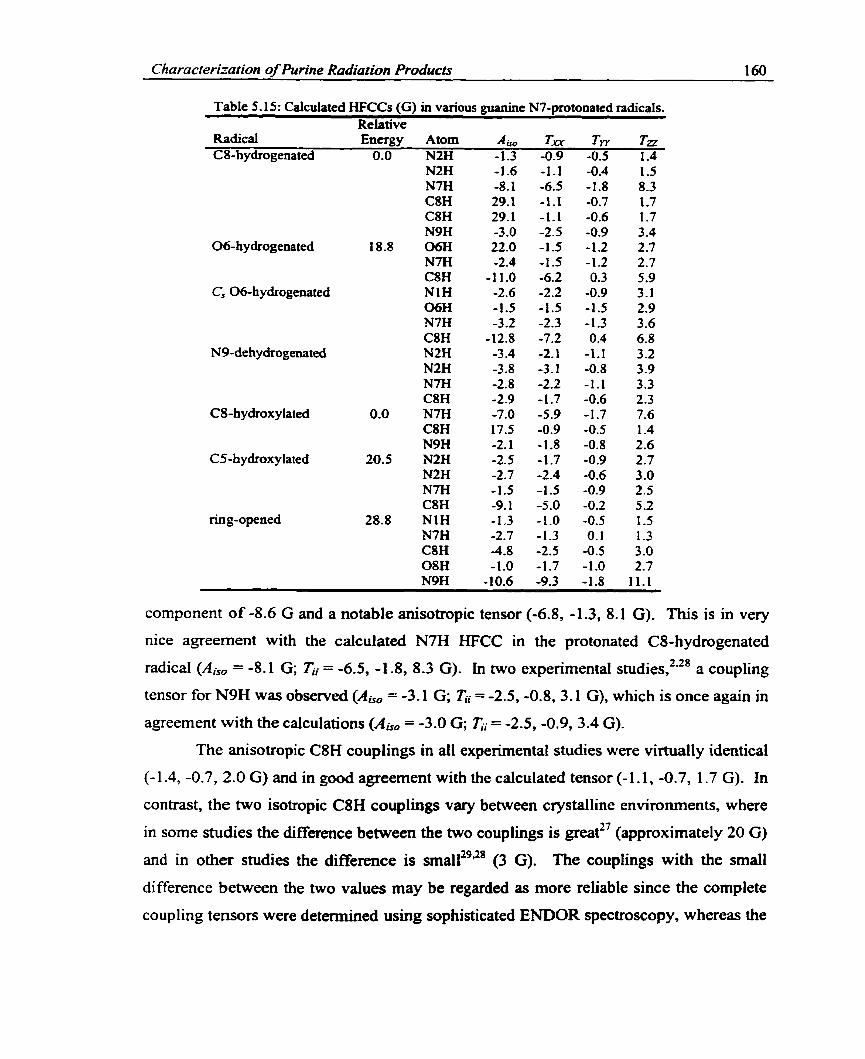
Characterization of Purine Radiation Products 160
Table 5.15: Calculated HFCCs (G) in various guanine N7-protonated radicals. Relative
Radical Energy Atom A, r.m Tw Tn CS-hydrogenated 0.0 N2H -1.3 -0.9 -0.5 1.4
06-hydrogenated
Cs 06-hydrogenated
N9-de hydrogenated
CS-hydroxy lated
CS-hydroxylated
ring-opened
-- -- - --
cornponent of -8.6 G and a notable anisotropic tensor (-6.8, -1.3, 8.1 G). This is in vexy
nice agreement with the calculated N7H HFCC in the protonated CS-hydrogenated
radical ( A , = -8.1 G; T,i = -6.5, -1 -8, 8.3 G). In two expenmental studies,*'* a coupling
tensor for N9H was observed (A, = -3.1 G; = -2.5, -0.8, 3.1 G), which is once again in
agreement with the calculations (A , = -3.0 G; zi = -2.5, -0.9, 3.4 G).
The anisotropic C8H couplings in al1 experimental studies were virtually identical
(- 1.4, -0.7, 2.0 G) and in good agreement with the calculated tensor (- 1.1, -0.7, 1.7 G). In
contrast, the two isotropie C8H couplings vary between crystaliine environments, where
in some studies the difference between the two couplings is greatz7 (approximately 20 G)
and in other studies the difference is ~ r n a l l ~ ~ " ~ (3 G). The couplings with the small
difference between the two values may be regarded as more reliable since the complete
coupling tenson were detennined using sophisticated ENDOR spectroscopy, whereas the

Charactenzation of Purine Radiation Products 161
couplings that deviated by 20 G were determined only through ESR. However,
differences in couplings extracted in each experiment may also arise due to the crystalline
environment, the temperature, or the different time scales employed. The calculations
indicate that the two C8 hydrogens have identical couplings, which are much smaller
(29.1 G) than those observed in any of the ENDOR experiments (approximately 34 G and
37 G). The problem of calculating couplings smaller than recorded expenmentally has
previously been discussed for similar thymine and adenine radicals. Due to the good
agreement between theory and experiment for al1 couplings besides the isotropic C8H
component, the calcu1ations support the experimental assignment of the protonated C8-
hydrogenated radical. It should be noted that the isotropic C8H couplings calculated for
the nonprotonated radical (36.9/37.2 G) are in excellent agreement with the couplings
assigned to the corresponding protonated radical. However, due to the excellent
agreement of the N7H HFCCs with experiment, it can be concluded that the radical is
protonated and the poor agreement with experimental results is due to the mode1
employed.
The N7-protonated 06-hydrogenated radical has been obsewed in studies on
crystals of G:HC~:H~O,'~ ~ ' G M P ( F A ) , ~ ~ G : H C I : ~ H ~ O ' ~ and G:HB~:H~o.~ The geometry
was calculated to exhibit distortions at C6, where 06H is located out of the molecular
plane to result in a large isotropic 0 6 H coupling which was not recorded experimentally.
The calculated couplings for the hydrogen at 0 6 (22.0 G) and C8 (- 1 1 .O G) are extremely
large (Table 5-15), while the corresponding experimental couplings are small (Table
5.14). Not even the anisotropic couplings for this radical are in agreement. Thus, it
seerns unlikely that the N7-protonated 0 6 hydrogen addition radical is responsible for the
spectra observed in these studies. Since hydrogen bonding interactions may result in a
planar geometry, a Cs radical was obtained through a fùll optimization, which possesses
one imaginary fiequency and lies 1.7 kcaYrnol higher in energy than the nonplanar
radical. Calculations on the planar radical yield a small O6H coupling which is expected
experimentally and, thus, the agreement between calculated couplings and experiment
could be considered to be improved over that obsewed for the nonplanar radical. A NI H
coupling was calculated in the planar radical that was not obtained for the nonplanar
form, however this coupling is still smaller than the experimental result. The

Cliaracterization of Purine Radiation Products 162
experimental and calculated couplings also disagree in the magnitude of the C8H
coupling, where the HFCCs obtained from the calculations are far too large relative to
those obtained in the experimental study.
The possibility that the observed radical is nonprotonated can be eliminated. In
particular, the C8H HFCC for the planar 06-hydrogenated radical is different fiom that
assigned to the N7-protonated 06-hydrogenated radical. Furthemore, clear couplings
were observed experimentally for N7H. To ensure that differences in the calculated and
experimental C8H couplings for the N7-protonated 06-hydrogenated radical do not arise
due to differences in the hydrogen bonding environment at N7, a series of calculations
were performed where the N7H bond was lengthened. The N1 and 06 hydrogen
couplings did not change with variations in the N7H bond length. The C8H couplings
(Table 5.16) also do not change substantially over the N7H bond lengths investigated.
Alternatively, the N7H anisotropic couplings show a decrease in magnitude with an
increase in bond length. Despite the great difference between the C8H couplings in the
planar protonated and nonprotonated radicals, neither of these couplings match those
assigned to the protonated 06-hydrogenated radical. However, the average of these
couplings (A,, = -8.4 G; 1;,- = -4.8,0.3,4.6 G) is in good agreement with the experimental
results (Ai5* = -7.8 G; = -4.0, -0.1, 4.2 G). Moreover, the average calculated NlH
couplings (Ais, = -2.8 G; z; = -2.4, -1.1, 3.5 G) are also in agreement with experiment
(AjsO = -3.4 G; I;I = -2.4, -0.5, 2.9 G). Any discrepancies between experimental and
calculated N7H couplings can also be explained in terms of differences in the N7H bond
Table 5.16: Variation in the planar, N7-protonated 06-hydrogenated guanine radical's C8H and N7H HFCCs (G) with respect to the N7H bond length. (A) Bond Length CSH N7H
A,, TYX Tw T' Am Tw TYY Tiz 0.908 -13.1 -7.3 0.4 6.9 -3.1 -2.9 -1.1 4.0 1.008. -12.8 -7.2 0.4 6.8 -3.2 -2.3 -1.3 3.6 1.108 -12.8 -7.1 0.4 6.7 -3.2 9 -1.4 3.3 1.208 -12.6 -7.0 0.4 6.6 -3.5 -1.6 -1.4 3.0 1.308 -12.4 -6.9 0.4 6.5 -4.0 -1.4 -1.3 2.7
unprotonated' -3.9 -2.3 -0.1 2.3 experimenta16 -7.8 -4.0 -0.1 4.2 -2.9 -1.8 -0.9 2.6
-The optimized bond lengtb for the planar N7-protonated 06-hydrogenated radical. t Calculated results for the C' 06-hydrogenated radical which is not protonated at N7. 'The experimcntal value was obta&ed i& an average of the r e d i s piesented in Table 5.14.

Characterization of Purine Radiation Producfi 163
length. The experimental N7H HFCCs are in better agreement with the calculations
performed at longer bond lengths than those performed at the optimized geometry. Thus,
a possible explanation for the observed spectra is a recorded averaging where some of the
06-hydrogenated radicals are protonated at N7 and some exist as nonprotonated species
or there exists a transfer between N7H and a neighboring Cl anion.
An additional explanation for the observed couplings is the guanine cation. The
average C8H experimental full tensor components (- 1 1.9, -8.0, -3 -5 G) and those
calculated for the guanine cation (-12.6, -8.8, -3.2 G) are in excellent agreement. In
addition the experimentally assigned N1 (-5.9, -3.9, -0.6 G) and N7 (-4.8, -3.8, -0.3 G)
tensors in the 06-hydrogenated radical are in excellent agreement with those calculated
for the two N2 hydrogens (-5.6, -3.8,O.l G) and (-4.3, -3.7, -0.1 G), respectively. Hence,
the expenmentally assigned N7-protonated 06-hydrogenated radical has HFCCs
remarkably similar to those calculated for the guanine cation, which could be forrned
through net N7 hydrogen removal from the parent molecule.
The only N7-protonated dehydrogenated radical reported expenmentally is the N9
centered radical, which was observed in G:HCI:H~O" and G:HCI:ZH~O.~~ On average
the spin density was determined to be 0.19 and 0.08 on N7 and N2, respectively
(calculated values: 0.10 and 0.13). Comparison of calculated (Table 5.1 5) and
experimental (Table 5.14) HFCCs for the N9-dehydrogenated radical cation indicates that
this radical is unlikely to be responsibfe for the observed spectrum. The variation in the
HFCCs with respect to the N7H bond length for the protonated N9-dehydrogenated
radical was investigated to account for differences in the hydrogen bonding scheme to N7
(not sho~n) .~ ' The amino and C8 hydrogen couplings do not change appreciably with
variations in the N7H bond length. The variations in N7H HFCCs are greater, but the
calculated results are still in poor agreement with experiment.
The experimental N7H anisotropic tensor (-3.7, -1.5, 5.1 G) is in much better
agreement with that calculated for C8H in the nonprotonated radical (-3.7, -0.7, 4.4 G)
than that calculated for N7H in the protonated radical (-2.2, -1.1, 3.3 G). In addition, the
two experimental N2H anisotropic couplings (-1.4, -0.5, 1.9 G) and (-1.7, -0.4, 2.1 G ) are
in excellent agreement with the two N2H couplings calculated for the nonprotonated N9-
dehydrogenated radical (-1.5, -0.9, 2.4 G) and (-2.2, -0.6, 2.8 G). The corresponding

Cltaracteniation of P urine Radiation Products 164
anisotropic tensors for the N7-protonated radical are (-2.1, - 1.1, 3.2 G) and (-3.1, -0.8, 3.9
G). Although mistaking a C8H coupling for an N7H coupling is highly unlikely, these
results indicate that the experimentally assigned N7-protonated N9-dehydrogenated
radical resemble couplings calculated for the corresponding nonprotonated radical more
closely. No other possibilities can be put forth for the observed radical at this time
through comparison of the experimental and calculated couplings.
Two N7-protonated hydroxylated radicals have been observed experimentally
which involve net hydroxyl radical addition to CS and C8. The C8-hydroxylated radical
was calculated to be 20.5 kcal/mol lower in energy than the CS radical. This is very
sirnilar to the energetics discussed for the nonprotonated radicals where the difference is
19.0 kcaWmo1. The protonated CS-hydroxylated radical was assigned in G:HB~:H~o.~
The experimental spin density was determined to be 0.13, 0.1 1 and 0.12 on CS, N7 and
N2, respectively, whereas the main components of the calculated spin density are 0.29,
0.12 and 0.37 on C8, C2 and C4, respectively. Clearly, the expenmental and calculated
spin density distributions are not in agreement. The agreement between experimental
(Table 5.14) and calculated (Table 5.15) HFCCs is also poor. In particuIar, the calculated
C8H anisotropic HFCC is approximately 3 G larger than that recorded in experiments
and the other couplings also do not correspond.
Other possibilities were discussed for the radical assigned in the experimental
study on G:HB~:H~o,~ including the CS-hydrogenated and N9-dehydrogenated radicals.
The CS-hydrogenated radical was concluded to be unlikely since a large CSH coupling
would be expected. The CSH HFCCs calculated for the nonprotonated CS-hydrogenated
radical (49.5 G) confimed that this radical is not responsible for the observed spectnim.
The possibility that the recorded HFCCs are due to the N7-protonated N9-
dehydrogenated radical was also dismissed in the expenmental paper since the spectrurn
was different from that observed in other crystals (Table 5.14). However, since the
previous experimental HFCCs for the N7-protonated N9-dehydrogenated radical were in
poor agreement with the calcu1ations, comparison of these couplings with those
calculated for the N7-protonated N9-dehydrogenated radical is required. The
experimental C8H isotropie (-3.7 G) and anisotropic (-1.9, -0.5, 2.4 G) couplings in
question are in excellent agreement with the calculated CSH coupiings in the N9-

Characteniarion of Punne Radiation Products 165
dehydrogenated radical (A, = -2.9 G; = -1.7, -0.6, 2.3 G). The experimental ( A , =
-3.2 G; Ki = -2.4, -1.0, 3.4 G) and calculated N7H couplings in the N9-dehydrogenated
radical (A, = -2.8 G; zi = -2.2, -1.1, 3.3 G) are also in excellent agreement.
Furthemore, both the experimental and the calculated radicals exhibit similar N2H
HFCCs. Thus, it can be concluded that the observed radical originally assigned to the
CS-hydroxylated radical is more than likely the N9-dehyhgenated radical. It should
also be noted that much better agreement is obtained between the calculated N9-
dehydrogenated HFCCs and the experimental CS-hydroxylated HFCCs than the
experimental "N9-dehydrogenated" couplings discussed previousiy.
The radical formed by net hydroxyl radical addition to C8 has been observed in
single crystals of G : H C ~ : ~ H ~ O . ~ ~ The observed spectrum consists of a large C8H
isotropic coupling (20.2 G) and a very small anisotropic tensor (-1.1, -0.6, 1.6 G), which
is in excellent agreement with the calculated values (A,, = 17.5 G; Ki = -0.9, -0.5, 1.4 G).
Experimentally, another isotropic coupling was observed for N7H (-8.5 G) which also
possesses great anisotropy (-6.5, -1.5, 8.0 G). The calculated N7H couplings (A,, = -7.0
G; ci = -5.9, -1.7, 7.6 G) are in agreement with expenment considering the local
environment has been shown to affect these couplings in other radicals (Table 5.16). A
small N9H coupling was also noted for this radical (expenmental and calculated isotropic
HFCCs of -2.2 and -2.1 G, respectively). Experimentally, it was speculated that the
observed spectnun could be due to the CS-hydrogenated radical where the additional
hydrogen is added to an in-plane position and, thus, only one large C8H coupling is
observed. This alternative seems very unlikely due to the excellent agreement between
experimental and calculated HFCCs for the CS-hydroxylated radical cation.
One last radical observed experimentally was thought to have a molecular
structure very similar to the N7-protonated CS-hydroxylated radicalS2 One expenmental
coupling was observed and assigned to C8H which has undergone significant
reorientation. The proposed radical is the N7-protonated ring-opened radical (Figure
5.3), which was calculated to be 28.8 kcaUmol higher in energy than the corresponding
ring-closed radical. The experimental coupling consists of an isotropic (-4.9 G ) and an
anisotropic (-2.6, O. 1, 2.4 G) C8H coupling. These results are in good agreement with the
C8H couplings calculated for this radical (A , = -4.8 G; zï = -2.5, -0.5, 3.0 G). In

Characten'ration of Purine Radiation Praducts 166
Figure 5.3: Smcnue of ring-opened, N7-protonated guanine radical cation.
addition to the C8H HFCC, the calculations indicate that couplings should be observed
for the hydrogen attached to N9 and possibly NI, N7 and (the hydroxyl oxygen) OS. A
more detailed experimental study is required for a definitive identification of this radical.
5.3.7 Experimenîaiiy CTnassigned Guanine Radical
In a study of S ' ~ G M P ~ crystals, a spectrum was left unassigned, aithough many
possible radicals were discussed. The unassigned spectnrm consists of a C8H coupling
(Abo = -7.2 G, Tii = -3.7, -0.3,4.0 G) and two couplings arking fiom hydrogens attached
to nitrogen atoms. One NH coupling, proposed to be N1H or an amino hydrogen,
possesses an isotropie component of -3.4 G and significant anisotropy (-2.6, -0.5, 2.9 G).
The second NH coupling was suggested to arise from N2H, N3H or altematively fiom a
hydrogen atom involved in a hydrogen bond with N7. The couplings frum this hydrogen
are slightly smaller than those discussed for the first NH ( A , = -3.0 G; ci = -1.9, -0.5,
2.4 G).
Two deprotonated radicals, formed via hydrogen removal fiom N1 or N2, were
proposed to give Rse to the unassigned spectrum. Comparison of the experirnental
couplings (Table 5.9) and those calculated for the N2-dehydrogenated radical (Table
5.1 1) indicates that this radical is unlikely to be responsible for the observed spectrum.
In particular, only one NH coupling was calculated for this radical, which furthemore is
significantly different fiom those discussed for the unidentified radical. The calculated
couplings for the NI-dehydrogenated radical (Table 5.1 1) match those elucidated in the
unassigned spectm more closely, although the anisotropic couplings disagree more than
expected for this component. It is interesting to note that the calculated anisotropic
couplings for the N9-dehydrogenated radical are in excellent agreement with those of the
unassigned radical. However, this radical is not expected in guanosine crystals since a
sugar group replaces the hydrogen at N9.

Characterization of Purine Radiation Products 167
The guanine cation was also proposed to have a spectrum similar to that
unassigned experimentally and the calculated HFCCs (Table 5.10) support the hypothesis
that the wiassigned spectnim is similar to this radical. The calcutated NH anisotropic
couplings are in remarkable agreement with those obtained experimentally, whereas the
calculated C8H anisotropic coupling is slightly larger than the recorded value. The
guanine cation was not seriously considered to be responsible for the observed spectnrm
since it is unlikely that this radical would be observed at 200 K. The unassigned
spectnim was also speculated to arise nom an Ni-protonated 06-hydrogenated radical.'
The calculations (Table 5.15) yield different KFCCs than those obtained experimentally
for both the planar and nonplanar geometries. However, the couplings extracted 6om the
unassigned spectrum are very similar to the average of the protonated and nonprotonated
06-hydrogenated couplings discussed in the preceding section. This possibility offers the
best explanation of the unassigned couplings to date.
Hole et commented on the similarity of the unassigned spectra to that
obtained for both the guanine anion and cation. In particular, they discussed the
mandatory care required when interpreting the resonance patterns of the inadiated DNA
bases, even for those spectra obtained from the sophisticated ENDOR technique. The
calculations support their comments since many of the calculated HFCCs for the
radiation products are very similar. Thus, even through the use of high-level calculations,
interpretation of the observed spectra is very difficult. This example illustrates the great
difficulties associated with the determination of DNA radiation products, even when the
problem is reduced to exarnining the individual bases.
5.3.8 Summury of Guanine Resuits
Disagreement between theoretical and experimental couplings for the guanine
anion was exhibited which was improved upon through consideration of a ptanar radical.
The calculated couplings for a planar 06-hydrogenated radical also match the
experimental results. Only one coupling tensor was extracted in the expenmental study
and, thus, for positive identification of the obsemed radical more experimental couplings
are required. In particular, the calculations indicate that the anion and its 06-protonated
form can be disthguished through the identification of the amino hydrogen HFCCs.

Characterization of Purine Radiation Products 168
Similarly, the couplings assigned to the guanine cation and the calculated couplings were
not in good agreement.
The N9-dehydrogenated radical was detemined to be the lowest energy radical in
this class and the corresponding N1 and N2 radicals are very close in energy. Only the
N2-dehydrogenated radical has been observed experimentally and the calculated HFCCs
are in excellent agreement with expenmental values. The C8 and CS-hydrogenated
radicals are the lowest and highest energy radicals in their class. Both of these radicals
have been observed experimentally and the calculations support their assignment. The
Cg-hydroxylated radical is the lowest energy radical formed via net hydroxy1 radical
addition. The couplings assigned to the C4-hydroxylated radical experimentdly are in
fair agreement with the calculated values if the principal components are considered. An
alternate explanation for these observed couplings can be sought in the formation of the
N2-dehydrogenated radical.
The good agreement obtained between theory and experiment for the
nonprotonated radicals is not immediately transferable to the N7-protonated guanine
radicals investigated in the present study. Among the protonated radicals, only the
experimental assignment of the CS-hydrogenated and CS-hydroxylated radicaIs is
supported by the calculations. The spectnun experimentally assigned to the protonated
06-hydrogenated radical was speculated to arise fiom an averaging of the planar forms of
the protonated and nonprotonated 06-hydrogenated radicals. ui addition, it was also
noted that the s p e c t m experimentally assigned to the N7-protonated 06-hydrogenated
radical contains HFCCs very similar to the guanine cation. The protonated N9-
dehydrogenated radical was concluded to be unlikely responsible for the observed
spectrum assigned experimentally to this radical. The only radical possessing couplings
similar to those observed experimentally is the nonprotonated N9-dehydrogenated
radical. No other possible product could be identified as leading to the experimental
HFCCs. In addition, the observed HFCCs thought to arise fiom the CS-hydroxylated
radical were detemined to originate fiom an other guanine radical, most probably the
N7-protonated N9-dehydrogenated radical. It was concluded that more experimental, as
well as theoretical, studies are required for the hi11 identification of N7-protonated
radicals generated upon irradiation of guanine denvatives. It should be noted that no

Characrenzation of Purine Radiation Products 169
environmental effects were included in the present study, which offers a possible
explanation for discrepancies between theory and expenment.
Through comparison of experimental and calculated hyperfine coupling constants,
it cm be determined that the N2-dehydrogenated radical is the major dehydrogenated
product formed in neutral guanine crystals. Formation of this radical produces a supply
of hydrogen atoms which will add pnmarily to CS and C8. In N7-protonated guanine
crystals, the primary radiation products elucidated through comparison of experimental
and calculated HFCCs are the C8-hydrogenated and CS-hydroxylated radicals. The
identification of al1 other radicals is speculative. In addition, the 06-hydrogenated
radical may be formed where the observed spectra exists as an average of that due to the
protonated and nonprotonated radicals. Contrary to adenine, there exist differences in
radical formation when N7-protonated crystals are considered. In particular, no
dehydrogenated radicals were characterized in the protonated crystals. Thus, questions
regarding how the hydrogen atoms are generated to fom net hydrogenated radicals &se.
However, al1 protonated crystals examined were hydrate derivatives. Thus, the most
likely mechanism for damage in these hydrate crystals involves water, where net
hydrogen atorn and net hydroxyl radical addition occurs at CS in guanine. The formation
of the CS-hydroxylated radical in hydrate crystals of guanine denvatives provides hirther
support for the proposed mechanism for damage in cytosine monohydrate crystals in
Chapter Four.
5.4 Cori clusions
Similar to the thymine results discussed in Chapter Four, the calculated and
experimental couplings obtained in adenine radicals are in very good agreement. The
calculations indicate that the cation has been observed, but only in initially protonated
crystals. In some instances, the calculated results were initially in poor agreement with
experiment. However, upon consideration of crystal effects, improved results were
obtained. For exarnple, the calculated couplings in the N3-hydrogenated radical were
different fiom the experimental results since a severely distorted radical was optimized.
Once the extensive hydrogen bonding schemes in the crystals, which result in planar
radicals, were taken into account, the calculated HFCCs were in much better agreement

Charact enfat ion of Purine Radiation Producfs 1 70
with experirnental results. Similar to the thymine C6-hydrogenated radical, the isotropic
HFCCs in the C2 and CS-hydrogenated radicals were calculated to be of equal
magnitude, while the radicals are slightly distorted at these positions in the expenrnents.
The only radical assignment not supported by the calculations is the protonated C4-
hydrogenated coupling. Experimentally, a small isotropic coupling was assigned to C4H
in this radical, but the calculations indicate that this hydrogen should possess a large
coupling. Consideration of the magnitude of the couplings obtained in other base
derivatives when hydrogen is added perpendicular to the molecular plane supports the
conclusions drawn h m the calculations that the observed radical was misidentified.
The calculations on the guanine anion and cation indicate that the experimental
assisment of these radicals is questionable. Altematively, the calculations support the
experimental assignment of al1 dehydrogenated and hydrogenated radicals identified in
nonprotonated crystals. The C4-hydroxylated radical was identified in one experimental
study, but the extracted couplings are only in fair agreement with the calculations
indicating M e r studies are required to identiQ this radical. The identification of the
CS-hydrogenated and hydroxylated radicals in protonated crystals was also supported by
the calculations. The assignment of the 06-hydrogenated radical in protonated crystals
can be defended by the calculations if an averaging between protonated and
nonprotonated radicals is considered. However, the assignment of the other radicals in
protonated crystals can be questioned through cornparison to the calculations. In
particular, the results for the N9-dehydrogenated and CS-hyciroxylated radicais are in
poor agreement. The couplings in these radicals were similar to the nonprotonated and
protonated N9-dehydrogenated radicais, respectively. Complete assignment of these
radicals is not possible without the resolution of more expenmental HFCCs.
Through cornparison of the calculated and experimental HFCCs the identity of
one radical in each of adenine and guanine derivatives was assigned to a net hydroxyl
radical addition product. In particular, the calculations support the experimental
assignment of the C4 and CS-hydroxylated radicals in fiozen aqueous solutions of
deoxyadenosine 5'-monophosphate and crystals of guanine hydrochloride dihydrate. If
the full coupling tensors are considered then the calculations also support the
experimental assignent of the C4-hydroxylated radical in crystals of guanosine 3',St-

Characrerization of Purine Radiation Producrs 171
cycIic monophosphate. These conclusions support the hypothesis put forth in Chapter
Four that net h y h x y l addition to the cytosine base may occur in cytosine monohydrate
crystals. The reactions of water with cytosine, as well as uracil and thymine, will be
considered in Chapter Seven.
S. 5 References
1. Nelson, W. H.; Sagstuen, E.; Hole, E. O.; Close, D. M. Radiat. Res. 1998, 149, 75.
2. Hole, E. O.; Sagstuen, E.; Nelson, W. H.; Close, D. M. Radiat. Res. 1991,125, 1 19.
3. Hole, E. O.; Nelson, W. H.; Sagstuen, E.; Close, D. M. Radiat. Res. 1992, 129, 1 19.
4. Close, D. M. Radiat. Res. 1993, 135, 1.
5. Hole, E. O.; Sagstuen, E.; Nelson, W. H.; Close, D. M. Radiat. Res. 1995, 144,258.
6. Close, D. M.; Nelson, W. H.; Sagstuen, E.; Hole, E. O. Radiar. Res. 1994, 13 7,300.
7. Lichter, J. J.; Gordy, W. Proc. Natf. Acad. Sei., USA 1968, 60,450.
8. Zehner, H.; Flossmann, W.; Westhof, E. 2. Natu$orsch. 1976,31C, 225.
9 . Zehner, H.; Westhof, E.; Flossmann, W.; Müller, A. 2. Natu$orsch. 1977, 32C, 1 .
10. Close, D. M.; Nelson, W. H. Radiat. Res. 1989, 117, 367.
11. Kar, L.; Bernhard, W . A. Radiat. Res. 1983, 93,232.
12. Nelson, W. H.; Sagstuen, E.; Hole, E. O.; Close, D. M. Radiat. Res. 1992, 131,272.
13. Colson, A.-O.; Sevilla, M. D. Int. J Radiat. Biof. 1995, 67, 627.
14. Sevilla, M. D.; Besler, B.; Colson, A. O. J Phys. Chem. 1995, 99, 1060.
15. Orlov, V. M.; Smimov, A. N.; Varshavsky, Y. M. Tet. Leu. 1976,48,4377.
16. Colson, A.-O.; Sevilla, M. D. J. Phys. Chem. 1996, 100,4420.
17. Hole, E. O.; Sagstuen, E.; Nelson, W. H.; Close, D. M. Radiat. Res. 1992, 129, 1 .
1 8. Wetmore, S. D.; Boyd, R. J.; Eriksson, L. A. J. Phys. Chem. B 1998,102, 10602.

Charactenzation of P urine Radiation Products 1 72
19. Gregoli, S.; Olast, M.; Bertinchamps, A. Radiat. Res. 1974, 60, 388.
20. Box, H. C.; Budzinski, E. E . J . Chem. Phys. 1976,64, 1593.
2 1. Sagstuen, E.; Hole, E. O.; Nelson, W. H.; Close, D. M. Radiat. Res. 1996, 146,425.
22. Hole, E. O.; Nelson, W. H.; Close, D. M.; Sagstuen, E. J. Chem. Phys. 1987,86, 5218.
23. Hole, E. O.; Sagstuen, E. Radiat. Res. 1987, 109, 190.
24. Kim, H.; Budzinski, E. E.; Box, H. C. J. Chem. Phys. 1989,90. 1448.
25. Close, D. M.; Sagstuen, E.; Nelson, W. H.J. Chem. Phys. 1985,82,4386.
26. Close, D. M.; Nelson, W. H.; Sagstuen, E. Radial. Res. 1987, IIZ, 283.
27. Close, D. M.; Sagstuen, E.; Nelson, W. H. Radiat. Res. 1988, 116,379.
28. Nelson, W. H.; Hole, E. O.; Sagstuen, E.; Close, D. M. h t . J . Radial. Biol. 1988,54, 963.
29. Sagstuen, E.; Hole, E. O.; Nelson, W. H.; Close, D. M. Radiat- Res. 1988,ff 6, 196.
30. Wetmore, S. D.; Boyd, R. J.; Eriksson, L. A.J. Phys. Chem. B 1998,102,9332.

ChlAPTER SIX Sugar Radicals in Irradiated DNA Components
6.1 Introduction
The formation of sugar radicals in irradiated DNA is of great interest since it is
widely accepted that single-strand breaks occur via these inter me dia te^.'^ Sugar radicals
c m be formed through direct mechanisms, in which alkoxyl or base radicals are formed
and radical character is transferred to the sugar group, and indirect mechanisms, where
hydrogen or hydroxyl radicals generated h m water radiolysis attack the sugar group. In
an important study of O-glucose, Schuchmann and von sonntag3 concluded that hydroxyl
radicals attack the six carbon atoms in this sugar to an qua1 extent. However, ESR
techniques have been unable to detect sugar radicals in irradiated D N A . ~ As mentioned
in Chapter One, Hole et al5 were the first to observe a large variety of sugar radicals in
their ENDOR study of 2'-deoxyguanosine 5'-monophosphate, where nine sugar radicals
were characterized. This provides a nice example of the power of the E N W R technique
since ESR did not easily detect these radicals. A subsequent ENDOR study of single
crystals of deoxyadenosine6 supported the hypothesis that many sugar radicals are
generated upon irradiation.
Theoretical investigations of carbon-centered sugar radicals have appeared in the
literat~e.'.~ in these studies, geometries, relative energies, spin density distributions and
hyperfine coupling constants were calculated at the HF level. Both studies were very
complete and carefully perfomed at the level of theory chosen. However, as discussed in
Chapter Two, Hartree-Fock overestimates the hyperfine coupling constants considerably
and electron correlation yields important contributions to this property.
It is of interest to calculate the HFCCs of possible radicals in the DNA sugar
moiety in order to assign the experimental spectra to specific radicals with confidence.
Once the radicals formed in single crystais are fidly characterized, expenmentalists will
have a better understanding of how to recognize these radicals in full DNA and be able to
answer an important question, namely whether sugar radicals are formed upon irradiation
of DNA.'

Swar Radicals in Ikradiated DNA Components 1 74
The structure and standard atomic numbering of a DNA nucleotide unit is
presented in Figure 6.1 (structure 1). Due to the number of atoms involved in the
nucleotides, a model system must be used. The model chosen represents phosphate
groups with hydroxyl groups and the DNA base with an amino group (Figure 6.1,
structure II). From previous studies it is known that in order to correctly describe ring
puckering an amino p u p must be present at Cl', although geornetrical effects generated
by replacing the amino group with cytosine are smaL8 The use of hydroxyl groups rather
than phosphate groups in the chernical model may also lead to some geornetrical
differences, although these will not be discussed in the present work. The model sugar
used within differs fkom those previously ernployed by the inclusion of a hydroxyl group
at the CS' position7 and phosphate groups have been implemented in the past rather than
hydroxyl g r o ~ ~ s . ~
Nuc leo tide Unit
Figure 6.1 : Structure and numbering of the sugar group present in DNA (T) and the model system used for the calculations presmted herein (m.
In the present chapter, the geometry and HFCCs of possible sugar radicals
generated through irradiation of DNA are examinai with density- fimctional theory . The
sugar radicals to be contemplated include hydrogen abstraction radicals formed by
removal of hydrogen fiom al1 carbon and oxygen atoms, radicals formed via removal of
either of the hydroxyl groups in the model system, as well as a variety of radicals which
lead to significant sugar ring alterations. Computational techniques applied to these
systems are identical to those previously discussed for the DNA bases in Chapter Four
and a discussion of the methods employed will not be repeated here.

Sugar Rudicals in Imadiated DNA Components 175
6.2 Background Discusswn of Sugar Radical Propetîies
Two different puckering modes were examineci for each possible radical
corresponding to north (N) and south (S) radicals, which are d e h e d according to where
the radical is located on the pseudorotation cycle.9 It is convenient to analyze the
puckering amplitudes in the sugar molecules through the use of the pseudorotational
phase angle9 dehned as
tan P = ( 0 4 + 4 ) - ( 0 3 +vol 20, (sin 36" +sin 72")
where the oj are the ring dihedral angles: uo = C4'O 1'C lfC2', u, = 0 1 'C 1 'C2'C3', v2 =
C 1 'CZ*C3'C4*, 0 3 = C2*C3'C4'011, and or = C3'C4'Ol 'C 1 '. The puckering amplitude7 (s,),
which is defined as
is also a useful parameter to indicate the degree of puckering in the sugar ring where a
low value of 7, indicates a relatively flat ring. Figure 6.2 depicts the pseudorotation
cycle and the relation of P to this cycle.g
Figure 6.2: The pseudorotation cycle for deoxyrïbose depicting the pseudorotational phase angle, the puckering modes and the location of the north and south conformers.
The puckering in the sugar molecules can be considered to be either an envelope
(E) form, where four atoms define a plane and the fifth atom is located out of this plane,
or a twist (T) form, where three atoms defhe a plane and the other huo atoms are
displaced on opposite sides of this plane.9 Displaced atoms are categorized as endo or

Sugar Radicals in Irradiated DNA Components 176
exa according to whether they are located on the sarne side or oppoçite side of CS',
respectively. A superscript (subscript) on the lefi side of the puckering symbol (E or T)
is used to represent endo (exo) puckering. For example, the C3'-endo and C2'-endo forms
which are observed in nonradical sugar molecules in A-DNA and B-DNA are both
envelope conformations and can be represented as 3~ and 'E, respectively. Pictorial
descriptions of sugar ring distortion are displayed in Figure 6.3. In general, E or T can
only approximately describe sugar ring puckering and intemediate levels of the twist
mode can be obtained. Intemediate twist modes correspond to the area between
divisions on the pseudorotation cycle and, thus, this cycle depicts al1 possible puckering
amplitudes for the sugar group.
Figure 6.3: Examples of the puckering modes exhiiited in the DNA sugar group: 'E represents C3' endo puckerïng, 'fi represents a twist conformation and *E represents C2' exo puckering.
6.3 Energetics and Geomeîrical Parameters
The relative energies of the hydrogen abstraction sugar radicals are displayed in
Table 6.1. From the results it can be seen that the C4'(S) and CZt(S) radicals are the
Iowest and highest energy radicals among those formed by hydrogen abstraction fiom a
carbon, respectively. The north and south type conformers for each of the Cl', C2', C3'
and C5' radicals are energetically separateci by less than 2 kcal/mol. A larger difference
is observed for the C4' pair, where the north conformer is 3.3 kcal/mol higher in energy
than the south counterpart. Alkoxyl radicals, forrned via hydrogen removal fiom a
hydroxyl group in the model sugar, are very high in energy lying on average 10.2 and
12.9 kcaUmol above the C4'(S) radical for the 03' and 05' hydrogen abstraction radicals,
respectively. Radicals formed through removal of a hydroxyl radical in the model system
correspond tu breakage of a phosphoester bond in DNA. The C3' centered radical is
approxirnately 3.5 kcal/mol lower in energy than the corresponding CS radical,
identifjmg this as a possible site for strand-breaks in DNA.

Sugar Radicals in Inadiated DNA Components 177
Table 6.1 : Relative energies (IccaVmol), puckering mode, pscudorotational phase angle (deg.) and puckering amplitude (T,,,) of hydrogen and hvdroxvl abstraction suaar radicalsi
Radical +ZPÉ R Energy P =ln
Hydrogen Rernoval Radicals C4'(S) 0.0 0.0 'tT 135.3 31.9 C5'(N) C3'(S) c 1 '(S) Cl'(N) C 3 ' 0 CS(S) C 4 ' 0
C2'(S) 03'(S) 03'(N)' 0 5 ' 0 05'(S)
Hydroxyl Removal Radicals C3'(S) O O ,E 113.5 30.0 C3'(N) 0.7 0.5 IzT -46.0 28.1 CS '(S) 3 -2 4.1 'E 154.2 39.3 C5'(N) 3.7 4.5 3 4 ~ 45.5 41.4
\ ~ h e MP2 geometry was u x d for single-point calculations (see text for tiirther detaiis).
The lowest energy product detennined differs from that reported when HF
geometries were obtained and energetic studies performed with higher-level single-point
calc~lat ions .~~~ However, al1 products were reported to have small relative energies and
the C2' carbon centered radicals were detennined to be the highest lying products. Due
to the relatively small stabilization energy of one radical over another, it is not surprising
that differences arise in the results once electron correlation is included in the geometry
optimizations and slight modifications in the mode1 systems are implemented.
Furthemore, in the previous studies it is not clear whether corrections were made for the
zero-point vibrational energy. Table 6.1 indicates that when the ZPE is not taken into
account, the magnitude of the energy difference between radicals increases and the
relative order of the radicals may change. For example, without ZPE the C3'(S) radical is
lower in energy than the two Cl' radicals, but inclusion of the ZPE indicates that the C 1 '
radicals are lower in energy.
nie sugar puckering modes for C2'(N), C2'(S) and C4'(S) hydrogen abstraction
radicals differ from those obtained in a previous s t ~ d ~ . ' ~ On the contrary, the puckering

Sugar Radicals in Ikradiuted DNA Components 178
modes for the CL', C3' and C4'0 radicals and the magnitude of the pseudorotational
phase angle are in good agreement with previous work. Miaskiewicz and 0sman7
assigned the C3' radical with P = 84.4O to a south type k envelope fom. From the
results in Table 6.1, it can be seen that this radical indeed possess an OE form with P =
103.3". C2'(S) and CS'(S) have the smallest and largest rm values (Table 6.1). These
results are in agreement with previous s t ~ d i e s , ~ which used phosphate rather than
hydroxyl groups, indicating that only small effects are generated by using hydroxyl
groups in the mode1 system. It has been suggested that the relatively flat structure for the
C2' radicals occurs since an oxygen atom is not present next to the radical center. The
other carbon centered radicals with oxygen next to the radical site are more pyramidal
due to interactions between the lone pairs and the unpaired electron.' The net C3'
hydroxyl abstraction radicals have lower puckering amplitudes relative to the similar CS'
radicals and are nearly planar in structure. This indicates that the C3' center is sp2
hybridized after radical formation.
For atl hydrogen and hydroxyl abstraction radicals, the major geometrical
alterations that occur upon radical formation involve the bonds and angles in which the
radical center is invo~ved. '~ The bonds between the radical center and surrounding atorns
are generally contracted between 0.04 and 0.07 A. The bond angle in which the radical
center is the central atom changes between 2 and 8". The remainder of the sugar ring
geometry is relatively unaffected. This is in agreement with results obtained at the HF
levele7
6.4 Hyperfine Coupling Constants
6.4.1 Dehydrogenated Carbon Centered Radicals
It has been argueci in the past that al1 sugar radicais can be generated £?om alkoxyl
radicals and, thus, no sugar radicals can be formed in DNA due to the lack of hydroxyl
groups." However, due ta the nurnber of sugar radicals generated in S'~GMP,' which
contains only one hydroxyl group, it was suggested that other mechanisms for the
formation of these radicals must be considered. For example, carbon centered radicals
have been proposed to be formed via hydrogen abstraction by hydroxyl or hydrogen

Sugar Radicafs in Iwadiated DNA Components 179
radicals. In addition, it has been suggested that carbon centered radicals cm possibly be
fonned through excitation followed by homolytic cleavage of CH bonds.' Experimental
HFCCs for dehydrogenated carbon centered radicals are displayed in Table 6.2.
Table 6.2: Experimental HFCCs (G) for sugar ndicals gencrated through hydrogni abstraction from a ring carbon.
Radical Molecule Atom A, Tm Tw Tz Cl' S1dGMP' "C2'H" 28.0 -2.1 -1.8 3 -9
SC1 and S B ~ ~ U "
"C2'HW "C2'H1' "C2'HW "C2'HW "C2'HW 11C2'H" "C2'H1* "C 1 'Hl1 "c2'H1' "C2'H" "C4'HW unassignedo "PH" VH1' "PH" "C3'H1' 11C5'H" "CS'H" "C3W" "C5'HW "CS'H" TS'H" "CS'H" "C5'H1' "C4'H1' 1iO51~wœ
"CS'H" "C4'H1' "C5'H1' "CS'H" "C4'H "
"05HW "C5'H" "OS'H" - - -- - -
*Principal tensor components.
The couplings present in the spectra of a number of irradiated DNA molecules
have been assigned to the Cl' in addition to hydrogen abstraction and CH
bond cleavage resulting fiom excitation, another mechanism for radical formation is
deprotonation of a parent sugar radical cation at the Cl' position. This mechanism was

Sugar Radicals in I d i a t e d DNA Components 180
suggested for the correspondhg C4' radical." In particular, it was proposed that if the
electron vacancy is primarily located on Ol', then the positively charged radka1 is most
likely stabitized through deprotonation at positions close to 01' (Le., C4' or Cl').
Hole et UL' detennitled that the mspin density at Cl' is 0.64, which is smaller than
the calculated value (0.75). Cornparison of experimental (Table 6.2) and theoretical
(Table 6.3) HFCCs indicates that the calculations support the experimental assignment of
the Cl' radical. In particular, the experimental results agree more closely with those
calculated for the N-type radical. Che of the CTH couplings calculated for the S-type
C 1' radical is significantly smaller (9.1 G) than the experimentaI results (approximately
18 G). This is a nice example of the effets of the puckering amplitude on the HFCCs. It
should be noted that although the C2'H isotropie cornponents differ between N and S-
type radicals, the anisotropic valuzs are alrnost identical.
Table 6.3: Calculated HFCCs (G) for dchydrogcnated sugar radicals. North South
Radical Atom A,, Tm Tw Tzz A i , Tm Tw Tzz Cl' C2'H 18.5 -1.4 -1.0 2.4 29.3 -1.4 -1.1 2.5
CZ'H C2' C2'H
Cl'H C3'H 03'H
C3' c2w C2'H 03'H C4'H
C4' C5'H CS'H C4'H O5'H
CS' C5'H c4'H O5'H
05' C5'H CS'H Cl'H
03' C3'H Cl'H
' M P ~ geomeiry was used in single-point calculations (see text for hinher deuils).
The C2' hydrogen abstraction radical has appeared in experimental studies only as
a minor product,S which is not surprising since it was predicted to be the highest energy
radical among carbon hydrogen abstraction adducts. From the expenmental study, an sp2

Suaar Radicals in fwadiated DNA Comuonents 181
configuration at the C2' center was suggested, which would involve rehybridization at
this position upon radical formation and explain the relatively flat structure of this
species. An experimental spin density of 0.89 was obtained for C2' (calculated value:
0.96). The experimental HFCCs for the C2' radical are in qualitative agreement with the
calculated values. The C2'H HFCC exhibits a great deai of anisotropy in the
experimental results which is supported by the calculations. The Cî'(S) and C2'0
calculated HFCCs difTer mainly in the C3'H coupling since a larger isotropic HFCC was
obtained for the north conformer (38.2 G) relative to the south confonner (7.9 G)-
Experimentally, no C3W coupling was detected, and thus the radical observed in the
expenment is most likely tu be in a south conhrmation.
The C3' dehydrogenated radical was observed in an investigation of irradiated
5'dGMP and the results indicated that C3' remains sp3 hybridized rather than
rehybridizing as discussed for the C2' radical.' Through comparison of the two
calculated C2'H couplings in the N and S-type conformers with experimental results, the
nature of the observed radical is difficutt to predict. However, the calculated HFCCs for
the N and S-type C3' radicals differ through the absence of a C4'H coupling in the latter
conformation. Since a large C4'H coupling was recorded expenmentally, the calculations
predict this radical to be present in the north conformation. The calculated values
indicate that 03'H has a small isotropic coupling and a relatively large anisotropic
contribution which were not detected in the experimental study. However,
expenmentally there was another coupling observed for which only the principal
components were resolved and assignment to a particular atom was not made. The
unassigned couplings are not unlike those of a C2' hydrogen and could possibly be due to
a C2'H in a ring with another conformation. The difference between the experimental
and the calculated isotropic hyperfine coupling constants in this radical could be due to
the presence of a phosphate group at the CS' position in the experimental study since it
has been previously detennined that the phosphate groups affect the HFCCs in the C3'
radical.'
Radicals fomed through hydrogen abstraction fiom the C4' position have been
observed in three different crystals: uridine 5'-monophosphate (~ '~uMP) , '~ inosine (rI,

Sugar Rudicals in Iwadiated DNA Components 182
which can be derived fkom adenosine by replacing the amino group at Cd with a hydroxyl
goup) '' and adenosine5-bromowacil (~A:sB~u). '' As previously discussed, the net C4'
hydrogen abstraction radical can be formed via deprotonation of the parent sugar radical
cation. Alternatively, it could arise through abstraction of a hydrogen at CS' followed by
a 4',5'-hydrogen atom transfer. The C4'IN) radical exhibits two CS'H couplings, one of
substantial magnitude (27.9 G), and no OS'H coupling, while the south confoxmer has a
significant 05'H coupling (5.6 G) and only one small CS'H coupling (6.2 G).
Experimentally, three substantial couplings of 36, 25 and 24 G were recovered in crystals
of ~ ' ~ u M P , ' ~ and two small couplings were observed at certain orientations for which
accurate HFCCs could not be evaluated. In r~ ," large C3'H (34.7 G) and CS'H (33.4 G),
as well as a small C5'H (3.4 G), couplings were obtained. In rA:SBrU, two couplings
were resolved corresponding to the C3' and CS' hydrogens (21.0 and 10.0 G,
respectively). Overall poor agreement between theoretical and experimental HFCCs for
this radical indicates that either better data must be obtained or other radical possibilities
must be considered. In particular, no anisotropic components, which are important for
cornparison to theoretical work, were isolated. Differences between theory and
experiment could arise due to alterations experienced when phosphates groups replace
the hydroxyl mode1 group.*
The CS' hydrogen abstraction radical has been assigned in studies of various DNA
constituents, 5.6.13.17 and differing results have been obtained for the HFCCs. In some
cases, ring-breaking or ring-opened radicals were believed to be present rather than this
carbon centered radical." Early studies which identified the CS' radical include
experiments performed on cytosine 3'-monophosphate (3'CMP), 5-chlorodeoxyuridine
(5CldU) and 5-bromodeoxyuridine ( sB~~u) . '~ The CS'H isotropic HFCCs obtained in
these studies are similar while the anisotropic CS'H components and the C4'H and 05'H
HFCCs depend on the crystal examined. Hole et al.' wigned four parallel lines in the
spectnim of irradiated 5'dGMP to CS' hydrogens in different conformers of the CS'
hydrogen abstraction radical. The magnitude of the isotropic CS'H coupling agrees with
those obtained in earlier studies, but once again the C5'H anisotropic, C4'H and 05'H
HFCCs difEer fiom those discussed above. The same group observed evidence of the

Sugar Radicals in Irradiated DNA Componenîs 183
formation of this radical in irradiated d ~ m . ~ From Table 6.2, it is evident that the CS'H
isotropic coupling recovered in dAm agrees well with those previously discussed, but the
anisotropic components resemble those obtained in earlier studies rather than those
obtained in 5'dGMP crystals. The calculated CS11 anisotropic coupling indicates that this
center should possess a large degree of anisotropy, which is in agreement with
expenment. However, the calculated isotropic C5W and C4'H couplings are smaller and
larger than those observed in the above experimental studies, respectively.
Alexander and Franklin observed a radical upon irradiation of deoxyadenosine
debated to be either the CS' radicai or a radical formed through breakage of the C4'01'
bond within the sugar ring." The radical was concluded to be the CS' radical and a
considerable degree of anisotropy, as seen in other experimental studies and in the
calculations, was recorded. However, the isotropic couplings are much smaller in
magnitude than those previously assigned indicating that reexamination of this radical is
necessary. These experimental results will be discussed in more detail below.
The experimental systems discussed above differ from the mode1 radical used in
the present study by a phosphate group at the CS' position. However, it was suggested
that a similar radical with the phosphate group replaced by a hydroxyl group was also
observed in the experimental specûum of ~ ' ~ G M P . ' Principal values typical of AOH)
couplings (Am = 16.3, Aw = 20.2, Azz = 28.1 G) were elucidated. The calculated values
for this coupling are however much smaller in magnitude.
Due to discrepancies between experimental and theoretical results, an in-depth
investigation of the couplings assigned to the CS' hydrogen abstraction radical is
required. Since significant effects on the HFCCs can be obsented with changes in
geometry, an investigation of the dependence of the HFCCs on rotation about the CS'C4'
bond was undertaken. The XCS1C4'C3', X = 05' or H5', dihedral angles in the north
conformer were varied by increments of 15" starting fiom the optimized geometry
(289.3" and 144.4" for X = HS' and OS', respectively) and single-point calculations
performed at each step. The results for the variation in W H , C5'H and 05'H HFCCs as a
tùnction of rotation angle are displayed in Figure 6.4. It is interesting to note that upon
ngid rotation, the isotropic component of the HFCCs changes considerably, while the

Sugar Radicals in Imadiated DNA Components 184
anisotropic components (not shown) do not differ more than twenty percent from the
values displayed in Table 6.3. On average, the rotation b h e r about the C4'CS' bond is
8.6 kcdmol, with maximum and minimum values occurring at 90° (1 4.4 kcaVmol) and
1 5" (1 -4 kcal/mol), respectively.
+C'4
+ C'S
Rotation Angle
Figure 6.4: The C4', CS' and 05' hydrogens* HFCCs (G) versus the rotation angle (deg.) about the CSC4' bond for the C S 0 radical.
The results in Figure 6.4 shed some light on the dependence of ihe HFCCs on
rotation about the CS'C4' bond. The caiculated CS'H isotropie HFCC does not reach the
experimental value (-22 G) obtained in S'dGMP, but cornes close to the value obtained in
dAm (- 1 7 G) upon a 300' rotation (- 16.7 G). The variation between 05'H and C4'H
results obtained for 3'CMP and SC1 or SBrdU can also be understood fiom these results.
For 3'CMP, the calculated values which satis@ both the C4' and 05' experirnental
couplings occur at a 1 30° rotation, where Ak0(OS'H) = 22.6 G and Ako(C4'H) = 8.1 G
(experirnental values are 20.8 and 4.5 G, respectively). Similady, results in agreement
with 5Cl and 5BrdU experimental WCCs occur upon a 150° rotation, where Ah(C4'H) =
17.7 G and A,(0S1H) = 10.3 G (experimental values: 18.9 and 8.6 G, respectively).
Hence, the calculated results agree very well with the HFCCs obtained experimentally in
these studies.

Sugar Rudicals in Irradiated DNA Componenls 185
At 130" rotation, the CS'H and C4W HFCCs (-14 G and 7 G, respectively)
obtained by Alexander and Franklin in dA are also in good agreement with the calculated
values (-16.7 G and 8.4 G). However, at this degree of rotation, a large 05'H KFCC is
also calculated (30.5 G) which was not detected in the experiment. Thus, the results of
Alexander and ~ranktin" cannot be understood through this rotation analysis and other
radical possibilities must be considered.
Allcoxyl radicals can be formed through abstraction of a hydrogen f?om 05' and
03', both of which have been assigned to expenmental In full DNA,
these radicals would be formed by breaking a bond within the phosphate group and,
hence, would lead to strand breaks. Bernhard and CO-workerd9 examined 05' alkoxyl
radicals and noted that these species are relatively unstable, decaying between 4 and 120
K. The experimental results (Table 6.4) indicate that the magnitude of the two CS'H
couplings varies with the compound considered, although the sum of the two couplings
fluctuates between a small range (134 to 145 G). The calculated results for both the north
and south type 05' radicals (Table 6.3) are very similar which is not surprising since the
radical center is outside the sugar ring and, hence, puckering effects on the HFCCs are
expected to be small. The calculated HFCCs consist of two large C5'H couplings (on
average 91 and 19 G) and a smaller Cl ' couplhg (approximately 3 G). The relative
magnitude of the two CS'H couplings differs fiom the expenmental results.
Table 6.4: Experimental HFCCs (G) for sugar alkoxyl radicals. Radical Molecule Atom Air0 Tm Tw Tzz 05'-akoxyl rTiJ "C5'H1' 80.4 -3.1 -1.7 4.8
"C5'H1' 71.3 -3.1 -1.7 4.8 upafi3 "C5'H1' 90.0 -1.0 -1.0 2.0
"C5'H1' 47.3 -1.3 -0.3 1.7 ~ A : H C I ' ~ "CS'H" 93.5 -3.3 -2.0 5.3
"C5'H" 48.0 -2.7 -1.9 4.5 S C I ~ U ' ~ "C5'HW 83.3 -4.1 -1.9 5.9
"CSW" 85.6 -3.5 -0.9 4.3 5 ~ r d ~ " "C5'HU 58.5 -3.3 -0.3 3.7
"CS'H" 57.3 -2.6 -2.1 4.6 ~ ' C M P ' ~ "C5'H" - 82
"C5'HW - 59 ~ A I I I ' ~ "C5'HW --. 100
"C5'Hl1 - 53 03'-akoxvl S ~ G W ~ O "C3'Hw 20.3 -4.7 -0.5 5.1

Sugar Radicals in Inadiated DNA Components 186
- O 30 6û 90 120 150 180 210 210 270 300 330 360
Rotation Angle
Figure 6.5: The C5' hydrogens' HFCCs (G) versus the rotation angle (deg.) about the CS'C4' bond and the sum of these couplings in the 0 5 ' 0 radical.
Due to the differences between experimental and calculated WCCs, the effixts of
rotation about the C5'C4' bond on the HFCCs were examined in the north-type radical
(Figure 6.5). Results are displayed as a fùnction of the 05'CS'C4'C3' angle with respect
to the optimized value (193.2O). The average (2.7 kcal/mol) and maximum (7.0 kcal/mol)
rotational barriers are much smaller than those observed for the CS' hydrogen abstraction
radical. The HFCCs Vary greatly upon rotation, although not in the smooth manner
observed for the CS' centered radical. in some instances, the rotation study clarifies
discrepancies behveen experiment and theory. For example, the C5'H expenmental
couplings observed in uracil-P-D-arabinofuroside ( ~ ~ a f ) " (90.0 G and 47.3 G) and
a d e n o s i n e : ~ ~ l ' ~ (93.5 and 48.0 G) are in better agreement with the results obtained upon
a 45" rotation (88.2 and 48.6 G) than the values calculated at the optirnized geometry. In
other instances, the rotation study does not explain the experimental results. For
example, the two C5'H couplings in 5clduL9 (83.3 and 85.6 G) and S B ~ ~ U ' ~ (58.5 and
57.3 G) are equal in magnitude. However, although the calculated C5'H couplings corne
close in value upon a 1 OS0 rotation (66.0 G and 7 1 .O G), the couplings are different fiom
those observed experimentally. It should be noted that there exists an extensive

Sugar Radicals in Irradiared DNA Componene 187
hydrogen-bonding scheme with respect to 05' in these ~ r ~ s t a l s ' ~ not accounted for
theoretically which offers a possible explanation for the disagreement between
experiment and theory. Aithough the calculated values for the surn of the C5'H couplings
(Figure 6.5) are on average slightly smaller than those obtained e ~ ~ e r i m e n t a l l ~ , ' ~ the
calculated surn varies over a srnall range of 25 G (experimental range: 22 G).
Furthermore, the ratios of the two calculated couplings Vary between 1 :1 and 1:s
(experimental ratios: 1 : 1 and 1 :6). The above information leads to the conclusion that the
calculations support the assignment of the 05' allcoxyl radical.
Significant isotropic (20.3 G) and anisotropic (-4.7, -0.5, 5.1 G) couplings
observed in irradiated S'dGMP were assigned to the 03' alkoxyl radical.520 The
calculated results for the south conformer are in poor agreement with experiment where
the magnitude of both the isotropic (12.5 G) and anisotropic components (-2.0, -1.7, 3.7
G) were calculated to be too small. Unfortunately, the north conformer bas not been
detected upon optimization with DFT, but it has been isolated at the HF and MP2 levels.
Since MP2 and DFT geometries are comparable, the MP2 optimized geometry was used
to study the HFCCs in the 0 3 ' 0 radical through B3LYP single-point calculations. The
calculated C3'H isotropic HFCC is much smaller in magnitude (3.2 G) than that obtained
for the south conformer (12.5 G) or the experimentaily obsewed radical (20.3 G). Ln
addition, the anisotropic couplings are nearly identical to those obtained for the south
conformer, which are in poor agreement with experiment. A possible explanation for the
poor agreement between experiment and theory for the 03' alkoxyl radicals can also be
sought in hydrogen bonding effects not accounted for in the calculations.
6.4.3 Radicals Formed Tlirough Breakage of a Phospkoester Band
Radicals formed through breakage of a phosphoester bond have been observed
experimentally (Table 6.5). In the sugar mode1 system, these radicals would be formed
through net removal of a hydroxyl radical nom CS' or C3'. These radicals would lead to
single-strand breaks in DNA. Hole et al.' observed the CS' radical in 5'dGMP crystals at
temperatures below 10 K indicating that this radical is unlikely to arise fiom a base
radical, but is probably fonned via direct reduction or excitation. The C3' centered
radical is approximately 3 kcaVmol lower in energy relative to the CS' centered radical,

Sugar Radicals in Irradiated DNA Com~orrents 188
however it has not been assigned in any experimental spectra,
Table 6.5: Experimental HFCCs (G) for the radical fomed through breakage of the c~'-oPo~-' bond in experimental crvstais. -
Molecule Atom A, T m Tw Tz~ S'dCMP" "C5'H" (2) -21.4 -8.3 1.0 7.4
The calculated HFCCs (Table 6.6) for the CS' centered radical consist of iarge
isotropic CS'H couplings of equal magnitude which possess a hi& degree of anisotropy.
The N and S conformers of this radical can be identified through the C4'H isotropic
coupling. Experimentally, this radical bas been observed in s ' ~ c M P ' ~ and ~ '~GMP, '
where two conformers have been identified in the later crystals. The experimental C5'H
couplings in both crystals are comparable in magnitude (Table 6.5). The calculated
isotropic C5'H couplings in both the N and S conformers are similar to those
expenmentally assigned, although the calculated anisotropic components are larger in
magnitude (Table 6.6). The experimentally observed C4'H couplings differ in the two
crystals. Cornparison to the calculated results indicates that the radical observed in
5'dCMP (36.0 G) is probably in a south-type conformation (3 1.8 G). Altematively, the
radical observed in 5'dGMP (6 G) is probably in a north-type confoxmation (12.8 G).
More detailed experimental work which identifies full anisotropic tenson for C4'H would
be beneficial.
Table 6.6: Calculated HFCCs (G) for sugar radicals resulting h m a breakage of a p hosp hoes ter bond.
North South Radical Atom AU, TYX Tw Ta Auo Tm Tw riz
CS CS'H -21.7 -13.4 -0.1 13.5 -22.6 -13.9 0.1 13.8

Sugar Radicals in Iwadiared DNA Components 189
Radicals involving more extensive damage to the ring than sole removal of a
hydrogen atom or breakage of a phosphoester bond have been identified experirnentally
(Table 6.7).5i'7*'8202' TheSe radicals include those generated through breaking bonds to
the sugar ring oxygen (Figure 6.6). A C4' radical, generated through breaking the C4'01'
bond, has been proposed e~~er imental l$* '~~ ' and HFCCs have been assigneci to this
radical in 5'dGMP and S'rUMP ~ r ~ s t a l s . ~ ~ ' The experimental results (Table 6.7) exhibit
differences in the magnitude of the coupiings. However, it is interesthg to note that the
surn of the CS'H couplings is very similar in both studies (61 and 64 G) indicating that
alternative conformers may be responsible for the differences. Calculations (Table 6.8)
reveal a large isotropie C4'H coupling (-2 1.3 G) possessing significant anisotropy (- 13.0,
0.0, 13.0 G), not uniike that assigned in 5'rUMP.*' Substantial C3'H and CSH couplings
were also obtained fkom the caiculations (32.4 and 32.8 G, respectively) which do not
CO mespond to those observed experhnentall y. Experimental studies of this radical with
partially deuterated sarnples would aid in the determination of the entire coupling tensor
to a greater degree of accuracy, whereby the deviations in experimental assignments
could possibly be unveiled. However, the C3', C4' and CS' hydrogens are not easily
replaced.
As previously mentioned Alexander and ~ rank l in '~ accredited observed couplings
to the CS-dehydrogenated radical, which was an assignment not supported by the
calculations. The ring-opened radical was aiso suggested as a precursor to the observed
spectnim. Cornparison of the results assigned to the CS hydrogen abstraction radical
(Table 6.2) with the expenmental and calculated values for the ring-opened radical,
suggests that this radical is also unlikely responsible for the observed couplings.
I Il Figure 6.6: The structure of mode1 C4' (1) and Cl' (II) centered radicals fomed through openhg the sugar ~ g .

Sugar Radicols in Irradiuted DNA Components 190
Table 6.7: Experimental HFCCs (G) for a variety of ring aitering radicals. Radical Molecule Atom A, Tm TYY Tn C4' ring-opened S1dGMP '" "CS'H" 27 - -
"CS 'H" 37 "C4'Hn 32.2
5 * r ~ ~ wc4rHw -18.8 -9.8 -0.2 10.0
"CS'H" 48 "C5'Hl1 13
ring-breakhg S'G& -15.5 -11.0 1 . 1 9.8 4.0 -1.4 -0.2 1.5
S'GMPO -16.9 -8.0 0.2 7.8 3.8 -1.6 -0.2 1.9
H 2 0 + H removal s'G&' ll&w -16.0 -11.7 1.9 9.8 "PH" 25.0 -3.8 0.8 3.0 "PH" 23.0 -6.7 -0.2 7.0 "PH" 12.9 -3.3 -0.7 4.1
'~rinci~al components.
A comesponding C l ' centered ring-opened radical (Figure 6.6) can also be
considered, although this radical has not been proposed in expenmental studies.
B3LYP/6-3 1 1 G(2dCp) single-point calculations indicate that the C4' centered radical lies
16.2 kcaVmol lower in energy than the corresponding Cl' radical, providing an
explanation for the Iack of detection of the Cl' centered radical. The calculations predict
a large C 1'H isotropic coupling (-1 1.9 G) which has considerable anisotropy. in addition,
both C2' hydrogens have large isotropic HFCCs. Some of the spin density was calculated
to be located on N1 (0.17), indicating that the unpaired spin density could be distributed
throughout the base to stabilize this radical. It is interesting to note that the magnitudes
of these couplings are not unlike those isolated in S'rUMP and assigned to different atoms
in the corresponding C4' centered radical.
The second series of ring breaking radicals is formed through removal of a
portion of the sugar ring (Figure 6.7). The radical depicted in structure 1 has been
proposed to be formed in nucleotides by abstraction of a hydrogen atom fiom the CS'
position by a base radical, fotlowed by breakage of the sugar ring and reorientation about
the C4'01' bond.' A very similar radical appears in structure II, where this radical was
observed only afkr irradiation at room temperature.20 The coupling constants in these
radicals were calculated using a mode1 system (stmcture III) that represents either the
phosphate5 or the carbonZO group (II) with a hydroxyl group. The expenmental results
(Table 6.7) include a large CS'H isotropic coupling (approximately -17 G) and a small

Sugar Radicals in Imadiated DNA Components 191
Table 6.8: Calculated HFCCs (G) for ring-altering radicals. Radical Atom Rbo Tm TW TZZ C4' riag-opened C2'H 2.2 -0.9 -0.7 1.6
C 1' ring-opened (Figure 6-69 I1)
ring-breaking (Figure 6.7, m)
ring-breaking with phosphorus (Figure 6.7, V) H 2 0 + H removal (Figure 6.8)
c4'H -21.3 C5'H 32.8 CS'H 3.8 05'H 3.4 Cl'H -11.9 C2W 12.5 C2'H 33.0 C4'H -2.8 C5'H -14.1 05'H -4.2 C5'H 0.0 0 5 ' P -21.1 C2'H -12.7 CI'H 27.3 - CS'H 11.2
C4'H isotropie coupling (4 G). The major difference in the two data sets is the magnitude
of the largest component of the anisotropic tensor. The CS'H couplings calculated using
the model system (Table 6.8) are in good agreement with the experimental results.
However, the anisotropic results agree more closely with those obtained from more
recent e~~erirnents.~ ' The C4'H and the hydrogen in the hydroxyl group also exhibit
notable couplings, however the latter coupling is not possible experimentally since a
hydroxyl group was used to model a phosphate or carbon group.
Hole et a lzo proposed that an alternative explanation for the couplings observed
in S'GMP is the radical displayed as structure IV (Figure 6.7) where a large experimental
coupling (-17 G) was suggested to arise fiom the phosphate group. The model system
displayed in structure V, was used to test this hypothesis. The calculated results indicate
that the phosphorus yields a similar coupling (-21 G) to that observed experimentally.
However, the calculated phosphorus anisotropic and experimental CS'H couplings do not
concw. Thus, due to the better agreement obtained for the ring-breaking radical modeled
by structure III, it can be concluded that the most likely structure for the observed radical
is that displayed as stnicture 1.
An explanation for the results obtained by Alexander and Franklin can also be
sought in the calculated couplings for structure III (Figure 6.7). Recall that couplings
assigned in their work were in poor agreement with results obtained for the CS' hydrogen

Sugar Radicals in lrradiated DNA Componentr 192
Figure 6.7: Mode1 systems used for various ring-breaking sugar radicals: radicals observed experimentally (1 and II), mode1 ringbreakhg radical (m), C5' centered radical proposed experimentally (IV) and the mode1 ring-breaking radical witb a phosphate group (V).
abstraction radical and the C4' cent& ring-opened radical. Cornparison of the results
obtained by Alexander and Franklin (Table 6.2) and the calculated results for structure III
leads to the conclusion that a radical similar to that depicted in structure 1 is most likely
to be responsible for the observed couplings. in addition, their results resemble the
values obtained in other experiments for the ring-breaking radicals (Table 6.7).
The radical displayed in Figure 6.8 can be fonned either through abstraction of
hydrogen fiom C2' followed by removal of water (C3 '4H and W H ) or through
abstraction of a hydrogen fkom C4' followed by removal of water (C3 '4H and C2'H).
This radical would lead to single-strand breaks in DNA and has been proposed to be the
precursor of four large couplings observed in ~ ' ~ G M P ~ ' (Table 6.7, Hz0 + H removal
radical). The optimized geometry of this radical is planar and only three large couplings
were obtained fiom the calculations (Table 6.8). The experimentally assigned a coupling
(-16.0 G) is not unlike that calculated for C21I (-12.7 G), although the magnitude of the
anisotropic couplings differ. Two of the experimentally assigned couplings (12.9 G
and 25 .O G) are similar in magnitude to CS'H and C 1'H HFCCs, respectively (1 1.2 G and
27.3 G). The fourth large coupling exhibiteci in the expenments (23.0 G) cannot be
accounted for in the calculations. It is possible that DFT has incorrectly predicted this
radical to be planar as discussed in Chapter Four and Five for select base radicals.

Sugar Radicals in inadiated DNA Componenrs 193
H H
Figure 6.8: Radical formed via H20 elimination fiom products formed by hydrogeri abstraction at C2' or C4'.
Another possible explanation could be that the third large P coupling arises fiom the
other CS'H. This couphg may not be observed in the calculations due to a fixed
orientation of the groups attacheci to CY, while experimentally rotation of this group
could be observed. Further insight into discrepancies between experimental and
theoretical results is not available without more detailed experimental and theoretical
studies.
6.5 Conclusions
in this chapter, possible sugar radicais formed upon irradiation of DNA were
examined with DFT. The radicals discussed include hydrogen abstraction radicals,
radicals forrned via breakage of a phosphoester bond, and different radicals arising from
significant alterations to the sugar ring. The energetics indicate that the C4' and C3'
south-type radicals are the lowest lying species for radicals formed via removal of a
hydrogen or a hydroxyl group, respectively. The C2' hydrogen abstraction radical is
higher in energy than any other carbon centered radical and has a relatively flat ring
structure. In al1 radicals, the sugar-ring geometry is primarily altered at the radical
center.
The calculated hyperfine couplings in the dehydrogenated radicals support the
experimental assignrnent of these radicals in most cases. The only carbon hydrogen
abstraction radical for which poor results were obtained is the C4' centered radical.
However, only the isotropie WCCs are avaiiable experimentally and elucidation of the
full coupling tenson for this radical is mandatory for the positive identification of this
species. For al1 other carbon-centered radicals, the agreement between expenment and
theory is extremely good despite the fact that crystai interactions were not accounted for
in the theoretical model. In particular, the couplings in the CS' radical were initially in

Srigar Radicals in Inadiated DNA Components 1 94
poor agreement with experiment. A study of the isotropic couplings versus rotation about
the CS'C4' bond was required to confidently support the experimental assignment of this
radical. The HFCCs obtained h m this rotation study agree well with the experimental
couplings and information about the radical conformation in the crystalline environment
can be obtained.
Through the calculations, differences in the couplings of north and south-type
radicals, arising k m distinct puckering amplitudes, c m be studied. Comparison of
calculated and experimental HFCCs Ied to some speculations about which radical foms
were present in the experiments. For example, the C3' radical was determined to be
observed in a north conformation since this was the only conformer to possess a large
C4'H coupling comparable to the experimental value. Information about the radical's
conformation is not directly attahable &om the experiments.
The calculated couplings for alkoxyl radicals were in poorer agreement with
expenment relative to the carbon-centered radicals. This was speculated to be due to
crystal interactions, since extensive hydrogen bonding schemes in the crystals are h o w n
to affect the HFCCs in alkoxyl radicals. These effects were not accounted for in the
calculations and thus differences between experimental and theoretical couplings were
evident for the 03' centered radicals. A rotation study analogous to that performed for
the CSf centered radical was required to support the experimental assignment for the 05'
centered radical.
The radical forrned through breakage of the C S 4 bond was also investigated and
the calculated results were in fair agreement with the experimental data. More
specifically, differences in the experimental couplings elucidated fiom unique studies
were detennined to arise due to different ring conformations in each study. Various ring-
altering radicals were also discussed and attempts to clari @ experimental discrepancies
were made. The calculations support the experimental identification of one ring-breaking
radical, which indicates that disniption of the ring is a possible side effect of radiation
darnage. However, shce the experimental spectra for these radicals were often weak, it
was determined that more detailed experimental data would be beneficial, including the
identification of more coupling tensors.

Sunar Radical. in Irradiated DNA Com~onents 195
The calculations presented within this chapter provide support for experimental
data which speculates that many different sugar radicals are formed upon irradiation of
DNA base denvatives. Ln fact, the calculations even defend the possibility of the
formation of damaging ring-altering radicals. This is very important information since
sugar radicals have not been assigneci in the spectra of fùll DNA. Positive identification
of sugar radicals in single crystals will aid in the detection of these radicals in irradiated
DNA. Understanding whether these radicals are formed in hl1 DNA or whether they
react to form other radicals will lead to significant information about the effects of
radiation on DNA.
6.6 References
1. von Sonntag, C . In ïïze Chernical Bais of Radiation Biofogy; Taylor and Francis: New York, 1987.
2. Becker, D.; Sevilla, M. D. In Advances in Radiation Biofogy; Academic Press: New York, 1 993.
3. Schuchrnann, M. N.; von Sonntag, C. J. Chem. Soc., Perkin Trans. 1977,2, 1958.
4. Close, D. M. Radiat. Res., 1997, 147,663.
5 . Hole, E. O.; Nelson, W. H.; Sagstuen, E.; Close, D. M. Radiat. Res. 1992, 129, 1 19.
6 . Close, D. M.; Nelson, W. H.; Sagstuen, E.; Hole, E. O. Radiat. Res. 1994, 137,300.
7. Miaskiewicz, K.; Osman, R. J. Am. Chem. Soc. 1994, 116,232.
8. Colson, A.-O.; Sevilla, M. D. J. Phys. Chem. 1995,99,3867.
9. Saenger, W. In Principles of Nucleic Acid Structure; Springer-Veriag: New York, 1984.
10. Wetmore, S. D.; Boyd, R. J.; Eriksson, L. A. J. Phys. Chem. B 1998,102, 7674.
1 1. Hüttermann, J . Uhamicroscopy, 1982,10,25.
12. Nelson, W. H.; Sagstuen, E.; Hole, E. O.; Close, D. M. Radiat. Res. 1992, 131,272.
13. Effects of Ioniring Radiation on DNA; Hüttemann, J . , Kohnleif, W., Teoule, R.,

Sugar Radicals in Iiradiared DNA Components 196
Bertinchamps, A. J., Eds.; Springer: Heidelberg, 1978.
14. Sagstuen, E. J: Mag. Res. 1981,44,518.
15. Hole, E. O.; Nelson, W. H.; Sagstuen, E.; Close, D. M. Radiat. Res. 1992,130, 148.
16. Kar, L.; Bernhard, W . A. Radial. Res. 1983,93,232.
1 7. Alexander, Jr., C.; Franklin, C. E. J. Chem. Phys. lWl,54, 1 909.
1 8. Rakvin, B.; Herak, J. N. Radiat. Res. l98l,88, 240.
19. Bernhard, W. A.; Close, D. M.; Hütterman, J.; Zehner, H. J. Chem. Phys. 1977, 67, 121 1.
20. Hole, E. O.; Sagstuen, E. Radiat. Res. 1987, 109, 190.
2 1 . Sagstuen, E. Radiat. Res. l980,84, 1 64.

C W T E R SE KEN Reactions Between Water and the DNA Bases
7.1 Introduction
Many studies have appeared in the literature which attempt to answer questions
regarding the role water plays in the formation of DNA radicals (to be discussed in more
detail in Chapter Eight). It is believed that water molecules in the hydration layer
surrounding DNA can lead to radical formation through two possible pathways. The first
(the direct pathway), involves reactions between hydroxyl radicais or hydrogen atoms
generated Liom irradiation of water and the DNA strand. The second (the indirect
pathway), involves transfer of the charge imposed on the water molecules by irradiation
to the DNA strand. The mechanism for radiation damage in hydrate crystals of base
derivatives is believed to follow an indirect damage pathway where water radicais do not
play a straightforward role in radical formation.
The calculations presented in Chapter Four provide new evidence that net
hydroxyl radical addition adducts are possible radiation damaged products in cytosine
monohydrate crystals. Additionally, comparisoa of theoretical and experimental HFCCs
obtained in monohydrate crystals of the purines, adenine and guanine, Ied to the
conclusion that net hydroxyl radical addition products are formed. The only logical
mechanisms for the formation of net hydroxylated radicals in monohydrate crystals of
base derivatives involve water molecules. Additionaliy, in cytosine monohydrate crystals
and in protonated monohydrate crystals of guanine derivatives the major radical products
were determined (Chapters Four and Five) to be formed through net addition of hydrogen
and hydroxyl radicals. More specifically, no net dehydrogenated products were
identified. If water is not a participant in the radiation damage pathway and no net
dehydrogenated radicals are fonned, questions arise about the origin of the hydrogens
adding to base derivatives to yield net hydrogenated species. These results indicate that
in hydrate crystals direct damage imposed by water radicals may be important. However,
the mechanism of radical formation is not well understood.
There exists a lot of interest in reactions between water and the DNA bases for a
variety of reasons. For example, primary products of hydroxyl radical addition have been
Reactions Between Wafer and the DNA Bases 198
shown to result in sugar radicals.' Additionally, bond formation bemeen DNA and
proteins (a DNA-protein cross-link) occurs due to hydroxyl radical addition to the DNA
bases, where the hydroxyl radicals are formed upon irradiation of the samples involving
water2 In the present chapter, the reactions between water and various DNA bases will
be discussed to obtain more information about possible reaction mechanisms in
monohydrate crystals and the preferred site for hydroxyl radical addition to the
nucleobases.
7.2 Reactions Beîween C'osine and Water
Many different radiation products have been identified which could be formed by
reactions between cytosine and water. Net hydroxyl radical addition to cytosine has been
show to lead to the formation of two main products: the CS and C6-hydroxylated
radica~s.'~~ Evidence exists that cross-links between cytosine and the amino acid tyrosine
are generated d e r the formation of the CS-hydroxylated cytosine radical." It is well
lcnown that when DNA is exposed to hydroxyl radicals, a deamination reaction can occur
at cytosine which converts this base into ~ r a c i l . ~ Alternative products resulting fkom
hydroxyl radical attack at cytosine include 5-hydmxycytosine and 3-carbarnoyl-4-
hydroxyhydantoin.5 Uracil glycol and urea have also been identified as byproducts of
radicals formed through hydroxyl radical addition to the CS or C6 positions in cytosine.
Additionally, products in which a bond is formed between the sugar moiety and the
cytosine base have been suggested to aise nom net cytosine hydroxylated products.4
These few examples illustrate the range in the nature of radical products that can be
fonned through reactions of cytosine and water. Thus, it is very important to understand
the underlying mechanisms for the formation of hydroxylated cytosine radicals. Initially,
hydroxyl radical addition to the CS position in cytosine will be discussed where a
systematic study has been carried out to determine the most appropriate computational
method to study these reactions.
7.2.1 The Reactron Profire for Hydroxyl Radical Addition to C S in C w n e
7.2.1. I Cornpututional Details
The B3LYP/6-3 lG(d,p) potential energy surface was scanned by increasing the
C S 0 bond length from that present in the CS-hydroxylated radical. The energy along this

Reactions Benveen Water and the DNA Bases 199
reaction coordinate continues to rise until the energy of the separated reactants is
obtained. This indicates that the reaction between cytosine and the hydroxyl radical
characterized by the B3LYP/6-31G(d,p) level is bamerless. Sirnilar scans were also
performed with the B3P86 and B3PW91 fùnctional combinations and the 6-31G(d,p)
basis set and similar results were obtained. The phenomena of barrierless reactions for
hydroxyl radical addition have been observeci previously at this level of theory6 It is
well known that the barrier heights predicted with DFT are often much lower than those
obtained with other ab initio methods? However, DFT techniques based on Becke's
hybnd fùnctional usually compensate for the faults of other exchange hctionals through
the inclusion of HF exchange.
Due to the flat potential energy surfaces predicted by DFT, an alternative method
is required to study these reactions. Mdler-Plesset perturbation theory would be the next
desirable level at which to investigate the potential energy surface under consideration.
MP2 has been used in the past with a great deal of success to study hydroxyl radical
addition and abstraction reactions. 8.9.10.1 1.12.13 However, due to limitations imposed by
cornputer resources, especially when a fiequency analysis is required for a system of the
size under consideration, lower levels of theory must be irnplemented.
The geometries for the species along the reaction profiIe were calculated at the
UHF/6-3 lG(d,p) level. Correlation has been shown to be important for the calculation of
transition state (TS) geometries. 8.9.10 in some instances, differences between HF and
MP2 TS geometries are small, where the bond lengths and angles Vary by less than 0.1 A and a few degrees, respectively.*" However, in other cases the geometries are not
comparable10 and differences in the conformation (for example, staggered versus eclipsed
groups) exist between geometries obtained at the HF and MP2 levek9 Another problem
with HF is that it overestimates banier heights. Therefore, subsequent single-point
calculations must be performed. A variety of computational methods (MP2 and DFT
based methods) and a larger basis set (6-31 lG(2dKp)) were used to obtain better
estimates of the transition barrier heights. The zero-point vibrational energy calculated at
the HW6-31G(d,p) level was used to correct the mergetics calculated at higher levels.
Transition barrier heights comparable to those calculated at geometries obtained with
higher levels of theory have been calculated with high-level single-point calculations on

Reactions Berneen Water and the DNA Bases 200
HF geometrïes.g These results indicated that the barrier heights are more dependent on
the level of correlation included in the single-point calculation than the explicit geometry
implemented. Therefore, the optimization of the geometries at the HF level followed by
higher-Ievel single-point calculations wifl yield satisfactory results for a preliminary
study of the reactions between the DNA bases and water.
An additional problem associated with the UHF level is that the unrestricted HF
wave b c t i o n is not a spin eigenfunction. Therefore, the calculations oflen suffer from a
great deal of spin contamination. This is also tme, in particular, for MP2 single-point
calculations on TSs. This problem can be remedied through the use of spin projection
with the PUHF and PMP2 methods. PMP2 single-point calculations at UHF geometries
have previously been shown to yield activation barriers in good agreement with
exPenment.' Calculations were perfoxmed with the GAUSSIAN 94 program package.'4
7.2.2.2 Geometries
The conformations of the reactant complex (RC), the transition state (TS) and the
product (P) for the addition of a hydroxyl radical to C5 in cytosine are displayed in
Figure 7.1, dong with select geometricai parameters. The reactant complex is a
configuration of the initial reactants that results in a lowering of the energy relative to
that of the reactants at infinite separation (R). From Figure 7.1, it is evident that the
energy of the addition complex is lowered relative to that of separated reactants due to
the formation of hydrogen bonds between the hydroxyl hydrogen and N3 in cytosine
(r(N3H) = 2.050 A) and the hydroxyl oxygen and an amino hydrogen (r(N4H-0) = 2.248
A). Interactions between the two reactants are also evident in the RC since the OH bond
length is 0.01 A longer than that in an isolatecl hydroxyl radical. The addition complex is
planar, including the arnino group. The spin contamination exhibited for this structure at
the UHF/6-3 lG(d,p) level is very small (<s2>. = 0.755 compared to 0.750 for a pure
doublet).
The transition state exhibits slight puckering, where the C5 position is located
13.3' out of the plane fomed by the remainder of the ring atoms. This distortion is due
to the interactions between the hydroxyl radical and the base moiety at this position. The
CS0 distance is 1.870 A and the hydrogen in the hydroxyl radical is pointed towards N3.
This orientation of the hydroxyl radical provides an explanation for the configuration of
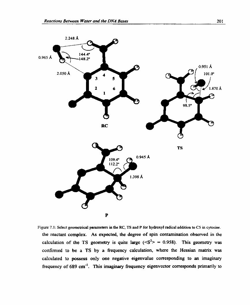
Reactions Between Water and the DNA Basa 20 1
Figure 7.1 : Select geometrical parameters in the RC, TS and P for hydroxyl radical addition to CS in cytosine.
the reactant complex. A s expected, the degree of spin contamination observed in the
calculation of the TS geometry is quite large (es2> = 0.958). This geometry was
confirmed to be a TS by a fiequency calculation, where the Hessian matrix was
calculated to possess o d y one negative eigenvaiue correspondhg to an imaginary
fkequency of 689 cm". This imaginary fiequency eigenvector corresponds primarily to

Reactions Beîween Water and the DNA Bases 202 - - - -
motion of O and C5 towards each other. The r(CS0) value in the h a l product is 1.398 A and the CS position is located farther out of the molecular plane (L(C5C6NIC2) =
-27.6"). The hydroxyl group has reoriented slightly such that the hydrogen is directed
towards the center of the molecular ring. The geornetry optimization performed for P
also suffers from a smail amount of spin contamination (<s% = 0.765).
7.2.1.3 Reaction Barrier Height
Figure 7.2 displays the reaction profile for hydroxyl addition to cytosine at C5
obtained through single-point calculations (on UHF/6-3 lG(d,p) geometries) with the 6-
31 lG(2dCp) basis set and the UHF, MP2 and B3LYP methods. The barrier heights
calculated, relative to the RC at the respective level of theory, are also given in the figure.
Table 7.1 displays the relative energy for the geometries along the reaction path (with
respect to the isolated reactants at the same level of theory) and the degree of spin
contamination in the single-point calculations.
Figure 7.2 conveys that the energy of the RC is 7.5 kcaVmol lower than that of the
separated reactants at the UHF Ievel. Despite the fact that M e spin contamination is
exhibited in the UHF single-point calculation (0.756), the projection of contarninating
spin States through the PUHF method leads to a greater lowering of the energy (9.5
kcallmol). MP2, PMP2 and B3LYP al1 predict an even greater lowering in the energy of
the RC relative to the isolated reactants (approximately 10.5 kcal.mo1). The stability
imposed b y the configuration involving hydrogen bonds relative to the iso lated reactants
is clearly seen.
The transition barrier predicted by HF theory (25.8 kcavmol) is extremely large
relative to the other barriers, as expected since this method is well known to drastically
overestimate barrier heights. The barrier calculated with MP2 (13.5 kcaWmo1) is
approximately half of the value obtained with UHF, which clearly displays the
importance of electron correlation. The spin contamination in the TS energy calculations
at these levels of theory is quite large (0.957), which is not surpnsing due to the
implementation of the unrestricted formalism. PUHF lowers the UHF barrier fkom 25.8
to 16.0 kcavmol, while the barrier predicted with PMP2 (4.7 kcavmol) is also much
smaller than that obtained with MP2 (13.5 kcdmol). The difference between the MP2

Reactions Between Water and the DNA Bases 203
RC TS Reaction Coordinate
Figure 7.2: Energetics for hydroxyl radical addition to cytosine at CS.
and PMP2 barrier heights is in agreement with previous suggestions that spin
contamination leads to an overestimation of MP2 banien by up to 10 kcavmol. l 5 PMP2
practically eliminates any spin contamination in the transition state (eZ> = 0.759). The
B3LYP barrier (2.5 kcavmol) is half the size of the value predicted by PMP2 and the spin
contamination of the TS (0.769) is relatively small.
Although B3LYP is the most widely used DFT functional combination in recent
theoretical studies, it is of interest to examine the bamier heights predicted with
alternative fûnctionals. The UHF/6-3 lG(d,p) surface was used to perforrn single-point
calculations on the RC and TS with a variety of functionals and these results are
displayed in Table 7.2. Barrier heights were calculated with the LYP, P86 and PW91
correlation functionals in combination with the B and B3 exchange functionals. From
Table 7.2, it can be seen that al1 "pure" DFT functionals examined predict negative
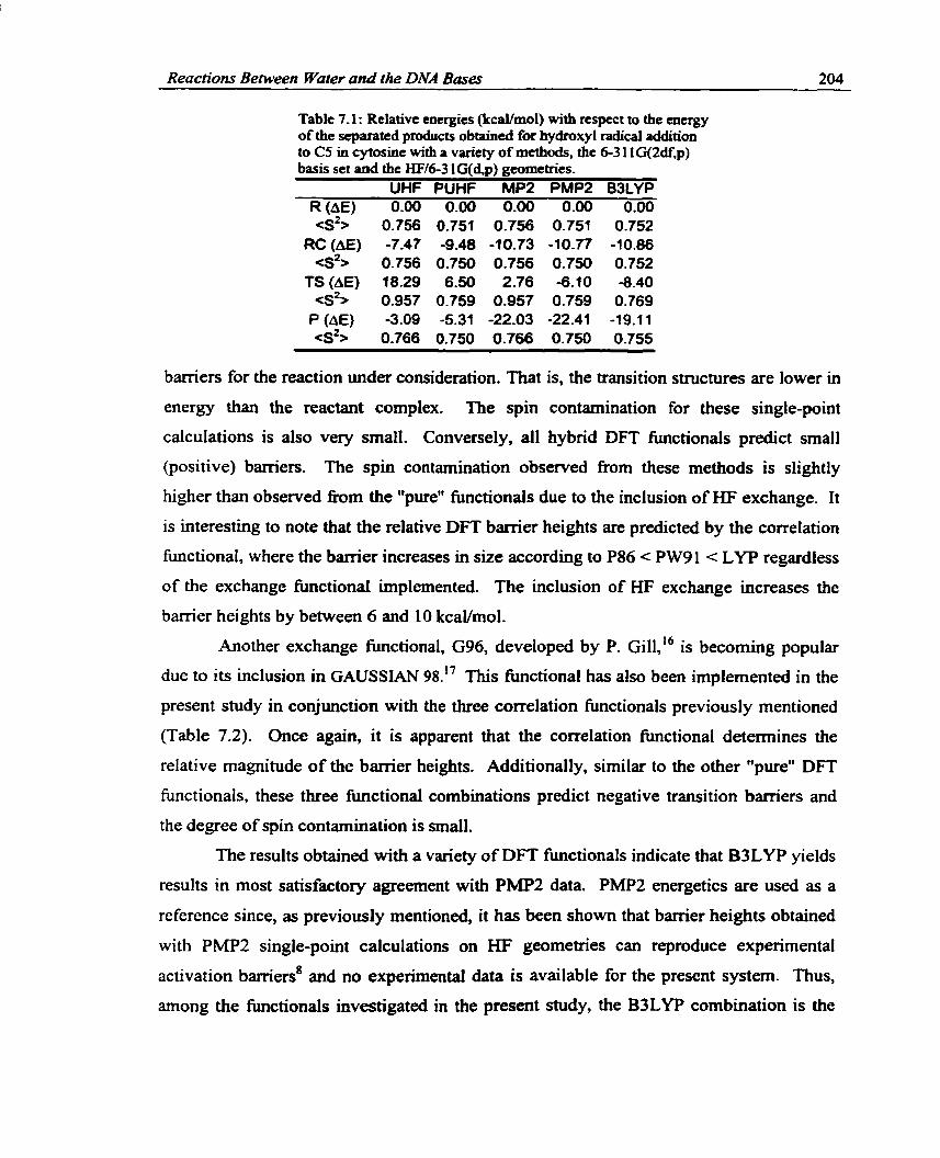
Reactions Between Water and the DNA Bases 204
Table 7.1 : Relative cnergies (kcdmol) with respect to the encrgy of the separatcd products obtained for hydroxyl radical addition to CS in cytosine wirh a varicty of methods, the 6-3 1 lG(2df,p) basis set and the KM-3 lG(4p) geometries.
UHF PUHF MP2 PMP2 B3LYP R (AE) 0.00 0.00 0.00 0.00 0.00 CS'> 0.756 0.751 0.756 0.751 0.752
RC (AE) -7.47 -9.48 -10.73 -10.77 -10.86 es2> 0.756 0.750 0.756 0.750 0.752
f S (AE) 18.29 6.50 2.76 -6.10 -8.40 G 2 > 0.957 0.759 0.957 0.759 0.769
P (A€) -3.09 -5.31 -22.03 -22.41 -19.11 <s2> 0.766 0.750 0.766 0.750 0.755
bamiers for the reaction under consideration. That is, the transition structures are lower in
energy than the reactant complex. The spin contamination for these single-point
calculations is also very small. Conversely, al1 hybnd DFT functionals predict small
(positive) barriers. The spin contamination observed nom these methods is slightly
higher than observed h m the "pure" hctionals due to the inclusion of HF exchange. It
is interesting to note that the relative DFT barrier heights are predicted by the correlation
fùnctional, where the barrier increases in size according to P86 c PW91 c LYP regardless
of the exchange bctional implemented. The inclusion of HF exchange increases the
barrier heights by between 6 and 10 kcaWmo1.
Another exchange functional, G96, developed by P. ~ i 1 1 , ' ~ is becoming popular
due to its inclusion in GAUSSUW 98." This functional has also been implemented in the
present study in conjunction with the three correlation functionals previously mentioned
(Table 7.2). Once again, it is apparent that the correlation fiinctionai determines the
relative magnitude of the barrier heights. Additionally, similar to the other "pure" DFT
functionals, these three fûnctional combinations predict negative transition barriers and
the degree of spin contamination is small.
The results obtained with a variety of DFT functionals indicate that B3LYP yields
results in most satisfactory agreement with PMP2 data. PMP2 energetics are used as a
reference since, as previously mentioned, it has been s h o w that banier heights obtained
with PMP2 single-point calculations on W geometries can reproduce expenmental
activation barriers' and no experimental data is available for the present system. Thus,
arnong the functionals investigated in the present shidy, the B3LYP combination is the

Reactions Beiween Water and the DNA Bases 205
Table 7-2: Banicr heights (kcaYmol) for the reaction of cytosine with the hydroxyl radical obtained with a variety of DFï fùnctionals, the 6-3 1 lG(2df,p) basis set and the
Fwic tional BLYP BP86 BPW91 B3LYP B3P86 B3PW91 G96LYP G96P86 G96PW91
Height - -3.7 -9.4 -4.8 2.5 0.5 1.6
-3 -7 -6.0 -4.9
most appropriate for the examination of reactions between water and various DNA bases.
The small barriet for the addition of a hydroxyl radical to CS in cytosine and the fact that
this reaction is predicted with high-level calculations to be exothermic should be noted.
In order to obtain more information about the possible reaction mechanism upon
irradiation of single crystais of cytosine monohydrate, alternative mechanisms must also
be considered.
7.2.2 Mechanism for Radiation Drrmage in Cflosine Monohydrate Crystais
In Chapter Four, three mechanisms for radiation damage in cytosine monohydrate
crystals were discussed. In summary, the first mechanism, which involved hydrogen
transfer between two cytosine units and generated the N1-dehydrogenated and N3-
hydrogenated radicals, was eliminated since the calculated HFCCs for these products did
not match the experimentally reported values. Instead, the calculated HFCCs predicted
the formation of a net CS-hydroxylated cytosine radical and therefore the darnage
mechanisms that were subsequently discussed involved the surrounding water molecules
in the cytosine monohydrate crystals. The second mechanism discussed in Chapter Four
(summarized by Equation 7.1) cos& 207 kcaVmol in the h t step and gains 149 kcaVmol
in the second step.
The third possible mechanism (sumrnarized by Equation 7.2) costs 298 kcaVmol in the

Reactions Beîween Water and the DNA Bases 206
k t step and releases 240 kcaVmol in the second step.
H 2 0 + C + C + H20' + C + CL + C(C5-hydroxylated) + C(N3-hydrogenafed) (7.2)
It was detennined in Chapter Four that the mechanism portrayed in Equation 7.1
is most likely to occur since cytosine is much larger than water and therefore a majority
of the ionizations are expected to occur on the cytosine moiety. Additionally, this
reaction has a lower energy cost for the h t step. However, it was also noted that
hydroxyl radical addition has been speculated to lead to strand breakdg and the lack of
detection of hydroxyl radicals in the DNA hydration layer has been postulated to be due
to radical addition to the bases.19 The small barrier heights predicted at the highest level
of theory employed in the present study for hydroxyl radical addition to C5 indicate that
this reaction is feasible. However, more work must be performed in order to eliminate
the possibility that the reaction outlined in Equation 7.1 occurs. As a fint approximation
to investigate this damage mechanism, the reaction between water and a cytosine cation
was investigated.
7.2.2. l Water Addnton to the Cytosine Ccrtron
Figure 7.3 displays the geometries calculated at the UHF/6-3 lG(d,p) level for the
RC, TS and product complex (PC) for water addition to C5 in the cytosine cation. in the
RC, the water oxygen is involved in hydrogen bonding with one of the arnino hydrogens
where r(N4H-0) equals 1.939 A. At this arrangement, the water oxygen is 3.43 1 A away
fiom the CS position. The spin contamination in the calculation of this structure is quite
large (<s2> = 0.9 1 7).
The C50 (1 -626 A) and OH bond distances (0.956 A) are lengthened in the TS
fiom those expected in an isolated CS-hydroxylated cytosine radical (1.398 A) and a
water molecule (0.943 A), respectively. The second OH bond length is stretched fiom
that calculated for an isolated water molecule to 1.438 A in the transition state for water
addition. Due to the interaction between the oxygen and the CS position in the cytosine
cation, slight pucicering is exhibited at the CS position in the transition state and the
hydrogen at CS is notably displaced h m the molecular plane. The spin contamination
observed in the UHF/6-3lG(d,p) calculation of the transition state (es2> = 0.89) is
smaller than that observed in the calculation of the transition state for hydroxyl radical

Reactions Between Wuter und the DNA Bases 207
Figure 7.3: Select geometrical parameters in the RC, TS and PC for water addition to the cytosine cation at C5.
addition to a neutral cytosine molecule (CS*> = 0.958). n i e geometry was confirmed to
be a TS through a frequency analysis, which predicted one imaginary fkequency (2310
cm-'), primarily corresponding to motion of H away fiom OH and O towards CS.

Reactions Beîween Wuter and the DNA Bases 208
The optimized structure for the product complex (PC, Figure 7.3) obtained
through a HF calculation, which does not suf3er £rom spin contamination, places the
hydrogen nucleus 4.488 A fiom the cytosine N3 nucleus, where the HN3C2 angle equals
149.5". The CS0 and OH bond lengths in the product complex (1.376 and 0.947 A) differ siightly fiom those obtained for an isolated CS-hydroxylated radical (1 -398 and
0.945 A). These differences may reflet variances in the molecular environment, such as
charge. Examination of the charge distribution in the PC indicates that the positive
charge resides exclusively on the cytosine portion of the complex. This indicates that the
hydrogen nucleus removed h m the cytosine by-product is in reality a hydrogen atom.
This is M e r confirmeci by the spin distribution, which thmugh high-level (B3LYP and
PMP2) calculations was determined to reside solely at the hydrogen nucleus. Re-
examination of the TS reveals that a hydrogen atom is leaving as a hydroxyl radical adds
to the cytosine cation. A B3LYP/6-31 lG(2dCp) single-point calculation indicates that
the spin and charge on the leaving hydrogen in the TS is 0.53 and 0.19, respectively.
Some spin was aiso calculated to reside on the water oxygen (-0.07) and CS in cytosine
(0.23). A PMPS single-point calculation using the sarne basis set indicates that the spin
and the charge on the leaving hydrogen are 0.80 and 0.08, respectively. Spin was aiso
calculated to be located at the water oxygen (-0.31) and the cytosine CS position (0.42),
which is quite different f?om that obtained with B3LYP. The PMP2 calculations clearly
indicate that as water adds to the cytosine cation, a concerted process occurs where a
bond is formed between the hydroxyl oxygen and CS in cytosine and the water OH bond
breaks yielding a hydrogen atom. Thus even in the TS, the leaving p u p is a hydrogen
atom, rather than a proton, and net hydroxyl radical addition to the cytosine cation is
occurring. From this discussion, it could be argued that since a hydrogen atom is
generated rather than a proton, the reaction between water and the cytosine cation is
unlikely to occur.
An explanation for why a hydrogen atom rather than a proton is leaving the
cytosine by-product in the reaction under examination can be found through
consideration of the ionization potentials of the species involved. In particular, the IP of
a hydrogen atom (calculated at the B3LYPI6-3 1 lG(Zdf,p) level) is 3 15.1 kcailmol.
Alternative1 y, the IP of the cytosine CS-hydroxylated radical (calculated at the same level

Reactions Behveen Water and the DNA Bases 209 ---
of theory with the HW6-31G(d,p) geometries) is 148.0 kcailmol. Therefore, a proton
cannot be expected as one of the products in the gas-phase reaction between the cytosine
cation and water, since the IP of a hydrogen atom is over twice the size of the IP of the
remaining radical. Therefore, in order to fom the net CS-hydroxylated radical fkom
water addition to the cytosine cation, an additional step must occur which involves
electron capture by the cytosine by-product.
The reaction between water and the cytosine cation emphasizes the importance of
hydroxyl radical attack, since in reality net hydroxyl radical addition to the cytosine
cation was observed in the reaction discussed above. The barriers caiculated through
B3LYP and PMP2 single-point calculations (66.5 and 65.6 kcaVmol, respectively) for the
reaction between water and the cytosine cation are much larger than those determined at
the same levels of theory for hydroxyl radical addition to neutral cytosine (2.5 and 4.7
kcavmol). Therefore, these results hint that hydroxyl radical addition to neutral cytosine
is a more favorable and simplistic pathway for the formation of the net CS-hydroxylated
radical than water addition to the cytosine cation, which involves hydrogen atom loss
followed by electron gain. It is important to stress that the results presented within
correspond to a gas-phase reaction between water and the cytosine cation. The more
complex problern of the reaction that occws in single crystals has not been solved. In
single crystals, the situation is quite different due to complex hydrogen bonding schemes.
Therefore, in single crystals proton transfer cm occur which may assist the reaction
between water and the cytosine cation. Thus, although the results presented within
indicate that hydroxyl radical addition is the most favorable reaction out of the two
considered, this preliminary study cannot be taken as an accurate prediction of results
expected in the solid state. More complex calculations that include a larger part of the
crystal environment, for example additional water andor cytosine molecules, are required
in order ro eliminate the reaction between water and a cytosine cation as a radiation
damage pathway in cytosine monohydrate crystals.
7.L3 The Reaction Profile for Uydroxyl Radical Addition to C6 in C~ytosine
Since it appears that hydroxyl radical addition to neutral cytosine is a feasible
means to generate the net CS-hydroxylated cytosine radical, it is of interest to investigate
hydroxyl radical attack at the C6 position. This is important since hydroxyl radical

Reactions Benveen Water and the DNA Bases 210
addition is believed to predominantly occur across the C5C6 double bond and
investigating both reactions wiIl lead to information about the favored site for hydroxyl
radical addition to cytosine. Additiondy, an experimental coupling left unassigneci in
single crystals of cytosine monohydrate was noted to be not unlike that calculated for the
C6-hydroxylated adduct, indicating that perhaps this radical is also fomed. Early studies
concluded that hydroxyl radical addition to cytosine, as well as to uracil, predominantly
occurs at ~ 5 . ~ ' More recent ESR spin-trapping studies detemine the production ratio of
these two radicals through investigating the ratio of the CS to Cd radical spin-trapped
products via computer simulations. It was determined that the ratio of CS:C6 products is
1 : 1.3 in a neutral solution of 2'-deoxycytidine.) However, this ratio may not accurately
reflect the initial ratio of attack since the trapping rate of the two radicals may be
different. Questions also aise in the use of computer simulations to determine the
relative abundance of two radicals whose spectra are highly similar, since changing the
relative abundance of each radical may have little effect on the appearance of the spectra.
P a alternative technique to detennine the ratio of CS to Cd-hydroxylated base radicals
involves monitoring the rate at which the generated radicals oxidize NNNW-tetramethyl-
p-phenylenediamine or reduce tetranitr~rnethane.~' This method is favorable since the
CS-hydroxylated product is strongly reducing, while the Cd-hydroxylated radical is
strongly oxidizing. Thus, the oxidizing and reducing rates can be used to detennine the
relative abundance of these radicals. With this technique, the C5C6 ratio was predicted
to be 8.7:l for cytosine. The results obtained fiom the calculations will pr im~ily be
compared to those obtained fiom the reduction and oxidation properties of the
hydroxylated radicals since these were specifically obtained for cytosine (the
computational mode1 system), rather than species that include a sugar group.
7-2.3.1 Geometries and Reacfion Bamœer Heights
The reactant complex for hydroxyl radical addition to C6 in cytosine is identical
to that discussed for addition to the CS position. The corresponding TS and P, calculated
at the UHFl6-3 lG(d,p) level, are displayed in Figure 7.4. The majority of the molecular
ring is planar in the TS with the exception of the C6 position, which lies 10.2" out of the
plane fonned by N1, C2 and N3. This distortion is similar to that observed in the
corresponding transition state for addition to CS, where the CS position was displaced by

Reactions Beîween Water and the DNA Bases 21 1
Figure 7.4: Select gmrnetical parameters in the TS and P for hydroxyl radical addition to C6 in cytosine.
13.3"- The C60 bond length in the transition state is equal to 1.896 A, which is 0.026 A longer than that calculated for the equivalent CS centered transition state. Signifiant
spin contamination is exhibited in the calculation which optimized the TS geometry
(<s2> = 0.889). The hydrogen in the hydroxyl group is orientated towards the N3
position in cytosine, providing a possible explanation for the similar addition complex
between the CS and C6 centered transition states. One imaginary frequency was obtained
for this geometry of 801 cm-', which is larger than that calculated for the C5 TS (689
cm-' 1. The hydroxyl hydrogen remains directed towards N3 in the Cd-hydroxylated
radical and the C60 bond length is 1.397 A, which is identical to the bond length in the
corresponding CS adduct. Sirnilar to the TS, considerable spin contamination is present
in the calculation of this geometry (e2> = 0.868). Unlike the optimized geometry for
the CS-hydroxylated radical, al1 of the ring atoms in the C6 adduct remain in the same
plane and the hydrogen and hydroxyl group at C6 are evenly distributed on either side.
Single-point calculations were perfonned on the TS and P with the 6-3 1 lG(Zdf,p)
basis set and the MP2 and B3LYP methods. As discussed for the previous reactions, the
MP2 method involves a high degree of spin contamination in calculations on transition

Reactions Between Water and the DNA Bases 212
states, where the eigenvalue of 4% was detennined to be 0.981 for the TS under
discussion. Therefore, ody the PMPZ and B3LYP barrier heights will be discussed,
where the eigenvalues of 4% were calculated to be 0.767 and 0.774 in the TS
calculations, respectively. The PMP2 and B3LYP barriers for hydroxyl radical addition
to C6 in cytosine are 6.2 and 4.4 kcaVmo1, respectively.
7.2.3.3 Cornpurison of Wydroxyl Addirion to C5 and C6 in Cflosine
Figure 7.5 compares the energetics for hydroxyl radical addition to the C5 and C6
positions in cytosine relative to the energy of the isolated reactants. A more complete
search of the potential energy surface for this reaction may reveal different RCs for these
two processes. The bamier heights calculated with PMPZ are both higher than the
corresponding heights calculated with DFT (B3LYP). However, the relative energies for
CS versus C6 addition are in agreement for both levels of theory. PMP2 predicts
hydroxyl radical addition to the C6 position to have a larger barrier than addition to CS
--- C6(PMP2) - CS(PMP2) - - - - - - C6(B3 LW) ---- C5(83LYP)
RC TS Reaction Coordinate
Figure 7.5: Energetics for hydroxyl radical addition to cytosine calculated with MP2 and B3LYP
Reactiorts Between Water and the DNA Bases 213 - -- - - -
by 1.5 k c ~ o l . B3LYP predicts the C6 addition barrier to be 1.9 kcaVmol higher than
the C5 barrier. This illustrates the predictive power the B3LYP fûnctional possesses
despite the fact that the barriers are estirnateci to be lower than the PMP2 vahes.
Additionally, both B3LYP and PMP2 predict the C6-hydroxylated product to be lower in
energy (by 1.5 and 2.5 kcavmol, respectively). Thus, both PMP2 and B3LYP single-
point calcuIations inàicate that the CS-hydroxylated radical is favored kinetically and the
Cd- hydroxylated product is favored thennodynamically . Due to the magnitude of the transition barrier heights and the relative stability of
the products, a mixture of the CS and Cd-hydroxylated radicals is expected, which was
observed experimentally. However, since the banier for formation of the C5-
hydroxylated radical is approxirnately 2 kcavmol lower in energy than that for formation
of the Co-hydroxylated barrier, a predominant attack at the CS position c m be
unders tood.
7.2.4 Summary of Cflosine Reacîiins
The present section discussed the reactions between products generated by the
irradiation of cytosine and water. Investigations of hydroxyl radical addition to cytosine
and water addition to the cytosine cation provided information about the mechanism of
radiation darnage to cytosine in the gas phase. In particular, prelirninary results indicate
that in the gas phase, net hydroxyl radical addition to neutral cytosine is the most feasible
reaction mechanism for formation of the cytosine CS-hydroxylated product. Comparison
of theoretical and experimental HFCCs (Chapter Four) determined that the cytosine C5-
hydroxylated radical is formed in these crystals. However, it was also speculated that
addition rnight occur at the Cd position since one experimental coupling left unassigned
was sirnilar to that calculated for the Cd-hydroxylated radical. Comparison of the
calculated barrier heights and the relative stability of the products led to the conclusion
that the CS-hydroxylated product is favored kinetically, while the C6-hydroxylated
product is favored thermodynarnically. Thus, the calculations support predictions that
both hydroxylated products can be fomed when hydroxyl radicals attack neutral
cytosine.

Reactions Between Water and the DNA Bues 214
7.3 Hydroxyl Radicuî Addition to Urucil
It is of interest to investigate the barriers for hydroxyl radical addition in other
DNA bases to obtain more information about the effects of the surrounding water on the
entire DNA strand. Additionally, it is intriguing to determine whether theoretical
techniques can reproduce differences observed experirnentally regarding the site
specificity of hydroxyl radical addition. Reactions between uracil and products generated
from irradiated water have been investigated to a great extent. In particular, many studies
have investigated the subsequent reactions of these by-products such as the formation of
sugar radical^^^ and subsequent strand breaks. 2324 The reactivity between uracil and
hydroxyl radicals is expected to be similar to cytosine, where hydroxyl radicals add to the
CSC6 double bond. ESR spin-trapping studies predict the ratio of C5 to C6 addition
products to be 1:2 for 2'-deoxyuridine, compared to the value of 1: 1.3 previously
discussed for 2'-deoxycytidine.' Through examination of redox properties, other studies
have indicated that addition to the CS position dominates in uracil, 1,3-dimethyluracil and
poly(U) in a 4-51, 4: 1 and 3:l ratio, respectively.zO Recall fkom Section 7.2.4 that the
CSC6 ratio for cytosine was determined to be 8.7:1, which indicates an increase in the
production of the Cd-hydroxylated radical in uracil relative to cytosine. Hydroxyl radical
addition to uracil will be discussed in the foilowing section to determine if these
differences c m be explained and to reveal more information about hydroxyl radical
addition to the nucleobases.
7.3.1 Geometnès
Unlike the cytosine hydroxyl addition reactions, different reactant complexes
were obtained at the HF/6-31G(d,p) level for uracil depending on the addition site. The
RC for hydroxyl radical addition to CS in uracil (Figure 7.6) involves interactions
between the hydrogen and oxygen in the hydroxyl radical and the 0 4 and N3H positions
in uracil, respectively. The HO4 and 0-HN3 bond lengths are 1.997 and 2.267 A, respectively, indicaîing stronger interactions between the hydroxyl group and the 0 4
position in uracil. The reactant complex for hydroxyl radical addition to C6 (Figure 7.7)
involves interactions between the hydrogen and oxygen in the hydroxyl group and the 0 2
and NlH positions in uracil, respectively. The HO2 and O-HNl bond lengths in this
complex are equal to 2.013 and 2.193 respectively. The calculateci eigenvalue of <s2>

Reactions Berneen Water and the DNA Bases 215
Figure 7.6: Select geometrical parameters for the RC, TS and P for hydroxyl radical addition to C5 Ui uracil.
is 0.755 for both addition complexes, and both are planar. Additionally, the bond length
in the hydroxyl radical in both complexes (0.962 A) is slightly elongated fiom that found
in an isolated hydroxyl radical (0.955 A) due to the interactions with uracil.
The transition states for hydmxyl radical addition to the CS and C6 position in
uracil explain the observed diffcrences in the reactant complexes. The CS centered TS
possesses a C50 bond length of 1.877 A and the hydrogen is directed away from the

Reactions Between Water and the DNA Bases 216
Figwe 7.7: Select parameters for the RC, TS and P for hydroxyl radical addition to C6 in uracil.
molecular ring towards the 0 4 center. This orientation and the relatively close proximity
of 0 4 and the hydroxyl hydrogen (2.690 A) explain interactions observed in the addition
complex between these two atoms. The CS position is displaced slightly (12.4O) fkorn a
molecular plane fomed by C4, N3 and C2. The C6 centered TS possesses a C60 bond
length of 1.905 A, which is longer than that observed for the C5 TS. This trend is sirnilar

Reactions Between Water and the DIVA Bases 217
to that observed for cytosine. The C6 position in the uracil TS is located out of the
molecular plane formed by the 0 t h ring atoms (by approximately 1 1"). A greater degree
of spin contamination was obtained in the calculations of the CS TS (<s2> = 0.998) than
the C6 TS (sS = 0.883). Both geometries were confimied to be TSs through a
fiequency analysis where the imaginary fiequencies were deteftnil3ed to be 696 (C5
addition) and 731 cm" (C6 addition).
The CS-hydroxylated radical is distorted to a greater extent than the
corresponding TS, where the C6 and CS positions are located on either side of the
molecular plane formed by NI, C2, N3 and C4. The CS0 bond leng! is 1.382 A and the
hydroxyl hydrogen is directed towards 04 where these two atoms are separated by 2.224
A. n i e orientation of the hydroxyl group at CS is slightly different nom that observed in
the TS, but illustrates the interaction between the hydrogen in this group and 04. A small
degree of spin contamination was exhibited in this geometry optimization (es2> = 0.763).
The C6-hydroxylated product exhibits less puckering than the C5 product, as observed
for the cytosine radicals. The Cd position is displaced slightly out of the molecular plane
@y 7.4" with respect to the plane formed by NI, C2 and N3), the C60 bond length is
1.395 A and the eigenvalue of <s2> is 0.813, which is larger than that calculated for the
C5 product.
7.3.2 Reactron Barrier Heights
Figure 7.8 compares the transition state barriers for hydroxyl radical addition to
the CS and C6 positions in uracil obtained through B3LYP/6-3 1 1 G(2df,p) single-point
calculations on the HW6-3 1 G(d,p) geometries. At this level of theory, the C5 and C6
RCs lead to a lowerïng of the energy by 9.1 and 9.9 kcaVmol with respect to the energy
of the isolated reactants. The activation barriers for the two uracil addition reactions are
very similar, where the barrier for C6 addition is 1 kcaVmol larger than that for C5
addition. Figure 7.8 also illustrates that the C6-hydroxylated product is 2.7 kcaVmol
lower in energy than the comesponding CS radical at this level of theory. The spin
contamination in the energy calculations was relatively small, where the largest degree of
contamination was observed for the transition states (CS% = 0.77).
The reaction profiles for hydroxyl radical addition to uracil can be compared to

Reacdons Beiween Water and the DNA Bases 218
RC TS Reac tion Coordinate
Figure 7.8: Relative energetics for hydroxyt radical addition to uracil calculated with B3LW.
those calculated for cytosine (Figures 7.8 and 7.5, respectively). One of the major
differences in the two sets of profiles is that two different addition complexes were found
for hydroxyl radical addition to uracil, while only one was found for addition to cytosine.
However, as previously mentioned, a more complete search may also reveal two different
RCs for the cytosine reactions. Similarities in the two reaction profiles also exist. First,
the RCs calculated for both uracil and cytosine lead to a lowering of the energy relative to
the isoIated reactants by approximately 10 kcal/mol. Secondly, the banier for addition to
the C6 position was determineci to be higher in both cytosine and uracil (by 1.9 and 1.0
kcal/mol, respectively). Finally, the Cd-hydroxylated product was determined to be
lower in energy than the corresponding CS radical in both cytosine and uracil (by 1.5 and
2.7 kcavmol, respectively). Thus, the différence in the transition barriers for the
formation of the two products fomed by hydroxyl radical addition is smaller for uracil
than for cytosine and the energy diffennce between the products is geater. This implies
that there is a greater preference for addition to C6 in uracil than in cytosine, which is

Reactions Between Water and the DNA Bases 219 - - - -
clearly seen by comparing the ratio of product formation determined experimentally for
uracil (CS:C6 4 5 1 ) and cytosine (8.7: 1) by examining the redox properties of the radical
products.
The trend of an increase in the production of the Cd-hydroxylated product in
uracil relative to cytosine is also observed h m the CS:C6 ratio predicted fiom ESR spin-
trapping studies for 2'4eoxyuridine (1 :2) and 2'deoxycytidine (1 : 1.3), although the
relative yield is reversed. One possible explanation for the difference in the predominant
site of attack detemiined for ESR spin-trapping studies (C6) and for the calculations (CS)
is the mode1 system employed. For example, the ESR spin-trapping studies were
performed on 2'-deoxyuridine, but the calculations were perfomed on uracil. The
discrepancy arises since a hydrogen bond was calculated to exist in the RC between the
hydroxyl radical and the 0 2 and the hydrogen at the N1 position in uracil. This is
problematic since in 2'-deoxyuridine, a sugar group replaces the hydrogen at NI. Thus,
hydrogen bonding of the hydroxyl radical to the N1 hydrogen cannot occur.
Additionally, the bulky sugar group may prohibit the hydroxyl radical from hydrogen
bonding to the 0 2 position. More work must be perfomed in order to transfer
conclusions obtained in the present study to full DNA, for example, finding alternative
RCs &or adding substituents to the N1 position of uracil.
7.3.3 Summmy of Uracil Readîons
Hydroxyl radical addition to the CS and C6 positions in uracil was discussed in
the current section. The geometries of the RC, TS and P were compared to those
previously considered for cytosine. Simïlar to cytosine, the activation bamier for the
formation of the uracil C6-hydroxylated radical is larger than that for the CS adduct,
while this product is Iower in energy than the CS analog. However, it is noted that the
bamier for addition to Cd in uracil is closer to the barrier for CS addition, than the
corresponding barriers in cytosine. Additionally, the C6-hydroxylated uracil radical is
more stable with respect to the CS uracil radical, bar, the Cd-hydroxylated cytosine
radical is to the corresponding CS adduct. This Somation was used to conclude that
there exists a greater preference for C6 addition in uracil than in cytosine. This
conclusion is supported by experiments that studied the redox properties of the radical
products, as well as ESR spin-trapping investigations.

Reactions Between Water and the DNA Buses 220 - - - - . - - - - - - -
7.4 Uydroxyl Radical Addition to Thymine
Hydroxyl radical addition to thymine has been noted to be different than addition
to either uracil or cytosine. The replacement of a hydrogen at the CS position in uracil
with a methyl group in thymine results in two main differences. First, hydroxyl radicals
can abstract hydrogen atoms fiom the methyl group in thymine to form the methyl-
dehydrogenated radical product. The formation of this allylic radical has been shown to
lead to cross-links between thymine and the arnino acid tyrosine.2b Secondly, it has been
noted that the preference of radical attack on the C5 and C6 centers is altered relative to
the attack observed in cytosine and uracil. ESR spin-trapping studies predict that the
ratio of C5:C6 products will be 2: 1 in thymidine, compared to 1:2 for 2'-deoxyuridine,
indicating an increase in the number of attacks at the carbon to which the methyl group is
attached.' Other studies predict that the methyl group leads to a decrease in the number
of attacks at the CS position. For example, the CS:C6 ratio determined by studying the
redox properties of the radical products changes fiom 4.5: 1 to 2: 1 when a rnethyt group is
added to CS in uracil to fom thymine.24 Thus, it is of interest to investigate hydroxyl
radical addition in attempts to clarify some of these discrepancies and to determine if
differences exist between uracil and thymine due to the replacement of a hydrogen by a
methyl group.
7.4.2 Geomeîries
S imilar to the uracil addition reactions, unique reactant complexes were found for
the thymine CS and C6 hydroxyl radical addition reactions (Figures 7.9 and 7.10). These
reactant complexes possess very similar geometrical properties to those observed for the
uracil RCs. For example, the RC related to CS addition involves interactions between the
hydroxyl hydrogen and oxygen and the thymine 04 and N3H positions. Additionally, the
RC related to C6 addition involves interactions between the hydroxyl hydrogen and
oxygen and the thymine 0 2 and N1H positions. These hydrogen bond lengths are
indistinguishable from those discussed for the corresponding uraci 1 RCs.
The C5 addition TS exhibits slight puckering at the CS position, while the
remainder of the ring atoms are in the sarne plane. The CS0 bond distance is 1.906 A, which is slightly iarger than the CS0 distances observed in the corresponding uracil and
cytosine transition States. The hydrogen in the hydroxyl group is onentated towards 0 4

Reactions Between Water and the DNA Bases 22 1
0.961 A
Figure 7.9: Select geometrical parameters in the RC, TS and P for hydroxyl radical addition to CS in thymine.
as predicted fiom the RC, where the HO4 distance is 2.682 A. The rnethyl group is
reoriented slightly fkom an eciipsed conformation with respect to the C5C6 bond in the
TS due to interactions with the hydroxyl group. The C 6 0 distance in the C6 related

Reactionr Between Water and the DNA Bases 222
transition state is 1.929 A, which is longer than that observed for the CS TS and slightly
longer than those observed for the C6 TS in uracil and cytosine. It is interesting to note
that for al1 three bases, the CO bond length for the C6 addition TS is longer than that in
the CS addition TS. The hydroxyl hydrogen is directed towards the center of the
Figure 7.10: Select geornetrical parameten in the RC, TS and P for hydroxyl radical addition to C6 in thymine.

Reactions Between Water and the DNA Bases 223
molecular ring in the C6 related TS and the C6 position is approximately IO0 out of the
molecular plane formed by the rernainder of the ring atoms. The spin contamination
exhibited for the thymine transition states is the largest discussed thus far for hydroxyl
radical addition, where the eigenvalues of 4% equal 1 .O24 and 1.01 8 for the C5 and C6
related transition states, respectively. These geometies were concluded to be TSs
through examination of the Hessian matrix which possesses one negative eigenvalue
corresponding to an imaginary frequency of 654 and 683 cm" for the CS and C6 related
TSs, respectively.
The thymine CS-hydroxylated radical possesses an orientation of the hydroxyl
group very similar to that discussed for the corresponding uracil radical. In particular, the
hydroxyl hydrogen is directed towards 04, where the HO4 distance is 2.224 A. The C50
bond length (1.390 A) is also very similar to that discussed for the uracil product. The
C6-hydroxylated thymine radical exhibits puckering at the C6 position, where this atom
is approximately 12S0 out of the plane fonned by the remainder of the ring atoms. The
corresponding uracil radical is planar. The deviation of the thymine radical fiom
planarity could be due to the methyl group at CS. The hydroxyl hydrogen is directed
towards the center of the ring in the C6-hydroxylated product and the Cd0 distance is
1.398 A. The spin contamination in the optimization of the C6 product (<s2> = 0.805) is
greater than that observed previously for both the related uracil and cytosine products and
for the thymine CS adduct (CS*> = 0.763).
7.4.2 Reaction Bar* Heighis
Figure 7.11 displays the reaction b h e r heights predicted by B3LYP/6-
3 1 lG(2dEp) single-point calculations on the HW6-3 1G(d,p) geometries. The energy of
the isolated reactants is lowered upon consideration of the RC by 8.9 and 9.9 kcaVmo1 for
the C5 and Cd addition profiles, respectively. B3LYP predicts the transition barrier for
hydroxyl radical addition to CS in thymine to be 2.4 kcaWmol, while the bamier is only
1.8 kcaYmol for C6 addition. Additionally, the C6-hydroxylated product is predicted to
be 6.2 kcaVmol lower in energy than the CS adduct.
Despite the fact that the stability observed for the RCs relative to the isolated
reactants is similar in al1 systems, several differences between the thymine reaction

Reactions Between Water and the DNA Bases 224
RC TS Reaction Coordinate
Figure 7.1 1: Relative energetics for hydroxyl radical addition to thymine calcdated with B3LYP.
profiles and those discussed for uracil and cytosine exist. Fust, the transition barriers for
C5 addition were determined to be smaller than the C6 barrier for uracil and cytosine, but
the converse was obtained for thymine. Thus, fiom a kinetic point of view the C6-
hydroxyiated product is favored rather than the CS species as determined for uracil and
cytosine. For al1 three bases, the C6-hydroxylated radical was determined to be lower in
energy than the CS product, indicating this product is favored thermodynamically.
However, for wacil and cytosine this energy difference was much smaller (2.7 and 1.6
kcavmol, respective1 y) than the di fference calculated for thymine (6.2 kcaVmo1). These
differences in the uracil, cytosine and thymine reaction profiles indicate that there exists
an even greater preference for addition to C6 in thymine, as predicted by examination of
the redox properties of the hydroxylated radicals.
As for uracil, the conclusions withh contradict ESR spin-trapping studies, which
predicted the C5 and C6 products to be formed in a 2:l ratio in thymidine, compared to
1 :2 for 2'-deo~~uridine.' Once again, discrepancies may arise due to the geometries

Reactions Between Water and the DNA Bases 225
calculated for the RCs, where the hydrogen bonding observed in the calculations cannot
occur in the expenments due to the presence of a sugar group. A more complete
investigation of substituent effects on the reaction between hydroxyl radicaIs and thymine
and complete characterization of reactant complexes are required to ciarifj. these
discrepancies.
7.4.3 Summary of TAiymine Reacnins
in the present section, the reactions involving hydroxyl radical addition to the
thymine C5C6 double bond were investigated. The geometries of the RC, TS and P were
discussed and compared to those calculated for uracil. Great similarities were observed
in the uracil and thymine geometries dong the reaction pathway. Cornparison of the
reaction barriers for C5 and C6 hydroxyl radical addition to thymine leads to conclusions
different fiom those reached for uracil and cytosine. For thymine, the Cd-hydroxylated
product is favored both kinetically (due to the lower barrier height) and
themodynamically (due to the 6.2 kcaVmol greater stability of this product relative to the
C5 adduct). The differences in the reaction profiles of thymine, uracil and cytosine
indicate that the methyl group stabilizes the TS involved in hydroxyl radical addition to
the C6 position and, therefore, addition to this position in favored. This trend is in
agreement with studies investigating the redox properties of hydroxylated base radicals.
7.5 Conclusions
The present chapter investigated reactions between water (or products generated
f?om irradiation of water) and the DNA bases. Initially a study was perfomed on
reactions involving cytosine in order to obtain more information about the mechanism of
radiation darnage in cytosine monohydrate crystals. Hydroxyl radical addition to neutral
cytosine and water addition to the cytosine cation were investigated in terms of addition
to the CS position. It was determined that for the gas-phase reactions, hydroxyl radical
addition to neutral cytosine appears to be the most feasible mechanism for the formation
of the CS-hydroxylated cytosine radical. These results are not directly transferable to
cytosine monohydrate crystals due to detailed hydrogen bonding in the crystals. More
complex mode1 systems must be used in order to determine the radiation damage
mechanism in these crystals.

Reactions Beîween Water and the DNA Bases 226
Once hydroxyl radical addition was determinecl to be the main paîhway for
hydroxylated radical formation, alternative hydroxylated products were investigated.
Forernost hydroxyl radical addition to the C6 position in cytosine was investigated to
determine if addition to this position is also feasible. The CS-hydroxylated product was
detennined to be favored kinetically (by approximately 1.9 kcal/mol) and the C6-
hydroxylated product favored therrnodynamically (by approxirnately 1.6 kcaYrno1).
Therefore, it was concluded that hydroxyl radical addition will occur to a greater extent at
the CS position, due to the 2 kcaYmo1 lower transition banier, but addition will also occur
at the C6 position due to the greater thermodynamic stability of this product.
Hydroxyl radical addition to the CS and C6 positions in uracil and thymine were
dso investigated and the results were compared to those obtained for cytosine. In uracil,
as in cytosine, it was detennined that the CS-hydroxylated product is favored kinetically
(by 1 .O kcallmol), while the C6-hydroxylated product is favored thermodynarnically (by
2.7 kcavmol). Due to energetics differences, relative to the cytosine reactions, it was
concluded that the Ca-hydroxylated product will be formed to a greater extent in uracil
than in cytosine. This trend was observed experimentally where the C5:C6 ratio was
determined to be 8.7: 1 and 4 3 1 when cytosine and uracil were exarnined, respectively,
in terms of the radical's redox properties. Alternatively, the thymine C6-hydroxylated
product was calcuiated to be favored both kinetically and thermodynamically. Since the
barrier heights were reversed relative to uracil and cytosine and the thymine C6-
hydroxylated adduct has a greater stability over the CS radical than observed for the
cytosine and uracil products, it was concluded that the methyl group in thymine leads to
favored addition to the C6 position. This conclusion is once again supported by the
experimental ratios for CS:C6 hydroxyl radical addition which were determined to be
4.51 and 2:1 for uracil and thymine, respectively. Discrepancies between these
conclusions and those obtained in alternative experimental studies were determined to be
due to differences in the mode1 systems employed and more detailed studies were
proposed.
It should be noted that these calculations were performed as an initial
investigation of the reactions between water and the DNA bases, in terms of an
optirnization at a low level of theory followed by higher level single-point calculations.

Reacriom Between Water and the DNA Bmes 227
Despite this fact, the relative energetics of the C S and C6-hydroxylated products are in
good agreement with those calculated in Chapter Four through optimization of the
geometries with DFT followed by higher level DFT single-point calculations. This
indicates that although this work represents an initial study, the results appear to be
tnistworthy. More work remains to be done, however, including calculations which
confirm the relationship between the reactant complexes, the TSs and the products.
7.6 References
Catterall, HI; Davies, M. J.; Gilbert, B. C. J . Chem. Soc. Perkin Tmns. 1992,2, 1379.
(a) Gajewski, E.; Dizdaroglu, M. Biochem. 1990, 29, 977; (b) Dizdaroglu, M.; Gajewski, E.; Reddy, P.; Margolis, S. A. Biochem. 1989,28,3625.
Davies, M. J.; Gilbert, B. C.; Hazlewood, C.; Polack, N. P. J. Chern. Soc. Perkin Tram. 1995,2, 1 3.
Hiraoka, W.; Kuwabara, M.; Sato, F.; Matsuda, A.; Ueda, T. Nucleic Acid Res. 1990, 18, 1217.
Téoule, R. Int. J . Radiat. Biol. 1987, 51, 573.
Llano, J.; Eriksson, L. A. J Phys. Chem. B 1999, in press.
Johnson, B. G.; Gonzales, C. A.; Gill, P. M. W.; Pople, J. A. Chem. Phys. Lett. 1994, 221, 100.
SekuSak, S.; Güsten, H.; Sabljic, A. J. Chem. Phys. 1995, 102,7504.
Melissas, V. S.; Truhlar, D. G. J. Phys. Chem. 1994,98,875.
10. Gonzalez, C.; McDouall, J. J. W.; Schlegel, H. B. J. Phys. Chem. 1990, 94, 7467.
1 1. McKee, M. L. J. Phys. Chem. 1993,97,10972.
12. Alvarez-Idaboy, J.; Diaz-Acosta, 1.; Vivier-Bunge, A. J. Comp. C h . 1998, 19,8 1 1.
13. (a) Martell, J. M.; Mehta, A. K.; Pacey, P. D.; Boyd, R. J. J. Phys. Chem. 1995, 99, 8661; (b) Martell, J. M.; Boyd, R. J. J. Phys. Chem. 1995,99, 13402.
14. Frisch, M. J.; Trucks, G. W.; Schlegel, H. B.; Gill, P. M. W.; Johnson, B. G.; Robb, M. A.; Cheeseman, J. R.; Keith, T. A.; Petersson, G. A.; Montgomery, J. A.;

Reactions Between Water and the DNA Bases 228
Raghavachari, K.; Ai-Laham, M. A.; Zakrzewske, V. G.; Ortiz, J. V.; Foresman, J. B.; Cioslowski, J.; Stefanov, B. B.; Nanayakkara, A.; Challacombe, M.; Peng, C. Y.; Ayala, P. Y.; Chen, W.; Wong, M. W.; Andres, J. L.; Replogle, E. S.; Gomperts, R.; Martin, RI L.; Fox, D. 1.; Binkley, J. S.; Defiees, D. J.; Baker, J.; Stewart, J. P.; Head- Gordon, M.; Gonzalez, C.; Pople, J. A. Gaussian 94 (Revision B.2); Gaussian, ïnc.: Pittsburgh, PA, 1995.
15. Gonzales, C.; Soza, C.; Schlegel, H. B. J Phys. Chem. 1989,93,2435.
16. GIIl, P. M. W. Mol. Phys. 1996,89, 433.
17. Frisch, M. J.; Trucks, G. W.; Schlegel, H. B.; Scuseria, G. E.; Robb, M. A.; Cheeseman, J. R.; Zakrzewski, V. G.; Montgomery, J. A., Jr.; Stratmann, R. E.; Burant, J. C.; Dapprich, S.; Millam, J. M.; Daniels, A. D.; Kudin, K. N.; Strain, M. C.; Farkas, O.; Tomasi, J.; Barone, V.; Cossi, M.; Carnrni, R.; Mennucci, B.; Pomeili, C. ; Adamo, C.; Clifford, S.; Ochterski, J.; Petersson, G. A.; Ayala, P. Y .; Cui, Q.; Morokuma, K.; Malick, D. K.; Rabuck, A. D.; Raghavachari, K.; Foresman, 1. B.; Ciosiowski, J.; Ortiz, J. V.; Stefanov, B. B.; Liu, G.; Liashenko, A.; Piskorz, P.; Komarorni, 1.; Gomperts, R.; Martin, R. L.; Fox, D. J.; Keith, T.; Al-Laham, M. A.; Peng, C. Y.; Nanayakkara, A.; Gonzalez, C.; Challacombe, M.; Gill, P. M. W.; Johnson, G.; Chen, W.; Wong, M. W.; Andres, J. L.; Gonzalez, C.; Head-Gordon, M.; Replogle, E. A.; Pople, J. A. Gaussian 98 (Revision A.4),Gaussian, Inc., Pittsburgh PA, 1998.
1 8. Wala, M.; Bothe, E.; Gomer, H.; Shulte-Frohlinde, D. J. Photochem. Pho~obiol. A, Chernistry 1990, 53,87.
19. (a) Becker, D.; La Vere, T.; Sevilla, M. D. Radiai. Res. 1994, 140, 123; (b) LaVere, T.; Becker, D., Sevilla, M. D. Radiat. Res. 1996,145, 673.
20. Hayon, E.; Sirnic, M. J. Am. Chem. Soc. 1973,95, 1029; Simic, M.; Hayon, E. h t . J. Radiat. Biol. 1972,22, 507.
2 1 . von S o ~ t a g , C.; Schuchmann, H.-P. Int. J Radiat. Biol. l986,49,l and references therein.
22. Hildenbrand, K.; Behrens, G.; Schulte-Frohlinde, D.; Herak, J.N. J. Chem. Soc. Perkin Trans l989,t, 283.
23. Catterall, H.; Davies, M. J.; Gilbert, B. C. J. Chem. Soc. Perkin Trans 1992,2, 1379.
24. Hildenbrand, K.; Schulte-Frohlinde, D. Int. J. Radiat. Biol. 1989,55,725.

C W T E R EIGIIT DNA Radiation Products
8.1 Introduction
The previous four chapters have discussed the effects of radiation on individual
DNA components in relation to experimental results obtained fkom single crystals of base
derivatives at low temperatures. Issues can now be addressed which question the
relevance of these studies to the identification of the radiation products in full DNA.
Earfy ESR wodc on DNA revealed that the classification of radiation products is a
difficult task. In particular, the f h t denvative of the absorption of DNA closely
resembles an extremely broad singlet, which indicates that there exists overlapping of the
spectra for each radical. This occurs since the DNA radicals are extremely sirnilar and
therefore the hyperfine couplings and g-factors are not sufficient to separate their spectra.
This chapter will discuss some of the trials and tribulations confionted by experimental
attempts to study the fiil1 DNA strand. This discussion will include a background of the
methods available to study the radiation effects on a molecule as cornplex as DNA. in
addition, the products identifieci in both early and more recent experimental work will be
analyzed and the effects of water on DNA radiation damage will be considered. Al1 of
this information, in addition to results obtained from single-crystal studies and
calculations, will be used to develop a picture of the effects of radiation on the entire
DNA strand.
8.2 Experimental Methods Avaüable to Stuày DNA
Studies have been perfomed on DNA both in the dry state and in aqueous
solutions.' Frozen aqueous solutions are often investigated and the effects of radiation on
these samples are quite complicated. Specifically, in addition to the formation of DNA
radicals upon exposure to radiation ("direct" effects), solvent (water) radicals cm be
generated. These solvent radicals can also give rise to base or sugar radicals by attacking
the DNA strand ("indirect" effects). Thus, the spectnun of an irradiated fkozen aqueous
solution is a superposition of the spectra of DNA and water radicals (primarily hydroxyl
radicals, hydrogen atoms and aqueous electrons). Since the hydroxyl radical is unstable

DNA Radiation Produc& 230 - - - - -- -
at temperatures above 1 10 K, the spectnun of fiozen aqueous solutions can be simplified
by annealing. Low temperature glasses have also been employed on occasion to
investigate full DNA. Typically these glasses are produceci using saturateci solutions of
LiCl or BeF2 in H20. The advantage of using low temperature glasses is that reactive
radicals c m be stabilized and the specificity of a reaction can be studied by caretùlly
selecting the glass-forming agent. For example, hydroxyl radicals are known to be
abundant in BeFr glasses, electrons in LiCl glasses or in the presence of strong bases
(NaOH) and hydrogen atoms in strong acids (H2S04). Lyophilized (fieeze-dried)
powders are often used to study the effects of radiation on the DNA strand, where the
powders have been prepared completely dry or with varyïng degrees of hydration. The
relative humidity (typically 76%) or the number of water molecules per nucleotide
(typically 2.5 to 1 1 ) characterize the level of hydration.
The methods discussed thus far yield a random orientation of the DNA molecuies
and therefore the resulting spectrum is composed of an overlap of the spectrurn of each
radical in al1 directions. These "powder" spectra are very broad and lack distinguishing
features. If the individual spectnim of each radical is nearly isotropic, then information
can be obtained fiom the powder spectra. However, if anisotropy exists in the hyperfine
couplings, then the resulting fine features wiI1 be smeared in the powder spectnim and
information will be lost. Ideally, single-crystal studies would be beneficial, but it is not
possible to prepare these samples for an entire DNA strand. A comrnon approach to
these problems is to use orientated fibers to study irradiated DNA, which are normally
equilibrated at 76% relative humidity. Onentated fibers have an advantage over other
methods since the spectra can be monitored at two orientations of the fiber (perpendicular
and parallel) relative to the magnetic field. This allows for the identification of some
species that may not be observable in randomly orientated sarnples.
Additional tactics used by experirnentalists investigating DNA include
implementing deuterated samples, for exarnple replacing Hz0 with DzO, to improve
spectral resolution or identim radical products. Altematively, additives cm be included
in the sample to hnprove the spectra. For exarnple, electron scavengers, most commonly
FeC13 or &Fe(CN)6], have been used to obtain vduable information about electron loss
centers. in addition, the spectnun with and without scavengers can be subtracted to yield

DNA Radiation Products 23 1
information about products formed through electron gain. Thermal anneding is also a
very usehl tool to diflerentiate between products. Annealing expeMents provide
information about lines which decay together and therefore represent one or more related
species. Altematively, information can be obtained about the relationship between the
decay of one product and the growth of another. In addition, the spectrum of DNA can
be compared to the spectra of base derivatives to identiQ products. Once a product has
been identified, its spectnim can be subtracted fiom the spectnim of full DNA for M e r
simplification. Computer simulations are often performed that use experimentally
derived parameters (for example, KFCCs) for the speculated radical products to mode1
the full DNA spectnun. Monnation is obtaùied by adjusting the parameten until a best
fit is obtained between the simulated and experimental spectra.
Through al1 of these techniques, information about the radicals generated in
irradiated DNA c m be obtained. The irnplementation of a variety of experimental
conditions allows for the determination of the dependence of radical formation on the
environment (for example, strand conformation, hydration level, Oz content). Despite
great efforts put forth by experimentalists, the exact identity of most radical products is
still unknown. However, advances have been made in the last few years and some
products have been confidently identified. The next section will be dedicated to a
discussion of the radicals identified thus far in studies on the fiil1 DNA strand.
8.3 Initial Characteritation of Radicafs Generated in DNA
8.3.1 Efectron Gain and Loss Centers
The firçt experimental work on fiil1 DNA emphasized the fornation of a radical
speculated to be denved from In these studies, the similarity between the
recorded spectra of DNA and thymine or thymidine was used to predict the formation of
a thymine radical. Salovey et al. poshilated that the detected radical possesses a fiagrnent
of the form -'C(CH3)-CHr, which would correspond to a thymine centered radical since
only thymine possesses a methyl g r ~ u ~ . ~ Work perfoxmed on DNA irradiated by
ultraviolet light confirmed that the recorded octet pattern arises fiom a radical fomed
through hydrogen atom addition to C6 in thymine [T(C6H), Figure 8.11.~ Studies
performed on orientated fibers also predicted that considerable amounts of T(C6H) is

DNA Radiation Producfi 232
T(C62r) Figure 8.1 : The f h t radical identifled in irradiatcd DNA: the thymine C6-hydrogenated radical.
formed.' The definitive identification of a thymine radical product led to the conclusion
that the thymine anion (Ty mut be initially fomed in irradiateci DNA, which was
proven shortly thereafter.6 Following these studies identifjring thymine as a damage site,
little progress was made to classi@ additional radiation products in fiil1 DNA for years,
although work continued on single crystals of base derivatives and other DNA subunits.
The model of radiation darnage to DNA was greatly enhanced through work
performed on orientatecl fibers by Gr&lund and c o w o r k e r ~ . ~ * ~ ~ ~ Radicals identified in
these studies were detennined to have ionic character with delocalized x-spin density,
and therefore were most probably base centered radicals. The radical mixture generated
in DNA was suggested to be composed of thymine (andlor cytosine) anions and guanine
(andor cytosine) cations.? The initial assumption that cytosine may also be damaged was
discardedg and the pichire of radiation darnage in DNA resulting in TL and guanine
cations (G") became known as the "two-component" model (Figure 8.2). Primarily, the
facts that the anionic radical converts to T(C6H) and that products generated Eom
cytosine anions were not observed were used to favor T- as the primary anion. The two-
Tm- GO+
Figure 8.2: The primary radical products generatcd according to the two-componcnt mode1 for DNA radiation damage.

DNA Radiation Products 233 -
component model was oflen criticized since it was believed that insacient prwf was
available to support this mechanism for DNA radiation damage.l0*" Altematively, the
evidence supporting the model continued to gyow.''
Cullis and coworkers" alluded that the two-component model for damage to
DNA seems surprising since ionizing radiation damages indiscrhinately. Thus, initial
electron gain and loss centers should include water, the phosphate group, the sugar
moiety and al1 four bases. Major cnticism of the two-component mode1 for radiation
damage in DNA arose since the spectrum assigned to TL is in poor agreement with that
obtained fiom single-crystal ~tudies. '~ In addition, the cytosine anion (CL, Figure 8.3)
yields a doublet with couplings approximately equal to those of T'. This indicates that it
will be difficult to distinguish between these two species and perhaps cytosine is also a
site for radiation damage.
C*-
Figure 8.3: The third radical identified as a major radiation damage product: the cytosine anion.
Additional support for a wider range of darnage to DNA than the sole production
of TL and G* began to appear in the literature. Bernhard and coworkers detemined that
CL is the predorninant electron gain radiation product in low temperature glasses of
oligonu~leotides'~ and that it may also be the major anion generated in DNA." Sevilla et
al? investigated the products in irradiated DNA through the use of computer analysis
and determined that CL is generated to a greater extent (77% of al1 anions) than TL
(23%) at 100 K. The use of computer simulations was later cautioned, however, since the
spectra of these two species are so similar that slight changes in the simulation input can
yield very different percentages.'3 ore evidence to support the favored formation of CL
in DNA was obtained in a study of the one-electron reduction potentials of the bases in
aqueous so~ut ions .~~ Since the rate of reduction of TL was dependent on the pH and that

DNA Radiation Products 234
of CL was not dependent on the pH, it was determined that CL is protonated. This
indicates that CL has a greater tendency to be protonateû by its base pair guanine than
thymine by adenine. This cm be unders td since the cytosine-guanine base pair takes
part in three hydrogen bonds, while the thymine-adenine base pair involves only two
hydrogen bonds. Thus, cytosine has the ability to accept one net proton and cytosine
should be the most easily reduced base in DNA. In addition, the spectrum assigneci to TL
in nondeuterated DNA samples did not change upon deuteration (specifically at CS-CH,
and C6H), indicating that some other species must be responsible for the spectrum,"
possibly C'.
Cullis et al.') examinai strand-breaks in DNA in order to determine if they occur
to a greater extent at positions next to thymine or guanine as predicted by the two-
component model. It was concluded that strand-breaks are not site-speci fic and there fore
all possible sites are darnaged in DNA. More importantly, this indicates that TL and C'
should be initially present in comparable amounts. Additional evidence suggesting that
cytosine is the predominant electron gain center was obtained fiom studies on fiozen
samples of both single and double-stranded D N A . ~ ~ It was detemiined that in single-
stranded DNA TL slightly prevails whereas in double-stranded DNA the production of
C' predorninates. The explanations for this difference include interstrand base-pairing
and base-stacking effects, which allow electrons to travel to more stable positions in
double-stranded DNA.
A debate over the site of electron loss in DNA has also appeared in the Iiterature.
This controveny was initiated when it was noted that the spectra of C* recordeci in solid-
state studies of nucleotides and nucleosides did not correspond to the spectrum recorded
in full DNA." From computer analyses, Sevilla et al. l 6 determined that over 90% of the
cations generated in DNA are centered on guanine implying that hoIe transfer fiom
adenine to guanine is complete in double-stranded DNA. Through investigations of the
strand-break specificity, Cullis et al." determined that some adenine cations could be
generated. In a more recent study, guanine end products accounted for 90% of the
eIectron loss products indicating that if holes are initially formed on adenine, or even
thymine or cytosine, they are quickly transferred to guanine." These studies support the
two-component model in that the vast majority of the cations formed in inadiated DNA

DNA Radiation Products 235 - -- - - -- -
are guanine centered. However, the results add the new dimension that it is also possible
for other (adenine, A*, Figure 8.4) cations to be forxned.
Figure 8.4: The a d d e cation, which may also be a product in hdiated DNA.
From the above studies, it can be concluded that radiation darnage in DNA is less
specific than initially assumed by the two-component model. More precisely, radical
centers on al1 four DNA bases can be expected upon irradiation of hi11 DNA. The
relative abundance of CL, TL, G* and A' in double-stranded DNA at 100 K is
approxirnately 42, 17, 38 and 3%, respectively, with no substantial amounts of the
pyrimidine cations or the purine anions.19 It is interesting to note that base anion
formation is favored over base cation formation in a 1.4:l ratio. This is reaiistic since
although equal numbers of cations anions must be initially fonned upon irradiation,
the relative degrees of radical stabilization do not have to be equivalent. However, it can
be speculated that this could also indicate that positive centers are formed elsewhere and
have been lefi undetected (for exarnple, on the sugar phosphate backbone as to be
discussed in a later sectionj.
8.3.2 Theoretical Redictions of Electron Gain and Loss Centers
More information about the specificity of electron gain and loss in DNA can be
obtained by calculating the ionization potentials and the electron affinities of the bases.
The ionization potentials have been previously caiculated with MP2 single-point
calculations on HF geometries.22 Table 8.1 compares the iPs for the nucleobases
obtained experimentally, with MP2 and with DFT (B3LYP caIculations presented in
Chapters Four and Five). It can be seen that the theoretical data is in good agreement
with the experimental results. In particular, al1 three sets of data predict the magnitude of
the ionization potential to follow the trend T > C > A > G. This indicates that an electron
is most easily removed fkom guanine. Additionally, a significant différence in the IP of

DNA Radiation Products 236
Table 8.1 : The adiabatic IPs and EAs (kcaVmol) of the DNA bases obtained at various tevels of theory and exDerimentallv.
DFT MP2 Exp. DFT Estimated T 196.0 204.2 204.6 -14.8 7.2 C 194.2 201.5 200.1 -13.8 4.8 A 182.3 188.6 190.5 -17.7 -7.2 G 171.8 176.6 179.3 -15.8 -16.7
guanine and the other three bases is noted. These results agree with expenmentai
predictions that the guanine cation is the major oxidation product in irradiated DNA.
Table 8.1 also displays adiabatic EAs calculated for the bases, since no
experimental data is available for this property. The DFT results were presented
previously in Chapters Four and Five. The "estimated EA" values were obtained by
correcting the HF Koopmanns EA by the calculated nuclear relaxation energyu The
"estimated EAs" predict negative values for the EA of adenine and guanine and positive
values for cytosine and thymine. The EA is defined as the energy required to add an
electron to a neutral molecule and calculated as the energy of the neutral molecule minus
the energy of the anion. Therefore, a negative value for the EA indicates that the anion is
higher in energy than the correspondhg neutral molecule and therefore energy is released
upon anion formation. A negative EA cannot be measured experirnentally due to the
dissociation of the anion into an electron and the neutral molecule before nuclear
relaxation. The trend in the "estimated Eh" is T > C > A > G, which is in agreement
with early studies on DNA which predicted that the thymine anion is the major reduction
product after irradiation. DFT predicted EAs were obtained by directly comparing the
energy of the neutral base with the respective base anion, which is expected to be more
reliable than the "estimated values". DFT predicts al1 bases to possess a negative EA.
Additionally, the trend predicted with DFT (C > T > G > A) is in contrast to that obtained
fiom the "estimated EAs". However, the DFT results support experimental data that
predicts cytosine to be the major reduction site in irradiated DNA. Contrary to the
difference between the IP of guanine and the IPs of the other bases, a very small
difference was calculated between the EA of cytosine and that of the other bases, in
particular thymine.
DNA Radiation Products 237 -- -- - - - - - - -- - - - -
It should be noted that d i f ise functions c m be important for the calculation of
anion geometries and E h and, thus, negative EAs may not be obtained through
calculations pedormed with larger basis sets. However, prelimuiary caiculations
performed on cytosine indicate that the cytosine anion geometry does not change
considerably upon inclusion of di f ise functions in the basis set for the heavy atoms. In
addition, through single-point calculations on this geometry with the 6-31 l+G(2d£p)
basis set, the EA was determineci to be -1.4 kcal/mol. Thus, even with large basis sets
DFT predicts the EA of cytosine to be negative. h can be speculated that the trend
calculated with DFT is however correct and therefore the DFT results support the
possibility that both cytosine and thymine anions are forrned, while the guanine cation is
the major oxidation product.
8.3.3 The Formation of Secondary Radicafs
The discussion thus far has focussed on the formation of ionic centers as the
primary radiation effectç in DNA at low ternperatures. At higher temperatures, or more
specifically those of biological systems, these ionic radicals are not expected to be stable,
but rather secondary species must be formed, which evolve fkom the ionic radical
products. The first clues to support this statement were obtained in early ESR
investigations of DNA which assigned an observed octet pattern to T(C6H). Concrete
evidence for the mechanisrn of formation of this radical was obtained by recognizing a
relationship between its growth and the decay of T'.* It was concluded that these species
are related by
T' + XH+ + T(C6H) + X (8-1)
where X represents an unknown proton source and is not restiicted to only one
~ ~ e c i e s . ~ ~ ' ~ The T(C6K) radical has been monitored in other studies as well. 13,1923
Despite the evidence for the formation of T(C6H), critics still speculated that CL is a
predominant damage site and perhaps a transfer of the anionic character fiom cytosine to
thymine, followed by protonation, can account for the high yield of T(C6H) in imadiated
DNA.'~ Evidence for this phenornenon has been obtained in CO-crystals of 1 -
methylcytosine and 5-fluorouracil (a thymine derivative). The primary radicals were
identified to be the cytosine anion and the uracil centered cation. However no cytosine

DNA Radiation Products 238
Figure 8.5: The secondary radicals identified in ESR studies on DNA in addition to T(C6H).
radicals formed by proton addition to CS or C6 were observe& but rather uracil products
evolving from the uracil anion were identified." in addition, it was later speculated that
the decay of the guanine cation is related to the growth of the product formed by
deprotonation at NI [G(Nl), Figure 8.5].'213
The radical formed via net loss of a hydrogen atom fiom the thymine methyl
group [T(CAZ)] has also been observed in highiy hydrated DNA ~amples.~' This radical
could be a proton source, but no relationship between this radical and T(C6H) was found.
In samples prepared with D20, the concentration of this radical was deterrnined to be
between 10 and 15%, although in nondeuterated sarnples its spectrum is less
pronounced.25 Evidence also exists that at 77 K, the cytosine anion is stabilized by
protonation at N3 [C(N3H)] by its guanine base pair.26 Additionally, in thymine
deuterated DNA sarnples, a deuteron has been detemined to add to the C6 position of the
cytosine anion [c(c~H)].~' These secondary radicals are displayed in Figure 8.5.
8.4 A Closer Look at DNA Radiation Products
As discussed above, progress in the identification of the radiation products in
DNA has been slow. Despite great advances in experimental techniques, only a few
products were initially identified. More specifically, the only component not under
debate is the octet assigned to the thymine C6-hydmgenated radical. Advances have
been made in the past few years, however, to identie more than two or three products in
one DNA sample. The most promising results were obtained by Hüttemann and
coworkers, in both orientated fibers2' and in randomly orientated DNA.~'~' The pnmary
conclusions fiom these studies will be summarized in the subsequent sections.
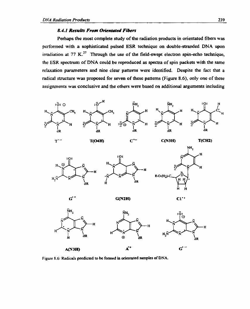
DNA Radiation Products 239 - -
8.4.1 Resula From Orientutai Fibers
Perhaps the most complete study of the radiation products in orientated fibers was
pet-fonned with a sophisticated pulsed ESR technique on double-stranded DNA upon
irradiation at 77 K." Through the use of the field-swept electron spin-echo technique,
the ESR spectnim of DNA could be reproduced as spectra of spin packets with the same
relaxation parameters and nine clear patterns were identified. Despite the fact that a
radical structure was proposed for seven of these pattems (Figure 8.6), only one of these
assignments was conclusive and the 0th- were based on additional arguments including
A(N3H) À+ G' -
Figure 8.6: Radicals predicted to bc formed in orientated simples of DNA.

DNA Radiation Products 240
simulations and mechanistic assumptions. The proposed radiation products will now be
discussed.
An observeci "doublet" (or rather, a group of related patterns differing with water
content and H/D exchange) was confïdently assigned to TL, although whether the anion
was protonated at 04 [T(04H)] could not be determined. Another component, - 7,12 previously assigned to T , was reassigned to CL, possibly protonated at N3
[C(N3H)]. This new assignment seems plausible since it rationalizes large, previously
unexplainable couplings as nitmgen interactions. G* was also determined to be present,
although it seemed unWtely that a charged species would be stable thermally. The
assignment to G* was supported since the amino-deprotonated guanine radical [C(N2 H)]
possesses a different spectrurn fiom that observed in DNA. These species were al1
discussed previously in the literature as possible damage products.
The first newly proposed radical product for orientated fibers was the radical
formed via net hydrogen atom removal from the methyl group in thymine [T(CH2)]. A
sugar radical was also speculated for this spectnrm, but this postulation was discarded
since the tenson are typical for a base radical and the spectnim of a deuterated sarnple
supported the assignment to T(CH2). A spectnun with features typical of an electron
interacting with a single nitrogen nucleus was tentatively assigned to a radical fonned via
proton loss at Cl' in the sugar moiety. In this sugar radical, the main part of the spin
density is restricted to interactions with the glycosidic nitrogen in a cytosine unit due to
the orientation of the base, the sugar moiety and the orbital at Cl' possessing the unpaired
electron. The possibility of net hydrogen loss occurring at a sugar next to the other bases
was not ruled out, although the radical assignment was made based on a s p e c t m
previously observed fiom cytidine. An assignment was made to the adenine radical
fomed via hydrogen addition to N3 [A(N3H)] based on cornparison with previous
expenmental results. However, the spectnim of this radical was not clear in full DNA
and differs from that obtained in the copolymer poly(A:U). Therefore, the assignment is
uncertain. The final spectrum addresseci was for a "singlet" previously assigned to G*
for which linle direct information could be obtained. Since G* was aiready assigned in
the s tudy under discussion, caution was advised and suggested assignments include the
guanine anion (63 or the adenine cation (A?, since the adenine anion is related to a

DNA Radiation Products 24 1
species already identified [A(N3H)]. As previously mentioned, two more components
could not be assigned due to insufficient information. Thus, this study on orientated
fiben clearly ïndicates that the damage to DNA is broader that initiaily expected since
products on al1 four bases and the sugar moiety were proposed. In addition, more work is
required in order to determine the exact identity of the radical products since structural
infonnation is difficult to obtain through the methods implemented.
8.4.2 Resufts fiom Randomly Orientated DNA Samples
The first study perf'omed on randornly orientated fibers, which detected more
than two or three ionic species, was perlormed on DNA equilibrated at various levels of
hydration, as well as on fiozen aqueous solutions.28 This experimental shidy was initially
proposed to clariQ discrepancies in the lit erature and uncondi tionally identi fi the
primary radiation products in DNA by cornparison with nucleotide patterns. In
lyophilized powders, G*, C' and TL were identified without any uncertainty for the fint
time. The spectra obtained for frozen aqueous solutions were very different from those
equilibrated at 76% relative humidity. In particular, the amount of G' is reduced
considerably in fiozen aqueous solutions. This is in agreement with previous work which
found that G* does not play a dominant role in the radiation chemistry of DNA in fkozen
aqueous solutions at 77 K." T(C6H) and T(CH2) were also assigned in this study.
A continuation of the study discussed above examined lyophilized DNA powders
in dry environments and equilibrated at 76% relative humidity. The goal of this work
was to directly analyze the DNA spectrum. More specifically, electron scavengers were
implemented rather than using results obtained fiom model systems. This approach
avoids questions associated with transferring results obtained from single crystals to fiiil
DNA and problems establishing near identical experimental conditions in the model
systems and in full DNA samples. As a result, many new radicals were identified besides
T(C6H) (Figure 8.7). A "triplet", previously discussed to be composed of more than one
individual spectnim and partially assigned to G: was assigned to the cytosine radical
formed via net hydrogen atom addition to the arnino group [C(N4H)]. This is the first
time this radical has been proposed for DNA. However, the hiplet assigneci to C(N4H)
has been identified in aqueous solutions of cytosine derivatives at 77 K, where it was
detemineci that protonation at N3 is more important for oligomen and therefore probably

DNA Radiation Products 242
wu C3' C4'. Figure 8.7: Radiation products speculated to be fonned in randornly orientated samptes of DNA.
more important for DNA as well." Despite the fact that the specmim did not change
upon deuteration of the sarnple, this wignment was determined to be practical since full
exchange of hydrogens is difficult to achieve in DNA sarnples.
A "doublet" was observed for a species formed via one electron gain. This was
speculated to arise from two very similar doublets, one of TL and one of CL or C(N3H).
A "quartet" component was observed which contained features very similar to that
previously assigned to T(CH2). Other features of the quartet component were noted to
be very similar to that assigned to the Cl' hydrogen abstraction deoxyribose radical in
cytosine and adenine containing nucleotides. Therefore it was deemed likely that a Cl'
radical (Cl''), fonned through an oxidation pathway, is generated in hl1 DNA.

DNA Radiation Producfi 243
Assignrnent to this radical was supported since the experimental conditions favored
electron loss products and the s p e c t m was not observed in RNA.
A cornmonly observed "singlet" pattern was assigned to G*. An additional
"broad doublet" was observed which contained typical nitrogen interactions. This was
the first study to acknowledge this pattern and it was speculated to be due to radical
addition at the C8 position of one of the purines (CS). The concentration of this radical
was too srnall for an assignment to be conclusive and it was only observed in certain dry
DNA sarnples. A "sharp singlet" was recorded for the first time and accredited to the
guanine radical formed via net hydrogen atom removal from N 1 [G(Nl)]. One additional
spectnun, denoted as "doublet/ox", was also observed and speculated to be due to the C4'
or CS' hydrogen abstraction sugar radical, but a definitive assignment could not be made.
At high doses of radiation, it was noted that the "quartet" and "octet" patterns provided a
stronger contribution to the DNA spectnun. An additional spectrum also appeared at
high doses which gave strong indications to be due to the C3' or C4' net hydrogen
abstraction radicals (C3" or C4").
These studies on orientated fibers and randomly orientated DNA are very
important to the field of radiation chemistry. Specifically, these papers were the first to
demonstrate the great variety of radicals that cm be identified in irradiated DNA. The
next section will discuss how the surrounding medium can influence the formation of
DNA radicals.
8.5 Effeects Of Water O n Radical Formation I n DNA
Since full DNA has been investigated in numerous environments (varying degrees
of relative hurnidity, fiozen aqueous solutions, prepared in DzO), it is possible to gain
some information about the effects of water on radical formation. Upon irradiation of
water, many different products can be formed:
'OH + eGMa + n + &O* + R+ + ~~0~ +
The first 14 water molecules @er nucleotide) in the hydration layer surrounding DNA
have approximately the same mass as DNA)~ and, therefore, will endure the sarne
nurnber of ionizations as the DNA strand. In addition, the hydration layer of DNA is
known to affect the DNA conformation, base stacking and hydrogen bonding between

DNA Radiation Products 244
base pairs.32 For example, by changing the level of hydration, the conformation of DNA
can be converted between A and B forrns. The level of DNA hydration has also been
shown to affect the ability of electrons to move throughout the hydration layer and, thus,
ultimately affect how they react with D N A . ~ ~ nius, it seems reasonable that the darnage
to DNA due to water will exist in a variety of forms. However, separating the effects due
to HzO and effects HzO imposes on the DNA strand (for exarnple, changes in
conformation) is difficult. 34.35
The primary hydration layer of DNA is composed of approximately 20 or 21
water molecules per nucleotide and is commonly referred to as "bound water".
Approxirnately 1 1 to 15 of these water molecules are bound very tightly to DNA. The
remainder are involved in hydrogen bonding to these 1 t to 15 water molecules rather
than being directly bonded to the DNA strand. Due to the hydrogen bonding scheme, the
water molecules in the primary hydration layer exhibit properties di fferent fiom
crystalline ice upon freezing. The secondary hydration layer is composed of water
molecules that cannot be distinguished fiom buk water upon crystallization and are
therefore denoted as "bu& water".
The exact effect of ionizing radiation on the water of hydration and, thus, the
formation of DNA damage in an aqueous environment is still under debate. More
specifically, it is unlaiown how the water molecules in the primary hydration layer are
affected by radiation. Theones exist which imply that upoa the application of ionizing
radiation, water cations and electrons are fonned, which in turn transfer their ionic
character to the DNA strand (Equations 8.2 to 8.4).
Hz0 -+ H*O* + e' (8-2)
e" + DNA -+ DNA' (8.3)
H~O* + DNA + DNA* + H 2 0 (8-4)
The damage resulting from these reactions is identical to that resulting fkom direct effects
(or direct io~zation of the DNA strand) and, thus, damage formed via this pathway is
known as quasi-direct effects. However, it is also possible to imagine that the water
cation transfers a proton to a neighboring water molecule in the hydration layer, which
would result in hydroxyl radicals (Equation 8.5).

DNA Radiation Products 245
H~O* + H 2 0 -r 'OH + H,O' (8.5)
This mechanism implies that hydroxyl radicals could react with DNA and the resulting
damage is said to arise fiom indirect effects. The primary water radicals which can yield
indirect effects include hydroxyl radicals, hydrogen atoms and aqueous electrons.
Many studies have investigated the effwts of the relative degree of hydration on
the production of DNA radiation damage. Perhaps the f k t indication of the dependence
of DNA damage on hydration was reported for fiozen aqueous solutions? It was
determined that the radical yield in wet DNA is twice the yield obtained in dry DNA. in
Iyophilized DNA, it was instead noted that radical yield increases with hydration to a
certain extent, but then a plateau is reached that cannot be surmounted by increasing the
level of hydration.23 Additionally, the yield of radical ions at 77 K was found to increase
by a factor of four upon inclusion of the primary DNA hydration ~ a ~ e r . ~ ' In this
experimental study, it was suggested that hydroxyl radicals are not generated in the
primary hydration layer, but are observed in the "buik" water where they do not interact
with the DNA strand. Examination of the effects of hydration on radical yield at 4 K
speculated that damage transfer h m water to DNA could be a reason for the lack of
detection of hydroxyl radicals or hydrogen atoms in this layer at 77 K.^^ Altematively, it
was suggested that the primary hydration layer could be less efficient at trapping fiee
radicals since radicals could quickly recombine in this ares? In addition to the dependence of the relative radical yields on the hydration level,
the absolute yields of the individual ion radicals have been determined.26 In dry DNA,
the radical composition was deterrnined to be approximately 12% A', 15% C(NJH),
32% TL and 41% G*. Upon hydration at 77 K and the application of low radiation
doses, radical yield became 27% C(N3H), 35% TL and 38% G*, which upon annealing
to 130 K became 37% C(N3H), 22% TL and 40% G'. At 77 K, high radiation doses
changed the yield to 52% C(N3H), 5% TL, 24% G*, 1% T(C6H) and 18% of an
unknown radical related to G* (possibly a sugar radical). Results obtained at high doses
and high temperahues indicate that TL converts to CL, which is speculated to be dnven
by a greater stabilization obtained by protonation of cytosine at N3 by its guanine base
pair. Preference of TL in dry DNA is speculated to occur since ab inifio calculations

DNA Radiation Products 246
predict that thymine possesses a higher electron affinity than cytosine, which becomes
nearly equal to that of cytosine upon hydration.22 Weiland et also determined that
the importance of the thymine anion decreases at high levels of hydration and cytosine
becomes the primary reduction site.
Darnage caused by the reiease of unaltered DNA bases has been determined to be
equivalent whether ionization of the primary hydration layer or only direct ionization is
considered, but increases when ionization of the secondary hydration layer is also
c~nsidered.)~ The darnage was determineci to be caused by charge transfer from water
cations formed in the primary hydration layer and by attack of hydroxyl radicals formed
in the loosely bound ~ a t e r . - ' ~ A more complete investigation of the effects of hydration
on base darnage fiom electron loss centers also indicates that the yields of unaltered bases
and base damage products (14 detected in total) in DNA including the primary hydration
Ievel and dry DNA are equivalent, but the yield increases with the inclusion of the
secondq hydration layerS2' The efficiency of strand breaks in DNA including the
primary hydration layer was also determined to be equivalent to dry DNA, but less than
when bulk water radicals are considered? These studies indicate that quasi-direct and
direct effects cause damage by similar mechanisms and therefore provide comparable
yields of darnage. In addition, once water molecules are included beyond the primary
hydration level, hydroxyl radicals are formed which increase the arnount of damage.
The preliminary ESR investigations âiscussed above did not detect hydroxyl
radicals, hydrogen atoms or free electrons in the primary hydration layer of DNA. This
evidence has been used to speculate that primary effects of the hydration layer must occur
via the quasi-direct pathway (Equations 8.3 and 8.4). This implies that holes are
transferred to DNA to form cationic and anionic base radicals faster than the water cation
can transfer a proton to a neighboring water molecule. However, it is possible that
hydroxyl radicals are formed, but are not detected or they rapidly react with the DNA
strand. Conversely, it appears to be accepted that hydroxyl radicals can be formed in the
secondary hydration layer, where water molecules are more IooseIy bound. Questions
addressing which process predominates in the primary hydration layer of DNA are
important since the damage by hole transfet or by hydroxyl radicals is very different.

DNA Radiation Products 247
A major revelation in the effects of the hydration layer on DNA radiation damage
was obtained in a study of y-irradiated DNA where hydroxyl radicals were observed in
low yields in the primary hydration layer.39 The direct detection of hydroxyl radicals was
used, as well as the yield of H a 2 formed via recombination of hydroxyl radicals, despite
the fact that hydroxyl radicals in the primary hydration layer have very broad ESR
spectra and therefore are difficult to detect. It was concluded that since only a low yield
of hydroxyl radicals could be detected, most of the oxidative darnage in the hydration
layer is transfmed to DNA. This is the h t direct evidence for hydroxyl radicai
formation in the hydration layer, as well as for charge transfer to DNA. It was later noted
that over the levels of hydration examined, some water molecules could be more loosely
bound and therefore these molecules could be resulting in the observed hydroxyl
radical^.^' This issue was reinvestigated by the same group in a more recent study? It
was detennined that the hydration layer of DNA cm be separated into three partitions: ( 1 )
the first 9 water molecules which do not form significant amounts of hydroxyl radicals,
but transfer their charge upon irradiation to DNA; (2) an additional 12 water molecules
completing the primary hydration layer which predominantly form hydroxyl radicals, but
unsubstantial charge transfer may also occur; and (3) bulk water which forms hydroxyl
radicals. No trapped electrons were found in the first two levels indicating al1 fiee
electrons transfer to DNA, however no electrons are transferred to DNA fkom the bulk
water. It is still possible that hydroxyl radicals were not detected in the first 9 water
molecules since they quickly react with DNA or they are ESR silent. Alternatively, since
these water molecules are tightly bound to the DNA phosphate groups, a charge transfer
mechanism seems more plausible.
Perhaps the most convincing evidence to support hydroxyl radical attack on the
DNA bases cornes from a study on aqueous BeFt glasses of base denvatives, where
products resulting fkom water reacting with the bases were obser~ed.~' Hydroxyl radicals
were found to add to the C5C6 double bond in cytosine and uracil, abstract a hydrogen
fiom the methyl group in thymine and add to C2 in adenine. These results are different
fiom those obtained in the liquid state where hydroxyl radicals add to the C5C6 double
bond in al1 pyrimidines42 and to C4, CS and C8 in purines.43 In addition, it has been

DNA Radiafion Producfs 248
determined in the liquid state that rather than direct attack of hydroxyl radicals at the
sugar moiety, hydroxyl radicals add to the bases and the radical center is transferred to
the sugar." Thus, differences exist between glasses and liquids. Differences also exist
between low temperature glasses and fiozen aqueous solutions where indirect and direct
or quasi-direct pathways are thought to predominate in the former and latter, respectively.
In particular, it has been shown that the relative concentration of the primary ions (TLy
CL and G? did not change upon the inclusion of hydroxyl radical scavengers in fully
hydrated fiozen DNA at 77 K. This indicates that hydroxyl radicals are not an important
source of DNA damage in this environment and no hydroxyl radical addition or
abstraction products are f~rmed.~' One study of full DNA speculated that a Cl' sugar
radical foms via hydroxyl abstraction of the relevant hydrogen, although it was also
noted that it could be fonned fiom a cationic radical.'' Formation of a radical lefi
unidentified in frozen DNA could also be due to hydroxyl radi~als.'~
Hüttermann et propsed a new mechanism for radiation damage in frozen
aqueous solutions. It was postulated that electrons pnmarily attack DNA and oxidation
pnmarily occurs at water. In thymidine 5'-monophosphate at 77 K, the primary radicals
formed were T" and hydroxyl radicals (fiom oxidation of water). Thus, direct oxidation
of thymine seems negligible as does hole transfer fiom the water cations to thymine.
Stable radicals were subsequentl y fonned through addition of h ydrox y 1 radicals
[T(C6OH)] and hydrogen atoms to C6 and abstraction of a hydrogen atom from the
methyl group by hydroxyl radicals. This is the first indication that in fiozen aqueous
solutions hydroxyl radicals can take part in the radiation darnage to DNA components.
This mechanistic pathway is different fiom that previously suggested, but it still explains
experimental obser~ations.~' The quintet spectra assigned to T(C60H) is under debate
since any ESR silent group added to this position will yield a sirnilar spectnim. For
example, an allyi radical could attack a neighboring thymine at C6 (dimer radical) or its
own sugar group (cyclic radical).48 However, none of these mechanisms are supported
by more recent work which detennined that the aIlyIic radical could be formed via a base
cation and indicates that hydroxyl radicals may not be directly related to its formation as
previously ~peculated.~~

DNA Radiation P roducts 249 -
Work on single crystals of DNA components has also suggested that water can be
involved in the initial ionization process. Studies on single crystals of guanine
denvatives determineci that it is necessary to consider ionization of the surrounding water
molecuIes in order to account for the formation of the identifieci r a d i c a ~ s . ~ ~ Shce al1 of
these crystals were initially protonated at N7, it was speculated that if water cations are
fomed, repulsion between the cationic base and the water cation leads to dissociation of
the latter resulting in protons and hydroxyl radicais. However, the work presented in
Chapter Four provides support that water may also be the primary site for oxidative
damage in cytosine monohydrate crystals. Additionally, investigation of the relevant
reaction mechanisms (Chapter Seven) indicates that hydroxyl radical addition to cytosine
occurs with very smail barrier heights. This is important infonnation since it indicates
that rather than direct transfer of the positive charge to the base, water radicals may
directly play an important role in the damage of single crystals even if the crystals are not
originally protonated. This also has important implications for fbll DNA, since the bases
are not necessarily protonated, but products generated f?om reactions with water
molecules may be formed.
Despite the efforts put forth, the influence of water on the formation of DNA
radicak can still be disputed. In particular, fiom the above studies, the direct role of
water on the formation of DNA radicals remains unknown. The transfer of these results
to the effect water has on radiation damage in cells can also be questioned. In particular,
within cells many organic molecules exist which can react with water radical^.^^ Additionally, molecules could be packed differently within cells making little room for
water and hence water damage may becorne less important. Altematively, living systems
are composed mostly of water and thus one would expect ionization to occur in the
surrounding medium. Thus, it is important to l e m more about how hydration affects
DNA damage in order to apply results fiom mode1 systems to damage generated in living
entities.
8.6 Formation of Sugar or Phosphate Radicds in DNA
As previously stated, ionizing radiation does not discriminate. Thus, since 50%
of DNA is composed of bases and 50% is composed of the sugar and phosphate

DNA Radiation Products 250
backbone, it seems strange that sugar and phosphate radicals were initially not observed
upon irradiation of hill DNA? It was originally suggested that in DNA the damage is
shifted fiom the sugar (where alkoxyl radicais are often observed in nucleotides but
cannot be formed without a strand break in DNA) to the bases." The rationale for the
lack of sugar radicals in irradiatecl DNA was that d l sugar radicals are generated fiom
aikoxyl radicals, but no hydroxyl groups are present in DNA to form these radicals.
However, Hole et al. were able to identiw at least nine different sugar radicals in
inadiated single crystals of 2'-deoxyguanosine 5'-rnon0phos~hate,~' which possesses only
one hydroxyl group, and the calculations presented in Chapter Six support these
experimental assignments. Despite experimental efforts, it was discouraging and very
curious that no sugar radicals were identified in hi11 DNA ~ a m ~ l e s . ' ~ Possible
explanations offered for the lack of detection of sugar radicals include a small abundance
of such radicals, multiple conformations for each radical and the similarity of the
spectrum of each radical." In addition it was noted that the sole use of ESR to examine
full DNA is inadequate for the detection of sugar radicals or, as mentioned, these radicals
could lead to base radicals upon a ~ e a l i n ~ . ~ ~ rhrough simulations, it was resolved that
the spectnim due to Cl' can be observed in DNA since the outer lines should be visible,
while the spectra of the C4' and CS' centered radicals are doublets hidden by the DNA
spectrum and the C2' and C3' radical signals should be barely visible. Thus, it is possible
that these radicals are fonned, but are lefl undetected.
Despite the problems associated with the identification of sugar radicals,
indications that these radicals are formed in hl1 DNA have appeared. For example,
evidence for the formation of formyl and peroxyl radicals in DNA sarnples with 66%
relative hurnidity lead to the conclusion that oxidation of the sugar-phosphate backbone
may influence the radiation damage rnechani~rn.~~ Hüttermann and coworkers 2829
provided the first direct evidence that these radicals are fomed in full DNA samples.
Through their carefiil analysis, it was possible to charactenze the spectra of select sugar
radicals in DNA. In particular, the Cl' and the C3', C4' or CS' radicals were proposed as
possible radiation products in DNA. In addition, studies perfomed with heavy ion beam
irradiation of DNA noted the resemblance between the sirnulated spectra of the C4' and
C3' radicals and the spectrurn obtaineâ for DNA."

DNA Radiation Products 25 1
in addition to sugar radicals, little evidence for the formation of phosphate
centered radicals has appeared in the literature. Studies on mode1 systems show that
electron capture at the phosphate group would result in cleavage of the phosphoester
bond.55J6 Additionally, sugar radicais of the fom 44'-'CS'H2 have been observed
(C5'@2), Figure 8.8) and the most likely mechanism for their formation is through
capture of an electron at a phosphate group? It has been assumed that electrons trans fer
to the DNA bases if they are captureci on the phosphates.56 Evidence to support ms fe r
of the radical site away fiom the phosphate groups was obtained by Steenken and
~ o l d b e r ~ e r o v a , ~ who showed that oxygen centered phosphate radicals efficiently
abstract hydrogen from C4'. The resulting C4' centered radical (S, Figure 8.8) undergoes
rapid elimination of the phosphate-ester group. Thus, the ease of the hydrogen transfer
Base
O
Figure 8.8: The fmt phosphate radicals observed in DNA.

DNA Radiation Prod~cts 252
removes the phosphate centered radicals quickly and therefore they camot be detected.
The only indication that phosphate centered radicals are formed in DNA was obtained
through irradiation by a heavy ion beam." Large couplings were obtained in this
experirnental study and assigned to phosphorus atoms in radicals displayed in Figure 8.8
(Pl and P t ) . Pl and P2 lead to a prompt DNA strand break. Radicals of the type P3
(Figure 8.8) could not be elimuiated in the experimental study under discussion.
Thus, despite early fàilures to detect radicais in the backbone of the DNA double
helix, recent experimental advances prove to be invaluable for the detemination of the
radiation darnage mechanism in DNA sarnples.
8.7 Major Radical Roducts Formed in Inadiated DNA
As mentioned previousfy, ionizing radiation damages indiscriminately and the
number of initial darnage products foxmed on a particular center is proportional to the
mass of the center under consideration. Therefore, upon irradiation of a DNA strand,
cationic and anionic centers will be formed at each base, the sugar moiety and the
phosphate group. These radicals are denoted primary radicals since they have no
observable precursors. Studies investigating the effects of the hydration layer on the
yield of darnage to the DNA strand (production of unaltered bases, base damage products
and strand breaks) have detennined that the yield of darnage increases upon consideration
of the hydration layer.21J4'38 This information indicates that the water surrounding DNA
plays a role in the formation of radiation damaged products. More specifically, since
living entities are largely composed of water, a mode1 of the radiation darnage to DNA
must also encompass the ionization of water molecules, which generates water cations
and electrons. These initial radiation products can transform into alternative products or
secondary radicals by protonation or deprotonation.
Due to the nature of the DNA double helix, it is possible for the initial damage to
be transferred through the DNA strand to produce more stable intermediate radical
products. Electron transfer has been reported to occur over as few as three base pairs to
as many as one hundreds' The consensus in the literature regarding radicals initially
formed upon irradiation of DNA is that the prhary electron loss center is guanine and
the primary electron gain centers are cytosine and thymine. The formation of these

DIVA Radiation Products 253
primary products is also supported by ab inirio2* and DFT calculations (Section 8.3.2).
Thus, if an adenine anion is fomed initially, the electron can be transferred throughout
the DNA strand to produce either a thymine or cytosine anion. Interbase electron transfer
is possible in DNA due to the small distance between base pairs, which results in an
overlap of the R-systems, and hydrogen bonding of the basesd3 Evidence for charge
transfer through the DNA strand can be obtained fiom a study that predicted thymine
anions to be present in slightly larger yields in single-stranded DNA, while the cytosine
anion clearly predominates in double-shanded DNA.'~ This phenornenon is aiso
supported by ab initio calculations which determined that base-pairing raises the EA of
cytosine relative to that of the isolated ba~e .2~
Altematively, long range hole transfer in DNA is considered to be more difficult.
However, evidence supporting hole transfer in some crystals does exist, which provides
evidence that hole transfer rnay also occur in D N A . ~ ~ For example, positive holes fotmed
on thymine, cytosine or adenine can be transfmed to guanine. Additional evidence for
hole transfer exists since it has been determined that the guanine-cytosine and adenine-
thymine base pairs have lower IPs than guanine or adenine, r e ~ ~ e c t i v e l ~ . ~ ~ The redox
properties of the base pairs suggest that the initial stabilization of base radicals may also
depend on proton transfer react ion~.~
The radiation products generated in DNA will be discussed in terms of how the
primary cation and anion radicals decay to form secondary radical products. This
discussion will encompass results fkom single crysta~s ,~~ the aqueous tat te:^** the
calculations presented in the previous chapters, as well as those obtained fiom ab initio
s t~dies ;~ and studies on orientateâ and randomly orientated DNA. 27 29
8.7.1 DNA Cations and Secandary Rudicals
As previously remarked, cations can be fomed via direct ionization of the DNA
strand. Base cations cm also be generated through transfer of the positive charge fiom
irradiated water molecuies in the hydration layer. Alternatively, sugar radicals cm be
fomed via transfer of the radical character from the base cations. Once formed, cations
can recapture an electron, generated fiom either ionization of water or the DNA strand, to

RNA Radiation Products 254
heal the damage. In addition, if a cation is formed on a site different fiom guanine,
transfer of the positive hole to guanine c m occur.
At low ternperatures, it is realistic to expect cations to be stabilized. However at
higher temperatures, or more specifically those of biological systems, the presence of
neutral radicals is more probable. Thus, if cations are stabilized for a sufficient period of
tirne on any DNA center, deprotonation is likely. However, in experimental studies on
DNA, it is difficult to determine the deprotonation state of the primary radical products.
This is clearly seen fkom the calculations performed on mode1 systems presented in the
previous chapters, which illustrate that there exists very little difference in, for example,
the spin densities of cations and their deprotonated counterparts.
The thymine cation has not been identified in experiments on single crystalsS3 and
ab inifio calculations predict that this base has the largest However, the radical
formed through net hydrogen atom removal tiom the methyl group [T(CHt)] has been
identified in al1 thymine den vat ive^,'^ an assignment which was supported by HFCCs
calculated with DFT (Chapter Four). Thus, assurning that the thymine cation is stabilized
for a sufficient penod of time in DNA to allow for deprotonation, the most abundant
secondary thymine radical would be fotmed via loss of a methyl proton. This hypothesis
is supported by the fact that T(CH2) has been identified in the most complete studies on
both orientated fibers2' and randamly onentated DNA. 2829 Studies of the redox
properties of base pairs indicate that one-electron oxidized thymine in DNA should be
characterized by both T* and T(N3), the radical formed by net hydrogen removal f?om
N3, implying proton transfer from T(N3) to A(N1) can occur." The T(N3) radical has
not been identified in single crystals through cornparison of calculated and experimental
HFCCs, even in studies on base pairs. Additionally, this radical has not been suggested
to be formed in full DNA. This indicates that proton transfer cannot compete with
deprotonation at the methyl group.
Little experimental evidence has been obtained for the formation of the cytosine
cation. Early ESR studies predicted that the cytosine cation is fomed in cytosine
monohydrate crystals, however, through the use of the ENDOR technique this
assignment was detemined to be ~n l ike l~ . '~ In single crystals of deoxycytidine 5'-
monophosphate, the cytosine cation was also postulated, but the HFCCs did not match

DNA Radiation h d u c t s 255
those calculated with DFT (Chapter Four). The only direct successor of this cation
discussed in the literature is that formed via net hydrogen loss at N1. in cytosine
monohydrate crystals, this radical product was postulated, but through comparison with
calculated HFCCs, a new mechanism was proposed involving oxidation at water rather
than at cytosine (Chapter Four). The N1 -deprotonated cytosine radical is irrelevant when
DNA is considered since the hydrogen at N1 is replaced with deoxyribose. Altematively,
sugar radicals have been observed in some cytosine den vat ive^.^) These radicals could
be fonned fiom the cytosine cation, where the cationic nature is transferred to
deox yribose and deprotonation subsequently occurs at the sugar moiety . The instabili ty
of the cytosine cation in single crystals indicates that upon irradiation of DNA, the
formation of the cytosine cation, or its secondary radical products, is unlikely. This is in
agreement with results obtained from the redox properties of the base pairs which
determined that the cytosine cation will not deprotonate since guanine is such a weak
base." in addition, since cytosine is base paired with guanine, which is well accepted to
be the ultimate cationic site in irradiated DNA, transfer of the positive charge fiom
cytosine to guanine (or the sugar moiety) is more likely than the foxmation of a cytosine
radical by deprotonation.
The adenine cation has not been confidently assigned through comparison of
calculated HFCCs and those obtained fiom single crystals of nonprotonated adenine
derivatives which are not CO-crystallized with another base derivative (Chapter Five).
However, a study performed on the CO-crystals of 1 methyluracil and 9-ethyladenine
detected the adenine cation at 10 IC6' and the HFCCs agree well with those presented in
Chapter Five. Additionally, the cation can be observed in protonated ~ r ~ s t a l s . ~ ~ The
extrerne conditions at which the adenine cation was observed in these studies are not
evident in fbll DNA.
Deprotonation of the adenine cation is expected to occur primarily at the amino
group [A(N6H)]. In single crystals it has been determined that this radical is formed if
one of the amino hydrogens is involved in a hydrogen bond to a site which can transfer
the damage fûrther away from the initiai adenine rno le~ule .~~ Altematively, in some
crystals it has been detennined that the hydrogen not involved in a hydrogen bond is lost.
In DNA, the proton could be transferred through the hydrogen bond formed with the

DNA Radiation Products 256 - - -- -- - ---
base-pair thymine, although fûrther transfer through a hydrogen bond network is not
possible. In cocrystals of 1-methylthymine and 9-methyladenine, no products formed
via deprotonation of the adenine cation were detected, which was believed to indicate that
proton transfer between adenine and thymine is ~ n l i k e l ~ . ~ ~ Additionally, although the
adenine cation and the amino-deprotonated counterpart were observed in CO-crystals of 1-
methyluracil and 9-ethyladenine,6' uracil and adenine acted as if they were isolated fiom
one another. These results Uidicate that stacking and hydrogen bonding effects are not
sufficient for radical stabilization. In solution, it has been detemined that although the
adenine cation is a strong acid, thymine is a poor base and therefore will not abstract a
proton fkom adenine.* A b initio calculations also predict that proton transfer is not
favorable in adenine and thymine ion pairs.22 These results indicate that the effects of
base pairing on the formation of the adenine cation or its secondary radicals in DNA are
unknown and hydrogen transfer between base pairs cannot be used to justiQ the most
abundant adenine deprotonated radical. An alternative possibility for the formation of
A(N6H) in DNA is that the hydrogen not involved in the base-pair hydrogen bonding
could be removed. in some adenine crystals, the Cl ' sugar radical (Cl ') was detected and
postulated to be formed fiom the adenine cation?) Thus, if an adenine cation is stabilized
for a time longer than that required to transfer its cationic character to guanine, either
deprotonation at the amino group or transfer of the cationic character to the sugar moiety
is expected.
As discussed, it is agreed in the literature that guanine is the major oxidation site
in DNA. A b initio calculations on base pairs indicate that the IP of the guanine-cytosine
base pair lowers to a greater extent than the Il? of the adenine-thymine base pair relative
to guanine and adenine, r e ~ ~ e c t i v e l ~ . ~ ~ This lends even more support to guanine being
the major positive center in DNA. Despite this fact, the HFCCs caIculated with DFT did
not support the experimental assignment to the guanine cation in single crystals (Chapter
Five). Deprotonation of the guanine cation is also expected in solution, however the
equilibrium constant was determind to be small. The primary product formed via
deprotonation of this cation in single crystals is the amino-dehydrogenated radical
[G(NZH)]. Altematively, in solution, deprotonation primady occurs at N 1 [G<?Y 1) J g3 In DNA, deprotonation at N1 or the amino group are bath possible due to transfer

DNA Radiation Products 257
through a hydrogen bond with cytosine. However, since N3 has been detennined to be
the most likely site for protonation in cytosine (to be discussed), transfer h m NI may be
favored in DNA. Ab initio calculations have determinecl that the guanine-cytosine base
pair cation can readily undergo proton transfer along the C(N3)-G(N1 H) bond, where the
activation banier was calculated to be 0.9 kcaVmol and the products are only 1.6
kcal/mol higher in energy.59 Altematively, if transfer does not occur through the
hydrogen bonds, but rather protons are released into the surrounding environment as
proposed for adenine, then the amino hydrogen not involved in a hydrogen bond can be
deprotonated. Only the G(N1) deprotonated product has been identified thus far in
studies of randomly onentated DNA.~'
It has been suggested that since the predicted total yield of anions is larger than
the total yield of cations in DNA, some cations may have been lefi undetected. This
provides evidence that oxidation may also occur on the sugar moiety in DNA.
Deoxyribose has an IP larger than the bases, but smaller than the phosphate group,22
indicating that cation formation could occur on this center. It should also be noted
however that calculations accounting for the phosphate hydration layer indicate that the
IP of the sugar and the phosphate groups are more similar to one an~ther.~' In single
crystals, direct oxidation of the sugar moiety is expected to result in alkoxyl radicals,
which are commonly observed in various base de ri vat ive^.'^ Other sugar radicals can be
formed directly fiom alkoxyl radicals. Altematively, hydrogen atoms can be abstracted
by neighboring molecules in the single crystals. Besides direct ionization of the sugar
group, oxidation of a base followed by transfer of the radical character to the sugar
moiety can result in deoxyribose radicals. However, transfer of radical character From
the sugar to the base was observed at 200 K in single crystals of 2'-deoxyguanosine 5'-
monophosphate. Thus, this pathway for sugar radical formation may not be relevant to
radiation effects on living systerns. Additionally, it should be clearly noted that the
mechanism of hole transfer fiom the sugar moiety to the bases will be competing with the
formation of neutral sugar radicals.
Any of the mechanisms discussed for the formation of sugar radicals can be
expected to lead to deprotonation at any of the carbons (Cl' to C53. In studies on single
crystals of base de ri vat ive^?''^ the Cl' position appears to be the favored site for

DNA Radiation h d u c t s 258
deprotonation. It is speculated that thymine and guanine derivatives are more Iikely to
deprotonate at the base rather than transfer character to the sugar group due to the
abundant formation of alternative deprotonated radicals. The Cl' centered radical has
been suggested as a product in onentated fibers" and randomly orientated D N A . ~ ~ ~ ' The
fonnation of the C3', C4' and CS' centered radicals was also postulated in DNA
~ a r n ~ l e s . ~ ~ On the contrary, the C2' radical has not been suggested to be fonned in DNA.
This is supported by both ab initio* and DFT (Chapter Six) calculations, since both
predicted the C2' radical to be much bigher in energy than the other carbon centered
radicals which are al1 very close in energy. Additional sugar radicais have been observed
in single-crystal studies (Chapter Six), which involve considerably more damage to the
sugar ring than breakage of one bond. The relevance of these structures to DNA is
udcnown at this tirne since none of these products have been obsemed in irradiated
samples.
Products formed by loss of an electron fiom the phosphate group have not been
identified in single-crystal studies of base denvatives or studies on full DNA.
Experiments and calculations indicate that the IP of the phosphate group in DNA or
outside the helix is low? However, if an environment which is more relevant to
biological systems is considered (for example, inclusion of solvation or countenon
effects), then the iP increases by a factor of 2 to 2.5.22 Thus, products generated by l o s
of an electron fiom the phosphate groups are unexpected in DNA. It is postulated that
these radicals are quickly repaired by capture of an electron.
The role the water encompassing the DNA strand plays in radiation damage
appears to be unsettled. However, it is agreed that water is pnmarily involved in the
radiation process through an oxidation type mechanism. Oxidation of water leads to free
electrons and H~O', which can dissociate to form protons and hydroxyl radicals. The
hydroxyl radicals can subsequently react with any of the undamaged bases or the sugar
group. ~ ~ u e o u s * ~ ~ ~ and solid state* results predict that the prirnary sites for hydroxyl
radical addition is across the CSC6 double bond in the pyrimidines and at C8 in the
purines, as well as C2 in adenine. In a study of randornly orientated DNA:~ a secondary
product was identified to be generated through radical addition to CS in one of the
purines. This species could be accredited to hydroxyl radical addition to C8 in guanine or

DNA Radiation Producis 259
adenine. Alternatively, hydroxyl radicals c m abstract a hydrogen atom to form, for
example, the thymine methy1-dehydrogenated radical or carbon centered radicals in
deoxyribose. Whether hydroxyl radicals prefer to abstract hydrogen fkom the sugar
moiety or add to the bases remains to be detennined.
In addition to products formed via ionization of water, the close contact between
water molecules in the hydration layer of DNA and the bases c m lead to protonation of
base anions and the formation of hydroxyl anions. For example, Steenken suggested that
upon formation of the adenine anion, proton transfer from T(N3) to A(N1) occurs,
forming the thymine anion, which is subsequently protonated by a nearby water molecule
to form hydroxyl anions? Thus, initial reduction of adenine could lead to an abundance
of negative charge in the hydration layer. Altematively, the adenine cation could transfer
non-hydrogen bonded amino-hydrogens to a neighboring water molecule. Thus, these
expenrnental results indicate that the charge can be transferred from bases in the DNA
strand to the hydration layer where it can be stabilized or additional water radicals can be
forrned to attack the base and the sugar moiety.
It should be noted that although the secondary radicals mentioned in the present
section were discussed in terms of formation fiom the pnmary cationic centers, other
pathways can Iead to the equivalent species. For example, upon irradiation of DNA it is
possible to generate excited species. The excess energy on these centers can be relieved
by dissociation of an X-H bond which would result in radical products equivalent to those
discussed above. Excitation could occur at the bases to yield for example T(CR2) or at
the sugar group to yield any of the net hydrogen atom removal radicals (Cl' to CS').
8.7.2 DNA Anions and Secondaty Rudiculs
The generation of cations through inadiation of DNA and its surrounding water
molecules yields a supply of electrons which can add to the DNA strand to generate
anionic centers. Similar to the cations, these anions may be stable under extreme
conditions, but they can be expected to rapidly protonate at elevated temperatures. The
protons can be obtained h m deprotonation of the base, sugar or water cations. The
protonation state of the anions in DNA is difficult to determine. In particular, if the
added proton lies in the molecular plane, which is often the c w , the resulting WCCs are

DNA Radiation Producrs 260
vexy small and extrernely ciifficuit to detect even with the sophisticated ENDOR
technique.
Through cornparison of data fiom single crystalss3 and DFT calculations (Chapter
Four), it can be determined that at 10 K the thymine and cytosine anions are protonated in
many different crystals. Since radicals formed through net hydrogen atom addition have
been observed with ENDOR spectroscopy even at low temperatures in single crystals, it
seems likely that thymine and cytosine radicals shoutd also exist as neutral species in
imadiated DNA. The most probable sites for protonatatim are 0 4 and N3 in thymine
[T(OQH)] and cytosine [C(N3E)], respectively. These protonation sites are even more
likely in full DNA samples due to the hydrogen bonding interactions between the base
pain. in particular, the ease of proton transfer along the C(N3)-G(NlH) bond in the
guanine-cytosine base pair cation has aiready been discussed and proton transfer has been
determined through ab initio calculaiions to be favorable in guanine-cytosine ion pain.22
Furthemore, if the cytosine anion is forme& which is a strong base, it is base paired with
guanine, which is a strong acid, and proton transfer is very favorable.* Both T(04H)
and C(N3H) have been speculated to be formed in full DNA. 27.29
It is also possible to protonate along the CSC6 double bond in both pyrimidines.
The thymine C6-hydrogenated radical was observed in the first ESR studies on irradiated
D N A ~ and has been identified with more advanced meth~ds.~"~ It is expected that this
radical is predominant since adenine is a weak acid. Therefore adenine cannot donate a
proton to its thymine base pair at the 0 4 position. Ab initio calculations have shown that
proton transfer ability across the T(N3I-I)-A(N1) bond in the adenine-thymine base pair
cation is poor.59 Although transfer between T(04H) and the adenine amino group was
not investigated, other calculations have show that proton transfer is not favorable in
adenine-thymine ion pairs.22 Additionally, single-crystal studies indicate that transfer
across a hydrogen bond where the acceptor is a ketyl oxygen (=O) represents less
favorable conditions for a successfûl proton t r an~fe r .~~ Thus, evidence exists suggesting
that proton transfer across the T(04)-A(N6H) hydrogen bond may be slow. Therefore,
other proton donating agents (such as water or fiee protons generated Eiom deprotonation
of base cations) have an oppomullty to react with the thymine anion. In particular,
protonation is expected to occur at C6 (or CS) in thymine [T(CoB) or T(CSH)].

DNA Radiation Pmducts 261
In addition to the C(N3H) product, the cytosine N4 protonated radical [C(NQH)]
has been proposed experimentally for full DNA s a ~ n ~ l e s ? ~ This radical has been
observed in single crystals of cytosine hydrochloridea and couplings calculated with
DFT for this radical are in good agreement with experiment even though the chlorine
counterions were not included in the mode1 ~ ~ s t e r n . ~ ' If protonation fiom a neighboring
guanine molecule is slow, then there exists the possibility for the formation of the N4-
hydrogenated radical. Moreover, the radicals formed by protonation across the CSC6
double bond [C(CSH) or C(CaII)] could be generated, both of which have been observed
in single crystals and the assignrnent is supported by DFT calculations (Chapter Four).
The C(C6H) product has also been observed in deuterated DNA samples, where a
deuteron adds to C6. However, as indicated by ab inifio calculations, proton transfer is
favorable in the guanine-cytosine base pair ions and C(N3H) is probably the most
predominant cytosine net hydrogen addition radical product.22 It is interesting to note
that cytosine has one more probable protonation product than thymine, which could offer
an explanation for the experimentally observed higher yield of the cytosine anion, since it
is difficult to detect the differences between the cytosine anion and its protonated analogs
with ESR.
The adenine anion has also been detemined to be protonated in single crystals at
very low temperatures. The main protonation site in single crystals is N3 [A(N3H)],
which is supported by DFT caiculations (Chapter Five). Additionally, protonation c m
occur at both C2 [A(C2H)] and C8 [A(CSH)], where these sites are favorable under
conditions where N3 is not involved in a hydrogen bond in single cry~tals.'~ in the
aqueous state, the adenine anion has been detemined to be able to accept a proton fiom
N3 in thymine at the N1 position." This can be followed by a 1,2-shifi to fom the
A(C2H) product.43 Only the A(N3H) product has been assigned in orientated DNA.~'
However, a product has been identified in randomly orientated DNA and assigned to a
net radical addition product at C8 in one of the purines:9 which could be associated with
A(C8H).
The guanine anion has been suggested as a product in some single crystals, but
since the other three bases were detennined to be protonated even at low temperatures
and the anion and its protonated fom possess similar characteristics, it is unlikely that the

DNA Radiation Products 262
guanine anion will be observed directly in irradiated DNA samples. Thruugh cornparison
of single crystal and calculated results, the primary protonation site for the guanine anion
is 06 [G(06H)]. In full DNA, this position is hydrogen bonded to the amino group of its
base-pair cytosine. However, the amino-dehydrogenated cytosine radical has not been
observed in either single crystals or irradiated DNA. Furthmore, f+om studies in
aqueous solutions it is known that cytosine is a weak acid." Thus, a simple proton
transfer mechanism seems unlikely. Cornparison of single crystal results and calculations
(Chapter Five) indicates that alternative sites for protonation include CS and CS.
Electron capture at the sugar group is not expected to occur. This is pnmarily due
to the fact that the electron affinities of the bases are much larger than that of the sugar
group and therefore they shield deoxyribose. However, a radical fonned by a rupture of
the phosphoester bond at CS' was determined to be fonned at 10 K in 2'-deoxyguanosine
5'-monophosphate (CSV(H2), Figure 8.8)." Since this radical was forrned at such low
temperatures, it rnust be generated through a reductive pathway at the sugar group rather
than through transfer of character from the base. Thus, although products generated fiom
electron capture at the sugar were not expected in the past, a reductive rnechanism
involving deoxyribose cannot be ruled out for radical formation. In addition, a similar
radical could be formed at the C3' position (C3'(H)). If these radicals are generated in
irradiated DNA, then a prompt strand break will occur. Alternatively, it has been
proposed that net hydrogen abstraction sugar radicals observed in 2'-deoxyguanosine 5'-
monophosphate could occur as a result of reduction at the sugar moiety," since hydrogen
abstraction radicals have been shown to be products of reduction pathways in related
sugars?
The phosphate group is also a possible site for electron capture. Two phosphate-
centered radicals were discussed in a previous section and speculated to be due to
electron gain on the phosphates at either C3' or CS' (Pl or PZ, Figure 8.8)? Radical
character could also be tramferrd to the sugar moiety. Altematively, as discussed in a
previous section, electron capture at the phosphate group could lead to elimination of this
group, or strand breaks in DNA, by the formation of the CS'(H2) or C3'0 sugar
products. This is thought to occur mainly through abstraction of hydrogen nom C4'
which foms a radical at this center. 51.57

DNA Radiation Products 263
It should be noted that the products discussed within could aIso be fonned via
hydrogen atom addition. These hydrogen atoms can be generated via recombination of
an electron and a proton or as products folbwing excitation of the bases or sugar moiety.
For example, in randomly orientated DNA a radical product was identified as being
formed b y radical addition to C8 in one of the purines (adenine or guanine).29
8.7.3 Summaq of DNA Radiarion Damage
Figure 8.9 summarizes the explmation provided in the previous sections for the
effects of radiation on the entire DNA strand and the smounding water molecules. The
diagram depicts the formation of the primary radicals (cation and anion radicals) on al1
bases (T, C, A, G), the phosphate group (P), the sugar moiety (S) and the smunding
water molecules 0. The transformation of each primary radical to secondary radicals
is also displayed. It should be noted that the (de) protonation of (cations) anions is in
strict cornpetition with electron transfer throughout the DNA strand. However, the
electron transfer mechanisms are not shown in the diagram for simplification. Thus, the
formation of secondary radical products is dependent on whether or not the (cation) anion
is stabilized for a sufficient period of time to allow for (de) protonation. Altematively, as
mentioned, hydrogen atoms or hydroxyl radicals can attack the undamaged bases to form
the radical products included in the model.
The rnodel presented in Figure 8.9 indicates that a primary product could directly
result in the formation of a secondary radical. For exarnple, the thymine cation can
deprotonate to form the methyl-dehydrogenated product. An alternative pathway could
be that the primary radicals react to f o m radical products on another center. For
example, the cytosine cation was determined not to deprotonate, but rather it results in a
sugar cation (indicated by a horizontal line in the figure), which subsequently forms a
sugar deprotonated radical. Another exarnple is water cations form hydroxyl radicals that
can abstract a hydrogen atom fiom the thymine methyl group or fkom deoxyribose. The
protons formed fiom the water cations, in addition to the hydroxyl radicals, can add to
any of the base anions to form protonated products (these processes are also indicated by
horizontal lines in the figure).

DIVA Radiation Products 264
C(C6rr) C4' CS'
Figure 8.9: A mode1 for radiation damage to DNA which includes darnage to the bases, the sugar moiety, the phosphate group and the sunounding water molecules.

DNA Radiation Products 265
From the mode1 developed in the present chapter and displayed in Figure 8.9, it
can be seen that the possibilities of radical formation in irradiated DNA are extremely
abundant. Since these are the most probable radical products in irradiated DNA, this
model may be usefiil when attempting to characterize the ESR spectra of DNA. In order
to narrow the fonnation of radical products further, more expenmental work must be
performed to d e out each product. For example, many expenmental studies have shown
that the formation of a specific raâicai cannot be elirninated solely due to the fact that its
signal is not observed with ESR, since often a strong ENDOR signal will be obtained
with the same sample. It is postulated that as experimental techniques become more
advanced and are able to characterize more products, evidence will be obtained to support
the current working model for radiation damage to DNA.
8.8 Conclusions
The discussion presented in the present chapter illustrates the diversity of radical
products generated in irradiated DNA samples. The knowledge of which radicals are
formed has important consequences for determining the type of damage exhibited (for
example, strand-breaks, tandem Lesions, DNA-protein cross-links, unaltered base
release). The model outlined above is extensively more complex than the original two-
component mode1 which speculated that initial radiation damage centers on the formation
of only two ionic radicals. Moreover, early researchers have claimed on occasion that the
"complexity of the DNA radical population" can be explained by the formation of four
radical^.)^ From the discussion within, it can be determined that this is clearly not tnie.
The determination of the radicals generated upon irradiation of DNA leads to a broader
area of research which can investigate how these radicals are fonned or, more
importantly, how they subsequently react to result in more permanent darnage to the
DNA strand.
1. Effects of Ionking Radiation on DNA; Hüttmann, J., KWleif, W., Teoule, R., Bertincharnps, A. J., Eds.; S p ~ g e r : Heidelberg, 1978.

DNA Radiation Pvoducts 266
2. Ehrenberg, A.; Ehrenberg, L.; Lofkoth, G. Nature 1963,200,376.
3. Salovey, R.; Shulman, R. G.; Walsh, W. M., Jr. J . Chem. Phys. 1963,39,839.
4. Pershan, P. S .; Shulman, R. G.; Wyluda, B. J.; Eisinger, J. Science 1964, 148,378.
5. Ehrenberg, A.; Rupprecht, A.; Strom, G. Science 1967, 157, 1317.
6. Orrnerod, M. G. Int. J . Radiat. Biol. 1965,9,291.
7. Gmlund, A.; Ehrenberg, A.; Rupprecht, A.; Str6m, G. Biochim. Biophys. Acta 1971, 254, 1 72.
8. Griislund, A.; Ehrenberg, A.; Rupprecht, A.; Tjaldin, B.; Str6m, G. Radiat. Res. 1975,61,488.
9. Gralund, A.; Ehrenberg, A.; Rupprecht, A.; StMrn, G.; Crespi, H . Int. J. Radiat. Biol. 1975,28,3 13.
10. Bernhard, W. A. Adv. Radiat. Biol. 1981,9, 199.
1 1. Kar, L.; Bernhard, W . A. Radial. Res. 1983, 93,232.
12. Hüttermann, J.; Voit, K.; Oloff, H.; Kohnlein, W.; Gralund, A.; Rupprecht, A. Faraday Discuss. Chem. Soc. 1984, 78,135.
13. Cullis, P. M.; McClymont, J. D.; Malone, M. E.; Mather, A. N.; Podrnore, 1. D.; Sweeney, M. C.; Syrnons, M. C . R. J . Chem. Soc., Perkin Truns 1992,2, 1695.
14. Bernhard, W. A. J. Phys. Chem. 1989,93,2187.
15. Bames, J.; Bernhard, W. A.; Mercer, K. R. Radiat. Res. 1991, 126, 104.
16. Sevilla, M. D.; Becker, D.; Yan, M.; Summerfield, S. R. J. Phys. Chem. 1991-95, 3409.
17. Steenken, S.; Telo, J. P.; Novais, H. M.; Candeias, L. P . J. Am. Chem. Soc. 1992, 114, 4701.
18. Zell, 1.; Hüttermann, J.; Griblund, A.; Rupprecht, A.; Kohnlein, W. Free Radical Res. Commun. 1989,6,105.
19. Yan, M.; Becker, D.; Summerfield, S.; Renke, P.; Sevilla, M. D. J. Phys. Chem. 1992, 96, 1938.

DNA Radiation Products 267
20. (a) Close, D. M.; Sagstuen, E.; Nelson, W. H. J. Chem. Phys. 1985,82,4386; (b) Hole, E. O.; Nelson, W. H.; Close, D. M.; Sagstuen, E. J . Chem. Phys. 1987, 86, 5218.
2 1. Swarts, S. G.; Becker, D.; Sevilla, M.; Wheeler, K. T. Radiat. Ra. 1996, 145,304.
22. Coison, A. -O.; Sevilla, M. D. Int. J. Radiat. Biol. 1995, 67,627.
23. Hüttennann, J.; Rohrig, M.; Kohnlein, W. [nt. J. Radiat. Biol. 1992,61,299.
24. Close, D. M.; Bernhard, W. A. Bull. Am. Phys. Soc. 1980, 25,416.
25. Lange, M.; Weiland, B.; Hiittennann, J. Inf. J . Radiat. Biol. 1995,68,475.
26. Wang, W.; Yan, M.; Becker, D.; Sevilla, M. D. Radiat. Res. 1994, /37,2.
27. Gatzweiler, W.; Hüttermann, J.; Rupprecht, A. Radiat. Re-. 1994, 138, 1 5 1.
28. Weiland, B.; Hüttennann, J.; van Toi, J. Acta Chem. Scan. 1997,51,585.
29. Weiland, B.; Hüttennann, J. Int. J. Radiat. Biol. 1998, 74, 341.
30. Lange, M.; Weiland, B.; Hüttermann, J. Int. J. Radiat. Biol. 1995,68,475.
3 1. Podmore, 1. D.; Malone, M. E.; Symons, M. C. R.; Cullis, P. M.; Dalgarno, B. G. J. Chem. Soc. Faraday Trans l99l,Z, 3647.
32. Saenger, W. Principles of Nucleic Acid Structure, Cantor, C . R., Ed.; Springer- Verlag: New York, 1984.
33 . van Lith, D.; de Haas, M. P.; Wannan, J. M.; Hummel, A. BiopoZymers 1983, 22, 807.
34. Swarts, S. G.; Sevilla, M. D.; Becker, D.; Tokar, C. J.; Wheeler, K. T. Radiat. Res. 1992,129,333.
35. Mroczka, N.; Bemhrad, W. A. Radiat. Res. 1993,135, 155.
36. Gregoli, S.; Olast, M.; Berîinchamps, A. Radiat. Res. 1982, 89,238.
37. Wang, W.; Becker, D.; Sevilla, M. D. Radiat. Res. 1993, U S , 146.
38. Ito, T.; Baker, S. C.; Stickiey, C. D.; Peak, J. G.; Peak, M. J. Int. J. Radiat. Biol. 1993, 63, 289.
39. Becker, D.; La Vere, T.; Sevilla, M. D. Radiat. Res. 1994, 140, 123.

DIVA Radiation Products 268
40. La Vere, T.; Becker, D.; Sevilla, M. D. Radiat. Ra. 1996, 145,673.
4 1. Ohlmann, J . ; Hüttermann, J . Int. J. Radiat. Biol. 1993, 63,427.
42. von Somtag, C.; Schuchmann, H.-P. Int. J . Radiat. Biol. 1986,49, 1.
43. Steenken, S. Chem. Rev. 1989,89,503.
44. Davies, M. J.; Gilbert, B. C.; Hazlewood, C.; Polack, N. P. J. Chem. Soc. Faraday Tmns 1995,2, 13.
45. Cullis, P. M.; Langman, S.; Podmore, I. D.; Symons, M. C. R. J. Chem. Soc. Faraday Trans 1990,86,3267.
46. Hüttennann, J.; Lange, M.; Ohlmann, J. Radiat. Res. 1992, 131, 18
47. Gregoli, S.; Olast, M.; Bertinchamps, A. Radiat. Res. 1974, 60,388; ibid 1976,65, 202; ibid 1977, 72,201.
48. Malone, M.; Symons, M. C. R.; Parker, A. W . J. Chern. Soc. Perkin Trans. 1993,2, 2067.
49. (a) Close, D. M.; Nelson, W. H.; Sagstuen, E. Radiat. Res. l987,I 12,283; (b) Close, D. M.; Sagstuen, E.; Nelson, W. H. Radiat. Res. 1988, 116,379; (c) Neison, W. H.; Hole, E. O.; Sagstuen, E.; Close, D. M. Int. J Radiat. Biol. *1988,54,963; (d) Hole, E. O.; Sagstuen, E.; Nelson, W. H.; Close, D. M. Radiat. Res. 1991, 125, 119.
5 1. Hole, E. O.; Nelson, W. H.; Sagstuen, E.; Close, D. M. Radiat. Res. 1992,129, 1 f 9.
52. Close, D. M. Radiat. Res. 1997, 147,663.
53. Close, D. M. Radiat. Res. 1993, 135, 1.
54. Becker, D.; Razskazovskii, Y.; Callaghan, M. U.; Sevilla, M. D. Radial. Res. 1996, 146,361.
55. Sandemd, A.; Sagstuen, E. J Chem. Soc. Faraday Trans. 1996,92,995.
56. Nelson, D. J.; Symons, M. C. R.; Wyatt, J. L. J. Chem. Soc. Faraday Trans. 1993,89, 1955.
57. Steenken, S.; Goldbergerova, L. J. Am. Chem. Soc. 1998,120,3928-

DNA Radiation Products 269
58. Sevilla, M. D.; Becker, D. In A Specialists Periodical Report Electron Spin Resonance, Vol. 14, Atherton, N . M.; Davis, M. J.; Gilbert, B. C.; Eds.; Royal Society of Chemistry: Cambridge, 1994, p. 130.
59. Hutter, M.; Clark, T. J. Am. Chern. Soc. 1996,118,7574.
60. Steenken, S . Free Radical Res. Commun. 1992, 16,349.
6 1 . Sagstuen, E.; Hole, E. O.; Nelson, W. H.; Close, D. M. Radiat. Res. 1998,149, 120.
62. Nelson, W. H.; Sagstuen, E.; Hole, E. O.; Close, D. M. Radiat. Res. 1998, 149, 75.
63. Sagstuen, E.; Hole, E. O.; Nelson, W. H.; Close, D. M. Radiat. Res. 1996, 146,425.
64. Hole, E. O.; Nelson, W. H.; Sagstuen, E.; Close, D. M. Radias. Res. 1998,149, 109.
65. Wetmore, S. D.; Boyd, R. J.; Himo, F.; Eriksson, L. A. J. Phys. Chem. B 1999, 103, 305 1 .
66. Sagstuen, E.; Lindgren, M.; Lund, A. Radiat. Res. 1991, 128,235.

C W T E R NINE Global Conclusions and Future Work
This thesis provides an in-depth investigation of the calculation of the main
property used to characterize radicals, namely the hyperfine coupling constant. The focus
of the work included within can be divided into two main categories. The first category
is a s w e y of how accurately different quantum chernical methods can calculate
hyperfine coupling constants for oxygen nuclei. The second category is the application
of methods that are h o w n to provide diable properties to an important chemicd
problem. This chapter summarizes the results obtained for these two topics separately in
terms of global conclusions and avenues for future research.
9.1 P e r m and Hydroxyl Radicafs
9.1.1 Conciusions
The properties of oxygen centered radicals were systematically studied for the
first tirne. Large peroxyl radicals were investigated through the use of density-fùnctional
theory. The hyperfine coupling constants were examineci as a function of both the DFT
fùnctional and the basis set. It was shown for alkyl peroxyl radicals, as well as the
hydroxyl radical, that the best agreement with experiment was obtained with the B3LYP
functional and the IGLO-III ba i s set. Other basis sets yielded similar results, although
decontraction of the s-space was ofien necessary. Through these calculations, it was
detennined that the terminal oxygen in peroxyl radicals possesses the main fiaction of the
unpaired electron. This information clarifies discrepancies in the literature regarding the
location of the unpaired spin in these molecules.
Despite the fact that results obtained with DFT were comparable to experiment,
the deviation between the two sets of data was larger than expected fiom studies of the
HFCCs in other radicals. A similar DFT investigation of the fluoroperoxyl radical did
not reproduce the expenmental results. Explanations for the deviations between
experirnent and theory included multi-reference, vibrational and matrix effects. The
former was investigated with multi-reference configuration interaction. MRCI
calculations on the hydroxyl radical, chosen for its small size, with a basis set that had

Global Conclusions and Future Work 27 1 - - ---- - - .. - - -
previously proven to be very good for N'Hz, failed to improve upon the DFT results.
Attempts were made to fûrther improve the MRCI results by adjusting the basis set
(adding more functions) and the reference space (using natural orbitals and/or using
additional criteria, such as the spin density matrix, to choose the reference
configurations). Al1 of these atternpts failed to improve upon the initial MRCI results.
Due to the surprisingly poor agreement between MRCI and experimental HFCCs,
other high-level ab initio methods were dso examined. The methods employed include
coupled-cluster and quadratic configuration interaction, both of which provided results
superior to those obtained with MRCI. In addition, the difference between implementing
an ROHF or UHF reference determinant was investigated with the CC method to
determine if MRCI failed due to the use of an ROHF reference determinant. Through
these calculations it was detemined that once a high enough level of electron conelation
is included in CC or QCI techniques (usually triple excitations), results in good
agreement with experimental "0 data can be obtained regaràless of the choice of
reference data. This implies that the MRCI method has difficulties improving upon the
ROHF reference determinant for the hydroxyl radical. This was conchded to be mainly
because each additional reference configuration only contributes a small amount to the
isotropie HFCC. Thus, to improve upon DFT results either the CC or QCI methods
should be implemented. MRCI appears to work very well in some cases (for exarnple,
NI&), but choosing the appropriate reference space is not always easy or practical under
constraints of cornputer resources.
The poor agreement between the experimental and theoretical couplings for the
fluoroperoxyl radical could be due to geometrical changes imposed by the experirnental
matrix or vibrational effects. These issues can be addressed through the use of combined
quantum mechanics and molecular dynamics techniques, where the radical is placed in a
cavity of a matrix consisting of rare gas atoms and the temperature is adjusted to match
the experimental conditions. Both MP2 and B3LYP were implemented as the QM
method and simulations were performed on Hûû and FOO in an argon matrix at 4 K.
The simulations did not drastically alter either the geometry or the HFCCs fkom those
obtained in static gas phase calculations. This indicates that neither the matrix nor
vibrational effects are to blarne for the poor agreement between theory and experiment,

Global Conclusions and Future Work 272
and suggests that conternporary DFT methods cannot adequately describe the HFCCs in
radicals such as HOO and F m .
The HO0 and FOO radicals were also examineci with the QCISD method. The
agreement with experiment is comparable with that observed for other oxygen centered
radicals. This ïndicates, once again, that QCISD must be relied upon if an accurate
description of oxygen coupiings is desired. Additionally, the geometry calculateci with
QCISD is in poor agreement with the experimental geometry for FOO, despite the fact
that both sets of HFCCs are in good agreement. This fact, in addition to discrepancies
observed for the related C l 0 0 molecule, was used to conclude that more accurate studies
must be perfonned to detennine the exact geometry of these radicals.
9.1.2 Future Work
The work outlined above concentrating on the hyperfine coupling constants of
peroxyl and hydroxyl radicals can be extended in several directions. Primarily, it is
evident that more work elucidating the optimal DFT functional and basis set combination
for the calculation of HFCCs is necessary. Design of speciai basis sets andior hctionals
to calculate this property would be extrernely beneficial. The former is important due to
the demands imposed on the types of baçis functions required to accurately calculate
HFCCs (Chapter Two) and the latter may be achieved through examination of the
electron density. Secondly, small inorganic peroxyl radicals must be examined more
closely. Discrepancies arise in the theoretical and experimental geornetries for FOO and
C l00 despite the fact that the corresponding HFCCs are in good agreement and this
property is sensitive to the molecular geometry. Through carefùl examination of DFT,
QCI and CC (including up to triple excitations), more information about the bond lengths
in these interesting radicals may be obtained.
9.2 DNA Radiation Products
9.2.1 Conclusions
The majonty of the work in the present thesis was dedicated to the investigation
of the effects of radiation on the DNA strand with an emphasis on the calculation of the
hyperfine coupling constants of the individual DNA components. Close agreement
between the DFT values and the results of experimental studies on single crystals of base

Global Conclusions and Future Work - -- - -
derivatives provideci strong support for the assignment of the spectra to specific radicals.
Alternatively, discrepancies between the experimental and computed HFCCs were used
to propose alternate assignrnents of the spectra. in addition to the four DNA bases, as
well as the RNA base uracil, the sugar moiety in the DNA strand was also investigated
through the implementation of a model system. Previous theoretical studies of DNA base
and sugar radicais have concentrateci on properties such as the ionization potentials and
electron affinities. The work presented within was the hrst to investigate radiation effects
through the calculation of accurate hyperfine coupling constants, the most important
property for the experimental identification of DNA radicals.
The calculated HFCCs obtained for thymine are in very good agreement with
experimentally derived parameters. This indicates that the level of theory chosen to
investigate the DNA components is adequate and reliable. The important observation for
thymine was that the calculations support the experimental prediction that the thymine
anion is protonated at 04 in single crystals. The hypothesis that a proton adds to this
position was supported by the calculation of a large coupling for the corresponding
hydrogen atom, which was determïned to be located out of the molecular plane.
Upon cornparison of the calculated HFCCs for cytosine with experimental results
obtained from cytosine monohydrate crystals, discrepancies in the data were observed. in
particular, the caiculations do not support the experimental assignment to a net
dehydrogenated product formed via oxidation of a cytosine unit. On the contrary, the
only explanation for the observed HFCCs is that the radical product should instead be
assigned to the product formed via net hydroxyl radical addition. The formation of this
product (the net CS-hydroxylated radical) and the other major product (the net N3
hydrogen atom addition radical) indicates that water is involved in the radiation darnage
in these crystals. This is a very important discovery for the radiation chemistry of DNA
since it has previously been speculated that water plays a minor role in radical formation
in single crystals of base derivatives and, thus, in DNA.
Investigation of adenine and guanine was important since many different crystals
of these bases have been studied which contain water. Therefore, species formed by
hydroxyl radical addition, similar to those proposed for cytosine monohydrate crystals,
may be observed. Additionally, in order to obtain a complete working model for the

Global Conclurions and Future Work 274
radiation effects on DNA, the purines must also be examined. An important conclusion
drawn from the work on the purines is that ail anions and cations generated in single
crystals are quickty protonated or deprotonated to form neutral radicais. Only under
extrerne conditions, such as crystals which are initiaily protonated or temperatures below
10 K, could the cations of these bases be observed. Thus, since ionic radicals are
believed to form neutral radicals at low temperatures in single crystals, this is aiso
expected to be hue in biologically relevant circurnstances. Furthexmore, in some crystals
the calculated HFCCs support the identification of net hydroxyl radical addition products
and the newly proposeci mechanism for radiation damage in cytosine monohydrate
crystals is supported.
Chapter Six examined the sugar moiety in DNA. The radicals examined include
those fonned by net hydmgen atom and hydroxyl radical abstraction fiom a mode1 sugar
group, as well as more complex radicals involving, for example, breakage of the sugar
ring. The calculations provide clear evidence that nurnerous radicals generated in single
crystals of base derivatives are centered on the sugar group. This is an important
observation since for a long tirne it was speculated that sugar radicals are not formed in
DNA. However, since concrete evidence exists that such radicals can be formed in single
crystals, it is reasonable to assume that these radicals can also be generated in DNA.
More experimental work can now be performed which searches for deoxyribose radicals
in full DNA sarnples.
Chapter Seven discussed the reactions between srnaIl nucteobases and water. The
transition barriers for hydroxyl radical addition to neutral cytosine and water addition to
the cytosine cation were examined. The gas-phase reaction for water addition to the
cytosine cation was concluded to be more complex and less feasible than hydroxyl
radical addition to neutral cytosine. Additionally, consideration of kinetic and
thermodynamic arguments led to the conclusion that hydroxyl radical addition to the C6
position in cytosine is also practical. Cornparison of the results obtained for the CS and
C6 addition reactions indicates that the conclusion that hydroxyl radicals more favorably
add to the C5 than the C6 position, in agreement with experimental studies on cytosine.
Hydroxyl radical addition to uracil and thymine was also investigated.
Experimentally, it was previously determined that hydroxyl radical addition to C6 is

Global Conclusions and Future Work 275
more favorable for uracil than cytosine. The smaller transition barriers and the greater
product stability calculated for the uracil Cd-hydroxylated radical support this trend. For
thymine, however, the C6-hydroxylated product was calculated to be favored both
kinetically and thermodynamically. Thus, hydroxyl radical addition is expected to occur
to a greater extent at the Cd position in thymine than in uracil and cytosine. This
conclusion is supporteci by speculation that the methyl group in thymine leads to an
increase in the product forrned by addition to the C6 site.
Through comparison of theoretical and experimental couplings a complete picture
of the radicals formed in irradiated single crystals of base derivatives is now available. In
addition, this information in conjunction with that obtained fiom studies on aqueous
solutions and fbll DNA samples was used to develop a model for the radiation damage in
DNA. This model includes darnage to al1 four bases, the sugar moiety, the phosphate
groups and the surrounding water molecules. Through the use of the model presented in
Chapter Eight expenmentalists studying full DNA samples will know which products are
most likely to be present in irradiated DNA and thus aid in the assignent of the spectra.
9.2.2 Future Work
From an experimental point of view, many different routes cm be taken in order
to broaden our knowledge of the effects of radiation on DNA. More work on single
crystais can be performed to clarify the discrepancies between experiment and theory
outlined in the present thesis. Investigation of 'k or "0 labeled crystals would yield
more information about the various radical products and allow for further comparison
with theoretically deterrnined couplings. This is important since many of the hydrogen
couplings are very similar in the DNA radical products. Thus, investigation of couplings
for other nuclei rnay allow for a clearer differentiation between products. Experimental
work on base pairs, which represent more realistic models for the bases present in DNA,
would also be advantageous. Work has appeared in the literature investigating co-
crystals of adenine and thymine (or uracil) derivatives' and interesting information has
been obtained about radical formation when the bases are paired. Examination of the co-
crystals of guanine and cytosine derivatives would yield more infornation about the
products generated kom these bases. Finally, more detailed expenmental work on full
DNA would be the best approach to identi@ radiation products in this complex molecule.

Global Conclusions and Future Work 276
ESR and ENDOR studies seem to be insufficient for the clear identification of products.
Thus, the development of more advanced ESR based methods would aid experimentalists
attempting to study the effects of radiation on the entire DNA sirand. It is postulated that
once more complete studies are performed, many more radical products will be identified
and the complexity of the effects of radiation on DNA will be better understood.
Another interesting experimental research topic would be to examine differences
in damage caused by ultraviolet light versus that caused by ionizing radiation. Some
work has been performed using UV light and different products have been identified
relative to those discussed for ionizing radiati~n.~ Additionally, some similarities in the
darnage caused by these two hdiat ion methods have been found to exist. However,
reasons for these differences and siniilarities are not well understood. This research
would have important implications for understanding the effects of ozone depletion on
the increase in skin cancer, for example.
Theoreticaily, more work is required to determine the radiation darnage processes
that occur in cytosine monohydrate crystals. In particular, improved agreement between
experimental and theoretical hypedne coupling constants would be advantageous to
conclusively determine the radiation products in these crystals. Furthemore, the results
presented within represent gas-phase reactions, which may not accurately descnbe the
processes occurrhg in single crystals, where hydrogen bonding effects may be important.
Investigation of a more substantial part of the crystal can be used to mode1 possible
reaction mechanisms, as well as to examine crystal effects on the cytosine radical
coupling constants. These calculations will aid in the determination if, for example,
hydrogen bonding increases the importance of the reaction between water and the
cytosine cation.
Additionaily, the results presented in Chapter Seven are preliminary in both the
level of theory employed and the fact that calculations must be performed in order to
veri@ the relationship between the reactant complex, the transition states and the
products. Further calculations at the HF level have isolateci unique reactant complexes
for the cytosine reactions and different RCs, than those reported in Chapter Seven, for
hydroxyl radical addition to CS in uracil and thymine. DFT single-point calculations on
these RCs indicate that the barriers for hydroxyl radical addition to the CS position in al1

Global Conclusions and Future Work 277
three bases are negative. However, the trends in the relative barrier heights remain the
same as those reported in Chapter Seven and thus the conclusions rernain unchanged. A
more complete investigation of these reactions is required, including geometry
optimizations at the MP2 level, in order to gain a greater understanding of the mechanisrn
for hydroxyl radical addition to the pyrimidines.
In addition to a more extensive investigation of the radiation damage mechanisrn
for cytosine monohydrate crystals, friture work can also aim to dari@ discrepancies
between experirnent and theory when comparing hydroxyl radical addition to the CS and
C6 positions in the srnall nucleobases. In particular, it was discussed in Chapter Seven
that experimental studies on 2'-deoxyuridine and thymidine reached conclusions different
from those obtained fiom the calculations. Discrepancies were believed to arise due to
the mode1 system employed in the calculations, where the sugar group present in the
expenrnents was replaced with a hydmgen, since sorne of the RCs involved hydrogen
bonding to this position in uracil. Thus, for example, as a first approximation, the
reactions between a hydroxyl radical and 1-methyluracil could be investigated to
detennine if alternative RCs are observed which alter the barrier heights for these
reactions fiom those detennined for uracil.
Future work should also concentrate on the mechanisms associated with the
formation of the main radical products and how these radicals subsequently yield
nonradical products (more permanent foms of radiation damage). The work presented in
the present thesis provides a ba is for understanding which radicals are foxmed in
irradiated DNA. Equally important questions remain regarding how these products are
generated and how they react once they are formed. For example, base radicals are
known to attack other bases to form dimers in solution and perhaps in single crystals.
The thymine dimer is the most well known product, however, the mechanism for dimer
formation or dimer repair is not well understood in terms of reaction intermediates.)
Altematively, tandem lesions are ofien formed in irradiated DNA. In oxygen
environments, these lesions include hydroxyl radical addition to guanine, degradation of
the pyrimidines to a formyl group and conversion of the methyl gmup in thymine to a
formyl goup4 Under anoxic conditions, covalent m a g e s are formed between adjacent

Global Conclusions and Fuîure Work 278 - - - - - - -
bases.' The mechanism for formation of these products is unknown and the dependence
of product formation on the environment is not well understood.
Another extrernely interesting topic which stems fkom the work presented is
related to the formation of sugar radicals. Sugar radicals play an important role in DNA
radiation damage since it is believed that strand breaks occur through the fonnation of
these radicals. If a double-strand break occurs in DNA, that is a break in both sides of the
double helix is generated, then the DNA molecule is not repaired, but rather the ce11 loses
its reproductive activity and eventually is destroyed. Strand breaks have been shown to
develop from both direct (direct formation of DNA radicais) and indirect (formation of
solvent radicals followed by attack of these radicals on the DNA strand) radiation
darnage mechanisms.
Strand breaks resulting fiom indirect effects are speculated to occur through base
radicals, formed via attack of hydroxyl radicals, which subsequently result in sugar
radicals. Experimental studies exist in the literature examining the attack of hydroxyl
radicals on RNA cornp~nents~*~ and discussing possible mechanisms for darnage transfer
to the sugar and mechanisms for strand break^.^ Aithough it has been postulated that
base radicals abstract hydrogen from the sugar moiety, which hydrogen and the radical
transfer rnechanism are not clear. Different mechanisms exist which involve net
hydrogen abstraction fiom C4' or C2'. Evidence that similar transfer reactions occur in
DNA has also been observed e ~ ~ e r i m e n t a l l ~ . ~ However, not al1 of the RNA products are
observed in DNA and explanations for these differences (besides removal of a hydroxyl
group) are vague.
Strand breaks formed via direct effects have also been documented and postulated
to be generated through base radical cations.'0n1 ' These cations can subsequently undergo
a variety of reactions including hydrogen abstraction fkom the sugar, deprotonation at the
sugar or deprotonation at the base followed by abstraction from the sugar. Some of the
postulated reactions disrupt the DNA strand through breaking phosphoester bonds in the
DNA backbone and others result in breakage of the bond between the base and the sugar
group, which results in unaltered base release. The relative importance of these
mechanisms and the mechanistic differences fkom indirect darnage pathways are poorly
understood.

Global Conclusions and Future Work 279
Studying the mechanisrn for DNA strand breaks will provide valuable information
about the radiation effects on this complex molecule. Additionally, information may be
obtained which would aid in the understanding of repair mechanisms for radiation
damage. Thiols are expected to provide an efficient means to protect against radiation
damage in biological systems.12 In this respect, thiols react with hydroxyl molecules to
prevent attack on the DNA strand. Altematively, thiols cm react with the target molecule
to inhibit strand breaks. Some ab initio calculations have been performed on mode1 thiol
systems, in order to obtain information about the ionization potentids &or electron
affinities in these ~ ~ s t e r n s . ' ~ However, the detailed mechanisrns for damage repair have
not been investigated and are very important in order to understand how a DNA strand
that has been affected by ionizing radiation can be restored.
Through the work discussed within and that proposed for future research, a
greater understanding of the effects of radiation on DNA and on the population will be
obtained. The work presented in this thesis provides a foundation fiom which to
investigate the primary effects of radiation on DNA since the identities of the radicals
generated in DNA are now known. The work proposed for future research provides a
means to study the second important area related to the effects of radiation on DNA,
namely how these fiee radicals react to form stable products. Lastly, once these
processes are well understood, research examining the effects of these products on
biologically active species can be undertaken and information on how to protect
organisms fiom radiation darnage will be obtained.
1. (a) Sagstuen, E.; Hole, E. O.; Nelson, W. H.; Close, D. M. Radial. Res. 1998, 149, 120; (b) Sagstuen, E.; Hole, E. O.; Nelson, W. H.; Close, D. M. Radial. Res. 1996, 146,425.
2. Doetsch, P. W.; Zastawny, T. H.; Martin, A. M.; Dizdaroglu, M. Biochem. 1995,34, 737.
3. (a) Katz, H.; Stallings, W.; Glusker, J. P.; Photochem. Photobiol. 1993, 57,609; (b) Podmore, 1. D.; Heelis, P. F.; Symons, M. C. R.; Pezeshk, A. J. Chem. Soc.. Chem. Commun. 1994, 1 0 5 ; (c) Pezeshk, A.; Podmore, 1. D.; Heelis, P. F.; Symons, M. C.

GIobal Conclusions and Fuwe Work 280
4.
5.
6 .
7.
8.
9.
IO.
R. J. Phys. Chern. 19%, 100, 19714; (d) Aida, M.; houe, F.; Kaneko, M.; Dupuis, M. J. Am. Chern. Soc. 1997,119, 12274.
(a) Budzinski, E. E.; Dawidzik, J. D.; Wallace, J. C.; Freund, H. G.; Box, H. C. Radiat. Res. 1995, 142, 107; (b) Budzinski, E. E.; Maccubbin, A. E.; Freund, H. G.; Wallace, J. C.; Box, H. C. Radiat. Res. 1993, 136, 17 1 ; (c) Schroder, E.; Budzinski, E. E.; Wallace, J. C.; Zimbrick, J. D.; Box, H. C. Int. J. Rodiat. Biol. 1995, 68, 509.
(a) Box, H. C.; Budzinski, E. E.; Dawidzik, J. B.; Gobey, J. S.; Freund, H. G. Free Rad. Bio. Med. 1997,23, 102 1 ; (b) Box, H. C.; Budzinski, E. E.; Dawidzik, J. D.; Wallace, J. C.; Evans, M. S.; Gobey, J. S. Radiat. Ra. 1996,145,641; (c) Box, H. C.; Budzinski, E. E.; Dawiàzik, J. B.; Wailace, I. C.; lijima, H . Radiat- Res. 1998, 149, 433.
CatteralI, H.; Davies, M. J.; GiIbert, B. C. J. Chem. Soc. Perkin Trans. 1992,2, 1379.
Hildenbrand, K.; Behrens, G.; Schulte-Frohlinde, D., Herak, J. N. J. Chem. Soc. Perkin Trans. 1989,2,283.
Hildenbrand, K.; Schulte-Frohlinde, D. In?. J. Radiai. Biol. 1989,55, 725.
Davies, M. J.; Gilbert, B. C.; Hazlewood, C.; Polack, N. P. J. Chem. Sac. Perkin Trans. 1995,2, 1 3.
Malone, M. E.; Cullis, P. M.; Symons, M. C. R.; Parker, A. W. J P hys. Chem., 1995,
1 1. Melvin, T.; Botchway, S. W.; Parker, A. W.; O'Neill, P. J. Am. Chem. Soc., 1996, 118, 10031.
12. Colson, A. O.; Sevilla, M. D. Inr. J. Radiat. Biol. 1995,67, 627 and references therein.
Related Documents