The Biological Function, Targeting Specificity and Redesign of PPR RNA Editing Factors Yueming K. Sun BSc (Hons), UWA This thesis is presented for the degree of Doctor of Philosophy of The University of Western Australia School of Molecular Sciences Australian Research Council Centre of Excellence in Plant Energy Biology 2017

Welcome message from author
This document is posted to help you gain knowledge. Please leave a comment to let me know what you think about it! Share it to your friends and learn new things together.
Transcript

The Biological Function, Targeting Specificity
and Redesign of PPR RNA Editing Factors
Yueming K. Sun
BSc (Hons), UWA
This thesis is presented for the degree of Doctor of Philosophy of
The University of Western Australia
School of Molecular Sciences
Australian Research Council Centre of Excellence in Plant Energy Biology
2017


i
Thesis Declaration
I, Yueming Sun, certify that:
This thesis has been substantially accomplished during enrolment in the degree.
This thesis does not contain material which has been accepted for the award of any other
degree or diploma in my name, in any university or other tertiary institution.
No part of this work will, in the future, be used in a submission in my name, for any other
degree or diploma in any university or other tertiary institution without the prior approval
of The University of Western Australia and where applicable, any partner institution
responsible for the joint-award of this degree.
This thesis does not contain any material previously published or written by another
person, except where due reference has been made in the text.
The work(s) are not in any way a violation or infringement of any copyright, trademark,
patent, or other rights whatsoever of any person.
The following approvals were obtained prior to commencing the relevant work described
in this thesis: GM DEALING (NLRD) – UWA IBC approval for RA/5/1/373 – Discovery
and characteristics of the molecular components and control mechanisms that drive
energy metabolism in plant cells; Gene Technology Awareness Session Certificate.
The work described in this thesis was funded by the Australian Research Council (grants
CE140100008 and FL140100179).
This thesis contains work prepared for publication, some of which has been co-authored.
Signature:
Date: 16/05/2017

ii
Authorship Declaration: Co-Authored Publications
This thesis contains work that has been prepared for publication.
Details of the work: To report that the chloroplast editing factor CLB19 shows an off-
target effect at the ycf3-i2 site
Location in thesis: Chapter 2.1
Student contribution to work: Ycf3 editing and splicing analyses of plant materials
received from Patricia Leon’s Group at the University of Mexico, including Col-0, clb19,
CLB19 and CLB19ΔE; Sequence alignment and analysis of ycf3-i2 in various plant
species; Ycf3 intron 2 secondary structure prediction; Data analysis with the predicted
binding and editing sites of CLB19; Writing the manuscript.
Student signature:
Date: 16/05/2017
I, Ian Small, certify that the student statements regarding their contribution to each of
the works listed above are correct.
Coordinating supervisor signature:
Date: 16/05/2017

iii
Abstract
There are 44 C-to-U RNA editing sites in Arabidopsis chloroplasts, facilitated by nucleus-
encoded, organelle-targeted and site-specific pentatricopeptide repeat (PPR) editing
factors. The factors specifying 30 of these sites are known, but for the remaining 13 sites,
the factors could only be guessed at, when this work began.
The PPR proteins CLB19, AEF4, and CREF3 were hypothesised to be the site
recognition factors for the ycf3-i2, rps14-2, and petL editing sites respectively, based on
alignments to the RNA sequence. Both CLB19 and CREF3 have known roles in editing
at other sites. The aef4 mutant was previously identified from a screen for embryo-lethal
mutants, but its role in editing was unconfirmed.
In this work, editing of ycf3-i2 by CLB19 was verified, but I was unable to establish a
significant biological function for this event. I conclude that ycf3-i2 editing events are off-
target effects of CLB19.
The specific correlation between rps14-2 editing and AEF4 gene expression was
established by expressing AEF4 at different levels in the embryo-lethal aef4 mutant
background. I conclude that AEF4 is the site specificity factor for rps14-2 in Arabidopsis
chloroplasts. The finding highlights the important biological function of organellar RNA
editing in plant development. AEF4 overexpression induced very few low-level off-
target editing events. Together with the CLB19 results, the investigation of off-target
effects of PPR editing factors extends our understanding of their targeting specificity.
I found that CREF3 is unlikely to be the site recognition factor for petL. In contradiction
to previous reports, I discovered that the L motifs in CREF3 are crucial to RNA
recognition, potentially distinguishing between the psbE and petL editing sites in vivo, if
not in vitro. Moreover, the functions of other types of PPR motifs and the functional
equivalence between PPR motifs of the same type in CREF3 were evaluated. Based on
this information, I used an iterative approach to redesign CREF3 in an attempt to induce
editing at other sites. However, these attempts did not lead to novel editing events. These
observations provide insights into the functional diversity of PPR motifs, and demonstrate
the challenges to be overcome in retargeting PPR editing factors.

iv
Acknowledgements
Scholarships: Australian Government Research Training Program (RTP) Scholarship,
Ad Hoc Scholarship supported by Ian Small, PEB travel grant, UWA Convocation
Postgraduate Travel Award.
Supervisors: Ian Small, Charles Bond, Catherine Colas des Francs-Small, Kalia Bernath-
Levin, and Mark Waters.
Mentor: Lyn Beazley
Graduate research coordinator: Allan McKinley
Lab colleagues: Peter Kindgren, Aaron Yap, Bernard Gutmann, Joanna Melonek,
Sandra Tanz, Kate Howell, Ian Castleden, Julian Tonti-Filippini, Xiao Zhong, Michael
Vacher, Jason Schmidberger, Michael Millman, Santana Royan, Lilian Sanglard, Suvi
Honkanen.
Colleagues from other labs: Ethan Ford, Jahnvi Flueger, Dennis Tan, Jonathan Cahn,
Philipp Bayer, Lei Li.
General support: Geetha Shute, Katherine Wellburn, Rosemarie Farthing, Hayden
Walker, Pej Baradaran Leylabadi, Karina Price, Adam Hamilton.
Friends: Christine Hui, Atiqah Lokman, Sue Ann Chew, Wei Lian Tan, Nurul Hidayah,
Rebecca Wong, Neha Gokhale-Agashe, Joseph Carpini, Si Hui Lim, Antonia Loibl,
Nithya Palanivelu, Jingjing Zhang, Wandi Zhao, Yunhan Wang, Di Lu.
Family: Mom, dad, grandma, aunty, uncle.
Special mention to Dory the regal blue tang fish.

v
Table of Contents
Thesis Declaration ......................................................................................................................... i
Authorship Declaration: Co-Authored Publications ....................................................................... ii
Abstract ........................................................................................................................................ iii
Acknowledgements ...................................................................................................................... iv
Table of Contents .......................................................................................................................... v
List of Figures ............................................................................................................................... vi
List of Tables ............................................................................................................................... vii
List of Abbreviations ................................................................................................................... viii
Chapter 1 General Introduction ..............................................................................................1
Chapter 2 Results ................................................................................................................ 13
2.1 The chloroplast editing factor CLB19 shows an off-target effect at the ycf3-i2 site ................................................................................................................... 13
2.1.1 Summary ............................................................................................................. 13
2.1.2 Introduction ......................................................................................................... 13
2.1.3 Materials and Methods ........................................................................................ 14
2.1.4 Results ................................................................................................................ 17
2.1.5 Discussion ........................................................................................................... 19
2.1.6 Figure Legends ................................................................................................... 22
2.2 Editing of chloroplast rps14 by PPR editing factor AEF4 is essential for Arabidopsis seed development ........................................................................... 25
2.2.1 Summary ............................................................................................................. 25
2.2.2 Introduction ......................................................................................................... 25
2.2.3 Materials and Methods ........................................................................................ 26
2.2.4 Results ................................................................................................................ 30
2.2.5 Discussion ........................................................................................................... 35
2.2.6 Figure Legends ................................................................................................... 38
2.3 Functional evaluation and redesign of CREF3 ................................................... 41
2.3.1 Summary ............................................................................................................. 41
2.3.2 Introduction ......................................................................................................... 41
2.3.3 Materials and Methods ........................................................................................ 42
2.3.4 Results ................................................................................................................ 49
2.3.5 Discussion ........................................................................................................... 60
2.3.6 Figure Legends ................................................................................................... 66
Chapter 3 General Discussion ............................................................................................. 75
References ................................................................................................................................. 81

vi
List of Figures
Figure 1.1 PLS-subfamily PPR editing factors ........................................................................... 12
Figure 1.2 General strategies for redesigning PPR proteins (inspired by an illustration by Bernard Gutmann, unpublished). ........................................................................ 12
Figure 2.1.1 CLB19 aligns with, binds to, and edits ycf3-i2 (This figure contains data entirely obtained by Peter Kindgren). .................................................................. 22
Figure 2.1.2 The ycf3-i2 editing defect in clb19 can be complemented by CLB19 but not CLB19 with the E domain deleted. ...................................................................... 23
Figure 2.1.3 CLB19 binding does not significantly affect ycf3 intron 2 splicing, but editing by CLB19 does. ................................................................................................... 23
Figure 2.1.4 Sequence conservation around the CLB19 binding site in ycf3 intron 2. .............. 23
Figure 2.1.5 Prediction of the ycf3 intron 2 secondary structure. ............................................... 24
Figure 2.1.6 Predicted binding sites of CLB19 across the Arabidopsis chloroplast genome. ............................................................................................................... 24
Figure 2.2.1 AEF4 is predicted to edit rps14-2 in Arabidopsis chloroplasts. .............................. 38
Figure 2.2.2 Correlation between rps14-2 editing and AEF4 gene expression in primary transformants (T1). ............................................................................................... 38
Figure 2.2.3 Correlation between rps14-2 editing and AEF4 gene expression in the T2 generation. ........................................................................................................... 39
Figure 2.2.4 RNA-seq analysis of ABI3:AEF4 and 35S:AEF4, in comparison with wild type Col-0. ................................................................................................................... 39
Figure 2.2.5 The effect of rps14-2 editing on Rps14 amino acid coding sequence. .................. 40
Figure 2.2.6 Hypothetical AEF4 editing sites in monocots. ........................................................ 40
Figure 2.2.7 Relationship between RNA-seq coverage and editing detection limit. .................. 40
Figure 2.3.1 CREF3 binds to both the psbE and the petL probe in vitro. ................................... 66
Figure 2.3.2 Predicting the petL editing specificity factor. .......................................................... 67
Figure 2.3.3 An in vivo system for functional evaluation of CREF3 motifs. ................................ 67
Figure 2.3.4 CREF3 N-terminal truncations. .............................................................................. 68
Figure 2.3.5 CREF3 L motif variants. ......................................................................................... 68
Figure 2.3.6 CREF3 L2 and S2 motif variants. ........................................................................... 69
Figure 2.3.7 Inserting flexible linkers between CREF3 C-terminal motifs. ................................. 69
Figure 2.3.8 Replacing the motif triplets in CREF3. ................................................................... 70
Figure 2.3.9 CREF3 redesign version 1 (dCREF3). ................................................................... 70
Figure 2.3.10 CREF3 redesign version 2 (d2CREF3). ............................................................... 71
Figure 2.3.11 CREF3 redesign version 3 (d2CREF3-X). ........................................................... 71
Figure 2.3.12 Extension of CREF3 motifs. ................................................................................. 72

vii
List of Tables
Table 1 Arabidopsis chloroplast editing sites and factors. ......................................................... 12

viii
List of Abbreviations
PPR – Pentatricopeptide Repeat
In vitro – outside the biological context
In vivo – within the biological context
In planta – within intact plants
A, G, I, C, T, U – Adenosine, Guanosine, Inosine, Cytidine, Thymidine, Uridine
PPE – Poisoned Primer Extension
RT-qPCR – Reverse Transcription and Quantitative PCR
CREF – Chloroplast Editing Factor
CRR – CHLORORESPIRATORY REDUCTION
OTP – ORGANELLE TRANSCRIPT PROCESSING
EMB – EMBRYO DEFECTIVE

1
Chapter 1 General Introduction
PPR-facilitated RNA editing in Arabidopsis chloroplasts
RNA editing is a post-transcriptional modification process that changes the RNA
sequence so that it is different from its corresponding genomic sequence. Where RNA
editing occurs in a coding region, it can alter the corresponding protein sequence. Where
RNA editing occurs in non-coding regions such as introns or untranslated regions
(UTRs), it can alter RNA secondary structure or protein binding and thus influence RNA
processing, stability or translation. RNA editing is a crucial process in gene expression
in plant organelles, i.e. mitochondria and chloroplasts. In flowering plants, it involves
mainly cytidine (C) to uridine (U) deamination. For example, in the model plant
Arabidopsis thaliana, over 600 C-to-U editing events have been detected in mitochondria
and 44 C-to-U editing events have been detected in chloroplasts (Giege and Brennicke,
1999, Chateigner-Boutin and Small, 2007, Bentolila et al., 2013, Ruwe et al., 2013).
This study focuses on RNA editing in chloroplasts. There are 34 major editing sites and
10 minor editing sites (=<10% editing in wild type Arabidopsis under normal conditions)
reported in Arabidopsis chloroplasts (Chateigner-Boutin and Small, 2007, Bentolila et al.,
2013, Ruwe et al., 2013). Out of the 34 major editing sites, 32 sites are in the coding
sequence of chloroplast transcripts, and alter the amino acid sequences of the protein
products (Table 1). These amino acid changes include H>Y, P>L, P>S, S>F, S>L, and
the creation of the start codon “AUG” from “ACG”. The other two major editing sites are
located in non-coding regions. Overall, the edited transcripts are broadly involved in
chloroplast functions such as transcription and translation (rpoA, rpoB, rpoC1, matK,
rps12, rps14, and rpl23), photosynthesis (psbE, psbF, psbZ, petL, atpF, ndhB, ndhD,
ndhF, and ndhG), proteolysis (clpP1) and lipid biosynthesis (accD).
RNA editing in plant organelles is facilitated by pentatricopeptide repeat (PPR) editing
factors. PPR editing factors are synthesised in the nucleus and transported into
organelles. They contain multiple tandem helix-loop-helix PPR motifs that specifically
bind to the RNA sequence just 5’ to the edited nucleotide C in a one-motif to one-base
manner. Thus, PPR editing factors act as site recognition factors of RNA editing in
organelles. Amino acid combinations at two fixed positions of PPR motifs specifically
recognise one of the four RNA bases, denoted as the PPR-RNA recognition code
(Barkan et al., 2012, Takenaka et al., 2013, Yagi et al., 2013a). At the C-terminus of PPR
proteins, there is a deaminase-like domain that is hypothesised to be part of a larger
editosome. Arabidopsis thaliana encodes 216 PPR editing factor candidates, forming
one of its largest protein families (Cheng et al., 2016).

2
In mutants lacking one of these site-specificity factors, the editing site(s) targeted by the
factor remain(s) completely unedited, leading to different phenotypes depending on the
function of the corresponding organellar transcript(s). The first PPR editing factor to be
described, CHLORORESPIRATORY REDUCTION 4 (CRR4), was identified from a
screen for mutants with decreased chloroplast NDH complex activity. CRR4 was
established to edit the start codon of ndhD (Kotera et al., 2005). More chloroplast PPR
editing factors and their editing sites were subsequently identified from the same or
similar mutant screens, including CRR21 (Okuda et al., 2007), CRR22 and CRR28
(Okuda et al., 2009), and LPA66 (Cai et al., 2009). The PPR editing factors YS1 (Zhou
et al., 2009) and CLB19 (Chateigner-Boutin et al., 2008) and their editing sites were
identified from screens for differences in leaf pigmentation. The PPR editing factor
DOT4/FLV and its editing site was identified from the studies of mutants with aberrant
leaf development (Serrano-Cartagena et al., 1999, Petricka et al., 2008, Hayes et al.,
2013). There was also an embryonic lethal mutant mapped to a gene (AT3G49170,
named as plAstid Editing Factor 4 in this thesis) encoding putative chloroplast PPR
editing factor (Cushing et al., 2005), although its editing site(s) was not determined.
The next batch of chloroplast PPR editing factors were identified by directly examining
the relationship between candidate PPR genes and chloroplast RNA editing. OTP80,
OTP81, OTP82, OTP84, OTP85 and OTP86 were identified by directly scanning all
chloroplast editing sites for defects using the high-throughput high-resolution melting of
amplicons (HRM) method (Hammani et al., 2009). RARE1 (Robbins et al., 2009), ELI1
(Hayes et al., 2013), and QED1 (also known as OTP81) (Wagoner et al., 2015) were
identified by using comparative genomic approaches to narrow down the number of PPR
gene candidates for a given chloroplast editing site. None of the loss of function mutants
corresponding to these PPR editing factor genes show obvious visible phenotypes,
which is probably why they were not identified from phenotypic screens.
After the PPR-RNA code was elucidated, prediction of PPR editing factor candidates for
a given editing site became possible, allowing the identification of additional factors, such
as CREF3 and CREF7 (Yagi et al., 2013b), and AEF1 (Yap et al., 2015). In retrospect,
all of the identified PPR editing factors can be aligned to their RNA targets using the
PPR-RNA code.
In summary, out of the 34 major editing sites in Arabidopsis chloroplasts, 19 PPR editing
factors have been identified accounting for 30 editing sites (Table 1). The editing factor
to site ratio is between 1:1 and 1:5. There are 4 major chloroplast editing sites for which
the editing factors have not been identified. In addition, none of the 10 minor editing sites
have been matched with a PPR editing factor. Possible approaches for completing the

3
inventory of the chloroplast editing factors include reverse genetics employing
predictions using the PPR-RNA recognition code or screening using forward genetics. A
SNaPshot method based on targeted primer extension was developed and successfully
used to identify multiple editing factors in mitochondria (Takenaka and Brennicke, 2009).
With the reduced cost of RNA-seq technology, forward genetic screens for RNA editing
defects can be performed using gene-specific RNA-seq, such as the STS-PCRseq
technique demonstrated in (Bentolila et al., 2013).
PPR editing factors
There are two subfamilies of PPR proteins, the P subfamily and the PLS subfamily,
referring to the types of PPR motif they consist of. PPR motifs are generally 35-amino
acids long, containing two α helices (a and b) connected by a central loop. PPR motifs
have recently been systematically defined in (Cheng et al., 2016). The RNA-recognising
positions are the fifth and last positions in each PPR motif using the defined numbering
system. P-subfamily PPR proteins contain 35-amino acid P-type motifs only, sometimes
with associated domains such as the MutS-related (SMR) domain, and are generally
implicated in RNA processing such as 5’/3’ end protection, splicing and translation
enhancement (reviewed in (Barkan and Small, 2014)).
PPR editing factors are PLS-subfamily PPR proteins. PLS-subfamily PPR proteins
typically contain a combination of P1-, S1-, SS- or L1-type motifs. The P1 motif contains
35 amino acids. The S1 (short) motif is shorter than the P1 motif, containing 31 to 34
amino acids. The SS motif is an S-like motif containing 31 amino acids. The L1 (long)
motif is sometimes longer than the P1 motif, containing 35 or more amino acids. The L1
motif also differs from P1 and S1 motifs throughout the central loop and helix b, leading
to a wider angle between helix a and helix b (Yan et al., 2017). There are also the L2-
and S2-type motifs that differ from the L1 and S1 motifs. The P1, S1, and SS motifs are
RNA-recognising motifs, whereas the interactions (if any) between RNA and the L1, L2,
and S2 motifs are unclear (Barkan et al., 2012, Takenaka et al., 2013, Yagi et al., 2013a,
Cheng et al., 2016).
Individual motifs of the same type differ in sequence - none of the amino acids in PPR
motifs are invariant. Consensus sequences of each type of motif were generated by
aligning thousands of PPR sequences from various evolutionary lineages (Figure 1.1 [A]
(Cheng et al., 2016)). The P1 and P2 motifs in PLS-subfamily PPR editing factors differ
from the P motif in P-family PPR proteins in helix a; the P1 and P2 motifs differ from each
other in the central loop, mainly by one amino acid. The L1 and L2 motifs differ in helix
b. The SS motif has the helix a of a P1 motif but the helix b of an S1 motif. The S2 motif

4
is significantly different from the S1 motif. The fifth and last RNA-recognition positions in
the L2 and S2 motifs are different from the other motifs. There are various arrangements
and numbers of PPR motifs in PPR editing factors, generally following the pattern of (P1-
L1-S1)n-P2-L2-S2, sometimes with one or more SS motif(s) between the triplets (Figure
1.1 [B]). The functional diversity between different types of PPR motifs needs to be
investigated. Moreover, the functional equivalence between PPR motifs of the same type
(i.e. synonymous motifs) also needs to be investigated.
PPR editing factors contain the following motifs immediately C-terminal to the S2 motif:
the PPR-like E1 and E2 motifs, followed by either a deaminase-like DYW domain or a
truncated DYW domain annotated as the E+ domain (Figure 1.1 [B]). The DYW domain
is required for RNA editing activity (Zehrmann et al., 2010, Boussardon et al., 2014,
Hayes et al., 2015, Wagoner et al., 2015). The E+ domain varies in length in different
PPR editing factors, but requires at least the “PG box” for RNA editing activity (Hayes et
al., 2013). In the editing factor CRR4 where there is an E+ domain, a separate protein
(DYW1) supplies the rest of the DYW domain in trans to complement the editing function
(Boussardon et al., 2012). This is hypothesised to be a general recruiting mechanism for
PPR editing factors containing the E+ domain.
Structural modelling of PLS proteins (Cheng et al., 2016) and crystal structures of P
proteins (Shen et al., 2016) suggest substantial interactions between PPR motifs, which
are primarily van der Waals interactions. As shown by the P protein structure, the
interactions are between the helix b of one P motif and the helix a of the next P motif. It
was also pointed out by Shen et al, that the van der Waals interaction between each pair
of adjacent P motifs contribute to the overall conformational plasticity of the P protein,
which ultimately controls the compression of the superhelix upon RNA binding.
The PPR-RNA one-to-one recognition code was elucidated by noting correlations
between amino acids in PPR motifs and aligned RNA bases. Significant associations
were identified between the fifth and last positions in a PPR motif and the aligned RNA
base, and can be converted into a scoring table for evaluating PPR-RNA alignments
(Barkan et al., 2012, Takenaka et al., 2013, Yagi et al., 2013a, Yap et al., 2015). The
strongest associations are described as the PPR-RNA code. For P motifs (taking P-, P1-
and P2- together), the codes include T5Nlast-A, T5Dlast -G, N5Slast -C, N5Dlast -U, and N5Nlast
-C/U. For S motifs (taking S1- and SS- together), the codes are S5Nlast/T5Nlast-A,
T5Dlast/S5Dlast-G, N5Tlast-C, and N5Dlast-U. There are also other associations that show
weaker significance. The association between L1, L2, and S2 motifs and aligned RNA
bases were also investigated, however the patterns are not as clear as for the P or S

5
motifs. Therefore, these motifs are thought not to be generally important for RNA
recognition.
According to PPR(P-type)-RNA crystal structures, PPR motifs recognise specific RNA
bases by a hydrogen bonding network between three residues – the RNA base, the fifth
and the last position in the PPR motif (Shen et al., 2016). The contribution of both
positions to hydrogen bonding with the RNA base highlights that all three chemical
moieties need to be positioned at the right distance from each other for optimal RNA
recognition. It is believed that the RNA-recognition mechanism is the same for S motifs
since they adopt similar PPR-RNA recognition code to P motifs.
Other positions in PPR motifs also contribute to RNA binding. Position 2 was initially
identified to correlate with aligned RNA bases (Yagi et al., 2013a). Later, two types of
PPR(P-type)-RNA crystal structures independently showed that the amino acids at
position 2 of adjacent PPR motifs sandwich the RNA base recognised by the first motif
(Yin et al., 2013, Shen et al., 2016). The observed position 2 combinations include R-V,
V-F, and F-V. Whether the combination contributes to specific RNA recognition is
unclear. In addition, lysine (K) at position 13 has been shown to be critical to PPR(P-
type)-RNA binding (Coquille et al., 2014, Shen et al., 2016). Salt bridges between the
positively charged K13 residue in motif n and the negatively charged phosphate group
of the nucleotide aligned to the motif n+2 are observed in PPR-RNA crystal structures
(Shen et al., 2016). Among the PLS motifs (Cheng et al., 2016), the K13 residue is highly
conserved in the S1- and SS-type motifs. The S2- and L2- type motifs contain conserved
positively charged amino acids arginine and histidine respectively. The P1-type motif
contains a conserved polar (but uncharged) amino acid glutamine, which could form
hydrogen bonds with the phosphate group. Position 13 is not conserved in the P2- and
L1- type motifs.
The chloroplast editosome
PPR editing factors are believed to be part of a larger organellar editosome. There are
three other protein families reported to be involved in organellar RNA editing: the
MORF/RIP family, the ORRM family, and the OZ family, reviewed in (Takenaka, 2014,
Sun et al., 2016). MORF/RIP proteins have been shown to directly interact with PPR
editing factors (Bentolila et al., 2012, Ramos-Vega et al., 2015, Wagoner et al., 2015),
and modulate their affinity towards RNA targets (Yan et al., 2017). ORRM proteins
generally do not directly interact with PPR editing factors (with the exception of ORRM1
(Sun et al., 2013)), instead they interact with MORF proteins (reviewed in (Shi et al.,
2017)). OZ proteins directly interact with both PPR editing factors and ORRM proteins

6
(Sun et al., 2015, Hackett et al., 2017). The mechanism through which ORRM and OZ
proteins affect RNA editing is unclear. Here I introduce these protein families in the
chloroplast editing context.
There are 10 MORF/RIP proteins in Arabidopsis targeted to plant organelles, among
which, MORF2/RIP2, MORF9/RIP9 and RIP1/MORF8 are implicated in chloroplast RNA
editing (Bentolila et al., 2012, Takenaka et al., 2012).
RIP1 (also called MORF8) was identified by immunoprecipitation using a tagged version
of the chloroplast PPR editing factor RARE1 (Bentolila et al., 2012). Editing defects in
the rip1 mutant were subsequently identified in 13/43 chloroplast editing sites and
108/368 mitochondrial editing sites. The rip1 mutant has severe growth and development
defects. RIP1 was shown to interact with the PPR motifs of RARE1. RIP1 also interacts
with another chloroplast PPR editing factor QED1/OTP81 (Wagoner et al., 2015).
Chloroplast-targeted MORF2 (also called RIP2) and MORF9 (also called RIP9) were
identified by sequence homology with MORF1 (also called RIP8), which was identified
through a mitochondrial editing defect screen (Takenaka and Brennicke, 2009). The
chloroplast editing defects in morf2 and morf9 mutants affect almost all chloroplast
editing sites, largely consistent with an independent analysis of editing defects in the
corresponding rip2 and rip9 mutants (Bentolila et al., 2013). Morf2 and morf9 mutants
show albino and partially albino phenotypes respectively. Specific interactions between
MORF2, MORF9 and chloroplast PPR editing factors were also established. MORF2
binds to QED1/OTP81 (Wagoner et al., 2015) shown by yeast two-hybrid (Y2H) assays
and to the E1-E2-E+ region of CLB19 shown by bimolecular fluorescence
complementation (BiFC) assays (Ramos-Vega et al., 2015). MORF9 also binds to
QED1/OTP81 shown by Y2H, however does not bind to CLB19 according to BiFC.
Recently, MORF9 was shown to bind LPA66 by co-elution of purified recombinant
proteins on a size exclusion column, and to enhance the binding affinity between LPA66
and its psbF target (Yan et al., 2017). The interaction between MORF9 and LPA66 was
likely between MORF9 and the L motifs in LPA66, as demonstrated in the crystal
structure of a PLS-MORF9 complex, in which MORF9 is complexed with a synthetic PLS-
subfamily PPR protein built upon motif consensus sequences and the PPR-RNA codes.
Interpreting all the data, MORF2 and MORF9 may bind to distinct motifs in PPR editing
factors.
Moreover, MORF/RIP proteins also form homo- or hetero- dimers and multimers, shown
by protein-protein interaction assays (reviewed in (Sun et al., 2016)) and crystal
structures (Haag et al., 2017, Yan et al., 2017). MORF/RIP proteins generally contain

7
seven β-strands (forming a β-sheet surface) and three α-helices, interspersed with four
major loops. According to the MORF1 multimer structure (Haag et al., 2017), the inter-
MORF interaction is either between the β-sheet surfaces, or between two C-terminal β
strands. Notably, the same β-sheet surface also mediates MORF interactions with L
motifs (Yan et al., 2017), implying that MORF-PPR and MORF-MORF interactions
compete with each other.
There are six ORRM proteins in Arabidopsis organelles, among which ORRM1 and
ORRM6 are targeted to chloroplasts and implicated in chloroplast RNA editing, reviewed
in (Shi et al., 2017). The orrm1 mutant does not show apparent physiological phenotypes
but is defective in editing at multiple chloroplast editing sites (Sun et al., 2013). The orrm6
mutant shows mild photosynthetic phenotypes and is defective at two major chloroplast
editing sites (Hackett et al., 2017). ORRM1 and ORRM6 interact with RNA sequences
around the affected editing sites with a certain level of specificity compared to the
unaffected editing sites (Sun et al., 2013, Hackett et al., 2017). In general, the putative
RNA-binding domain RRM in ORRM proteins is required and sufficient for their functions
in RNA editing (reviewed in (Shi et al., 2017)). ORRM proteins also interact with
MORF/RIP proteins, through which they may indirectly interact with PPR editing factors.
Therefore it is likely that ORRM proteins contribute to RNA editing through both protein-
RNA and protein-protein interactions (reviewed in (Shi et al., 2017)).
There are four OZ proteins in Arabidopsis, only one of which has been investigated (Sun
et al., 2015). The oz1 mutant shows a yellow seedling phenotype and is defective at
multiple chloroplast editing sites. The OZ1 protein interacts with PPR editing factors and
ORRM proteins, but hardly interacts with MORF/RIP proteins. No studies on OZ-RNA
interaction have been reported to date.
I see the emerging picture of an organellar editosome as follows. The editosome
components assemble around an RNA editing site with the PPR-RNA interaction or
ORRM-RNA interaction as the first point of contact. Either way, the other components
may be recruited through PPR-MORF/RIP-ORRM interactions. The OZ protein may be
recruited by interaction with the PPR and/or ORRM proteins. There are additional
proteins identified that affect organellar RNA editing, reviewed in (Sun et al., 2016),
however it is difficult to place them in the picture of the editosome described above. Such
editosomes (estimated to be at least 200 kDa) have yet to be detected in vivo. Recent
mitochondrial complexome data show that some MORF/RIP and ORRM proteins can be
detected in mitochondrial complexes, yet all appear predominantly in the 90-100 kDa
fractions (Senkler et al., 2017). PPR editing factors cannot be detected, probably due to
8
low abundance. These data suggest that the editosome may be only transiently
assembled at the RNA editing sites.
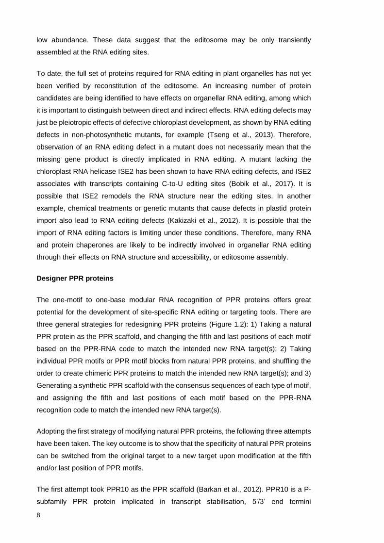
To date, the full set of proteins required for RNA editing in plant organelles has not yet
been verified by reconstitution of the editosome. An increasing number of protein
candidates are being identified to have effects on organellar RNA editing, among which
it is important to distinguish between direct and indirect effects. RNA editing defects may
just be pleiotropic effects of defective chloroplast development, as shown by RNA editing
defects in non-photosynthetic mutants, for example (Tseng et al., 2013). Therefore,
observation of an RNA editing defect in a mutant does not necessarily mean that the
missing gene product is directly implicated in RNA editing. A mutant lacking the
chloroplast RNA helicase ISE2 has been shown to have RNA editing defects, and ISE2
associates with transcripts containing C-to-U editing sites (Bobik et al., 2017). It is
possible that ISE2 remodels the RNA structure near the editing sites. In another
example, chemical treatments or genetic mutants that cause defects in plastid protein
import also lead to RNA editing defects (Kakizaki et al., 2012). It is possible that the
import of RNA editing factors is limiting under these conditions. Therefore, many RNA
and protein chaperones are likely to be indirectly involved in organellar RNA editing
through their effects on RNA structure and accessibility, or editosome assembly.
Designer PPR proteins
The one-motif to one-base modular RNA recognition of PPR proteins offers great
potential for the development of site-specific RNA editing or targeting tools. There are
three general strategies for redesigning PPR proteins (Figure 1.2): 1) Taking a natural
PPR protein as the PPR scaffold, and changing the fifth and last positions of each motif
based on the PPR-RNA code to match the intended new RNA target(s); 2) Taking
individual PPR motifs or PPR motif blocks from natural PPR proteins, and shuffling the
order to create chimeric PPR proteins to match the intended new RNA target(s); and 3)
Generating a synthetic PPR scaffold with the consensus sequences of each type of motif,
and assigning the fifth and last positions of each motif based on the PPR-RNA
recognition code to match the intended new RNA target(s).
Adopting the first strategy of modifying natural PPR proteins, the following three attempts
have been taken. The key outcome is to show that the specificity of natural PPR proteins
can be switched from the original target to a new target upon modification at the fifth
and/or last position of PPR motifs.
The first attempt took PPR10 as the PPR scaffold (Barkan et al., 2012). PPR10 is a P-
subfamily PPR protein implicated in transcript stabilisation, 5’/3’ end termini
9
determination (Pfalz et al., 2009, Prikryl et al., 2011), and translation enhancement
(Zoschke et al., 2013). PPR10 contains 19 intact PPR motifs that recognise 17 nt
conserved RNA tracts in the atpI-atpH and psaJ-rpl33 intergenic regions in maize. Motif
6 [ND] and motif 7 [NN] were chosen for modifications to retarget PPR10. Naturally motif
6 and 7 target the sequence “UC” in the context of GUAUCCUUAACCAUUUC. Motif 6
and 7 were simultaneously modified to target one of the four new targets “GG”, “AA”,
“UU”, or “CC” in the same sequence context. In vitro binding assays (REMSA) show
successful retargeting to the intended new targets “AA” or “GG” with the PPR-RNA codes
TN-A and TD-G respectively. However, the PPR10 variants cannot differentiate between
the new targets “CC”, “UU” and the original target “CU” using the codes NS-C, ND-U,
and/or NN-C/U. The codes ND and NS show only preferences towards U and C
respectively, whereas the code NN targets C or U equally well. This was predicted from
the calculation of associations, and is consistent with the recent published Bind-n-seq
data of PPR10, showing that the sequence preference at motif 6 and 7 is either “UC” or
“UU” (Miranda et al., 2017).
GUAUCCUUAACCAUUUC = Original target
GUAGGCUUAACCAUUUC
GUAAACUUAACCAUUUC
GUAUUCUUAACCAUUUC
GUACCCUUAACCAUUUC
Another attempt took SOT1 as the PPR scaffold (Zhou et al., 2017). SOT1 is a P-
subfamily PPR-SMR protein with endonuclease activity implicated in rRNA processing
in chloroplasts (Wu et al., 2016, Zhou et al., 2017). SOT1 contains 9 typical PPR motifs
that recognise 9 nt in the 5’ region of the 23S-4.5S rRNA precursor. The SMR domain
cleaves the rRNA precursor to the 3’ of the binding site. Motifs 1 to 4 were chosen for
modifications to retarget SOT1. Naturally motifs 1 to 4 target the sequence “UGGA” in
the context of AUGGACGUUGAUA…AGG (73 nt). Motifs 1 to 4 were simultaneously
modified to target “GUUC” in the same sequence context. In vitro binding assays
(REMSA) and RNA cleavage assays both show successful retargeting of SOT1 to the
intended new target. Notably, the new target was chosen so that the redesigned SOT1
only needs to differentiate between purines and pyrimidines. Specificity between purines
and between pyrimidines was not tested.

10
AUGGACGUUGAUA…AGG (73 nt) = Original target
AGUUCCGUUGAUA…AGG (73 nt)
The only attempt so far using PLS-subfamily proteins took CLB19 and OTP82 as the
PPR scaffolds (Kindgren et al., 2015). CLB19 is an RNA editing factor in chloroplasts
that edits two sites in clpP1 and rpoA transcripts (Chateigner-Boutin et al., 2008, Ramos-
Vega et al., 2015). CLB19 contains 8 PPR motifs, in the order of P1-L1-S1-SS-P1-L1-
S1-P2, that recognise 8 nt 5’ to its editing sites. Four of these nucleotides are shared
between the clpP1 and rpoA sites. There are two L1 motifs, one SS motif, and one S
motif in CLB19 aligning to the four nucleotides that differ between clpP1 and rpoA. The
two L1 motifs were shown not to contribute to RNA recognition. The SS and S1 motifs
were modified one at a time, from targeting pyrimidines to targeting purines, aiming to
shift CLB19’s targeting specificity towards one of the two editing sites. In vitro binding
assays (REMSA) showed that recombinant CLB19 binds to the rpoA probe better than
the clpP1 probe. CLB19’s binding preference for rpoA could be decreased or increased,
but its binding preference could not be shifted to favour clpP1 over rpoA. In planta editing
assays showed that rpoA is edited at a slightly higher percentage compared to clpP1 in
Arabidopsis chloroplasts. CLB19’s editing specificity could be shifted towards either rpoA
or clpP1.
…AGAAGCCC… (26 nt) = clpP1
…ACACGUGC… (26 nt) = rpoA
Adopting the third strategy of designing synthetic PPR proteins, the following two
attempts have been taken. The key outcome is to show that synthetic PPR proteins can
bind to their intended target(s), and that the specificity of the synthetic PPR proteins can
be switched from one target to another upon modification at the fifth and/or last position
of PPR motifs.
The first attempt took a consensus P motif sequence as the PPR scaffold, with artificial
N-terminal cap and C-terminal solvating helix (Coquille et al., 2014). The first and last
position of each motif were assigned to match the intended RNA target. In vitro binding
assays (REMSA) with multiple designed PPR variants showed successful targeting to 1)
8-nt polyU, polyA, or polyC; 2) UGUAUAUA or UCUAUAUA; and 3) GCUUCGCU or
GCCCCGCU.
The second attempt also took a consensus P motif sequence as the PPR scaffold, with
natural N-terminal (173 aa) and C-terminal (50 aa) caps taken from PPR10 (Shen et al.,

11
2016). The first and last position of each motif were assigned to match the intended RNA
target. In vitro binding assays (REMSA) with four designed PPR variants showed
successful targeting to four 10-nt RNA probes with 2-nt tandem variations located in the
centre: UUUUUUUUUU, UUUUCCUUUU, UUUUAAUUUU, or UUUUGGUUUU.
Moreover, RNA-bound crystal structures were obtained from this experiment,
demonstrating the structural basis for specific PPR-RNA recognition by hydrogen
bonding.
Results based on the second strategy of creating chimeric PPR proteins have not been
reported so far. This strategy is based on the assumption that all the motifs in a PPR
protein are functionally equivalent apart from recognising different RNA bases or blocks.
The assumption should be tested prior to attempting the second strategy.
Although there are multiple examples of successful PPR retargeting shown in vitro by
binding and/or enzymatic assays, only the attempted CLB19 and OTP82 retargeting
examples reported in vivo results. Evidence for a complete switch of PPR targeting
specificity in vivo is still absent. In order to obtain such data, RNA editing provides the
most convenient outcome for studying PPR retargeting in vivo.
Aims of this study
The general aim of this study is to better understand the specificity of PPR editing factors,
in order to develop customisable RNA editing tools. I will attempt to do this by predicting
and characterising PPR editing factors, by analysing their off-target effects, and by
modifying them to target new RNA editing site(s).
The specific aims are:
1) To characterise the function of low-level ycf3 intron 2 editing by CLB19 (Chapter 2.1);
2) To characterise AEF4 as the editing specificity factor for rps14-2 (37092) and to
detect potential off-target editing events (Chapter 2.2);
3) To evaluate the function of CREF3 motifs (Chapter 2.3);
4) To redesign CREF3 to target new editing sites in Arabidopsis chloroplasts (Chapter
2.3).

12
Table and Figures
Table 1 Arabidopsis chloroplast editing sites and factors.
Figure 1.1 PLS-subfamily PPR editing factors
A) Consensus sequences of PPR motifs (Cheng et al. 2016). Red boxes highlight
differences between P1 and P2, and L1 and L2 motifs; Green boxes highlight
similarity between P1-helix a and SS-helix a, and S1-helix b and SS-helix b.
B) Domain structure of PLS-subfamily PPR editing factors.
Figure 1.2 General strategies for redesigning PPR proteins (inspired by an illustration
by Bernard Gutmann, unpublished).

Table 1. Arabidopsis chloroplast editing sites and factors.
Transcript SiteNo.
CDSpos.
Strand Genomepos.
Transition Editing factor AGI Editingdomain
Mutantphenotype
Reference
accD 1 794 + 57868 S>L RARE1 AT5G13270 DYW none Robbins et al., 2009accD 2 1568
(3'UTR)+ 58642 N/A OTP81/QED1 AT2G29760 DYW yellow leaf
patchesHammani et al., 2009;Wagoner et al., 2015
atpF N/A 92 - 12707 P>L AEF1 AT3G22150 E+ pale green Yap et al., 2015clpP1 N/A 559 - 69942 H>Y CLB19/PDE247 AT1G05750 E+ pale yellow Chateigner-Boutin et al.,
2008matK N/A 640 - 2931 H>Y OTP81/QED1 AT2G29760 DYW yellow leaf
patchesHammani et al., 2009;Wagoner et al., 2015
ndhB 1 149 - 97016 S>L AEF2 AT1G18485 DYW none unpublishedndhB 2 467 - 96698 P>L CRR28 AT1G59720 DYW NDH deficiency Okuda et al., 2009ndhB 3 586 - 96579 H>Y unidentifiedndhB 4 746 - 96419 S>F CRR22 AT1G11290 DYW NDH deficiency Okuda et al., 2009ndhB 5 830 - 95650 S>L ELI1 AT4G37380 DYW none Hayes et al., 2013ndhB 6 836 - 95644 S>L OTP82 AT1G08070 DYW none Hammani et al., 2009ndhB 7 872 - 95608 S>L OTP81/QED1 AT2G29760 DYW yellow leaf
patchesHammani et al., 2009;Wagoner et al., 2015
ndhB 8 1255 - 95225 H>Y CREF7 AT5G66520 DYW none Yagi et al., 2013ndhB 9 1481 - 94999 P>L OTP84 AT3G57430 DYW none Hammani et al., 2009ndhD 1 2 - 117166 (T)>start CRR4 AT2G45350 E+ NDH deficiency Kotera et al., 2005ndhD 2 383 - 116785 S>L CRR21 AT5G55740 E+ NDH deficiency Okuda et al., 2007ndhD 3 674 - 116494 S>L CRR28 AT1G59720 DYW NDH deficiency Okuda et al., 2009ndhD 4 878 - 116290 S>L OTP85 AT2G02980 DYW none Hammani et al., 2009ndhD 5 887 - 116281 P>L CRR22 AT1G11290 DYW NDH deficiency Okuda et al., 2009ndhF N/A 290 - 112349 S>L OTP84 AT3G57430 DYW none Hammani et al., 2009ndhG N/A 50 - 118858 S>F OTP82 AT1G08070 DYW none Hammani et al., 2009petL N/A 5 + 65716 P>L unidentifiedpsbE N/A 214 - 64109 P>S CREF3 AT3G14330 DYW none Yagi et al., 2013

Table 1. Arabidopsis chloroplast editing sites and factors. (cont.)
Transcript SiteNo.
CDSpos.
Strand Genomepos.
Transition Editing factor AGI Editingdomain
Mutantphenotype
Reference
psbF N/A 77 - 63985 S>F LPA66 AT5G48910 DYW pale green(slight)
Cai et al., 2009
psbZ N/A 50 + 35800 S>L OTP84 AT3G57430 DYW none Hammani et al., 2009rpl23 N/A 89 - 86055 S>L OTP80 AT5G59200 E+ none Hammani et al., 2009rpoA N/A 200 - 78691 S>F CLB19/PDE247 AT1G05750 E+ pale yellow Chateigner-Boutin et al.,
2008rpoB 1 338 - 25992 S>L YS1 AT3G22690 DYW pale yellow Zhou et al., 2009rpoB 2 551 - 25779 S>L OTP81/QED1 AT2G29760 DYW yellow leaf
patchesHammani et al., 2009;Wagoner et al., 2015
rpoB 3 2432 - 23898 S>L CRR22 AT1G11290 DYW NDH deficiency Okuda et al., 2009rpoC1 N/A 488 - 21806 H>Y DOT4/FLV AT4G18750 DYW aberrant leaf
developmentHayes et al., 2013
rps12 N/A intron 1 - 69553 N/A OTP81/QED1 AT2G29760 DYW yellow leafpatches
Hammani et al., 2009;Wagoner et al., 2015
rps14 1 80 - 37161 S>L OTP86 AT3G63370 DYW none Hammani et al., 2009rps14 2 149 - 37092 P>L AEF4 AT3G49170 DYW embryonic lethal unpublished

E1 E2P2 L2 S2 (PG) DYWPLS/SS
E1 E2 E+P2 L2 S2PLS/SSB
A
(Cheng et al. 2016)
Figure 1.1 PLS-subfamily PPR editing factors.A) Consensus sequences of PPR motifs (Cheng et al. 2016). Red boxes highlight differencesbetween P1 and P2, and L1 and L2 motifs; Green boxes highlight similarity between P1-helix aand SS-helix a, and S1-helix b and SS-helix b.B) Domain structure of PLS-subfamily PPR editing factors.

TN
TD
ND
NS
TN
TD
ND
NS
TN
TN
TD
ND
NS
TN
TD
ND
NS
TN
Strategy 1Modifying natural PPR proteins
TN
TD
ND
NS
TN
TD
ND
NS
TN
Strategy 3Synthetic PPR proteins
TN
TD
ND
NS
TN
TD
ND
NS
TN
ND
NS
TN
TD
Strategy 2Chimeric PPR proteins
Figure 1.2 General strategies for redesigning PPR proteins (inspired by an illustration byBernard Gutmann, unpublished).

13
Chapter 2 Results
2.1 The chloroplast editing factor CLB19 shows an off-target
effect at the ycf3-i2 site
2.1.1 Summary
Deep next-generation sequencing enables detection of low-rate editing events. RNA-seq
experiments have expanded the number of Arabidopsis chloroplast editing sites from 34
to 43, by identifying novel low-level editing sites. In this work, I experimentally verified
that the low-level editing site at ycf3 intron 2 (ycf3-i2) is a true editing site, edited by the
CLB19 editing factor. However, ycf3-i2 editing does not appear to have a significant
biological function as far as I have examined. I conclude that ycf3-i2 editing events are
off-target effects of the CLB19 editing factor. As ycf3-i2 is one of the first off-target editing
sites to be identified in Arabidopsis chloroplasts, this work illustrates that NGS
techniques are sufficiently sensitive and robust to quantify low-level (and potentially off-
target) editing events. Investigation of off-target effects of PPR editing factors will extend
our understanding of the specificity of these editing factors.
2.1.2 Introduction
There are 34 major C-to-U editing sites in Arabidopsis chloroplasts (Chateigner-Boutin
and Small, 2007). The editing percentage is generally above 80%, quantified by
measuring the proportion of transcripts containing the edited base. Previous research
efforts have focused on identifying and characterising the specificity factors of these
major editing sites by forward or reverse genetics. To date, editing specificity factors
have been identified for 30 of the 34 major editing sites in Arabidopsis chloroplasts. In
order to further investigate PPR-RNA editing specificity, it is important to examine
promiscuous action of known editing factors potentially indicated by their minor editing
sites (=<10% editing).
In addition to the 34 major editing sites identified in Arabidopsis chloroplasts, there are
ten minor editing sites discovered from RNA-seq datasets (Bentolila et al., 2013, Ruwe
et al., 2013). Ruwe et al. suggested that these editing events are not essential because
selection has not acted towards increasing the editing efficiency. Editing specificity
factors have yet to be identified for any of the minor editing sites, nor have their biological
functions been tested. This work set out to verify experimentally one of the minor editing
sites by identifying its specificity factor and investigating its biological function, if any.

14
Among the minor editing sites identified by Ruwe et al., the RNA sequence of the site in
ycf3 intron 2 (ycf3-i2) aligns with the PPR motifs of CHLOROPLAST BIOGENESIS 19
(CLB19, encoded by At1G05750). CLB19 was previously identified as the editing
specificity factor for two major chloroplast editing sites in clpP1 and rpoA (Chateigner-
Boutin et al., 2008). It was hypothesised that CLB19 also edits the minor site ycf3-i2.
Previous unpublished work by Peter Kindgren (UWA) suggested that CLB19 is required
for ycf3-i2 editing, as the clb19 mutant lacks editing at ycf3-i2.
Ycf3 intron 2 is a group II intron. Group II introns are structurally conserved but diverse
in sequence. Their conserved secondary structure contains six domains branching out
from a central wheel that joins the two splice junctions into close proximity (Lehmann
and Schmidt, 2003). The edited C in the ycf3-i2 editing site is not part of the few
conserved group II intron sequences. On the other hand, it was unclear whether ycf3-i2
binding and/or editing alters the conserved intron structure and subsequently affects
splicing. It was hypothesised that ycf3-i2 editing by CLB19 affects ycf3 intron 2 splicing.
This chapter tests the hypothesis by comparing ycf3-i2 editing and ycf3 intron 2 splicing
between wild type Col-0, clb19 mutant, clb19 mutant complemented with full-length
CLB19 (functioning in both binding and editing) (Ramos-Vega et al., 2015), and clb19
mutant complemented with CLB19 with its E domain deleted (CLB19ΔE, functioning in
binding without editing) (Ramos-Vega et al., 2015).
2.1.3 Materials and Methods
For the confirmation of editing defect and complementation:
DNase treatment of RNA and cDNA synthesis
Arabidopsis seedling RNA samples (clb19 mutant, clb19 mutant complemented with full-
length CLB19, and clb19 mutant complemented with CLB19 with its E domain deleted)
were received from Patricia Leon’s laboratory at the University of Mexico. Of the total
RNA, 2.5 μg was treated with TURBO DNase (Ambion) according to the manufacturer’s
instructions. Completion of DNase treatment was verified by PCR targeting chloroplast
genomic DNA. Of the DNase-treated RNA, 1 μg was used for cDNA synthesis using
random primers (150 ng/μl, Invitrogen) and SuperScript III reverse transcriptase
(Invitrogen) according to the manufacturer’s instructions. Synthesised cDNA was diluted
1 in 100 and used as PCR template.

15
Poisoned primer extension (PPE) analysis
PCR was conducted using PrimeSTAR polymerase (Clontech) according to the
manufacturer’s instructions. The primers used for PCR were:
Forward: TTCGGGCATTAGAACGAAAC
Reverse: AAAGGTAACTATCCCGCTTTCA
The PCR product size is 526 bp.
The PCR cycling conditions are:
35 cycles of 98 ℃, 10 sec; 55 ℃, 15 sec; 72 ℃, 35 sec
Poisoned primer extension (PPE) was carried out as described by Chateigner-Boutin
and Small (2007), with nucleotide mix containing dideoxycytidine.
The primer used for PPE is:
6’FAM-TCGAAATATGAAGTGAAGACTAGATATGCC
For the splicing analyses:
Plant materials provided by Patricia Leon (University of Mexico)
Arabidopsis seeds (clb19 mutant, clb19 mutant complemented with full-length CLB19,
and clb19 mutant complemented with CLB19 with its E domain deleted) were received
from Patricia Leon’s laboratory at the University of Mexico.
Plant growth conditions
Arabidopsis seeds were surface sterilised with 70% ethanol + 0.05% Triton X-100 for
five minutes and washed with 100% ethanol before being dried in the fume hood.
Sterilised seeds were sowed on plates (half-strength MS medium, 0.5% sucrose, and
0.8% agar), stratified at 4 ℃ in the dark for three days, germinated and grown under
long-day conditions (16h light/8h dark cycle, approximately 120 μmol photons m-2 s-1).

16
RNA extraction, DNase treatment of RNA and cDNA synthesis
Total RNA from five to eight 7-day old seedlings (fresh weight < 10 mg) were isolated
using the PureZOL reagent (Bio-Rad) according to the manufacturer’s instructions. Total
RNA (600 ng) was treated with TURBO DNase (Ambion) according to the manufacturer’s
instructions. Completion of DNase treatment was verified by PCR targeting chloroplast
genomic DNA. Of the DNase-treated RNA, 200-300 ng was used for cDNA synthesis
using random primers (150 ng/μl, Invitrogen) and SuperScript III reverse transcriptase
(Invitrogen) according to the manufacturer’s instructions. Synthesised cDNA was diluted
1 in 10 and used as PCR template.
RT-PCR and quantitative RT-PCR
Splicing was first analysed by RT-PCR and visualised on an agarose gel, then quantified
by quantitative RT-PCR.
RT-PCR primers were:
ycf3_qF1: TTCGGGCATTAGAACGAAAC
ycf3_qF2: AGTTGGTTGTCGAGCCGTAT
ycf3_qR: TCCAATACTCAGCGGCTTG
Statistical Analyses
Significance grouping was determined using one-way ANOVAs based on Tukey’s
Honestly Significant Differences (HSD) test (R Program v3.2.3, package “agricolae”).
Sequence Alignment
Twenty-three ycf3 sequences from various plant species were aligned using the
Geneious software built-in ClustalW Alignment function.
RNA secondary structure prediction
The secondary structure of ycf3 intron 2 was predicted by the online tool RNAstructure
(http://rna.urmc.rochester.edu/RNAstructureWeb/) with the following constraints file
applied according to the consensus group II intron sequence and structure described in
(Lehmann and Schmidt, 2003). More specifically, single-stranded (SS) at positions 1-5
corresponds to the 5’ exon-intron junction; single-stranded (SS) at positions 185, 186,
362, 363, 364 and pairs 187-361, 188-360 corresponds to the ζ site.

17
Constraints file SS: 1 2 3 4 5 185 186 362 363 364 -1 Pairs: 187 361 188 360 -1 -1
Editing site prediction
Potential binding and editing sites of CLB19 were predicted using an in-house script
written by Ian Small. Histograms were generated by matplotlib v1.5.3.
2.1.4 Results
CLB19 is the specificity factor for ycf3-i2 editing
Previous experiments done by Peter Kindgren supported the hypothesis that CLB19 is
the specificity factor for ycf3-i2 editing. As shown in Figure 2.1.1 [A, B], CLB19 binding
motifs align with the ycf3-i2 cognate sequence, and CLB19 binds to the ycf3-i2 RNA
probe in vitro. Moreover, ycf3-i2 editing is defective in clb19 mutant. To establish that the
effect on ycf3-i2 editing is specific to clb19 mutant and not a pleiotropic effect of its severe
physiological defects, editing of ycf3-i2 was also quantified in ys1-1 and otp51-1 mutants,
which have similar physiological phenotypes (creamy seedlings) as clb19. The editing of
ycf3-i2 is normal in both ys1-1 and otp51-1 mutants (Figure 2.1.1 [C]). This strongly
suggests that ycf3-i2 editing defect in clb19 is specifically due to the loss of CLB19
instead of its secondary effects.
In this work, ycf3-i2 editing was further quantified in clb19 mutant complemented with
full-length CLB19, and clb19 mutant complemented with CLB19 with its E domain (E1-
E2-E+) deleted. The E domain is believed to be part of the larger CLB19 editosome.
Therefore, CLB19 without its E domain is not expected to be active in editing. As shown
in Figure 2.1.2, the ycf3-i2 editing defect in clb19 can be rescued by the expression of
full-length CLB19. The CLB19 with deleted E domain does not complement the ycf3-i2
editing defect.

18
CLB19 binding does not affect ycf3 intron 2 splicing, but editing by CLB19 does
I then investigated whether binding and/or editing at ycf3-i2 is involved in ycf3 intron 2
splicing. The splicing efficiency was quantified in Arabidopsis wild type Col-0, clb19
mutant, clb19 mutant complemented with full-length CLB19. To test whether binding and
editing at ycf3-i2 have separate effects on ycf3 intron 2 splicing, I also included clb19
complemented with CLB19 with its E domain deleted, which does not complement ycf3-
i2 editing, but should still be able to bind. 7-day-old seedlings of each genotype were
harvested (Figure 2.1.3 [A]). The PCR primers are illustrated in Figure 2.1.3 [B]. A
splicing defect is usually indicated by both over-accumulation of the unspliced form and
loss or decrease of the spliced form. As shown in Figure 2.1.3 [C, D], although there is
an over-accumulation of unspliced transcripts in the clb19 mutant and the transgenic line
expressing CLB19 with its E domain deleted, the quantity of spliced transcripts is not
significantly different between all four genotypes examined. The results indicate that
CLB19 binding does not affect ycf3 intron 2 splicing, but editing by CLB19 does.
Evolutionary conservation of the ycf3-i2 editing site
I checked the conservation of the ycf3-i2 editing site during evolution by aligning ycf3
intron 2 sequences from a variety of dicot and monocot species. As shown in Figure
2.1.4, in dicots, all species have a relatively conserved sequence matching the CLB19
binding site, but the C to be edited is not always conserved, nor is it always encoded as
the edited nucleotide T. For example, in Citrus sinensis, it is encoded as G. The putative
CLB19 binding site is disrupted in monocot sequences. So far, no target sites have been
identified for CLB19 in monocots.
Ycf3 intron 2 is a group II intron. Group II introns are structurally conserved, yet
sequence-wise diverse. I reasoned that the conservation pattern of ycf3-i2 site may be
shaped by the conservation of ycf3 intron 2 structure. I predicted the ycf3 intron 2
structure according to the model group II intron structure (Lehmann and Schmidt, 2003).
I then mapped the ycf3-i2 site to this predicted structure. As shown in Figure 2.1.5, the
ycf3-i2 site sits in the structurally conserved D1 arm, upstream of the ζ site. The ζ site is
an example of a limited number of group II intron sites that are conserved in both
sequence and structure.
Predicted binding and editing sites of CLB19 across Arabidopsis chloroplast
genome
Since the Arabidopsis chloroplast genome is fully transcribed from both stands, there are
256,413 potential binding sites for CLB19 (not counting one of the inverted repeat

19
regions). Prediction scores were calculated according to the PPR-RNA code for all the
potential binding sites, and plotted as a histogram (Figure 2.1.6 [A]). A subset of the data
containing potential editing sites where C is at the editing position (n=44,959), and a
subset containing potential editing sites where YC is at the -1/editing positions
(n=37,888) are also plotted. YC is specified since there is very rarely a G observed at
the -1 position of any Arabidopsis chloroplast editing site. The only case reported so far
is the ndhG editing site in Arabidopsis ecotype Cvi-0 (Tillich et al., 2005). Figure 2.1.6
(B, C) show expanded views of the YC editing subset. It shows that all three natural
CLB19 editing sites score higher than two standard deviations above the mean score of
all potential editing sites. On the contrary, all other major chloroplast editing sites score
lower than two standard deviations above the mean score. Ycf3-i2 scores 3.84 which is
the highest among all potential editing sites. RpoA scores 1.98. ClpP1 scores 1.96 which
is the lowest among the three natural CLB19 editing sites. There are 494 potential editing
sites that score equivalent or higher than ClpP1. Among the top 20 sites, 2 sites are
located in unannotated regions, 8 sites are located in anti-sense RNAs, but others are
located in stably accumulating transcripts such as ycf1, ycf2, ycf3, rpoA 3’UTR, rpoC1,
psaA, and rpl32 5’UTR. If only considering CLB19 binding, there are 3,591 potential
binding sites that score equivalent or higher than clpP1.
2.1.5 Discussion
The ycf3-i2 editing site is likely an off-target site of CLB19
C-to-U RNA editing in plant organelles is likely a mechanism for correcting T-to-C
mutations in the genome (Sun et al., 2016). In the case where a start codon is created
by ACG-to-AUG editing, it may also function in regulating gene expression (Kotera et al.,
2005). Therefore, defects in editing lead to loss-of-function of various organellar gene
products, resulting in a variety of plant developmental phenotypes, such as changes in
leaf pigmentation and aberrant leaf development. An off-target editing site is a site that
lacks such biological implications. Defects in editing at off-target sites should have no
significant impact on plant development. Among 18 characterised editing factors in
Arabidopsis chloroplasts, there are six editing factors that edit multiple sites. It is possible
that among these multiple editing sites, one or more are off-targets. To identify off-target
sites, editing needs to be knocked out one site at a time to examine biological effects
specific to the site. Although editing preference can be altered between multiple editing
sites by modifying the target specificity of the editing factor (Kindgren et al., 2015), editing
at any single site has not yet been selectively knocked out using this approach. Therefore
it is generally difficult to identify off-target sites among the major editing sites.

20
The ycf3-i2 site is a special case. Ycf3-i2 sits in an intron therefore clearly does not
correct any mutations in the ycf3 coding sequence. Furthermore, ycf3-i2 editing is
unlikely to have any significant effects on ycf3 intron 2 splicing. Although the clb19 editing
mutants show over-accumulation of unspliced ycf3 transcripts, it is likely due to
pleiotropic effects of the defective editing of the sites in clpP1 or rpoA rather than ycf3-
i2. Taken together, ycf3-i2 editing appears to lack any significant biological function, and
therefore is likely to be an off-target editing site of CLB19. A similar case is the rps12-i1
site in chloroplasts, which is one of the five sites edited by the PPR editing factor
QED1/OTP81 (Wagoner et al., 2015). Rps12 splicing is not affected in the otp81/qed1
mutant (Hammani et al., 2009), indicating that rps12-i2 may also be an off-target site of
QED1/OTP81.
Why is ycf3-i2 edited at low efficiency
The ycf3-i2 site is a minor editing site edited at 10-15%, whereas the major editing sites
in Arabidopsis chloroplasts are mostly edited at 80-100%. CLB19 PPR motifs match
equally well to the ycf3-i2 site, and recombinant CLB19 binds equally well to the ycf3-i2
probe in vitro, compared to the rpoA and the clpP1 probes. An obvious question is why
is the ycf3-i2 site poorly edited by CLB19, compared to the rpoA site (80%-90%) and the
clpP1 site (75%-85%)?
There are several potential explanations. Firstly, the ycf3-i2 site sits in an intron, which
is likely to have a limited lifetime relative to exons, reducing its likelihood of being bound
and edited. The only other intronic editing site in Arabidopsis chloroplast is in rps12 intron
1 edited by OTP81/QED1 at 30% (Hammani et al., 2009, Ruwe et al., 2013). Secondly,
ycf3 intron 2 is a type II intron that contains highly conserved structures. Binding of
CLB19 to the ycf3-i2 site would compete with such intron structures as demonstrated in
Figure 2.1.5. It has been shown that artificially-introduced RNA secondary structures
reduce CLB19 binding to clpP1 probes in vitro (Kindgren et al., 2015). Therefore, the
binding in vivo between CLB19 and the ycf3-i2 site in the presence of the complete
structured intron may be lower than the binding measured in vitro to short unstructured
probes. Thirdly, the nucleotides immediately next to the edited C differ from the
consensus sequence established from examining many hundreds of sites in plant
organelles. All major editing sites in Arabidopsis chloroplasts, except ndhD (117166),
have a pyrimidine at position -1 from the edited C. The exception, ndhD, which has an A
at position -1, is relatively poorly edited at 45% (Ruwe et al., 2013). Mutating the U at -1
to an A reduced editing of psbE and petB in tobacco plastids (Miyamoto et al., 2004) by
around 50%. Furthermore, altering the U at -1 to a G reduced binding of CLB19 to the

21
rpoA site in vitro (Ramos-Vega et al., 2015). Therefore, the A preceding the edited C in
ycf3-i2 might have an inhibiting effect on editing.
Ycf3-i2 editing site is not conserved between monocots and dicots
Neither the CLB19 binding site, nor the to-be-edited cytidine at ycf3-i2, is highly
conserved during evolution. The CLB19 binding site is conserved only in dicots but not
in monocots. The to-be-edited cytidine is not even conserved in dicots, and neither is it
always encoded as T. This is further evidence that editing of ycf3-i2 is unlinked to
significant biological effects.
On the other hand, ycf3-i2 editing has not been eliminated during evolution, presumably
because it does not create harmful mutations in ycf3 and the editing level is so low that
it does not titrate CLB19 and co-factors which would compromise editing at other
biologically-relevant sites. The other two major editing sites of CLB19, clpP1 and rpoA,
are edited sufficiently despite the presence of ycf3-i2 editing. Therefore off-target editing
at ycf3-i2 does not have detrimental biological effects that could be selected against
during evolution.
The editing specificity of CLB19 is surprisingly high
It is not surprising that CLB19 shows off-target effects in Arabidopsis chloroplasts,
considering that so far only six effective RNA targeting motifs have been characterised
in CLB19 (Kindgren et al., 2015). As shown in predictions based on the canonical PPR-
RNA code, there are 3,591 potential binding sites in Arabidopsis chloroplasts, including
494 potential editing sites, which match with the CLB19 PPR motifs equally well or better
compared to the known editing sites of CLB19. It is thus surprising that only three sites
are edited by CLB19.
The lack of off-target editing sites implies that there are additional factors affecting the
editing specificity in vivo. Firstly, in addition to the interactions between the fifth and last
position of CLB19 motifs and the aligned RNA bases (canonical PPR-RNA interaction),
there may be non-canonical interactions between CLB19 and its RNA targets. For
example, PPR10 is believed to have various non-canonical interactions with its target
RNA demonstrated by crystal structures (Yin et al., 2013) and Bind-n-seq assays
(Miranda et al., 2017). Secondly, other components in the editosome may also contribute
to editing specificity. So far there are three protein families identified to be part of the
editosome – RIP/MORF, ORRM, and OZ (reviewed in (Sun et al., 2016)). MORF proteins
interact with PPR editing factors and enhance their binding affinity and/or specificity to
the RNA target(s) (Yan et al., 2017). ORRM proteins show RNA binding affinity (Sun et

22
al., 2013), therefore potentially conferring RNA targeting specificity. Thirdly, RNA editing
factors such as CLB19 may have specific structural preference for their targets. On one
hand, PPR-RNA binding may prefer unstructured RNA, as RNA secondary structure
blocks CLB19 binding in vitro (Kindgren et al., 2015). On the other hand, the RNA editing
enzyme may have structural requirements for RNA substrates. For example, the ADAR
deaminase that converts adenosine to inosine strongly prefers double-stranded RNA
substrates (Kuttan and Bass, 2012). Finally, there could be expression and co-
expression limitations. For example, the target RNA’s half-life may be too short to be
edited, or the target RNA and the editing factor are not expressed at the same time or in
close proximity to each other. The above arguments will be discussed in the following
chapters in light of new experimental results.
Technical implications on RNA editing quantification
The ycf3-i2 editing site was initially identified by RNA-seq (Ruwe et al., 2013). The work
in this chapter experimentally verified the minor editing site and identified its editing
specificity factor. From a technical point of view, it shows that current high-throughput
sequencing techniques allow RNA editing to be reliably detected at low level (=< 10%)
to reveal minor editing sites. It would be possible to identify minor editing events at even
lower efficiency compared to ycf3-i2 given sufficient coverage and robust statistics. Such
analyses will be further discussed in Chapter 2.2.
2.1.6 Figure Legends
Figure 2.1.1 CLB19 aligns with, binds to, and edits ycf3-i2 (This figure contains data
entirely obtained by Peter Kindgren).
A) CLB19 motifs (5th and last amino acid positions) are aligned with its two known editing
sites, clpP1 and rpoA, as well as the new editing site, ycf3-i2. Dark green boxes
indicate matches. Light green boxes indicate partial matches. Red boxes indicate
mismatches. “C”s indicate the edited cytidines. The P1-, L1-, and S1-type motifs are
labelled as P, L, and S motifs for simplicity.
B) RNA electrophoretic mobility shift assay (REMSA) of recombinant CLB19
simultaneously incubated with the clpP1 (Cy5-
CAGCAACAGAAGCCCAAGCUCAUGGA), rpoA (Cy3-
AUGUAUUACACGUGCAAAAUCUGAGA), and ycf3-i2 (6FAM-
AGACUAGAUAUGCCUAAAUACUUUCU) probes. 700 pM of each probe was
incubated with increasing rCLB19 concentrations (87.5, 175, 350, and 700 nM).
C) Poisoned primer extension (PPE) analysis of ycf3-i2 editing in 7-day-old seedlings of
Col-0, clb19-2, ys1-1, and otp51-1. Each lane represents a biological replicate.

23
“Edited” and “Unedited” indicates the edited and unedited band respectively. “%
Edited” indicates editing percentage calculated by the ratio of band intensities
Edited/(Edited+Unedited).
Figure 2.1.2 The ycf3-i2 editing defect in clb19 can be complemented by CLB19 but
not CLB19 with the E domain deleted.
Poisoned primer extension (PPE) analysis of ycf3-i2 editing in 7-day-old seedlings of
Col-0, clb19-2, clb19-2 expressing full-length CLB19 (CLB19), and clb19-2 expressing
CLB19 with truncated E domain (CLB19ΔE). RNA samples were received from the
University of Mexico. “Edited” and “Unedited” indicate the extension products obtained
using cDNA templates corresponding to the edited and unedited transcripts respectively.
“% Edited” indicates editing percentage calculated by the ratio of band intensities
Edited/(Edited+Unedited).
Figure 2.1.3 CLB19 binding does not significantly affect ycf3 intron 2 splicing, but
editing by CLB19 does.
A) 7-day old seedlings at the time of harvesting. For the CLB19 sample, seeds were
collected from plants homozygous for the clb19 mutation. For the clb19 and the
CLB19ΔE samples, seeds were collected from plants heterozygous for the clb19
mutation (but homozygous for the CLB19ΔE transgene), and only albino seedlings
(clb19/clb19) were harvested for RNA extraction.
B) Illustration of the ycf3 gene and the primers used for splicing analysis. The primers
used for RT-PCR were ycf3_qF1 and ycf3_qR. Two pairs of primers were used for
quantitative RT-PCR. The pair ycf3_qF1 and ycf3_qR were used to quantify the
spliced transcript; the pair ycf3_qF2 and ycf3_qR were used to quantify the unspliced
transcript.
C) RT-qPCR analysis of ycf3 intron 2 splicing. Transcript expression is normalised to
the expression of the UBQ11 transcript. Error bars show SE with n=3. One-way
ANOVA and Tukey’s Honestly Significant Difference (HSD) test were performed for
the spliced and the unspliced ycf3 transcripts respectively, significant grouping
(p<0.05) is indicated by “a”, “ab”, or “b”.
D) Ratio of ycf3 intron 2 spliced transcript vs. unspliced transcript, calculated from the
data presented in (C). Error bars show SE with n=3.
Figure 2.1.4 Sequence conservation around the CLB19 binding site in ycf3 intron 2.
Alignment of ycf3 intron 2 sequences at the CLB19 binding site in representative dicot
and monocot species, next to the conserved ζ site of group II introns. The CLB19 binding

24
site is not conserved in monocot species. The edited C is highlighted with the closed box.
The edited C is not conserved in Brachypodium hygrometrica, Citrus sinensis, Daucus
carota and Lonicera japonica (dashed box).
Figure 2.1.5 Prediction of the ycf3 intron 2 secondary structure.
Prediction of the ycf3 intron 2 secondary structure by RNAstructure
(http://rna.urmc.rochester.edu/RNAstructureWeb/) based on calculation of the lowest
free energy (-137.8 kcal mol-1) with folding constraints applied according to the
conserved sequences and structural features of group II introns (shown in the inset)
(Lehmann and Schmidt, 2003). Characteristic domains of group II introns are assigned
as D1 – D6. The ζ site is boxed. The CLB19 site is underlined.
Figure 2.1.6 Predicted binding sites of CLB19 across the Arabidopsis chloroplast
genome.
(A) Distribution of site prediction scores for CLB19 across the Arabidopsis chloroplast
genome. Light grey bars indicate all potential binding sites across the genome
(n=256,413); Mid grey bars indicate all potential editing sites with C encoded at the
editing position (n=44,959); Dark grey bars indicate all potential editing sites with YC
encoded at the -1 position and the editing position (n=37,888). Red lines indicate the
natural CLB19 editing sites (clpP1, rpoA, and ycf3-i2).
(B) Distribution of prediction scores of potential YC editing sites for CLB19 (n=37,888).
The mean score is labelled. 1SD, 2SD, 3SD, and 4SD indicate one, two, three, and
four standard deviation(s) from the mean. Red lines indicate the natural CLB19
editing sites (clpP1, rpoA, and ycf3-i2); Green lines indicate the other 32 major
chloroplast editing sites.
(C) A zoom-in view of the histogram shown in (B). Red lines indicate the natural CLB19
editing sites, clpP1, rpoA, ycf3-i2, with labels.

TRGII
HTN
ND
TD N
NTND
5th positionlast position
E1 E2 E+P L S L S P L2 S2SS P
TN N
a g c u Ca c cclpP1 g a a g c aa a a u Ca c urpoA c a g g c aa a u a Ca u c c uycf3-i2 u a g a
CLB19A
B
C
Figure 2.1.1 CLB19 aligns with, binds to, and edits ycf3-i2 (This figure contains data entirelyobtained by Peter Kindgren).A) CLB19 motifs (5th and last amino acid positions) are aligned with its two known editing sites,clpP1 and rpoA, as well as the new editing site, ycf3-i2. Dark green boxes indicate matches. Lightgreen boxes indicate partial matches. Red boxes indicate mismatches. “C”s indicate the editedcytidines. The P1-, L1-, and S1-type motifs are labelled as P, L, and S motifs for simplicity.B) RNA electrophoretic mobility shift assay (REMSA) of recombinant CLB19 simultaneouslyincubated with the clpP1 (Cy5-CAGCAACAGAAGCCCAAGCUCAUGGA), rpoA (Cy3-AUGUAUUACACGUGCAAAAUCUGAGA), and ycf3-i2 (6FAM-AGACUAGAUAUGCCUAAAUACUUUCU) probes. 700 pM of each probe was incubated withincreasing rCLB19 concentrations (87.5, 175, 350, and 700 nM).C) Poisoned primer extension (PPE) analysis of ycf3-i2 editing in 7-day-old seedlings of Col-0,clb19-2, ys1-1, and otp51-1. Each lane represents a biological replicate. “Edited” and “Unedited”indicates the edited and unedited band respectively. “% Edited” indicates editing percentagecalculated by the ratio of band intensities Edited/(Edited+Unedited).

neg.control
Edited control
Unedited control
Col-0 clb19-2 CLB19 CLB19ΔE17 16 12017 00 0001212 % Edited
Edited
Unedited
ycf3-i2
Figure 2.1.2 The ycf3-i2 editing defect in clb19 can be complemented by CLB19 but notCLB19 with the E domain deleted.Poisoned primer extension (PPE) analysis of ycf3-i2 editing in 7-day-old seedlings of Col-0,clb19-2, clb19-2 expressing full-length CLB19 (CLB19), and clb19-2 expressing CLB19 withtruncated E domain (CLB19ΔE). RNA samples were received from the University of Mexico.“Edited” and “Unedited” indicate the extension products obtained using cDNA templatescorresponding to the edited and unedited transcripts respectively. “% Edited” indicates editingpercentage calculated by the ratio of band intensities Edited/(Edited+Unedited).

Col-0clb19 CLB19CLB19ΔE
1 mm
0
2.0
4.0
6.0
8.0
10.0
clb19 CLB19ΔE Col-0 CLB19
Normalisedtranscript
spliced (ycf3_qF1, ycf3_qR)unspliced (ycf3_qF2, ycf3_qR)
abab
bb
b
0
0.1
0.2
0.3
0.4
0.5
0.6
ycf3-i2
spliced/unspliced
clb19 CLB19ΔE Col-0 CLB19
A
CLB19 site
exon1 exon2 exon3intron1 intron2
Ycf3 gene
ycf3_qF1 ycf3_qF2 ycf3_qR
B
C D
Figure 2.1.3 CLB19 binding does not significantly affect ycf3 intron 2 splicing, but editingby CLB19 does.A) 7-day old seedlings at the time of harvesting. For the CLB19 sample, seeds were collectedfrom plants homozygous for the clb19 mutation. For the clb19 and the CLB19ΔE samples, seedswere collected from plants heterozygous for the clb19 mutation (but homozygous for theCLB19ΔE transgene), and only albino seedlings (clb19/clb19) were harvested for RNA extraction.B) Illustration of the ycf3 gene and the primers used for splicing analysis. The primers used forRT-PCR were ycf3_qF1 and ycf3_qR. Two pairs of primers were used for quantitative RT-PCR. Thepair ycf3_qF1 and ycf3_qR were used to quantify the spliced transcript; the pair ycf3_qF2 andycf3_qR were used to quantify the unspliced transcript.C) RT-qPCR analysis of ycf3 intron 2 splicing. Transcript expression is normalised to theexpression of the UBQ11 transcript. Error bars show SE with n=3. One-way ANOVA and Tukey’sHonestly Significant Difference (HSD) test were performed for the spliced and the unspliced ycf3transcripts respectively, significant grouping (p<0.05) is indicated by “a”, “ab”, or “b”.D) Ratio of ycf3 intron 2 spliced transcript vs. unspliced transcript, calculated from the datapresented in (C). Error bars show SE with n=3.
a
a
a

Figure 2.1.4 Sequence conservation around the CLB19 binding site in ycf3 intron 2.Alignment of ycf3 intron 2 sequences at the CLB19 binding site in representative dicot and monocot species, next to the conserved ζ site of group II introns. The CLB19binding site is not conserved in monocot species. The edited C is highlighted with the closed box. The edited C is not conserved in Brachypodium hygrometrica, Citrussinensis, Daucus carota and Lonicera japonica (dashed box).
* Conserved among all the species listed
Insertions unique to M. truncatula, D. carota and O. sativa at respective positionsInsertions unique to the monocot species listed, which disrupts the potential CLB19 binding site
• Conserved at the CLB19 binding site among all the dicot species listed
Arabidopsis lyrataArabidopsis thalianaBrassica rapaCarica papayaGlycine maxMalus domesticaManihot esculentaMedicago truncatulaPopulus trichocarpaRicinus communisSolanum lycopersicumSolanum tuberosumTheobroma cacaoVitis ViniferaBoea hygrometricaCitrus sinensisDaucus carotaLonicera japonicaBrachypodium distachyonOryza sativa (ssp. japonica)Sorghum bicolorSpirodela polyrhizaZea mays
*CLB19 site
P L S S P L S P L S* * ** * * * * * * * * * *** * * • • • • •
Dicots
Monocots
ζ site

D3
D4
D5D6
GT
G
C
G
ACA T
10
TCTCCA
CT
A
T
20
A
G
A
A
A
A
A
A
A
G
30
A
A
C
G
A
A
C
G
CT
40AGTCA
ATA
AA
50
AT A
CTAG
A
A
A
60
C
A
C
A
A
A
A A AA
70
GGGCTTTCT
A
C
AT
AA
G
CA
TC
90
GC
CTAAAA C
G
100
AT
TTTTAT
CG A
C
TGA TG
A C
A
120
AA T
ACA T
AAC
130
TTA C
T
CA CT
C
140
GAAA
T A T GA
A
150
G T G A A GA
C T A
160
GA T A
T GC C T A
170A
AT A
C T
T
T
C
T
180T
T
TC
TA
T
GG
A
190
TA
AA
AA
AA
GA
200
TTTAATTGAT
210
AG
A
GGAAGC A
220
C CG
T A AA
GA T
230
C A A T T G G A
A
A
G GTTT
TG
G A C
250
C
G
AC
AA CAC
A260A
G
AG
CT
GT
TT
270
AT
TT
ATA
TC
A
280
TAA T
ATGA G
ATAA
AT
CT
TA
GGA
A
T
CT
AC
TT
310
ATGTA A
TAGA
320
GTA A
T
C
CCC
G330
AAGGT
ATTGA
340
GCAGC
G
GTGT 350
AGCATCAGAT
360
CC T A
AAGA
CA
370
G TA A G T C T T T
380
T T
CTTTTTTA
390 TG
AAG
TATGAAAAAG T
CTTT
TTCAAAGA T T
C
TATATAAAT
430
TT
TTATAT
GA
440AA G C
G GG A T A
450
G T
T
ACCTT
TC
460
AGAAAA
T
T
CT
470
A AT
ATCTAAT
480
A ACA
GTG
GA
C
490
ATG
C
T
TTTT
T
500
TT
CT
G
AA
GG
T
510A
G G A GAAAAG
520ATAAAACTTA
530TTATAT T A A T
540
T A AT A
TAT
A TAT
A TA
A
T C A A AA T G
AA
AGTT
570
TA G
A
A
C
T
C
A
T580
G T A A T T A CTC
590
T C T T T T G T T T
600
A A T C C A A G A A
610
A A A C A A A A A A
620
T C T AT A
AG A A
630
A
T C TC A
TTC
A
640
ATT
G AAC
ATT
650
CAA
T
A TA G
TA
660
T
A
A
A
GT
TC
AA
670
AG
TA
GGT T
A
A680
AGTT
AA
AAA
A
690
CTGG
T
A
CA
CC
700
AAAACAAAAG
710
AGT
TGGTTGT
720
C
GAGCCGTAT
730
G
AGG
T
AGGAA
740
ACT
C
TC
A
AGT750ACGGTTCTCAG
GG
A
GGGAA
T
770
TGATCC
G
CCT
780
ATTCCG
AC
D1 D2
Figure 2.1.5 Prediction of the ycf3 intron 2 secondary structure.Prediction of the ycf3 intron 2 secondary structure by RNAstructure(http://rna.urmc.rochester.edu/RNAstructureWeb/) based on calculation of the lowest freeenergy (-137.8 kcal mol-1) with folding constraints applied according to the conserved sequencesand structural features of group II introns (shown in the inset) (Lehmann and Schmidt, 2003).Characteristic domains of group II introns are assigned as D1 – D6. The ζ site is boxed. The CLB19site is underlined.
CLB19 site
ζ site

14000
12000
10000
8000
6000
4000
2000
0 -4 -3 -2 -1 0 1 2 3 4
Num
berofsites
Score
1SD 2SD 3SD 4SDmean
Score-4 -3 -2 -1 0 1 2 3 4
2000
1500
1000
500
0
Num
berofsites
Figure 2.1.6 Predicted binding sites of CLB19 across the Arabidopsis chloroplast genome.A) Distribution of site prediction scores for CLB19 across the Arabidopsis chloroplast genome.Light grey bars indicate all potential binding sites across the genome (n=256,413); Mid grey barsindicate all potential editing sites with C encoded at the editing position (n=44,959); Dark greybars indicate all potential editing sites with YC encoded at the -1 position and the editingposition (n=37,888). Red lines indicate the natural CLB19 editing sites (clpP1, rpoA, and ycf3-i2).B) Distribution of prediction scores of potential YC editing sites for CLB19 (n=37,888). The meanscore is labelled. 1SD, 2SD, 3SD, and 4SD indicate one, two, three, and four standard deviation(s)from the mean. Red lines indicate the natural CLB19 editing sites (clpP1, rpoA, and ycf3-i2); Greenlines indicate the other 32 major chloroplast editing sites.C) A zoom-in view of the histogram shown in (B). Red lines indicate the natural CLB19 editingsites, clpP1, rpoA, ycf3-i2, with labels.
2SD 3SD 4SD
1.5 2.0 2.5 3.0 3.5 4.0
70
60
50
40
30
20
10
0
Num
berofsites
Score
clpP rpoA ycf3-i2
A
B
C

25
2.2 Editing of chloroplast rps14 by PPR editing factor AEF4 is
essential for Arabidopsis seed development
2.2.1 Summary
Pentatricopeptide repeat (PPR) editing factors recognising specific organellar RNA
editing sites can be predicted using the PPR-RNA recognition code. Based on this code,
PLASTID EDITING FACTOR 4 (AEF4, encoded by AT3G49170) is a good candidate for
the site-specificity factor for the rps14-2 editing site in Arabidopsis chloroplasts. Plants
lacking AEF4 die as embryos, posing technical difficulties for analyses. By partial
complementation and overexpression of AEF4, I could show that among all major
chloroplast editing sites, only rps14-2 editing tightly correlates with AEF4 gene
expression level. In addition, AEF4 overexpression induced very few low-level non-
specific editing events. I conclude that AEF4 is the site-specificity factor for rps14-2 in
Arabidopsis chloroplasts. A loss-of-function mutation of AEF4 leads to embryonic
lethality, implying that editing of the rps14-2 site is essential for embryogenesis. The
finding highlights the importance of organellar RNA editing in plant development.
2.2.2 Introduction
Out of 34 major editing sites in Arabidopsis chloroplasts, 19 PPR editing factors have
been identified accounting for 30 editing sites (Chapter 1, Table 1) (Kotera et al., 2005,
Okuda et al., 2007, Chateigner-Boutin et al., 2008, Cai et al., 2009, Hammani et al.,
2009, Okuda et al., 2009, Robbins et al., 2009, Zhou et al., 2009, Hayes et al., 2013,
Yagi et al., 2013b, Wagoner et al., 2015, Yap et al., 2015). Editing factors for the following
four sites remained unidentified prior to this work: ndhB-3 (96579), ndhB-1 (97016), petL
(65716), and rps14-2 (37092). The PPR-RNA recognition code (Barkan et al., 2012) was
used to predict the editing factors for these 4 sites, leading to the hypotheses that
PLASTID EDITING FACTOR 2 (AEF2, encoded by AT1G18485) is the editing factor for
ndhB (97016), and PLASTID EDITING FACTOR 4 (AEF4, encoded by AT3G49170) is
the editing factor for rps14-2 (37092).
Chloroplast editing factor mutants show a variety of functional and developmental
defects, such as changes in leaf pigmentation (e.g. the aef1 mutant) (Yap et al., 2015)
and aberrant leaf shapes (e.g. the dot4/flv mutant) (Hayes et al., 2013). Mutants of these
editing factors lose the functions of the gene products encoded by the targeted
transcripts, appearing as surrogate mutants of the corresponding chloroplast genes.
Unlike other chloroplast editing factors, the mutant of PLASTID EDITING FACTOR 4
(AEF4) is inviable, initially identified from an embryo-lethal mutant screen as emb2261
(Cushing et al., 2005), instead of being recovered from a mutant screen for typical

26
chloroplast functional defects. The emb2261/aef4 mutant stalls at the heart stage during
seed development (Cushing et al., 2005). Emb2261/aef4 is the first embryonic lethal
mutant related to a chloroplast PPR RNA editing factor.
One approach to study embryo-lethal mutants is to perform partial complementation.
Using the seed-specific ABI3 promoter to drive the gene of interest for partial
complementation has been successful in studying the embryo-lethal mutants emb506
(Despres et al., 2001), emb2394, and emb2654 (Aryamanesh et al., 2017), among which
emb2654 is a chloroplast PPR splicing factor mutant. I hypothesised that ABI3-promoter-
driven AEF4 constructs could partially complement the aef4 mutant such that it could
complete seed development. AEF4 expression would then fade away as the ABI3
promoter loses its activity, leaving only the aef4 mutant background from the seedling
stage onward. In this way, the partial complementation method provides an opportunity
to obtain enough plant tissues to examine RNA editing in the aef4 mutant background.
2.2.3 Materials and Methods
Cloning of plant transformation constructs
The AEF4 gene fragment was amplified from start codon to stop codon from Col-0
genomic DNA with the attB recombination sites using PrimeSTAR polymerase
(Clontech). The AEF4 PCR product was purified by QIAquick PCR purification kit
(Qiagen), cloned into the donor vector pDONR207 using Gateway BP Clonase
(Invitrogen), and transformed into E. coli competent cells (DH5α). The AEF4 gene
fragment was then cloned from the entry vector pDONR207 to the plant expression
vectors pH7WG containing the ABI3 promoter (Aryamanesh et al., 2017) (ABI3:AEF4),
or pGWB2 (EMBL) containing the 35S promoter (35S:AEF4), using Gateway LR Clonase
(Invitrogen), and transformed into E. coli competent cells (DH5α). Positive clones for
each construct were confirmed by Sanger sequencing. The verified constructs were
transformed into Agrobacterium tumefaciens competent cells (GV3101).
Plant growth, transformation, and selection
Arabidopsis seeds harvested from heterozygous AEF4/aef4 (SALK_024975) plants were
surface sterilised with 70% ethanol + 0.05% Triton-X100 for 5 minutes and washed with
100% ethanol before being dried in the fume hood. Sterilised seeds were sowed on
plates (half-strength MS medium and 0.8% agar), stratified at 4 ℃ in the dark for three
days, germinated and grown under long-day conditions (16h light/8h dark cycle,

27
approximately 120 μmol photons m-2 s-1). Four-week-old seedlings were genotyped with
the following primer combinations.
For SALK_024975 T-DNA insertion,
SALK_024975_RP: CTTTCTCGAGTGCATTCAAGG
LBb1.3: ATTTTGCCGATTTCGGAAC
For AEF4 genomic DNA:
SALK_024975_RP: CTTTCTCGAGTGCATTCAAGG
SALK_024975_LP: TATATTTGGTGAGCATTCGGG
Heterozygous AEF4/aef4 plants were selected and grown under the conditions listed
above. The primary stems were trimmed to induce branching. Upon flowering, the
heterozygous AEF4/aef4 plants were transformed with the ABI3:AEF4 or 35S:AEF4
constructs by floral dip, a method for Agrobacterium-mediated transformation of
Arabidopsis (Clough and Bent, 1998).
Seeds harvested from the dipped plants were germinated and selected on hygromycin
B (25 μg/ml). Surviving primary transformants (T1) were transferred to soil, and
genotyped for homozygosity of the T-DNA insertion in the AEF4 gene with the same set
of primers listed above, except that the reverse primer SALK_024975_LP2 (annealed to
the 3’UTR of the native AEF4 gene) was used to distinguish between the native AEF4
gene and the AEF4 transgenes.
SALK_024975_LP2: GTGTATCTAAATCTCAAAGTCACC
Seeds harvested from the primary transformants were germinated and grown without
hygromycin B selection, because all the selected primary transformants carry
homozygous T-DNA insertions in the AEF4 gene, and seeds that do not carry any copies
of the transgenes would die at the embryo stage.
RNA extraction, DNase treatment of RNA and cDNA synthesis
Total RNA from flowering tissue of the primary transformants (T1) were isolated using
the PureZOL reagent (Bio-Rad) according to the manufacturer’s instructions. Total RNA
(2 μg) was treated with TURBO DNase (Ambion) according to the manufacturer’s
instructions. Completion of DNase treatment was verified by PCR targeting chloroplast

28
genomic DNA. Of the DNase-treated RNA, 900 ng was used for cDNA synthesis using
random primers (150 ng/μl) and SuperScript III reverse transcriptase (Invitrogen)
according to the manufacturer’s instructions.
RNA extraction, DNase treatment of RNA and cDNA synthesis from seedling tissue of
the following plant generation (T2), and the subsequent PPE and RT-qPCR analyses
were performed following similar protocols, with slightly different amounts of total RNA
starting materials. The input amounts for each subsequent step were adjusted
accordingly.
Reverse transcription and poisoned primer extension (PPE) analysis
Synthesised cDNA was diluted 1 in 100 and used as PCR template. PCR was conducted
using PrimeSTAR polymerase (Clontech) according to the manufacturer’s instructions.
The primers used for PCR were:
Forward: TCGCTAAGTGAGAAATGGAAAA
Reverse: CGTCGATGAAGACGTGTAGG
The PCR product size was 80 bp.
The PCR cycling conditions were:
40 cycles of 98 ℃, 10 sec; 58 ℃, 15 sec; 72 ℃, 4 sec
Poisoned primer extension (PPE) was carried out as described by Chateigner-Boutin
and Small (2007), with nucleotide mix containing dideoxythymidine (ddT).
The primer used for PPE was:
6’FAM-AAATGGAAAATTCATGGAAAATTACAAT
Reverse transcription and quantitative PCR (RT-qPCR)
Two pairs of quantitative PCR primers were designed for the AEF4 gene, and tested with
a variety of Arabidopsis cDNA materials and dilutions using RT-PCR. One pair of AEF4
qPCR primers were selected due to its specificity.
Forward: CGTACGTTTCTTGGAGCTTGCAG
Reverse: TTCCCATTTCCCTGCACAAGCG

29
The efficiency of the AEF4 qPCR primer pair (EAEF4), together with the efficiency of the
reference gene CACS (AT5G46630, encoding a clathrin adaptor complex subunit) qPCR
primer pair (ECACS), was determined by RT-qPCR, using a concentration gradient of
mixed cDNA materials (undiluted, 1 in 2, 1 in 4, 1 in 8, 1 in 16 dilutions). The quantification
cycle numbers (Cq) were plotted against log10(cDNA dilution factors). Then the slopes
were used to calculate primer efficiency numbers by the formula: [10^(-1/slope)]-1.
Reverse-transcribed cDNA from the PPE experiment was diluted 1 in 4 and used as
qPCR template. Quantitative PCR was performed using the Quantinova mix (Qiagen)
according to manufacturer’s instructions on a Lightcycler 480 machine (Roche Molecular
Diagnostics). AEF4 gene expression was normalised to expression of the reference gene
CACS by the formula: (1+EAEF4)^(35-CqAEF4)/(1+ECACS)^(35- CqCACS).
RNA-seq and data analysis
RNA-seq libraries were prepared using TruSeq Stranded Total RNA LT Kit with Ribo-
zero plant (Illumina), quantified using KAPA Library Quantification Kit for Illumina
platforms (KAPABIOSYSTEMS, KK4854), and pooled in an equimolar ratio. Sequencing
was performed on an Illumina HiSeq1500 sequencer in the rapid run mode.
Adaptor sequences were trimmed from the RNA-seq reads by Cutadapt (Martin, 2011).
The trimmed reads were aligned to the Arabidopsis chloroplast genome using the BWA
mem function (Li and Durbin, 2009). The aligned sam files were converted first to bam
files then to sorted bam files using Samtools view and sort functions respectively (Li et
al., 2009). Mpileup files were generated from sorted bam files using Samtools mpileup
function with -l skip-indels. Nucleotide composition for each position and on each strand
of the Arabidopsis chloroplast genome were counted using an in-house script written by
Ian Small. The counting results were deposited into a MySQL database.
C-to-U editing at each major chloroplast editing site was quantified by the following
formula: number of C reads/(number of C reads + number of T reads)%.
Novel minor C-to-U editing sites correlating with AEF4 expression were identified by
applying the following criteria sequentially:
1) The nucleotide is encoded as C on the examined strand of the Arabidopsis
chloroplast genome;
2) The number of putatively edited reads (containing a T instead of a C at the site) is
greater than 5 in all three biological replicates of at least one genotype;
3) The editing follows one of the following patterns across three genotypes:

30
ABI3:AEF4 = Col-0 < 35S:AEF4
ABI3:AEF4 < Col-0 = 35S:AEF4
ABI3:AEF4 < Col-0 < 35S:AEF4;
4) The differences in editing between genotypes are significant according to
significance grouping determined using one-way ANOVAs based on Tukey’s
Honestly Significant Differences (HSD) test (R Program v3.2.3, package “agricolae”).
5) The “edited” sequencing reads (i.e. “T”) count is significantly higher than the “A” and
“G” counts, if any, which indicate sequencing error, according to significance
grouping described in 4).
Editing site prediction
Editing sites for AEF4 orthologues were predicted using an in-house script written by Ian
Small.
2.2.4 Results
AEF4 aligns to the rps14-2 editing site
According to the PPR-RNA code, we predicted that the rps14-2 site is edited by PLASTID
EDITING FACTOR 4 (AEF4, encoded by AT3G49170). As shown in Figure 2.2.1 [A],
there are five fully-matching and five partially-matching AEF4 motifs to the rps14-2
editing site. L motifs are believed not to be involved in RNA recognition (Barkan et al.,
2012). However, the third L motif of AEF4 partially matches the aligned A nucleotide with
the amino acid combination SD, and the fourth L motif fully matches the aligned A
nucleotide with the amino acid combination SN.
The aef4 mutant is embryo-lethal
The T-DNA insertion in the SALK_024975 line was mapped by PCR and Sanger
sequencing, between the nucleotides 1809 and 1810 of the AEF4 gene, within the region
encoding the L2 motif (Figure 2.2.1 [B]). Siliques of the heterozygous AEF4/aef4 plants
contain ¾ green seeds (AEF4/AEF4 or AEF4/aef4) and ¼ white seeds (aef4/aef4). No
homozygous plants (aef4/aef4) can be recovered, consistent with the previous
characterisation of the embryo-lethal mutant emb2261 of the same gene.
To partially complement the aef4 mutant with ABI3:AEF4, I first compared the gene
expression profiles of ABI3 and AEF4 across different Arabidopsis developmental stages
31
(Figure 2.2.1 [C]) (Schmid et al., 2005). I noticed that AEF4 expression level is generally
very low, although it is generally higher than ABI3 expression beyond seed development.
There is one major expression peak (mature pollen) and two minor expression peaks
(mature seed and leaf 7) for AEF4. In mature pollen and leaf 7, AEF4 expression is
significantly higher than ABI3.
Both ABI3:AEF4 and 35S:AEF4 rescue the embryo-lethal phenotype of aef4
AEF4/aef4 heterozygotes were transformed with the ABI3:AEF4 and 35S:AEF4
constructs. I expected that the ABI3:AEF4 primary transformants would segregate into
two phenotypes. Seedlings that carry the transgene in wild type (AEF4/AEF4) or
heterozygous mutant background (AEF4/aef4) would look like wild type, and seedlings
that carry the transgene in homozygous mutant background (aef4/aef4) would show a
strong chloroplast-deficient phenotype as the ABI3 promoter activity fades away.
However, all seedlings resembled the wild type. Subsequent genotyping revealed that
around 1/5 (12 out of 62) seedlings carry the ABI3:AEF4 transgene in a homozygous
mutant background (aef4/aef4). This result indicates that the ABI3 promoter activity
beyond seed stage drives sufficient expression of AEF4 to complement the lethal
phenotype of aef4. I also identified seven 35S:AEF4 lines in the homozygous mutant
background (aef4/aef4) with wild-type appearance.
Rps14-2 editing correlates with AEF4 gene expression in primary transformants
(T1)
Since the partial complementation did not yield mutant-looking plants as expected, I
sought to check whether there were any subtle rps14-2 editing defects in the partially
complemented lines. Flower tissues were harvested from the ABI3:AEF4 and 35S:AEF4
primary transformants (Figure 2.2.2 [A]), where the expression level of ABI3 and AEF4
are most significantly different. All the primary transformants were screened for AEF4
gene expression and rps14-2 editing, in comparison with wild type Col-0. AEF4
expression was quantified by RT-qPCR (Figure 2.2.2 [B]). Rps14-2 editing was
quantified by PPE (Figure 2.2.2 [C]). The normalised AEF4 expression values were
plotted against the corresponding rps14-2 editing percentages (Figure 2.2.2 [D]). Rps14-
2 editing level correlates with AEF4 expression in the primary transformants. In general,
ABI3:AEF4 lines show decreased or comparable AEF4 expression level to wild type,
correlating to lower or comparable rps14-2 editing level. 35S:AEF4 over-expression lines
show increased AEF4 expression and saturated rps14-2 editing at almost 100%.
Interestingly, the two lowest-expressing ABI3:AEF4 lines (T113 and T161) showed a
delayed growth phenotype compared to other transgenic lines and wild type Col-0

32
(Figure 2.2.2 [A]). However, I suspected that this was an artefact of the hygromycin B
selection applied to the primary transformants.
The correlation between rps14-2 editing and AEF4 gene expression is maintained
in the T2 generation
To confirm the correlation between rps14-2 editing and AEF4 expression, and to obtain
more plant material for downstream experiments, I performed the correlation analysis in
the following plant generation (T2). I selected three ABI3:AEF4 lines that showed lower
than wild-type levels of AEF4 expression in their primary transformants, and three
35S:AEF4 lines that showed higher than wild-type levels of AEF4 expression in their
primary transformants. Plant tissues were harvested from 18-day-old seedlings (Figure
2.2.3 [A]).
In addition, I grew the other seedlings until the mature plant stage to examine whether
the delayed growth phenotype could be observed again for the ABI3:AEF4 lines.
However, the phenotype disappeared in T2 plants. I suspect that the delayed growth
phenotype was specific to the primary transformants, likely due to the hygromycin B
selection. hygromycin B was not used to select T2 plants, because the seeds not carrying
transgenes would die as embryos, already serving as a selection method.
Since the ABI3:AEF4 transgene is expected to segregate among T2 seeds, AEF4
expression level may also segregate with the transgene copies. Subsequently, rps14-2
editing level may also segregate. And if AEF4 expression level increases, rps14-2 editing
may be rescued to wild type level. To prevent the segregation from affecting the results,
multiple T2 seedlings of each ABI3:AEF4 transgenic line were first screened for AEF4
expression. As shown in Figure 2.2.3 [B], the difference in AEF4 expression among T2
seedlings propagated from the same ABI3:AEF4 primary transformant is roughly 2-fold,
consistent with a 1:2 segregation ratio of the transgene. I suspected that the higher-
expressing seedlings were homozygous for the ABI3:AEF4 transgene, whereas the
lower-expressing seedlings were heterozygous for the transgene. Three putatively
heterozygous seedlings (*) were selected as biological replicates of each ABI3:AEF4 line
for further analysis, as I wanted to keep the AEF4 expression level consistently low in
order to reveal potential rps14-2 editing defects. Similar screening for AEF4 expression
was not performed for the 35S:AEF4 lines, as rps14-2 editing was already saturated in
the 35S:AEF4 primary transformants.
An average of the AEF4 expression level was taken between three biological replicates
of each genotype (Figure 2.2.3 [C]), and rps14-2 editing was quantified accordingly
(Figure 2.2.3 [D]). Rps14-2 editing was then plotted against AEF4 expression in the T2

33
samples, in comparison with wild type Col-0. As shown in Figure 2.2.3 [E], the correlation
is maintained in the T2 generation. Taking the above results together, I concluded that
AEF4 is the editing specificity factor for the rps14-2 site.
Rps14-2 is the only major editing site of AEF4
I then asked whether rps14-2 is the only major editing site of AEF4. If AEF4 edits other
major editing sites, their editing level should also correlate with AEF4 gene expression,
following a similar pattern to editing of the rps14-2 site. Inspired by Ruwe et al. (2013), I
quantified the editing level at all major chloroplast editing sites in ABI3:AEF4, wild type
Col-0, and 35S:AEF4 by RNA-seq.
The transgenic line showing the lowest AEF4 expression level was chosen for
ABI3:AEF4, and the transgenic line showing the highest AEF4 expression level was
chosen for 35S:AEF4. A separate batch of 18-day-old T2 seedlings was obtained as
described in the previous section. Three seedlings (biological replicates) were chosen
for each of ABI3:AEF4, 35S:AEF4 and wild type Col-0. Prior to RNA-seq library
preparation, RNA quality was checked on an Agilent Screentape. According to the RNA
gel running patterns (Figure 2.2.4 [A]), there is no difference between the genotypes in
terms of ribosomal RNA accumulation, indicating that ribosome assembly in ABI3:AEF4
is not significantly different from wild type, despite reduced editing of rps14-2. This
observation is consistent with the lack of visible growth phenotypes in ABI3:AEF4.
About 300 million sequencing reads were obtained in total, equivalent to about 33 million
reads per sample. About 60% of the total reads were aligned to the Arabidopsis
chloroplast genome, and the nucleotide combination at each positon on each strand was
counted. Editing at major chloroplast editing sites were quantified as (number of T
reads)/(number of T reads + number of C reads)%. As shown in Figure 2.2.4 [B], only
rps14-2 editing positively correlates with AEF4 expression. On the other hand, some
editing sites (e.g. the site at position 69553) correlate negatively with AEF4 expression,
although to a lesser extent than the positive correlation with rps14-2.
In addition, I checked whether the previously reported minor chloroplast editing sites
(Bentolila et al., 2013, Ruwe et al., 2013) could be detected. As shown in Figure 2.2.4
[C, D], eight out of ten minor editing sites were reliably detected in this work.
Low-level editing at five new sites may also correlate with AEF4 expression
I then examined all possible editing events, not limited to the known chloroplast editing
sites. As shown in Figure 2.2.4 [E, F], five novel editing sites show similar correlation

34
with AEF4 expression compared to the rps14-2 site, although these editing events occur
at much lower levels (below 2%). Two of the sites, 49225 (ndhK 3’UTR) and 64508 (psbE
5’UTR), are only significantly edited in 35S:AEF4, suggesting that they are specifically
induced by AEF4 overexpression. As shown in Figure 2.2.4 [G], the 49225 (ndhK 3’UTR)
site has three fully-matching nucleotides and three partially-matching nucleotides to the
AEF4 motifs. The 64508 (psbE 5’UTR) site has four fully-matching nucleotides and four
partially-matching nucleotides to the AEF4 motifs. Unlike the ycf3-i2 off-target site
described in Chapter 2.1, the z-scores of these two sites are very low. However, the high
expression level of AEF4 may have compensated for the non-optimal alignment to the
editing sites.
Rps14-2 editing converts P51 to L51 in RPS14 and L51 is conserved in species
that lack RNA editing
As shown in Figure 2.2.5 [A], rps14-2 editing changes the 51st codon from CCA, encoding
proline, to CUA, encoding leucine. Lack of rps14-2 editing leads to embryonic lethality,
implying L51 is important to Rps14 function.
Rps14 is an essential ribosomal subunit in E. coli, but E. coli lacks C-to-U RNA editing.
I reasoned that Rps14 should already encode L51 instead of P51 in the genome of E.
coli. Rps14 sequences from E. coli and a few other species that lack RNA editing were
aligned (Figure 2.2.5 [B]). Indeed, L51 instead of P51 is encoded in the genomic rps14
sequence of all these species.
AEF4 is predicted to edit alternative sites in monocots where the rps14-2 site is
not conserved
Conservation of the AEF4 gene and rps14-2 editing site across various species was
investigated. The rps14-2 editing site is not conserved in monocot species, where L51 is
already encoded in the chloroplast genome. However, monocot species still carry a
putative orthologue of AEF4, indicating that AEF4 may have a different function in these
species. Other potential editing sites recognised by putative AEF4 orthologues were
predicted in the monocot species Brachypodium distachyon, Oryza sativa, Sorghum
bicolor, and Zea mays. The likelihood of recognition was estimated by calculating z-
scores, which indicate the number of standard deviations an editing site alignment is
from the mean prediction score of all possible alignments in the chloroplast genome.
Firstly, the z-scores of the alignments of the rps14-2 sequences to their corresponding
AEF4 orthologues are very low in monocots compared to the case for Arabidopsis AEF4
(Figure 2.2.6 [A], Column rps14-2). Secondly, there are two alternative editing sites that
show high z-scores for monocot AEF4 orthologues (Figure 2.2.6 [A], Columns rps2_142

35
and petB_670). T is already encoded in the Arabidopsis chloroplast genome at both
sites. The petB_670 site is a verified editing site in Zea mays (referred to as petB-1 in
(Peeters and Hanson, 2002)). These potential editing sites are conserved among all four
monocot species (Figure 2.2.6 [B]). I hypothesise that the AEF4 orthologues in monocots
have distinct editing sites compared to Arabidopsis.
2.2.5 Discussion
From AEF4 loss of function to embryonic lethality
This work demonstrated that AEF4 is the editing specificity factor for the rps14-2 editing
site in Arabidopsis chloroplasts. Lack of AEF4 likely leads to a lack of rps14-2 editing.
Unedited rps14 transcripts encode proline (P51) instead of leucine (L51) at position 51
in the Rps14 protein. L51 is highly conserved in species that lack RNA editing. Proline is
a rigid amino acid with regards to protein backbone flexibility. Introduction of proline may
divert the backbone and thus change the position of surrounding residues. Therefore,
proline is a poor substitute for flexible amino acids (e.g. leucine) in protein structures,
and the L51P mutation is likely to change Rps14 structure and function. Rps14 is an
essential ribosomal subunit (Ahlert et al., 2003, Shoji et al., 2011), thus non-functional
Rps14 will lead to non-functional chloroplast translation machinery. Chloroplast
translation defects can be linked to broader plant developmental defects such as defects
in embryogenesis and leaf/flower morphology (Tiller and Bock, 2014). During
embryogenesis, loss of chloroplast translation leads to loss of the essential accD (acetyl-
coA carboxylase D) gene product, resulting in embryonic lethality (Bryant et al., 2011).
Some plants encode a chloroplast-targeted acetyl-CoA carboxylase in the nucleus that
can compensate for a loss of chloroplast translation during embryogenesis. In certain
species such as Hordeum vulgare (barley), Zea mays (maize) and Brassica napus
(turnip), the compensating effect is strong enough to let embryogenesis proceed, and an
albino seedling phenotype is shown as the result of the loss of chloroplast translation
(Bryant et al., 2011). In Arabidopsis, the compensating effect is generally very weak due
to poor expression of the compensating acetyl-CoA carboxylase gene ACC2, especially
in developing siliques, leading to embryonic lethality (Bryant et al., 2011). However, the
compensating effect can be enhanced by various genetic modifiers of the ACC2 gene
present in different Arabidopsis ecotypes, such as Jl-3, Bensheim-1, and Tsu-0, where
loss of chloroplast translation only leads to seedling lethality rather than embryonic
lethality (Parker et al., 2014). Therefore, I predict that the severity of the aef4 mutant
phenotype would also depend on the expression of ACC2, and that the aef4 mutation
would be seedling-lethal, rather than embryo-lethal, in the above-mentioned ecotypes.

36
The AEF4 expression level required to complement the aef4 mutant phenotype is
very low
The partial complementation of the aef4 mutant by ABI3:AEF4 did not work as expected.
Comparing the expression profiles (Schmid et al., 2005) of ABI3, AEF4, and the other
successfully partially-complemented EMB genes (Despres et al., 2001, Aryamanesh et
al., 2017), I found that the expression level of the other EMB genes are 10-100 times
higher than ABI3 beyond the seed stage, whereas the expression level of AEF4 is of the
same order of magnitude as ABI3. Therefore the residual AEF4 expression driven by
ABI3 beyond the seed stage is likely to have been sufficient to complement the aef4
mutant phenotype. In conclusion, the required AEF4 expression level is very low.
One potential explanation for the low expression of AEF4 is that AEF4 could be a high-
efficiency editing factor, therefore does not need to be expressed at high level to edit
sufficient amounts of rps14 transcripts. AEF4 is one of the chloroplast editing factors that
contain the most PPR motifs, with 4 fully-RNA-matching motifs and 4 partially-RNA-
matching motifs. Potentially AEF4 has high binding affinity, contributing to high editing
efficiency. Another potential explanation is that rps14 transcripts could have a very slow
turn-over rate.
It seems that AEF4 is only expressed at the level needed. This raises interesting
questions about how chloroplast RNA editing is regulated. One potential layer of
regulation could be on the expression of editing factors. This work, together with other
results described in Chapter 2.3, shows that RNA editing level correlates with the
expression level of PPR editing factors. Therefore, the regulation of editing factor
expression may contribute towards the tight control over off-target and non-specific
editing observed so far.
AEF4 shows few off-target sites even upon overexpression
It has been discussed in Chapter 2.1 that CLB19 shows many fewer off targets than
predicted, based on the RNA-seq data obtained from wild type Arabidopsis. In this work,
I took a step further and examined the off-target effects in an editing factor
overexpression scenario. The editing events induced by AEF4 are rare in number (<6
sites) and low in editing level (<2%) at the depth being sequenced. Moreover, the off-
target events appear not to be at recognition sites for AEF4 and thus may be indirect
effects. This adds to the evidence supporting that off-target effects are surprisingly rare
for chloroplast RNA editing factors. I continue to suspect that there are mechanism(s)
that tightly control RNA editing and safeguard against unwanted off-target effects.

37
Negative correlation between editing and AEF4 expression
As well as the positive correlation between rps14-2 editing and AEF4 gene expression,
there is also negative correlation between editing and AEF4 gene expression at some
other major editing sites in Arabidopsis chloroplasts (positions 2931, 69553, 95608,
95644, 95650, 96579, and 117166). This may be due to the titration of shared editosome
components. These editing sites share similar requirements with rps14-2 for the
identified editing co-factors MORF2/RIP2, MORF9/RIP9 (Takenaka et al., 2012), and
ORRM1 (Sun et al., 2013). However, some other editing sites that also share the same
editing co-factors do not show negative correlations with AEF4 expression. Either there
are more co-factors involved yet to be identified, or it indicates that the co-factor titration
effect is specific to a certain time point (or certain cells) when (or where) only the sites
affected are simultaneously edited.
Quantifying RNA editing by RNA-seq
Editing levels as low as 0.1% were quantified in this work. It requires unbiased
experimental design, sufficient sequencing coverage, and reliable statistics to
differentiate low-level editing events from artefacts. In the experimental design, wild type
was compared with under- and over-expression of a particular editing factor, allowing
novel low-level editing sites to be revealed by correlation with the editing specificity
factor. Three biological replicates were prepared for each genotype, so that statistical
analyses could be performed. The sequencing depth also plays an important role in
revealing low-level editing events. Coverage of at least 5000 reads is needed at a
particular site to justify editing at 0.1%, according to the criteria of having more than five
edited reads. Regarding statistics, one-way ANOVA and Tukey’s Honestly Significant
Difference (HSD) test were performed to determine 1) whether the number of edited
reads shown as “T” is significantly higher than the number of “G” or “A” reads likely
coming from sequencing errors; and 2) whether the difference between genotypes is
significant. Lastly, alignments between the induced editing sites and the editing
specificity factor and prediction scores of each editing factor-site pair were checked.
The editing detection limit of 0.1% in this work is similar to that published recently for a
CRISPR-based DNA editing system (Kim et al., 2017). However, unlike DNA-seq, RNA-
seq coverage is not even due to differential gene expression. Figure 2.2.7 shows the
number of reads required for different levels of editing detection limit. It means that the
lowest-expressing genes need to meet the read number requirement in order to achieve
a certain editing detection limit across the entire chloroplast transcriptome. Since the

38
chloroplast genome is fully transcribed from both strands at drastically different levels
(Shi et al., 2016), deeper sequencing is required to reveal low level editing at all positions.
2.2.6 Figure Legends
Figure 2.2.1 AEF4 is predicted to edit rps14-2 in Arabidopsis chloroplasts.
A) PLASTID EDITING FACTOR 4 (AEF4) motifs are aligned with the rps14-2 editing
site in Arabidopsis chloroplasts. Dark green boxes indicate matches. Light green
boxes indicate partial matches. Red boxes indicate mismatches. “C” indicates the
edited cytidine. The P1-, L1-, and S1-type motifs are labelled as P, L, and S motifs
for simplicity.
B) The aef4 mutant is embryo-lethal. The SALK_024975 mutant line carries a T-DNA
insertion in the first exon of the AEF4 (AT3G49170) gene. Siliques of heterozygous
AEF4/aef4 plants contain ¾ green seeds (AEF4/AEF4 or AEF4/aef4) and ¼ white
seeds (aef4/aef4). No homozygous plants can be recovered.
C) The gene expression level of AEF4 and ABI3 (Schmid et al., 2005) is plotted across
Arabidopsis developmental stages. A partial complementation approach
(ABI3:AEF4) is illustrated based on differences between the two gene expression
profiles.
Figure 2.2.2 Correlation between rps14-2 editing and AEF4 gene expression in
primary transformants (T1).
A) Mature plants of the primary transformants (T1) of ABI3:AEF4 and 35S:AEF4 in
homozygous aef4 mutant background, in comparison with wild type Col-0.
B) AEF4 gene expression level in the flowering tissue of ABI3:AEF4 and 35S:AEF4
primary transformants (T1), in comparison with wild type Col-0, quantified by RT-
qPCR and normalised to the expression of the reference gene CACS.
C) Poisoned primer extension (PPE) analysis of rps14-2 editing in the flowering tissue
of ABI3:AEF4 and 35S:AEF4 primary transformants (T1), in comparison with wild type
Col-0.
D) Relationship between rps14-2 editing quantified by poisoned primer extension (PPE)
analysis in (C) and AEF4 gene expression quantified by RT-qPCR in (B), in the
flowering tissue of ABI3:AEF4 and 35S:AEF4 primary transformants (T1), in
comparison with wild type Col-0.

39
Figure 2.2.3 Correlation between rps14-2 editing and AEF4 gene expression in the T2
generation.
A) 18-day old seedlings (T2) of three independent transgenic lines of ABI3:AEF4 and
35S:AEF4, in comparison with wild type Col-0. ABI3:AEF4 lines 1, 2 and 3 were
derived from T113, T141 and T161 respectively. 35S:AEF4 lines 1, 2 and 3 were
derived from T112, T115 and T126 respectively.
B) Normalised AEF4 expression in ABI3:AEF4 transgenic lines 1, 2, and 3. Six
biological replicates were quantified for each transgenic line. Each biological
replicate corresponds to a single 18-day old seedling. The samples labelled with *
were chosen for calculation of the average AEF4 expression in each transgenic line
shown in (C), and for the following rps14-2 editing analysis shown in (D).
C) Normalised AEF4 gene expression in 18-day-old seedlings (T2) of ABI3:AEF4 and
35S:AEF4, with each bar representing an independent transgenic line, in comparison
with wild type Col-0. Error bars show SE, n=3.
D) Poisoned primer extension (PPE) analysis of rps14-2 editing in 18-day-old seedlings
(T2) of ABI3:AEF4 and 35S:AEF4, with each bar representing an independent
transgenic line, in comparison with wild type Col-0. Error bars show SE, n=3.
E) Relationship between rps14-2 editing quantified by poisoned primer extension (PPE)
analysis shown in (D), and AEF4 gene expression quantified by RT-qPCR shown in
(C), in 18-day-old seedlings (T2) of ABI3:AEF4 and 35S:AEF4, in comparison with
wild type Col-0. Both horizontal and vertical error bars show SE, n=3.
Figure 2.2.4 RNA-seq analysis of ABI3:AEF4 and 35S:AEF4, in comparison with wild
type Col-0.
A) Screentape image of RNA samples extracted from ABI3:AEF4 (line 1) and 35S:AEF4
(line 2), in comparison with wild type Col-0. Each lane represents one RNA sample
extracted from a single 18-day-old seedling (T2) as one biological replicate.
B) RNA editing quantified at all 34 major chloroplast editing sites in ABI3:AEF4 and
35S:AEF4, in comparison with wild type Col-0. The rps14-2 site is highlighted by an
arrow. Error bars show SE, n=3. One-way ANOVA and Tukey’s Honestly Significant
Difference (HSD) test were performed at each position, significant grouping (p<0.05)
is indicated by “a”, “b”, or “c”.
C) RNA editing detected at the minor chloroplast editing sites (Bentolila et al., 2013,
Ruwe et al., 2013) in ABI3:AEF4 and 35S:AEF4, in comparison with wild type Col-0.
Error bars show SE, n=3.
40
D) The corresponding RNA reads counted at each minor chloroplast editing site. The
numbers indicate the average of all samples, including three biological replicates of
ABI3:AEF4, 35S:AEF4, and Col-0.
E) New low-level editing sites detected following the same editing pattern as rps14-2.
Error bars show SE, n=3. One-way ANOVA and Tukey’s Honestly Significant
Difference (HSD) test were performed at each position, significant grouping (p<0.05)
is indicated by “a”, “ab”, or “b”.
F) The corresponding RNA reads counted at each new chloroplast editing site induced
by AEF4 overexpression. The numbers indicate the average of three biological
replicates for each genotype of ABI3:AEF4, 35S:AEF4, and Col-0.
G) AEF4 motifs are aligned with the ndhK 3’UTR and the psbE 5’UTR editing sites, in
comparison with the rps14-2 editing site. Dark green boxes indicate matches, and
light green boxes indicate partial matches. “C”s indicate the edited cytidines. Z-score
indicates the number of standard deviations an editing site alignment is from the
mean prediction score of all possible alignments in the chloroplast genome.
Figure 2.2.5 The effect of rps14-2 editing on Rps14 amino acid coding sequence.
A) Rps14-2 editing converts the second position of the 51st codon in Rps14 to U,
changing P51 to L51.
B) L51 is encoded in the rps14 genomic sequence from species lacking RNA editing,
such as Marchantia polymorpha, Synechocystis sp. PCC 6803, and Escherichia coli.
Figure 2.2.6 Hypothetical AEF4 editing sites in monocots.
A) The rps14-2 editing site is encoded as T in the chloroplast genomes of monocot
species Brachypodium distachyon, Oryza sativa, Sorghum bicolor, and Zea mays.
The z-score for the alignment between rps14-2 and each AEF4 orthologue is given.
In addition, two alternative editing sites (rps2_142 and petB_670) are listed for
comparison.
B) Alignments of the rps2_142 and the petB_670 editing sites in monocot species and
Arabidopsis. PetB_670 is a verified editing site in Zea mays, referred to as petB-1 by
(Peeters and Hanson, 2002).
Figure 2.2.7 Relationship between RNA-seq coverage and editing detection limit.
Given the criteria that “The number of edited reads T is bigger than 5 in all three biological
replicates of at least one genotype”, the number of RNA-seq reads required is plotted
against the editing detection limit. Both axes are logarithmic.

VKA
a u c c c C
T C AD N
TD
SDCS
TN
SN
NNNS N
NN
TN
u g g a a a a u u a c a
AEF45th positionlast position
rps14-2 (37092)
SDNS
D Nu c a
L S P E1 E2 DYWL S P L S P L S P L S P L2 S2A
B
C
1
10
100
1000
10000
GeneExpression(Log)
Arabidopsis Developmental StagesABI3:AEF4->aef4
ABI3 AEF4
Complemented:Wild type phenotypes
Partially Complemented:Mutant-like phenotypes
Seed
Root
Shoot
Flower
1 mm green seed green seed white seed
Col-0 AEF4/aef4 heterozygotes
SALK_0249751 1000 2000 2647
AT3G49170
exon1 exon2intron
Figure 2.2.1 AEF4 is predicted to edit rps14-2 in Arabidopsis chloroplasts.A) PLASTID EDITING FACTOR 4 (AEF4) motifs are aligned with the rps14-2 editing site inArabidopsis chloroplasts. Dark green boxes indicate matches. Light green boxes indicate partialmatches. Red boxes indicate mismatches. “C” indicates the edited cytidine. The P1-, L1-, and S1-type motifs are labelled as P, L, and S motifs for simplicity.B) The aef4 mutant is embryo-lethal. The SALK_024975 mutant line carries a T-DNA insertion inthe first exon of the AEF4 (AT3G49170) gene. Siliques of heterozygous AEF4/aef4 plants contain¾ green seeds (AEF4/AEF4 or AEF4/aef4) and ¼ white seeds (aef4/aef4). No homozygous plantscan be recovered.C)The gene expression level of AEF4 and ABI3 (Schmid et al., 2005) is plotted across Arabidopsisdevelopmental stages. A partial complementation approach (ABI3:AEF4) is illustrated based ondifferences between the two gene expression profiles.

AA
Figure 2.2.2 Correlation between rps14-2 editing and AEF4 gene expression in primarytransformants (T1).A) Mature plants of the primary transformants (T1) of ABI3:AEF4 and 35S:AEF4 in homozygousaef4 mutant background, in comparison with wild type Col-0.

0
1
2
3
4
5
NormalisedAEF4Expression
4 5 9 1013 173038 414750 61Col-0ABI3:AEF4
4 6 12131526 2735S:AEF4
1 2 3
B
D
Rps14-2Editing
0%
20%
40%
60%
80%
100%
0 1 2 3 4 5AEF4 Normalised Expression
T1 samples
Cneg.control
Edited
Unedited
47 50 61ABI3:AEF4
4 6 12 13 15 26 27 Col-0-135S:AEF4
92 88 72 93 92 95 65 96 94 79 90 89Col-0-2
Editing%
Edited
Unedited
Primer
T1 line No.
neg.control
Edited
Unedited
Col-0-3
4 5 9 10 13 17 3038 4188 87 83 87 89 64 91 9395 67 Editing%
Edited
Unedited
ABI3:AEF4
Primer
T1 line No.ABI3:AEF4
Figure 2.2.2 Correlation between rps14-2 editing and AEF4 gene expression in primarytransformants (T1). (cont.)B) AEF4 gene expression level in the flowering tissue of ABI3:AEF4 and 35S:AEF4 primarytransformants (T1), in comparison with wild type Col-0, quantified by RT-qPCR and normalised tothe expression of the reference gene CACS.C) Poisoned primer extension (PPE) analysis of rps14-2 editing in the flowering tissue of ABI3:AEF4and 35S:AEF4 primary transformants (T1), in comparison with wild type Col-0.D) Relationship between rps14-2 editing quantified by poisoned primer extension (PPE) analysisin (C) and AEF4 gene expression quantified by RT-qPCR in (B), in the flowering tissue of ABI3:AEF4and 35S:AEF4 primary transformants (T1), in comparison with wild type Col-0.

A
NormalisedAEF4Expression
ABI3:AEF4 line 1biological replicates
60.00
0.01
0.02
0.03
0.04
0.05
1 2 3 4 5
* * *
0.00
0.10
0.20
0.30
0.40
1 2 3 4 5 6
* * *
ABI3:AEF4 line 2biological replicates
0.00
0.05
0.10
0.15
1 2 3 4 5 6
* * *
ABI3:AEF4 line 3biological replicates
B
0.00
1.00
2.00
3.00
4.00
1 2 3 1 2 3
Col-0
0.15
0.01 0.17
0.06 0.
492.86
1.54
NormalisedAEF4Expression
ABI3:AEF4 35S:AEF4
86%
58%
87%78%96%98%97%
0%
20%
40%
60%
80%
100%
ABI3:AEF4 35S:AEF41 2 3 1 2 3
Col-0
Rps14-2editing
C D
AEF4 Normalised Expression
Rps14-2Editing
Col-0ABI3:AEF435S:AEF4
0%
20%
40%
60%
80%
100%
0 1 2 3 4
T2 samples
E
Figure 2.2.3 Correlation between rps14-2 editing and AEF4 gene expression in the T2 generation.
A) 18-day old seedlings (T2) of three independent transgenic lines of ABI3:AEF4 and 35S:AEF4, in comparison with wildtype Col-0. ABI3:AEF4 lines 1, 2 and 3 were derived from T113, T141 and T161 respectively. 35S:AEF4 lines 1, 2 and 3 werederived from T112, T115 and T126 respectively.
B) Normalised AEF4 expression in ABI3:AEF4 transgenic lines 1, 2, and 3. Six biological replicates were quantified for eachtransgenic line. Each biological replicate corresponds to a single 18-day old seedling. The samples labelled with * werechosen for calculation of the average AEF4 expression in each transgenic line shown in (C), and for the following rps14-2editing analysis shown in (D).
C) Normalised AEF4 gene expression in 18-day-old seedlings (T2) of ABI3:AEF4 and 35S:AEF4, with each bar representingan independent transgenic line, in comparison with wild type Col-0. Error bars show SE, n=3.
D) Poisoned primer extension (PPE) analysis of rps14-2 editing in 18-day-old seedlings (T2) of ABI3:AEF4 and 35S:AEF4,with each bar representing an independent transgenic line, in comparison with wild type Col-0. Error bars show SE, n=3.
E) Relationship between rps14-2 editing quantified by poisoned primer extension (PPE) analysis shown in (D), and AEF4gene expression quantified by RT-qPCR shown in (C), in 18-day-old seedlings (T2) of ABI3:AEF4 and 35S:AEF4, incomparison with wild type Col-0. Both horizontal and vertical error bars show SE, n=3.

1 2 3 1 2 3 1 2 3ABI3:AEF4 Col-0 35S:AEF4A
0%
20%
40%
60%
80%
100%
2931 12707 21806 23898 25779 25992 35800 37092 37161 57868 58642 63985 64109 65716 69553 69942 78691 86055 94999 95225 95608 95644 95650 96419 96579 96698 97016 112349 116281 116290 116494 116785 117166 118858
Editing%
Position of major editing sites in Arabidopsis chloroplast genome
ABI3:AEF4 Col-0 35S:AEF4
Rps14-2
a b b
ab
c
a b c
a ba a ba a ba a ba
ab
a
B
Figure 2.2.4 RNA-seq analysis of ABI3:AEF4 and 35S:AEF4, in comparison with wild type Col-0.A) Screentape image of RNA samples extracted from ABI3:AEF4 (line 1) and 35S:AEF4 (line 2), in comparison with wild type Col-0. Each lane represents one RNA sampleextracted from a single 18-day-old seedling (T2) as one biological replicate.B) RNA editing quantified at all 34 major chloroplast editing sites in ABI3:AEF4 and 35S:AEF4, in comparison with wild type Col-0. The rps14-2 site is highlighted by anarrow. Error bars show SE, n=3. One-way ANOVA and Tukey’s Honestly Significant Difference (HSD) test were performed at each position, significant grouping (p<0.05)is indicated by “a”, “b”, or “c”.

Position a c g t13210 1 7395 0 32743350 0 522 0 6445095 0 1280 0 2549209 0 2842 0 8068453 0 884 0 3396439 0 2052 1 9396457 0 1945 0 5597012 0 1057 0 7
D
0.0%
2.0%
4.0%
6.0%
8.0%
10.0%
12.0%
14.0%
13210 43350 45095 49209 68453 96439 96457 97012atpH3’UTR
ycf3-i2 rps43’UTR
ndhK3’UTR
antisense ndhBCDS
ndhBCDS
ndhBCDS
Editing%
ABI3:AEF4 Col-0 35S:AEF4C
Figure 2.2.4 RNA-seq analysis of ABI3:AEF4 and 35S:AEF4, in comparison with wild typeCol-0. (cont.)C) RNA editing detected at the minor chloroplast editing sites (Bentolila et al., 2013, Ruwe et al.,2013) in ABI3:AEF4 and 35S:AEF4, in comparison with wild type Col-0. Error bars show SE, n=3.D) The corresponding RNA reads counted at each minor chloroplast editing site. The numbersindicate the average of all samples, including three biological replicates of ABI3:AEF4, 35S:AEF4,and Col-0.

ABI3:AEF4 Col-0 35S:AEF4Position a c g t a c g t a c g t564 2 7941 1 6 0 7939 1 8 1 7922 1 159099 1 7164 0 1 1 6977 0 3 0 6959 1 749225 0 2665 0 0 0 2647 0 0 0 2918 0 1164508 0 410 0 0 0 365 0 0 0 392 0 7117300 2 7809 0 4 1 7791 0 5 3 7930 0 9
F
VKAT C A
D NTD
SD
CSTN
SN
NNNS N
NN
TN
AEF45th positionlast position
SDNS
D N
L S P E1 E2 DYWL S P L S P L S P L S P L2 S2
a u c u u Ca a u c a g a a a a a andhK 3’UTR (49225) g a gc a c c c Cg a a a u u a u g u a apsbE 5’UTR (64508) a a a
aa
a u c c c Cu g g a a a a u u a c arps14-2 (37092) u c a az-score2.40
-0.21-0.15
G
0.0%
0.5%
1.0%
1.5%
2.0%
564 9099 49225 64508 117300psbE5’UTR
Editing%
ndhK3’UTR
abb a
psbACDS
trnGintron
psaC3’UTR
abb a abba
b b
ab b
aABI3:AEF4 Col-0 35S:AEF4E
Figure 2.2.4 RNA-seq analysis of ABI3:AEF4 and 35S:AEF4, in comparison with wild typeCol-0. (cont.)E) New low-level editing sites detected following the same editing pattern as rps14-2. Error barsshow SE, n=3. One-way ANOVA and Tukey’s Honestly Significant Difference (HSD) test wereperformed at each position, significant grouping (p<0.05) is indicated by “a”, “ab”, or “b”.F) The corresponding RNA reads counted at each new chloroplast editing site induced by AEF4overexpression. The numbers indicate the average of three biological replicates for eachgenotype of ABI3:AEF4, 35S:AEF4, and Col-0.G) AEF4 motifs are aligned with the ndhK 3’UTR and the psbE 5’UTR editing sites, in comparisonwith the rps14-2 editing site. Dark green boxes indicate matches, and light green boxes indicatepartial matches. “C”s indicate the edited cytidines. Z-score indicates the number of standarddeviations an editing site alignment is from the mean prediction score of all possible alignmentsin the chloroplast genome.

UCC CCA CCG CGUSer Pro Pro Arg
49 50 51 52
UCC CUA CCG CGUSer Leu Pro Arg
aef4 mutant
Primary rps14 transcript CAAMutated Rps14 protein Gln
aa No.
Mature rps14 transcriptFunctional Rps14 protein
CAAGln
AEF4 Editing Factor
53A
40 50 60Arabidopsis thaliana (mature) K- WKI HGK LQS L PRNSAPTR L
Marchantia polymorpha K- WEF QKK LQS L PRNSAPTR LSynechocystis sp. PCC 6803 K- LAVHQK LQDL PRNSAPNRR
ERWNAV LK LQTL PR DS S PSRQEscherichia coliNo RNA editing
B
Figure 2.2.5 The effect of rps14-2 editing on Rps14 amino acid coding sequence.A) Rps14-2 editing converts the second position of the 51st codon in Rps14 to U, changing P51to L51.B) L51 is encoded in the rps14 genomic sequence from species lacking RNA editing, such asMarchantia polymorpha, Synechocystis sp. PCC 6803, and Escherichia coli.

Species rps14-2 rps2_142 petB_670Arabidopsis thaliana C 1.8 T 1.6 T 1.6Brachypodium distachyon T 0.26 C 3.2 C 2.6Oryza sativa (ssp. japonica) T 0.24 C 2.6 T 0.14Sorghum bicolor T -0.07 C 3.2 C 2.6Zea mays T -0.07 C 3.2 C 2.6
A
True editing site
Arabidopsis thalianaBrachypodium distachyonOryza sativa (ssp. japonica)
Sorghum bicolorZea mays
petB_670
Arabidopsis thalianaBrachypodium distachyon
Sorghum bicolorZea mays
rps2_142
Oryza sativa (ssp. japonica)
B
Figure 2.2.6 Hypothetical AEF4 editing sites in monocots.A) The rps14-2 editing site is encoded as T in the chloroplast genomes of monocot speciesBrachypodium distachyon, Oryza sativa, Sorghum bicolor, and Zea mays. The z-score for thealignment between rps14-2 and each AEF4 orthologue is given. In addition, two alternativeediting sites (rps2_142 and petB_670) are listed for comparison.B) Alignments of the rps2_142 and the petB_670 editing sites in monocot species andArabidopsis. PetB_670 is a verified editing site in Zea mays, referred to as petB-1 by (Peeters andHanson, 2002).

1
10
100
1000
10000
100000
0.01% 0.10% 1% 10% 100%Editing%
No.readsrequired
Figure 2.2.7 Relationship between RNA-seq coverage and editing detection limit.Given the criteria that “The number of edited reads T is bigger than 5 in all three biologicalreplicates of at least one genotype”, the number of RNA-seq reads required is plotted against theediting detection limit. Both axes are logarithmic.

41
2.3 Functional evaluation and redesign of CREF3
2.3.1 Summary
PPR proteins offer significant potential to be developed as specific RNA targeting tools.
However, PPR editing factors contain multiple types of motifs, and for each type, the
motif sequences are different both within a single protein and across different proteins.
The diversity of motifs in PPR editing factors poses complications for redesign. This work
aimed to address some of these complications by evaluating the functions of individual
motifs of CHLOROPLAST RNA EDITING FACTOR 3 (CREF3, encoded by AT3G14330).
In contradiction to previous reports which concluded that L motifs are seemingly not
involved in binding specificity, I discovered that the L motifs in CREF3 are crucial to RNA
recognition. Moreover, the functions of other types of PPR motifs in CREF3 and the
functional equivalence between synonymous PPR motifs in CREF3 were also evaluated.
Based on this information, I attempted an iterative approach to redesign CREF3 for
retargeting. However, I could not achieve customised RNA editing by solely modifying
the PPR binding motifs. This work provides insights into the functional diversity of PPR
motifs, and demonstrates the challenges to retargeting PPR editing factors.
2.3.2 Introduction
The one-motif to one-base recognition mode of PPR protein is conceptually similar to
the DNA recognition mode of TALE protein (Boch et al., 2009, Moscou and Bogdanove,
2009). The fifth and last amino acids of each PPR motif constitute the RNA-recognition
code, similar to the RVD in TALE protein. PPR proteins show significant potential to be
developed as specific RNA targeting tools, where the RNA-recognition code of PPR
motifs can be specifically matched with RNA target sequence(s) (Yagi et al., 2014).
PLS-class PPR editing factors contain multiple types of PPR motifs, including the P1-,
P2-, L1-, L2-, S1-, S2- and SS-type, and different types of PPR motifs have distinct
sequences (Cheng et al., 2016). In addition, PPR motifs of the same type contain
different amino acid sequences both within a single protein and across different proteins.
The variety of PPR motifs poses complications for redesign. Firstly, the RNA-recognition
code is only elucidated for P1/P2-type and S1/SS-type motifs, whereas functions of L1-,
L2, and S2-type motifs remain unclear. Secondly, whether PPR motifs of the same type
(referred to as “synonymous motifs” hereof) are functionally equivalent both within a
single protein and across different proteins remains unclear. This work aims to address
some aspects of these questions by evaluating the motif function of CHLOROPLAST
RNA EDITING FACTOR 3 (CREF3, encoded by AT3G14330). Herein the P1-, L1-, and
S1-type motifs are referred to as P, L, and S motifs for simplicity.

42
CREF3 is the site recognition factor for the psbE editing site in Arabidopsis chloroplasts
(Yagi et al., 2013b). According to the PPR-RNA code, CREF3 was also hypothesised to
be the site recognition factor for the petL editing site in Arabidopsis chloroplasts.
However, petL editing is not defective in the cref3 mutant (Aaron Yap, unpublished). The
cis-elements at the psbE and petL editing sites aligned to the CREF3 motifs are
identical, except for two RNA bases aligning to the L motifs. It was concluded from
previous reports that L motifs are not involved in RNA recognition (Barkan et al., 2012,
Takenaka et al., 2013). However, the fifth and last positions of CREF3 L motifs match
the corresponding RNA bases at the psbE site, while mismatching the petL site,
consistent with the editing preference of CREF3. Moreover, it has been shown that the
cis-elements at the psbE editing site aligning to the two L motifs are critical in editing
assays in vitro (Hayes and Hanson, 2007), and mutating these cis-elements significantly
impairs editing at the psbE site. Therefore, I hypothesised that the CREF3 L motifs are
involved in psbE site recognition. The hypothesis was tested by modifying CREF3 L
motifs and quantifying psbE editing induced by the modified CREF3 variants in planta.
Using a similar strategy, the following points were also investigated: 1) the functions of
the degenerate N-terminal motifs were ascertained by serial deletions; 2) the functions
of the L2 and S2 motifs were ascertained by modifying the RNA-recognising positions or
the motif backbone; 3) flexibility in the site of RNA editing was investigated by inserting
a linker between C-terminal motifs; and 4) the functional equivalence between
synonymous motifs in CREF3 was tested by triplet duplication and extension.
Moving towards redesigning PPR editing factors, one strategy is to modify the RNA-
recognising amino acids of natural PPR proteins to match the desired RNA targets. Using
this strategy, predictable alteration of preferences between multiple natural editing sites
has been achieved for the chloroplast editing factors CLB19 and OTP82 (Kindgren et al.,
2015), which is so far the only in planta study reported for redesigning PPR editing
factors. This work takes the same strategy for CREF3 redesign, aiming more ambitiously
to switch its editing site to new sites in Arabidopsis chloroplasts.
2.3.3 Materials and Methods
Protein expression and purification
The CREF3 protein expression plasmid was provided by Peter Kindgren. Briefly, the
sequence encoding CREF3 with the 1-L motif partially truncated was cloned into the
protein expression vector pETG-41K (EMBL). The expressed protein contains a 6×His
tag followed by a MBP tag fused to the N-terminus of truncated CREF3.

43
The His-MBP-CREF3 protein was expressed in the E. coli Rosetta 2 (DE3) strain
(Novagen). Cells were grown in 500 ml of buffered LB medium (tryptone 10 g/L, yeast
extract 5 g/L, NaCl 10 g/L, with 10 mM HEPES-KOH pH 8.0) with antibiotics (kanamycin
50 μg/ml) at 37°C, 220 rpm, until OD600 reached 0.36. The culture was then grown at
16°C, 220 rpm, for 30 minutes, and induced with 0.1 mM isopropyl β -D-1-
thiogalactopyranoside (IPTG) for 16 hrs. Cells were harvested by centrifugation at 1669
g, 4°C, for 15 minutes. The pellet was dissolved in 40 ml lysis buffer pH 8.8 (500 mM
NaCl, 50 mM HEPES-KOH, 10 mM imidazole, and 7 mM β-mercaptoethanol). Cells were
lysed by homogenisation (Avestin C5) and the lysate was centrifuged at 13,000 g, 4°C,
for 15 minutes. The soluble protein fraction was purified using Ni-charged resin (Bio-rad)
in batch mode. The protein was eluted twice each with 3.5 ml elution buffer pH 8.8 (500
mM NaCl, 50 mM Tris-HCl, and 250 mM imidazole). The eluted protein was analysed on
SDS-PAGE gel and dialysed at 4 °C overnight in dialysis buffer pH 8.7 (500mM NaCl,
50mM Tris-HCl, 50%glycerol, 1 mM EDTA, and 7 mM β-mercaptoethanol).
RNA electrophoresis mobility shift assay (REMSA)
The dialysed protein concentration was determined with a NanoDrop spectrophotometer
(ND-1000, Thermo Fisher Scientific). The protein was diluted accordingly with the
dialysis buffer. RNA electrophoresis mobility shift assays (REMSA) were run according
to (Kindgren et al., 2015).
For each binding reaction, 10 μl 2.5× binding buffer (2.5×THE pH8.8 [85 mM Tris, 165
mM HEPES, and 0.25 mM EDTA], 200 mM NaCl, 12.5 mM DTT, 5 mg/ml heparin, and
0.1 mg/mL BSA) was combined with 5 μl protein dilution and incubated at room
temperature for 10 min.
For single-probe binding reactions, 5’-labelled (Fluorescein [Fl] or Cy5) probes (Sigma-
Aldrich) diluted to 2.5 nM were heated for 2 min at 94°C followed by incubation on ice for
at least 4 min. Of the denatured probes, 10 μl were added to the binding reaction for a
final concentration of 1 nM.
For binding reactions with unlabelled probes as competitors, the unlabelled psbE probe
diluted to 25 nM (1×), 250 nM (10×), and 2.5 μM (100×) were heated for 2 min at 94°C
followed by incubation on ice for at least 4 min. Of the denatured probes, 1 μl was added
to the binding reaction and incubated at 25°C for 5 min, before adding the labelled probe
as described above.
For competing binding with two labelled-probes (Fl- and Cy5-), the diluted probes (3.75
nM each) were mixed in a 1:1 ratio and heated for 2 min at 94°C followed by incubation

44
on ice for at least 4 min. Of the denatured two-probe mixture, 10 μl were added to the
binding reaction for a final concentration of 0.75 nM each.
The binding reactions (25 μl) were incubated at 25°C for 15 min and 15 μl were loaded
onto a prerun 5% native gel (in 1×THE) at 4°C. The gel was run at 100V, 4°C, for 40 min,
and imaged with a Typhoon Trio imager (GE Healthcare). Fl-labelled probes were
excited by a 488-nm laser and detected through a 520-nm band-pass filter. Cy5-labelled
probes were excited by a 633-nm laser and detected through a 670-nm band-pass filter.
The fraction of bound probes was determined using ImageQuant (GE Healthcare).
Editing factor prediction
PetL editing factor candidates were predicted using an in-house script written by Ian
Small. The PPR motif annotations were obtained from the PPR Gene Database
(http://plantppr.genomics.cn:8080/plantppr/) (Cheng et al., 2016), with the annotation
of the PPR protein encoded by AT4G35130 corrected by Bernard Gutmann.
Rapid amplification of 5’ cDNA ends (5’RACE)
Rapid amplification of 5’ cDNA ends (5’RACE) of CREF3 transcripts was performed
using SMARTer RACE 5’/3’ Kit (Clontech) according to the manufacturer’s instructions.
900 ng DNase-treated Col-0 RNA was used for cDNA synthesis. CREF3-specific
fragments were amplified from undiluted cDNA using PrimeSTAR polymerase according
to the manufacturer’s instructions, with the following primer combination.
Forward: 10×UPM provided by the SMARTer Kit
Reverse: ACCAGCTCGCCCCAAGATGTCAACCAAACAAGCATAATGC (CREF3 gene-
specific).
The PCR products were gel purified, cloned into pGEMT-Easy (Promega) according to
the manufacturer’s instructions, and sequenced.
Cloning of plant transformation constructs
For cloning into the plant expression vector pGWB2 (EMBL), the CREF3 gene or variants
thereof were mutagenised as needed, then assembled and amplified with attB
recombination sites using PrimeSTAR polymerase (Clontech). The PCR product was
purified by either QIAquick Gel Extraction Kit or QIAquick PCR Purification Kit (Qiagen),
cloned into the donor vector pDONR207 using Gateway BP Clonase (Invitrogen), and
transformed into competent E. coli cells (DH5α). The CREF3 gene or variants thereof

45
were then transferred from the donor vector pDONR207 to the plant expression vector
pGWB2 (EMBL) using Gateway LR Clonase (Invitrogen), and transformed into
competent E. coli cells (DH5α). Positive clones for each construct were confirmed by
Sanger sequencing. The verified constructs were transformed into competent
Agrobacterium tumefaciens cells (GV3101).
For construction of the plant expression vector pAEF3-Ali, three fragments were
prepared for Gibson assembly: 1) the plant expression vector pAlligator 2 (Bensmihen
et al., 2004) was digested with HindIII and SalI to remove the CaMV 35S promoter and
the Gateway cassette; 2) the native CREF3 promoter and UTR sequence was amplified
with appropriate overlapping arms from Col-0 genomic DNA; 3) the 4×c-Myc tag was
amplified with the GGSGGS linker, the AscI restriction site, and appropriate overlapping
arms from the plant expression vector pGWB17 (EMBL). About 50 fmol of each fragment
were combined in the Gibson assembly reaction, incubated at 50°C for 60 min, and
transformed into competent E. coli cells (DH5α). Positive clones were confirmed by
Sanger sequencing.
For cloning into the plant expression vector pAEF3-Ali, the following fragments were
prepared for Gibson assembly: 1) pAEF3-Ali was linearised with AscI; 2) one or more
fragments of the CREF3 gene or variants thereof were mutagenised as needed and
amplified with appropriate overlapping arms using PrimeSTAR polymerase (Clontech).
Each fragment was combined in the Gibson assembly reaction according to the following
guidelines – 25 fmol of fragments larger than 1 kb; 75 fmol for fragments between 500
bp and 1kb; and 125 fmol for fragments smaller than 500 bp. The Gibson assembly
reaction was incubated at 50°C for 1-12 hrs depending on the number of fragments to
be assembled, and transformed into competent E. coli cells (DH5α). Positive clones for
each construct were confirmed by Sanger sequencing.
Plant growth, transformation, and selection
Arabidopsis seeds harvested from homozygous cref3 mutant plants (SALK_077977)
were surface sterilised with 70% Ethanol + 0.05% Triton-X100 for 5 min and washed with
100% ethanol before being dried in a fume hood. Sterilised seeds were sowed on plates
(half-strength MS medium and 0.8% agar), stratified at 4 ℃ in the dark for 3 days,
germinated and grown under long-day conditions (16h light/8h dark cycle, approximately
120 μmol photons m-2 s-1). The primary stems were trimmed to induce branching. Upon
flowering, the cref3 plants were transformed with constructs of CREF3 or its variants by
floral dip, a method for Agrobacterium-mediated transformation of Arabidopsis (Clough
and Bent, 1998).

46
Seeds harvested from plants dipped with the pGWB2 constructs were selected on half-
strength MS agar plates with Hygromycin B (25 μg/ml). Surviving primary transformants
(T1) were transferred to soil and screened for transgene expression by immunoblotting
against the c-Myc tag.
Seeds harvested from plants dipped with the pAEF3-Ali constructs were sowed on half-
strength MS agar and selected under a stereo microscope fitted with a fluorescence
adapter (Royal Blue, Nightsea) to detect GFP expression in the seed coat. Glowing
seeds of the primary transformants (T1) were transferred to a new plate, grown for about
10 days, then transferred to soil and screened for transgene expression by
immunoblotting against the c-Myc tag.
Wild type Col-0 plants were grown and transformed using the same protocols as cref3.
The chloroplast editing factor mutants, flv (SALK_139995) and ys1 (SALK_123515),
were grown and transformed using similar protocols, except that 0.5% sucrose was
added to the half-strength MS medium.
Protein extraction and immunoblotting
Three leaves from a 3-week old plant were collected into a 2-ml tube carrying one
stainless steel ball (3-mm diameter) and the tube was snap-frozen in liquid nitrogen. The
frozen tissue samples were held in a rack pre-chilled in liquid nitrogen and ground to a
fine powder using a mixer mill at 30/s for 1 min. Soluble proteins were extracted using
50 μl of protein extraction buffer (50 mM Tris-HCl pH 7.5, 150 mM NaCl, 10% glycerol,
0.1% Tween-20, 1 mM DTT and 1×Complete protease inhibitors [Roche]), incubated on
ice for 5 min. Tissue debris was pelleted by centrifugation at 20,817 g, 4°C, for 8 min. 20
μl of soluble proteins were combined with 4 μl of 6×SDS-PAGE protein sample buffer,
denatured at 95°C for 5 min, and separated on a 10% SDS-PAGE gel (TGX Stain-Free
FastCast, Bio-rad). The protein gel was directly imaged (Gel Doc, Bio-rad), and blotted
onto PVDF membrane (Immun-Blot, Bio-rad) using a semi-dry transfer cell (Trans-Blot,
Bio-rad). The Blot was directly imaged after protein transfer, blocked in 1×TBS-T with
1% Blocking Reagent (Roche) at room temperature for 1 h, and incubated with anti-c-
Myc primary antibody solution (GenScript, 1: 2,000 dilution in 1×TBS-T with 0.2%
Blocking Reagent [Roche]) overnight at 4°C. After removing the primary antibody
solution, the blot was washed with 1×TBS-T solution (4×5min) and incubated with anti-
Mouse IgG-HRP secondary antibody solution (Sigma, 1:10,000 dilution in 1×TBS-T with
0.2% Blocking Reagent [Roche]) for 1-2 hrs. After removing the secondary antibody
solution, blots were washed with 1×TBS-T solution (4×5min). HRP (horseradish
peroxidase) activity was detected using Clarity Western ECL substrate (Bio-rad).

47
Chemiluminescence images were collected with an ImageQuantRT ECL system (GE
Healthcare).
RNA extraction and editing analysis
Total RNA from seedling or leaf tissue of the primary transformants (T1) was isolated
using the PureZOL reagent (Bio-Rad) according to the manufacturer’s instructions. Total
RNA was treated with TURBO DNase (Ambion) according to the manufacturer’s
instructions. Completion of DNase treatment was verified by PCR targeting chloroplast
genomic DNA. Complementary DNA (cDNA) was synthesised from the DNase-treated
RNA using random primers and SuperScript III reverse transcriptase (Invitrogen)
according to the manufacturer’s instructions. Synthesised cDNA was diluted and used
as PCR template. PCR was conducted using PrimeSTAR polymerase (Clontech)
according to the manufacturer’s instructions. The following primer pairs and conditions
were used.
For psbE_64109 only (80 bp):
Forward: AAGGCATTCCATTAATAACAGG
Reverse: TGGGTCCTCCTAAAAAGATCTAC
40 cycles of 98°C, 10 sec; 60°C, 15 sec; 72°C, 5 sec
For both psbE_64109 and psbE_64078 (366 bp):
Forward: ACAGGAGAACGTTCTTTTGC
Reverse: TCGTTGGATGAACTGCATTG
40 cycles of 98°C, 10 sec; 60°C, 15 sec; 72°C, 20 sec
For ndhB_95252 (99 bp):
Forward: GGCTCTCTCTTTAGCTCTATGTC
Reverse: GCCTGCCATCCACACCAGAATA
40 cycles of 98°C, 10 sec; 60°C, 15 sec; 72°C, 5 sec
For ycf1_128321 (276 bp):

48
Forward: GGACCAAGAGGTATCCACCGA
Reverse: ACGAGAGTTACAAATGGTTTTTCAAACC
40 cycles of 98°C, 10 sec; 58°C, 15 sec; 72°C, 15 sec
For rpoC1_21806 (324 bp):
Forward: TTTTCTTTTGCTAGGCCCATAACT
Reverse: CTTAGCTAATTCCATACGTCTAAC
40 cycles of 98°C, 10 sec; 60°C, 15 sec; 72°C, 20 sec
For rpoB_25992 (318 bp):
Forward: TTTGGAAAACCAGTAGGAATATGC
Reverse: CTCGTAGATTCAAACCCATAGC
40 cycles of 98°C, 10 sec; 60°C, 15 sec; 72°C, 20 sec
RNA editing was quantified either by poisoned primer extension (PPE) as described by
Chateigner-Boutin and Small (2007) or by Sanger sequencing of the PCR products. The
following primers and dideoxynucleotides were used for PPE.
For psbE_64109, 6’FAM-CTAAATTCATCGAGTTGTTCCAAAG, ddG
For psbE_64078, 6’FAM-TTTGGAACAACTCGATGAATTTAGTAGAT, ddC
For ndhB_95252, 6’FAM-CTATGTCTCTTATCCCTAGGAGGTCTTCCT, ddT
For ycf1_128321, 6’FAM-AGTTTTATAGTTATAGTATGTTCGAACGTG, ddG
DNA extraction and genotyping
For less than 12 samples, DNA was extracted using the DNeasy Plant Kit (Qiagen). For
more than 12 samples, DNA was extracted using a high-throughput method. Briefly, plant
tissues were harvested in 8-strip cluster tubes each carrying one stainless steel ball (3-
mm diameter) and ground in 300 μl DNA extraction buffer (100 mM Tris-HCl pH 8.0, 50
mM EDTA pH 8.0, 500 mM NaCl, and 1% PVP-40). Proteins were precipitated by adding
37.5 μl 10% SDS and 100 μl 5 M potassium acetate. Proteins and tissue debris were
pelleted by centrifugation at 3220 g for 30 min. The supernatant was saved and DNA

49
was precipitated by adding 0.7 volume isopropanol and incubating at -20°C for at least
15 min. DNA was then pelleted by centrifugation at 3220 g for 45 min. DNA pellets were
washed with 70% ethanol, briefly dried, and resuspended in 50 μl R25 (1×TE with 25
μg/ml RNase A). Typically, 2 μl was used in a 20 μl PCR reaction.
For cref3 mutant genotyping, the following primer pairs were used.
For SALK_077977 T-DNA insertion,
LBb1.3: ATTTTGCCGATTTCGGAAC
SALK_077977_RP: ATCGAACACCTTACGTGCATC
For CREF3 genomic DNA:
SALK_077977_LP: AAAGAGGATCTAACGGCGAAG
SALK_077977_RP: ATCGAACACCTTACGTGCATC
For CREF3 transgene sequence verification, the following primer pairs were used. The
forward primer sits in the CREF3 presequence, and the reverse primer sits in the NOS
terminator.
Forward: GGTCTCTCTAAATCAACCAAAC
Reverse: GCCAAATGTTTGAACGATCTGC
2.3.4 Results
CREF3 binds to both the psbE and the petL probe in vitro
As shown in Figure 2.3.1 [A], CREF3 motifs align with both psbE and petL editing sites
in Arabidopsis chloroplasts. Extended 5’ and 3’ sequences around the editing sites are
also included to illustrate the sequences of psbE and petL RNA probes used in REMSA.
Recombinant MBP-CREF3 protein was expressed in the E. coli Rosetta 2 (DE3) strain
and purified using a nickel-charged column. The purified protein was analysed on an
SDS-PAGE gel (Figure 2.3.1 [B]), shown as a single band at the size just above 100 kDa
as expected (107kDa).
RNA electrophoresis mobility shift assay (REMSA) was first performed with different
concentrations of the purified MBP-CREF3 protein and psbE probes. As shown in Figure
50
2.3.1 [C], probes bound to MBP-CREF3 migrate slower on the REMSA gel. The bound
band intensity increases as the MBP-CREF3 protein concentration increases from 0.75
μM to 3 μM. With the protein concentration kept at 3 µM, the bound band intensity
decreases as the unlabelled psbE competitor concentration increases from 1-fold to 100-
fold. Moreover, MBP by itself does not bind Fl-psbE probes. Taking the results together,
it can be concluded that CREF3 binds to the psbE probe specifically.
REMSA was then performed with different concentrations of the purified MBP-CREF3
protein and the Fl-labelled rpoB probe, which is a non-specific probe for CREF3. As
shown in Figure 2.3.1 [D], CREF3 binds to Fl-rpoB probes, however the bound band
intensity is weak and does not increase as CREF3 concentration increases from 0.75
μM to 3 μM. It indicates that CREF3 binds to Fl-rpoB probes weakly and non-specifically.
Next, REMSA was performed with a broader range of concentrations of the purified MBP-
CREF3 protein (94 nM to 3 μM, 2-fold gradient). The Fl-labelled psbE probe, Fl-labelled
petL probe, and Cy5-labelled petL probe were tested separately in the binding reactions.
As shown in Figure 2.3.1 [E], CREF3 binds specifically to both psbE and petL probes,
and the type of dye used for RNA probe labelling does not affect the binding reactions.
Finally, REMSA was performed with an even broader range of MBP-CREF3 protein
concentration (12 nM to 3 μM, 2-fold gradient). The Fl-labelled psbE probe and the Cy5-
labelled petL probe were tested simultaneously in the binding reactions. As shown in
Figure 2.3.1 [F], CREF3 binds to psbE and petL probes equally well in vitro.
Attempts to predict the petL editing specificity factor
To test the hypotheses on how CREF3 differentiates between psbE and petL editing
sites in vivo, it is important to identify the actual petL editing specificity factor first,
comparison can then be made with CREF3 to identify the determinants of target
specificity.
Prediction of the petL editing specificity factor was attempted using the PPR-RNA code.
As shown in Figure 2.3.2 [A], 11 candidates including CREF3 were obtained. Eight
candidates are characterised PPR proteins that are known not to edit petL. The three
remaining candidates, encoded by AT1G13410, AT4G35130, and AT4G21300, are
aligned with the petL editing site (Figure 2.3.2 [B]). The PPR protein encoded by
AT1G13410 has the highest prediction score among all 11 candidates, with 6 fully-
matching motifs, out of 8 motifs in total. The PPR protein encoded by AT4G35130 ranks
in the middle, with 6 fully-matching and 1 partially-matching motifs, out of 14 motifs in

51
total. The PPR protein encoded by AT4G21300 ranks last, with 6 fully-matching and 3
partially-matching motifs, out of 18 motifs in total (42% matching).
Unpublished results in the lab (Millman et al.) verified that the PPR protein encoded by
AT1G13410 is a mitochondrial editing factor, and petL editing is not defective in a mutant
lacking this protein. Testing of the other two gene candidates is still ongoing.
An in vivo system for evaluating CREF3 variants
I could not amplify the full-length CREF3 cDNA according to the annotation in the TAIR
database (Figure 2.3.3 [A]). I thus performed a 5’RACE experiment and identified the 5’
end sequence of CREF3. As shown in Figure 2.3.3 [B], the CREF3 start codon is 681 bp
downstream from the original annotation. I obtained a T-DNA insertion line for CREF3
and mapped the insertion between the nucleotides 1118 and 1119 in the original CREF3
gene model suggested by TAIR, within the first PPR motif (1-L) (Figure 2.3.3 [C]).
The strategy to evaluate the functions of CREF3 PPR motifs consisted of three steps: 1)
to express CREF3 variants modified at the motif(s) of interest in the relevant mutant
background; 2) to test the stability of CREF3 variants by immunoblotting; and 3) to test
the function of CREF3 variants by quantifying RNA editing at the relevant sites.
Two versions of the transgene were designed (Figure 2.3.3 [D]), and expressed using
two types of plant expression vectors (Figure 2.3.3 [E]). The CaMV 35S promoter-driven
constructs were built with the readily available plant expression vector pGWB2 (EMBL).
The pGWB2 vector contains the CaMV 35S promoter sequence followed by a Gateway
cassette. CREF3 and its variants were cloned with the intron spliced, and with 4 copies
of a c-Myc tag and a GGSGGS linker inserted right in front of the first PPR motif. The
pGWB2 vector contains a Hygromycin resistance gene as the selection marker for plant
transformation.
Constructs containing the native CREF3 promoter were built with the home-made
pAEF3-Ali vector modified from the plant expression vector pAlligator2 (Bensmihen et
al., 2004). Unlike pGWB2, the pAlligator2 vector contains an EGFP gene driven by a
seed-coat-specific promoter as the selection marker. I replaced the CaMV 35S promoter
and the Gateway cassette in pAlligator2 with the following sequences: CREF3 promoter
and UTR sequences of 1681 bp, followed by the CREF3 sequence including the intron
up until the first PPR motif, followed by 4 copies of c-Myc tag and a GGSGGS linker,
finished with an AscI restriction site used to linearise the vector and to insert CREF3 (or
variants thereof). This design avoids having to clone the sequences shared by all
constructs each time.

52
The N-terminal motifs of CREF3 are degenerate
In CREF3, the first three motifs at the N-terminus do not follow the canonical PPR-RNA
code. CREF3 motif sequences were aligned to the consensus sequences of each type
of motif (L, S or P), and the sequence similarity to the consensus was calculated. As
shown in Figure 2.3.4 [A], the similarity between the first two N-terminal motifs (1-L and
2-S) and their corresponding consensus sequences is lower compared to the other
CREF3 motifs of the same type, indicating that the 1-L and 2-S sequences are
degenerate. As to the 3-P motif, although the motif sequence is conserved with the
consensus compared to the other P motifs in CREF3, the fifth and last positions encoded
as alanine (A) and glycine (G) are not capable of forming hydrogen bond with the aligned
RNA base. Since hydrogen bonding plays an important part in PPR-RNA recognition
(Shen et al., 2016), I hypothesised that the motifs 1-L, 2-S and 3-P are not fully functional
as RNA recognition motifs and that they may be dispensable in CREF3.
To test this idea, I performed serial truncation of these three N-terminal motifs. As shown
in Figure 2.3.4 [B], the 1-L motif was truncated in CREF3-v1, the 1-L and 2-S motifs were
truncated in CREF3-v2, and all three motifs were truncated in CREF3-v3. These
truncated CREF3 variants and the wild type CREF3 were expressed in the cref3 mutant
background. In this and the following experiments, multiple primary transformants were
screened by immunoblotting for each CREF3 variant, and two to three lines were chosen
for subsequent RNA analysis. As shown in Figure 2.3.4 [C], all three CREF3 variants
were expressed and accumulated in the transgenic plants. CREF3-v1 and CREF3-v2
complemented the psbE editing phenotype in the cref3 mutant to the wild type level,
whereas CREF3-v3 only partially complemented psbE editing to 30%-60% depending
on the protein expression level. These results show that the 1-L and 2-S motifs are
dispensable, however, the 3-P motif is required for optimal editing activity of CREF3.
The L motifs in CREF3 are involved in RNA recognition
Previously, robust and specific correlation between L motif and the aligned RNA base
could not be established (Barkan et al., 2012, Takenaka et al., 2013, Yagi et al., 2013a).
The correlations between canonical amino acid combinations identified for P or S motifs
and the aligned RNA bases are either weak (S5Nlast-A and S5Dlast-G) or non-existent
(N5Slast-C, N5Dlast-U). There are also weak correlations between non-canonical amino
acid combinations and the aligned RNA bases (e.g. P5Dlast-U, I5Nlast-C/U). Therefore, L
motifs are generally believed not to be important in RNA recognition.
CREF3 presents a special case, where the fifth and last positions of 4-L and 7-L motifs
encode canonical amino acid combinations (SD and TD) matching the aligned RNA

53
bases, G in both cases. In addition, these two positions show specificity towards G in an
in vitro editing assay (Hayes and Hanson, 2007). Furthermore, these two positions are
the only positions that differentiate between the psbE and petL editing sites, with psbE
edited by CREF3, and petL apparently not edited by CREF3. Therefore, I hypothesised
that the L motifs in CREF3 are involved in RNA recognition.
To test the hypothesis, four CREF3 L motif variants were generated with modifications
at the fifth and last positions of 4-L and 7-L, aiming to target away from G (a purine) to C
or U (pyrimidines) (Figure 2.3.5 [A]). Two sets of amino acid combinations were
considered – 1) the canonical PPR-RNA code NS-C and ND-U (v4 and v5); and 2) the
observed association between L motifs and aligned RNA bases PD-U and IN-C/U (v6
and v7). As shown in Figure 2.3.5 [B], all four CREF3 variants were expressed and
accumulated in the transgenic plants. Only CREF3-v4, with ND at 4-L and NS at 7-L,
could induce partial psbE editing at 16%, compared to 100% induced by the wild type
CREF3. All other CREF3 variants were unable to induce psbE editing. It indicates that
the fifth and last positions in 4-L and 7-L in CREF3 are involved in G recognition during
psbE editing.
Investigating functions of the L2 and S2 motifs in CREF3
Correlation between L2 or S2 motifs and the aligned RNA base was tested previously,
although with a relatively small sample size compared to the studies on P or S motifs
(Takenaka et al., 2013). Only one of the amino acid positions may be involved. The
amino acid threonine (T) at the fifth position of L2 was shown to correlate with A and
discriminate U. Valine (V), encoded at the fifth position of CREF3-L2, does not seem to
discriminate between RNA bases.
Three amino acids at the last position of S2 were shown to correlate with the aligned
RNA bases. Aspartic acid (D) correlates with G, and Asparagine (N) correlates with A.
Threonine (T), similar to the serine (S) encoded at the last position of CREF3-S2, weakly
correlates with U and discriminates G, consistent with the U at the psbE editing site
aligned to CREF3-S2. On the other hand, aspartic acid (D) also ranks high among the
amino acids associated with A or U, and appears two out of three times where there is a
C aligned to the S2 position, suggesting that aspartic acid (D) may not discriminate
between RNA bases. To test this hypothesis, the CREF3-S2 [AD] variant (CREF3-v8)
was generated (Figure 2.3.6 [A]).
Moreover, it is possible that the backbones of S2 and L2 indirectly contribute to targeting
specificity through specific protein-protein interactions with editing co-factors that have
intrinsic RNA specificity. To test this hypothesis, CREF3-L2 was replaced with the L2

54
motif from the editing factor CRR28, and at the same time, CREF3-S2 was replaced with
the S2 motif from the editing factor OTP81/QED1 (CREF3-v9) (Figure 2.3.6 [B]). CRR28-
L2 and OTP81/QED1-S2 contain the same amino acids at the fifth and last positions
compared to the corresponding motifs in CREF3, yet different backbone sequences. The
sequence similarity between the two L2 motifs is 33%. The sequence similarity between
the two S2 motifs is 59%.The variable amino acids mostly reside in helix b, consistent
with the protein-protein interaction surface of PPR proteins preliminarily verified by (Yan
et al., 2017).
As shown in Figure 2.3.6 [C], CREF3-v8 was expressed and accumulated in transgenic
plants to a similar level as the wild type CREF3. CREF3-v8 fully complemented psbE
editing, indicating that the S-to-D mutation aligned to a U at the psbE site does not inhibit
editing, and that D does not discriminate U. CREF3-v9 was expressed much more
weakly, however could still partially complemented psbE editing to about 80%. These
results imply that the L2 and S2 motifs from other PPR editing factors destabilise the
CREF3 structure.
Attempts to shift CREF3 editing by one position
Immediately to the 3’ of the psbE editing site (annotated at the +1 position), there is
another cytidine, which is not edited by CREF3. I suspected this is because the editing
domains of CREF3 cannot reach the +1 position. To test this hypothesis, and to explore
the flexibility at the C-terminus of CREF3, a 10-aa flexible linker (GGGGS)2 was inserted
in between various C-terminal motifs of CREF3. As shown in Figure 2.3.7 [A], the linker
was inserted between the P2 and L2 motifs (CREF3-v10), between the S2 and E1 motifs
(CREF3-v11), or between the E2 motif and the DYW domain (CREF3-v12). C-to-U
editing at the +1 position was expected.
CREF3-v10, v11 and v12 were expressed in the cref3 mutant background, where there
is no editing at both the psbE site and the +1 position. As shown in Figure 2.3.7 [B, D],
all three variants were expressed and accumulated in the transgenic plants. At the
original psbE site, editing was fully induced by CREF3-v10 and CREF3-v11, whereas
was only partially induced by CREF3-v12 to 10%. On the other hand, no editing was
induced at the +1 position by any of the variants. The results indicate that the flexible
linker is looped out in both CREF3-v10 and CREF3-v11.
In parallel, CREF3-v10, v11 and v12 were also expressed in the wild type Col-0
background, where there is full editing at the psbE site and no editing at the +1 position.
This was to account for the possibility that editing at the +1 position requires U instead
of C at the psbE editing site. As shown in Figure 2.3.7 [C, E], all three variants were

55
expressed and accumulated in the transgenic plants. Editing at the psbE site was not
affected, and no editing was induced at the +1 position by any of the variants, consistent
with the results shown in the cref3 background.
The two LSP triplets in CREF3 are not interchangeable
As shown in Figure 2.3.8 [A], the six RNA-recognising motifs in CREF3 can be split into
two LSP triplets recognising similar nucleotide combinations “GYY” (Y=C or U) at the
psbE editing site. The triplet A consists of motifs 4 to 6 (L1-S1-P1). The triplet B consists
of motifs 7 to 9 (L1-S1-P2).
The consensus sequences of the P1- and P2-type motifs are largely the same, with the
only significant difference at position 14 (Cheng et al., 2016). P1-type motifs
predominantly encode asparagine (N), whereas P2-type motifs predominantly encode
histidine (H). In CREF3, the 6-P1 motif in the triplet A encodes lysine (K) at position 14,
whereas the 9-P2 motif in the triplet B encodes asparagine (N) at position 14. Therefore,
the P1 vs. P2 classification between 6-P1 and 9-P2 is most likely due to their positions
in CREF3, rather than their motif sequences.
I hypothesised that these two LSP triplets are functionally equivalent in CREF3. To test
the hypothesis, two CREF3 variants were generated by replacing the triplet B with A or
A with B, and recoding the fifth and last positions of each motif to match the psbE editing
site (CREF3-AA and CREF3-BB) (Figure 2.3.8 [B]).
CREF3-AA and CREF3-BB were expressed in the cref3 mutant background. As shown
in Figure 2.3.8 [C], CREF3-AA and CREF3-BB were expressed not nearly as well as the
wild type CREF3, and neither of them complemented psbE editing. It implies that both
CREF3-AA and CREF3-BB are unstable, and the two LSP triplets are not
interchangeable.
Attempts to retarget CREF3
An iterative approach was taken to retarget CREF3 to new editing sites. In CREF3
redesign version 1 (dCREF3), only the fifth and last positions of P and S motifs were
open for modifications (Figure 2.3.9 [A]). Potential RNA editing sites that are normally
not edited were searched for in the Arabidopsis chloroplast genome with the sequence
criteria AGGNNGNNNHNNUCC, taking RNA bases aligned to PPR motifs (2-S to 11-
S2) and the DYW domain into consideration. Two potential editing sites were chosen on
ndhB and ycf1 respectively (Figure 2.3.9 [B]). The variant dCREF3-v1 targets the
ndhB_95252 site. The variant dCREF3-v2 targets the ycf1_128321 site. Both dCREF3-

56
v1 and dCREF3-v2 show two mismatches and one partial match with the original psbE
editing site.
The variants dCREF3-v1 and dCREF3-v2 were expressed in the cref3 mutant
background, in comparison with the wild type CREF3. As shown in Figure 2.3.9 [C], both
variants were expressed and accumulated in the transgenic plants. Up to 30% of psbE
editing was induced by the dCREF3 variants. As shown in Figure 2.3.9 [D, E], no editing
at ndhB_95252 or ycf1_123821 was induced. Therefore, CREF3 redesign version 1 was
not successful. It is possible that 1) the N-terminal motifs (especially 1-L that was not
considered for editing site selection) interfere with RNA binding in an unpredictable way;
2) the PPR-like E1 and E2 motifs also confer RNA specificity; and 3) the ndhB and ycf1
transcripts are not accessible to CREF3. In addition, the options of new editing sites were
limited by only modifying the specificity of four PPR motifs. Based on these results, a
new strategy was developed as CREF3 redesign version 2 (d2CREF3).
In CREF3 redesign version 2 (d2CREF3) (Figure 2.3.10 [A]), the N-terminal 1-L and 2-S
motifs were truncated to prevent them from interfering with RNA recognition; Six motifs
of P, S, and L were open for modifications; The last position of S2 was modified to
aspartic acid (D) that has been observed to align with all four types of bases. Potential
RNA editing sites that are normally not edited were searched for on the psbE transcript,
to ensure that it is located on a transcript accessible to CREF3. The criteria for potential
editing sites were set as GAUC, taking positions aligned with E1 and E2 into
consideration. As shown in Figure 2.3.10 [B], a potential editing site 18 bp downstream
of the natural psbE editing site was chosen. The variant d2CREF3-v1 targets the original
psbE editing site (psbE_64109). The variant d2CREF3-v2 targets the new psbE editing
site (psbE_64078).
The variants d2CREF3-v1 and d2CREF3-v2 were expressed in the cref3 mutant
background. As shown in Figure 2.3.10 [C], the variants d2CREF3-v1 and d2CREF3-v2
were expressed and accumulated in the cref3 mutant background. The variant
d2CREF3-v1 complemented psbE_64109 editing to about 70%. It indicates that CREF3
activity is compromised upon both N-terminal truncation and S2[AD] modification,
although none of these modifications significantly affect CREF3 activity individually. The
variant d2CREF3-v2 did not complement psbE_64109 editing, nor did it induce editing
at the new site psbE_64078. In addition, the variant d2CREF3-v2 was also expressed in
the wild type Col-0 background, to account for any potential effects of psbE_64109
editing on psbE_64078 editing. Again, negative results were obtained for psbE_64078
editing (data not shown).

57
It was noticed that targeting a completely new editing site that is normally not edited
would face the risk that the editing site is not accessible due to RNA secondary structures
or interaction with other RNA binding proteins. To address this concern, CREF3 redesign
version 3 (d2CREF3-X) was initiated in parallel with version 2. As shown in Figure 2.3.11
[A], CREF3 redesign version 3 (d2CREF3-X) was based on CREF3 redesign version 2
(d2CREF3). In addition, natural chloroplast RNA editing sites were chosen as targets,
since these sites must be accessible to the editing machinery. Moreover, E1-E2-DYW
motifs in the natural PPR editing factors were taken to replace the E1-E2-DYW in CREF3
to account for possible editing specificity. The chloroplast editing sites rpoC1_21806 and
rpoB_25992 were chosen. They are edited by the PPR editing factors DOT4/FLV (Hayes
et al., 2013) and YS1 (Zhou et al., 2009) respectively. As shown in Figure 2.3.11 [B],
FLV contains 8 matches and 3 partial matches with its editing site rpoC1_21806, while
d2CREF3-FLV contains 6 matches with the site. The modified d2CREF3-FLV no longer
contain any matches with the original psbE editing site. As shown in Figure 2.3.11 [E],
YS1 contains 7 matches and 3 mismatches with its editing site rpoB_25992, while
d2CREF3-YS1 contains 6 matches with the site. The modified d2CREF3-YS1 contains
1 match, 1 partial match, and 4 mismatches with the original psbE editing site.
FLV and d2CREF3-FLV were expressed in the flv mutant background. The flv mutant
shows aberrant leaf shape (Hayes et al., 2013). YS1 and d2CREF3-YS1 were expressed
in the ys1 mutant background. Ys1 mutant shows cream leaf colour (Zhou et al., 2009).
By observing the phenotypes of the primary transformants, both the flv and ys1
phenotypes are rescued by the natural editing factors FLV and YS1 respectively, but not
by d2CREF3-FLV or d2CREF3-YS1. Three to five primary seedlings of the primary
transformants were pooled for each genotype and RNA editing at rpoC1_21806 and
rpoB_25992 was quantified. As shown in Figure 2.3.11 [C, F], RNA editing in d2CREF3-
X transgenic plants was not rescued, consistent with the plant phenotypes. RNA editing
in the transgenic plants expressing the native editing factors was over-complemented.
As shown in Figure 2.3.11 [D, G], more primary transformants were screened by
immunoblotting for d2CREF3-X variant expression. The d2CREF3-X variants were
expressed and accumulated to a similar level compared to the corresponding natural
editing factors in the transgenic plants. In addition, the variants d2CREF3-FLV and
d2CREF3-YS1 were also expressed in the cref3 mutant background to evaluate their off-
targeting effects at the original psbE site. No psbE editing was induced (data not shown).
Attempts to extend CREF3 motifs
From a redesign point of view, different numbers of PPR motifs may be required to target
different RNA sequences in planta. CREF3 is one of the shortest PPR editing factors in

58
Arabidopsis chloroplasts, containing 9 PPR motifs (not including motifs from L2
onwards). The longest PPR editing factors in Arabidopsis chloroplasts (OTP86 and
CRR21) contain 20 PPR motifs, about twice as many as CREF3. The longest
mitochondrial PPR editing factor contains 23 PPR motifs (Hartel et al., 2013). Therefore,
I explored extensions of CREF3.
As shown in Figure 2.3.12 [A], three arrangements of PPR triplets can be considered in
CREF3 – LSP, SPL, or PLS. CREF3 motifs can be extended in any of these three ways.
Three CREF3 extension variants were designed with 22 PPR motifs, including 21 motifs
fully matching the psbE editing site. The motif arrangements include six-fold extension
of a particular type of triplet, sandwiched by the native N-terminal and C-terminal motifs
of CREF3. The variants were named CREF3-LSP21, CREF3-SPL21, and CREF3-
PLS21 respectively.
The CREF3 motif extension variants were cloned by Gibson assembly of triplet
fragments. Since each triplet being extended contains the same sequence except for the
nucleotides encoding the fifth and/or the last positions, it would be difficult to assemble
them in the correct order and ratio. Therefore, the shortest possible length (20 bp) of
overlapping arm was chosen and mutagenised to be unique in each triplet fragment for
Gibson assembly. Figure 2.3.12 [B] shows the example of CREF3-LSP21 cloning.
Briefly, each Gibson assembly fragment spanning one LSP triplet was generated by two
rounds of PCR, including site-directed mutagenesis and assembly PCR. Unique 20-bp
Gibson assembly arms, designed by silent mutations of the corresponding amino acids,
were introduced at both ends of the PCR products. Six LSP triplet fragments were then
assembled in order, together with the N-terminal and C-terminal CREF3 fragments, into
the pAEF3-Ali plant expression vector in one Gibson assembly reaction.
All three CREF3 extension variants were successfully assembled. While screening for
the positive clones in E. coli, a variety of sizes were observed, indicating the co-presence
of truncated assembly products. The full-length positive clones were sequence-verified
and transformed into Agrobacterium, where the clone sizes were verified to be full-length.
CREF3-LSP21, CREF3-SPL21, and CREF3-PLS21 were then expressed in the cref3
mutant background.
As shown in Figure 2.3.12 [C], 10 primary transformants of CREF3-LSP21 were
screened by immunoblotting, and two sizes were observed, none of which was the full-
length CREF3-LSP21. In general, the longer version was expressed more weakly
compared to the shorter version and the wild type CREF3, indicating that it is less stable.
PsbE editing was quantified in the primary transformants where a protein band was

59
detected by immunoblotting. Transgenic plants expressing the LSP-truncated version 1
failed to complement psbE editing, whereas transgenic plants expressing the LSP-
truncated version 2 complemented psbE editing to 30%-70%. The LSP truncations were
then tracked to the plant DNA level and sequenced. As shown in Figure 2.3.12 [D)] both
versions were truncated in the middle of the PPR motifs. LSP-Truncated 1 aligns to the
psbE editing site with patches of one, two, and three matches, interspersed by five
mismatches in total. Therefore, lack of editing is likely due to the mismatches between
the truncated protein and the psbE editing site. LSP-Truncated 2 aligns to the psbE
editing site with seven matches, including six adjacent motifs exactly the same as the
wild type CREF3. Therefore, the editing activity of LSP-Truncated 2 at the psbE site is
likely due to the structural similarity shared with the wild type CREF3, rather than any
new motifs introduced by extensions.
As shown in Figure 2.3.12 [E], 10 primary transformants of CREF3-SPL21 were
screened by immunoblotting, and two sizes were observed, one of which was the full-
length CREF3-SPL21, and the other was a truncated product. In general, the full-length
version was expressed more weakly compared to the truncated version and the wild type
CREF3, indicating that it is less stable. PsbE editing was quantified in the primary
transformants where a protein band was detected by immunoblotting. All transgenic
plants failed to complement psbE editing. The SPL truncation was tracked to the plant
DNA level and sequenced. As shown in Figure 2.3.12 [F], the shorter SPL protein was
truncated in the middle of the PPR motifs, and was also mutated at the 4-P and 5-L
motifs compared to the original design. The SPL-Truncated&Mutated protein aligns to
the psbE editing site with two matches clustered together and 2 partially marches
elsewhere, interspersed by four mismatches in total. Therefore, lack of editing is likely
due to the mismatches between the truncated protein and the psbE editing site.
As shown in Figure 2.3.12 [G, H], 13 primary transformants of CREF3-PLS21 were
screened by immunoblotting, and the size close to the expected full length was observed.
CREF3-PLS21 was expressed more weakly compared to the wild type CREF3,
indicating that it is unstable. All transgenic plants failed to complement psbE editing.

60
2.3.5 Discussion
The discrepancy between CREF3 RNA binding and editing
The RNA binding specificity of CREF3 as shown by the in vitro binding assay (Figure
2.3.1) does not agree with the editing specificity of CREF3 in vitro (Hayes and Hanson,
2007) or in planta (Aaron Yap, unpublished data; Figure 2.3.5). CREF3 binds to both
psbE and petL probes specifically and does not differentiate between the two in vitro.
However, both in vitro and in planta editing assays indicate that CREF3 is able to
differentiate between the two editing sites. According to in vitro editing assays (Hayes
and Hanson, 2007), G-to-U mutation at the psbE editing site aligning to the 4-L motif of
CREF3 leads to over 90% reduction in editing, G-to-C mutation aligning to the 7-L motif
leads to over 70% reduction in editing. Therefore it is likely that a combination of the
above mentioned mutations, as appears at the petL editing site, would lead to even more
severe editing defects. Regarding the in planta experiments, firstly, petL editing is not
affected in the cref3 mutant (Aaron Yap, unpublished data), implying that CREF3 is not
the editing specificity factor for the petL editing site; Secondly, the L motif modification
experiments in planta indicate that the 4-L and 7-L motifs of CREF3 are involved in RNA
recognition (Figure 2.3.5). Taken together, these editing assays indicate that CREF3
does not edit the petL site.
One could argue that CREF3 does edit petL, but is redundant with another unidentified
petL editing factor. This is not likely to be the case. RNA editing in plant organelles
requires co-factors. So far the following three co-factor families have been identified in
the organellar editosome (reviewed in (Sun et al., 2016)): the protein-interacting
MORF/RIP family, the protein- and RNA-interacting ORRM family, and the protein-
interacting OZ family. There are three MORF proteins identified to function in chloroplast
RNA editing: MORF2, MORF9 (Takenaka et al., 2012), and RIP1/MORF8 (Bentolila et
al., 2012). Organellar editing sites are differentially affected in each morf mutant. MORF
proteins interact with PPR editing factors (Bentolila et al., 2012, Ramos-Vega et al.,
2015, Wagoner et al., 2015, Yan et al., 2017). Differential editing phenotypes shown in
the morf mutants indicate different MORF protein preferences of the corresponding PPR
editing factor. PsbE editing is reduced by 70% of the natural editing level in morf2, while
not affected in morf9 or morf8/rip1. PetL editing is reduced by 35% in morf2, by 60% in
morf8/rip1, and is completely absent in morf9. Thus morf9 may serve as a surrogate petL
editing mutant. Therefore, CREF3 is enhanced by MORF2. The unidentified petL editing
factor requires MORF9, while enhanced by MORF2 and MORF8. The absence of petL
editing in morf9 mutants cannot be complemented by CREF3 and MORF2, implying that

61
CREF3 and the unidentified petL editing factor do not function redundantly in editing
petL.
Investigating the discrepancy between CREF3 RNA binding in vitro and editing in
planta
There are two possibilities to explain the discrepancy: either CREF3 binds petL in planta
but does not edit, or CREF3 does not bind petL in planta. If CREF3 binds the petL editing
site but does not edit, one would expect an increase of petL editing in the cref3 mutant
and a decrease of petL editing in CREF3 overexpression lines. However, there is no
consistently convincing evidence supporting this hypothesis as far as I have examined.
Nevertheless, the “binding but not editing in planta” scenario would imply specificity
conferred by the deaminase enzymes. Some characterised deaminases acting on DNA
or RNA show nearest-neighbour requirements. For example, the A-to-I deaminase
ADAR prefers the editing context to be YAG (Kuttan and Bass, 2012); the C-to-U
deaminase APOBEC3 prefers the editing context to be TC (A3A) or CC (A3G) (Rathore
et al., 2013). The deaminase-like DYW domain may also show such preferences, so that
the CREF3-DYW prefers the editing context UCC at the psbE site, rather than the editing
context CCU at the petL site.
There are two possibilities behind the “not binding in planta” scenario, not mutually
exclusive to each other. Firstly, the petL editing site is in close proximity to the petL start
codon, where other proteins would bind for translation. The petL RNA target may not be
accessible to CREF3, due to RNA secondary structures or protein-RNA interactions. The
unidentified petL editing factor may target a longer cis-element compared to CREF3 as
shown in Figure 2.3.2 [B]. Secondly, CREF3 may be more specific with help from co-
factors. At the time of thesis preparation, new results on the relationship between PLS
and MORF proteins were published (Yan et al., 2017). REMSAs with a PLS-MORF
complex show that MORF9 protein increases the binding affinity between the chloroplast
editing factor LPA66 and its target psbF, consistent with psbF editing defects found in
the morf9 mutant (Takenaka et al., 2012). Moreover, the synthetic PLS structure shows
that the fifth and last positions of L motifs are not positioned in an appropriate
conformation for RNA recognition. The structure of the PLS-MORF complex shows that
MORF binding induces conformational changes of PLS repeats and deforms the L motif
to place the fifth and last amino acids in the correct conformation for RNA-recognising
distance, implying that MORF proteins may also increase the binding specificity between
the PPR and its target RNA when the L motifs are involved in RNA recognition, as they
are in the case of CREF3. In CREF3, the 4-L and 7-L motifs have G-recognising amino
acid combinations, however, possibly only upon MORF binding do they confer target
62
specificity. Therefore, in the future, MORF proteins should be added to the REMSA to
test whether this enhances CREF3 binding specificity in vitro in differentiating between
the psbE and petL probes.
L motifs in CREF3 and beyond
In contradiction to the previous conclusions (Barkan et al., 2012, Takenaka et al., 2013,
Yagi et al., 2013a), two L motifs in CREF3 (4-L and 7-L) contribute strongly to RNA
recognition and follow the canonical PPR recognition code for G. Mutagenising the
relevant positions of these two L motifs (CREF3-v4-7) led to more dramatic psbE editing
defects than mutagenising the equivalent positions in three P or S motifs (dCREF3-v1-
2).
The next step is to test whether L motifs can recognise RNA bases other than G. It is
probable that L motifs following canonical PPR-RNA codes in other PPR proteins also
contribute to RNA recognition. However, the canonical codes in L motifs are associated
only with purines (A or G) (Yap et al., 2015). The L motif codes associated with
pyrimidines are different from those seen in P or S motifs. For L motifs, the amino acid
combination PD associates with U (Yagi et al., 2013a, Yap et al., 2015). However, proline
may affect the helical structure of L motifs. Alpha helices are formed by hydrogen
bonding between the amino groups and the carboxyl groups of the protein backbone 3-
4 amino acids apart from each other. The amino group in proline is also part of its side
chain forming a five-membered ring, which prevents hydrogen bonding and causes the
helix to kink. Most prolines in helices occur in the first turn between the first and fourth
positions, where the missing hydrogen bond does not significantly affect the helical
structure. Since the L motifs with the PD code encodes proline at the fifth position in the
first helix, they may form different helical structure compared to other PPR motifs.
Therefore L [PD] motifs may recognise U with a different mechanism to the hydrogen-
bonding mechanism observed with the canonical PPR-RNA codes.
In addition to RNA recognition, synthetic L motifs based on a consensus sequence have
been shown to contribute to protein-protein interactions with MORF9 (Yan et al., 2017).
The residue K29 in the synthetic L motif is critical to forming hydrogen bonds with
MORF9. Without K29, the interaction between PLS and MORF9 is completely abolished.
K29 is conserved in 4-L and 7-L of CREF3. Moreover, residues L19/I23/Y26 and H24/I28
in the synthetic L motif are involved in forming hydrophobic grooves with the
neighbouring P and S motif on each side. The hydrophobic grooves each accommodates
two hydrophobic residues from MORF9, supporting the essential hydrogen bond in the
middle. However, 4-L of CREF3 contains Q26 instead of Y26, 7-L of CREF3 contains

63
T19 instead of L19 and Q26 instead of Y26. Since Q (glutamine) and T (threonine) are
not hydrophobic, the grooves in CREF3 may not be as hydrophobic as in the synthetic
PLS protein. Having said that, psbE editing by CREF3 is not affected in morf9 but morf2
(Takenaka et al., 2012). The interaction between CREF3 and MORF2 could be different
from that proposed for MORF9.
Potential function of the 3-P motif in CREF3
In CREF3, the 3-P motif is required for optimal psbE editing. The 3-P motif could
potentially contribute to the following aspects of CREF3 functioning: RNA binding affinity
and/or interaction with MORF/RIP proteins.
Regarding RNA binding affinity, the backbone of 3-P may interact with RNA non-
specifically, enhancing the binding efficiency between CREF3 and the psbE target, and
subsequently enhancing psbE editing efficiency. Non-specific interaction with the RNA
backbone has been reported for both natural and synthetic PPR proteins. For example,
the lysine at position 13 (K13) of synthetic PPR motifs is essential for RNA binding,
through interaction with the RNA phosphate group (Coquille et al., 2014, Shen et al.,
2016). In addition, hydrophobic amino acids (e.g. valine) at position 2 of adjacent PPR
motifs in both natural and synthetic PPR proteins sandwich an RNA base via Van der
Waals interaction (Yin et al., 2013, Shen et al., 2016).
Regarding interaction with MORF/RIP proteins, the 3-P motif may be important to
support potential hydrogen bonding between MORF/RIP and the 4-L motif, which could
enhance the binding affinity between CREF3 and psbE. As mentioned above, in the
synthetic PLS-MORF complex (Yan et al., 2017), two amino acids (L21 and F24) in the
P motif, together with three amino acids (L19, I23, and Y26) in the L motif, form a
hydrophobic groove. This hydrophobic groove embeds two amino acids from MORF9,
supporting one side of the essential hydrogen bonding between MORF9 and the L motif.
L2 and S2 motifs as spacers
Replacing CREF3-L2 and CREF3-S2 with equivalent L2 and S2 motifs from other editing
factors significantly impairs protein stability in planta, indicating that the replacement L2
and S2 motifs do not fold well with the native CREF3 motifs. Moreover, the position of
the psbE editing site is maintained faithfully and no editing at the +1 position was
induced, even when a flexible linker was inserted between P2 and L2 or between S2 and
E2. Taken together with the L2 and S2 domain swap results, it indicates that the
structural conformation between P2 and L2 and between S2 and E1 is not flexible. The

64
inflexibility contributes to the precise positioning of the editing domain, implying that L2
and S2 motifs act as spacers.
The precision of editing has previously been demonstrated by experiments where
sequences between the cis-element and the editing site were deleted, resulting in the
editing sites shifting, maintaining the same distance to the upstream cis-element
(Hermann and Bock, 1999, Hayes and Hanson, 2007). The precision of editing is a
significant advantage of the PPR editing system for potential applications such as gene
therapy. Single-nucleotide resolution is yet to be achieved using other engineered base-
editing systems, due to the flexibility between the binding and editing components
(Komor et al., 2016).
The linker inserted between E2 and DYW significantly impairs psbE editing at the original
position and yet is not effective in inducing editing at the +1 position. The linker insertion
is between E2 and the essential PG box in DYW in a predicted loop region (Hayes et al.,
2013). The PG box forms a β-sheet structure and is predicted to be part of the DYW zinc
binding site. The inserted flexible linker either disrupts important functions of the loop, or
it releases the zinc binding site and leaves the deaminase enzyme unstructured.
The order of the CREF3 motifs matters
This work demonstrate that the motif arrangement in CREF3 cannot be shuffled without
losing function, indicating that PPR motifs of the same type are not equivalent to each
other. Replacing (CREF3-v9, CREF3-AA/BB) or shuffling (CREF3-
LSP21/SPL21/PLS21) CREF3 motifs or motif triplets tends to destabilise the protein.
This could be due to structural constraints. For example, there are extensive interactions
between PPR motifs, as demonstrated by structural modelling (Cheng et al., 2016) and
crystal structures (Yin et al., 2013, Coquille et al., 2014, Shen et al., 2016, Yan et al.,
2017). Therefore, each PPR motif has to be compatible with the upstream and
downstream motifs, leaving little room for rearrangement. Moreover, it was pointed out
by Shen et al, that the interaction between each pair of adjacent PPR motifs contribute
to the overall conformational plasticity of a PPR protein, which ultimately controls the
compression of the superhelix upon RNA binding. Therefore, any subtle differences
introduced to the interactions between PPR motifs may accumulate and affect the overall
superhelical architecture of a PPR protein.
This has implications for constructing synthetic PPR proteins as chimaeras, which is one
of the three strategies of constructing new PPR proteins. Simply combining PPR motifs
or motif blocks recognising different sequences is unlikely to work. Special consideration

65
needs to be given to the packing between two PPR motifs at the chimeric interfaces, and
the overall superhelical architecture.
Challenges to be overcome for retargeting organellar RNA editing
This work demonstrates that retargeting natural editing factors cannot yet be achieved
by recoding the PPR RNA binding motifs. The knowledge gaps are described below.
Full characterisation of the organellar editosome is yet complete. RNA editing specificity
may not be only conferred by PPR binding, but also determined by other components in
the editosome. One piece of evidence is that the nucleotides right next to the edited
cytidine can determine the editing outcome (Farre et al., 2001). These two nucleotides
are within reach of the DYW domain encoded in or partially recruited by PPR editing
factors (Boussardon et al., 2012). Other deaminases such as APOBEC also show
substrate specificity at the two nearest-neighbour positions (Rathore et al., 2013).
Therefore, the DYW domain may directly determine the editing specificity. The potential
editing specificity has been taken into consideration in this work, either by keeping the
editing context as UCH when choosing new editing sites (CREF3 redesign version 1 and
2), or by keeping the E1-E2-DYW domain from the natural PPR editing factors (CREF3
redesign version 3).
Another piece of evidence is that organellar RNA editing requires the RNA-interacting
ORRM proteins (Sun et al., 2013, Hackett et al., 2017). Organellar editing sites are
differentially affected by ORRM family members, implying that targeting specificity is
involved. ORRM proteins can bind directly to PPR proteins (Sun et al., 2013), or possibly
through MORF proteins (Hackett et al., 2017). This suggests that PPR proteins may
indirectly determine editing specificity through protein-protein interactions in addition to
their direct contribution via RNA sequence recognition. In summary, when retargeting
natural PPR editing factors to new editing sites, consideration needs to be given both to
the RNA-interacting surface and the protein-interacting surface of PPR editing factors.
This work did not take PPR-ORRM interactions into consideration, because none of the
relevant RNA editing sites is significantly affected in the orrm mutants identified so far.
Characterisation of the cis-elements associated with the organellar RNA editing sites is
also incomplete. So far, only the primary sequences of PPR editing factor binding sites
are considered. The structural and other protein-interacting information at those sites is
not taken into account. It has been shown that PPR proteins bind to single-stranded RNA
(Yin et al., 2013, Shen et al., 2016), and RNA secondary structure inhibits PPR binding
(Kindgren et al., 2015). However, RNA exhibits unique secondary structures and

66
interactions with other RNA binding proteins/complexes in vivo. Therefore, RNA editing
sites may have distinct accessibility to PPR editing factors in vivo.
Firstly, a cytidine and its 5’ sequence may or may not be accessible for editing, as they
could be tightly bound by other RNA or proteins and the interactions cannot be competed
with PPR editing factors. Secondly, assuming an editing site is accessible, the strength
of its secondary structure or RNA-protein interaction may determine the affinity required
for a PPR editing factor to remodel these interactions. This could possibly explain the
results where d2CREF3-FLV cannot reconstitute rpoC1 editing. Although d2CREF3-FLV
fully matches six nucleotides at the rpoC1 site, its affinity may not be sufficient, as the
native FLV editing factor matches eleven nucleotides (including three partial matches)
across a 16-nt targeted sequence. Thirdly, the 5’ nucleotides may have different levels
of accessibility, since some nucleotides may be paired/bound and other nucleotides may
be unpaired/open. Ideally the corresponding PPR editing factors should match with the
accessibility pattern. The contribution of each PPR motif in natural editing factors to
targeting specificity is not the same (Kindgren et al., 2015), it is possible that evolution
has selected this pattern according to the target accessibility pattern. This could possibly
explain the results where d2CREF3-YS1 cannot reconstitute rpoB(25992) editing.
Although both d2CREF3-YS1 and the native YS1 fully matches six to seven nucleotides
at the rpoB(25992) site, they match different nucleotides 5’ to the edited cytidine. It is
possibly due to the accessibility pattern at the rpoB(25992) site, that the native YS1
matches six non-consecutive nucleotides within an 11-nt targeted sequence. In
summary, when retargeting natural PPR editing factors to new editing sites,
consideration needs to be given to the accessibility of the site.
In conclusion, further characterisation of the organellar editosome and the cis-elements
of editing sites are required to address the challenges to retargeting RNA editing.
2.3.6 Figure Legends
Figure 2.3.1 CREF3 binds to both the psbE and the petL probe in vitro.
(A) CREF3 motifs are aligned with the Arabidopsis chloroplast psbE and petL editing
sites. Dark green boxes indicate matches to CREF3 motifs. Transparent boxes
indicate sequence conservation between the psbE and petL editing sites in addition
to the matches to CREF3 motifs. “C”s indicate the edited cytidines.
(B) Purified MBP-CREF3 analysed by SDS-PAGE. MBP-CREF3 is shown as a single
band with a molecular mass close to the predicted mass (107 kDa).
(C) Binding of MBP-CREF3 to the Fl-psbE probe, and with presence of the unlabelled
psbE probe as competitors. From left to right on the REMSA gel: Different

67
concentrations of MBP-CREF3 (0.75, 1.5, and 3 µM) incubated with 1 nM Fl-psbE; 3
µM MBP-CREF3 incubated with 1 nM Fl-psbE and different concentrations of
unlabelled psbE (1x, 10x, and 100x); and 6 µM MBP incubated with 1 nM Fl-psbE.
(D) Binding of MBP-CREF3 to a non-specific probe (Fl-rpoB). REMSA of different
concentrations of MBP-CREF3 (0.75, 1.5, and 3 µM) incubated with 1 nM Fl-rpoB.
(E) Binding of MBP-CREF3 to the Fl-psbE, Fl-petL, and Cy5-petL probes separately.
From left to right: REMSA of different concentrations of MBP-CREF3 (94, 187, 375,
750, 1500, and 3000 nM) incubated with 1 nM of Fl-psbE, Fl-petL, or Cy5-petL.
Binding was quantified as the percentage of fraction bound compared to the total
amount of probe, and plotted against log[CREF3] (nM). Error bars indicate SE, n=3.
(F) Simultaneous binding of MBP-CREF3 to the Fl-psbE probe in competition with the
Cy5-petL probe. The images show the same REMSA gel visualised with different
filters to reveal Fl-psbE and Cy5-petL separately. Increasing concentrations of MBP-
CREF3 (12, 24, 47, 94, 187, 375, 750, 1500, and 3000 nM) were incubated with 0.75
nM probe each. Binding was quantified as the percentage of fraction bound
compared to the total amount of probe, and plotted against log[CREF3] (nM). Error
bars indicate SE, n=3.
Figure 2.3.2 Predicting the petL editing specificity factor.
(A) Top 11 candidates for the petL editing specificity factor
(B) Three uncharacterised PPR candidates shown in (A), encoded by AT1G13410,
AT4G35130, and AT4G21300, are aligned with the petL editing site in Arabidopsis
chloroplast. Dark green boxes indicate matches to the PPR motifs. Light green boxes
indicate partial matches. Red boxes indicate mismatches. “C”s indicate the edited
cytidines.
Figure 2.3.3 An in vivo system for functional evaluation of CREF3 motifs.
(A) The CREF3 (AT3G14330) gene model suggested by the TAIR database
(http://www.arabidopsis.org), with two introns in the presequence. The first PPR motif
(1-L) starts from the nucleotide 1022.
(B) CREF3 gene model corrected by the 5’RACE experiment, with the start codon shifted
681 bp downstream, and only one intron present in the presequence.
(C) Map of the CREF3 T-DNA insertion line SALK_077977. T-DNA is inserted between
the nucleotides 1118 and 1119 in the gene model suggested by TAIR, within the first
PPR motif (1-L).
(D) Illustration of the CREF3 transgene models. Top: The expression of CREF3 or
variants thereof is driven by the CaMV 35S promoter, without the intron, tagged with

68
four copies of c-Myc, followed by a GGSGGS flexible linker in front of the first PPR
motif; Bottom: The expression of CREF3 or variants thereof is driven by the native
CREF3 promoter and UTR sequence, with intron included, also tagged with four
copies of c-Myc, followed by a flexible GGSGGS linker in front of the first PPR motif.
(E) Illustration of the CREF3 transgene expression vectors, corresponding to the two
transgene models shown in (D). Left: The Gateway plant expression vector pGWB2
(EMBL). Transgenes are inserted by Gateway recombination. Right: the home-made
plant expression vector pAEF3-Ali, carrying the CREF3 native promoter and UTR
sequences, followed by four copies of c-Myc tag, a flexible GGSGGS linker, and an
AscI restriction site used for vector linearization and transgene insertion by Gibson
assembly. The vector pAEF3-Ali was modified from the Gateway plant expression
vector pAlligator2 (Bensmihen et al., 2004), carrying an EGFP reporter driven by the
seed coat-specific promoter At2S3 as the plant transformation selection marker.
Figure 2.3.4 CREF3 N-terminal truncations.
(A) Alignments of CREF3 motifs with the corresponding L, S, or P motif consensus
sequences (Cheng et al., 2016). Bold letters indicate highly conserved amino acids
in the consensus sequences. Dots indicate non-conserved amino acids in CREF3
motifs compared to the consensus sequences. Percentages in parentheses indicate
the degree of sequence similarity between CREF3 motifs and the consensus
sequences.
(B) Illustration of CREF3 N-terminal truncation variants. In CREF3-v1, 1-L was truncated;
In CREF3-v2, 1-L and 2-S were truncated; In CREF3-v3, 1-L, 2-S and 3-P were
truncated.
(C) Protein and RNA analyses of transgenic plants expressing CREF3 N-terminal
truncation variants (in comparison with wild type CREF3) in the cref3 mutant
background using the pAEF3-Ali plant expression vector. Two independent
transgenic lines were selected for each CREF3 variant. Top: Immunoblotting with the
anti-c-Myc antibody; Middle: Blot image after protein transfer prior to antibody
incubation; Bottom: Quantification of editing at the psbE site.
Figure 2.3.5 CREF3 L motif variants.
(A) Illustration of CREF3 L motif variants. The fifth and last positions of 4-L and 7-L were
modified to target pyrimidines according to either the P motif-RNA code (CREF3-v4
and CREF3-v5) or the observed association between L motifs and RNA bases
(CREF3-v6 and CREF3-v7).

69
(B) Protein and RNA analyses of transgenic plants expressing CREF3 L motif variants
(in comparison with wild type CREF3) in the cref3 mutant background using the
pGWB2 plant expression vector. Note: The wild type CREF3 shows the same data
presented in Figure 2.3.9 (C). Three independent transgenic lines were selected for
each CREF3 variant. Top: Immunoblotting with the anti-c-Myc antibody; Middle: Blot
image after protein transfer prior to antibody incubation; Bottom: Quantification of
editing at the psbE site.
Figure 2.3.6 CREF3 L2 and S2 motif variants.
(A) Illustration of the CREF3 S2 [AD] variant (CREF3-v8), where the last position of
CREF3-S2 motif was modified from S to D.
(B) Illustration of the CREF3 L2-S2 replacement variant (CREF3-v9). Top: The CREF3-
L2 motif was replaced by the L2 motif from chloroplast editing factor CRR28 and the
CREF3-S2 motif was replaced by the S2 motif from chloroplast editing factor
OTP81/QED1. Bottom: Alignments of CREF3-L2 with CRR28-L2, and CREF3-S2
with OTP81/QED1-S2. Dots indicate amino acids conserved between the two aligned
sequences (including the fifth and last amino acids). Percentages indicate the degree
of sequence similarity between the two aligned sequences. Amino acids that belong
to the helix a or helix b of each motif are boxed separately.
(C) Protein and RNA analyses of transgenic plants expressing CREF3 L2 and S2 motif
variants (in comparison with wild type CREF3) in the cref3 mutant background using
the pAEF3-Ali plant expression vector. Two independent transgenic lines were
selected for each CREF3 variant. Top: Immunoblotting with the anti-c-Myc antibody;
Middle: Blot image after protein transfer prior to antibody incubation; Bottom:
Quantification of editing at the psbE site.
Figure 2.3.7 Inserting flexible linkers between CREF3 C-terminal motifs.
(A) Illustration of the CREF3 C-terminal linker variants (CREF3-v10, v11, and v12). In
CREF3-v10, the flexible linker (GGGGS)2 was inserted between the P2 and L2
motifs; In CREF3-v11, the flexible linker (GGGGS)2 was inserted between the S2 and
E1 motifs; In CREF3-v12, the flexible linker (GGGGS)2 was inserted between the E2
motif and the DYW domain.
(B) Protein and RNA analyses of transgenic plants expressing CREF3 C-terminal flexible
linker variants in the cref3 mutant background using the pAEF3-Ali plant expression
vector. Two independent transgenic lines were selected for each CREF3 variant.
Top: Immunoblotting with the anti-c-Myc antibody; Middle: Blot image after protein

70
transfer prior to antibody incubation; Bottom: Quantification of editing at the psbE
site.
(C) Protein analyses of transgenic plants expressing CREF3 C-terminal flexible linker
variants in the wild type Col-0 background using the pAEF3-Ali plant expression
vector. Top: Immunoblotting with the anti-c-Myc antibody; Bottom: Blot image after
protein transfer prior to antibody incubation.
(D) Sanger sequencing of the PCR products around position 0 and +1 of the psbE editing
site, prepared from the transgenic plants expressing CREF3 C-terminal flexible linker
variants in the cref3 mutant background.
(E) Sanger sequencing of the PCR products around position 0 and +1 of the psbE editing
site, prepared from the transgenic plants expressing CREF3 C-terminal flexible linker
variants in the wild type Col-0 background.
Figure 2.3.8 Replacing the motif triplets in CREF3.
(A) Illustration of two sets of CREF3 LSP motif triplets both targeting the nucleotides
“GYY”. Triplet A: motifs 4-6; Triplet B: motifs 7-9.
(B) Illustration of CREF3 triplet replacement variants. In CREF3-AA, the backbone of
triplet B was replaced by triplet A while maintaining the matches with the psbE editing
site through the fifth and last amino acids of each motif. In CEF3-BB, the backbone
of triplet A was replaced by triplet B while maintaining the matches with the psbE
editing site.
(C) Protein and RNA analyses of transgenic plants expressing CREF3-AA or CREF3-BB
(in comparison with wild type CREF3) in the cref3 mutant background using the
pAEF3-Ali plant expression vector. Two independent transgenic lines were selected
for each CREF3 variant. Top: Immunoblotting with the anti-c-Myc antibody; Middle:
Blot image after protein transfer prior to antibody incubation; Bottom: Quantification
of editing at the psbE site.
Figure 2.3.9 CREF3 redesign version 1 (dCREF3).
(A) Illustration of CREF3 redesign version 1. The fifth and last positions of motifs 5-S, 6-
P, 8-S, and 9-P were modified to match new potential editing sites in Arabidopsis
chloroplasts. All other motifs were kept the same.
(B) Two redesigned CREF3 editing factors are aligned to their chosen target sites, in
comparison with the psbE editing site. The variant dCREF3-v1 targets a potential
editing site at ndhB_95252. The variant dCREF3-v2 targets a potential editing site at
ycf1_128321. Dark green boxes indicate matches. Light green boxes indicate partial
mismatches. Red boxes indicate mismatches. “C”s indicate the edited cytidines.

71
(C) Protein and RNA analyses of transgenic plants expressing redesigned CREF3
version 1 (in comparison with wild type CREF3) in the cref3 mutant background using
the pGWB2 plant expression vector. Three independent transgenic lines were
selected for each redesigned CREF3. Top: Immunoblotting with the anti-c-Myc
antibody; Middle: Blot image after protein transfer prior to antibody incubation;
Bottom: Quantification of editing at the psbE site.
(D) Editing at the new targeting site ndhB_95252 was elucidated by poison primer
extension (PPE).
(E) Editing at the new targeting site ycf1_123821 was elucidated by poison primer
extension (PPE).
Figure 2.3.10 CREF3 redesign version 2 (d2CREF3).
(A) Illustration of CREF3 redesign version 2. The N-terminal 1-L and 2-S motifs were
truncated. Six motifs (4-L, 5-S, 6-P, 7-L, 8-S, and 9-P2) were modified to match
potential editing sites in Arabidopsis chloroplasts. The last position of the S2 motif
was modified to D. All other motifs were kept the same.
(B) Two redesigned CREF3 editing factors aligned to the natural psbE editing site
(psbE_64109) and a new potential editing site on the psbE transcript (psbE_64078)
downstream to the natural editing site. The redesigned variant d2CREF3-v1 targets
the original psbE editing site (psbE_64109). The redesigned variant d2CREF3-v2
targets psbE_64078. Dark green boxes indicate matches. Transparent boxes
indicate sequence similarity between psbE_64109 and psbE_64078 in addition to
the matches. “C”s indicate the edited cytidines.
(C) Protein and RNA analyses of transgenic plants expressing redesigned CREF3
version 2 (in comparison with wild type CREF3) in the cref3 mutant background using
the pAEF3-Ali plant expression vector. Two independent transgenic lines were
selected for each redesigned CREF3. Top: Immunoblotting with the anti-c-Myc
antibody; Middle: Blot image after protein transfer prior to antibody incubation;
Bottom: Quantification of editing at the psbE site.
Figure 2.3.11 CREF3 redesign version 3 (d2CREF3-X).
(A) Illustration of CREF3 redesign version 3. The N-terminal 1-L and 2-S motifs were
truncated. Six motifs (4-L, 5-S, 6-P, 7-L, 8-S, and 9-P2) were modified to fully match
the natural editing sites of choice in Arabidopsis chloroplasts. The E1-E2-DYW
domain in CREF3 was replaced with the E1-E2-DYW domains from the natural PPR
editing factors responsible for editing the chosen sites. The last position of the S2
motif was modified to D.

72
(B) The chloroplast editing factor FLV and d2CREF3-FLV are aligned with the
rpoC1_21806 editing site. Dark green boxes indicate matches. Light green boxes
indicate partial matches. Red boxes indicate mismatches. “C”s indicate the edited
cytidines.
(C) Complementation of editing in transgenic plants expressing FLV and d2CREF3-FLV
in the flv mutant background, using the pAEF3-Ali plant expression vector. 3 to 5
seedlings representing independent transgenic lines for each genotype were
combined for editing quantification.
(D) Multiple primary transgenic lines were screened for the expression of FLV and
d2CREF3-FLV by immunoblotting. Top: Immunoblotting with the anti-c-Myc antibody;
Bottom: Blot image after protein transfer prior to antibody incubation.
(E) The chloroplast editing factor YS1 and d2CREF3-YS1 are aligned with the
rpoB_25992 editing site. Dark green boxes indicate matches. Light green boxes
indicate partial matches. Red boxes indicate mismatches. “C”s indicate the edited
cytidines.
(F) Complementation of editing in transgenic plants expressing YS1 and d2CREF3-YS1
in the ys1 mutant background, using the pAEF3-Ali plant expression vector. 3 to 5
seedlings representing independent transgenic lines for each genotype were
combined for editing quantification
(G) Multiple primary transgenic lines were screened for the expression of YS1 and
d2CREF3-YS1 by immunoblotting. Top: Immunoblotting with the anti-c-Myc
antibody; Bottom: Blot image after protein transfer prior to antibody incubation.
Figure 2.3.12 Extension of CREF3 motifs.
(A) Illustration of CREF3 extension using three different arrangements of motif triplets.
Triplet LSP: motifs 4-6; Triplet SPL: motifs 5-7; Triplet PLS: motifs 6-8. The 1-L and
2-S motifs were truncated. The last position of S2 motif was modified to D. The triplet
LSP, SPL, or PLS was extended six times in each variant (CREF3-LSP21, CREF3-
SPL21, or CREF3-PLS21), while being sandwiched by the native N-terminal and C-
terminal motifs of CREF3. The extended CREF3 variants were designed to fully
match the psbE editing site.
(B) Illustration of the CREF3-LSP21 cloning strategy by Gibson assembly of LSP triplet
fragments. One of the six LSP triplet fragments is indicated by the double-headed
arrow. The fragment was generated by two rounds of PCR using mutagenising
primers indicated by the single-headed arrows. The unique 20-bp Gibson assembly
arms introduced by PCR are indicated by boxes shaded with stripe patterns. Cloning
of CREF3-SLP21 and CREF3-PLS21 followed the same strategy.

73
(C) Protein and RNA analyses of transgenic plants expressing CREF3-LSP21 (in
comparison with wild type CREF3) in the cref3 mutant background using the pAEF3-
Ali plant expression vector. Ten primary transformants were screened for CREF3-
LSP21 expression and psbE editing. Top: Immunoblotting with the anti-c-Myc
antibody; Middle: Blot image after protein transfer prior to antibody incubation;
Bottom: Quantification of editing at the psbE site.
(D) Illustration of the LSP-Truncated 1 and LSP-Truncated 2 proteins indicated in (C),
aligning with the psbE editing site. Dark green boxes indicate matches. Light green
boxes indicate partial matches. Red boxes indicate mismatches. “C”s indicate the
edited cytidines.
(E) Protein and RNA analyses of transgenic plants expressing CREF3-SPL21 (in
comparison with wild type CREF3) in the cref3 mutant background using the pAEF3-
Ali plant expression vector. Ten primary transformants were screened for CREF3-
SPL expression and psbE editing. Top: Immunoblotting with the anti-c-Myc antibody;
Middle: Blot image after protein transfer prior to antibody incubation; Bottom:
Quantification of editing at the psbE site.
(F) Illustration of the truncated and the full-length CREF3-SPL proteins indicated in (E),
aligning with the psbE editing site. The SLP-Truncated protein also carried mutations
in the 4-P and 5-L motifs. Dark green boxes indicate matches. Light green boxes
indicate partial matches. Red boxes indicate mismatches. “C”s indicate the edited
cytidines.
(G) Protein and RNA analyses of transgenic plants expressing CREF3-PLS21 (in
comparison with wild type CREF3) in the cref3 mutant background using the pAEF3-
Ali plant expression vector. Thirteen primary transformants were screened for
CREF3-PLS expression and psbE editing. Top: Immunoblotting with the anti-c-Myc
antibody; Middle: Blot image after protein transfer prior to antibody incubation;
Bottom: Quantification of editing at the psbE site.
(H) Illustration of the full-length CREF3-PLS21 protein.

74
[Blank Page]

CREF3 L1
S2
P3
L4
S5
P6
L7
S8
P29
L210
S2
5th position T S A S N N T N N V Alast position N T G D N S D D D A S
11
E1 E2 DYW
psbE c a g g c c g u u u u g a u C cpetL g a g u c c c u u c a u g c C u
auuaauaaauuuuauu
uuugacua
5’5’
3’3’
A
Fl psbE
Cy5 petL
[CREF3]
B
U
Fl petL
0
20
40
60
80
2.0 2.5 3.0 3.5 4.0
Fractionbound(%)
Log[CREF3] (nM)
petL-Fl psbE-FlpetL-Cy5100
E
Fl psbECy5 petL
Fl psbECy5 petL
[CREF3]
B
U
Fl channel Cy5 channel 1.0 2.0 3.0 4.0
Fractionbound(%)
Log[CREF3] (nM)
psbE-FlpetL-Cy5
0
20
40
60
80
100F
0.75 1.5 3 33 3 MBP1x 10x100x
[CREF3]/μMunlabelled psbE
0- --- -
Fl psbE U
Fl psbE B
Fl rpoB
U
B
[CREF3]MBP-CREF3 (107kDa)
1007550
25
B C D
Figure 2.3.1 CREF3 binds to both the psbE and the petL probe in vitro.A) CREF3 motifs are aligned with the Arabidopsis chloroplast psbE and petL editing sites. Dark green boxesindicate matches to CREF3 motifs. Transparent boxes indicate sequence conservation between the psbE andpetL editing sites in addition to the matches to CREF3 motifs. “C”s indicate the edited cytidines.B) Purified MBP-CREF3 analysed by SDS-PAGE. MBP-CREF3 is shown as a single band with a molecular massclose to the predicted mass (107 kDa).C) Binding of MBP-CREF3 to the Fl-psbE probe, and with presence of the unlabelled psbE probe ascompetitors. From left to right on the REMSA gel: Different concentrations of MBP-CREF3 (0.75, 1.5, and 3µM) incubated with 1 nM Fl-psbE; 3 µM MBP-CREF3 incubated with 1 nM Fl-psbE and differentconcentrations of unlabelled psbE (1x, 10x, and 100x); and 6 µM MBP incubated with 1 nM Fl-psbE.D) Binding of MBP-CREF3 to a non-specific probe (Fl-rpoB). REMSA of different concentrations of MBP-CREF3 (0.75, 1.5, and 3 µM) incubated with 1 nM Fl-rpoB.E) Binding of MBP-CREF3 to the Fl-psbE, Fl-petL, and Cy5-petL probes separately. From left to right: REMSAof different concentrations of MBP-CREF3 (94, 187, 375, 750, 1500, and 3000 nM) incubated with 1 nM of Fl-psbE, Fl-petL, or Cy5-petL. Binding was quantified as the percentage of fraction bound compared to the totalamount of probe, and plotted against log[CREF3] (nM). Error bars indicate SE, n=3.F) Simultaneous binding of MBP-CREF3 to the Fl-psbE probe in competition with the Cy5-petL probe. Theimages show the same REMSA gel visualised with different filters to reveal Fl-psbE and Cy5-petL separately.Increasing concentrations of MBP-CREF3 (12, 24, 47, 94, 187, 375, 750, 1500, and 3000 nM) were incubatedwith 0.75 nM probe each. Binding was quantified as the percentage of fraction bound compared to the totalamount of probe, and plotted against log[CREF3] (nM). Error bars indicate SE, n=3.

AT1G134105th positionlast position
petL editing site
1
S2 3 4 5
P6
L7
S8
P29
L210
S2
MN
ND
NN
TD
ND
ND
VEGD
E1 E2
a g u c c c u u c a u g c C u
E+SS SSSS
TD
NN
AT4G35130
5th positionlast position
petL editing site
15 16
L2 S21 2 3 4
AG
GT
E1 E2 DYW5 6 7 8 9 10 11 12 13 14
ND
PD
IH
ND
TN
AN
SS L SP PL S P L S P L S P2
TD
ND
NDMD
TNND
NN
ND
a g u c c c u u c a u g c C uguuu ua
AT4G21300
5th positionlast position
19
P220
L2 S21 2 3 4
E1 E2185 6 7 8 9 10 11 12 13 14 15 16 17
E+PL S PLli S PL S PL S PL S L S
SDa
TNg
VRu
CDc
NDc
SDc
SNu
NDu
LQc
ADa u g c C u
SDg
NDu
NNu
SDu
NDu
DEa
NDu
PNu
ESa
SDupetL editing site
PPR candidate gene Prediction score z-score Subcellular location Gene annotation Reference
AT1G13410 3.73 3.48 mt (predicted) A mitochondrialediting factor Millman et al. unpublished
AT4G18750 3.10 2.48 cp FLV Hayes et al. 2013
AT1G15510 3.04 2.40 cp ECB2 Yu et al. 2009
AT3G03580 2.54 1.82 mt MEF26 Arenas -M et al. 2014
AT3G26540 2.49 2.37 mt (predicted) RGFR3 ; w/o E or DYW Shinohara et al. 2016
AT4G35130 2.28 2.04 cp (predicted) Unknown NA
AT4G32430 2.27 2.15 mt GRS1 Xie et al. 2016
AT1G08070 2.03 2.01 cp EMB3102/OTP82 Hammani et al. 2009
AT1G59720 2.00 2.24 cp CRR28 Okuda et al. 2009
AT4G21300 1.80 1.86 cp (predicted) Unknown NA
AT3G14330 1.62 2.23 cp AEF3 Yagi et al. 2013
A
B
Figure 2.3.2 Predicting the petL editing specificity factor.A) Top 11 candidates for the petL editing specificity factorB) Three uncharacterised PPR candidates shown in (A), encoded by AT1G13410, AT4G35130,and AT4G21300, are aligned with the petL editing site in Arabidopsis chloroplast. Dark greenboxes indicate matches to the PPR motifs. Light green boxes indicate partial matches. Redboxes indicate mismatches. “C”s indicate the edited cytidines.

the first PPR motif
1 200 400 600 800 1000 1200 1400 1600 1800 2000 2200 2400 2600 2764 bp
exon 1 intron 1 exon 2 intron 2 exon 3
CREF3 AT3G14330
exon intron exon
exon intron exonSALK_077977C
35S promoter (835 bp)
4*c-myc - GGSGGS
CREF3 promoter and UTR (1681 bp)4*c-myc - GGSGGS
D
4×c-Myc tag
CREF3 promoter and UTRRB LB
At2S3 promoter
AscI
intron
GGSGGS linkerNos terminator
35S terminatorEGFP reporter
E
A
B
Figure 2.3.3 An in vivo system for functional evaluation of CREF3 motifs.A) The CREF3 (AT3G14330) gene model suggested by the TAIR database(http://www.arabidopsis.org), with two introns in the presequence. The first PPR motif (1-L) startsfrom the nucleotide 1022.B) CREF3 gene model corrected by the 5’RACE experiment, with the start codon shifted 681 bpdownstream, and only one intron present in the presequence.C) Map of the CREF3 T-DNA insertion line SALK_077977. T-DNA is inserted between thenucleotides 1118 and 1119 in the gene model suggested by TAIR, within the first PPR motif (1-L).D) Illustration of the CREF3 transgene models. Top: The expression of CREF3 or variants thereofis driven by the CaMV 35S promoter, without the intron, tagged with four copies of c-Myc,followed by a GGSGGS flexible linker in front of the first PPR motif; Bottom: The expression ofCREF3 or variants thereof is driven by the native CREF3 promoter and UTR sequence, with intronincluded, also tagged with four copies of c-Myc, followed by a flexible GGSGGS linker in front ofthe first PPR motif.E) Illustration of the CREF3 transgene expression vectors, corresponding to the two transgenemodels shown in (D). Left: The Gateway plant expression vector pGWB2 (EMBL). Transgenes areinserted by Gateway recombination. Right: the home-made plant expression vector pAEF3-Ali,carrying the CREF3 native promoter and UTR sequences, followed by four copies of c-Myc tag, aflexible GGSGGS linker, and an AscI restriction site used for vector linearization and transgeneinsertion by Gibson assembly. The vector pAEF3-Ali was modified from the Gateway plantexpression vector pAlligator2 (Bensmihen et al., 2004), carrying an EGFP reporter driven by theseed coat-specific promoter At2S3 as the plant transformation selection marker.

E F T F A S V L S AC A NL GA L E QGKQ I HG Y V I K S - G F E S D. . . . . . . L . A C . . . . . L . . G . . I . . . . . . . . . . . . .. F . . . . . L . A C . . L . . L . . G . . I H . . . . K . - . . . . D. . T . . . . L . A C . . . . A L . . G K . I H . . . . K S - . . . . D
1 10 20 30 35
Consensus L1-L (17%)4-L (31%)7-L (37%)
V F V G NAL I DMYAK C G S I E DAR K V F D EMP E R D. . . . . . L I . . . . . C . . . . . A R K . F D . . . . . . . . .. . V . N . L . . . Y . . . G . . . D A R K V F D . M . E R .V . . . N . L . D MY . K C G . . E . . R . V F D . M . . . D
1 10 20 30 34Consensus S2-S (26%)5-S (48%)8-S (52%)
V V S WNA MI S G Y A QNGQS E E A L E L F R E MQR S GV K P D. . . W A M . . G Y . . NG . . . . A L . . . . . M. . S . . . PV V . WN . . I S . . . . . . . . . E . . . L F R . MQ . . . . . . .. . S WN . M . . . Y . NG . . E E . . . L F . . M. . S G V . P D
1 10 20 35
Consensus P3-P (34%)6-P (34%)9-P (51%)
A G
1 2 3 4 5 6 7 8 9 10 11
S P L S P L S P2 L2 S2 E1 E2 DYWCREF3-v1
P L S P L S P2 L2 S2 E1 E2 DYWCREF3-v2
L S P L S P2 L2 S2 E1 E2 DYWCREF3-v3
anti-c-Myc
Blot
75 kDa
75 kDa
CREF3
1 2cref3
1 2 1 2v1 v2 v3
100%
0%
96% 92%95% 95%
32%
61%
0%
20%
40%
60%
80%
100%
psbEediting
A
B
C
Figure 2.3.4 CREF3 N-terminal truncations.A) Alignments of CREF3 motifs with the corresponding L, S, or P motif consensus sequences(Cheng et al., 2016). Bold letters indicate highly conserved amino acids in the consensussequences. Dots indicate non-conserved amino acids in CREF3 motifs compared to theconsensus sequences. Percentages in parentheses indicate the degree of sequence similaritybetween CREF3 motifs and the consensus sequences.B) Illustration of CREF3 N-terminal truncation variants. In CREF3-v1, 1-L was truncated; InCREF3-v2, 1-L and 2-S were truncated; In CREF3-v3, 1-L, 2-S and 3-P were truncated.C) Protein and RNA analyses of transgenic plants expressing CREF3 N-terminal truncationvariants (in comparison with wild type CREF3) in the cref3mutant background using the pAEF3-Ali plant expression vector. Two independent transgenic lines were selected for each CREF3variant. Top: Immunoblotting with the anti-c-Myc antibody; Middle: Blot image after proteintransfer prior to antibody incubation; Bottom: Quantification of editing at the psbE site.
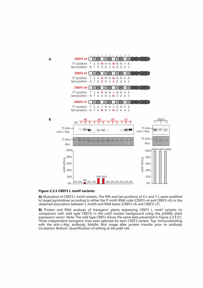
CREF3 v65th position T S A P N N I N N V Alast position N T G D N S N D D A S
L S P L S P L S P2 L2 S2 E1 E2 DYW
CREF3 v41 2 3 4 5 6 7 8 9 10
5th position T S A N N N N N N V Alast position N T G D N S S D D A S
11
L S P L S P L S P2 L2 S2 E1 E2 DYW
CREF3 v55th position T S A N N N N N N V Alast position N T G S N S S D D A S
L S P L S P L S P2 L2 S2 E1 E2 DYW
CREF3 v75th position T S A I N N I N N V Alast position N T G N N S N D D A S
L S P L S P L S P2 L2 S2 E1 E2 DYW
v71 2 3
Blot
NCv6
1 2 3v4
1 2 3v5
1 2 3
anti-c-Myc75 kDa
75 kDa
0% 4%0% 0%
16%16%
0% 0% 0% 0% 0% 0%0%
20%
40%
60%
80%
100%
psbEediting
0%
CREF31 2 3
anti-c-Myc
Blot
75 kDa
75 kDa
0%
100%100%100%
20%
40%
60%
80%
100%
psbEediting
A
B
Figure 2.3.5 CREF3 L motif variants.A) Illustration of CREF3 L motif variants. The fifth and last positions of 4-L and 7-L were modifiedto target pyrimidines according to either the P motif-RNA code (CREF3-v4 and CREF3-v5) or theobserved association between L motifs and RNA bases (CREF3-v6 and CREF3-v7).B) Protein and RNA analyses of transgenic plants expressing CREF3 L motif variants (incomparison with wild type CREF3) in the cref3 mutant background using the pGWB2 plantexpression vector. Note: The wild type CREF3 shows the same data presented in Figure 2.3.9 (C).Three independent transgenic lines were selected for each CREF3 variant. Top: Immunoblottingwith the anti-c-Myc antibody; Middle: Blot image after protein transfer prior to antibodyincubation; Bottom: Quantification of editing at the psbE site.

CREF3-v81 2 3 4 5 6 7 8 9 10
5th position T S A S N N T N N V Alast position N T G D N S D D D A D
11
L S P L S P L S P2 L2 S2 E1 E2 DYW
CREF3-v95th position T S A S N N T N N V Alast position N T G D N S D D D A S
L S P L S P L S P2 L2 S2 E1 E2 DYW
A
B
C
anti-c-Myc
Blot
75 kDa
75 kDa
CREF3
1 2cref3 v8
1 2v9
100%
0%
100% 100%
78%84%
0%
20%
40%
60%
80%
100%
psbEediting
CREF3 L2CRR28 L2 33%
OTP81/QED1 S2CREF3 S2 59%
Figure 2.3.6 CREF3 L2 and S2 motif variants.A) Illustration of the CREF3 S2 [AD] variant (CREF3-v8), where the last position of CREF3-S2 motifwas modified from S to D.B) Illustration of the CREF3 L2-S2 replacement variant (CREF3-v9). Top: The CREF3-L2 motif wasreplaced by the L2 motif from chloroplast editing factor CRR28 and the CREF3-S2 motif wasreplaced by the S2 motif from chloroplast editing factor OTP81/QED1. Bottom: Alignments ofCREF3-L2 with CRR28-L2, and CREF3-S2 with OTP81/QED1-S2. Dots indicate amino acidsconserved between the two aligned sequences (including the fifth and last amino acids).Percentages indicate the degree of sequence similarity between the two aligned sequences.Amino acids that belong to the helix a or helix b of each motif are boxed separately.C) Protein and RNA analyses of transgenic plants expressing CREF3 L2 and S2 motif variants (incomparison with wild type CREF3) in the cref3 mutant background using the pAEF3-Ali plantexpression vector. Two independent transgenic lines were selected for each CREF3 variant. Top:Immunoblotting with the anti-c-Myc antibody; Middle: Blot image after protein transfer prior toantibody incubation; Bottom: Quantification of editing at the psbE site.

A
B
psbEEditing
(position
0)
anti-c-Myc
Blot
0%
20%
40%
60%
80%
100%
1 2v10 v11 v12
89%100% 100%100%
12% 10%
1 2 1 2cref3
0%
1 2v10 v11 v12
1 2 1 2Col-0
anti-c-Myc
Blot
C
cref3
CREF3 1
2
v10
v11
v12
1
2
1
2
1
2
0 +1Position 0 +1Position
Col-0
v10
v11
v12
1
2
1
2
1
2
D
CREF3-v101 2 3 4 5 6 7 8 9 10 11
L S P L S P L S P2 L2 S2 E1 E2 DYW
CREF3-v11 L1
S2
P3
L4
S5
P6
L7
S8
P29
L210
S211
E1 E2 DYW
CREF3-v12 L1
S2
P3
L4
S5
P6
L7
S8
P29
L210
S211
E1 E2 DYW
psbE editing site c a g g c c g u u u u g a u C C u0 +1
L1
S2
P3
L4
S5
P6
L7
S8
P29
L210
S211
E1 E2 DYWCREF3
E
Figure 2.3.7 Inserting flexible linkers between CREF3 C-terminal motifs.A) Illustration of the CREF3 C-terminal linker variants (CREF3-v10, v11, and v12). In CREF3-v10,the flexible linker (GGGGS)2 was inserted between the P2 and L2 motifs; In CREF3-v11, the flexiblelinker (GGGGS)2 was inserted between the S2 and E1 motifs; In CREF3-v12, the flexible linker(GGGGS)2 was inserted between the E2 motif and the DYW domain.B) Protein and RNA analyses of transgenic plants expressing CREF3 C-terminal flexible linkervariants in the cref3 mutant background using the pAEF3-Ali plant expression vector. Twoindependent transgenic lines were selected for each CREF3 variant. Top: Immunoblotting with theanti-c-Myc antibody; Middle: Blot image after protein transfer prior to antibody incubation;Bottom: Quantification of editing at the psbE site.C) Protein analyses of transgenic plants expressing CREF3 C-terminal flexible linker variants in thewild type Col-0 background using the pAEF3-Ali plant expression vector. Top: Immunoblottingwith the anti-c-Myc antibody; Bottom: Blot image after protein transfer prior to antibodyincubation.D) Sanger sequencing of the PCR products around position 0 and +1 of the psbE editing site,prepared from the transgenic plants expressing CREF3 C-terminal flexible linker variants in thecref3 mutant background.E) Sanger sequencing of the PCR products around position 0 and +1 of the psbE editing site,prepared from the transgenic plants expressing CREF3 C-terminal flexible linker variants in thewild type Col-0 background.

anti-c-Myc
Blot
75 kDa
75 kDa
CREF3 1 2NC 1 2A A B B
100%
0% 0% 0% 0% 0%0%
20%
40%
60%
80%
100%
psbEediting
CREF3-AA L1
S2
P3
L4
S5
P6 7 8 9
L210
S2
5th position T S A S N N T N N V Alast position N T G D N S D D D A S
11
E1 E2 DYWL S P
CREF3-BB L1
S2
P3 4 5 6
L7
S8
P29
L210
S2
5th position T S A S N N T N N V Alast position N T G D N S D D D A S
11
E1 E2 DYWL S P2
CREF3 L1
S2
P3
L4
S5
P6 7 8 9
L210
S2
5th position T S A S N N T N N V Alast position N T G D N S D D D A S
11
E1 E2 DYWL S P2
Triplet A Triplet BA
B
C
psbE c a g g c c g u u u u g a u C c
Figure 2.3.8 Replacing the motif triplets in CREF3.A) Illustration of two sets of CREF3 LSP motif triplets both targeting the nucleotides “GYY”. TripletA: motifs 4-6; Triplet B: motifs 7-9.B) Illustration of CREF3 triplet replacement variants. In CREF3-AA, the backbone of triplet B wasreplaced by triplet A while maintaining the matches with the psbE editing site through the fifthand last amino acids of each motif. In CEF3-BB, the backbone of triplet A was replaced by tripletB while maintaining the matches with the psbE editing site.C) Protein and RNA analyses of transgenic plants expressing CREF3-AA or CREF3-BB (incomparison with wild type CREF3) in the cref3 mutant background using the pAEF3-Ali plantexpression vector. Two independent transgenic lines were selected for each CREF3 variant. Top:Immunoblotting with the anti-c-Myc antibody; Middle: Blot image after protein transfer prior toantibody incubation; Bottom: Quantification of editing at the psbE site.

probeedited
unedited
probeedited
Col-0
cref3 dv1
1 2CREF31 2
ndhB_95252
1 2 3 4 5 6 7 8 9 10
5th position A V Alast position G A S
11
P L S P L S P2 L2 S2 E1 E2 DYWdCREF3 L S
T SN T
SD
TD
A
dCREF3 v1 L1
S2
P3
L4
S5
P6
L7
S8
P29
L210
S2
5th position T S A S S T T N N V Alast position N T G D N D D D S A S
11
E1 E2 DYW
ndhB_95252 u a g g a g g u c u u c c u C cpsbE editing site c a g g c c g u u u u g a u C c
dCREF3-v2 L1
S2
P3
L4
S5
P6
L7
S8
P29
L210
S2
5th position T S A S S N T S N V Alast position N T G D N D D N D A S
11
E1 E2 DYW
ycf1_128321 a a g g a u g a u g a a u u C cpsbE editing site c a g g c c g u u u u g a u C c
B
NCdv1
1 2 3CREF3
1 2 3dv2
1 2 3
anti-c-Myc
Blot
75 kDa
75 kDa
0%
24%21%16%7%
16%
29%
100%100%100%
20%
40%
60%
80%
100%
psbEediting
C
D
probeedited
Col-0
cref3 CREF3
1 2unedited
dv21 2
probeunedited
editedycf1_128321
E
Figure 2.3.9 CREF3 redesign version 1 (dCREF3).A) Illustration of CREF3 redesign version 1. The fifth and last positions of motifs 5-S, 6-P, 8-S, and9-P were modified to match new potential editing sites in Arabidopsis chloroplasts. All othermotifs were kept the same.B) Two redesigned CREF3 editing factors are aligned to their chosen target sites, in comparisonwith the psbE editing site. The variant dCREF3-v1 targets a potential editing site at ndhB_95252.The variant dCREF3-v2 targets a potential editing site at ycf1_128321. Dark green boxes indicatematches. Light green boxes indicate partial mismatches. Red boxes indicate mismatches. “C”sindicate the edited cytidines.C) Protein and RNA analyses of transgenic plants expressing redesigned CREF3 version 1 (incomparison with wild type CREF3) in the cref3 mutant background using the pGWB2 plantexpression vector. Three independent transgenic lines were selected for each redesigned CREF3.Top: Immunoblotting with the anti-c-Myc antibody; Middle: Blot image after protein transfer priorto antibody incubation; Bottom: Quantification of editing at the psbE site.D) Editing at the new targeting site ndhB_95252 was elucidated by poison primer extension (PPE).E) Editing at the new targeting site ycf1_123821 was elucidated by poison primer extension (PPE).

1 2 3 4 5 6 7 8 9 10
5th position A V Alast position G A D
11
P L S P L S P2 L2 S2 E1 E2 DYWd2CREF3A
d2CREF3-v11 2
P3
L4
S5
P6
L7
S8
P29
L2 S2
5th position A S N N T N N V Alast position G D N S D D D A D
E1 E2 DYW
psbE transcript ca g g c c g u u u u g a u C c uuuggaacaacucgaug
1 2 3 4 5 6 7 8 9
E1 E2 DYWP
AGa
L
TNa
S
NDu
P
NDu
L
NDu
S
SNa
P2
TDg
L2
VAu
S2
ADa g a u C u
d2CREF3-v2
3 ’5 ’18 bp
psbE_64109 psbE_64078
B
anti-c-myc
Blot
75 kDa
75 kDa
1 2 1 2CREF3 cref3d2v1 d2v2
0%
20%
40%
60%
80%
100%
psbE(64109)editing
100%
71% 70%
0% 0% 0%
C
Figure 2.3.10 CREF3 redesign version 2 (d2CREF3).A) Illustration of CREF3 redesign version 2. The N-terminal 1-L and 2-S motifs were truncated. Six motifs (4-L, 5-S, 6-P, 7-L, 8-S, and 9-P2) were modified to match potential editing sitesin Arabidopsis chloroplasts. The last position of the S2 motif was modified to D. All other motifs were kept the same.B) Two redesigned CREF3 editing factors aligned to the natural psbE editing site (psbE_64109) and a new potential editing site on the psbE transcript (psbE_64078) downstream to thenatural editing site. The redesigned variant d2CREF3-v1 targets the original psbE editing site (psbE_64109). The redesigned variant d2CREF3-v2 targets psbE_64078. Dark green boxesindicate matches. Transparent boxes indicate sequence similarity between psbE_64109 and psbE_64078 in addition to the matches. “C”s indicate the edited cytidines.C) Protein and RNA analyses of transgenic plants expressing redesigned CREF3 version 2 (in comparison with wild type CREF3) in the cref3 mutant background using the pAEF3-Ali plantexpression vector. Two independent transgenic lines were selected for each redesigned CREF3. Top: Immunoblotting with the anti-c-Myc antibody; Middle: Blot image after proteintransfer prior to antibody incubation; Bottom: Quantification of editing at the psbE site.

1 2 3 4 5 6 7 8 9 10
5th position A V Alast position G A D
11
P L S P L S P2 L2 S2 E1 E2 DYWd2CREF3-XA
75 kDa100 kDa
anti-c-myc
75 kDa100 kDa
Blot
flv 1 2 3 4 5 6 7 8 1 2 3 4FLV d2-FLV
flv FLV
d2CREF3-FLVCol-0
0%
20%
40%
60%
80%
100%
0% 0%
90%
18%
rpoC1(21806)editing
D
BFLV
5th positionlast position
1 2 3 4
E1 E2185 6 7 8 9 10 11 12 13 14 15 16 17
P2 L2 S2P PL S PL S L L SS
NSc
P
TDg
L
TDa
S
NDu
P
NDu
ADa
NDc
TDg
VTa
ADg g u u C a
VEa
NDu
NDu
SKu
NDu
SRc
NDc
CDurpoC1 (21806)
DYW
1 2 3 4 5 6 7 8 9
d2CREF3-FLV
5th positionlast position
E1 E2
c
P
AGg
L S P L S P2 L2
VAa
S2
ADg g u u C aauuu ucc urpoC1 (21806)
DYW
TNa
NDu
NDu
TNa
NTc
TDg
gpsbE editing site g c c g u u u u g a u C c
C
Figure 2.3.11 CREF3 redesign version 3 (d2CREF3-X).A) Illustration of CREF3 redesign version 3. The N-terminal 1-L and 2-S motifs were truncated.Six motifs (4-L, 5-S, 6-P, 7-L, 8-S, and 9-P2) were modified to fully match the natural editing sitesof choice in Arabidopsis chloroplasts. The E1-E2-DYW domain in CREF3 was replaced with theE1-E2-DYW domains from the natural PPR editing factors responsible for editing the chosensites. The last position of the S2 motif was modified to D.B) The chloroplast editing factor FLV and d2CREF3-FLV are aligned with the rpoC1_21806 editingsite. Dark green boxes indicate matches. Light green boxes indicate partial matches. Red boxesindicate mismatches. “C”s indicate the edited cytidines.C) Complementation of editing in transgenic plants expressing FLV and d2CREF3-FLV in the flvmutant background, using the pAEF3-Ali plant expression vector. 3 to 5 seedlings representingindependent transgenic lines for each genotype were combined for editing quantification.D) Multiple primary transgenic lines were screened for the expression of FLV and d2CREF3-FLVby immunoblotting. Top: Immunoblotting with the anti-c-Myc antibody; Bottom: Blot imageafter protein transfer prior to antibody incubation.

75 kDa100 kDa
anti-c-myc
75 kDa100 kDa
Blot
1 2 3 4 5 6 1 2 3 4 5 6 7YS1 d2-YS1
0% 0%
92%83%
rpoB
(25992)editing
ys1 YS1
d2CREF3-YS1Col-0
0%
20%
40%
60%
80%
100%
YS1
5th positionlast position
1 2 3 4 5 6 7 8 9 10 11 12 13 14 15 16
P
ND
L
LW
S
NT
SS
NN
P
ND
L
MD
S
TD
P2
TD
L2
VE
S2
GN
E1 E2S
SN
P
TN
L
PD
P
ND
L
VN
S
NN
rpoB (25992) u c c c u u g g a a c u u C uuaua ag
DYW
1 2 3 4 5 6 7 8 9
d2CREF3-YS1
5th positionlast position
E1 E2 DYW
rpoB (25992) u
P
AGc
L
NSc
S
NNc
P
NDu
L
NDu
S
SDg
P2
TDg
L2
VAa
S2
ADa c u u C uuaua ag
gpsbE editing site g c c g u u u u g a u C c
E
F G
Figure 2.3.11 CREF3 redesign version 3 (d2CREF3-X). (cont.)E) The chloroplast editing factor YS1 and d2CREF3-YS1 are aligned with the rpoB_25992 editingsite. Dark green boxes indicate matches. Light green boxes indicate partial matches. Red boxesindicate mismatches. “C”s indicate the edited cytidines.F) Complementation of editing in transgenic plants expressing YS1 and d2CREF3-YS1 in the ys1mutant background, using the pAEF3-Ali plant expression vector. 3 to 5 seedlings representingindependent transgenic lines for each genotype were combined for editing quantificationG) Multiple primary transgenic lines were screened for the expression of YS1 and d2CREF3-YS1by immunoblotting. Top: Immunoblotting with the anti-c-Myc antibody; Bottom: Blot imageafter protein transfer prior to antibody incubation.

CREF3-PLS211 2
P3
L4
S5
P6
L7
S8
P29
L210
S2
5th position A N V Alast position G D A D
11
E1 E2 DYW
6x
CREF3-SPL211 2
P3
L4
S5
P6
L7
S8
P29
L210
S2
5th position A N N V Alast position G D D A D
11
E1 E2 DYW
6x
1 2 3 4 5 6 7 8 9 10
5th position Alast position G
V AA D
11
P L S P L S P2 L2 S2 E1 E2 DYWCREF3-LSP21
6x
T N ND D D
A
L motif S motif P motif
LSP triplet-1
L motif S motif P motif
LSP triplet-2
An assembly fragment
B
5th positionlast positionUnique assembly arm (20 bp)PCR primers
Figure 2.3.12 Extension of CREF3 motifs.A) Illustration of CREF3 extension using three different arrangements of motif triplets. Triplet LSP:motifs 4-6; Triplet SPL: motifs 5-7; Triplet PLS: motifs 6-8. The 1-L and 2-S motifs were truncated.The last position of S2 motif was modified to D. The triplet LSP, SPL, or PLS was extended six timesin each variant (CREF3-LSP21, CREF3-SPL21, or CREF3-PLS21), while being sandwiched by thenative N-terminal and C-terminal motifs of CREF3. The extended CREF3 variants were designed tofully match the psbE editing site.B) Illustration of the CREF3-LSP21 cloning strategy by Gibson assembly of LSP triplet fragments.One of the six LSP triplet fragments is indicated by the double-headed arrow. The fragment wasgenerated by two rounds of PCR using mutagenising primers indicated by the single-headedarrows. The unique 20-bp Gibson assembly arms introduced by PCR are indicated by boxesshaded with stripe patterns. Cloning of CREF3-SLP21 and CREF3-PLS21 followed the samestrategy.
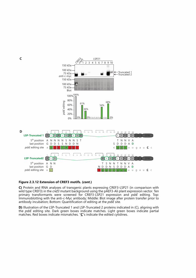
5th positionlast position
LSP-Truncated2 L S P2 L2 S2 E1 E2 DYWL S PP L S P
AG
ND
ND
VA
AD
NDN
NTD
NS
TD
NN
23 241 2 3 4 20 21 225 6 7 8 9 10 11 12 13 14 15 16 17 18 19
SD
c a g g c c g u u u u g a u C capsbE editing site
5th positionlast position
LSP-Truncated 1 L S P2 L2 S2 E1 E2 DYWP L S P L S P L S P
AG
ND
ND
VA
AD
ND
NSNS
ND
ND
ND
SN
SN
TS
TD
23 241 2 3 4 20 21 225 6 7 8 9 10 11 12 13 14 15 16 17 18 19
psbE editing site g a u C cg u u u ua g g c ccaaa u
anti-c-myc75 kDa100 kDa
150 kDa
Blot75 kDa100 kDa
150 kDa
CREF3cref31 2 3 4 5 6 7 8 9 10
LSP21
0%20%40%60%80%100%
psbEediting
100%
61%
36%54%
68%
0% 0%n.a.n.a.n.a. 0%n.a.
Truncated 1Truncated 2
C
D
Figure 2.3.12 Extension of CREF3 motifs. (cont.)C) Protein and RNA analyses of transgenic plants expressing CREF3-LSP21 (in comparison withwild type CREF3) in the cref3mutant background using the pAEF3-Ali plant expression vector. Tenprimary transformants were screened for CREF3-LSP21 expression and psbE editing. Top:Immunoblotting with the anti-c-Myc antibody; Middle: Blot image after protein transfer prior toantibody incubation; Bottom: Quantification of editing at the psbE site.D) Illustration of the LSP-Truncated 1 and LSP-Truncated 2 proteins indicated in (C), aligning withthe psbE editing site. Dark green boxes indicate matches. Light green boxes indicate partialmatches. Red boxes indicate mismatches. “C”s indicate the edited cytidines.

SPL-Truncated&Mutated5th positionlast position
S23
P224
L2 S21 2
P3
L4
AG
ND
ND
VA
AD
E1 E2 DYW20 21 22
S5
P6
L7
S8
P9
L10 11 12 13 14 15 16 17 18 19
ND
ND
TS
ND
ND
ND
SN
psbE editing site g a u C cu u u ugca g g cca
Blot75 kDa100 kDa
150 kDaanti-c-myc
75 kDa100 kDa
150 kDaCREF3cref31 2 3 4 5 6 7 8 9 10
SPL21
Full-length
Truncated
0%20%40%60%80%100%
psbEediting
100%
0% 0% 0%n.a.n.a. 0% 0% 0% 0% 0% 0%
E
F
CREF3-SPL21
5th positionlast position
S23
P224
L2 S21 2
P3
L4
AG
ND
ND
VA
AD
E1 E2 DYWS20
P21
L22
S5
P6
L7
S8
P9
L10
S11
P12
L13
S14
P15
L16
S17
P18
L19
ND
NSNS
ND
ND
ND
SN
SN
ND
TN
SN
TNNS
SN
TD
TD
NS
TD
NN
Figure 2.3.12 Extension of CREF3 motifs. (cont.)E) Protein and RNA analyses of transgenic plants expressing CREF3-SPL21 (in comparison withwild type CREF3) in the cref3mutant background using the pAEF3-Ali plant expression vector. Tenprimary transformants were screened for CREF3-SPL expression and psbE editing. Top:Immunoblotting with the anti-c-Myc antibody; Middle: Blot image after protein transfer prior toantibody incubation; Bottom: Quantification of editing at the psbE site.F) Illustration of the truncated and the full-length CREF3-SPL proteins indicated in (E), aligningwith the psbE editing site. The SLP-Truncated protein also carried mutations in the 4-P and 5-Lmotifs. Dark green boxes indicate matches. Light green boxes indicate partial matches. Red boxesindicate mismatches. “C”s indicate the edited cytidines.
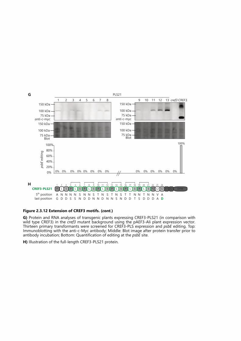
anti-c-myc75 kDa100 kDa
150 kDa
75 kDa100 kDa
150 kDa
anti-c-myc
75 kDa100 kDa
150 kDa
BlotBlot75 kDa100 kDa
150 kDa
CREF3cref31 2 3 4 5 6 7 8 9 10PLS21
11 12 13
0%20%40%60%80%100%
psbEediting
100%
0% 0% 0%0% 0% 0% 0% 0% 0%0% 0% 0% 0% 0%
G
P2 L2 S2CREF3-PLS21
5th positionlast position
ND
VA
AD
E1 E2 DYWP L S
AG
P L SP L S P L S P L S P L S P L S23 241 2 3 4 20 21 225 6 7 8 9 10 11 12 13 14 15 16 17 18 19
NDND
NS
NS
SN
ND
ND
SN
TN
ND
SN
TN
NS
SN
TD
TD
NTNS
TDND
H
Figure 2.3.12 Extension of CREF3 motifs. (cont.)G) Protein and RNA analyses of transgenic plants expressing CREF3-PLS21 (in comparison withwild type CREF3) in the cref3 mutant background using the pAEF3-Ali plant expression vector.Thirteen primary transformants were screened for CREF3-PLS expression and psbE editing. Top:Immunoblotting with the anti-c-Myc antibody; Middle: Blot image after protein transfer prior toantibody incubation; Bottom: Quantification of editing at the psbE site.H) Illustration of the full-length CREF3-PLS21 protein.

75
Chapter 3 General Discussion
The specificity of PPR editing factors in Arabidopsis chloroplasts
PPR editing factors in Arabidopsis chloroplasts are generally very specific. Out of the 31
characterised major editing sites, there are 13 single-target PPR editing factors (AEF1,
CREF3, CREF7, CRR21, CRR4, DOT4/FLV, ELI1, LPA66, OTP80, OTP85, OTP86,
RARE1, and YS1), plus AEF4 reported in this work (Chapter 2.2), responsible for 14
editing sites; there are six multi-target editing factors (CLB19, CRR22, CRR28, OTP82,
OTP84 and OTP81/QED1), responsible for the other 17 editing sites. The editing factor
to site ratio is between 1:1 and 1:5. My work provides further evidence for the high
specificity of RNA editing in Arabidopsis chloroplasts. First, the off-target editing events
of CLB19 at ycf3-i2 are at a low level (Chapter 2.1, Figure 2.1.2). Second, the off-target
editing events of AEF4 are rare and at low levels even upon over-expression (Chapter
2.2, Figure 2.2.4). Third, two highly similar editing sites psbE and petL can be
distinguished (discussed in Chapter 2.3).
What determines the specificity of PPR editing factors in chloroplasts? Considering the
intrinsic features of PPR editing factors, the editing specificity is predominantly
determined by the interaction between the fifth and last position of a P- or S-type PPR
motif and the aligned RNA base. The L-type motif, when encoded with similar RNA-
recognising amino acids, also contributes to targeting specificity, as I found in Chapter
2.3 (Figure 2.3.5). I showed that the L motifs in CREF3 are involved in G-recognition
following the canonical PPR-RNA code. The one-motif to one-base recognition code
forms the basis of editing site prediction methods (Barkan et al., 2012, Takenaka et al.,
2013, Yagi et al., 2013a). However, if these are the only determinants of RNA editing
specificity, there should be more editing events in chloroplasts than are observed. In
Chapter 2.1 (Figure 2.1.6), I showed that 494 predicted editing sites of CLB19 in
Arabidopsis chloroplasts match equivalently or better to its PPR binding motifs than the
three known editing sites. Moreover, editing in planta also seems to be more specific
than PPR binding in vitro. In Chapter 2.3 (Figure 2.3.1), I showed that recombinant
CREF3 protein does not distinguish between the psbE and petL probes in binding
reactions in vitro, in contradiction to the in planta editing analysis.
So why are PPR editing factors in chloroplasts much more specific than expected from
in silico predictions and in vitro binding results? First, there could be non-canonical yet
sequence-specific interactions between PPR motifs and target RNA. For example, both
the PPR10 (a P-subfamily protein) crystal structure (Yin et al., 2013) and the Bind-n-seq
assay with PPR10 (Miranda et al., 2017) showed that canonical PPR codes can have

76
alternative RNA selectivity. Moreover, there are many other non-canonical combinations
of amino acids encoded at the fifth and last position of PPR motifs, the functions of which
remain unclear. Second, PPR-RNA recognition could be conditional, especially in the
case of L-type motifs. A recent publication on MORF9/RIP9 and PLS-type PPR proteins
showed that L motif-A recognition following the PPR-RNA code is facilitated by
MORF9/RIP9 binding in vitro (Yan et al., 2017). The results imply that L motifs may
contribute to editing specificity only upon MORF/RIP binding in planta. Third, the editing
specificity (Farre et al., 2001) may be determined by substrate specificity of the
deaminase domain in PPR editing factors, as demonstrated for other deaminases such
as ADAR (Kuttan and Bass, 2012) and APOBEC (Rathore et al., 2013) (discussed in
Chapter 2.1 & 2.3). Fourth, PPR editing factors specifically recruit co-factors in planta
that may confer RNA specificity. For example, the PPR editing factors CRR28 and
OTP82 specifically bind to an editing co-factor ORRM1, and ORRM1 also shows some
level of specificity towards the RNA targets of CRR28 and OTP82 (Sun et al., 2013).
Last, not all predicted sites or sites recognised in vitro may be accessible to PPR editing
factors in planta, where PPR editing factors and the predicted target transcripts are not
co-expressed, and RNA forms secondary structures or is bound by other proteins. RNA
secondary structure prevents PPR binding in vitro (Kindgren et al., 2015), implying that
it would inhibit RNA editing in planta. RNA structures and protein-RNA interactions can
be partially modulated by RNA chaperones and helicases. A recent study on chloroplast
RNA helicase ISE2 reported editing defects at 12 sites in co-suppressed transgenic lines,
and association between ISE2 and chloroplast transcripts involved in editing (Bobik et
al., 2017). It implies that this RNA helicase may be required to remodel RNA structures
or protein-RNA interactions at the editing sites.
In conclusion, the specificity of PPR editing factors in planta is probably determined by
combinatorial effects including the intrinsic specificity of PPR editing factors (including
binding and editing), PPR specificity modulated by recruited co-factors, intrinsic
specificity of recruited co-factors, and editing site accessibility.
Consequences of off-target editing events
In general, what possible effects might off-target editing have? First, instead of
suppressing genome mutations, off-target editing may induce silent or missense point
mutations if it happens in a coding region. Second, off-target editing may alter post-
transcriptional regulation. It could terminate translation by creating premature stop
codons from arginine (R-CGA) or glutamine (Q-CAA/CAG) codons. It could also disrupt
intron splice sites by eliminating C-G pairing in essential stem-loop structures. Moreover,
the consequences of off-target editing events could be secondary, as editing may disrupt

77
or create other PPR binding sites, subsequently affecting RNA processing, splicing, and
stability.
In this work, the off-target editing events detected for CLB19 and AEF4 are rare and only
barely detectable. I did not observe any visible or molecular phenotypes as a
consequence of these off-target editing events (Chapter 2.1, Figure 2.1.3; Chapter 2.2,
Figure 2.2.3 & 2.2.4). This illustrates that when considering the effects of editing events,
one should consider the proportion of transcripts that are edited. No chloroplast editing
sites are 100% edited in all cells (Ruwe et al., 2013), showing that complete conversion
to the edited form is not necessary for significant consequences. This is further
demonstrated by the case of rps14-2 editing in this work, where the proportion of edited
transcript can be reduced from 86% to 58% without apparently affecting the function(s)
of the rps14 gene product (Chapter 2.2, Figure 2.2.3 & 2.2.4). Therefore, a low proportion
of non-functional transcripts created by off-target editing events are unlikely to cause
phenotypic effects in plants. On the other hand, off-target editing events could create
dominant mutations that lead to functional defects. Loss of rpoC1 editing leads to
relatively severe phenotypic effects even though as few as 20% of rpoC1 transcripts are
edited (Chapter 2.3, Figure 2.3.11), implying that low-level off-target editing could cause
significant effects if it causes a gain of function, rather than a loss of function.
Off-target events could be consequential even in the absence of editing, as binding of
PLS-subfamily PPR proteins have been shown to affect other RNA processing events,
including editing of other sites (e.g. (Hayes et al., 2015)), splicing (e.g. (Yap et al., 2015)),
and 5’ or 3’ end processing (e.g. (Hammani et al., 2016)).
Interplay between RNA processing events and implications for inducing editing at
novel sites
There are multiple RNA processing events in chloroplasts that occur between
transcription and translation, including maturation, splicing, editing and so on (Stern et
al., 2010). It is generally believed that these events do not happen in a particular order,
but this may not always be true, as shown by known examples of the dependency of one
RNA processing event on another. Taking splicing and editing as an example, splicing
of some group II introns, such as the trans-splicing group II intron c/d in Oenothera
mitochondrial nad1 (Börner et al., 1995) and the group II intron in potato mitochondrial
rps10 gene (Castandet et al., 2010), are dependent on editing sites at critical positions
of the intron structures. In these cases, editing precedes splicing. On the other hand,
some exon editing sites close to splice sites are only edited in spliced RNA, such as the
ndhA editing site located 12-nt from the intron-exon splice site in spinach chloroplasts

78
(Schmitz-Linneweber et al., 2001) and the atp9 editing site located 8-nt from a splice site
in moss (Ichinose et al., 2013). In these cases, splicing precedes editing.
There could also be multiple coordinated RNA processing events. Taking petL as an
example, the petL editing site is in close proximity to the petL start codon (Chapter 2.3).
Therefore the petL editing specificity factor is likely to bind in the 5’UTR region of petL,
where at least two other proteins and/or associated complexes are bound. The petL
editing site is about 30 nt downstream of the binding site of the P-class PPR protein
PGR3 implicated in petL operon RNA stabilisation and translation activation (Cai et al.,
2011). The petL editing site is 11 nt downstream of the Shine-Dalgarno sequence that is
bound by the ribosome during translation initiation (Cai et al., 2011). Based on the size
of typical PPR editing factor binding sites, the petL editing factor binding site is likely to
overlap with the Shine-Dalgarno sequence bound by initiating ribosomes. Therefore,
petL editing is potentially coordinated with the other two petL processing events.
Interplay between RNA processing events has implications for inducing editing at novel
sites by designer PPR editing factors. As previously discussed in Chapter 2.3, editing
site accessibility should be considered while designing site-recognition PPR editing
factors. Moreover, the site accessibility may be dynamically altered by other RNA
processing events at different co-transcriptional or post-transcriptional stages. For
example, splicing may introduce new binding sites for PPR editing factors. Interactions
with other PPR proteins and/or the ribosome may block access for PPR editing factors.
Therefore, it is important to accurately evaluate the accessibility of desired editing sites,
with regards to the other relevant RNA processing events. In vivo RNA structure probing
techniques can reveal the “average” structure of each transcript across different RNA
processing stages (e.g. DMS-MaPseq (Zubradt et al., 2017)). Ideally, it should be
combined with RNA fractionation techniques to evaluate the structure of a desired editing
site at a particular RNA processing stage, and to identify the optimal stage to target for
editing. Nascent RNA, containing mostly the pre-processed RNA species, can be tagged
by run-on techniques (Core et al., 2008) or isolated by immunoprecipitation (Churchman
and Weissman, 2011). Ribosome-associated RNA, containing mostly the mature and
translating RNA species, can be isolated by polysome gradients and/or
immunoprecipitation.
Perspectives for the design of PPR editing factors
There are three strategies proposed for designing PPR proteins in general – 1) modifying
natural PPR proteins; 2) creating chimeric PPR proteins; and 3) constructing synthetic

79
PPR proteins. Here I discuss the advantages and limitations of each strategy in the
context of designing PPR editing factors.
The strategy of modifying natural PPR editing factors is based on existing models of
PPR-editing site recognition (e.g. CREF3-psbE), and alters individual RNA-recognising
motifs to match the desired RNA editing site(s). The advantage is that the template of a
natural editing factor is already fully functional in the in vivo context, at both the RNA-
binding surface and the protein-interacting surface. Minimal engineering is required,
focused only on modifying the targeting specificity. The first limitation is that the natural
editing factors contain non-canonical interactions with RNA that are probably selected
and fixed by evolution. These interactions are difficult to identify and even more difficult
to modify to match other RNA bases. The second limitation is that the motif arrangement
of natural editing factors, including the order and number of motifs, is not flexible for
modifications (examples are provided in Chapter 2.3). This inflexibility also poses
complications for designing chimeric PPR proteins by shuffling PPR motifs/blocks of
diverse origins.
The strategy of constructing synthetic PPR editing factors is based on artificial helix-loop-
helix protein scaffolds derived from the consensus sequences of natural PPR motifs, and
adds the RNA-recognising and protein-interacting features as desired. Synthetic PPR
proteins could potentially solve many of the issues met with natural PPR proteins, as
demonstrated by the promising biochemical and structural data obtained for both the P-
subfamily proteins (Coquille et al., 2014, Gully et al., 2015, Shen et al., 2016) and a PLS-
subfamily protein (Yan et al., 2017). First, the synthetic PPR motifs only bind and
recognise RNA following the designed canonical interactions. Second, the motif
arrangement of synthetic PPR proteins is uniform, therefore is flexible to be extended or
shortened as needed. However, synthetic PPR proteins probably have other limitations,
since the consensus sequences of PPR motifs are potentially biased based on current
knowledge. For example, the consensus sequences of different types of PPR motifs all
encode ND or NN as the combination of the fifth and last positions. Without knowing that
these two are the RNA-recognising positions, ND or NN would have been kept in the
synthetic PPR motifs, and as a result, the synthetic PPR motifs would only target C or U.
Such biases may have been unintentionally maintained at other positions, especially at
the protein-interacting surface, potentially leading to missing features in the synthetic
PPR proteins. There are no reports of synthetic PPR proteins being functional in planta
so far.
Although the two PPR design strategies are distinct in nature, fundamentally they both
rely on the understanding of how the organellar editing system works. By identifying new

80
PPR editing factors and investigating off-target effects, I have extended our
understanding of PPR targeting specificity. By testing modified editing factors in planta,
I have gained insights into the functional diversity of PPR motifs, and demonstrated the
challenges to be overcome in retargeting PPR editing factors. My work has helped
advance ideas on how to design PPR proteins that could work within the editing complex
in planta. As many as possible of the following questions need to be answered to guide
PPR editing factor design in the future. Can the PPR-RNA recognition code be optimised
by involving other amino acids in PPR motifs? How is the specificity of editing
determined? How do PPR motifs interact with the co-factors? How do the co-factors
modulate PPR specificity? Do the co-factors confer targeting specificity? Are there
distinct RNA structural features at the editing sites?

81
References
Ahlert, D., Ruf, S. & Bock, R. 2003. Plastid protein synthesis is required for plant development
in tobacco. Proceedings of the National Academy of Sciences, 100, 15730-15735.
Aryamanesh, N., Ruwe, H., Sanglard, L. V. P., Eshraghi, L., Bussell, J. D., Howell, K. A.,
Small, I. & Des Francs-Small, C. C. 2017. The Pentatricopeptide Repeat Protein
EMB2654 Is Essential for Trans-Splicing of a Chloroplast Small Ribosomal Subunit
Transcript. Plant Physiology, 173, 1164-1176.
Barkan, A., Rojas, M., Fujii, S., Yap, A., Chong, Y. S., Bond, C. S. & Small, I. 2012. A
combinatorial amino acid code for RNA recognition by pentatricopeptide repeat
proteins. PLoS Genet, 8, e1002910.
Barkan, A. & Small, I. 2014. Pentatricopeptide repeat proteins in plants. Annu Rev Plant Biol,
65, 415-42.
Bensmihen, S., To, A., Lambert, G., Kroj, T., Giraudat, J. & Parcy, F. 2004. Analysis of an
activated ABI5 allele using a new selection method for transgenic Arabidopsis seeds.
FEBS Lett, 561, 127-31.
Bentolila, S., Heller, W. P., Sun, T., Babina, A. M., Friso, G., Van Wijk, K. J. & Hanson, M. R.
2012. RIP1, a member of an Arabidopsis protein family, interacts with the protein
RARE1 and broadly affects RNA editing. Proc Natl Acad Sci U S A, 109, E1453-61.
Bentolila, S., Oh, J., Hanson, M. R. & Bukowski, R. 2013. Comprehensive high-resolution
analysis of the role of an Arabidopsis gene family in RNA editing. PLoS Genet, 9,
e1003584.
Bobik, K., Mccray, T. N., Ernest, B., Fernandez, J. C., Howell, K. A., Lane, T., Staton, M. &
Burch-Smith, T. M. 2017. The chloroplast RNA helicase ISE2 is required for multiple
chloroplast RNA processing steps in Arabidopsis thaliana. Plant J.
Boch, J., Scholze, H., Schornack, S., Landgraf, A., Hahn, S., Kay, S., Lahaye, T., Nickstadt, A.
& Bonas, U. 2009. Breaking the code of DNA binding specificity of TAL-type III
effectors. Science, 326, 1509-12.
Börner, G. V., Mörl, M., Wissinger, B., Brennicke, A. & Schmelzer, C. 1995. RNA editing of a
group II intron in Oenothera as a prerequisite for splicing. Molecular and General
Genetics MGG, 246, 739-744.
Boussardon, C., Avon, A., Kindgren, P., Bond, C. S., Challenor, M., Lurin, C. & Small, I. 2014.
The cytidine deaminase signature HxE(x)n CxxC of DYW1 binds zinc and is necessary
for RNA editing of ndhD-1. New Phytol, 203, 1090-5.
Boussardon, C., Salone, V., Avon, A., Berthome, R., Hammani, K., Okuda, K., Shikanai, T.,
Small, I. & Lurin, C. 2012. Two interacting proteins are necessary for the editing of the
NdhD-1 site in Arabidopsis plastids. Plant Cell, 24, 3684-94.
Bryant, N., Lloyd, J., Sweeney, C., Myouga, F. & Meinke, D. 2011. Identification of nuclear
genes encoding chloroplast-localized proteins required for embryo development in
Arabidopsis. Plant Physiol, 155, 1678-89.
Cai, W., Ji, D., Peng, L., Guo, J., Ma, J., Zou, M., Lu, C. & Zhang, L. 2009. LPA66 is required
for editing psbF chloroplast transcripts in Arabidopsis. Plant Physiol, 150, 1260-71.
Cai, W., Okuda, K., Peng, L. & Shikanai, T. 2011. PROTON GRADIENT REGULATION 3
recognizes multiple targets with limited similarity and mediates translation and RNA
stabilization in plastids. Plant J, 67, 318-27.
Castandet, B., Choury, D., Begu, D., Jordana, X. & Araya, A. 2010. Intron RNA editing is
essential for splicing in plant mitochondria. Nucleic Acids Res, 38, 7112-21.
Chateigner-Boutin, A. L., Ramos-Vega, M., Guevara-Garcia, A., Andres, C., De La Luz
Gutierrez-Nava, M., Cantero, A., Delannoy, E., Jimenez, L. F., Lurin, C., Small, I. &
Leon, P. 2008. CLB19, a pentatricopeptide repeat protein required for editing of rpoA
and clpP chloroplast transcripts. Plant J, 56, 590-602.
Chateigner-Boutin, A. L. & Small, I. 2007. A rapid high-throughput method for the detection
and quantification of RNA editing based on high-resolution melting of amplicons.
Nucleic Acids Res, 35, e114.
Cheng, S., Gutmann, B., Zhong, X., Ye, Y., Fisher, M. F., Bai, F., Castleden, I., Song, Y., Song,
B., Huang, J., Liu, X., Xu, X., Lim, B. L., Bond, C. S., Yiu, S. M. & Small, I. 2016.

82
Redefining the structural motifs that determine RNA binding and RNA editing by
pentatricopeptide repeat proteins in land plants. Plant J, 85, 532-47.
Churchman, L. S. & Weissman, J. S. 2011. Nascent transcript sequencing visualizes
transcription at nucleotide resolution. Nature, 469, 368-373.
Clough, S. J. & Bent, A. F. 1998. Floral dip: a simplified method for Agrobacterium-mediated
transformation of Arabidopsis thaliana. Plant J, 16, 735-43.
Coquille, S., Filipovska, A., Chia, T., Rajappa, L., Lingford, J. P., Razif, M. F., Thore, S. &
Rackham, O. 2014. An artificial PPR scaffold for programmable RNA recognition. Nat
Commun, 5, 5729.
Core, L. J., Waterfall, J. J. & Lis, J. T. 2008. Nascent RNA sequencing reveals widespread
pausing and divergent initiation at human promoters. Science, 322, 1845-8.
Cushing, D. A., Forsthoefel, N. R., Gestaut, D. R. & Vernon, D. M. 2005. Arabidopsis emb175
and other ppr knockout mutants reveal essential roles for pentatricopeptide repeat (PPR)
proteins in plant embryogenesis. Planta, 221, 424-36.
Despres, B., Delseny, M. & Devic, M. 2001. Partial complementation of embryo defective
mutations: a general strategy to elucidate gene function. Plant J, 27, 149-59.
Farre, J. C., Leon, G., Jordana, X. & Araya, A. 2001. cis Recognition elements in plant
mitochondrion RNA editing. Mol Cell Biol, 21, 6731-7.
Giege, P. & Brennicke, A. 1999. RNA editing in Arabidopsis mitochondria effects 441 C to U
changes in ORFs. Proc Natl Acad Sci U S A, 96, 15324-9.
Gully, B. S., Shah, K. R., Lee, M., Shearston, K., Smith, N. M., Sadowska, A., Blythe, A. J.,
Bernath-Levin, K., Stanley, W. A., Small, I. D. & Bond, C. S. 2015. The design and
structural characterization of a synthetic pentatricopeptide repeat protein. Acta
Crystallogr D Biol Crystallogr, 71, 196-208.
Haag, S., Schindler, M., Berndt, L., Brennicke, A., Takenaka, M. & Weber, G. 2017. Crystal
structures of the Arabidopsis thaliana organellar RNA editing factors MORF1 and
MORF9. Nucleic Acids Res, 45, 4915-4928.
Hackett, J. B., Shi, X., Kobylarz, A. T., Lucas, M. K., Wessendorf, R. L., Hines, K. M.,
Bentolila, S., Hanson, M. R. & Lu, Y. 2017. An Organelle RNA Recognition Motif
Protein Is Required for Photosystem II Subunit psbF Transcript Editing. Plant Physiol,
173, 2278-2293.
Hammani, K., Okuda, K., Tanz, S. K., Chateigner-Boutin, A. L., Shikanai, T. & Small, I. 2009.
A study of new Arabidopsis chloroplast RNA editing mutants reveals general features
of editing factors and their target sites. Plant Cell, 21, 3686-99.
Hammani, K., Takenaka, M., Miranda, R. & Barkan, A. 2016. A PPR protein in the PLS
subfamily stabilizes the 5'-end of processed rpl16 mRNAs in maize chloroplasts.
Nucleic Acids Res, 44, 4278-88.
Hartel, B., Zehrmann, A., Verbitskiy, D. & Takenaka, M. 2013. The longest mitochondrial
RNA editing PPR protein MEF12 in Arabidopsis thaliana requires the full-length E
domain. RNA Biol, 10, 1543-8.
Hayes, M. L., Dang, K. N., Diaz, M. F. & Mulligan, R. M. 2015. A conserved glutamate residue
in the C-terminal deaminase domain of pentatricopeptide repeat proteins is required for
RNA editing activity. J Biol Chem, 290, 10136-42.
Hayes, M. L., Giang, K., Berhane, B. & Mulligan, R. M. 2013. Identification of two
pentatricopeptide repeat genes required for RNA editing and zinc binding by C-terminal
cytidine deaminase-like domains. J Biol Chem, 288, 36519-29.
Hayes, M. L. & Hanson, M. R. 2007. Identification of a sequence motif critical for editing of a
tobacco chloroplast transcript. RNA, 13, 281-8.
Hermann, M. & Bock, R. 1999. Transfer of plastid RNA-editing activity to novel sites suggests
a critical role for spacing in editing-site recognition. Proc Natl Acad Sci U S A, 96,
4856-61.
Ichinose, M., Sugita, C., Yagi, Y., Nakamura, T. & Sugita, M. 2013. Two DYW subclass PPR
proteins are involved in RNA editing of ccmFc and atp9 transcripts in the moss
Physcomitrella patens: first complete set of PPR editing factors in plant mitochondria.
Plant Cell Physiol, 54, 1907-16.

83
Kakizaki, T., Yazu, F., Nakayama, K., Ito-Inaba, Y. & Inaba, T. 2012. Plastid signalling under
multiple conditions is accompanied by a common defect in RNA editing in plastids. J
Exp Bot, 63, 251-60.
Kim, D., Lim, K., Kim, S. T., Yoon, S. H., Kim, K., Ryu, S. M. & Kim, J. S. 2017. Genome-
wide target specificities of CRISPR RNA-guided programmable deaminases. Nat
Biotechnol, 35, 475-480.
Kindgren, P., Yap, A., Bond, C. S. & Small, I. 2015. Predictable alteration of sequence
recognition by RNA editing factors from Arabidopsis. Plant Cell, 27, 403-16.
Komor, A. C., Kim, Y. B., Packer, M. S., Zuris, J. A. & Liu, D. R. 2016. Programmable editing
of a target base in genomic DNA without double-stranded DNA cleavage. Nature, 533,
420-4.
Kotera, E., Tasaka, M. & Shikanai, T. 2005. A pentatricopeptide repeat protein is essential for
RNA editing in chloroplasts. Nature, 433, 326-30.
Kuttan, A. & Bass, B. L. 2012. Mechanistic insights into editing-site specificity of ADARs.
Proc Natl Acad Sci U S A, 109, E3295-304.
Lehmann, K. & Schmidt, U. 2003. Group II introns: structure and catalytic versatility of large
natural ribozymes. Crit Rev Biochem Mol Biol, 38, 249-303.
Li, H. & Durbin, R. 2009. Fast and accurate short read alignment with Burrows-Wheeler
transform. Bioinformatics, 25, 1754-60.
Li, H., Handsaker, B., Wysoker, A., Fennell, T., Ruan, J., Homer, N., Marth, G., Abecasis, G. &
Durbin, R. 2009. The Sequence Alignment/Map format and SAMtools. Bioinformatics,
25, 2078-9.
Martin, M. 2011. Cutadapt removes adapter sequences from high-throughput sequencing reads.
EMBnet.journal, 17, 10-12.
Miranda, R. G., Rojas, M., Montgomery, M. P., Gribbin, K. P. & Barkan, A. 2017. RNA-
binding specificity landscape of the pentatricopeptide repeat protein PPR10. Rna, 23,
586-599.
Miyamoto, T., Obokata, J. & Sugiura, M. 2004. A site-specific factor interacts directly with its
cognate RNA editing site in chloroplast transcripts. Proc Natl Acad Sci U S A, 101, 48-
52.
Moscou, M. J. & Bogdanove, A. J. 2009. A simple cipher governs DNA recognition by TAL
effectors. Science, 326, 1501.
Okuda, K., Chateigner-Boutin, A. L., Nakamura, T., Delannoy, E., Sugita, M., Myouga, F.,
Motohashi, R., Shinozaki, K., Small, I. & Shikanai, T. 2009. Pentatricopeptide repeat
proteins with the DYW motif have distinct molecular functions in RNA editing and
RNA cleavage in Arabidopsis chloroplasts. Plant Cell, 21, 146-56.
Okuda, K., Myouga, F., Motohashi, R., Shinozaki, K. & Shikanai, T. 2007. Conserved domain
structure of pentatricopeptide repeat proteins involved in chloroplast RNA editing. Proc
Natl Acad Sci U S A, 104, 8178-83.
Parker, N., Wang, Y. & Meinke, D. 2014. Natural variation in sensitivity to a loss of chloroplast
translation in Arabidopsis. Plant Physiol, 166, 2013-27.
Peeters, N. M. & Hanson, M. R. 2002. Transcript abundance supercedes editing efficiency as a
factor in developmental variation of chloroplast gene expression. Rna, 8, 497-511.
Petricka, J. J., Clay, N. K. & Nelson, T. M. 2008. Vein patterning screens and the defectively
organized tributaries mutants in Arabidopsis thaliana. Plant J, 56, 251-63.
Pfalz, J., Bayraktar, O. A., Prikryl, J. & Barkan, A. 2009. Site-specific binding of a PPR protein
defines and stabilizes 5' and 3' mRNA termini in chloroplasts. Embo j, 28, 2042-52.
Prikryl, J., Rojas, M., Schuster, G. & Barkan, A. 2011. Mechanism of RNA stabilization and
translational activation by a pentatricopeptide repeat protein. Proc Natl Acad Sci U S A,
108, 415-20.
Ramos-Vega, M., Guevara-Garcia, A., Llamas, E., Sanchez-Leon, N., Olmedo-Monfil, V.,
Vielle-Calzada, J. P. & Leon, P. 2015. Functional analysis of the Arabidopsis thaliana
CHLOROPLAST BIOGENESIS 19 pentatricopeptide repeat editing protein. New
Phytol, 208, 430-41.
Rathore, A., Carpenter, M. A., Demir, O., Ikeda, T., Li, M., Shaban, N. M., Law, E. K.,
Anokhin, D., Brown, W. L., Amaro, R. E. & Harris, R. S. 2013. The local dinucleotide

84
preference of APOBEC3G can be altered from 5'-CC to 5'-TC by a single amino acid
substitution. J Mol Biol, 425, 4442-54.
Robbins, J. C., Heller, W. P. & Hanson, M. R. 2009. A comparative genomics approach
identifies a PPR-DYW protein that is essential for C-to-U editing of the Arabidopsis
chloroplast accD transcript. Rna, 15, 1142-53.
Ruwe, H., Castandet, B., Schmitz-Linneweber, C. & Stern, D. B. 2013. Arabidopsis chloroplast
quantitative editotype. FEBS Lett, 587, 1429-33.
Schmid, M., Davison, T. S., Henz, S. R., Pape, U. J., Demar, M., Vingron, M., Scholkopf, B.,
Weigel, D. & Lohmann, J. U. 2005. A gene expression map of Arabidopsis thaliana
development. Nat Genet, 37, 501-6.
Schmitz-Linneweber, C., Tillich, M., Herrmann, R. G. & Maier, R. M. 2001. Heterologous,
splicing-dependent RNA editing in chloroplasts: allotetraploidy provides trans-factors.
EMBO J, 20, 4874-83.
Senkler, J., Senkler, M., Eubel, H., Hildebrandt, T., Lengwenus, C., Schertl, P., Schwarzlander,
M., Wagner, S., Wittig, I. & Braun, H. P. 2017. The mitochondrial complexome of
Arabidopsis thaliana. Plant J, 89, 1079-1092.
Serrano-Cartagena, J., Robles, P., Ponce, M. R. & Micol, J. L. 1999. Genetic analysis of leaf
form mutants from the Arabidopsis Information Service collection. Mol Gen Genet,
261, 725-39.
Shen, C., Zhang, D., Guan, Z., Liu, Y., Yang, Z., Yang, Y., Wang, X., Wang, Q., Zhang, Q.,
Fan, S., Zou, T. & Yin, P. 2016. Structural basis for specific single-stranded RNA
recognition by designer pentatricopeptide repeat proteins. Nat Commun, 7, 11285.
Shi, C., Wang, S., Xia, E. H., Jiang, J. J., Zeng, F. C. & Gao, L. Z. 2016. Full transcription of
the chloroplast genome in photosynthetic eukaryotes. Sci Rep, 6, 30135.
Shi, X., Hanson, M. R. & Bentolila, S. 2017. Functional diversity of Arabidopsis organelle-
localized RNA-recognition motif-containing proteins. Wiley Interdiscip Rev RNA.
Shoji, S., Dambacher, C. M., Shajani, Z., Williamson, J. R. & Schultz, P. G. 2011. Systematic
chromosomal deletion of bacterial ribosomal protein genes. J Mol Biol, 413, 751-61.
Stern, D. B., Goldschmidt-Clermont, M. & Hanson, M. R. 2010. Chloroplast RNA metabolism.
Annu Rev Plant Biol, 61, 125-55.
Sun, T., Bentolila, S. & Hanson, M. R. 2016. The Unexpected Diversity of Plant Organelle
RNA Editosomes. Trends Plant Sci, 21, 962-973.
Sun, T., Germain, A., Giloteaux, L., Hammani, K., Barkan, A., Hanson, M. R. & Bentolila, S.
2013. An RNA recognition motif-containing protein is required for plastid RNA editing
in Arabidopsis and maize. Proc Natl Acad Sci U S A, 110, E1169-78.
Sun, T., Shi, X., Friso, G., Van Wijk, K., Bentolila, S. & Hanson, M. R. 2015. A zinc finger
motif-containing protein is essential for chloroplast RNA editing. PLoS Genet, 11,
e1005028.
Takenaka, M. 2014. How Complex Are the Editosomes in Plant Organelles? Molecular Plant,
7, 582-585.
Takenaka, M. & Brennicke, A. 2009. Multiplex single-base extension typing to identify nuclear
genes required for RNA editing in plant organelles. Nucleic Acids Res, 37, e13.
Takenaka, M., Zehrmann, A., Brennicke, A. & Graichen, K. 2013. Improved computational
target site prediction for pentatricopeptide repeat RNA editing factors. PLoS One, 8,
e65343.
Takenaka, M., Zehrmann, A., Verbitskiy, D., Kugelmann, M., Hartel, B. & Brennicke, A. 2012.
Multiple organellar RNA editing factor (MORF) family proteins are required for RNA
editing in mitochondria and plastids of plants. Proc Natl Acad Sci U S A, 109, 5104-9.
Tiller, N. & Bock, R. 2014. The translational apparatus of plastids and its role in plant
development. Mol Plant, 7, 1105-20.
Tillich, M., Funk, H. T., Schmitz-Linneweber, C., Poltnigg, P., Sabater, B., Martin, M. & Maier,
R. M. 2005. Editing of plastid RNA in Arabidopsis thaliana ecotypes. Plant J, 43, 708-
15.
Tseng, C. C., Lee, C. J., Chung, Y. T., Sung, T. Y. & Hsieh, M. H. 2013. Differential regulation
of Arabidopsis plastid gene expression and RNA editing in non-photosynthetic tissues.
Plant Mol Biol, 82, 375-92.

85
Wagoner, J. A., Sun, T., Lin, L. & Hanson, M. R. 2015. Cytidine deaminase motifs within the
DYW domain of two pentatricopeptide repeat-containing proteins are required for site-
specific chloroplast RNA editing. J Biol Chem, 290, 2957-68.
Wu, W., Liu, S., Ruwe, H., Zhang, D., Melonek, J., Zhu, Y., Hu, X., Gusewski, S., Yin, P.,
Small, I. D., Howell, K. A. & Huang, J. 2016. SOT1, a pentatricopeptide repeat protein
with a small MutS-related domain, is required for correct processing of plastid 23S-4.5S
rRNA precursors in Arabidopsis thaliana. Plant J, 85, 607-21.
Yagi, Y., Hayashi, S., Kobayashi, K., Hirayama, T. & Nakamura, T. 2013a. Elucidation of the
RNA recognition code for pentatricopeptide repeat proteins involved in organelle RNA
editing in plants. PLoS One, 8, e57286.
Yagi, Y., Nakamura, T. & Small, I. 2014. The potential for manipulating RNA with
pentatricopeptide repeat proteins. Plant J, 78, 772-82.
Yagi, Y., Tachikawa, M., Noguchi, H., Satoh, S., Obokata, J. & Nakamura, T. 2013b.
Pentatricopeptide repeat proteins involved in plant organellar RNA editing. RNA Biol,
10, 1419-25.
Yan, J., Zhang, Q., Guan, Z., Wang, Q., Li, L., Ruan, F., Lin, R., Zou, T. & Yin, P. 2017.
MORF9 increases the RNA-binding activity of PLS-type pentatricopeptide repeat
protein in plastid RNA editing. Nature Plants, 3, 17037.
Yap, A., Kindgren, P., Colas Des Francs-Small, C., Kazama, T., Tanz, S. K., Toriyama, K. &
Small, I. 2015. AEF1/MPR25 is implicated in RNA editing of plastid atpF and
mitochondrial nad5, and also promotes atpF splicing in Arabidopsis and rice. Plant J,
81, 661-9.
Yin, P., Li, Q., Yan, C., Liu, Y., Liu, J., Yu, F., Wang, Z., Long, J., He, J., Wang, H.-W., Wang,
J., Zhu, J.-K., Shi, Y. & Yan, N. 2013. Structural basis for the modular recognition of
single-stranded RNA by PPR proteins. Nature, 504, 168-171.
Zehrmann, A., Verbitskiy, D., Hartel, B., Brennicke, A. & Takenaka, M. 2010. RNA editing
competence of trans-factor MEF1 is modulated by ecotype-specific differences but
requires the DYW domain. FEBS Lett, 584, 4181-6.
Zhou, W., Cheng, Y., Yap, A., Chateigner-Boutin, A. L., Delannoy, E., Hammani, K., Small, I.
& Huang, J. 2009. The Arabidopsis gene YS1 encoding a DYW protein is required for
editing of rpoB transcripts and the rapid development of chloroplasts during early
growth. Plant J, 58, 82-96.
Zhou, W., Lu, Q., Li, Q., Wang, L., Ding, S., Zhang, A., Wen, X., Zhang, L. & Lu, C. 2017.
PPR-SMR protein SOT1 has RNA endonuclease activity. Proc Natl Acad Sci U S A,
114, E1554-e1563.
Zoschke, R., Watkins, K. P. & Barkan, A. 2013. A rapid ribosome profiling method elucidates
chloroplast ribosome behavior in vivo. Plant Cell, 25, 2265-75.
Zubradt, M., Gupta, P., Persad, S., Lambowitz, A. M., Weissman, J. S. & Rouskin, S. 2017.
DMS-MaPseq for genome-wide or targeted RNA structure probing in vivo. Nat Meth,
14, 75-82.
Related Documents


