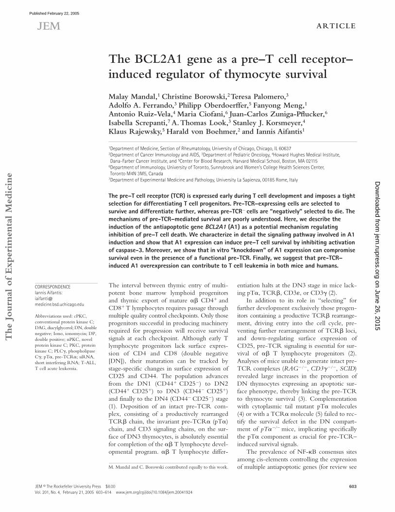
The Journal of Experimental Medicine JEM © The Rockefeller University Press $8.00 Vol. 201, No. 4, February 21, 2005 603–614 www.jem.org/cgi/doi/10.1084/jem.20041924 ARTICLE 603 The BCL2A1 gene as a pre–T cell receptor– induced regulator of thymocyte survival Malay Mandal, 1 Christine Borowski, 2 Teresa Palomero, 3 Adolfo A. Ferrando, 3 Philipp Oberdoerffer, 5 Fanyong Meng, 1 Antonio Ruiz-Vela, 4 Maria Ciofani, 6 Juan-Carlos Zuniga-Pflucker, 6 Isabella Screpanti, 7 A. Thomas Look, 3 Stanley J. Korsmeyer, 4 Klaus Rajewsky, 5 Harald von Boehmer, 2 and Iannis Aifantis 1 1 Department of Medicine, Section of Rheumatology, University of Chicago, Chicago, IL 60637 2 Department of Cancer Immunology and AIDS, 3 Department of Pediatric Oncology, 4 Howard Hughes Medical Institute, Dana-Farber Cancer Institute, and 5 Center for Blood Research, Harvard Medical School, Boston, MA 02115 6 Department of Immunology, University of Toronto, Sunnybrook and Women’s College Health Sciences Center, Toronto M4N 3M5, Canada 7 Department of Experimental Medicine and Pathology, University La Sapienza, 00185 Rome, Italy The pre–T cell receptor (TCR) is expressed early during T cell development and imposes a tight selection for differentiating T cell progenitors. Pre-TCR–expressing cells are selected to survive and differentiate further, whereas pre-TCR cells are “negatively” selected to die. The mechanisms of pre-TCR–mediated survival are poorly understood. Here, we describe the induction of the antiapoptotic gene BCL2A1 (A1) as a potential mechanism regulating inhibition of pre–T cell death. We characterize in detail the signaling pathway involved in A1 induction and show that A1 expression can induce pre–T cell survival by inhibiting activation of caspase-3. Moreover, we show that in vitro “knockdown” of A1 expression can compromise survival even in the presence of a functional pre-TCR. Finally, we suggest that pre-TCR– induced A1 overexpression can contribute to T cell leukemia in both mice and humans. The interval between thymic entry of multi- potent bone marrow lymphoid progenitors and thymic export of mature CD4 and CD8 T lymphocytes requires passage through multiple quality control checkpoints. Only those progenitors successful in producing machinery required for progression will receive survival signals at each checkpoint. Although early T lymphocyte progenitors lack surface expres- sion of CD4 and CD8 (double negative [DN]), their maturation can be tracked by stage-specific changes in surface expression of CD25 and CD44. The population advances from the DN1 (CD44 CD25 ) to DN2 (CD44 CD25 ) to DN3 (CD44 CD25 ) and finally to the DN4 (CD44 CD25 ) stage (1). Deposition of an intact pre-TCR com- plex, consisting of a productively rearranged TCR chain, the invariant pre-TCR (pT) chain, and CD3 signaling chains, on the sur- face of DN3 thymocytes, is absolutely essential for completion of the T lymphocyte devel- opmental program. T lymphocyte differ- entiation halts at the DN3 stage in mice lack- ing pT, TCR, CD3, or CD3 (2). In addition to its role in “selecting” for further development exclusively those progen- itors containing a productive TCR rearrange- ment, driving entry into the cell cycle, pre- venting further rearrangement of TCR loci, and down-regulating surface expression of CD25, pre-TCR signaling is essential for sur- vival of T lymphocyte progenitors (2). Analyses of mice unable to generate intact pre- TCR complexes (RAG / , CD3 / , SCID) revealed large increases in the proportion of DN thymocytes expressing an apoptotic sur- face phenotype, thereby linking the pre-TCR to thymocyte survival (3). Complementation with cytoplasmic tail mutant pT molecules (4) or with a TCR molecule (5) failed to rec- tify the survival defect in the DN compart- ment of pT mice, implicating specifically the pT component as crucial for pre-TCR– induced survival signals. The prevalence of NF-B consensus sites among cis-elements controlling the expression of multiple antiapoptotic genes (for review see M. Mandal and C. Borowski contributed equally to this work. CORRESPONDENCE Iannis Aifantis: iaifanti@ medicine.bsd.uchicago.edu Abbreviations used: cPKC, conventional protein kinase C; DAG, diacylglycerol; DN, double negative; Iono, ionomycin; DP, double positive; nPKC, novel protein kinase C; PKC, protein kinase C; PLC, phospholipase C; pT, pre-TCR; siRNA, short interfering RNA; T-ALL, T cell acute leukemia. on June 26, 2015 jem.rupress.org Downloaded from Published February 22, 2005

Welcome message from author
This document is posted to help you gain knowledge. Please leave a comment to let me know what you think about it! Share it to your friends and learn new things together.
Transcript
The
Journ
al o
f Exp
erim
enta
l M
edic
ine
JEM © The Rockefeller University Press $8.00Vol. 201, No. 4, February 21, 2005 603–614 www.jem.org/cgi/doi/10.1084/jem.20041924
ARTICLE
603
The BCL2A1 gene as a pre–T cell receptor–induced regulator of thymocyte survival
Malay Mandal,
1
Christine Borowski,
2
Teresa Palomero,
3
Adolfo A. Ferrando,
3
Philipp Oberdoerffer,
5
Fanyong Meng,
1
Antonio Ruiz-Vela,
4
Maria Ciofani,
6
Juan-Carlos Zuniga-Pflucker,
6
Isabella Screpanti,
7
A. Thomas Look,
3
Stanley J. Korsmeyer,
4
Klaus Rajewsky,
5
Harald von Boehmer,
2
and Iannis Aifantis
1
1
Department of Medicine, Section of Rheumatology, University of Chicago, Chicago, IL 60637
2
Department of Cancer Immunology and AIDS,
3
Department of Pediatric Oncology,
4
Howard Hughes Medical Institute, Dana-Farber Cancer Institute, and
5
Center for Blood Research, Harvard Medical School, Boston, MA 02115
6
Department of Immunology, University of Toronto, Sunnybrook and Women’s College Health Sciences Center, Toronto M4N 3M5, Canada
7
Department of Experimental Medicine and Pathology, University La Sapienza, 00185 Rome, Italy
The pre–T cell receptor (TCR) is expressed early during T cell development and imposes a tight selection for differentiating T cell progenitors. Pre-TCR–expressing cells are selected to survive and differentiate further, whereas pre-TCR
�
cells are “negatively” selected to die. The mechanisms of pre-TCR–mediated survival are poorly understood. Here, we describe the induction of the antiapoptotic gene
BCL2A1
(A1) as a potential mechanism regulating inhibition of pre–T cell death. We characterize in detail the signaling pathway involved in A1 induction and show that A1 expression can induce pre–T cell survival by inhibiting activation of caspase-3. Moreover, we show that in vitro “knockdown” of A1 expression can compromise survival even in the presence of a functional pre-TCR. Finally, we suggest that pre-TCR–induced A1 overexpression can contribute to T cell leukemia in both mice and humans.
The interval between thymic entry of multi-potent bone marrow lymphoid progenitorsand thymic export of mature
��
CD4
�
andCD8
�
T lymphocytes requires passage throughmultiple quality control checkpoints. Only thoseprogenitors successful in producing machineryrequired for progression will receive survivalsignals at each checkpoint. Although early Tlymphocyte progenitors lack surface expres-sion of CD4 and CD8 (double negative[DN]), their maturation can be tracked bystage-specific changes in surface expression ofCD25 and CD44. The population advancesfrom the DN1 (CD44
�
CD25
�
) to DN2(CD44
�
CD25
�
) to DN3 (CD44
�
CD25
�
)and finally to the DN4 (CD44
�
CD25
�
) stage(1). Deposition of an intact pre-TCR com-plex, consisting of a productively rearrangedTCR
�
chain, the invariant pre-TCR
�
(pT
�
)chain, and CD3 signaling chains, on the sur-face of DN3 thymocytes, is absolutely essentialfor completion of the
��
T lymphocyte devel-opmental program.
��
T lymphocyte differ-
entiation halts at the DN3 stage in mice lack-ing pT
�
, TCR
�
, CD3
�
, or CD3
�
(2).In addition to its role in “selecting” for
further development exclusively those progen-itors containing a productive TCR
�
rearrange-ment, driving entry into the cell cycle, pre-venting further rearrangement of TCR
�
loci,and down-regulating surface expression ofCD25, pre-TCR signaling is essential for sur-vival of
��
T lymphocyte progenitors (2).Analyses of mice unable to generate intact pre-TCR complexes (
RAG
�
/
�
,
CD3
�
�
/
�
,
SCID
)revealed large increases in the proportion ofDN thymocytes expressing an apoptotic sur-face phenotype, thereby linking the pre-TCRto thymocyte survival (3). Complementationwith cytoplasmic tail mutant pT
�
molecules(4) or with a TCR
�
molecule (5) failed to rec-tify the survival defect in the DN compart-ment of
pT
�
���
mice, implicating specificallythe pT
�
component as crucial for pre-TCR–induced survival signals.
The prevalence of NF-
�
B consensus sitesamong cis-elements controlling the expressionof multiple antiapoptotic genes (for review see
M. Mandal and C. Borowski contributed equally to this work.
CORRESPONDENCEIannis Aifantis: [email protected]
Abbreviations used: cPKC, conventional protein kinase C; DAG, diacylglycerol; DN, double negative; Iono, ionomycin; DP, double positive; nPKC, novel protein kinase C; PKC, protein kinase C; PLC
�
, phospholipase C
�
; pT
�
, pre-TCR
�
; siRNA, short interfering RNA; T-ALL, T cell acute leukemia.
on June 26, 2015jem
.rupress.orgD
ownloaded from
Published February 22, 2005

THE BCL2A1 GENE AS A POTENTIAL REGULATOR OF PRE–T CELL SURVIVAL | Mandal et al.
604
reference 6), in combination with the definitive placementof NF-
�
B as a distal effector of pre-TCR signaling (7, 8), es-tablishes an indirect association between the pre-TCR andthe survival of developing thymocytes. The search for distalmediators of pre-TCR–induced survival has met with littlesuccess. The
�
selection process proceeds normally in pro-genitors lacking pro-survival BCL-2 family members BCL-2or BCL-x
L
(9, 10), and neither BCL-2 nor BCL-x
L
trans-genes restore
��
T lymphocyte development in pre-TCR–deficient mice (11–13). T lymphocyte progenitors lackingthe pro-survival Bcl-2 family member MCL-1 fail to progressthrough the DN2 to DN3 developmental transition, impli-cating MCL-1 as a crucial mediator of early cytokine-derived,rather than later pre-TCR–derived, survival signals (14).Further refuting a critical role for BCL-2 is the observationthat post-
�
selection thymocytes contain lower levels of Bcl-2protein than those that have not yet entered
�
selection (15).However, a recent report presented conflicting evidence,suggesting that BCL-2 expression is induced by the pre-TCR in vitro (16).
Similarly, very little is known about the signaling path-ways inducing death in pre-TCR
�
thymocytes. The initialsuggestion that the Fas-induced death pathway is essential forpre–T cell death has been challenged by recent studies ofNewton et al. (17), in which the thymocyte profiles of
RAG-1
���
lpr
mice were found to be similar to those of their
RAG-1
���
littermates. Fas is a member of the TNF receptorfamily, whose members interact via their death domains withcytoplasmic adaptors including FADD and TRADD (18).The same investigators have shown that
RAG-1
���
FADD
���
pre–T cells differentiate and reach the double positive (DP)stage (17). These important observations suggested thatFADD could be essential for the transduction of death signalwithin pre–T cells and fueled the search for an active pre-TCR–dependent mechanism of cell death inhibition.
In this report, we direct our search toward antiapoptoticgenes potentially targeted by pre-TCR–induced NF-
�
Btranscription factors. We identify the pro-survival A1 geneas a direct transcriptional target of both pre-TCR and NF-
�
B. Moreover, we connect transcriptional activation of A1to PLC
�
, mobilization of Ca
2
�
, and activation of proteinkinase C (PKC) kinases. We also show that A1 expressioninhibits pre–T cell death by suppressing the activation of theeffector caspase-3. Finally, we show that A1 is up-regulatedin both human and mouse pre–T cell leukemias, making it apossible target of the transformation event. Our observa-tions suggest that through NF-
�
B activation and A1 expres-sion, the pre-TCR ensures the promotion of physiologicalT cell development.
RESULTSPre-TCR–nonexpressing cells die due to the activation of effector caspase-3
Evidence definitively demonstrating pre-TCR–mediatedprevention of imminent apoptosis has not been documented.Here, we addressed this issue both in vivo and in vitro. The
percentage of proapoptotic, annexin V
�
thymocytes was sig-nificantly larger in mice lacking pre-TCR components
(pT
�
���
,
RAG-1
���
)
than in their WT counterparts (Fig. 1A). To demonstrate that pre-TCR expression provides asurvival advantage, sorted pre-TCR
�
DN3
RAG-1
���
andpre-TCR
�
DN4 WT thymocytes were cultured in a lowconcentration of 1 ng/ml IL-7. As evident from Fig. 1 B,pre-TCR–deficient cells die at a significantly higher rate(Fig. 1 B). Similar results were obtained in the absence ofIL-7 (not depicted).
To gain insight into the mechanism of this cell death, weanalyzed activation of the distal effector caspase-3 in apoptoticthymocytes. Using a fluorescently labeled antibody specificfor the active form of caspase-3, we detected activation of cas-pase-3 specifically in the pre-TCR
�
DN3 subset of
RAG-1
���
thymocytes ex vivo (Fig. 1 C). As receptors for prosurvivalcytokines IL-7 and stem cell factor are down-regulated at theDN4 stage, these cells do not receive any pre-TCR–indepen-dent survival signals. To further support these observationsand to increase the sensitivity of detection of active caspase-3,
Figure 1. Pre-TCR� thymocytes die by apoptosis due to caspase-3 activation. (A) Percentage of thymocytes binding annexin V in the CD4�8� lineage� compartment of WT, Rag-1�/�, and pT��/� thymi. (B) In vitro pre–T cell survival. Pre-TCR� (hatched bars) and pre-TCR� (solid bars) CD4�8� lineage� thymocytes were cultured in the absence of exogenous factors. Survival rates were determined by annexin V staining at the indi-cated time points. (C) Flow cytometric detection of caspase-3 activation. The left plot illustrates the CD25/CD44 surface profile of Rag-1�/� thy-mocytes. The right dot plots illustrate caspase-3 activation within indi-cated cellular compartments. (D) Activation of caspase-3 in Rag-1�/�
thymocytes is down-regulated upon anti-CD3� injection. Positive (WT fibroblasts treated with brefeldin A) and negative (caspase-3�/� fibroblasts treated with brefeldin A) controls are included. Blots were reprobed with anti-actin as a loading control.
on June 26, 2015jem
.rupress.orgD
ownloaded from
Published February 22, 2005

JEM VOL. 201, February 21, 2005
605
ARTICLE
we probed
RAG-1
���
thymocyte lysates with a second anti-body specific for the activated (cleaved) form of caspase-3.The activation of caspase-3 observed in this system was com-parable to that induced by in vitro treatment of mouse em-bryonic fibroblasts with brefeldin A (Fig. 1 D). Mimickingpre-TCR signaling by CD3
�
cross-linking relieved the DN3block of
RAG-1
���
thymocytes (7) and decreased caspase-3activity in these cells, adding one more piece of evidence di-rectly connecting pre-TCR signaling with thymocyte survival(Fig. 1 D). An identical pattern of activation was also seen forthe effector caspase 9 (unpublished data).
BCL-2, MCL-1, and BCL-xL are not targets of the pre-TCRConflicting reports described the roles of BCL-2 and BCL-xL in pre–T cell death, and recent data indicated that yet an-other antiapoptotic BCL-2 family member, MCL-1, mayregulate survival of thymocytes at an earlier, cytokine-medi-ated stage (14). To test whether pre-TCR signaling inducesexpression of these prosurvival factors, we performed real-time RT-PCR analysis using cDNA isolated from sortedDN3, DN4, and DP thymocytes. As evident from Fig. 2,none of these factors were induced by the pre-TCR. MCL-1expression was high in DN3 cells. In agreement with pre-vious reports, it decreased in DN4 and peaked again at theDP stage. BCL-xL expression was low in both DN3 andDN4 populations, peaking at the DP stage. BCL-2 expres-sion seemed to be actively down-regulated upon pre-TCRexpression, reaching a minimum at the DP stage (Fig. 2 A).Similar results showing no up-regulation of BCL-xL andMCL-1 and a down-regulation of BCL-2 were obtained us-ing two distinct pre-TCR� (SCIET27 and LR2) and pre-TCR� (SCB29 and LR2wtpT�) cell lines (Fig. 2 B and un-published data; reference 19).
A1 gene is a direct target of pre-TCR signalingAfter excluding BCL-2, BCL-xL, and MCL-1 as antiapop-totic pre-TCR transcriptional targets, we focused specificallyon antiapoptotic BCL2 family genes whose cis regulatory el-ements contain NF-�B consensus sites. The BCL2A1 (A1)gene cluster (three A1 genes: A1a, A1b, and A1d; onepseudogene: A1c) fit this description. Expression of this NF-�B–responsive gene cluster is induced by inflammatory stim-uli, B cell receptor stimulation, and CD40 signaling (20–22).
Figure 2. BCL-2, BCL-xL, and MCL-1 are not induced by pre-TCR signaling. (A) Quantitative real-time RT-PCR analysis of BCL-2, MCL-1, and BCL-xL expression in the indicated thymocyte subsets. (B) Quantita-tive RT-PCR analysis of gene expression in the LR2 and LR2wtpT� cell lines. Actin expression was used to normalize gene expression.
Figure 3. Pre-TCR signaling induces A1 expression both in vivo and in vitro. (A) Semiquantitative and real-time RT-PCR analysis of A1 expression in ex vivo thymocyte subsets. (B) Semiquantitative PCR analysis of A1 expression in pre-TCR� (lane 2, SCB29; lane 4, LR2-WTpT�) and pre-TCR� (lane 1, SCIET27; lane 3, LR2) cell lines. (C) Pre-TCR complexes containing truncated pT� chains fail to induce A1 expression. Semiquantitative PCR-quantitated A1
expression in LR2 cells expressing (lane 1) no pT� chain, (lane 2) a WT pT� chain, (lane 3) a pT� chain lacking its entire cytoplasmic domain, or (lane 4) a pT� chain lacking its proline-rich domains. (D) Isoform-specific pattern of A1 expression in DN4 pre–T cells. A1 cDNA amplified from DN thymocytes (using A1XhoUp1 and A1BamLow1 primers) was left nondigested (lane 1), digested with NsiI (lane 2), or digested with BglII (3).
on June 26, 2015jem
.rupress.orgD
ownloaded from
Published February 22, 2005

THE BCL2A1 GENE AS A POTENTIAL REGULATOR OF PRE–T CELL SURVIVAL | Mandal et al.606
A1 expression protects cells from various death stimuli in-cluding TNF-� stimulation, p53 activation, B cell receptortriggering, and IL-3 deprivation (23–25). Our gene expres-sion analyses identified the A1 gene cluster as a potential pre-TCR target (Fig. 3). Sorted RAG-1��� pre-TCR� DN3thymocytes lacked A1 transcripts, whereas sorted preTCR�
WT DN4 thymocytes expressed abundant A1 transcripts.Only after mimicking pre-TCR signaling by injection ofanti-CD3� antibody was A1 expression detected in RAG-1��� thymocytes (Fig. 3 A). These observations were repro-duced multiple times using different primer pairs and wereconfirmed by quantitative real-time PCR (Fig. 3 A). To testwhether A1 induction was a direct effect of pre-TCR signal-ing, we compared A1 expression in pre-TCR� (SCIET27and LR2) and pre-TCR� (SCB29 and LR2-WTpT�) pre–Tcell lines (7). SCIET27 and LR2 are parental pre-TCR�
lines. Introduction of TCR� (in SCIET) and TCR� pT�(in LR2) results in the generation of the pre-TCR� SCB29and LR2-WTpT� lines (5, 7). As shown in Fig. 3 B, pre-TCR expression induced A1 expression in both cell lines.Moreover, LR2 cell lines engineered to express truncatedforms of pT� (mutations of the cytoplasmic tail of the mole-cule) illustrated the crucial nature of the pT� cytoplasmic tailin pre-TCR–mediated A1 induction (Fig. 3 C). These ob-servations are consistent with our previous report, whichshowed that thymocytes expressing truncated pT� chainsunderwent abnormal � selection characterized by defectsboth in proliferation and in cell survival (4).
The existence of three A1 isoforms, each encoded by adifferent gene, prompted us to investigate which isoforms ofA1 were expressed in pre–T cells. To this end, A1 cDNAamplified from DN4 cells was digested with restriction en-zymes whose recognition sites are located in isoform-specificsequence polymorphisms (26). The enzyme NsiI was unableto digest A1 cDNA amplified from pre–T cells (purifiedDN4), indicating that A1a is either not expressed or ex-pressed at undetectable levels in these cells. In contrast, di-gestion with the BglI enzyme strongly suggested that A1d isone of the major isoforms expressed in pre–T cells (Fig. 3D). In addition to verifying these results, sequencing indi-cated that A1b was also expressed in pre–T cells, albeit at alevel lower than that of A1d (not depicted). These observa-tions suggest an isoform-specific pattern of A1 expression inpre–T cells.
Activation of phospholipase C� (PLC�), Ca2�, and NF-�B are critical for A1 inductionThe above observations place A1 expression downstream ofthe pre-TCR, but the signaling components connectingpre-TCR signals with A1 induction remain unidentified.We initially addressed the role of NF-�B in pre-TCR–induced A1 expression. As mentioned above, pre-TCR� celllines (SCB29 and LR2WTpT�), but not their pre-TCR–deficient counterparts (SCIET27 and LR2), contained abun-dant A1 transcripts. To inhibit NF-�B activation in SCB29and LR2WTpT� cell lines, we generated a retroviral vector
Figure 4. A1 is a target of the PLC�-Ca2�-NF-�B pathway. (A) A1 is a transcriptional target of NF-�B. Semiquantitative RT-PCR analysis of A1 expression in pre-TCR� cell lines after the inhibition of NF-�B. In the absence of pre-TCR signaling, 50 ng/ml PMA plus 1 g/ml Iono treatment can induce A1 expression (B), as well as NF-�B nuclear translocation and Ca2� mobilization (C). (D) Ca2� mobilization is essential for A1 transcrip-
tional induction. A1 expression was examined by semiquantitative RT-PCR after chelation of intracellular Ca2� by 1 mM TMB8. (E) PLC� activation is essential for A1 induction. A1 expression levels were examined by semi-quantitative and real-time RT-PCR in pre-TCR� cell lines (N3T and SCB29) incubated with 10 M of the specific PLC� inhibitor U73122.
on June 26, 2015jem
.rupress.orgD
ownloaded from
Published February 22, 2005

JEM VOL. 201, February 21, 2005 607
ARTICLE
encoding a mutated nondegradable form of the inhibitor ofNF-�B � (IKB�; reference 8). When expressed in pre-TCR� SCB29 and LR2WTpT� cell lines, IKB� completelyinhibited pre-TCR–induced A1 expression, suggesting anessential role of NF-kB in pre-TCR–dependent up-regula-tion of A1 (Fig. 4 A). These observations are consistent withthose of Voll et al. (8), who demonstrated that pre–T cell–specific expression of an IKB� transgene (driven by thep56lck promoter) slowed pre-TCR–driven �� T lymphocytedevelopment.
We have previously shown that activation of PLC� andCa2� mobilization are essential for the transduction of pre-TCR signals (7). To address the role of these two mediatorsin pre-TCR–induced A1 expression, we used the PMA andionomycin (Iono), which have been shown to be able tomimic pre-TCR signaling (7). Treatment with PMA andIono rapidly induced A1 expression in a pre-TCR–deficient
line (Fig. 4 B), and Rag-1��� purified DN3 thymocytes (notdepicted). Because this treatment induces both Ca2� mobili-zation as well as nuclear translocation of NF-�B (Fig. 4 C),we tested whether A1 expression depends on one or both ofthese stimuli. Pretreatment of these cell lines with an endo-plasmic reticulum Ca2� chelator, TMB-8, totally abrogatedA1 expression (Fig. 4 D) and NF-�B activation (7), high-lighting Ca2� as an essential second messenger. To testwhether A1 induction was dependent on PLC� activation,pre-TCR� SCB29 and N3-T (a leukemic cell line that hasup-regulated A1 expression in response to Notch activationand overexpression of the pre-TCR) cell lines were incu-bated with the specific PLC� inhibitor U73122, which in-hibits Ca2� signaling in pre–T cells (7). As shown in Fig. 4 E,inhibition of PLC� activity totally blocked the ability of thepre-TCR (and of PMA plus Iono treatment) to induce A1expression in both cell lines. Similar results were obtained
Figure 5. PKC is essential for pre-TCR–induced A1 expression. (A) SCIET27 cells were infected with a retrovirus expressing constitutively active forms of PKC (SCI-PKC�CAT) or Akt (SCI-Myr-Akt). A1 expression was compared with uninfected SCIET27 by RT-PCR. (B) In vitro inhibition of PKC activity using 1 M of the PKC inhibitor calphostin, abrogates A1 expression induced by PMA plus Iono in SCIET27 cells. (C) Inhibition of PKC activity blocks normal T cell development and induces pre–T cell death in
an OP-9-DL1 culture. WT DN2 thymocytes were cultured in the presence or absence of calphostin or DMSO control. Differentiation (left) and sur-vival (right) were examined by staining with anti-CD4 and anti-CD8 anti-bodies and annexin V. Data are representative of at least four independent experiments using 0.1 M calphhostin. (D and E) SCIET27 cells expressing PKC�CAT are more resistant to irradiation- (480 rads) induced death (left) due to the attenuation of caspase-3 activation (right).
on June 26, 2015jem
.rupress.orgD
ownloaded from
Published February 22, 2005

THE BCL2A1 GENE AS A POTENTIAL REGULATOR OF PRE–T CELL SURVIVAL | Mandal et al.608
using a third pre-TCR� cell line, LR2-WTpT� (not de-picted). The inhibition was complete regardless of the initiallevel of A1 expression.
PKC activation is essential for A1 induction and survivalPLC� activity generates both inositol triphosphate, whichinitiates Ca2� mobilization, and diacylglycerol (DAG), whichactivates downstream kinases such as PKC. To address thepotential role of PKC in pre-TCR–induced A1 expression,we expressed a constitutively active form of PKC (PKC��CAT) in pre-TCR–deficient (and thus A1�) SCIET andLR2 cell lines. PKC activation strongly induced A1 expres-sion in the SCIET (Fig. 5 A) and LR2 (not depicted) celllines. To establish PKC specificity, we expressed an activated(myristoylated) form of PKB/Akt (Myr-Akt), another kinaseinvolved in the transduction of pre-TCR signals (unpub-lished data). As shown in Fig. 5 A, Myr-Akt failed to up-regulate A1 expression, classifying pre-TCR–induced A1expression as strictly dependent on PKC.
To further illustrate PKC involvement, pre-TCR�
SCIET and LR2 cells were pretreated with calphostin, a po-tent and specific PKC inhibitor. Calphostin treatment totallyblocked PMA plus Iono–induced A1 expression in SCIET(Fig. 5 B) and LR2 (not depicted) cell lines. To demonstratethat PKC inhibition adversely affected pre–T cell survival anddifferentiation, we used a novel in vitro culture system (27).In this system, OP-9 stromal cells expressing the Notchligand Delta-1 (OP9-DL1) support thymic-independent de-velopment of T cell progenitors. Sorted DN2 cells were cul-tured with OP9-DL1 cells and supplemented with various
concentrations of calphostin or DMSO control. As shown inFig. 5 C, calphostin, but not DMSO treatment, significantlyblocked progression to the DP stage of T cell development.This effect was due to decreased survival, as �85% of thecalphostin-treated cells were proapoptotic. Also, the majority(60%) of calphostin-treated cells remained CD4�8�CD25�
(not depicted). To directly connect PKC activation and A1expression to pre–T cell survival, we compared survival ki-netics of irradiated (400 rads) pre-TCR–deficient SCIET27cells expressing either PKC�CAT-EGFP or EGFP. PKC��CAT expression provided a significant protection from apop-tosis (Fig. 5 D), which correlated with the ability of PKC toprevent caspase-3 activation (Fig. 5 E). Identical results wereobtained using a second pre-TCR� line (LR2).
A1 as a regulator of pre–T cell survivalTo study the function of A1 in the promotion of pre–T cellsurvival, we ectopically expressed A1d (the most commonlyexpressed A1 isoform) in SCIET pre–T cells (4). A1d andEGFP were expressed on a single transcript after infectionwith a bicistronic A1-IRES-EGFP retrovirus. This retrovi-rally encoded A1 protein was properly expressed and tar-geted to the mitochondria (not depicted). As pre–T cells sus-tain DNA breaks generated during ongoing recombinationat their TCR loci, we investigated A1’s ability to inhibitdeath induced by DNA-damaging ultraviolet irradiation.A1d expression efficiently suppressed irradiation-inducedcell death in SCIET (Fig. 6 A) and LR2 (not depicted) pre–T cells. Mechanistically, the inhibition of cell death resultedfrom an A1-mediated reduction in caspase-3 activation (Fig.
Figure 6. A1 induces pre–T cell survival. SCIET27 cells expressing EGFP (black dashed and gray lines) or A1-EGFP (black line) were irradi-ated (gray, control; black, A1-expressing) and left in culture for the indi-cated time periods. Survival was detected using annexin V labeling (A). Caspase-3 activation was measured 4 h after irradiation (B). Retroviral expression of A1 induces survival and differentiation of Rag-1�/� pre-TCR�
thymocytes. Ratios of infected (A1�) and uninfected (A1�) RAG-1�/�
thymocytes at the indicated time points after injection into Rag-2�/��c�/�
hosts (C). Differentiation of A1� and A1� thymocytes was analyzed 6 wk after injection by surface staining with antibodies specific for CD4, CD8, and CD25 (D). (E) A1 expression does not alter differentiation of WT thy-mocytes. CD4/CD8 surface profiles of WT thymocytes infected with A1-IRES-EGFP or IRES-EGFP retroviruses were examined 3 wk after injection into Rag-2�/��c�/� hosts.
on June 26, 2015jem
.rupress.orgD
ownloaded from
Published February 22, 2005
JEM VOL. 201, February 21, 2005 609
ARTICLE
6 B). Similar results were obtained with dexamethasonetreatment as a death stimulus (not depicted).
To support these in vitro observations using primarythymocytes, we infected lineage- (CD19, Ter-119, Gr-1,NK1.1, �� TCR, � TCR, Mac-1, CD11c) negative bonemarrow progenitors sorted from (Ly5.1�) RAG-1�/� animalswith the A1d-IRES-EGFP retrovirus. Alymphoid irradiated(350 rads) Rag�/��c�/� (Ly5.2�) hosts were injected with theLy5.1� A1-infected bone marrow cells. Thymic reconstitu-tion was analyzed at various time intervals after cell transfer.Retroviral A1d bestowed a significant survival advantageupon developing thymocytes (Fig. 6 C). Although injected ata 40:60 ratio of EGFP�A1�/EGFP�A1�, 3 wk’s time re-versed this ratio to 65:35 (Fig. 6 C). Four additional weeksresulted in a near absence of EGFP� A1� cells in reconstitutedthymi, suggesting that EGFP�A1� thymocytes out-competedtheir coinjected EGFP� A1� counterparts. Interestingly, de-
spite their lack of a pre-TCR complex, these RAG-1�/�
EGFP�A1� thymocytes displayed signs of differentiation.More than half of the cells up-regulated CD4 and CD8 core-ceptor surface expression (Fig. 6 D). Importantly, annexinV staining revealed little apoptosis among the A1�EGFP�
population and high percentages of apoptotic cells in theEGFP� population (not depicted). No A1�EGFP� cells weredetected in the periphery, due to the inability of the RAG-1�/� cells to express selectable TCRs (not depicted). Theseresults suggest that A1 expression, which is physiologicallytriggered by pre-TCR signaling, induces pre–T cell survivaland ensures differentiation. To illustrate that A1 expressiondoes not affect the physiological progression of T lymphocytedevelopment, we repeated the above reconstitution experi-ments using bone marrow progenitors sorted from WT mice.No difference was apparent between the differentiation kineticsof WT A1�EGFP� and WT A1�EGFP� progenitors when
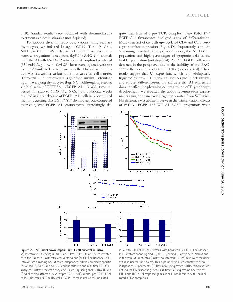
Figure 7. A1 knockdown impairs pre–T cell survival in vitro. (A) Effective A1 silencing in pre–T cells. Pre-TCR� N3T cells were infected with the Banshee-EGFP retroviral vector alone (siEGFP) or Banshee-EGFP retroviruses encoding one of three independent siRNA complexes specific for A1 (A1-A, A1-C, and A1-D). Semiquantitative and real-time RT-PCR analyses illustrate the efficiency of A1 silencing using each siRNA. (B and C) A1 silencing affects survival of pre-TCR� (N3T), but not pre-TCR� (LR2), cells. Uninfected N3T or LR2 cells (EGFP�) were mixed at the indicated
ratio with N3T or LR2 cells infected with Banshee-EGFP (EGFP) or Banshee-EGFP vectors encoding siA1-A, siA1-C, or siA1-D complexes. Alterations in the ratio of uninfected (EGFP�) to infected (EGFP�) cells were recorded at the indicated time points. This experiment is a representative of four independent experiments. (D) Retrovirally expressed siRNA complexes do not induce IFN response genes. Real-time PCR expression analysis of IFIT-1 and IRF-7 IFN response genes in cell lines infected with the indi-cated siRNA complexes.
on June 26, 2015jem
.rupress.orgD
ownloaded from
Published February 22, 2005

THE BCL2A1 GENE AS A POTENTIAL REGULATOR OF PRE–T CELL SURVIVAL | Mandal et al.610
injected in age- and sex-matched hosts (Fig. 6 E). These ob-servations are consistent with the recently reported normal Tcell development in thymi of mice expressing an A1a trans-gene driven by a p56Lck promoter (28).
To demonstrate that A1 is necessary for pre–T cell sur-vival, we studied pre–T cell survival in the absence of A1 ex-pression. The existence of three independent A1 isoformsprecludes gene-targeting approaches. Multiple consecutivetargeting steps would be necessary, as the genes encoding thethree A1 isoforms are not all clustered together on chromo-some 9. In addition, the significant conservation in bothexon and intron sequences shared among the A1 isoformswould make design of gene-targeting vectors extremely dif-ficult. Therefore, we used stable RNA interference to knockdown A1 expression in a T lymphocyte–specific manner. Toeliminate concern about redundancy among A1 isoforms,we generated short interfering RNAs (siRNAs) capable ofknocking down expression of all three A1 isoforms. ThesesiRNAs were expressed using a self-inactivating retroviruscarrying a U6 Pol-II promoter able to drive expression of
siRNAs. We tested three different siRNAs, and achievedoptimal interference with siRNA-A (Fig. 7 A). Real-timeRT-PCR analysis showed silencing efficiency at 75–85%(Fig. 7 A). The effects of A1 deficiency on pre-TCR func-tion were assessed in the pre-TCR� N3-T cell line (Fig. 8D). N3-T cells expressing each of the three different siRNAs(EGFP�) were mixed at a ratio of 50:50 with EGFP�N3-Tcells. As shown in Fig. 7 B, N3-T cells expressing A1-spe-cific siRNAs exhibited a survival disadvantage, proportionalin severity to the efficiency of A1 knockdown. This defect isdue to loss of survival, as at any given time of the culture, theproportion of cells binding annexin V was two to four timeshigher in the population expressing A1-siRNA (siRNA-A)when compared with cells expressing a control EGFP-siRNA (not depicted). No significant differences in cell cy-cle progression were observed between cells expressingEGFP-siRNA and A1-siRNA constructs (not depicted).When infected with the same siRNAs, neither 3T3, 293T,nor LR2 cells (Fig. 7 C) showed survival defects, suggestingthat the effect of A1 knockdown is specific to cells that spe-
Figure 8. A1 is a target of T cell transformation both in mouse and man. (A) Relative expression levels of pT� and specific T cell differentia-tion marker genes (rows) across 43 human T cell leukemia samples (columns). Normalized gene expression levels are represented in colors. Groups of samples were identified (top) according to the expression of the indicated T cell differentiation markers. Expression of pT� clearly correlates with a group of genes including p56lck, CD3�, CD4, and CD8. pT� expression is completely absent in early CD34� pro–T cell leukemias. This is a pictorial representation of the expression levels with no intention to represent spe-
cific gene expression levels. (B) A1 and BCL-2 expression was quantitated by real-time RT-PCR analysis in selected representative T-ALL samples. BCL2 and A1 expression were compared in pro–T cell and cortical T-ALL samples using a Wilcoxon rank sum test, showing a significant correlation of BCL2 expression with pro–T T-ALL samples and of A1 expression with cortical T-ALL samples. (C) Surface phenotype characterization of a murine T cell leukemia line (N3T). (D) Expression of A1 in N3T cells. Real-time RT-PCR comparison between N3T, DN3, DN4, SCIET27 (our expression base), and LR2 cells.
on June 26, 2015jem
.rupress.orgD
ownloaded from
Published February 22, 2005

JEM VOL. 201, February 21, 2005 611
ARTICLE
cifically up-regulate A1 due to pre-TCR overexpression(Fig. 8). Moreover, BCL-xL and BCL-2 expression re-mained unaltered in N3-T cells that undergo apoptosis dueto the A1 deficiency, suggesting that these two genes are notinvolved in the maintenance of N3-T cell survival (unpub-lished data).
To exclude nonspecific siRNA effects due to potentialIFN responses, we have selected two well-characterized IFN-responsive genes and studied their expression in pre–T cellsexpressing our siRNAs (29). There was no specific up-regu-lation of either IFN-induced protein 1 or IFN regulatory fac-tor 7 in our siRNA-expressing pre–T cells (Fig. 7 D).
A1 expression is a molecular target of pre–T cell transformationDeregulated pre-TCR signaling triggers aggressive pre–Tcell leukemia in mice (30, 31). Genes encoding moleculesinvolved in pre-TCR signaling (p56Lck, ZAP-70, TCR�,CD3�, and cyclin D3) are overexpressed in human T cellacute leukemias (T-ALLs) arising from early to late corticalthymocytes, which are characterized by the expression of theoncogenes HOX-11� and TAL-1�. Thus, it is conceivablethat the survival advantage conferred by pre-TCR–inducedantiapoptotic genes like A1 may contribute to the transfor-mation process (not as a bona fide oncogene but as a targetof the transformation event). To connect pre-TCR and A1expression to a precise window of thymocyte differentiation,we used the patient expression profile data presented by Fer-rando et al. (32) to analyze the expression of pT� within thethree different clusters of human T-ALL cases. As shown inFig. 8 A, pT� expression is seen primarily within Hox-11�
early cortical thymocytes, rather than within LYL1� CD34�
progenitors or TAL-1� mature T cells. To link A1 expres-sion with induction of T cell leukemia, we selected repre-sentative cases from each cluster of human T-ALL cases andperformed quantitative real-time RT-PCR. As evident fromFig. 8 B, A1 expression correlated only with HOX-11 orTAL-1–expressing samples. This is consistent with the no-tion that A1 is induced only after pre-TCR signaling andpeaks at the DP stage of development. Interestingly, BCL-2expression peaked early in development (in CD3� CD34�
samples) and declined after the appearance of the pre-TCRand A1 (Fig. 8 B). This observation is consistent with ourprevious expression analysis of A1 and BCL-2 during physi-ological T cell development (Figs. 2 and 3).
To complement these studies, we analyzed A1 expres-sion in a mouse model of T cell leukemia. In this model, in-troduced by Bellavia et al. (33), T cell–specific expression ofa constitutively active form of Notch-3 induces malignanttransformation of thymocytes. We have chosen Notch-3 be-cause “nearest neighbor” analysis of the T-ALL databaseused above had shown that pT� and Notch-3 share a strik-ingly identical expression profile (unpublished data). TheNotch transformation is strictly dependent on pre-TCR sig-naling (31). N3-T, an immature HSAhi CD25lo CD4� CD8�
cell line derived from the thymus of a Notch-3 transgenic
animal, most likely represents the pre-TCR� DN3 to DN4transitional stage of thymocyte development (Fig. 8 C; refer-ence 33). Surface pre-TCR levels on N3-T cells are evenhigher than those on WT DN4 cells (Fig. 8 C). In line withour human T-ALL studies, N3-T cells expressed A1 at levelssignificantly higher that those in WT DN4 thymocytes or inDN cell lines (Fig. 8 D). As shown in Fig. 7 B, A1 is crucialfor in vitro survival of N3-T cells, as A1 “knockdown” con-ferred a significant survival disadvantage.
DISCUSSIONThe identification of NF-�B as a pre-TCR target (7, 8) andthe phenotype of transgenic mice expressing an inhibitor ofthe NF-�B pathway (I�B�) specifically in pre–T cells (8)initiated our search for NF-�B–responsive downstream ef-fectors regulating pre–T cell survival. Together with impor-tant reports directly connecting A1 transcription to NF-�Bactivation (34, 35), the recent observation of endogenousc-Rel binding directly to cis elements regulating A1 geneexpression (36) directed our search toward A1. Here, weshow that A1 expression is directly induced by the pre-TCR. Moreover, A1 appears to be the only antiapoptoticgene induced during � selection, as neither members of theBCL2 family nor members of the IAP family are induced bythe pre-TCR (Fig. 2 and unpublished data).
One can argue, however, that the data presented areequally consistent with the notion that A1 is a Notch target,as Notch can also activate the NF-kB pathway. This is an in-triguing possibility that could provide insights in the mecha-nism of cooperation between Notch and the pre-TCR (37).However, multiple lines of evidence argue against this sce-nario and highlight the unique ability of the pre-TCR toregulate A1 expression. Initially, expression of activatedforms of Notch-1 (N1-IC) in pre-TCR� lines (SCIET27and LR2) failed to up-regulate A1 expression (unpublisheddata). Moreover, specific mutation of the pre-TCR (cyto-plasmic truncation) blocked any A1 expression without af-fecting Notch expression (Fig. 3). Finally, DN thymocytesfrom pT��/� mice that overexpress active Notch-3 showactivation neither of PKC nor of NF-�B, suggesting that thepre-TCR, rather than Notch, regulates the activity of thispathway in developing pre–T cells (unpublished data).
Why the pre-TCR uses A1 rather than the prototypicalantiapoptotic molecules BCL-2 or BCL-xL is not yet under-stood. However, overexpression of BCL-2 is unable to re-store normal developmental progression in pre-TCR–defi-cient pT���� (unpublished data) or RAG-1�/� mice (11).The preferential use of A1 might be due to the antiprolifera-tive properties ascribed to BCL-2. Introduction of a BCL-2transgene into WT mice actually impaired the proliferation ofTCR�-selected DN4 thymocytes (38). It was suggested thatthis antiproliferative effect depends on the ability of BCL-2 tobind to and sequester the calcium-dependent calcineurinphosphatase, thereby inhibiting NF-AT nuclear translocationand activation (39, 40). This interaction between BCL-2 andcalcineurin depends on conserved regions in the BH4 domain
on June 26, 2015jem
.rupress.orgD
ownloaded from
Published February 22, 2005

THE BCL2A1 GENE AS A POTENTIAL REGULATOR OF PRE–T CELL SURVIVAL | Mandal et al.612
of BCL-2, which are also present in BCL-xL and MCL-1. Incontrast, all three A1 genes lack these conserved calcineurininteraction sites (unpublished data). Thus, BCL-2, BCL-xL,and MCL-1, but not A1, may prevent NF-AT activation,which is important for pre-TCR function (7). Consistentwith this notion, Gonzalez et al. (28) recently have shownthat overexpression of A1, but not BCL-2, allowed normalproliferation of mature activated T lymphocytes.
Our studies underline the role of PKC as an intermediatetransducer of pre-TCR signals. PKC isoenzymes are groupedinto two major subfamilies. The activation of conventionalPKCs (cPKCs) as well as novel PKCs (nPKCs) typically in-volves recruitment to membranes and interaction with or al-losteric activation by DAG. Ca2� binding is an essential re-quirement for cPKCs, but not nPKCs (41). PKC�, cPKC,PKC�, and nPKC, are the major PKCs expressed in thy-mocytes (41). In our experiments, PMA plus Iono treatmentinduced A1 expression in pre-TCR� cells, whereas Ionoalone did not. Also, PMA plus Iono in the presence of theER Ca2� chelator, TMB-8, induced neither A1 expressionnor NF-�B activation. As induction of NF-�B activation andA1 expression required both PMA and Iono, it is tempting tosuggest that pre-TCR signaling activates PLC�, which mobi-lizes Ca2�, which, along with DAG, activates only cPKCisoenzymes (i.e., PKC�). Consistent with this view is the lackof a pre-TCR–dependent phenotype in mice lacking thePKC� isoenzyme (42). However, we cannot exclude thatPKC�, although not absolutely essential, plays a secondaryrole amplifying PKC�-mediated signals to the nucleus.
A recent study illustrated PKC’s importance in pre-TCR–mediated allelic exclusion at the TCR� locus (43). Separatework demonstrated that although activation of PKC can par-tially restore DN to DP transition in RAG-1�/� mice, it is notable to drive thymocyte expansion (a phenotype identical tothose of mice reconstituted with A1� RAG-1�/� progenitors,presented here; reference 37). Perhaps the bifurcation in pre-TCR signaling that occurs downstream of the LAT/SLP-76complex leads in one direction toward PKC-independent sig-nals inducing thymocyte expansion (Raf/Ras, Akt) and in theother direction toward PKC-dependent signals establishingTCR� allelic exclusion and ensuring survival.
The critical role of A1 in transmitting pre-TCR–inducedantiapoptotic signals and the previous correlation of de-regulated pre-TCR signaling with T cell leukemia, promptedus to analyze the patterns of A1 expression in human T-ALLcases and in a Notch-induced model of mouse leukemia. Wefound a unique correlation between A1 expression and re-ceipt of pre-TCR signals and have shown that A1 knock-down of A1 expression can affect survival of a pre–T cellleukemic line. We recently identified cyclin D3 as an essen-tial mediator of pre-TCR–induced proliferative signals andsuggested that cyclin D3 expression was essential for trans-formation of pre–T cells (44). A1 overexpression may coop-erate with cyclin D3 to provide a survival signal renderingtransformed pre–T cells resistant to apoptotic signals. Thesefindings could have clinical importance because TAL1� leu-
kemias are resistant to chemotherapy potentially due to theoverexpression of the antiapoptotic A1 (32).
The pre-TCR possesses the unique ability to overridecell intrinsic (i.e., DNA damage-induced) and extrinsic (i.e.,death receptor–induced) apoptotic stimuli. We describe anovel mechanism by which the pre-TCR meets this respon-sibility, namely transcriptional induction of A1. Our data donot exclude the possibility that the pre-TCR may use addi-tional mechanisms of cell death inhibition or that non-pre-TCR–induced antiapoptotic factors (like BCL-xL or BCL-2)could assist A1 in the regulation of cell survival. Indeed, thepre-TCR could directly down-regulate the transcription ofproapoptotic genes or it could regulate the stability and deg-radation of proapoptotic molecules at a posttranslationallevel, via phoshporylation and/or ubiquitination. These po-tential alternative regulation mechanisms as well as the extentof functional cooperation of A1 with other antiapoptoticgenes during � selection are currently under investigation.
MATERIALS AND METHODSMice and cell lines. C57BL/6, pT����, and RAG-2�/� mice were kept inthe sterile Carlson Barrier animal facility. All animal experiments were per-formed in accordance to the guidelines of the Institutional Animal Care andUse Committee of the university. For pre-TCR mimicking experiments,RAG-2�/� mice were injected with anti-CD3 as described previously (7). Allcell lines were cultured in RPMI containing 10% fetal bovine serum (Sigma-Aldrich), 1% penicillin/streptomycin, n and 0.1% �-mercaptoethanol (GIBCOBRL). The caspase-3�/� fibroblasts were as described previously (45).
Patient material. Samples of cryopreserved lymphoblasts from children andyoung adults treated in Total Therapy studies XI–XIII at St. Jude Children’sResearch Hospital were obtained with informed consent at the time of diag-nosis before any chemotherapy was given. All protocols and consent formswere approved by the hospital’s institutional review board, and informed con-sent was obtained from parents, guardians, or patients (as appropriate).
RT-PCR and quantitative RT-PCR. Total cellular RNA was isolatedby using an RNeasy kit (QIAGEN). Each cDNA template was made fromtotal RNA using the Superscript II-RT kit (Invitrogen). PCR reactionswere performed with RedTaq (Sigma-Aldrich). Transcripts of all murineA1 isoforms were detected using two primer pairs: forward-1, 5�-cat-taactggggaaggattgtgac-3�, reverse-2 5�-gcagaaaagtcagccagccagatt-3�, and for-ward-2, 5�-tcatgcatatccactccctggctgagc- 3�, reverse-2, 5�-gtcctgtcatctgca-gaaaagtcagcc-3�. Murine � actin transcripts were detected using 5�-tggaatc-ctgtggcatccatgaaac-3� as the forward primer and 5�-taaaacgcagctcagtaa-cagtccg-3� as the reverse primer. For detection of antiapoptotic genes weused the following: BCL2F, 5�-atcttctccttccagcct-3�; BCL2R, 5�-tcattcaac-cagacatgc-3�; MCL-1F, 5�-agagcgctggagaccctg-3�; MCL-1R, 5�-ctatcttatta-gatatgccagacc-3�; BCL-xL forward, 5�-tggagtcagtttagtgatgtc-3�; and BCL-xL reverse, 5�-gctcgattgttcccgtagag-3�.
Quantitative analysis of cDNA amplification was assessed by incorpora-tion of SYBR Green into dsDNA. PCR reactions containing 1 ug cDNAtemplate, 0.5 M each of the primers, and SYBR Green PCR Master Mix(Applied Biosystems) were performed in a total volume of 25 l. Gene ex-pression was analyzed using the ABI PRISM 7700 Sequence Detector (Ap-plied Biosystems) and ABI Prism Sequence Detection Software version1.9.1 (Applied Biosystems). Normalization of samples was performed by di-viding the value of the unknown gene by the value of the endogenous ref-erence genes (� actin). Quadruplicate reactions were performed using allcDNA samples. For the real-time PCR analysis using human leukemic sam-ples, total RNA from cryopreserved lymphoblasts was extracted using theRNAqueous kit (Ambion).
on June 26, 2015jem
.rupress.orgD
ownloaded from
Published February 22, 2005

JEM VOL. 201, February 21, 2005 613
ARTICLE
The cDNA was analyzed by quantitative PCR using the SYBR GreenRT-PCR Core Reagents (Applied Biosystems). Quantitative PCR analyseswere performed with an ABI PRISM 7700 Sequence Detection System in-strument (Applied Biosystems) in a total volume of 25 l. Relative expres-sion was calculated for each gene using the CT method. Expression levelswere normalized according to expression of GAPDH housekeeping gene.We used the following primers: GAPDH forward, 5�-gaaggtgaaggtcggagt-3�; GAPDH reverse, 5�-gaagaggtgatgggatttct-3�; BCL2 forward, 5�-gattgtg-gccttctttgagttcg-3�; BCL2 reverse, 5�-cgtacagttccacaaaggcatcc-3�; BCL2-A1forward, 5�-ccccggatgtggatacctataagg-3�; and BCL2-A1 reverse 5�-attttc-ccagcctccgttttgc-3�.
Microarray analysis. Expression data from 43 T cell lymphoblastic leuke-mia samples reported by Yeoh et al. (46). Classification, subtype discovery,and prediction of outcome in pediatric acute lymphoblastic leukemia bygene expression profiling was also according to Yeoh et al. (46). Normaliza-tion of global fluorescence differences across arrays and calculation of geneexpression values were preformed using dChip (http://www.dchip.org).Normalization of relative expression levels across all 43 samples and visual-ization of selected T cell differentiation marker genes (TCR-related genes)were performed using Gene Cluster (http://www.broad.mit.edu/cancer/software/genecluster2/gc2.html). Additional information concerning the sam-ples and the microarray analysis can be found at: http://www.stjuderesearch.org/data/ALL1.
Flow cytometric analysis and cell sorting. Anti-CD4 (L3T4), CD8(53-6.7), CD25 (3C7), CD44 (IM7), B220 (RA3-6B2), CD11c (HL3),NK1.1 (PK136), TCR� (H57-597), TER-119 (TER-119), Mac-1 (M1/70),Gr-1 (RB6-8C5), and heat stable antigen (M1/69) mAbs were from BD Bio-sciences. These mAbs were directly coupled to FITC, PE, CYC, APC, or bi-otin. Surface marker expression on thymocytes and peripheral T cells was vi-sualized using a FACSCalibur (Becton Dickinson) and analyzed with Flow-Joand CELLQuest software. Cell sorting was performed using a Mo-Flo (Dako-Cytomation). DN thymocytes were isolated as described by Aifantis et al. (7).
Retroviral constructs and progenitor infections. MigR1 (30) and apreviously described moloney murine leukemia virus–based retroviral vec-tor (4) were used for retroviral transduction experiments. PKC�CAT (aconstitutively active form of PKC) constructs were cloned upstream of theMigR1 IRES-GFP cassette (37). Mouse A1d cDNA was amplified fromWT thymocyte cDNA using the following primers: A1XhoUp1, 5�-gcctc-gagcaacagcctccagatatgattaggg-3� and A1BamLow1, 5�-cggatcctaaccattctc-ctgggagccaagg-3�. After amplification, the PCR product was sequenced andcloned in the XhoI–BamHI sites of the moloney murine leukemia virus–based vector upstream of an IRES-EGFP cassette (4). Retroviral superna-tants were generated as described previously (47).
Retrovirally expressed A1 siRNA. We have used the Banshee-EGFP(48) retroviral vector (provided by J. Alberola-Ila, California Institute ofTechnology, Pasadena, CA) to express small interfering RNAs in pre–Tcells. The cloning procedure has been described by Hernandez-Hoyos et al.(48). The following siRNA sequences were used: siA1-A, 5�-ggaaat-gctctttctcctcaa-3�; siA1-C, 5�-gagcagattgccctggatgta-3�; and siA1-D, 5�-ggagaatggatacggcggaat-3�. After cloning, sequencing was used to ensure theinsertion of the correct sequence. Concentrated retrovirus was generated asdescribed above.
T cell differentiation in OP9-DL1 culture. 2 � 103 sorted DN2 orDN3 thymocytes were placed in each well of a 24-well tissue culture platecontaining a confluent monolayer of OP9-DL1 cells in DMEM (Life Tech-nologies) supplemented with 15% FCS, penicillin/streptomycin, L-gluta-mine, and 5 ng/ml rIL-7 and rFlt3L (PeproTech) as described previously(27). The PKC inhibitor calphostin C (or DMSO) was added (100 nM) inwells. Differentiation was analyzed after 8 d, and viability was assessed bypropidium iodide staining.
Active caspase-3 assay. SCIET27, SCIET27 transfected with PKC��
CAT, or SCIET27 transfected with A1 were irradiated (480 rads). At vari-ous time points after irradiation, 106 cells were harvested, washed, andfixed/ permeabilized. Active caspase-3 was detected using a PE-conjugatedactive caspase-3 mAb (BD Biosciences).
Western blot analysis. Rag-2�/� sorted CD25� or CD25�44� (after anti-CD3 injections) cells were used. As controls for the caspase-3 activation, wehave used embryonic fibroblasts from either WT or caspase-3��� mice. Totalprotein lysates were prepared as described by Aifantis et al. (7). The proteinblots were probed with anti-cleaved caspase-3 (Cell Signaling) and anti-actin(Santa Cruz Biotechnology, Inc.) as an internal loading control.
Cytosolic Ca2� measurements. For Ca2� measurements, we have used2 � 106 SCIET and SCB29 cells loaded with 1 M Fura2-AM (MolecularProbes) dissolved in IMDM containing 1% FBS for 30 min at 37�C in thedark. The exact protocol followed is described elsewhere (7).
Nuclear extracts and electrophoretic mobility shift assay. 5–10 � 106
SCIET cells were used for the preparation of nuclear protein extracts. Thedetails of the exact protocol used can be found elsewhere (7).
We would like to thank G. Founari and L. Scorrano for experimental help and stimulating discussions. We are grateful to J. Alberola-Ila and G. Hernandez-Hoyos for the Banshee vector, B. Reizis for the LR2 cells, and R. Flavell for the Casp-3���
fibroblasts.We would like to acknowledge the Saint Jude Cancer Center Support grant (CA-
21765). This work was supported by the Leukemia Research Foundation, the Cancer Research Foundation, the Cancer Research Institute, and the V Foundation For Cancer Research (all to I. Aifantis). J.-C. Zuniga-Pflucker is supported by an Investigator award from the Canadian Institutes of Health Research (CIHR). M. Ciofani is supported by a Studentship award from the CIHR. I. Screpanti was supported by Associazione Italiana per la Ricerca sul Cancro. A. Ruiz-Vela is supported by a fellowship from the European Molecular Biology Organization. T. Palomero is a recipient of a fellowship from the Lady Tata Memorial Trust. C. Borowski was supported by a National Science Foundation Graduate Research Fellowship.
The authors have no conflicting financial interests.
Submitted: 16 September 2004Accepted: 12 January 2005
REFERENCES1. Godfrey, D.I., J. Kennedy, T. Suda, and A. Zlotnik. 1993. A develop-
mental pathway involving four phenotypically and functionally distinctsubsets of CD3�CD4�CD8� triple-negative adult mouse thymocytesdefined by CD44 and CD25 expression. J. Immunol. 150:4244–4252.
2. von Boehmer, H., I. Aifantis, J. Feinberg, O. Lechner, C. SaintRuf, U.Walter, J. Buer, and O. Azogui. 1999. Pleiotropic changes controlledby the pre-T cell receptor. Curr. Opin. Immunol. 11:135–142.
3. Haks, M.C., P. Krimpenfort, J.H. van den Brakel, and A.M. Kruis-beek. 1999. Pre-TCR signaling and inactivation of p53 induces crucialcell survival pathways in pre-T cells. Immunity. 11:91–101.
4. Aifantis, I., C. Borowski, F. Gounari, H.D. Lacorazza, J. Nikolich-Zugich, and H. von Boehmer. 2002. A critical role for the cytoplasmictail of pTalpha in T lymphocyte development. Nat. Immunol. 3:483–488.
5. Borowski, C., X. Li, I. Aifantis, F. Gounari, and H. Von Boehmer.2004. Pre-TCR� and TCR� are not interchangeable partners ofTCR� during T lymphocyte development. J. Exp. Med. 199:607–615.
6. Karin, M., and A. Lin. 2002. NF-kappaB at the crossroads of life anddeath. Nat. Immunol. 3:221–227.
7. Aifantis, I., F. Gounari, L. Scorrano, C. Borowski, and H. von Boeh-mer. 2001. Constitutive pre-TCR signaling promotes differentiationthrough Ca2� mobilization and activation of NF-kappaB and NFAT.Nat. Immunol. 2:403–409.
8. Voll, R.E., E. Jimi, R.J. Phillips, D.F. Barber, M. Rincon, A.C. Hay-day, R.A. Flavell, and S. Ghosh. 2000. NF-kappaB activation by thepre-T cell receptor serves as a selective survival signal in T lymphocyte
on June 26, 2015jem
.rupress.orgD
ownloaded from
Published February 22, 2005

THE BCL2A1 GENE AS A POTENTIAL REGULATOR OF PRE–T CELL SURVIVAL | Mandal et al.614
development. Immunity. 13:677–689.9. Veis, D.J., C.M. Sorenson, J.R. Shutter, and S.J. Korsmeyer. 1993.
Bcl-2-deficient mice demonstrate fulminant lymphoid apoptosis, poly-cystic kidneys, and hypopigmented hair. Cell. 75:229–240.
10. Motoyama, N., F. Wang, K.A. Roth, H. Sawa, K. Nakayama, I.Negishi, S. Senju, Q. Zhang, and S. Fujii. 1995. Massive cell death ofimmature hematopoietic cells and neurons in Bcl-x-deficient mice. Sci-ence. 267:1506–1510.
11. Maraskovsky, E., L.A. O’Reilly, M. Teepe, L.M. Corcoran, J.J. Pe-schon, and A. Strasser. 1997. Bcl-2 can rescue T lymphocyte develop-ment in interleukin-7 receptor-deficient mice but not in mutant rag-1�/� mice. Cell. 89:1011–1019.
12. Strasser, A., A.W. Harris, L.M. Corcoran, and S. Cory. 1994. Bcl-2expression promotes B- but not T-lymphoid development in scidmice. Nature. 368:457–460.
13. Chao, D.T., and S.J. Korsmeyer. 1997. BCL-XL-regulated apoptosisin T cell development. Int. Immunol. 9:1375–1384.
14. Opferman, J.T., A. Letai, C. Beard, M.D. Sorcinelli, C.C. Ong, andS.J. Korsmeyer. 2003. Development and maintenance of B and T lym-phocytes requires antiapoptotic MCL-1. Nature. 426:671–676.
15. Voll, R.E., E. Jimi, R.J. Phillips, D.F. Barber, M. Rincon, A.C. Hay-day, R.A. Flavell, and S. Ghosh. 2000. NF-kappa B activation by thepre-T cell receptor serves as a selective survival signal in T lymphocytedevelopment. Immunity. 13:677–689.
16. Murga, C., and D.F. Barber. 2002. Molecular mechanisms of pre-Tcell receptor-induced survival. J. Biol. Chem. 277:39156–39162.
17. Newton, K., A.W. Harris, and A. Strasser. 2000. FADD/MORT1regulates the pre-TCR checkpoint and can function as a tumour sup-pressor. EMBO J. 19:931–941.
18. Hsu, H., J. Huang, H.B. Shu, V. Baichwal, and D.V. Goeddel. 1996.TNF-dependent recruitment of the protein kinase RIP to the TNF re-ceptor-1 signaling complex. Immunity. 4:387–396.
19. Groettrup, M., K. Ungewiss, O. Azogui, R. Palacios, M.J. Owen,A.C. Hayday, and H. von Boehmer. 1993. A novel disulfide-linkedheterodimer on pre-T cells consists of the T cell receptor beta chainand a 33 kd glycoprotein. Cell. 75:283–294.
20. Hinz, M., P. Loser, S. Mathas, D. Krappmann, B. Dorken, and C. Schei-dereit. 2001. Constitutive NF-kappaB maintains high expression of acharacteristic gene network, including CD40, CD86, and a set of anti-apoptotic genes in Hodgkin/Reed-Sternberg cells. Blood. 97:2798–2807.
21. Bernal, A., R.D. Pastore, Z. Asgary, S.A. Keller, E. Cesarman, H.C.Liou, and E.J. Schattner. 2001. Survival of leukemic B cells promotedby engagement of the antigen receptor. Blood. 98:3050–3057.
22. Chen, C., L.C. Edelstein, and C. Gelinas. 2000. The Rel/NF-kappaBfamily directly activates expression of the apoptosis inhibitor Bcl-x(L).Mol. Cell. Biol. 20:2687–2695.
23. Duriez, P.J., F. Wong, K. Dorovini-Zis, R. Shahidi, and A. Karsan.2000. A1 functions at the mitochondria to delay endothelial apoptosisin response to tumor necrosis factor. J. Biol. Chem. 275:18099–18107.
24. D’Sa-Eipper, C., and G. Chinnadurai. 1998. Functional dissection ofBfl-1, a Bcl-2 homolog: anti-apoptosis, oncogene-cooperation and cellproliferation activities. Oncogene. 16:3105–3114.
25. Craxton, A., P.I. Chuang, G. Shu, J.M. Harlan, and E.A. Clark. 2000.The CD40-inducible Bcl-2 family member A1 protects B cells fromantigen receptor-mediated apoptosis. Cell. Immunol. 200:56–62.
26. Hatakeyama, S., A. Hamasaki, I. Negishi, D.Y. Loh, F. Sendo, and K.Nakayama. 1998. Multiple gene duplication and expression of mousebcl-2-related genes, A1. Int. Immunol. 10:631–637.
27. Schmitt, T.M., and J.C. Zuniga-Pflucker. 2002. Induction of T celldevelopment from hematopoietic progenitor cells by delta-like-1 invitro. Immunity. 17:749–756.
28. Gonzalez, J., A. Orlofsky, and M.B. Prystowsky. 2003. A1 is a growth-permissive antiapoptotic factor mediating postactivation survival in Tcells. Blood. 101:2679–2685.
29. Sledz, C.A., M. Holko, M.J. de Veer, R.H. Silverman, and B.R. Wil-liams. 2003. Activation of the interferon system by short-interferingRNAs. Nat. Cell Biol. 5:834–839.
30. Allman, D., F.G. Karnell, J.A. Punt, S. Bakkour, L. Xu, P. Myung,G.A. Koretzky, J.C. Pui, J.C. Aster, and W.S. Pear. 2001. Separation
of Notch1 promoted lineage commitment and expansion/transforma-tion in developing T cells. J. Exp. Med. 194:99–106.
31. Bellavia, D., A.F. Campese, S. Checquolo, A. Balestri, A. Biondi, G.Cazzaniga, U. Lendahl, H.J. Fehling, A.C. Hayday, L. Frati, et al.2002. Combined expression of pTalpha and Notch3 in T cell leukemiaidentifies the requirement of preTCR for leukemogenesis. Proc. Natl.Acad. Sci. USA. 99:3788–3793.
32. Ferrando, A.A., D.S. Neuberg, J. Staunton, M.L. Loh, C. Huard, S.C.Raimondi, F.G. Behm, C.H. Pui, J.R. Downing, D.G. Gilliland, et al.2002. Gene expression signatures define novel oncogenic pathways inT cell acute lymphoblastic leukemia. Cancer Cell. 1:75–87.
33. Bellavia, D., A.F. Campese, E. Alesse, A. Vacca, M.P. Felli, A. Balestri,A. Stoppacciaro, C. Tiveron, L. Tatangelo, M. Giovarelli, et al. 2000.Constitutive activation of NF-kappaB and T-cell leukemia/lymphomain Notch3 transgenic mice. EMBO J. 19:3337–3348.
34. Zong, W.X., L.C. Edelstein, C. Chen, J. Bash, and C. Gelinas. 1999.The prosurvival Bcl-2 homolog Bfl-1/A1 is a direct transcriptional tar-get of NF-kappaB that blocks TNFalpha-induced apoptosis. GenesDev. 13:382–387.
35. Van Antwerp, D.J., S.J. Martin, I.M. Verma, and D.R. Green. 1998.Inhibition of TNF-induced apoptosis by NF-kappa B. Trends Cell Biol.8:107–111.
36. Edelstein, L.C., L. Lagos, M. Simmons, H. Tirumalai, and C. Gelinas.2003. NF-kappa B-dependent assembly of an enhanceosome-like com-plex on the promoter region of apoptosis inhibitor Bfl-1/A1. Mol. Cell.Biol. 23:2749–2761.
37. Ciofani, M., T.M. Schmitt, A. Ciofani, A.M. Michie, N. Cuburu, A.Aublin, J.L. Maryanski, and J.C. Zuniga-Pflucker. 2004. Obligatoryrole for cooperative signaling by pre-TCR and Notch during thy-mocyte differentiation. J. Immunol. 172:5230–5239.
38. O’Reilly, L.A., A.W. Harris, and A. Strasser. 1997. bcl-2 transgeneexpression promotes survival and reduces proliferation of CD3�CD4�CD8� T cell progenitors. Int. Immunol. 9:1291–1301.
39. Linette, G.P., Y. Li, K. Roth, and S.J. Korsmeyer. 1996. Cross talk be-tween cell death and cell cycle progression: BCL-2 regulates NFAT-mediated activation. Proc. Natl. Acad. Sci. USA. 93:9545–9552.
40. Shibasaki, F., E. Kondo, T. Akagi, and F. McKeon. 1997. Suppressionof signalling through transcription factor NF-AT by interactions be-tween calcineurin and Bcl-2. Nature. 386:728–731.
41. Tan, S.L., and P.J. Parker. 2003. Emerging and diverse roles of proteinkinase C in immune cell signalling. Biochem. J. 376:545–552.
42. Sun, Z., C.W. Arendt, W. Ellmeier, E.M. Schaeffer, M.J. Sunshine, L.Gandhi, J. Annes, D. Petrzilka, A. Kupfer, P.L. Schwartzberg, and D.R.Littman. 2000. PKC-theta is required for TCR-induced NF-kappaB acti-vation in mature but not immature T lymphocytes. Nature. 404:402–407.
43. Michie, A.M., J.W. Soh, R.G. Hawley, I.B. Weinstein, and J.C.Zuniga-Pflucker. 2001. Allelic exclusion and differentiation by proteinkinase C-mediated signals in immature thymocytes. Proc. Natl. Acad.Sci. USA. 98:609–614.
44. Sicinska, E., I. Aifantis, L. Le Cam, W. Swat, C. Borowski, Q. Yu,A.A. Ferrando, S.D. Levin, Y. Geng, H. von Boehmer, and P. Sicin-ski. 2003. Requirement for cyclin D3 in lymphocyte development andT cell leukemias. Cancer Cell. 4:451–461.
45. Cheng, E.H., M.C. Wei, S. Weiler, R.A. Flavell, T.W. Mak, T. Lind-sten, and S.J. Korsmeyer. 2001. BCL-2, BCL-X(L) sequester BH3 do-main-only molecules preventing BAX- and BAK-mediated mitochon-drial apoptosis. Mol. Cell. 8:705–711.
46. Yeoh, E.J., M.E. Ross, S.A. Shurtleff, W.K. Williams, D. Patel, R.Mahfouz, F.G. Behm, S.C. Raimondi, M.V. Relling, A. Patel, et al.2002. Classification, subtype discovery, and prediction of outcome inpediatric acute lymphoblastic leukemia by gene expression profiling.Cancer Cell. 1:133–143.
47. Ory, D.S., B.A. Neugeboren, and R.C. Mulligan. 1996. A stable hu-man-derived packaging cell line for production of high titer retrovirus/vesicular stomatitis virus G pseudotypes. Proc. Natl. Acad. Sci. USA. 93:11400–11406.
48. Hernandez-Hoyos, G., M.K. Anderson, C. Wang, E.V. Rothenberg,and J. Alberola-Ila. 2003. GATA-3 expression is controlled by TCRsignals and regulates CD4/CD8 differentiation. Immunity. 19:83–94.
on June 26, 2015jem
.rupress.orgD
ownloaded from
Published February 22, 2005
Related Documents