RESEARCH ARTICLE Open Access The battle of the sexes starts in the oviduct: modulation of oviductal transcriptome by X and Y-bearing spermatozoa Carmen Almiñana 1,2 , Ignacio Caballero 1 , Paul Roy Heath 3 , Saeedeh Maleki-Dizaji 4 , Inmaculada Parrilla 2 , Cristina Cuello 2 , Maria Antonia Gil 2 , Jose Luis Vazquez 2 , Juan Maria Vazquez 2 , Jordi Roca 2 , Emilio Arsenio Martinez 2 , William Vincent Holt 1 and Alireza Fazeli 1* Abstract Background: Sex allocation of offspring in mammals is usually considered as a matter of chance, being dependent on whether an X- or a Y-chromosome-bearing spermatozoon reaches the oocyte first. Here we investigated the alternative possibility, namely that the oviducts can recognise X- and Y- spermatozoa, and may thus be able to bias the offspring sex ratio. Results: By introducing X- or Y-sperm populations into the two separate oviducts of single female pigs using bilateral laparoscopic insemination we found that the spermatozoa did indeed elicit sex-specific transcriptomic responses. Microarray analysis revealed that 501 were consistently altered (P-value < 0.05) in the oviduct in the presence of Y-chromosome-bearing spermatozoa compared to the presence of X-chromosome-bearing spermatozoa. From these 501 transcripts, 271 transcripts (54.1%) were down-regulated and 230 transcripts (45.9%) were up-regulated when the Y- chromosome-bearing spermatozoa was present in the oviduct. Our data showed that local immune responses specific to each sperm type were elicited within the oviduct. In addition, either type of spermatozoa elicits sex-specific signal transduction signalling by oviductal cells. Conclusions: Our data suggest that the oviduct functions as a biological sensor that screens the spermatozoon, and then responds by modifying the oviductal environment. We hypothesize that there might exist a gender biasing mechanism controlled by the female. Keywords: X and Y-chromosome bearing spermatozoa, Oviduct, Transcriptome, Sex selection Background For many years gender allocation of offspring in mammals, including humans, has been regarded as a matter of chance, depending on whether an X- or a Y- chromosome- bearing spermatozoon reaches the oocyte first. Since an equal number of X- and Y- spermatozoa are produced dur- ing spermatogenesis [1], and fertilization is a random event, it stands to reason that in each generation equal numbers of males and females should be born. Evidence from the field and laboratory challenges this classic dogma and suggests that some kind of adaptive control of offspring gender may exist in mammals [2,3]. Evidence for this ability exists in many invertebrates and some avian species are able to adjust their progeny sex ratio pre- dictably in response to environmental conditions [4]. Numerous factors such as population density, resource availability (famine), season, mother ’ s age, levels of hormones, time of insemination and stress are known to influence the sex ratio in mammals [5-8]. However, the biological mechanism(s) through which mammals can bias the offspring ratio is still unknown. Several hypothetical mechanisms have been proposed to explain sex ratio skewing in mammals. On the male side, any shift from the expected 1:1 sex ratio among off- spring has been related to intrinsic differences in sperm motility, viability and fertilization ability of the two * Correspondence: [email protected] 1 Academic Unit of Reproductive and Developmental Medicine, Department of Human metabolism, The University of Sheffield, Level 4, The Jessop Wing, Tree Root Walk, Sheffield S10 2SF, UK Full list of author information is available at the end of the article © 2014 Almiñana et al.; licensee BioMed Central Ltd. This is an Open Access article distributed under the terms of the Creative Commons Attribution License (http://creativecommons.org/licenses/by/2.0), which permits unrestricted use, distribution, and reproduction in any medium, provided the original work is properly credited. The Creative Commons Public Domain Dedication waiver (http://creativecommons.org/publicdomain/zero/1.0/) applies to the data made available in this article, unless otherwise stated. Almiñana et al. BMC Genomics 2014, 15:293 http://www.biomedcentral.com/1471-2164/15/293

Welcome message from author
This document is posted to help you gain knowledge. Please leave a comment to let me know what you think about it! Share it to your friends and learn new things together.
Transcript
Almiñana et al. BMC Genomics 2014, 15:293http://www.biomedcentral.com/1471-2164/15/293
RESEARCH ARTICLE Open Access
The battle of the sexes starts in the oviduct:modulation of oviductal transcriptome by X andY-bearing spermatozoaCarmen Almiñana1,2, Ignacio Caballero1, Paul Roy Heath3, Saeedeh Maleki-Dizaji4, Inmaculada Parrilla2,Cristina Cuello2, Maria Antonia Gil2, Jose Luis Vazquez2, Juan Maria Vazquez2, Jordi Roca2, Emilio Arsenio Martinez2,William Vincent Holt1 and Alireza Fazeli1*
Abstract
Background: Sex allocation of offspring in mammals is usually considered as a matter of chance, being dependenton whether an X- or a Y-chromosome-bearing spermatozoon reaches the oocyte first. Here we investigated thealternative possibility, namely that the oviducts can recognise X- and Y- spermatozoa, and may thus be able to biasthe offspring sex ratio.
Results: By introducing X- or Y-sperm populations into the two separate oviducts of single female pigs using bilaterallaparoscopic insemination we found that the spermatozoa did indeed elicit sex-specific transcriptomic responses.Microarray analysis revealed that 501 were consistently altered (P-value < 0.05) in the oviduct in the presence ofY-chromosome-bearing spermatozoa compared to the presence of X-chromosome-bearing spermatozoa. From these501 transcripts, 271 transcripts (54.1%) were down-regulated and 230 transcripts (45.9%) were up-regulated when theY- chromosome-bearing spermatozoa was present in the oviduct. Our data showed that local immune responsesspecific to each sperm type were elicited within the oviduct. In addition, either type of spermatozoa elicitssex-specific signal transduction signalling by oviductal cells.
Conclusions: Our data suggest that the oviduct functions as a biological sensor that screens the spermatozoon,and then responds by modifying the oviductal environment. We hypothesize that there might exist a genderbiasing mechanism controlled by the female.
Keywords: X and Y-chromosome bearing spermatozoa, Oviduct, Transcriptome, Sex selection
BackgroundFor many years gender allocation of offspring in mammals,including humans, has been regarded as a matter ofchance, depending on whether an X- or a Y- chromosome-bearing spermatozoon reaches the oocyte first. Since anequal number of X- and Y- spermatozoa are produced dur-ing spermatogenesis [1], and fertilization is a randomevent, it stands to reason that in each generation equalnumbers of males and females should be born. Evidencefrom the field and laboratory challenges this classic dogmaand suggests that some kind of adaptive control of
* Correspondence: [email protected] Unit of Reproductive and Developmental Medicine, Departmentof Human metabolism, The University of Sheffield, Level 4, The Jessop Wing,Tree Root Walk, Sheffield S10 2SF, UKFull list of author information is available at the end of the article
© 2014 Almiñana et al.; licensee BioMed CentCommons Attribution License (http://creativecreproduction in any medium, provided the orDedication waiver (http://creativecommons.orunless otherwise stated.
offspring gender may exist in mammals [2,3]. Evidence forthis ability exists in many invertebrates and some avianspecies are able to adjust their progeny sex ratio pre-dictably in response to environmental conditions [4].Numerous factors such as population density, resourceavailability (famine), season, mother’s age, levels ofhormones, time of insemination and stress are knownto influence the sex ratio in mammals [5-8]. However,the biological mechanism(s) through which mammalscan bias the offspring ratio is still unknown.Several hypothetical mechanisms have been proposed
to explain sex ratio skewing in mammals. On the maleside, any shift from the expected 1:1 sex ratio among off-spring has been related to intrinsic differences in spermmotility, viability and fertilization ability of the two
ral Ltd. This is an Open Access article distributed under the terms of the Creativeommons.org/licenses/by/2.0), which permits unrestricted use, distribution, andiginal work is properly credited. The Creative Commons Public Domaing/publicdomain/zero/1.0/) applies to the data made available in this article,
Almiñana et al. BMC Genomics 2014, 15:293 Page 2 of 11http://www.biomedcentral.com/1471-2164/15/293
gametes types [9]. On the female side, the condition ofthe reproductive tract and the penetrability of the oo-cyte’s zona pellucida, which varies according to the tim-ing of insemination relative to ovulation, have beensuggested to influence differentially the ability of X- or Y-spermatozoa to fertilize oocytes [10]. Once fertilizationhas occurred, the milieu of the oviduct, and subsequentlythe environment of the uterus, may affect the develop-mental rates of XX-embryos and XY-embryos [11,12].Given that the female investment in the offspring is
considerably larger than that of the male, it is moreprobable that a mechanism to bias the offspring sex ratiois operated by the mother [13]. Furthermore, it is morelikely that such a hypothetical mechanism would operatein the oviduct immediately before, or at the time of,fertilization because it is less costly to females than othersuggested mechanisms acting later during pregnancy[2,11]. The mammalian oviduct is the venue for import-ant reproductive events such as sperm and oocytetransport, sperm binding and release, fertilization andearly-embryonic development [14]. In addition, the ovi-duct is implicated in the selection of spermatozoa, beingcapable of distinguishing between good and poor spermquality [15].Here, we address the question of whether the female may
distinguish between the presence of X and Y-spermatozoain the oviduct before fertilization occurs. We tested thispossibility by examining whether the presence of the X-and Y-spermatozoa elicit different transcriptomic responseswithin the oviduct. To test our hypothesis we used an
Figure 1 Schematic representation of the experimental design. SowsY-spermatozoa migration between oviducts, both uterine horns were cut uX-spermatozoa and the contralateral oviduct was inseminated with Y-spermahours following laparoscopic insemination, oviductal tissues containinreproductive tract in all animals.
in vivo pig model that directly compared the oviduct con-taining Y-spermatozoa to the contralateral oviduct fromthe same animal, but containing X-spermatozoa (Figure 1).The advantages of this model were: 1) that minimize theconfounding factors known to bias the sex ratio [2] sinceboth oviducts analyzed were from the same animal andtherefore were under the same nutritional, health and hor-monal environment, and 2) that avoid the possibility thatoocytes could mask the oviductal responses towards X-and Y-spermatozoa, because only sows showing multiplepre-ovulatory follicles were selected for this study. It haspreviously been reported that, like spermatozoa, oocyteselicit distinct proteomic alterations [16].Our study add a complete new layer of competition to
the mating game, since up to date most studies of off-spring sex ratio are based on epidemiological studies,showing a traditional maternal dominance or lately amale influence in specific species. We open up a newperspective in “the battle of the sexes”, suggesting thatthis battle starts in the oviduct and providing the firstmolecular evidence of a sex-specific sperm recognitionsystem in the oviduct.
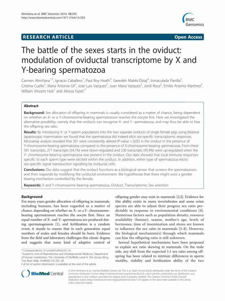
Results and discussionOur work showed that the presence of X- and Y-spermatozoa did indeed elicit different transcriptomicresponses within the oviduct (Figure 2A). Around 2% oftranscripts (501 out of 24123 probes from AffymetrixPorcine Chip) were consistently altered (P-value < 0.05)
were subjected to laparoscopic surgery. To prevent X- andsing titanium staples. Then, one oviduct was inseminated withtozoa (3 × 10⁵ spermatozoa/100 μl) from the same animal. Twenty-fourg X- and Y-sperm samples were collected from each side of the
Figure 2 The presence of Y-spermatozoa elicited different transcriptome response within the oviduct when compared to X-spermatozoa.A: Cluster heat map analysis of the transcriptional profiles obtained from oviductal samples inseminated with X-spermatozoa and Y-spermatozoa.Each row represents a different gene, and each column displays gene expressions at different samples (X1-X4 for oviductal samples inseminatedwith X-spermatozoa; Y1-Y2 for oviductal samples inseminated with Y-spermatozoa). Data values displayed as yellow and blue represent elevatedand reduced expression, respectively. B: A Volcano plot depicting significant changes in gene expression between oviductal samples inseminatedwith Y-spermatozoa and X-spermatozoa. Each of the 23,124-oligonucleotide probes is represented by a dot in the graph. The x-axis represents the foldchange and the y-axis represents the statistical significance (−log10 of p-value). Transcripts showing significant differences in gene expression(501 probes, p < 0.05) are above the broken line.
Almiñana et al. BMC Genomics 2014, 15:293 Page 3 of 11http://www.biomedcentral.com/1471-2164/15/293
in the oviduct in the presence of Y-chromosome-bearingspermatozoa compared to the presence of X-chromosome-bearing spermatozoa (Figure 2B). From these 501 transcripts,271 transcripts (54.1%) were down-regulated and 230transcripts (45.9%) were up-regulated when the Y-chromosome-bearing spermatozoa was present in theoviduct. A complete list of the transcripts altered in theoviduct inseminated with Y- chromosome bearing sperm-atozoa compared to X -chromosome bearing spermatozoais presented in the Additional file 1.To obtain a biologically meaningful overview of the al-
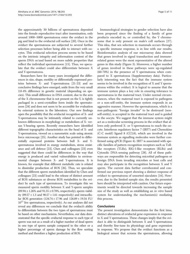
tered transcripts in the presence of Y-chromosome-bearingspermatozoa compared to X-chromosome-bearing sperm-atozoa, genes differently expressed were organized intodifferent categories and subcategories according to KEGGdatabase hierarchy. The functional categories with highernumber of genes were: signal transduction, immune sys-tem, digestive system and endocrine system. The pathwaysin which these altered transcripts were involved withare presented in Table 1. Interestingly, a higher numberof genes involved in signal transduction and immunesystem were up-regulated (60-70%) in the presence ofY-chromosome bearing spermatozoa when compared to
X- chromosome-bearing spermatozoa. Other interest-ing subcategories with high numbers of transcripts in-volved were: nervous system, cell growth and death, cellcommunication, signalling molecules and interaction,folding, sorting and degradation and transport and ca-tabolism. The results of all data pathways classificationare available in Additional file 2.Our data provide the first evidence to show how sperm-
atozoa carrying the Y- or X-chromosome can modulatethe oviductal response by activating specific signallingpathways in a gender specific manner. These data implythat the female reproductive tract recognizes the presenceof X- or Y-chromosome-bearing spermatozoa in theoviduct before fertilization occurs. On this basis, wehypothesize that a sex-specific sperm recognition sys-tem exists in the female reproductive tract. Further-more, we propose that this sperm recognition systemcan modulate the gender selection of the offspring. Ourhypothesis challenges two long-held assumptions: 1) onthe female side, that the reproductive tract is a passiveparticipant in sperm selection [17] and 2) on the maleside, that there are no differences in morphology, meta-bolic activity or functional abilities of X and Y sperm that
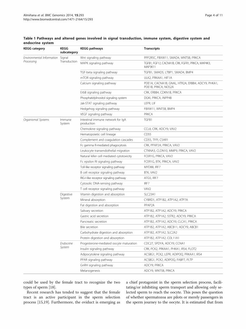
Table 1 Pathways and altered genes involved in signal transduction, immune system, digestive system andendocrine system
KEGG category KEGGsubcategory
KEGG pathways Transcripts
Environmental InformationProcessing
SignalTransduction
Wnt signaling pathway PPP2R5C, FBXW11, SMAD4, WNT5B, PRKCA
MAPK signaling pathway TGFB1, FGF12, CACNA1B, CRK, FGFR1, PRKCA, MAP4K3,MAP3K11
TGF-beta signaling pathway TGFB1, SMAD5, LTBP1, SMAD4, BMP4
mTOR signaling pathway ULK2, PRKAA1, HIF1A
Calcium signaling pathway PDE1A, CACNA1B, GNAL, HTR2A, ERBB4, ADCY9, PHKA1,PDE1B, PRKCA, NOS2A
ErbB signaling pathway CRK, ERBB4, CDKN1B, PRKCA
Phosphatidylinositol signaling system DGKI, PRKCA, INPP4B
Jak-STAT signaling pathway LEPR, LIF
Hedgehog signaling pathway FBXW11, WNT5B, BMP4
VEGF signaling pathway PRKCA
Organismal Systems ImmuneSystem
Intestinal immune network for IgAproduction
TGFB1
Chemokine signaling pathway CCL8, CRK, ADCY9, VAV2
Hematopoietic cell lineage CD55
Complement and coagulation cascades CD55, TFPI, C5AR1
Fc gamma R-mediated phagocytosis CRK, PPAP2A, PRKCA, VAV2
Leukocyte transendothelial migration CTNNA3, CLDN10, MMP9, PRKCA, VAV2
Natural killer cell mediated cytotoxicity FCER1G, PRKCA, VAV2
Fc epsilon RI signaling pathway FCER1G, BTK, PRKCA, VAV2
Toll-like receptor signaling pathway MYD88, IRF7
B cell receptor signaling pathway BTK, VAV2
RIG-I-like receptor signaling pathway ATG5, IRF7
Cytosolic DNA-sensing pathway IRF7
T cell receptor signaling pathway VAV2
DigestiveSystem
Vitamin digestion and absorption SLC23A1
Mineral absorption CYBRD1, ATP1B2, ATP1A2, ATP7A
Fat digestion and absorption PPAP2A
Salivary secretion ATP1B2, ATP1A2, ADCY9, PRKCA
Gastric acid secretion ATP1B2, ATP1A2, SSTR2, ADCY9, PRKCA
Pancreatic secretion ATP1B2, ATP1A2, ADCY9, CLCA1, PRKCA
Bile secretion ATP1B2, ATP1A2, ABCB11, ADCY9, ABCB1
Carbohydrate digestion and absorption ATP1B2, ATP1A2, SLC2A2
Protein digestion and absorption ATP1B2, ATP1A2, COL11A1
EndocrineSystem
Progesterone-mediated oocyte maturation CDC27, SPDYA, ADCY9, CCNA1
Insulin signaling pathway CRK, PCK2, PRKAA1, PHKA1, IRS4, FLOT2
Adipocytokine signaling pathway ACSBG1, PCK2, LEPR, ADIPOQ, PRKAA1, IRS4
PPAR signaling pathway ACSBG1, PCK2, ADIPOQ, FABP7, PLTP
GnRH signaling pathway ADCY9, PRKCA
Melanogenesis ADCY9, WNT5B, PRKCA
Almiñana et al. BMC Genomics 2014, 15:293 Page 4 of 11http://www.biomedcentral.com/1471-2164/15/293
could be used by the female tract to recognize the twotypes of sperm [18].Recent research has tended to suggest that the female
tract is an active participant in the sperm selectionprocess [15,19]. Furthermore, the oviduct is emerging as
a chief protagonist in the sperm selection process, facili-tating/or inhibiting sperm transport and allowing only se-lected sperm to reach the oocyte. This poses the questionof whether spermatozoa are pilots or merely passengers inthe sperm journey to the oocyte. It is estimated that from
Almiñana et al. BMC Genomics 2014, 15:293 Page 5 of 11http://www.biomedcentral.com/1471-2164/15/293
the approximately 30 billions of spermatozoa depositedinto the female reproductive tract after insemination, onlyaround 1000–5000 spermatozoa enter the oviduct in thepig and bind to the oviductal cell surface [20]. Once in theoviduct the spermatozoa are subjected to several furtherselection processes before being able to interact with oo-cytes. This oviductal selection process seems to be basedon the intrinsic integrity and information content of thesperm DNA or/and based on more subtle properties thatreflect the individual spermatozoon [15]. Thus, we specu-late that the oviduct could also differentiate between Xand Y-spermatozoa.Researchers have for many years investigated the differ-
ences in size, shape, motility or differentially expressed pro-teins between X- and Y-spermatozoa [21-23] and noconclusive findings have emerged, aside from the very small2.8-4% difference in genetic material (depending on spe-cies). This small difference in DNA content, due to the armof the X chromosome that is not present on the Y, is tightlypackaged in a semi-crystalline form inside the spermato-zoon [24] and does not seem to be accessible for evaluationby external systems in the laboratory. Our new findingsopen the possibility that the oviductal recognition of X- andY-spermatozoa may be intimately related to currently un-known differences in morphology or metabolism of X- ver-sus Y-bearing sperm. Recently researchers have identifieddifferent topographic characteristics on the head of X- andY-spermatozoa, viewed on a nanometric scale using atomicforce microscopy [25]. Another study has pointed to differ-entially expressed proteins between bull X- and Y-spermatozoa involved in energy metabolism, stress resist-ance and cell defence [23]. Chen and colleagues [23] evensuggested that there could be differences in the way thatenergy is produced and varied vulnerabilities to environ-mental changes between X- and Y-spermatozoa. It isknown, for example that different metabolic rate is relatedto dissimilar production of ROS [26]. Thus, we speculatethat the different sperm metabolism identified by Chen andcolleagues [23] could lead to the release of distinct amountof ROS substances or diverse ROS metabolites to the ovi-duct by each type of spermatozoa. To investigate this wemeasured sperm motility between X and Y-sperm samples(89.94 ± 1.26% and 91.55 ± 0.78%, respectively), sperm viabil-ity (89.57 ± 1.3 and 90.57 ± 1.61 respectively) and intracellu-lar ROS generation (124.74 ± 17.96 and 126.89 ± 19.54 FU/1012 live spermatozoa, respectively). As our analyses did notreveal any differences we conclude that the oviduct’s abilityto differentiate between the two types of spermatozoa mustbe based on other mechanisms. Nevertheless, our data dem-onstrated that the specific oviductal response to each type ofsperm was not as a result of a higher number of dead spermin one type of sperm sample compared to the other or ahigher percentage of sperm damage by the flow sortingmethod and therefore a higher production of ROS.
Immunological strategies to gender selection have alsobeen proposed since the finding of a family of geneproducts encoded in, or controlled by, the Y chromo-some that is only present on male’s cell surfaces [27].This idea, that sex selection in mammals occurs througha specific immune response, is in line with our results.Bioinformatics analysis of our microarray data showedthat genes involved in signal transduction and immune-related genes were the most representative of the alteredgenes in this study (Figure 3). Moreover, a higher numberof genes involved in these pathways were up-regulated(60-70%) in the presence of Y spermatozoa when com-pared to X spermatozoa (Supplementary data). Particu-larly interesting was the fact that the immune systemseems to be involved in the recognition of X and Y sperm-atozoa within the oviduct. It is logical to assume that theimmune system plays a key role in ensuring tolerance tospermatozoa in the maternal tract. Under normal circum-stances, when the maternal tract is exposed to pathogensor a non-self-entity, the immune system responds in anaggressive manner. However the spermatozoon, which is anon-pathogenic “foreign invader” to be destroyed (non-self-entity), is accepted in the maternal tract and is guidedto the oocyte. We suggest that the immune system mightact as a molecular screening process in the oviduct that al-lows only preferred X- or Y-spermatozoa to reach the oo-cyte. Interferon regulatory factor 7 (IRF7) and Chemokine(C-C motif) ligand 8 (CCL8), which are involved in theimmune system as signalling molecules were further con-firmed using qPCR (Figure 4). IRF7 is associated with spe-cific families of pattern recognition receptors such as Toll-like receptors (TLRs), RIG-I-like receptors (RLRs) andCytosolic DNA-sensing pathway [28]. All of these path-ways are responsible for detecting microbial pathogens orforeign DNA from invading microbes or host cells andmay also participate in the recognition between X and Ysperm. The current data further corroborated and con-firmed our previous report showing a distinct response ofoviduct to spermatozoa of unsorted ejaculates [16]. How-ever, due to the limited sample size, the results presentedhere should be interpreted with caution. Our future exper-iments would be directed towards increasing the samplesize of the study as well as establishing an in vitro basedsystem for understanding the mechanism(s) mediatingthis process.
ConclusionsThe present investigation demonstrates for the first time,distinct alterations of oviductal gene expression in responseto X and Y spermatozoa. These changes imply that the ovi-duct is able to distinguish between X and Y-spermatozoaand thereby fine-tune its physiology and gene expressionin response. We propose that the oviduct functions as abiological sensor that screens the spermatozoa, allowing
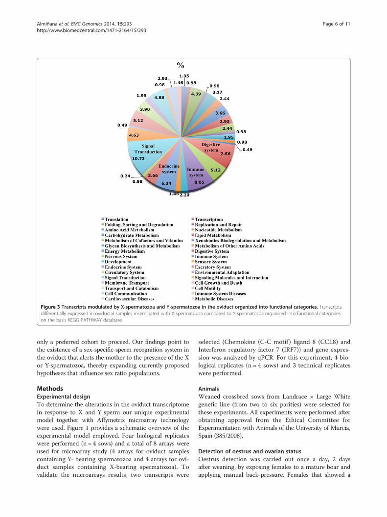
Figure 3 Transcripts modulated by X-spermatozoa and Y-spermatozoa in the oviduct organized into functional categories. Transcriptsdifferentially expressed in oviductal samples inseminated with X-spermatozoa compared to Y-spermatozoa organized into functional categorieson the basis KEGG PATHWAY database.
Almiñana et al. BMC Genomics 2014, 15:293 Page 6 of 11http://www.biomedcentral.com/1471-2164/15/293
only a preferred cohort to proceed. Our findings point tothe existence of a sex-specific-sperm recognition system inthe oviduct that alerts the mother to the presence of the Xor Y-spermatozoa, thereby expanding currently proposedhypotheses that influence sex ratio populations.
MethodsExperimental designTo determine the alterations in the oviduct transcriptomein response to X and Y sperm our unique experimentalmodel together with Affymetrix microarray technologywere used. Figure 1 provides a schematic overview of theexperimental model employed. Four biological replicateswere performed (n = 4 sows) and a total of 8 arrays wereused for microarray study (4 arrays for oviduct samplescontaining Y- bearing spermatozoa and 4 arrays for ovi-duct samples containing X-bearing spermatozoa). Tovalidate the microarrays results, two transcripts were
selected (Chemokine (C-C motif ) ligand 8 (CCL8) andInterferon regulatory factor 7 (IRF7)) and gene expres-sion was analyzed by qPCR. For this experiment, 4 bio-logical replicates (n = 4 sows) and 3 technical replicateswere performed.
AnimalsWeaned crossbred sows from Landrace × Large Whitegenetic line (from two to six parities) were selected forthese experiments. All experiments were performed afterobtaining approval from the Ethical Committee forExperimentation with Animals of the University of Murcia,Spain (385/2008).
Detection of oestrus and ovarian statusOestrus detection was carried out once a day, 2 daysafter weaning, by exposing females to a mature boar andapplying manual back-pressure. Females that showed a
Figure 4 Validation of the microarray results by qPCR analysis.CCL8 (chemokine (C-C motif) ligand 8) and IRF7 (interferonregulatory factor 7) expression values (normalized based on ß–actinand Ubiquitin B expression values) in oviductal samples inseminatedwith X-spermatozoa compared to oviductal samples inseminatedwith Y-spermatozoa. The expression of both transcripts in theoviductal samples inseminated with Y-spermatozoa was significantlydifferent from that of the oviductal samples inseminated withY-spermatozoa (P < 0.05).
Almiñana et al. BMC Genomics 2014, 15:293 Page 7 of 11http://www.biomedcentral.com/1471-2164/15/293
standing estrous reflex were considered to be in heatand the ovaries were scanned by transrectal ultrasonog-raphy [29]. Only sows showing multiple pre-ovulatoryfollicles (diameter of antrum >6 mm) were selected forexperiments. Inseminations were carried out within 2–3 hafter the ultrasonography and oviduct samples collectiontook place 24 h later (approximately 12-24 h before ovula-tion took place in most of the sows).
Semen collection and flow cytometric sortingSperm-rich fractions from three fertile mature boars,that had previously sired offspring, were collected bygloved-hand method and extended in Beltsville ThawingSolution (BTS) to 150 × 106 spermatozoa/mL [30]. Aftercollection, samples were evaluated for normality (motil-ity >80%, membrane integrity > 85%, total sperm countper ejaculate > 20 × 109; acrosomal abnormalities < 10%,abnormal sperm morphology < 15%) [31].The extended semen was then processed for sperm
sorting following the general procedure as previously de-scribed [32]. Briefly, 1 mL of extended semen was stained
with Hoechst-33342 fluorophore (0.3 mM per 1 × 10⁶spermatozoa) and incubated for 1 h in darkness at 35°C.After incubation, samples were passed through a 30 mmnylon mesh filter to remove debris or clumped sperm-atozoa. Then, 1 μl/ml of food dye (1 mg/ml; WarnerJenkinson Company Inc., St. Louis, MO, USA FD&C 40solution) was added to the sample to identify deadsperm and kept 15 minutes at room temperature indark. The stained spermatozoa were separated using ahigh speed MoFlo SX flow cytometer/sperm sorter(Dako Colorado Inc., Fort Collins, Co, USA), equippedwith a solid state laser (ultraviolet wavelength, 351-363 nm) at 175 mW (Spectra Physics 1330, Terra BellaAvenue, Mountain View, CA, USA). Spermatozoa wereseparated into X- and Y- chromosome bearing popula-tions with 91% purity of Y-spermatozoa and 93% of X-spermatozoa. Both the X sperm and the Y sperm popula-tions were recovered from the three boars. Consequently,in one side the three X-insemination doses were pooledand in the other side the three Y-insemination doses fromthe three boars were also pooled and both doses finally di-luted in BTS to 3 × 10⁵ spermatozoa/100 μl before laparo-scopic insemination.
Sperm motility, viability and functionality of X- andY-sperm samplesThe motility of X- and Y-sperm samples was evaluated ob-jectively using a computer-assisted analysis system (ISAS;Proiser R + D, Paterna, Spain). The sperm motility vari-able recorded was the overall percentage of motile sperm-atozoa (average path velocity (VAP) = 20 mm/s) [31].The viability of the spermatozoa was evaluated by sim-
ultaneous cytometric assessment of the plasma and acro-somal membrane integrity by using a triple-fluorescenceprocedure as described by Martinez-Alborcia et al., [33].Briefly, a 100 μl sperm sample (30 × 106 cells/ml inPBS) was transferred to culture tubes containing 2.5 μlH-42 (0.05 mg/ml in PBS), 2 μl propidium iodide (PI,0.5 mg/ml in PBS, Molecular Probes Europe BV,Leiden, The Netherlands) and 2 μl fluorescein-conjugatedpeanut agglutinin (PNA-FITC, 200 μg/ml in PBS). Thesamples were mixed and incubated at 38°C in the dark for10 min. Immediately before analysis by flow cytometry,400 μl PBS was added to each X- and Y- sperm samplesand mixed. The fluorescence spectra of PI and PNA-FITCwas detected using a 670 nm long-pass (LP) filter and a530/30 nm BP filter, respectively. The spermatozoa ana-lyzed were categorized into four categories: (1) intactplasma and acrosomal membranes (PI-/PNA-FITC-);(2) intact plasma membrane and damaged acrosome(PI-/PNA-FITC+); (3) damaged plasma membrane andintact acrosome (PI+/PNA-FITC-); or (4) damaged plasmaand acrosomal membranes (PI+/PNA+). The viable
Almiñana et al. BMC Genomics 2014, 15:293 Page 8 of 11http://www.biomedcentral.com/1471-2164/15/293
spermatozoa exhibited intact plasma and acrosomal mem-branes and this was expressed as a percentage of the totalcells.The functionality of the spermatozoa was assessed by the
intracellular ROS production in X and Y-sperm samples.The intracellular production of ROS by each sperm samplewas measured using 5-(and- 6) chloromethyl-20,70-dichlor-odihydrofluorescein diacetate acetylester (CM-H2DCFDA),described by Martinez-Alborcia et al., [33].CM-H2DCFDA is freely permeable across cell mem-
branes and becomes incorporated into the hydrophobicregions of the cell. Upon entering the cell, the acetatemoiety of CM-H2DCFDA is cleaved by cellular esterasesto leave the impermeant and non-fluorescent molecule20,70-dichlorodihydro- fluorescein (H2DCF). The H2DCFis oxidized by hydrogen peroxide (H2O2) into dichloro-fluorescein (DCF), which fluoresces at 530 nm followingexcitation at 488 nm. For each X-and Y-sperm sample,two different 50 μl aliquots of mTBM-diluted spermato-zoa were diluted in 950 μl PBS containing (1) 1.25 μl H-42(0.05 mg/ml in PBS), 1 μl PI (1 mg/ml in PBS), 1 μlH2DCFDA (1 mM in DMSO) and 1 μl tert-butylhydrogenperoxide (1 mM in purified water) to induce oxidativestress (first aliquot; used to measure induced ROS for-mation) or (2) 1.25 μl H-42, 1 μl PI and 1 μl H2DCFDA(second aliquot; used to measure basal ROS formation).The samples were incubated at 38ªC in the dark for10 min before flow cytometry. The mean fluorescenceintensity of DCF (induced minus basal) was expressedas fluorescence units (FU) per 1012 live spermatozoa.
Intraoviductal laparoscopic inseminationIntraoviductal laparoscopic inseminations were carriedout within 2–3 h of the ultrasonography. Sows were se-dated by azaperone administration (2 mg/kg body weight,i.m.), anaesthetized with sodium thiopenthal (7 mg/kgbody weight, i.v.) and maintained under anesthesia withisofluorane (3.5–5%). Intraoviductal laparoscopic insemi-nations were carried as described by Almiñana et al., [34].Briefly, each sow was placed in the supine position and apneumoperitoneum was established. The abdominal cavitywas insufflated with CO2 to 14 mmHg. Two accessoryports were placed in the right and left part of the hemi ab-domen, which provided access for laparoscopic Duval for-ceps for manipulating the uterine horn and grasping theoviduct for the insemination, respectively. To prevent Xand Y sperm migration between oviducts, both uterinehorns were cut using titanium staples (EndoGIA Universal60/4.8; Tyco heathcare, Mansfield, MA). To perform theinsemination, the oviduct was grasped with the Duval for-ceps in the isthmus region and the sperm dose containingY sorted spermatozoa (3 × 10⁵ spermatozoa/100 μl.) wasflushed into one oviduct (above of the ampullar region indirection to isthmus). The procedure was then repeated in
the contralateral oviduct but injecting the X sperm dosecontaining the X-chromosome bearing sperm (Figure 1).After both oviducts were inseminated, the trocars were re-moved and minor suture was required. Following surgery,sows were returned to their accustomed environment.
Sample collection and RNA preparationTwenty-four hours following laparoscopic insemination,oviduct tissues containing X and Y sperm populationswere collected from each side of the reproductive tractin each animal. Sows were sedated as previously described.To avoid the presence of oocytes that could mask the ovi-duct response to X and Y sperm, only oviduct samplesfrom sows showing no signs of ovulation were collected.In addition, oviducts were flushed with Phosphate Buff-ered Saline medium (PBS, 30 ml) and the absence of oo-cytes was verified by careful examination of oviductflushings under a stereomicroscope. In cases where it wasunsure whether the sow had ovulated oviduct sampleswere discarded.Oviducts were opened longitudinally and epithelial cells
were isolated by scraping the mucosal epithelial layer witha glass slide. Scraped cells from the uterine horn sampleswere transferred immediately to Tri Reagent (Sigma,Sigma-Aldrich Co, Madrid, Spain), homogenised, snap-frozen in liquid nitrogen and stored at -80°C until fur-ther processing.Total RNA was isolated using a standard procedure in-
volving phenol:chloroform extraction followed by ethanolprecipitation. The quantity (NanoDrop 1000 spectro-photometer) and the quality (Agilent 2100 Bioanalyser;Agilent Technologies) of the RNA samples were ana-lysed. Only samples with satisfactory quality as indi-cated by the absence of degradation of the ribosomalRNA were used for microarrays and quantitative Real-Time Reverse Transcriptase-Polymerase Chain Reaction(qPCR).
Microarrays hybridizationAffymetrix Porcine Genome gene expression arrays(Affymetrix, Santa Clara, CA) were used in this study.Total RNA samples were prepared according to the Affy-metrix Technical Manual (www.affymetrix.com). Briefly,200 ng of total RNA was converted into cDNA using anoligo(dT) which also carries the binding site for T7 RNApolymerase. Superscript II (Affymetrix) was used to carryout this reaction. After first strand synthesis, residualRNA was degraded by addition of RNaseH and a double-stranded cDNA molecule was generated using DNA Poly-merase I and DNA ligase. This double stranded moleculewas used as a substrate for the T7 RNA polymerase toproduce multiple copies of the cRNA using the AffymetrixIVT labelling system. The cRNA molecules produced in-corporated biotin labelled ribonucleotides, which acted as
Almiñana et al. BMC Genomics 2014, 15:293 Page 9 of 11http://www.biomedcentral.com/1471-2164/15/293
a target for the subsequent detection of hybridization,using fluorescently labelled streptavidin. 13 μg of cRNAmolecules were heat fragmented and injected to the Por-cine GeneChips in a hybridization solution according tothe Affymetrix protocol. Hybridization took place over-night in a rotating hybridization oven at 60 rpm, 45°C for16 hours. The GeneChip arrays were washed using theAffymetrix Fluidics Station. After washing, the GeneChiparrays were scanned using Affymetrix GC3000 scanner.The resultant images were analysed using the MicroarraySuite software version 5.1 (Affymetrix). At the detectionlevel each probe set was designated as present, absent ormarginal. Only present transcripts were consideredexpressed. Microarray experiments were carried out ac-cording to MIAME guidelines and the complete experi-mental data can be obtained online from the NCBI GeneExpression Omnibus (http://www.ncbi.nlm.nih.gov/geo/)submission number GSE47139.
Microarray data and bioinformatics analysisMicroarrays analysis was performed using Taverna work-flow management system (http://www.taverna.org.uk.)[35,36]. Microarrays data were normalized using RobustMultichip Average (RMA) method [37]. Differentiallyexpressed genes between oviduct inseminated with Xspermatozoa and Y spermatozoa were detected throught-tests that were applied to normally distributed datausing the Limma R/Bioconductor packages [38] withBenjamini-Hochberg false discovery rate and multipletesting correction to control for Type I errors [39]. Fromthe list of differentially expressed genes, the genes thatpass the specified threshold for p-value = 0.05 were se-lected. Using Affymetrix Porcine Annotation in combin-ation with the improved annotation provided by Tsaiet al., [40] for these porcine arrays, differentially expressedtranscripts from our microarray data were annotated. Toobtain a biologically meaningful overview of the alteredtranscripts in the presence of Y-chromosome-bearingspermatozoa compared to X-chromosome-bearing sperm-atozoa, genes differently expressed were organized into
Table 2 Primers used for qPCR analysis
Gene symbol Affymetrix porcine probe Primer
CCL8 SSc.9957.1.A1_at Forward
Reverse
IRF7 SSc.2573.9.1_S1_at Forward
Reverse
Ubiquitin B Reference gene Forward
Reverse
β-actin Reference gene Forward
Reverse
different categories and subcategories according to KEGGdatabase hierarchy.
Quantitative real-time reverse transcriptase polymerasechain reactionGene expression profiles derived from microarray analyseswere confirmed using quantitative real-time reverse tran-scriptase polymerase chain reaction (qPCR). The primersused for qPCR are listed in Table 2. Amplified PCR prod-ucts were sequenced with forward and reverse primers toverify the resulting product.Total RNA from the oviduct samples (inseminated
with Y sperm or inseminated with X sperm) was treatedthree times with DNase I (DNA-free kit; Ambion.) to re-move genomic DNA contamination from samples. First-strand cDNA synthesis was performed using High CapacitycDNA Reverse Transcription Kit (Appied Biosystems).Negative controls were prepared without inclusion of theenzyme (non-reverse transcription controls, RT controls).Reverse transcriptase PCR (RT-PCR) was performed ac-cording manufacturer instructions. To evaluate the size ofthe PCR products 10 μl of each sample was resolved on a1.2% agarose gel and electrophoresis was performed with1× TAE buffer and a voltage of 110 V for 40–50 min. Thebands were visualized by using an ultraviolet transillumin-ation, and digital images were obtained.SYBR Green Jump Start (Sigma) master mix (containing
10 μl SYBR Green, 7 μl H2O, 1 μl of forward and reverseprimers and 1 μl cDNA) was added to each well of PCRplate and amplification was performed under the follow-ing conditions: 40 cycles of 95° for 30 s, 55° for 1 min and72° for 1 min. Samples without template and RT controls(without the addition of enzyme) for each primer set wereincluded to identify contamination. Triplicate measure-ments for each group of samples were carried out.Quantitative PCR was performed using Mx3005P QPCR(Stratagene, Waldbronn, Germany). The quantificationdata were analyzed using MxPro QPCR software version4.01. Quantitative PCR data were analyzed using the com-parative CT method [41]. The results were expressed asmean ± SEM arbitrary gene expression values, normalized
Sequence Product size (pb)
5′ GCGAGATGGCATTTCTCTCT 3′ 119
5′ CACACTTCGGCTTACAAGAGG3′
5′ GCTGGATGAAGCCAGAACA 3′ 97
5′ GGCCCAGGCCTTAAAGAT 3′
5′ GTCTGAGGGGTGGCTGCTAA 3′ 85
5′ TGGGGCAAATGGCTAGAGTG 3′
5′ CCTCCCTGGAGAAGAGCTA 3′ 131
5′ CTTCATGATGGAGTTGAAGGT 3′
Almiñana et al. BMC Genomics 2014, 15:293 Page 10 of 11http://www.biomedcentral.com/1471-2164/15/293
on the basis of the two reference expression (ß-actin andUbiquitin B). Statistical analysis was performed usingpaired T-test to evaluate the significance of difference be-tween expression values of oviduct inseminated with Ysperm versus oviduct inseminated with X sperm (in SPSS,version 14.0 (SPSS Inc., Chicago, IL)). The threshold forsignificance was set at p < 0.05.
Availability of additional filesThe data set supporting the results of this article is avail-able in the additional files and also is available onlinefrom the NCBI Gene Expression Omnibus (http://www.ncbi.nlm.nih.gov/geo/) submission number GSE47139.
Additional files
Additional file 1: List of candidate transcripts differentially expressedin the oviduct in the presence of Y-bearing spermatozoa compared toX-bearing spermatozoa (P-value < 0.05).
Additional file 2: Transcripts modulated by X and Y-chromosomebearing spermatozoa organized into functional categories accordingto KEGG database.
Competing interestsThe authors declare no competing financial interests.
Authors’ contributionsConceived and designed the experiments: JMV JR EAM CA CI AF. Performedthe experiments: CI CC IP MAG JLV JMV JR EAM PRH CA AF. Analyzed thedata: PRH MDS CA AF. Contributed reagents/materials/analysis tools: JMV JREAM PRH AF. Wrote the paper: CA HWV AF. All authors read and approvedthe final manuscript.
AcknowledgementsThis work was supported by the European Union Seventh FrameworkProgramme-Marie Curie COFUND (FP7/2007-2013) under UMU IncomingMobility Programme ACTion (U-IMPACT) Grant Agreement 267143, MarieCurie IEF Grant Agreement 236708, Minister of Science and Innovation(MICINN; AGL2009-12091), Seneca Foundation (GERM04543/07), COSTACTION FA0702, Sexing Technologies (Navasota, TX, USA) and Centre for theDevelopment of Industrial Technology (CDTI; 2008/0268).
Author details1Academic Unit of Reproductive and Developmental Medicine, Departmentof Human metabolism, The University of Sheffield, Level 4, The Jessop Wing,Tree Root Walk, Sheffield S10 2SF, UK. 2Department of Medicine and AnimalSurgery, Veterinary Faculty, Regional Campus of International Excellence,University of Murcia, Spain Espinardo 300071, E-30071 Murcia, Murcia, Spain.3Sheffield Institute for Translational Neuroscience (SITraN), Sheffield, UnitedKingdom. 4Department of Computer Science, University of Sheffield,Sheffield, UK.
Received: 26 December 2013 Accepted: 17 April 2014Published: 21 May 2014
References1. McClung CE: The accessory chromosome-sex determinant? Biol Bull 1902,
3:43–84.2. Grant VJ, Chamley LW: Can mammalian mothers influence the sex of
their offspring peri-conceptually? Reproduction 2010, 140(3):425–433.3. Tarin JJ, Bernabeu R, Baviera A, Bonada M, Cano A: Sex selection may be
inadvertently performed in in-vitro fertilization-embryo transferprogrammes. Hum Reprod 1995, 10(11):2992–2998.
4. West SA, Herre EA, Sheldon BC: Evolution. The benefits of allocating sex.Science 2000, 290(5490):288–290.
5. Hardy ICW: Possible factors influencing vertebrate sex ratios: anintroductory overview. Appl Anim Behav Sci 1997, 51:217–241.
6. James WH: The variations of human sex ratio at birth during and afterwars, and their potential explanations. J Theor Biol 2009, 257(1):116–123.
7. Kruuk LE, Clutton-Brock TH, Albon SD, Pemberton JM, Guinness FE: Populationdensity affects sex ratio variation in red deer. Nature 1999,399(6735):459–461.
8. Zorn B, Sucur V, Stare J, Meden-Vrtovec H: Decline in sex ratio at birth after10-day war in Slovenia: brief communication. Hum Reprod 2002,17(12):3173–3177.
9. Shettles LB: Sperm morphology and sex ratios. J Urol 1961, 86:450–455.10. Dominko T, First NL: Relationship between the maturational state of
oocytes at the time of insemination and sex ratio of subsequent earlybovine embryos. Theriogenology 1997, 47(5):1041–1050.
11. Rosenfeld CS, Roberts RM: Maternal diet and other factors affectingoffspring sex ratio: a review. Biol Reprod 2004, 71(4):1063–1070.
12. Gomez E, Caamano JN, Corrales FJ, Diez C, Correia-Alvarez E, Martin D, TrigalB, Carrocera S, Mora MI, Pello-Palma J, Moreno JF, Muñoz M: Embryonic sexinduces differential expression of proteins in bovine uterine fluid.J Proteome Res 2013, 12(3):1199–1210.
13. Trivers RL, Willard DE: Natural selection of parental ability to vary the sexratio of offspring. Science 1973, 179(4068):90–92.
14. Hunter RH: The fallopian tubes in domestic mammals: how vital is theirphysiological activity? Reprod Nutr Dev 2005, 45(3):281–290.
15. Holt WV, Fazeli A: The oviduct as a complex mediator of mammaliansperm function and selection. Mol Reprod Dev 2010, 77(11):934–943.
16. Georgiou AS, Snijders AP, Sostaric E, Aflatoonian R, Vazquez JL, Vazquez JM,Roca J, Martinez EA, Wright PC, Fazeli A: Modulation of the oviductalenvironment by gametes. J Proteome Res 2007, 6(12):4656–4666.
17. Smith RL: Sperm Competition and the Evolution of Animal Mating Systems.London, UK: Academic; 1984.
18. Hossain AM, Barik S, Kulkarni PM: Lack of significant morphologicaldifferences between human X and Y spermatozoa and their precursorcells (spermatids) exposed to different prehybridization treatments.J Androl 2001, 22(1):119–123.
19. Birkhead TR, Pizzari T: Postcopulatory sexual selection. Nat Rev Genet 2002,3(4):262–273.
20. Rodriguez-Martinez H, Tienthai P, Suzuki K, Funahashi H, Ekwall H,Johannisson A: Involvement of oviduct in sperm capacitation and oocytedevelopment in pigs. Reprod Suppl 2001, 58:129–145.
21. Hendriksen PJM: Do X and Y spermatozoa differ in proteins?Theriogenology 1999, 52:1295–1307.
22. Cui KH: Size differences between human X and Y spermatozoa andprefertilization diagnosis. Mol Hum Reprod 1997, 3(1):61–67.
23. Chen X, Zhu H, Wu C, Han W, Hao H, Zhao X, Du W, Qin T, Liu Y, Wang D:Identification of differentially expressed proteins between bull X and Yspermatozoa. J Proteomics 2012, 77:59–67.
24. Ward WS, Coffey DS: DNA packaging and organization in mammalianspermatozoa: comparison with somatic cells. Biol Reprod 1991,44(4):569–574.
25. Carvalho JO, Silva LP, Sartori R, Dode MA: Nanoscale differences in theshape and size of x and y chromosome-bearing bovine sperm headsassessed by atomic force microscopy. PLoS One 2013, 8(3):e59387.
26. Garratt M, Brooks RC: Oxidative stress and condition-dependent sexualsignals: more than just seeing red. Proc Biol Sci 2012, 279(1741):3121–3130.
27. Mohammadi AA, Tetro JA, Filion LG: Epitope selection to male specificantigens for sex selection in swine. J Reprod Immunol 2011, 89(1):46–54.
28. Honda K, Taniguchi T: IRFs: master regulators of signalling by toll-likereceptors and cytosolic pattern-recognition receptors. Nat Rev Immunol2006, 6(9):644–658.
29. Bolarin A, Roca J, Rodriguez-Martinez H, Hernandez M, Vazquez JM, Martinez EA:Dissimilarities in sows’ ovarian status at the insemination time could explaindifferences in fertility between farms when frozen-thawed semen is used.Theriogenology 2006, 65(3):669–680.
30. Pursel VG, Johnson LA: Freezing of boar spermatozoa - fertilizing-capacitywith concentrated semen and a new thawing procedure. J Anim Sci1975, 40(1):99–102.
31. Gil MA, Hernandez M, Roca J, Alminana C, Lucas X, Cuello C, Vazquez JM,Martinez EA: Pentoxifylline added to freezing or post-thaw extendersdoes not improve the survival or in vitro fertilising capacity of boarspermatozoa. Reproduction 2010, 139(3):557–564.
Almiñana et al. BMC Genomics 2014, 15:293 Page 11 of 11http://www.biomedcentral.com/1471-2164/15/293
32. Johnson LA, Flook JP, Hawk HW: Sex preselection in rabbits: live birthsfrom X and Y sperm separated by DNA and cell sorting. Biol Reprod 1989,41(2):199–203.
33. Martinez-Alborcia MJ, Valverde A, Parrilla I, Vazquez JM, Martinez EA, Roca J:Detrimental effects of non-functional spermatozoa on the freezability offunctional spermatozoa from boar ejaculate. PLoS One 2012, 7(5):e36550.
34. Alminana C, Heath PR, Wilkinson S, Sanchez-Osorio J, Cuello C, Parrilla I, GilMA, Vazquez JL, Vazquez JM, Roca J, Martinez EA, Fazeli A: Early developingpig embryos mediate their own environment in the maternal tract. PLoSOne 2012, 7(3):e33625.
35. Hull D, Wolstencroft K, Stevens R, Goble C, Pocock MR, Li P, Oinn T:Taverna: a tool for building and running workflows of services. NucleicAcids Res 2006, 34(Web Server issue):W729–W732.
36. Oinn T, Addis M, Ferris J, Marvin D, Senger M, Greenwood M, Carver T,Glover K, Pocock MR, Wipat A, Li P: Taverna: a tool for the compositionand enactment of bioinformatics workflows. Bioinformatics 2004,20(17):3045–3054.
37. Bolstad BM, Irizarry RA, Astrand M, Speed TP: A comparison ofnormalization methods for high density oligonucleotide array databased on variance and bias. Bioinformatics 2003, 19(2):185–193.
38. Smyth GK: Linear models and empirical bayes methods for assessingdifferential expression in microarray experiments. Stat Appl Genet Mol Biol2004, 3(1):3. http://www.statsci.org/smyth/pubs/ebayes.pdf.
39. Benjamini Y, Hochberg Y: Controlling the false discovery rate - a practicaland powerful approach to multiple testing. J Roy Stat Soc B Met 1995,57(1):289–300.
40. Tsai S, Cassady JP, Freking BA, Nonneman DJ, Rohrer GA, Piedrahita JA:Annotation of the affymetrix porcine genome microarray. Anim Genet2006, 37(4):423–424.
41. Livak KJ, Schmittgen TD: Analysis of relative gene expression data usingreal-time quantitative PCR and the 2(−delta delta C(T)) method. Methods2001, 25(4):402–408.
doi:10.1186/1471-2164-15-293Cite this article as: Almiñana et al.: The battle of the sexes starts in theoviduct: modulation of oviductal transcriptome by X and Y-bearingspermatozoa. BMC Genomics 2014 15:293.
Submit your next manuscript to BioMed Centraland take full advantage of:
• Convenient online submission
• Thorough peer review
• No space constraints or color figure charges
• Immediate publication on acceptance
• Inclusion in PubMed, CAS, Scopus and Google Scholar
• Research which is freely available for redistribution
Submit your manuscript at www.biomedcentral.com/submit
Related Documents