The Autonomic Nervous System Dr. Zeenat Zaidi The Autonomic Nervous System Concerned with the innervation and control of visceral organs, smooth muscle.
Dec 13, 2015
Welcome message from author
This document is posted to help you gain knowledge. Please leave a comment to let me know what you think about it! Share it to your friends and learn new things together.
Transcript

The Autonomic Nervous The Autonomic Nervous SystemSystem
Dr. Zeenat ZaidiDr. Zeenat Zaidi
The Autonomic Nervous System
• Concerned with the innervation and control of visceral organs, smooth muscle and glands
• Regulates and coordinates visceral functions: heart rate, blood pressure, respiration, digestion, urination & reproduction
• The majority of the activities of the autonomic system do not impinge on consciousness
• The control exerted by the system is extremely rapid and widespread
• Along with the endocrine system, its primary function is homeostasis of the internal environment
Organization of the Autonomic Nervous SystemOrganization of the Autonomic Nervous System
• Like the somatic nervous system:Like the somatic nervous system: It is dIt is distributed both in the central and
peripheral nervous system It has both afferent & efferent components
and contains afferent neurons, efferent neurons and interneurons
The visceral receptors include chemoreceptors, baroreceptors, and osmoreceptors. Ischemia or stretch can cause extreme pain
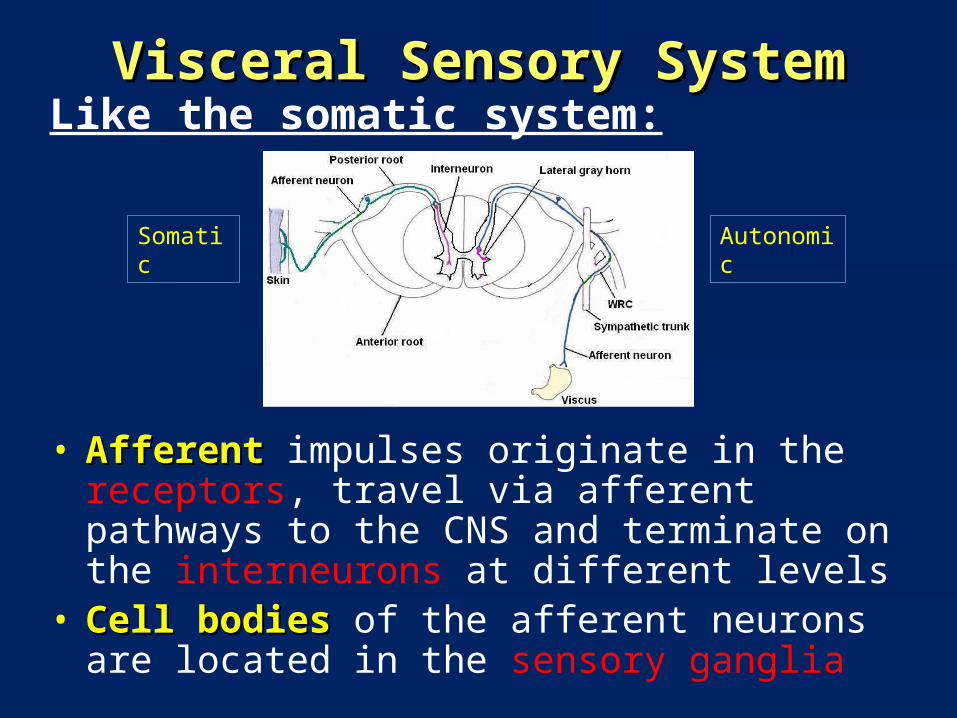
Visceral Sensory SystemVisceral Sensory SystemLike the somatic system:
• AfferentAfferent impulses originate in the receptors, travel via afferent pathways to the CNS and terminate on the interneurons at different levels
• Cell bodies Cell bodies of the afferent neurons are located in the sensory ganglia
Somatic Autonomic

Visceral motor system Visceral motor system is different from somatic motor system in
many respects
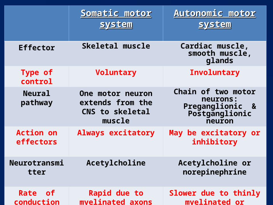
Somatic motor systemSomatic motor system Autonomic motor systemAutonomic motor system
Effector Skeletal muscle Cardiac muscle, smooth muscle, glands
Type of control Voluntary Involuntary
Neural pathway One motor neuron extends from the CNS to skeletal
muscle
Chain of two motor neurons: Preganglionic &
Postganglionic neuron
Action on effectors
Always excitatory May be excitatory or inhibitory
Neurotransmitter Acetylcholine Acetylcholine or norepinephrine
Rate of conduction
Rapid due to myelinated axons
Slower due to thinly myelinated or unmyelinated
axons
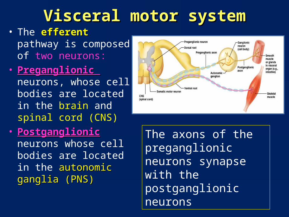
Visceral motor systemVisceral motor system• The efferent efferent pathway is
composed of two neurons:
• Preganglionic Preganglionic neurons, whose cell bodies are located in the brain and spinal cord (CNS)
• PostganglionicPostganglionic neurons whose cell bodies are located in the autonomic ganglia (PNS)autonomic ganglia (PNS)
The axons of the preganglionic neurons synapse with the postganglionic neurons
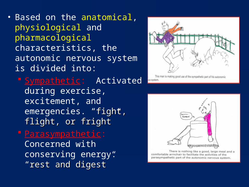
• Based on the anatomical, physiological and pharmacological characteristics, the autonomic nervous system is divided into: Sympathetic: Activated
during exercise, excitement, and emergencies. “fight, fight, flight, or frightflight, or fright”
Parasympathetic: Concerned with conserving energy. “rest rest and digestand digest”
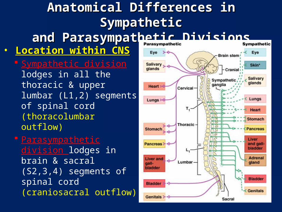
Anatomical Differences in SympatheticAnatomical Differences in Sympatheticand Parasympathetic Divisionsand Parasympathetic Divisions
• Location within CNSLocation within CNS Sympathetic division
lodges in all the thoracic & upper lumbar (L1,2) segments of spinal cord (thoracolumbar outflow)
Parasympathetic division lodges in brain & sacral (S2,3,4) segments of spinal cord (craniosacral outflow)
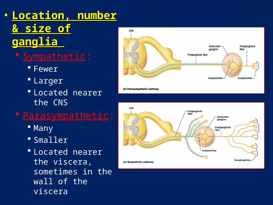
• Location, number & Location, number & size of ganglia size of ganglia Sympathetic:
Fewer Larger Located nearer the
CNS Parasympathetic:
Many Smaller Located nearer the
viscera, sometimes in the wall of the viscera
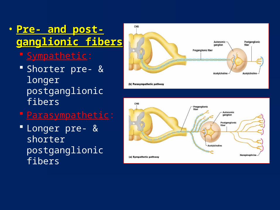
• Pre- and post-Pre- and post-ganglionic fibersganglionic fibers Sympathetic: Shorter pre- &
longer postganglionic fibers
Parasympathetic: Longer pre- &
shorter postganglionic fibers
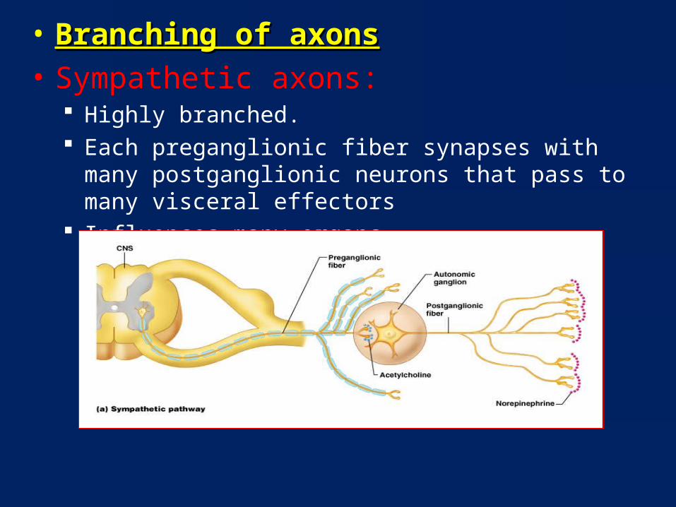
• Branching of axonsBranching of axons• Sympathetic axons:
Highly branched. Each preganglionic fiber synapses with many postganglionic
neurons that pass to many visceral effectors Influences many organs
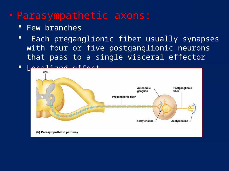
• Parasympathetic axons: Few branches Each preganglionic fiber usually synapses with four or five
postganglionic neurons that pass to a single visceral effector Localized effect
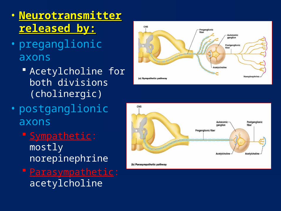
• Neurotransmitter Neurotransmitter released by:released by:
• preganglionic axons Acetylcholine for both
divisions (cholinergic)
• postganglionic axons Sympathetic: mostly
norepinephrine Parasympathetic:
acetylcholine
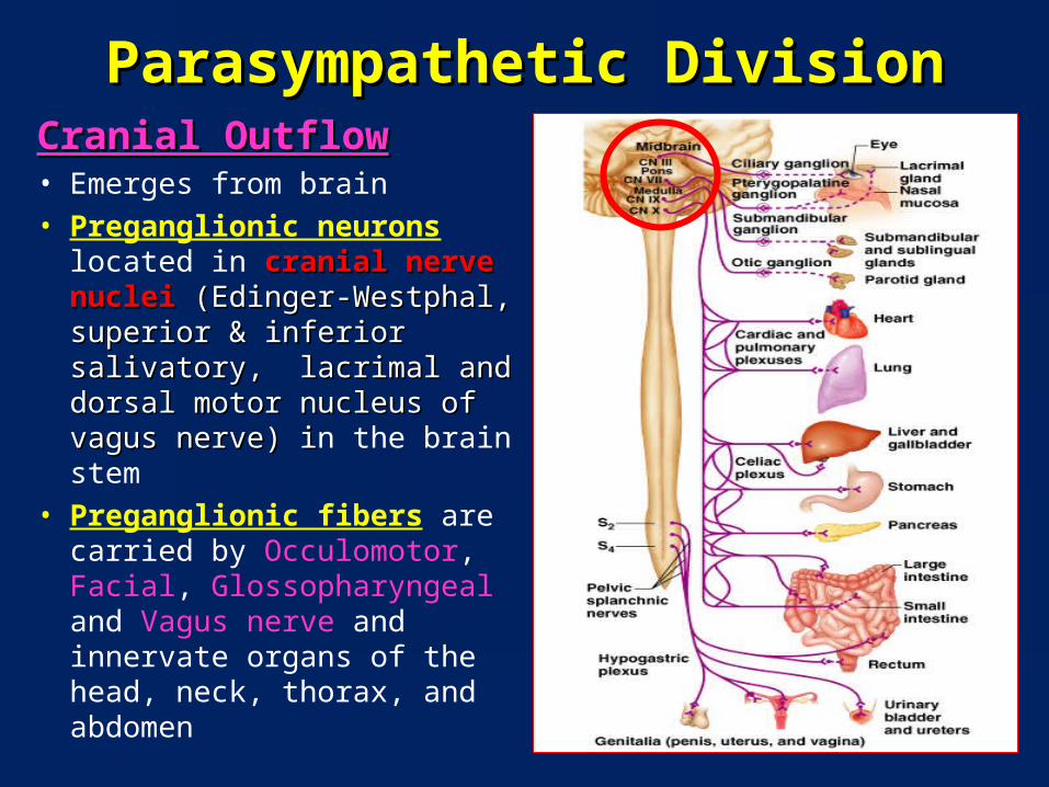
Parasympathetic DivisionParasympathetic DivisionCranial OutflowCranial Outflow• Emerges from brain• Preganglionic neurons located
in cranial nerve nuclei cranial nerve nuclei (Edinger-Westphal, superior & (Edinger-Westphal, superior & inferior salivatory, lacrimal inferior salivatory, lacrimal and dorsal motor nucleus of and dorsal motor nucleus of vagus nerve) ivagus nerve) in the brain stem
• Preganglionic fibers are carried by Occulomotor, Facial, Glossopharyngeal and Vagus nerve and innervate organs of the head, neck, thorax, and abdomen
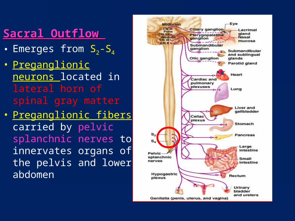
Sacral Outflow Sacral Outflow • Emerges from S2-S4
• Preganglionic neurons located in lateral horn of spinal gray matter
• Preganglionic fibers carried by pelvic splanchnic nerves to innervates organs of the pelvis and lower abdomen
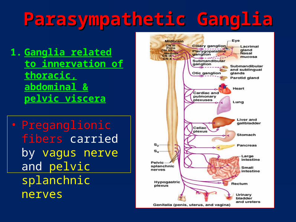
Parasympathetic GangliaParasympathetic Ganglia
1. Ganglia related to innervation of thoracic, abdominal & pelvic viscera
• Preganglionic fibers carried by vagus nerve and pelvic splanchnic nerves
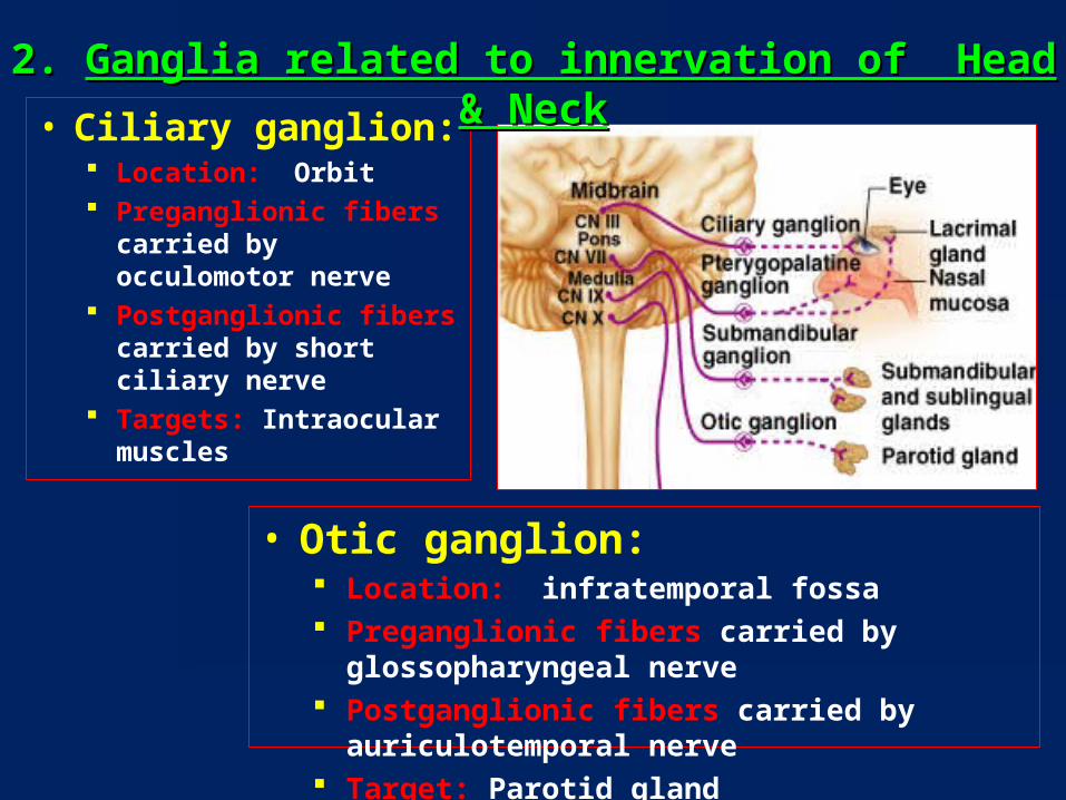
• Ciliary ganglion: Location: Orbit Preganglionic fibers carried
by occulomotor nerve Postganglionic fibers
carried by short ciliary nerve
Targets: Intraocular muscles
2. 2. Ganglia related to innervation of Head & NeckGanglia related to innervation of Head & Neck
• Otic ganglion: Location: infratemporal fossa Preganglionic fibers carried by glossopharyngeal nerve Postganglionic fibers carried by auriculotemporal nerve Target: Parotid gland

• Pterygopalatine ganglion: Location:
Pterygopalatine fossa
Preganglionic fibers carried by facial nerve
Postganglionic fibers carried by maxillary nerve
Targets: Lacrimal gland, nasal & palatine mucosal gland
• Submandibular ganglion: Location: Submandibular region Preganglionic fibers carried by facial nerve Postganglionic fibers carried by lingual
nerve Targets: Submandibular & sublingual
glands
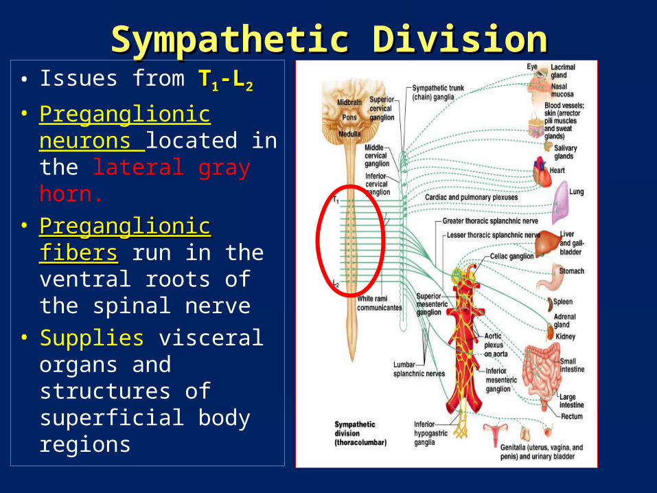
Sympathetic DivisionSympathetic Division• Issues from TT11-L-L22
• Preganglionic neurons located in the lateral gray horn.
• Preganglionic fibersPreganglionic fibers run in the ventral roots of the spinal nerve
• Supplies visceral organs and structures of superficial body regions
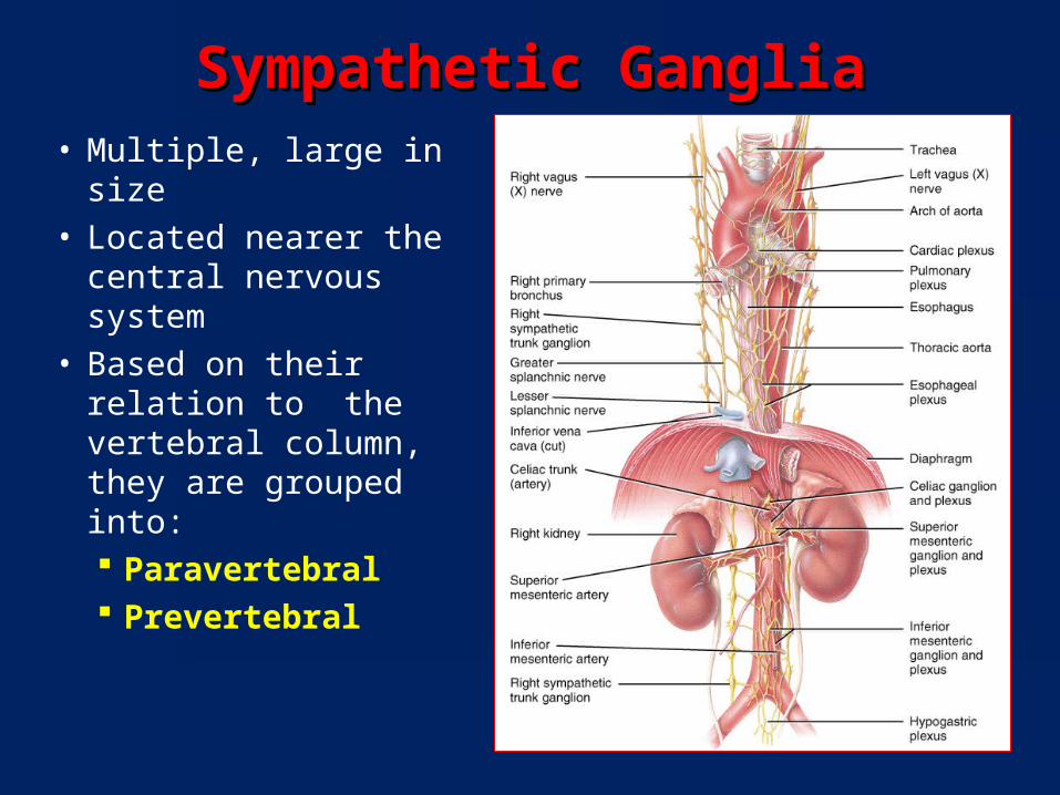
Sympathetic GangliaSympathetic Ganglia• Multiple, large in size• Located nearer the
central nervous system• Based on their relation
to the vertebral column, they are grouped into: Paravertebral Prevertebral
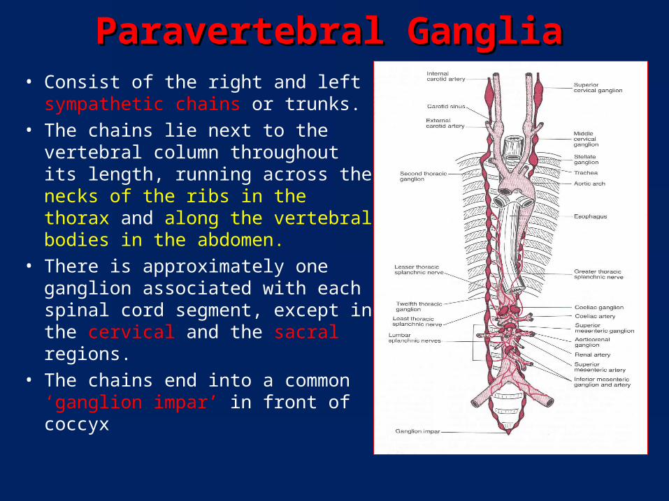
Paravertebral GangliaParavertebral Ganglia• Consist of the right and left
sympathetic chains or trunks. • The chains lie next to the vertebral
column throughout its length, running across the necks of the ribs in the thorax and along the vertebral bodies in the abdomen.
• There is approximately one ganglion associated with each spinal cord segment, except in the cervical and the sacral regions.
• The chains end into a common ‘ganglion impar’ in front of coccyx
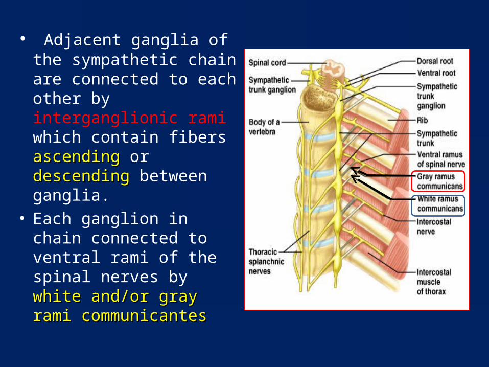
• Adjacent ganglia of the sympathetic chain are connected to each other by interganglionic rami which contain fibers ascendingascending or descending descending between ganglia.
• Each ganglion in chain connected to ventral rami of the spinal nerves by white and/or gray rami white and/or gray rami communicantes communicantes
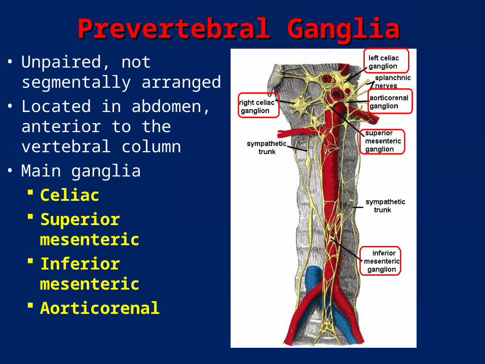
Prevertebral GangliaPrevertebral Ganglia• Unpaired, not segmentally
arranged• Located in abdomen,
anterior to the vertebral column
• Main ganglia Celiac Superior mesenteric Inferior mesenteric Aorticorenal
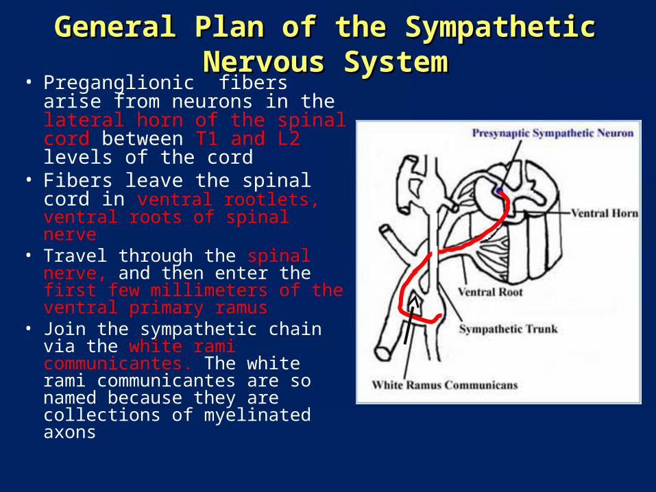
General Plan of the Sympathetic Nervous SystemGeneral Plan of the Sympathetic Nervous System• Preganglionic fibers arise from
neurons in the lateral horn of the spinal cord between T1 and L2 levels of the cord
• Fibers leave the spinal cord in ventral rootlets, ventral roots of spinal nerve
• Travel through the spinal nerve, and then enter the first few millimeters of the ventral primary ramus
• Join the sympathetic chain via the white rami communicantes. The white rami communicantes are so named because they are collections of myelinated axons
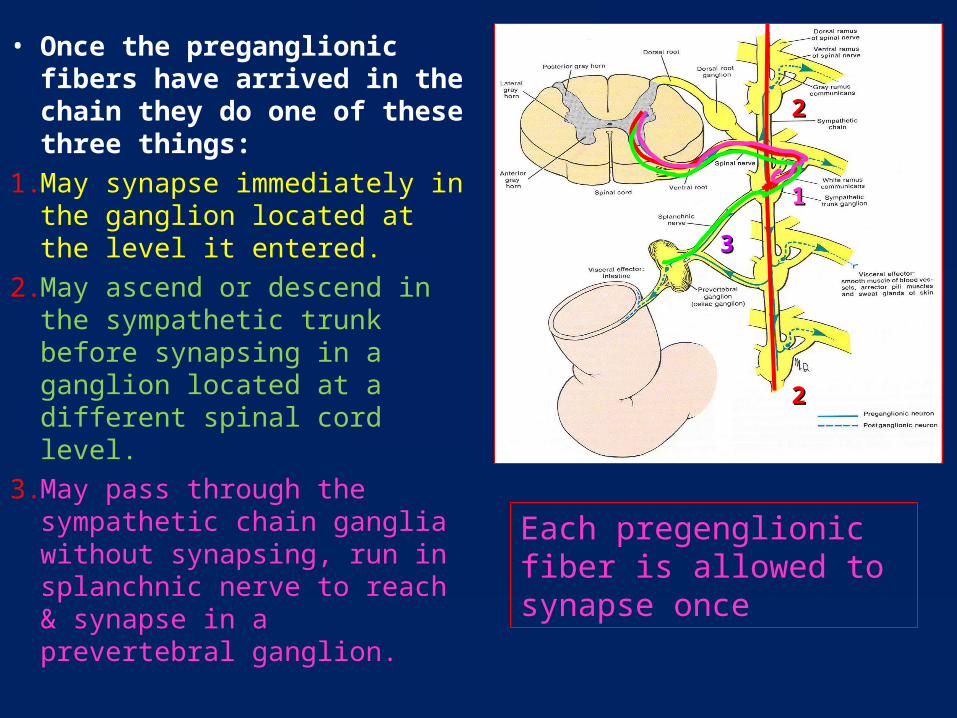
• Once the preganglionic fibers have arrived in the chain they do one of these three things:
1. May synapse immediately in the ganglion located at the level it entered.
2. May ascend or descend in the sympathetic trunk before synapsing in a ganglion located at a different spinal cord level.
3. May pass through the sympathetic chain ganglia without synapsing, run in splanchnic nerve to reach & synapse in a prevertebral ganglion.
11
22
22
33
Each pregenglionic fiber is allowed to synapse once
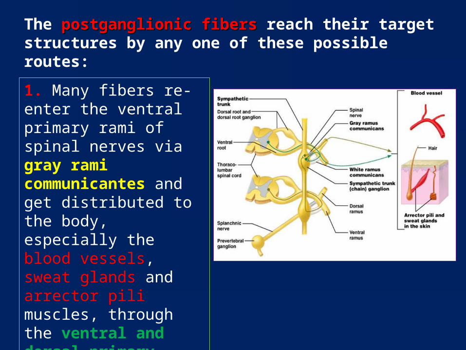
The postganglionic fibers postganglionic fibers reach their target structures by any one of these possible routes:
1. Many fibers re-enter the ventral primary rami of spinal nerves via gray rami communicantes and get distributed to the body, especially the blood vessels, sweat glands and arrector pili muscles, through the ventral and dorsal primary rami.

2. Other fibers leave the ganglia and travel directly to their target organs. This is how postsynaptic sympathetic fibers reach the organs of the thorax.
3. Some fibers form peri-vascular plexuses along blood vessels to reach their targets e.g. fibers reaching organs in the head, and in the abdomen and pelvis.
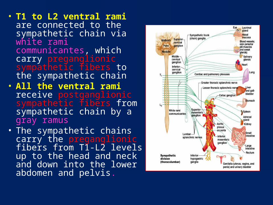
• T1 to L2 ventral rami are connected to the sympathetic chain via white rami communicantes, which carry preganglionic sympathetic fibers to the sympathetic chain
• All the All the ventral rami receive postganglionic sympathetic fibers from sympathetic chain by a gray ramus
• The sympathetic chains carry the preganglionic fibers from T1-L2 levels up to the head and neck and down into the lower abdomen and pelvis.
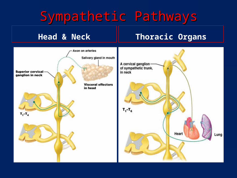
Sympathetic PathwaysSympathetic PathwaysHead & Neck Thoracic Organs

Sympathetic PathwaysSympathetic Pathways
Abdominal Organs Pelvic Organs
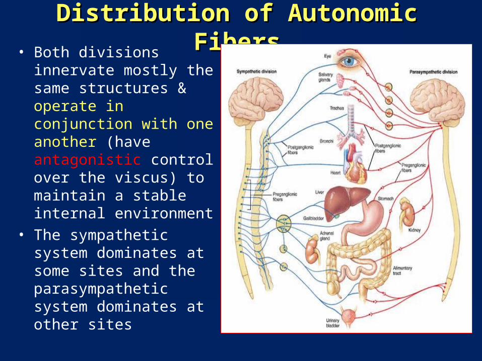
Distribution of Autonomic FibersDistribution of Autonomic Fibers• Both divisions innervate
mostly the same structures & operate in conjunction with one another (have antagonistic control over the viscus) to maintain a stable internal environment
• The sympathetic system dominates at some sites and the parasympathetic system dominates at other sites
• Some viscera do not possess dual control e.g. arrector piliarrector pili muscle is made to contract by the sympathetic activity, has no parasympathetic supply
• Sweat glands:Sweat glands: Postganglionic neurons involved with stress-related
excretion release norepinephrine (“sweaty palms”) Postganglionic neurons involved with thermoregulation
release acetylcholine
• Kidneys:Kidneys:• Postganglionic neurons to the smooth muscle of the renal
vascular bed release dopamine
Exceptions in the sympathetic nervous system
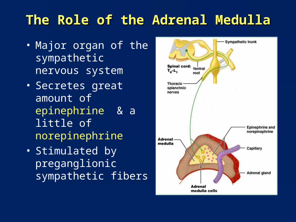
The Role of the Adrenal MedullaThe Role of the Adrenal Medulla
• Major organ of the sympathetic nervous system
• Secretes great amount of epinephrine & a little of norepinephrine
• Stimulated by preganglionic sympathetic fibers

Enteric Nervous SystemEnteric Nervous System• Intrinsic nervous system that directly
controls the gastrointestinal system• Composed of two plexuses of nerve
cells and fibers located in the wall of gastrointestinal tract from the esophagus to the anal canal
• Submucous or Meisner’s plexus lies in the submucosa, is mainly concerned with the control of the glands in the mucous membrane
• Myenteric or Aurbach’s plexus lies between the circular and the longitudinal muscle layer, controls the muscle and movements of the gut wall
Communicates with the CNS through the parasympathetic (eg, via the vagus nerve) and sympathetic (eg, via the prevertebral ganglia) nervous systems
Visceral ReflexesVisceral Reflexes• Parasympathetic reflexes Parasympathetic reflexes include: gastric and
intestinal reflexes, defecation, micturition, direct light reflexes, swallowing reflex, coughing reflex, baroreceptor reflex and sexual arousal.
• Sympathetic reflexes Sympathetic reflexes include: cardio-accelaratory reflex, vasomotor reflex, pupillary reflex and ejaculation (in males).

• All visceral reflexes are polysynaptic.• The simplest visceral reflex arc
consists of:1. a receptor2. a sensory neuron3. an interneuron, &4. Two motor neurons (pre- &
postganglionic)
• Long reflexes: processed in CNS, similar to polysynaptic somatic reflex
• Short reflexes : bypass CNS entirely, processed in ganglia, e.g. enteric NS in walls of digestive tract
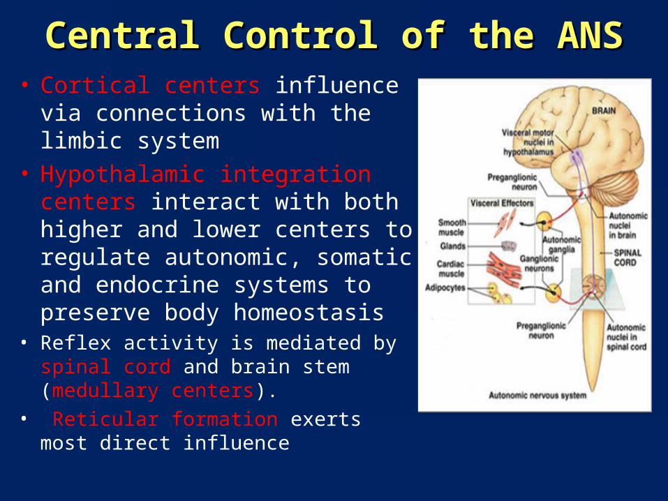
Central Control of the ANSCentral Control of the ANS• Cortical centers influence via
connections with the limbic system• Hypothalamic integration centers
interact with both higher and lower centers to regulate autonomic, somatic and endocrine systems to preserve body homeostasis
• Reflex activity is mediated by spinal cord and brain stem (medullary centers).
• Reticular formation exerts most direct influence
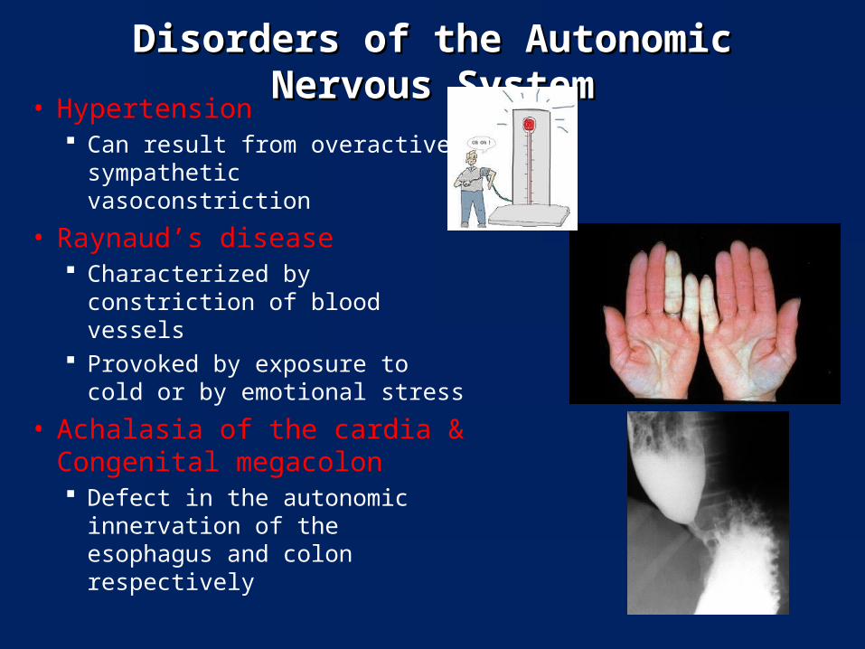
Disorders of the Autonomic Nervous SystemDisorders of the Autonomic Nervous System• Hypertension
Can result from overactive sympathetic vasoconstriction
• Raynaud’s disease Characterized by constriction
of blood vessels Provoked by exposure to cold
or by emotional stress
• Achalasia of the cardia & Congenital megacolon Defect in the autonomic
innervation of the esophagus and colon respectively
• Primary autonomic failure: A chronic degenerative disease of the nervous system leading to fainting attacks, incontinence of urine and bowel, and impotence
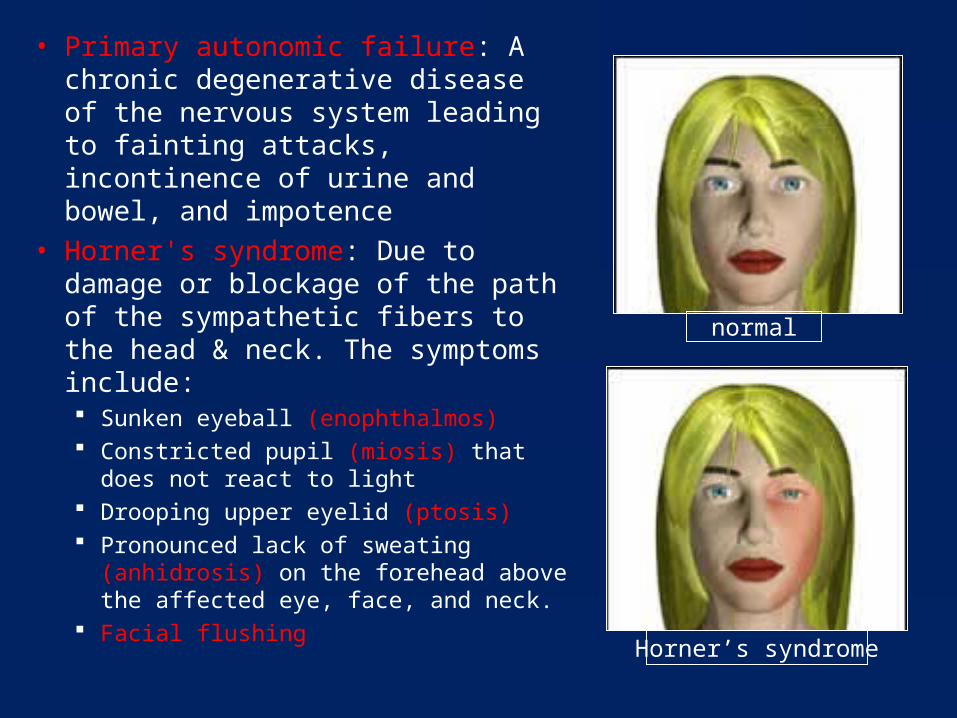
• Horner's syndrome: Due to damage or blockage of the path of the sympathetic fibers to the head & neck. The symptoms include: Sunken eyeball (enophthalmos) Constricted pupil (miosis) that does not
react to light Drooping upper eyelid (ptosis) Pronounced lack of sweating
(anhidrosis) on the forehead above the affected eye, face, and neck.
Facial flushing
normal
Horner’s syndrome
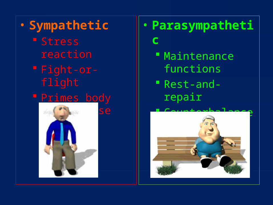
• Sympathetic Stress reaction Fight-or-flight Primes body for
intense skeletal muscle activity
• Parasympathetic Maintenance
functions Rest-and-repair Counterbalances
sympathetic function

Related Documents