Short Communication The Autoimmune Regulator (AIRE), Which Is Defective in Autoimmune Polyendocrinopathy-Candidiasis-Ectodermal Dystrophy Patients, Is Expressed in Human Epidermal and Follicular Keratinocytes and Associates With the Intermediate Filament Protein Cytokeratin 17 Vipul Kumar,* Luis A. Pedroza,* †‡ Emily M. Mace,* Steven Seeholzer,* George Cotsarelis, § Antonio Condino-Neto, † Aimee S. Payne, § and Jordan S. Orange* From the Division of Immunology,* Department of Pediatrics, Children’s Hospital of Philadelphia, and the Department of Dermatology, § University of Pennsylvania School of Medicine, Philadelphia, Pennsylvania; the Department of Immunology, † Institute of Biomedical Sciences, University of Sao Paulo, Sao Paulo, Brazil; and Center for Investigation in Pediatrics, ‡ School of Medicine, Campinas State University, Sao Paulo, Brazil Autoimmune polyendocrinopathy-candidiasis-ecto- dermal dystrophy (APECED) syndrome, which is caused by mutation of the autoimmune regulator (AIRE) gene, is a highly variable disease characterized by multiple endocrine failure, chronic mucocutane- ous candidiasis, and various ectodermal defects. AIRE is a transcriptional regulator classically expressed in medullary thymic epithelial cells, monocytes, macro- phages, and dendritic cells. Previous studies have sug- gested that AIRE can shuttle between the nucleus and cytoplasm of cells, although its cytoplasmic functions are poorly characterized. Through mass spectrometry analysis of proteins co-immunoprecipitating with cy- toplasmic AIRE, we identified a novel association of AIRE with the intermediate filament protein cytoker- atin 17 (K17) in the THP-1 monocyte cell line. We confirmed AIRE expression in HaCaT epidermal kera- tinocytes, as well as its interaction with K17. Confocal microscopy of human fetal and adult scalp hair folli- cles demonstrated a cytoplasmic pattern of AIRE staining that moderately colocalized with K17. The cytoplasmic association of AIRE with the intermediate filament network in human epidermal and follicular keratinocytes may provide a new path to understand- ing the ectodermal abnormalities associated with the APECED syndrome. (Am J Pathol 2011, 178:983–988; DOI: 10.1016/j.ajpath.2010.12.007) The autoimmune polyendocrinopathy-candidiasis-ecto- dermal dystrophy (APECED) syndrome is characterized by development of autoimmunity toward select endocrine tissue (mostly in the form of adrenocortical failure and/or hypoparathyroidism), an immune deficiency resulting in chronic candidiasis, as well as various ectodermal disor- ders that include tooth enamel hypoplasia, vitiligo, alope- cia, and nail dystrophy. 1 Since the formal classification of the disease, 2 a single gene has been identified as causative and encodes the Autoimmune Regulator (AIRE) protein. 3 The AIRE protein contains several key domains, including two plant homeodomain (PHD) zinc fingers, four LXXLL motifs, a Sp100, AIRE-1, NucP41/75, DEAF-1 SAND do- main, and a recently identified Caspase Recruitment Do- main (CARD) at the N-terminus, 4 the last of which has been Supported by the Children’s Hospital of Philadelphia and Jeffrey Modell Diagnostic Center (J.S.O.), Coordenação de Aperfeiçoamento de Pessoal de Nível Superior (Brazil) (L.A.P.), Fundação de Amparo à Pesquisa do Estado de São Paulo grant 09/51747-3 (L.A.P. and A.C.N.), Conselho Na- cional de Desenvolvimento Científico e Tecnológico grant 501332/2010-3 (A.C.N.), and the Penn Skin Disease Research Center (NIAMS P30 AR057217). Accepted for publication December 1, 2010. V.K., L.A.P., A.S.P., and J.S.O. contributed equally to this work. Address reprint requests to Jordan S. Orange, M.D., Ph.D., Associate Professor of Pediatrics, Children’s Hospital of Philadelphia, Division of Immunology, Department of Pediatrics, University of Pennsylvania School of Medicine, 3615 Civic Center Blvd., ARC-1016H, Philadelphia, PA 19104; or Aimee S. Payne, M.D., Ph.D, Assistant Professor of Dermatology, Depart- ment of Dermatology, University of Pennsylvania School of Medicine, 217A Clinical Research Building, 415 Curie Blvd., Philadelphia, PA, 19104. E-mail: [email protected] or [email protected]. The American Journal of Pathology, Vol. 178, No. 3, March 2011 Copyright © 2011 American Society for Investigative Pathology. Published by Elsevier Inc. All rights reserved. DOI: 10.1016/j.ajpath.2010.12.007 983

Welcome message from author
This document is posted to help you gain knowledge. Please leave a comment to let me know what you think about it! Share it to your friends and learn new things together.
Transcript
The American Journal of Pathology, Vol. 178, No. 3, March 2011
Copyright © 2011 American Society for Investigative Pathology.
Published by Elsevier Inc. All rights reserved.
DOI: 10.1016/j.ajpath.2010.12.007
Short Communication
The Autoimmune Regulator (AIRE), Which Is Defective inAutoimmune Polyendocrinopathy-Candidiasis-EctodermalDystrophy Patients, Is Expressed in Human Epidermaland Follicular Keratinocytes and Associates With the
Intermediate Filament Protein Cytokeratin 17Vipul Kumar,* Luis A. Pedroza,*†‡
Emily M. Mace,* Steven Seeholzer,*George Cotsarelis,§ Antonio Condino-Neto,†
Aimee S. Payne,§ and Jordan S. Orange*From the Division of Immunology,* Department of Pediatrics,
Children’s Hospital of Philadelphia, and the Department of
Dermatology,§ University of Pennsylvania School of Medicine,
Philadelphia, Pennsylvania; the Department of Immunology,†
Institute of Biomedical Sciences, University of Sao Paulo,
Sao Paulo, Brazil; and Center for Investigation in Pediatrics,‡
School of Medicine, Campinas State University, Sao Paulo, Brazil
Autoimmune polyendocrinopathy-candidiasis-ecto-dermal dystrophy (APECED) syndrome, which iscaused by mutation of the autoimmune regulator(AIRE) gene, is a highly variable disease characterizedby multiple endocrine failure, chronic mucocutane-ous candidiasis, and various ectodermal defects. AIREis a transcriptional regulator classically expressed inmedullary thymic epithelial cells, monocytes, macro-phages, and dendritic cells. Previous studies have sug-gested that AIRE can shuttle between the nucleus andcytoplasm of cells, although its cytoplasmic functionsare poorly characterized. Through mass spectrometryanalysis of proteins co-immunoprecipitating with cy-toplasmic AIRE, we identified a novel association ofAIRE with the intermediate filament protein cytoker-atin 17 (K17) in the THP-1 monocyte cell line. Weconfirmed AIRE expression in HaCaT epidermal kera-tinocytes, as well as its interaction with K17. Confocalmicroscopy of human fetal and adult scalp hair folli-cles demonstrated a cytoplasmic pattern of AIREstaining that moderately colocalized with K17. Thecytoplasmic association of AIRE with the intermediatefilament network in human epidermal and follicular
keratinocytes may provide a new path to understand-ing the ectodermal abnormalities associated with theAPECED syndrome. (Am J Pathol 2011, 178:983–988; DOI:
10.1016/j.ajpath.2010.12.007)
The autoimmune polyendocrinopathy-candidiasis-ecto-dermal dystrophy (APECED) syndrome is characterizedby development of autoimmunity toward select endocrinetissue (mostly in the form of adrenocortical failure and/orhypoparathyroidism), an immune deficiency resulting inchronic candidiasis, as well as various ectodermal disor-ders that include tooth enamel hypoplasia, vitiligo, alope-cia, and nail dystrophy.1 Since the formal classification ofthe disease,2 a single gene has been identified as causativeand encodes the Autoimmune Regulator (AIRE) protein.3
The AIRE protein contains several key domains, includingtwo plant homeodomain (PHD) zinc fingers, four LXXLLmotifs, a Sp100, AIRE-1, NucP41/75, DEAF-1 SAND do-main, and a recently identified Caspase Recruitment Do-main (CARD) at the N-terminus,4 the last of which has been
Supported by the Children’s Hospital of Philadelphia and Jeffrey ModellDiagnostic Center (J.S.O.), Coordenação de Aperfeiçoamento de Pessoalde Nível Superior (Brazil) (L.A.P.), Fundação de Amparo à Pesquisa doEstado de São Paulo grant 09/51747-3 (L.A.P. and A.C.N.), Conselho Na-cional de Desenvolvimento Científico e Tecnológico grant 501332/2010-3(A.C.N.), and the Penn Skin Disease Research Center (NIAMS P30AR057217).
Accepted for publication December 1, 2010.
V.K., L.A.P., A.S.P., and J.S.O. contributed equally to this work.
Address reprint requests to Jordan S. Orange, M.D., Ph.D., AssociateProfessor of Pediatrics, Children’s Hospital of Philadelphia, Division ofImmunology, Department of Pediatrics, University of Pennsylvania Schoolof Medicine, 3615 Civic Center Blvd., ARC-1016H, Philadelphia, PA 19104;or Aimee S. Payne, M.D., Ph.D, Assistant Professor of Dermatology, Depart-ment of Dermatology, University of Pennsylvania School of Medicine, 217AClinical Research Building, 415 Curie Blvd., Philadelphia, PA, 19104. E-mail:
[email protected] or [email protected].983
984 Kumar et alAJP March 2011, Vol. 178, No. 3
shown to be a mutational hotspot in AIRE that affects itstransactivating potential.5
Prior structural and subcellular analyses of AIRE havedemonstrated its strong nuclear localization with a highDNA binding capacity. These characteristics are thoughtto allow AIRE to transcriptionally modulate ectopic pe-ripheral tissue antigen expression in medullary thymicepithelial cells. In this capacity, AIRE facilitates T cellnegative selection through ensuring the full and appro-priate spectrum of self-antigen expression in the thy-mus.5 Other roles for AIRE, however, have been raised asa feature of its biology. Exogenously expressed AIRE-GFP or wild-type AIRE in COS-1 and Hela cells demon-strates that AIRE has the capacity to traffic between thenucleus and cytoplasm.6 Although the functional signifi-cance of extranuclear AIRE remains unknown, it can lo-calize to intermediate filaments and/or microtubules6–8 inthe cytoplasm.
Although a plausible mechanism is in place for the roleof AIRE in promoting autoimmunity, there has been littleresearch regarding the pathogenesis of ectodermal dys-trophy (ED) and chronic candidiasis in APECED patients.Aberrant adaptive immunity in APECED has been pro-posed to result in the immunodeficiency with Candidasusceptibility,9 and the ectodermal manifestations10,11
have been considered secondary. However, other stud-ies have demonstrated AIRE expression in monocytes,macrophages, and dendritic cells,12 suggesting thatAIRE defects in other cell lineages may contribute topathogenesis. The current study began in an effort tocharacterize the role of AIRE in monocytes, but led to theunexpected finding of an association between AIRE andthe intermediate filament protein cytokeratin 17 (K17).This association was further investigated in human skin,where we identified the expression of AIRE in epidermalkeratinocytes and an association of AIRE with K17 in bothHaCaT cells and hair follicles of the human scalp. Ourfindings indicate that AIRE has a broader expressionprofile than was previously appreciated and raises thepossibility for novel mechanisms underlying AIRE-asso-ciated ectodermal disorders.
Materials and Methods
Cell Lines, Tissue, and Antibodies
The THP-1 cell line stably expressing a secreted embryonicalkaline phosphatase (SEAP) reporter construct under thecontrol of an NF-�B promoter (THP-1 Blue; InvivoGen, SanDiego, CA) was grown in RPMI supplemented with 2mmol/L L-glutamine, 1.5 g/L sodium bicarbonate, 4.5 g/Lglucose, 10 mmol/L HEPES, 1.0 mmol/L sodium pyruvate,penicillin, and streptomycin. Medium was supplementedwith 200 �g/ml Zeocin to maintain selection of the reporterconstruct. HaCaT cells were grown in DMEM supple-mented with 10% Fetal Bovine Serum, 2 mmol/L L-glu-tamine, 1.0 mmol/L sodium pyruvate, penicillin, andstreptomycin. For experiments with human tissue, thisstudy used only normally discarded human scalp ob-
tained anonymously from plastic surgical proceduresthrough the Cooperative Human Tissue Network and wasapproved by the University of Pennsylvania’s InstitutionalReview Board office as an exempt protocol. Fetal humantissue was obtained from Advanced Bioscience Re-sources (Alameda, CA). Antibodies used had specificitiesagainst the following human proteins: AIRE-1 (rabbit poly-clonal H-300, used in confocal microscopy and THP-1 cellimmunoprecipitation; goat polyclonal D-17, used in immu-nblot), cytokeratin 17 (K17) (mouse monoclonal E3, used inconfocal microscopy, and rabbit polyclonal EPR1624Y,used in co-immunoprecipitation). All aforementioned anti-bodies were obtained from Santa Cruz Biotechnology(Santa Cruz, CA) with the exception of the rabbit polyclonalantibody against human K17, which was obtained fromAbcam (Cambridge, MA); HRP-conjugated anti-mouse Igand HRP-conjugated anti-rabbit Ig (GE Health care, Pisca-taway, NJ), and HRP-conjugated anti-goat Ig (Santa Cruz)were used as secondaries in Western blotting.
Analysis of AIRE Expression, Putative AIREInteracting Proteins, and SubsequentCo-Immunoprecipitation
To study constitutive interacting proteins with AIRE, THP-1cells (1.5E7) were lysed in 1 mL lysis buffer (200 mmol/LNaCl, 1% NP-40, 10 mmol/L Tris-HCl [pH 7.5], 5 mmol/LEDTA, and 10% glycerol) supplemented with protease andphosphatase inhibitors. Cytoplasmic fractions obtained af-ter centrifugation of whole cell lysates were incubated with20 �g/ml of AIRE-specific IgG-coated or IgG isotype con-trol–coated protein G agarose (Invitrogen, Carlsbad, CA)for 2 hours. After incubation with lysate, beads were washedthrice and bound proteins eluted with NuPage sample buf-fer and run on a NuPAGE SDS gel (Invitrogen). The gel wassubsequently stained with Colloidal Coomassie (Invitrogen)according to the manufacturer’s instructions to identifyunique proteins in the anti-AIRE precipitate compared toisotype precipitate. The gel was evaluated in the ProteinCore Facility of the Children’s Hospital of Philadelphia andthe protein band at �50 kDa studied using vacuum matrix-assisted laser desorption (vMALDI) mass spectrometry aspreviously described.13 Reciprocal co-immunoprecipitationwas performed using a similar procedure with HaCaT cells(5E6 cells/condition) as described previously20 and ana-lyzed by Western blot using the appropriate antibodies de-scribed in Cell Lines, Tissue, and Antibodies. RNA was iso-lated from HaCaT, THP-1, and COS-1 cells using theRNAEasy Kit (Qiagen, Valencia, CA) and cDNA synthe-sized using Applied Biosystems’ High Capacity cDNA syn-thesis kit (Applied Biosystems, Carlsbad, CA). Applied Bio-systems Taqman Gene expression assay (assay ID:Hs00230833_m1) was used to assess the presence ofAIRE, and expression was compared to cDNA from THP-1cells (AIRE-positive control) and COS-1 cells (AIRE-nega-tive control),7 with concurrent amplification of actin used asan amplification control. DNA was resolved on a 1% aga-
rose gel.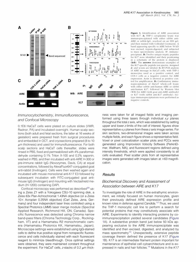
AIRE-K17 Association in Keratinocytes 985AJP March 2011, Vol. 178, No. 3
Immunocytochemistry, Immunofluorescence,and Confocal Microscopy
0.1E6 HaCaT cells were plated on culture slides (VWR,Radnor, PA) and incubated overnight. Human scalp sec-tions (both adult and fetal sections, the latter at 16 weeks ofgestation) were prepared fresh from surgical proceduresand embedded in OCT, and cryosections prepared (8 to 10�m thickness) and used for immunofluorescence. For bothscalp sections and HaCaT cells thereafter, slides wererinsed in PBS, fixed and permeabilized with 4% paraformal-dehyde containing 0.1% Triton X-100 and 0.2% saponin,washed in PBS, and then incubated with anti-AIRE H-300 orpre-immune rabbit IgG (Novozymes, Davis, CA) at equalconcentrations, followed by AlexaFluor647-conjugated goatanti-rabbit (Invitrogen). Cells were then washed again andincubated with mouse monoclonal anti-K17 E3 followed bysubsequent incubation with FITC-conjugated goat anti-mouse IgG (Invitrogen) and mounting with Vectashield me-dium (H-1200) containing DAPI.
Confocal microscopy was performed as described20 us-ing a Zeiss Z1 with a Yokogawa CSU-10 spinning disk, aZeiss 63x Plan-Achrochromat 1.43NA objective (or a Zeiss10� Acroplan 0.25NA objective) (Carl Zeiss, Jena, Ger-many) and four independent laser lines controlled using aSpectral Photonics LMM5 laser merge module (SpectralApplied Research, Richmond Hill, ON, Canada). Spe-cific fluorescence was detected using Chroma narrowband-pass filters (Chroma Technology Corp., Rocking-ham, VT) and a Hamamatsu R2 monochromatic cam-era (Hamamatsu Photonics KK, Hamamatsu, Japan).Microscope settings were established using IgG-stainedcells to define true positive signal from nonspecific fluores-cence and cells individually stained with each fluorescentreagent to minimize bleedthrough. Once optimal settingswere obtained, they were maintained constant throughout
the experiment. For HaCaT cells, z-stacks of 0.2 �m thick-ness were taken for all imaged fields and imaging per-formed using three lasers through individual x,y planesthroughout the total z axis, which was established by settingupper and lower z-limits of the cell of interest; figures showrepresentative x,y planes from these z axis image series. Forskin sections, two-dimensional images were taken acrossmultiple fields, and each figure shows a representative field.Voxel or pixel colocalization scatter plots and PCCs weregenerated using Improvision Volocity Software (PerkinEl-mer, Waltham, MA), and fluorescent regions defined usingintensity thresholds, which were applied consistently to allcells evaluated. Pixel scatter plots from all representativeimages were generated with images taken at �63 magnifi-cation.
Results
Biochemical Discovery and Assessment ofAssociation between AIRE and K17
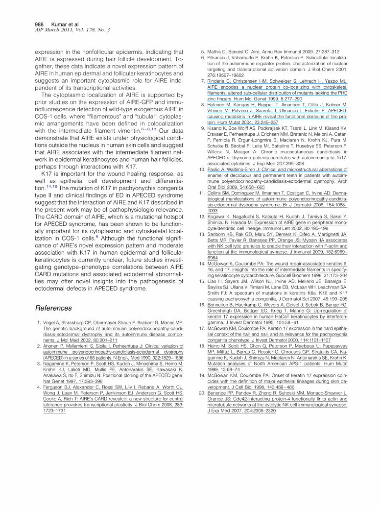
To investigate the role of AIRE in the extrathymic environ-ment, we initially focused on human monocytes, giventheir previously defined AIRE expression profile andknown roles in defense against Candida.12 Thus, we usedthe THP-1 monocytic cell line to perform a search forpotential proteins that may constitutively associate withAIRE. Experiments to identify interacting proteins by co-immunoprecipitation yielded several candidates (Figure1A). A substantive protein band just below 50 kDa ap-pearing exclusive to the AIRE immunoprecipitate wasidentified and then excised, digested, and analyzed bymass spectrometry13. Unexpectedly, extensive peptidecoverage therein defined the presence of K17 (Figure1B). K17 is an intermediate filament protein important formaintenance of epithelial cell cytoarchitecture and is ex-
Figure 1. Identification of AIRE associationwith K17. A: THP-1 cytoplasmic lysate wasimmunoprecipitated with either rabbit anti-AIRE or rabbit-IgG, resolved on an SDS gel,and then stained with Colloidal Coomassie. Aband appearing specific to AIRE below 50 kDwas excised, trypsin-digested, and subjectedto mass spectrometric analysis. IP, immuno-precipitate. B: Twelve unique tryptic peptidesfor K17 were identified and their positioningin a schematic of the protein is displayed(left). The arrows demonstrate examples ofthe K17-specific peptide ion spectra. Assigned�- and �-ions are labeled. C: RT-PCR analysisof AIRE message in HaCaT cells, with THP-1monocytes used as a positive control, andCOS-1 cells as a negative control, for AIREexpression. Actin is showed as positive con-trol for amplification. D: Confirmatory immu-noprecipitation of HaCaT whole-cell lysatewith either rabbit anti-human AIRE or rabbitanti-human K17, followed by Western blot(WB) for AIRE (with goat anti-AIRE antibody)or K17 (with rabbit anti-K17 antibody). Iso-type immunoprecipitations were performed asnegative controls.
pressed in nails and hair follicles.14 Mutations in the K17
nd adul
986 Kumar et alAJP March 2011, Vol. 178, No. 3
gene are associated with a distinct ectodermal disorder,pachyonychia congenita type II.15 Given certain clinicalsimilarities between pachyonychia congenita type IIand the ectodermal abnormalities found in someAPECED syndrome patients, we further investigatedthe potential interaction of AIRE with K17 in humankeratinocytes. Initially, we used the HaCaT keratino-cyte cell line to evaluate this interaction because it hadbeen previously reported to express K17.16 We firstdetermined the expression of AIRE in the HaCaT cellline by using reverse-transcription polymerase chainreaction (RT-PCR) (Figure 1C). To confirm an associa-tion between AIRE and K17 in HaCaT cells, co-immu-noprecipitation followed by immunoblot analysis wasperformed with specific anti-AIRE and anti-K17 anti-bodies. K17 was identified in AIRE immunoprecipitate(Figure 1D, left) and AIRE in K17 immunoprecipitate(Figure 1D, right). Thus, AIRE is expressed in HaCaTcells and biochemically associates with K17.
Subcellular Localization of AIRE and K17 byConfocal Microscopy
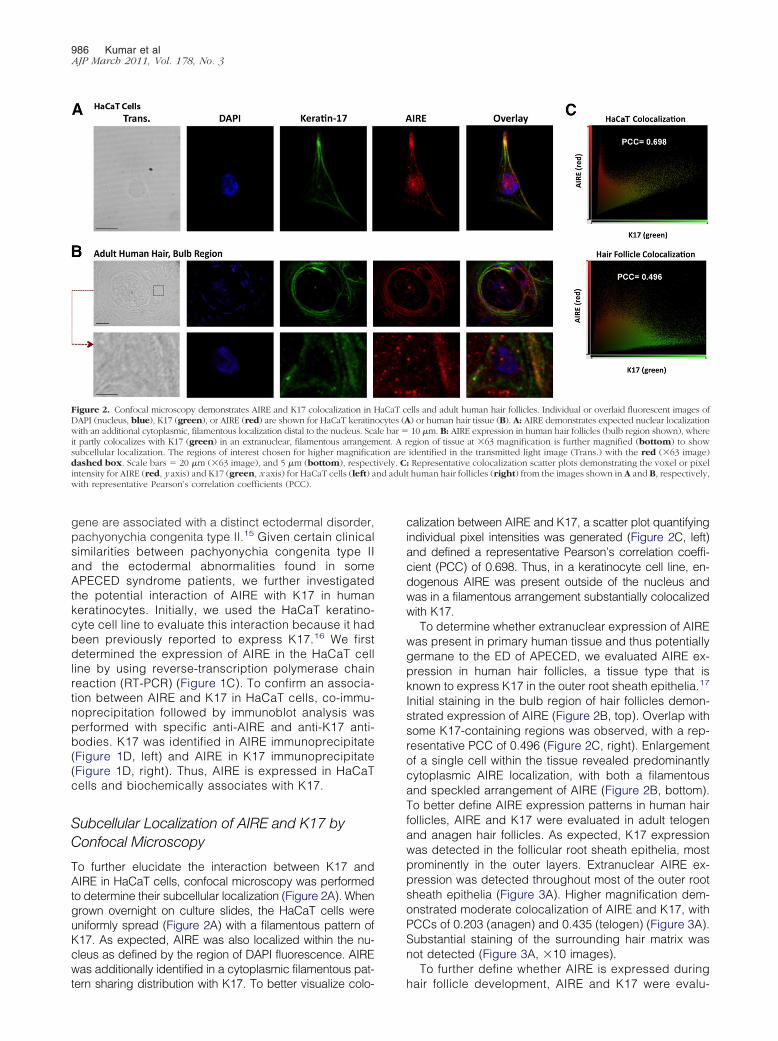
To further elucidate the interaction between K17 andAIRE in HaCaT cells, confocal microscopy was performedto determine their subcellular localization (Figure 2A). Whengrown overnight on culture slides, the HaCaT cells wereuniformly spread (Figure 2A) with a filamentous pattern ofK17. As expected, AIRE was also localized within the nu-cleus as defined by the region of DAPI fluorescence. AIREwas additionally identified in a cytoplasmic filamentous pat-
Figure 2. Confocal microscopy demonstrates AIRE and K17 colocalization inDAPI (nucleus, blue), K17 (green), or AIRE (red) are shown for HaCaT keratinwith an additional cytoplasmic, filamentous localization distal to the nucleus. Scait partly colocalizes with K17 (green) in an extranuclear, filamentous arrangemsubcellular localization. The regions of interest chosen for higher magnificadashed box. Scale bars � 20 �m (�63 image), and 5 �m (bottom), respecintensity for AIRE (red, y axis) and K17 (green, x axis) for HaCaT cells (left) awith representative Pearson’s correlation coefficients (PCC).
tern sharing distribution with K17. To better visualize colo-
calization between AIRE and K17, a scatter plot quantifyingindividual pixel intensities was generated (Figure 2C, left)and defined a representative Pearson’s correlation coeffi-cient (PCC) of 0.698. Thus, in a keratinocyte cell line, en-dogenous AIRE was present outside of the nucleus andwas in a filamentous arrangement substantially colocalizedwith K17.
To determine whether extranuclear expression of AIREwas present in primary human tissue and thus potentiallygermane to the ED of APECED, we evaluated AIRE ex-pression in human hair follicles, a tissue type that isknown to express K17 in the outer root sheath epithelia.17
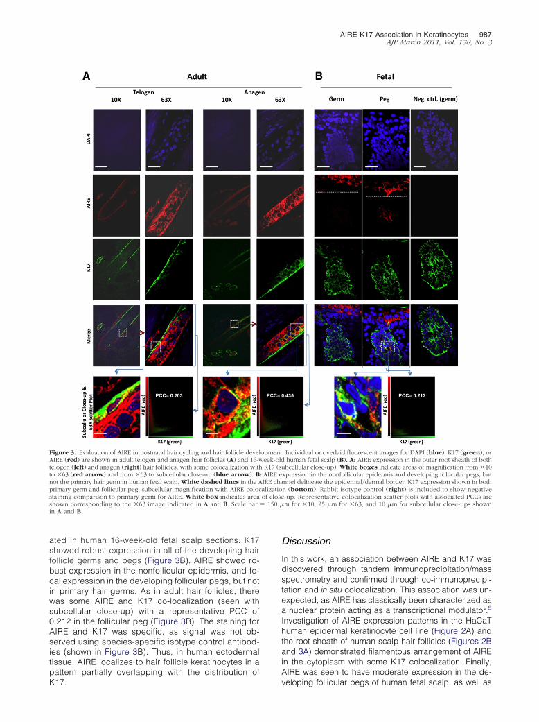
Initial staining in the bulb region of hair follicles demon-strated expression of AIRE (Figure 2B, top). Overlap withsome K17-containing regions was observed, with a rep-resentative PCC of 0.496 (Figure 2C, right). Enlargementof a single cell within the tissue revealed predominantlycytoplasmic AIRE localization, with both a filamentousand speckled arrangement of AIRE (Figure 2B, bottom).To better define AIRE expression patterns in human hairfollicles, AIRE and K17 were evaluated in adult telogenand anagen hair follicles. As expected, K17 expressionwas detected in the follicular root sheath epithelia, mostprominently in the outer layers. Extranuclear AIRE ex-pression was detected throughout most of the outer rootsheath epithelia (Figure 3A). Higher magnification dem-onstrated moderate colocalization of AIRE and K17, withPCCs of 0.203 (anagen) and 0.435 (telogen) (Figure 3A).Substantial staining of the surrounding hair matrix wasnot detected (Figure 3A, �10 images).
To further define whether AIRE is expressed during
ells and adult human hair follicles. Individual or overlaid fluorescent images of) or human hair tissue (B). A: AIRE demonstrates expected nuclear localization10 �m. B: AIRE expression in human hair follicles (bulb region shown), where
egion of tissue at �63 magnification is further magnified (bottom) to showidentified in the transmitted light image (Trans.) with the red (�63 image): Representative colocalization scatter plots demonstrating the voxel or pixelt human hair follicles (right) from the images shown in A and B, respectively,
HaCaT cocytes (Ale bar �ent. A r
tion aretively. C
hair follicle development, AIRE and K17 were evalu-
� 150 �
AIRE-K17 Association in Keratinocytes 987AJP March 2011, Vol. 178, No. 3
ated in human 16-week-old fetal scalp sections. K17showed robust expression in all of the developing hairfollicle germs and pegs (Figure 3B). AIRE showed ro-bust expression in the nonfollicular epidermis, and fo-cal expression in the developing follicular pegs, but notin primary hair germs. As in adult hair follicles, therewas some AIRE and K17 co-localization (seen withsubcellular close-up) with a representative PCC of0.212 in the follicular peg (Figure 3B). The staining forAIRE and K17 was specific, as signal was not ob-served using species-specific isotype control antibod-ies (shown in Figure 3B). Thus, in human ectodermaltissue, AIRE localizes to hair follicle keratinocytes in apattern partially overlapping with the distribution of
Figure 3. Evaluation of AIRE in postnatal hair cycling and hair follicle develAIRE (red) are shown in adult telogen and anagen hair follicles (A) and 16-wtelogen (left) and anagen (right) hair follicles, with some colocalization withto �63 (red arrow) and from �63 to subcellular close-up (blue arrow). Bnot the primary hair germ in human fetal scalp. White dashed lines in the Aprimary germ and follicular peg; subcellular magnification with AIRE colocstaining comparison to primary germ for AIRE. White box indicates areashown corresponding to the �63 image indicated in A and B. Scale barin A and B.
K17.
Discussion
In this work, an association between AIRE and K17 wasdiscovered through tandem immunoprecipitation/massspectrometry and confirmed through co-immunoprecipi-tation and in situ colocalization. This association was un-expected, as AIRE has classically been characterized asa nuclear protein acting as a transcriptional modulator.5
Investigation of AIRE expression patterns in the HaCaThuman epidermal keratinocyte cell line (Figure 2A) andthe root sheath of human scalp hair follicles (Figures 2Band 3A) demonstrated filamentous arrangement of AIREin the cytoplasm with some K17 colocalization. Finally,AIRE was seen to have moderate expression in the de-
. Individual or overlaid fluorescent images for DAPI (blue), K17 (green), orhuman fetal scalp (B). A: AIRE expression in the outer root sheath of both
ubcellular close-up). White boxes indicate areas of magnification from �10xpression in the nonfollicular epidermis and developing follicular pegs, butnnel delineate the epidermal/dermal border. K17 expression shown in bothn (bottom). Rabbit isotype control (right) is included to show negative-up. Representative colocalization scatter plots with associated PCCs arem for �10, 25 �m for �63, and 10 �m for subcellular close-ups shown
opmenteek-oldK17 (s
: AIRE eIRE chaalizatioof close
veloping follicular pegs of human fetal scalp, as well as
988 Kumar et alAJP March 2011, Vol. 178, No. 3
expression in the nonfollicular epidermis, indicating thatAIRE is expressed during hair follicle development. To-gether, these data indicate a novel expression pattern ofAIRE in human epidermal and follicular keratinocytes andsuggests an important cytoplasmic role for AIRE inde-pendent of its transcriptional activities.
The cytoplasmic localization of AIRE is supported byprior studies on the expression of AIRE-GFP and immu-nofluorescence detection of wild-type exogenous AIRE inCOS-1 cells, where “filamentous” and “tubular” cytoplas-mic arrangements have been defined in colocalizationwith the intermediate filament vimentin.6–8,18 Our datademonstrate that AIRE exists under physiological condi-tions outside the nucleus in human skin cells and suggestthat AIRE associates with the intermediate filament net-work in epidermal keratinocytes and human hair follicles,perhaps through interactions with K17.
K17 is important for the wound healing response, aswell as epithelial cell development and differentia-tion.14,19 The mutation of K17 in pachyonychia congenitatype II and clinical findings of ED in APECED syndromesuggest that the interaction of AIRE and K17 described inthe present work may be of pathophysiologic relevance.The CARD domain of AIRE, which is a mutational hotspotfor APECED syndrome, has been shown to be function-ally important for its cytoplasmic and cytoskeletal local-ization in COS-1 cells.8 Although the functional signifi-cance of AIRE’s novel expression pattern and moderateassociation with K17 in human epidermal and follicularkeratinocytes is currently unclear, future studies investi-gating genotype–phenotype correlations between AIRECARD mutations and associated ectodermal abnormali-ties may offer novel insights into the pathogenesis ofectodermal defects in APECED syndrome.
References
1. Vogel A, Strassburg CP, Obermayer-Straub P, Brabant G, Manns MP:The genetic background of autoimmune polyendocrinopathy-candi-diasis-ectodermal dystrophy and its autoimmune disease compo-nents. J Mol Med 2002, 80:201–211
2. Ahonen P, Myllarniemi S, Sipila I, Perheentupa J: Clinical variation ofautoimmune polyendocrinopathy-candidiasis-ectodermal dystrophy(APECED) in a series of 68 patients. N Engl J Med 1990, 322:1829–1836
3. Nagamine K, Peterson P, Scott HS, Kudoh J, Minoshima S, Heino M,Krohn KJ, Lalioti MD, Mullis PE, Antonarakis SE, Kawasaki K,Asakawa S, Ito F, Shimizu N: Positional cloning of the APECED gene.Nat Genet 1997, 17:393–398
4. Ferguson BJ, Alexander C, Rossi SW, Liiv I, Rebane A, Worth CL,Wong J, Laan M, Peterson P, Jenkinson EJ, Anderson G, Scott HS,Cooke A, Rich T: AIRE’s CARD revealed, a new structure for centraltolerance provokes transcriptional plasticity. J Biol Chem 2008, 283:
1723–17315. Mathis D, Benoist C: Aire. Annu Rev Immunol 2009, 27:287–3126. Pitkanen J, Vahamurto P, Krohn K, Peterson P: Subcellular localiza-
tion of the autoimmune regulator protein. characterization of nucleartargeting and transcriptional activation domain. J Biol Chem 2001,276:19597–19602
7. Rinderle C, Christensen HM, Schweiger S, Lehrach H, Yaspo ML:AIRE encodes a nuclear protein co-localizing with cytoskeletalfilaments: altered sub-cellular distribution of mutants lacking the PHDzinc fingers. Hum Mol Genet 1999, 8:277–290
8. Halonen M, Kangas H, Ruppell T, Ilmarinen T, Ollila J, Kolmer M,Vihinen M, Palvimo J, Saarela J, Ulmanen I, Eskelin P: APECED-causing mutations in AIRE reveal the functional domains of the pro-tein. Hum Mutat 2004, 23:245–257
9. Kisand K, Boe Wolff AS, Podkrajsek KT, Tserel L, Link M, Kisand KV,Ersvaer E, Perheentupa J, Erichsen MM, Bratanic N, Meloni A, CetaniF, Perniola R, Ergun-Longmire B, Maclaren N, Krohn KJ, Pura M,Schalke B, Strobel P, Leite MI, Battelino T, Husebye ES, Peterson P,Willcox N, Meager A: Chronic mucocutaneous candidiasis inAPECED or thymoma patients correlates with autoimmunity to Th17-associated cytokines, J Exp Med 207:299–308
10. Pavlic A, Waltimo-Siren J: Clinical and microstructural aberrations ofenamel of deciduous and permanent teeth in patients with autoim-mune polyendocrinopathy-candidiasis-ectodermal dystrophy. ArchOral Biol 2009, 54:658–665
11. Collins SM, Dominguez M, Ilmarinen T, Costigan C, Irvine AD: Derma-tological manifestations of autoimmune polyendocrinopathy-candidia-sis-ectodermal dystrophy syndrome. Br J Dermatol 2006, 154:1088–1093
12. Kogawa K, Nagafuchi S, Katsuta H, Kudoh J, Tamiya S, Sakai Y,Shimizu N, Harada M: Expression of AIRE gene in peripheral mono-cyte/dendritic cell lineage. Immunol Lett 2002, 80:195–198
13. Sanborn KB, Rak GD, Maru SY, Demers K, Difeo A, Martignetti JA,Betts MR, Favier R, Banerjee PP, Orange JS: Myosin IIA associateswith NK cell lytic granules to enable their interaction with F-actin andfunction at the immunological synapse. J Immunol 2009, 182:6969–6984
14. McGowan K, Coulombe PA: The wound repair-associated keratins 6,16, and 17. Insights into the role of intermediate filaments in specify-ing keratinocyte cytoarchitecture, Subcell Biochem 1998, 31:173–204
15. Liao H, Sayers JM, Wilson NJ, Irvine AD, Mellerio JE, Baselga E,Bayliss SJ, Uliana V, Fimiani M, Lane EB, McLean WH, Leachman SA,Smith FJ: A spectrum of mutations in keratins K6a. K16 and K17causing pachyonychia congenita, J Dermatol Sci 2007, 48:199–205
16. Bonnekoh B, Huerkamp C, Wevers A, Geisel J, Sebok B, Bange FC,Greenhalgh DA, Bottger EC, Krieg T, Mahrle G: Up-regulation ofkeratin 17 expression in human HaCaT keratinocytes by interferon-gamma. J Invest Dermatol 1995, 104:58–61
17. McGowan KM, Coulombe PA: Keratin 17 expression in the hard epithe-lial context of the hair and nail, and its relevance for the pachyonychiacongenita phenotype. J Invest Dermatol 2000, 114:1101–1107
18. Heino M, Scott HS, Chen Q, Peterson P, Maebpaa U, PapasavvasMP, Mittaz L, Barras C, Rossier C, Chrousos GP, Stratakis CA, Na-gamine K, Kudoh J, Shimizu N, Maclaren N, Antonarakis SE, Krohn K:Mutation analyses of North American APS-1 patients. Hum Mutat1999, 13:69–74
19. McGowan KM, Coulombe PA: Onset of keratin 17 expression coin-cides with the definition of major epithelial lineages during skin de-velopment. J Cell Biol 1998, 143:469–486
20. Banerjee PP, Pandey R, Zheng R, Suhoski MM, Monaco-Shawver L,Orange JS: Cdc42-interacting protein-4 functionally links actin andmicrotubule networks at the cytolytic NK cell immunological synapse.
J Exp Med 2007, 204:2305–2320
Related Documents