The Artisan Lens: Effects on Vision Quality, the Corneal Endothelium and Vision-Related Quality of Life Ruchi Saxena

Welcome message from author
This document is posted to help you gain knowledge. Please leave a comment to let me know what you think about it! Share it to your friends and learn new things together.
Transcript
The Artisan Lens: Effects on Vision Quality, the Corneal Endothelium and
Vision-Related Quality of Life
Ruchi Saxena

Financial support for the publication of this thesis was provided by the following:
Prof Dr Henkes StichtingStichting Leids Oogheelkundig Ondersteunings Fonds
Alcon Nederland B.V.Allergan B.V.
Christelijke Stichting tot Praktisch Hulpbetoonaan Visueel Gehandicapten van alle Gezindten
D.O.R.C. InternationalErgra Low Vision
Laméris Ootech B.V.Medical Workshop
Novartis Pharma B.V.Oculenti Contactlenspraktijken
Ophtec B.V.Pfizer B.V.
Stichting tot Verbetering van het Lot der Blinden in NederlandThéa Pharma
URSAPHARM Benelux B.V.
R. Saxena, 2009
No part of this thesis may be reproduced or transmitted in any form or by any means without the permission of the author.Layout and print by Ridderprint, RidderkerkISBN/EAN: 978-90-5335-186-4
The Artisan Lens: Effects on vision quality, the corneal endothelium and
vision-related quality of life
De Artisan lens: Effect op de kwaliteit van de visus, het corneale endotheel
en de visus-gerelateerde kwaliteit van leven
Proefschrift
ter verkrijging van doctor aan de Erasmus Universiteit Rotterdam
op gezag van de rector magnificus
Prof.dr. S.W.J. Lamberts en volgens besluit van het College voor Promoties
De openbare verdediging zal plaatsvinden op
woensdag 13 mei 2009 om 11.45 uur
door
Ruchi Saxenageboren te Lucknow, India

PROMOTIECOMMISSIE
Promotoren Prof.dr. G. van Rij Prof.dr. G.P.M. Luyten
Overige leden Prof.dr. J.C. van Meurs Prof.dr. L. Feenstra Prof.dr. P.J. Ringens

The Artisan Lens: Effects on Vision Quality, the Corneal Endothelium and
Vision-Related Quality of Life
Ruchi Saxena
Contents
Chapter 1 General Introduction 7
Part 1: Objective Results: Clinical Analysis of the Artisan Lens Chapter 2 The Influence of Incision-Induced Astigmatism and Axial 43 Lens Position on the Correction of Myopic Astigmatism with the Artisan Toric Phakic Intraocular Lens Chapter 3 Three-year Follow-up of the Artisan Phakic Intraocular 61 Lens for Hypermetropia Chapter 4 Long-Term Follow-up of Endothelial Cell Change After 77 Artisan Phakic Intraocular Lens Implantation
Part 2: Subjective Results: The Quality of Life of Myopic Patients Chapter 5 Vision-Related Quality of Life of Myopic Patients 95Chapter 6 Quality of Life Before and After Refractive Surgery 109
Part 3: Special Applications of the Artisan Lens Chapter 7 The Use of an Anterior Chamber Phakic Intraocular 129 Lens in the Treatment of Anisometropic Amblyopia Chapter 8 Iris-fixated Phakic IOLs to Correct Postoperative 137 Anisometropia in Unilateral Cataract Patients with Bilateral High Myopia Chapter 9 Discussion 149Chapter 10 Summary / Samenvatting 155
Appendix National Eye Institute Visual Functioning Questionnaire-25 (NEI-VFQ-25) 165 Refractive Status and Vision Profile (RSVP) 181
Dankwoord 189
Curriculum Vitae 195

1 Chapter 1
General Introduction
1.1 Refractive Errors 9
1.2 A Brief History of Correcting Refractive Error
1.2.1 Glasses and Contact Lenses 13
1.2.2 Refractive Surgery 14
1.2.2.1 Radial Keratotomy 15
1.2.2.2 Excimer Laser Procedures 15
1.2.2.3 Phakic Intraocular Lens Implantations and the 16
Artisan Lens
1.3 Quality of Life 22
1.3.1 Choice Based Evaluation Methods 23
1.3.2 Standardized Questionnaires 23
1.4 Goals of this Thesis 25
General Introduction 9
1.1 Refractive Errors
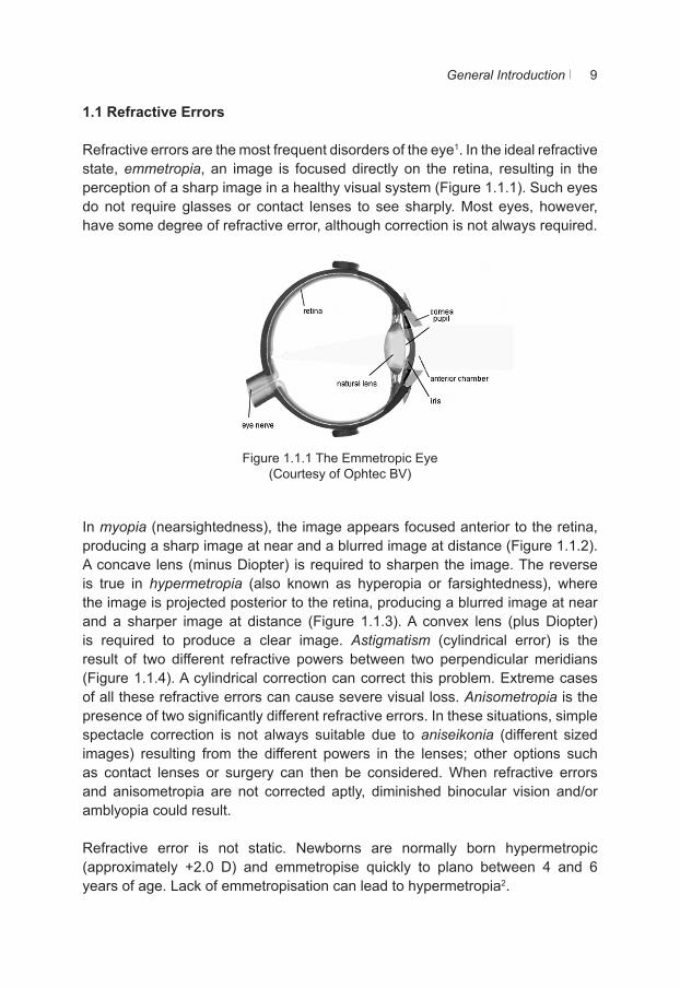
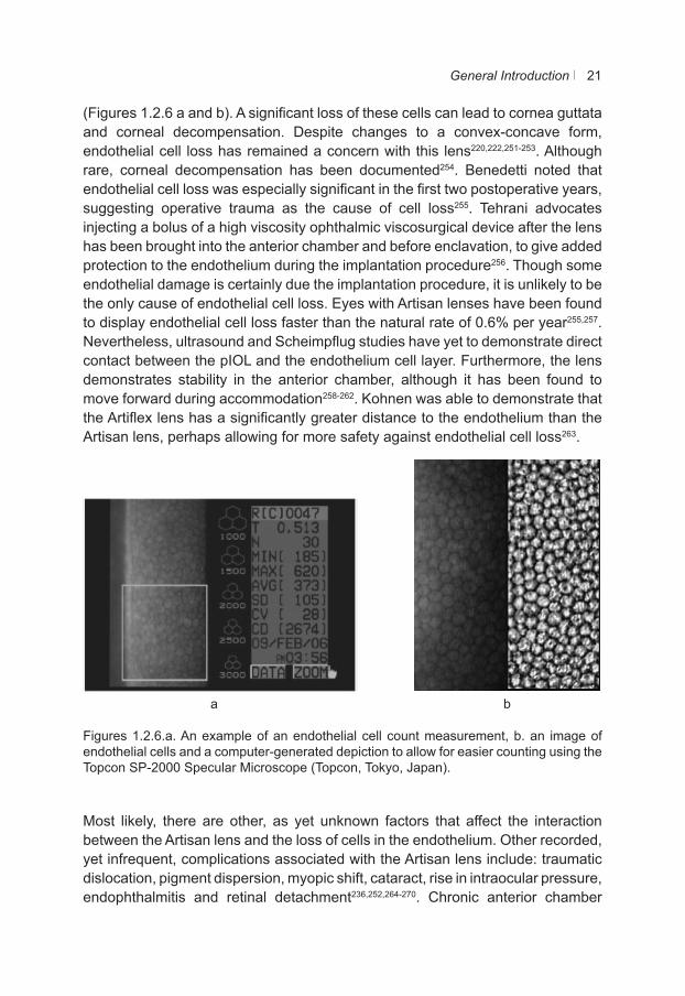
Refractive errors are the most frequent disorders of the eye1. In the ideal refractive state, emmetropia, an image is focused directly on the retina, resulting in the perception of a sharp image in a healthy visual system (Figure 1.1.1). Such eyes do not require glasses or contact lenses to see sharply. Most eyes, however, have some degree of refractive error, although correction is not always required.
Figure 1.1.1 The Emmetropic Eye
(Courtesy of Ophtec BV)
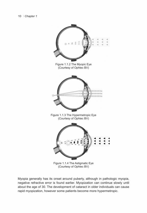
In myopia (nearsightedness), the image appears focused anterior to the retina, producing a sharp image at near and a blurred image at distance (Figure 1.1.2). A concave lens (minus Diopter) is required to sharpen the image. The reverse is true in hypermetropia (also known as hyperopia or farsightedness), where the image is projected posterior to the retina, producing a blurred image at near and a sharper image at distance (Figure 1.1.3). A convex lens (plus Diopter) is required to produce a clear image. Astigmatism (cylindrical error) is the result of two different refractive powers between two perpendicular meridians (Figure 1.1.4). A cylindrical correction can correct this problem. Extreme cases of all these refractive errors can cause severe visual loss. Anisometropia is the presence of two significantly different refractive errors. In these situations, simple spectacle correction is not always suitable due to aniseikonia (different sized images) resulting from the different powers in the lenses; other options such as contact lenses or surgery can then be considered. When refractive errors and anisometropia are not corrected aptly, diminished binocular vision and/or amblyopia could result.
Refractive error is not static. Newborns are normally born hypermetropic (approximately +2.0 D) and emmetropise quickly to plano between 4 and 6years of age. Lack of emmetropisation can lead to hypermetropia2.
Chapter 110
Figure 1.1.2 The Myopic Eye(Courtesy of Ophtec BV)
Figure 1.1.3 The Hypermetropic Eye(Courtesy of Ophtec BV)
Figure 1.1.4 The Astigmatic Eye(Courtesy of Ophtec BV)
Myopia generally has its onset around puberty, although in pathologic myopia, negative refractive error is found earlier. Myopization can continue slowly until about the age of 30. The development of cataract in older individuals can cause rapid myopization, however some patients become more hypermetropic.
General Introduction 11
Myopia is the most common form of refractive error, being most frequent in Asia3. It has been described as having reached epidemic proportions in South East Asia, where studies among schoolchildren have found a prevalence of 81% in Taiwan, 73% in Singapore and 62% in rural China4-6. Frequencies are lower in other Eastern countries, attaining 54% in Jordan, between 20 to 37% in India and Pakistan, 33% in Turkey and 3.4% in Iran7-12. It is less common in Europe, with 11.9% in Finland, 13% among Polish schoolchildren and 33% among the Danes13-16. Although there definitely appear to be large differences in the frequency of myopia around the world, population comparisons are hampered by the diverse criteria used to define refractive errors, the specific population groups being studied (such as registered schoolchildren and military recruits), different age groups and whether cycloplegic drops are used for the determination of refractive error17.
Myopia is generally divided into two categories, simple myopia (between –1.0 and –6.0 D) and pathologic myopia (more than –6.0 D). Complications as a result of pathological myopia abound. Patients run a higher risk of severe visual loss through retinal detachment, myopic chorioretinal degeneration, cataract and open-angle glaucoma18-34. The chance of such complications occurring is greater as the severity of myopia increases, although even eyes with simple myopia are not completely immune.
Animal experiments have been able to induce myopia through exposure to flashing lights or continuous illumination, form deprivation, lid suture, or goggling eyes with translucent lenses or lenses with different myopic refractive errors35-50. Most of these models imply that a visual feedback mechanism based on blurred images on the retina are fundamental to increases in axial length, and hereby the development of myopia. However, accelerated axial length increase can also be observed if the optic nerve has been severed51. Thus, the cause of myopia still remains elusive.
Many studies have revealed the highly hereditary nature of myopia. Twin studies on high myopia have shown a greater concordance among monozygotic twins than dizygotic twins52,53. So far, 15 loci associated with myopia have been established (MYP 1-15) although to date, no consistent associated genes have been found54-68. Still, different inheritance patterns between simple myopia and pathologic myopia have been implicated68-72. Genetic links in myopia could partially explain the dissimilar distribution of refractive errors amongst different ethnicities. The Baltimore Eye Survey found that 28.1% of adult urban subjects of European descent (living in the Baltimore area) were myopic, as opposed to 19.4% of African Americans73. Examinations of children of different ethnic backgrounds living in the same area have also found similar distributions74,75.
Chapter 112
Still, genes are not the only factor associated with refractive error; environmental aspects also appear to have a part in its development76,77. The effects of environment become apparent when studying individuals of similar ethnic backgrounds living in different places. Saw et al., observed that Chinese, Malay and Indian children living in Singapore expressed significantly more myopia than their ethnic counterparts residing in Kuala Lumpur, Malaysia78. Pertinent risk factors for nearsightedness have been found, whereby near work is considered the greatest; a study examining monozygotic and dizygotic twins and their reading habits was able to find a significantly higher concordance in myopia among monozygotic twins with comparable reading habits79. Furthermore, adult-onset myopia is most common in individuals who are involved in intensive near work, such as microscopists, carpet weavers and workers using visual display terminals80-82. Near work is thought to have perpetrated the rise of myopia in urban Asia, where school-age children are becoming increasingly younger, leading to longer periods of near work at a younger age4,73,83-91. Animal experiments have been able to confirm the association between near focus and myopization found in humans 5, 92-98. Moreover, a consistent correlation has also been found between both levels of education and intelligence with myopia, although both these factors may be associated with the greater amount near work among these individuals5,16,77,88,89,99-102. Urban areas display significantly more myopia than rural areas, although yet again, this could be confounded by higher levels of education (and thus near work) among urban dwellers77,86,100,103-105.
Nearsightedness is further associated with introvert personalities and a decreased chance of developing schizophrenia106,107. These relationships are not necessarily associated with the visual environment of the individual, but more with the chemical and genetic relationships of myopia with these traits. According to some studies, myopia is also significantly more common among women10,13,107-110.
The role of light exposure and the physical effects of melatonin associated with myopia remain contentious94,95,107,111-119. Still, outdoor activity has been shown to protect against myopia11,120. While one could surmise that this could be caused by less accommodation during sport, indoor sport activities do not appear to prevent nearsightedness120.
Hypermetropia is a less common refractive error and unlike myopia, more common among Caucasians121. Studies have shown frequencies of 28% in Finland, 27.4% of Malays residing in Singapore, 27.1% in Pakistan, 16.6% in Iran, 9.9% in the United States and 9.8% among South Indians 3.6%1,8,12,13,100,122. A limited number of family studies have shown that inheritability of hypermetropia exists53,123-125. In individual populations, hypermetropia shows a U-shaped distribution, with higher incidences among prepubescent children and then again among older individuals without cataract13,121,126,127.
General Introduction 13
Hypermetropia is much more often associated with amblyopia, decreased stereovision and strabismus, and can hereby pose a threat to the young eye. Also, the smaller ocular structures associated with hypermetropia makes hypermetropic eyes more prone to vision threatening acute and chronic angle closure glaucoma among older patients128-131.
Animal experiments show a propensity towards hypermetropia among restrained animals and with the use of convex or hypermetropic lenses37,44-47,49,132. Similar to myopia, the exact physiological events leading to hypermetropia are unknown, however it appears to be caused by changes in axial length and corneal flattening44, 45.
Outdoor activity induces hypermetropic tendencies, in direct contrast to the near work associated with myopia120. Furthermore, farsightedness is more common among individuals with a lower socio-economic status133,134. Surveys have also determined a higher frequency of hypermetropia among youngsters with developmental delays135-138. Also, hypermetropia is associated with extrovert personalities, as opposed to introvert personalities among myopes106.
Recent attempts at slowing the progression of myopia among children with ocular distillation of pirenzepine, a selective M1 muscarinic receptor antagonist, have been mildly successful. Still, pirenzepine has yet to halt myopization altogether139-141. Until refractive errors can be prevented effectively, we are simply left with the option to correct them with spectacles, contact lenses or surgery.
1.2 A Brief History of Correcting Refractive Error
1.2.1 Glasses and Contact Lenses
For centuries, people have been trying to correct ametropia. Before the invention of lenses, scholars would be hired to read to those who no longer could read due to their vision. Others would use containers filled with water to magnify their scripts. In 1000 AD, “reading stones” (magnifying glasses passed over reading materials) were used by monks to allow for studying scriptures. The first documented use of spectacles was either in Florence or Pisa, Italy where two convex lenses were used to magnify images142,143. With the advent of Gutenberg’s printing press in 1476, demand for spectacles increased throughout Europe143,144. Initially, there were single handheld lenses, which were then adapted to two lenses connected at in the middle, held with one hand. Nearly 350 years later, Benjamin Scarlett, a London optician, finally designed spectacles that were suspended on both ears143.
Chapter 114
The renowned inventor, Benjamin Franklin, has been accredited with the creation of “double spectacles”, which have evolved into the bifocals of today142. The basis of efficiently grinding several lenses at once, although significantly altered, was originally created by John Marshall of London, England145.
The first rough blueprints of the contact lens are accredited to da Vinci, Descartes, de la Hire and Young146,147. Contact lenses were first implemented independently near the end of the nineteenth century by three different inventors146. The French ophthalmologist Eugėne Kalt created contact lenses from the bottom of glass test tubes to counterbalance the severe astigmatism in keratoconus in 1888. The same year, Adolf Eugen Fick, a German physiologist, described the use of afocal contact lenses to counteract the optical effects of corneal distortions. Months later, the German physician August Müller, presented his dissertation on scleral contact lenses with a refractive power, based on experiments to correct his own myopia of -14.00 D148. Due to occurrence of severe corneal edema, Müller became disparaged with his idea and left Ophthalmology to pursue Orthopedic Surgery. Otto Himmler, a renowned maker of microscopes, manufactured these first glass-blown contact lenses for both Müller and Fick at a respective 4 and a whopping 38 Deutschmark price tag at his factory in Berlin149. The more current corneal contact lenses of today were invented by Kevin Tuohy in 1948. Professor Otto Wichterle of Prague patented a centrifugal casting machine to produce lenses of hydroxyethylmethacrylate and glycol diester (hydrogel) in 1961. These lenses were much more comfortable than the polymethyl methacrylate (PMMA) hard lenses which were in use at the time. In 1972, these “soft lenses” were introduced to the larger public by Bausch & Lomb146. Unlike glasses, contact lenses do not diminish retinal size with increasing myopia nor do they reduce visual fields148. Furthermore, they are lightweight and virtually invisible.
Currently, contact lenses have many different ophthalmic applications, such as keratoconus, aniseikonia, color blindness, ocular deformity and diplopia150-164. More frivolous applications also exist, such as changing ones eye color with the notoriously troublesome colored contact lens160,162,163,165-168. Nowadays, contact lenses are fabricated from a variety of materials and offer comfortable long-term wear and excellent refractive correction for 125 million people worldwide146,169. Still, microbial keratitis, often associated with corneal hypoxia and poor patient hygiene, dry eye and the development of contact lens intolerance, allergy and giant papillary conjunctivitis remain drawbacks of this form of refractive correction146,170-175.
1.2.2 Refractive Surgery
The idea of reducing corneal curvature to correct myopia has existed for centuries. The ancient Chinese apparently slept with sandbags over their eyes in order
General Introduction 15
to flatten their corneas. In the 19th century, Dr. J. Ball introduced small mallet that would flatten the cornea through the eyelid176. Luckily, today’s more refined innovations have taken over these rudimentary techniques.
1.2.2.1 Refractive Keratotomy
Dr. Lans, from Leiden, the Netherlands corrected astigmatism in rabbits using the methods of keratectomy, keratotomy and thermoplasty in 1898. In 1933, Tokyo’s Dr. Sato discovered that the astigmatism of his keratoconus patient decreased after breaks in the Descemet membrane. This led him to perform radial incisions in the anterior and posterior corneas of nearly 700 patients between 1951 and 1959. The formation of bullous keratopathy due to his toying with the corneal endothelium eventually led him to abandon this practice176-179.
The Radial Keratotomy (RK) procedure was introduced by several Russian eye surgeons in the early 1970s, the most renowned being Svyatoslav Fyodorov176-178. The procedure creates a flattening of the central cornea by incising the epithelial and stromal layers of the midperipheral cornea. The six steps to the procedure are as follows: 1. administration of appropriate anesthesia; 2. accurate marking of the visual axis; 3. marking the appropriate sized optical zone; 4. measuring the corneal thickness; 5. accurately setting the depth of the blade so as to be as close to the Descemet membrane as possible; and 6. making a predetermined number of corneal incisions (usually eight) to adequately flatten the cornea178. Problems associated with RK include overcorrection and progressive hypermetropic shift, diurnal fluctuations, corneal perforation and a lack of stability due to different wound healing patterns among patients of different age, whereby younger patients endure more refractive regression177,180-183.
Until the mid 1990s, RK was the most common procedure to correct myopia in the United States, having been performed by approximately 10% of ophthalmologists177. Its popularity diminished with the advent of the more refined excimer laser techniques.
1.2.2.2 Excimer Laser Treatments
The excimer (“excited dimer”) laser uses a 193 nm argon fluoride excimer laser to reshape the anterior corneal stroma through a procedure known as photoablation179,183,184. The laser was originally used to etch silicone computer chips in the 1970s by IBM185. Dr. Steven Trokel and Rangaswamy Srinivasan first described its use on the eye in 1983 to reshape freshly enucleated cow eyes. They wrote that the benefit, apart from its great precision, was that there was no damage
Chapter 116
to the neighboring tissue nor was there any sign of tissue disorganization186. Dr. Seiler from Germany, was the first to use it on a functioning human eye in 1987176. In 1988, John Marshall, along with Stephen J. Koons and Charles R. Munnerlyn, described the Photorefractive Keratectomy (PRK) procedure in rabbit eyes187. The Food and Drug Administration (FDA) approved its use on human eyes in the United States in 1995188.
Currently, there are three main forms of excimer laser treatments available: PRK, Laser in situ Keratomileusis (LASIK), and Laser-Assisted Sub-Epithelial Keratectomy (Laser Epithelial Keratomileusis or LASEK). PRK was the first to be performed; during this procedure, the corneal epithelium is first removed and the laser treatment then takes place on the stroma. Thanks to Pallikaris of Greece and Buratto of Italy, who combined PRK with Jose Barraquer’s invention, the microkeratome, LASIK was born. It was first carried out in 1989 by Pallikaris176. This procedure entails the making of a stromal flap of 8 – 10 mm diameter and 100 to 180 μm thickness with a microkeratome, the laser procedure, and then the replacement of the flap immediately thereafter. It allows for a nearly painless procedure, minimal haze and faster visual rehabilitation than PRK, although current technology allows for similar visual results between both188-190.
LASIK can correct higher refractive errors than PRK, although flap-related complications, which can be quite troublesome, can occur191-195. The femtosecond laser is the newest innovation in LASIK, allowing for thinner, more precise flaps with fewer complications, with comparable visual outcomes30,188,196,197. Other serious complications associated with LASIK are corneal ectasia, corneal perforation, diffuse lamellar keratitis, interface debris and epithelial ingrowth188,195,198-200.
LASEK was independently introduced by Dimitri Azar and Massimo Camellin in 1999. It combines both the PRK and LASIK procedures. Diluted ethanol is used to create an epithelial flap (like in PRK), which is replaced on the eye after ablation takes place (like LASIK). Postoperative pain, haze and visual rehabilitation are less bothersome than with PRK, but more problematic than with LASIK201.
All excimer laser procedures can be associated with postoperative keratitis, dry eye and visual disturbances such as starbursts and halos188,195,202-205. Currently, LASEK and PRK are indicated for low to moderate myopia, whereas LASIK is indicated for moderate to high myopia up to -10.0 D.
1.2.2.3 Phakic Intraocular Lens Implantations and the Artisan Lens
Several different Phakic Intraocular Lens (pIOLs) are available today – from anterior chamber angle-supported and iris-fixated lenses to posterior chamber lenses.
General Introduction 17
English ophthalmologist Dr. Harold Ridley (1906-2001) invented the first intraocular lens. He had observed polymethylmethacrylate (PMMA) splinters that would occasionally become lodged in the eyes of World War II Fighter pilots after returning from missions did not lead to ocular inflammation. This inspired him to create an intraocular lens made of PMMA which he placed in an eye after a cataract operation in 1949. These first “top secret” experiments were an absolute failure – not only did he render his first patient highly myopic (-14.0 D), the up to 108 mg weight of the lens was so heavy that it would often tumble into the vitreous cavity behind206-208. Initially, Ridley was not appreciated by his peers, however he eventually went on to receive numerous prestigious awards and was knighted by Queen Elizabeth II in 2000206.
Since Ridley’s pioneering work, many different pseudophakic lenses were created. Strampelli, Dannheim, Baron and then Barraquer were some of the first to design phakic IOLs to correct high myopia in the 1950s. These angle-fixated lenses led to many serious complications, such as extreme endothelial cell loss and Uveitis-Glaucoma-Hyphema Syndrome (UGH-Syndrome), leading many of these lenses to be explanted208,209. Georges Baikoff finally published work on his angle-fixated phakic IOL in 1991, which was adapted to prevent severe endothelial cell loss. Still, the new lens was also unable to escape the considerable side-effects of iridopathy, pupil ovalization, low-grade uveitis, peripheral synechiae and sectorial atrophy of the iris209.
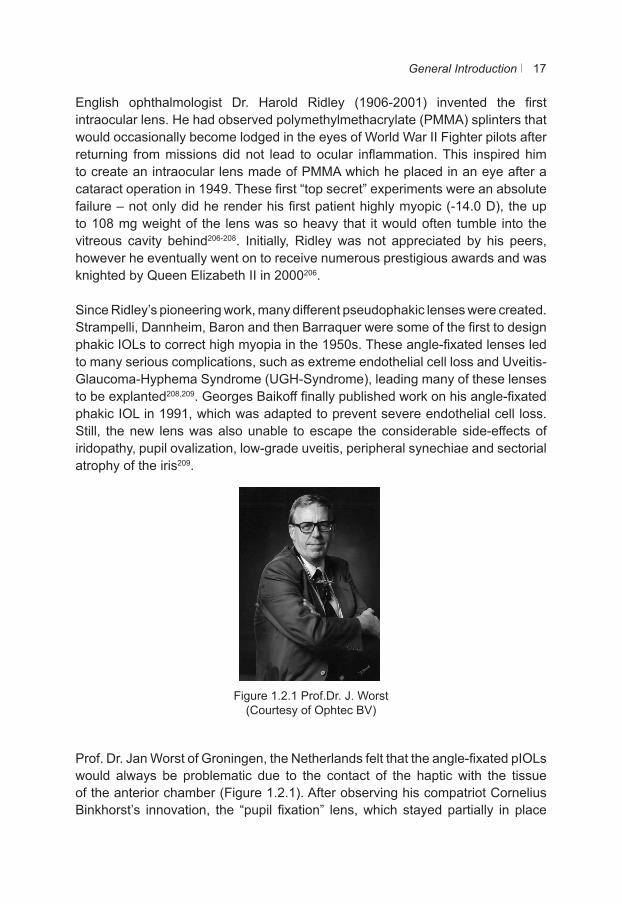
Figure 1.2.1 Prof.Dr. J. Worst (Courtesy of Ophtec BV)
Prof. Dr. Jan Worst of Groningen, the Netherlands felt that the angle-fixated pIOLs would always be problematic due to the contact of the haptic with the tissue of the anterior chamber (Figure 1.2.1). After observing his compatriot Cornelius Binkhorst’s innovation, the “pupil fixation” lens, which stayed partially in place
Chapter 118
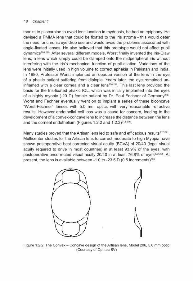
thanks to pilocarpine to avoid lens luxation in mydriasis, he had an epiphany. He devised a PMMA lens that could be fixated to the iris stroma - this would deter the need for chronic eye drop use and would avoid the problems associated with angle-fixated lenses. He also believed that this prototype would not affect pupil dynamics208,210. After several different models, Worst finally invented the Iris-Claw lens, a lens which simply could be clamped onto the midperipheral iris without interfering with the iris’s mechanical function of pupil dilation. Variations of the lens were initially used in high volume to correct aphakia in Pakistan and India. In 1980, Professor Worst implanted an opaque version of the lens in the eye of a phakic patient suffering from diplopia. Years later, the eye remained un-inflamed with a clear cornea and a clear lens208,211. This last lens provided the basis for the Iris-fixated phakic IOL, which was initially implanted into the eyes of a highly myopic (-20 D) female patient by Dr. Paul Fechner of Germany208. Worst and Fechner eventually went on to implant a series of these biconcave “Worst-Fechner” lenses with 5.0 mm optics with very reasonable refractive results. However endothelial cell loss was a cause for concern, leading to the development of a convex-concave lens to increase the distance between the lens and the corneal endothelium (Figures 1.2.2 and 1.2.3)212-216.
Many studies proved that the Artisan lens led to safe and efficacious results217-221. Multicenter studies for the Artisan lens to correct moderate to high Myopia have shown postoperative best corrected visual acuity (BCVA) of 20/40 (legal visual acuity required to drive in most countries) in at least 93.9% of the eyes, with postoperative uncorrected visual acuity 20/40 in at least 76.8% of eyes222-225. At present, the lens is available between -1.0 to -23.5 D (0.5 increments)226.
Figure 1.2.2: The Convex – Concave design of the Artisan lens, Model 206, 5.0 mm optic (Courtesy of Ophtec BV)
General Introduction 19
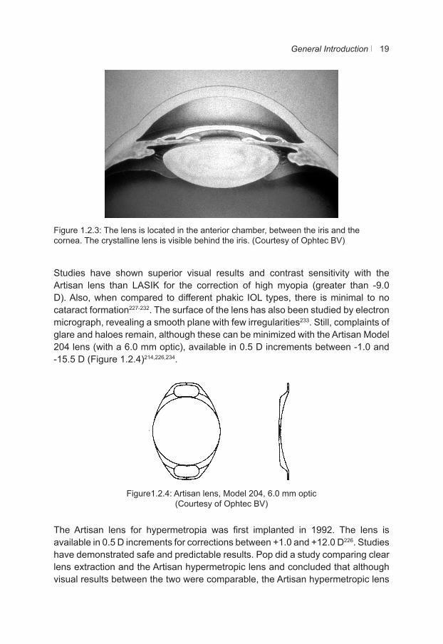
Figure 1.2.3: The lens is located in the anterior chamber, between the iris and the cornea. The crystalline lens is visible behind the iris. (Courtesy of Ophtec BV)
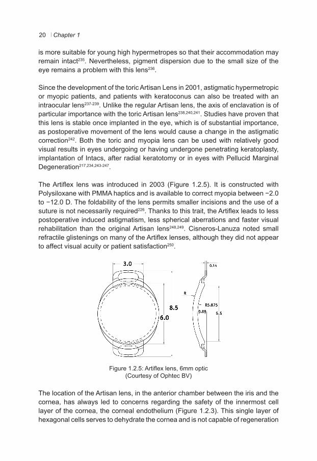
Studies have shown superior visual results and contrast sensitivity with the Artisan lens than LASIK for the correction of high myopia (greater than -9.0 D). Also, when compared to different phakic IOL types, there is minimal to no cataract formation227-232. The surface of the lens has also been studied by electron micrograph, revealing a smooth plane with few irregularities233. Still, complaints of glare and haloes remain, although these can be minimized with the Artisan Model 204 lens (with a 6.0 mm optic), available in 0.5 D increments between -1.0 and -15.5 D (Figure 1.2.4)214,226,234.
Figure1.2.4: Artisan lens, Model 204, 6.0 mm optic(Courtesy of Ophtec BV)
The Artisan lens for hypermetropia was first implanted in 1992. The lens is available in 0.5 D increments for corrections between +1.0 and +12.0 D226. Studies have demonstrated safe and predictable results. Pop did a study comparing clear lens extraction and the Artisan hypermetropic lens and concluded that although visual results between the two were comparable, the Artisan hypermetropic lens
Chapter 120
is more suitable for young high hypermetropes so that their accommodation may remain intact235. Nevertheless, pigment dispersion due to the small size of the eye remains a problem with this lens236.
Since the development of the toric Artisan Lens in 2001, astigmatic hypermetropic or myopic patients, and patients with keratoconus can also be treated with an intraocular lens237-239. Unlike the regular Artisan lens, the axis of enclavation is of particular importance with the toric Artisan lens238,240,241. Studies have proven that this lens is stable once implanted in the eye, which is of substantial importance, as postoperative movement of the lens would cause a change in the astigmatic correction242. Both the toric and myopia lens can be used with relatively good visual results in eyes undergoing or having undergone penetrating keratoplasty, implantation of Intacs, after radial keratotomy or in eyes with Pellucid Marginal Degeneration217,234,243-247.
The Artiflex lens was introduced in 2003 (Figure 1.2.5). It is constructed with Polysiloxane with PMMA haptics and is available to correct myopia between −2.0 to −12.0 D. The foldability of the lens permits smaller incisions and the use of a suture is not necessarily required226. Thanks to this trait, the Artiflex leads to less postoperative induced astigmatism, less spherical aberrations and faster visual rehabilitation than the original Artisan lens248,249. Cisneros-Lanuza noted small refractile glistenings on many of the Artiflex lenses, although they did not appear to affect visual acuity or patient satisfaction250.
Figure 1.2.5: Artiflex lens, 6mm optic(Courtesy of Ophtec BV)
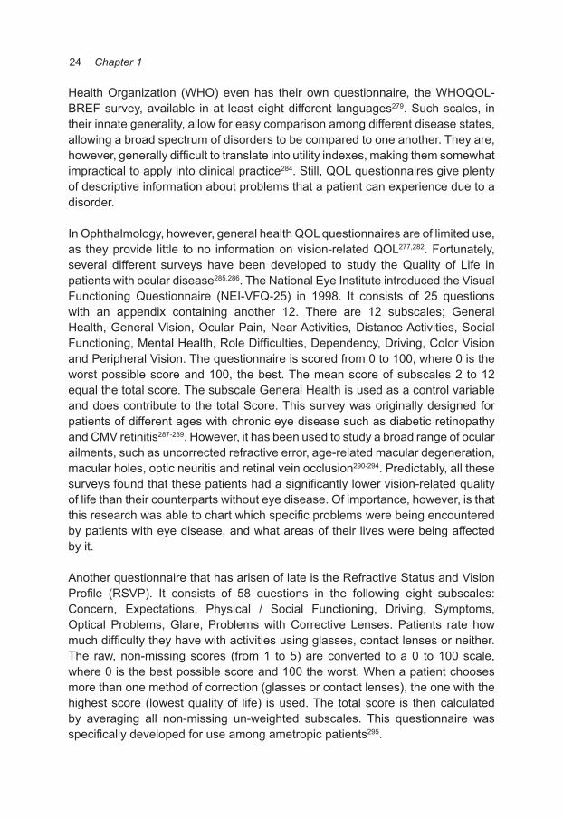
The location of the Artisan lens, in the anterior chamber between the iris and the cornea, has always led to concerns regarding the safety of the innermost cell layer of the cornea, the corneal endothelium (Figure 1.2.3). This single layer of hexagonal cells serves to dehydrate the cornea and is not capable of regeneration
General Introduction 21
(Figures 1.2.6 a and b). A significant loss of these cells can lead to cornea guttata and corneal decompensation. Despite changes to a convex-concave form, endothelial cell loss has remained a concern with this lens220,222,251-253. Although rare, corneal decompensation has been documented254. Benedetti noted that endothelial cell loss was especially significant in the first two postoperative years, suggesting operative trauma as the cause of cell loss255. Tehrani advocates injecting a bolus of a high viscosity ophthalmic viscosurgical device after the lens has been brought into the anterior chamber and before enclavation, to give added protection to the endothelium during the implantation procedure256. Though some endothelial damage is certainly due the implantation procedure, it is unlikely to be the only cause of endothelial cell loss. Eyes with Artisan lenses have been found to display endothelial cell loss faster than the natural rate of 0.6% per year255,257. Nevertheless, ultrasound and Scheimpflug studies have yet to demonstrate direct contact between the pIOL and the endothelium cell layer. Furthermore, the lens demonstrates stability in the anterior chamber, although it has been found to move forward during accommodation258-262. Kohnen was able to demonstrate that the Artiflex lens has a significantly greater distance to the endothelium than the Artisan lens, perhaps allowing for more safety against endothelial cell loss263.
a b
Figures 1.2.6.a. An example of an endothelial cell count measurement, b. an image of endothelial cells and a computer-generated depiction to allow for easier counting using the Topcon SP-2000 Specular Microscope (Topcon, Tokyo, Japan).
Most likely, there are other, as yet unknown factors that affect the interaction between the Artisan lens and the loss of cells in the endothelium. Other recorded, yet infrequent, complications associated with the Artisan lens include: traumatic dislocation, pigment dispersion, myopic shift, cataract, rise in intraocular pressure, endophthalmitis and retinal detachment236,252,264-270. Chronic anterior chamber
Chapter 122
reaction is rare, although perhaps more common among the Artiflex model of this lens259,271,272.
Artisan Lens-power calculations are performed using the Van der Heijde formula, taking into account spectacle refraction, corneal power and the depth of the anterior chamber208. Nonetheless, residual refractive error is relatively common after pIOL implantation. This can be corrected with the use of glasses or contact lenses as the anterior part of the eye is left unaffected. More gregarious surgeons combine this technique with the excimer laser to correct the remaining ametropia251,273-275.
1.3 Quality of Life
Refractive surgical procedures have become nearly “everyday” procedures. Unlike other common operations such as cataract operations or hip replacement, refractive surgery is nearly always performed in healthy eyes. In fact, most refractive procedures consider a diseased eye as a contraindication to surgery. Generally, studies regarding refractive surgery techniques evaluate the following aspects: best corrected visual acuity (BCVA), uncorrected visual acuity (UCVA), postoperative refractive error (Predictability), Stability of the postoperative refractive error, Safety (% eyes with loss of 2 or more lines of BCVA), Efficacy (% eyes with UCVA 20/20), Safety Index (mean postoperative BCVA / mean preoperative BCVA) and Efficacy Index (mean postoperative UCVA / mean preoperative BCVA)276. Although these evaluations are critical to determine the medical success of an intervention, positive objective results do not necessarily mean that a patient is pleased with the results. Patient-oriented parameters such as patient satisfaction and quality of life (QOL) can add a new dimension in evaluating success in such procedures, allowing new insights on subjective improvements owing to the technique, as well as side-effects and complaints surrounding it. Clinical experience certainly provides awareness on many of these questions, however quantifying these issues is more concrete. Quantification is especially useful considering that eye specialists have been found to underestimate the quality of life of their patients277,278. Stein et al., found that ophthalmologists overestimated the QOL of patients with respect to cataract and macular disease, yet slightly underestimated QOL of patients with glaucoma277. Quality of life measurements can lend insight into the effects of disease on patients and can also allow more understanding as to how a certain treatment has helped.
The World Health Organization defines the Quality of Life as: “An individual’s perception of their position in life, in the context of the culture and value systems in which they live, and in relation to their goals, expectations, standards, and concerns. It is a broad ranging concept, affected in a complex way by the person’s physical health, psychological state, level of independence, social relationships,
General Introduction 23
and their relationship to salient features of their environment279.
Currently, there are two main approaches to quantify QOL:
1.3.1 Choice Based Evaluation Methods
The Time Trade-Off Method is based on the scenario where a patient is asked how many years of his remaining life he is willing to trade-off for perfect health/vision/hearing etc. The Utility-Method is a calculation based on the Time Trade-Off Method, where 0.0 indicates the worst possible health and 1.0 indicates the best possible health. For example, a patient who is expected to live for 20 years and is willing to trade 4 of these years for perfect vision will have a Utility Index of 20/20 - 4/20 = 16/20 = 0.8). The Standard Gamble Method asks the patient how much of a risk he or she would be willing to take to allow for perfect health, considering that the treatment which would lead to the cure could lead to either perfect health or to death. The Utility Index for someone willing to take a 60% risk of death for the chance of perfect health would be 1.0 - 0.6 = 0.4. Quality-Adjusted Life-Years (QALYs) combine both the quantity and quality of life produced by a medical intervention. It is a product of life expectancy and a measure of the quality of the remaining life-years. A QALY places a weight on time in different health states. A year of perfect health is worth 1.0; however, a year of less than perfect health life expectancy is worth less than 1.0. Death is considered to be equivalent to 0.0, however, some health states may be considered worse than death and have negative scores (based on Utility Indices). For example, if a patient is to survive for one year on Treatment A in moderate health, his QALY will be 1 year x 0.5 = 0.5. However, if the patient were to undergo Treatment B and would live for 1 year and 3 months, and be in excellent health, his QALY would be 1.25 x 1 (excellent health) = 1.25. A patient not receiving any treatment would survive 6 months with severe pain and suffering. The QALY would then be 0.5 (years) x -0.6 (approximately) = -0.3. These numbers would then be used in cost-utility analysis to see which treatment is the most beneficial and provides the lowest cost per QALY280,281. The advantage of these methods is that they can be easily compared with different diseases and also among the diverse medical subspecialties. However, they are not very descriptive, and allow for little added data information282.
1.3.2 Standardized Questionnaires
Standardized Quality of Life surveys also exist. Examples are the 36-Item Short-Form Survey (SF-36), Nottinham Health Profile, Symptom Rating Scale, The Sickness Impact Profile and The Quality of Well Being Scale277,283. The World
Chapter 124
Health Organization (WHO) even has their own questionnaire, the WHOQOL-BREF survey, available in at least eight different languages279. Such scales, in their innate generality, allow for easy comparison among different disease states, allowing a broad spectrum of disorders to be compared to one another. They are, however, generally difficult to translate into utility indexes, making them somewhat impractical to apply into clinical practice284. Still, QOL questionnaires give plenty of descriptive information about problems that a patient can experience due to a disorder.
In Ophthalmology, however, general health QOL questionnaires are of limited use, as they provide little to no information on vision-related QOL277,282. Fortunately, several different surveys have been developed to study the Quality of Life in patients with ocular disease285,286. The National Eye Institute introduced the Visual Functioning Questionnaire (NEI-VFQ-25) in 1998. It consists of 25 questions with an appendix containing another 12. There are 12 subscales; General Health, General Vision, Ocular Pain, Near Activities, Distance Activities, Social Functioning, Mental Health, Role Difficulties, Dependency, Driving, Color Vision and Peripheral Vision. The questionnaire is scored from 0 to 100, where 0 is the worst possible score and 100, the best. The mean score of subscales 2 to 12 equal the total score. The subscale General Health is used as a control variable and does contribute to the total Score. This survey was originally designed for patients of different ages with chronic eye disease such as diabetic retinopathy and CMV retinitis287-289. However, it has been used to study a broad range of ocular ailments, such as uncorrected refractive error, age-related macular degeneration, macular holes, optic neuritis and retinal vein occlusion290-294. Predictably, all these surveys found that these patients had a significantly lower vision-related quality of life than their counterparts without eye disease. Of importance, however, is that this research was able to chart which specific problems were being encountered by patients with eye disease, and what areas of their lives were being affected by it.
Another questionnaire that has arisen of late is the Refractive Status and Vision Profile (RSVP). It consists of 58 questions in the following eight subscales: Concern, Expectations, Physical / Social Functioning, Driving, Symptoms, Optical Problems, Glare, Problems with Corrective Lenses. Patients rate how much difficulty they have with activities using glasses, contact lenses or neither. The raw, non-missing scores (from 1 to 5) are converted to a 0 to 100 scale, where 0 is the best possible score and 100 the worst. When a patient chooses more than one method of correction (glasses or contact lenses), the one with the highest score (lowest quality of life) is used. The total score is then calculated by averaging all non-missing un-weighted subscales. This questionnaire was specifically developed for use among ametropic patients295.
General Introduction 25
Rose and Takashima studied the effects of myopia on the quality of life. Both found that patients with high myopia express a lower QOL. This was not only related to higher costs, but also to the psychological, practical and cosmetic issues associated with higher degrees of myopia296,297. Castanon Holguin also found that there were also cosmetic issues among younger patients with myopia. Older children, and children residing in more populated areas were more likely to demonstrate trepidation about the appearance of glasses or about being teased, thus affecting their compliance with spectacle wear298.
To date, many studies regarding postoperative satisfaction and subjective outcomes after refractive surgery have been done. These studies have all shown satisfaction with uncomplicated refractive surgical procedures, whereby the degree of satisfaction often correlated with postoperative problems such as haze and night vision disturbances299-307. In 2007, a comparative study was published regarding the Quality of Life among three different groups: Emmetropes, Myopes using contact lenses or glasses and Former Myopes who had undergone refractive surgery. They found that Emmetropes and the Refractive Surgery patients had comparable QOL results, and that the Myopes had significantly lower QOL scores308. With the exception of two studies, however, none compared preoperative and postoperative results of the same individual patients302,307. So far, no study has looked at QOL before and after surgery, and compared these results with individuals not seeking refractive surgery.
1.4 Goals of this Thesis
The first part of this thesis aims to evaluate objective results of the Artisan lens. The first study discusses the effects of incision-induced astigmatism and the effects of planned versus actual positioning of the Toric Artisan pIOL on postoperative astigmatism. The second study analyzes the visual results and safety of the Artisan lens for hypermetropia. As mentioned previously, hypermetropic eyes are especially sensitive due to their small size, possibly making them prone to complications not witnessed in myopic eyes implanted with the Artisan lens. Thirdly, the long-term effects of the Artisan lens on the corneal endothelium are assessed. The relationship between endothelial cell loss and the depth of the anterior chamber is evaluated.
The second part of this thesis considers the subjective results of patients with myopia in the form of Quality of Life scores. In Chapter 5, satisfied contact lens wearers are asked to complete both the RSVP and NEI-VFQ-25 questionnaires. The relationship of their scores in relation to their degree of myopia is examined. Chapter 6 analyzes the QOL scores of patients undergoing either excimer laser
Chapter 126
procedures or implantation of the Artisan lens. Patients are given both the RSVP and the NEI-VFQ-25 questionnaires before treatment and then again two and twelve months postoperative. The Quality of Life scores are then compared with each other and with contact lens wearers not seeking refractive surgery (from the aforementioned work in Chapter 5), to determine: 1. If the Quality of Life improves after refractive surgery 2. If patients seeking refractive surgery have poorer preoperative QOL scores than individuals not seeking refractive surgery and 3. If the QOL scores of operated (and thus nearly emmetropic) patients are higher than myopic contact lens wearers not seeking refractive surgery.
The third part of this thesis looks at unique uses of the Artisan lens. Chapter 7 involves a case of a child with severe anisometric amblyopia. Due to problematic issues with contact lenses, an Artisan lens is implanted in the highly myopic eye. The postoperative outcomes are discussed. Chapter 8 is a report on five patients having undergone cataract extraction in one eye and, due to the resulting anisometropia, an Artisan lens implantation in the other eye. Patients completed two postoperative RSVP questionnaires, one in relation to each eye. The results of the two are then compared.
General Introduction 27
References
1. Kempen JH, Mitchell P, Lee KE, et al. The prevalence of refractive errors among adults in the United States, Western Europe, and Australia. Arch Ophthalmol 2004;122(4):495-505.
2. Mutti DO. To emmetropize or not to emmetropize? The question for hyperopic development. Optom Vis Sci 2007;84(2):97-102.
3. Saw SM. A synopsis of the prevalence rates and environmental risk factors for myopia. Clin Exp Optom 2003;86(5):289-94.
4. Lin LL, Shih YF, Hsiao CK, Chen CJ. Prevalence of myopia in Taiwanese schoolchildren: 1983 to 2000. Ann Acad Med Singapore 2004;33(1):27-33.
5. Quek TP, Chua CG, Chong CS, et al. Prevalence of refractive errors in teenage high school students in Singapore. Ophthalmic Physiol Opt 2004;24(1):47-55.
6. Congdon N, Wang Y, Song Y, et al. Visual disability, visual function, and myopia among rural chinese secondary school children: the Xichang Pediatric Refractive Error Study (X-PRES)--report 1. Invest Ophthalmol Vis Sci 2008;49(7):2888-94.
7. Mallen EA, Gammoh Y, Al-Bdour M, Sayegh FN. Refractive error and ocular biometry in Jordanian adults. Ophthalmic Physiol Opt 2005;25(4):302-9.
8. Shah SP, Jadoon MZ, Dineen B, et al. Refractive errors in the adult pakistani population: the national blindness and visual impairment survey. Ophthalmic Epidemiol 2008;15(3):183-90.
9. Raju P, Ramesh SV, Arvind H, et al. Prevalence of refractive errors in a rural South Indian population. Invest Ophthalmol Vis Sci 2004;45(12):4268-72.
10. Dandona R, Dandona L, Srinivas M, et al. Population-based assessment of refractive error in India: the Andhra Pradesh eye disease study. Clin Experiment Ophthalmol 2002;30(2):84-93.
11. Onal S, Toker E, Akingol Z, et al. Refractive errors of medical students in Turkey: one year follow-up of refraction and biometry. Optom Vis Sci 2007;84(3):175-80.
12. Fotouhi A, Hashemi H, Khabazkhoob M, Mohammad K. The prevalence of refractive errors among schoolchildren in Dezful, Iran. Br J Ophthalmol 2007;91(3):287-92.
13. Aine E. Refractive errors in a Finnish rural population. Acta Ophthalmol (Copenh) 1984;62(6):944-54.
14. Czepita D, Mojsa A, Ustianowska M, et al. Prevalence of refractive errors in schoolchildren ranging from 6 to 18 years of age. Ann Acad Med Stetin 2007;53(1):53-6.
15. Czepita D, Zejmo M, Mojsa A. Prevalence of myopia and hyperopia in a population of Polish schoolchildren. Ophthalmic Physiol Opt 2007;27(1):60-5.
16. Kessel L, Hougaard JL, Mortensen C, et al. Visual acuity and refractive errors in a suburban Danish population: Inter99 Eye Study. Acta Ophthalmol Scand 2004;82(1):19-24.
17. Negrel AD, Maul E, Pokharel GP, et al. Refractive Error Study in Children: sampling and measurement methods for a multi-country survey. Am J Ophthalmol 2000;129(4):421-6.
18. Xu L, Wang Y, Wang S, et al. High myopia and glaucoma susceptibility the Beijing Eye Study. Ophthalmology 2007;114(2):216-20.
19. Wong TY, Foster PJ, Johnson GJ, Seah SK. Refractive errors, axial ocular dimensions, and age-related cataracts: the Tanjong Pagar survey. Invest Ophthalmol Vis Sci 2003;44(4):1479-85.
20. Tornquist R, Stenkula S, Tornquist P. Retinal detachment. A study of a population-based patient material in Sweden 1971-1981. I. Epidemiology. Acta Ophthalmol (Copenh) 1987;65(2):213-22.
21. Tillery WV, Lucier AC. Round atrophic holes in lattice degeneration--an important cause of phakic retinal detachment. Trans Sect Ophthalmol Am Acad Ophthalmol Otolaryngol 1976;81(3 Pt 1):509-18.
22. Soubrane G. Choroidal neovascularization in pathologic myopia: recent developments in diagnosis and treatment. Surv Ophthalmol 2008;53(2):121-38.
23. Mitchell P, Hourihan F, Sandbach J, Wang JJ. The relationship between glaucoma and myopia: the Blue Mountains Eye Study. Ophthalmology 1999;106(10):2010-5.
24. Mayama C, Suzuki Y, Araie M, et al. Myopia and advanced-stage open-angle glaucoma. Ophthalmology 2002;109(11):2072-7.
Chapter 128
25. Glacet-Bernard A, Benyelles N, Dumas S, et al. Photodynamic therapy vs limited macular translocation in the management of subfoveal choroidal neovascularization in pathologic myopia: a two-year study. Am J Ophthalmol 2007;143(1):68-76.
26. Colin J, Robinet A, Cochener B. Retinal detachment after clear lens extraction for high myopia: seven-year follow-up. Ophthalmology 1999;106(12):2281-4; discussion 5.
27. Chou SC, Yang CH, Lee CH, et al. Characteristics of primary rhegmatogenous retinal detachment in Taiwan. Eye 2007;21(8):1056-61.
28. Chihara E, Liu X, Dong J, et al. Severe myopia as a risk factor for progressive visual field loss in primary open-angle glaucoma. Ophthalmologica 1997;211(2):66-71.
29. Chan WM, Ohji M, Lai TY, et al. Choroidal neovascularisation in pathological myopia: an update in management. Br J Ophthalmol 2005;89(11):1522-8.
30. Chan A, Ou J, Manche EE. Comparison of the femtosecond laser and mechanical keratome for laser in situ keratomileusis. Arch Ophthalmol 2008;126(11):1484-90.
31. Celorio JM, Pruett RC. Prevalence of lattice degeneration and its relation to axial length in severe myopia. Am J Ophthalmol 1991;111(1):20-3.
32. Burton TC, Brown CK, Haimann MH. Predicting age of onset for individuals at risk for phakic retinal detachment. Trans Am Ophthalmol Soc 1983;81:149-61.
33. Burton TC. The influence of refractive error and lattice degeneration on the incidence of retinal detachment. Trans Am Ophthalmol Soc 1989;87:143-55; discussion 55-7.
34. Jonas JB, Martus P, Budde WM. Anisometropia and degree of optic nerve damage in chronic open-angle glaucoma. Am J Ophthalmol 2002;134(4):547-51.
35. Yinon U, Koslowe KC, Lobel D, et al. Lid suture myopia in developing chicks: optical and structural considerations. Curr Eye Res 1982;2(12):877-82.
36. Tse DY, Lam CS, Guggenheim JA, et al. Simultaneous defocus integration during refractive development. Invest Ophthalmol Vis Sci 2007;48(12):5352-9.
37. Sivak JG, Barrie DL, Callender MG, et al. Optical causes of experimental myopia. Ciba Found Symp 1990;155:160-72; discussion 72-7.
38. Schmid KL, Wildsoet CF. Contrast and spatial-frequency requirements for emmetropization in chicks. Vision Res 1997;37(15):2011-21.
39. Schmid KL, Wildsoet CF. The sensitivity of the chick eye to refractive defocus. Ophthalmic Physiol Opt 1997;17(1):61-7.
40. Schmid K, Wildsoet CF. Natural and imposed astigmatism and their relation to emmetropization in the chick. Exp Eye Res 1997;64(5):837-47.
41. Pardue MT, Faulkner AE, Fernandes A, et al. High susceptibility to experimental myopia in a mouse model with a retinal on pathway defect. Invest Ophthalmol Vis Sci 2008;49(2):706-12.
42. Nevin ST, Schmid KL, Wildsoet CF. Sharp vision: a prerequisite for compensation to myopic defocus in the chick? Curr Eye Res 1998;17(3):322-31.
43. Mutti DO, Zadnik K, Murphy CJ. The effect of continuous light on refractive error and the ocular components of the rat. Exp Eye Res 1998;67(6):631-6.
44. Irving EL, Sivak JG, Callender MG. Refractive plasticity of the developing chick eye. Ophthalmic Physiol Opt 1992;12(4):448-56.
45. Irving EL, Callender MG, Sivak JG. Inducing myopia, hyperopia, and astigmatism in chicks. Optom Vis Sci 1991;68(5):364-8.
46. Irving EL, Callender MG, Sivak JG. Inducing ametropias in hatchling chicks by defocus--aperture effects and cylindrical lenses. Vision Res 1995;35(9):1165-74.
47. Guo SS, Sivak JG, Callender MG, Herbert KL. Effects of continuous light on experimental refractive errors in chicks. Ophthalmic Physiol Opt 1996;16(6):486-90.
48. Cheng ZY, Li JH, Li R, Li JQ. [Effect of time limited form deprivation on the development of myopia in guinea pigs]. Zhonghua Yan Ke Za Zhi 2004;40(3):183-5.
49. Shen W, Sivak JG. Eyes of a lower vertebrate are susceptible to the visual environment. Invest Ophthalmol Vis Sci 2007;48(10):4829-37.
50. Sivak JG. The role of the lens in refractive development of the eye: animal models of ametropia. Exp Eye Res 2008;87(1):3-8.
51. Hung GK, Ciuffreda KJ. A unifying theory of refractive error development. Bull Math Biol 2000;62(6):1087-108.
General Introduction 29
52. Lin LL, Chen CJ. Twin study on myopia. Acta Genet Med Gemellol (Roma) 1987;36(4):535-40.
53. Hammond CJ, Snieder H, Gilbert CE, Spector TD. Genes and environment in refractive error: the twin eye study. Invest Ophthalmol Vis Sci 2001;42(6):1232-6.
54. Zhou J, Young TL. Evaluation of Lipin 2 as a candidate gene for autosomal dominant 1 high-grade myopia. Gene 2005;352:10-9.
55. Young TL, Ronan SM, Alvear AB, et al. A second locus for familial high myopia maps to chromosome 12q. Am J Hum Genet 1998;63(5):1419-24.
56. Young TL, Ronan SM, Drahozal LA, et al. Evidence that a locus for familial high myopia maps to chromosome 18p. Am J Hum Genet 1998;63(1):109-19.
57. Young TL. Dissecting the genetics of human high myopia: a molecular biologic approach. Trans Am Ophthalmol Soc 2004;102:423-45.
58. Stambolian D, Ibay G, Reider L, et al. Genomewide linkage scan for myopia susceptibility loci among Ashkenazi Jewish families shows evidence of linkage on chromosome 22q12. Am J Hum Genet 2004;75(3):448-59.
59. Scavello GS, Paluru PC, Ganter WR, Young TL. Sequence variants in the transforming growth beta-induced factor (TGIF) gene are not associated with high myopia. Invest Ophthalmol Vis Sci 2004;45(7):2091-7.
60. Paluru PC, Scavello GS, Ganter WR, Young TL. Exclusion of lumican and fibromodulin as candidate genes in MYP3 linked high grade myopia. Mol Vis 2004;10:917-22.
61. Paluru PC, Nallasamy S, Devoto M, et al. Identification of a novel locus on 2q for autosomal dominant high-grade myopia. Invest Ophthalmol Vis Sci 2005;46(7):2300-7.
62. Paluru P, Ronan SM, Heon E, et al. New locus for autosomal dominant high myopia maps to the long arm of chromosome 17. Invest Ophthalmol Vis Sci 2003;44(5):1830-6.
63. Lin HJ, Wan L, Tsai Y, et al. The TGFbeta1 gene codon 10 polymorphism contributes to the genetic predisposition to high myopia. Mol Vis 2006;12:698-703.
64. Lam DS, Tam PO, Fan DS, et al. Familial high myopia linkage to chromosome 18p. Ophthalmologica 2003;217(2):115-8.
65. Jacobi FK, Zrenner E, Broghammer M, Pusch CM. A genetic perspective on myopia. Cell Mol Life Sci 2005;62(7-8):800-8.
66. Han W, Yap MK, Wang J, Yip SP. Family-based association analysis of hepatocyte growth factor (HGF) gene polymorphisms in high myopia. Invest Ophthalmol Vis Sci 2006;47(6):2291-9.
67. Hammond CJ, Andrew T, Mak YT, Spector TD. A susceptibility locus for myopia in the normal population is linked to the PAX6 gene region on chromosome 11: a genomewide scan of dizygotic twins. Am J Hum Genet 2004;75(2):294-304.
68. Farbrother JE, Kirov G, Owen MJ, et al. Linkage analysis of the genetic loci for high myopia on 18p, 12q, and 17q in 51 U.K. families. Invest Ophthalmol Vis Sci 2004;45(9):2879-85.
69. Zhang Q, Guo X, Xiao X, et al. Novel locus for X linked recessive high myopia maps to Xq23-q25 but outside MYP1. J Med Genet 2006;43(5):e20.
70. Wang IJ, Chiang TH, Shih YF, et al. The association of single nucleotide polymorphisms in the 5’-regulatory region of the lumican gene with susceptibility to high myopia in Taiwan. Mol Vis 2006;12:852-7.
71. Scavello GS, Jr., Paluru PC, Zhou J, et al. Genomic structure and organization of the high grade Myopia-2 locus (MYP2) critical region: mutation screening of 9 positional candidate genes. Mol Vis 2005;11:97-110.
72. Ibay G, Doan B, Reider L, et al. Candidate high myopia loci on chromosomes 18p and 12q do not play a major role in susceptibility to common myopia. BMC Med Genet 2004;5:20.
73. Katz J, Tielsch JM, Sommer A. Prevalence and risk factors for refractive errors in an adult inner city population. Invest Ophthalmol Vis Sci 1997;38(2):334-40.
74. Ip JM, Huynh SC, Robaei D, et al. Ethnic differences in the impact of parental myopia: findings from a population-based study of 12-year-old Australian children. Invest Ophthalmol Vis Sci 2007;48(6):2520-8.
75. Ip JM, Huynh SC, Robaei D, et al. Ethnic differences in refraction and ocular biometry in a population-based sample of 11-15-year-old Australian children. Eye 2008;22(5):649-56.
Chapter 130
76. Wu SY, Nemesure B, Leske MC. Refractive errors in a black adult population: the Barbados Eye Study. Invest Ophthalmol Vis Sci 1999;40(10):2179-84.
77. Morgan I, Rose K. How genetic is school myopia? Prog Retin Eye Res 2005;24(1):1-38.78. Saw SM, Goh PP, Cheng A, et al. Ethnicity-specific prevalences of refractive errors vary in Asian
children in neighbouring Malaysia and Singapore. Br J Ophthalmol 2006;90(10):1230-5.79. Chen CJ, Cohen BH, Diamond EL. Genetic and environmental effects on the development of
myopia in Chinese twin children. Ophthalmic Paediatr Genet 1985;6(1-2):353-9.80. Tokoro T. Effect of visual display terminal (VDT) work on myopia progression. Acta Ophthalmol
Suppl 1988;185:172-4.81. Simensen B, Thorud LO. Adult-onset myopia and occupation. Acta Ophthalmol (Copenh)
1994;72(4):469-71.82. McBrien NA, Adams DW. A longitudinal investigation of adult-onset and adult-progression of
myopia in an occupational group. Refractive and biometric findings. Invest Ophthalmol Vis Sci 1997;38(2):321-33.
83. Lin LL, Shih YF, Tsai CB, et al. Epidemiologic study of ocular refraction among schoolchildren in Taiwan in 1995. Optom Vis Sci 1999;76(5):275-81.
84. Lin LL, Hung PT, Ko LS, Hou PK. Study of myopia among aboriginal school children in Taiwan. Acta Ophthalmol Suppl 1988;185:34-6.
85. Saw SM, Hong RZ, Zhang MZ, et al. Near-work activity and myopia in rural and urban schoolchildren in China. J Pediatr Ophthalmol Strabismus 2001;38(3):149-55.
86. Xu L, Li J, Cui T, et al. Refractive error in urban and rural adult Chinese in Beijing. Ophthalmology 2005;112(10):1676-83.
87. Wu HM, Seet B, Yap EP, et al. Does education explain ethnic differences in myopia prevalence? A population-based study of young adult males in Singapore. Optom Vis Sci 2001;78(4):234-9.
88. Wong TY, Foster PJ, Hee J, et al. Prevalence and risk factors for refractive errors in adult Chinese in Singapore. Invest Ophthalmol Vis Sci 2000;41(9):2486-94.
89. Wensor M, McCarty CA, Taylor HR. Prevalence and risk factors of myopia in Victoria, Australia. Arch Ophthalmol 1999;117(5):658-63.
90. Maul E, Barroso S, Munoz SR, et al. Refractive Error Study in Children: results from La Florida, Chile. Am J Ophthalmol 2000;129(4):445-54.
91. Hsu SL, Chang CH, Lai YH, et al. Refractive status of mountain aborigine schoolchildren in southern Taiwan. Kaohsiung J Med Sci 2008;24(3):120-5.
92. Yinon U. Myopia induction in animals following alteration of the visual input during development: a review. Curr Eye Res 1984;3(4):677-90.
93. Schmid KL, Brinkworth DR, Wallace KM. Emmetropisation responses when visual information is presented at only one or two near target planes in chick. Clin Exp Optom 2003;86(5):308-16.
94. Prepas SB. Light, literacy and the absence of ultraviolet radiation in the development of myopia. Med Hypotheses 2008;70(3):635-7.
95. Konstantopoulos A, Yadegarfar G, Elgohary M. Near work, education, family history, and myopia in Greek conscripts. Eye 2008;22(4):542-6.
96. Ip JM, Saw SM, Rose KA, et al. Role of near work in myopia: findings in a sample of Australian school children. Invest Ophthalmol Vis Sci 2008;49(7):2903-10.
97. Harb E, Thorn F, Troilo D. Characteristics of accommodative behavior during sustained reading in emmetropes and myopes. Vision Res 2006;46(16):2581-92.
98. Belkin M, Yinon U, Rose L, Reisert I. Effect of visual environment on refractive error of cats. Doc Ophthalmol 1977;42(2):433-7.
99. Saw SM, Cheng A, Fong A, et al. School grades and myopia. Ophthalmic Physiol Opt 2007;27(2):126-9.
100. Dandona R, Dandona L, Naduvilath TJ, et al. Refractive errors in an urban population in Southern India: the Andhra Pradesh Eye Disease Study. Invest Ophthalmol Vis Sci 1999;40(12):2810-8.
101. Au Eong KG, Tay TH, Lim MK. Education and myopia in 110,236 young Singaporean males. Singapore Med J 1993;34(6):489-92.
102. Czepita D, Lodygowska E, Czepita M. Are children with myopia more intelligent? A literature review. Ann Acad Med Stetin 2008;54(1):13-6; discussion 6.
General Introduction 31
103. Murthy GV, Gupta SK, Ellwein LB, et al. Refractive error in children in an urban population in New Delhi. Invest Ophthalmol Vis Sci 2002;43(3):623-31.
104. Ip JM, Rose KA, Morgan IG, et al. Myopia and the urban environment: findings in a sample of 12-year-old Australian school children. Invest Ophthalmol Vis Sci 2008;49(9):3858-63.
105. Dandona R, Dandona L, Srinivas M, et al. Refractive error in children in a rural population in India. Invest Ophthalmol Vis Sci 2002;43(3):615-22.
106. Gawron VJ. Ocular accommodation, personality, and autonomic balance. Am J Optom Physiol Opt 1983;60(7):630-9.
107. Caspi A, Vishne T, Reichenberg A, et al. Refractive errors and schizophrenia. Schizophr Res 2008.
108. Czepita D, Mojsa A, Ustianowska M, et al. Role of gender in the occurrence of refractive errors. Ann Acad Med Stetin 2007;53(2):5-7.
109. Wang Q, Klein BE, Klein R, Moss SE. Refractive status in the Beaver Dam Eye Study. Invest Ophthalmol Vis Sci 1994;35(13):4344-7.
110. Sperduto RD, Seigel D, Roberts J, Rowland M. Prevalence of myopia in the United States. Arch Ophthalmol 1983;101(3):405-7.
111. Czepita D, Goslawski W, Mojsa A. Occurrence of refractive errors among students who before the age of two grew up under the influence of light emitted by incandescent or fluorescent lamps. Ann Acad Med Stetin 2005;51(1):33-6.
112. Zadnik K, Jones LA, Irvin BC, et al. Myopia and ambient night-time lighting. CLEERE Study Group. Collaborative Longitudinal Evaluation of Ethnicity and Refractive Error. Nature 2000;404(6774):143-4.
113. Zadnik K. Association between night lights and myopia: true blue or a red herring? Arch Ophthalmol 2001;119(1):146.
114. Quinn GE, Shin CH, Maguire MG, Stone RA. Myopia and ambient lighting at night. Nature 1999;399(6732):113-4.
115. Padmanabhan V, Shih J, Wildsoet CF. Constant light rearing disrupts compensation to imposed- but not induced-hyperopia and facilitates compensation to imposed myopia in chicks. Vision Res 2007;47(14):1855-68.
116. Midelfart A. Association between light exposure during the night and myopia in children. Arch Ophthalmol 2002;120(3):406-7.
117. Loman J, Quinn GE, Kamoun L, et al. Darkness and near work: myopia and its progression in third-year law students. Ophthalmology 2002;109(5):1032-8.
118. Czepita D, Goslawski W, Mojsa A. Refractive errors among students occupying rooms lighted with incandescent or fluorescent lamps. Ann Acad Med Stetin 2004;50(2):51-4.
119. Appen RE, Mares-Perlman J. Are the sky and night lights falling? Arch Ophthalmol 2000;118(5):701-2.
120. Rose KA, Morgan IG, Ip J, et al. Outdoor activity reduces the prevalence of myopia in children. Ophthalmology 2008;115(8):1279-85.
121. Ip JM, Robaei D, Kifley A, et al. Prevalence of hyperopia and associations with eye findings in 6- and 12-year-olds. Ophthalmology 2008;115(4):678-85 e1.
122. Saw SM, Chan YH, Wong WL, et al. Prevalence and risk factors for refractive errors in the Singapore Malay Eye Survey. Ophthalmology 2008;115(10):1713-9.
123. Wojciechowski R, Congdon N, Bowie H, et al. Familial aggregation of hyperopia in an elderly population of siblings in Salisbury, Maryland. Ophthalmology 2005;112(1):78-83.
124. Teikari J, Koskenvuo M, Kaprio J, O’Donnell J. Study of gene-environment effects on development of hyperopia: a study of 191 adult twin pairs from the Finnish Twin Cohort Study. Acta Genet Med Gemellol (Roma) 1990;39(1):133-6.
125. Lyhne N, Sjolie AK, Kyvik KO, Green A. The importance of genes and environment for ocular refraction and its determiners: a population based study among 20-45 year old twins. Br J Ophthalmol 2001;85(12):1470-6.
126. Bourne RR, Dineen BP, Ali SM, et al. Prevalence of refractive error in Bangladeshi adults: results of the National Blindness and Low Vision Survey of Bangladesh. Ophthalmology 2004;111(6):1150-60.
Chapter 132
127. Shufelt C, Fraser-Bell S, Ying-Lai M, et al. Refractive error, ocular biometry, and lens opalescence in an adult population: the Los Angeles Latino Eye Study. Invest Ophthalmol Vis Sci 2005;46(12):4450-60.
128. Tarczy-Hornoch K. The epidemiology of early childhood hyperopia. Optom Vis Sci 2007;84(2):115-23.
129. Robaei D, Rose KA, Kifley A, et al. Factors associated with childhood strabismus: findings from a population-based study. Ophthalmology 2006;113(7):1146-53.
130. Robaei D, Kifley A, Mitchell P. Factors associated with a previous diagnosis of strabismus in a population-based sample of 12-year-old Australian children. Am J Ophthalmol 2006;142(6):1085-8.
131. Bolinovska S. Hyperopia in preschool and school children. Med Pregl 2007;60(3-4):115-21.132. Hueter RE, Murphy CJ, Howland M, et al. Refractive state and accommodation in the eyes of
free-swimming versus restrained juvenile lemon sharks (Negaprion brevirostris). Vision Res 2001;41(15):1885-9.
133. Brody BL, Roch-Levecq AC, Klonoff-Cohen HS, Brown SI. Refractive errors in low-income preschoolers. Ophthalmic Epidemiol 2007;14(4):223-9.
134. Williams C, Northstone K, Howard M, et al. Prevalence and risk factors for common vision problems in children: data from the ALSPAC study. Br J Ophthalmol 2008;92(7):959-64.
135. Nielsen LS, Skov L, Jensen H. Vision screening in children with developmental delay can be improved: analysis of a screening programme outside the ophthalmic clinic. Dev Med Child Neurol 2007;49(7):508-12.
136. Stewart RE, Woodhouse JM, Cregg M, Pakeman VH. Association between accommodative accuracy, hypermetropia, and strabismus in children with Down’s syndrome. Optom Vis Sci 2007;84(2):149-55.
137. Ghising R, Shakya S, Rizyal A, et al. Prevalence of refractive error in mentally retarded students of Kathmandu Valley. Nepal Med Coll J 2007;9(4):262-5.
138. Ebeigbe JA, Akpalaba R. Ocular health status of subjects with Down’s syndrome in Benin City, Nigeria. Afr J Med Med Sci 2006;35(3):365-8.
139. Siatkowski RM, Cotter S, Miller JM, et al. Safety and efficacy of 2% pirenzepine ophthalmic gel in children with myopia: a 1-year, multicenter, double-masked, placebo-controlled parallel study. Arch Ophthalmol 2004;122(11):1667-74.
140. Siatkowski RM, Cotter SA, Crockett RS, et al. Two-year multicenter, randomized, double-masked, placebo-controlled, parallel safety and efficacy study of 2% pirenzepine ophthalmic gel in children with myopia. J Aapos 2008;12(4):332-9.
141. Tan DT, Lam DS, Chua WH, et al. One-year multicenter, double-masked, placebo-controlled, parallel safety and efficacy study of 2% pirenzepine ophthalmic gel in children with myopia. Ophthalmology 2005;112(1):84-91.
142. Letocha CE. The invention and early manufacture of bifocals. Surv Ophthalmol 1990;35(3):226-35.
143. Kravetz RE. Spectacles. Am J Gastroenterol 2005;100(10):2149-50.144. Letocha CE, Dreyfus J. Early prints depicting eyeglasses. Arch Ophthalmol 2002;120(11):1577-
80.145. Bryden DJ, Simms DL. John Marshall: the making of true spectacles. Bmj 1994;309(6970):1713-
4.146. Key JE. Development of contact lenses and their worldwide use. Eye Contact Lens 2007;33(6
Pt 2):343-5; discussion 62-3.147. Bullimore MA. Let every eye negotiate for itself and trust no agent. Optom Vis Sci
2003;80(10):663.148. Pearson RM, Efron N. Hundredth anniversary of August Muller’s inaugural dissertation on
contact lenses. Surv Ophthalmol 1989;34(2):133-41.149. Wollensak G. Otto Himmler: first manufacturer of lathe cut corneo-scleral lenses. Surv
Ophthalmol 2004;49(3):374; author reply -5.150. Yildirim N, Basmak H, Sahin A. Prosthetic contact lenses: adventure or miracle. Eye Contact
Lens 2006;32(2):102-3.
General Introduction 33
151. Szczotka-Flynn LB, Patel S. Menicon Z rigid gas permeable lenses for keratoconus and irregular corneas: a retrospective case series. Eye Contact Lens 2008;34(5):254-60.
152. Swarbrick HA, Nguyen P, Nguyen T, Pham P. The ChromaGen contact lens system: colour vision test results and subjective responses. Ophthalmic Physiol Opt 2001;21(3):182-96.
153. Roberts CJ, Adams GG. Contact lenses in the management of high anisometropic amblyopia. Eye 2002;16(5):577-9.
154. Ozkurt Y, Oral Y, Karaman A, et al. A retrospective case series: use of SoftPerm contact lenses in patients with keratoconus. Eye Contact Lens 2007;33(2):103-5.
155. Olali C, Mohammed M, Ahmed S, Gupta M. Contact lens for failed pupilloplasty. J Cataract Refract Surg 2008;34(11):1995-6.
156. Migneco MK. Contact lens management of aniseikonia and photophobia induced by trauma. Eye Contact Lens 2005;31(6):252-3.
157. Kanemoto M, Toshida H, Takahiro I, Murakami A. Prosthetic soft contact lenses in Japan. Eye Contact Lens 2007;33(6 Pt 1):300-3.
158. Estrada LN, Rosenstiel CE. Prosthetic contact lenses: a role in the treatment of ruptured RK incision with iris damage. Clao J 2002;28(3):107-8.
159. Dortzbach RK, Woog JJ. Choice of procedure. Enucleation, evisceration, or prosthetic fitting over globes. Ophthalmology 1985;92(9):1249-55.
160. Cole CJ, Vogt U. Medical uses of cosmetic colored contact lenses. Eye Contact Lens 2006;32(4):203-6.
161. Bator KK, Salituro SM. Prosthetic soft contact lenses and you. Eye Contact Lens 2005;31(5):215-8.
162. Tay E, Plant GT. Unilateral involuntary eyelid closure induced by diplopia that did not remit with contact lens occlusion. J Neuroophthalmol 2005;25(1):53-5.
163. Burger DS, London R. Soft opaque contact lenses in binocular vision problems. J Am Optom Assoc 1993;64(3):176-80.
164. Zadnik K, Barr JT, Steger-May K, et al. Comparison of flat and steep rigid contact lens fitting methods in keratoconus. Optom Vis Sci 2005;82(12):1014-21.
165. Steinemann TL, Fletcher M, Bonny AE, et al. Over-the-counter decorative contact lenses: Cosmetic or Medical Devices? A Case Series. Eye Contact Lens 2005;31(5):194-200.
166. Ozkagnici A, Zengin N, Kamis O, Gunduz K. Do daily wear opaquely tinted hydrogel soft contact lenses affect contrast sensitivity function at one meter? Eye Contact Lens 2003;29(1):48-9.
167. Insler MS, Hendricks C, George DM. Visual field constriction caused by colored contact lenses. Arch Ophthalmol 1988;106(12):1680-2.
168. Cavanagh HD. Over the counter cosmetic colored contact lenses: deja vu (disaster!) all over again! Eye Contact Lens 2003;29(4):195.
169. Barr J. Contact Lens Spectrum: 2004 Annual Report. Contact Lens Spectrum, January 2005 ed. Unknown: Lippincott Williams & Wilkins Vision Care Group, 2005; v. 2008.
170. Szczotka-Flynn L, Diaz M. Risk of corneal inflammatory events with silicone hydrogel and low dk hydrogel extended contact lens wear: a meta-analysis. Optom Vis Sci 2007;84(4):247-56.
171. Stapleton F, Keay L, Jalbert I, Cole N. The epidemiology of contact lens related infiltrates. Optom Vis Sci 2007;84(4):257-72.
172. Katelaris CH. Giant papillary conjunctivitis--a review. Acta Ophthalmol Scand Suppl 1999(228):17-20.
173. Foulks GN. Prolonging contact lens wear and making contact lens wear safer. Am J Ophthalmol 2006;141(2):369-73.
174. Dumbleton K. Noninflammatory silicone hydrogel contact lens complications. Eye Contact Lens 2003;29(1 Suppl):S186-9; discussion S90-1, S92-4.
175. Donshik PC. Contact lens chemistry and giant papillary conjunctivitis. Eye Contact Lens 2003;29(1 Suppl):S37-9; discussion S57-9, S192-4.
176. Clario.org. History and Development. unknown ed. Berlin, Germany: Clario.org, 2008; v. 2008.
177. Rowsey JJ, Morley WA. Surgical correction of moderate myopia: which method should you choose? I. Radial keratotomy will always have a place. Surv Ophthalmol 1998;43(2):147-56.
178. Robin JB. Radial keratotomy: procedures. Indian J Ophthalmol 1990;38(3):103-6.
Chapter 134
179. Taneri S, Zieske JD, Azar DT. Evolution, techniques, clinical outcomes, and pathophysiology of LASEK: review of the literature. Surv Ophthalmol 2004;49(6):576-602.
180. Waring GO, 3rd, Lynn MJ, McDonnell PJ. Results of the prospective evaluation of radial keratotomy (PERK) study 10 years after surgery. Arch Ophthalmol 1994;112(10):1298-308.
181. McDonnell PJ, Nizam A, Lynn MJ, Waring GO, 3rd. Morning-to-evening change in refraction, corneal curvature, and visual acuity 11 years after radial keratotomy in the prospective evaluation of radial keratotomy study. The PERK Study Group. Ophthalmology 1996;103(2):233-9.
182. Kemp JR, Martinez CE, Klyce SD, et al. Diurnal fluctuations in corneal topography 10 years after radial keratotomy in the Prospective Evaluation of Radial Keratotomy Study. J Cataract Refract Surg 1999;25(7):904-10.
183. Fong CS. Refractive surgery: the future of perfect vision? Singapore Med J 2007;48(8):709-18; quiz 19.
184. Infeld DA, O’Shea JG. Excimer laser ophthalmic surgery: evaluation of a new technology. Postgrad Med J 1998;74(875):524-8.
185. Bellis M. History of Lasers. about.com, 1997; v. 2008.186. Trokel SL, Srinivasan R, Braren B. Excimer laser surgery of the cornea. Am J Ophthalmol
1983;96(6):710-5.187. Munnerlyn CR, Koons SJ, Marshall J. Photorefractive keratectomy: a technique for laser
refractive surgery. J Cataract Refract Surg 1988;14(1):46-52.188. Wilson SE. Clinical practice. Use of lasers for vision correction of nearsightedness and
farsightedness. N Engl J Med 2004;351(5):470-5.189. Shortt AJ, Allan BD. Photorefractive keratectomy (PRK) versus laser-assisted in-situ
keratomileusis (LASIK) for myopia. Cochrane Database Syst Rev 2006(2):CD005135.190. Shortt AJ, Bunce C, Allan BD. Evidence for superior efficacy and safety of LASIK over
photorefractive keratectomy for correction of myopia. Ophthalmology 2006;113(11):1897-908.191. Sharma N, Ghate D, Agarwal T, Vajpayee RB. Refractive outcomes of laser in situ keratomileusis
after flap complications. J Cataract Refract Surg 2005;31(7):1334-7.192. Leung AT, Rao SK, Cheng AC, et al. Pathogenesis and management of laser in situ
keratomileusis flap buttonhole. J Cataract Refract Surg 2000;26(3):358-62.193. Lam DS, Leung AT, Wu JT, et al. Management of severe flap wrinkling or dislodgment after
laser in situ keratomileusis. J Cataract Refract Surg 1999;25(11):1441-7.194. Harissi-Dagher M, Todani A, Melki SA. Laser in situ keratomileusis buttonhole: classification and
management algorithm. J Cataract Refract Surg 2008;34(11):1892-9.195. Sridhar MS, Rao SK, Vajpayee RB, et al. Complications of laser-in-situ-keratomileusis. Indian
J Ophthalmol 2002;50(4):265-82.196. Sutton G, Hodge C. Accuracy and precision of LASIK flap thickness using the IntraLase
femtosecond laser in 1000 consecutive cases. J Refract Surg 2008;24(8):802-6.197. Choi SK, Kim JH, Lee D, et al. Creation of an extremely thin flap using IntraLase femtosecond
laser. J Cataract Refract Surg 2008;34(5):864-7.198. Woodward MA, Randleman JB, Russell B, et al. Visual rehabilitation and outcomes for ectasia
after corneal refractive surgery. J Cataract Refract Surg 2008;34(3):383-8.199. Randleman JB, Trattler WB, Stulting RD. Validation of the Ectasia Risk Score System for
preoperative laser in situ keratomileusis screening. Am J Ophthalmol 2008;145(5):813-8.200. Randleman JB, Woodward M, Lynn MJ, Stulting RD. Risk assessment for ectasia after corneal
refractive surgery. Ophthalmology 2008;115(1):37-50.201. Yee RW, Yee SB. Update on laser subepithelial keratectomy (LASEK). Curr Opin Ophthalmol
2004;15(4):333-41.202. Hersh PS, Steinert RF, Brint SF. Photorefractive keratectomy versus laser in situ keratomileusis:
comparison of optical side effects. Summit PRK-LASIK Study Group. Ophthalmology 2000;107(5):925-33.
203. Chang MA, Jain S, Azar DT. Infections following laser in situ keratomileusis: an integration of the published literature. Surv Ophthalmol 2004;49(3):269-80.
204. Apple DJ, Werner L. Complications of cataract and refractive surgery: a clinicopathological documentation. Trans Am Ophthalmol Soc 2001;99:95-107; discussion -9.
General Introduction 35
205. Ahn CS, Clinch TE, Moshirfar M, et al. Initial results of photorefractive keratectomy and laser in situ keratomileusis performed by a single surgeon. J Cataract Refract Surg 1999;25(8):1048-55.
206. Trivedi RH, Apple DJ, Pandey SK, et al. Sir Nicholas Harold Ridley. He changed the world, so that we might better see it. Indian J Ophthalmol 2003;51(3):211-6.
207. Patel AS, Carson DR, Patel PH. Evaluation of an unused 1952 Ridley intraocular lens. J Cataract Refract Surg 1999;25(11):1535-9.
208. Budo C, ed. The Artisan Lens, 1 ed. Vol. 1. El Dorado, Panama: City of Knowledge, 2004; 264.
209. Lovisolo CF, Reinstein DZ. Phakic intraocular lenses. Surv Ophthalmol 2005;50(6):549-87.210. Bootsma SJ, Tahzib NG, Eggink FA, et al. Evaluation of pupil dynamics after implantation of
artisan phakic intraocular lenses. J Refract Surg 2006;22(4):367-71.211. Landesz M, Worst JG, Van Rij G, Houtman WA. Opaque iris claw lens in a phakic eye to correct
acquired diplopia. J Cataract Refract Surg 1997;23(1):137-8.212. Menezo JL, Cisneros A, Hueso JR, Harto M. Long-term results of surgical treatment of high
myopia with Worst-Fechner intraocular lenses. J Cataract Refract Surg 1995;21(1):93-8.213. Landesz M, Worst JG, van Rij G. Long-term results of correction of high myopia with an iris
claw phakic intraocular lens. J Refract Surg 2000;16(3):310-6.214. Landesz M, Worst JG, Siertsema JV, van Rij G. Correction of high myopia with the Worst
myopia claw intraocular lens. J Refract Surg 1995;11(1):16-25.215. Fechner PU, van der Heijde GL, Worst JG. The correction of myopia by lens implantation into
phakic eyes. Am J Ophthalmol 1989;107(6):659-63.216. Landesz M, Worst JG, Siertsema JV, Van Rij G. Negative implant. A retrospective study. Doc
Ophthalmol 1993;83(4):261-70.217. Tahzib NG, Eggink FA, Odenthal MT, Nuijts RM. Artisan iris-fixated toric phakic and aphakic
intraocular lens implantation for the correction of astigmatic refractive error after radial keratotomy. J Cataract Refract Surg 2007;33(3):531-5.
218. Lifshitz T, Levy J. Secondary artisan phakic intraocular lens for correction of progressive high myopia in a pseudophakic child. J Aapos 2005;9(5):497-8.
219. Landesz M, van Rij G, Luyten G. Iris-claw phakic intraocular lens for high myopia. J Refract Surg 2001;17(6):634-40.
220. Benedetti S, Casamenti V, Marcaccio L, et al. Correction of myopia of 7 to 24 diopters with the Artisan phakic intraocular lens: two-year follow-up. J Refract Surg 2005;21(2):116-26.
221. Asano-Kato N, Toda I, Hori-Komai Y, et al. Experience with the Artisan phakic intraocular lens in Asian eyes. J Cataract Refract Surg 2005;31(5):910-5.
222. Stulting RD, John ME, Maloney RK, et al. Three-year results of Artisan/Verisyse phakic intraocular lens implantation. Results of the United States Food And Drug Administration clinical trial. Ophthalmology 2008;115(3):464-72 e1.
223. Maloney RK, Nguyen LH, John ME. Artisan phakic intraocular lens for myopia:short-term results of a prospective, multicenter study. Ophthalmology 2002;109(9):1631-41.
224. Budo C, Hessloehl JC, Izak M, et al. Multicenter study of the Artisan phakic intraocular lens. J Cataract Refract Surg 2000;26(8):1163-71.
225. Alexander L, John M, Cobb L, et al. U.S. clinical investigation of the Artisan myopia lens for the correction of high myopia in phakic eyes. Report of the results of phases 1 and 2, and interim phase 3. Optometry 2000;71(10):630-42.
226. Opthec. Focus on Perfection. Groningen, The Netherlands, 2008; v. 2008.227. Nio YK, Jansonius NM, Wijdh RH, et al. Effect of methods of myopia correction on visual acuity,
contrast sensitivity, and depth of focus. J Cataract Refract Surg 2003;29(11):2082-95.228. Menezo JL, Peris-Martinez C, Cisneros-Lanuza AL, Martinez-Costa R. Rate of cataract
formation in 343 highly myopic eyes after implantation of three types of phakic intraocular lenses. J Refract Surg 2004;20(4):317-24.
229. Malecaze FJ, Hulin H, Bierer P, et al. A randomized paired eye comparison of two techniques for treating moderately high myopia: LASIK and artisan phakic lens. Ophthalmology 2002;109(9):1622-30.
Chapter 136
230. Lombardo AJ, Hardten DR, McCulloch AG, et al. Changes in contrast sensitivity after Artisan lens implantation for high myopia. Ophthalmology 2005;112(2):278-85.
231. Chung SH, Lee SJ, Lee HK, et al. Changes in higher order aberrations and contrast sensitivity after implantation of a phakic artisan intraocular lens. Ophthalmologica 2007;221(3):167-72.
232. el Danasoury MA, Waring GO, 3rd, el Maghraby A, Mehrez K. Excimer laser in situ keratomileusis to correct compound myopic astigmatism. J Refract Surg 1997;13(6):511-20.
233. Kohnen T, Baumeister M, Magdowski G. Scanning electron microscopic characteristics of phakic intraocular lenses. Ophthalmology 2000;107(5):934-9.
234. Moshirfar M, Barsam CA, Parker JW. Implantation of an Artisan phakic intraocular lens for the correction of high myopia after penetrating keratoplasty. J Cataract Refract Surg 2004;30(7):1578-81.
235. Pop M, Payette Y. Refractive lens exchange versus iris-claw Artisan phakic intraocular lens for hyperopia. J Refract Surg 2004;20(1):20-4.
236. Baikoff G, Bourgeon G, Jodai HJ, et al. Pigment dispersion and Artisan phakic intraocular lenses: crystalline lens rise as a safety criterion. J Cataract Refract Surg 2005;31(4):674-80.
237. Budo C, Bartels MC, van Rij G. Implantation of Artisan toric phakic intraocular lenses for the correction of astigmatism and spherical errors in patients with keratoconus. J Refract Surg 2005;21(3):218-22.
238. Bartels MC, Santana NT, Budo C, et al. Toric phakic intraocular lens for the correction of hyperopia and astigmatism. J Cataract Refract Surg 2006;32(2):243-9.
239. Alio JL, Mulet ME, Gutierrez R, Galal A. Artisan toric phakic intraocular lens for correction of astigmatism. J Refract Surg 2005;21(4):324-31.
240. Guell JL, Vazquez M, Malecaze F, et al. Artisan toric phakic intraocular lens for the correction of high astigmatism. Am J Ophthalmol 2003;136(3):442-7.
241. Dick HB, Alio J, Bianchetti M, et al. Toric phakic intraocular lens: European multicenter study. Ophthalmology 2003;110(1):150-62.
242. Tehrani M, Dick HB, Schwenn O, et al. Postoperative astigmatism and rotational stability after artisan toric phakic intraocular lens implantation. J Cataract Refract Surg 2003;29(9):1761-6.
243. Nuijts RM, Abhilakh Missier KA, Nabar VA, Japing WJ. Artisan toric lens implantation for correction of postkeratoplasty astigmatism. Ophthalmology 2004;111(6):1086-94.
244. Kanellopoulos AJ. Penetrating keratoplasty and Artisan iris-fixated intraocular lens implantation in the management of aphakic bullous keratopathy. Cornea 2004;23(3):220-4.
245. Kamburoglu G, Ertan A, Bahadir M. Implantation of Artisan toric phakic intraocular lens following Intacs in a patient with keratoconus. J Cataract Refract Surg 2007;33(3):528-30.
246. Dighiero P, Guigou S, Mercie M, et al. Penetrating keratoplasty combined with posterior Artisan iris-fixated intraocular lens implantation. Acta Ophthalmol Scand 2006;84(2):197-200.
247. de Vries NE, Tahzib NG, Webers CA, et al. Use of Verisyse/Artisan phakic intraocular lens for the reduction of myopia in a patient with pellucid marginal degeneration. Cornea 2008;27(2):241-5.
248. Tahzib NG, MacRae SM, Yoon G, et al. Higher-order aberrations after implantation of iris-fixated rigid or foldable phakic intraocular lenses. J Cataract Refract Surg 2008;34(11):1913-20.
249. Coullet J, Guell JL, Fournie P, et al. Iris-supported phakic lenses (rigid vs foldable version) for treating moderately high myopia: randomized paired eye comparison. Am J Ophthalmol 2006;142(6):909-16.
250. Cisneros-Lanuza A, Hurtado-Sarrio M, Duch-Samper A, et al. Glistenings in the Artiflex phakic intraocular lens. J Cataract Refract Surg 2007;33(8):1405-8.
251. Munoz G, Alio JL, Montes-Mico R, et al. Artisan iris-claw phakic intraocular lens followed by laser in situ keratomileusis for high hyperopia. J Cataract Refract Surg 2005;31(2):308-17.
252. Guell JL, Morral M, Gris O, et al. Five-year follow-up of 399 phakic Artisan-Verisyse implantation for myopia, hyperopia, and/or astigmatism. Ophthalmology 2008;115(6):1002-12.
253. Moshirfar M, Holz HA, Davis DK. Two-year follow-up of the Artisan/Verisyse iris-supported phakic intraocular lens for the correction of high myopia. J Cataract Refract Surg 2007;33(8):1392-7.
254. Kim M, Kim JK, Lee HK. Corneal endothelial decompensation after iris-claw phakic intraocular lens implantation. J Cataract Refract Surg 2008;34(3):517-9.
General Introduction 37
255. Benedetti S, Casamenti V, Benedetti M. Long-term endothelial changes in phakic eyes after Artisan intraocular lens implantation to correct myopia: five-year study. J Cataract Refract Surg 2007;33(5):784-90.
256. Tehrani M, Dick HB. The “sandwich technique” for iris-fixated phakic intraocular lens implantation. J Refract Surg 2006;22(1):96-8.
257. Bourne WM, Nelson LR, Hodge DO. Central corneal endothelial cell changes over a ten-year period. Invest Ophthalmol Vis Sci 1997;38(3):779-82.
258. Baumeister M, Buhren J, Kohnen T. Position of angle-supported, iris-fixated, and ciliary sulcus-implanted myopic phakic intraocular lenses evaluated by Scheimpflug photography. Am J Ophthalmol 2004;138(5):723-31.
259. Yamaguchi T, Negishi K, Yuki K, et al. Alterations in the anterior chamber angle after implantation of iris-fixated phakic intraocular lenses. J Cataract Refract Surg 2008;34(8):1300-5.
260. Sekundo W, Bissmann W, Tietjen A. Behaviour of the phakic iris-claw intraocular lens (Artisan /Verisyse) during accommodation: an optical coherence biometry study. Eur J Ophthalmol 2007;17(6):904-8.
261. Pop M, Payette Y, Mansour M. Ultrasound biomicroscopy of the Artisan phakic intraocular lens in hyperopic eyes. J Cataract Refract Surg 2002;28(10):1799-803.
262. Pop M, Mansour M, Payette Y. Ultrasound biomicroscopy of the iris-claw phakic intraocular lens for high myopia. J Refract Surg 1999;15(6):632-5.
263. Kohnen T, Cichocki M, Koss MJ. Position of rigid and foldable iris-fixated myopic phakic intraocular lenses evaluated by Scheimpflug photography. J Cataract Refract Surg 2008;34(1):114-20.
264. Senthil S, Reddy KP. A retrospective analysis of the first Indian experience on Artisan phakic intraocular lens. Indian J Ophthalmol 2006;54(4):251-5.
265. Diab F, Al-Rashaed S, Al-Motowa S. Endophthalmitis after Artisan phakic intraocular lens implantation for correction of high myopia. J Refract Surg 2006;22(4):332-3.
266. Yoon H, Macaluso DC, Moshirfar M, Lundergan M. Traumatic dislocation of an Ophtec Artisan phakic intraocular lens. J Refract Surg 2002;18(4):481-3.
267. Kohnen T, Cichocki M, Buhren J, Baumeister M. Intermittent myopic shift of 4.0 diopters after implantation of an Artisan iris-supported phakic intraocular lens. J Cataract Refract Surg 2005;31(7):1444-7.
268. Ioannidis A, Nartey I, Little BC. Traumatic dislocation and successful re-enclavation of an Artisan phakic IOL with analysis of the endothelium. J Refract Surg 2006;22(1):102-3.
269. de Sanctis U, Mutani B, Grignolo FM. Long-term endothelial cell loss after traumatic dislocation and repositioning of Artisan phakic IOL. J Refract Surg 2008;24(5):546-8.
270. Baikoff G. Anterior segment OCT and phakic intraocular lenses: a perspective. J Cataract Refract Surg 2006;32(11):1827-35.
271. Tahzib NG, Eggink FA, Frederik PM, Nuijts RM. Recurrent intraocular inflammation after implantation of the Artiflex phakic intraocular lens for the correction of high myopia. J Cataract Refract Surg 2006;32(8):1388-91.
272. Alio JL, Mulet ME, Shalaby AM. Artisan phakic iris claw intraocular lens for high primary and secondary hyperopia. J Refract Surg 2002;18(6):697-707.
273. Meltendorf C, Cichocki M, Kohnen T. Laser in situ keratomileusis following the implantation of iris-fixated phakic intraocular lenses. Ophthalmologica 2008;222(2):69-73.
274. Guell JL, Vazquez M, Gris O, et al. Combined surgery to correct high myopia: iris claw phakic intraocular lens and laser in situ keratomileusis. J Refract Surg 1999;15(5):529-37.
275. Guell JL, Vazquez M, Gris O. Adjustable refractive surgery: 6-mm Artisan lens plus laser in situ keratomileusis for the correction of high myopia. Ophthalmology 2001;108(5):945-52.
276. Koch DD. Measuring patient outcomes after refractive surgery. J Cataract Refract Surg 2001;27(5):645-6.
277. Stein JD. Disparities between ophthalmologists and their patients in estimating quality of life. Curr Opin Ophthalmol 2004;15(3):238-43.
278. Brown GC, Brown MM, Sharma S. Difference between ophthalmologists’ and patients’ perceptions of quality of life associated with age-related macular degeneration. Can J Ophthalmol 2000;35(3):127-33.
Chapter 138
279. The World Health Organization Quality of Life assessment (WHOQOL): position paper from the World Health Organization. Soc Sci Med 1995;41(10):1403-9.
280. Philips C TG. What is a QALY. What is...? series, 2001; v. 1.281. Measuring effectiveness and cost effectiveness: the QALY. National Institute for Health and
Excellence, 2007; v. 2008.282. Brown GC. Vision and quality-of-life. Trans Am ophthalmol soc 1999;97:473-511. Am J
Ophthalmol 2000;129(6):833.283. Ware JE, Jr. John E. Ware Jr. on health status and quality of life assessment and the next
generation of outcomes measurement. Interview by Marcia Stevic and Katie Berry. J Healthc Qual 1999;21(5):12-7.
284. Brennan DS, Spencer AJ. Mapping oral health related quality of life to generic health state values. BMC Health Serv Res 2006;6:96.
285. Massof RW, Rubin GS. Visual function assessment questionnaires. Surv Ophthalmol 2001;45(6):531-48.
286. Margolis MK, Coyne K, Kennedy-Martin T, et al. Vision-specific instruments for the assessment of health-related quality of life and visual functioning: a literature review. Pharmacoeconomics 2002;20(12):791-812.
287. Mangione CM, Lee PP, Pitts J, et al. Psychometric properties of the National Eye Institute Visual Function Questionnaire (NEI-VFQ). NEI-VFQ Field Test Investigators. Arch Ophthalmol 1998;116(11):1496-504.
288. Mangione CM, Lee PP, Gutierrez PR, et al. Development of the 25-item National Eye Institute Visual Function Questionnaire. Arch Ophthalmol 2001;119(7):1050-8.
289. Klein R, Moss SE, Klein BE, et al. The NEI-VFQ-25 in people with long-term type 1 diabetes mellitus: the Wisconsin Epidemiologic Study of Diabetic Retinopathy. Arch Ophthalmol 2001;119(5):733-40.
290. Deramo VA, Cox TA, Syed AB, et al. Vision-related quality of life in people with central retinal vein occlusion using the 25-item National Eye Institute Visual Function Questionnaire. Arch Ophthalmol 2003;121(9):1297-302.
291. Lindblad AS, Clemons TE. Responsiveness of the National Eye Institute Visual Function Questionnaire to progression to advanced age-related macular degeneration, vision loss, and lens opacity: AREDS Report no. 14. Arch Ophthalmol 2005;123(9):1207-14.
292. Hirneiss C, Neubauer AS, Gass CA, et al. Visual quality of life after macular hole surgery: outcome and predictive factors. Br J Ophthalmol 2007;91(4):481-4.
293. McDonnell PJ, Mangione C, Lee P, et al. Responsiveness of the National Eye Institute Refractive Error Quality of Life instrument to surgical correction of refractive error. Ophthalmology 2003;110(12):2302-9.
294. Cole SR, Beck RW, Moke PS, et al. The National Eye Institute Visual Function Questionnaire: experience of the ONTT. Optic Neuritis Treatment Trial. Invest Ophthalmol Vis Sci 2000;41(5):1017-21.
295. Vitale S, Schein OD, Meinert CL, Steinberg EP. The refractive status and vision profile: a questionnaire to measure vision-related quality of life in persons with refractive error. Ophthalmology 2000;107(8):1529-39.
296. Rose K, Harper R, Tromans C, et al. Quality of life in myopia. Br J Ophthalmol 2000;84(9):1031-4.
297. Takashima T, Yokoyama T, Futagami S, et al. The quality of life in patients with pathologic myopia. Jpn J Ophthalmol 2001;45(1):84-92.
298. Castanon Holguin AM, Congdon N, Patel N, et al. Factors associated with spectacle-wear compliance in school-aged Mexican children. Invest Ophthalmol Vis Sci 2006;47(3):925-8.
299. Tahzib NG, Bootsma SJ, Eggink FA, Nuijts RM. Functional outcome and patient satisfaction after Artisan phakic intraocular lens implantation for the correction of myopia. Am J Ophthalmol 2006;142(1):31-9.
300. Tahzib NG, Bootsma SJ, Eggink FA, et al. Functional outcomes and patient satisfaction after laser in situ keratomileusis for correction of myopia. J Cataract Refract Surg 2005;31(10):1943-51.
General Introduction 39
301. Rushood AA, Nassim HM, Azeemuddin T. Patient satisfaction after photorefractive keratectomy for low myopia using the visual analogue scale. J Refract Surg 1997;13(5 Suppl):S438-40.
302. Lesueur L, Munoz-Sastre MT, Mullet E, et al. [Predictors of quality of life in refractive surgery]. J Fr Ophtalmol 2003;26(7):699-709.
303. Landesz M. Refractive Surgical Techniques Evaluation of clinical studies on the Phakic Iris Claw Lens and on the applications of the 193-nm Argon Fluoride Excimer Laser. Department of Ophthalmology. Groningen: Rijksuniversiteit Groningen, 1995; v. Doctor of Philosophy.
304. Hashemi H, Fotouhi A, Foudazi H, et al. Prospective, randomized, paired comparison of laser epithelial keratomileusis and photorefractive keratectomy for myopia less than -6.50 diopters. J Refract Surg 2004;20(3):217-22.
305. Brunette I, Gresset J, Boivin JF, et al. Functional outcome and satisfaction after photorefractive keratectomy. Part 1: development and validation of a survey questionnaire. Ophthalmology 2000;107(9):1783-9.
306. Hadden OB, Ring CP, Morris AT, Elder MJ. Visual, refractive, and subjective outcomes after photorefractive keratectomy for myopia of 6 to 10 diopters using the Nidek laser. J Cataract Refract Surg 1999;25(7):936-42.
307. Schein OD, Vitale S, Cassard SD, Steinberg EP. Patient outcomes of refractive surgery. The refractive status and vision profile. J Cataract Refract Surg 2001;27(5):665-73.
308. Chen CY, Keeffe JE, Garoufalis P, et al. Vision-related quality of life comparison for emmetropes, myopes after refractive surgery, and myopes wearing spectacles or contact lenses. J Refract Surg 2007;23(8):752-9.


Part 1:
Objective Results: Clinical Analysis of the Artisan Lens


2Chapter 2
The Influence of Incision-Induced Astigmatism and Axial Lens Position on the Correction of Myopic Astigmatism with the Artisan Toric
Phakic Intraocular Lens
MC Bartels R Saxena TJTP van den Berg G van Rij PGH Mulder GPM Luyten
Ophthalmology. 2006 Jul;113(7):1110-7

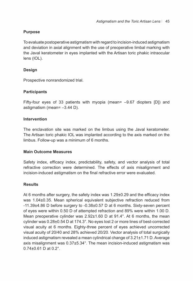
Astigmatism and the Toric Artisan Lens 45
Purpose
To evaluate postoperative astigmatism with regard to incision-induced astigmatism and deviation in axial alignment with the use of preoperative limbal marking with the Javal keratometer in eyes implanted with the Artisan toric phakic intraocular lens (IOL).
Design
Prospective nonrandomized trial.
Participants
Fifty-four eyes of 33 patients with myopia (mean= –9.67 diopters [D]) and astigmatism (mean= –3.44 D).
Intervention
The enclavation site was marked on the limbus using the Javal keratometer. The Artisan toric phakic IOL was implanted according to the axis marked on the limbus. Follow-up was a minimum of 6 months.
Main Outcome Measures
Safety index, efficacy index, predictability, safety, and vector analysis of total refractive correction were determined. The effects of axis misalignment and incision-induced astigmatism on the final refractive error were evaluated.
Results
At 6 months after surgery, the safety index was 1.29±0.29 and the efficacy index was 1.04±0.35. Mean spherical equivalent subjective refraction reduced from -11.39±4.86 D before surgery to -0.38±0.57 D at 6 months. Sixty-seven percent of eyes were within 0.50 D of attempted refraction and 89% were within 1.00 D. Mean preoperative cylinder was 2.92±1.60 D at 91.4°. At 6 months, the mean cylinder was 0.28±0.54 D at 174.3°. No eyes lost 2 or more lines of best-corrected visual acuity at 6 months. Eighty-three percent of eyes achieved uncorrected visual acuity of 20/40 and 28% achieved 20/20. Vector analysis of total surgically induced astigmatism revealed a mean cylindrical change of 3.21±1.71 D. Average axis misalignment was 0.37±5.34°. The mean incision-induced astigmatism was 0.74±0.61 D at 0.2°.

Chapter 246
Conclusions
Implantation of the myopic toric IOL leads to safe, efficacious, and predictable results. The level of unpredictability caused by minor axis IOL misalignment has minimal effects on the residual refractive error. The procedure of axis alignment with the Javal keratometer seems to be an accurate method of marking the eye for toric IOL implantation. Incision-induced astigmatism can result in an overcorrection of the cylinder. A systematic undercorrection of -0.50 D for attempted cylindrical outcome could result in an achieved correction closer to emmetropia.
Astigmatism and the Toric Artisan Lens 47
Achieving emmetropia or other desired refractions is challenging when spherical ametropia is combined with astigmatism. Keratorefractive procedures with an excimer laser have proven to be accurate for the treatment of mild to moderate myopia combined with astigmatism1. Among higher refractive errors, however, such procedures can lead to flap complications and irreversible weakening of the cornea and problems associated with small optical treatment zones2,3. Over the last few years, studies on diverse phakic intraocular lenses (IOLs) have demonstrated satisfactory results in the correction of high ametropia4-6.
The Artisan toric phakic IOL (Ophtec, Groningen, The Netherlands) can be used for the combination of ametropia and astigmatism. It is an iris-fixated anterior chamber implant of Perspex CQ-UV polymethyl methacrylate with ultraviolet filtration (Ophtec). Its overall diameter is 8.5 mm with an optical zone diameter of 5.00 mm. The myopic toric Artisan IOL is available in half-diopter (D) increments with a cylindrical power up to 7.5 D and a spherical power from –3.0 to –23.5 D. Two models of toric phakic IOLs are available to allow lens insertion on the correct axis through a superior or temporal incision, according to the surgeon preference. In models A and B, the axis runs through the claws at 0° and 90°, respectively.
Several incision types can be used: corneal, corneoscleral, limbal, or scleral tunnel incisions5. Several studies published recently on the outcome of the toric phakic IOL have demonstrated satisfactory results7-10. The Artisan lens also has been shown to be rotationally stable8,11. However, a drawback of the lens is that it requires an incision of 5.2 to 5.5 mm. This incision can induce corneal astigmatism.
Precise enclavation of the lens is paramount. Especially in higher degrees of astigmatism, minimal misalignment greatly reduces the corrective value of the lens. Approximately one third of the cylindrical correction is lost if the IOL is rotated 10° off the axis12. Some surgeons mark the intended axis of enclavation on the iris with the argon or yytrium–aluminum–garnet laser7. Although this procedure seems to be accurate, laser burns can cause inflammatory reactions, iris bleeding, or endothelial cell loss13,14. Because laser burns must be placed at least 1 week before lens implantation, they also create logistical problems. Furthermore, laser burns may disappear into the iris folds after enclavation, which preclude the evaluation of accurate lens position. Other surgeons use a digital image system in which the axis is projected on the iris. This image can be used during surgery to find the correct lens position. At our center, the intended position of enclavation is marked on the corneal limbus using the Javal keratometer (Haag Streit, Bern, Switzerland) directly before anesthesia.
In this single-center prospective study, we evaluated the safety, efficacy, and predictability of the toric Artisan myopia phakic IOL. We further assessed the
Chapter 248
influence of accurate axis lens placement using corneal markings with the Javal keratometer, and studied the effect of astigmatism induced by corneoscleral incisions on the total surgically induced refraction.
Patients and Methods
Fifty-four consecutive eyes of 33 patients receiving toric phakic IOLs were enrolled in this prospective study. Lens implantations were performed by one surgeon (GL) between January 2000 and January 2004. Inclusion criteria consisted of the following: (1) general good health; (2) a minimum of 18 years of age; (3) stable refraction for a minimum of 1 year; (4) astigmatism more than 1.5 D combined with myopia; (5) absence of ocular pathological features; (6) endothelial cell count more than 2000 cells/mm2; (7) anterior chamber depth more than 3.0 mm (including corneal thickness); (8) mesopic pupil size limited to 5.0 mm or less, although larger pupils were included after informing the patients about the increased risk of haloes and glare. Institutional ethics committee approval and informed consent in accordance with the Helsinki Declaration was obtained for each patient.
Before preoperative examination, patients were requested to discontinue contact lens wear for a minimum of 14 days to avoid the possibility of contact lens-induced corneal warpage. The examination included best spectacle-corrected visual acuity (BSCVA) in Snellen notation, slit-lamp biomicroscopy, endothelial cell count (Topcon SP-2000-P), keratometry (autokeratometer, Topcon KR 7000P), A-scan immersion biometry, applanation tonometry, measurement of mesopic pupil diameter (Colvard pupillometer), and indirect ophthalmoscopy. Furthermore, objective refraction was measured with cyclopentolate hydrochlorate 1.0% eyedrops to exclude any accommodative error in subjective refraction. If large differences were found between the 2 refractive errors, subjective refraction was measured again and used to calculate the power of the lens. The power of the IOL, including the intended axis of enclavation, was calculated according to the Van der Heijde formula15. Model A was implanted in 53 eyes and model B in 1 eye.
When subjective and corneal astigmatism coincided, intended axes were marked before surgery onto the corneal limbus with a surgical marker guided by the reflected images of the Javal keratometer. If the subjective and corneal astigmatism differed, the cornea was marked on the basis of the subjective measurements, using the reflected image as a reference point. Myotic drops (pilocarpine 4%) were administered to prepare the iris for lens fixation. Surgery was performed with retrobulbar anesthesia (41 eyes of 26 patients) or general anesthesia (13 eyes of 7 patients), according to the patient needs.
Astigmatism and the Toric Artisan Lens 49
A corneoscleral bevelled incision of 5.5 mm was made at the steep meridian and 2 paracenteses were placed 8 mm apart at either side. The anterior chamber was opened and filled with viscoelastic fluid (Healon, AMO, Santa Ana, CA) to maintain its depth and to protect the endothelium. After introduction of the lens into the anterior chamber with holding forceps (Ophtec REF D02-70), it was positioned onto the desired axis and then fixed onto the midperipheral iris stroma with a disposable enclavation needle. A slit iridotomy was performed at approximately 12 o’clock to prevent pupillary block glaucoma; thereafter, the viscoelastic material was irrigated manually5. The incision was closed with a 10-0 nylon running suture. Tobramycin 0.3% eye ointment was administered once directly after surgery. Postoperative treatment included ketorolac and dexamethasone 0.1% eyedrops 4 times daily for 4 weeks. If both eyes were to be operated on, the interventions were separated by a minimum of 2 weeks.
Follow-up examinations were scheduled at 1 day, 1 week, 1 month, 2 months, 6 months, and 1 year after surgery, and on a yearly basis thereafter. Postoperative examinations included slitlamp biomicroscopy, endothelial cell count (from 6 months after surgery), keratometry, applanation tonometry, subjective and objective refraction, uncorrected visual acuity (UCVA) and BSCVA. Within the first 6 postoperative weeks, the suture was dissected or removed if it created undesirable corneal astigmatism. After 6 weeks, the suture was removed if it caused discomfort or had loosened. At the 1-month follow-up, the Javal keratometer was used to determine the postoperative IOL axis alignment. Axis misalignment was defined as the difference between intended and achieved axis. The postoperative IOL position was measured by paraxial illuminating the IOL and projecting the Javal reflections between the claws of the IOL. This was done without prior knowledge of the intended axis of implantation (MB). Furthermore, patients were asked if they experienced haloes or glare. All data were collected prospectively from patient charts.
Statistical Analysis
To analyze BSCVA, UCVA, safety index (mean postoperative BSCVA / mean preoperative BSCVA), and efficacy index (mean postoperative UCVA / mean preoperative BSCVA), Snellen visual acuity first was converted into logarithm of the minimum angle of resolution notation to calculate the mean and then transformed back to the geometric mean Snellen visual acuity. Refraction was calculated with vector analysis12. The astigmatism vector levels were estimated using the mixed model analysis of variance (SAS software; SAS Institute, Cary, NC). The model accounts for a possible inclusion of 2 eyes of 1 patient.
Cylindrical refractions were transformed into double-angle vectors and rectangular
Chapter 250
coordinates as described by Holladay et al12. The double-angle vector plots chart the cylinders horizontally (parallel, x-coordinates) and vertically (orthogonal, y-coordinates). After calculation, the horizontal and vertical components were transformed back to cylindrical notation. Total surgically induced refractive change in astigmatism was calculated with the use of vector analysis using cylindrical subjective refraction results12.
Incision-induced astigmatism was defined as the vector of the change that occurred based on preoperative and postoperative keratometry values. This was calculated by first averaging the incision-induced astigmatism of each individual eye after 2 months to correct for measurement errors. In this analysis, eyes in which additional operations were performed after implantation were excluded. Furthermore, the single eye with a model B lens implantation also was excluded, because the incision was on the flat axis.
Comparison of data between preoperative and postoperative periods were performed with the Student t test for paired data using a level of significance of P=0.05. Changes between preoperative and postoperative periods and differences between postoperative periods also were analyzed using mixed-model analysis of variance using a level of significance of P=0.05.
Results
Patient Population
All 54 eyes of 33 patients were followed-up for a minimum of 6 months. At 1 year, follow-up data were available for 45 eyes of 27 patients, and at 2 years, data were available for 20 eyes of 14 patients. Mean follow-up was 17.1±11.4 months. Twenty-three of the 33 patients were female (69.7%). Mean age was 39.5±2.0 years (range, 19–57 years). Average axial length was 27.34±0.27 mm (range, 23.72–32.54 mm), and average anterior chamber depth was 3.66±0.31 mm (range, 3.18–4.32 mm). Mesopic pupil diameter averaged 4.7±0.9 mm (range, 3.0 –7.0 mm).
Visual Acuity and Refraction
Preoperative refractive measurements, along with the postoperative spherical equivalent of subjective refraction, UCVA, BSCVA, and percentages of eyes within ±1.00 D or ±0.50 D of emmetropia at 6 months, 1 year, and 2 years are presented in Table 1. The deviation of the achieved spherical equivalent correction from attempted spherical equivalent correction at 6 months is presented in Figure 1.
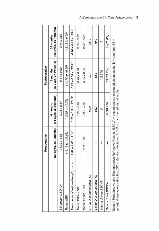
Astigmatism and the Toric Artisan Lens 51
Tabl
e 1:
Pre
oper
ativ
e an
d P
osto
pera
tive
Ref
ract
ive
Res
ults
: BS
CVA
= b
est s
pect
acle
cor
rect
ed v
isua
l acu
ity; D
= d
iopt
ers;
SE
=
sphe
rical
equ
ival
ent r
efra
ctio
n; S
D =
sta
ndar
d de
viat
ion;
UC
VA =
unc
orre
cted
vis
ual a
cuity
.
P
reop
erat
ive
Pos
tope
rativ
e
(54
Eye
s, 3
3 P
atie
nts)
6
mon
ths
(54
Eye
s, 3
3 P
atie
nts)
12
mon
ths
(45
Eye
s, 2
7 P
atie
nts)
24
mon
ths
(20
Eye
s, 1
4 P
atie
nts)
SE
(mea
n) ±
SD
(D)
−11.
39 ±
4.8
6 −0
.38
± 0.
57
−0.4
4 ±
0.62
−0
.44
± 0.
51
Ran
ge (S
E)
(−2.
13 to
−25
.63)
(−
2.25
to +
0.75
) (−
2.75
to +
0.50
) (−
2.13
to 0
.00)
Mea
n ve
ctor
ial a
stig
mat
ism
(D) ×
axi
s 2.
92 ±
1.6
0 ×
91.4
° 0.
28 ±
0.5
4 ×
174.
3°
0.23
± 0
.54
× 17
3.9°
0.
26 ±
0.4
3 ×
175.
4°
Mea
n U
CV
A ±
SD
0.72
± 0
.28
0.70
± 0
.28
0.75
± 0
.26
Mea
n B
SC
VA
± S
D
0.71
± 0
.23
0.88
± 0
.23
0.85
± 0
.25
0.94
± 0
.25
± 1.
00 D
of e
mm
etro
pia
(%)
—
88.9
90
.7
95.0
± 0.
50 D
of e
mm
etro
pia
(%)
—
66.7
65
.1
70.0
Loss
≥ 2
line
s B
SC
VA
—
0
1 (2
.2%
) 0
Gai
n ≥
1 li
ne B
SC
VA
—
40
(74.
1%)
33 (7
3.3%
) 14
(70.
0%)
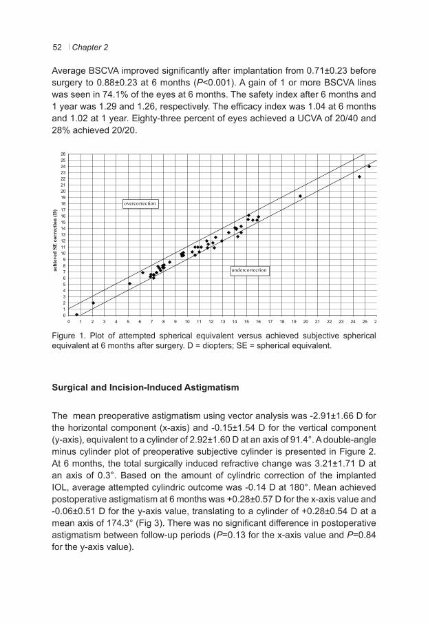
Chapter 252
Average BSCVA improved significantly after implantation from 0.71±0.23 before surgery to 0.88±0.23 at 6 months (P<0.001). A gain of 1 or more BSCVA lines was seen in 74.1% of the eyes at 6 months. The safety index after 6 months and 1 year was 1.29 and 1.26, respectively. The efficacy index was 1.04 at 6 months and 1.02 at 1 year. Eighty-three percent of eyes achieved a UCVA of 20/40 and 28% achieved 20/20.
Figure 1. Plot of attempted spherical equivalent versus achieved subjective spherical equivalent at 6 months after surgery. D = diopters; SE = spherical equivalent.
Surgical and Incision-Induced Astigmatism
The mean preoperative astigmatism using vector analysis was -2.91±1.66 D for the horizontal component (x-axis) and -0.15±1.54 D for the vertical component (y-axis), equivalent to a cylinder of 2.92±1.60 D at an axis of 91.4°. A double-angle minus cylinder plot of preoperative subjective cylinder is presented in Figure 2. At 6 months, the total surgically induced refractive change was 3.21±1.71 D at an axis of 0.3°. Based on the amount of cylindric correction of the implanted IOL, average attempted cylindric outcome was -0.14 D at 180°. Mean achieved postoperative astigmatism at 6 months was +0.28±0.57 D for the x-axis value and -0.06±0.51 D for the y-axis value, translating to a cylinder of +0.28±0.54 D at a mean axis of 174.3° (Fig 3). There was no significant difference in postoperative astigmatism between follow-up periods (P=0.13 for the x-axis value and P=0.84 for the y-axis value).
01234567891011121314151617181920212223242526
0 1 2 3 4 5 6 7 8 9 10 11 12 13 14 15 16 17 18 19 20 21 22 23 24 25 26
attempted SE correction (D)
ach
ieve
d SE
cor
rect
ion
(D)
undercorrection
overcorrection
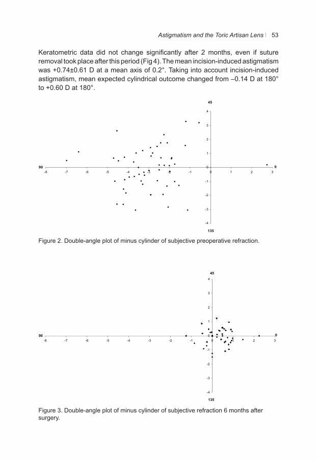
Astigmatism and the Toric Artisan Lens 53
Keratometric data did not change significantly after 2 months, even if suture removal took place after this period (Fig 4). The mean incision-induced astigmatism was +0.74±0.61 D at a mean axis of 0.2°. Taking into account incision-induced astigmatism, mean expected cylindrical outcome changed from –0.14 D at 180° to +0.60 D at 180°.
Figure 2. Double-angle plot of minus cylinder of subjective preoperative refraction.
Figure 3. Double-angle plot of minus cylinder of subjective refraction 6 months after surgery.
45
-4
-3
-2
-1
0
1
2
3
4
-8 -7 -6 -5 -4 -3 -2 -1 0 1 2 3
135
90 0
45
-4
-3
-2
-1
0
1
2
3
4
-8 -7 -6 -5 -4 -3 -2 -1 0 1 2 3
135
90 0
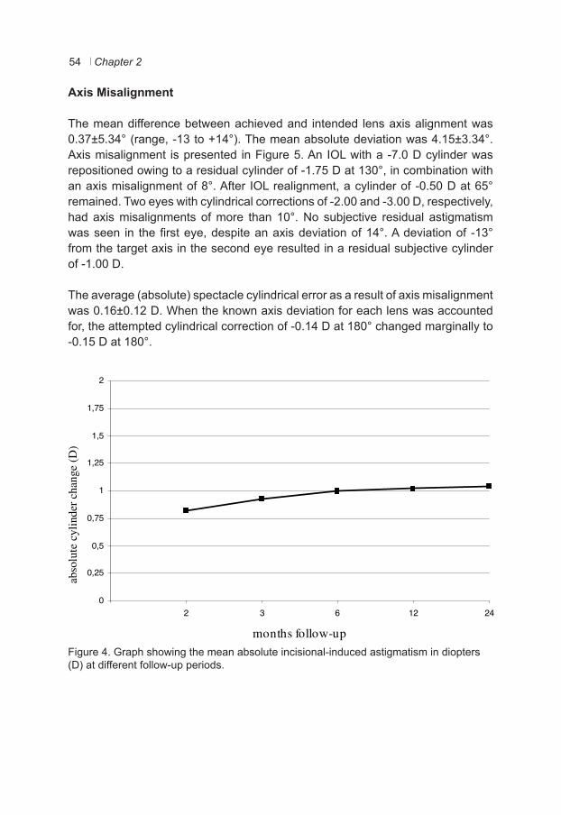
Chapter 254
Axis Misalignment
The mean difference between achieved and intended lens axis alignment was 0.37±5.34° (range, -13 to +14°). The mean absolute deviation was 4.15±3.34°. Axis misalignment is presented in Figure 5. An IOL with a -7.0 D cylinder was repositioned owing to a residual cylinder of -1.75 D at 130°, in combination with an axis misalignment of 8°. After IOL realignment, a cylinder of -0.50 D at 65° remained. Two eyes with cylindrical corrections of -2.00 and -3.00 D, respectively, had axis misalignments of more than 10°. No subjective residual astigmatism was seen in the first eye, despite an axis deviation of 14°. A deviation of -13° from the target axis in the second eye resulted in a residual subjective cylinder of -1.00 D.
The average (absolute) spectacle cylindrical error as a result of axis misalignment was 0.16±0.12 D. When the known axis deviation for each lens was accounted for, the attempted cylindrical correction of -0.14 D at 180° changed marginally to -0.15 D at 180°.
Figure 4. Graph showing the mean absolute incisional-induced astigmatism in diopters (D) at different follow-up periods.
0
0,25
0,5
0,75
1
1,25
1,5
1,75
2
2 3 6 12 24
……
..
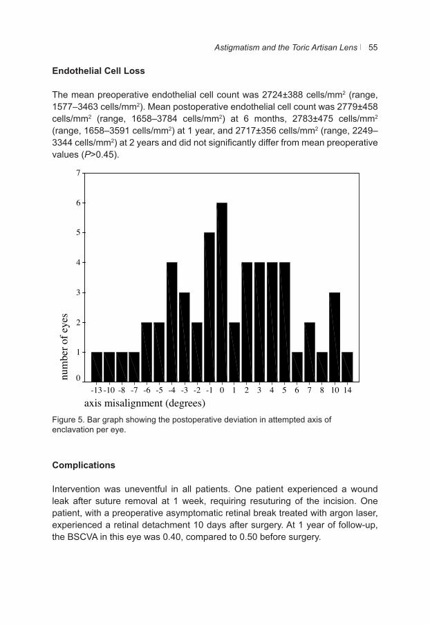
Astigmatism and the Toric Artisan Lens 55
Endothelial Cell Loss
The mean preoperative endothelial cell count was 2724±388 cells/mm2 (range, 1577–3463 cells/mm2). Mean postoperative endothelial cell count was 2779±458 cells/mm2 (range, 1658–3784 cells/mm2) at 6 months, 2783±475 cells/mm2 (range, 1658–3591 cells/mm2) at 1 year, and 2717±356 cells/mm2 (range, 2249–3344 cells/mm2) at 2 years and did not significantly differ from mean preoperative values (P>0.45).
Figure 5. Bar graph showing the postoperative deviation in attempted axis of enclavation per eye.
Complications
Intervention was uneventful in all patients. One patient experienced a wound leak after suture removal at 1 week, requiring resuturing of the incision. One patient, with a preoperative asymptomatic retinal break treated with argon laser, experienced a retinal detachment 10 days after surgery. At 1 year of follow-up, the BSCVA in this eye was 0.40, compared to 0.50 before surgery.
axis misalignment (degrees)1410876543210-1-2-3-4-5-6-7-8-10-13
num
ber o
f eye
s
7
6
5
4
3
2
1
0
Chapter 256
The mean preoperative intraocular pressure was 15.3±3.4 mmHg. Seven eyes experienced a temporary intraocular pressure higher than 21 mmHg within the first month of surgery (range, 22–30 mmHg), although this normalized in all eyes after discontinuing topical corticosteroids. The mean postoperative intraocular pressure (15.7±3.4 mmHg) did not differ significantly from preoperative values (P>0.25).
One eye in the study experienced a significant loss of lines after cataract developed 1 year after surgery. Visual acuity decreased from 0.4 before surgery to 0.2 after surgery. We are not aware of any surgically induced reason for the development of the cataract.
No pigment dispersion or pupillary block occurred in any eye during follow-up. Seven of the 33 patients noted having more difficulty with haloes or glare. One of these patients had a mesopic pupil size larger than 5 mm. All patients were satisfied with the outcome of surgery. No patient considered removal of the lens.
Discussion
The safety, efficacy, and predictability of phakic toric Artisan lens implantation for the correction of myopia and astigmatism in this study were high. Although the toric Artisan lens was investigated in a large European study, the effect of axis misalignment and incision-induced astigmatism on the final outcome was not analyzed7. Based on our calculations, mean intended astigmatism correction ought to have been -0.14 D. However, our study demonstrated a slight overcorrection of +0.28 D. By analyzing the effect of incision-induced astigmatism, we found that an against-the-rule astigmatism of 0.74 D could be introduced by making a 5.5 mm incision on the steep axis. This induced astigmatism also can result after cataract surgery16,17. Taking the latter into account, the expected cylinder outcome should have been a 0.60 D overcorrection. Instead, we observed an overcorrection of only 0.28 D. To evaluate if this discrepancy was a result of axis misalignment, we also analyzed its effect on the refractive outcome. We found an absolute axis deviation of 4.15±3.34°, comparable with the results of Tehrani et al8. Because rotation of the lens has not been observed as a result of its firm fixation onto the midperipheral iris stroma, axial misplacement of an iris-claw lens must be caused by incorrect alignment of the lens during the surgical procedure. Because proper surgical alignment of the IOL is a prerequisite for the success of toric phakic IOL implantation, a precise method of axis marking is critical. Although most surgeons use preoperative laser iridotomies to mark the axis, we used limbal marking with the Javal keratometer in this study. This method does not incur the risk of intraocular inflammation and also can be practical, because
Astigmatism and the Toric Artisan Lens 57
marking can take place immediately before surgery, avoiding extra patient visits. One disadvantage of this method, however, is that the markings can fade or disappear during preoperative preparation of the eye. We encountered this in 1 eye, resulting in an enclavation 14° from the intended axis of implantation. Another lens required realignment after developing a subjective postoperative cylinder of -1.75 D. The effect of the misalignment on the final cylindrical outcome, however, was minimal. The mean attempted cylindrical outcome changed marginally from -0.14 D at 180° to -0.15 D at 180° when the known axis error for each lens was accounted for. Both the incision-induced astigmatism and axis misalignment could not fully explain the discrepancy between the expected and achieved cylindrical corrections. Fixed factors such as the lens only being available in half-diopter increments and a calibration error of ±0.3 D of the lens itself could have influenced the achieved correction. Also, our data are based on subjective refraction only. Our impression is that patients tend to experience less astigmatism than was objectively observed10. We do not have a good explanation for this interesting phenomenon.
Refractive results in this study however, were predictable and efficacious and resembled those achieved by LASIK in lower degrees of myopia and astigmatism1. Approximately 90% of the eyes in our study were within 1.00 D of emmetropia. These data compare favorably those of LASIK for moderate to high myopia and astigmatism, where studies report 41% to 76% of eyes being within the same range1,18–22. LASIK, however, tends to result in lower predictability among higher refractive errors23,24. The large amount of stromal tissue ablated with the excimer laser in these higher degrees of myopia and astigmatism also predisposes eyes to corneal ectasia and associated visual problems25. Clear lens extraction with toric IOL implantation is another option for such eyes. Studies have shown that clear lens extractions carry a higher risk of retinal detachment and further result in the loss of accommodation in younger patients26. Moreover, rotational stability may be a problem with toric IOLs in the capsular bag27. Compared with LASIK or clear lens extraction, the correction of moderate to high myopia with astigmatism seems to be safer and more predictable with the implantation of phakic IOLs. Furthermore, unlike excimer laser procedures, the eye is not affected by the amount of refractive correction. Posterior chamber IOL implantation requires a smaller incision (3.2 mm) compared with the toric Artisan lens (5.5 mm)28
–30. However, reports on phakic toric posterior chamber IOLs for the correction of myopia combined with astigmatism currently are limited31. The potential for cataractogenesis and pigment dispersion with posterior chamber IOLs is also a crucial long-term concern, as is the potential for postoperative lens rotation27,32,33. One patient in this study also experienced a cataract, although it is unclear if this was the result of the intraocular surgery.
Chapter 258
In our study, 70% or more of the cases exhibited a gain of 1 or more lines of BSCVA. Such improvements in visual acuity also have been reported in other studies and have been attributed to the increase in the size of the retinal image compared with spectacle correction34–36.
Successful correction of myopia and astigmatism with a toric phakic IOL depends on several variables. In this study, we report not only the accuracy of the refractive results, but also the refractive contribution of the deviation between intended and achieved axis of implantation and the input of incision-induced astigmatism. We believe that understanding these parameters will allow greater insight into toric phakic IOL implantation.
In conclusion, toric myopic Artisan lens implantation leads to highly predictable, effective, and safe results. Marking the enclavation site for toric phakic IOL implantation with the use of the Javal keratometer seems to be a safe and reliable method. Accounting for incision-induced astigmatism could increase predictability further. To compensate for this, a systematic undercorrection of -0.50 D for attempted cylindric outcome is advised when using a corneoscleral incision of 5.5 mm.
Astigmatism and the Toric Artisan Lens 59
References
1. Sugar A, Rapuano CJ, Culbertson WW, et al. Laser in situ keratomileusis for myopia and astigmatism: safety and efficacy: a report by the American Academy of Ophthalmology. Ophthalmology 2002;109:175-87.
2. Seiler T, Koufala K, Richter G. Iatrogenic keratectasia after laser in situ keratomileusis. J Refract Surg 1998;14:312-7.
3. Seiler T, Holschbach A, Derse M, et al. Complications of myopic photorefractive keratectomy with the excimer laser. Ophthalmology 1994;101:153-60.
4. Landesz M, van Rij G, Luyten G. Iris-claw phakic intraocular lens for high myopia. J Refract Surg 2001;17:634-40.
5. Budo C, Hessloehl JC, Izak M, et al. Multicenter study of the Artisan phakic intraocular lens. J Cataract Refract Surg 2000;26:1163-71.
6. Saxena R, Landesz M, Noordzij B, et al. Three-year follow-up of the Artisan phakic intraocular lens for hypermetropia. Ophthalmology 2003;110:1391-5.
7. Dick HB, Alio J, Bianchetti M, et al. Toric phakic intraocular lens: European multicenter study. Ophthalmology 2003;110:150-62.
8. Tehrani M, Dick HB, Schwenn O, et al. Postoperative astigmatism and rotational stability after artisan toric phakic intraocular lens implantation. J Cataract Refract Surg 2003;29:1761-6.
9. Guell JL, Vazquez M, Malecaze F, et al. Artisan toric phakic intraocular lens for the correction of high astigmatism. Am J Ophthalmol 2003;136:442-7.
10. Bartels MC, van Rij G, Luyten GP. Implantation of a toric phakic intraocular lens to correct high corneal astigmatism in a patient with bilateral marginal corneal degeneration. J Cataract Refract Surg 2004;30:499-502.
11. Baumeister M, Buhren J, Kohnen T. Position of angle-supported, iris-fixated, and ciliary sulcus-implanted myopic phakic intraocular lenses evaluated by Scheimpflug photography. Am J Ophthalmol 2004;138:723-31.
12. Holladay JT, Moran JR, Kezirian GM. Analysis of aggregate surgically induced refractive change, prediction error, and intraocular astigmatism. J Cataract Refract Surg 2001;27:61-79.
13. Robin AL, Pollack IP. A comparison of neodymium: YAG and argon laser iridotomies. Ophthalmology 1984;91:1011-6.
14. Pollack IP, Robin AL, Dragon DM, et al. Use of the neodymium:YAG laser to create iridotomies in monkeys and humans. Trans Am Ophthalmol Soc 1984;82:307-28.
15. van der Heijde GL, Fechner PU, Worst JG. [Optical consequences of implantation of a negative intraocular lens in myopic patients]. Klin Monatsbl Augenheilkd. 1988;193:99-102.
16. Lundstrom M, Barry P, Leite E, et al. 1998 European cataract outcome study report from the european cataract outcome study group. J Cataract Refract Surg 2001;27:1176-84.
17. Kohnen T, Koch DD. Methods to control astigmatism in cataract surgery. Curr Opin Ophthalmol 1996;7:75-80.
18. Hersh PS, Brint SF, Maloney RK, et al. Photorefractive keratectomy versus laser in situ keratomileusis for moderate to high myopia. A randomized prospective study. Ophthalmology 1998;105:1512-22, discussion 22-3.
19. Steinert RF, Hersh PS. Spherical and aspherical photorefractive keratectomy and laser in-situ keratomileusis for moderate to high myopia: two prospective, randomized clinical trials. Summit technology PRK-LASIK study group. Trans Am Ophthalmol Soc 1998;96:197-221; discussion 21-7.
20. Sanders DR, Vukich JA. Comparison of implantable contact lens and laser assisted in situ keratomileusis for moderate to high myopia. Cornea 2003;22:324-31.
21. FDA. Premarket Approval Application. Kremer excimer laser system serial number KEZ 940202 for laser in situ keratomileusis (LASIK) for the correction of primary myopia with or without astigmatism. Available at: http://www.fda.gov/cdrh/pdf/970005.html. Accessed October 24, 2005.
22. FDA. Premarket Approval Application. Nidek EC-5000 excimer laser system. Available at: http://www.fda.gov/cdrh/pdf/p970053s002.html. Accessed October 24,2005.
Chapter 260
23. Perez-Santonja JJ, Bellot J, Claramonte P, et al. Laser in situ keratomileusis to correct high myopia. J Cataract Refract Surg 1997;23:372-85.
24. Guell JL, Muller A. Laser in situ keratomileusis (LASIK) for myopia from -7 to -18 diopters. J Refract Surg 1996;12:222-8.
25. Pallikaris IG, Kymionis GD, Astyrakakis NI. Corneal ectasia induced by laser in situ keratomileusis. J Cataract Refract Surg 2001;27:1796-802.
26. Arne JL. Phakic intraocular lens implantation versus clear lens extraction in highly myopic eyes of 30- to 50-year-old patients. J Cataract Refract Surg 2004;30:2092-6.
27. Gerten G, Michels A, Olmes A. [Toric intraocular lenses. Clinical results and rotational stability]. Ophthalmologe 2001;98:715-20.
28. Lackner B, Pieh S, Schmidinger G, et al. Outcome after treatment of ametropia with implantable contact lenses. Ophthalmology 2003;110:2153-61.
29. Pesando PM, Ghiringhello MP, Tagliavacche P. Posterior chamber collamer phakic intraocular lens for myopia and hyperopia. J Refract Surg 1999;15:415-23.
30. Sanders DR, Doney K, Poco M. United States Food and Drug Administration clinical trial of the Implantable Collamer Lens (ICL) for moderate to high myopia: three-year follow-up. Ophthalmology 2004;111:1683-92.
31. Gimbel HV, Ziemba SL. Management of myopic astigmatism with phakic intraocular lens implantation. J Cataract Refract Surg 2002;28:883-6.
32. Fink AM, Gore C, Rosen E. Cataract development after implantation of the Staar Collamer posterior chamber phakic lens. J Cataract Refract Surg 1999;25:278-82.
33. Brandt JD, Mockovak ME, Chayet A. Pigmentary dispersion syndrome induced by a posterior chamber phakic refractive lens. Am J Ophthalmol 2001;131:260-3.
34. Guell JL, Vazquez M, Gris O. Adjustable refractive surgery: 6-mm Artisan lens plus laser in situ keratomileusis for the correction of high myopia. Ophthalmology 2001;108:945-52.
35. Fechner PU, Haubitz I, Wichmann W, et al. Worst-Fechner biconcave minus power phakic iris-claw lens. J Refract Surg 1999;15:93-105.
36. Applegate RA, Howland HC. Magnification and visual acuity in refractive surgery. Arch Ophthalmol 1993;111:1335-42.

3Chapter 3
Three-year Follow-up of the Artisan Phakic Intraocular Lens for Hypermetropia
R Saxena MA Landesz B Noordzij BGPM Luyten G
Ophthalmology. 2003 Apr;110(7):1391-1395

Artisan for Hypermetropia 63
Purpose
We report the postoperative results of the Artisan Hyperopia phakic intraocular lens.
Design
Prospective, nonrandomized trial.
Participants
Twenty-six eyes of 13 self-selected patients with refractive error ranging from +3.00 to +11.00 diopters (D).
Intervention
Patients with hypermetropia were implanted with the Artisan Hyperopia phakic IOL. Mean follow-up was 22.4 months (range 3–36 months).
Main outcome measures
Predictability, stability, efficacy, loss of best spectacle-corrected visual acuity, and complications.
Results
At six months, 90.9% (20 of 22 eyes) were ±1.00 D of intended correction and 81.8% (18 eyes) were ±1.00 D of emmetropia. The mean spherical equivalent was stable within 0.25 D during the entire 3-year follow-up period. Twenty-four eyes (92.3%) had a postoperative best spectacle-corrected visual acuity of 0.50 or better at all of their individual follow-up examinations. No patient lost 2 or more lines after the procedure. There was a significant negative correlation between anterior chamber depth and endothelial cell loss. Two patients experienced posterior synechiae with pigment deposits in both eyes. One of these patients had convex irides and underwent implant removal within 2 years with a consequent clear lens extraction and posterior chamber lens implantation.
Conclusions
Implantation of the Artisan Hyperopic lens leads to accurate and stable refractive results with no significant loss of vision. More attention should be paid to convex irides and shallow anterior chambers during the preoperative screening to avoid unnecessary complications.
Chapter 364
High hyperopia has proven to be a challenge to correct. Procedures with the excimer laser have shown great potential, although many authors find that they are only safe in cases of low to moderate hyperopia1-5. Thanks to the introduction of phakic intraocular lenses (IOL), however, correcting higher degrees of farsightedness has now become possible.
Phakic IOLs can be an attractive alternative to other types of refractive interventions, such as refractive keratectomy, LASIK, and photorefractive keratectomy. Studies on diverse phakic IOLs have demonstrated that visual results are stable and predictable6-11. The Food and Drug Administration currently is undergoing clinical trials for several different types of hyperopic and myopic phakic IOLs (unpublished), although some of these lenses are already being used in clinical settings outside the United States6-8,12.
One of these phakic IOLs is the Artisan Myopia lens (model 203W; Ophtec, Groningen, The Netherlands), first used clinically in 1986. The competency of this lens was scrutinized during the European multicenter study, with refractive errors ranging from −5.00 to −20.00 diopters (D) 6. The authors concluded that the lens was safe, stable, efficacious and predictable. The first clinical implant of the Artisan Hyperopia lens took place in 1992. In this single-center investigation, we analyzed the predictability, stability, and visual acuity results of this phakic IOL, as well as its possible effect on the corneal endothelium and its rate of complications in hyperopic patients.
Materials and Methods
Intraocular Lens
The Artisan Hyperopia lens is an iris-fixated anterior chamber implant made of ultraviolet-absorbing clinical quality polymethyl methacrylate, Perspex CQ-UV. The lens is vaulted to allow maximum clearance between itself and the cornea and the crystalline lens. It has a 5-mm diameter optic with a total length of 8.5 mm and an absolute height of 1.00 mm. It is available in half-diopter increments between +1.00 and +12.00 D.
Patients
Twenty-six eyes of 13 patients (5 female and 8 male) were fitted with the Artisan Hyperopia lens implant between May 1996 and June 2000 at Erasmus MC in Rotterdam, The Netherlands. Patient age ranged from 28 to 59.5 years,
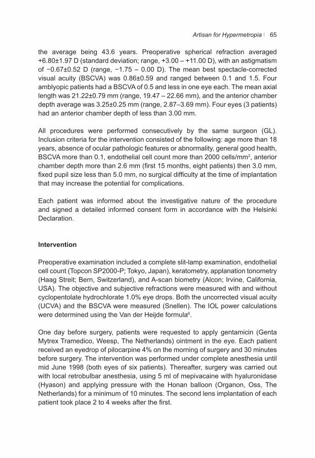
Artisan for Hypermetropia 65
the average being 43.6 years. Preoperative spherical refraction averaged +6.80±1.97 D (standard deviation; range, +3.00 – +11.00 D), with an astigmatism of −0.67±0.52 D (range, −1.75 – 0.00 D). The mean best spectacle-corrected visual acuity (BSCVA) was 0.86±0.59 and ranged between 0.1 and 1.5. Four amblyopic patients had a BSCVA of 0.5 and less in one eye each. The mean axial length was 21.22±0.79 mm (range, 19.47 – 22.66 mm), and the anterior chamber depth average was 3.25±0.25 mm (range, 2.87–3.69 mm). Four eyes (3 patients) had an anterior chamber depth of less than 3.00 mm.
All procedures were performed consecutively by the same surgeon (GL). Inclusion criteria for the intervention consisted of the following: age more than 18 years, absence of ocular pathologic features or abnormality, general good health, BSCVA more than 0.1, endothelial cell count more than 2000 cells/mm2, anterior chamber depth more than 2.6 mm (first 15 months, eight patients) then 3.0 mm, fixed pupil size less than 5.0 mm, no surgical difficulty at the time of implantation that may increase the potential for complications.
Each patient was informed about the investigative nature of the procedure and signed a detailed informed consent form in accordance with the Helsinki Declaration.
Intervention
Preoperative examination included a complete slit-lamp examination, endothelial cell count (Topcon SP2000-P; Tokyo, Japan), keratometry, applanation tonometry (Haag Streit; Bern, Switzerland), and A-scan biometry (Alcon; Irvine, California, USA). The objective and subjective refractions were measured with and without cyclopentolate hydrochlorate 1.0% eye drops. Both the uncorrected visual acuity (UCVA) and the BSCVA were measured (Snellen). The IOL power calculations were determined using the Van der Heijde formula6.
One day before surgery, patients were requested to apply gentamicin (Genta Mytrex Tramedico, Weesp, The Netherlands) ointment in the eye. Each patient received an eyedrop of pilocarpine 4% on the morning of surgery and 30 minutes before surgery. The intervention was performed under complete anesthesia until mid June 1998 (both eyes of six patients). Thereafter, surgery was carried out with local retrobulbar anesthesia, using 5 ml of mepivacaine with hyaluronidase (Hyason) and applying pressure with the Honan balloon (Organon, Oss, The Netherlands) for a minimum of 10 minutes. The second lens implantation of each patient took place 2 to 4 weeks after the first.
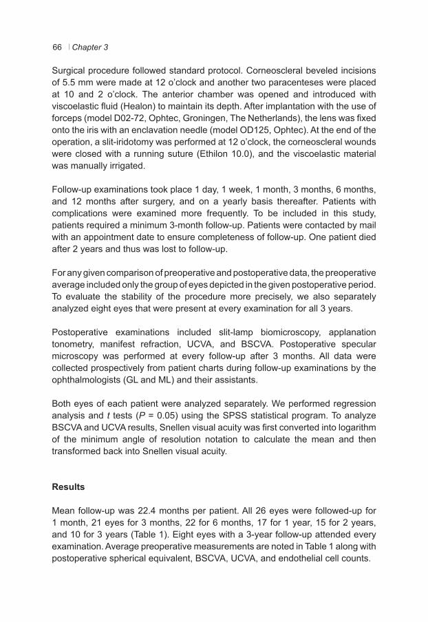
Chapter 366
Surgical procedure followed standard protocol. Corneoscleral beveled incisions of 5.5 mm were made at 12 o’clock and another two paracenteses were placed at 10 and 2 o’clock. The anterior chamber was opened and introduced with viscoelastic fluid (Healon) to maintain its depth. After implantation with the use of forceps (model D02-72, Ophtec, Groningen, The Netherlands), the lens was fixed onto the iris with an enclavation needle (model OD125, Ophtec). At the end of the operation, a slit-iridotomy was performed at 12 o’clock, the corneoscleral wounds were closed with a running suture (Ethilon 10.0), and the viscoelastic material was manually irrigated.
Follow-up examinations took place 1 day, 1 week, 1 month, 3 months, 6 months, and 12 months after surgery, and on a yearly basis thereafter. Patients with complications were examined more frequently. To be included in this study, patients required a minimum 3-month follow-up. Patients were contacted by mail with an appointment date to ensure completeness of follow-up. One patient died after 2 years and thus was lost to follow-up.
For any given comparison of preoperative and postoperative data, the preoperative average included only the group of eyes depicted in the given postoperative period. To evaluate the stability of the procedure more precisely, we also separately analyzed eight eyes that were present at every examination for all 3 years.
Postoperative examinations included slit-lamp biomicroscopy, applanation tonometry, manifest refraction, UCVA, and BSCVA. Postoperative specular microscopy was performed at every follow-up after 3 months. All data were collected prospectively from patient charts during follow-up examinations by the ophthalmologists (GL and ML) and their assistants.
Both eyes of each patient were analyzed separately. We performed regression analysis and t tests (P = 0.05) using the SPSS statistical program. To analyze BSCVA and UCVA results, Snellen visual acuity was first converted into logarithm of the minimum angle of resolution notation to calculate the mean and then transformed back into Snellen visual acuity.
Results
Mean follow-up was 22.4 months per patient. All 26 eyes were followed-up for 1 month, 21 eyes for 3 months, 22 for 6 months, 17 for 1 year, 15 for 2 years, and 10 for 3 years (Table 1). Eight eyes with a 3-year follow-up attended every examination. Average preoperative measurements are noted in Table 1 along with postoperative spherical equivalent, BSCVA, UCVA, and endothelial cell counts.
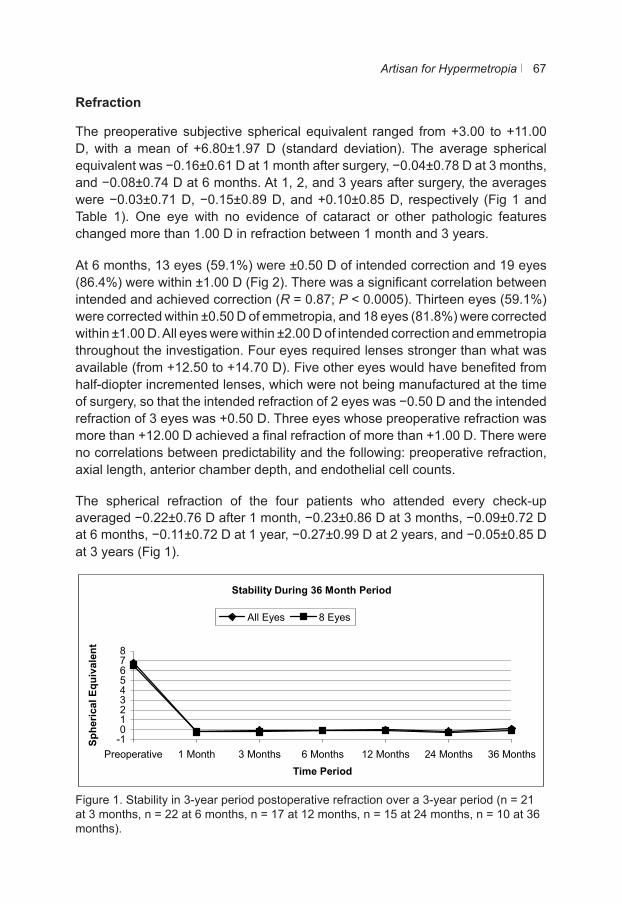
Artisan for Hypermetropia 67
Refraction
The preoperative subjective spherical equivalent ranged from +3.00 to +11.00 D, with a mean of +6.80±1.97 D (standard deviation). The average spherical equivalent was −0.16±0.61 D at 1 month after surgery, −0.04±0.78 D at 3 months, and −0.08±0.74 D at 6 months. At 1, 2, and 3 years after surgery, the averages were −0.03±0.71 D, −0.15±0.89 D, and +0.10±0.85 D, respectively (Fig 1 and Table 1). One eye with no evidence of cataract or other pathologic features changed more than 1.00 D in refraction between 1 month and 3 years.
At 6 months, 13 eyes (59.1%) were ±0.50 D of intended correction and 19 eyes (86.4%) were within ±1.00 D (Fig 2). There was a significant correlation between intended and achieved correction (R = 0.87; P < 0.0005). Thirteen eyes (59.1%) were corrected within ±0.50 D of emmetropia, and 18 eyes (81.8%) were corrected within ±1.00 D. All eyes were within ±2.00 D of intended correction and emmetropia throughout the investigation. Four eyes required lenses stronger than what was available (from +12.50 to +14.70 D). Five other eyes would have benefited from half-diopter incremented lenses, which were not being manufactured at the time of surgery, so that the intended refraction of 2 eyes was −0.50 D and the intended refraction of 3 eyes was +0.50 D. Three eyes whose preoperative refraction was more than +12.00 D achieved a final refraction of more than +1.00 D. There were no correlations between predictability and the following: preoperative refraction, axial length, anterior chamber depth, and endothelial cell counts.
The spherical refraction of the four patients who attended every check-up averaged −0.22±0.76 D after 1 month, −0.23±0.86 D at 3 months, −0.09±0.72 D at 6 months, −0.11±0.72 D at 1 year, −0.27±0.99 D at 2 years, and −0.05±0.85 D at 3 years (Fig 1).
Figure 1. Stability in 3-year period postoperative refraction over a 3-year period (n = 21 at 3 months, n = 22 at 6 months, n = 17 at 12 months, n = 15 at 24 months, n = 10 at 36 months).
Stability During 36 Month Period
-1012345678
Preoperative 1 Month 3 Months 6 Months 12 Months 24 Months 36 MonthsTime Period
Sphe
rical
Equ
ival
ent
All Eyes 8 Eyes
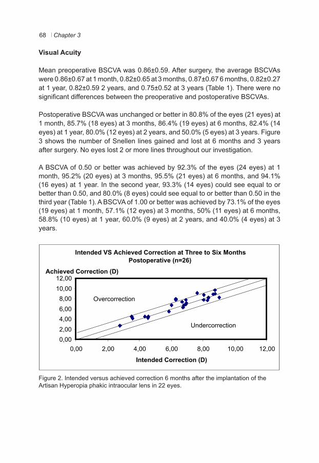
Chapter 368
Visual Acuity
Mean preoperative BSCVA was 0.86±0.59. After surgery, the average BSCVAs were 0.86±0.67 at 1 month, 0.82±0.65 at 3 months, 0.87±0.67 6 months, 0.82±0.27 at 1 year, 0.82±0.59 2 years, and 0.75±0.52 at 3 years (Table 1). There were no significant differences between the preoperative and postoperative BSCVAs.
Postoperative BSCVA was unchanged or better in 80.8% of the eyes (21 eyes) at 1 month, 85.7% (18 eyes) at 3 months, 86.4% (19 eyes) at 6 months, 82.4% (14 eyes) at 1 year, 80.0% (12 eyes) at 2 years, and 50.0% (5 eyes) at 3 years. Figure 3 shows the number of Snellen lines gained and lost at 6 months and 3 years after surgery. No eyes lost 2 or more lines throughout our investigation.
A BSCVA of 0.50 or better was achieved by 92.3% of the eyes (24 eyes) at 1 month, 95.2% (20 eyes) at 3 months, 95.5% (21 eyes) at 6 months, and 94.1% (16 eyes) at 1 year. In the second year, 93.3% (14 eyes) could see equal to or better than 0.50, and 80.0% (8 eyes) could see equal to or better than 0.50 in the third year (Table 1). A BSCVA of 1.00 or better was achieved by 73.1% of the eyes (19 eyes) at 1 month, 57.1% (12 eyes) at 3 months, 50% (11 eyes) at 6 months, 58.8% (10 eyes) at 1 year, 60.0% (9 eyes) at 2 years, and 40.0% (4 eyes) at 3 years.
Figure 2. Intended versus achieved correction 6 months after the implantation of the Artisan Hyperopia phakic intraocular lens in 22 eyes.
Intended VS Achieved Correction at Three to Six Months Postoperative (n=26)
12,00Achieved Correction (D)
0,002,004,006,008,0010,00
0,00 2,00 4,00 6,00 8,00 10,00 12,00
Intended Correction (D)
Overcorrection
Undercorrection
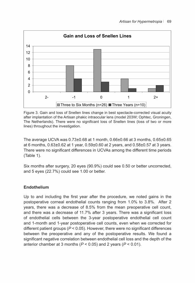
Artisan for Hypermetropia 69
Figure 3. Gain and loss of Snellen lines change in best spectacle-corrected visual acuity after implantation of the Artisan phakic intraocular lens (model 203W; Ophtec, Groningen, The Netherlands). There were no significant loss of Snellen lines (loss of two or more lines) throughout the investigation.
The average UCVA was 0.73±0.68 at 1 month, 0.66±0.66 at 3 months, 0.65±0.65 at 6 months, 0.63±0.62 at 1 year, 0.59±0.60 at 2 years, and 0.58±0.57 at 3 years. There were no significant differences in UCVAs among the different time periods (Table 1).
Six months after surgery, 20 eyes (90.9%) could see 0.50 or better uncorrected, and 5 eyes (22.7%) could see 1.00 or better.
Endothelium
Up to and including the first year after the procedure, we noted gains in the postoperative corneal endothelial counts ranging from 1.0% to 3.8%. After 2 years, there was a decrease of 8.5% from the mean preoperative cell count,and there was a decrease of 11.7% after 3 years. There was a significant loss of endothelial cells between the 3-year postoperative endothelial cell countand 1-month and 1-year postoperative cell counts, even when we corrected for different patient groups (P < 0.05). However, there were no significant differences between the preoperative and any of the postoperative results. We found a significant negative correlation between endothelial cell loss and the depth of the anterior chamber at 3 months (P < 0.05) and 2 years (P < 0.01).
Gain and Loss of Snellen Lines
0
2
4
6
8
10
12
14
2- -1 0 1 2+
Three to Six Months (n=26) Three Years (n=10)
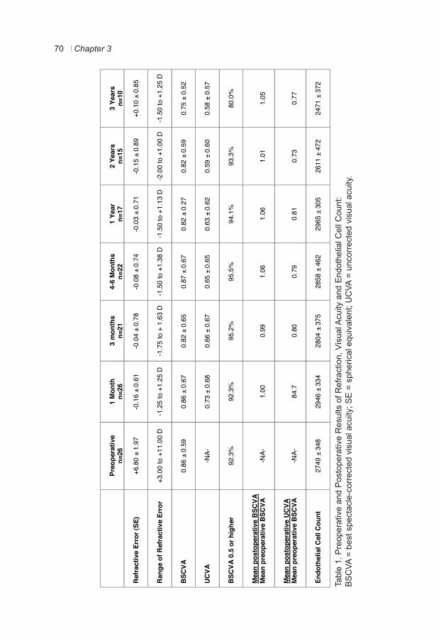
Chapter 370
Tabl
e 1.
Pre
oper
ativ
e an
d P
osto
pera
tive
Res
ults
of R
efra
ctio
n, V
isua
l Acu
ity a
nd E
ndot
helia
l Cel
l Cou
nt:
BS
CVA
= b
est s
pect
acle
-cor
rect
ed v
isua
l acu
ity; S
E =
sph
eric
al e
quiv
alen
t; U
CVA
= u
ncor
rect
ed v
isua
l acu
ity.
Preo
pera
tive
n=26
1
Mon
th
n=26
3
mon
ths
n=21
4-
6 M
onth
s n=
22
1
Year
n=
17
2
Year
s n=
15
3
Year
s n=
10
Refr
activ
e Er
ror (
SE)
+6
.80
± 1.
97
-0
.16
± 0.
61
-0
.04
± 0.
78
-0
.08
± 0.
74
-0
.03
± 0.
71
-0
.15
± 0.
89
+0
.10
± 0.
85
Rang
e of
Ref
ract
ive
Erro
r
+3
.00
to +
11.0
0 D
-1.2
5 to
+1.
25 D
-1.7
5 to
+ 1
.63
D
-1.5
0 to
+1.
38 D
-1.5
0 to
+1.
13 D
-2.0
0 to
+1.
00 D
-1.5
0 to
+1.
25 D
BSC
VA
0.
86 ±
0.5
9
0.86
± 0
.67
0.
82 ±
0.6
5
0.87
± 0
.67
0.
82 ±
0.2
7
0.82
± 0
.59
0.
75 ±
0.5
2
UCVA
-N
A-
0.
73 ±
0.6
8
0.66
± 0
.67
0.
65 ±
0.6
5
0.63
± 0
.62
0.
59 ±
0.6
0
0.58
± 0
.57
BSC
VA 0
.5 o
r hig
her
92
.3%
92
.3%
95.2
%
95
.5%
94.1
%
93
.3%
80.0
%
Mea
n po
stop
erat
ive
BSC
VA
Mea
n pr
eope
rativ
e B
SCVA
-N
A-
1.
00
0.
99
1.
06
1.
06
1.
01
1.
05
Mea
n po
stop
erat
ive
UCVA
M
ean
preo
pera
tive
BSC
VA
-N
A-
84
.7
0.
80
0.
79
0.
81
0.
73
0.
77
Endo
thel
ial C
ell C
ount
27
49 ±
348
2946
± 3
34
28
04 ±
375
2858
± 4
62
29
65 ±
305
2611
± 4
72
24
71 ±
372
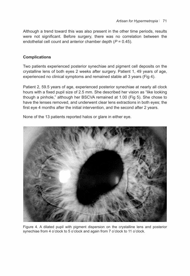
Artisan for Hypermetropia 71
Although a trend toward this was also present in the other time periods, results were not significant. Before surgery, there was no correlation between the endothelial cell count and anterior chamber depth (P = 0.45).
Complications
Two patients experienced posterior synechiae and pigment cell deposits on the crystalline lens of both eyes 2 weeks after surgery. Patient 1, 49 years of age, experienced no clinical symptoms and remained stable all 3 years (Fig 4).
Patient 2, 59.5 years of age, experienced posterior synechiae at nearly all clock hours with a fixed pupil size of 2.5 mm. She described her vision as “like looking though a pinhole,” although her BSCVA remained at 1.00 (Fig 5). She chose to have the lenses removed, and underwent clear lens extractions in both eyes; the first eye 4 months after the initial intervention, and the second after 2 years.
None of the 13 patients reported halos or glare in either eye.
Figure 4. A dilated pupil with pigment dispersion on the crystalline lens and posterior synechiae from 4 o’clock to 5 o’clock and again from 7 o’clock to 11 o’clock.
Figure 4. A dilated pupil with pigment dispersion on the crystalline lens and posterior synechiae from 4 o’clock to 5 o’clock and again from 7 o’clock to 11
o’clock.
Figure 5. Pigment dispersion on the crystalline lens and posterior synechiae
encircling the pupil. This patient subsequently underwent implant removal and a clear lens extraction and thereafter, a posterior chamber lens implantation in
both eyes.
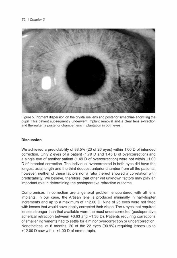
Chapter 372
Figure 4. A dilated pupil with pigment dispersion on the crystalline lens and posterior synechiae from 4 o’clock to 5 o’clock and again from 7 o’clock to 11
o’clock.
Figure 5. Pigment dispersion on the crystalline lens and posterior synechiae
encircling the pupil. This patient subsequently underwent implant removal and a clear lens extraction and thereafter, a posterior chamber lens implantation in
both eyes.
Figure 5. Pigment dispersion on the crystalline lens and posterior synechiae encircling the pupil. This patient subsequently underwent implant removal and a clear lens extraction and thereafter, a posterior chamber lens implantation in both eyes.
Discussion
We achieved a predictability of 88.5% (23 of 26 eyes) within 1.00 D of intended correction. Only 2 eyes of a patient (1.79 D and 1.45 D of overcorrection) and a single eye of another patient (1.49 D of overcorrection) were not within ±1.00 D of intended correction. The individual overcorrected in both eyes did have the longest axial length and the third deepest anterior chamber from all the patients; however, neither of these factors nor a ratio thereof showed a correlation with predictability. We believe, therefore, that other yet unknown factors may play an important role in determining the postoperative refractive outcome.
Compromises in correction are a general problem encountered with all lens implants. In our case, the Artisan lens is produced minimally in half-diopter increments and up to a maximum of +12.00 D. Nine of 26 eyes were not fitted with lenses that would have ideally corrected their vision. The 4 eyes that required lenses stronger than that available were the most undercorrected (postoperative spherical refraction between +0.63 and +1.38 D). Patients requiring corrections of smaller increments had to settle for a minor overcorrection or undercorrection. Nonetheless, at 6 months, 20 of the 22 eyes (90.9%) requiring lenses up to +12.00 D saw within ±1.00 D of emmetropia.
Artisan for Hypermetropia 73
The mean spherical equivalent remained within 0.25 D during the 3 years of follow-up. However, because not all patients were present at each of the examinations and their follow-up was not of equal length, we decided to examine 8 eyes of 4 patients who were present at every follow-up examination. The greatest difference we found among this group was 0.22 D between years 2 and 3. Three of the 8 eyes (3 of 4 patients) increased in refraction (two from −1.00 to −0.50 D and one from emmetropia to +0.75 D). One of these patients, however, did not experience a reduction in visual acuity in the same time frame. A longer follow-up may be required to determine the cause of this change.
All but a single patient had a postoperative BSCVA 0.5 or higher until the third year of the investigation, when the BSCVA of another patient dropped from 0.5 to 0.4 (from a preoperative 0.5). The one patient who remained at less than 0.5 throughout the investigation was amblyopic. Her preoperative BSCVA was 0.1 and improved to 0.2 after phakic IOL implantation.
The mean postoperative BSCVA at every examination period was better than the mean preoperative BSCVA, indicating that, on average, the Artisan Hyperopia phakic IOL improved the BSCVA. Comparing the mean postoperative UCVA to the preoperative BSCVA led to less positive results and indicated that many patients still benefited from glasses or contact lenses after acquiring the implant. As mentioned before, ideal corrections were hampered by the availability of the lens of the exact corrective error. More importantly, however, is that hyperopic patients experience a smaller image size when their refraction is corrected, thereby limiting postoperative BSCVA improvement. This may explain why patients implanted with the Artisan lens for myopia saw, on average, better without correction after surgery than they did before surgery with correction and that the hyperopic patients did not6-8.
The Artisan lens maintains a relatively favorable position in the eye. As long as the anterior chamber is deep enough, the lens keeps a healthy distance from the corneal endothelium and avoids the posterior chamber completely. Still, anterior chamber implants do run the risk of damaging the corneal endothelial layer, mostly through intraoperative trauma or through rubbing of the eyes. There were no significant differences between preoperative and postoperative endothelial cell counts in our study, even after correcting for different groups of patients. Nevertheless, our investigation did indicate a loss of 11.7% after 3 years. Even after correcting for a naturally occurring decrease of 0.6% a year, endothelial cell loss remained high (10.1% after 3 years), indicating that endothelial cell loss remains a cause for concern. Also, we found a significant negative correlation between endothelial cell loss and the anterior chamber depth at 3 and 24 months, although not at any other period. The increased loss of cells in shallower anterior chambers may have been the result of the endothelium undergoing more trauma during the implantation process and thereafter, although the inconsistencies
Chapter 374
among the different periods and in the endothelial counts themselves indicate that further investigation is required. Nevertheless, we propose that a minimum anterior chamber depth of 3.0 mm be incorporated in the inclusion criteria, instead of the current 2.6 mm recommended by the manufacturer. Fechner et al made same recommendation when studying a similar iris-fixated lens and further suggested a minimum anterior chamber depth of 3.5 mm in younger patients, considering that the depth decreases with increasing age11,14. This may prevent endothelial damage during surgery and the complications mentioned above.
The most serious complication we encountered was the appearance of pigment deposits on the crystalline lens of both eyes of two patients, along with posterior synechiae. We found that the asymptomatic patient, patient 1, had convex irides. The constant contact of the lens with the iris could have created the complication. We could not find a satisfactory explanation for the synechiae of patient 2 because ophthalmologic examination showed no further abnormalities and surgery had proceeded without any complications. The anterior chamber depths and axial lengths of their eyes were at neither extremes of the ranges measured within our group of patients. Patient 1 had an anterior chamber depth of 3.30 mm in both eyes and an axial length of 20.60 and 21.20 mm in the left and right eye, respectively. Patient 2 had an anterior chamber depth of 3.00 mm in both eyes with axial lengths measuring 21.10 and 21.40 mm in the respective left and right eyes. The only common factor between the 2 patients is that they were both operated under general anesthesia; however, four other patients with the same form of anesthesia did not experience this complication. During the American Food and Drug Administration trials of the same lens, 2 of the 14 eyes taking part in the study experienced analogous problems. Furthermore, an eye had a fibrin pupillary membrane 6 weeks after surgery. These complications were ascribed mainly to the implantation procedure (unpublished). These findings emphasize that special care should be taken to avoid disturbing the internal structures of the eye during surgery and that careful preoperative screening is required with hyperopic eyes, which tend to be smaller, have shallow anterior chambers, and have a higher frequency of convex irides. After we began regarding convex irides as a contraindication, we no longer encountered such complications. We also advise that iris abnormalities, such as convex irides, likewise be considered contraindications for anterior chamber lenses in general.
Conclusions
The Artisan Hyperopic phakic IOL provides predictable, stable, and efficacious results in patients with high degrees of refractive error, with minimal loss of BSCVA. Strict patient screening is necessary to prevent complications. Further study on long-term complications, endothelial cell loss, and stability with a greater number of eyes is required.
Artisan for Hypermetropia 75
References
1. Argento CJ, Cosentino, MJ. Laser in situ keratomileusis for hyperopia. J Cataract Refract Surg 1998;24:1050-8.
2. Esquenazi S, Mendoza A. Two-year follow-up of laser in situ keratomileusis for hyperopia. J Refract Surg 1999;15:648-52.
3. Göker S, Er H, Kahvecioglu C. Laser in situ keratomileusis to correct hyperopia from +4.25 to +8.00 diopters. J Refract Surg 1998;14:26-30.
4. Zadok D, Maskaleris G, Montes M, et al. Hyperopic laser in situ keratomileusis with the Nidek EC-5000 excimer laser. Ophthalmology 2000;107:1132-7.
5. Vinciguerra P, Epstein D, Radice P, Azzolini M. Long-term results of photorefractive keratectomy for hyperopia and hyperopic astigmatism. J Refract Surg 1998;14:S183-5.
6. Budo C, Hessloehl JC, Izak M, et al. Multicenter study of the Artisan phakic intraocular lens. J Cataract Refract Surg 2000;26:1163-71.
7. Landesz M, Worst JGF, van Rij G. Long-term results of correction of high myopia with an iris claw phakic intraocular lens. J Refract Surg 2000;16:310-6.
8. Landesz M, van Rij G, Luyten GPM. Iris-claw phakic intraocular lens for high myopia. J Refract Surg 2001;17(6):634-40.
9. Pesando PM, Ghiringhello MP, Tagliavacche P. Posterior chamber collamer phakic intraocular lens for myopia and hyperopia. J Refract Surg 1999;15:415-23.
10. Davidorf JM, Zaldivar R, Oscherow S. Posterior chamber phakic intraocular lens for hyperopia +4 to +11 diopters. J Refract Surg 1998;14:306-11.
11. Fechner PU, Singh D, Wulff K. Iris-claw lens in phakic eyes to correct hyperopia: preliminary study. J Cataract Refract Surg 1998;24:48-56.
12. Rosen E, Gore C. Staar collamer posterior chamber phakic intraocular lens to correct myopia and hyperopia. J Cataract Refract Surg 1998;24:596-606.
13. Bourne WM, Nelson LR, Hodge DO. Central corneal endothelial cell changes over a ten-year period. Invest Ophthalmol Vis Sci 1997;38:779-82.
14. Hosny M, Alió JL, Claramonte P, et al. Relationship between anterior chamber depth, refractive state, corneal diameter, and axial length. J Refract Surg 2000;16:336-40.


4Chapter 4
Long-Term Follow-up of Endothelial Cell Change After Artisan Phakic Intraocular Lens Implantation
R Saxena SS Boekhoorn PGH Mulder B Noordzij G van Rij GPM Luyten
Ophthalmology. 2008 Apr;115(4):608-613

ECD After the Artisan Lens 79
Objective
To report endothelial cell densities (ECDs) and their correlation to anterior chamber depth (ACD) after implantation of the Artisan intraocular phakic lens.
Design
Prospective observational case series.
Participants
Three hundred eighteen eyes of 173 myopic patients treated with the Artisan iris-fixated phakic intraocular lens (IOL).
Methods
Eyes with an ACD ranging between 2.89 and 4.50 mm were implanted with the Artisan phakic IOL. Endothelial cell density measurements were performed preoperatively and at each follow-up examination using a noncontact specular microscope.
Main Outcome Measures
Endothelial cell density (cells per square millimeter).
Results
Follow-up ranged between 1 (82 eyes) and 7 years (13 eyes) (mean, 35.3±20.7 [standard deviation] months per eye). After 3 years, there was a significant loss in ECD (P≤0.03). At 5 years, mean observed endothelial cell loss was 8.3% (5.3% corrected for a natural endothelial cell loss of 0.6% a year). Endothelial cell density loss remained progressive throughout our follow-up period. After 3 years, a significant negative correlation between ACD and endothelial cell loss was revealed (P≤0.03). Patient age, gender, refractive error, incision size, and side of the eye were not correlated to ECD loss. All corneas remained clear throughout the study.

Chapter 480
Conclusion
After 3 years, a significant ECD loss was revealed. This ECD loss was significantly negatively correlated to the ACD. We therefore suggest that eyes just meeting the minimum ECD requirement have greater ACDs to compensate for possible greater endothelial cell loss and that patients with shallow anterior chambers have higher ECDs. Artisan phakic lens implantation in young eyes narrowly meeting the minimum criteria of endothelial cell density (2000 cells/mm2) and ACD (2.6 mm) should perhaps be reevaluated, due to longer exposure to higher rates of endothelial cell loss.
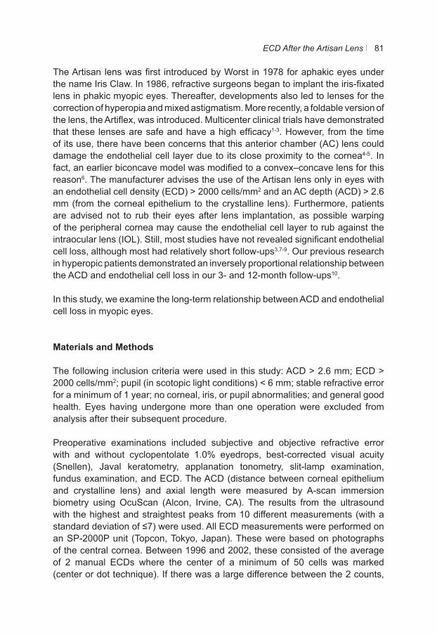
ECD After the Artisan Lens 81
The Artisan lens was first introduced by Worst in 1978 for aphakic eyes under the name Iris Claw. In 1986, refractive surgeons began to implant the iris-fixated lens in phakic myopic eyes. Thereafter, developments also led to lenses for the correction of hyperopia and mixed astigmatism. More recently, a foldable version of the lens, the Artiflex, was introduced. Multicenter clinical trials have demonstrated that these lenses are safe and have a high efficacy1-3. However, from the time of its use, there have been concerns that this anterior chamber (AC) lens could damage the endothelial cell layer due to its close proximity to the cornea4-5. In fact, an earlier biconcave model was modified to a convex–concave lens for this reason6. The manufacturer advises the use of the Artisan lens only in eyes with an endothelial cell density (ECD) > 2000 cells/mm2 and an AC depth (ACD) > 2.6 mm (from the corneal epithelium to the crystalline lens). Furthermore, patients are advised not to rub their eyes after lens implantation, as possible warping of the peripheral cornea may cause the endothelial cell layer to rub against the intraocular lens (IOL). Still, most studies have not revealed significant endothelial cell loss, although most had relatively short follow-ups3,7-9. Our previous research in hyperopic patients demonstrated an inversely proportional relationship between the ACD and endothelial cell loss in our 3- and 12-month follow-ups10.
In this study, we examine the long-term relationship between ACD and endothelial cell loss in myopic eyes.
Materials and Methods
The following inclusion criteria were used in this study: ACD > 2.6 mm; ECD > 2000 cells/mm2; pupil (in scotopic light conditions) < 6 mm; stable refractive error for a minimum of 1 year; no corneal, iris, or pupil abnormalities; and general good health. Eyes having undergone more than one operation were excluded from analysis after their subsequent procedure.
Preoperative examinations included subjective and objective refractive error with and without cyclopentolate 1.0% eyedrops, best-corrected visual acuity (Snellen), Javal keratometry, applanation tonometry, slit-lamp examination, fundus examination, and ECD. The ACD (distance between corneal epithelium and crystalline lens) and axial length were measured by A-scan immersion biometry using OcuScan (Alcon, Irvine, CA). The results from the ultrasound with the highest and straightest peaks from 10 different measurements (with a standard deviation of ≤7) were used. All ECD measurements were performed on an SP-2000P unit (Topcon, Tokyo, Japan). These were based on photographs of the central cornea. Between 1996 and 2002, these consisted of the average of 2 manual ECDs where the center of a minimum of 50 cells was marked (center or dot technique). If there was a large difference between the 2 counts,
Chapter 482
another ECD measurement would be performed. After 2002, we used automated measurements with the retracing method by means of the IMAGEnet software program (Topcon), with the same specular microscope11. All complete cells on the image were used to measure the cell density. The value of 0.6% per year was used to estimate natural endothelial cell loss12.
Each patient was informed about the procedure and signed a detailed informed consent form in accordance with the Helsinki Declaration. The lens power was calculated using the van der Heijde formula. All lens implantations were performed according to protocol by one experienced surgeon (GPML)10. We received approval from the institutional review board/ethics committee of our institution for this study.
Statistics
We used paired Student’s t tests to test for differences between preoperative and postoperative endothelial cell densities. A mixed model analysis of variance was applied to determine a relationship between endothelial cell loss and ACD, correcting for the factors age, gender, left or right eye, refractive error (higher power IOLs are thicker), and lens type (Artiflex, 5.0 mm, and 6.0 mm). Spatial correlation structure of the repeated measurements within an eye was employed. SAS for Windows software (SAS Institute, Cary, NC) and SPSS statistical software (version 11.0, SPSS Inc., Chicago, IL) were used for data analysis.
Results
Patient demographics are listed in Table 1. Three hundred eighteen eyes implanted with the Artisan Myopia Claw Lens of 173 patients with a minimum follow-up of 1 year were included. Patients were operated between February 1997 and September 2004. Mean follow-up was 35.3±20.7 months per eye. Patients were excluded from the study after they had undergone lens exchange (n = 5), cataract extraction (CE) (n = 4), or retinal detachment surgery (n = 1) or experienced a pupillary block glaucoma (n = 1). The surgeon (GPML) chose to include one eye with an ECD of 1993 cells/mm2 (which narrowly missed the inclusion criteria) after conferring with the patient. Data of some of these eyes have been published in previous reports2,3,8.
ECD After the Artisan Lens 83
Table 1. Patient Population Demographics (318 Eyes, 173 Patients)
Table 2 shows the endothelial cell loss between 3 months and 7 years postoperatively. There is an initial yet insignificant rise in endothelial cells in the first postoperative year. Endothelial cell loss was significant from 3 years onwards, also when corrected for an expected natural cell loss of 0.6% per year12.
Figures 1-8 display the relationship between ACD and ECD, corrected for patient age, refractive error, gender, left or right eye, and lens type. After 3 years, a significant negative correlation was revealed.
All corneas remained clear throughout our follow-up period. There were no significant differences in endothelial cell loss between the 3 different lens types (Artiflex, 5 mm, and 6 mm require incisions of 3.2, 5.2, and 6.2 mm, respectively). Age and gender of the patient and refractive error of the lens (difference in lens thickness) did not show a correlation with endothelial cell loss, nor did the side of the eye.
Discussion
In our study, we found a significant endothelial cell loss in our patient population starting at 3 years after implantation of the Artisan iris-fixated phakic IOL. Endothelial cell loss in our population, 8.3% (5.3% when correcting for natural loss), was relatively higher than that of other studies13-16. However, some of these studies had a relatively shorter follow-up13, 14, 16. Additionally, similar to other reports, there was an initial yet insignificant increase in cells the first postoperative year1, 7, 10, 16. Though we did not account for this preoperatively, we believe that the increase in ECD may have been caused by endothelial cell migration from the peripheral to the central cornea after contact lens discontinuation by some of our patients, although this pattern of cell distribution has been disputed17-19. Moreover, ECD measurements have been found to be less accurate immediately postoperatively20. The size of the incision did not appear to play a role in cell loss,
Table 1: Patient Population Demographics Patient Population (318 eyes, 173 patients)
Age (years) 38.5 ± 10.1 (range 18 – 61) Male 34.1 %
Mean Spherical Refractive Error (Diopters) -12.25 ± 4.20 (range –1.00 to –23.50) Mean Anterior Chamber Depth (mm) 3.70 ± 0.30 (range 2.89 to 4.50) Mean preoperative Endothelial Cell Density (cells / mm2) 2817 ± 356 (range 1993 to 3979) Type of Lens (number) 5.0 mm 152 (57 toric)
6.0 mm 149
Artiflex 17
Chapter 484
Tabl
e 2.
Mea
n E
ndot
helia
l Cel
l Den
sity
(EC
D) a
nd S
tand
ard
Dev
iatio
n (S
D) o
ver 7
Yea
rs: N
A =
not a
pplic
able
. t t
est,
preo
pera
tive
vers
us
post
oper
ativ
e, P
≤0.0
3.
Ti
me
Per
iod
N
umbe
r of
eye
s
Mea
n E
ndot
helia
l C
ell D
ensi
ty
(Cel
ls /
mm
2 ± S
D)
Mea
n E
ndot
helia
l C
ell D
ensi
ty C
hang
e (C
ells
/ m
m2 ±
SD
)
Mea
n O
bser
ved
Per
cent
age
End
othe
lial
Cel
l Den
sity
Cha
nge
Mea
n E
xpec
ted
Per
cent
age
EC
D C
hang
e in
an
un-o
pera
ted
eye
with
0.6
% c
ell l
oss/
year
Mea
n O
bser
ved
– E
xpec
ted
EC
D
Cha
nge
P
reop
erat
ive
31
8 28
17 ±
356
N
A
N
A
NA
N
A
3
mon
ths
22
0 28
56 ±
385
-3
4 ±
269
+1
.9
-0
.2
+2.1
6
mon
ths
24
8 28
48 ±
425
-3
7 ±
296
+1
.7
-0
.3
+2
.0
1
year
25
0 28
13 ±
426
-7
± 2
90
+0
.5
-0
.6
+1.1
2
year
s
16
8 27
77 ±
376
42
± 3
27
-0
.8
-1
.2
+0.4
3
year
s
12
2 27
29 ±
342
96
± 4
06*
-2
.2
-1
.8
-0.4
4
year
s
69
2616
± 3
07
291
± 50
2*
-6
.5
-2
.4
-4.1
5
year
s
51
2581
± 2
93
285
± 50
1*
-8
.3
-3
.0
-5.3
6
year
s
28
2560
± 2
70
380
± 60
8*
-9
.1
-3
.5
-5.5
7
year
s
13
2451
± 2
56
557
± 80
5*
-1
2.6
-4
.1
-8.5
Tabl
e 2:
Mea
n E
ndot
helia
l Cel
l Den
sity
(EC
D) a
nd s
tand
ard
devi
atio
n (S
D) o
ver
seve
n ye
ars.
*T
-tes
t Pre
oper
ativ
e ve
rsus
Pos
tope
rativ
e, p≤0
.03
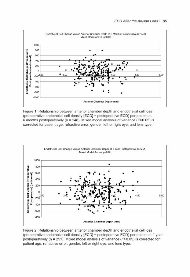
ECD After the Artisan Lens 85
Figure 1. Relationship between anterior chamber depth and endothelial cell loss (preoperative endothelial cell density [ECD] − postoperative ECD) per patient at 6 months postoperatively (n = 248). Mixed model analysis of variance (P>0.05) is corrected for patient age, refractive error, gender, left or right eye, and lens type.
Figure 2. Relationship between anterior chamber depth and endothelial cell loss (preoperative endothelial cell density [ECD] − postoperative ECD) per patient at 1 year postoperatively (n = 251). Mixed model analysis of variance (P>0.05) is corrected for patient age, refractive error, gender, left or right eye, and lens type.
Endothelial Cell Change versus Anterior Chamber Depth at 6 Months Postoperative (n=248)Mixed Model Anova, p>0.05
-1000
-800
-600
-400
-200
0
200
400
600
800
1000
2,50 3,00 3,50 4,00 4,50 5,00
Anterior Chamber Depth (mm)
Endo
thel
ial C
ell C
hang
e (P
reop
erat
ive
-Po
stop
erat
ive)
(cel
ls/m
m2)
Figure 1: Displays the relationship between the anterior chamber depth (mm) and endothelial cell loss (cells/mm2) (preoperative
endothelial cell density - postoperative endothelial cell density) per patient. The Mixed Model Analysis of Variance (ANOVA) is
corrected for patient age, refractive error, gender, left or right eye and lens type.
Figure
Endothelial Cell Change versus Anterior Chamber Depth at 1 Year Postoperative (n=251)Mixed Model Anova, p>0.05
-800
-600
-400
-200
0
200
400
600
800
1000
2,50 3,00 3,50 4,00 4,50 5,00
Anterior Chamber Depth (mm)
Endo
thel
ial C
ell C
hang
e (P
reop
erat
ive
-Po
stop
erat
ive)
(cel
ls/m
m2)
Figure 2: Displays the relationship between the anterior chamber depth (mm) and endothelial cell loss (cells/mm2) (preoperative
endothelial cell density - postoperative endothelial cell density) per patient. The Mixed Model Analysis of Variance (ANOVA) is
corrected for patient age, refractive error, gender, left or right eye and lens type.
Figure
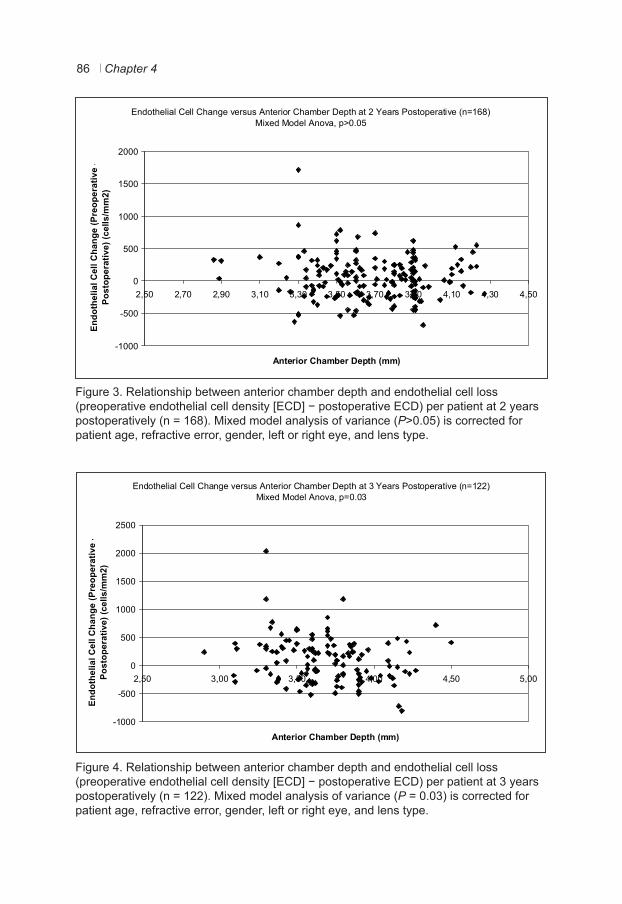
Chapter 486
Figure 3. Relationship between anterior chamber depth and endothelial cell loss (preoperative endothelial cell density [ECD] − postoperative ECD) per patient at 2 years postoperatively (n = 168). Mixed model analysis of variance (P>0.05) is corrected for patient age, refractive error, gender, left or right eye, and lens type.
Figure 4. Relationship between anterior chamber depth and endothelial cell loss (preoperative endothelial cell density [ECD] − postoperative ECD) per patient at 3 years postoperatively (n = 122). Mixed model analysis of variance (P = 0.03) is corrected for patient age, refractive error, gender, left or right eye, and lens type.
Endothelial Cell Change versus Anterior Chamber Depth at 2 Years Postoperative (n=168)Mixed Model Anova, p>0.05
-1000
-500
0
500
1000
1500
2000
2,50 2,70 2,90 3,10 3,30 3,50 3,70 3,90 4,10 4,30 4,50
Anterior Chamber Depth (mm)
Endo
thel
ial C
ell C
hang
e (P
reop
erat
ive
-Po
stop
erat
ive)
(cel
ls/m
m2)
Figure 3: Displays the relationship between the anterior chamber depth (mm) and endothelial cell loss (cells/mm2) (preoperative
endothelial cell density - postoperative endothelial cell density) per patient. The Mixed Model Analysis of Variance (ANOVA) is
corrected for patient age, refractive error, gender, left or right eye and lens type.
Figure
Endothelial Cell Change versus Anterior Chamber Depth at 3 Years Postoperative (n=122)Mixed Model Anova, p=0.03
-1000
-500
0
500
1000
1500
2000
2500
2,50 3,00 3,50 4,00 4,50 5,00
Anterior Chamber Depth (mm)
Endo
thel
ial C
ell C
hang
e (P
reop
erat
ive
-Po
stop
erat
ive)
(cel
ls/m
m2)
Figure 4: Displays the relationship between the anterior chamber depth (mm) and endothelial cell loss (cells/mm2) (preoperative
endothelial cell density - postoperative endothelial cell density) per patient. The Mixed Model Analysis of Variance (ANOVA) is
corrected for patient age, refractive error, gender, left or right eye and lens type.
Figure
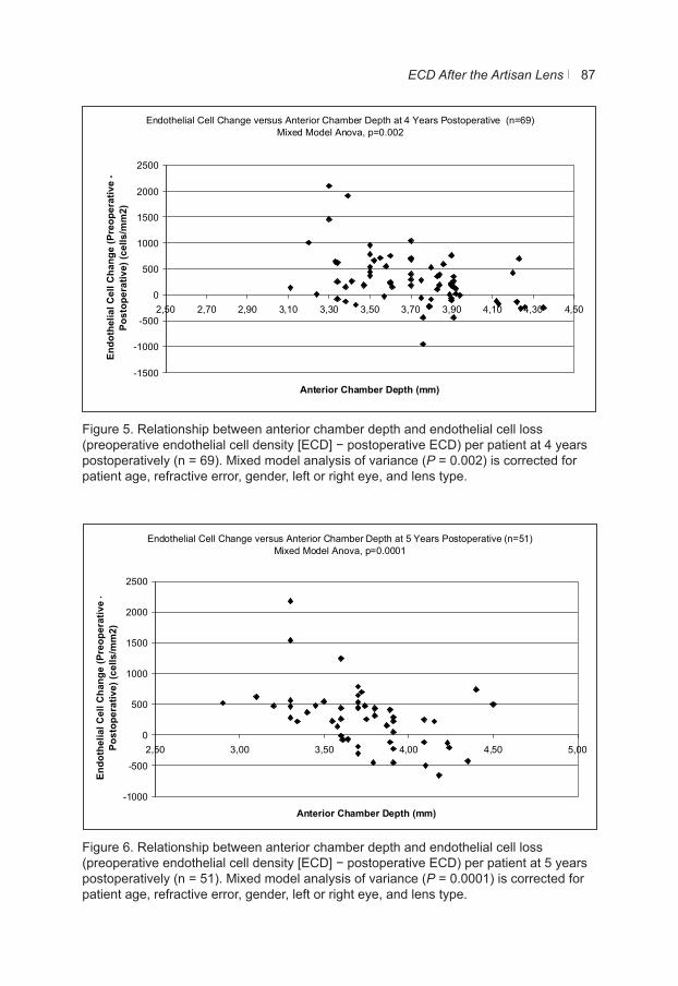
ECD After the Artisan Lens 87
Figure 5. Relationship between anterior chamber depth and endothelial cell loss (preoperative endothelial cell density [ECD] − postoperative ECD) per patient at 4 years postoperatively (n = 69). Mixed model analysis of variance (P = 0.002) is corrected for patient age, refractive error, gender, left or right eye, and lens type.
Figure 6. Relationship between anterior chamber depth and endothelial cell loss (preoperative endothelial cell density [ECD] − postoperative ECD) per patient at 5 years postoperatively (n = 51). Mixed model analysis of variance (P = 0.0001) is corrected for patient age, refractive error, gender, left or right eye, and lens type.
Endothelial Cell Change versus Anterior Chamber Depth at 4 Years Postoperative (n=69)Mixed Model Anova, p=0.002
-1500
-1000
-500
0
500
1000
1500
2000
2500
2,50 2,70 2,90 3,10 3,30 3,50 3,70 3,90 4,10 4,30 4,50
Anterior Chamber Depth (mm)
Endo
thel
ial C
ell C
hang
e (P
reop
erat
ive
-Po
stop
erat
ive)
(cel
ls/m
m2)
Figure 5: Displays the relationship between the anterior chamber depth (mm) and endothelial cell loss (cells/mm2) (preoperative
endothelial cell density - postoperative endothelial cell density) per patient. The Mixed Model Analysis of Variance (ANOVA) is
corrected for patient age, refractive error, gender, left or right eye and lens type.
Figure
Endothelial Cell Change versus Anterior Chamber Depth at 5 Years Postoperative (n=51)Mixed Model Anova, p=0.0001
-1000
-500
0
500
1000
1500
2000
2500
2,50 3,00 3,50 4,00 4,50 5,00
Anterior Chamber Depth (mm)
Endo
thel
ial C
ell C
hang
e (P
reop
erat
ive
-Po
stop
erat
ive)
(cel
ls/m
m2)
Figure 6: Displays the relationship between the anterior chamber depth (mm) and endothelial cell loss (cells/mm2) (preoperative
endothelial cell density - postoperative endothelial cell density) per patient. The Mixed Model Analysis of Variance (ANOVA) is
corrected for patient age, refractive error, gender, left or right eye and lens type.
Figure
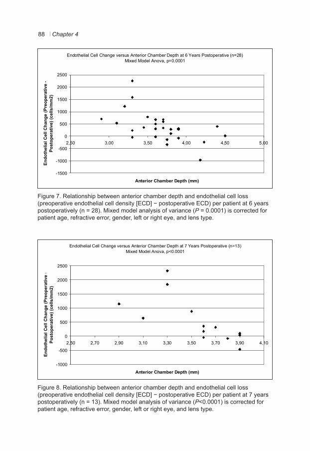
Chapter 488
Figure 7. Relationship between anterior chamber depth and endothelial cell loss (preoperative endothelial cell density [ECD] − postoperative ECD) per patient at 6 years postoperatively (n = 28). Mixed model analysis of variance (P = 0.0001) is corrected for patient age, refractive error, gender, left or right eye, and lens type.
Figure 8. Relationship between anterior chamber depth and endothelial cell loss (preoperative endothelial cell density [ECD] − postoperative ECD) per patient at 7 years postoperatively (n = 13). Mixed model analysis of variance (P<0.0001) is corrected for patient age, refractive error, gender, left or right eye, and lens type.
Endothelial Cell Change versus Anterior Chamber Depth at 6 Years Postoperative (n=28)Mixed Model Anova, p=0.0001
-1500
-1000
-500
0
500
1000
1500
2000
2500
2,50 3,00 3,50 4,00 4,50 5,00
Anterior Chamber Depth (mm)
Endo
thel
ial C
ell C
hang
e (P
reop
erat
ive
-Po
stop
erat
ive)
(cel
ls/m
m2)
Figure 7: Displays the relationship between the anterior chamber depth (mm) and endothelial cell loss (cells/mm2) (preoperative
endothelial cell density - postoperative endothelial cell density) per patient. The Mixed Model Analysis of Variance (ANOVA) is
corrected for patient age, refractive error, gender, left or right eye and lens type.
Figure
Endothelial Cell Change versus Anterior Chamber Depth at 7 Years Postoperative (n=13)Mixed Model Anova, p<0.0001
-1000
-500
0
500
1000
1500
2000
2500
2,50 2,70 2,90 3,10 3,30 3,50 3,70 3,90 4,10
Anterior Chamber Depth (mm)
Endo
thel
ial C
ell C
hang
e (P
reop
erat
ive
-Po
stop
erat
ive)
(cel
ls/m
m2)
Figure 8: Displays the relationship between the anterior chamber depth (mm) and endothelial cell loss (cells/mm2) (preoperative
endothelial cell density - postoperative endothelial cell density) per patient. The Mixed Model Analysis of Variance (ANOVA) is
corrected for patient age, refractive error, gender, left or right eye and lens type.
Figure
ECD After the Artisan Lens 89
reinforcing previously reported results14. Other studies on the Artisan lens do confirm our findings of progressive endothelial cell loss4, 5. Continued endothelial cell loss has also been noted in pseudophakic eyes 10 years after CE21.
Furthermore, we found a significant negative correlation between ACD and loss of endothelium. Menezo et al also found a correlation with ACD after 6 months of their 4-year endothelial study and noted significant postoperative endothelial cell loss. As the cell hexagonality remained stable after 2 years, loss of endothelium was attributed to the implantation procedure rather than to the lens itself. The correlation between ACD and cell loss after half a year was considered to be a possible consequence of intraoperative contact15. In our study, the negative correlation between endothelial cell loss and ACD only became evident after 3 years and remained significant up to our 7-year follow-up. The factors mentioned previously, as well as corneal wound reorganization, may have blurred a relationship between ACD and decrease in ECD in earlier postoperative years15,22.
One patient with preoperative ECDs of 2910 (right eye) and 2706 (left eye) had ECDs of 586 (right) and 863 (left) after 7 years with clear corneas. Serial ECD measurements made the first postoperative year (7 times) showed a continual progressive loss; however, the greatest ECD loss occurred between years 1 and 2 (before the switch to IMAGEnet). We could not explain the dramatic decrease in ECD, as the corneas were unremarkable preoperatively, as were the implant procedures. The patient did not rub his eyes and was not known to have allergies. He did undergo uncomplicated strabismus surgery on both eyes within 1 year of Artisan lens implantation, although the procedures took place after the greatest amount of cell loss had already taken place.
We performed endothelial cell counts using the center or dot technique before 2002 and the retracing method thereafter11. Thus, the cell measurements of our longer follow-up patients were calculated using 2 different counting methods on the same specular microscope. On regular inspection, the calibration was checked but showed no differences before and after 2002. Although significant ECD loss was only noted after 3 years of follow-up (whereby some patients were operated before we switched to the IMAGEnet program), a similar pattern of loss was found among our hyperopic patients, with whom all follow-up examinations took place before the IMAGEnet system was being used10. Nonetheless, it has been demonstrated that the center or dot technique is not as precise as the retracing method, which could question the accuracy of our (preoperative) measurements, although these could have been both underestimated or overestimated11. Analysis of cell hexagonality and polymegathism would have strengthened this study, but unfortunately, these data were not available on most eyes operated before 2002.
Endothelial Cell Change versus Anterior Chamber Depth at 6 Years Postoperative (n=28)Mixed Model Anova, p=0.0001
-1500
-1000
-500
0
500
1000
1500
2000
2500
2,50 3,00 3,50 4,00 4,50 5,00
Anterior Chamber Depth (mm)
Endo
thel
ial C
ell C
hang
e (P
reop
erat
ive
-Po
stop
erat
ive)
(cel
ls/m
m2)
Figure 7: Displays the relationship between the anterior chamber depth (mm) and endothelial cell loss (cells/mm2) (preoperative
endothelial cell density - postoperative endothelial cell density) per patient. The Mixed Model Analysis of Variance (ANOVA) is
corrected for patient age, refractive error, gender, left or right eye and lens type.
Figure
Endothelial Cell Change versus Anterior Chamber Depth at 7 Years Postoperative (n=13)Mixed Model Anova, p<0.0001
-1000
-500
0
500
1000
1500
2000
2500
2,50 2,70 2,90 3,10 3,30 3,50 3,70 3,90 4,10
Anterior Chamber Depth (mm)
Endo
thel
ial C
ell C
hang
e (P
reop
erat
ive
-Po
stop
erat
ive)
(cel
ls/m
m2)
Figure 8: Displays the relationship between the anterior chamber depth (mm) and endothelial cell loss (cells/mm2) (preoperative
endothelial cell density - postoperative endothelial cell density) per patient. The Mixed Model Analysis of Variance (ANOVA) is
corrected for patient age, refractive error, gender, left or right eye and lens type.
Figure
Chapter 490
The study is further limited by the number of patients available for follow-up examinations; at 3 years, data from only 122 of the 318 eyes were available, and at 7 years, from only 13. This partial follow-up was due to the majority of patients having been operated on more recently, whereby the data are not yet available.
Although our study showed significant endothelial cell loss, all of the corneas remained clear. Still, we do find the cell loss noteworthy and offer an approach to the inclusion criteria other than what the manufacturer advises. First, considering the progressive endothelial cell loss we encountered, we recommend lifelong patient follow-up. Patients should be made aware of this before being operated. We further suggest a stricter inclusion criteria for younger patients, proposing ACDs of ≥3.5 mm. This could offset the decrease in ACD with age, which we have found to be 0.12 mm/decade (Cheng YYY, Bhagwandien ACE, Wolfs RCW, Luyten GPM, submitted for publication, 2006). Moreover, it could partially compensate for cell loss after lens implantation. Analogous suggestions were also made by Fechner et al in 199823. Most importantly, we advise that some of the inclusion criteria no longer be looked at individually but as a whole: patient age, ACD, and ECD should be considered jointly. For example, young patients with relatively shallow ACs and ECDs just above 2000 cells/mm2 should perhaps no longer be considered for implantation of these lenses. Longer follow-up studies of endothelium cell loss in relation to the ACD are necessary before definitive criteria, including patient age, can be reasonably determined. We suggest that surgeons use their clinical judgment when assessing young potential candidates narrowly meeting the inclusion criteria for the Artisan phakic IOL.
Our study showed a significant progressive endothelial cell loss after 3 years and a significant negative correlation between endothelial cell loss and ACD. We suggest caution in operating young patients who narrowly meet the inclusion criteria for ACD and ECDs.
ECD After the Artisan Lens 91
References:
1. Bartels MC, Santana NT, Budo C, et al. Toric phakic intraocular lens for the correction of hyperopia and astigmatism. J Cataract Refract Surg 2006;32(2):243-9.
2. Bartels MC, Saxena R, van den Berg TJ, et al. The influence of incision-induced astigmatism and axial lens position on the correction of myopic astigmatism with the Artisan toric phakic intraocular lens. Ophthalmology 2006;113(7):1110-7.
3. Budo C, Hessloehl JC, Izak M, et al. Multicenter study of the Artisan phakic intraocular lens. J Cataract Refract Surg 2000;26(8):1163-71.
4. Landesz M, Worst JG, van Rij G. Long-term results of correction of high myopia with an iris claw phakic intraocular lens. J Refract Surg 2000;16(3):310-6.
5. Menezo JL, Cisneros A, Hueso JR, Harto M. Long-term results of surgical treatment of high myopia with Worst-Fechner intraocular lenses. J Cataract Refract Surg 1995;21(1):93-8.
6. Landesz M, Worst J, Siertsema J, van Rij G. Negative implant. A retrospective study. Doc. Ophthalmol. 1993;83:261-70.
7. Guell JL, Vazquez M, Malecaze F, et al. Artisan toric phakic intraocular lens for the correction of high astigmatism. Am J Ophthalmol 2003;136(3):442-7.
8. Landesz M, van Rij G, Luyten G. Iris-claw phakic intraocular lens for high myopia. J Refract Surg 2001;17(6):634-40.
9. Pop M, Payette Y. Initial results of endothelial cell counts after Artisan lens for phakic eyes: an evaluation of the United States Food and Drug Administration Ophtec Study. Ophthalmology 2004;111(2):309-17.
10. Saxena R, Landesz M, Noordzij B, Luyten GP. Three-year follow-up of the Artisan phakic intraocular lens for hypermetropia. Ophthalmology 2003;110(7):1391-5.
11. van Schaick W, van Dooren BT, Mulder PG, Volker-Dieben HJ. Validity of endothelial cell analysis methods and recommendations for calibration in Topcon SP-2000P specular microscopy. Cornea 2005;24(5):538-44.
12. Bourne WM, Nelson LR, Hodge DO. Central corneal endothelial cell changes over a ten-year period. Invest Ophthalmol Vis Sci 1997;38(3):779-82.
13. Alio JL, de la Hoz F, Perez-Santonja JJ, et al. Phakic anterior chamber lenses for the correction of myopia: a 7-year cumulative analysis of complications in 263 cases. Ophthalmology 1999;106(3):458-66.
14. Benedetti S, Casamenti V, Marcaccio L, et al. Correction of myopia of 7 to 24 diopters with the Artisan phakic intraocular lens: two-year follow-up. J Refract Surg 2005;21(2):116-26.
15. Menezo JL, Cisneros AL, Rodriguez-Salvador V. Endothelial study of iris-claw phakic lens: four year follow-up. J Cataract Refract Surg 1998;24(8):1039-49.
16. Asano-Kato N, Toda I, Hori-Komai Y, et al. Experience with the Artisan phakic intraocular lens in Asian eyes. J Cataract Refract Surg 2005;31(5):910-5.
17. Amann J, Holley GP, Lee SB, Edelhauser HF. Increased endothelial cell density in the paracentral and peripheral regions of the human cornea. Am J Ophthalmol 2003;135(5):584-90.
18. Perez-Santonja JJ, Sahla HF, Alio JL. Evaluation of endothelial cell changes 1 year after excimer laser in situ keratomileusis. Arch Ophthalmol 1997;115(7):841-6.
19. Wiffen SJ, Hodge DO, Bourne WM. The effect of contact lens wear on the central and peripheral corneal endothelium. Cornea 2000;19(1):47-51.
20. Wirbelauer C, Wollensak G, Pham DT. Influence of cataract surgery on corneal endothelial cell density estimation. Cornea 2005;24(2):135-40.
21. Bourne WM, Nelson LR, Hodge DO. Continued endothelial cell loss ten years after lens implantation. Ophthalmology 1994;101(6):1014-22; discussion 22-3.
22. Werblin TP. Long-term endothelial cell loss following phacoemulsification: model for evaluating endothelial damage after intraocular surgery. Refract Corneal Surg 1993;9(1):29-35.
23. Fechner PU, Singh D, Wulff K. Iris-claw lens in phakic eyes to correct hyperopia: preliminary study. J Cataract Refract Surg 1998;24(1):48-56.


Part 2
Subjective Results: The Quality of Life of Myopic Patients


5Chapter 5
Vision-Related Quality of Life of Myopic Patients
R Saxena W Vreugdenhil PGH Mulder G van Rij GPM Luyten
Submitted
Med. Library
Erasmus MC
EMBARGO - not published yet


6Chapter 6
Quality of Life of Before and After Refractive Surgery
R Saxena W Vreugdenhil M Alwani R Arkenstijn MA Landesz MAH Luger MC Bartels G van Rij GPM Luyten
Submitted
Med. Library
Erasmus MC
EMBARGO - not published yet


Part 3
Special Applications of the Artisan Lens


7Chapter 7
The Use of the Artisan Phakic Intraocular Lens in the Treatment of Anisometropic Amblyopia
R SaxenaHM van MinderhoutGPM Luyten
J Cataract Refract Surg 2003; 29:835–838

Anisometropic Amblyopia and the Artisan Lens 131
High unilateral myopia is difficult to treat in young children and often leads to anisometropic amblyopia1–5. Factors such as the depth of the amblyopia and the age of the patient play a key role in the effectiveness of treatment. Success is also frequently hampered by practical problems such as those created by glasses and contact lenses. Thus far, however, patient compliance has been considered the greatest therapeutic impediment6,7. In such cases, refractive surgery with the excimer laser or phakic intraocular lens (IOL) implantation can be a solution. There is, however, limited experience in treating myopic children with these procedures1,2.
As excimer laser corrections are confined to refractive errors of approximately -8.00 to -10.00 diopters (D), effective surgical correction of high myopia in children can only be done with phakic IOLs. Studies of phakic IOLs to correct anisometropic amblyopia in the pediatric population have been reported3–5. Concerns about IOL implantation in young patients include cataract formation, pupil alteration, endothelial cell loss, postoperative inflammation, and a possible myopic shift as the patient ages.
Artisan phakic IOL implantation is a reliable and safe procedure in adults8,9. Although the IOL is considered experimental in several parts of the world, it has been implanted in patients in The Netherlands since 1991. Thus, when we were confronted with a young anisometropic amblyopic patient with restricted treatment options, we decided to correct his high unilateral myopia using the Artisan phakic IOL. Recently, Chipont and coauthors reported such a case with an 18-month follow-up5. Here, we present a patient with 3-year postoperative results.
Case Report
A 4-year-old boy with high unilateral myopia and distinct anisometropic amblyopia in the right eye presented in February 1997. Thirty minutes after 2 drops of cyclopentolate 1%, the child had a cycloplegic refraction of -14.00 +3.00 x115 in the right eye and was emmetropic in the left eye. The cycloplegic best corrected visual acuity (BCVA) in the myopic eye was 0.17 (Amsterdam Picture Chart [APK], translated from the Dutch, Amsterdamse Plaatjeskaart). Stereoscopic vision could not be detected. A spectacle was prescribed with a -10.00 +3.00x115 correction in the right eye and plano in the left eye along with 2 hours a day of left-side occlusion therapy. A refractive undercorrection was deemed necessary to reduce the effects of aniseikonia.
In June 1997, the BCVA in the right eye, measured with a full correction of -14.00 -3.00 x 115, had improved to 0.83 (APK); however, stereovision remained negative

Chapter 7132
(Lang and Titmus fly tests). Nonetheless, the patient reported that glasses were uncomfortable as they were constantly lopsided; he therefore refused to wear them. This lack of cooperation made it difficult to continue the patch therapy, and the BCVA with full correction had decreased to 0.56 (APK). Thus, a toric contact lens of -11.50 +2.50 x 110 was prescribed for the right eye.
After wearing the lens for 2 months and increasing the daily occlusion therapy to 8 hours, the patient had a poor BCVA at 0.50 (APK); however, the Titmus test was positive. Furthermore, during this period, the boy repeatedly lost his contact lens. Considering that neither glasses nor the contact lens had proved to be worthwhile therapy, refractive surgery was considered. The case was discussed thoroughly in our department and with Jan Worst, MD, the creator of the lens. A decision was made to correct the unilateral refractive error with an Artisan phakic IOL and to continue occlusion therapy. Considering the lens had only been studied in adults, the possible short- and long-term complications reported in the literature were discussed with the child’s parents, who were told its use in children was experimental. As they were unhappy with their son’s varying progress, they agreed to the operation and signed an informed consent. The aim was slight myopia so that both eyes would eventually have about equal refractive errors. The hope was that further changes in error would occur concurrently in both eyes as the patient grew.
In February 1998, the patient had a -15.00 D Artisan phakic IOL (model 206w; 5.0 mm optic) implanted in the right eye under general anesthesia. The surgical procedure and the IOL power calculation were performed according to standard protocol. The patient received gentamicin ointment 1 day before the procedure. Pilocarpine 4% drops were applied the morning of and 30 minutes before surgery. A bevelled 5.5 mm corneoscleral incision was made at 12 o’clock, and 2 paracenteses were placed at 10 o’clock and 2 o’clock. The anterior chamber was opened and filled with sodium hyaluronate 1% (Healon) to maintain its depth. After the IOL was implanted with a DO2–72 forceps (Ophtec), it was fixated to the iris with enclavation needles (model OD125, Ophtec). At the end of surgery, a slit iridotomy was performed, the corneoscleral wound was closed with a running suture, and the viscoelastic material was manually irrigated. Gentamicin ointment was applied to the eye.
The early postoperative period was without complications. Occlusion therapy was continued for 8 hours a day. Four months postoperatively, the cycloplegic subjective refraction was -1.50 +1.00 x 135 in the operated eye and -1.00 in the left eye. Snellen BCVA (E chart) was 0.65 and 0.80, respectively. At 29 months, the cycloplegic subjective refraction was -0.75 +0.75 x 155 in the operated eye and -2.00 in the left eye. Stereovision had improved (positive Lang Test of 600”
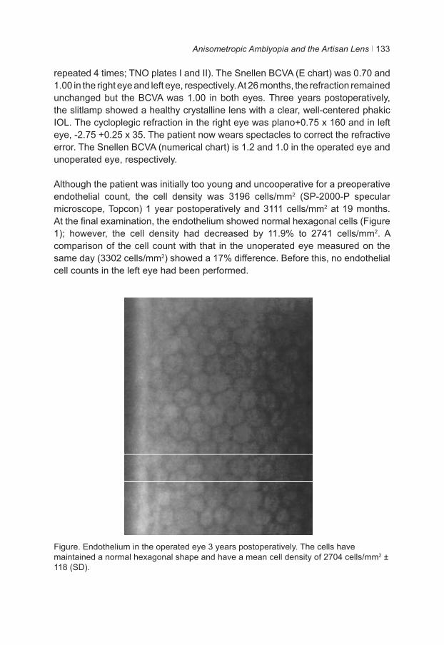
Anisometropic Amblyopia and the Artisan Lens 133
repeated 4 times; TNO plates I and II). The Snellen BCVA (E chart) was 0.70 and 1.00 in the right eye and left eye, respectively. At 26 months, the refraction remained unchanged but the BCVA was 1.00 in both eyes. Three years postoperatively, the slitlamp showed a healthy crystalline lens with a clear, well-centered phakic IOL. The cycloplegic refraction in the right eye was plano+0.75 x 160 and in left eye, -2.75 +0.25 x 35. The patient now wears spectacles to correct the refractive error. The Snellen BCVA (numerical chart) is 1.2 and 1.0 in the operated eye and unoperated eye, respectively.
Although the patient was initially too young and uncooperative for a preoperative endothelial count, the cell density was 3196 cells/mm2 (SP-2000-P specular microscope, Topcon) 1 year postoperatively and 3111 cells/mm2 at 19 months. At the final examination, the endothelium showed normal hexagonal cells (Figure 1); however, the cell density had decreased by 11.9% to 2741 cells/mm2. A comparison of the cell count with that in the unoperated eye measured on the same day (3302 cells/mm2) showed a 17% difference. Before this, no endothelial cell counts in the left eye had been performed.
Figure. Endothelium in the operated eye 3 years postoperatively. The cells have maintained a normal hexagonal shape and have a mean cell density of 2704 cells/mm2 ± 118 (SD).
Chapter 7134
Discussion
Treating high anisometropic amblyopia is frequently disappointing. Amblyopic treatment is encumbered by several problems6,7. Although children are usually capable of compensating for aniseikonia caused by high anisometropia, full spectacle correction is not generally prescribed. Glasses have practical hindrances such as prismatic effects. In addition to our patient’s complaint of the glasses being lopsided on his face, there are cosmetic issues and glasses provide far from ideal binocular vision. Contact lenses minimize the magnification differences between the eyes but can be difficult to manage in young children10. Therefore, resistance to spectacles and contact lenses may negatively influence the treatment of amblyopia in many cases. However, even after a young patient’s refractive error has been surgically corrected, one must always emphasize that patch therapy must be diligently continued.
The European multicenter study of the Artisan phakic IOL in 518 adult eyes established a safety index (postoperative BCVA/preoperative BCVA) of 1.31 and an efficacy index (postoperative uncorrected visual acuity/preoperative BCVA) of 1.03 3 years postoperatively8. We found similar results in our study9. As the lens is safe and has a good, predictable refractive outcome, we decided to use it to correct the patient’s refraction even though it has not been extensively tested in children. The child’s refusal to wear the glasses and repeated loss of his contact lens were affecting patch therapy compliance. Thus, we believed that the problems created by glasses and contact lenses outweighed the possible risks created by IOL implantation. By correcting the anisometropia with the Artisan lens, amblyopia treatment was facilitated by encouraging the patient to use the highly myopic eye and increasing occlusion therapy compliance. We believe this allowed the patient to develop stereovision. However, different optotypes were used preoperatively and postoperatively. Experience with the methods of visual acuity measurement and increasing patient cooperation may have distorted the amount of visual improvement.
Our patient was too young to allow valid preoperative endothelial cell measurement; however, comparison between the operated and un-operated eye 3 years after IOL implantation revealed a 17% difference. Earlier postoperative cell counts reveal much higher densities, indicating that rapid cell loss occurred after 19 months. We surmise that the child had begun rubbing his eyes, although we could not definitely establish this. During the multicenter investigation of the Artisan lens in adults, an initial endothelial cell loss of 4.8% was detected, which levelled off to 0.7%8. This rate of decline is not significantly different from that found in the general population. In our own series of adult myopic eyes treated with the Artisan phakic IOL, we observed no endothelial cell loss9. Basti et al. report no
Anisometropic Amblyopia and the Artisan Lens 135
difference in short-term corneal endothelial cell loss between adult and pediatric cataract patients, although it is questionable whether this comparison is valid among patients with an anterior chamber lens; in these cases, the endothelial layer can be affected when patients rub their eyes11. Therefore, endothelial cell loss remains a cause of concern in pediatric patients with an Artisan IOL.
Although intraocular surgery in children is more delicate and prone to inflammatory reactions, there were no adverse reactions or complications in our case. In a similar case of Chipont and coauthors, a low degree of uveitis was detected5. It is, nonetheless, possible that the complication rate in this younger population is higher as the clearance between the lens and the corneal endothelium can decrease over time12. The patient could also become more myopic with age, in which case additional correction with glasses or contact lenses can be prescribed. In more extreme cases, the lens can be exchanged. In our case, the postoperative refractive outcome was still stable at the patient’s last examination at 3 years.
Studies in which anisometropic amblyopia was treated with photorefractive keratectomy yielded promising results in children with myopia1,2. Nonetheless, excimer procedures are limited to low and moderate myopia, leaving phakic IOLs as the only feasible surgical option for young patients with high myopia.
BenEzra and coauthors and Lesueur and Arne treated anisometropic amblyopia with phakic posterior chamber IOLs. Although they achieved positive results, posterior chamber IOLs can have serious complications including angle-closure glaucoma, cataract, and chronic inflammation, all of which require fastidious long-term follow-up3,4. One advantage of a posterior chamber IOL is the reduced risk of corneal endothelial cell loss, which is especially beneficial in children prone to rubbing their eyes4. The angle-supported phakic anterior chamber IOL, another alternative to iris-fixated IOLs, can also lead to complications such as retinal changes and pupil ovalization13. Furthermore, eye growth can compromise the stability of the lens, making it an inappropriate choice in developing eyes.
The Artisan lens has not shown early serious complications; however, long-term follow-up results are not available. In our patient, the Artisan phakic IOL was effective in treating anisometropic amblyopia. Although the IOL can provide viable correction of high unilateral myopia in children, its implantation should be considered only if conventional therapy fails.
Chapter 7136
References
1. Nano HD Jr, Muzzin S, Irigaray LF. Excimer laser photorefractive keratectomy in pediatric patients. J Cataract Refract Surg 1997; 23(5):736–739.
2. Alió JL, Artola A, Claramonte P, et al. Photorefractive keratectomy for pediatric myopic anisometropia. J Cataract Refract Surg 1998; 24(3):327–330.
3. BenEzra D, Cohen E, Karshai I. Phakic posterior chamber intraocular lens for the correction of anisometropia and treatment of amblyopia. Am J Ophthalmol 2000;
130(3):292–296.4. Lesueur LC, Arne JL. Phakic posterior chamber lens implantation in children with high myopia.
J Cataract Refract Surg 1999; 25 (12):1571–1575.5. Chipont EM, García-Hermosa P, Alió JL. Reversal of myopic anisometropic amblyopia with
phakic intraocular lens implantation. J Refract Surg 2001; 17(4):460–462.6. Simonsz HJ, Polling JR, Voorn R, et al. Electronic monitoring of treatment compliance in
patching for amblyopia. Strabismus 1999; 7 (2):113–123 7. Newsham D. Parental non-concordance with occlusion therapy. Br J Ophthalmol 2000;
84(9):957–962.8. Budo C, Hessloehl JC, Izak M, et al. Multicenter study of the Artisan phakic intraocular lens. J
Cataract Refract Surg 2000; 26(8):1163–1171.9. Landesz M, van Rij G, Luyten G. Iris-claw phakic intraocular lens for high myopia. J Refract
Surg 2001; 17(6): 634–640.10. García M, González C, Pascual I, Fimia A. Magnification and visual acuity in highly myopic
phakic eyes corrected with an anterior chamber intraocular lens versus by other methods. J Cataract Refract Surg 1996; 22(10):1416–1422.
11. Basti S, Aasuri MK, Reddy S, et al. Prospective evaluation of corneal endothelial cell loss after pediatric cataract surgery. J Cataract Refract Surg 1998; 24(11):1469–1473.
12. Hosny M, Alió JL, Claramonte P, et al. Relationship between anterior chamber depth, refractive state, corneal diameter, and axial length. J Refract Surg 2000; 16 (3):336– 340.
13. Allemann N, Chamon W, Tanaka HM, et al. Myopic angle-supported intraocular lenses; two-year follow-up. Ophthalmology 2000; 107(8):1549–1554.

8Chapter 8
Iris-Fixated Phakic IOLs to Correct Postoperative Anisometropia in Unilateral Cataract Patients with
Bilateral High Myopia
R Saxena K van der Torren
M Veckeneer GPM Luyten
This article was published in the form a letter in: J Cataract Refract Surg. 2004 Oct;30(10):2240-1

Postoperative Anisometropia and the Artisan Lens 139
Purpose
To determine the visual outcome and the quality of life of five bilaterally myopic unilateral cataract patients implanted with a phakic intraocular lens (pIOL) in their sound eye.
Methods
Refraction and Snellen visual acuity were measured before and after undergoing both phacoemulsification in one eye and pIOL implantation in the other. Patients (3 presbyopic) completed the Dutch translation of the Refractive Status and Vision Profile (RSVP) for each eye after undergoing both procedures. Follow-up was between 6 and 18 months.
Results
Postoperatively, 4 out of 5 patients were within 1.00 D of emmetropia in the pseudophakic eye and all 5 out of 5 patients were within 1.00 D of emmetropia in the pIOL implanted eye. The safety index of the phakic eyes was 1.40 and the efficacy index was 0.88. There were no significant differences in the quality of life subscale measurements nor in their total scores (p>0.05). Patients significantly preferred reading with their phakic eye than with their pseudophakic eye (p=0.02).
Conclusion
PIOLs are an option for bilateral myopic patients with unilateral cataract. A larger study needs to be conducted to verify if presbyopic patients prefer reading with their phakic eye.
Chapter 8140
At present, unilateral cataract patients with myopia may choose to adjust their nearsightedness while undergoing cataract surgery. However, once their eye is treated to (nearly) full refractive correction, highly bilateral myopic patients are rendered substantially anisometropic. Contact lenses are certainly an option, although not all patients adjust to them well. Some patients desire more enduring solutions, so that they may have similar refractive errors in both eyes at all times.
Many such permanent alternatives are currently available for myopes. Not only can they consider the Excimer laser, but also phakic intraocular lenses (IOLs) and clear lens extractions (CLE). As our experience with the Artisan lens has been encouraging in both bilateral and unilateral implantations, we felt that this phakic IOL would be an interesting option for five relatively young patients with unilateral cataract1-3. Pure objective postoperative measurements, however, could not allow evaluation of how the patients experience the unique situation of pseudophakia in one eye and a phakic IOL in the other. To gain insight into this, the five patients postoperatively completed the Refractive Status and Visual Profile (RSVP), a vision-related quality of life questionnaire, for each eye4,5.
In this report, we discuss the objective and subjective results of five bilaterally myopic patients who underwent a cataract extraction and posterior chamber lens implantation in one eye and an Artisan phakic IOL implantation in the other.
Methods
Phacoemulsification and Artisan lens implantation preoperative examinations and procedures were performed according to standard protocol3. The following pseudophakic IOLs were used: three AMO SI40 foldable lenses (Allergan Medical Optics, Nieuwegein, the Netherlands), one Acrysof foldable lens (Alcon, Gorinchem, the Netherlands) and one polymethyl methacrylate (PMMA) lens (Flex 652, Domilens, Lyon, France). All operations were performed by G.L. unless otherwise noted.
Objective and subjective refractions were measured with and without the use of cyclopentolate hydrochlorate 1.0% eye drops. Both the uncorrected visual acuity (UCVA) and the best spectacle corrected visual acuity (BSCVA) were converted from the Snellen to the logarithmic logMAR notation to calculate the mean and then reverted back to Snellen notation. IOL power calculations were determined using the Van der Heijde formula1,2.
Postoperative Anisometropia and the Artisan Lens 141
To assess the subjective aspect of the treatment, patients received two copies of the Dutch consensus translation of the RSVP. Patients were asked to fill out the categories of the two identical questionnaires (concerns, expectations, functioning, driving, symptoms, optic problems, glare, and problems with glasses / contact lenses) simultaneously, so as to be able to compare their eyes. The total score is an average of the eight subscales, which are all weighted equally. The RSVP contains a total of 58 questions4,5.
Questionnaires were sent out between six and twenty months after Artisan lens implantation. All five patients responded within two weeks. Statistics were performed on the Microsoft Excel Program.
Case Reports
Preoperative and postoperative refractions and BSCVAs are noted in Table 1.
Patient 1
The patient was a 54-year-old male bilateral high myope with a nuclear cataract in his right eye. In August 2000, an Allergan SI40 lens of 13 D (6.0 mm optic) was implanted in this eye after undergoing phacoemulsification. His left eye was fitted with a 6.0 mm -8.5 D Artisan lens two weeks later. At one month postoperative, the refraction was S+0.50 C-1.50 x 22 in the pseudophakic eye, with a corresponding BSCVA of 0.70. The patient required a YAG capsulotomy in October 2000 and was diagnosed the following month with a local rhegmatogenic retinal detachment (RD) in the same eye, created by a tear in an area of lattice degeneration. The RD was reattached by scleral buckling (encircling band and scleral buckle) (M.V.) after which the eye had a refraction of S plan C -2.00 x 10 and a BSCVA of 0.70. One year later, the BSCVA of the right eye could be improved to 1.00 with a glass of S -1.25 C -1.50 x 2. The refraction of left eye remained stable throughout at S –0.75 C –0.25 x 142 with a BSCVA of 1.00.
Patient 2
Patient two was a 51-year-old bilaterally myopic male with a distinct astigmatism in the right eye and a nuclear cataract in his left eye, which created a monocular diplopia. Phacoemulsification was performed and an Allergan SI140 NB lens (6.5 mm optic, 11 D) was implanted in the eye to correct the problem in January 2001. The following week, a toric Artisan lens with S-7.5 x -3.00 D was implanted in the right eye. Nine months later, the patient’s UCVA was 1.00 in his right eye and 0.80 in his left. A glass of S +0.75 C-0.50 increased the visual acuity in his left eye by two lines.
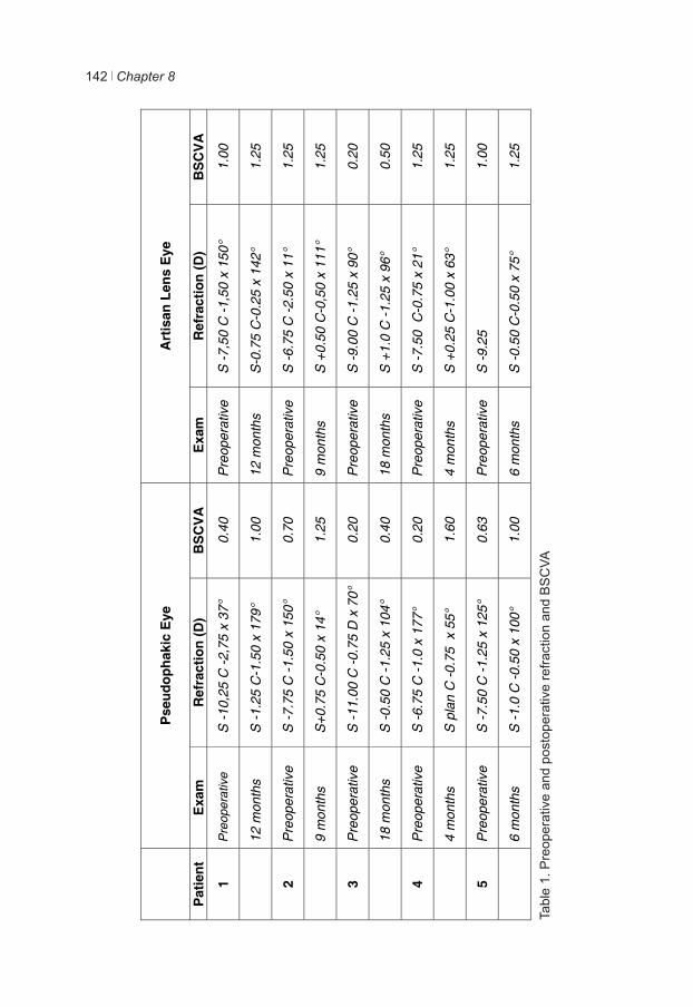
Chapter 8142
Pse
udop
haki
c E
ye
Art
isan
Len
s E
ye
Pat
ient
E
xam
R
efra
ctio
n (D
) B
SC
VA
E
xam
R
efra
ctio
n (D
) B
SC
VA
1 P
reop
erat
ive
S -1
0,25
C -2
,75
x 37
°
0.40
P
reop
erat
ive
S -7
,50
C -1
,50
x 15
0°
1.00
12
mon
ths
S -1
.25
C-1
.50
x 17
9°
1.00
12
mon
ths
S-0
.75
C-0
.25
x 14
2°
1.25
2 P
reop
erat
ive
S -7
.75
C -1
.50
x 15
0°
0.70
P
reop
erat
ive
S -6
.75
C -2
.50
x 11
° 1.
25
9
mon
ths
S+0
.75
C-0
.50
x 14
° 1.
25
9 m
onth
s S
+0.
50 C
-0,5
0 x
111°
1.
25
3 P
reop
erat
ive
S
-11.
00 C
-0.7
5 D
x 7
0°
0.20
P
reop
erat
ive
S
-9.0
0 C
-1.2
5 x
90°
0.20
18
mon
ths
S -0
.50
C -1
.25
x 10
4°
0.40
18
mon
ths
S +
1.0
C -1
.25
x 96
° 0.
50
4 P
reop
erat
ive
S -6
.75
C -1
.0 x
177
° 0.
20
Pre
oper
ativ
e S
-7.5
0 C
-0.7
5 x
21°
1.25
4
mon
ths
S p
lan
C -0
.75
x 5
5°
1.60
4
mon
ths
S +
0.25
C-1
.00
x 63
° 1.
25
5 P
reop
erat
ive
S -7
.50
C -1
.25
x 12
5°
0.63
P
reop
erat
ive
S -9
.25
1.
00
6
mon
ths
S -1
.0 C
-0.5
0 x
100°
1.
00
6 m
onth
s S
-0.5
0 C
-0.5
0 x
75°
1.25
Ta
ble
1. P
reop
erat
ive
and
post
oper
ativ
e re
fract
ion
and
BS
CVA

Postoperative Anisometropia and the Artisan Lens 143
Patient 3
A 54-year-old male high myope had a bilateral myopic retinal degeneration and a cortinuclear cataract in his right eye. After phacoemulsification, his eye was suited with an Acrysof 6.0 mm foldable lens (8 D) in May of 2000 (K.T.). Two weeks hereafter, the refraction in his phakic eye was corrected with an Artisan 6.0 mm lens (-10.5 D). Although his visual acuity remained relatively low due to retinal pathology, his VA had improved by two Snellen lines in his pseudophakic eye and by three lines in his phakic eye when examined eighteen months postoperative.
Patient 4
This 33-year-old colour-blind male myope was diagnosed with a nuclear cataract causing monocular diplopia. As the vision in the eye deteriorated, the patient was treated for his cataract with phacoemulsification and lens implantation (FLEX 652, 6.5 mm optic, 9 D). Two months later, his right eye underwent a phakic lens (Artisan 6.0 mm, -9.5 D) implantation. The last check-up six months postoperative showed no abnormalities and stable refraction in both eyes. His pseudophakic eye could achieve a visual acuity of 1.60 without correction.
Patient 5
A highly myopic female patient 37 years of age was diagnosed with a conjunctival MALT lymphoma in her right eye. As a consequence of radiotherapy, the eye developed keratoconjunctivitis sicca and required punctum plugs on the upper and lower lacrimal ducts two and again three years after the initial diagnosis. During this last examination, a posteriocapsular cataract was also diagnosed in this eye. The patient underwent phacoemulsification and lens implantation (Allergan SI40 NB, 12 D) three months later. Postoperative complaints of aniseikonia with the use of spectacles led to the myopia in her left eye being corrected with the Artisan 6.0 mm lens (-10.0 D) four months hereafter. Six months postoperative, the patient had a stable refraction and a BSCVA of 1.00 or higher in each eye.
Four of the five patients had a postoperative BSCVA of 1.00 or higher in their pseudophakic eye, three of the five patients were within 1.00 D of emmetropia and all five within 2.00 D. All patients were within 1.00 D of emmetropia in their Artisan implanted eye and three within 0.50 D. Three of the five phakic eyes showed a postoperative improvement in their BSCVA and the other two were unchanged at 1.25. The safety index (mean postoperative BSCVA / mean preoperative BSCVA)
Chapter 8144
was 1.40. The efficacy index (mean postoperative UCVA / mean preoperative BSCVA) was 0.88.
RSVP Questionnaire
There were no significant differences between the eyes in any of the subscales of the RSVP (Table 2). The eyes with the Artisan lens scored on average better in the subscales concerns, functioning, symptoms and glare, while the pseudophakic eyes were, on average, better for driving. There were no differences noted by any of the patients in the subscales expectations, optic problems and problems with glasses/contact lenses.
Two different total scores were calculated, one including all the subscales and one disregarding the subscales “expectations” and “problems with glasses and/or contact lenses”, as these two subscales are dependent on the individual patients and not on the individual eyes. The difference between the average scores of the eyes then increased by one point, but remained insignificant (Student’s paired t-test, p>0.05).
In all but one question, patients experienced no average significant difference between the phakic and pseudophakic eyes. When asked to rate their satisfaction with reading and near vision, 4 of the 5 patients expressed a preference for their phakic eye while one preferred neither (patient 3) (p=0.02).
Discussion
When a relatively young highly myopic patient develops a cataract in one eye, it is questionable if he or she will be willing to or even should undergo an operation in their phakic eye. Aversion to yet another operation, not wanting to risk their other eye, being satisfied with their current method of correction or electing for monovision are all reasons for leaving the healthy eye untouched. However, some patients may find their anisometropia troublesome, albeit only having to deal with it until they put on their corrective lenses. Certain patients may not be able to tolerate contact lenses and will have to deal with aniseikonia with the use of spectacles. Yet others may just want to have their refractive error about the same in each eye permanently. Our five patients with unilateral cataract had the myopia of their sound eye corrected with the Artisan lens.
Three of the five pseudophakic eyes were within 1.00 D of emmetropia, and all five within 2.00 D. All patients showed an improvement in visual acuity and all
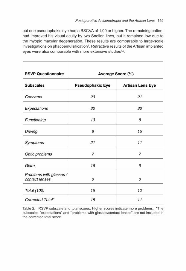
Postoperative Anisometropia and the Artisan Lens 145
but one pseudophakic eye had a BSCVA of 1.00 or higher. The remaining patient had improved his visual acuity by two Snellen lines, but it remained low due to the myopic macular degeneration. These results are comparable to large-scale investigations on phacoemulsification6. Refractive results of the Artisan implanted eyes were also comparable with more extensive studies1,2.
Table 2. RSVP subscale and total scores: Higher scores indicate more problems. *The subscales “expectations” and “problems with glasses/contact lenses” are not included in the corrected total score.
RSVP Questionnaire Average Score (%)
Subscales Pseudophakic Eye Artisan Lens Eye
Concerns 23 21
Expectations 30 30
Functioning 13 8
Driving 8 15
Symptoms 21 11
Optic problems 7 7
Glare 16 6
Problems with glasses / contact lenses 0 0
Total (100) 15 12
Corrected Total* 15 11
Table 2. RSVP subscale and total scores: Higher scores indicate more problems. *The subscales “expectations” and “problems with glasses/contact lenses” are not included in the corrected total score.
Chapter 8146
RSVP
The Refractive Status and Visual Profile Questionnaire survey led to some interesting results. The patients only experienced one significant difference between their eyes – they preferred reading with their phakic eye. Such a distinction was not made for vision in general (with or without refractive correction). Although this partiality would seem reasonable considering that their preferred eye was their only phakic eye, one must note that two of the four patients who expressed this preference were above the age of fifty. Their partiality for the phakic eye could indicate that one should consider leaving the healthy lenses of presbyopic patients intact and not opt for a clear lens extraction too early, although a much greater number of patients would need to be assessed to be able to conclude this. The one patient of 54 years who felt he read equally well with both eyes (Patient 3) suffered from bilateral macular degeneration and recorded only one difference between the eyes throughout the whole questionnaire: his vision was more often cloudy or foggy in his phakic eye.
As a group, there were no significant differences among the eyes in any of the subscales. While it appeared that, on average, patients had more problems driving with their Artisan eye, this difference lay in one individual, Patient 4, who had a confirmed higher visual acuity in his pseudophakic eye. They also tended to function better with their phakic eyes, however this was due to two patients who had had troublesome pseudophakic eyes (patient 1 and 5, with RD and keratoconjunctivitis sicca respectively). Although the bulk of the difference in the subscale “symptoms” is due to patient 5 (with keratoconjunctivitis sicca in her cataract operated eye), 4 out of 5 patients noted a difference in their “vision being cloudy or foggy”. Three experienced more trouble with their pseudophakic eyes and one (Patient 3) with his Artisan implanted eye. Patient 4 did not record any difference.
One shortcoming of our small RSVP survey is that the questions were not also posed preoperatively. This would have allowed a quantitative measurement of the change in vision-related quality of life. In addition, patients should have been asked to complete the questionnaire during identical postoperative periods, instead of between six and eighteen months after Artisan lens implantation. Further variability was also incurred by the periods between the cataract treatment and pIOL implantation, which varied between one week and four months. Moreover, the sample size was limited.
Before selecting the Artisan lens to correct the refractive error for the healthy eye, all possible options available were discussed with the patients. Clear lens extraction (CLE) and implantation of a multifocal lens, which would have rendered both eyes pseudophakic with similar refractions, were considered. However, as
Postoperative Anisometropia and the Artisan Lens 147
CLEs increase the risk of retinal detachment in myopic eyes, they were rejected7. The postoperative RSVP results also indicated that CLEs may not be ideal for younger presbyopes, considering that two of our three patients above the age of fifty favoured reading with their Artisan-implanted eye and did not show this preference for vision in general. Posterior chamber phakic IOLs, on the other hand, do retain accommodation in young patients, however they too may have a higher rate of RD and can also induce pupillary block glaucoma and cataract8,9. LASIK could have been performed in our patients with refractive errors less than -10.00 D.
Previous positive experience with the Artisan lens led to its choice1-3. Furthermore, preoperative biometry for pseudophakic lens implantation could be performed reliably with the Artisan lens in place if a cataract was to develop. Removal of the iris-fixated lens, phacoemulsification and pseudophakic lens implantation could then also take place in one procedure.
This case series demonstrates the possibility of using the Artisan lens for anisometropia after the treatment of unilateral cataract in myopic patients. The five patients undergoing this treatment significantly preferred reading with their eye implanted with the Artisan pIOL, without displaying the same preference for their vision in general. More patients need to be considered before definitive conclusions can be made.
Chapter 8148
References
1. Budo C, Hessloehl JC, Izak M, et al. Multicenter study of the Artisan phakic intraocular lens. J Cataract Refract Surg 2000;26(8):1163-1171.
2. Landesz M, van Rij G, Luyten GPM. Iris-claw phakic intraocular lens for high myopia. J Refract Surg. 2001 Nov-Dec;17(6):634-40.
3. Saxena R, van Minderhout HM, Luyten GP. Anterior chamber iris-fixated phakic intraocular lens for anisometropic amblyopia. J Cataract Refract Surg 2003 Apr;29(4):835-8
4. Vitale S, Schein OD, Meinert CL, Steinberg EP. The Refractive Status and Vision Profile; a questionnaire to measure vision-related quality of life in persons with refractive error. Ophthalmology 2000;107(8):1529-1539.
5. Schein OD, Vitale S, Cassard SD, Steinberg EP. Patient outcomes of refractive surgery; The Refractive Status and Vision Profile. J Cataract Refract Surg 2001;27(5):665-673.
6. Murphy C, Tuft SJ, Minassian DC. Refractive error and visual outcome after cataract extraction. J Cataract Refract Surg 2002;28(1):62-66.
7. Colin J, Robinet A. Clear lensectomy and implantation of a low-power posterior chamber intraocular lens for correction of high myopia. Ophthalmology 1997;104(1):73-78.
8. Ruiz-Moreno JM, Alió JL, Pérez-Santonja JJ, De La Hoz F. Retinal detachment in phakic eyes with anterior chamber intraocular lenses to correct severe myopia. Am J Ophthalmol 1999;127(3):270-275.
9. Uusitalo RJ, Aine E, Sen NH, Laatikainen L. Implantable contact lens for high myopia. J Cataract Refract Surg 2002;28(1):29-36.

9Chapter 9
Discussion

Discussion 151
The methods of correction of refractive errors have evolved immensely, especially throughout the 20th Century. “Coke Bottle” glasses are no longer the norm for persons requiring refractive correction. Technological improvements in lens development have allowed for thinner glasses, although this has not been able to alleviate the change in image size associated with higher refractive errors. Contact lenses do surmount this problem, and due to improvements in materials, allow for more tolerability and longer wear. Nevertheless, contact lens intolerance due to allergy or over-wear still exists. For these patients, and for those seeking freedom from contact lenses or spectacles, refractive surgery has been able to provide many answers.
This thesis studied many different aspects surrounding refractive surgery and has many clinical consequences associated with it:
The evaluation of the toric Artisan lens displayed excellent visual results with reliable axial alignment of the lens. The observation of consistent incision-induced astigmatism of nearly two thirds of a Diopter led to advice on fine-tuning postoperative astigmatism by systematically correcting for the incision-induced astigmatism. We were also able to demonstrate that our method of using the Javal keratometer to mark the corneal limbus before the operation leads to relatively precise surgical alignment of the lens. A larger series of patients and a study on whether pre-emptively correcting the incision-induced astigmatism leads to more precise astigmatic correction is advised.
Our study of hypermetropic eyes revealed that the Artisan lens for hypermetropia achieves excellent visual results. However, the appearance of posterior synechiae in 15% of the eyes (4 of 26) and eventual removal and phacoemulsification of clear lenses in nearly 8% of the eyes (2 of 26) suggests that these eyes are more prone to postoperative problems than myopic eyes receiving the Artisan lens. These last eyes had convex shaped irides. Special preoperative attention to the anatomy of hyperopic eyes can prevent these complications. Furthermore, the indication of an inverse relationship between anterior chamber depth and endothelial cell loss prompted our seven-year follow-up study on endothelial cell density (ECD) and the Artisan lens. Further analysis is required to see if implementation of our advice does in fact lead to less endothelial cell loss.
The significant relationship between endothelial cell loss and the anterior chamber depth helped to better understand why some eyes suffer a more profound ECD loss. This awareness also allowed us to advise surgeons to assess their patients differently to what the manufacturer advises. Instead of simply checking if the patient meets all the different requirements for lens implantation, we suggested that certain aspects should be evaluated concurrently; young patients just
Chapter 9152
narrowly meeting the inclusion criteria for ECD and anterior depth should perhaps no longer be considered for the Artisan lens to avoid severe endothelial cell loss or even corneal decompensation in the distant future. Supplementary research is required to identify the mechanism behind this relationship and which relationship of anterior chamber depth, endothelial cell density and age should keep a surgeon from operating.
The Vision-Related Quality of Life (VR-QOL) surveys identified 4 key points: 1. Myopia is negatively correlated with VR-QOL. 2. Patients seeking refractive surgery have a lower VR-QOL than those not seeking refractive surgery, mainly due to their no longer wanting to wear contact lenses or glasses and their not wanting the problems associated with them. This suggests that more comfortable and convenient methods of refractive correction could dissuade some patients from undergoing refractive surgery.3. After undergoing uncomplicated refractive surgery, VR-QOL increases.4. Despite Excimer laser surgery and a nearly emmetropic postoperative state, these patients, on average, do not score better on certain VR-QOL surveys than patients who have not permanently corrected their myopia. This implies that there may be some inherent differences between these two patient populations.Further analysis is required to identify which patients would benefit most, and which patients would benefit least from refractive surgery.
The good visual results of the Artisan lens implanted in a four-year old boy with unilateral high myopia are very encouraging. This can be an option for refractory cases with severe patient compliance issues. Still, many more cases with longer follow-ups extending into adulthood or later are needed before long-term complications (especially regarding corneal endothelial cell loss) can be excluded and refractive surgery can be deemed safe in the pediatric population.
The five-patient case report studying the independent RSVP scores of individual eyes demonstrated that adult patients (presbyopic or nearly presbyopic) show virtually no preference for a myopic eye corrected with a pseudophakic lens after phacoemulsification or corrected with an Artisan lens. However, there remains a preference to read with the phakic eye. Although this study is small, it does demonstrate that reading with a phakic eye is preferred to a pseudophakic eye. This may help to evaluate patients seeking refractive surgery who are considering clear lens extractions. Maintaining some degree of accommodation may be the preferred route if the crystalline lens is clear.
To this day, no refractive surgeon can guarantee 20/20 vision without correction in the healthiest of eyes. Much more research on the known and as yet unknown stumbling blocks of refractive surgery is required before it can be perfected.

Discussion 153
More precise lens calculations and algorithms provide better results everyday, as does progress in technology such as smaller incisions, better quality lasers and instruments. In addition, advancements in research, such as the genetic and environmental factors surrounding refractive errors could lead to sound preventive treatments and measures to prevent ametropia. More research regarding Vision-Related Quality of Life can help to guide surgeons in their quest for excellence.
Chapter 9154
Pioneer Harold Ridley faced much ostracism when his new invention, the intraocular lens, was introduced half a century ago. He recounted some comments he had received throughout the years about his intraocular lens during the Gullstrand Lecture, in Stockholm, Sweden, 1993 (as published in Trivedi et al., Sir Nicholas Harold Ridley. He changed the world, so that we might better see it. Indian J Ophthalmol 2003;51(3)211-6.)
‘Dr. Ridley, Why don’t you… GO HOME!’ (Philadelphia)
‘Would you have one of THESE THINGS put in your son’s eye?’ (Oxford)
‘As long as I remain in charge of this department no implant will ever be done’(European Professor of Ophthalmology)
‘This operation should never be done’ (Chicago)
‘It offends the first principle of ophthalmic surgery and could cause malignant disease’ (USA)
‘Rayners should be prosecuted for supplying intraocular lenses’
Ridley eventually had his invention placed in his own eye and suffered only minor complications.
Needless to say, the world’s perspective on the intraocular lens has changed. No doubt the inventors of tomorrow will also be facing such reactions. If these future innovators can persevere and look past the criticism, it will undoubtedly lead to newer and fresher developments in Ophthalmology and change the field of eye care.

10Chapter 10
Summary / Samenvatting

Summary / Samenvatting 157
Summary
The phakic Artisan lens is an intraocular lens (IOL) that can be implanted in the anterior chamber of the eye to correct nearsightedness, farsightedness and/or astigmatism, without having to remove the natural lens of the eye. This lens can be an ideal option for eyes that are not suitable for other methods of refractive surgery, or for patients who are not comfortable undergoing technically irreversible surgical interventions. This thesis examines the visual results of the Artisan lens implanted in hypermetropic and astigmatic eyes and its effect on the corneal endothelium. Also, it investigates the Vision-Related Quality of Life (VR-QOL) of myopic contact lens wearers and compares this with patients having gone either bilateral Artisan lens implantation or Excimer laser treatments for myopia. Furthermore, unique applications of the Artisan lens are considered.
Chapter 1 is a general introduction of the subjects outlined in this thesis. The essentials of myopia and hypermetropia are summarized. Studies have found that myopia is increasing in alarming rates in Asia. Although inheritance and near work have been recognized as substantial risk factors for ametropia, there are no known truly effective methods to either prevent or undo myopia or hypermetropia. Until recently, one could only correct refractive errors with glasses or contact lenses. Although this usually leads to an increased visual acuity, patients still have to endure the negative aspects of wearing glasses, such as troublesome changes in image size and feelings of self-consciousness. Contact lens wearers risk infections and the development of intolerance. These aspects can affect Vision-Related Quality of Life in some ametropic individuals. Advances in refractive surgery, such as the development of Excimer laser techniques and the Artisan phakic IOL, have allowed patients with either myopia or hypermetropia to decrease their dependence on spectacles or lenses. The development of VR-QOL questionnaires allows us to scrutinize the effects of refractive surgery among these patients.
In Chapter 2, we analyze postoperative astigmatism as a factor of incision-induced corneal astigmatism and axial alignment in a series of 54 eyes implanted with the Artisan toric phakic intraocular lens. Also, we calculate both the Safety Index and the Efficacy Index of the lens. All axes are labelled preoperatively on the cornea with a surgical marker with the use of a Javal keratometer. Mean incision-induced corneal astigmatism and mean axial misalignment of the lens are found to be nominal (0.75 ± 0.61 D and 0.37° ± 5.34° respectively). Both the Safety and Efficacy indexes are securely above 1.0, implying good postoperative visual results. We find that the implantation of the toric Artisan lens leads to safe and efficacious results, and that axial misalignment of the lens has minimal effects on astigmatism. However, the consistent minor effect of the incision-induced
Chapter 10158
astigmatism might warrant a systematic undercorrection of -0.50D to fine tune postoperative cylindrical error.
In Chapter 3, we evaluate the results of 26 eyes of 13 patients implanted with the Artisan lens for hypermetropia. The study, with a 22-month follow-up, demonstrates stable and predictable results, with nearly 91% of eyes achieving ±1.00D of intended refractive error. However, 2 patients (4 eyes) develop posterior synaechiae with pigment deposits. One of these patients eventually undergoes bilateral lens removal and clear lens extractions. Furthermore, postoperative analysis of the corneal endothelium reveals large, although statistically insignificant endothelial cell loss, and a negative correlation between anterior chamber depth and the decrease of endothelial cell density.
In Chapter 4, we investigate the postoperative decrease in endothelial cell densities (ECD) in 318 myopic eyes that have had Artisan lenses for up to 7 years. Not only do we find a significant loss of cells after three years, we also find a significant negative correlation between the decline in endothelial cell density and the depth of the anterior chamber. This loss shows no correlation with any of the following: patient age, gender, refractive error or the size of the incision.
The Vision-related Quality of Life (VR-QOL) of myopic contact lens wearers not seeking refractive surgery is assessed in Chapter 5. This study, with the use of the Dutch translations of the Refractive Vision and Status Profile (RSVP) and the National Eye Institute’s Visual-Functioning Questionnaire-25 (NEI-VFQ-25), determines that VR-QOL is adversely affected by myopia. The greater the myopia, the worse one’s vision-related QOL. This remains independent of both gender and age. One year after completion of the initial questionnaire, the subjects complete the questionnaires once again, revealing stable results.
In Chapter 6, we study the effects of refractive surgery on VR-QOL. We find that nearsighted individuals seeking either bilateral Excimer laser treatments or Artisan pIOL implants express lower VR-QOL scores on both the RSVP and the NEI-VFQ-25 than their myopic counterparts not seeking treatment. This is mostly due to their eagerness to be rid of their contact lenses or glasses. Also, we find that patients undergoing uncomplicated refractive surgery experience a significant increase in their postoperative VR-QOL scores. Lastly, we observe that both Excimer laser and Artisan lens patients score significantly better postoperatively on the RSVP than the myopic contact lens wearers. However, with the use of the NEI-VFQ-25, only Artisan patients have significantly higher VR-QOL scores than contact lens wearers (when correcting for preoperative refractive error). The (nearly) emmetropic patients having undergone Excimer laser treatments do not score significantly better than myopic contact lens wearers, who remain myopic.
Summary / Samenvatting 159
The implantation of the Artisan lens in the right eye of a four-year old male patient with severe unilateral myopia is documented in Chapter 7. Occlusion therapy had been severely hampered by poor patient compliance with glasses and contact lenses, leading to anisometric amblyopia with a visual acuity varying between 0.50 and 0.83 in the worse eye and an absence of stereovision. Three years after unremarkable surgery, the patient has a best corrected visual acuity of 1.2 in the operated eye (versus 1.0 in the other eye) and demonstrates some stereovision. Still, there is a demonstrable difference in endothelial cell density between both eyes.
In Chapter 8, we study both the objective and subjective results of a unique situation where five bilaterally myopic patients (three of which presbyopic) undergo a cataract extraction in one eye and an Artisan lens implantation in the other eye (to correct the resulting anisometropia). Patients show excellent refractive results bilaterally, four out of five being within 1.00 D of emmetropia in the pseudophakic eye and all five being within 1.00 D of emmetropia in the phakic Artisan eye. The patients are also requested to complete two RSVP questionnaires simultaneously, one for each eye. The results of the RSVP demonstrate very similar results for both eyes, however there is a significant preference to read with the eye implanted with the Artisan lens.
Chapter 10160
Samenvatting
De Artisan lens is een intra-oculaire lens (IOL) die in de voorste oogkamer geïmplanteerd kan worden. De Artisan lens corrigeert bij- en verziendheid en/of astigmatisme, zonder dat de natuurlijke lens van het oog verwijderd hoeft te worden. Deze lens kan een ideale optie zijn voor ogen die niet geschikt zijn voor andere methoden van refractiechirurgie, of voor patiënten die liever geen irreversibele operatie willen ondergaan. Dit proefschrift onderzoekt de visuele resultaten van de Artisan lens in hypermetrope en astigmate ogen en het effect van de lens op het cornea endotheel. Ook wordt de visueel gerelateerde kwaliteit van leven (Vision-Related Quality of Life ofwel VR-QOL) van myope contactlensdragers uitgezocht en vergeleken met de VR-QOL van patiënten die bilaterale Artisan lens implantaties of excimerlaser behandelingen voor myopie ondergaan. Bovendien worden unieke toepassingen van de Artisan lens bekeken.
Hoofdstuk 1 is een algemene introductie tot de onderwerpen die in dit proefschrift worden besproken. De hoofdzaken van myopie en hypermetropie zijn samengevat. Uit verschillende studies blijkt dat de prevalentie van myopie in Azië in zorgbarende aantallen toeneemt. Genetische aanleg en omgevingsfactoren zijn belangrijke risicofactoren voor het ontwikkelen van een refractieafwijking. Desondanks zijn er geen voldoende effectieve mogelijkheden bekend om invloed uit te oefenen op de ontwikkeling of toename van een myopie of hypermetropie. Tot voor kort waren een bril of contactlenzen de enige manier om refractieafwijkingen te corrigeren. Al leiden deze tot een visusverbetering, patiënten moeten alsnog de ongunstige aspecten hiervan verdragen, zoals vervelende veranderingen in beeldgrootte en een negatief zelfbeeld. Contactlensdragers riskeren infecties en het ontwikkelen van contactlensintolerantie. Dit kan bij sommige mensen de VR-QOL negatief beïnvloeden. De opmars van de refractiechirurgie, zoals de ontwikkeling van de excimerlaser en van de Artisan phake IOL, leidt tot minder afhankelijkheid van bril en/of contactlenzen bij patiënten met myopie of hypermetropie. De ontwikkeling van de VR-QOL vragenlijsten laat ons de effecten van refractiechirurgie op de kwaliteit van leven bij deze patiënten onderzoeken.
In Hoofdstuk 2 onderzoeken wij in een reeks van 54 ogen het postoperatieve astigmatisme veroorzaakt door geïnduceerd astigmatisme door de (wond-)incisie en de axiale positie van de Artisan torische phake intraoculaire lens. Ook berekenen wij de Safety- en Efficacy Index. Preoperatief zijn alle assen met een chirurgische marker op de cornea getekend met behulp van de Javal keratometer. Het gemiddeld “chirurgisch geïnduceerd” corneaal astigmatisme en de gemiddelde axiale afwijking van de lens zijn nominaal (respectievelijk 0.75 ± 0.61 D en 0.37° ± 5.34°). Zowel de Safety Index als de Efficacy Index zijn meer dan 1.0, hetgeen een goede postoperatieve visuele resultaten impliceert. Wij
Summary / Samenvatting 161
vinden dat de implantatie van de torische Artisan lens tot een veilige en effectieve uitkomst leidt, en dat de axiale afwijking van de lens een zeer gering effect op astigmatisme heeft. Echter het minimale, maar altijd aanwezige effect van astigmatisme geïnduceerd door de incisie zou een systematische ondercorrectie van -0.50 D kunnen rechtvaardigen om postoperatieve cilindrische afwijkingen te kunnen verminderen.
In Hoofdstuk 3 worden de resultaten de Artisan hypermetrope lens geëvalueerd in 26 ogen van 13 patiënten. Deze studie, met een follow-up van 22 maanden, toont stabiele en voorspelbare resultaten, waarbij bijna 91% van de ogen binnen 1.00 D van de voorgenomen refractieafwijking uitkomen. Toch ontwikkelen 2 patiënten (4 ogen) synechiae posteriores met pigmentdeposities. Bij één van deze patiënten zijn beide Artisan lenzen verwijderd en is er een bilaterale heldere lens extractie (een cataractextractie bij een heldere lens) verricht. Uit deze studie blijkt dat er een groot, echter statistisch niet significant endotheelcelverlies is en dat er bovendien een negatieve correlatie bestaat tussen de voorste oogkamer diepte en de daling van de endotheelcel dichtheid.
In Hoofdstuk 4 onderzoeken wij de daling van de endotheelcel dichtheid in 318 myope Artisan lens geïmplanteerde ogen tot 7 jaar postoperatief. Wij vinden niet alleen een significant verlies van endotheelcellen na drie jaar maar ook een significante negatieve correlatie tussen de daling in endotheelcel dichtheid en de diepte van de voorste oogkamer. Dit verlies toont geen correlatie met zowel leeftijd als geslacht, refractieafwijking van het oog of incisiegrootte.
In Hoofdstuk 5 wordt de visueel gerelateerde kwaliteit van leven van bijziende contactlensdragers die geen refractiechirurgie willen ondergaan beoordeeld. Deze studie, waarbij de Nederlandse vertalingen van de Refractive Status and Vision Profile (RSVP) en de Visual Functioning Questionnaire van de National Eye Institute (NEI-VFQ-25) worden gebruikt, laat zien dat de VR-QOL ongunstig door myopie wordt beïnvloed. Hoe groter de myopie, hoe slechter de VR-QOL; dit is onafhankelijk van zowel geslacht als leeftijd. Een jaar na initiële voltooiing van de vragenlijsten, worden de vragenlijsten opnieuw ingevuld. Dit laat onveranderde resultaten zien.
Hoofdstuk 6 beschrijft de gevolgen van refractiechirurgie voor de VR-QOL. Uit deze studie blijkt dat myope individuen die bilateraal excimer laserbehandelingen of Artisan lens implantaties willen ondergaan slechter scoren dan hun bijziende tegenhangers die niet geopereerd willen worden op zowel de RSVP als de NEI-VFQ-25. Dit is met name toe te schrijven aan lage scores vanwege het feit dat ze graag van hun contactlenzen of bril af willen zijn. Na ongecompliceerde refractiechirurgie zien wij een significante toename in de VR-QOL scores. Ten
Chapter 10162
slotte vinden wij dat zowel excimerlaser als Artisan lens patiënten beduidend beter postoperatief scoren op de RSVP dan de myope contactlensdragers. Echter bij de NEI-VFQ-25 halen slechts de Artisan lens patiënten significant hogere scores dan de contactlensdragers (wanneer er gecorrigeerd wordt voor de preoperatieve refractieafwijking). De (bijna) emmetrope excimerlaserpatiënten hebben geen significant hogere scores op de NEI-VFQ-25 dan de contactlensdragers, die overigens nog steeds myoop zijn.
Hoofdstuk 7 documenteert de implantatie van de Artisan lens in het rechter oog van een vier jaar oud patiëntje met een ernstige unilaterale myopie, resulterend in een anisometropie amblyopie. Door slecht gebruik van de bril en contactlens wordt de occlusietherapie sterk belemmerd, met een wisselende visus tussen 0.50 en 0.83 in het slechtste oog. Tevens is er geen dieptezien. Drie jaar na de ongecompliceerde chirurgie, heeft het patiëntje een visus van 1.2 in het geopereerde oog (tegenover 1.0 in het linkeroog) en er is grof dieptezien aanwezig.
In Hoofdstuk 8 worden zowel de objectieve als subjectieve resultaten bestudeerd van een unieke situatie waarbij vijf myope patiënten (drie hiervan presbyoop) een cataractextractie ondergaan in één oog en een Artisan lens implantatie in het andere oog (om het resulterende anisometropie te corrigeren). Postoperatief zijn vier van de vijf pseudofake ogen binnen 1.00 D van emmetropie en alle vijf Artisan lenzen geïmplanteerde ogen binnen 1.00 D van emmetropie. De patiënten worden verzocht om twee RSVP-vragenlijsten simultaan in te vullen, één voor elk oog. De RSVP toont zeer gelijkaardige resultaten voor beide ogen, echter met een significante voorkeur om te lezen met het oog met de Artisan lens.

AppendixAppendix

National Eye Institute Visual Functioning Questionnaire – 25
Nederlandse Consensus Vertaling (VFQ-25/NL)
versie 2001
VERSIE OM ZELF IN TE VULLEN (gezichtsscherpte > 0.5)
G.W. van der Sterre1, E.S. van de Graaf1, C.A. Verezen2561, C.F.M. Meulendijks3, J.S.A.G. Schouten4,
R. Saxena1, J.R. Polling1, L.J. van Rijn5, C.B. Hoyng6, M.L. Essink-Bot7, H.J. Simonsz1
Juli 2001 1 Erasmus Medisch Centrum Rotterdam, afdeling Oogheelkunde, Rotterdam 2 Ergra Low Vision consultants, Den Haag 3 Universitair Medisch Centrum St Radboud, afdeling Medical Technology Assessment, Nijmegen 4 Capgroep Epidemiologie, Universiteit Maastricht, Maastricht 5 Vrije Universiteit Medisch Centrum, afdeling Oogheelkunde, Amsterdam 6 Universitair Medisch Centrum St Radboud, afdeling Oogheelkunde, Nijmegen 7 Erasmus Medisch Centrum Rotterdam, Instituut Maatschappelijke Gezondheidszorg, Rotterdam
______________________________________________________________________
RAND hereby grants permission to use the "National Eye Institute Visual Functioning Questionnaire 25” (VFQ-25) July 1996, in accordance with the following conditions which shall be assumed by all to have been agreed to as a consequence of accepting and using this document:
1. Changes to the NEI VFQ-25 -July 1996 may be made without the written permission of RAND. However, all such changes shall be clearly identified as having been made by the recipient.
2. The user of this NEI VFQ-25 - July 1996 accepts full responsibility, and agrees to hold RAND harmless, for the accuracy of any translations of the NEI VFQ-25 Test Version -July 1996 into another language and for any errors, omissions, misinterpretations, or consequences thereof.
3. The user of this NEI VFQ-25 - July 1996 accepts full responsibility, and agrees to hold RAND harmless, for any consequences resulting from the use of the NEI VFQ-25.
4. The user of the NEI VFQ-25 -July 1996 will provide a credit line when printing and distributing this document or in publications of results or analyses based on this instrument acknowledging that it was developed at RAND under the sponsorship of the National Eye Institute.
5. No further written permission is needed for use of this NEI VFQ-25 -July 1996. ______________________________________________________________________
© R 1996
Naam:
Registratie nummer:
Pre-interventie KvL:
Post-interventie KvL:
Datum Score

Dit is een vragenlijst met uitspraken over problemen die met uw gezichtsvermogen te maken hebben, of over gevoelens die u over uw gezichtsvermogen heeft. Als u een bril of contactlenzen heeft, ga er dan bij de beantwoording van de vragen van uit dat u deze draagt. INSTRUCTIES: 1. In het algemeen willen we dat mensen deze vragenlijst zelf proberen in te vullen. Als
u vindt dat u hulp nodig heeft, aarzel dan niet om het de projectmedewerkers te vragen, ze helpen u graag.
2. Beantwoord alstublieft alle vragen, tenzij u verzocht wordt vragen over te slaan omdat ze niet van toepassing zijn.
3. Beantwoord de vragen door het juiste cijfer te omcirkelen.
4. Als u niet zeker weet hoe een vraag te beantwoorden, geef dan het best mogelijke antwoord en maak een aantekening in de linker kantlijn.
5. Als u per ongeluk de verkeerde mogelijkheid omcirkeld heeft, dan zet u door deze cirkel een kruis en omcirkelt u het antwoord dat volgens u het juiste is.
6. Vul de vragenlijst in zonder uw antwoorden met uw vrienden of familie te bespreken.
7. De vragen hebben betrekking op uw situatie gedurende de afgelopen maand.
8. Er is een versie met grote letters voor het geval uw gezichtsvermogen tekort schiet om de lijst in te kunnen vullen.
9. Als u nog vragen heeft kunt u terecht bij leden van de staf van het project, zij zullen u graag helpen.
VERKLARING VAN VERTROUWELIJKHEID: Alle informatie die identificatie mogelijk maakt van enig persoon die deze vragenlijst heeft ingevuld, zal als strikt vertrouwelijk worden beschouwd. Deze informatie zal uitsluitend worden gebruikt voor het doel van dit onderzoek, en zal niet worden onthuld of vrijgegeven voor enig ander doel, zonder voorafgaande toestemming, geopenbaard of gepubliceerd worden, uitgezonderd als vereist bij de wet.

-1- versie 2001
© R 1996
Visual Functioning Questionnaire - 25
DEEL 1 – ALGEMENE GEZONDHEID EN GEZICHTSVERMOGEN 1. Hoe zou u uw algehele gezondheidstoestand omschrijven:
(omcirkel één cijfer)
Uitstekend .......................1 Zeer goed ........................2 Goed ................................3 Redelijk............................4 Slecht...............................5
2. Zou u op dit moment zeggen dat uw gezichtsvermogen met beide ogen samen (met bril of contactlenzen, als u deze draagt), uitstekend, goed, redelijk, slecht, zeer slecht is, of bent u volledig blind?
(omcirkel één cijfer)
Uitstekend .......................1 Goed ................................2 Redelijk............................3 Slecht...............................4 Zeer slecht.......................5 Volledig blind ..................6
-2- versie 2001
© R 1996
3. Hoe vaak maakt u zich zorgen over uw gezichtsvermogen?
(omcirkel één cijfer)
Nooit…………………………….. 1 Zelden…………………………... 2 Soms……………………………. 3 Vaak…………………………….. 4 Altijd…………………………….. 5
4. Hoeveel pijn of ongemak heeft u in en rond uw ogen gehad (bijvoorbeeld branderigheid, jeuk of pijn)?
(omcirkel één cijfer)
Geen………………………….…. 1 Licht…………………………….. 2 Matig……………………………. 3 Ernstig………………………….. 4 Heel ernstig……………………. 5
DEEL 2 – MOEITE MET HET UITVOEREN VAN ACTIVITEITEN De volgende vragen gaan over de moeite die u misschien met sommige activiteiten heeft, met bril op of contactlenzen in, mocht u die voor deze activiteit nodig hebben.
5. Hoeveel moeite heeft u om normale krantendruk te lezen?
(omcirkel één cijfer) Geen enkele moeite…………………………………………………….….. 1 Een beetje moeite……………………………………………………..…… 2 Matige moeite…………………………………………………………..…. . 3 Enorme moeite…………………………………………………………..…. 4 Hiermee gestopt vanwege het slechte gezichtsvermogen……..…... 5 Hiermee gestopt om andere redenen, of op u niet van toepassing. 6
-3- versie 2001
© R 1996
6. Hoeveel moeite heeft u met werkzaamheden of hobby’s, waarbij u goed dichtbij moet kunnen zien, zoals koken, naaien, dingen in huis repareren, of bij het gebruik van handgereedschap?
(omcirkel één cijfer) Geen enkele moeite…………………………………………………….….. 1 Een beetje moeite……………………………………………………..…… 2 Matige moeite…………………………………………………………..…. . 3 Enorme moeite…………………………………………………………..…. 4 Hiermee gestopt vanwege het slechte gezichtsvermogen……..…... 5 Hiermee gestopt om andere redenen, of op u niet van toepassing. 6
7. Hoeveel moeite heeft u, vanwege uw gezichtsvermogen, met het vinden van
iets op een volle plank?
(omcirkel één cijfer) Geen enkele moeite…………………………………………………….….. 1 Een beetje moeite……………………………………………………..…… 2 Matige moeite…………………………………………………………..…. . 3 Enorme moeite…………………………………………………………..…. 4 Hiermee gestopt vanwege het slechte gezichtsvermogen……..…... 5 Hiermee gestopt om andere redenen, of op u niet van toepassing. 6
8. Hoeveel moeite heeft u met het lezen van straatnaamborden of de namen van winkels?
(omcirkel één cijfer)
Geen enkele moeite…………………………………………………….….. 1 Een beetje moeite……………………………………………………..…… 2 Matige moeite…………………………………………………………..…. . 3 Enorme moeite…………………………………………………………..…. 4 Hiermee gestopt vanwege het slechte gezichtsvermogen……..…... 5 Hiermee gestopt om andere redenen, of op u niet van toepassing. 6
-4- versie 2001
© R 1996
9. Hoeveel moeite kost het u, vanwege uw gezichtsvermogen, om een afstapje, een trap of een stoeprand af te stappen bij slechte verlichting of ’s nachts?
(omcirkel één cijfer) Geen enkele moeite…………………………………………………….….. 1 Een beetje moeite……………………………………………………..…… 2 Matige moeite…………………………………………………………..…. . 3 Enorme moeite…………………………………………………………..…. 4 Hiermee gestopt vanwege het slechte gezichtsvermogen……..…... 5 Hiermee gestopt om andere redenen, of op u niet van toepassing. 6
10. Hoeveel moeite heeft u, vanwege uw gezichtsvermogen, om dingen opzij op te merken terwijl u er langs loopt?
(omcirkel één cijfer) Geen enkele moeite…………………………………………………….….. 1 Een beetje moeite……………………………………………………..…… 2 Matige moeite…………………………………………………………..…. . 3 Enorme moeite…………………………………………………………..…. 4 Hiermee gestopt vanwege het slechte gezichtsvermogen……..…... 5 Hiermee gestopt om andere redenen, of op u niet van toepassing. 6
11. Hoeveel moeite heeft u, vanwege uw gezichtsvermogen, om te zien hoe mensen reageren op wat u zegt?
(omcirkel één cijfer) Geen enkele moeite…………………………………………………….….. 1 Een beetje moeite……………………………………………………..…… 2 Matige moeite…………………………………………………………..…. . 3 Enorme moeite…………………………………………………………..…. 4 Hiermee gestopt vanwege het slechte gezichtsvermogen……..…... 5 Hiermee gestopt om andere redenen, of op u niet van toepassing. 6
-5- versie 2001
© R 1996
12. Hoeveel moeite heeft u, vanwege uw gezichtsvermogen, met het uitzoeken en combineren van uw eigen kleding?
(omcirkel één cijfer) Geen enkele moeite…………………………………………………….….. 1 Een beetje moeite……………………………………………………..…… 2 Matige moeite…………………………………………………………..…. . 3 Enorme moeite…………………………………………………………..…. 4 Hiermee gestopt vanwege het slechte gezichtsvermogen……..…... 5 Hiermee gestopt om andere redenen, of op u niet van toepassing. 6
13. Hoeveel moeite heeft u, vanwege uw gezichtsvermogen, om bij mensen op visite te gaan, op feesten of in restaurants?
(omcirkel één cijfer)
Geen enkele moeite…………………………………………………….….. 1 Een beetje moeite……………………………………………………..…… 2 Matige moeite…………………………………………………………..…. . 3 Enorme moeite…………………………………………………………..…. 4 Hiermee gestopt vanwege het slechte gezichtsvermogen……..…... 5 Hiermee gestopt om andere redenen, of op u niet van toepassing. 6
14. Hoeveel moeite heeft u, vanwege uw gezichtsvermogen, met het uitgaan om bioscoopfilms, theater of sportevenementen te zien?
(omcirkel één cijfer) Geen enkele moeite…………………………………………………….….. 1 Een beetje moeite……………………………………………………..…… 2 Matige moeite…………………………………………………………..…. . 3 Enorme moeite…………………………………………………………..…. 4 Hiermee gestopt vanwege het slechte gezichtsvermogen……..…... 5 Hiermee gestopt om andere redenen, of op u niet van toepassing. 6
-6- versie 2001
© R 1996
15. Rijdt u momenteel auto, tenminste af en toe? (omcirkel één cijfer)
Ja………………………………………….….…….……..1 Ga naar vraag 15c Nee………………………………………..…………..…...2
15a. Indien nee, heeft u nooit auto gereden of heeft u het autorijden opgegeven?
(omcirkel één cijfer)
Ik heb nooit auto gereden……….……...………...…1 Ga naar vraag 17 Ik heb het autorijden opgegeven……………...…...2
15b. ALS U HET AUTORIJDEN HEEFT OPGEGEVEN: Was dat voornamelijk vanwege uw gezichtsvermogen, voornamelijk om een andere reden, of vanwege zowel uw gezichtsvermogen als om een andere reden?
(omcirkel één cijfer) Voornamelijk mijn gezichtsvermogen………………………...…..1 Ga naar vraag 17
Voornamelijk om andere redenen...…………………………….….2 Ga naar vraag 17 Zowel mijn gezichtsvermogen als om andere redenen…..……3
Ga naar vraag 17
15c. ALS U MOMENTEEL AUTORIJDT, hoeveel moeite heeft u met autorijden overdag in een bekende omgeving?
(omcirkel één cijfer)
Geen enkele moeite…………………………………………….……. 1 Een beetje moeite……………………………………………..……... 2 Matige moeite…………………………………………………..…….. 3 Enorme moeite…………………………………………………..…… 4
-7- versie 2001
© R 1996
16. Hoeveel moeite heeft u om ’s nachts auto te rijden?
(omcirkel één cijfer)
Geen enkele moeite…………………………………………………….….. 1 Een beetje moeite……………………………………………………..…… 2 Matige moeite…………………………………………………………..…. . 3 Enorme moeite…………………………………………………………..…. 4 Hiermee gestopt vanwege het slechte gezichtsvermogen……..…... 5 Hiermee gestopt om andere redenen, of op u niet van toepassing. 6
16a. Hoeveel moeite heeft u met het rijden onder moeilijke omstandigheden, zoals bij slecht weer, tijdens het spitsuur, op de snelweg of in stadsverkeer?
(omcirkel één cijfer)
Geen enkele moeite…………………………………………………….….. 1 Een beetje moeite……………………………………………………..…… 2 Matige moeite…………………………………………………………..…. . 3 Enorme moeite…………………………………………………………..…. 4 Hiermee gestopt vanwege het slechte gezichtsvermogen……..…... 5 Hiermee gestopt om andere redenen, of op u niet van toepassing. 6
-8- versie 2001
© R 1996
DEEL 3 – OMGAAN MET PROBLEMEN MET HET ZIEN De volgende vragen gaan over hoe de dingen die u doet, beïnvloed worden door uw gezichtsvermogen. Omcirkel bij elke vraag het nummer om aan te geven of de uitspraak voor u altijd, meestal, soms, zelden of nooit geldt.
(omcirkel één cijfer op elke regel) Altijd Meestal Soms Zelden Nooit
17. Krijgt u minder voor elkaar, vanwege uw gezichtsvermogen, dan u zou willen?
1 2 3 4 5
18. Bent u, vanwege uw gezichts-vermogen, beperkt in hoe lang u kunt werken of andere activiteiten kunt volhouden?
1 2 3 4 5
19. In hoeverre weerhoudt pijn of ongemak in of rond de ogen, bijvoorbeeld branden, jeuken of pijn, u ervan om de dingen te doen die u zou willen doen?
1 2 3 4 5
-9- versie 2001
© R 1996
Omcirkel na elk van de volgende uitspraken het voor u meest passende antwoord om aan te geven dat de uitspraak voor u helemaal juist is, over het algemeen juist is, over het algemeen onjuist is, of helemaal onjuist is, of dat u het niet zeker weet.
(omcirkel één cijfer op elke regel) Helemaal
juist Over het algemeen
juist
Weet het niet zeker
Over het algemeen
onjuist
Helemaal onjuist
20. Ik blijf vanwege mijn gezichtsvermogen meestal thuis
1 2 3 4 5
21. Ik voel me vaak gefrustreerd vanwege mijn gezichtsvermogen
1 2 3 4 5
22. Ik heb veel minder controle over wat ik doe, vanwege mijn gezichts-vermogen
1 2 3 4 5
23. Vanwege mijn gezichts-
vermogen moet ik teveel vertrouwen over wat andere mensen me vertellen
1 2 3 4 5
24. Ik heb veel hulp van
anderen nodig vanwege mijn gezichtsvermogen
1 2 3 4 5
25. Ik maak me zorgen dat
ik dingen doe, vanwege mijn gezichtsvermogen, die mezelf of anderen in verlegenheid brengen
1 2 3 4 5
-10- versie 2001
© R 1996
Bijlage optionele vragen
SUBSCHAAL: ALGEMENE GEZONDHEID A1. Hoe zou u uw gezondheid beoordelen, op een schaal van 0 tot 10, waarbij 0 zo slecht als dood is en 10 de best mogelijke gezondheid?
(omcirkel één cijfer op elke regel)
0 1 2 3 4 5 6 7 8 9 10
Slechtste
Beste
SUBSCHAAL: ALGEMEEN GEZICHTSVERMOGEN A2. Hoe zou u nu uw gezichtsvermogen beoordelen (met bril op of contactlenzen in, als u deze draagt) op een schaal van 0 tot 10 waarbij 0 zo slecht is als blindheid en 10 het best mogelijke gezichtsvermogen?
(omcirkel één cijfer op elke regel)
0 1 2 3 4 5 6 7 8 9 10 Slechtste
Beste
SUBSCHAAL: DICHTBIJ ZIEN A3. Hoeveel moeite heeft u om, met bril op, kleine lettertjes in een telefoonboek, op een medicijnflesje of in officiële stukken te lezen?
(omcirkel één cijfer) Geen enkele moeite…………………………………………………….….. 1 Een beetje moeite……………………………………………………..…… 2 Matige moeite…………………………………………………………..…. . 3 Enorme moeite…………………………………………………………..…. 4 Hiermee gestopt vanwege het slechte gezichtsvermogen……..…... 5 Hiermee gestopt om andere redenen, of op u niet van toepassing. 6
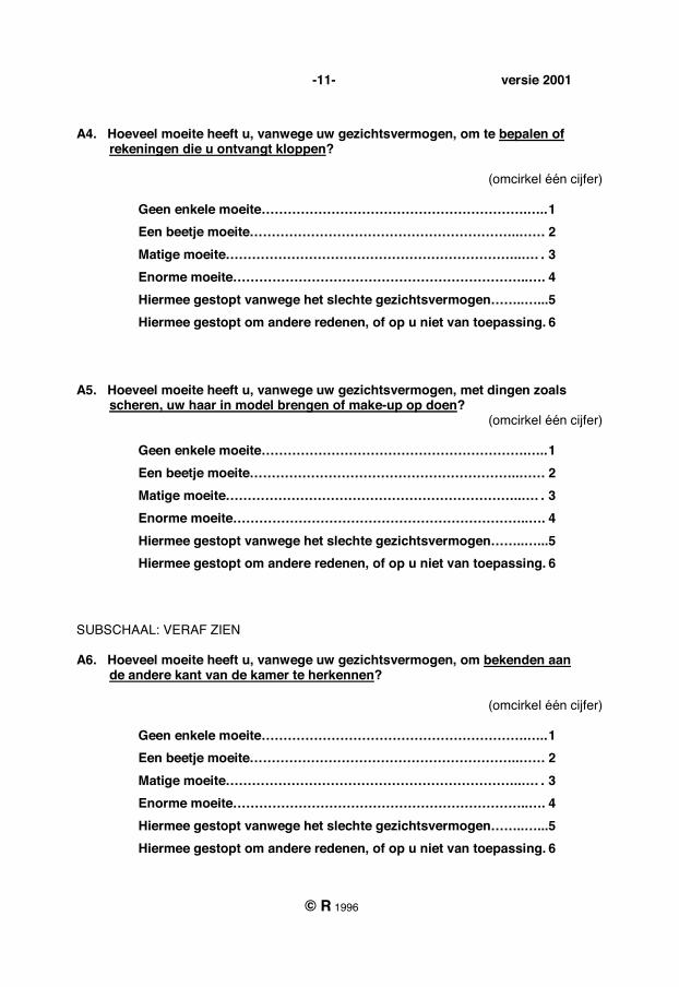
-11- versie 2001
© R 1996
A4. Hoeveel moeite heeft u, vanwege uw gezichtsvermogen, om te bepalen of rekeningen die u ontvangt kloppen?
(omcirkel één cijfer) Geen enkele moeite…………………………………………………….….. 1 Een beetje moeite……………………………………………………..…… 2 Matige moeite…………………………………………………………..…. . 3 Enorme moeite…………………………………………………………..…. 4 Hiermee gestopt vanwege het slechte gezichtsvermogen……..…... 5 Hiermee gestopt om andere redenen, of op u niet van toepassing. 6
A5. Hoeveel moeite heeft u, vanwege uw gezichtsvermogen, met dingen zoals scheren, uw haar in model brengen of make-up op doen?
(omcirkel één cijfer) Geen enkele moeite…………………………………………………….….. 1 Een beetje moeite……………………………………………………..…… 2 Matige moeite…………………………………………………………..…. . 3 Enorme moeite…………………………………………………………..…. 4 Hiermee gestopt vanwege het slechte gezichtsvermogen……..…... 5 Hiermee gestopt om andere redenen, of op u niet van toepassing. 6
SUBSCHAAL: VERAF ZIEN A6. Hoeveel moeite heeft u, vanwege uw gezichtsvermogen, om bekenden aan de andere kant van de kamer te herkennen?
(omcirkel één cijfer) Geen enkele moeite…………………………………………………….….. 1 Een beetje moeite……………………………………………………..…… 2 Matige moeite…………………………………………………………..…. . 3 Enorme moeite…………………………………………………………..…. 4 Hiermee gestopt vanwege het slechte gezichtsvermogen……..…... 5 Hiermee gestopt om andere redenen, of op u niet van toepassing. 6
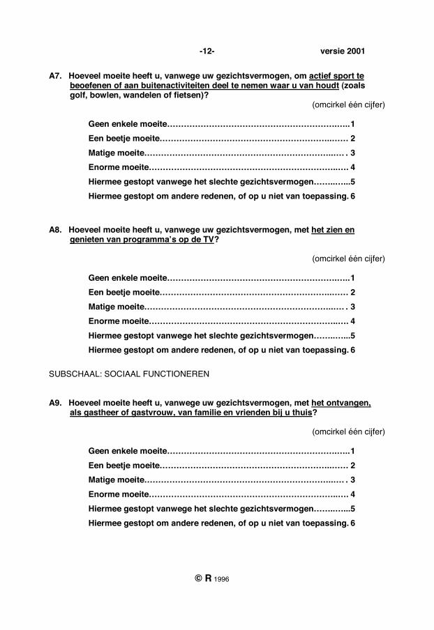
-12- versie 2001
© R 1996
A7. Hoeveel moeite heeft u, vanwege uw gezichtsvermogen, om actief sport te beoefenen of aan buitenactiviteiten deel te nemen waar u van houdt (zoals golf, bowlen, wandelen of fietsen)?
(omcirkel één cijfer) Geen enkele moeite…………………………………………………….….. 1 Een beetje moeite……………………………………………………..…… 2 Matige moeite…………………………………………………………..…. . 3 Enorme moeite…………………………………………………………..…. 4 Hiermee gestopt vanwege het slechte gezichtsvermogen……..…... 5 Hiermee gestopt om andere redenen, of op u niet van toepassing. 6
A8. Hoeveel moeite heeft u, vanwege uw gezichtsvermogen, met het zien en genieten van programma’s op de TV?
(omcirkel één cijfer) Geen enkele moeite…………………………………………………….….. 1 Een beetje moeite……………………………………………………..…… 2 Matige moeite…………………………………………………………..…. . 3 Enorme moeite…………………………………………………………..…. 4 Hiermee gestopt vanwege het slechte gezichtsvermogen……..…... 5 Hiermee gestopt om andere redenen, of op u niet van toepassing. 6
SUBSCHAAL: SOCIAAL FUNCTIONEREN A9. Hoeveel moeite heeft u, vanwege uw gezichtsvermogen, met het ontvangen, als gastheer of gastvrouw, van familie en vrienden bij u thuis?
(omcirkel één cijfer) Geen enkele moeite…………………………………………………….….. 1 Een beetje moeite……………………………………………………..…… 2 Matige moeite…………………………………………………………..…. . 3 Enorme moeite…………………………………………………………..…. 4 Hiermee gestopt vanwege het slechte gezichtsvermogen……..…... 5 Hiermee gestopt om andere redenen, of op u niet van toepassing. 6
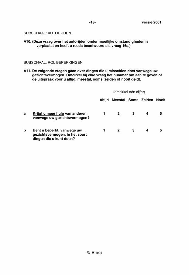
-13- versie 2001
© R 1996
SUBSCHAAL: AUTORIJDEN A10. (Deze vraag over het autorijden onder moeilijke omstandigheden is
verplaatst en heeft u reeds beantwoord als vraag 16a.) SUBSCHAAL: ROL BEPERKINGEN A11. De volgende vragen gaan over dingen die u misschien doet vanwege uw gezichtsvermogen. Omcirkel bij elke vraag het nummer om aan te geven of de uitspraak voor u altijd, meestal, soms, zelden of nooit geldt. (omcirkel één cijfer) Altijd Meestal Soms Zelden Nooit
a Krijgt u meer hulp van anderen, vanwege uw gezichtsvermogen?
1 2 3 4 5
b Bent u beperkt, vanwege uw gezichtsvermogen, in het soort dingen die u kunt doen?
1 2 3 4 5
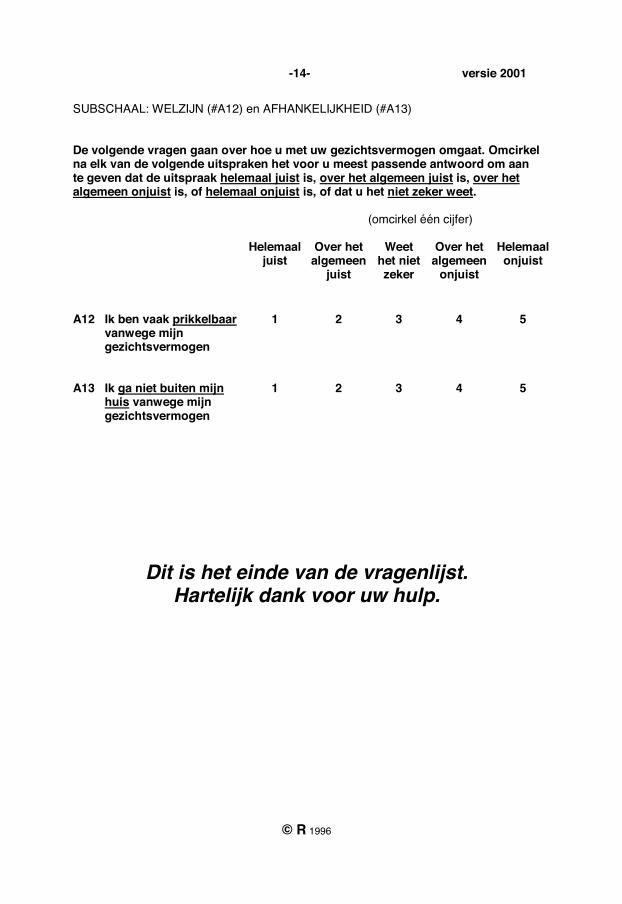
-14- versie 2001
© R 1996
SUBSCHAAL: WELZIJN (#A12) en AFHANKELIJKHEID (#A13) De volgende vragen gaan over hoe u met uw gezichtsvermogen omgaat. Omcirkel na elk van de volgende uitspraken het voor u meest passende antwoord om aan te geven dat de uitspraak helemaal juist is, over het algemeen juist is, over het algemeen onjuist is, of helemaal onjuist is, of dat u het niet zeker weet. (omcirkel één cijfer) Helemaal
juist Over het algemeen
juist
Weet het niet zeker
Over het algemeen
onjuist
Helemaal onjuist
A12 Ik ben vaak prikkelbaar vanwege mijn gezichtsvermogen
1 2 3 4 5
A13 Ik ga niet buiten mijn huis vanwege mijn gezichtsvermogen
1 2 3 4 5
Dit is het einde van de vragenlijst. Hartelijk dank voor uw hulp.

Refractive Status and Vision Profile – NL Vertaling
© 1998, The Johns Hopkins University All rights reserved.
REFRACTIVE STATUS AND VISION PROFILE
Nederlandse Consensusvertaling
R Saxena, KL de Roon Hertoge, AW Baan, M Larsen, HJ Simonsz, NID te West en GPM Luyten 1. Ziekenhuis / Kliniek: _____________________________________________________________ 2. Patiëntnummer: _________________________________________________________________ 3. Naam Patiënt (achter-, voornaam): __________________________________________________ 4. Geboortedatum (dag/maand/jaar): __________________________________________________ 5. Telefoon (thuis): ___________________________ (werk): ______________________________ 6. Datum (dag/maand/jaar): _________________________________________________________ 7. Behandelend Arts: _______________________________________________________________

Refractive Status and Vision Profile – NL Vertaling
© 1998, The Johns Hopkins University 1 All rights reserved.
REFRACTIVE STATUS AND VISION PROFILE
ACHTERGRONDINFORMATIE
Patiëntinformatie 1. Leeftijd
jaar
2. Geslacht: Vrouw ( 1) Man ( 2)
Bril- en Contactlensgebruik 3. Ik droeg in de afgelopen maand bij het kijken in de
verte:
a. Alleen een bril ( 1) b. Voornamelijk een bril, soms contactlenzen
( 2) c. Ongeveer even vaak een bril als contactlenzen
( 3) d. Voornamelijk contactlenzen, soms een bril
( 4) e. Alleen contactlenzen ( 5)
4. Droeg u in de afgelopen maand een aparte bril (of
bifocale bril) om dichtbij te kunnen zien? ( ja 1) ( nee
2) 5. Indien u in de afgelopen maand contactlenzen droeg,
waren dat dan: a. Harde zuurstofdoorlaatbare ( 1) b. Zachte lenzen ( 2) c. Wegwerplenzen ( 3) d. Geen lenzen in de afgelopen maand gedragen
( 4) 6. Indien u in de afgelopen maand contactlenzen droeg,
droeg u ze ooit de hele nacht? ( ja 1) ( nee
2) 7. Zo ja (bij vraag 6), hoeveel nachten heeft u ze
gewoonlijk ingehouden voordat u ze uitdeed?
Wij zijn geïnteresseerd in uw gezichtsvermogen gedurende de afgelopen maand. Beantwoordt alstublieft de volgende drie vragen door een getal tussen 0 en 10 op te schrijven, waarbij 0 volledig blind betekent en 10 een perfect gezichtsvermogen: 8. Schat uw gezichtsvermogen met bril, gedurende
de afgelopen maand: (geen bril in de afgelopen maand gedragen) 9. Schat uw gezichtsvermogen met contactlenzen,
gedurende de afgelopen maand: (geen contactlenzen in de afgelopen maand gedragen) 10. Schat uw gezichtsvermogen zonder bril of
contactlenzen, gedurende de afgelopen maand: 11. Hoe tevreden was u, gedurende de afgelopen
maand, met uw vermogen om te kunnen lezen en om werk, dat dichtbij zien vereist, te kunnen verrichten (met de bril of contactlenzen die u gewoonlijk gebruikt bij het lezen, indien u dat deed) (slechts één aankruisen): Heel ontevreden ( 1) Ontevreden ( 2) Noch tevreden, noch ontevreden ( 3) Tevreden ( 4) Heel tevreden ( 5) Niet van toepassing ( 6)
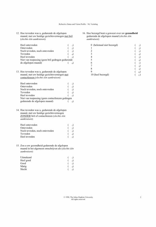
Refractive Status and Vision Profile – NL Vertaling
© 1998, The Johns Hopkins University All rights reserved.
2
12. Hoe tevreden was u, gedurende de afgelopen maand, met uw huidige gezichtsvermogen met bril (slechts één aankruisen): Heel ontevreden ( 1) Ontevreden ( 2) Noch tevreden, noch ontevreden ( 3) Tevreden ( 4) Heel tevreden ( 5) Niet van toepassing (geen bril gedragen gedurende de afgelopen maand) ( 6)
13. Hoe tevreden was u, gedurende de afgelopen maand, met uw huidige gezichtsvermogen met contactlenzen (slechts één aankruisen): Heel ontevreden ( 1) Ontevreden ( 2) Noch tevreden, noch ontevreden ( 3) Tevreden ( 4) Heel tevreden ( 5) Niet van toepassing (geen contactlenzen gedragen gedurende de afgelopen maand) ( 6)
14. Hoe tevreden was u, gedurende de afgelopen maand, met uw huidige gezichtsvermogen ZONDER bril of contactlenzen (slechts één aankruisen): Heel ontevreden ( 1) Ontevreden ( 2) Noch tevreden, noch ontevreden ( 3) Tevreden ( 4) Heel tevreden ( 5)
15. Zou u uw gezondheid gedurende de afgelopen maand in het algemeen omschrijven als (slechts één aankruisen): Uitstekend ( 1) Heel goed ( 2) Goed ( 3) Matig ( 4) Slecht ( 5)
16. Hoe bezorgd bent u geweest over uw gezondheid gedurende de afgelopen maand (slechts één aankruisen):
0 (helemaal niet bezorgd) ( 1) 1 ( 2) 2 ( 3) 3 ( 4) 4 ( 5) 5 ( 6) 6 ( 7) 7 ( 8) 8 ( 9) 9 ( 10) 10 (heel bezorgd) ( 11)

Refractive Status and Vision Profile – NL Vertaling
© 1998, The Johns Hopkins University All rights reserved.
3
REFRACTIVE STATUS AND VISION PROFILE
VRAGENLIJST
Gezichtsvermogen Beantwoord de vragen alstublieft voor zover ze op u van toepassing zijn over de afgelopen maand. 17. Ik maak me zorgen over mijn gezichtsvermogen (slechts één aankruisen):
Nooit ( 1) Zelden ( 2) Soms ( 3) Vaak ( 4) Altijd ( 5)
18. Mijn gezichtsvermogen is een zorg in mijn leven (slechts één aankruisen):
Nooit ( 1) Zelden ( 2) Soms ( 3) Vaak ( 4) Altijd ( 5)
19. Mijn gezichtsvermogen remt mij in mijn doen en
laten (slechts één aankruisen):
Nooit ( 1) Zelden ( 2) Soms ( 3) Vaak ( 4) Altijd ( 5)
20. Ik ben gefrustreerd over mijn gezichtsvermogen
(slechts één aankruisen):
Nooit ( 1) Zelden ( 2) Soms ( 3) Vaak ( 4) Altijd ( 5)
21. Mijn gezichtsvermogen maakt mij minder zelfstandig (slechts één aankruisen):
Nooit ( 1) Zelden ( 2) Soms ( 3) Vaak ( 4) Altijd ( 5)
22. Vanwege mijn gezichtsvermogen zijn er dingen die
ik bang ben om te doen (slechts één aankruisen):
Nooit ( 1) Zelden ( 2) Soms ( 3) Vaak ( 4) Altijd ( 5)
23. Ik zou een gezichtsvermogen kunnen accepteren dat
niet helemaal perfect is als ik geen bril of contactlenzen meer nodig zou hebben (slechts één aankruisen):
Sterk mee oneens ( 1) Mee oneens ( 2) Noch mee eens, noch mee oneens ( 3) Mee eens ( 4) Sterk mee eens ( 5) Geen bril of contactlenzen nodig om het best mogelijke gezichtsvermogen te halen ( 6)
24. Zolang ik goed genoeg kan zien om auto te rijden
zonder bril of contactlenzen, zou ik het niet erg vinden om een gezichtsvermogen te hebben dat niet helemaal perfect is (slechts één aankruisen):
Sterk mee oneens ( 1) Mee oneens ( 2) Noch mee eens, noch mee oneens ( 3) Mee eens ( 4) Sterk mee eens ( 5)
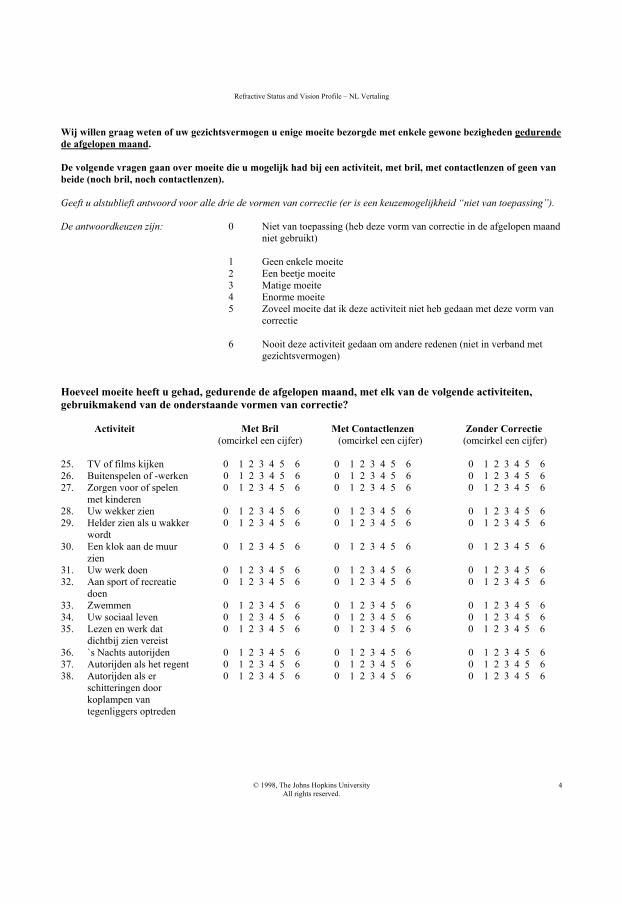
Refractive Status and Vision Profile – NL Vertaling
© 1998, The Johns Hopkins University All rights reserved.
4
Wij willen graag weten of uw gezichtsvermogen u enige moeite bezorgde met enkele gewone bezigheden gedurende de afgelopen maand. De volgende vragen gaan over moeite die u mogelijk had bij een activiteit, met bril, met contactlenzen of geen van beide (noch bril, noch contactlenzen). Geeft u alstublieft antwoord voor alle drie de vormen van correctie (er is een keuzemogelijkheid “niet van toepassing”). De antwoordkeuzen zijn: 0 Niet van toepassing (heb deze vorm van correctie in de afgelopen maand niet gebruikt)
1 Geen enkele moeite 2 Een beetje moeite 3 Matige moeite 4 Enorme moeite 5 Zoveel moeite dat ik deze activiteit niet heb gedaan met deze vorm van
correctie 6 Nooit deze activiteit gedaan om andere redenen (niet in verband met
gezichtsvermogen)
Hoeveel moeite heeft u gehad, gedurende de afgelopen maand, met elk van de volgende activiteiten, gebruikmakend van de onderstaande vormen van correctie?
Activiteit Met Bril Met Contactlenzen Zonder Correctie (omcirkel een cijfer) (omcirkel een cijfer) (omcirkel een cijfer)
25. TV of films kijken 0 1 2 3 4 5 6 0 1 2 3 4 5 6 0 1 2 3 4 5 6 26. Buitenspelen of -werken 0 1 2 3 4 5 6 0 1 2 3 4 5 6 0 1 2 3 4 5 6 27. Zorgen voor of spelen 0 1 2 3 4 5 6 0 1 2 3 4 5 6 0 1 2 3 4 5 6
met kinderen 28. Uw wekker zien 0 1 2 3 4 5 6 0 1 2 3 4 5 6 0 1 2 3 4 5 6 29. Helder zien als u wakker 0 1 2 3 4 5 6 0 1 2 3 4 5 6 0 1 2 3 4 5 6 wordt 30. Een klok aan de muur 0 1 2 3 4 5 6 0 1 2 3 4 5 6 0 1 2 3 4 5 6
zien 31. Uw werk doen 0 1 2 3 4 5 6 0 1 2 3 4 5 6 0 1 2 3 4 5 6 32. Aan sport of recreatie 0 1 2 3 4 5 6 0 1 2 3 4 5 6 0 1 2 3 4 5 6 doen 33. Zwemmen 0 1 2 3 4 5 6 0 1 2 3 4 5 6 0 1 2 3 4 5 6 34. Uw sociaal leven 0 1 2 3 4 5 6 0 1 2 3 4 5 6 0 1 2 3 4 5 6 35. Lezen en werk dat 0 1 2 3 4 5 6 0 1 2 3 4 5 6 0 1 2 3 4 5 6
dichtbij zien vereist 36. `s Nachts autorijden 0 1 2 3 4 5 6 0 1 2 3 4 5 6 0 1 2 3 4 5 6 37. Autorijden als het regent 0 1 2 3 4 5 6 0 1 2 3 4 5 6 0 1 2 3 4 5 6 38. Autorijden als er 0 1 2 3 4 5 6 0 1 2 3 4 5 6 0 1 2 3 4 5 6
schitteringen door koplampen van tegenliggers optreden

Refractive Status and Vision Profile – NL Vertaling
© 1998, The Johns Hopkins University All rights reserved.
5
Wij willen graag weten of u bepaalde problemen ervoer met uw ogen of gezichtsvermogen gedurende de afgelopen maand. De volgende vragen gaan over elk probleem met bril, met contactlenzen of met geen van beide (noch bril, noch contactlenzen). Geef alstublieft antwoord voor alle drie de vormen van correctie (er is een keuzemogelijkheid “niet van toepassing”). De antwoordkeuzen zijn: 0 Niet van toepassing (heb deze vorm van correctie in de afgelopen maand niet gebruikt)
1 Geen enkele last 2 Een beetje last 3 Matige last 4 Enorme last 5 Zoveel last dat ik deze vorm van correctie niet gebruik
Hoeveel last heeft u gehad, gedurende de afgelopen maand, van de volgende dingen, bij het gebruiken van de bril, contactlenzen of geen van beide? Probleem Met Bril Met Contactlenzen Zonder Correctie
(omcirkel een cijfer) (omcirkel een cijfer) (omcirkel een cijfer)
39. Het geïrriteerd aanvoelen 0 1 2 3 4 5 0 1 2 3 4 5 0 1 2 3 4 5 van uw ogen
40. Tocht (van de verwarming 0 1 2 3 4 5 0 1 2 3 4 5 0 1 2 3 4 5 of airconditioning) die in uw ogen blaast
41. Het overgevoelig zijn van 0 1 2 3 4 5 0 1 2 3 4 5 0 1 2 3 4 5 uw ogen voor licht
42. Pijn in uw ogen 0 1 2 3 4 5 0 1 2 3 4 5 0 1 2 3 4 5 43. Veranderingen van uw 0 1 2 3 4 5 0 1 2 3 4 5 0 1 2 3 4 5
gezichtsvermogen in de loop van de dag
44. Mistig of wazig zien 0 1 2 3 4 5 0 1 2 3 4 5 0 1 2 3 4 5 45. Schitteringen 0 1 2 3 4 5 0 1 2 3 4 5 0 1 2 3 4 5
(weerspiegelingen van glimmende oppervlakten, sneeuw)
46. Dingen die er met het ene 0 1 2 3 4 5 0 1 2 3 4 5 0 1 2 3 4 5 oog anders uitzien dan met het andere
47. Het zien van kringen rondom 0 1 2 3 4 5 0 1 2 3 4 5 0 1 2 3 4 5 lichten
48. Zien in schemerlicht 0 1 2 3 4 5 0 1 2 3 4 5 0 1 2 3 4 5 49. Uw dieptezien 0 1 2 3 4 5 0 1 2 3 4 5 0 1 2 3 4 5 50. Dingen lijken vervormd 0 1 2 3 4 5 0 1 2 3 4 5 0 1 2 3 4 5 51. Het schatten van afstand 0 1 2 3 4 5 0 1 2 3 4 5 0 1 2 3 4 5
bij het op- of afstappen van treden (trappen, stoepranden)
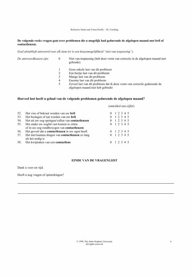
Refractive Status and Vision Profile – NL Vertaling
© 1998, The Johns Hopkins University All rights reserved.
6
De volgende reeks vragen gaat over problemen die u mogelijk had gedurende de afgelopen maand met bril of contactlenzen. Geef alstublieft antwoord voor elk item (er is een keuzemogelijkheid “niet van toepassing”). De antwoordkeuzen zijn: 0 Niet van toepassing (heb deze vorm van correctie in de afgelopen maand niet gebruikt)
1 Geen enkele last van dit probleem 2 Een beetje last van dit probleem 3 Matige last van dit probleem 4 Enorme last van dit probleem 5 Zoveel last van dit probleem dat ik deze vorm van correctie gedurende de
afgelopen maand niet heb gebruikt
Hoeveel last heeft u gehad van de volgende problemen gedurende de afgelopen maand? (omcirkel een cijfer) 52. Het vies of bekrast worden van uw bril 0 1 2 3 4 5 53. Het beslagen of nat worden van uw bril 0 1 2 3 4 5 54. Het uit uw oog springen/vallen van contactlenzen 0 1 2 3 4 5 55. Het onder uw ooglid vast komen te zitten 0 1 2 3 4 5
of in uw oog rondbewegen van contactlenzen 56. Het gevoel dat u contactlenzen in uw ogen heeft 0 1 2 3 4 5 57. Het niet kunnen dragen van contactlenzen zo lang 0 1 2 3 4 5
als het nodig is 58. Het kwijtraken van een contactlens 0 1 2 3 4 5
EINDE VAN DE VRAGENLIJST Dank u voor uw tijd. Heeft u nog vragen of opmerkingen?


DankwoordDankwoord

Dankwoord 191
This thesis could not have been completed without the help, directly and indirectly, of many people. I’d like to take this opportunity to thank the following:
Allereerst was mijn onderzoek nooit tot stand gekomen zonder de medewerking van alle patiënten die zo vriendelijk zijn geweest om steeds zorgvuldig de grote aantallen aan vragenlijsten in te vullen.
Mijn promoter, Prof.dr. G.P.M. Luyten. Beste Gré, zonder jouw enthousiasme voor de oogheelkunde en de refractiechirurgie was ik nooit aan dit project begonnen. Je hebt het heel leuk weten te houden. Ik ben dankbaar dat je naast de oogheelkunde, mij ook uiteindelijk enthousiast hebt kunnen maken voor het koken!
Mijn promoter en opleider, Prof.dr. G. van Rij. Ik wil u bedanken voor uw betrokkenheid bij mijn onderzoek en voor de gelegenheid om mijn opleiding tot oogarts in het Erasmus MC te mogen doen. Dank voor de aanmoediging.
De leden van de kleine commissie: Prof.dr. J.C. van Meurs, Prof.dr. L. Feenstra en Prof.dr. P.J. Ringens. Hartelijk dank voor uw snelle beoordeling van het manuscript en het plaatsnemen in de promotiecommissie. Prof. Dr. J.J.V. Busschbach, Dr. R.M.M.A Nuijts en Dr. C.C.W. Klaver, hartelijk dank voor het plaatsnemen in de grote promotiecommissie. Beste Caroline, ik ben jou dank verschuldigd voor je kritisch commentaar op mijn proefschrift. Het is er zeker beter op geworden.
Dr. M.C. Bartels. Beste Marjolijn, ik ben jou dankbaar niet alleen als medeauteur, maar ook als mentor. Ik heb van jou veel kunnen leren. Ik heb grote bewondering voor de manier waarop je zaken regelt. Deventer heeft een zeer goede aanwinst gekregen.
Willem Vreugdenhil, aan jou ben ik veel dank verschuldigd voor alle patiënten die je zo keurig hebt verzameld voor het onderzoek. Dank je.
Monika Landesz, Michiel Luger en alle collega’s van Vision Clinics. Hartelijk dank voor de gastvrijheid en de prettige samenwerking. Christianne Verschoor en Koen Paarlberg, ook veel dank voor jullie hulp als ik tussendoor nog data nodig had.
Dr. H.J. Duivenvoorden, hartelijk dank voor uw expertise ten aanzien van de vragenlijsten en de power analyse aan het begin van mijn promotietraject.
Prof. dr. J. Worst en Erik-Jan Worst, hartelijk dank voor de gelegenheid om de Artisan cursus te mogen volgen in Groningen. Natuurlijk ben ik Professor Worst

Dankwoord192
ook erg dankbaar voor het ontwikkelen van zo’n prachtige lens die ik heb mogen onderzoeken. Berry Everts, Gut-Win Chun, Mark van Nes en de medewerkers van Ophtec, dank voor het beantwoorden van alle weetjes over de lens en het verstrekken van de mooie Artisan plaatjes
De co-auteurs: Sharmila Boekhoorn, Ellen van Minderhout, Paul Mulder, Tom van den Berg, Kors van der Torren en Mark Veckeneer dank ik voor hun inbreng aan de artikelen. Bart van Dooren, je bent geen co-auteur geweest, maar je hebt zeker een bijdrage geleverd aan mijn endotheelmanuscript. Redmer van Leeuwen, hartelijk dank voor jou commentaar op mijn kwaliteit van leven artikel.
Nèvine te West, Mikko Larsen and Alexander Baan, my bilingual Med School Buddies: Thank you so much for your work on the RSVP translation. I really appreciate you guys taking time out to help me on this and spreading your linguistic magic! Ook wil ik de medeauteurs van de VFQ-25 bedanken, in het bijzonder Jan Roelof Polling, Geertje van der Sterre, Elizabeth van de Graaf, Anton Verezen en Huib Simonsz. Natuurlijk kan ik hierbij mijn tweetalige familieleden, vrienden en collega’s niet vergeten, die de tijd hebben genomen om alle vragenlijsten niet één, maar zelfs twee keer in te vullen zodat ik de vertaling kon controleren. Hartelijk dank voor jullie bijdrage.
Jeannette Noordzij, dankzij je uitleg over (en bijdrage aan) de endotheelcel metingen, jouw patiëntenonderzoek en keurig bijhouden van alle data heb ik mijn artikelen (ook samen met jou) kunnen schrijven. Suzanne van de Weghe, dank voor jouw steeds nauwkeurige vooronderzoeken en attente suggesties. Tineke Muijlwijk wil ik bedanken voor mailtjes tussendoor over de OK planning, zodat mijn vragenlijsten op tijd opgestuurd werden.
Verder heeft natuurlijk iedereen op de poli Oogheelkunde direct of indirect een bijdrage geleverd aan dit proefschrift, hetzij door de patiënten te boeken op de poli, ze te onderzoeken, of uitleg te geven over de gang van zaken rondom de operaties. Mijn huidige AIOS-collega’s Alberta, Olivera, Isabelle, Jackelien, Dominiek, Sjoukje, Anne en Jordie (hmmm…nee, niemand vergeten…), dank voor jullie interesse in de vorderingen. Charles Vervaet, dank voor alle uitleg over contactlenzen! Hierbij kan ik de fantastische OK zusters en verpleegkundigen op de dagbehandeling niet vergeten. Heel hartelijk dank (Oesters anyone?!).
Nicole van Basten samen hebben wij “de la” goed onderhouden op de 5de! Ik wil je heel erg bedanken voor alle gezelligheid op de 5de. Ook natuurlijk voor alles wat je voor mij hebt gedaan gedurende de afgelopen jaren – hulp bij mijn Nederlands, enveloppen op tijd versturen tijdens mijn co-schappen en alle kleine dingetjes – je stond altijd klaar voor mij, meestal met een kopje thee en chocola!
Dankwoord 193
Mijn andere collega’s op de (oude) vijfde: Yanni, Aartie en Judith. Toen Kasper wegging om huizen te bouwen, werd ik bang dat ik het nogal eenzaam zou vinden. Gelukkig kreeg ik alsnog gezelschap in het zweethokje! Helaas met jullie was “de la” nog beter dan ooit onderhouden. Ik ben blij dat wij allemaal de opleiding oogheelkunde hebben mogen doen. Norbert, dankzij jou waren onze magen gevuld met de verse broodjes die je altijd meenam van de poli. Dank!
Roger Wolfs. Altijd zo betrouwbaar. Bij ieder computerdrama kon ik jou bellen en jij bedacht altijd een oplossing! Ik waardeer heel erg hoe je altijd klaar staat om anderen te helpen, zelfs ’s avonds en in het weekend! Ik heb bewondering voor de rust die je uitstraalt, met name op de OK (in het bijzonder als de operateur naast jou niet zo rustig is!). Ik heb veel van jou kunnen leren. Dank je.
Rosalien Arkesteijn en Munira Alwani. Er is veel bereikt dankzij jullie harde werken. Het leven is ook door jullie slimme programma’s ook nog eens makkelijker voor mij gemaakt. Hartelijk dank voor jullie inzet (ook soms heel vroeg op de ochtend!). Anna Michalska, ook jij hartelijk dank voor het invoeren van zoveel gegevens en uiteraard ook voor de gezelligheid.
Even the cover of this book did not come on its own without outside help. I’d like to thank Dr. Raymond Stein for providing the very suitable image of the “glasses in the garbage” used on the cover of my thesis.
Yolanda, dank dat ik zo vaak over de vloer mocht komen om stukjes uit te werken met Gré en dan altijd mocht meegenieten van het eten!
Myrthe, Maaike, Marije en Wendy (en natuurlijke ook de mannen!). Dank voor de gezellige afleiding! Ik vind het zo mooi dat wij jaren na het afronden van de studie toch nog zo vaak bij elkaar komen. Jullie zijn de afgelopen moeilijke tijd ook een grote steun voor mij geweest.
Aan mijn paranimfen Emine Kiliç en Kasper de Roon Hertoge: Emine dankzij jou continue en irritante vragen over mijn proefschrift is mijn boekje eindelijk af! Ik wil jou ook van harte danken voor je vriendschap. Jouw komst in de assistentengroep heeft geleid tot een veel gezelligere opleiding voor mij. Jouw expertise in onderzoek en “paranimferen” heeft veel aspecten van mijn promotietraject ook makkelijker gemaakt. En Kasper: er werd misschien meer gekletst dan gewerkt, maar jij zorgde ervoor dat het toch heel gezellig bleef in het kleine zweethokje op de 5e. Jij met je tranen en ik met mijn kwaliteit van leven. Wat ontzettend leuk was het toen jij ook als arts-assistent bij de Oogheelkunde begon!
Dankwoord194
Of course, I cannot forget my family in Holland. Thank you for making my move here from Canada so much easier. Roli, you even decided to get Artisan lenses! I got to see your “Vision-Related Quality of Life” improve right before of my eyes! Taji and Bhabi, I cannot thank you enough for putting up with me in your home for two whole years! You are both such unassuming people, I hope you realize how much you mean to us. Taji, I am very lucky to have such a talented researcher in the family, thank you for all your time and advice.
To my Parents. Even though you’re so far away, you manage to be an amazing support system for both Ollie and I – thank you. Also, thank you so much for putting up with me! I really am very fortunate for having such kind, understanding and loving parents. You both also helped me a lot by reviewing this manuscript all the way from Canada. Without your encouragement and support, I would have never ended up where I am. Nup, the eternal party planner - thanks for all your ideas surrounding “het promotiefeestje”.
Finally, the love of my life, Oliver. As requested, I shall mention Christmas 2005, when you spent the whole day inputting data into that crazy Excel worksheet, so that I would have my presentation ready on time! It amazes me, that after fourteen years together and all that we’ve been through, you still keep me laughing. Throughout everything, you’ve been wonderful, kind, supportive, incrediblypatient and always optimistic. Words are just not enough.
195Curriculum Vitae
Curriculum Vitae
Ruchi Saxena was born during her parents’ holiday in Lucknow, India on December 11th, 1972. At the time, her parents were residing in Delft, the Netherlands, where her older sister, Nupur, was born 2 years previously. At the age of nine months, her family moved to Belgium, and at the age of 5, to Montreal, Quebec, Canada. In 1983, the family moved yet again, this time to Oakville, Ontario, Canada. She graduated in 1992 from Queen Elizabeth Park School, also in Oakville, with a Diploma in French Immersion. In 1996, she graduated from the University of Toronto, Toronto, Ontario, Canada, with an Honours Bachelor of Science, majoring in Human Biology and Zoology. During her stay at the University, she served as President of the New College Residence Council for two years. For her work, she received the Katherine Grace Baker Memorial Award in 1995 and the Gordon Cressy Student Leadership Award in 1996. After following a course in Vertebrate Regeneration and performing microscopic surgery on lizards in her final year, she decided that she wanted to pursue a career in Medicine.
After moving to the Netherlands in 1996 and completing a Dutch language course in 1997, Ruchi began her Medical Studies at the Erasmus University in Rotterdam. In her third year, she followed an elective course on refractive surgery offered by Dr. G.P.M. Luyten at the Department of Ophthalmology, Erasmus MC. This course further stimulated her interest in the field and she went on to publish two articles on refractive surgery, which she had written as her Masters thesis. These studies led to her being awarded the Gerrit Jan Mulder Prize in 2003. During her Medical Clerkship, she continued to work on her research in refractive surgery. This work was in cooperation with Dr. M.A. Landesz and Dr. M.A.H. Luger at Vision Clinics in Delft and with Willem Vreugdenhil from Oculenti Contact Lens Specialists at the Rotterdam Eye Hospital. She received her Medical Diploma in 2004 and is currently completing her specialization in Ophthalmology at Erasmus MC, under Prof. Dr. G. van Rij. The bulk of her research and writing was done during her Medical Clerkship and later, during her specialization.
Unfortunately, Ruchi has inherited the myopia that plagues 11 of the 12 grandchildren on her father’s side of the family. Sadly, her eyes are not suitable for refractive surgery and she has to make do with contact lenses. On April 13th 2006, Ruchi married Oliver Gnjec. As you can imagine, she kept her own name.

197List of Publications
List of Publications
Saxena R, Minderhout H, Luyten G The Use of the Artisan™ Phakic Intraocular Lens in the Treatment of Anisometropic Amblyopia. J Cataract Refract Surg. 2003 Apr;29(4):835-8.
Saxena R, Landesz M, Noordzij B, Luyten G. Three-year Follow-up of the Artisan Phakic Intraocular Lens for Hypermetropia. Ophthalmology. 2003 Apr;110(7):1391-1395.
Saxena R, van der Torren K, Veckeneer M, Luyten G. Iris-fixated phakic IOLs to correct postoperative anisometropia in unilateral cataract patients with bilateral high myopia. J Cataract Refract Surg. 2004 Oct;30(10):2240-1.
Bartels M, Saxena R, van den Berg T, van Rij G, Mulder P, Luyten G. The influence of incisional induced astigmatism and axial positional accuracy on the correction of myopic astigmatism with the Artisan toric phakic intraocular lens. Ophthalmology. 2006 Jul;113(7):1110-7.
Saxena R, Boekhoorn S, Mulder P, Noordzij B, van Rij G, Luyten G. Long-Term Follow-up of Endothelial Cell Change After Artisan Phakic Intraocular Lens Implantation. Ophthalmology. 2008 Apr;115(4):608-613.
Saxena R, Vreugdenhil W, Mulder P,van Rij G, Luyten G. Vision-Related Quality of Life of Myopic Patients. Submitted.
Saxena R, Vreugdenhil W, Alwani M, Arkenstijn R, Landesz M, Luger M, Bartels M, van Rij G, Luyten G. Quality of Life of Before and After Refractive Surgery. Submitted.

Related Documents