THE APPLICATION OF PROGNOSTIC TISSUE MARKERS TO PROSTATE CANCER: CLINICAL AND EXPERIMENTAL STUDIES

Welcome message from author
This document is posted to help you gain knowledge. Please leave a comment to let me know what you think about it! Share it to your friends and learn new things together.
Transcript

THE APPLICATION OF PROGNOSTIC TISSUE MARKERS TO PROSTATE
CANCER: CLINICAL AND EXPERIMENTAL STUDIES

omslag: Christine Dijkstra, zander titel, 1996 gedrukt door: Haveka BV, Alblasserdam
ISBN: 90-9010322-8
NUGI: 743
THE APPLICATION OF PROGNOSTIC TISSUE MARKERS TO PROSTATE
CANCER: CLINICAL AND EXPERIMENTAL STUDIES
De toepassing van prognostische weefsel markers bij prostaat carcinoom: klinische en
experimentele studies
PROEFSCHRIFT
ter verkrijging van de graad van doctor aan de Erasmus U niversiteit Rotterdam
op gezag van de Rector Magnificus prof. dr P.W.c. Akkermans, M.A.
en volgens het besluit van het college voor promoties. De open bare verdediging zal plaatsvinden op
woensdag 19 maart 1997 om 13.45 UUl'.
door
Marinus Adrianus Noordzij geboren te Rotterdam
PROMOTIE COMMISSIE
Promotores:
Overige leden
Co-promotor
prof. dr F.H. Schroder prof. dr T.H. van der Kwast
prof. dr F.J.W. ten Kate prof. dr S.W.]. Lamberts prof. dr J.A. Schalken
dr G.]. van Steenbrugge
Dit proefschrift werd bewerkt binnen de afdelingen urologie (experimentele oncologie) en pathologie van de Faculteit der geneeskunde en gezondheids-wetenschappen, Erasmus Universiteit, Rotterdam. De uitgave van dit proefschrift is mogelijk gemaakt mede dankzij financie1e bijdragcn van de Stichting Urologische Wetenschappelijk Onderzoek (SUW'O) Rotterdam, de Stichting urologie 1973, Hoechst Marion Roussel Nederland bv en Yamanouchi Nederland bv.
4

opgedragen aan mijn ouders
5

CONTENTS
List of abbreviations ....................................... 9
I Prognostic markers in prostate cancer ......................... 11 Introduction 12 Natural course 13 Treatment 14 Prognostic markers 15 Conclusion 39 Appendix (statistical analysis of survival times) 40
Scope of the thesis ....................................... 42
II The prognostic influence of neuroendocrine cells in prostate cancer: results of a long-term follow-up study with patients treated by radical prostatectomy .......................................... 45
Abstract 46 Introduction
Material and methods Results Discussion
III The prognostic value of CD44 isoforms in prostate cancer patients
46 47 49 55
treated by radical prostatectomy ............................. 59 Abstract 60 Introduction
Material and methods Results
Discussion
IV The prognostic value of pre-treatment expression of androgen receptor
60 62 65 77
and bcl-2 in hormonally treated prostate cancer patients ............. 81 Abstract 82 Introduction Material and methods Results Discussion
V Neuroendocrine cells in the normal, hyperplastic and neoplastic
82
83 85 90
prostate ............................................... 93 Abstract 94 Introduction 94
7
VI
Neuroendocrine cells in the normal prostate Neuroendocrine cells in the hyperplastic prostate Neuroendocrine cells in the neoplastic prostate Secretion products and hormone sensitivity Growth modulation by neoplastic neuroendocrine cells Specific neuropeptides Neuroendocrine differentiation in prostatic tumor models Conclusion
Neuroendocrine differentiation in human prostatic tumor models Abstract Introduction Material and methods Results Discussion
95 97 98
100 101 103 105 106
107 108 108 110
115
124
VII Determination of Ki·67 defined growth fraction by monoclonal antibody MIB·l in formalin·fixed, paraffin.embedded prostatic cancer
VIII
8
tissues ............................................... 129 Abstract 130 Introduction Material and methods Results Discussion
General Discussion Prognostic markers Neuroendocrine differentiation
130 131 135 137
141 142 146
References ............................................ 149
Summary . . . . . . . . . . . . . . . . . . . • . . . . . . . . . . . . . . . . . . . . . . . .. 177
Sanlenvatting .......................................... 181
Dankwoord .. . . . . . . . . . . . . . . . . . . . . . . . . . . . . . . . . . . . . . . .. 185
Curriculum vitae ....................................... 187
Additional publications . . . . . . . . . . . . . . . . . . . . . . . . . . . . . . . . . .. 189
LIST OF ABBREVIATIONS
5-HT serotonin NE neuroendocrine ABC avidin-biotin complex NRF nuclear roundness factor ACTH adreno-corticotrope NSE neuron specific enolase
hormone PAM peptidyl glycine-,,-AJCC American joint committee ami dating mono-oxygenase
on cancer PBS phosphate buffered saline APES aminopropyl-trietoxy-silane PCNA proliferating cell nuclear APUD amine precursor uptake and antigen
decarboxylation PCR polymerase-chain reaction AR androgen receptor PI prognostic index bp base pairs PIN prostatic intraepithelial BPH benign prostatic hyperplasia neoplasia BrdU bromo-deoxy uri dine PSA prostate specific antigen eDNA complementary DNA ROC receiver operating CgA chromogranin A characteristic curve CgB chromogranin B RT-PCR reverse-transcriptase CGRP calcitonin gene-related polymerase-chain reaction
peptide SCPC small cell prostate cancer CI 95% confidence interval SMS somatostatin CT calcitonin SPF S-phase fraction DAB 3,3' -diaminobenzidine- TGF transforming growth factor
hydrochloride TNM tumor, nodes and metastasis DCC dextran coated charcoal TRUS transrectal ultrasound dNTP deoxynucleotide- TSA tyramide signal
triphosphate amplification EGF epidermal growth factor TUR transurethral resection EGF-R epidermal growth factor- UICC Union internationale contre
receptor Ie cancer FGF fibroblast growth factor VEGF vascular endothelial growth FISH fluorescence in~situ factor
hybridization WHO World Health Organization GRP gastrin-related peptide GSS Gleason sum score H&E hematoxylin & eosin ISH in~situ hybridization LH-RH luteinizing hormone~
releasing hormone M-MLV moloney~murine leukemia
VIrUS
MRI magnetic resonance imaging
9

CHAPTER I
PROGNOSTIC MARKERS IN PROSTATE CANCER
M.A. Noordzij; T.H. van der Kwast; G.J. van Steenbrugge; F.H. Schroder
From the departments of Urology and Pathology, Erasmus University, Rotterdam, The Netherlands
Submitted for publication
prognostic markers in prostate cancer
INTRODUCTION
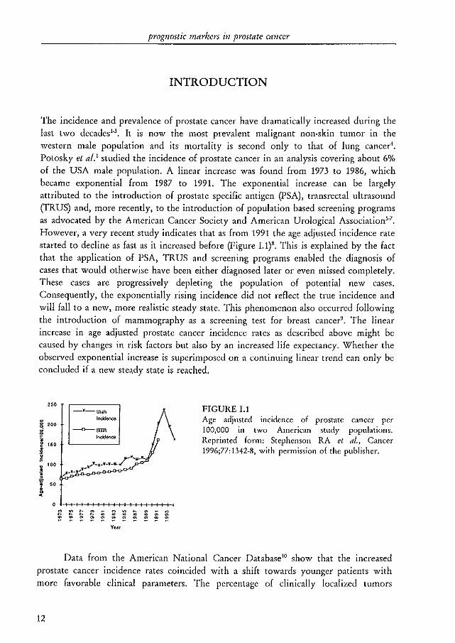
The incidence and prevalence of prostate cancer have dramatically increased during the last two decades l -}. It is now the most prevalent malignant non-skin tumor in the western male population and its mortality is second only to that of lung cancer4•
Potosky et at.' studied the incidence of prostate cancer in an analysis covering about 6% of the USA male population. A linear increase was found from 1973 to 1986, which became exponential from 1987 to 1991. The exponential increase can be largely attributed to the introduction of prostate specific antigen (PSA) , transrectal ultrasound (TRUS) and, more recently, to the introduction of population based screening programs as advocated by the American Cancer Society and American Urological Associations.7.
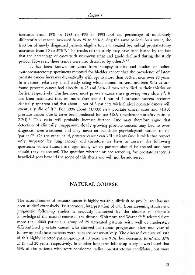
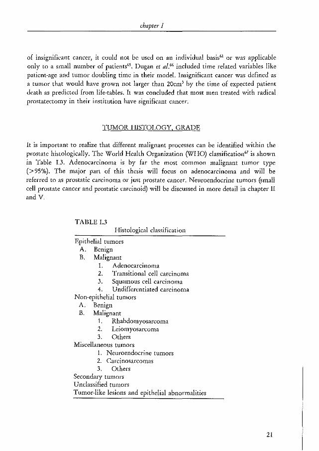
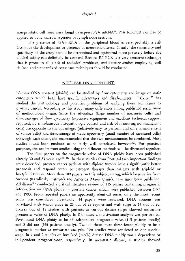
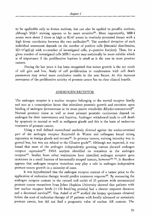
However, a very recent study indicates that as from 1991 the age adjusted incidence rate started to decline as fast as it increased before (Figure I.1)'. This is explained by the fact that the application of PSA, TRUS and screening programs enabled the diagnosis of cases that would otherwise have been either diagnosed later or even missed completely. These cases are progressively depleting the population of potential new cases. Consequently, the exponentially rising incidence did not reflect the true incidence and will fall to a new, more realistic steady state. This phenomenon also occurred following the introduction of mammography as a screening test for breast cancer9. The linear increase in age adjusted prostate cancer incidence rates as described above might be caused by changes in risk factors but also by an increased life expectancy. Whether the observed exponential increase is superimposed on a continuing linear trend can only be concluded if a new steady state is reached.
250
8. 200 g 'i ~ 150
i i 100 , f 50
-·-Ulah
''''''''''''
§ ~ ~ ~ ~ ~ * ~ * : E Year
FIGURE 1.1 Age adjusted incidence of prostate cancer per 100,000 in two American study populations. Reprinted form: Stephenson RA et al., Cancer 1996;77:1342+8, with permission of the publisher.
Data from the American National Cancer Database!O show that the increased prostate cancer incidence rates coincided with a shift towards younger patients with more favorable clinical parameters. The percentage of clinically localized tumors
12
chapter I
increased from 19% in 1986 to 49% in 1993 and the percentage of moderately differentiated cancer increased from 39 to 58% during the same period. As a result, the fraction of newly diagnosed patients eligible for, and treated by, radical prostatectomy increased from 10 to 29%10. The results of this study may have been biased by the fact that the percentage of cases with unknown stage and grade declined during the study period. However, these trends were also described by others2, 3, 8,
It has been known for years from autopsy studies and studies of radical cystoprostatectomy specimens removed for bladder cancer that the prevalence of latent prostate cancer increases dramatically with age to more than 50% in men over 80 years. In a recent, relatively small study using whole mount prostate sections Sakr et alY found prostate cancer foci already in 28 and 34% of men who died in their thirties or forties, respectively. Furthermore, most prostate cancers are growing very slowlyll, It has been estimated that no more than about lout of 4 prostate cancers becomes clinically apparent and that about 1 out of 3 patients with clinical prostate cancer will eventually die of itIJ. For 1996 about 317,000 new prostate cancer cases and 41,400 prostate cancer deaths have been predicted for the USA (incidence! mortality ratio ~ 7.7:1)14. This ratio will probably increase further. One may therefore argue that detection of clinically unapparent, slowly growing prostate cancers may lead to overdiagnosis, over·treatment and may mean an avoidable psychological burden to the 'patient'I'. On the other hand, prostate cancer can kill patients (and is with that respect only surpassed by lung cancer) and therefore we have to answer the following questions: which tumors are significant, which patients should be treated and how should they be treated? The question whether or not screening for prostate cancer is beneficial goes beyond the scope of this thesis and will not be addressed.
NATURAL COURSE
The natural course of prostate cancer is highly variable, difficult to predict and has not been studied extensively. Furthermore, interpretation of data from screening-studies and prognostic follow·up studies is seriously hampered by the absence of adequate knowledge of the natural course of the disease. Whitmore and Warner l6• 17 selected from more than 4000 patients a group of 75 untreated patients with well or moderately differentiated prostate cancer who showed no tumor progression after one year of follow-up and these patients were managed conservatively. The disease free survival rate of this highly selected patient·group at 10 years was 91%, but decreased to 67 and 37% at 15 and 20 years, respectively. In another long·term follow·up study it was found that 59% of the patients who were considered radical prostatectomy candidates, but were
13
prognostic tnarkers in prostate cancer
managed conservatively eventually died of prostate cancer18. In studies from Sweden including patients with T 1.z tumors, the opposite was found (10% prostate cancer deaths after a mean follow·up period of 12.5 years)19.2!. These studies have been criticized for a number of favorable selection criteria applied to the study population". Chodak et al.23 performed a meta·analysis on the results of a total of 828 patients published in 6 studies, including the Swedish studies. Tumor specific survival for grade 1 and 2 tumors was identical to the life·expectancy of the general population, but decreased significantly for grade 3 tumors. Metastasis free survival at 10 years was about 70%, 55%, and 20% for grade 1, 2, and 3 tumors, respectively. This meta-analysis was based on data from studies that all suffered from methodologic problems and it is questionable if definite conclusions can be drawn from this study. It, however, appears that the majority of prostate cancers have the potential to progress, but also that it may take considerable time to occur. It is clear that in older patients with small, well differentiated tumors the likelihood of tumor progression is determined mainly by factors not related to the primary prostate tumor (such as the presence of cardiovascular diseases, diabetes mellitus or other malignancies).
TREATMENT
Regarding the choice of treatment it is important to realize that patients with prostate cancer generally are of advanced age, but also that most prostate cancers will progress, be it after a considerable period of time. A number of patients, especially older patients with small tumors, can be managed conservativelyH, which means a careful follow-up program in which (endocrine) treatment will start only at the moment that progression is noted. Most patients with clinically localized prostate cancer (i.e., a tumor that is not growing beyond the prostatic capsule) are offered a form of curative treatment: external beam radiotherapyH or radical prostatectomy25. For years the choice between those two has been the subject of debate (reviewed by Hartford and Zietman26). In a number of studies, surgical therapy was found to be superior to radiotherapy, but patients referred for radiotherapy in general showed unfavorable characteristics (high age, larger tumors and an unknown lymph node status). A very recent study has shown that co·morbidity is a significant and independent prognostic marker for patients with clinicall y localized prostate cancer treated with endocrine therapy17. Since healthier patients are more likely radical prostatectomy candidates, another bias is probably introduced. Some long·term studies did not find differences between the two curative treatments. Randomized clinical trials comparing the two types of treatment have not been completed, however. It is at present not very well known whether definite treatment for clinically localized
14
chapter J
prostate cancer is beneficial to the patient. Only one randomized study that included 142 patients compared radical prostatectomy with 'placebo'''. At 15 years of follow-up there was no difference in survival between the two groups in 95 evaluable patients. The study used, however, overall survival as an endpoint, the two treatment arms were not balanced according to grade and stage and the statistical power was insufficient to make a comparison between the treatment arms. For these reasons it is not possible to draw a valuable conclusion from this study.
Tumors that are either metastasized or locally extensive cannot be cured anymore and these patients usually receive endocrine treatment. Endocrine treatment by means of a bilateral orchiectomy or with antiandrogens gives a response in about 40-50% of the patients (partial and complete response), but almost all tumors will eventually progress and tumor specific survival rates are in general not improved29,30.
Symptoms of urinary obstruction can be handled by a trans-urethral resection ('fUR). Androgen independent or hormone refractOlY prostate cancer (that is a tumor which is able to grow despite castration levels of androgen) is difficult to treat. Prostate cancer is resistant to most cytotoxic drugs, although some cytostatic treatment regimens seem promising3
!. Symptomatic metastatic bone lesions can be treated by radiotherapy.
PROGNOSTIC MARKERS
A large number of authors has recognized the need to distinguish the relatively indolent prostate cancers which will not kill the patient from the potentially lethal tumors (often described by the metaphor 'the pussy cats and the tigers'). This classification only considers tumor characteristics, but it will be clear that the prognosis also depends on patient based factors like age and general health condition.
The term prognosis for patients with established primary or metastatic prostate cancer can be defined as the prediction of future behavior of the tumor, either in the absence of or after application of therapy". With the assessment of prognostic markers attempts are being made to predict the clinical course of the disease in a specific patient. A prognostic marker can be defined as a qualitative or quantitative alteration or deviation from normal of a molecule, substance or process that can be detected by some kind of assay and that is correlated with prognosis". Ideally, knowledge of a prognostic marker should lead to clinical decisions that in turn should result in improved clinical outcome as defined by overall survival, disease-free survival, quality of life or costs of care.
It is important to distinguish prognostic markers from response markers and surrogate end.point markers (although some markers may fit to several categories).
15
prognostic markers in prostate cancer
Response markers are markers that usually can be determined repeatedly and that can be used to monitor response to a certain therapy (for example the extent of bone metastasis during endocrine treatment). Surrogate end-point markers are markers that are strongly correlated with and can replace 'hard' end-point markers like clinical progression or tumor related death (for example a rising serum PSA level after an initial decline following radical prostatectomy). In this review the term prognostic marker will refer to the above described definition.
Several variables are strongly correlated with prostate cancer patient outcome and are indeed used to make clinical decisions, notably: tumor-stage and to a lesser extent tumor-grade. Nevertheless, it is as yet for most patients difficult, if not impossible, to predict disease outcome on an individual basis. This is mainly due to a large heterogeneity among prostate tumors) even if from the same stage and grade. It also implies that the clinical course of the disease cannot be predicted on an individual basis by the assessment of only one or two prognostic variables. Moreover) with increasing knowledge about the biology of the disease other (adjuvant or neo-adjuvant) treatments might become available that make current prognostic schemes inadequate or even useless for certain patient categories. For this reason a substantial part of prostate cancer research is dedicated to the search and application of new prognostic tumor markers.
The College of American pathologists divided prognostic markers in several categories (working classification for prognostic markers)l3. I. Markers well supported by clinical literature and generally used in patient management. II. Markers extensively studied biologically andlor clinically. III. Markers that currently do not meet criteria for categOlT I or II. Category II was sub-divided in II •. tested in clinical trials, and lIb. biological and correlative studies done) few clinical outcome studies. It is clear that only the categories I and Ila represent true prognostic markers (that is, markers that can be used for clinical decision making). Most of the prognostic markers investigated at present fall into categories lIb or III.
It is important to investigate new prognostic markers together with established markers using multi-regression analysis techniques, since most markers are found to be strongly correlated with especially tumor grade. Given the slow growth and progression of prostate cancer, follow-up studies should be of long-term to ensure the inclusion of sufficient numbers of events. Archival material of patients with long-term follow-up should be used with special concern. The patients that have been followed longest (for more than 10 years) determine the long-term prognosis and are probably different from contemporaty patients due to the stage and grade shift which occurred over the past decade 10. Thus, especially at the long-term) progression and tumor-specific death rates may be overestimated.
Since this thesis focusses on prognostic tissue markers) emphasis will be given to these markers. Not all prognostic markers will be discussed extensively, neuroendocrine differentiation will be discussed in full detail in chapter II and V.
16
chapter J
TUMOR EXTENT, STAGE
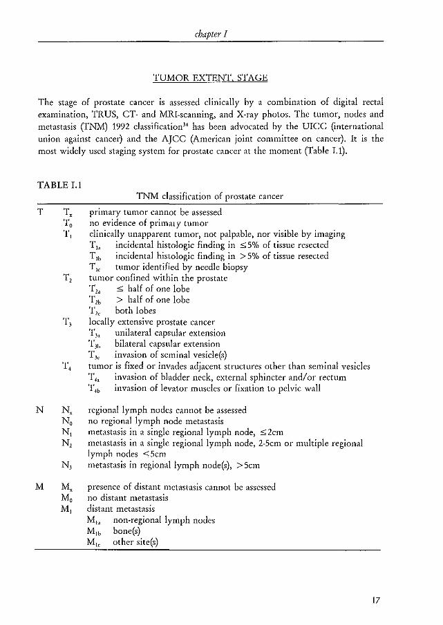
The stage of prostate cancer is assessed clinically by a combination of digital rectal examination, TRVS, CT- and MRI-scanning, and X-ray photos, The tumor, nodes and metastasis (TNM) 1992 classification" has been advocated by the VICC (international union against cancer) and the AJCC (American joint committee on cancer), It is the most widely used staging system for prostate cancer at the moment (Table I,l),
TABLE I.1 TNM classification of prostate cancer
T Tx primal)' tumor cannot be assessed To no evidence of primaty tumor 1'1 clinically unapparent tumor, not palpable, nor visible by imaging
T]a incidental histologic finding in :::;; 5% of tissue resected T lb incidental histologic finding in > 5% of tissue resected T" tumor identified by needle biopsy
T2 tumor confined within the prostate 'f" ,; half of one lobe T'b > half of one lobe 1'" both lobes
T 3 locally extensive prostate cancer T.h unilateral capsular extension T3b bilateral capsular extension T3c invasion of seminal vesicle(s)
T 4 tumor is fixed or invades adjacent structures other than seminal vesicles T4a invasion of bladder neck, external sphincter and/or rectum T4b invasion of levator muscles or fixation to pelvic wall
N N, regional lymph nodes cannot be assessed No no regional lymph node metastasis Nt metastasis in a single regional lymph node, s2cm N, metastasis in a single regional lymph node, 2-Scm or multiple regional
lymph nodes ,; Scm N, metastasis in regional lymph node(s), > Scm
M Mx presence of distant metastasis cannot be assessed Mo no distant metastasis M t distant metastasis
Mh non-regional lymph nodes Mlh bone(s) M" other site(s)
17
prognostic markers in prostate cancer
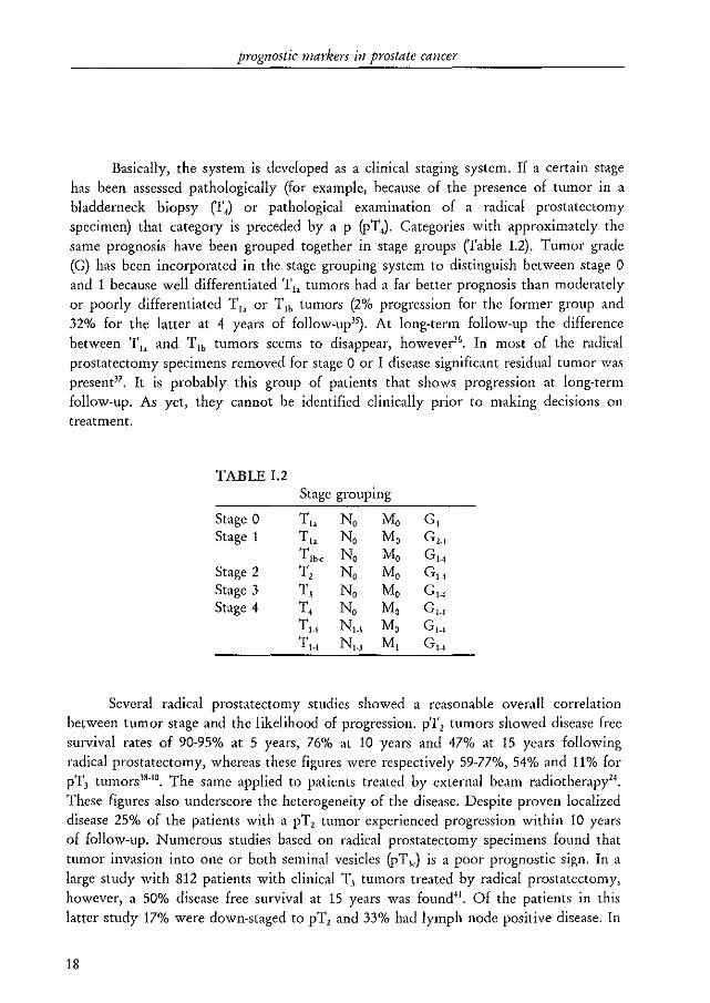
Basically, the system is developed as a clinical staging system. If a certain stage has been assessed pathologically (for example, because of the presence of tumor in a bladderneck biopsy (T,) or pathological examination of a radical prostatectomy specimen) that categOlY is preceded by a p (PT,). Categories with approximately the same prognosis have been grouped together in stage groups (Table 1.2). Tumor grade (G) has been incorporated in the stage grouping system to distinguish between stage 0 and 1 because well differentiated 1'" tumors had a far better prognosis than moderately or poorly differentiated 1'1, or 1'lb tumors (2% progression for the former group and 32% for the latter at 4 years of follow·up'~. At long·term follow·up the difference between Til and T'b tumors seems to disappear, however36. In most of the radical prostatectomy specimens removed for stage 0 or I disease significant residual tumor was present". It is probably this group of patients that shows progression at long·term follow·up. As yet, they cannot be identified clinically prior to making decisions on treatment.
TABLE 1.2 Stage grouping
Stage 0 1'1, No Mo G I Stage 1 1'1' No Mo G,~
1'11>< No Mo GI~ Stage 2 1', No Mo G H Stage 3 1', No Mo G H Stage 4 1', No Mo GI~
TI~ N'_l Mo GI~ TI~ N'_J MI GI~
Several radical prostatectomy studies showed a reasonable overall correlation between tumor stage and the likelihood of progression. pT, tumors showed disease free survival rates of 90·95% at 5 years, 76% at 10 years and 47% at 15 years following radical prostatectomy, whereas these figures were respectively 59~77%, 54% and 11% for pT, tumorsJ8~o. The same applied to patients treated by external beam radiotherapy". These figures also underscore the heterogeneity of the disease. Despite proven localized disease 25% of the patients with a pT2 tumor experienced progression within 10 years of follow-up. Numerous studies based on radical prostatecromy specimens found that tumor invasion into one or both seminal vesicles (PTJe) is a poor prognostic sign. In a large study with 812 patients with clinical 1'J tumors treated by radical prostatectomy, however, a 50% disease free survival at 15 years was found4l
. Of the patients in this latter study 17% were down-staged to pT, and 33% had lymph node positive disease. In
18
chapter J
addition, 60% of these patients received adjuvant treatment (radiation therapy, endocrine therapy or both). Of interest, van den Ouden et al." found that the prognosis of patients with clinical T) tumors was identical to that of patients with T'_l tumors if the poorly differentiated tumors were excluded. This can also be inferred from the above mentioned study".
The concordance between the clinical and pathological T-category has been investigated in several studies. Of the patients with TIe prostate cancer (most often screening patients) treated by radical prostatectomy, 20-49% are finally classified as having a pTJ tUffiOl·41--l+. For T21.b and T2<: tumors the figures are 25-46% and 48-78%, respcctively25, 45--17, Thus, a patient with clinically localized prostate cancer has a substantial risk of extra prostatic extension and, consequently, of disease progression following radical treatment. Huland et al." tried to improve the results of clinical staging by combining clinical stage with biopsy data and pre-operative PSA level in 257 consecutive patients with Tt.; or Tl prostate cancer. Digital rectal examination could not predict pathological stage or PSA progression. Biopsy results and PSA levels correlated well with pT stage, but only in 30% of the clinical 1', patients the clinical outcome (detectable post-operative PSA) could be predicted.
Like T-stage, the assessment of N-stage by clinical means is also unreliable. For this reason, in most institutions a radical prostatectomy is preceded by a pelvic lymph node dissection. If tumor is found in the lymph nodes on pathological examination of frozen sections, the radical prostatectomy is canceled. It has, however, been suggested that the probability of lymph node metastasis can reasonably well be predicted clinically in subsets of patients by taking into account variables like T~categ01y, tumor grade or PSA "-". The deletion of the pelvic lymph node dissection would be beneficial for the patients with positive lymph nodes who would not have to undergo a major surgical procedure anymore and would also improve cost efficacy of radical prostatectomies.
TUMOR EXTENT, VOLUME
For many tumor types tumor volume is part of the TN1v1 classification system. Due to several factors, this is more complicated for prostate cancer. 1. It is often difficult to recognize prostate cancer macroscopically. 2. Because prostate cancer most often invades and grows in between benign glands clinical assessments are unreliable. 3. Prostate cancer is multifocal in about half of the casesS1• 4. Estimation of prostate cancer volume requires the investigation of whole mount prostate sections, processed according to a standard protocol making use of sophisticated computerized equipment.
Tumor volume estimated in such a way has been shown to correlate with tumor grade and stage53
.S6
. The relationship between tumor grade and tumor volume is influenced by the localization of the tumor. Centrally located tumors can be large at presentation, but are generally more differentiated. For this reason, McNe,ll et al. ss .
19
prognostic markers in prostate cancer
suggested that the volume of the poorly differentiated part of a tumor would predict the clinical course best. Many papers established the relationship between tumor volume and other prognostic markers, but only few of them investigated the prognostic value following radical prostatectomy. Epstein et al." studied this issue in the radical prostatectomy specimens of 185 patients with pT<3cpNo prostate cancer and found a prognostic value of tumor volume at univariate analysis, but only of Gleason grade at multivariate analysis. A strong prognostic value of tumor volume was found in two earlier studies5s
, 59. These studies made use of prostates that were not completely embedded and the tumor volume was expressed as a percentage of total prostatic volume.
The possibility to predict total tumor volume using prostate biopsies has been investigated by several authors. The volume of a single biopsy only makes up a very small fraction of total prostatic volume, thus sampling errors may severely inhibit prediction of tumor volume. This might be less a problem in patients with a palpable lesion, compared to the random biopsies from patients with a Tic tumor. Pellcr et al.B simply counted the number of biopsies containing cancer in the sextant biopsies of 102 patients treated by radical prostatectomy. Strong correlations were found between the number of positive biopsies and several pathological parameters. On the one hand this finding supports the presumption that larger tumors will be present in more biopsies. On the other hand tumor multifocality and the irregular growth pattern of prostate cancer would suggest a more equivocal relationship. Haggman et al.60 systematically sampled 60 radical prostatectomy specimens with 10 biopsies taken with a special biopti~gun and found a significant correlation betwecn biopsy and prostatectomy tumor volume. Cupp et al." also found a significant correlation in the material of 130 patients,
but based on their results they stated that on an individual basis biopsy parameters cannot predict total tumor volume reliably (standard error of estimate 6.1ml). In this study the percentage of biopsy length occupied by tumor showed the strongest correlation. Terris et al.6l came to the same conclusion. Thus, although biopsy assessed tumor volume is significantly correlated with total tumor volume, it can probably not be used individually.
Yet another issue that has received considerable attention is the ability to predict the presence of insignificant prostate cancer (the real pussy cats) using biopsy parameters. Dietrick et a/.63 concluded from a study of 110 prostatectomy specimens that the presence of less than 3mm of tumor in one of the 6 biopsies reliably identified insignificant cancer (defined as a tumor of <C.Scc). In another study from the same institution in which the same definitions were applied, only 30% of the 'insignificant' cancers at biopsy had a volume of :::;;O.5ml at radical prostatectomy6.4. Likewise, from the study of Cupp et al." it can be calculated that only 2 of 15 patients (13%) that fulfilled this criterion had a tumor volume of <O.5cc. Several authors combined different parameters (biopsy grade, calculated tumor volume, clinical stage, tumor doubling time, age, and serum PSA) to enhance the prediction of insignificant prostate cancer6l
, 65,66. Although, a combination of parameters better correlated with the presence
20
chapter 1
of insignificant cancer, it could not be used on an individual basis61 or was applicable only to a small number of patients". Dugan et al." included time related variables like patient-age and tumor doubling time in their model. Insignificant cancer was defined as a tumor that would have grown not larger than 20cm3 by the time of expected patient death as predicted from life-tables. It was concluded that most men treated with radical prostatectomy in their institution have significant cancer.
TUMOR HISTOLOGY, GRADE
It is important to realize that different malignant processes can be identified within the prostate histologically. The World Health Organization (\'\THO) classification" is shown in Table I.3. Adenocarcinoma is by far the most common malignant tumor type (> 95%). The major part of this thesis will focus on adenocarcinoma and will be referred to as prostatic carcinoma or just prostate cancer. Neuroendocrine tumors (small cell prostate cancer and prostatic carcinoid) will be discussed in more detail in chapter II and V.
TABLE I.3 Histological classification
Epithelial tumors A. Benign B. Malignant
1. Adenocarcinoma 2. Transitional cell carcinoma 3. Squamous cell carcinoma 4. Undifferentiated carcinoma
Non-epithelial tumors A. Benign B. Malignant
1. Rhabdomyosarcoma 2. Leiomyosarcoma 3. Others
Miscellaneous tumors 1. Neuroendocrine tumors 2. Carcinosarcomas 3. Others
Secondary tumors Unclassified tumors Tumor-like lesions and epithelial abnormalities
21
prognostic markers in prostate cancer
Numerous prostate cancer grading systems have been developed over the past decades. The grading systems attempt to predict clinical patient outcome based on tumor characteristics like tissue architecture and cellular variables. Boecking et al.6s
described 3 criteria that a grading system should fulfill: 1. each diagnostic criterion should be correlated with biological behavior and prognosis. 2. it should display sufficient reproducibility. 3. Grading of random biopsies should be representative for the whole tumor.
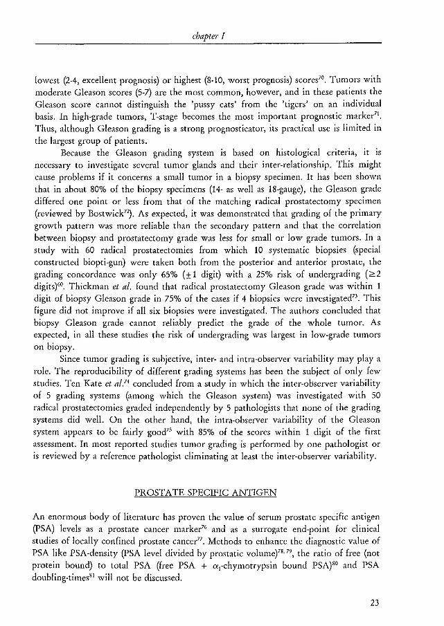
Of the many prostate cancer grading systems that have been developed the Gleason grading system is at present the most widely used and most often reported in the (American) prostatic literature". It is also the grading system that was used for the studies described in this thesis and therefore the discussion will focus on this grading system only. The Gleason system is unique in that it takes into account the histological heterogeneity of prostate cancer. The system considers only histological characteristics and recognizes 5 primary growth patterns representing the transition from well differentiated (pattern 1) to undifferentiated (pattern 5) prostate cancer (Figure 1.2). The scores of the two most common patterns are added, yielding a Gleason grade or Gleason sum score that may range from 2·10. An advantage of the Gleason system is that it enables the incorporation of tumor heterogeneity in all kinds of studies, for example: genetic studies) studies on the expression level of certain compounds and morphologic studies.
2 3 4 5
FIGURE 1.2 Simplified drawing of the Gleason grading system.
Gleason grade correlates with tumor stage and the independent prognostic value of the Gleason system has been established in numerous studies among which several large series45, 70. The prognostic value was found to be largest in tumors with either the
22
chapter I
lowest (2-4, excellent prognosis) or highest (S-10, worst prognosis) scores70• Tumors with
moderate Gleason scores (5-7) are the most common, however, and in these patients the Gleason score cannot distinguish the 'pussy cats' from the 'tigers' on an individual basis. In high-grade tumors, T-stage becomes the most important prognostic marker71
,
Thus, although Gleason grading is a strong prognosticator, its practical use is limited in the largest group of patients.
Because the Gleason grading system is based on histological criteria, it is necessary to investigate several tumor glands and their inter-relationship. This might cause problems if it concerns a small tumor in a biopsy specimen. It has been shown that in about SO% of the biopsy specimens (14- as well as IS-gauge), the Gleason grade differed one point or less from that of the matching radical prostatectomy specimen (reviewed by Bostwick"). As expected, it was demonstrated that grading of the primary growth pattern was more reliable than the secondary pattern and that the correlation between biopsy and prostatectomy grade was less for small or low grade tumors. In a study with 60 radical prostatectomies from which 10 systematic biopsies (special constructed biopti-gun) were taken both from the posterior and anterior prostate, the grading concordance was only 65% (± 1 digit) with a 25% risk of undergrading (22 digits)". Thickman et al. found that radical prostatectomy Gleason grade was within 1 digit of biopsy Gleason grade in 75% of the cases if 4 biopsies were investigated". This figure did not improve if all six biopsies were investigated. The authors concluded that biopsy Gleason grade cannot reliably predict the grade of the whole tumor. As expected) in all these studies the risk of undergrading was largest in low-grade tumors on biopsy.
Since tumor grading is subjective} inter- and intra-observer variability may playa role. The reproducibility of different grading systems has been the subject of only few studies. Ten Kate et al." concluded from a study in which the inter-observer variability of 5 grading systems (among which the Gleason system) was investigated with 50 radical prostatectomies graded independently by 5 pathologists that none of the grading systems did well. On the other hand, the intra-observer variability of the Gleason system appears to be fairly good" with S5% of the scores within 1 digit of the first assessment. In most reported studies tumor grading is performed by one pathologist or is reviewed by a reference pathologist eliminating at least the inter-observer variability.
PROSTATE SPECIFIC ANTIGEN
An enormous body of literature has proven the value of serum prostate specific antigen (PSA) levels as a prostate cancer marker76 and as a surrogate end-point for clinical studies of locally confined prostate cancer71. Methods to enhance the diagnostic value of PSA like PSA-density (PSA level divided by prostatic volume)'" 79, the ratio of free (not protein bound) to total PSA (free PSA + "'I-chymotrypsin bound PSA)80 and PSA doubling-timesSI will not be discussed.
23
prognostic markers in prostate cancer
PSA at the tissue level as identified by immunohistochemical staining methods has been the subject of fewer studies. PSA is strongly expressed in the glands of the normal and hyperplastic prostate82
, Most prostate tumors express PSA 83 and it is generally accepted that the immunohistochemically defined expression level is inversely correlated with tumor grade", although not all studies confirmed this". The authors of the latter study also did not find a correlation between tissue expression and serum levels, whereas others did86, Lymph node metastasis express PSA in about 90%82, In a (male) patient with a metastasis of an unknown primary, this can be helpful to establish the diagnosis. A complicating factor is that several recent studies have shown that PSA is not entirely prostate specific, but can also be expressed by, among others, breast tissues87, 88, salivary gland tumors89 and non-prostatic neuroendocrine tumors82
,
The prognostic value of tissue PSA expression has been studied on biopsy specimens9J-.91 and TUR specimens containing T Ib prostate cancer93 , In 80 patients with advanced prostate cancer PSA was a significant prognostic marker at univariate analysis but not at multivariate analysis (p~0.48)90. Stege et al." found PSA to be a significant prognostic marker at the univariate and multivariate level (together with cytologic tumor grade) in 67 consecutive patients who all received endocrine therapy. In a large study from Norway with patients with locally confined (n~150) or metastatic (n~116) tumors, who were treated with endocrine or radiation therapy, tissue PSA expression had no prognostic value92
, This study used overall survival as end-point, which is probably inappropriate. In 13 out of 18 Tlb prostate cancer patients who were managed conservatively, PSA immunohistochemistry of the tumor in the TUR specimen could predict the clinical outcome93 ,
The studies used different methods to quantify the PSA content and also the patient groups and outcome variables varied considerably. Thus, the prognostic value of tissue PSA levels is not clear, Since most authors found tissue and serum PSA levels to be correlated and since serum is much easier to obtain (repeatedly), tissue PSA is unlikely to become an important prognostic tissue marker.
A new issue currently receiving considerable attention is the demonstration of circulating PSA producing cells by the reverse-transcriptase polymerase-chain reaction (RT-PCR) to detect PSA-mRNA (sometimes erroneously called a micrometastasis detection assay)". Men with BPH or no prostatic disease at all showed negative results whereas about 40% and 80% of the patients with localized and metastatic prostate cancer, respectively, showed positive results9-1, In more recent studies from the same institution the RT-PCR assay better predicted pT-stage than the pre-operative serum PSA level, with an overall sensitivity and specificity of 73 and 90%, respectively'" ". The presence of circulating PSA-producing cells was of independent prognostic value in predicting PSA progression following radical prostatectomy in 94 patients with short follow-up". Others obtained, however, contradictOlY findings. In a study with 107 prostate cancer patients only lout of 7 (14%) and 21 out of 57 (37%) blood samples from metastasized untreated and androgen in4ependent tumors, respectively, showed positive results". In an experimental study employing a nested RT-PCR method several
24
chapter J
non-prostatic cell lines were found to express PSA mRNA". PSA RT-PCR can also be applied to bone marrow aspirates or lymph node sections.
The presence of PSA-mRNA in the peripheral blood is very probably a risk factor for the development or presence of metastatic disease. Clearly, the sensitivity and specificity of the assay should be determined and optimized more precisely before the clinical utility can definitely be assessed. Because RT-PCR is a velY sensitive technique that is prone to all kinds of technical problems, multi-center studies employing well defined and standardized consensus techniques should be conducted.
NUCLEAR DNA CONTENT
Nuclear DNA content (ploidy) can be studied by flow cytometry and image or static cytometry which both have specific advantages and disadvantages. Falkmer" has studied the methodology and potential problems of applying these techniques to
prostate cancer. According to this study) many differences among published series were of methodologic origin. Since the advantage Oarge number of measured cells) and disadvantages of flow cytometry (expensive equipment and excellent technical support required, no simultaneous histopathologic control and risk of measuring non-malignant cells) are opposite to the advantages (relatively easy to perform and only measurement of tumor cells) and disadvantage of static cytometly (small number of measured cells) outweigh each other, she recommended that the two measurements be combined. Most studies found both methods to be fairly well correlated, howeverloo. For practical purposes, the results from studies using the different methods will be discussed together.
The first papers on the prognostic value of DNA ploidy have been published already 30 and 23 years agolOl. 101. In these studies from Portugal two important findings were described: prostate cancer patients with diploid tumors have a significantly better prognosis and respond better to estrogen therapy than patients with triploid or hexaploid tumors. More than 100 papers on this subject, among which large series from Sweden (Karolinska Institute) and America (Mayo Clinic), have since been published. Adolfsson 103 conducted a critical literature review of 115 papers containing prognostic information on DNA ploidy in prostate cancer which were published between 1973 and 1993. From repeated papers on apparently identical series, only the most recent paper was considered. Eventually, 44 papers were reviewed. DNA content was correlated with tumor grade in 23 out of 28 reports and with stage in 14 out of 20. Sixteen out of 18 studies with patients at various disease stages showed univariate prognostic value of DNA ploidy. In 8 of these a multivariate analysis was performed. Five found DNA ploidy to be of independent prognostic value (415 patients totally) and 3 did not (365 patients totally). Two of these latter three found ploidy to be a prognostic marker at univariate analysis. Ten studies were restricted to one specific stage. In I and 2 studies on localized (spT,) disease DNA ploidy was a dependent or independent prognosticator, respectively. In metastatic disease, 4 studies showed
25
prognostic markers in prostate cancer
prognostic value at univariate analysis and 3 did not. All 3 studies that included a multivariate analysis found independent prognostic value of ploidy.
Ploidy assessments were of independent prognostic value in 3 studies of patients with clinically localized prostate cancer treated with external beam radiation therapyl".I". Another study did not confirm this lO7 • The findings of the first studies on ploidy in hormonally treated patients have been confirmedlos.llI . In two of these ploidy was of independent prognostic value I". 110. In one study neither tumor grade nor ploidy was of prognostic significancell2.
Several authors described the concordance between biopsy and surgical specimen based tumor ploidy l13-115. As can he expected, the concordance was highest if an aneuploid population was found in the biopsy specimen. Tumor heterogeneity may also pose a problem. But, overall the correlation was high.
Two recent consensus meeting reports describe the clinical applicability and limitations of ploidy assessments in prostate cancer: the \VHO conference on early diagnosis and prognostic parameters in localized prostate cancerlOO and the college of American pathologists conference on dinical relevance of prognostic markers in solid tumors)). It is important to realize that most of the authors/contributors appearing on these reports belong to the ploidy 'supporters'. The WHO report lOO discusses only T, and 1') tumors and the contributors agreed that if surveillance is a treatment option, knowledge of ploidy is of clinical value. Furthermore, it was strongly advised to study DNA ploidy in clinical trials, particularly in patients with localized disease. The American pathologists conference concluded that DNA ploidy can be used for clinical decision-making only in patients with 1') or pN + tumors who are offered subsequent hormonal therapy"'. The two reports agreed on the prognostic impact of DNA ploidy but clearly disagreed on its current clinical position.
A more sensitive method of measuring DNA ploidy is DNA in-situ hybridization (ISH) of interphase cells l17. Both fluorescence (FISH) and non-fluorescence methods have been developed. Basically, these techniques visualize individual chromosomes by specific binding of a labeled probe to a particular DNA sequence (mostly localized at the centromere region). It can thus be used to investigate loss or gain of single chromosomes or even parts of chromosomes. FISH has been used on cell suspensions and paraffin·embedded tissues. The quantification of (F) ISH spots in paraffin-embedded tissue sections is somewhat complicated because not all spots in a nucleus need to be present in a tissue section. Thus, the distribution of the real number of spots may interfere with the distribution of countable spots. Persons et al.1I1 studied the correlation between the three methods described above in the paraffin-embedded specimens of 34 prostate cancer patients. In 28 cases the three methods agreed, whereas FISH (probes for centromeres of chromosome 8 and 12) identified 2 additional aneuploid cases. Other studies confirmed that (F)ISH and flow cytometry are reasonabl y well correlated, but that (F)ISH is more sensitivellS • 119. AIel'S et al. l20 have shown that ploidy as defined by probes for chromosome 1 and Y may vary between the Gleason patterns of one tumor, within one Gleason region and even within one
26
chapter J
of the p21 gene, the protein product of which (ras-p21) in turn inhibits proliferation and 'decides' whether the cell will die by apoptosis or will repair its DNA damage. Aberrant (i.e., mutated or truncated) p53 has lost its cell-cycle control function and may thus playa role in tumor biology. The Li-Fraumeni syndrome, an inherited autosomal disease, characterized by the development of several malignancies during infancy, was shown to be caused by a germ-line mutation in the p53 gene ll'. DNA analysis of several clinical tumors subsequently identified the presence of mutations in the p53 gene in a fraction of the investigated specimens suggesting that p53 may play a role in tumor biologyl35. Antibodies against p53 were soon developed and enabled the study of p53 expression in more detaiP". The p53 antibodies recognize wild-type p53 as well as most mutant forms. Since mutated p53 has a much longer half-life than the wild-type molecule, immunohistochemical identification of p53 has usually been associated with the mutant phenotype. But, results of immunohistochemical studies need to be analyzed with care since false-positive and false-negative results have been described, especially with highly-sensitive staining methods"'. It has been recommended to verify the immunohistochemical data with DNA analysis137,
The first pape" on p53 in prostate cancer identified mutated DNA in 5 out of 29 tumors (17%) whereas no mutations were found in 34 BPH specimens138, 139. The
presence of mutated p53 correlated with neither stage nor grade. Most immunohistochemical studies found comparable low percentages of untreated primary tumors with p53 mutations « 20%) 14~147. Others described higher percentages, reaching from 22% to 80%148-151. In one of these immunoreactivity was in general cytoplasmicl48 , which is at variance with other reports. In the studies in which the immunohistochemical results were confirmed by DNA analysis an excellent correlation was foundHl. 153, 154. There is no agreement on whether tumor-stage or grade is associated with p53 mutations in untreated tumors. Several authors found such correlations 140, 145,
15.', while others did not138. 139, 142, 147, 151, The frequency of p53 mutations increased from
primary to metastatic cancer to androgen independent and irradiation insensitive cancerlH, 150, 155.157. Especially the association between p53 mutations and androgen resistance ISO, lSI'> and radiation insensitivity157 is interesting since pre-treatment analysis
might stratify patients in those who will and those who will not benefit from these treatment modalities. But, clinical trials have to substantiate this. The presence of p53 mutations in primary tumors had no predictive value for the presence of lymph node metastasis143 •
The prognostic influence of p53 mutations has been investigated by several authors. In a study of Visakorpi et al. l40 patients with a p53 mutation had a higher progression l"<He with a relative risk of 12. This study contained 137 patients who were treated by radical prostatectomy, radiotherapy or endocrine therapy. A multivariate analysis was not performed. Thomas et al.14~ found a prognostic value of p53 mutations on the time to progression and tumor related death in 68 patients. The presence of mutations was correlated with stage and grade. Since a multivariate analysis was not performed, the prognostic value may also be attributed to these relationships. In 45
29
prognostic markers in prostate cancer
patients with a Tia tumor with Gleason grade 4 or less, a trend towards a worse prognosis (crude survival) of patients with a p53 mutation was found (p <0.08 at univariate analysis)141. Vesalainen found no relationship between p53 mutation and prognosis in the biopsy specimens of 139 patients with T,.,M, tumors'''. Half of the patients were managed conservatively, the others received radical therapy (n ~ 6) or endocrine therapy (n~32). Thus, the patient sample was rather heterogenous. Another study made use of an even more heterogenous patient population with clinical stages varying from T1.l to N+158. At univariate analysis the presence of pS3 mutations was a prognosticator for time to progression only in patients with Gleason grade 2-7 tumors. Bubendorf et al.145 studied the issue in 137 patients treated by radical prostatectomy. The presence of p53 mutations was related to grade and proliferation rate (Ki-67% immunostaining) but showed no prognostic significance. In a relatively small study with 40 patients treated with a radical prostatectomy, pS3 mutations had prognostic value on the time to PSA progression'5l. In this study 80% of the tumors showed p53 immunostaining which is an extremely high figure compared to other studies. The authors explained this by the application of a strict quality control scheme and newer antigen retrieval techniques. It can, however, not be excluded that ultra-sensitive pS3 assays identify wild-type p53 as well. Another study with 175 patients treated by radical prostatectomy found pS3 expression in 65% of the cases 152. Expression of p53 was an independent prognostic marker for PSA progression.
Mirchandani et al.'" studied the heterogeneity of p53 mutations at the DNA level. In the case of multifocality lesions with and without p53 mutations were often found and even within one lesion, areas with wild-type and mutant pS3 could be identified in a number of cases. The authors conclude that this heterogeneity may explain the large differences in published series on p53 in prostate cancer. Additional explanations are possible. A number of (polyclonal) antibodies were not ve,y well characterized. The immunohistochemical procedures, methods of quantification and definitions of cut·off levels also differed among studies. Finally, most studies petformed so far used patient groups from several stages and receiving different treatments. The correlation between biopsies and matching prostatectomies has not been studied.
Ras-p21 expression was increased in prostate tumors as compared to benign prostatic tissues1S9-162 and a correlation between its expression level and tumor grade or stage was found in three of these studies 1s9, 160, 162. Sumiya et al. ls9 found no prognostic influence of ras-p21 in 62 patients with metastasized prostate cancer receiving endocrine therapy. In contrast, Agnantis et al. 162 found a prognostic value in surgically treated patients by comparing the 5-year crude survival rates of patients with and without rasp21 expression which is statistically an inappropriate method. Moreover, the relationship between ras-p21 expression and tumor differentiation could possibly explain this finding.
The number of prognostic studies on ras-p21 in prostate cancer is too limited to draw conclusions at this moment. It can also not definitely be concluded whether p53 can be used as a prognostic marker in prostate cancer. The studies differ too much,
30
chapter [
both in terms of patients and methodology, and in only a small number of studies multivariate analysis on the prognostic impact has been performed. It might be a useful marker for the selection of patients to be treated with either hormonal or irradiation therapy. Since ras·p21 and p53 interact at a functional level, it could be useful to combine ras·p21 and p53 in prognostic studies as well.
EPIDERMAL GROWTH FACTOR RECEPTOR AND C-ERBB-2
The epidermal growth factor receptor (EGF-R) is a key modulator of normal prostatic homeostasis (reviewed by Steinerl"). Stimulation of the receptor by the epidermal growth factor (EGF) or transforming growth factor-ex (TGF-ex) promotes proliferation which in the normal prostate is balanced by the inhibitory activity of TGF-l1l". In prostate cancer EGF related peptides appear to playa functional role in tumor growth with TGF-ex as the most important peptide (reviewed by Sherwood and Leel"). CerbB-2 is a receptor that shows considerable homology with EGF-RI65, but does not need EGF to be activated. Although the ligands of these receptors might be quite different, most prostatic studies combine the two and for this reason they will be discussed together.
The first study on EGF-R in the prostate quantified receptor content of BPH and cancerous tissues166 . The expression was lower in prostate cancer as compared to benign tissues and decreased with increasing grade. Immunohistochemically a similar pattern was foundB8, 167, The studies on C-erbB-2 differ considerably, some authors found no immunoreactivity at aH156. 167 or only in a very small minority of cases (4/266)" whereas others described 36 to 100% of the investigated tumors to be positivel68.170, Differences among the antibodies and visualization techniques may explain the disagreements, although 2 studies with completely different outcome used the same polyclonal antibody (NCL CBll) and staining method"· I". Only one study correlated C-erbB-2 immunostaining with tumor gracie l6S, a significant relationship was not found. The expression level of the two most important ligands of the EGF-R has been studiedl'l. 172. EGF was present in 6% of 52 BPH specimens, in 40% of 45 localized tumors and in all 20 metastatic tumors 17l• TGF-Q' was present in 15% of benign tissues and in 53% of the tumors17l,
In 45 patients with T h tumors, EGF-R showed no prognostic value, whereas C-erbB-2 <lid (at the univariate level)I". Visakorpi et al. l67 studied 147 patients and found a prognostic value of EGF-R at univariate analysis (progression and tumor specific survival) but not at multivariate analysis. In a study of 124 radical prostatectomy specimens with pT <3c prostate cancer, C-erbB-2 was of independent prognostic value together with Gleason grade, DNA ploidy and nuclear morphomeuy115. Moul et al. l73
studied 105 radical prostatectomy specimens and found no correlation between EGF-R expression and tumor stage, grade or prognosis.
31
prognostic markers in prostate cancer
Although biologically an important role of these molecules could be expected in prostate cancer, this is not reflected by a clear cut relationship with clinical factors such as tumor grade, stage and prognosis. As a consequence EGF and EGF-R will probably not be used as prognostic markers clinically. Illustrative is the fact that only two papers were published in 1996, all the other mentioned papers were published before 1995.
E-CADHERIN AND A-CA TENIN
E-cadherin is a member of a family of calcium dependent intercellular adhesion molecules. E-cadherin is located at the cellular surface and is coupled to catenin and thereby connected to the cytoskeleton. Immunohistochemically, normal prostatic glands showed a velY intense membranous staining of E-cadherin while the expression in tumors was in general decreased 174-176. Most studies described 3 patterns of immunostaining in prostate cancer: a pattern comparable to that found in benign glands, a totally negative and a heterogenous staining pattern. A decreased expression in tumors implies a deranged intercellular attachment and an increased metastatic potential of the tumor. All authors described an inverse relationship between E-cadherin expression and tumor grade and stageI74-178 . In one of these} biopsy specimens of 56 patients who subsequently underwent a radical prostatectomy were investigated l7s
•
Although a correlation with stage and grade was found, the pattern of E-cadherin staining of the biopsy could not predict the presence of lymph node metastasis.
Immunohistochemistry of E-cadherin using formalin-fixed, paraffin embedded material is possible, although after more than 12 hours of formalin fixation the results become unreliable (E. Ruiter and J.A. Schalken, personal communication). This impairs the applicability of E-cadherin to archival prostatectomy specimens since in most institutions the duration of formalin fixation is unknown and variable} but regularly extends 12 hours. In a recent study, a microwave based antigen retrieval procedure was used on routinely fixed material and the authors claimed good results with this method, howeverl76.
Umbas et a/. m used frozen sections from 42 radical prostatectomy specimens) 29 primary and 18 palliative TUR specimens. In the former two patient groups E-cadherin expression was a statistically significant prognostic marker for time to progression at univariate analysis. A multivariate analysis was not reported by the authors, probably due to the heterogenous patient sample and the relatively small numbers. Since a strong association with both tumor-grade and stage was found, the prognostic value of Ecadherin may be the consequence of these strong associations.
Because E-cadherin is functionally coupled to catenin, its presence at the intercellular membranes does not necessarily imply a functional complex. Decreased or altered catenin expression may also he responsible for an abnormal function. Two studies that have only be published as abstract combined E-cadherin and ",-catenin immunohistochemistry in prostate cancer l79, 180, Umbas et a/. 179 investigated 20 radical
32
chapla I
prostatectomy specimens and 32 advanced tumors. Overall, in 16 of these 52 tumors Ecadherin and a-catenin expression was normal, whereas in 23 tumors the expression of both molecules was abnormal. Likewise, the prognosis of these patients was best and worst, respectively. In 13 locally advanced tumors with normal E-cadherin staining, an abnormal a-catenin staining was related to an intermediate prognosis, but the numbers were small. In the other study the biopsy specimens of 44 surgically treated patients were used ISO. Abnormal E-cadherin staining was found in 18 cases and was an independent predictor for time to PSA pl'Ogression together with pre-operative PSA and biopsy Gleason grade. E-cadherin and a-catenin expression were concordant in more than 90% of the cases, but the prognostic value of a-catenin was not mentioned.
Although only a limited number of studies have been published as yet the overall agreement between the studies, the straight forward correlation with tumor biology and the convenient way of assessments of the results (normal or abnormal in most studies) make these markers potentially useful. But, more studies need to be performed to determine first an independent prognostic value and second the clinical applicability.
PROLIFERATION
Because one of the hallmarks of malignancy is uncontrolled growth, intuitively one would assume that assessments of the proliferative activity of a tumor would bear prognostic significance. Several methods to measure the proliferative activity are available: counting of mitotic figul'es"t, determination of the S-phase fraction (SPF) with flow cytometty, labeling of replicating DNA with tritiated thymidine or bromodeoxyuridine (BrdU), and finally, antibodies to proliferation related antigens (Ki-67lSl, MIB-l!'l, and proliferating cell nuclear antigen (PCNA)!"'). Ki-67 and MIB-! are both directed against the same antigen. The Ki-67 antigen as well as PCNA are expressed only during the G!, S, M, and G, phases of the cell cycle, but not during Go. PCNA has a long half-life and is expressed during DNA repair as well. Assessment of SPF is cumbersome, and although a prognostic value has been reported in prostate cancerl85, 185,
it will not be discussed. Likewise, the study of BrdU incorpOl"<1tion requires either the infusion of BrdU to a patient lS6 or incubation of the surgical specimen with BrdU. BrdU may have mutagenic effects'S? and the ex vivo incubation method is not reliable. At present most studies use immunohistochemical methods to identify proliferating cells, and the practical use of BrdU labeling and SPF determination in a clinical setting is not recommended.
Several studies compared the proliferation rate in BPH with that of prostate cancer and found, independent of the technique used, an increased rate in cancerous tissues'SS.I9l. The mean prostate cancer Ki-67, MIB-l and peNA labeling indexes among .111 studies were about 4%, 7.5% and 19%, respectively. The correlation between Ki-67 and PCNA indexes was only weak (0.32 - 0.8) and depended strongly on the method of
33
prognostic markers in prostate cancer
fixation for PCNA stainingl90,193. Some authors described a correlation with grade or stageHl, 190, 192, 19-1--196, others did not lS9, In, 197-199. Several authors described variable labeling
indexes in different areas of a tumor. Oomens et al. '" found in patients with repeated biopsies that the Ki-67 labeling
index decreased after the start of endocrine therapy. In an extension of this prospective study Santerse et al. (unpublished observation) found that Ki·67 labeling indexes started to increase already up to 6 months before PSA progression became apparent in these hormonally treated patients. Harper et al. '" studied the pre-treatment TUR specimens of 86 hormonally treated patients and found a prognostic value of Ki·67 only in patients with metastatic disease (univariate analysis of cancer specific survival). Using a more or less identical patient sample she also found a weak prognostic value of PCNN". In another study with only 17 hormonally treated patients the Ki-67 index showed only a prognostic trend (not statistically significant). In a study with 45 patients McLoughlin et al. '91 found no prognostic significance of Ki-67. However, in this study the prognostic impact was determined by comparing the Ki-67 labeling indexes of patients with and without progression, which is probably an inappropriate statistical method (in fact, this technique analyses the influence of prognosis on Ki-67 labeling!). Several studies with patients that were either untreated or hormonally treated found an independent prognostic value of PCNAI4l· 191. I". Three larger studies investigated the prognostic value of PCNA125 or MIB·1 195, 199 in radical prostatectomy specimens. The PCNA study found no prognostic value, the others found an independent prognostic value of the MIB·l labeling index. In a study with a diverse patient sample (stage T H ,
M~I> different treatments) the number of mitotic figures per high power field as simply counted in a routinely stained tissue slide was a significant prognostic marker at univariate analysis181
. At multivariate analysis an independent value was found only for patients with T I.,MO tumors. One study investigated the prognostic value of MIB·l labeling in lymph node metastasis of 50 patients undergoing Il5I-implantation therapy'oo. A prognostic influence on the time to PSA progression was found at univariate analysis, but not at multivariate analysis (only DNA ploidy significant). The study may however have been biased because 32 out of 82 (39%) tissue sections contained less than 500 tumor cells and were excluded for immunohistochemistlY.
Researchers clearly do not agree on the prognostic impact of proliferation rates in different patient groups. The prognostic value of proliferative activity in biopsies of patients undergoing radical prostatectomy has not been reported. In addition, the reported heterogeneity within areas of one tumor may potentially hamper the routine application.
\Vhether a study found a correlation with prognosis, tumor stage or grade seems not to depend on the method of determination of the proliferative activity. The expression of PCNA is not limited to progression through the cell cycle, as illustrated by the higher labeling indexes and marginal correlation with Ki·67 indexes. For this reason PCNA may be less desirable. Since PCNA is functionally related to p53 and p21133, it might be useful to study these 3 antigens together. Ki·67 was initially thought
34
chapter 1
to be applicable only to frozen sections, but can also be applied to paraffin sections, although Mffi-l staining appears to be more sensitive"!, More importantly, Mffi-l scores were about 2 times as high as Ki-67 scores in routinely processed tissues with a high linear correlation between the two antibodies'01, The standard deviation of each individual assessment depends on the number of positive cells (binomial distribution, SD~v'np(l-p) with n~number of investigated cells, p~positive fraction), Thus, for a given number of investigated cells Mffi-l scores may statistically be more reliable which is of importance if the proliferative fraction is small as is the case in most prostate tumors.
During the last years it has been recognized that tumor growth is the net result of cell gain and loss, Study of cell proliferation in combination with cell death parameters may reveal more conclusive results in the near future. At this moment assessment of the proliferative activity of prostate cancer has no clear clinical benefit.
ANDROGEN RECEPTOR
The androgen receptor is a nuclear receptor belonging to the steroid receptor family and acts as a transcription factor that stimulates prostatic growth and secretion upon binding of androgen (testosterone or its more potent metabolite dihydro-testosterone)", Normal prostatic tissue as well as most primary prostatic carcinomas depend on androgen for their maintenance and function, Androgen withdrawal leads to cell death by apoptosis in normal as well as malignant glands and this is the basis of endocrine
treatment of prostate cancer.
Using a well defined monoclonal antibody directed against the amino-terminal part of the androgen receptor Ruizeveld de Winter and colleagues found strong expression in benign glands and stroma202 , In primary tumors, staining intensity was in general less, but was not related to the Gleason grade20), Although not expected, it was found that most of the androgen independently growing tumors showed androgen receptor expression204 , DNA analysis identified no mutations in the androgen recepto~03, Studies from other institutions have identified androgen receptor gene mutations in a small fraction of hormonally escaped tumors, however2°5, 2e6, It therefore appears that androgen receptor mutations may play a role in androgen independent prostate cancer growth in a minority of cases,
It was hypothesized that the androgen receptor content of a tumor prior to the application of endocrine therapy would predict treatment response207. By measuring the androgen receptor content in the cytosol and nuclei of 23 patients with metastasized prostate cancer researchers from Johns Hopkins University showed that patients with low nuclear receptor levels « 110 fmollmg protein) had a shorter response duration and a decreased survival"7, Van Aubel et at.''' studied the biopsy specimens obtained before the start of endocrine therapy of 37 patients with locally advanced or metastatic prostate cancer, but did not find a prognostic value of nuclear AR content. The
35
prognostic tnarkers in prostate cancer
correlation between nuclear AR content and time to progression was used to evaluate the prognostic impact in the latter study. In a later study from Johns Hopkins a polycloncal antibody was used to quantify androgen receptor content109
• A marked heterogeneity within and between samples was found, but AR content showed no prognostic value (In only 17 patients). In a more recent study, this heterogeneity was quantitated using an image analysis systemllO. Patients with tumors with a high variance of staining intensity did worse as defined by treatment response. Tilley et a/.lIl also used image analysis to study two antibodies (to the carboxyl- and amino-terminal part of the protein, respectively) in biopsy specimens of 30 hormonally treated patients of all stages ('r,M, - T,M,,,J. The two antibodies showed quantitative differences, indicating that receptors may differ among tumors. The mean staining intensity was a prognostic marker in these patients. Other studies confirmed the prognostic value of androgen receptor content in hormonally treated patients at the univariate and multivariate level, with higher expression being related to a better prognosis212-1H.
Conflicting data have been published as to whether the androgen receptor level is of independent prognostic value in the pre·treatment tumor specimens of hormonally treated prostate cancer patients. It is however clear that AR determinations cannot be used to select patients who will and who will not benefit from endocrine treatment on an individual basis. Possibly, a combination with other markers (PS3, neuroendocrine markers and DNA ploidy) could enhance the results. A potential problem is the use of a biopsy of the primary tumor to determine the androgen receptor content in a patient with metastatic disease. The androgen receptor content of the metastatic lesions might be different from the primary tumor. One study compared the expression level in 12 lymph node metastasis and matching radical prostatectomy specimens2l5. Only one metastasis showed no androgen receptor immunoreactivity in contrast to the radical prostatectomy specimen.
APOPTOSIS
Apoptosis or programmed cell death is a second mechanism of cell death besides necrosis2l(,. Apoptosis is a very important mechanism which plays a role in a large number of physiologic processes (for example: emblyonal development, selection of immuno-competent cells and tissue homeostasis). The histologic characterization of apoptotic cells has been described and is generally accepted. It was found that the apoptotic pathway is closely related to the cell cycle (reviewed by Meikrantz and Schlegel2l1. Actually, during progression from the G,- to the S-phase of the cell cycle, the cell 'decides' whether to proceed with DNA replication and finally mitosis, or to activate the apoptotic pathway (the cell cycle check point),l7. Since apoptosis is closely related to proliferation, it might playa role in malignancy as well.
It has been shown that physiological androgen levels inhibit apoptosis of prostatic epithelial cellsm. In contrast, following androgen withdrawal prostate (cancer)
36
chapter I
cells die by apoptosis l19 • At present there is still a debate going on as to whether p53 expression and cell cycle progression are required for prostatic epithelial cells to undergo apoptosis following androgen withdrawal'1O· 11'. Probably both pathways are
possible. The ability of prostatic tumor cells to undergo apoptosis might be related to the androgen sensitive phenotype.
Bcl-2 is a protein that is able to block the apoptotic pathway1l1. Expression of this protein may playa role in androgen independent prostate tumor growth. Indeed, transfection of bcl-2 in the androgen dependent prostatic tumor cell line LNCaP resulted in a cell line that grew androgen independently in vitro and in ViV0113. If the cell cycle check point is not functioning due to an elevated bcl-2 level, the cells may survive with more and more genetic alterations leading to increasingly aberrant tumor behavior.
Bcl-2 labeling was found in the basal cell layer of normal prostatic glands!'" 114. m Primary prostatic tumors expressed bcl·2 in 24%_62%145, 152, 22-1-226, In androgen
independent tumors both the number of positive tumors and the intensity per tumor is increased124
· llS. In a study with 325 heterogenous patients (with respect to tumor-stage and grade and treatment) the presence (yes or no) of apoptotic cells was correlated with tumor grade and was a prognostic indicator for tumor specific survival, but only at the univariate leveP17. Almost all researchers have found apoptotic cells in areas with proliferating cells (e.g., intestinal crypts, breast during weaning and tumors), although in tumors the numbers were variable217
. Thus, a (semi)-quantitative determination seems preferable. Such a study has been performed on 28 radical prostatectomy specimens containing Gleason grade 6 prostate cancer1l8. The number of apoptotic cells was of
prognostic value to predict the time to PSA progression. A multivariate analysis was not performed, since only Gleason grade 6 tumors were investigated.
The prognostic value of bcl-2 has only been studied in radical prostatectomy specimensH5. 152. A prognostic value was found at univariate analysis, but not at multivariate analysis1-l-s. This study combined bcl-2, p53 and Ki-67 immunostaining, Bcl-2 expression was not related to Ki-67 staining, and a statistically not significant correlation was found with p53 expression. Interestingly, patients could be separated in 3 prognostically different groups. A group with low bcl-2 and Ki-67 (best prognosis), a group with high bcl-2 and Ki-67 (worst prognosis) and the remaining patients (intermediate prognosis). The other study found an independent prognostic value of bcl-2 expression on the time to PSA progressionlSl . Interestingly, p53 expression was also investigated by these authors and the combination of both parameters better predicted clinical outcome.
Besides bcl-2 more proteins have been identified that either block (bel-family) or promote (bax) apoptosis. The expression of bcl-2, bcl-x and mcl-l (all apoptosis inhibitors) was shown to be related to prostatic tumor grade1l6, Bax was expressed by all tumors and the level was independent of tumor gr.lde226 , \Vestin et. alY9 reported on an interesting study with biopsy specimens of 18 prostate cancer patients obtained one day before and 7 days after the start of endocrine therapy. In 3 out of 18 no effect was seen.
37
prognostic markers in prostate cancer
In the other 15 the Ki-67 proliferation rate decreased and in 6 of these the number of apoptotic cells increased. This group showed in the initial biopsy well or moderately differentiated tumors without expression of bcl-2, p53 or c-myc. The 12 tUillors of which the frequency of apoptotic cells was unaltered or even decreased, were moderately or poorly differentiated and expressed bcl-2, p53 or c-myc in the initial biopsy. These results suggest that the likelihood of remission can be predicted on the basis of biopsy specimens.
The role of apoptotic cells and bcl-2 expression in clinical tumors is at present not completely known. It is however clear, both from experimental studies and the clinical studies performed so far, that the study of apoptosis may become a clinical useful parameter. Prognostic studies on the expression level of other apoptosis related proteins should be performed (preferably combining the several proteins) on several patient groups. Because of the close relationship between apoptosis and proliferation, these studies should include proliferation markers as well.
OTHER MARKERS
TGF-f!1 inhibits epithelial cell growth (see under EGF), but it also stimulates angiogenesis and cell motility and suppresses immune reactions230 • The expression of TGF-Bl increases from benign to cancerous prostatic tissues 23o. 131 • It can be found intraand extra-cellularly. The level of extra-cellular TGF-61 increased with tumor grade"'. Serum levels of TGF-Bl were also increased in patients with prostate cancer compared to BPH controls and the level in tumors was related to tumor stage2)1. Some recent studies suggest that TGF-62 and the type-I and -II TGF-6 receptors are also important to study, but clinical studies are lacking as yet233 .. m .
Tumor growth requires angiogenesis. The microvessel density has been studied in prostate cancer using antibodies to either factor-VIII (von Willebrand factor) or CD3423
6-139. Microvessel density was higher in tumors compared to adjacent benign tissue and was correlated with tumor-stage and grade. Microvessel density was a prognostic indicator in the TUR specimens of 25 patients treated with radiotherapy!3S.
Several markers were the subject of only one or few studies. A prognostic value was found for (among others): tumor infiltrating lymphocytes (predictor of good prognosis in 325 heterogenous patients)91, Sial Lewis-X antigen140 and CD44 expression (chapter IIl)14I. Several markers were related to tumor stage and/or grade: hyaluronidase activity (an extracellular matrix degrading enzyme)"', high mobility group I protein (as identified by mRNA in-situ hybridization),", hepatocyte growth factor and its receptor c-met1H and oncoantigen-519145• Negative findings were published on c_rnyc H1
,
Cathepsin-D'" and Rb-gene product"'. Some markers appear to be of interest, but clinical studies have not been
performed yet. KAI-1 was identified as a metastasis suppressor gene in A T6.1 cells"', and the gene product was absent in clinical prostate cancer metastasis2-l9. Telomerase is
38
chapter [
an enzyme that prevents chromosome shortening during repetitive replications and was found to be absent in benign prostatic tissues, but present in prostate cancer150
•
T elomerasc activity could also be measured in biopsy specimens251, The expression of
fibroblastic growth factor-8 (FGF-8) as well as its receptor (FGF-R type J) is upregulated by androgens and appears to be essential for androgen induced growthl5l
• Normal adult fat tissues did not express FGFw 8, whereas in vitro growing prostatic tumor cell lines did.
CONCLUSION
Although much effort has been put in the study of potential prognostic markers during the last years, presently no markers, apart from tumor-stage and grade can be used for clinical decision making, although it has been suggested that DNA ploidy can be used to select patients for endocrine therapy or watchful waiting. Based on the heterogeneity of prostate cancer it is to be expected that only combinations of markers will reveal sufficient prognostic power. Biopsy techniques may have to change substantially to improve sampling of heterogeneous tumors. Future studies should thus focus on combinations of markers that are derived from biological knowledge of the disease, for example a combination of cell~adhesion molecules to predict metastatic capacity. It will also be important to include patient derived factors like concomitant diseases and patient~age. Because basic prostate cancer research very probably leads to the identification of potential new prognostic markers, it will be necessary to develop clinical facilities (tissue-, serum- and data-banks) which allow the evaluation of candidate prognostic markers in a multifactorial setting. Tumor~stage and grade are strongly correlated with patient outcome. A multivariate analysis should be performed whenever possible and it should include those factors of which the clinical value already has been established. The general discussion on prognostic markers (chapter VIII) reviews the problems associated with the assessment of prostate cancer prognosis and provides recommendations for future prognostic marker research.
39
prognostic markers in prostate cancer
APPENDIX
The analysis of survival times requires specialized statistical techniques for two reasons. First, the event of interest (death, progression or something else) almost never occurs in all subjects. These subjects are generally referred to as censored subjects. Second, the duration of follow-up generally varies greatly between the subjects. In the studies described in this thesis two types of analysis were used: the Kaplan-Meier method15l and Cox's proportional hazards modeP54. These are the two techniques that are most often being used to analyze survival data and will for a better understanding of the results briefly be described.
KAPLAN-MEIER METHOD
The Kaplan.Meier method253 is a technique to graph survival functions. The survival period is divided in many small time periods and the probability of survival until a certain time point, p(t), is calculated simply as the conditional likelihood of surviving all the preceding small time periods p(t·1). If in such a time period no event occurred, the conditional probability of surviving that period is 1. Thus, the probabilities have only to be computed when one or more events occur (NenntJ. Censored subjects do not influence the survival curve, they only decrease the number of subjects at risk (Nat rid,) as of the time-point they are censored. This can be represented mathematically by the following recursive formula:
(1)
The Kaplan-Meier method results in a step function and the clllve should be drawn accordingly. If the subjects can be divided in two or more groups, for example based on a prognostic factor or treatment, the Kaplan·Meier curve can simply be constructed for the different groups separately. The median slllvival time can be estimated from the time point when the curve crosses the survival probability of 0.5. The tails of the curves become unstable due to the small number of subjects at risk and should therefore be interpreted with care.
The curves cannot be used to draw conclusions on the differences between the groups. This should be done by applying a suitable statistical test, usually the logrank test or the logrank test for trend in the case of more than two ordered groups. The logrank test tests the hypothesis that the number of events is equally divided over the groups based on the null hypothesis that the groups origin from one population. The logrank statistic gives no information about the magnitude of difference between the
40
chapter J
groups. For this purpose, the hazard ratio (the observed over the expected number of events between the groups) with its 95% confidence interval can be computed.
COX'S PROPORTIONAL HAZARDS MODEL
The Kaplan-Meier method is not suitable to study the influence of several variables together on survival. For these purposes Cox's proportional hazards model was developed'''. Fitting of the model results in a hazard function H(t), the risk of dying in a very short time intelval following time-point (t):
H(t) ~ Ho(t) x exp(BIXI + B,X, + ... + B"X,J (2)
For each variable entered the model computes a coefficient (BI, B" ... , B,,) that fit the data best and tests if the coefficients are statistically significantly different from o. If so, that variable is of independent value. No special distribution of the variables is assumed, the only assumption is that the variables have a constant influence over time (proportional hazards). This can and should be tested for. Several techniques can be used to obtain the most relevant model (comparable to ordinalY multi-regression analysis).
XI' Xl> ... , X" represent the values of the variables in an individual. Ho(t) represents the baseline hazard. It is calculated by taking all variables 0 (exp(O) ~ 1). For each individual the outcome of BIXI + B,X, + ... + B"X. can be computed and is often referred to as the prognostic index (PI). By defining PI ranges, patients can be separated into groups with different prognosis. It follows from formula (2) that a marker that is associated with an increased hazard will have a coefficient Bk that is greater than 0 and vice versa. It can thus easily be seen from the output of the computer program if a marker is associated with a good or a bad prognosis. The hazard difference of two individuals or groups with a different PI only depends on the PI of both subjects or groups and is, as a consequenceJ constant over time (proportional hazard):
H(t), ! H(t), ~ exp(PI, - PI,) (3)
The coefficients as shown in the multivariate analysis of the chapters II-IV are depicted as hazard ratios, that is exp(BJ. A hazard ratio of > 1 indicates a prognostic marker that is associated with an adverse prognosis and vice versa.
41
prognostic markers in prostate cancer
SCOPE OF THE THESIS
The need to identify reliable prognostic markers for prostate cancer has been highlighted in this chapter. Since no reliable markers that predict patient outcome on an individual basis are available at present, study of all markers that might be of value is warranted. Two of such markers were studied in radical prostatectomy specimens (chapter II and Ill) and two were studied in the pre-treatment TUR specimens of hormonally treated patients (chapter IV). All these studies made use of routinely formalin-fixed, paraffin-embedded prostate cancer specimens. The immunohistochemically defined expression level was correlated with Gleason grade, tumor-stage and prognosis. Multivariate analysis using Cox's proportional hazards model was performed to identify independent prognostic markers.
Chapter II describes the prognostic value of neuroendocrine cells in prostate cancer. The rationale for studying neuroendocrine cells in prostate cancer is described in full detail in chapter V. In brief, neuroendocrine cells are present in benign and malignant prostatic glands. These cells produce neuropeptides and biogenic amines some of which have growth factor activity. Moreover, prostatic neuroendocrine cells do not express the androgen receptor and are considered primarily androgen independent. Thus, neuroendocrine cells may influence prostate cancer growth and may be involved in the androgen independent phenotype.
Chapter III describes the prognostic value of CD44 expression in radically treated prostate cancer patients. CD44 forms a group of heavily glycosilated molecules formed by alternative splicing of a single mRNA"'. Splice variants containing the v6-exon appear to playa causal role in the metastatic cascade156
. Tumor related death of prostate cancer patients treated by a radical prostatectomy is mainly caused by metastasis and for this reason knowledge about the metastatic potential of a tumor is very relevant. CD44 expression in prostate cancer has only been studied in experimental tumor models; clinical data are lacking as yet.
The prognostic value of androgen receptor and bcl-2 expression in hormonally treated prostate cancer patients is discussed in chapter IV. Androgen effects are mediated through the nuclear androgen receptor, a member of the steroid receptor superfamily. It was hypothesized that decreased androgen receptor expression would predict a poor prognosis of hormonally treated patients and as described above (section on androgen receptOlJ this has indeed been reponed by several authors, although some did not found a prognostic value of androgen receptor expression. Endocrine treatment of prostate cancer leads to tumor reduction by decreased proliferation and increased apoptosis. Bcl-2 is a molecule that inhibits the apoptotic pathway and thus, bcl-2 expression of prostatic tumor cells might predict a poor response on endocrine treatment. The prognostic value of bcl-2 expression in such patients has not been reported, however.
42
chapter I
Chapter V reviews the concept of neuroendocrine differentiation in prostatic tissues with emphasis on experimental aspects and its influence on prostatic tumor cell biology. The experimental study of neuroendocrine cells in prostate cancer partially depends on the availability of experimental models containing these cells. An immunohistochemical survey was conducted to identify human prostatic xenograft models containing neuroendocrine cells and to characterize these cells. These studies arc described in chapter VI.
Determination of proliferative activity of tumors by immunohistochemistry with antibodies against proliferation related antigens has been applied to a large number of tumors, among which prostate cancer (see above under proliferation). The most extensively studied proliferation marker is monoclonal antibody Ki-67. It was initially thought that this antibody is applicable to frozen tissue sections only, although recent studies showed that this is not the case. A new antibody against the Ki-67 antigen (MIB-!), applicable to routinely processed tissues, has been commercially available as of 1992. The prostatic cancer xenograft model PC-82 was used to compare the Ki-67 and MlB-l defined proliferative indexes at several growth rates (chapter VII). Tumor growth was manipulated by maintaining different androgen concentrations in the tumor bearing mice and by castration of the mice.
The general discussion (chapter VIII) gives a brief discussion about the results of the studies described in chapter II~VIIJ but describes mainly potential new research directions for the study of prognostic markers and neuroendocrine differentiation in prostate cancer.
43

CHAPTER II
THE PROGNOSTIC INFLUENCE OF NEUROENDOCRINE CELLS IN
PROSTATE CANCER: RESULTS OF A LONG-TERM FOLLOW-UP STUDY WITH
PATIENTS TREATED BY RADICAL PROSTATECTOMY
M.A. Noordzij; T.H. van der Kwast; G.J. van Steenbrugge; W.J.c. Hop; F.H. Schroder
From the departments of Urology, Pathology, and Epidemiology and Biostatistics, Erasmus University,
Rotterdam, The Netherlands
Int J Cancer 1995;62:252-8 reprinted with permission
neuroendocrine cells in prostate cancer
ABSTRACT
The distribution of immunohistochemically defined neuroendocrine (NE) cells in benign, pre-cancerous and neoplastic prostatic tissues and the prognostic value of these cells in prostate cancer were studied in the radical prostatectomy specimens of 90 patients from whom complete long-term follow-up data were available. The tissue blocks containing all the different Gleason patterns observed in a particular tumor were selected and immunostained. Since chromogranin B stained only a few cells compared to chromogranin A (CgA), NE cells were only defined by their reactivity with CgA. A semiquantificative CgA score was assessed for all distinct pathological areas. Cox's regression model was used to analyze the inflnence of final TNM classification (TNM, 1992), Gleason sum score (GSS), age and CgA score on the probability of progression and tumor specific death.
NE cells were demonstrated in all normal prostatic tissues and in most hyperplastic and intra-epithelial neoplastic lesions. CgA staining was seen in 78% of the tumors. CgA scores were not related with Gleason growth patterns, GSS or TNM classification and had no prognostic value. The independent prognostic variables in Cox's regression model were: GSS and pT stage for progression and GSS for tumor specific survival.
Theoretically, NE cells could influence tumor behavior and this discrepancy suggests the need for experimental studies to investigate the role of NE cells in the normal and neoplastic pl'ostate.
INTRODUCTION
Prostate cancer has become the most prevalent tumor in males in the western world and its mortality in those countries is only surpassed by that of lung cancer4 . A major part of prostate cancer research is directed towards the identification of prognostic factors, with the aim of better predicting individual patient outcome and optimizing treatment methods.
Neuroendocrine (NE) cells form a smale intrinsic component of normal prostatic epithelium and are also found in benign prostatic hyperplasia (BPI-I) and prostatic carcinomal57
, 258. The role of prostatic NE cells is unknown but, according to their function in better studied organs Oungs, intestinal tract), probably concerns the maintenance of homeostasis and the regulation of secretion (reviewed by Noordzij et
46
chapter II
a!.25~. NE cells secrete neuropeptides (serotonin, calcitonin and others) and specific neuroendocrine products (chromogranins, neuron specific enolase)15S, 260.262, Prostatic NE
cells are most often identified by immunohistochemical methods using specific antibodies for NE markers (e.g. chromogranin A, serotonin and neuron-specific enolase). It is important to realize that several histopathological categories of prostatic tumors with NE differentiation (i.e. containing NE cells) exist: small cell prostatic cancer, prostatic carcinoid, adenocarcinomas with focal NE differentiation and mixed tumors 257
, The most common pattern IS an ordinary adenocarcinoma with focal NE differentiation.
Several authors have reported a prognostic value of NE differentiation in prostatic adenocarcinoma163016S, Others could not confirm these results, however69,270,
The results of these studies have not been subjected to multiple regression analysis and the follow-up periods in general were relatively shorr. In the present long-term followup study the prognostic influence of immunohistochemically defined NE cells present in radical prostatectomy specimens was studied.
MATERIAL AND METHODS
PATIENTS
During the period 1977-1987, 144 patients were underwent surgety in the Academic Hospital, Rotterdam for non-metastasized, clinically localized (T,-T" TNM 1992) or locally advanced (T,) prostate cancer. 43 patients had positive lymph nodes at operation (frozen sections of pelvic lymph nodes). None of these patients underwent radical prostatectomy, with the exception of 2 younger patients who had micrometastases. Thus, radical prostatectomy was performed on a total of 103 patients. The patients were followed regularly and, if necessaty treated during follow-up for local recurrence or metastatic disease. The mean follow-up period was 86 months. All data concerning diagnosis, radical prostatectomy, pathology and follow-up were prospectively stored in a comprehensive database. Clinical progression was defined as histologic.1Ily or cytologically proven local recurrence or the appearance of distant metastases. Tumor specific death was defined as death due to direct tumor effects or metastases or death due to tumor therapy.
47
neuroendocrine cells in prostate cancer
TISSUE
All radical prostatectomy specimens were fixed in 10% buffered formalin, pH 7.4 and totally embedded in paraffin. All hematoxylin and eosin (H&E) slides were reviewed by one pathologist (THvdK) to identify the different Gleason growth patterns, to restage the tumors (TNM, 1992) and to obtain the Gleason sum scores. Only adenocarcinomas were included in the study. From each radical prostatectomy specimen the paraffin blocks containing all the different Gleason growth patterns observed in that particular tumor were selected (usually 2 to 3 blocks per tumor). If present, prostatic intraepithelial neoplasia (PIN) was graded according to Bostwick and Brawer'''. PIN-3 was termed high grade-PIN, whereas PIN-l and PIN-2 were taken together as low-grade PIN. The histological material of 4 patients could not be identified and one specimen was of poor quality. Furthermore, in 5 specimens the previously diagnosed tumor could not be found (final stage pTO) and the remaining paraffin blocks of 3 patients no longer contained any tumor. All these 13 patients were excluded from analysis, leaving 90 evaluable patients.
IMMUNOHISTOCHEMISTRY
Tissue sections of 5,um thickness were cut and mounted on 3-amino-propyltriethoxysilane (Sigma, United States) coated slides171. Following rehydration through xylene and a graded alcohol series, and blocking of the endogenous peroxidase activity with 3% hydrogen peroxide in 100% methanol, the slides were placed in a sequenza® incubator (Shandon, United Kingdom) and subsequently incubated with: normal goat serum (DAKO, Dakopatts, Denmark) diluted 1:10, mouse monoclonal anti-CgA (clone LK2HI0, Organon technika, The Netherlands) diluted 1:60 or rabbit polyclonal antiCgB (kind gift of Dr. R. Fischer-Colbrie, University of Innsbruck, Austria) diluted 1:750, biotinylated goat-ant i-mouse antibody (DAKO) for visualization of CgA diluted 1:400, or biotinylated goat-anti-rabbit antibody (DAKO) for visualization of CgB diluted 1:400, and finally with peroxidase conjugated avidin-biotin complex (DAKO) diluted 1:1:200. All compounds were diluted in pH~7.4 phosphate buffered saline (PBS). The slides were rinsed with PBS in between each step except for the step following pre-incubation. The antibody-antigen binding was visualized with 0.075% 3,3'-diaminobenzidine-tetrahydrochloride (Fluka, Germany) with 0.08% hydrogen peroxide as substrate. The slides were lightly counterstained with hematoxylin, dehydrated and covered. Negative controls were included by replacing the primary antibody by PBS in a number of slides. Normal pancreas and normal prostatic epithelium which was present in most tissue sections served as positive controls.
48
chapter II
QUANTIFICA nON
Tissue slides adjacent to the immunohistochemically stained slides were routinely stained with H&E and the different Gleason patterns were identified and marked. If present, BPH areas and PIN lesions were also marked. The markings were copied on to the matching areas of the immunostained slides. A cell was identified as an NE cell if a brown, granular, cytoplasmic staining pattern was visible after immunostaining. The level of NE differentiation was assessed semi-quantificatively: -: no positive cells visible; ±: a few positive cells, widely scattered; +: some positive cells, more regularly distributed; + +: more numerous, regularly distributed positive cells or small clusters of positive cells; + + +: numerous positive cells or larger clusters of positive cells. egA scores were assessed in normal prostatic epithelium, BPI-! areas, low and high grade PIN lesions and in each Gleason pattern observed in a particular tumor. Normal, hyperplastic and PIN areas were also investigated in the specimens of the 5 patients with stage pTo disease. If more areas from the same pathological category were identified within one prostate, the highest score was taken as the CgA score for that categmy. The tumor CgA score was defined as the highest CgA score observed in all areas of that tumor.
STATISTICS
statistical analysis was performed with the statistical computer packages SPSS and STATA. Survival tables were constructed by the Kaplan-Meier method. The logrank test and logrank test for trend were used in the univariate analysis. Cox's regression model was used for multivariate survival analysis. Other statistical methods were the sign test, x2-test, Student's t-test and the Kruskal-\Vallis test.
RESULTS
CLINICAL
The mean age of the 90 evaluable patients at operation was 62 years (range 47-74). The preoperative staging of all patients was T1.,N,Mo' Of these, 22 patients (24%) had a final stage of pT" while 66 (73%) and 2 patients (2%) had pT, and pT, disease, respectively. Of 7 patients (8%) with lymph node metastasis, 5 (6%) patients were in stage pNI and 2 (2%) were in stage pN,. The 2 patients with positive lymph nodes at operation were
49
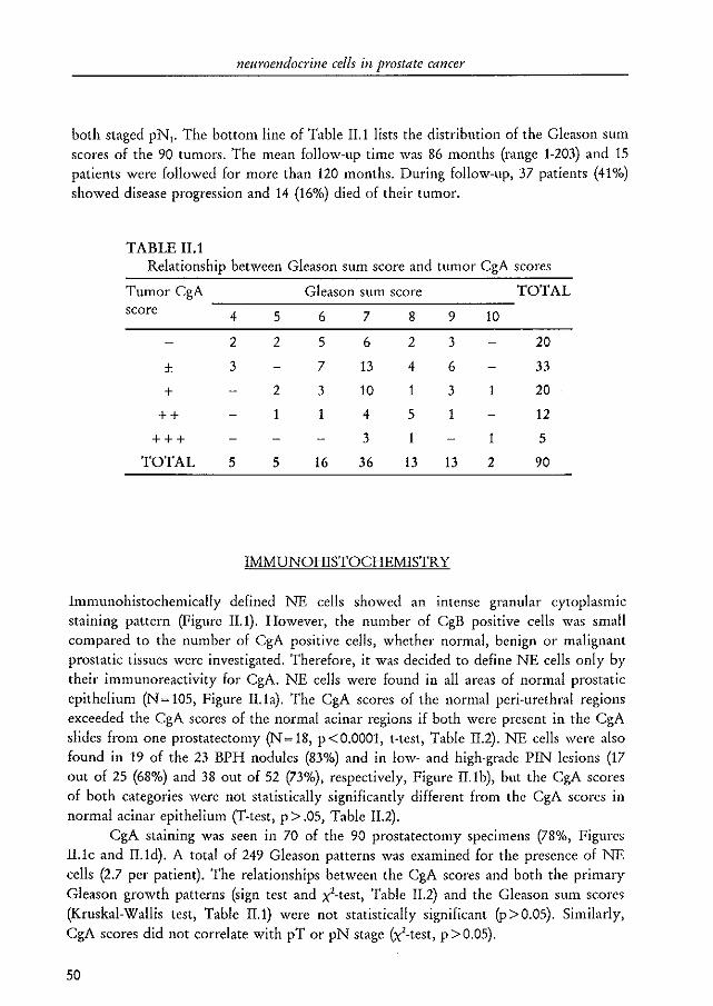
neuroendocrine cells in prostate cancer
both staged pN,. The bottom line of Table ILl lists the distribution of the Gleason sum scores of the 90 tumors. The mean follow-up time was 86 months (range 1-203) and 15 patients were followed for more than 120 months. During follow-up, 37 patients (41%) showed disease progression and 14 (16%) died of their tumor.
TABLE ILl Relationship between Gleason sum score and tumor CgA scores
Tumor CgA Gleason sum score TOTAL score 4 5 6 7 8 9 10
2 2 5 6 2 3 20
± 3 7 13 4 6 33
+ 2 3 10 1 3 20
++ 1 1 4 5 1 12
+++ 3 1 1 5
TOTAL 5 5 16 36 13 13 2 90
IMMUNOHISTOCHEMISTRY
Immunohistochemically defined NE cells showed an intense granular cytoplasmic staining pattern (Figure 11.1). However, the number of CgB positive cells was small compared to the number of egA positive cells, whether normal, benign or malignant prostatic tissues were investigated. Therefore, it was decided to define NE cells only by their immunoreactivity for CgA. NE cells were found in all areas of normal prostatic epithelium (N ~ 105, Figure 1I.1a). The CgA scores of the normal peri-urethral regions exceeded the CgA scores of the normal acinar regions if both were present in the CgA slides from one prostatectomy (N~18, p<O.OOOl, t-test, Table 11.2). NE cells were also found in 19 of the 23 BPH nodules (83%) and in low- and high-grade PIN lesions (17 out of 25 (68%) and 38 out of 52 (73%), respectively, Figure 1I.1b), but the CgA scores of both categories were not statistically significantly different from the CgA scores in normal acinar epithelium (T-test, p> .05, Table !I.2).
CgA staining was seen in 70 of the 90 prostatectomy specimens (78%, Figures 1I.1c and ILld). A total of 249 Gleason patterns was examined for the presence of NE cells (2.7 per patient). The relationships between the CgA scores and both the primary Gleason growth patterns (sign test and x'-test, Table 11.2) and the Gleason sum scores (Kruskal-Wallis test, Table 11.1) were not statistically significant (p>0.05). Similarly, CgA scores did not correlate with pT or pN stage (x'-test, p > 0.05).
50
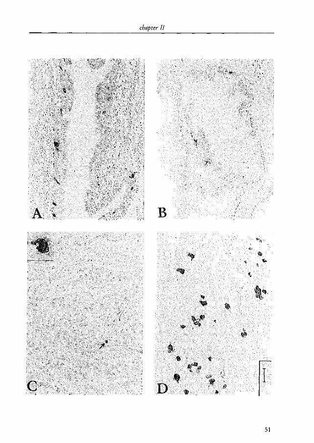
chapter II
B
"
• , .~ .. ,
51
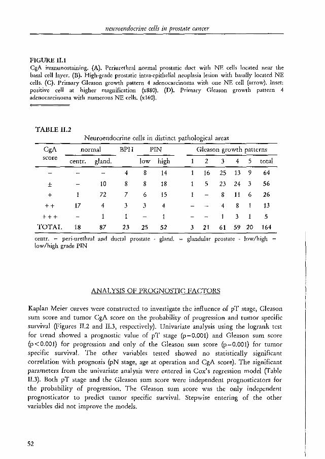
neuroendocrine cells in prostate cancer
FIGURE 11.1 egA inuuunostaining. (A). Periurethral normal prostatic duct with NE cells located near the basal cell layer. (B). High-grade prostatic intra-epithelial neoplasia lesion with basally located NE cells. (C). Primary Gleason growth pattern 4 adenocarcinoma with one NE cell (arrow). Inset: positive cell at higher magnification (x880). (D). Primary Gleason growth pattern 4 adenocarcinoma with numerous NE cells. (x160).
TABLEII.2 Neuroendocrine cells in distinct pathological areas
CgA normal BPH PIN Gleason growth patterns score centro gland. low high 1 2 3 4 5 total
4 8 14 1 16 25 13 9 64
± 10 8 8 18 1 5 23 24 3 56
+ 1 72 7 6 15 1 8 11 6 26
++ 17 4 3 3 4 4 8 1 13
+++ 1 1 1 3 1 5
TOTAL 18 87 23 25 52 3 21 61 59 20 164
centro ~ peri-urethral and ductal prostate gland. glandular prostate - low/high ~
low/high grade PIN
ANALYSIS OF PROGNOSTIC FACTORS
Kaplan Meier curves were constructed to investigate the influence of pT stage, Gleason sum score and tumor egA score on the probability of progression and tumor specific survival (Figures II.2 and II.3, respectively). Univariate analysis using the logrank test for trend showed a prognostic value of pT stage (p ~ 0.001) and Gleason sum score (p < 0.001) for progression and only of the Gleason sum score (p~O.OOI) for tumor specific survival. The other variables tested showed no statistically significant correlation with prognosis (pN stage, age at operation and CgA score). The significant parameters from the univariate analysis were entered in Cox's regression model (Table II.3). Both pT stage and the Gleason slim score were independent prognosticators for the probability of progression. The Gleason sum score was the only independent prognosticator to predict tumor specific survival. Stepwise entering of the other variables did not improve the models.
52
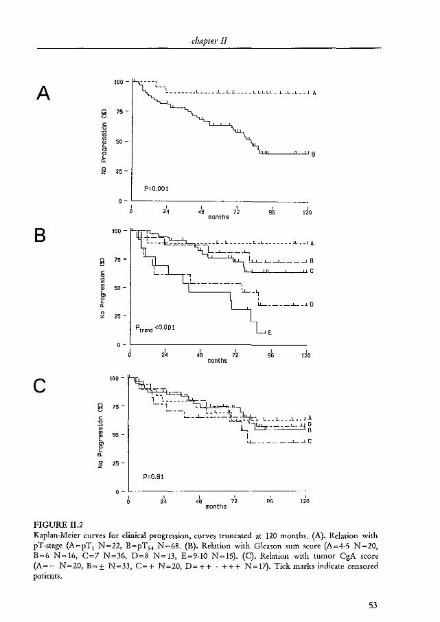
chapter 11
100 - ~ -_.,
A ---~ _____ _ .1,. ____ ..L _ L..L ____ L L L I.L _..L.L.L __ J A
E 75 -
c S '" '" 50 -ru L
'" 8 0 L "-0 25 -z
P"O.OOl
0-I I I I , I 0 24 48 72 96 120
months
B 100 -
!l 75 -
c C 0 ." '" '" 50 -~ L
'" 0 , " LL-. ___ ...l...-.J 0 a. 0
25 -z
Ptrend <0.001 E
0-I I I I I I 0 24 ,. 72 96 120
months
C 100 -
1"-~ _______
!l 75 - --, ---, --'---:1\;
C '--_...1..-. ___ , __ . L __ .1.. ___ L __ 1 A
0 L!:...1...=, -0 ~ L.
____ L._
'" B '" ru 50 - I L <.1...... ____ ---1-J C '" 0 L "-0 25-z
P"O.81
0-I I I I I , 0 24 ,. 72 96 120
months
FIGURE II.2 Kaplan-Meier curves for clinical progression, curves truncated at 120 months. (A). Relation with pT-stage (A~pT, N~22, B~pTH N~68. (B). Relation with Gleason sum score (A~4·5 N~20, B~6 N~16, C~7 N~36, D~8 N~13, E~9-10 N~15). (C). Relation with tumor CgA score (A~- N~20, B~± N~33, C~+ N~20, D~++ . +++ N~17). Tick marks indicate censored patients.
53
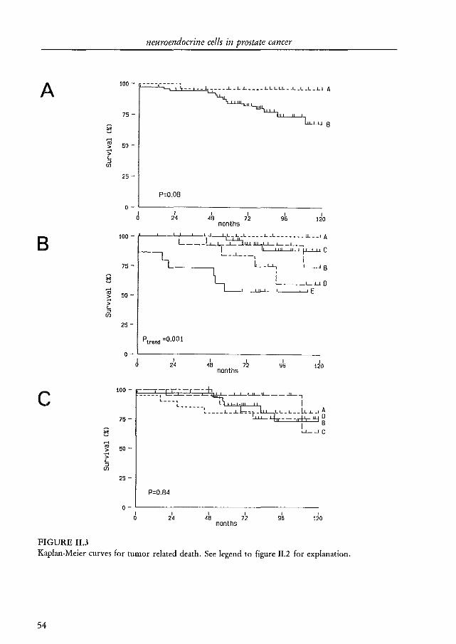
A
§ ~
ro > -" > L 0
if>
8
§ ~ ro -~ > L 0
if>
c § ~ ro -~ > L 0
if>
FIGURE II.3
neuroendocrine cells in prostate cancer
100 - --------.
75 -
50 -
25-
o-r
0 r
" r
120
B
100 - /-_~_-LJ'---~-';~~L_1._L ____ 1._L _______ lL __ I A L __ ~.I.__....L_ LL....L....--L.~
75 -
50 -
25-
Ptrend =0.001
o· r r
0 " 100 - --- -------f,
75 -
50 -
25-
O-r
0
P=O.84
r
" ,
48
I C '-_-.1..- __ ,
r 96 do
-L..L....LlL....I1....- __ ..lL.....,
months
r 72
I LL_L __ LL.1._! ~
, 95
I B ~~C
Kaplan-Meier curves for tumor related death. See legend to figure II.2 for explanation.
54
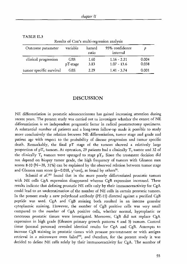
chapter II
TABLE II.3 Results of Cox's multi-regression analysis
Outcome parameter variable hazard 95% confidence p ratio
clinical progression GSS 1.60 pT-stage 3.83
tumor specific survival GSS 2.29
DISCUSSION
interval
1.16 - 2.21 1.07 - 13.6
1.41 - 3.74
0.004 0.038
0.001
NE differentiation in prostatic adenocarcinoma has gained increasing attention during recent years. The present study was carried out to investigate whether the extent of NE differentiation is an independent prognostic factor in radical prostatectomy specimens. A substantial number of patients and a long-term follow-up made it possible to study more conclusively the relation between NE differentiation, tumor stage and grade and patient age with respect to the probability of disease progression and tumor specific death. Remarkably, the final pT stage of the tumors showed a relatively large proportion of pT, tumors. At operation, 29 patients had a clinically T, tumor and 32 of the clinically T, tumors were upstaged to stage pT,. Since the treatment decision did not depend on biopsy tumor grade, the high frequency of tumors with Gleason sum scores 8-10 (N ~ 28, 31%) can be explained by the observed relation between tumor stage and Gleason sum score (p~0.018, x'-test), as found by others".
Schmid et al.'61 found that in the more poorly differentiated prostatic tumors with NE cells CgA expression disappeared whereas CgB expression increased. These results indicate that defining prostatic NE cells only by their immunoreactivity for CgA could lead to an underestimation of the number of NE cells in certain prostatic tumors. In the present study a new polyclonal antibody (PE-ll) directed against a purified CgB peptide was used. CgA and CgB staining both resulted in an intense granular cytoplasmic staining. However, the number of CgB positive cells was very small compared to the number of CgA positive cells, whether normal, hyperplastic or cancerous prostatic tissues were investigated. Moreover, CgB did not replace CgA expression in high grade (Gleason primary growth patterns 4 and 5) tumors. Control tissue (normal pancreas) revealed identical results for CgA and CgB. Attempts to increase CgB staining in prostatic tissues with pronase pre-treatment or with antigen retrieval in a microwave oven failed 273, and therefore, for the present study it was decided to define NE cells solely by their immunoreactivity for CgA. The number of
55
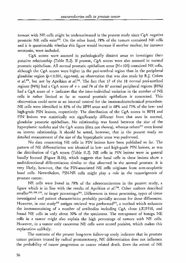
neuroendocrine cells in prostate cancer
tumors with NE cells might be underestimated in the present study since CgA negative prostatic NE cells exist15
'. On the other hand, 78% of the tumors contained NE cells and it is questionable whether this figure would increase if another marker, for instance serotonin, were included.
CgA scores were assessed in pathologically distinct areas to investigate their putative relationship (fable IT.2). If present, CgA scores were also assessed in normal prostatic epithelium. All normal prostatic epithelium areas (N ~ 105) contained NE cells, although the CgA scores were higher in the peri-urethral region than in the peripheral glandular region (p <0.001, sign-test), an observation that was also made by R.J. Cohen et al."" but not by Aprikian et al."'. The fact that 17 of the 18 normal peri-urethral regions (94%) had a CgA score of + + and 74 of the 87 normal peripheral regions (85%) had a CgA score of + indicates that the inter-individual variation in the number of NE cells is rather limited as far as normal prostatic epithelium is concerned. This observation could serve as an internal control for the immunohistochemical procedure. NE cells were identified in 83% of the BPH areas and in 68% and 73% of the low- and high-grade PIN lesions, respectively. The distribution of the CgA scores in BPH and PIN lesions was statistically not significantly different from that seen in normal, glandular prostatic epithelium. No relationship was found between the size of the hyperplastic nodules and the CgA scores (data not shown), whereas others'" even found an inverse relationship. It should be noted, however, that in the present study no detailed measurement of the size of the hyperplastic areas was performed.
No data concerning NE cells in PIN lesions have been published so far. The pattern of NE differentiation was identical in low- and high-grade PIN lesions, as was the distribution of CgA scores (fable !I.2). NE cells in PIN lesions were in general basally located (Figure IT.lb), which suggests that basal cells in these lesions show a multidirectional differentiation similar to that observed in the normal prostate. It is very likely, however, that the PIN-associated NE cells originate from non-neoplastic basal cells. Nevertheless, PIN-NE cells might play a role in the tumorigenesis of prostate cancer.
NE cells were found in 78% of the adenocarcinomas in the present study, a figure which is in line with the results of Aprikian et al."'. Other authors described smaller63, 268. 270, or larger percentages264• Differences in tissue processing, types of tissue investigated and patient characteristics probably partially account for these differences. However, in one study270 antigen retrieval was performed273 , a method which enhances the immunostaining of a number of antibodies including CgA clone LK2HI0, and found NE cells in only about 30% of the specimens. The entrapment of benign NE cells in a tumor might also explain the high percentage of tumors with NE cells. However, in a tumor only cancerous NE cells were scored positive, which makes this explanation unlikely.
The outcome of the present long-term follow-up study indicates that in prostate cancer patients treated by radical prostatectomy, NE differentiation does not influence the probability of tumor progression or cancer related death. Even the extent of NE
56
chapter II
differentiation, as defined by increasing tumor CgA scores (Table II.l), did not show a tendency towards a poor prognosis (p > 0.05, logrank test for trend). The distribution of the tumor CgA scores was statistically not significantly different from that of normal glandular epithelium CgA scores (p>0.05, Hest). Some other recent reports also tempered initial enthusiasm regarding the prognostic influence of NE cells in prostate cancer'''' "'. In the study of Aprikian et al.'" this concerned the prognostic value of NE cells in metastatic lesions. M.K. Cohen et al.'70 found no prognostic influence of NE cells (CgA or neuron specific enolase positive cells) in 38 patients with clinically stage II (AlCC) prostate cancer treated by radical prostatectomy. The follow-up period in their study was relatively short (maximum 77 months) and the relationship between tumor grade and NE differentiation was not discussed in detail. In two studies, Cohen et al.'''· '" described a striking prognostic influence of CgA positive cells in initial biopsies and TUR specimens with incidental prostate cancer. Abrahamsson et al.'64 studied the number of CgA positive cells in repeated biopsy specimens of 24 hormonally treated patients and generally found, with time, an increase in number of CgA positive cells which was paralleled by dedifferentiation of the tumor. The relation between tumor grade and NE differentiation in their study, which might have explained part or all of the prognostic value, was not confirmed by Aprikian et al.m, and the data of the present study do not support this either. It should be stressed, however, that the study of Abrahamsson et al.'64 was based on hormonally treated patients. Since the NE cells in normal and neoplastic prostatic tissues do not contain androgen receptors176
, their presence in a prostatic tumor might be related to androgen independence. The studies of Abrahamsson et al.'64 and R.J. Cohen et al."'· '68 may have been biased by the fact that only small tumor fragments were investigated (biopsies and TUR specimens containing small incidental carcinomas). Due to the widely scattered distribution of NE cells in most tumors, the likelihood of obtaining positively stained cells most probably correlates with both tumor volume and the size of the investigated tumor area. Since tumor volume is a predictor of progressions7, the prognostic value found in these studies may be partially explained by this relation. Furthermore, a substantial risk of obtaining false negative results exists, especially if only small numbers or small foci of positive cells are present. In the present study, Cox's regression identified both pT classification and Gleason sum score as independent prognosticators for progression, but only Gleason sum score for tumor related death. This can be explained by the abovementioned relationship between pT classification and Gleason sum score combined with the small number of events in the survival analysis (N~ 14, 16%).
As mentioned above, prostatic NE cells do not express the androgen receptor and therefore are considered to be primarily androgen independene76
, In addition, prostatic NE cells secrete neuropeptides and some of these pep tides show growth modulating activity (reviewed by Noordzij et al.2Sl If non-NE prostatic tumor cells contain receptors for these neuropeptides, tumor growth may be modulated by such peptides. Moreover, some neuropeptides were able to stimulate steroid receptors in a
ligand independent fashion277. It is possible that the NE cells are related with the
57
neuroendocrine cells in prostate cancer
androgen indepence of a tumor. On the other hand, the presence of NE cells in prostate cancer might be simply an epiphenomenon determined, for instance, by stromal-epithelial interactions. The function of NE cells in the normal and, especially, the neoplastic prostate remains largely unclear as yet and has to be clarified experimentally. Such studies, using well defined, newly established, prostatic tumor xenograft models with NE cells are in progress at our institution.
58
CHAPTER III
THE PROGNOSTIC VALUE OF CD44 ISOFORMS IN PROSTATE CANCER PATIENTS TREATED BY RADICAL
PROSTATECTOMY
M.A. Noordzij; G.]. van Steenbrugge; N.S. Verkaik; F.H. Schroder; T.H. van der Kwast
From the Departments of Urology and Pathology, Erasmus University, Rotterdam, The Netherlands
Clin Cancer Res, accepted for publication
CD44 in clinical prostate cancer
ABSTRACT
CD44 forms a group of transmembranous glycoproteins formed by alternative splicing of a single mRNA. The expression of v6·exon containing variants correlates with metastasis and poor prognosis in a number of malignancies. The distribution and prognostic value of CD44s, CD44v5 and CD44v6 was studied immunohistochemically in the radical prostatectomy specimens of 97 patients with prostate cancer and in 12 lymph node metastasis. The mean follow.up period was 84 months. The percentage of CD44 immunoreactive cells was scored semiquantificatively. CD44 mRNA expression was studied in 9 prostate cancer and 8 benign prostatic hyperplasia (BPH) samples by RT·PCR.
Benign prostatic glands almost always expressed CD44s, CD44v6, and at a lower intensity CD44v5. CD44 scores decreased from low· to high.grade prostatic intraepithelial neoplasia. CD44s, CD44v5 and CD44v6 were expressed in 86%, 23% and 69% of the adenocarcinomas, respectively. Gleason sum score (GSS) and pT·stage were inversely correlated with CD44s and CD44v6 scores. CD44 was not found in the lymph node metastatic tumor cells. At the mRNA level 89% of the tumors and all BPH samples expressed CD44s. CD44v6·10 mRNA was present in 44% and 75% of the tumors and BPH samples, respectively. Loss of CD44s and CD44v6 predicted an adverse prognosis at univariate analysis. The independent prognosticators identified by multival'iate analysis were: GSS, pT.stage and CD44s for clinical progression; GSS and CD44s for PSA progression; and GSS for tumor specific survival. Loss of CD44s expression in prostate adenocarcinoma predicts a poor prognosis, independent of stage and grade.
INTRODUCTION
Prostate cancer is the most prevalent non-skin tumor in western males and its mortality is second only to that of lung cancel". The incidence and prevalence of the disease have been increasing dramatically over the last decade, which can partially be explained by the introduction of prostate specific antigen (PSA) as a serum tumor marker'- 278 _ On average, patients are now diagnosed at an earlier stage of the disease and often are candidates for curative treatment by means of a radical prostatectomylO. At present there are no tumor markers that are sufficiently able to divide patient groups in those
60
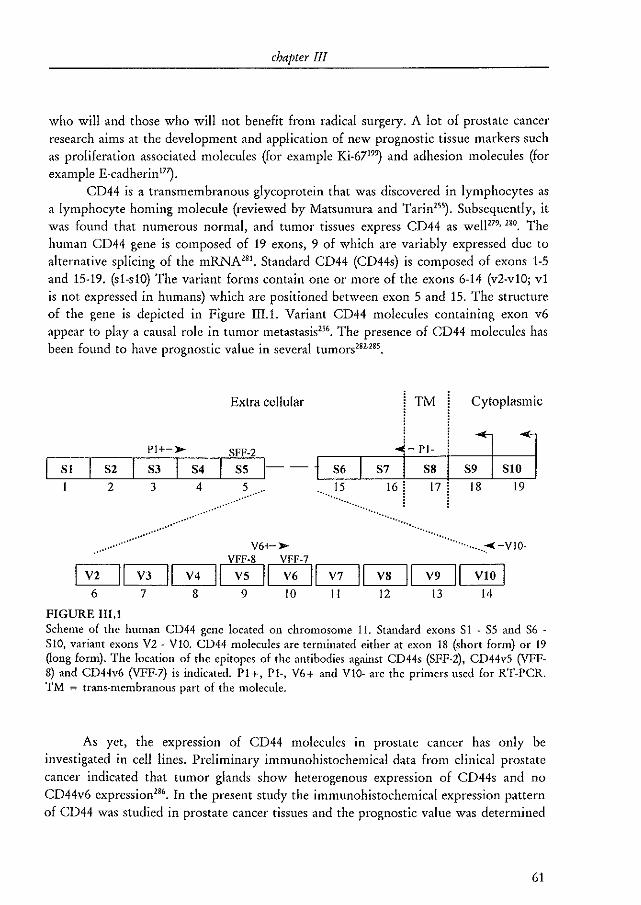
chapter III
who will and those who will not benefit from radical surgery. A lot of prostate cancer research aims at the development and application of new prognostic tissue markers such as proliferation associated molecules (for example Ki_6719~ and adhesion molecules (for example E-cadherinI7~.
CD44 is a transmembranous glycoprotein that was discovered in lymphocytes as a lymphocyte homing molecule (reviewed by Matsumura and Tarin255
). Subsequently, it was found that numerous normal, and tumor tissues express CD44 as we11279
,280, The human CD44 gene is composed of 19 exons, 9 of which are variably expressed due to
alternative splicing of the mRNA2S1 • Standard CD44 (CD44s) is composed of exons 1-5 and 15-19. (sl-s10) The variant forms contain one or more of the exons 6-14 (v2-vl0; vl is not expressed in humans) which are positioned between exon 5 and 15. The structure of the gene is depicted in Figure III.l. Variant CD44 molecules containing exon v6 appear to playa causal role in tumor metastasis"'. The presence of CD44 molecules has been found to have prognostic value in several tumors282-285,
Extra cellular TM Cytoplasmic
Pl+->- srr-2 ...i - PI-
I sJ SIOJ S7 I S8 16 i 17 ! 18 19
L-S~I~~S~2~~S3 __ L-S~4 __ L-S~5~1----1L_s~6~ __ ~~~~ __ ~L-~~ I 2 3 4 5 "." 15
................... .... ." ,,'
,,' . " ...... ,,'
........ . ........ .
V6+-~
vrF-8 VFF-7
. .... ..................
""""""'::< -v 10-
V2 I~I V4 II V5 II V6 I'-I '-'7-'11 V8 II V9 II VIO 6 7 '---=8---' 9 to II 12 I3 14
FIGURE III.I Scheme of the human CD44 gene located on chromosome 11. Standard exans S 1 - 5S and S6 -S10, variant exons V2 - VI0. CD44 molecules are terminated either at exon 18 (short form) or 19 Qong form). The location of the epitopes of the antibodies against CD44s (SFF-2), CD44vS (VFF-8) and CD44v6 (VFF-7) is indicated. PI+, PI-, V6+ and VIO- are the primers used for RT-PCR. TM = trans-membranous part of the molecule.
As yet, the expression of CD44 molecules in prostate cancer has only be investigated in cell lines. Preliminaty immunohistochemical data from clinical prostate cancer indicated that tumor glands show heterogenous expression of CD44s and no CD44v6 expression2S6
, In the present study the immunohistochemical expression pattern of CD44 was studied in prostate cancer tissues and the prognostic value was determined
61
CD44 in clinical prostate cancer
in patients treated by radical prostatectomy. In addition, CD44 mRNA expression was checked by the reverse transcriptase polymerase chain-reaction (RT-PCR).
MATERIAL AND METHODS
PATIENTS
In the period 1980-1988, 159 patients were operated at the University Hospital, Rotterdam, The Netherlands for prostate cancer (f,.,N,.Mo, TNM classification 199234).
In 17 of these the tumor was diagnosed in a trans-urethral resection (fUR) specimen performed for benign prostatic hyperplasia (BPH) i.e., stage Tl'~' In 49 patients the pelvic lymph node dissection specimen contained metastatic tumor on examination of frozen sections. These patients did not undergo subsequent radical prostatectomy, except for two younger patients with micrometastasis, leaving 112 patients treated by radical prostatectomy. The patients were followed regularly and all data concerning diagnosis, treatment and follow-up were stored prospectively in a comprehensive database. Clinical progression was defined as cytologically or histologically proven local recurrence or the appearance of distant metastasis. Tumor death was defined as death due to direct tumor effects, metastasis or tumor therapy. PSA progression was defined as a PSA-level of 21.0 at two subsequent measurements, in which case the first elevated PSA-level was considered the date of failure. PSA progression was only considered in patients with a complete PSA history (N ~29). If necessary patients were treated during follow-up for local recurrence or metastatic disease. One patient was lost to follow-up. Two patients died within one month following radical prostatectomy from myocardial infarction and pulmonary embolism, respectively.
TISSUES
All radical prostatectomy specimens were fixed in 10% buffered formalin and totally embedded in paraffin. The hematoxylin & eosin stained slides were reviewed by a single experienced genitourinary pathologist (fHvdK) to obtain the Gleason sum score and to stage the tumors according to the TNM-1992 classification system. The material of 10 patients could not be retrieved and one tumor was a metastasis of a large bowel carcinoma. The previously diagnosed tumor was not found in the radical prostatectomy specimen of three patients with a T".b·tumor (final stage pTo). All these 14 patients were excluded from analysis, leaving 97 patients. Of each radical prostatectomy
62
chapter III
specimen 2-3 tissue blocks were selected that contained at least all the Gleason growth patterns observed in that tumor and, if applicable, also contained normal prostatic glands, BPH andlor prostatic intraepithelial neoplasia (PIN). PIN lesions were graded according to Bostwick & Brawer'''. PIN-I and PIN-II were taken together as low-grade PIN, PIN-III was termed high-grade PIN. To investigate the expression of CD44 in metastatic prostate cancer twelve paraffin-embedded parailiac lymph nodes containing a prostate cancer metastasis were also selected.
The expression of CD44 mRNA was studied in tumor samples from 9 radical prostatectomy specimens and 8 BPH samples derived from simple prostatectomies. These radical prostatectomy samples were not from the specimens that were used for the follow-up study because fresh frozen tissues have only been collected as of 1989. Directly after removal of the prostate macroscopical tumor lesions (or BPH tissue) were identified and a piece of tumor tissue was removed in a sterile fashion, snap-frozen in liquid nitrogen chilled isopentane {Merck, Darmstadt, Germany) and stored at -80°C until use.
IMMUNOHISTOCHEMISTRY
The selected tissue blocks of the radical prostatectomy specimens and the lymph nodes were immunostained with monoclonal antibodies against CD44s (clone SFF-2), CD44v5 (clone VFF-8) and CD44v6 (clone VFF-7), all from Bender MedSystems, Vienna, Austria282, 183, 187-289, CD44vS has not been related to malignant processes, but was
included for comparison with CD44s and CD44v6. For visualization of antigenantibody binding an ultra-sensitive method (tyramide signal amplification system (TSA), DuPont NEN, Boston, MA), was used'''. This method uses the activity of second antibody coupled horseradish peroxidase to catalyze the deposition of a large number of biotin molecules near the peroxidase molecule. The biotin molecules can be made visible by fluorochrome- or enzyme-labeled streptavidin. This procedure was used because preliminary results showed that the more well known avidin-biotin complex method l91 yielded false-negative results in a substantial number of cases.
Tissue sections were cut at Slt-m and mounted on 3-aminopropyl-trietoxysilane (Sigma chemical Co., St. Louis, MO) coated glass slides272. After deparaffinization through xylene and ethanol endogenous peroxidase activity was blocked by rinsing the slides for 10 minutes in 3% hydrogen peroxide in ethanol. The slides were rinsed with distilled water and placed in a 10 mmolll citrate buffer at pH~6.0. Antigen retrieval was performed in a microwave oven at 700\v/ for 3xS minutesw . After cooling and rinsing with PBS the slides were placed in a Sequenza immunostaining system (Shandon, Uncorn, UK). Following a 15 minute pre-incubation with 10% normal goat serum (DAKO, Glostrup, Denmark) in PBS, the slides were incubated for 90 minutes with either the primary antibody or PBS (negative control). The antibodies were diluted in PBS at 1:200 (SFF-2 and VFF-7) or 1:160 (VFF-8). Subsequently, the slides were
63
CD44 in clinical prostate cancer
incubated for 30 minutes with a blocking buffer consisting of 0.15 molll TRIS·buffer (Gibco Brl, Breda, The Netherlands) at pH 7.5, 0.15 molll Nacl (Merck) and 0.5% blocking reagent (DuPont, TSA.kit). After a 30 minute incubation with a 1 to 50 diluted horseradish peroxidase·labeled goat anti mouse antibody (DAKO) an amplification solution consisting of biotinyl tyramide (DuPont) 1 to 50 diluted in 50% distilled water and 50% amplification diluent (DuPont) was applied. Finally, the slides were incubated with 1 to 400 diluted horseradish peroxidase·labeled streptavidin. All the components of the TSA.system were diluted in the blocking buffer. In between the incubations the slides were rinsed three times in a buffer consisting of 0.1 molll TRIS, 0.15 molll NaCI and 0.05% TWEEN·20 (Merck). The antigen·antibody binding was visualized with 0.75mg/ml PBS of diaminobenzidine hydrochloride (Fluka, Neu·Ulm, Germany) with 0.08% hydrogen peroxide as substrate. The slides were counterstained with Mayer's hematoxylin, dehydrated and covered.
The cellular localization of CD44 immunostaining was examined in detail in 12 randomly chosen sections using a confocal laserscan microscope system (Carl Zeis Jena, Germany). A fluorescent TSA method, which uses FITC·labeled streptavidin during the final incubation, was applied to these sections. The nuclei were counterstained with propidium iodide and the slides were covered with vectashield (Vector, Burlingame,
CAl·
QUANTIFICATION
The different Gleason growth patterns as well as areas of normal prostatic glands, BPH and PIN were identified and marked on the immunohistochemically stained tissue slides. The slides were studied at 25x magnification without knowledge of the clinical outcome of the patients. The percentage of CD44 positive cells (s, v5 and v6) in a particular area was scored semiquantificatively as; < 10%, 10-25%, 25-50% and > 50%. Scores were obtained for all pathological categories present in the tissue slides of one prostate. A tumor CD44 score was obtained by taking the lowest score observed in any of the primary Gleason growth patterns present in a tumor.
CD44 mRNA expression
Total RNA of the snap·frozen BPH and tumor samples was prepared by the guanidine isothiocyanate / cesium chloride centrifugation method using the RNAzol·B kit (Campro, Veenendaal, The Netherlands) as per the manufacturer's protoco!' First strand eDNA was produced with O.5l'g of the isolated total RNA using random hexamer primers and M-MLV reverse transcriptase in the presence of RNase inhibitor and dNTPs (all from Perkin Elmer Cetus, Norwalk, CTj in a final volume of lOI'!. The oligonucleotide primers were chosen upstream and downstream of the insertion point
64
cbapter III
of alternatively spliced mRNA''', the pOSltiOnS are indicated in Figure II!.l. The primers used were: (Pl+) sense 5'-GACACTATTGCTTCAATGCTTCAGC and (Pl-) antisense 5'-GATGCCAAGATGATCAGCCATTCTGCAAT. Thus, PCR of cDNA should amplify CD44s as well as all possible variant forms and should result in distinguishable PCR products. In addition, two exon specific primers were used to characterize the splice variantsl9l
: (V6+), sense 5'-TCCAGGCAACTCCTAGTAGT and (Vl0-), antisense, 5'-CTGATAAGGAACGATTGACA. The following combinations were used for PCR: Pl+ with Pl-, Pl+ with Vl0-, and V6+ with Pl-. PCR was performed with the total volume of cDNA using super Taq DNA polymerase (Sphaero, HT biotechnology, UK) in a thermal cycler (Perkin Elmer Cetus). The PCRmix was heated to 94°C for 4 minutes followed by 40 cycles of 1 minute at 94°C, 1 minute at 60°C and 1 minute at noc. After the last cycle, the tubes were kept at noc for 10 minutes. The PCR products were separated on a 1% agarose gel in the presence of ethidium bromide and analyzed under UV-light.
STATISTICS
Statistical analysis was performed using the SPSS and STA TA statistical computer packages. The sign-test, xl-test for trend and Spearman's rank correlation test were used to analyze the relation between several pathological voriables and CD44 expression. For the analysis of survival data Kaplan-Meier curves wcre constructed and the logrank test for trend was performed. Multivariate survival analysis was performed using Cox's proportional hazards model.
RESULTS
CLINICAL
After exclusion of the patients that were not appropriate (n~ 1 lost to follow-up, n ~ 10 radical prostatectomy specimens could not be retrieved, n=3 final stage pTo and n=1 tumor was not of prostatic origin), 97 patients were left for analysis. After clinical evaluation all patients were staged T,_,N,Mo' The T-stage distribution was: T b .b in 17, 1', in 50 and 1', in 30. The mean age at operation was 63 years (range 45-76). The final T-stage distribution was pT, in 25 patients; and pT".b, pT" and pT, in 29, 41 and 2 patients, respectively. Lymph node metastasis were found in 7 patients (pN, in 5 and pN, in 2). The patients with positive lymph nodes at operation were both at stage pN,.
65
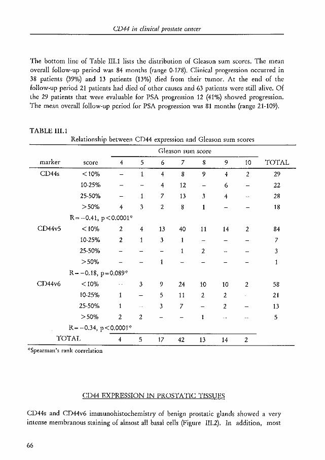
CD44 in clinical prostate cancer
The bottom line of Table III.1 lists the distribution of Gleason sum scores. The mean overall follow-up period was 84 months (range 0·178). Clinical progression occurred in 38 patients (39%) and 13 patients (13%) died from their tumor. At the end of the follow-up period 21 patients had died of other causes and 63 patients were still alive. Of the 29 patients that were evaluable for PSA progression 12 (41%) showed progression. The mean overall follow-up period for PSA progression was 81 months (range 21-109).
TABLE III.l Relationship between CD44 expression and Gleason sum scores
Gleason sum score
marker score 4 5 6 7 8 9 10 TOTAL
CD44s <10% 1 4 8 9 4 2 29
10·25% 4 12 6 22
25-50% 1 7 13 3 4 28
>50% 4 3 2 8 1 18
R~-0.41, p<O.OOO1'"
CD44v5 <10% 2 4 13 40 11 14 2 84
10-25% 2 1 3 1 7
25-50% 1 2 3
>50% 1 1
R~ -0.18, p~0.089'}
CD44v6 <10% 3 9 24 10 10 2 58
10-25% 1 5 11 2 2 21
25-50% 1 3 7 2 13
>50% 2 2 1 5
R~ -0.34, p<O.OOOI';
TOTAL 4 5 17 42 13 14 2
'~Spearman's rank correlation
CD44 EXPRESSION IN PROSTATIC TISSUES
CD44s and CD44v6 immunohistochemistry of benign prostatic glands showed a very intense membranous staining of almost all basal cells (Figure III.2). In addition, most
66
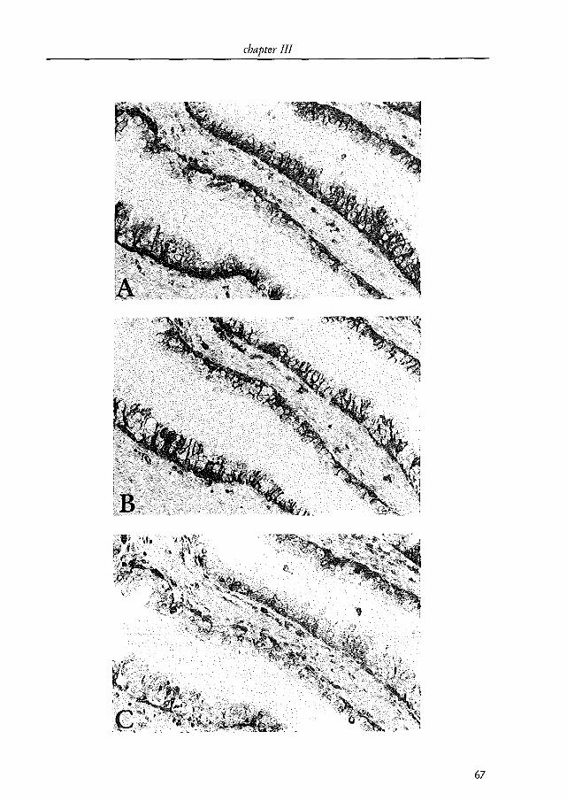
chapter III
67
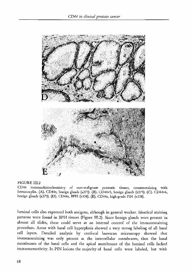
CD44 in clinical prostate cancer
FIGURE 111.2 CD44 immunohistochemistry of non-malignant prostatic tissues, counterstallllllg with hematoxylin. (A). CD44s, benign glands (x273). (B). CD44vS, benign glands (x273). (C). CD44v6, benign glands (x273). (D). CD44s, BPH (xI38). (E). CD44s, high-grade PIN (x 138).
luminal cells also expressed both antigens, although in general weaker. Identical staining patterns were found in BPH tissues (Figure III.2). Since benign glands were present in almost all slides, these could serve as an internal control of the imnnmostaining procedure. Areas with basal cell hyperplasia showed a velY strong labeling of all basal cell layers. Detailed analysis by confocal laserscan microscopy showed that immunostaining was only present at the intercellular membranes, thus the basal membranes of the basal cells and the apical membranes of the luminal cells lacked immunoreactivity. In PIN lesions the majority of basal cells were labeled, but with
68
chapter III
69
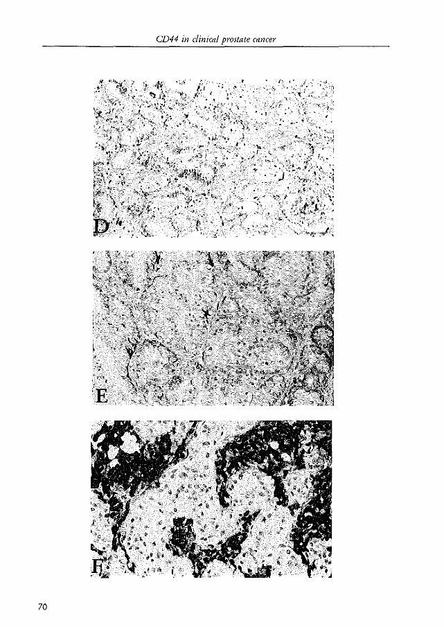
CD44 in clinical prostate cancer
,~ ·Jf~t
"R
70
chapter III
FIGURE I1I.3 CD44 immunohistochemistry of prostatic cancer, counterstallllllg with hematoxylin. (A-C). Gleason growth pattern 3 adenocarcinoma with strong labeling of CD44s (A), CD44v5 (B) and CD44v6 (e) (x 138). (D). Gleason growth pattern 3 tumor with reduced CD44s labeling (x138). (E). Gleason growth pattern 4 tumor with reduced CD44v6 labeling. Note basal localization of immunoreactive cells (x 138). (F). Prostate cancer lymph node metastasis. Lymphocytes strongly immunoreactive with CD44s, metastatic tumor cells negative (x273).
TABLE m.2 Relationship between CD44 expression and Gleason growth patterns
marker
CD44s
CD44v5
CD44v6
>~Sign.tcst
score
<10%
10-25%
25-50%
>50%
p~0.04F
<10%
10-25%
2S-50%
>50%
p~O.4F
<10%
10-2S%
2S-S0%
>SO%
p~0.2S*
1
1
1
2
1
Gleason growth pattern
2
2
1
5
8
11
5
4
2
2
8
3
9
14
17
20
53
4
1
2
26
15
11
8
4
20
14
24
10
S8
4
S
1
34
15
10
9
5
14
7
2
1
22
18
3
3
TOTAL
45
36
49
40
142
13
6
5
82
35
27
26
decreased intensity as compared to normal glands (Figure m.2). The luminal cell layer of PIN lesions showed reduced immunostaining of both CD44s and CD44v6. The scores of CD44s and CD44v6 were lower in high-grade PIN lesions than in low-grade PIN lesions (p <O.OS, x'-test for trend). The patterns of CD44vS labeling were comparable, but with a much lower intensity. In all areas the percentage of CD44vS
71
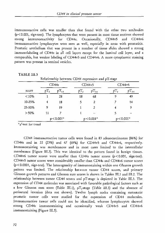
CD44 in clinical prostate cancer
immunoreactive cells was smaller than that found with the other two antibodies (p <0.001, sign-test). The lymphocytes that were present in most tissue sections showed strong immunoreactivity for CD44s. Occasionally, CD44v5 and CD44v6 immunoreactive lymphocytes were seen as well, especially in areas with prostatitis. Prostatic urothelium that was present in a number of tissue slides showed a strong immunolabeling of CD44s in all cell layers except for the luminal cell layer, and a comparable, but weaker labeling of CD44v5 and CD44v6. A more cytoplasmic staining pattern was present in seminal vesicles.
TABLE III.3 Relationship between CD44 expression and pT-stage
CD44s CD44v5 CD44v6
score pT, pTH pT, pTJ4 pT, pTH
<10% 1 28 18 68 9 49
10-25% 4 18 5 2 7 14
25-50% 9 19 1 2 4 9
>50% 11 7 1 5
P <0.001':' p<O.OOl': p <0.001'"
'~xl-test for trend
CD44 immunoreactive tumor cells were found in 83 adenocarcinomas (86%) for CD44s and in 22 (23%) and 67 (69%) for CD44v5 and CD44v6, respectively. Immunostaining was membranous and in most cases limited to the intercellular membranes (Figure III.3). This was identical to the pattern found in benign tissues. CD44v6 tumor scores were smaller than CD44s tumor scores (p < 0.001, sign-test). CD44v5 tumor scores were considerably smaller than CD44s and CD44v6 tumor scores (p <0.0001, sign-test). The heterogeneity of immunostaining within one Gleason growth pattern was limited. The relationship between tumor CD44 scores, and primary Gleason growth patterns and Gleason sum scores is shown in Tables III.1 and !IL2. The relationship between tumor CD44 scores and pT-stage is depicted in Table IlL3. The expression of CD44 molecules was associated with favorable pathological factors such as a low Gleason sum score (Table III. 1), pT,-stage (Table III.3) and the absence of perineural invasion (data not shown). Twelve lymph nodes containing metastatic prostatic tumor cells were studied for the expression of CD44 molecules. Immunoreactive tumor cells could not be identified, whereas lymphocytes showed strong CD44s immunostaining and occasionally weak CD44v5 and CD44v6 immunostaining (Figure 1II.3).
72
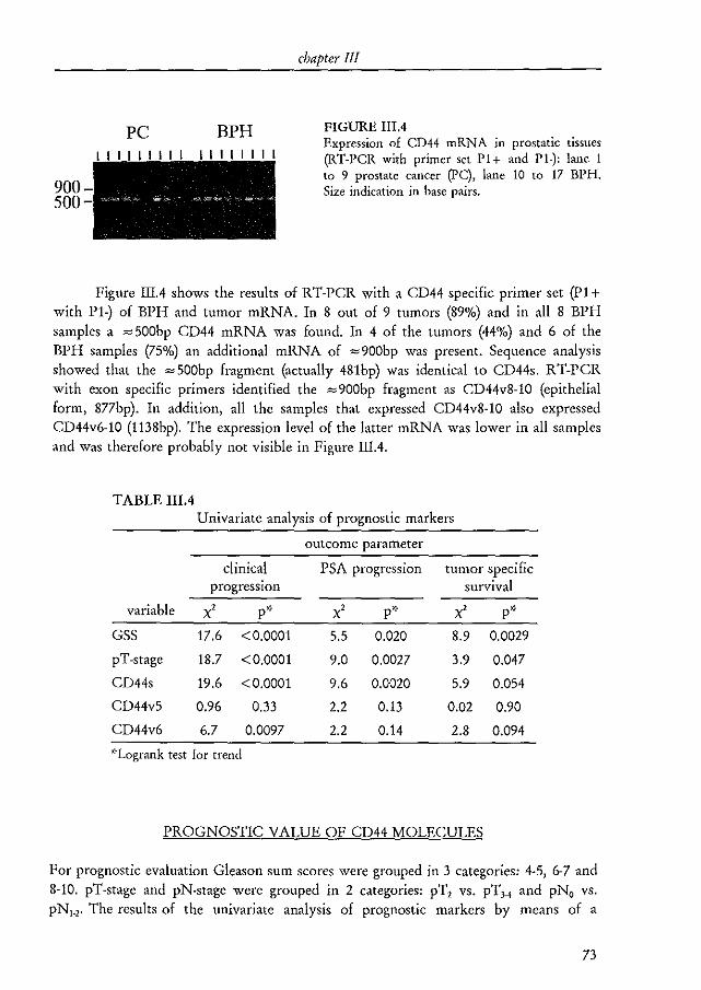
900 500
PC BPH
chapter III
FIGURE III.4 Expression of CD44 mRNA in prostatic tissues (RT-peR with primer set PI+ and PI-): lane I to 9 prostate cancer (PC), lane 10 to 17 BPH. Size indication in base pairs.
Figure IIIA shows the results of RT-PCR with a CD44 specific primer set (P1+ with P1-) of BPH and tumor mRNA. In 8 out of 9 tumors (89%) and in all 8 BPH samples a ~ 500bp CD44 mRNA was found. In 4 of the tumors (44%) and 6 of the BPH samples (75%) an additional mRNA of ~900bp was present. Sequence analysis showed that the ~ SOObp fragment (actually 481bp) was identical to CD44s. R T-PCR with exon specific primers identified the ~900bp fragment as CD44v8-10 (epithelial form, 877bp). In addition, all the samples that expressed CD44v8·10 also expressed CD44v6-10 (1138bp). The expression level of the latter mRNA was lower in all samples and was therefore probably not visible in Figure IIIA.
TABLE lIlA Univariate analysis of prognostic markers
outcome parameter
clinical PSA progression tumor specific progression survival
variable x' p'} X' p* X' p'}
GSS 17.6 <0.0001 5.5 0.020 8.9 0.0029
pT-stage 18.7 <0.0001 9.0 0.0027 3.9 0.047
CD44s 19.6 <0.0001 9.6 0.0020 5.9 0.054
CD44vS 0.96 0.33 2.2 0.13 0.02 0.90
CD44v6 6.7 0.0097 2.2 0.14 2.8 0.094
'}Logrank test for trend
PROGNOSTIC VALUE OF CD44 MOLECULES
For prognostic evaluation Gleason sum scores were grouped in 3 categories: 4-5, 6-7 and 8-10. pT-stage and pN-stage were grouped in 2 categories: pT, vs. pT,~ and pNa vs. pN,_,. The results of the univariate analysis of prognostic markers by means of a
73
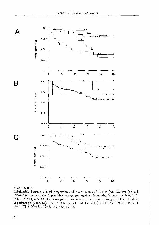
1.00 -
A 0.75 -
• • ~ c OJ 0.50 -" • • L rn a L a
0.25 -
0.00 -I 0
B
0.50 -
0.25 -
0.00 -I
0
C 1.00 -
0.]5 -• l' c .~ 0.50 -" • • L
~ a
0.25 -
0.00 -I
0
FIGURE 1II.5
CD44 in clinical prostate cancer
I 24
I 24
I I 4B 72
I I 96 120
do
Relationship between clinical progression and tumor scores of CD44s (A), CD44v5 (B) and CD44v6 (C), respectively. Kaplan-Meier curves, truncated at 120 months. Groups: 1 < 10%, 2 10-25%, 3 25-50%, 4 > 50%. Censored patients arc indicated by a number along their line. Numbers of patients per group: (A). 1 N~29. 2 N~22. 3 N~28. 4 N~18; (B). 1 N~86. 2 N~7. 3 N~3. 4 N~l; (C). 1 N~58. 2 N~21. 3 N~13. 4 N~5.
74
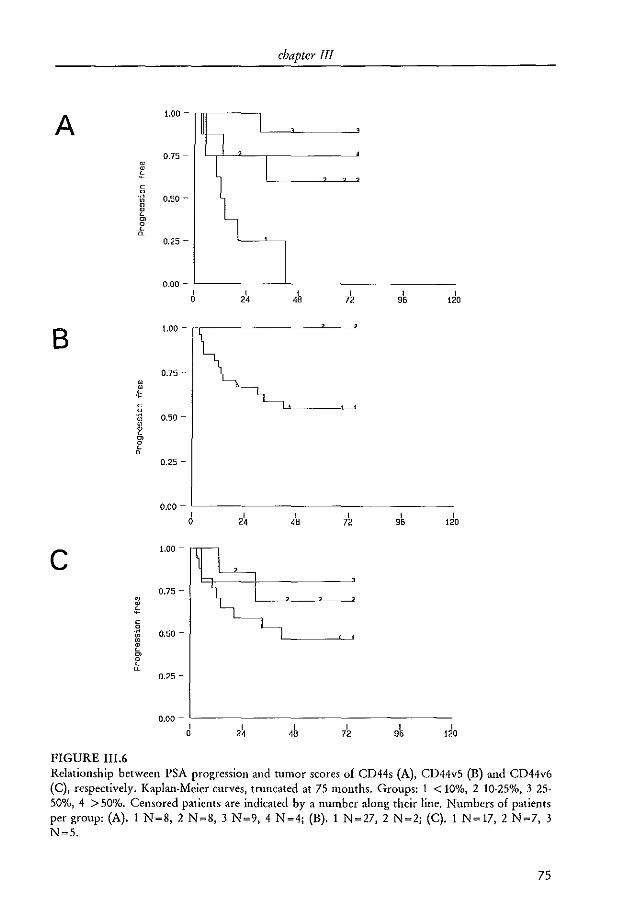
chapter III
A 1.00 ~
I n 0.75 -•
I ! l c 0
·rl 0.50 -" • • " ~ 0
" ~ 0.25 -
0.00 -I I I I I I
0 24 48 72 96 120
B 1.00 -
0.75 -• • " c 0 ~ 0.50 -" " • " ~ 0
" ~ 0.25 -
0.00 -I I I I I I
0 24 48 72 9' 120
C 1.00 -
0.75 -• ~ c .::: 0.50 -" • • " ~ 0
" ~ 0.25 -
0.00 -I
2\ I I I I
0 48 72 9' 120
FIGURE III.6 Relationship between PSA progression and tumor scores of CD44s (A). CD44v5 (B) and CD44v6 (C), respectively. Kaplan-Meier curves, truncated at 75 months. Groups: 1 < 10%, 2 10-25%, 3 25-50%, 4 > 50%. Censored patients are indicated by a number along their line. Numbers of patients per group: (A). 1 N~8. 2 N~8. 3 N~9. 4 N~4; (B). 1 N~27. 2 N~2; (C). 1 N~17. 2 N~7. 3 N~5.
75
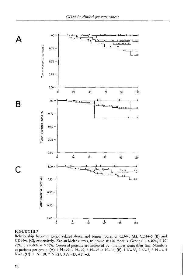
CD44 in clinical prostate cancer
A 1.00 -
rl ro 0.75 -~ " 0 • 1! 0.50 -<1 • rr • " 0 E 0.25 -0 ~
0.00 -I I I I I I
0 24 48 72 95 120
B 1.00 -
'-LiL
.. 0,75 -~ >
" ~ 0 • S 0.50 -u • rr • " 0 E 0.25 -0 ~
0.00 -I 2~ I I I I
0 48 72 95 120
C 1.00 -
.. 0.75 -
~ " 0 • ~ 0.50 -<1 • rr •
" E 0.25 -0 ~
0.00 -I I I I I I
0 24 48 72 95 120
FIGURE III.7 Relationship between tumor related death and tumor scores of CD44s (A), CD44v5 (B) ,Uld CD44v6 (C), respectively. Kaplan-Meier curves, tnlllcated at 120 lllonths. Groups: 1 < 10%, 2 10-25%, 3 25-50%, 4 > 50%. Censored patients are indicated by a number along their line. Numbers of patients per group: (A). 1 N ~29, 2 N ~22, 3 N ~28, 4 N ~ 18; (B). 1 N ~86, 2 N ~7, 3 N ~3, 4 N~l;(c).1 N~58, 2 N~21, 3 N~13, 4 N~5.
76
chapter III
logrank test for trend are shown in Table IlIA. In addition to the well known prognostic value of the Gleason sum score and pT-stage, CD44s and, to a lesser extent, CD44v6 were also found to have prognostic value. For both CD44s and CD44v6 the prognosis was better if the percentage of immunoreactive cells was higher. pN-stage, age at operation (p > 0.25 in both cases) and CD44v5 expression (Table IIIA) did not show prognostic value. The relationships between CD44 expression, and the several outcome parameters, are shown in the Kaplan-Meier curves of Figures III.5, III.6 and III.7. To investigate whether or not CD44 molecules also showed independent prognostic value, the variables were stepwise entered in Cox's proportional hazards model. Table IIL5 shows the models that best fitted the data with all individual variables at p < 0.05. The expression of CD44s was of independent value in predicting clinical and PSA progression. In both cases the hazard ratio was < 1, indicating a favorable prognosticator. The inclusion of CD44v6 expression did not improve the models. The Gleason sum score was the only independent variable to predict tumor specific survival. If only patients with pT, tumors were considered, identical results were found for the prediction of clinical progression and tumor related death (data not shown). Because CD44 expression has been shown to be associated w.ith the metastatic process, the predictive value of CD44 was also investigated for the risk of local recurrence and metastasis separately. Both CD44s and CD44v6 expression showed a significant relationship, but at multivariate analysis only CD44s expression was of independent value, together with Gleason sum score.
TABLE IlLS Multivariate analysis of prognostic markers
outcome parameter variable hazard 95% confidence p ratio
dincial progression GSS 2.20 pT-stage 4.89 CD44s 0.674
PSA progression GSS 2.90 CD44s 0.381
tumor specific survival GSS 4043
Cox's proportional hazards model
DISCUSSION
interval
1.20 - 4.04 1.11 - 21.5
00467·0.974
1.09 - 7.73 0.179 - 0.974
1.55 - 12.7
0.011 0.036 0.036
0.042 0.012
0.006
A number of functions attributed to CD44 may play a role in tumor biology"'. For this reason, studying the prognostic value of CD44 molecules in malignant processes is
77
CD44 in clinical prostate cancer
warranted. The availability of a group of prostate cancer patients treated by radical prostatectomy and with long·term postoperative follow-up, allowed us to study the distribution of CD44 positive cells in benign and malignant prostatic tissues and to investigate the prognostic value of these cells in prostate cancer. It is important to recognize two factors that may hamper the interpretation of the results of the present study. First, the distributions of pT-stages and Gleason sum scores show a large proportion of locally extensive and/or poorly differentiated tumors. This can be explained by the fact that 30 (31%) of the patients were operated with a clinical T, tumor and that more than half of the clinical T, tumors were upstaged to pT, which is a well known fact"'. The distributions of pT-stages and Gleason sum scores have changed over the past decade", which makes the patient sample from the present study difficult to compare with contemporary patients. Sub-group analysis of patients with pT, tumors, however, did not change the results significantly. Second, the PSA progression data are based on a small sample of patients and should therefore be interpreted with care.
For tissue markers whose presence indicates a poor prognosis the most intensely stained area of the tumor is generally used for grading or scoring based on the assumption that this part will determine the prognosis. It was initially thought that this also would apply to the expression of CD44 in prostate cancer. When the results were analyzed it became clear that the opposite was true. For this reason the tumor score was defined as the lowest score obtained in any area of a tumor (compare for example the expression of E-cadherin in prostate cancerI7l).
Immunohistochemistry of CD44s and CD44v6 in benign prostatic tissues was very much alike: intense immunolabeling was found in almost all basal cells and most luminal cells (Figure III.2). It was believed that variant CD44 molecules were expressed preferentially by malignant cells 255. 19), but several studies demonstrated high expression of these molecules in many normal organs among which the prostate279, 280, Staining of benign prostatic glands could be used as internal control for the immunohistochemical procedure. The staining pattern was membranous, and appeared to be limited to the intercellular membranes. This suggests that CD44 plays a role in the attachment of benign prostatic cells to each other rather than to the extracellular matrix.
PIN lesions showed reduced immunolabeling of CD44 (Figure III.3). In highgrade PIN CD44 scores were lower than in low-grade PIN suggesting that CD44 molecules are related to the maturation of prostatic cells. Since high-grade PIN is the putative precursor of prostate cancer, one would expect even lower CD44 scores in tumors. Indeed, if high-grade PIN and adenocarcinoma were present in one tissue slide, CD44s and CD44v6 scores tended to be lower in the tumor (p <0.01, sign-test).
Most tumors expressed CD44s and CD44v6 (86% and 69%, respectively). In general tumor CD44 scores were inversely correlated with Gleason sum scores (Table III.l), Gleason growth patterns (Table III.2), pT-stage (Table III.3) and the presence of perineural invasion. Prostatic tumor cells metastatic to a lymph node did not show immnunolabeling of CD44 (Figure III.3). Although CD44vS was present in only 23% of
78
chapter III
the tumors, the patterns were comparable with CD44s and CD44v6. These results suggest that prostatic CD44 expression gradually decreases from benign, to premalignant, to low- grade tumor, to high-grade tumor and finally disappears in metastatic cancer.
CD44 was also detected at the mRNA level (Figure lIlA), confirming that prostatic tissues indeed produce CD44 molecules. The percentage of tumors with CD44s (8/9, 89%) and CD44v6-10 (4/9, 44%) mRNA was comparable to the immunohistochemical findings. Since normal prostatic tissues express these molecules as well, it cannot be excluded that the mRNA was derived from contaminating benign cells.
The first experimental studies with prostate cancer cell lines showed expression of CD44s and splice variants, including v6 containing molecules at the mRNA and protein level'''' 295.297. Lokeshwar et al.295 showed that a neutralizing CD44 antibody inhibited cell proliferation and basement membrane invasion activity of PC-3 and TSU-Prl cells. Furthermore, it was shown that prostatic CD44 molecules interact with hyaluronic acid and the cytoskeleton"'. These results indicate an active role of CD44 in the malignant behavior of prostate cancer. This is apparently at variance with the results from the present study. Since in vitro growing cell lines may have changed considerably during long-term culture, these experimental results may therefore not hold true for the tissue of origin.
CD44s and to a lesser extent CD44v6 were found to be of prognostic value in predicting clinical progression and PSA progression (Table IlIA, Figures IlLS, III.6 and III.7) with loss of expression being associated with an increased risk. As described above, CD44 expression was correlated with several pathological factors. This might explain part or all of the prognostic value of CD44. But, multivariate analysis showed that decreased expression of CD44s is an independent predictor for clinical progression and PSA-progression (Table III.S). Most studies on CD44 expression in clinical tumors have, however, found a positive correlation with pathological variables and an adverse prognosis2810184, 289, 298.300. In other studies such correlations were not found301 , or CD44
expression was even correlated with favorable prognostic markers or a better prognosis'S5· 292. 302. It appears therefore that the biological role of CD44 molecules is not identical in all organs and tumors. It could very well be that in tumors arising in epithelia that normally do not express CD44 (for example gastrointestinal epithelium'SO), acquired expression of CD44 is correlated with an adverse outcome256, 282, 298, with CD44 acting as a growth and metastasis promoting molecule. Prostatic epithelium normally expresses CD44 and the presence of CD44 in prostatic tumors could be related to a less malignant tumor, with CD44 acting as an intercellular adhesion molecule that conserves tissue architecture and inhibits local spread and metastatic behavior.
In conclusion, CD44 immunohistochemistly can be performed reliably on formalin-fixed, paraffin embedded prostatic tissues with an internal control present, but requires an ultra~sensitive immunostaining protocol. CD44 molecules are expressed in normal prostatic epithelium as well as in prostatic adenocarcinoma. Loss of CD44s
79
CD44 in clinical prostate cancer
expression is correlated with pathological variables and is an independent prognostic marker for clinical and PSA progression. Additional studies with more patients with locally confined tumors are needed. Intra-tumoral heterogeneity of CD44 expression is limited which makes it a candidate tumor marker applicable to biopsy specimens. Experimental studies on CD44 expression in prostate cancer have focussed on the adverse role of CD44; given the results of the present study, the opposite should be considered as well.
80
CHAPTER IV
THE PROGNOSTIC VALUE OF PRETREATMENT EXPRESSION OF
ANDROGEN RECEPTOR AND BCL-2 IN HORMONALLY TREATED PROSTATE
CANCER PATIENTS
M.A. Noordzij; J.F.A.T. Bogdanowicz; C. van Krimpen; T.H. van der Kwast; G.J. van Steenbrugge
From the departments of Urology and Pathology, Erasmus University, Rotterdam, The Netherlands and the departments of Urology and Pathology, Reinier de Graaf
Gasthuis, Delft, The Netherlands
submitted for publication
chapter IV
ABSTRACT
Purpose. To determine the prognostic value of bcl-2 and androgen receptor (AR) expression in the pre-treatment trans-urethral resection (TUR) specimens of hormonally treated prostate cancer patients. Material and methods. A total of 68 pre-treatment TUR specimens, 30 radical prostatectomy specimens and 21 palliative TUR specimens with androgen independent prostate cancel' were stained with a monoclonal antibody against bcl-2. AR immunohistochemistry was performed on the pre-treatment TUR specimens only. The results were scored semiquantitatively and were correlated with tumor-stage and grade and with the occurrence of clinical progression or tumor related death. Results. Bcl-2 expression by adenocarcinoma cells was found in 17%, 32% and 24% of the radical prostatectomy, pre-treatment TUR and palliative TUR specimens, respectively. The bcl-2 scores did not correlate with tumor-stage or grade. AR was expressed in 88% of the pre· treatment TUR specimens. AR scores were marginally related to tumor-grade, but not to tumor-stage. A prognostic value of bcl-2 or AR in the pre-treatment TUR specimens was not found. When a combined bcl-2/ AR score was used, this parameter was an independent prognostic marker to predict clinical progression, together with Gleason grade and stage classification. Gleason grade was the only independent prognostic marker to predict tumor related death. Conclusions. The expression of bcl-2 and AR in pre·treatment prostate cancer specimens is not related to the prognosis of hormonally treated prostate cancer. Bcl~2 expression is not increased in endocrine therapy resistant prostate cancer. Surprisingly, a combined bcl·2/ AR score acts as an independent prognosticator for clinical progression.
INTRODUCTION
Endocrine treatment leads to tumor regression 111 about 40·50% of prostatic adenocarcinomas (complete and partial response), but with time almost all tumors will progress to an androgen independent state'"30. Androgen action is mediated through the nuclear androgen receptor (AR), a ligand dependent transcription factor which is a member of the steroid receptor superfamily303, It was hypothesized that the expression level of AR protein in untreated prostate cancer would bear prognostic value for patients treated with hormonal therapy1Ol, Several studies indeed showed some
82
androgen receptor and bcl·2 expression in prostate cancer
prognostic value of AR expression in such patient groupSl07.111-2l4, although others failed
to identify a relationship'''. Androgen withdrawal leads to activation of the apoptotic cell death pathway in
prostate cancer cells119. Bcl-2 is a molecule that inhibits the ability of cells to undergo apoptosis221• Bcl~2 is expressed in about lout of 4 primary prostate cancers 145, 152, 224, 226.
A higher figure of 60% was found in another study21S. Increased expression was found in androgen independent prostate cancer22A, 115, implying that prostate cancers that express bcl-2 may be less responsive to endocrine treatment. The prognostic value of bcl-2 in hormonally treated prostate cancer patients has as yet not been reported. Because of the large variation in reported bcl~2 expression patterns in primary and endocrine therapy resistant prostatic tumors, we studied bcl-2 expression in radical prostatectomy specimens, trans-urethral resection (fUR) specimens obtained prior to endocrine treatment (pre-treatment TUR) and palliative TUR specimens containing endocrine therapy resistant prostate cancer. The prognostic value of bcl-2 and AR expression was investigated in the pre-treatment TUR specimens.
MATERIAL AND METHODS
patients
The pre-treatment TUR specimens of 68 hormonally treated patients obtained from 1987 to 1990 were used. In 12 of these (18%), prostate cancer was an incidental finding. Thirteen patients underwent a pelvic lymph node dissection and 10 of these contained metastatic prostate cancer. Metastatic disease was found in 34 patients (50%). The TNM 1992 stage-grouping classification was used. All but 5 of the patients received total androgen blockade by the anti-androgen flutamide combined with either bilateral orchiectomy or a gonadotropin agonist. Four patients were treated with a bilateral orchiectomy alone and one patient only received flutamide. The follow-up data of the patients was obtained retrospectively. The mean patient age was 76 (range 56-92). From 3 patients the clinical progression status was unknown and in 1 patient the cause of death could not be retrieved.
In addition to the pre-treatment TUR specimens, 30 radical prostatectomy specimens of patients with T1.JNoMo prostate cancer and palliative TUR specimens of 21 patients with endocrine therapy resistant prostate cancer were analyzed.
83
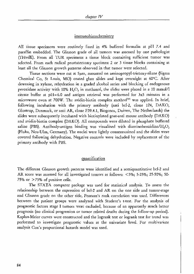
chapter IV
immunohistochemistry
All tissue specimens were routinely fixed in 4% buffered formalin at pH 7.4 and paraffin embedded. The Gleason grade of all tumors was assessed by one pathologist (THvdK). From all TUR specimens a tissue block containing sufficient tumor was selected. From each radical prostatectomy specimen 2 or 3 tissue blocks containing at least all the Gleason growth patterns observed in that tumor were selected.
Tissue sections were cut at 5,um, mounted on aminopropyl~trietoxy~silane (Sigma Chemical Co, St Louis, MO) coated glass slides and kept overnight at 60"C. After dewaxing in xylene, rehydration in a graded alcohol series and blocking of endogenous peroxidase activity with 10% H,O, in methanol, the slides were placed in a 10 mmolll citrate buffer at pH=6.0 and antigen retrieval was performed for 3xS minutes in a microwave oven at 700W. The avidin-biotin complex method291 was applied. In brief, following incubation with the primary antibody (anti bcl-2, clone 124, DAKO, Glostrup, Denmark, or anti AR, clone F39.4.1, Biogenex, Duiven, The Netherlands) the slides were subsequently incubated with biotinylated goat-anti mouse antibody (DAKO) and avidin-biotin complex (DAKO). All compounds were diluted in phosphate buffered saline (PBS). Antibody-antigen binding was visualized with diaminobenzidine/H,O, (Fluka, Neu-Vlm, Germany). The nuclei were lightly counterstained and the slides were covered following dehydration. Negative controls were included by replacement of the primary antibody with PBS.
quantification
The different Gleason growth patterns were identified and a semiquantitative bcl-2 and AR score was assessed for all investigated tumors as follows: < 5%; 5-25%; 25-50%; 50-75% or > 75% of positive cells.
The STATA computer package was used for statistical analysis. To assess the relationship between the expression of bcl-2 and AR on the Olle side and tumor-stage and Gleason grade on the other side, Pearson's rank correlation was used. Differences between the patient groups were analyzed with Student's t-test. For the analysis of prognostic factors stage I tumors were excluded, because of an apparently much better prognosis (no clinical progression or tumor related deaths during the follow-up period). Kaplan-Meier curves were constructed and the logrank test or logrank test for trend was performed to investigate prognostic val ues at the univariate level. For multivariate analysis Cox's proportional hazards model was used.
84
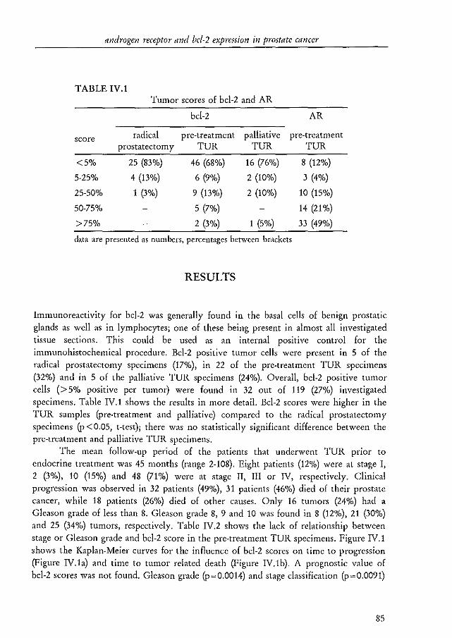
androgen receptor and bcl-2 expression in prostate cancer
TABLE IV.l Tumor scores of bcl-2 and AR
bcl-2 AR
score radical pre-treatment palliative pre-treatment prostatectomy TUR TUR TUR
<5% 25 (83%) 46 (68%) 16 (76%) 8 (12%)
5-25% 4 (13%) 6 (9%) 2 (10%) 3 (4%)
25-50% 1 (3%) 9 (13%) 2 (10%) 10 (15%)
50-75% 5 (7%) 14 (21%)
>75% 2 (3%) 1 (5%) 33 (49%)
data are presented as numbers, percentages between brackets
RESULTS
Immunoreactivity for bcl-2 was generally found in the basal cells of benign prostatic glands as well as in lymphocytes; one of these being present in almost all investigated tissue sections. This could be used as an internal positive control for the immunohistochemical procedure. Bcl-2 positive tumor cells were present in 5 of the radical prostatectomy specimens (17%») in 22 of the pre-treatment TUR specimens (32%) and in 5 of the palliative TUR specimens (24%). Overall, bcl-2 positive tumor cells (> 5% positive per tumor) were found in 32 out of 119 (27%) investigated specimens. Table IV.l shows the results in more detail. Bcl-2 scores were higher in the TUR samples (pre-treatment and palliative) compared to the radical prostatectomy specimens (p < 0.05, t-test); there was no statistically significant difference between the pre-treatment and palliative TUR specimens.
The mean follow-up period of the patients that underwent TUR prior to endocrine treatment was 45 months (range 2-108). Eight patients (12%) were at stage I, 2 (3%), 10 (15%) and 48 (71%) were at stage II, III or IV, respectively. Clinical progression was observed in 32 patients (49%), 31 patients (46%) died of their prostate cancer, while 18 patients (26%) died of other causes. Only 16 tumors (24%) had a Gleason grade of less than 8. Gleason grade 8, 9 and 10 was found in 8 (12%), 21 (30%) and 25 (34%) tumors, respectively. Table IV.2 shows the lack of relationship between stage or Gleason grade and bcl-2 score in the pre-treatment TUR specimens. Figure IV.l shows the Kaplan-Meier curves for the influence of bcl-2 scores on time to progression (Figure IV.la) and time to tumor related death (Figure IV.lb). A prognostic value of bcl-2 scores was not found. Gleason grade (p~0.0014) and stage classification (p~0.0091)
85
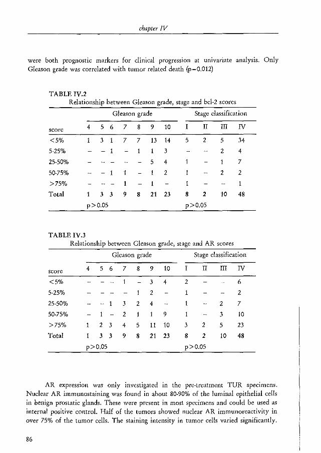
chapter IV
were both prognostic markers for clinical progression at univariate analysis. Only Gleason grade was correlated with tumor related death (p~0.012)
TABLE IV.2 Relationship between Gleason grade, stage and bcl-2 scores
Gleason grade Stage classification
score 4 5 6 7 8 9 10 I II III IV
<5% 1 3 1 7 7 13 14 5 2 5 34
5-25% - 1 1 1 3 2 4
25-50% 5 4 1 1 7
50-75% - 1 1 1 2 2 2
>75% 1 1 1 1
Total 1 3 3 9 8 21 23 8 2 10 48
p>O.OS p>0.05
TABLE IV.3 Relationship between Gleason grade, stage and AR scores
Gleason grade Stage classification
score 4 5 6 7 8 9 10 I II III IV
<5% 1 3 4 2 6
5-25% 1 2 1 2
25-50% - 1 3 2 4 1 2 7
50-75% 2 1 1 9 1 3 10
>75% 1 2 3 4 5 11 10 3 2 5 23
Total 1 3 3 9 8 21 23 8 2 10 48
p>O.OS p>O.OS
AR expression waS only investigated in the pre-treatment TUR specimens. Nuclear AR immunostaining was found in about 80-90% of the luminal epithelial cells in benign prostatic glands. These were present in most specimens and could be used as internal positive control. Half of the tumors showed nuclear AR immunoreactivity in over 75% of the tumor cells. The staining intensity in tumor cells varied significantly.
86
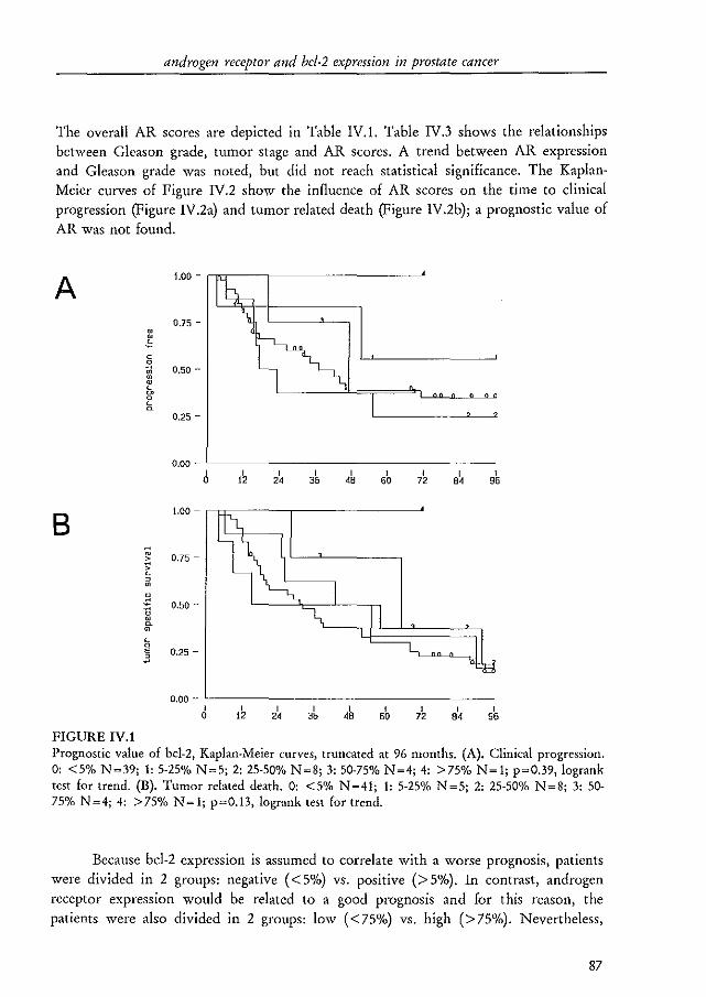
androgen receptor and bcl·2 expression in prostate cancer
The overall AR scores are depicted in Table IV.l. Table IV.3 shows the relationships between Gleason grade) tumor stage and AR scores. A trend between AR expression and Gleason grade was noted, but did not reach statistical significance. The Kaplan. Meier curves of Figure IV.2 show the influence of AR scores on the time to clinical progression (Figure IV.2a) and tumor related death (Figure IV.2b); a prognostic value of AR was not found.
A
B
FIGURE IV.I
1.00 - "",------,-----------'
0.75 -
c
~ 0.50-• • L ~
~ ~
0.25 -
0.00 - '--------------------I
35 I
48 I
50
1.00 - "'~---,_------~
0.75 -
0.50 -
0.25 -
B\ I 95
0.00 - '-----------------I o
I 12
I 2' I 35 B\ I
95
Prognostic value of bcl·2, Kaplan-Meier curves, truncated at 96 months. (A). Clinical progression. 0: <5% N~39; I: 5·25% N~5; 2: 25·50% N~8; 3: 50·75% N~4; 4: >75% N~l; p~0.39, logrank test for trend. (B). Tumor related death. 0: <5% N=41; 1: 5·25% N=5; 2: 25·50% N=8; 3: 50-75% N =4; 4: > 75% N = 1; p=O.13, lograllk test for trend.
Because bcl-2 expression is assumed to correlate with a worse prognosis, patients were divided in 2 groups: negative « 5%) vs. positive (> 5%). In contrast, androgen receptor expression would be related to a good prognosis and for this reason, the patients were also divided in 2 groups: low « 75%) vs. high (> 75%). Nevertheless,
87
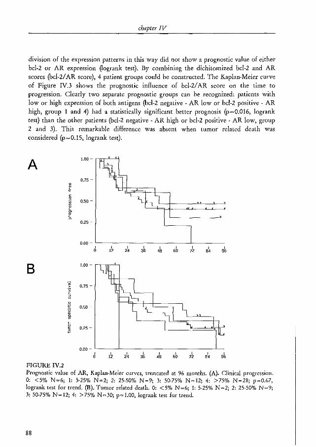
chapter IV
division of the expression patterns in this way did not show a prognostic value of either bcl-2 or AR expression ~ogrank test)_ By combining the dichitomized bcl-2 and AR scores (bcl-2/ AR score), 4 patient groups could be constructed. The Kaplan-Meier curve of Figure IV.3 shows the prognostic influence of bcl-2/ AR score on the time to progression. Clearly two separate prognostic groups can be recognized: patients with low or high expression of both antigens (bcl-2 negative - AR low or bcl-2 positive - AR high, group 1 and 4) had a statistically significant better prognosis (p~O.016, logrank test) than the other patients (bcl-2 negative - AR high or bcl-2 positive - AR low, group 2 and 3). This remarkable difference was absent when tumor related death was considered (p~O.15, logrank test).
A
B
FIGURE IV.2
ru • ~
ru > ~ L , ru
u
" U ru u ru
L o E
~
1.00 ~ rr.r"---'1
0.75 -
0.50 -
0.25 -
0.00 - '--------------'------
2~
1.00 - ""r-',
0.75 -
0.50 -
0.25 -
I 35
I 60
I 72
0.00 - '-------'--------------"---I
35 I
4B I
72 I .,
Prognostic value of AR, Kaplan-Meier curves, tnmcated at 96 months. (A). Clinical progression. 0: <5% N~6; I: 5-25% N~2; 2: 25-50% N~9; 3: 50-75% N~12; 4: >75% N~28; p~0.67, logrank test for trend. (B). Tumor related death. 0: < 5% N ~6; I: 5-25% N ~2; 2: 25-50% N ~9; 3: 50-75% N ~ 12; 4: > 75% N ~30; p~ 1.00, logrank test for trend.
88
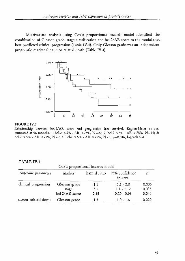
androgen receptor and bel·2 expression in prostate cancer
Multivariate analysis using Cox's proportional hazards model identified the combination of Gleason grade, stage classification and bcl-2/ AR score as the model that best predicted clinical progression (Table IV.4). Only Gleason grade was an independent prognostic marker for tumor related death (Table IV.4).
1.00 -
0.75 -ru
~ c 0
ru 0.50 -ru ru L ~ 0 L ~
0,25 -
0.00 -I I I I I I I I I
0 12 2' 36 " 60 72 ., 96
FIGURE IV.3 Relationship between bcl·2/ AR score and progression free survival, Kaplan-Meier curves, truncated at 96 months. 1: bcl·2 <5% - AR <75%, N=20; 2: bcl·2 <5% - AR >75%, N=19; 3: bcl-2 >5% - AR <75%, N~9; 4: bcl-2 >5% - AR >75%, N~9; p~O.016, logrank test.
TABLE IV.4 Cox's proportional hazards model
outcome parameter marker hazard ratio 95% confidence p interval
clinical progression Gleason grade 1.5 1.1 - 2.0 0.006 stage 3.5 1.1 - 11.2 0.035
bcl-2/ AR score 0.45 0.20 - 0.98 0.045
tumor related death Gleason grade 1.3 1.0 - 1.6 0.020
89
chapter IV
DISCUSSION
Several authors have investigated prognostic tumor markers that will allow for the identification of those patients who will not benefit from endocrine treatment, for example, tumor DNA.ploidy'" and expression of mutant p53'50. Although bcl·2 expression in prostate cancer has been related to the androgen independent phenotype"" "', clinical studies on the prognostic value of hormonally treated patients have as yet not been published. The present study was performed to investigate the prognostic value of pre-treatment expression of bcl-2 and AR in the tumor specimens of such patients.
Bcl·2 expression was found in only a minority of untreated tumors Crable IV.l), a finding that is in line with most published series1S1, 224, 116, Bcl-2 scores were higher in the pre-treatment TUR specimens compared to the radical prostatectomy specimens, suggesting that the expression of bcl·2 is related to advanced disease14'.215. A relationship with Gleason grade or TNM stage classification could not be demonstrated in the pre· treatment TOR specimens, however ('rable IV.2). The bcl·2 scores of androgen independent tumors (palliative TOR specimens) were not statistically significant different from the pre·treatment TOR specimens which is in contrast with the literature"" "'. McDonnell et al.'" found bcl·2 expression more frequently in androgen independent tumors (10/13) compared to primary prostate cancers (6/19). Androgen independence was defined by these authors as a tumor that did not show an initial response on endocrine therapy. Moreover, 11 of these were derived from metastatic lesions and 5 were small cell carcinomas. Thus, the resuits are difficult to compare with those of the present study. Colombel et al.'" applied the same antibody as used in the present study to frozen and paraffin·embedded prostate cancer sections. In their study bcl·2 expression was found in 62% of the primary tumors and in all hormonally treated tumors, which are extremely high figures compared to the literature and to those obtained in the present studyH5, 151,124,116, This is even more remarkable since the latter authors did not perform antigen retrieval on the paraffin.embedded sections, a procedure that is now recommended by the manufacturer and that was used in the present study, In the present study, the internal positive controls were positive in all cases and thus, we considered our results as valid.
Most nuclei (> 80%) of the luminal cells in all benign prostatic glands displayed AR immunoreactivity. Likewise, most of the tumors in the pre·treatment TUR specimens expressed AR, as found by others as welF03, 209, 111_214. In the present study a statistically non·significant trend between Gleason grade and AR scores was found Crable IV.3). Most studies found a weak correlation or no correlation at all between tumor·stage or grade and AR expression. In one study, AR expression was strongly correlated with Gleason grade"'. The heterogeneity of AR immunostaining within tumors, is a consistent finding203, 109, 211, 112, 214, liS.
90
androgen receptor and bcl-2 expression in prostate cancer
Both markers tested in the present study showed no prognostic value. For bcl-2 it was expected that expression in pre-treatment TUR specimens would be correlated with a worse prognosis (Figure IV.!). Two studies with radical prostatectomy specimens demonstrated that bcl-2 expression was associated with an adverse prognosis at the univariate level Hs or multivariate level1S2. The prognosis of clinically localized prostate cancer is determined mainly by the presence of extensive disease at the time of operation, and thus, the results of the latter studies cannot simply be extended to the present study. In node positive breast cancer patients treated with adjuvant hormonalor chemotherapy, bcl-2 expression was even correlated with a better prognosisJ04 • These authors suggested that bcl-2 expression in tumors arising from epithelia with basal cell expression of bcl-2 Oike breast and prostate epithelium), might predict a better prognosis due to a less aggressive mechanism of transformation. In addition, other mechanisms of androgen escape might be clinically more important. Actually, the results of the palliative TOR specimens support this. A recent experimental study with sublines of the Dunning R3327 rat prostate cancer model showed that bcl-2 expression is not an absolute requirement for androgen independent prostate cancer growth305 .
AR expression was also not correlated with patient outcome (Figure IV.2). Several studies did show such a prognostic value212.2H • Others reported that not the percentage of positive cells but the degree of immunostaining heterogeneity210 or the mean immunostaining intensiti ll would determine the prognosis. Both studies made use of computerized image analysis systems and in the latter study significant data could only be obtained when the results of two antibodies, against the C- and N·terminal PaIts of the AR, were combined. Possibly, analysis of the results from the present study in this way might reveal significant results as well. But, since evaluation of clinically suitable markers should be fast and simple, an easily applicable semiquantitative system was used.
Strikingly, a combined bcl-2/ AR score was a prognostic marker for the prediction of clinical progression at the univariate and multivariate level (Figure IV.3, Table IVA). Since endocrine treatment does not improve patient survivapc6, the finding that only Gleason grade was an independent marker to predict tumor related death was not surprising. The dichitomized variables showed no individual prognostic value and if both were entered in Cox's proportional hazards model, also no independent prognostic value was found. Possibly, some interaction is present, because tumors that express bcl-2 (> 5% of the tumor cells) had a favorable prognosis only if AR expression was high (> 75%) and tumors without bcl-2 had a good prognosis only if AR expression was low «75%). As yet a cell biological explanation for these findings is not apparent.
In conclusion, bcl-2 is expressed in a minority of prostate cancers, whether androgen dependent or independent and the expression level is not related to tumorstage or grade. Bcl-2 and AR expression show no prognostic value in the pre-treatment specimens of hormonally treated prostate cancer patients. A combined bcl-2/ AR score is of independent prognostic value to predict clinical progression, but not tumor related
9!

chapter IV
death. Since this is the first study on the prognostic value of bcl-2 in hormonally treated prostate cancer, additional studies on bcl-2 and other related markers are needed.
92
CHAPTER V
NEUROENDOCRINE CELLS IN THE NORMAL, HYPERPLASTIC AND
NEOPLASTIC PROSTATE
M.A. Noordzij; G.]. van Steenbrugge; T.H. van der Kwast; F .H. Schroder
From the departments of Urology and Pathology, Erasmus University, Rotterdam, The Netherlands
Urol Res 1995;22:333-41 reprinted with permission
chapter V
ABSTRACT
Neuroendocrine cells can be demonstrated in normal, hyperplastic and neoplastic prostatic tissues. The products secreted by these cells can be used as tissue and/or serum markers but may also have biological effects. Neuroendocrine cells in prostate cancer most probably do not contain the androgen receptor and are therefore primarily androgen independent. Some of the neuropeptides secreted by the neuroendocrine cells may act as growth factor by activation of membrane receptors in an autocrine-paracrine fashion or by ligand independent activation of the androgen receptor in neighboring non-neuroendocrine cells. Evidence is accumulating from experiments with tumor models that neuropeptides indeed can influence the growth of prostatic tumor cells. Future research on neuroendocrine differentiation Inay answer some questions concerning the biological behavior of clinical prostatic tumors.
INTRODUCTION
In most developed countries prostate cancer has became the most prevalent tumor in men and in these countries its mortality is only being surpassed by lung cancer4, 307, A lot of research done in the field of prostate cancer is directed towards the identification of factors involved in tumor progression and the change from androgen dependency towards independency. Neuroendocrine differentiation is a possible factor which has received increasing attention during the last years301
,
Neuroendocrine (NE) cells are also known as APUD (amine precursor uptake and decarboxylation) cells or endocrine-paracrine cells' ''. The concept of NEI APUD cells was worked out by Pearse and co-workers from the diffuse endocrine epithelial organ concept of FeyrterJO
•• '''. The APUD system consists of a group of apparently unrelated endocrine cells located in endocrine and non-endocrine ti~suesJ which share a number of cytochemical, ultrastructural and functional characteristics30s
• In more detail, the endocrine/NE system consists of: the classical endocrine organs (e.g. adrenals, parathyroid gland), clusters of endocrine cells (e.g. pancreatic islets of Langerhans), dispersed epithelial endocrine cells (e.g. gastrointestinal NE cells), neurons and ganglia and the paraganglion system'IO.
The clinical aspects of NE differentiation in prostatic carcinoma have been reviewed in more detail in a number of papers by Di Sant'Agnese257, 311, 312. This review
94

prostatic neuroendocrine cells
will therefore emphasize the experimental aspects: the role of NE cells in androgen insensitivity, tumor progression and tumor growth. Nevertheless, some clinical data will be provided to understand the concept and the questions that emerged from it.
FIGURE V.I Prostatic neuroendocrine celis, chromogranin A starnrng with immunoperoxidase technique, nuclear counterstaining with hematoxylin (900x). (A). Closed-type neuroendocrine cell with cytoplasmic process (arrow). (B). Open-type neuroendocrine cell.
NEUROENDOCRINE CELLS IN THE NORMAL PROSTATE
In normal prostatic epithelium, NE cells are found among the well known prostate specific antigen (PSA) producing exocrine cells and basal cells (Figure V.l)31J. Such cells have been described for the first time by Pret! already in 1944 as the argentaffine basal celisHl . In this study the argentaffine cells were identified by silver staining procedures as well as autofluorescence techniques3H . Prostatic NE cells are located in the glandular and ductal epithelium and they form only a small part of the epithelial cell compartment. They clearly belong to the 'dispersed epithelial endocrine cell' type)!'. NE cells are found throughout the whole prostate although they are more abundant in the periurethral and ductal regions274. In the guinea pig prostate the number of NE cells increased with age315
, a phenomenon which was not described for humans174. NE cells
in the glandular acini but not in the periurethral and ductal epithelium of the human prostate were found to disappear soon after birth and to reappear at puberty'''. These
95
chapter V
findings suggest a direct or indirect effect of circulating androgens on the acinar NE cells but not on periurethral and ductal NE cells.
Morphologically, two types of prostatic NE cells can be recognized: an open type reaching to the glandular lumen and a closed type separated by other cells from the lumen"'. The latter type often has dendritic cytoplasmic processes interdigitating between neighboring cells26'. Prostatic NE cells are ultrastructurally characterized by so· called dense core granules or neurosecretOlY granules316
, Based on the variations in the ultrastructural morphology of the granules a greater heterogeneity of NE cells was suggested316
,
The heterogeneity of this cell type is also apparent from the variety of products (biogenic amines or peptide hormones) which they secrete. Chromogranin A and B (CgA and CgB), secretogranin II, neuron specific enolase (NSE) and serotonin (5.HT) are found in most, if not all prostatic NE cells'58.260,,'. A small number of these cells also contains calcitonin (CT) and related peptides like calcitonin gene related peptide (CGRP) and katacalcinJl7·J21. In addition, somatostatin (SMS)J2I· "', bombesin/gastrin related peptide (GRP)J17, midportion of the II chain of thyroid stimulating hormonelll
.
314, glycoprotein hormone (X_chainJ25 and parathyroid hormone related protein have been
found"'. The chromogranins are acidic glycoproteins which are widely expressed in NE
cells327. Various biologically active peptides can be released from the CgA and CgB molecules by enzymatic actionJ27, 328. A third chromogranin (chromogranin C) is now known as secretogranin IP17, 329. Most of the immunohistochemical studies on NE differentiation have been performed with antibodies to CgA or 5-HT. NSE ('Y-enolase, a subtype of the glycolytic enzyme enolase) was thought to be an exclusive marker of endocrine and NE cells, but it has been shown that NSE is secreted by a large variety of other cell types3
}o.
The heterogeneous morphology of the neurosecretory granules and the diversity of secreted products suggest that these cells exert a number of distinct functions, which are up to now virtually unknown. In parallel with functions of NE cells in more extensively studied systems (i.e. lung, pancreas and adrenals) a role of these cells in growth and differentiation, and in maintenance of homeostasis has been suggested))1-3)).
Some of the neuropeptides share growth factor activity (reviewed in refs. 331•33
').
Relatively high levels of CT, GRP/bombesin and SMS have been found in human semen337-339. Exposure to salmon-CT (seT) decreased sperm motility in vitroHO
,
Furthermore, sCT also increased the secretion of prostatic alkaline phosphatase by rat ventral prostate explants in a dose dependent manner'41. These studies indicate that prostatic NE cells might also have some exocrine functions,
According to Feyrter's original concept of the diffuse endocrine organ, all NE cells throughout the body were thought to originate from the neural crest"'. However, nowadays a local origin of the NE cells in most tissues is suggested and has been proven, for example, in colorectal epitheliumlf-l, A multidirectional differentiation has been postulated for normal tissues as well as for tumors arising in these tissues
96
prostatic neuroendocrine cells
(reviewed by DeLellis"'). In a recent immunohistochemical study, some prostatic NE cells expressed basal cell specific cytokeratins and a few NE cells in hyperplastic glands displayed immunoreactivity for PSA 258, JH. These observations suggest a common differentiation pathway of prostatic secretory, NE and basal cells and it is now generally accepted that prostatic NE cells indeed originate from the prostate. It may be hypothesized that the basal cell layer contains the prostatic stem cells and that these cells give rise to both the exocrine and NE phenotypes of the glandular epithelium.
NEUROENDOCRINE CELLS IN THE HYPERPLASTIC PROSTATE
The presence of NE cells in hyperplastic prostatic tissue has been demonstrated using silver staining techniques'''' "6. In one study, up to 16 out of 20 hyperplastic nodules displayed NE differentiation"'. However, in an other study it was found that hyperplastic nodules in general contained less argentaffin NE cells compared to the adjacent normal epithelium346
• This was confirmed in a recent study using immunohistochemistry with a 5-HT antibody and chromatographic quantification of the 5-HT content of tissue homogenates27S. On the other hand, Abrahamsson et al. found more NE cells in hyperplastic prostatic tissue than in normal glands as defined by immunoreactivity for 5-HT, thyroid stimulating hormone and CT161. Aprikian et al. found immunohistochemically defined NE cells in all investigated hyperplastic specimens, although the relation with the adjacent normal glands was not studied"'. Whether or not neuroendocrine cells playa role in the pathogenesis of benign prostatic hyperplasia is not clear at the moment. The study of Cockett et al. gives some support to this idea since they found more NE cells in small hyperplastic nodules compared to normal prostate or large hyperplastic nodules27S. This suggests that the growth of the more immature small hyperplastic nodules is stimulated by NE products. Alternatively, the presence of NE cells may simply reflect an enhanced proliferative activity of glandular epithelial cells.
97
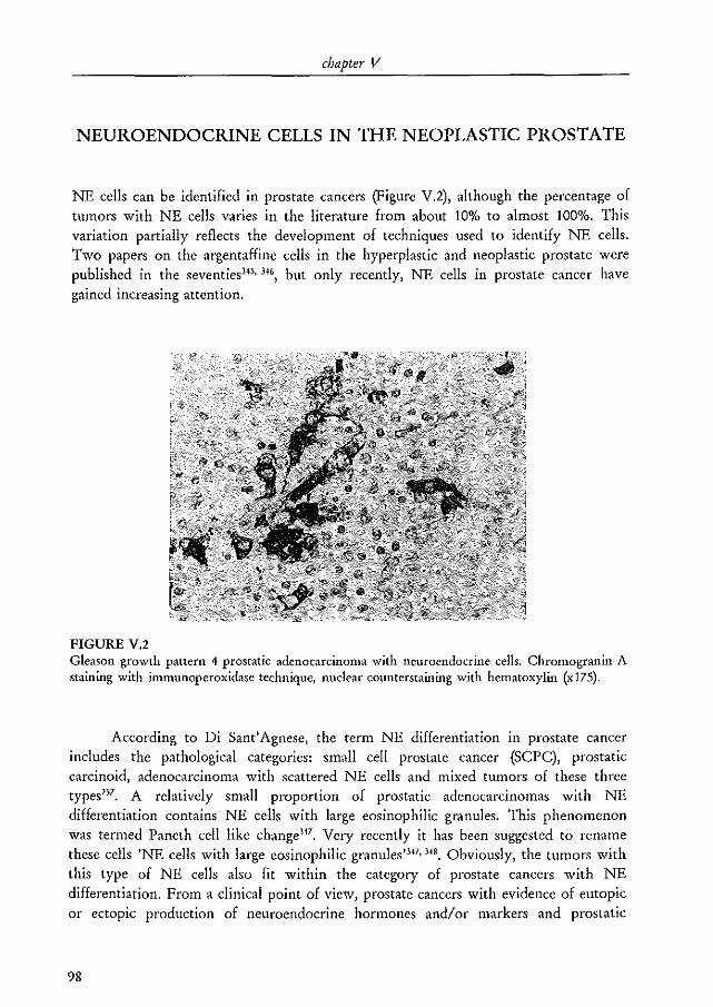
chapter V
NEUROENDOCRINE CELLS IN THE NEOPLASTIC PROSTATE
NE cells can be identified in prostate cancers (Figure V.2), although the percentage of tumors with NE cells varies in the literature from about 10% to almost 100%. This variation partially reflects the development of techniques used to identify NE cells. Two papers on the argentaffine cells in the hyperplastic and neoplastic prostate were published in the seventies'''' 34', but only recently, NE cells in prostate cancer have gained increasing attention.
FIGURE V.2 Gleason growth pattern 4 prostatic adenocarcinoma with neuroendocrine cells. ChroIllogranin A staining with immunoperoxidase technique, nuclear counterstaining with hematoxylin (x17S).
According to Di Sant' Agnese, the term NE differentiation in prostate cancer includes the pathological categories: small cell prostate cancer (Sepe), prostatic carcinoid, adenocarcinoma with scattered NE cells and mixed tumors of these three types257
• A relatively small proportion of prostatic adenocarcinomas with NE differentiation contains NE cells with large eosinophilic granules. This phenomenon was termed Paneth cell like change"'. Very recently it has been suggested to rename these cells 'NE cells with large eosinophilic granules''''' 34'. Obviously, the tumors with this type of NE cells also fit within the categOlY of prostate cancers with NE differentiation. From a clinical point of view, prostate cancers with evidence of eutopic or ectopic production of neuroendocrine hormones and/or markers and prostatic
98
prostatic neuroendocrine cells
malignancies associated with paraneoplastic syndromes linked to NE differentiation should also be included in the definition''',
Undifferentiated small cell cancers, also referred to as NE carcinomas, occur in various organs, but most often in the lungs. SCPC is a relatively rare disease accounting for ± 1-5% of all prostate cancers, and has been described in a few larger studies"',35', It is a highly malignant disease with a mean survival time of 7-17 months'''' 351, In about half of the patients the small cell component is preceded by a common adenocarcinoma suggesting that at least in a number of patients the small cell carcinoma may arise in a common adenocarcinoma. In line with this suggestion, a number of mixed tumors was found''', 351, Not all sepe showed NE differentiation, howeve"", Prostatic carcinoid is a very rare entity, Only some case reports have been published as yetm-358 . Carcinoids occur especially in the digestive system (appendix) and they are in general relatively benign359. It appears that prostatic carcinoid tumors behave more aggressively257, 357, although detailed studies with follow-up data are lacking, Mixed carcinoidadenocarcinoma tumors have been found and it has been suggested that carcinoid formation in a hormonally treated adenocarcinoma might be a selective effect of the treatment357,
The most common pattern of NE differentiation in prostate cancer is a prostatic adenocarcinoma with scattered NE cells, The number of NE cells within an adenocarcinoma varies from patient to patient and their presence may have prognostic significance263, 264.266-268. Some authors found a relation between the tumor grade and the number of NE cells'''' J1J, but others did not"', NE cells were identified in about 50% of lymph node and bone metastases of prostatic adenocarcinomas269, This proves that NE cells are an intrinsic component of the adenocarcinoma and are not derived from preexistent benign glands, The presence of NE cells in metastatic lesions had no prognostic value169. Serum levels of egA showed a 60% correlation with the immunohistochemical staining of this protein and the presence of metastatic disease was better predicted by serum egA levels than by tissue immunoreactivity]", The possible correlation between tumor grade and the number of NE cells in a tumor may well account for part or all of the reported prognostic value of NE differentiation, which would indicate that the number of NE cells just reflects the level of dedifferentiation of a tumor. This possibility can be investigated with long term clinical follow-up studies using multiple regression analysis.
99
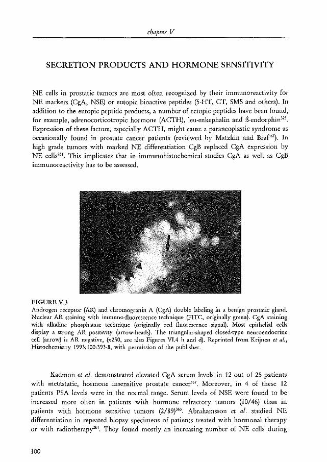
chapter V
SECRETION PRODUCTS AND HORMONE SENSITIVITY
NE cells in prostatic tumors are most often recognized by their immunoreactivity for NE markers (CgA, NSE) or eutopic bioactive peptides (5-HT, CT, SMS and others). In addition to the eutopic peptide products, a number of ectopic pep tides have been found, for example, adrenocorticotropic hormone (ACTH), leu-en kephalin and fl-endorphin323
•
Expression of these factors, especially ACTH, might cause a para neoplastic syndrome as occasionally found in prostate cancer patients (reviewed by Matzkin and Braf'61). In high grade tumors with marked NE differentiation CgB replaced CgA expression by NE cells"l. This implicates that in immunohistochemical studies CgA as well as CgB immunoreactivity has to be assessed.
FIGURE V.3 Androgen receptor (AR) and chrolllogranin A (egA) double labeling in a benign prostatic gland. Nuclear AR staining with immuno-fluorescence technique (FITe, originally green). egA staining with alkaline phosphatase technique (originally red fluorescence signal). Most epithelial cells display a strong AR positivity (arrow-heads). The triangular-shaped closed-type neuroendocrine cell (arrow) is AR negative, (x250, see also Figures VIA b and d). Reprinted from Krijnen et al., Histochemistry 1993;100:393·8, with permission of the publisher.
Kadmon et al. demonstrated elevated CgA serum levels in 12 out of 25 patients with metastatic, hormone insensitive prostate cance~6l. Moreover, in 4 of these 12 patients PSA levels were in the normal range. Serum levels of NSE were found to be increased more often in patients with hormone refractory tumors (10/46) than in patients with hormone sensitive tumors (2/89)'63. Abrahamsson et al. studied NE differentiation in repeated biopsy specimens of patients treated with hormonal therapy or with radiotherapy'''. They found mostly an increasing number of NE cells during
100
prostatic neuroendocrine cells
follow-up paralleled by dedifferentiation and hormonal escape (i.e. progression) of the tumor. These results raise the question whether NE cells in prostate tumors are androgen sensitive or not. NE cells in benign and malignant prostatic tissues contained rarely, if at all androgen receptor immunoreactivity (Figute V.3)"'· 3M. In an other study, using a different antibody for the androgen receptor, prostatic NE cells generally expressed the androgen receptor'''. Very recently the authors of the latter study confirmed the findings of the former two studies"', leading to the conclusion that NE cells in the normal or neoplastic prostate do not contain the androgen receptor. It has been shown that the androgen receptor content of a prostatic tumor does not predict androgen (in)sensitivity2".20'. It is, however, unlikely that androgen receptor negative tumor cells will respond to androgen withdrawal. Therefore, NE cells in prostate cancer most probably form a primarily androgen insensitive tumor cell population.
GROWTH MODULATION BY NEOPLASTIC NEUROENDOCRINE CELLS
A number of peptides produced by NE prostatic tumor cells (S-HT, GRP/bombesin, CT, SMS) exhibit growth factor activities mediated by a membrane receptor. This may represent a way of paracrine or autocrine growth modulation332, 335, 336. Most of the
prostatic adenocarcinoma cells surrounding NE cells contain the androgen receptor, even in androgen refractory carcinomas276
, 364. It was found in COS cells transfected with steroid hormone receptors that DOPA activated several steroid hormone receptors (progesteron, estrogen, vitamin D and thyroid hormone-B receptors) in a ligandindependent fashion277. In this way NE tumor cells might influence the growth of neighboring non NE tumor cells by androgen independent activation of the androgen receptor in a paracrine manner. This possibility warrants further investigation. It should be stressed that neuropeptides may enhance or inhibit growth, depending on the specific nature of the neuropeptide and properties of the target cell. Bonkhoff et at. found in an immunohistochemical study that proliferating cells in normal, hyperplastic and cancerous prostatic tissues were usually, but not necessarily located in proximity of clusters of NE cells'66. This supports the concept of paracrine growth regulation by prostatic NE cells.
The following hypothesis can be postulated: NE cells in a prostatic adenocarcinoma form a subset of primarily androgen independent tumor cells which modulate the growth of neighboring non-NE tumor cells by the secretion of neuropeptides in a paracrine manner (Figure VA). Granted this hypothesis, one would expect that following androgen withdrawal the proportion of NE cells will iucrease.
101
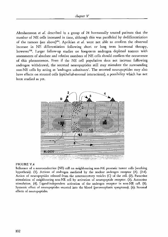
chapter V
Abrahamsson et al. described in a group of 24 hormonally treated patients that the number of NE cells increased in time, although this was paralleled by dedifferentiation of the tumors (see above}"'. Aprikian et al. were not able to confirm the observed increase in NE differentiation following short or long term hormonal therapy, however'''. Larger follow-up studies on long-term androgen depleted tumors with assessments of absolute and relative numbers of NE cells should confirm the occurrence of this phenomenon. Even if the NE cell population does not increase following androgen withdrawal, the secreted neuropeptides still may stimulate the surrounding non-NE cells by acting as 'androgen substitutes'. The secreted neuropeptides may also have effects on stromal cells (epithelial-stromal interactions), a possibility which has not been studied as yet.
FIGURE VA Influence of a neuroendocrine (NE) cell on neighbouring non-NE prostatic tumor cells (working hypothesis). (1). Actions of androgen mediated by the nuclear androgen receptor (A). (2-4). Action of neuropeptides released from the neurosecretory vesicles (C) of the cell. (2). Paracrine stimulation of neighbouring non-NE cell by activation of neuropeptide receptor. (3). Autocrine stimulation. (4). Ligand-independent activation of the androgen receptor in non-NE cell. (5), Systemic effect of neuropeptides secreted into the blood (paraneoplastic symptoms). (6). Stromal effects of neuropeptides.
102
prostatic neuroendocrine cells
SPECIFIC NEUROPEPTIDES
SEROTONIN
Serotonin (5-H1) is well known as neurotransmitter and vasoactive peptide and some recent repOlts indicate that 5-HT also has growth factor activity"" "', 5-HT stimulated DNA synthesis in hamster fibroblasts proved to be mediated by activation of the 5-HT1b receptor367
. This receptor is only expressed in rodents. Further experiments indicated that in humans 5-HT mediates the proliferation of smooth muscle cells mediated through activation of the 5-HTld receptor''', 5-HT might influence tumor growth indirectly by changing the local blood flow in a tumor due to its vasoactive action, It is very likely that 5-l-IT plays a role in the prostate since it is expressed by most if not all prostatic NE cells258, 260, 162. Unfortunately, it is as yet not known if human prostatic tumor cells contain 5-I-IT receptors and if so, which subtypes. As mentioned above, 5-HT may also exert its effect by ligand independent activation of the androgen recepto ~77.
BOMBESIN/GASTRIN RELATED PEPTIDE
Gastrin related peptide (GRP) is the mammalian analogue of the amphibian peptide bombesin"9, GRP stimulated the growth of cultured normal bronchial epithelial cells in a dose dependent mannerll1, In vitro studies with cultured pulmonary NE cells demonstrated that treatment with GRP increased the number of NE cells and stimulated their 5-HT expression while treatment with 5-HT did noe", It has also been shown that GRP stimulated growth of small cell lung cancer cells370, This effect could be blocked in vivo by an antibody against GRP and in vitro by GRP analogues which prevent binding of GRP to its receptorl7', 371, III vitro studies with the androgen independent prostatic cancer cell line PC-3 similarly showed a growth stimulatOlY action of GRP which could be blocked by an anti-GRP antibody''', Saturable GRP binding sites were demonstrated on PC-3 cells, but no immunoreactivity for GRP was demonstrated in these cells, excluding an autocrine action of GRpJ7l , The GRP antagonist RC-3095 was able to inhibit the growth of the androgen dependent human xenograft PC-82, the Dunning R-3327H rat prostate tumor and the androgen independent the DU-145 and PC-3 prostatic in vitro cell linesJ7l-376, Saturable GRP binding sites were demonstrated on cells of all these models, Altogether, evidence exists that GRP has a potential role in the growth of (neoplastic) prostatic tissue,
103
chapter V
CALCITONIN
The human (hCT) and salmon (sCT) subtypes of CT can be demonstrated in a subset of normal and neoplastic prostatic cells158,JI7.311, 3H, 377. In conditioned medium of cultures
of prostate cancer cells immunoreactive CT was found in 4-fold higher concentration as compared to cultures of BPH cells"9, III vivo administration of scT to rats induced ornithine decarboxylase (a key enzyme associated with cell cycle progression and growth) in a number of organs)78, It should be realized that CT also inhibits the pituitary secretion of luteinizing hormone which is an important hormone in the mediation of androgen secretion379 • In addition, the secretion of prolactin, which influences the action of androgens on the prostate, was also found to be decreased by CT380, Therefore, a pituitary mediated growth inhibiting effect of CT on prostatic cancer growth is also expected, The direct growth modulating effects of CT have been studied in a few tumor systems, T-47D breast cancer cells contained high affinity receptors for CT and in vitro growth of these cells was dose dependently inhibited by CT38I, 38
1, Comparable results were found in cells of the human gastric carcinoma cell
line KA TO 1II3", Following CT administration intracellular cAMP levels increasedJS',J8J,
This increase was also found in four out of six renal adenocarcinoma cell lines and only these four cell lines were growth inhibited by CT administration''', In a panel of 13 small cell I ung cancer cell lines two contained CT and only one was able to bind CT while no growth effect upon CT administration was found385, It has recently been shown that sCT dose dependently increased the cAMP concentration and the DNA synthesis in cells of the in vitro human prostatic cancer cell line LNCaP386, This growth stimulatory effect of CT is at variance with the results from other non prostatic tumor model systemsJSl,3", Therefore, the effects of CT have to be confirmed in additional studies using other prostatic tumor models including in vivo models because CT also has systemic effects which may influence prostatic tumor growth.
SOMATOSTATIN
Abrahamsson et ai, found somatostatin (SMS) immunoreactivity in 12 out of 40 prostatic adenocarcinomas323, a result that has not been confirmed in by Aprikian et al,l", SMS receptors were found neither in 17 prostatic carcinomas nor in 2 BPH specimens387, On the other hand, binding sites for several SMS analogues were demonstrated in normal and neoplastic prostatic tissues38S . A number of experimental studies concerning the growth modulating effects of SMS in prostate cancer have been published, Several SMS analogues (sandostatin, somatuline, RC-160, RC-121) decreased without exception the growth of prostatic tumor models in vivo (Dunning R-3327 and R-3327H, PC-82, DU-145, PC-3) and ill vitro (LNCaP) 3n, 374, 376, 389,395, In the Dunning
R-3327H rat tumor the SMS effect potentiated the castration induced growth inhibition even when the tumors became androgen independent3'n, however, this was not
104
prostatic neuroendocrine cells
confirmed by others"'. In experiments with heterotransplants of the androgen independent PC-3 cell line, SMS was able to inhibit growth only if the tumors were small {10 mm')'''. Combination treatment of tumor models with an SMS analog and luteinizing hormone-releasing hormone (LH-RH) analog (D-TRp6 LH-RH) resulted in a stronger growth inhibition than treatment with only one of the components373, 391, 394.
SMS binding sites were demonstrated on cells of the PC-82373, Du-145"\ PC-3"6 and Dunning R-3327H39I models. SMS and prolactin binding sites were found to be down regulated in Dunning R-3327H tumors treated with analogues of SMS and LH-RH39I. As noted above, prolactin may have a stimulating effect on prostate cancer and the down regulation of the prolactin receptor by SMS might partially explain the growth inhibiting effects of SMS. All these results strongly suggest a direct growth inhibiting effect of SMS (analogues) in prostate cancer.
The effects of some of the neuropeptides on prostate cancer growth have been investigated quite extensively. The relation with androgen levels and androgen receptor activity, the action of a combination of neuropeptides and the mechanisms of action are poorly understood at the moment. Furthermore, it is not clear whether the amounts of neuropeptides secreted by the NE cells are sufficient for biological activity on neighboring cells. For some of the neuropeptides, the presence of the corresponding receptor is not clear. Possibly other, as yet unknown, factors produced by prostatic NE cells may even be more important.
NEUROENDOCRINE DIFFERENTIATION IN PROSTATIC TUMOR MODELS
To enhance our knowledge of NE differentiation in prostate cancer, experimental models with NE cells are urgently needed. A heterotransplant able model of a small cell prostatic carcinoma has been establishedJ9~39s. A SCPC is biologically different from an adenocarcinoma and therefore, this model is probably not useful for the study of the paracrine role of NE cells in prostatic adenocarcinoma. None of the available prostatic tumor models contains NE cells as defined by immunoreactivity for CgA, 5-HT, CT, SMS, NSE and thyroid stimulating hormone, although two heterotransplant able human tumor models which were recently established in this laboratory contain CgA positive cells (unpublished observation).
Possibly, NE differentiation can be induced in non-NE prostatic tumor models. Transfection of v-rasH into DMS-53 small cell lung cancer cell line cells resulted in a cell
105
chapter V
line with increased NE featuresJ99, Transfection of both c-raf-l and c-myc oncogenes
into SV-40 immortalized bronchial epithelial cells resulted in the generation of heterotransplantable large cell carcinoma cell lines with a NE phenotype4OO, 401, These studies indicate that NE differentiation can be induced and that it is associated with the expression of certain (proto) oncogenes. Future experiments have to show if a similar approach can be applied to prostate cancer.
CONCLUSION
Knowledge about the function of NE cells in the human prostate and prostate cancer is limited. Evidence is however accumulating that NE cells and tumors with NE cells are related to the androgen independent and poorly differentiated types of prostate cancer. However, at the moment it cannot be excluded that NE differentiation is only an epiphenomenon associated with dedifferentiation of a tumor, There is evidence that the secretion products of prostatic NE cells affect prostate cancer growth and possibly also affect tumor differentiation, Research directed towards identifying the role of NE cells in prostate cancer is likely to contribute to the understanding of the transition of androgen dependent to androgen independent prostate cancer.
106
CHAPTER VI
NEUROENDOCRINE DIFFERENTIATION IN HUMAN PROSTATIC TUMOR
MODELS
M.A. Noordzij; W.M. van Weerden; C.M.A. de Ridder; T.H. van der Kwast; F.H. Schroder; G.J. van Steenbrugge
From the departments of Urology and Pathology, Erasmus University, Rotterdam, The Netherlands
Am J Pathol 1996;149:859-71 reprinted with permission
chapter lIT
ABSTRACT
Neuroendocrine (NE) cells can be identified in benign and malignant prostatic epithelia. Factors regulating their presence and their functions are poorly understood, mainly due to a lack of suitable experimental models. A total of 15 in vitro and in vivo prostatic cancer tumor models, including a number of newly established in vivo models, were studied immunohistochemically for the presence of NE cells under different hormonal conditions. None of the in vitro models (PC-3, DU 145, LNCaP, TSU) contained NE cells. Five of the 7 xenograft models established at this laboratory contained NE cells. In 3 of these, NE cells were found only in the initial mouse passages. In the other 2 (PC-295 and PC-310), the NE phenotype was stable. NE features were confirmed by transmission electron microscopy and by Western analysis of chromogranin A expression. Immunohistochemical double labeling experiments confirmed that NE cells in prostate cancer are post-mitotic (no Ki-67 expression) and do not express the androgen receptor. In the PC-295 and PC-310 models, short-term androgen withdrawal resulted in a rapidly increased number of NE cells. A time-course experiment with PC-295 bearing mice strongly suggests that this increase occurred by induction of NE differentiation rather than by rapid proliferation and subsequent differentiation or selective persistence. In conclusion, these models are suitable to resolve fundamental questions with regard to the presence and functions of NE cells in human prostate cancer.
INTRODUCTION
Neuroendocrine (NE) or endocrine-paracrine cells are characterized by a number of structural, cytochemical and functional properties308• The dispersed NE system consists of single or clustered NE cells located in the epithelium of several non-NE organs Oungs, gastro-intestinal tract)llO. The normal prostate likewise contains epithelial NE cells belonging to this system. Prostatic NE cells secrete neuropeptides and biogenic amines (reviewed by Noordzij et al.15l In concordance with the functions of NE cells in other epithelial organs, it has been suggested, but not proven, that prostatic NE cells play a role in the maintenance of homeostasis and/or regulation of secretion of
108
neuroendocrine cells in prostate cancer
prostatic glandular epitheliumHl • In addition, there is some evidence that these cells have exocrine functions as wel1337,401.
NE cells are present in the majority of prostate cancers403. It is of importance to
distinguish three types of NE differentiation within prostate cancer. Small cell prostate cancer and prostatic carcinoid are relatively rare and are considered pure neuroendocrine tumors with a poor prognosis257
. Conventional adenocarcinoma with scattered or clustered NE cells represents the most common type of prostate cancer with NE differentiation. The NE cells in both benign and malignant prostatic epithelium can be identified immunohistochemically with antibodies against products secreted by these cells or by routine transmission electron microscopy which visualizes the neurosecretory granules (also known as dense core granules or neuroendocrine granules)261. 316. 4". Chromogranin A (CgA) is a 68kDa acidic glycoprotein which is secreted by most, if not all, NE cells. It may playa role in the excretory pathway of neuropeptides and is most likely a precursor of several functional peptides (e.g., pancreastatin and chromostatin) which are formed by proteolytic cleavage of the CgA molecule'17.405. Antibodies against CgA are frequently being used as pan·NE markers. During recent years it has become clear that some of the factors secreted by NE cells (e.g., serotonin and gastrin related peptide) display growth factor activity mediated by a corresponding membrane receptor335, 33(,. Bombesin (gastrin related peptide), for example, has been shown to stimulate the proliferation of the prostatic tumor cell line PC.3 which has saturable bombesin binding sites371 • This stimulation could be inhibited by antibodies against the peptide. Prostatic NE cells in benign and malignant prostatic epithelium do not contain androgen receptor protein and are therefore considered to be androgen independent, at least for their existence176• All in all, this suggests that NE cells in a prostatic adenocarcinoma influence the growth of neighboring non-NE tumor cells in an androgen independent manner. Consequently, NE cells may playa role in the progression of prostate cancer towards androgen independence. The NE cells themselves, however, appear to be in a post-mitotic state4Cio
• For experimental studies on NE differentiation in prostate cancer, tumor models containing these cells are needed. In the present study, the occurrence of immunohistochemically defined NE cells was studied under different hormonal conditions in most of the commonly available in vitro prostatic tumor cell lines and in a number of in vivo human prostatic tumor xenograft models. Transmission electron microscopy and Western imrnunoblotting were performed to confirm the immunohistochemical data. To study the androgen receptor expression in prostatic NE cells and to assess their post-mitotic state, double labeling immunohistochemistry was performed.
109
chapter VI
MATERIALS AND METHODS
Tumor models
A panel of human prostatic cancer in vitro cen lines and in vivo xenograft models was used. This panel included the more well known, commonly available, in vitro cell lines (PC-3"', DU 14540', TSU'09 and LNCaP4!O) and in vivo xenograft models (PC-EWHI and PC-S2m) as well as a set of in vivo xenograft models more recently established at this laboratory (PC-13J, PC-135, PC-295, PC-310, PC-324, PC-329, PC-339, PC-346 and PC-374). The latter models are considered to be representative for the different clinical manifestations of prostate cancer (androgen dependent vs. independent; primary tumor vs. metastatic lesion) and are described elsewhere in more detail 413. Two sub-lines of the parental LNCaP-FGC cell line gymph node carcinoma of the prostate, fast growing colony) i.e., LNO gymph node origin) and R (resistant)4I' were also included. PC-346i is an androgen independent sub-line derived from the PC-346 xenograft model. Both the parental and PC-346i tumor models can be propagated in vitro as well as in vivo. The main characteristics of the investigated models are shown in Table VI.1.
Xenografts
Xenografts were serially transplanted in nude mice of the Balblc or NMRI strain by subcutaneous implantation of small tumor fragments under ether anesthesia. The mice were obtained from the breeding facilities of the Erasmus University. Androgen dependent xenografts were transplanted in androgen supplemented male or female mice whereas androgen independent xenografts were generally transplanted in intact mice. Animals were supplemented with testosterone (Sigma, St. Louis, MO) containing custom-made Silastic implants415. Androgen withdrawal was achieved by removal of the Silastic implants and by castration of male mice under hypnorm anesthesia. The mice were sacrificed at different intervals following androgen withdrawal. After sacrifice of the animals the tumors were removed and divided in smaller fragments that were either snap frozen in liquid nitrogen and stored at -SO"C or fixed in 4% buffered formalin and embedded in paraffin. In a number of cases small tumor fragments were fixed in glutardialdehyde for routine electron microscopy. The pancreas, adrenal and bowel tissues of some mice were removed, formalin-fixed and paraffin-embedded as well.
110
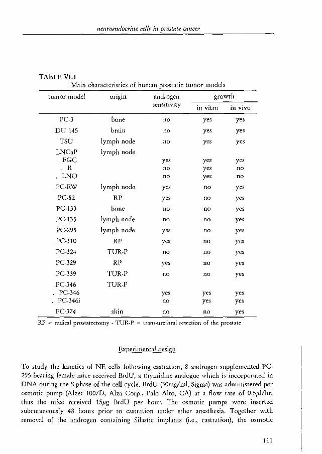
neuroendocrine cells in prostate cancer
TABLE VI.1 Main characteristics of human prostatic tumor models
tumor model ongm androgen growth sensitivity in vitro in vivo
PC-3 bone no yes yes
DU 145 brain no yes yes
TSU lymph node no yes yes
LNCaP lymph node FGC yes yes yes
R no yes no LNO no yes no
PC-EW lymph node yes no yes
PC-82 RP yes no yes
PC-133 bone no no yes
PC-135 lymph node no no yes
PC-295 lymph node yes no yes
PC-3lO RP yes no yes
PC-324 TUR-P no no yes
PC-329 RP yes no yes
PC-339 TUR-P no no yes
PC-346 TUR-P . PC-346 yes yes yes
PC-346i no yes yes
PC-374 skin no no yes
RP = radical prostatectomy - TUR-P = trans-urethral resection of the prostate
EXRerimental design
To study the kinetics of NE cells following castration, 8 androgen supplemented PC-295 bearing female mice received BrdU, a thymidine analogue which is incorporated in DNA during the S-phase of the cell cycle. BrdU (30mg/ml, Sigma) was administered per osmotic pump (Alzet lO07D, Alza Corp., Palo Alto, CAl at a flow rate of O.5l'l/hr, thus the mice received 151'g BrdU per hour. The osmotic pumps were inserted subcutaneously 48 hours prior to castration under ether anesthesia. Together with removal of the androgen containing Silastic implants (i.e., castration), the osmotic
111
chapter VI
pumps were removed and 2 mice were sacrificed either directly (To) or after 3 (T.,) or 6 (T~) days. In two mice androgens were re-substituted 3 days following androgen withdrawal and these mice were sacrificed after another period of 7 days (T.]+,). This method ensured sufficient BrdU incorporation in cells that were cyeling prior to castration. Antibodies against BrdU allow for the detection of cells in the S-phase416
•
Tumor volumes were calculated from two caliper measured perpendicular tumor diameters (D, and DJ according to the formula:
V II (0 X D2)3/2 tumor~6X 1 111
The tumors of these mice were processed as described above. In addition, bowel and skin tissues were embedded in paraffin to assess the efficacy of BrdU-incorporation. The microscopical (Sx) pictures of routine H&E sections of the tumors were digitized. The total tumor-area (Atumor) in these sections was determined using the KS·400 software (Kontron Elektrotechnik, Oberkochen, Germany). The number of NE cells was counted in the total tumor-area present in a particular section. Because prostatic NE cells are considered to be androgen independent, androgen withdrawal in this androgen dependent tumor model will lead to a selective decrease of non-NE tumor cells and thereby to an increased frequency of NE cells. Besides this, castration will have other effects like apoptosis, possibly some necrosis, murine stromal proliferation and swelling of the cells. All these factors will have their effect on total tumor volume and thus, an increased NE cell fraction observed following androgen withdrawal might in fact represent a concentration effect. To account for this potential concentration effect the
number of NE cells (NNE) per tumor area unit was adjusted to the decrease in tumor volume from the time of castration (To) to the time of sacrifice (T.,) according to the formula:
corrected NNE 121
Cell cuI tures
Cells were cultured in RPMI-!640 medium (Gibco BRL, Breda, The Netherlands) supplied with 7.5% fetal calf serum (Hyelone, Logan, Utah), 2mM glutamine (Gibco BRL), SOIU/ml penicillin (Gibco BRL) and SOl'g/ml streptomycin (Gibco BRL) in 5% COl at 37°C. For experiments, cells were cultured in quadriperm culture chambers (Nunc, Roskilde, Denmark) on 3-amino-propyl-trietoxy-silane (APES, Sigma) coated glass slides271
, Experimental media contained dextran charcoal treated, i.e. androgen depleted, serum, either with or without 1O"'M of the not metabolizable synthetic androgen R188! (!7fl-hydroxy-!7a-methyl-estra-4,9,11-trien-3-one, New England
112
neuroendocrine cells in prostate cancer
Nuclear, Boston, MA). After four days the slides were rinsed with phosphate buffered saline (PBS, pH~7.4), air dried, wrapped in aluminum foil and stored at -20°C until use.
Immunohistochemistry
For the identification of NE cells, tissue sections andlor cell cultures of the several tumor models were immunostained with antibodies against CgA (clone LK2Hl0, Organon Technika, Oss, The Netherlands)'" and S-HT (rabbit polyclonal antibody, Eurodiagnostics, Apeldoorn, The Netherlands). If available, the paraffin-embedded patient tumor material from which a tumor model originated was also immunostained.
Frozen cell culture slides were brought to room temperature and were fixed in 4% buffered formalin for 10 minutes at room temperature. Subsequently, the slides were rinsed in PBS and processed for two minutes in 100% methanol and for 4 minutes in 100% acetone, both at -20°C. Paraffin embedded xenografts were cut at SlIm (21'm for double immunostaining) and the tissue sections were mounted on APES-coated glass slides. The slides were kept overnight at 60°C and were dehydrated through xylene and ethanol. Endogenous peroxidase activity was blocked with 3% hydrogen peroxide in methanol for 10 minutes. After rinsing with tap water, the slides were placed in a 10mM citrate buffer (PH 6.0) and antigen retrieval was performed in a microwave oven at 700\'(1 for 3xS minutes17l. The slides were allowed to cool down to room temperature and were rinsed with PBS. For immunohistochemical staining the tissue slides and air dried cell culture slides were placed in a Sequenza immunostaining system (Shandon, Uncorn, United Kingdom). The slides were pre-incubated with normal goat serum (DAKO, Glostrup, Denmark) diluted 1:10 for 15 minutes. The primary antibody was applied for 1 hour and subsequently the slides were incubated for 30 minutes with a 1:400 diluted biotinylated goat anti-mouse or goat anti-rabbit antibody (DAKO) for monoclonal and polyclonal primary antibodies, respectively. Finally, a horseradishperoxidase labeled streptavidin-biotin complex (DAKO) was applied for 30 minutes. The complex was prepared at least 30 minutes prior to use at a dilution of 1:1:200. In between each step, except following the pre-incubation, the slides were rinsed 3x with PBS. All compounds were diluted in PBS. Immunostaining was visualized with lSOmg diaminobenzidine hydrochloride (DAB, Fluka, Neu-Ulm, Germany) in 200ml PBS with 0.08% hydrogen peroxide as substrate. The slides were lightly counter stained with Mayer's hematoxylin, dehydrated and covered.
Double labeling immunohistochemistty was performed on tumors with CgA immuno-reactive cells using monoclonal antibodies against the androgen receptor (clone F39.4, kind gift of Dr. A.O. Brinkmann, Dept. of Endocrinology &. Reproduction, Erasmus University)"', Ki-67 antigen (clone MIB-l, Immunotech, Marseille, France)'" or BrdU (clone IIBS, kind gift of Dr. B. Schutte, Dept. of Pathology, University of Maastricht, The Netheriands)4I6 combined with anti.CgA (clone LK2H10) as the second primary antibody. BrdU/CgA·immunostaining was performed only in the PC-29S time·
113
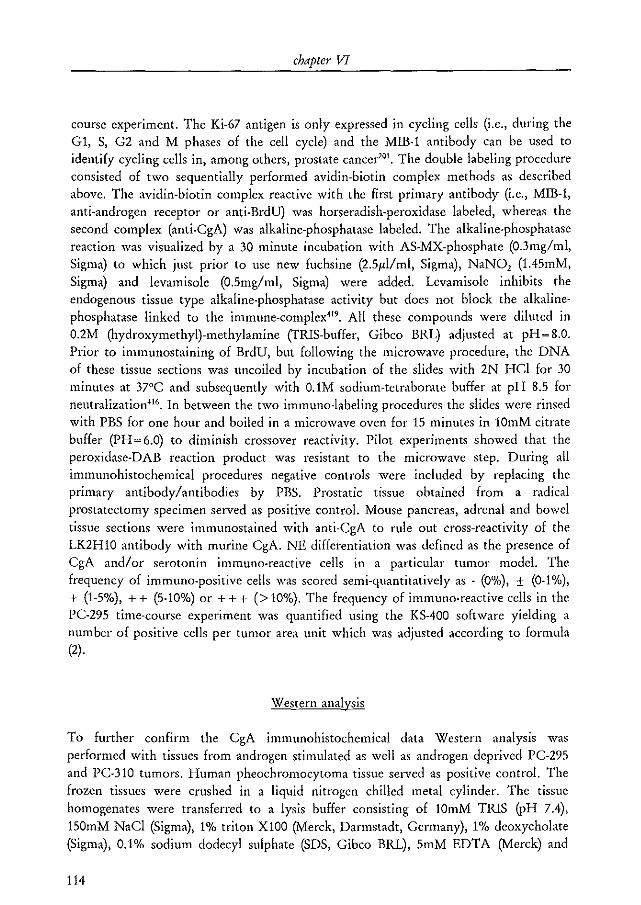
chapter VI
course experiment. The Ki-67 antigen is only expressed in cycling cells (i.e., during the Gl, S, G2 and M phases of the cell cycle) and the MIB-l antibody can be used to identify cycling cells in, among others, prostate cance,"". The double labeling procedure consisted of two sequentially performed avidin-biotin complex methods as described above. The avidin-biotin complex reactive with the first primary antibody (i.e., MIB-l, anti-androgen receptor or anti-BrdU) was horseradish-peroxidase labeled, whereas the second complex (anti-CgA) was alkaline-phosphatase labeled. The alkaline-phosphatase reaction was visualized by a 30 minute incubation with AS-MX-phosphate (0.3mg/ml, Sigma) to which just prior to use new fuchsine (2.5/'lIml, Sigma), NaNO, (1.4SmM, Sigma) and levamisole (O.5mg/ ml, Sigma) were added. Levamisole inhibits the endogenous tissue type alkaline-phosphatase activity but does not block the alkalinephosphatase linked to the immune-complex"'. All these compounds were diluted in 0.2M (hydroxymethyl)-methylamine (TRIS-buffer, Gibco BRL) adjusted at pH~8.0. Prior to immunostaining of BrdU, but following the microwave procedure, the DNA of these tissue sections was uncoiled by incubation of the slides with 2N HCI for 30 minutes at 37"C and subsequently with O.IM sodium-tetraborate buffer at pH 8.5 for neutralization-l16 • In between the two immuno-Iabeling procedures the slides were rinsed with PBS for one hour and boiled in a microwave oven for 15 minutes in 10mM citrate buffer (PH~6.0) to diminish crossover reactivity. pilot experiments showed that the peroxidase-DAB reaction product was resistant to the microwave step. During all immunohistochemical procedures negative controls were included by replacing the primary antibody/antibodies by PBS. Prostatic tissue obtained from a radical prostatectomy specimen served as positive control. Mouse pancreas, adrenal and bowel tissue sections were immunostained with anti-egA to rule out cross-fcactivity of the LK2HI0 antibody with murine CgA. NE differentiation was defined as the presence of egA and/or serotonin immuno-reactive cells in a particular tumor model. The frequency of immuno-positive cells was scored semi-quantitatively as - (0%), ± (0-1%), + (1-5%), + + (5-10%) or + + + (> 10%). The frequency of immuno-reactive cells in the PC-29S time-course experiment was quantified using the KS-400 software yielding a number of positive cells per tumor area unit which was adjusted according to formula (2).
\Y/ estern analysis
To further confirm the CgA immunohistochemical data Western analysis was performed with tissues from androgen stimulated as well as androgen deprived PC-295 and PC-310 tumors. Human pheochromocytoma tissue served as positive control. The frozen tissues were crushed in a liq\1id nitrogen chilled metal cylinder. The tissue homogenates were transferred to a lysis buffer consisting of 10mM TRlS (PH 7.4), 150mM NaCI (Sigma), 1% triton XI00 (Merck, Darmstadt, Germany), 1% deoxycholate (Sigma), 0.1% sodium dodecyl sulphate (SDS, Gibco BRL), SmM EDTA (Merck) and
114
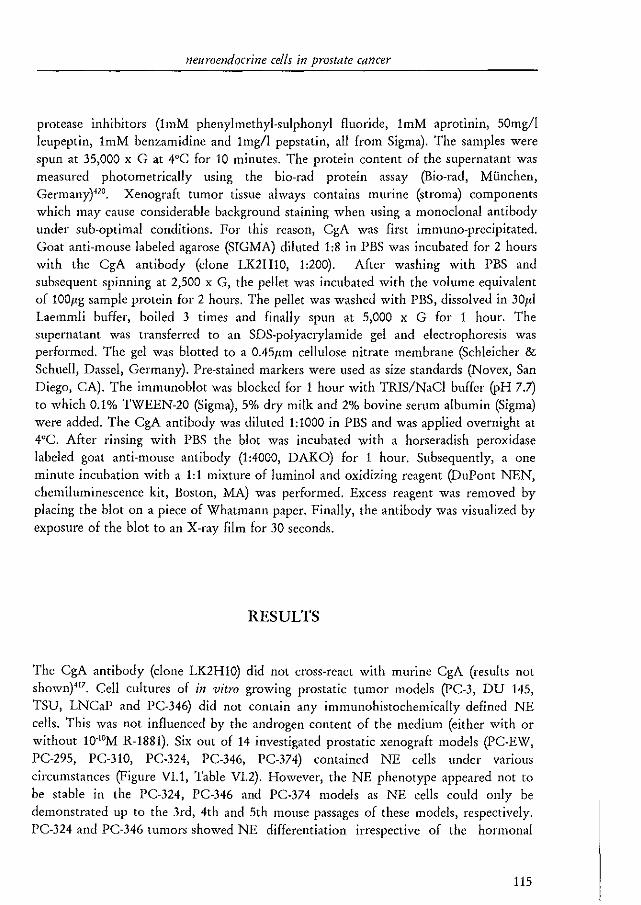
neuroendocrine cells in prostate cancer
protease inhibitors (lmM phenylmethyl-sulphonyl fluoride, lmM aprotmm, 50mg/1 leupeptin, lmM benzamidine and lmg/I pepstatin, all from Sigma). The samples were spun at 35,000 x G at 4"C for 10 minutes. The protein content of the supernatant was measured photometrically using the bio-rad protein assay (Bio-rad, Miinchen, Germany)420. Xenograft tumor tissue always contains murine (stroma) components which may cause considerable background staining when using a monoclonal antibody under sub-optimal conditions. For this reason, egA was first immuno-precipitated. Goat anti-mouse labeled agarose (SIGMA) diluted 1;8 in PBS was incubated for 2 hours with the CgA antibody (clone LK2Hl0, 1;200). After washing with PBS and subsequent spinning at 2,500 x G, the pellet was incubated with the volume equivalent of lOOl'g sample protein for 2 hours. The pellet was washed with PBS, dissolved in 301'1 Laemmli buffer, boiled 3 times and finally spun at 5,000 x G for 1 hour. The supernatant was transferred to an SDS-polyacrylamide gel and electrophoresis was performed. The gel was blotted to a 0.451'm cellulose nitrate membrane (Schleicher &
Schuell, Dassel, Germany). Pre-stained markers were used as size standards (Novex, San Diego, CAl. The immunoblot was blocked for 1 hour with TRIS/NaCI buffer (PH 7.7) to which 0.1% TWEEN-20 (Sigma), 5% dry milk and 2% bovine serum albumin (Sigma) were added. The CgA antibody was diluted 1;1000 in PBS and was applied overnight at 4"C. After rinsing with PBS the blot was incubated with a horseradish peroxidase labeled goat anti-mouse antibody (1;4000, DAKO) for 1 hour. Subsequently, a one minute incubation with a 1; 1 mixture of luminal and oxidizing reagent (DuPont NEN, chemiluminescence kit, Boston, MA) was performed. Excess reagent was removed by placing the blot on a piece of Whatmann paper. Finally, the antibody was visualized by exposure of the blot to an X-ray film for 30 seconcls.
RESULTS
The CgA antibody (clone LK2Hl0) did not cross-react with murine CgA (results not shown)417. Cell cultures of in vitro growing prostatic tumor models (pC,3, DU 145, TSU, LNCaP and PC-346) did not contain any immunohistochemically defined NE cells. This was not influenced by the androgen content of the medium (either with or without 10·IOM R-1881). Six out of 14 investigated prostatic xenograft models (pC-EW, PC-295, PC,310, PC-324, PC-346, PC-374) contained NE cells under various circumstances (Figure VI.1, Table VI.2). However, the NE phenotype appeared not to
be stable in the PC-324, PC-346 and PC-374 models as NE cells could only be demonstrated up to the 3rd, 4th and 5th mouse passages of these models, respectively. PC-324 and PC-346 tumors showed NE differentiation irrespective of the hormonal
115
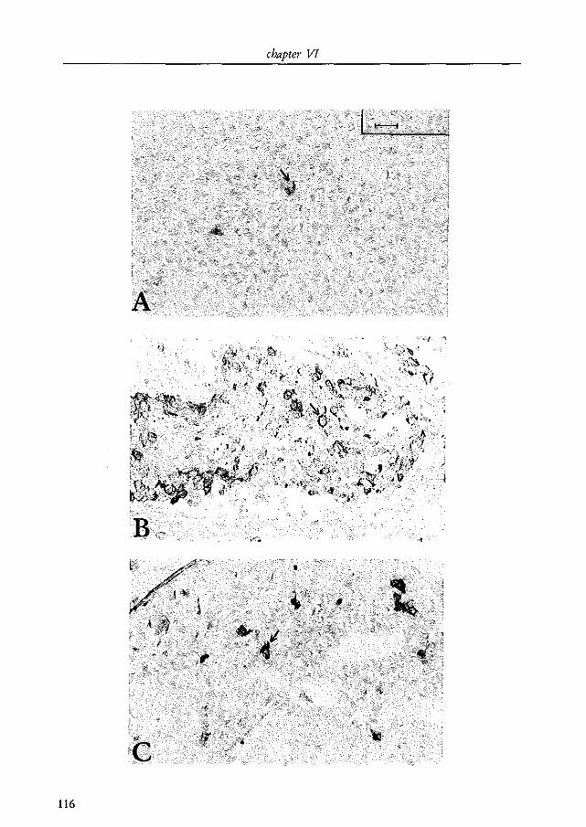
chapter VI
)
116
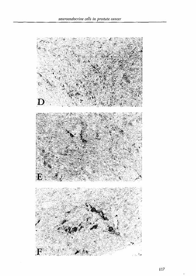
neuroendocrine cel/s in prostate cancer
D
117
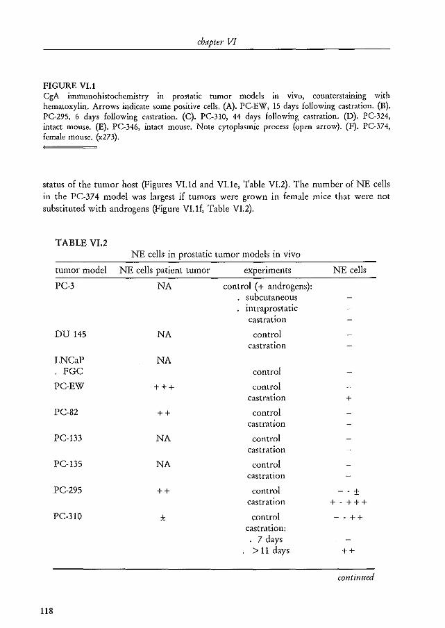
chapter VI
FIGURE VI.! egA iuununohistochemistry in prostatic tumor models in vivo, counterstarnmg with hematoxylin. Arrows indicate some positive cells. (A). PC-E\'7, 15 days following castration. (B). PC-295, 6 days following castration. (C). PC.310, 44 days following castration. (D). PC.324, intact mouse. (E). PC-346, intact mouse. Note cytoplasmic process (open arrow). (F). PG374, female mouse. (x273).
status of the tumor host (Figures vud and VUe, Table VI.2). The number of NE cells in the PC-374 model was largest if tumors were grown in female mice that were not substituted with androgens (Figure VUf, Table VI. 2).
TABLE VI.2 NE cells in prostatic tumor models in vivo
tumor model NE cells patient tumor experiments NE cells
PC-3 NA control (+ androgens): subcutaneous intraprostatic castration
DU 145 NA control castration
LNCaP NA FGC control
PC-EW +++ control castration +
PC-82 ++ control castration
PC-133 NA control castration
PC-135 NA control castration
PC-295 ++ control - - ± castration +-+++
PC-310 ± control --++ castration:
7 days > 11 days ++
continued
118
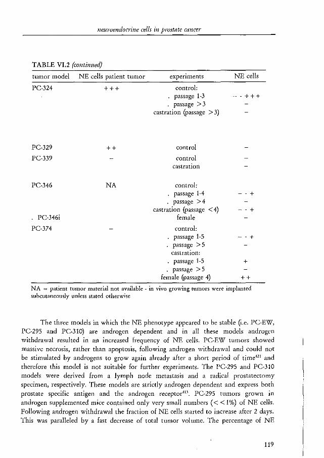
neuroendocrine cells in prostate cancer
TABLE VI.2 (continued)
tumor model NE cells patient tumor experiments
PC-324 + + + control:
PC-329
PC-339
PC-346
. PC-346i
PC-374
++
NA
· passage 1-3 · passage >3
castration (passage > 3)
control
control castration
control: passage 1-4 passage >4
castration (passage < 4) female
control: passage 1-5 passage> 5 castration:
· passage 1-5 · passage> 5
female (passage 4)
NE cells
--+++
- - +
- - +
--+
+
++
NA = patient tumor material not available - in vivo growing tumors were implanted subcutaneously unless stated otherwise
The three models in which the NE phenotype appeared to be stable (i.e. PC-EW, PC-295 and PC-310) are androgen dependent and in all these models androgen withdrawal resulted in an increased frequency of NE cells. PC-EW tumors showed massive necrosis, rather than apoptosis, following androgen withdrawal and could not be stimulated by androgens to grow again already after a short period of time'" and therefore this model is not suitable for further experiments. The PC-295 and PC-310 models were derived from a lymph node metastasis and a radical prostatectomy specimen, respectively. These models are strictly androgen dependent and express both prostate specific antigen and the androgen receptor4D , PC-295 tumors grown in androgen supplemented mice contained only very small numbers « < 1%) of NE cells. Following androgen withdrawal the fraction of NE cells started to increase after 2 days. This was paralleled by a fast decrease of total tumor volume. The percentage of NE
119
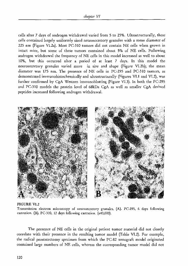
chapter VI
cells after 7 days of androgen withdrawal varied from 5 to 25%. Ultrastructurally, these cells contained largely uniformly sized neurosecretOlY granules with a mean diameter of 225 nm (Figure VI.2a). Most PC-310 tumors did not contain NE cells when grown in intact mice, but some of these tumors contained about 5% of NE cells. Following androgen withdrawal the frequency of NE cells in this model increased as well to about 10%, but this occurred after a period of at least 7 days. In this model the neurosecretory granules varied more in size and shape (Figure VL2b); the mean diameter was 175 nm. The presence of NE cells in PC-295 and PC-310 tumors, as demonstrated immunohistochemically and ultrastructurally (Figures VI.1 and VI.2), was further confirmed by CgA Western immunoblotting (Figure VI.3). In both the PC-295 and PC-310 models the protein level of 68kDa CgA as well as smaller CgA derived peptides increased following androgen withdrawal.
FIGURE VI.2 Transmission electron microscopy of neurosecretory granules. (A). PC·295, 6 days following castration. (B). PC-310, 12 days following castration. (x40,OOO).
The presence of NE cells in the original patient tumor material did not closely correlate with their presence in the resulting tumor model (Table VI.2). For example, the radical prostatectomy specimen from which the PC-82 xenograft model originated contained large numbers of NE cells, whereas the corresponding tumor model did not
120
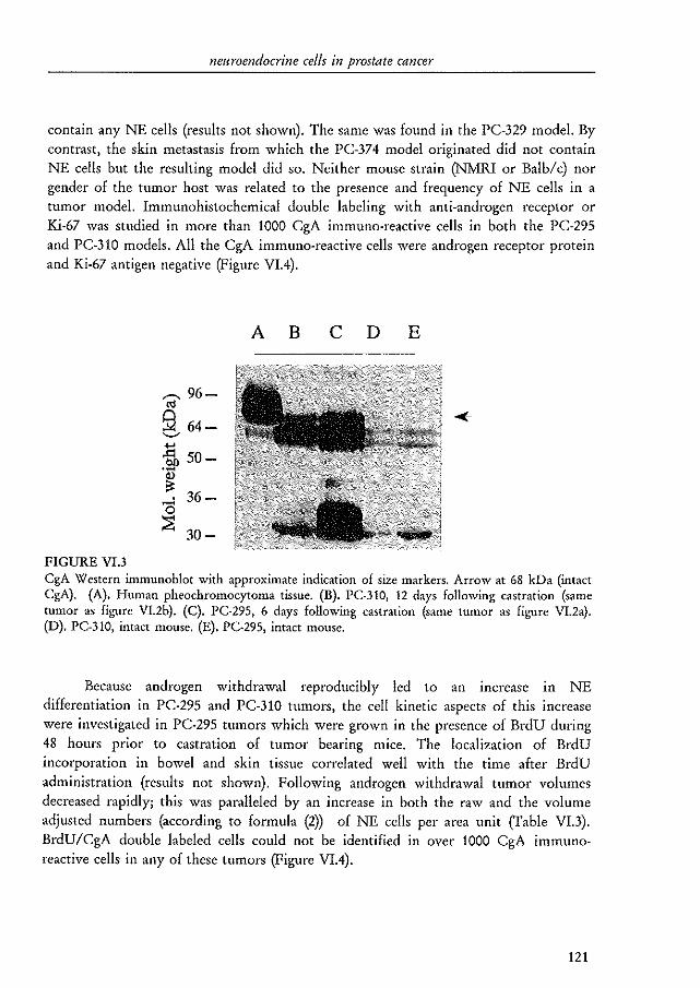
neuroendocrine cells in prostate cancer
contain any NE cells (results not shown). The same was found in the PC-329 model. By contrast, the skin metastasis from which the PC-374 model originated did not contain NE cells but the resulting model did so. Neither mouse strain (NMRI or Balb/c) nor gender of the tumor host was related to the presence and frequency of NE cells in a tumor model. Immunohistochemical double labeling with anti-androgen receptor or Ki·67 was studied in more than 1000 CgA immuno-reactive cells in both the PC-295 and PC-310 models. All the CgA immuno-reactive cells were androgen receptor protein and Ki-67 antigen negative (Figure VIA).
FIGURE VI.3
..--. 96-'" 5:l 64-'--'
i150-.~
. 36-
~ 30-
A B c D E
egA \"1 estern immunoblot with approximate indication of size markers, Arrow at 68 kDa (intact CgA), (A). Human pheochromocytoma tissue, (B). PC-310J 12 days following castration (same tumor as figure V12b). (C). PC-295, 6 days following castration (same tumor as figure VL2a). (D). PC-31O, intact mouse, (E). PC-295, intact mouse.
Because androgen withdrawal reproducibly led to an increase 1ll NE differentiation in PC-295 and PC-310 tumors, the cell kinetic aspects of this increase were investigated in PC-295 tumors which were grown in the presence of BrdU during 48 hours prior to castration of tumor bearing mice. The localization of BrdU incorporation in bowel and skin tissue correlated well with the time after BrdU administration (results not shown). Following androgen withdrawal tumor volumes decreased rapidly; this was paralleled by an increase in both the raw and the volume adjusted numbers (according to formula (2» of NE cells per area unit (Table VI.3). BrdU/CgA double labeled cells could not be identified in over 1000 CgA immunoreactive cells in any of these tumors (Figure VIA).
121
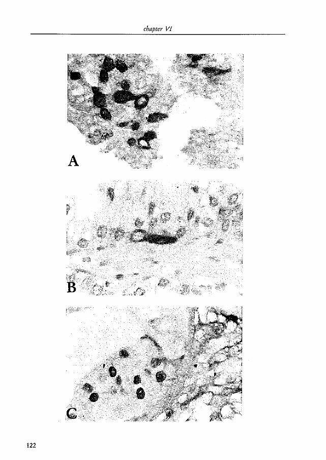
chapter VI
A
122
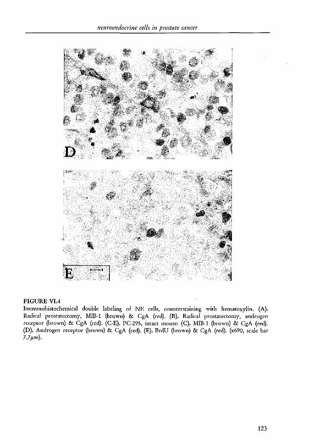
neuroendocrine cells in prostate cancer
FIGURE VI.4 Immunohistochemical double labeling of NE cells, counterstallllllg with hematoxylin. (A). Radical prostatectomy, MIB-! (brown) & CgA (red). (B). Radical prostatectomy, androgen receptor (brown) & CgA (red). (C·E). PC-295, intact mouse: (C). MIB-! (brown) & CgA (red). (D). Androgen receptor (brown) & CgA (red). (E). BrdU (brown) & CgA (red). (x690, scale bar 7.711m).
123
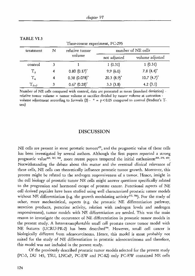
chapter VT
TABLE VI.3 Time-course experiment, PC-295
treatment N relative tumor number of NE cells volume not adjusted volume adjusted
control 3 1 1 (0.31) 1 (0.31)
T.) 4 0.80 (0.17)' 9.9 (6.0) 7.8 (4.4)'
T. 4 0.38 (0.078)' 20.3 (8.9)' 10.7 (4.7)'
T.3+7 3 0.67 (0.28)' 3.3 (3.8) 4.2 (3.1)
Number of NE cells compared with control, data are presented as mean (standard deviation) -relative tumor volume = tumor volume at sacrifice divided by tumor volume at castration -volume adjustment according to formula (2) - ,~ = p<O.OS compared to control (Student's Ttest)
DISCUSSION
NE cells are present in most prostatic tumors40\ and the prognostic value of these cells
has been investigated by several authors. Although the first papers reported a strong prognostic valuel64, 167, 268} more recent papers tempered the initial enthusiasm169, 270, 403,
Notwithstanding the debate about this matter and the eventual clinical relevance of these cells, NE cells can theoretically influence prostatic tumor growth. Moreover, this process might be related to the androgen responsiveness of a tumor. Hence, insight in the cell biology of prostatic tumor NE cells might answer questions specifically related to the progression and hormonal escape of prostate cancer. Functional aspects of NE cell derived pep tides have been studied using well characterized prostatic tumor models without NE differentiation (e.g. the growth modulating activityl71· 386). For the study of other, more mechanistical, aspects (e.g. the prostatic NE differentiation pathway, secretion products, paracrine activity, relation with androgen levels and androgen responsiveness), tumor models with NE differentiation are needed. This was the main reason to investigate the occurrence of NE differentiation in prostatic tumor models in the present study. A heterotransplantable small cell prostate cancer tumor model with NE features (UCRU·PR.2) has been described)". However, small cell cancer is biologically different from adenocarcinoma. Hence, this model is most probably not suited for the study of NE differentiation in prostatic adenocarcinoma and therefore, this model was not included in the present study.
Of the previously described prostatic tumor models selected for the present study (PC-3, DU 145, TSU, LNCaP, PC-EW and PC-82) only PC-EW contained NE cells
124
neuroendocrine cells in prostate cancer
(Table VL2). Although not extensively described, it was claimed that the LNCaP and PC-3 cell lines also would contain NE cells in vitro411 • We were not able to confirm these results even not when the immunostaining protocol of these investigators was used. De BruIne et al. have shown that NE cells were always present when a colon cancer cell line (NCI-H716) was grown in nude mice, but that NE cells could be detected in vitro only when the cells were cultured in matrigel or with certain extracellular matrix components4l3 . This suggests that stromal-epithelial interactions are important for the induction or maintenance of NE differentiation. PC-3 and LNCaP tumors grown in vivo, either subcutaneously (LNCaP) or both subcutaneously and orthotopically (PC-3), did not contain NE cells in the present study (Table VL2), substantiating the observation that these models indeed do not show NE differentiation. A number (5 out of 7) of human prostatic tumor xenograft modelsHl contained NE cells under different hormonal circumstances. As reported4l7, the employed LK2H10 antibody did not react with murine CgA in the present study (results not shown) and thus, all CgA immuno-reactive (i.e., NE) cells were assumed to be of human origin. Furthermore, CgA Western immunoblotting identified the presence of intact CgA (68 kDa) as well as proteolytic break down products in both the PC-295 and PC-310 models (Figure VI.3)417. Finally, the presence of NE cells was confirmed ultrastructurally by the identification of neurosecretOlY granules (Figure VI.2). These granules are considered conclusive evidence for NE cells404 • The expression of several break down products of CgA varies among tissues"" which may explain the differences between pheochromocytoma and prostatic tissues as shown in Figure VI.3. The 94 kDa band present in pheochromocytoma tissue may very well represent pro-CgA"'. CgA expression has been demonstrated in non·NE cells e.g., in rat alveolar type II cells"5 and in the submandibular salivary glands of mice"'. In the former study, it was hypothesized that CgA might play a role as a Ca'+ .binding protein, in the latter study the CgA immuno·reactive cells were not well characterized. It cannot be excluded that in some of the tumor models presently studied CgA was expressed in exocrine cells (for example the more diffuse staining pattern visible in the PC-324 tumor model). The NE phenotype of the more important PC-295 and PC-31O tumor models was confirmed by transmission electron microscopy (Figure VL2).
As shown in Table VI.2, the NE phenotype is lost in ongoing mouse passages of the PC·324, PC-346 and PC-374 models. It is now generally assumed that prostatic NE cells are post-mitotic and are derived from locally differentiated non-NE cells"'. Possibly, the NE cells from the original patient tumor were still present in the initial mouse passages of the PC-324 and PC·346 models, but gradually disappeared. In contrast, the patient tumor of the PC-374 model did not contain NE cells, whereas the initial mouse passages did, however. This suggests that environmental factors played a temporal role in the appearance of NE differentiation in this model. The selection of non-NE tumor cells with time may be explained by the presence of NE inhibitory factors or by the absence of essential NE induction or maintenance factors. This may be caused by the tumor itself, the host stroma or by a combination of these (stroma-
125
chapter Vl
epithelial interactions). Another explanation might be that the tumor models loose the capacity of NE differentiation caused by de-differentiation due to long-term in vivo propagation. The fact that normal prostatic tissue always contains NE cells and that the majority of tumors, irrespective of their glandular differentiation pattern, do so as well"" strongly suggests that differentiation into the NE phenotype is an intrinsic property of prostatic cells. This is also illustrated by the presence of NE cells in the PC-374 model, in contrast to the patient material. Likewise, Bang et al. were able to induce terminal NE differentiation in LNCaP and in a highly metastatic sub-line of PC-3 by treatment of cell cultures with cyclic-AMP analogs or with phospho-diesterase inhibitors"'. It should be noted that, contrary to the findings of the present study, these investigators also found NE features in untreated cell cultures. The possibility that the PC-295 and PC-310 models Qike PC-324, PC-346 and PC-374) will loose the NE phenotype in the future cannot be ruled out at this moment. Which factors are important for the induction, maintenance or disappearance of NE differentiation in prostatic tumor models will be the subject of future studies.
In the present study, some initial experiments were performed to compare the characteristics of the NE cells found in the tumor models with known characteristics of these cells in prostatic tissue. Because the NE phenotype was found to be stable in the PC-295 and PC-310 models, only these models were studied. Similar to the findings in clinical prostate cancer, the NE cells present in the tumor models (more than 1000 cells studied), whether grown in intact or androgen deprived mice, did express neither androgen receptor protein nor Ki-67 antigen (Figure VIA). Androgen supplemented PC-295 and PC-310 tumors expressed the androgen receptor and Ki-67 in about 80% and 10% of the tumor cells, respectively. The probability that all the NE cells (albeit present in small numbers) in these tumors would be negative for both antigens by chance is extremely small (P < <0.0001). Analogous to the NE cells in benign illld malignant prostatic tissues, these observations confirm the post-mitotic and androgen independent nature of prostatic NE cells. The observation by Bonkhoff et al.'" that in human prostatic tumors with clusters of NE cells the Ki-67 immun~-reactive cells were located in close proximity to those clusters was not confirmed in the present study. However, proliferation decreased following castration in these androgen dependent models, at least at the short~term. Long-term castration experiments will show whether the NE cells can overcome the growth inhibiting effects of androgen withdrawal by paracrine growth stimulation and hence, playa role in androgen independent growth of prostate cancer.
Abrahamsson et al. studied the course of NE differentiation in hormonally treated patients and found in general that the number of NE cells increased when the tumor progressed to androgen independence'''. Civantos et al. investigated the effects of neo-adjuvant androgen withdrawal therapy in patients treated by a radical prostatectomy and compared the data with non pre-treated prostatectomy specimens428
.
More prostatectomy specimens of pre-treated patients contained NE cells, although the results were not statistically significant. These findings are similar to our observations of
126
neuroendocrine cells in prostate cancer
increased numbers of NE cells and increased CgA protein levels in short-term androgen deprived PC-EW, PC-295 and PC-310 tumors and implicates that either directly or indirectly androgen regulated genes play a role in the prostatic NE differentiation pathway. Interestingly, it has been shown that CgA expression is inversely correlated with estrogen levels in the female rat pituitary (estrogen receptor positive), but not in the adrenal (estrogen receptor negative)"" 430. Although pituitary and adrenal cells belong to the endocrine system, these findings also point to a relationship between steroids and NE differentiation.
Whether or not prostatic NE cells already are post-mitotic before the NE phenotype appears is not clear and therefore a time-course experiment with PC-295 tumor bearing mice, which had received BrdU for 48 hours, was performed. The raw and volume adjusted number of NE cells per area unit of tumor tissue increased following androgen withdrawal (Table V1.3). This strongly indicates that both a relative and an absolute increase in the number of NE cells is combined with a selective deletion of androgen dependent non-NE tumor cells (the major cause of the total tumor volume decrease). Thus, the selection of pre-existing NE cells after androgen withdrawal alone does not account for the observed increase in NE cells. Forty-eight hours of BrdU labeling ensured that all cells that were cycling prior to castration were labeled. The fact that no BrdU-labeling was found in over 1000 CgA immuno-reactive cells (Figure VIA), combined with the fact that proliferation falls to a very low level shortly after castration, indicates that NE differentiation of prostatic tumor cells only takes place in cells that are in the Go-phase already for a longer period of time. Since CgA is only expressed by fully NE differentiated cells, another post-mitotic cell type might exist, with a phenotype somewhere in between the exocrine and NE phenotypes which gives rise to the NE cells appearing following castration. Testing of such a hypothesis awaits the generation of immunohistochemical or molecular-biological markers for 'early' NE cells, prior to their expression of egA.
In conclusion, NE cells can be identified in human prostatic tumor xenograft models, but the phenotype is not always stable. In the PC-295 and PC-310 tumor models NE differentiation can be induced reliably by castration of tumor bearing mice. The NE cells found in these models are most likely androgen independent for their persistence, do not express the androgen receptor and are post-mitotic. These findings are concordant with NE cells found in clinical prostate cancer. The cells that became NE differentiated following androgen withdrawal were most probably post-mitotic already before the NE phenotype was induced. These models are suitable to study fundamental issues concerning the presence and function of NE cells in prostate cancer.
127

1
1
1
1
1
1
1
1
1
1
1
1
1
1
1
1
1
1
1
1
1
1
1
1
1
1
1
1
1
1
1
1
1
1
CHAPTER VII
DETERMINATION OF THE KI-67 DEFINED GROWTH FRACTION BY THE
MONOCLONAL ANTIBODY MIB-l IN FORMALIN-FIXED, PARAFFIN
EMBEDDED PROSTATIC CANCER TISSUES
M.A. Noordzij; T.H. van der Kwast; G.J. Steenbrugge; W.M. van Weerden; M.H.A. Oomen; Fritz H. Schroder
From the departments of Urology and Pathology, Erasmus University Rotterdam, The Netherlands.
Prostate 1995;27: 154-9 reprinted with permission
chapter VII
ABSTRACT
The applicability of MIB-!, a monoclonal antibody directed against the Ki-67 antigen was studied in the PC-82 and LNCaP prostatic tumor models at various levels of proliferative activity_ Statistically significant correlations were found in LNCaP cultures between Ki-67 and MIB-! scores (r = 0.84, p<O.OO!) and in PC-82 tumors between MIB-! scores and paraffin tissue Ki-67 (PKi-67),(r=0.90, p<O.OO!), frozen tissue Ki-67 (fKi-67), (r=0.86, p<O.OO!), and BrdU uptake (r=0.70, p<O.OO!), respectively. pKi-67 scores doubled the fKi-67 scores which may be due to methodological differences. MIB-! scores exceeded both the fKi-67 and pKi-67 scores. The affinity of MIB-! for the antigen is much higher than the affinity of Ki-67 which may explain the differences. MIB-! is a promising means of evaluating the presence of only minute amounts the Ki-67 antigen in paraffin embedded human tumor material, especially of relatively slowly growing tumors.
INTRODUCTION
Since its description in !983 much attention has been paid to the mouse monoclonal antibody Ki-67!81_ Ki-67 is directed against a nuclear antigen expressed in human cells during the G!, $, G2 and M phases of the cell cycle, but not during the GO phase"'_ Quiescent (GO) cells which enter the cell cycle do not express the antigen in the early G! phase"!_ The proliferative activity of a number of human tumors including prostatic carcinoma has been studied by the application of Ki-67, as reviewed by Brown and Gatter",,431_ Routine studies were hampered by the fact that Ki-67 was thought to be applicable only to fresh frozen tissues_ MIB-! is a recently described mouse monoclonal antibody raised against recombinant parts of the Ki-67 antigen, which is applicable to routinely processed, paraffin embedded tissues following antigen retrieval27J, 418_ It has recently been shown that Ki-67 also reacts with paraffin tissue sections using this technique433
_ DNA replicating cells can be identified by a monoclonal antibody to BrdU (bromodeoxy-uridine), a thymidine analogue incorporated into DNA in the $ phase of the cell cycleH
'_ BrdU-uptake has been studied in human tumors'''·''_ The present study was performed to investigate the applicability of MIB-! to routinely processed prostate cancer speCImens.
Two human prostatic tumor models were used in the present study: the PC-82 xenograft model which is serially transplantable in athymic nude mice"', and the in
130
proliferative activity of tIImors
vitro LNCaP cell line"'. The growth rate of both hormone dependent tumor models can be manipulated by growing the tumors at various androgen concentrations410, 438.
The potency of using the Ki-67 antibody for monitoring hormonal responses in the PC-S2 tumor has been described previously"'.
In the present study Mill-1 scores were correlated with fKi·67 (frozen tissue Ki-67), pKi-67 (paraffin tissue Ki·67) and BrdU scores. This was achieved by application of the antibodies to frozen or paraffin tissue sections from the same PC-S2 tumor or to slides with acetone fixed LNCaP cells. Hormonal manipulation of the PC-S2 tumors and LNCaP cells resulted in tissues at various levels of proliferative activity. A number of radical prostatectomy specimens embedded 6 to 16 years ago was stained with Mill·1 in order to investigate the applicability of Mill·1 to archival clinical tissues.
MATERIAL AND METHODS
Experimental models
PC-S2 tumor cells were subcutaneously implanted in nude mice of the Balblc background. Tumor growth was manipulated by substitution of tumor bearing mice with various androgen levels and could be arrested by androgen withdrawal4l8• BrdU (10 mg/kg) was injected intraperitoneally one hour prior to sacrifice of the host animal. After sacrifice of the animal, tumor sections were either snap frozen in liquid nitrogen chilled isopentanyl 99% and stored at -so 'c or routinely fixed in 4% formalin in PBS (phosphate buffered saline, pH~7.4) and embedded in paraffin. Routinely processed prostatectomy specimens embedded 6 to 16 years ago were obtained from the department of pathology. To avoid the risk of detachment of cells or tissues from the glass slides, APES (3-aminopropyltriethoxysilane) coated slides were used in all experiments27l
,
LNCaP-FGC (fast growing colony) cells were seeded on APES coated slides. The cells were grown in 7.5% fetal calf serum at standard conditions as described e1sewhere414
. The culture medium from exponentially growing cultures was replaced (after 2 days) by 5% DCC (dextran-coated charcoal) treated, i.e. androgen depleted, serum. Slides were fixed in acetone for 10 minutes at 0, 24, 48, 72 and 96 hours following androgen withdrawal. The LNCaP experiments were performed in triplicate.
131
chapter VII
Frozen-tissue-Ki-67 and BrdU staining
The Ki-67 and BrdU staining procedures have been described previously'''' "', Briefly, monoclonal antibody Ki-67 (DAKO, Denmark) and a monoclonal antibody specific for BrdU (Eurodiagnostics, The Netherlands) were applied to 51'm thick frozen and paraffin sections, respectively, Ki-67 was diluted 1:5 in PBS, The antibody-antigen binding was visualized using an indirect two-steps peroxidase method with DAB (3,3'diaminobenzidine tetrahydrochloride) as chromogen, Prior to application of the BrdU antibody, these sections were subsequently placed in 2N Hcl for 30 minutes to uncoil the DNA and in a borate buffer at pH 8,5 for neutralization"',
Paraffin tissue Ki-67 (pKi-67) and MIB-l staining
The procedure used for MIB-l staining is the result of the optimization of the procedure recommended by the manufacturer. An identical procedure was used for pKi-67 staining except for the primalY antibody, Paraffin PC-82 and prostatectomy sections of 5 I'm thickness were cut and mounted on APES-coated slides"', The slides were kept overnight at 60 °C, After rehydration, the slides were placed in 10 mM citrate buffer adjusted at pH 6,0, whereafter antigen retrieval was performed in a microwave oven at 700\'il for 15 minutes27J, Distilled water was regularly added to
prevent drying of the slides due to evaporation, The slides were allowed to cool down to room temperature and were subsequently rinsed in PBS. Antigen retrieval was omitted for the acetone-fixed LNCaP cells, To avoid non-specific staining both tissue sections and LNCaP cultures were pre-incubated for 15 minutes at 37 DC with normal goat serum (DAKO, Denmark) diluted 1:10 in PBS, Overnight incubation with MIB-l (lmmunotech, France) diluted 1:200 in PBS or with Ki-67 (DAKO, Denmark) diluted 1:10 in PBS was carried out at 4°C. After rinsing in PBS, the sections were incubated for 30 minutes at room temperature with a 1:400 dilution of biotinylated goat antimouse serum (DAKO) in PBS containing 3% normal goat and 3% normal human serum, After rinsing in PBS a final incubation with avidin biotin complex (ABC) consisting of streptavidin and biotinylated horseradish peroxidase diluted in PBS, as described by the manufacturer (DAKO) was performed for 30 minutes at room temperature, The staining was visualized with 0,075% DAB (Fluka, Germany) in PBS with 0.08% hydrogen peroxide as substrate for 7 minutes. Sections were rinsed in distilled water and counterstained with Mayer's hematoxylin, After dehydration, sections were mounted with malinol.
132
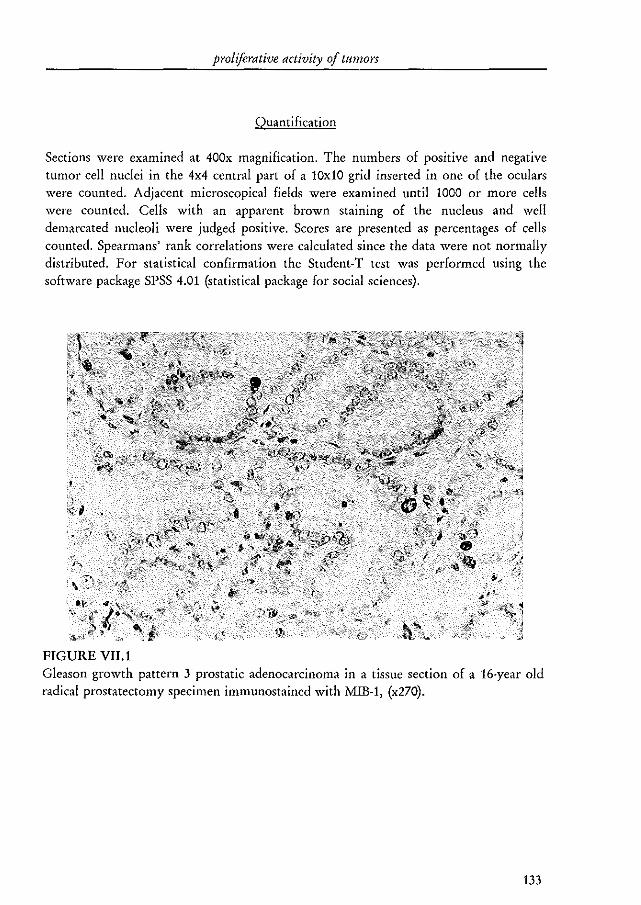
proliferative activity 0/ tltmors
Quantification
Sections were examined at 400x magnification. The numbers of positive and negative tumor cell nuclei in the 4x4 central part of a 10x10 grid inserted in one of the oculars were counted. Adjacent microscopical fields were examined until 1000 or more cells were counted. Cells with an apparent brown staining of the nucleus and well demarcated nucleoli were judged positive. Scores are presented as percentages of cells counted. Spearmans' rank correlations were calculated since the data were not normally distributed. For statistical confirmation the Student-T test was performed using the software package SPSS 4.01 (statistical package for social sciences).
FIGURE VII. 1
,:;:1"fjJ" ~ '~',
\,~
Gleason growth pattern 3 prostatic adenocarcinoma in a tissue section of a 16·year old radical prostatectomy specimen immunostained with MIB-1, (x270).
133
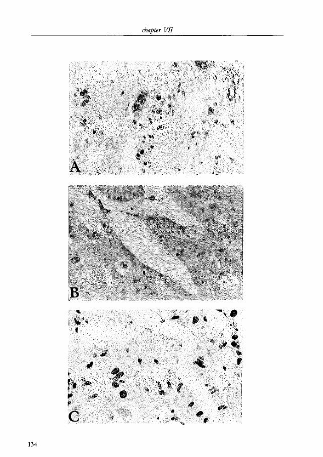
134
~ iflIIJ'~
"til' ~
chapter VII
•
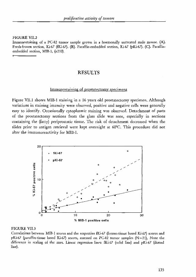
proliferative activity of tumors
FIGURE VII.2 Immunostaining of a PC-82 tumor sample grown in a hormonally untreated male mouse. (A). Fresh-frozen section, Ki-67 (fKi-67). (B). Paraffin-embedded section, Ki-67 (PKi-67). (C). Paraffinembedded section, MIB-!, (x2!O).
RESULTS
Immunostaining of prostatectomy specimens
Figure VII.! shows MIB-! staining in a 16 years old prostatectomy specimen. Although variations in staining intensity were observed, positive and negative cells were generally easy to identify. Occasionally cytoplasmic staining was observed. Detachment of parts of the prostatectomy sections from the glass slide was seen, especially in sections containing the (fatty) periprostatic tissue. The risk of detachment decreased when the slides prior to antigen retrieval were kept overnight at 60'C. This procedure did not alter the immunoreactivity for MIB-1.
20 0
fKI·67
pKI-67 /
.!2 0 /
/ .. 0 / 0 /
• / > 0 / 0 . ., / •• 10 0
/
0 / 00 <>
'" / 0
<- 0 / 0 '\' /
Q / • 0
/ • / 0
'" / • / 0 • • °0 ...... ./ / • •
~ .oCI' '0.
0 • • 0 10 20 30
% MIB-1 positive cells
FIGURE VII.3 Correlations between MID-1 scores and the respective fKi-67 (frozeIHissue based Ki-67) scores and pKi-67 (paraffin-tissue based Ki-67) scores, assessed on PC-82 nllllOr samples (N=21). Note the difference in scaling of the axes. Linear regression lines: fKi-67 (solid line) and pKi-67 (dotted line).
135
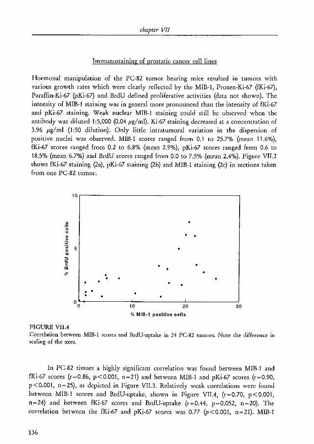
chapter VII
Immunostaining of prostatic cancer cell lines
Hormonal manipulation of the PC-82 tumor bearing mice resulted in tumors with various growth rates which were clearly reflected by the MlB-1, Frozen-Ki-67 (fKi-67), Paraffin-Ki-67 (PKi-67) and BrdU defined proliferative activities (data not shown). The intensity of MIB-1 staining was in general more pronounced than the intensity of fKi-67 and pKi-67 staining. Weak nuclear MlB-1 staining could still be observed when the antibody was diluted 1:5,000 (0.04 Jig/ml). Ki-67 staining decreased at a concentration of 3.96 Jig/ml (1:50 dilution). Only little intratumoral variation in the dispersion of positive nuclei was observed. MIB-1 scores ranged from 0.1 to 25.7% (mean 11.6%), fKi-67 scores ranged from 0.2 to 6.8% (mean 2.9%), pKi-67 scores ranged from 0.6 to 18.5% (mean 6.7%) and BrdU scores ranged from 0.0 to 7.5% (mean 2.4%). Figure VII.2 shows fKi-67 staining (2a), pKi-67 staining (2b) and MIB-l staining (2c) in sections taken from one PC-82 tumor.
10r------------------------------------,
.!! • .. u
• • • ~ •• S • 0 0.
:::l 'l! • • en • .. • • • • • • • • •
• • • • • • 0
0 10 20 30
% MIB·1 positive cells
FIGURE VIlA Correlation between MIB-l scores and BrdU-uptake in 24 PC-82 tumors. Note the difference in scaling of the axes.
In PC-82 tissues a highly significant correlation was found between MIB-l and fKi-67 scores (r~0.86, p <0.001, n~21) and between MIB-l and pKi-67 scores (r~0.90, p<O.OOl, n~25), as depicted in Figure VII.3. Relatively weak correlations were found between MlB-1 scores and BrdU-uptake, shown in Figure VIlA, (r~0.70, p<O.OOl, n~24) and between fKi-67 scores and BrdU-uptake (r~OA4, p~0.052, n~20). The correlation between the fKi-67 and pKi-67 scores was 0.77 (P <0.001, n~21). MIB-1
136
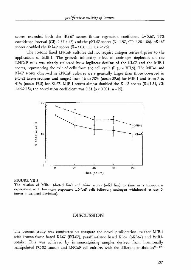
proliferative activity of tumors
scores exceeded both the fKi·67 scores Oinear regression coefficient fl~3.67, 95% confidence interval (Cl): 2.87-4.47) and the pKi·67 scores (fl~ 1.57, CI: 1.28-1.86). pKi.67 scores doubled the fKi-67 scores (fl~2.03, CI: 1.31-2.75).
The acetone fixed LNCaP cultures did not require antigen retrieval prior to the application of MIB·1. The growth inhibiting effect of androgen depletion on the LNCaP cells was clearly reflected by a loglinear decline of the Ki·67 and the MIB·l scores, representing the exit of cells from the cell cycle (Figure VII,5). The MIB·l and Ki·67 scores observed in LNCaP cultures were generally larger than those observed in PC-82 tissue sections and ranged from 16 to 70% (mean 39.6) for MIB·l and from 7 to 41% (mean 19.8) for Ki·67. MIB·l scores almost doubled the Ki·67 scores (fl~1.81, CI: 1.44·2.18), the correlation coefficient was 0.84 (p <0.001, n~ 15).
.!! .. u
• > :;; .;; 0 "-
'"
FIGURE VII.S
100r-----------------------------------------,
10
o 24 48
Time (hours)
72 96
The relation of MIB-l (dotted line) and Ki-67 scores (solid line) to time in a time-course experiment with hormone responsive LNCaP cells following androgen withdrawal at day 0, (mean ± standard deviation).
DISCUSSION
The present study was conducted to compare the novel proliferation marker MIB·! with frozen·tissue based Ki·67 (fKi·67), paraffin·tissue based Ki·67 (pKi·67) and BrdUuptake. This was achieved by immunostaining samples derived from hormonally manipulated PC-82 tumors and LNCaP cell cultures with the different antibodies l8l• '16.
137
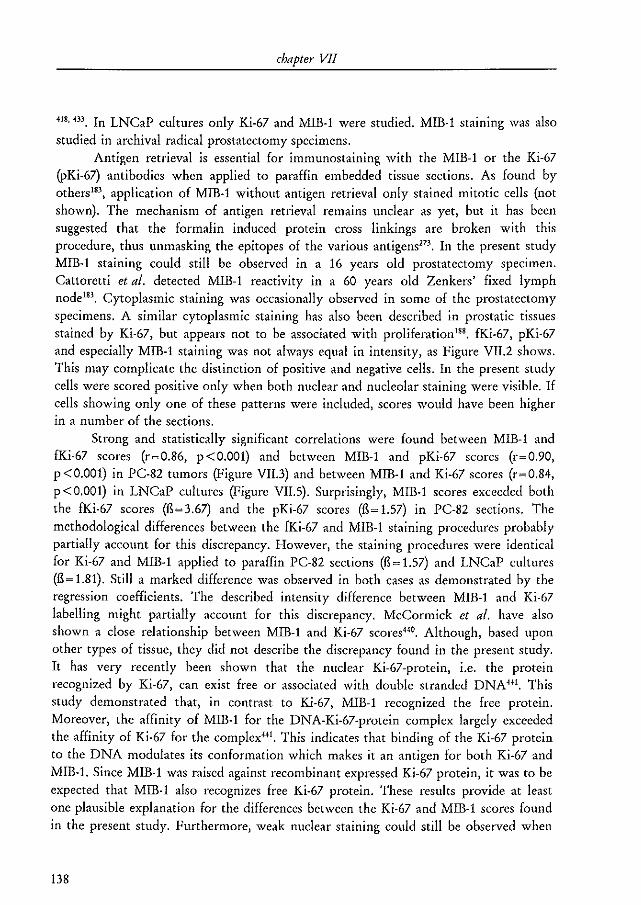
chapter VJI
418.433. In LNCaP cultures only Ki-67 and MIB-l were studied. MIB-l staining was also studied in archival radical prostatectomy specimens.
Antigen retrieval is essential for immunostaining with the MIB-l or the Ki-67 (PKi-67) antibodies when applied to paraffin embedded tissue sections. As found by others l83
, application of MIB-l without antigen retrieval only stained mitotic cells (not shown). The mechanism of antigen retrieval remains unclear as yet, but it has been suggested that the formalin induced protein cross linkings are broken with this procedure, thus unmasking the epitopes of the various antigens273 . In the present study MIB-l staining could still be observed in a 16 years old prostatectomy specimen. Cattoretti et al. detected MIB-l reactivity in a 60 years old Zenkers' fixed lymph node18J. Cytoplasmic staining was occasionally observed in some of the prostatectomy specimens. A similar cytoplasmic staining has also been described in prostatic tissues stained by Ki-67, but appears not to be associated with proliferationlS8 • fKi-67, pKi-67 and especially MIB-l staining was not always equal in intensity, as Figure VIL2 shows. This may complicate the distinction of positive and negative cells. In the present study cells were scored positive only when both nuclear and nucleolar staining were visible. If cells showing only one of these patterns were included, scores would have been higher in a number of the sections.
Strong and statistically significant correlations were found between MIB-l and fKi-67 scores (r~0_86, p<O_OOl) and between MIB-l and pKi-67 scores (r~0.90,
p <0_001) in PC-82 tumors (Figure VII.3) and between MIB-l and Ki-67 scores (r~0.84, p<O.OOl) in LNCaP cultures (Figure VII.5)_ Surprisingly, MIB-l scores exceeded both the fKi-67 scores (fl~3.67) and the pKi-67 scores (fl~ 1.57) in PC-82 sections. The methodological differences between the fKi-67 and MIB-1 staining procedul'es probably partially account for this discrepancy. However, the staining procedures were identical for Ki-67 and MIB-l applied to paraffin PC-82 sections (fl~1.57) and LNCaP cultures (fl~ 1.81). Still a marked difference was observed in both cases as demonstrated by the regression coefficients_ The described intensity difference between MIB-l and Ki-67 labelling might partially account for this discrepancy. McCormick et al. have also shown a close relationship between MIB-l and Ki-67 scores440• Although, based upon other types of tissue, they did not describe the discrepancy found in the present study. It has very recently been shown that the nuclear Ki-67-protein, i_e. the protein recognized by Ki-67, can exist free or associated with double stranded DNA'41. This study demonstrated that, in contrast to Ki-67, MIB-l recognized the free protein_ Moreover, the affinity of MIB-l for the DNA-Ki-67-protein complex largely exceeded the affinity of Ki-67 for the complex44l
• This indicates that binding of the Ki-67 protein to the DNA modulates its conformation which makes it an antigen for both Ki-67 and MIB-1. Since MIB-l was raised against recombinant expressed Ki-67 protein, it was to be expected that MIB-l also recognizes free Ki-67 protein_ These results provide at least one plausible explanation for the differences between the Ki-67 and MIB-l scores found in the present study. Furthermore, weak nuclear staining could still be observed when
138
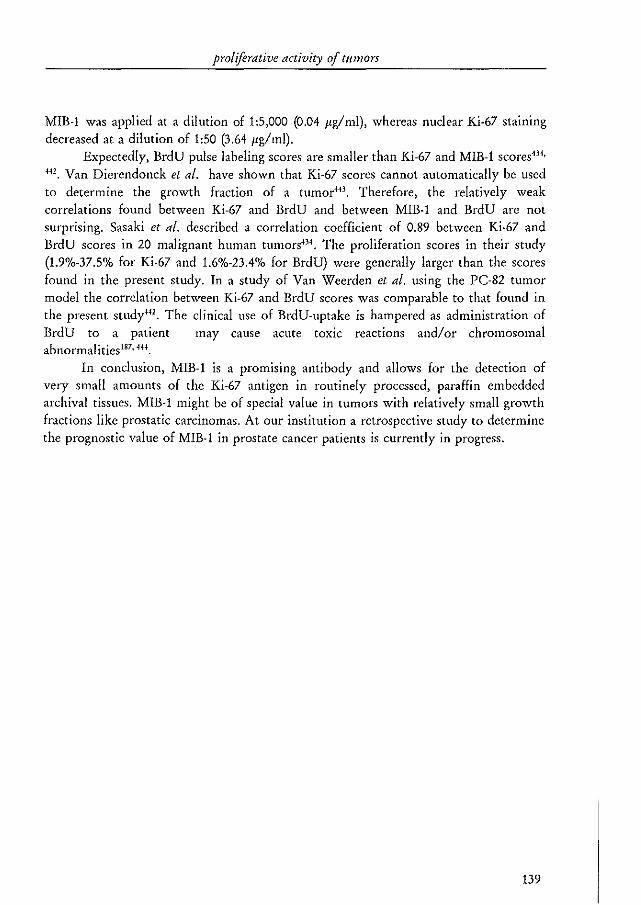
proliferative activity 0/ tumors
MIB-I was applied at a dilution of 1:5,000 (0.04 I'g/ml), whereas nuclear Ki-67 staining decreased at a dilution of 1:50 (3.64 I'g/ml).
Expectedly, BrdU pulse labeling scores are smaller than Ki-67 and MIB-I scores"" 442 Van Dierendonck et al. have shown that Ki-67 scores cannot automatically be used to determine the growth fraction of a tumorH3 . Therefore, the relatively weak correlations found between Ki-67 and BrdU and between MIB-I and BrdU are not surprising. Sasaki et al. described a correlation coefficient of 0.89 between Ki·67 and BrdU scores in 20 malignant human tumors434 . The proliferation scores in their study (1.9%-37.5% for Ki-67 and 1.6%-23.4% for BrdU) were generally larger than the scores found in the present study. In a study of Van Weerden et al. using the PC-82 tumor model the correlation between Ki-67 and BrdU scores was comparable to that found in the present study"'. The clinical use of BrdU-uptake is hampered as administration of BrdU to a patient may cause acute toxic reactions and/or chromosomal abnormalities1s7. 444.
In conclusion, MIB-I is a promising antibody and allows for the detection of very small amounts of the Ki-67 antigen in routinely processed, paraffin embedded archival tissues. MIB-I might be of special value in tumors with relatively small growth fractions like prostatic carcinomas. At our institution a retrospective study to determine the prognostic value of MIB·1 in prostate cancer patients is currently in progress.
139


CHAPTER VIII
GENERAL DISCUSSION
chapter VIII
PROGNOSTIC MARKERS
The main questions that have to be answered when a clinician is faced with a patient with prostate cancer are: is this patient to be treated and if so, how should he be treated? With the assessment of prognostic markers an attempt is being made to predict the biological behavior of the tumor in this specific patient, in order to improve patient care. It will be clear that the prognosis is not only based on tumor-derived factors, nevertheless the studies described in this thesis focus on tumor-based factors, and so will this discussion.
In patients who are to be treated with curative intent, the prognostic markers have to identify those patients in whom local progression or distant metastasis is already present at the time of diagnosis or treatment. In other words, in these patients the prognostic factors should give a reliable prediction of actual tumor-stage. In all other patients, for example expectively managed patients and hormonally treated patients, the prognostic factors have to predict the future biological behavior of a tissue that may change considerably with time. In addition, the prognosis of patients with advanced tumors is mainly determined by the metastatic lesions, whereas prognostic markers are usually determined in the primary tumor. Primary tumor cells afC very probably not representative for metastatic tumor cells which form a highly selected cell population. It therefore appears that prediction of the prognosis of radically treated patients would be easier, but even for these patients it is at present not possible to reliably predict the clinical course for each individual.
Chapter I provides a review on most of the prognostic tissue markers that have been studied in prostate cancer. For almost all reviewed markers, except for tumor stage and tumor grade, conflicting results have been published or only one or a small number of reports have been published. As has been discussed in chapter I, a large number of studies are impaired by methodologic shortcomings like: small sample size, patient sample not well described, heterogenous patient sample with regard to stage distribution or treatment, short follow-up period, inappropriate end-point, inappropriate statistics or absence of multivariate analysis. These shortcomings probably influenced the results of several studies and may very well be responsible for part of the conflicting results. It is clear that the conclusion of the College of American Pathologists Conference on clinical relevance of prognostic markers in solid tumors33 , namely that no prognostic markers except for tumor-stage and Gleason score can be used clinically for decision making in prostate cancer patients, still holds true.
Another, more basic problem in the application of new prognostic tumor markers is inherent to the current prognostic system (TNM + grade)"5. This is a mutually exclusive categorical classification system that for a number of reasons cannot be extended with new markers. The number of categories increases very rapidly with increasing numbers of variables (TO-IN~)M~l; 5x4x2 ~ 40). If grade is incorporated the
142
general discllssion
number becomes 160 (M.D. Anderson or WHO grading, range 1-4, 4x40) or even 360 (Gleason grading, range 2-10, 9x40). With stage grouping the number of categories would be 20 or 45, respectively. The accuracy of the system is directly proportional to the number of patients per category. Thus, with new variables the accuracy may fall dramatically. In addition, the TNM categories are ordered by a worsening prognosis. The only way to add a new variable will be to study a large patient sample and create a new classification based on the results of all the variables tested. Finally, the TNM system is static, when there is a change in the data underlying the system, the system itself has to be adapted. For example, the introduction of the PSA RT-PCR assay may in the future classify patients that are now at stage I, II or III as stage IV. This would affect the overall prognosis of all classes and would make a new classification necessary. Despite these comments, the TNM/grade variables on itself are of major clinical value, and should therefore be part of each future prognostic system.
The chapters II-IV of this thesis describe studies on the application of prognostic tissue markers to prostate cancer. The extent of neuroendocrine differentiation as defined by expression of CgA did not show a prognostic value in patients treated with a radical prostatectomy (chapter Il). This will be discussed below.
Neither the expression of the apoptosis inhibitor bcl-2 nor androgen receptor protein was of prognostic value in hormonally treated patients (chapter IV), whereas it was expected that the expression levels would be correlated with an adverse and good prognosis, respectively, Bcl-2 scores were higher in advanced tumors compared to radical prostatectomy specimens, but were not further increased in hormonally independent tumors. This suggests that bcl-2 expression is related to the presence of advanced tumor, and that bcl-2 independent pathways are more important for the acquisition of hormone independent prostatic tumor growth, These data are in contrast with published studies1l4, 115. Most authors described a prognostic value of androgen receptor expressionlll.ll4, Others found that only the heterogeneity of expression as determined by image analysis was of prognostic valuello, lII, It would be of interest to analyze the androgen receptor immunostained tissue sections presented in chapter IV with image analysis as well, to investigate whether this would indeed result in a prognostic value. A combined bcl-2Iandrogen receptor score was of independent prognostic value to predict clinical progression, however.
Loss of expression of the cellular adhesion molecule CD44s was an independent prognostic marker to predict PSA progression and clinical progression in patients treated by radical prostatectomy (chapter III). Yet, it added only little to pT-stage and Gleason grade. The area under the receiver operating characteristic curve (ROC curve, a graph that displays sensitivity against 1 - specificity) increased from 0.76 to only 0.79 in the prediction of clinical progression if CD44s expression was included. This means, as an example, that at a sensitivity of 0.75 the specificity increased from 0.61 to 0.69. Since this was the first study on CD44 expression in clinical prostate cancer, it will be interesting to know the results of other studies. The observation that CD44 expression was completely absent in lymph node metastatic tumor cells deserves further
143
chapter VIII
investigation in patients of whom primary as well as metastatic lesions Oymphatic or hematogenic) are available.
IMPROVEMENT OF PROSTATE CANCER PROGNOSTICATION
How can the assessment of the prognosis of prostate cancer patients be improved? First of all, as described above the interpretation of many studies on prognostic factors is impaired by methodologic shortcomings. Burke and Henson"" 446 described a number of criteria that reports of clinical outcome studies on prognostic tumor markers should fulfill: a description of the sampling method, a description of the assay and, if applicable, assessments of different types of variability of the assay, a description of cutoff point criteria and whether the cut· off point was selected before the data were analyzed, a description of the subject inclusion criteria, characteristics of the subjects, number of subjects and events, the duration of follow-up and a description and justification of the multivariate model used. If these criteria are met, journal editors should be able to conclude on the scientific quality of a submitted paper. If feasible, it may be worthwhile to perform a meta-analysis on studies of promising markers on which conflicting results have been published (for example, nuclear DNA content). Promising markers should be studied prospectively according to consensus techniques in a multi-center setting. Second, with increasing knowledge about prostate cancer biology new markers that may predict patient outcome will become available and for this reason (and others) basic prostate cancer research is very important. Third) in patients who are not treated with curative intent, repeated marker determinations (response mark,,) may overcome the problem that the marker has to predict future tumor behavior. Since serum can be obtained very easily, these markers should preferably be serum-based. FOUlth, it is essential that potential markers are being tested using multivariate analysis techniques since most markers are correlated with tumor-stage or tumor.grade (chapter I). In addition, pre·operative serum PSA levels, tumor-stage and grade are all strong prognostic indicators for patient outcome. Multivariate analysis techniques are able to identify independent prognostic markers, which implicates having an additional value over the other variables adjusted for.
If a prognostic study identifies a set of markers A,B,C and D as independent prognostic markers than the multi-regression model assumes that all these variables play a role in all patients (see appendix to chapter 1}. But, it may very well be that in one patient A and B cause progression whereas in another patient it is A and C while in a third patient only D is sufficient to cause tumor progression. It is also possible that a certain marker has only prognostic value in subgroups of patients. Modelling systems should be able to account for this and should thus be flexible. Some recent studies suggest that artificial network analysis could be a useful solution"" "'. A neural network system is a form of artificial intelligence which has been described as an electronic analogue of the nervous systemH7. It is too complex to discuss extensively in
144
general discussion
this context, but basically, a neural network system 'learns' to distinguish different outcomes of events by receiving information of known events and their outcomes. Theoretically, neural network analysis of prognostic variables should allow the construction of survival probability curves for individual patients. Neural network analysis has several advantages: it is insensitive to missing values, new variables can be easily entered and tested, it is flexible, no specific distribution of data is assumed, proportional hazards are not required (which is a strong assumption of Cox's regression model) and it is model independent. Furthermore, it would allow the inclusion of the TNM/grade variables without the disadvantages of the TNM/grade system as described above. The disadvantages are that it is a complex technique (one of the A]CC criteria of prognostic systems is that they should be easily applicable) and that the underlying model is difficul t to interpret. A pilot study has demonstrated that neural network analysis might be useful in prostate cancer screening as well as in predicting prostate cancer prognosis4--'9.
It is recommended that studies of prognostic tissue markers be performed according to the following scheme: 1. Identification of a potential marker 2. Development, testing and standardization of the assay 3. Application of the marker to easy accessible and well defined samples (e.g.,
radical prostatectomy specimens) to: study the distribution in benign, pre-malignant and malignant tissues study tissue heterogeneity study the relationship with tumor-stage and tumor-grade.
4. Assessment of prognostic value in radically and hormonally treated patients at the univariate and multivariate level. If possible, including data on the ability of the marker to improve the sensitivity and specificity in predicting clinical progression or tumor death.
5. If an independent prognostic value is found by several independent researchers: assessment of the prognostic value in biopsy specimens using clinical tumor-stage and biopsy tumor-grade. Investigate whether the marker can be used as surrogate end-point marker or response marker.
6. If the independent prognostic value is confirmed: perform prospective multicenter studies using consensus techniques, quantification methods and cut-off values.
7. Perform clinical trials in which the patients are grouped according to the new marker.
In conclusion, potential markers should be identified by basic prostate cancer research andlor from the literature on other tumor types. Investigation of the prognostic value should be done on a trial and error basis according to the scheme described above. For multivariate analysis with a large number of variables flexible modelling systems should
145
chapter VIII
be used. If a marker is to be used clinically, this should be properly studied by the conduction of randomized clinical trials.
NEUROENDOCRINE DIFFERENTIATION
Since the publication of the study described in chapter II (1995), a few additional clinical studies on the prognostic value of NE cells in prostate cancer patients treated by radical prostatectomy have been published, but did, unfortunately, not clarify the picture. Bubendorf et at.'" found no prognostic value in 137 patients, whereas Weinstein et at.''' found an independent prognostic value in 104 patients, but only if the analysis was restricted to the 59 Gleason grade 5 and 6 tumors in which case Gleason grade was of no prognostic value anymore. Both studies found no correlation between CgA immunostaining and tumor grade or stage. Berner et at.14l studied immunohistochemically the NSE expression patterns in the TUR specimens of 80 patients who subsequently underwent pelvic lymph node dissection and either a radical prostatectomy (f,.,pNo) or radiotherapy (fJ4pNo) and did not find a correlation with tumor stage or grade or prognosis. Thus, for surgically treated patients it can still not be concluded whether the extent of NE differentiation is a prognostic factor. Nevertheless, it appears that prostatic NE cells do not have a strong influence on tumor growth or the metastatic cascade.
Two studies showed that 5·HT antagonists'" and 5-HT uptake inhibitors'" were able to dose dependently inhibit the growth of prostate cancer cell lines, substantiating the observation that 5-HT may play a role in prostate cancer growth (chapter V). Bombesin (GRP) has been shown to stimulate growth of PC-3 cells (chapter V). A recent study of Aprikian et at.''' identified GRP receptors on PC-3, DU 145 and LNCaP cells. Treatment of DU 145 and PC3 cells with GRP or GRP analogues resulted in an intracellular Ca2
+ mobilization, but a growth influencing effect was not observed. A recent immunohistochemical study showed expression of the angiogenic peptides VEGF (vascular endothelial growth factor) and TGF-a'54 in NE cells of benign and malignant prostatic tissues, although the expression was not exclusively limited to the NE cells. Interestingly, a relationship between the presence of NE cells (CgA labeling) and neovascularization (endothelium labeling) of prostate cancer has been describedJ55 • In addition, VEGF can increase vascular permeability and may thus increase the metastatic ability of tumor cells"'. Because (NE cell derived) TGF-a binds to the EGF-R which is expressed by prostatic tumor cells (chapter I), it may act as a mitogenic peptide in these cells. This study suggests that NE cells play an additional role in prostatic tumor biology that is distinct from the secretion of neuropeptides or
146
general discussion
biogenic amines or from their putative role in androgen independent tumor growth. Expression of TGF-a has also been demonstrated in pure NE tumors (midgut carcinoid tumors, pheochromocytomas and medullary thyroid carcinomas)'''. It will be interesting to study the prognostic value of VEGF and TGF-a in prostatic tumors.
For hormonally treated patients, NE differentiation appears to have a prognostic value. In addition to the studies already discussed in the chapters II and V, Krijnen et al. (accepted for publication) found an independent prognostic value of CgA immunostaining to predict hormonal escape in the pre-treatment TUR specimens of hormonally treated patients. Sheaff et "I.''' described a prognostic value of Il-human chorionic gonadotrophin immunostaining, a peptide that is expressed by a subset of prostatic NE cells (chapter V). No defenite conclusions can be drawn from this study since the patient selection and statistical methods were probably inappropriate. A number of studies have pointed towards a relationship between prostatic neuroendocrine cells and the androgen independent phenotype of prostate cancer (discussed in chapters V and VI) and the results of the prognostic studies with hormonally treated patients also support this.
It will be clear that experimental studies are needed in order to clarify the role of NE cells in prostatic (tumor) biology. The two xenograft models described in chapter VI (pC-295 and PC-310) will certainly be of value, especially because the NE phenotype could be induced readily and reproducibly by castration of the tumor bearing mice. In addition, it would be very useful if NE differentiation could also be induced in in vitro growing cell lines. Shen et al. were indeed able to induce a neuronal phenotype in the LNCaP cell line by culturing the cells without androgens"'. Currently, attempts are being made in our laboratory to culture PC-295 cells in vitro.
The mechanism of the relationship between androgen withdrawal and the induction of NE differentiation is not known. A relationship between estrogen levels and expression of NE features has also been described in the rat pituitary'~29, .. 130 and
recently it was shown that estrogen upregulates the expression of S-HT2~ receptors in rat female brain'''. Thus, steroids appear to influence both the effector and affector parts of the NE system. Knowledge about the mechanism of induction of NE differentiation in prostatic (tumor) cells is important and may reveal essential clinical information.
An issue that has received little or no attention is the definition of the prostatic NE cell. Thus far, most researchers have used immunohistochemistty or Western blotting with antibodies against neuroendocrine cell specific products (CgA, NSE, 5-HT and others). By far, the most often used technique is CgA immunohistochemistry. In the studies described in chapters II and VI the NE cells were also defined by their expression of CgA. In addition, the data of chapter VI were confirmed by electron microscopy. Expression of the above mentioned immunologically defined markers is probably neither specific nor sensitive (G.].M. Martens, personal communication). Other factors appear more specific and are essential for neuroendocrine function like prohormone convertases (PCI/PC3 and PC2)"" "I, 7B2'" and the enzyme peptidyl-
147
chapter VIII
glycine a-amidating mono-oxygenase (PAM)"J. PAM expression has been demonstrated at the mRNA level in normal rat prostate tissues'''. A preliminary study with human benign and malignant prostatic tissues showed cytoplasmic expression of PAM at the protein (immunohistochemistly) and mRNA (in situ hybridization) level in a subset of epithelial cells (M.A. Noordzij and A. Treston, unpublished observations). Immunohistochemical double labeling experiments (PAM + egA) with these tissues showed that PAM was not expressed by the NE cells, but by surrounding non-NE cells. Studies on the expression of the other markers are currently in progress at our institution. Thus, a fundamental discussion of how to define the prostatic NE cell seems warranted.
The questions that have to be studied in the near future are: what are the functions of NE cells in the normal prostate, if any? Do NE cells playa role in BPH? Do NE cells playa role in prostatic carcinogenesis? Is NE differentiation in prostate cancer of predictive value and should patients with large numbers of NE cells in their tumor be treated differently? Is the putative prognostic value of NE cells just related to the number of NE cells or restricted to the expression of one Of more active peptides? Do neuroendocrine cells influence prostate cancer growth and if so, which peptides are responsible for this effect and can it be used to target therapy? Do the NE cells playa role in the transition of prostate cancer from androgen dependency to independence? What is the mechanism of increased NE differentiation following androgen withdrawal?
In conclusion, although a considerable number of studies have been published on NE differentiation in prostate cancer, its prognostic value in most patient categories is still controversial. Prostatic NE cells appear to influence tumor growth via several distinct mechanisms and may play a role in androgen independent tumor growth, but experimental data are sparse. Prostatic tumor models that contain these cells are now available and will allow more fundamental studies, but first of all, the prostatic NE cell should be satisfactorily defined.
148

REFERENCES
1. Potosky AL, Miller BA, Albertsen PC, Kramer BS: The role of increasing detection in the rising incidence of prostate cancer. J Am Med Assoc 1995, 273:548-552
2. Jacobsen SJ, Katusic SK, Bergstralh EJ, Oesterling JE, Ohrt D, Klee GG, Chute CG, Lieber MM: Incidence of prostate cancer diagnosis in the eras before and after serum prostate-specific antigen testing. J Am Med Assoc 1995,274:1445-1449
3. Severson RK, Montie JE, Porter AT, Demers RY: Recent trends in incidence and treatment of prostate cancer among elderly men. J Nat! Cancer Inst 1995, 87:532-534
4. van der Gulden J\V'J, Kiemeney LALM. Verbeek ALM, Straatman H: Mortality trends from prostate cancer in The Netherlands (1950·1989). Prostate 1994, 24:33·38
5. Schroder FH, Damhuis RAM, Kirkels \V'J, De Koning HJ, Kranse R, Nijs HGT, Blijenberg BG: European randomized study of screening for prostate cancer - the Rotterdam pilot studies. IntJ Cancer 1996, 65:145-151
6. Srivastava S, Rossi SC: Early detection research program at the NCr (review). Int J Cancer 1996, 69:35·37
7. Thompson IM, Zeidman EJ: Current urological practice: routine urological examination and early detection of carcinoma of the prostate. J Urol 1992, 148:326-330
8. Stephenson RA, Smart CR, Mineau GP, James BC, Janerich DT, Dibble RL: The fall in incidence of prostate carcinoma: on the down side of a prostate specific antigen induced peak incidence- data from the Utah cancer registry. Cancer 1996,77:1342-1348
9. Harris JR, Lippman ME, Veronesi V, \Villett \Yf: Breast cancer (I), review. N Engl J Med 1992,327:319·328
10. Mettlin CJ, Murphy GP, Ho R, Menck HR: The national cancer data base report on longitudinal observations on prostate cancer. Cancer 1996. 77:2162-2166
11. Sakr \YfA, Haas GP, Cassin BF, Pontes JE, Crissman JD: The frequency of carcinoma and intraepithelial neoplasia of the prostate in young male patients. J Urol 1993, 150:379-385
12. Schmid HP, McNeal JE. Stamey TA: Observations on the doubling time of prostate cancer: The use of serial prostate-specific antigen in patients with untreated disease as a measure of increasing cancer volume. Cancer 1993, 71:2031-2040
13. Coffey DS: Prostate cancer - an overview of an increasing dilemma. Cancer 1993, 71(Suppl):880.863
14. Parker SL, Tong T, Bolden S, \Yfingo PA: Cancer statistics 1996. CA Cancer J Clin 1996, 65:5·27
15. Adami HO, Baron JA, Rothman KJ: Ethics of a prostate cancer screening trial (review). Lancet 1994, 343:958·960
16. \V'hitmore Jr \YfF, \Yfarner JA, Thompson Jr IM.: Expectant management of localized prostate cancer. Cancer 1991, 67:1091-1096
17. \Yfarner J, Whitmore Jr WF: Expectant management of clinically localized prostatic cancer. J Uro11994, 152:1761·1765
18. Aus G, Hugosson J. Norlen L: Long-term survival and mortality in prostate cancer treated with noncurative intent (review). J Urol 1995, 154:460-465
19. Johansson JE, Adami BO, Andersson SO, Bergstrom R, Krusemo UB, Kraaz \Yf: Natural history of localised prostatic cancer: a population-based study in 223 untreated patients. Lancet 1989, 1:799·803
20. Johansson JE: Expectant management of early stage prostatic C,Ulcer: Swedish experience. J Urol 1994, 152:1753·1756
21. Adolfsson J, Carstensen J, Lowhagen L: Deferred treatment in clinically localised prostatic carcinoma. Br J Uroll992, 69:183-187
22. Nadler RB, Andriole GL: \Yfho is best benefited by radical prostatectomy? Hematol Oncol Clin North Am 1996, 10:581·593
150
references
23. Chodak GW, Thisted RA, Gerber GS, Johansson JE, Adolfsson J, Jones GW, Chisholm GD, Moskovitz B, Livne PM, \Varner J: Results of conservative management of clinically localized prostate cancer. N Engl J Med 1994, 330:242-248
24. Zagars GK, von Eschenbach AC, Ayala AG: Prognostic factors in prostate cancer. Analysis of 874 patients treated with radiation therapy. Cancer 1993,72:1709-1725
25. \Valsh PC, Lepor H: The role of radical prostatectomy in the management of prostate cancer. Cancer 1987,60:526-537
26. Hartford AC, Zietman AL: Prostate cancer: who is best benefited by external beam radiation therapy? Hematol Oneal Clin North Am 1996, 10:595·609
27. Albertsen PC, Fryback DG, Storer BE, Kalan TF, Fine J: The impact of co-morbidity on life-expectancy among men with localizcd prostate C'Ulcer. J Uroll996, 156:127-132
28. Madsen PO, Graversen PH, Gasser TC, Corle DK: Treatment of localized prostatic cancer. Radical prostatectomy versus placebo. A IS-year follow-up. Scand J Urol Nephrol Suppl 1988, 110:95·100
29. Pavone-Macaluso M, De Voogt HJ, Viggiano G, Barasolo E, Lardennois B, De Pauw M, Sylvester R: Comparison of diethylstilbestrol, cyproterone acetate and medroxyprogesterone acetate in the treatment of advanced prostatic cancer: final analysis of a randomized phase ill trial of the European Organization for Research and Treatment of Cancer urological group. JUral 1986, 136:624·631
30. Smith PH, Suciu S, Robinson MRG, Richards B, Bastable JRG, Glashan R\'(f, Bouffioux C, Lardennois B, \Villiams RE, De Pauw M, Sylvester R: A comparison of the effect of diethylstilbestrol with low dose estranlUstine phosphate in the treatment of advanced prostatic cancer: final analysis of a phase III trial of the European Organization for Research on Treatment of Cancer. JUral 1986, 136:619-623
31. Mani S, Vogelzang NJ: Is "off-protocol" chemotherapy for androgen-independent carcinoma of the prostate arranted? Hematol Oncol Clin North Am 1996, 10:749-768
32. Hayes DF, Bast RC, Desch CE, Fritsche H, Kemeny ME, Jessup JM, Locker GY, MacDonald JS, Mennel RG, Norton L, Ravdin P, Taube S, \'linn RJ: Tumor marker grading system: a framework to evaluate clinical utility of tumor markers. J Nat! Cancer Inst 1996,88:1456·1468
33. Grignon DJ, Hammond H: College of American pathologists conference XXVI on clinical relevance of prognostic markers in solid tumors. Report of the prostate cancer working group. Arch Pathol Lab Med 1995, 119:1122-1126
34. Schrodcr FH, Hermanek P, Denis L, Fair \'(fR, Gospodarowicz MK, Macaluso P: The TNM classification of prostate cancer. Prostate 1992, Supp!. 4:129-138
35. Cantrell BB, DeKlerk DP, Eggleston JC, Boitnott JK, \'lalsh PC: Pathological factors that influence prognosis in stage A prostatic cancer: the influence of extent versus grade. J Ural 1981, 125:516-520
36. B1ute ML, Zincke H, Farrow GM: Long-term followup of young patients with stage A adenocarcinoma of the prostate. J Urol 1986, 136:840-843
37. Larsen MP, Carter HB, Epstein JI: Can stage Al tumor extent be predicted by transurethral resection tumor volume, per cent or grade? A study of 64 radical prostatectomies with comparison to prostates removed for stages A2 and B disease. J Urol 1991, 146:159·163
38. van den Ouden D, Davidson PJT, Hop \Yi, Schroder FH: Radical prostatectomy as a lllonotherapy for locally advanced (stage T3) prostate cancer. J Urol1994, 151:646-651
39. Middleton RG, Smith Jr JA, Melzer RB, Hamilton PE: Patient survival and local recurrence rate following radical prostatectomy for prostatic carcinoma. J Urol 1986, 136:422-424
151
40. Schellhammer PF: Radical prostatectomy. Patterns of local failure and survival in 67 patients. Urology 1988, 31:191·197
41. Lerner SE, BIute ML, Zincke H: Extended experience with radical prostatectomy for clinical stage T3 prostate cancer: Outcome and contemporary morbidity. J Urol 1995, 154:1447·1452
42. Epstein JI, Walsh PC, Carmichael M, Brendler CB: Pathological and clinical findings to predict tumor extent of nonpalpable (stage T1c) prostate cancer, J Am Med Assoc 1994, 271:368·374
43. Scaletscky R, Koch MO, Eckstein CW, Bicknell SL, Gray Jr GF, Smith Jr JA: Tumor volume and stage in carcinoma of the prostate detected by elevations in prostate specific antigen. J Uro11994, 152:129·131
44. Stormont TJ, Farrow GM, Myers RP, Blute ML, Zincke H, \X'ilson TM, Oesterling JE: Clinical stage BO or TIc prostate cancer: nonpalpable disease identified by elevated serum prostate-specific antigen concentration. Urology 1993,41:3-8
45. Zincke H, Bergstralh ML, Blute L, Myers RP, Barrett DM, Lieber MM, Martin SK, Oesterling JE: Radical prostatectomy for clinically localized prostate cancer: long-term results of 1143 patients from a single instiultion. J Clin Onco11994, 12:2254-2263
46. Kleer E, Larson-Keller 11, Zincke H, Oesterling JE: Ability of preoperative semlll prostate-specific antigen value to predict pathologic stage and DNA ploidy. Influence of clinical stage and tumor grade. Urology 1993, 41:207-216
47. \Veldon VE, Tavel FR, Neuwirth H, Cohen R: Patterns of positive specimen margins and detectable prostate specific antigen a&er radical perineal prostatectomy. JUral 1995, 153:1565·1569
48. Huland H. Hammerer P, Henke RP, Huland E: Preoperative prediction of tumor heterogeneity and recurrence after radical prostatectomy for localized prostatic carcinoma with digital rectal examination, prostate specific antigen and the results of 6 systematic biopsies. J UroI1996, 155:1344·1347
49. Roach III M, Marquez C, Yuo HS, Narayan P, Coleman L, Nseyo UO, Navvab Z, Carroll PR: Predicting the risk of lymph node involvement using the pre-treatment prostate specific antigen and Gleason score in men with clinically localized prostate cancer. Int J Radiat Oncol Bioi Phys 1994, 28:33·37
50. Narayan P, Fournier G, Gajendran V, Leidich R, Lo R. \Volf Jr JS, Jacob G, Nicolaisen G, Palmer K, Freiha F: Utility of preoperative semm prostate-specific antigen concentration and biopsy Gleason score in predicting risk of pelvic lymph node metastases in prostate cancer. Urology 1994,44:519-525
51. Bangma CH, Hop \VC, Schroder FH: Eliminating the need for per-operative frozen section analysis of pelvic lymph nodes during radical prostatectomy. Br J Urol 1995, 76:595·599
52. Miller GJ, Cygan JM: Morphology of prostate cancer: the effects of 1l1ultifocality on histological grade, tumor volume and capsule penetration. J Uro11994, 152:1709-1713
53. Peller PA, Young DC, Marmaduke DP, Marsh WL, Badalement RA: Sextant prostate biopsies. A histopathologic correlation with radical prostatectomy specinlens. Cancer 1995, 75:530·538
54. McNeal JE, Villers AA, Redwine EA, Freiha FS, Stamey TA: Capsular penetration in prostate cancer: significance for natural history and treatment. Am J Surg Pathol 1990, 14:240·247
55. McNeal JE, Villers AA, Redwine EA, Freiha SS, Stanley TA: Histologic differentiation, cancer volume, and pelvic lymph node metastasis in adenocarcinoma of the prostate. Cancer 1990, 66:1225·1233
152
references
56. Stamey T A, McNeal JE, Freiha FS, Redwine E: Morphometric and clinical studies on 68 consecutive radical prostatectomies. J Urol 1988, 139:1235-1241
57. Epstein J1, Carmichael M, Partin A \Yf, Walsh PC: Is tumor volume an independent predictor of progression following radical prostatectomy? A multivariate analysis of 185 clinical stage B adenocarcinomas of the prostate with 5 years of follow lip. J Urol 1993, 149:1478-1481
58. Humphrey PA, \'{falter PJ, Currin SM, Vollmer RT: Histologic grade, DNA ploidy, and intraglandular tumor extent as indicators of tuOlor progression of clinical stage B prostatic carcinoma. A direct comparison. Am J Surg Pathol 1991, 15:1165-1170
59. Humphrey PA, Vollmer RT: Intraglandular tumor extent and prognosis in prostatic carcinoma: application of a grid method to prostatectomy specimens. Hum Pathol 1990, 21:799-804
60. Haggman M, Nybacka 0, Nordin B, Busch C: Standardized in vitro mapping with multiple core biopsies of total prostatectomy specimens: localization and prediction of tumour volume and grade. Br JUral 1994, 74:617-625
61. Cupp MR, Bostwick DG, Myers RP, Oesterling JE: The volume of prostate cancer in the biopsy specimen cannot reliably predict the quantity of cancer in the radical prostatectomy specimen on an individual basis. J Urol 1995, 153:1543-1548
62. Terris MK, Henay DJ, Johnstone I1vI, McNeal JE, Stamey TA: Prediction of prostate cancer volume using prostate-specific antigen levels, transrectal ultrasound, and systematic sextant biopsies. Urology 1995, 45:75-80
63. Dietrick DD, McNeal JE, Stamey TA: Core cancer length in ultrasound-guided systematic sextant biopsies: a preoperative evaluation of prostate cancer volume. Urology 1995, 45:987-992
64. Terris MK, McNeal JE, Stamey TA: Detection of clinically significant prostate cancer by transrectal ultrasound-guided systematic biopsies. J Urol 1992, 148:829-832
65. Irwin MB, Trapasso JG: Identification of insignificant prostate cancers: analysis of preoperative parameters. Urology 1994, 44:862-867
66. Dugan JA, Bostwick DG, Myers RP, Qian J, Bergstralh EJ. Oesterling JE: The definition and preoperative prediction of clinically insignificant prostate cancer. J Am Med Assoc 1996, 275:288-294
67. Ruizeveld de \Vinter JA: Androgen receptor expression in human prostate cancer (thesis). Rotterdam, Erasmus University. 1994, pp.
68. Bocking A, Kiehn J. Heinzel-\Vach M: Combined histologic grading of prostatic carcinoma. Cancer 1982, 50:288-294
69. Gleason DF: Classification of prostatic carcinomas. Cancer Chemother Rep 1966, 50:125-128
70. Epstein J1, Partin A \Yf, Sauvageot J, \'(Ialsh PC: Prediction of progression following radical prostatectomy: a multivariate analysis of 721 with long-term follow-up. Am J Surg Pathol 1996, 20:286-292
71. Ohori M, Goad JR. \Yfheeler TM, Eastham TM, Thompson Te, Scardino PT: Can radical prostatectomy alter the progression of poorly differentiated prostate cancer? J Urol 1994, 152:1843-1849
72. Bostwick DG: Grading prostate cancer. Am J elin Pathol 1994, 102:538·556 73. Thickmatl D, Speers \"{fC. Philpott PJ, Shapiro H: Effect of the number of core biopsies
of the prostate on predicting Gleason score of prostate cancer. J UroI1996, 156:110-113 74. ten Kate FJ\V, Galiee MP\V. Schmitz PIM, Joebsis AC, van der Heul RO, Prins MEF,
Blom JHM: Controversy in grading of prostatic carcinoma: interobserver reproducibility of five different grading systems. World J Urol 1986, 4:147-152
153
75. Gleason DF: Histologic grading of prostate cancer: a perspective. Hum Pathol 1992, 23:273-279
76. Oesterling JE: Prostate specific antigen: a critical assessment of the most useful tumor marker for adenocarcinoma of the prostate. J Uro11991, 145:907·923
77. Partin AW, Pound CR, Clemens JQ, Epstein JI, Walsh PC: Serum PSA after anatomic radical prostatectomy. The Johns Hopkins experience after 10 years. Urol Clin North Am 1993, 20:713-725
78. Benson MC, Whang IS, Pantuck A, Ring K, Kaplan SA, Olsson CA, Cooner WI-!: Prostate specific antigen density: a means of distinguishing benign prostatic hypertrophy and prostate cancer. J Urol 1992, 147:815-816
79. Humphrey PA, Baty J, Keetch D: Realationship between serum prostate specific antigen, needle biopsy findings, and histopathologic features of prostatic carcinoma in radical prostatectomy tissues. Cancer 1995, 75:1842·1849
80. Christensson A, Bjork T, Nilsson 0, Dahlen U, Matikainen M, .T., Cockett ATK, Abrahamsson ATK, Lilja H: Serum prostate specific antigen complexed to alfa1·antichymotrypsin as an indicator of prostate cancer. J Urol 1993, 150:100·105
81. D'Amico AV, Hanks GE: Linear regressive analysis using prostate specif.ic antigen doubling tinle for predicting tumor biology and clinical outcome in prostate cancer. Cancer 1993, 72:2638-2643
82. Epstein JI: PSA and PAPas immunohistochemical markers in prostate cancer (review). Urol Clin N Am 1993, 20:757-770
83. Aspinall JO, Bentel JM, Horsfall DJ, Haagensen DE, Marshall YR, Tilley WD: Differential expression of apolipoprotein-D and prostate specific antigen in benign and malignant prostate tissues. J Urol 1995, 154:622-628
84. Aihara M, Lebovitz RM, \Vheeler TM, Kinner BM, Ohori M, Scardino PT: Prostate specific antigen and Gleason grade: an immunohistochemical study of prostate cancer. J Urol 1994, 151:1558-1564
85. Vitali A, Ardoino S, Durante P, Ferro MA, Li Causi F, Parodi C, Sanguineti G, Gaffuri M, Paerachino M, Salvadori RP: Correlation between immunohistochemical patterns and serum levels of PSA and PSAP in prostatic pathology: evaluation of 198 prostatic fine needle biopsies. Anticancer Res 1994, 14:1503-1507
86. Grob BM, Schellhammer PF, Brassi! DN, \Vright Jr GL: Changes in inullunohistochemical staining of PSA, PAP, and TURP-27 following irradiation therapy for clinically localized prostate cancer. Urology 1994, 44:525-529
87. Yu H, Dianlandis EP, Sutherland DJ: Immunoreactive prostate-specific antigen levels in female and male breast tumors and its association with steroid hormone receptors and patient age. Clin Biochem 1994, 27:75-79
88. Monne M, Croce eM, Yu H, Diamandis EP: Molecular characterization of prostate-specific antigen messenger RNA expressed in breast tUlllors. Cancer Res 1994, 54:6344·6347
89. van Krieken JH: Prostate marker immunoreactivity in salivary gland neoplasms. A rare pitfall in inImunohistochemistry. Am J Surg Pathol 1993, 17:410-414
90. Sakai H, Yogi Y, Minami Y, Yushita Y, Kanetake H, Saito Y: Prostate specific antigen and prostatic acid phosphatase immunoreactivity as prognostic indicators of advanced prostatic carcinoma. J Urol 1993, 149:1020·1023
91. Stege R, Tribukait B, Lundh B, Carlstrom K, Pousette A, Hasenson M: Quantitative estinlation of tissue prostate specific antigen, deoxyribonucleic acid ploidy and cytologiml grade in fine needle aspiration biopsies for prognosis of hormonally treated prostatic carcinoma. J Urol 1992, 148:833-837
154
references
92. Berner A, Harvei S, Tretli S, Fossa SD, Nesland JM: Prostatic carcinoma: a multivariate analysis of prognostic factors. Br J Cancer 1994, 69:924-930
93. Mohler JL, Partin A \Yf, Epstein JI, Becker RL, Mikel UV, Sesterhenn lA, Mostofi FK, Gleason DF, Sharief Y, Coffey DS: Prediction of prognosis in untreated stage A2 prostatic carcinoma. Cancer 1992, 69:511-519
94. Katz AE, Olsson CA, Raffo AJ, Cama C, Perlman I-I, Seaman E, O'Toole KM, McMahon D, Benson MC, Buttyan R: Molecular staging of prostate cancer with the use of an enhanced reverse transcriptase-PCR assay. Urology 1994, 43:765-775
95. Katz AE, de Vries GM, Begg MD, Raffo AJ, Cama C, O'Toole C, Buttyan R, Benson MC, Olsson CA: Enhanced reverse tfOUlscriptase-polymerase chain reaction for prostate specific antigen as an indicator of tme pathologic stage in patients with prostate cancer. Cancer 1995, 75:1642-1648
96. Olsson CA. de Vries GM, Raffo AJ. Benson MC, O'Toole K, Cao Y, Buttyan RE, Katz AE: Preoperative reverse transcriptase polymerase chain reaction for prostate specific antigen predicts treatment failure following radical prostatectomy. ] Urol 1996, 155:1557-1562
97. Ghossein RA, Scher HI, Gerald WL, Kelly WK, Curley T, Amsterdam A, Zhang ZF, Rosai J: Detection of circulating tumor cells in patients with localized and metastatic prostatic carcinoma: clinical inlplications. J Clin Oncol 1995, 13:1195-2000
98. Smith MR, Biggar S, Hussain M: Prostate-specific antigen messenger RNA is expressed in non-prostate cells: implications for detection of micrometastases. Cancer Res 1995. 55:2640-2644
99. Falkmer UG: Methodologic sources of errors in image and flow cytometric DNA assessments of the malignancy potential of prostatic carcinoma. Hum Pathol 1992, 23:360-367
100. Schroder FI-I, Tribukait B, Backing A, deVere-\Yfhite R, Koss L, Lieber MM, Stenkvist B, Zetterberg A: Clinical utility of cellular DNA measurements in prostate carcinoma. Scand J Urol Nephrol Suppl 1994, 162:51-64
101. Tavares AS, Costa J, Maia JC: Correlation between ploidy and prognosis in prostatic carcinoma. J Urol 1973, 109:676-679
102. Tavares AS, Costa J, de Carvalho A, Reis M: Tumour ploidy and prognosis in carcinoma of the bladder and prostate. Br J Cancer 1966, 20:438-441
103. Adol£sson J: Prognostic value of deoxyribonucleic acid content in prostate cancer: a review of current results. Int ] Cancer 1994, 58:211-216
104. Stephenson RA, James BC, Gay H, Fair WR, \Vhitmore \VF, Melamed MR: Flow cytometry of prostate cancer: relationship of DNA content to surival. Cancer Res 1987, 47:2504-2509
105. Song J, Cheng WS, Cupps RE, Earle JD, Farrow GM, Lieber MM: Nuclear deoxyribonucleic acid content measured by static cytometry: important prognostic association for patients with clinically localized prostate carcinoma treated by external beanl radiotherapy. J Urol 1992, 147:794-797
106. Gauwitz MD, Pollack A, el-Naggar AK) Terry NH, von Eschenbach AC, Zagars GK: The prognostic significance of DNA ploidy in clinically localized prostate cancer treated with radiation therapy. Int J Radiat Oncol BioI Ph)" 1994, 28:821-828
107. Centeno BA, Zietman AL, Shipley WU, Sobczak ML, Shipley JW, Preffer FI, Boyle BJ, Colvin RB: Flow cytometric analysis of DNA ploidy, percent S-phase fraction, and total proliferative fraction as prognostic indicators of local control and survival following radiation therapy for prostate carcinoma. Int J Radiat Oncol BioI Phys 1994, 30:309-315
108. Zetterberg A, Forsslund G: Ploidy level and tumor progression in prostatic carcinoma. Acta Onco11991, 30:193-199
155
109. Forsslund G, Esposti PL, Nilsson B, Zetterberg A: The prognostic significance of nuclear DNA content in prostatic carcinoma. Cancer 1992, 69:1432·1439
110. van den Ouden 0, Tribukait B, Blorn JHM, Fossa SO, Kurth KH, ten Kate F]\V. Heiden T, \Vang N, Schroder FH. The Europe.Ul Organization for Research and Treatment of Cancer genitourinary group: Deoxyribonucleic acid ploidy of core biopsies and metastatic lymph nodes of prostate cancer patients: impact on time to progression. J Urol 1993, 150:400-406
111. Persons DL, Takai K, Gibney OJ, Katzmann JA, Lieber MM, Jenkins RB: Comparison of fluorescence in situ hybridization with flow cytometry and static unage analysis in ploidy analysis of paraffin-embedded prostate adenocarculOma. Hum Pathol 1994, 25:678-683
112. Jorgensen T, Yogesan K, Skjorten F, Berner A, Tveter KJ, Danielsen HE: Histopathological grading and DNA ploidy as prognostic markers in metastatic prostatic cancer. Br J Cancer 1995, 71:1055-1060
113. Sassi I, Mangili F, Sanvito F, Andreassi A, Cantaboni A: Reproducibility of FCM-DNA ploidy analysis UI prostatic cancer - comparison between needle biopsy and surgical specimens. Anal Cell Pathol 1993,5:17-21
114. Ross IS, Figge H, Bui HX, del Rosario AD, Jennings TA. RifkUI MD, Fischer HAG: Prediction of pathologic stage and postprostatectomy disease recurrence by DNA ploidy analysis of initial needle biopsy specimens of prostate cancer. Cancer 1994, 74:2811-2818
115. Leung CS, Zbieranowski I, Derners I. Murray 0: DNA image cytometry of prostatic carcinoma: a comparison of needle core biopsy and subsequent prostatectomy specunens. Mod Pathol 1994,7:195-199
116. Zincke H, Bergstralh EJ, Larson-Keller 11. Farrow GM, Myers RP, Lieber MM. Barrett DM, Rife CC, Gonchoroff NJ: Stage 01 prostate C<Ulcer treated by radical prostatectomy and adjuvant hormonal treatment. Evidence for favorable survival in patients with DNA diploid tumors. Cancer 1992, 70:311-323
117. Debruyne PMJ, Collins VP, van Dekken H, Jenkins RB, Klocker H, Schalken JA, Sesterhenn IA: Cytogenetics of prostate cancer. Scand J Urol Nephrol Suppl 1994. 162:65-71
118. Takahashi S, Qian J, Brown JA. Alcaraz A. Bostwick DG, Lieber MM, Jenkins RB: Potential markers of prostate cancer aggressiveness detected by fluorescence in situ hybridization UI needle biopsies. Cancer Res 1994, 54:3574-3579
119. Alers JC, Krijtenburg PJ, Vissers KJ. Bosman FT, van der Kwast TH, van Dekkcn H: Interphase cytogenetics of prostatic adenocarcinoma and precursor lesions: analysis of 25 radical prostatectomies and 17 adjacent prostatic uttraepithelial neoplasias. Genes Chromosom Cancer 1995, 12:241-250
120. Alers Je, Krijtenburg PJ, Vissers CJ, Bosman FT, van der Kwast TH, van Dckken H: Cytogenetic heterogeneity and histologic tumor growth patterns in prostatic cancer. Cytometry 1995, 21:84-94
121. Konig JJ, Teubel W, Romijn JC, Schroder PH, I-Iagemeijer A: Gain and loss of chromosomes I, 7, 8, 10, 18, and Y in 46 prostate cancers. Hum Pathol 1996, 27:720-727
122. Alcaraz A, Takahashi S, Brown JA, Herath JF. Bergstralh EJ, Larson-Keller n. Lieber MM, Jenkins RB: Aneuploidy and aneusomy of chromosome 7 detected by fluorescence in situ hybridization are markers of poor prognosis in prostate cancer. Cancer Res 1994, 54:3998-4002
123. Diamond DA, Berry SJ, Jewett HJ, Eggleston JC, Coffey DS: A new method to assess metastatic potential of human prostate cancer: relative nuclear roundness. J Urol 1982. 128:729-734
156
references
124. Partin A \VI, Steinberg GD, Pitcock RV, \VIu L, Piantadosi S, Coffey DS, Epstein JI: Use of nuclear morphometry, Gleason histologic scoring, clinical stage, and age to predict disease-free survival anlOng patients with prostate cancer. Cancer 1992, 70:161·168
125. Veltri R\VI, Partin A \VI, Epstein JI, Marley GM, Miller CM, Singer DS, Patton KP, Criley SR, Coffey DS: Quantitative nuclear morphometry, Markovian texture descriptors, and DNA content captured on a CAS-200 image analysis system, combined with PCNA and HER-2/neu immunohistochemistry for prediction of prostate cancer progression. J Cell Biochem Suppl 1994, 19:249-258
126. Blom JHM, Ten Kate FJ\V, Schroeder FH, Van der Heul RO: Morphometrically estimated variation in nuclear size: a useful tool in grading prostate cancer. Urol Res 1990, 18:93-99
127. Vesalainen S, Lipponen P, Talja M, Kasurinen J: Nuclear morphometry is of independent prognostic value only in Tl prostatic adenocarcinoma. Prostate 1995,27:110·117
128. Miller J, Horsfall DJ, Skinner JM, Rao DM, Leong AS, Marshall VR: Nuclear shape and prognosis following orchiectomy in stage D2 prostate cancer. Prostate 1994,24:306·312
129. Kim D, Charlton JD, Coggins JM, Mohler JL: Semiautomated nuclear shape analysis of prostatic carcinoma and benign prostatic hyperplasia. Anal Quant Cytol Histol 1994, 16:400-414
130. Tardif CP, Partin A\V, Qaqish B, Epstein JP, Mohler JL: Comparison of nuclear shape in aspirated and histologic specimens of prostatic carcinoma. Anal Quant Cytol Histo} 1992, 14:474-482
131. Mohler JL, Metts JC, Zhang XZ, Maygarden SJ: Nuclear morphometry in automatic biopsy and radical prostatectomy specimens of prostatic carcinoma. A comparison. Anal Quant Cytol Histol 1994, 16:415-420
132. Mohler JL, Figlesthaler \VIM, Zhang XZ, Partin A \VI, Maygarden SJ: Nuclear shape analysis for the assessment of local invasion and metastases in clinically localized prostate carcinoma. Cmcer 1994, 74:2996-3001
133. Carson DA, Lois A: Cancer progression and p53. Lancet 1995, 346:1009-1011 134. Malkin D, Li FP, Strong LC, Fraumeni JFJ, Nelson CE, Kim DH, Kassel J, Gryka MA,
Bischoff FZ, Tainsky MA, Friend SH: Germ line pS3 mutations in ,\ fanlilial syndrome of breast cancer, sarcomas, and other neoplasms. Science 1990,250:1233-1238
135. Nigro JM, Baker SJ, Preisinger AC, Jessup JM, Hostetter R, Cleary K, Bigner SH, Davidson N, Baylin S, Devilee P, al e: Mutations in the p53 gene occur in diverse human tumour types. Nature 1989, 342:705-708
136. Porter PL, Gown AM, Kramp SG, Coltrera MD: \Videspread pS3 over expression in human malignant tumors. An immunohistochemical study using methacarn-fixed, embedded tissue. Am J Pathol 1992, 140:145·153
137. Battifora H: PS3 immunohistochemistry: a word of caution (editorial). Hum Pathol 1994, 25:435-436
138. Mellon K, Thompson S, Charlton RG, Marsh C, Robinson M, Lane DP, Harris AL, Horne CH, Neal DE: pS3, c-erbB-2 and the epidermal growth factor receptor in the benign and malignant prostate. J Uro11992, 147:496·499
139. Thompson SJ, Mellon K, Charlton RG, Marsh C, Robinson M, Neal DE: P53 and Ki-67 inullunoreactivity in human prostate cancer and benign hyperplasia. Br J Urol 1992, 69:609-613
140. Visakorpi T, Kallioniemi OP, Heikkinen A, Koivula T. Isola J: Small subgroup of aggressive, highly proliferative prostatic carcinomas defined by pS3 accumulation. J Nat! Cancer Inst 1992, 84:883-887
157
141. Fox SB. Persad RA. Royds J. Kore RN. Silcoeks PB. Collins CC: p53 and e-mye expression in stage Ai prostatic adenocarcinoma: useful prognostic determinance? J Urol 1993. 150:490-494
142. Vesalainen SLB, Lippollcn PK, Talja MT, Alhava EM, Syrjanen KJ: Proliferating cell nuclear antigen and p53 expression as prognostic factors in Tl-2MO prostatic adenocarcinoma. Int J Cancer 1994, 58:303-308
143. Berner A, \Vaere H. Paus NE, Danielsen HE, Fossa FD: DNA ploidy, semrn prostate specific antigen, histological grade and immunohistochemistry as predictive parameters of lymph node metastases in TI-T3/MO prostatic adenocarcinoma. Be JUral 1995, 75:26-32
144. Mirehandani D. Zheng J. Miller GJ. Ghosh AK. Shibata DK. Cote RJ. Roy Burman P: Heterogeneity in intratumor distribution of p53 mutations in human prostate cancer. Am J Pathol 1995. 147:92-101
145. Bubendorf L, Sauter G, Moch H, Jordan P, Blochlinger A, Gasser TC, Mihatch MJ: Prognostic significance of Bcl-2 in clinically localized prostate cancer. Am J Pathol 1996, 148:1557-1565
146. Thomas DJ, Robinson M, King P, Hasan T, Charlton R, Martin J. Carr T\V, Neal DE: P53 expression and clinical outcome in prostate cancer. Br J Urol 1993,72:778-781
147. Dinjens WNM, van cler \Veidcn M, Schroder FH, Bosman FT, Trapman J: Frequency and characterization of p53 mutations in primary and metastatic human prostate cancer. Int J Cancer 1994. 56:630-633
148. van Veldhuizen PJ, Sadasivan R, Garcia F, Austenfeld MS, Stephens RL: Mutant p53 expression in prostate carcinoma. Prostate 1993, 22:23-30
149. van VelcillUizen PJ, Sadasivan R, Cherian R, Dwyer T, Stephens RL: P53 Expression in incidental prostatic cancer. Am J Med Sci 1993, 305:275-279
150. Heidenberg HE, Sesterhenn lA, Gaddipati JP, Weghorst CM, Buzard GS, Moul JW, Srivastava S: Alteration of the tumor suppressor gene p53 in a high fraction of hormone refractory prostate cancer. J Urol 1995, 154:414-421
151. Stricker HJ, Jay JK, Linden MD, Tamboli P, Amin MB: Determining prognosis of clinically localized prostate cancer by immunohistochemical detection of mutant p53. Urology 1996. 47:366-369
152. Bauer 11, Sesterhenn lA, Mostofi FK, McLeod DG, Srivastava S, Moul J\V: Elevated levels of apoptosis regulator proteins p53 and bd-2 are independent prognostic biomarkers in surgically treated clinically localized prostate cancer. J Urol 1996, 156:1511-1516
153. Bookstein R, MacGrogan D, Hilsenbeck SG, Sharkey F, Allred DG: p53 Is mutated in a subset of advanced-stage prostate cancers. Cancer Res 1993, 53:3369-3373
154. Navone NM, Troncoso P, Pisters LL, Goodrow TL, Palmer JL, Nichols \V\Yf, von Eschenbach AC, Conti CJ: P53 protein accumulation and gene mutation in the progression of human prostate carcinoma. J Natl Cancer Inst 1993, 85:1657-1669
155. Allsbrook \YfCJ, Simms \y/\Yf: Histochemistry of the prostate. Hum Pathol 1992, 23:297-305
156. Berner A, Nesland JM, \Vaehre H, Silde J, Fossa SD: Hormone resistant prostatic adenocarcinoma. An evaluation of prognostic factors in pre- and post-treatmellt specimens. Br J Cancer 1993. 68:380-384
157. Prendergast NJ. Atkins MR. Sch,,"e Ee. Paulson DF. Walter PJ: P53 immunohistochemical and genetic alterations are associated at high incidence with post-irradiated locally persistent prostate carcinoma. J Urol 1996, 155:1685-1692
158. Shurbaji MS, Kalbfleisch JH, Thurmond TS: Inlmunohistochemical detection of p53 protein as a prognostic indicator in prostate cancer. Hum Pathol 1995,26:106-109
158
references
159. Sumiya H, Masai M, Akimoto S, Yatani RJ Shimazaki J: Histochemical examination of expression of ras p21 protein and R-1881 binding protein in human prostatic cancers. Eur J Cancer 1990, 26:786·789
160. Viola MV, Fromowitz F, Oravez S, Deb S, Finkel G, Lundy J, Hand P, Thor A, Schlom J: Expression of ras oncogene p21 in prostate cancer. N Engl J Med 1986,314:133-137
161. Bushman EC, Nayak RN, Bushman \V: Immunohistochemical staining of ras p21: staining in benign and malignant prostate tissue. J Urol 1995, 153:233-237
162. Agnantis N], Constantinidou AE, Papaevagelou M, Apostolikas N: Comparative immunohistochemical study of ras-p21 oncoprotein in adenomatous hyperplasia and adenocarcinoma of the prostate gland. Anticancer Res 1994, 14:2135-2140
163. Steiner MS: Review of peptide growth factors in benign prostatic hyperplasia and urologic malignancy. J Urol 1995, 153:1085·1096
164. Sherwood ER, Lee C: Epidermal growth factor-related peptides and the epidermal growth factor receptor in normal and malignant prostate. \Vorld J Uro11995, 13:290-296
165. Semba K, Kamata N, Toyoshima K, Yamamoto T: A v-erbB-related protooncogene, c-erbB-2, is distinct from the c-erbB-lI epidermal growth factor-receptor gene and is amplified in a human salivary gland adenocarcinoma. Proc Natl Sci USA 1985, 82:6497·6501
166. Maddy SQ, Chisholm GD, Busuttil A, Habib FK: Epidermal growth factor receptors in human prostate cancer: correlation with histological differentiation of the tumour. Br J Cancer 1989, 60:41-44
167. Visakorpi T, Kallioniemi OP, Koivula T, Harvey J, Isola ]: Expression of epidermal growth factor receptor and ERBB2 (HER-2/Neu) oncoproteul in prostatic carcinomas. Mod Pathol 1992, 5:643·648
168. Gu K, Mes-Masson AM, Gauthier J, Saad F: Overexpression of her-2/neu in human prostate cancer and benign hyperplasia. Cancer Lett 1996,99:185-189
169. Fox SB, Persad RA, Coleman N, Day CA, Silcocks PB, Collins CC: Prognostic value of c-erbB-2 and epidermal growth factor receptor in stage Al (rIa) prostatic adenocarcinoma. Br J Urol 1994, 74:214·220
170. Sadasivan R, Morgan R, Jennings S, Austenfeld M, van Veldhuizen P, Stephens R, Noble M: Overexpression of Her-2/neu may be an indicator of poor prognosis in prostate cancer. J Urol 1993, 150:126·131
171. Fowler JEJ, Lau JL, Ghosh L, Mills SE, Mounzer A: Epidermal growth factor and prostatic carcinoma: an immunohistochemical study. JUral 1988, 139:857-861
172. Lloyd SN, Brown IL, Leake RE: Transforming growth factor-alpha expression in benign and malignant human prostatic disease. Iut ] BioI Markers 1992, 7:27-34
173. Moul )W, Maygarden SJ, Ware JL, Mohler JI, Maher PD, Schenkman NS, Ho CK: Cathepsin D and epidermal growth factor receptor immunohistochemistry does not predict recurrence of prostate cancer in patients undergoing radical prostatectomy. JUral 1996, 155:982·985
174. Umbas R, Schalken JA, Aalders T\'(f, Carter BS, Karthaus HFM, Schaafsma HE, Debruyne FM], Isaacs \VB: Expression of the cellular adhesion molecule E-cadherin is reduced or absent in high-grade prostate cancer. Cancer Res 1992,52:5104-5109
175. Otto T, Rembrink K, Goepel M, Meyer-Schwickerath M, Rubben H: E-cadherin: a marker for differentiation and invasiveness in prostatic carcinoma. Ural Res 1993, 21:359·362
176. Cheng L, Nagabhushan M, Pretlow TP, Amini SB, Pretlow TG: Expression of E-Cadherin in primary and metastatic prostate cancer. Am] Pathol 1996, 148:1375·1380
159
177. Umbas R, Isaacs \VB, Bringuier PB, Schaafsma HE, Karthaus HFM, Oosterhof GON, Debmyne FMJ, Schalken JA: Decreased E·cadherin expression is associated with poor prognosis in patients with prostate cancer. Cancer Res 1994, 54:3929-3933
178. Ross JS, Figge HL, Bui HX, del Rosario AD, Fisher HA, Nazeer T, Jennings TA, Ingle R, Kim DN: E-cadherin expression in prostatic carcinoma biopsies: correlation with tumor grade, DNA content, pathologic stage, and clinical outcofne. Mod Pathol 1994, 7:835·841
179. Umbas R, Isaacs 'VB, Bringuier PP, Xue Y, Debruyne FMJ, Schalken JA: Relation between aberrant alfa-catenin expression and loss of E-cadherin function in prostate cancer (abstract #962). JUral 1995, 153 Suppl:469a
180. Andenoro J, Madewell L, Kattan M, \Vheeler T, Morton RA: Prognostic value of immunohistochemical staining for E-cadherin and a-catenin expression on prostate cancer needle biopsies (abstract #959). JUral 1996, 155 Suppl:550a
181. Vesalainen S, Lipponen P, Syrjanen K: Mitotic activity and prognosis in prostatic adenocarcinoma. Prostate 1995, 26:80-86
182. Gerdes J, Schwab U, Lemke H, Stein H: Production of a mouse Ill.onodonal antibody reactive with a human nuclear antigen associated with cell proliferation. Int ] Cancer 1983,31:13·20
183. Cattoretti G, Becker MHG, Key G, Duchrow M, Schluter CG, ], Gerdes J: Monoclonal antibodies against recombinant parts of the Ki-67 antigen (MIB-l and MIB-3) detect proliferating cells in microwave·processed formalin-fixed paraffin sections. J Pathol 1992, 168:357-363
184. Miyachi K, Fritzler M], Tan EM: Autoantibody to a nuclear antigen in proliferating cells. J Immunol 1978, 121:2228-2234
185. Visakorpi T: Proliferative activity determined by DNA flow cytometry and proliferating cell nuclear antigen (PCNA) immunohistochemistry as a prognostic factor in prostatic carcinoma. J PathoI1992, 168:7-13
186. Nemoto R, Hattori K, Uchida K, Shimazui T, Nishijinla Y, KOlso K, Harada M: S-phase fraction of human prostate adenocarcinoma studied with in vivo broIIlodeoxyuridine labeling. Cancer 1990, 66:509-514
187. Morris SM: The genetic toxicology of 5-bromodeoxyuridine in manlmalian cells_ Mutat Res 1991, 258:161-188
188. Gallee MP, Visser de Jong E, ten Kate FJ, Schroder FH, van der Kwast TH: Monoclonal antibody Ki-67 defined growth fraction in benign prostatic hyperplasia and prostatic cancer. JUral 1989, 142:1342-1346
189. Oomens EHGM, van Steenbrugge GJ, van der Kwast TH, Schroder FH: Application of the monoclonal antibody Ki-67 on prostate biopsies to assess the growth fraction of human prostatic carcinoma. J Urol 1991, 145:81-85
190. Harper ME, Glynne Jones E, Goddard L, 'Vilson DW, Matenhelia SS, Conn IG, Peeling WB, Griffiths K: Relationship of proliferating cell nuclear antigen (PCNA) in prostatic carcinomas to various clinical parameters. Prostate 1992, 20:243-253
191. McLoughlin J, Foster CS, Price P, 'Villiams G, Abel PD: Evaluation of Ki-67 monoclonal antibody as prognostic indicator for prostatic carcinoma_ Brit] Ural 1993, 72:92*97
192. Botticelli AR, Criscuolo M, Martinelli AM, BotticeUi L, Filoni A, Migaldi M: Proliferating cell nuclear antigen/ cyclin in incidental carcinoma of the prostate. Virchows Archiv A Pathol Anat 1993, 423:365-368
193. Hepburn PJ, Glynne Jones E, Goddard L, Gee JM'V, Harper :ME: Cell proliferation in prostatic carcinoma: comparative analysis of Ki-67, MIB-1 and PCNA. Histochem] 1995, 27:196-203
160
references
194. Spires SE, Banks ER, Davey DD, Jennings CD, \Vood DP, Cibull ML: Proliferating cell nuclear antigen in prostatic adenocarcinoma: correlation with established prognostic indicators. Urology 1994, 43:660·666
195. Bettencourt MC, Bauer JJ, Sesterhenn lA, Mostofi FK, McLeod DG, Moul JW: Ki-67 expression is a prognostic marker of prostate cancer recurrence after radical prostatectomy. J UroI1996, 156:1064·1068
196. Naito S, Sakamoto N, Kotoh S, Goto K, Koga H, Hasegawa S, Matsumoto T, Kumazawa J: Proliferating cell nuclear antigen in needle biopsy specinlens of prostatic carcinoma. Eur UroI1994,26:164·169
197. Sadi MV, Barrack ER: Determination of growth fraction in advanced prostate cancer by Ki-67 immunostaining and its relationship to the time to tumor progression after hormonal therapy. Cancer 1991, 67:3065·3071
198. Harper ME, Goddard L, Wilson DW, Matanhelia SS, Conn IG, Peeling WB, Griffiths K: Pathological and clinical associations of Ki-67 defined grmvth fractions in human prostatic carcinoma. Prostate 1992, 21:75-84
199. Bubendorf L, Sauter G, Moch H, Schmid HP, Gasser TC, Jordan P, Mihatch MJ: Ki67 labelling index: an independent predictor of progression in prostate cancer treated by radical prostatectomy. J Pathol 1996, 178:437·441
200. Cher MI, Stephenson RA, James BC, Carroll PR: Cellular proliferative fraction of metastatic lymph nodes predicts survival in stage Dl (TxN+MO) prostate cancer. J Urol 1996, 155:1674·1677
201. Noordzij MA, van der Kwast TH, van Steenbmgge GJ, van \Veerden W, Oomen MHA, Schroder FH: Determination of Ki·67 defined growth fraction by monoclonal antibody MIB-l informalin-fixed, paraffin-embedded prostatic cancer tissues. Prostate 1995, 27: 154·159
202. Ruizeve1d de \Vinter JA, Trapman J, Venney M, Mulder E, Zegers ND, van der Kwast TH: Androgen receptor expression in human tissues: an immunohistochemical study. J Histochem Cytochem 1991, 39:927·936
203. Ruizeveld de Winter JA, Janssen PJA, Sleddens HMEB, Verleun MOOljman MCT, Trapman J, Brinkman AO, Sallterse AB, Schroder FH, van der Kwast TH: Androgen receptor status in localized and locally progressive hormone refractory human prostate cancer. Am J Pathol 1994, 144:735·746
204. van der Kwast TH, Schalken JA, Ruizeveld de Winter JA, van Vroonhoven CCJ, Mulder E, Trapman J: Androgen receptors in endocrine-ther.lpy-resistant human prostate cancer. 1m J Cancer 1991, 48:189·193
205. Gaddipati JP, McLeod DG, Heidenberg HB, Sesterhenn lA, Finger lvIJ, Moul J\V, Srivastava S: Frequent detection of codon 877 mutation in the androgen receptor gene in advanced prostate callcers. Cancer Res 1994, 54:2861-2864
206. Taplin ME, Bubley GJ, Shuster TD, Frantz ME, Spooner AE, Ogata GK, Keer HN, Balk SP: Mutation of the androgen-receptor gene in metastatic androgen-independent prostate cancer. N EnglJ Med 1995,332:1393·1398
207. Trachtenberg J, \Valsh PC: Correlation of prostatic nuclear androgen receptor content with duration of response and surival following hormonal therapy in advanced prostate cancer. J Urol 1982, 127:466·471
208. Van Aubel 0, Bolt-De Vries J. Blankenstein MA, Schroder FH: Prediction of time to progression after orchiectomy by the nuclear androgen receptor content from multiple biopsy specimens in patients with advanced prostate cancer. Prostate 1988, 12:191-198
209. Sadi MV, \Valsh PC, Barrack ER: Immunohistochemic.ll study of ,Uldrogen receptors in metast,ltic prostate cancer. Cancer 1991, 67:3057-3064
161
210. Sadi MV, Barrack ER: Image analysis of androgen receptor immunostaining in metastatic prostate cancer. Heterogeneity as a predictor of response to hormonal therapy. Cancer 1993, 71:2574·2580
211. Tilley WD, Lim·Tio SS, Horsfall DJ, Aspinall JO, Marshall VR, Skinner JM: Detection of discrete androgen receptor epitopes in prostate cancer by immunostaining: measurement by color video inmage analysis. Cancer Res 1994, 54:4096-4102
212. Pertschuk LP, Macchia RJ, Feldman JG, Brady KA, Levine M, Kim DS, Eisenberg KB, Rainford E, Prins GS, Greene GL: Immunocytochemical assay for androgen receptors in prostate cancer: a prospective study of 63 cases with long-term follow-up. Ann Surg Oncol 1994, 1:495·503
213. Pertschuk LP, SchaeHer H, Feldman JG, Macchia RJ, Kim YD, Eisenberg K, Braithwaite L, Axiotis CA, Prins G, Greene GL: Immunostaining for prostate cancer androgen receptor in paraffin identifies a subset of men with a poor prognosis. Lab Invest 1995, 73:302·305
214. Takeda H, Akakura K, Masai M, Akimoto S, Yatani R, Shinlazaki J: Androgen receptor content of prostate carcinoma cells estimated by immunohistochemistry is related to prognosis of patients with stage D2 prostate cancer. Cancer 1996, 77:934-940
215. Hobisch A, Culig Z, Radmayr C, Bartsch G, Klocker H: Androgen receptor status of lymph node metastases from prostate cancer. Prostate 1996,28:129-135
216. \Vyllie AH, Kerr JFR, Currie AR: Cell death: the significance of apoptosis. Int Rev Cytol 1980,68:251·306
217. Meikrantz W, Schlegel R: Apoptosis and the cell cycle. J Cell Biochem 1995, 58:160·174 218. Isaacs JT: Antagonistic effect of androgen on prostatic cell death. Prostate 1984, 5:545-557 219. Kyprianou N, English HF, Isaacs JT: Programmed cell death during regression of PC-82
human prostate cancer following androgen ablation. Cancer Res 1990, 50:3748-3753 220. Colombel M, Olsson CA, Ng PY, Bunyan R: Hormone-regulated apoptosis results from
reentry of differentiated prostate cells onto a defective cell cycle. Cancer Res 1992, 52:4313·4319
221. Berges RR, Fumya Y, Remington L, English HF, Jacks T, Isaacs JT: Cell proliferation, DNA repair, and p53 function are not required for progranmled death of prostatic glandular cells induced by androgen ablation. Proc Nat! Acad Sci USA 1993, 90:8910·8914
222. Hockenbery D, Nunez G, Millinlan C, Schreiber RD, Korsmeyer SJ: Bcl-2 is an inner mitochondrial membrane protein that blocks programmed cell death. Nature 1990, 348:334·336
223. Raffo AJ, Perlman H, Chen M\V, Day ML, Streitman JS, Bunyan R: Overexpression of bcl-2 protects prostate cancer cells from apoptosis in vitro and confers resistance to androgen depletion in vivo. Cancer Res 1995, 55:4438-4445
224. McDonnell TJ, Troncoso P, Brisbay SM, Logothetis C, Chung L \VK, Hsieh JT, Tu SM, Campbell ML: Expression of the proto oncogene bcl-2 in the prostate and its association with emergence of androgen-independent prostate cancer. Cancer Res 1992, 52:6940-6944
225. Colombel M, SynmIans F, Gil S, O'Toole KM, Chopin D, Benson M, Olsson CA, Korsmeyer S, Buttyan R: Detection of the apoptosis-suppressing oncoprotein bcl-2 in hormone-refractory human prostate cancers. AnI J Pathol 1993, 143:390-400
226. Krajewska M, Krajewski S, Epstein JI, Shabaik A, Sauvageot J, Song K, Kitada S, Reed JC: Immunohistochemical analysis of bcl-2, bax, bel-X, and mel-l expression in prostate cancers. Am J Pathol 1996, 148:1567·1576
227. Vesalainen S, Lipponen P, Talja M, Syrjanen K: Histological grade, perineural infiltration, tumour-infiltrating lymphocytes and apoptosis as determinants of long-term prognosis in prostatic adenocarcinoma. Eur J Cancer 1994, 30A:1797-1803
162
references
228. Aihara M, Scardino PT, Truong LD, Wheeler TM, Goad JR, Yang G, Thompson TC: The frequency of apoptosis correlates with the prognosis of Gleason grade 3 adenocarcinoma of the prostate. Cancer 1995, 75:522-529
229. \Vestin P, Stattin P, Damber EJ, Bergh A: Castration therapy induces apoptosis in a minority and decreases cell proliferation in a majority of human prostatic tumors. Am J Pathol 1995, 146:1368-1375
230. Eastham JA, TnlOng LD, Rogers E, Kattan M. Flanders K, C., Scardino PT, Thompson TC: Transforming growth factor-fi1: comparative immunohistochemical localization in human primary and metastatic prostate cancer. Lab Invest 1995,73:628-635
231. Tmong LD, Kad1l10n D, Mccune BK, Flanders KC, Scardino PT, Thompson TC: Association of transforming growth factor-beta1 with prostate cancer an immunohistochemical study. Hum Patho! 1993, 24:4-9
232. 1vanovic V, Melman A, Davis-Joseph B, Valcic M, Geliebter J: Elevated plasma levels of TGF-fil in patients with invasive prostate cancer. Nature Med 1995, 1:282-284
233. Knabbe C, Klein H, Zugmaier G, Voigt KD: Hormonal regulation of transforming growth factor fi-2 expression in human prostate cancer. J Steroid Biochem Mol BioI 1993, 47: 137-142
234. Killl IY, Ahn HJ, Zeiner Dj, Shaw J\V, Sensibar JA, Kim JH, Kato M, Lee C: Genetic change in transforming growth factor fi (fGF-fi) receptor type I gene correlates with insensitivity to TGF·fil in human prostate cancer cells. Cancer Res 1996, 56:44-48
235. Kinl IY, Zeiner DJ, Sensibar JA, AIl11 HJ, Park L, Kinl JH, Lee C: Modulation of sensitivity to transforming growth factor-fi (TGF-fH) and the level of type II TGF-fi receptor in LNCaP cells by dihydrotestosterone. Exp Cell Res 1996, 222:103-110
236. \Veidner N, Carroll PR, Flax J, Blumenfeld \V, Folkman J: Tumor angiogenesis correlates with metastasis in invasive prostate carcinoma. Am J Pathol 1993, 143:401-409
237. Brawer MK, Deering RE, Brown M, Preston SD, Bigler SA: Predictors of pathologic stage in prostatic carcinoma. The role of neovascularity. Cancer 1994, 73:678·687
238. Hall MC, Troncoso P, Pollack A, Zhau BY, Zagars GK, Chung L\VK, von Eschenbach AC: Significance of tumor angiogenesis in clinically localized prostate carcinoma treated with external beam radiotherapy. Urology 1994, 44:869-875
239. Vartanian RK, Weidner N: Endothelial cell proliferation in prostatic carcinoma and prostatic hyperplasia: correlation with Gleason's score, microvessel density, and epithelial cell proliferation. Lab Invest 1995, 73:844-850
240. Jorgensen T, Berner A, Kaalhus 0, Tveter KJ, Danielsen HE, Bryne M: Up-regulation of the oligosaccharide sialyl LewisX: a new prognostic paranleter in metastatic prostate cancer. Cancer Res 1995, 55:1817-1819
241. Kallakury BVS, Yang F, Figge J, Smith KE, Kausik SJ, Tacy NJ, Fisher HAG, Kaufman R, Figge H, Ross JS: Decreased levels of CD44 protein and mRNA in prostate carcinoma: correlation with tumor grade and ploidy. Cancer 1996, 78:1461-1469
242. Lokeshwar VB, Lokeshwar BL, Pham HT, Block NL: Association of elevated levels of hyaluronidase, a matrix-degrading enzyme, with prostate cancer progression. Cancer Res 1996, 56:651-657
243. Tamimi Y, van der Poel HG, Denyn MM, Umbas R, Karthaus HFM, Debntyne FMJ, Schalken JA: Increased expression of high mobility group protein 1(Y) in high grade prostate cancer determined by in situ hybridization. Cancer Res 1993, 53:5512-5516
244. Humphrey PA, Zhu X, Zarnegar R, Swanson PE, Ratcliff TL, Vollmer RT, Day ML: Hepatocyte growth factor and its receptor (c-MET) in prostatic carcinoma. Am J Patho! 1995, 147:386-396
245. Epstein J1, Carmichael M, Partin A\V: OA-519 (fatty acid synth.\se) as an independent predictor of pathologic state in adenocarcinoma of the prostate. Urology 1995, 45:81-86
163
246. Ross JS, Nazeer T, Figge HL, Fisher HA, Rifkin MD: Quantitative immunohistochemical determination of cathepsin D levels in prostatic carcinoma biopsies. Correlation with tumor grade, stage, PSA level, and DNA ploidy status. Am J Clin Pathol 1995, 104:36-41
247. Vesalainen S, Lipponen P: Expression of retinoblastoma gene (Rb) protein in T12MO prostatic adenocarcinoma. J Cancer Res Clin Oneal 1995, 121:429·433
248. Dong JT, Lamb PW, Rinker Schaeffer CW, Vukanovic J, Ichikawa T, Isaacs JT, Barrett JC: KAI1, a metastasis suppressor gene for prostate cancer on human chromosome llpl1.2. Science 1995, 268:884-886
249. Dong JT, Ichikawa T, Yoshie 0, Barrett JC, Isaacs JT: Reduction of KAI·I expression in human prostate cancer (abstract #4084). Proc Am Assoc Cancer Res 1996, 37:595
250. Sommerfeld HJ, Meeker AK, Piatyszek MA, Bova GS, Shay JW, Coffey DS: Telomerase activity: a prevalent marker of malignant human prostate tissue. Cancer Res 1996, 56:218-222
251. Meeker AK, Sauvageot J, Coffey DS: Telomerase activity is a diagnostic marker for human prostate cancer and is applicable to needle biopsies (abstract #3851). Proc Am Assoc Cancer Res 1996, 37:561
252. Koga M, Kasayama S, Matsumoto K, Sato B: Molecular mechanism of androgen·dependent growth in transformed cells. Pathway from basic science to clinical application. ] Steroid Biochem Mol Bioi 1995, 54:1-6
253. Kaplan EL, Meier P: Nonparametric estimation from incomplete observations. J Am Stat Assoc 1958, 53:457-481
254. Cox DR: Regression models and life4 tables. J R Stat Soc 1972,34:187·220 255. Matsumura Y, Tarin D: Significance of CD44 gene products for cancer diagnosis and
disease evaluation. Lancet 1992, 340:1053·1058 256. Gunthert U, Hofmann M, Ruby W, Reber S, Zoller M, Haussmann I, Matzku S, \Venzel
A, Ponta H, Herrlich D: A new variant of glycoprotein CD44 confers metastatic potential to rat carcinoma cells. Cell 1991.65:13·24
257. Di Sant'Agnese PA: Neuroendocrine differentiation in carcinoma of the prostate . diagnostic, prognostic, and therapeutic implications. Cancer 1992, 70:254~268
258. Aprikian AG, Cordon Cardo C, Fair WR, Reuter VE: Characterization of neuroendocrine differentiation in human benign prostate and prostatic adenocarcinoma. Cancer 1993, 71:3952-3965
259. Noordzij MA, van Steenbnlgge GJ. van der Kwast TH, Schroder FH: Invited Editorial: Neuroendocrine cells in the normal, hyperplastic and neoplastic prostate. Urol Res 1995, 22:333-341
260. Di Sant'Agnese PA, De Mesy Jensen KL, Chuirukian C], Agarwal MM: Human prostatic endocrine-paracrine (APUD) cells. Arch Pathol Lab Med 1985, 109:607-612
261. Schmid K\V, Hetpap B, Tatsch M. Kirchm.air R, Dockhorn Dworniczak B. Bocker \V, Fischer Colbrie R: Immunohistochemical localization of chromogranins A and B and secretogranin II in normal, hyperplastic and neoplastic prostate. Histopathol 1994. 24:233-239
262. Abrahamsson PA, \Vadstr6m LB. Alulllets J. Falkmer S, Grimclius L: Peptide·hormone· and serotonin-immunoreactive cells in normal and hyperplastic prostate glands. Pathol Res Pract 1986, 181:675-683
263. Cohen RJ. Glezerson G, Haffejee Z, Afrika D: Prostatic carcinoma: histological and immunohistological factors affecting prognosis. Br J Uro11990. 66:405·410
264. Abrahamsson PA, Falkmer S, Falt K, Grimelius L: The course of neuroendocrine differentiation in prostatic carcinomas. An immunohistochemical study testing chromogranin A as an "endocrine marker". Pathol Res Pract 1989, 185:373·380
164
references
265. Turbat Herrera EA, Herrera GA, Gore I, Lott RL, Grizzle \VE, Bonnin JM: Neuroendocrine differentiation in prostatic carcinomas. A retrospective autopsy study. Arch Pathol Lab Med 1988, 112:1100-1105
266. Bono A V, Pozzi E: Endocrine-paracrine cells in prostatic carcinoma and clinical course of the disease. Eur Urol 1985, 11:195-198
267. Dauge MC, Delmas V: APUD type endocrine tumour of the prostate. Incidence and prognosis in association with adenocarcinoma. Prog Clin Bioi Res 1987, A:529-531
268. Cohen RJ, Glezerson G, Haffejee Z: Neuro-endocrine cel1s--a new prognostic parameter in prostate cancer. Br J Urol 1991, 68:258-262
269. Aprikian A, Cordon Cardo C, Fair \VR, Zhang ZF, Bazinett M, Hamdy SM, Reuter VE: Neuroendocrine differentiation in metastatic prostatic adenocarcinoma. J Urol 1994, 151:914-919
270. Cohen MK, Arber DA, Coffield S, Keegan GT, McClintock J, Speights VO: Neuroendocrine differentiation in prostatic a.denocarcinoma and its relationship to tumor progression. Cancer 1994, 74:1899-1903 '
271. Bostwick DG, Brawer MK: Prostatic intra-epithelial neoplasia and early invasion in prostate cancer. Cancer 1987, 59:788-794
272. Maddox PH, Jenkins D: 3-Aminopropyltriethoxysilane (APES): a new advance in section acUlesion. J Clin Pathol 1987,40:1256-1257
273. Shi SR, Key ME, Kalra KL: Antigen retrieval in formalin-fixed, paraffin-embedded tissues: an enhancement method for immunohistochemical staining based on microwave oven heating of tissue sections. J Histochem Cytochem 1991,39:741-748
274. Cohen RJ, Glezerson G, Taylor LF, Gmndle HAJ, Naude JH: The neuroendocrine cell population of the human prostate gland. J Urol 1993, 150:365-368
275. Cockett TK, Di Sant'Agnese PA, Gopinath P, Schoen SR, Abrahamsson PA: Relationship of neuroendocrine cells of prostate and serotonin to benign prostatic hyperplasia. Ural 1993,42:512-519
276. Krijnen JLM, Janssen PJA, Ruizeveld de \Vinter JA, van Krimpen H, Schroder FH, van der Kwast TH: Do neuroendocrine cells in human prostate cancer express androgen receptor? Histochem 1993, 100:393-398
277. Power RF, Mani SK, Codina J, Conneely OM, O'Malley B\V: Dopaminergic and ligand-independent activation of steroid hormone receptors. Science 1991,254:1636-1639
278. Zhau HE, Pisters LL, Hall MC, Zhao LS, Troncoso P, Pollack A, Chung L\'{fK: Biomarkers associated with prostate cancer progression. J Cell Biochem (suppl) 1994, 19:208-216
279. Mackay CR, Terpe H-J, Stauder R, Marston \VL, Stark H, Gunthert U: Expression and modulation of CD44 variant isoforms in humans. J Cell BioI 1994, 124:71-82
280. Fox SB, Fawcett J, Jackson DG, Collins I, Gatter KC, Harris AL, Gearing A, Simmons DL: Normal human tissues, in addition to some tumors, express multiple different CD44 isoforms. Cancer Res 1994, 54:4539-4546
281. Screaton GR, Bell MV, Jackson DG, Comelis FB, Gerth U, Bell JI: Genomic stntcture of DNA encoding the lymphocyte homing receptor CD44 reveals at least 12 alternatively spliced exons. Pro Nat! Acad Sci USA 1992,89:12160-12164
282. Mulder JWR, Kruyt PM, Sewnath M, Oosting J, Seldenrijk CA, Weidema WF, Offerhaus GJA, Pals ST: Colorectal cancer prognosis and expression of exon-v6-comaining CD44 proteins. Lancet 1994, 344: 1470-1472
283. Kaufmann M, Heider K-H, SinH H-P, Von Minckwitz G, Pont,l H, Herrlich P: CD44 variant exon epitopes in primary breast cancer and length of survival. Lancet 1995, 345:615-619
165
284. Stauder R, Eisterer \V, Thaler J, Gunthert U: CD44 variant isoforrns in non-hodgkin's lymphoma: a new independent prognostic factor. Blood 1995, 85:2885-2899
285. Combaret V, Lasset C, Frappaz D, Bouvier R, Thiesse P, Rebillard A-C, Philip T, Favrot MC: Evaluation of CD44 prognostic value in neuroblastoma: comparison with other prognostic factors. Eur J Cancer 1995, 31A:545-549
286. Stevens J\V, Palechek PL, Griebling TL, Midura RJ, Rokhlin O\V, Cohen MB: Expression of CD44 isoforms in human prostate tumor ceHIines. Prostate 1996, 28:153-161
287. Koopman G, Heider KH, Horst E, Adolf GR, van den Berg G, Ponta H, Herrlich P, Pals ST: Activated human lymphocytes and aggressive non-Hodgkin's lymphomas express a homologue of the rat metastasis-associated variant of CD44. J Exp Med 1993, 177:897-904
288. Wielenga VJM, Heider KH, Offerhaus GJA, Adolf GR, van den Berg FM, Ponta H, Herrlich P, Pals ST: Expression of CD44 variant proteins in human colorectal cancer is related to tumor progression. Cancer Res 1993, 53:4754-4756
289. Kainz C, Kohlberger P, Sliutz G, Tempfer C, Heinz! H, Reinthaller A, Breitenecker G, Koelbl H: Splice variants of CD44 in human cervical cancer stage ill to IIB. Gynecol Oncol 1995, 57:383-387
290. Adams JC: Biotin amplification of biotin and horseradish peroxidase signals in histochemical stains. J Histochem Cytochem 1992,40:1457-1463
291. Hsu SM, Raine L, Fanger H: Use of avidin-biotin-peroxidase complex (ABC) in immunoperoxidase techniques: a comparison between ABC and unlabeled antibody (PAP) procedures. J Histochem Cytochem 1981,29:577-580
292. Friedrichs K, Franke F, Lisboa B-\V, Kugler G, Gille I, Terpe H-J, H6lzel P, Maass H, Giinthert U: CD44 isoforms correlate with cellular differentiation but not with prognosis in human breast cancer. Cancer Res 1995, 55:5424-5433
293. Tarin D, Matsumura Y: Deranged activity of the CD44 gene and other loci as biomarkers for progression to metastatic malignancy. J Cell Biochem 1993, 17G:173-185
294. Corless CL: Evaluating early-stage prostate cancer: what treatment criteria best guide therapeutic decision making? Hematol Oncol Clin North Am 1996, 10:565-579
295. Lokeshwar BL, Lokeshwar VB, Block NL: Expression of CD44 in prostate cancer cells: Association wih cell proliferation and Invasive Potential. Anticancer Res 1995, 15:1191-1198
296. Liu AY: Expression of CD44 in prostate cancer cells. Cancer Lett 1993,79:63-69 297. \Velsh CF, Zhu D, Bourguignon Ly\V: Interaction of CD44 variant isoforms with
hyaluronic acid and the cytoskeleton in human prostate cancer cells. J Cell Physiol 1995, 164:605·612
298. \Vashillgton K, Gottfried MR, TeIen MJ: Expression of the cell adhesion molecule CD44 in gastric adenocarcinoma. Hum Pathol 1994,25:1043-1049
299. Kainz C, Kohlberger P, Tempfer C, Sliutz G, Gitsch G, Reinthaller A, Breitenecker G: Prognostic value of CD44 splice variants in human stage ill cervical e<Ulcer. Eur J Cancer 1995, 31A:1706-1709
300. Joensllu H, Klemi PJ, Toikkanen S, Jalkanen S: Glycoprotein CD44 expression and its association with survival in breast cancer. Anl J Pathol 199\ 143:867-874
301. Cannistra SA, Abu-Jawdeh G, Niloff J, Strobel T, Swanson L, Andersen J, Ottensmeier C: CD44 variant expression is a common feature of epithelial ovarian cancer: lack of association with standard prognostic factors. J Clin Oncol 1995, 13:1912-1921
302. Salmi M, Gron-Virta K, Sointu P, Grenman R, Kalimo H, Jalkanen S: Regulated expression of exon v6 containing isoforms of CD44 in man: downregulation during malignant transformation of tumors of squamocellular origin. J Cell BioI 1993, 122:431-442
166
references
303. Evans RM: The steroid and thyroid hormone receptor superfamily. Science 1988, 240:889-895
304. Gasparini G, Barabareschi M, Doglioni C, Palma PD, Mauri FA, Boracchi P, Bevilacqua P, Caffo 0, Morelli L, Verderio P, Pezzella F, Harris AL: Expression of bcl-2 protein predicts efficacy of adjuvant treatments in operable node-positive breast cancer. Clin Cancer Res 1995, 1:189-198
305. Furuya Y, Krajewski S, Epstein JI, Reed JC, Isaacs JT: Expression of bcl-2 and the progression of human and rodent prostatic cancers. Clin Cancer Res 1996, 2:389-398
306. Robson M, Dawson N: How is androgen-dependent metastatic prostate cancer best treated? Hematal Oncal Clin North Am 1996, 10:727-747
307. National: National Cancer Institute roundtable on prostate cancer: Future research directions. Cancer Res 1991, 51:2498-2505
308. Pearse AGE: The cytochemistry and ultrastructure of polypeptide hormone-producing cells of the APUD series and the embryologic, physiologic and pathologic implications of the concept. J Histochem Cytochem 1969, 17:303-313
309. Feyrter F: Uber diffuse endocrine epitheliale Organe. Leipzig, Barth,J.A., 1938, pp. 310. Erlandson RA, Nesland JM: Tumors of the endocrine/ neuroendocrine system: An
overview. Ultrastruct Pathol 1994, 18: 149-170 311. Di Sant'Agnese PA: Neuroendocrine differentiation and prostatic carcinoma. The concept
'comes of age'. Arch Pathal Lab Med 1988, 112:1097-1099 312. Di Sant'Agnese PA: Neuroendocrine differentiation in human prostatic carcinoma. Hum
Pathal 1992, 23:287-296 313. Abrahamsson PA, Di Sant' Agnese PA: Neuroendocrine cells in the human prostate gland.
J Androl 1993, 5:307-309 314. Pred K: Zur Frage der Endokrinie der menslichen Vorsteherdriise. Virchows Arch A
1944,312:392-404 315. Di Sant'Agnese PA, Davis NS, Chen M, De Mesy Jensen KL: Age-related changes in the
neuroendocrine (endocrine-paracrine) cell population and the serotonin content of the guinea pig prostate. Lab Invest 1987, 57:729-736
316_ Di Sant'Agnese PA, De Mesy Jensen KL: Endocrine-paracrine cells of the prostate and prostatic urethra: An ultrastructural study. Hum Pathol 1984, 15:1034-1041
317. Di Sant'Agnese PA: Calcitoninlike immunoreactive and bombesinlike inlffitlllOreactive endocrine-paracrine cells of the human prostate. Arch Pathol Lab Med 1986, 11:412-415
318. Fetissof F, Bertrand G, Guilloteau D, Dubois MP, Lanson Y, Arbeille B: Calcitonin immunoreactive cells in prostate gland and cloacal derived tissues. Virchows Arch A Pathal Anat 1986, 409:523-533
319. Shah GV, Noble MJ, Austenfeld M, Weigel J, Deltos LJ, Mebust WK: Presence of calcitonin-like immunoreactivity (iCT) in human prostate ghUld: evidence for iCT secretion by cultured prostate cells. Prostate 1992, 21:87-97
320. Di Sant'Agnese PA, De Mesy Jensen KL, Ackroyd RK: Calcitonin, katacalcin, and calcitonin gene-related peptide in the human prostate. An immunocytochemical and inlluunoelectron microscopic study. Arch Pathol Lab Med 1989, 113:790-796
321. Davis NS, Di Sant'Agnese PA, Ewing JF, Mooney RA: The neuroendocrine prostate: characterization and quantitation of calcitonin in the human gland. J Urol 1989, 142:884-888
322. Di Sant'Agnese PA, De Mesy Jensen KL: Somatostatin and/or somatostatinlike immunoreactive endocrine-paracrine cells in the human prostate gland. Arch Pathol Lab Med 1984, 108:693-696
167
323. Abrahamsson PA, Wadstrom LB, Almuets J, Falkmer S, Grimelius L: Peptide.hormone· and serotonin-immunoreactive tumour cells in carcinoma of the prostate. Pathol Res Pract 1987, 182:298-307
324. Abrahamsson PA, Lilja H: Partial characterization of a thyroid-stinlUlating hormone-like peptide in neuroendocrine cells of the human prostate gland. Prostate 1989, 14:71-81
325. Fetissof F, Arbeille B, Guilloteau D, Lamon Y: Glycoprotein hormone alpha-chain-immunoreactive endocrine cells in prostate and cloacal-derived tissues. Arch Pathol Lab Med 1987, 111:836-840
326. Iwamura M, \Vu G, Abrahamsson P, .A., Di Sant'Agnese PA, Cocken ATK, Denos LJ: Parathyroid hormone· related protein is expressed by prostatic neuroendocrine cells. Urology 1994, 43:667-674
327. \'{{inkler H, Fischer Colbrie R: The chromogranins A and B: The first 25 years and future perspectives. Neuroscience 1992, 49:497·528
328. Mouiand AJ, Bevan S, \Vhite JR, Hendy GN: Human chromogranin A gene: Molecular cloning, structural analysis, and neuroendocrine cell-specific expression. J BioI Chem 1994, 269:6918-6926
329. Hagn C, Schmid K\'{{, Fischer Colbrie R, \'\finkler H: Chromogranin A, Band C in human adrenal medulla and endocrine tissues. Lab Invest 1986, 55:405-411
330. Haimoto H, Koshikawa T, Nagura H, Kato K: Immunohistochemical localization of t-enolase in normal human tissues other than nervous and neuroendocrine tissues. Lab Invest 1985, 52:257-263
331. Grube D: The endocrine cells of the digestive system: amines, pep tides, and modes of action. Anat Embryol1986, 175:151-162
332. \Villey Je, Lechner JF, Harris CC: Bombesin and the C·terminal tetradecapeptide of gastrin-releasing peptide are growth factors for normal human bronchial epithelial cells. Exp Cell Res 1984, 153:245-248
333. Speirs V, Bienkowski E, \'\fong V, Cutz E: Paracrine effects of bombesin/gastrin related peptide and other growth factors on pulmonary neuroendocrine cells in vitro. Anat Rec 1993, 236:53-61
334. Tutton pJ, Barkla DH: Biogenic amines as regulators of the proliferative activity of normal and neoplastic intestinal epithelial cells (review). Anticancer Res 1987,7:1·12
335. Seuwen K, Pouyssegur J: Serotonin as a growth factor. Biochem Pharmacal 1990, 39:985-990
336. Dalsgaard CJ, Hultgardh Nilsson A, Haegerstrand A, Nilsson J: Neuropeptides as growth factors. Possible roles in human diseases. Regul Pept 1989, 25:1·9
337. Sjoberg HE, Arver S, Bucht E: High concentration of immunoreactive calcitonin of prostatic origin in human semin. Acta Physiol Scand 1980, 110:101·102
338. Gnessi L, Ulisse S, Fabbri A, Jannini EA, Bonifacio V, Moretti C, Spera G, Isidori A: Isolation of a human seminal plasma peptide with bombesin-like activity. Fertil Steril 1989,51:1034-1039
339. Sasaki A, Yoshinaga K: Immunoreactive somatostatin in male reproductive system in humans. J Clin Endocrinol Metab 1989, 68:996-999
340. Gnessi L, Silvestroni L, Fabbri A, Moretti C, Panerai AE, Bonifacio V, Fraioli F: Salmon calcitonin inhibits human sperm motility in vitro. Biochem Biophys Res Comm 1984, 125:199-204
341. Latif A, Nakhla A: Calcitonin releases acid phosphatases from rat ventral prostate explants. Life Sci 1994, 54:561-565
342. Kirkland SC: Clonal origin of columnar, mucous, and endocrine cell lineages in human colorectal epithelium. Cancer 1988, 61:1359-1363
168
references
343. DeLellis RA, Tischler AS, \Volfe HJ: Multidirectional differentiation in neuroendocrine neoplasms. J Histochem Cytochem 1984, 32:899-904
344. Bonkhoff H, Stein U, Remberger K: Multidirectional differentiation in the normal, hyperplastic, and neoplastic human prostate: Simultaneous demonstration of cell-specific epithelial markers. Hum Pathol 1994, 25:42-46
345. Azzopardi JG, Evans DJ: Argentaffin cells in prostatic carcinoma: Differentiation from lipofuscin and melanin in prostatic epithelium. J Pathol 1971, 104:247-251
346. Kazzaz BA: Argentaffin and argyrophil cells in the prostate. J Patho! 1974, 112:189-193 347. Adlakha H, Bostwick DG: Paneth cell-like change in prostatic adenocarcinoma represents
neuroendocrine dtlferentiation: Report of 30 cases. Hum Pathol 1994,25:135-139 348. Di Sant' Agnese PA: Neuroendocrine differentiation in prostatic adenocarcinoma does not
represent tnle Paneth cell differentiation (editorial). Hum Patho11994, 25:115-116 349. Ro JY, Tetu B, Ayala AG, Ordonez NG: Small cell carcinoma of the prostate. II.
Immunohistochemical and electron microscopic studies of 18 cases. Cancer 1987, 59:977-982
350. Oesterling JE, Hauzeur CG, Farrow GM: Small cell anaplastic carcinoma of the prostate: A clinical, pathological and immunological study of 27 patients. J Urol 1992. 147:804-807
351. Tetu B, Ro JY, Ayala AG, Johnson DE, Logothetis CJ, Ordonez NG: Small cell carcinoma of the prostate part I: A clinicopathologic study of 20 cases. Cancer 1987, 59:1803-1809
352. Schron DS, Gipson T, Mendelsohn G: The histogenesis of small cell carcinoma of the prostate: An immunohistochemical study. Cancer 1984, 53:2478-2480
353. \Vasserstein P\V, Goldman RL: Primary carcinoid of the prostate. Urology 1979, 13:318-320
354. Montasser AY, Ong MG, Mehta VT: Carcinoid tumor of the prostate associated with adenocarcinoma. Cancer 1979, 44:307-310
355. \Vasserstein P\V. Goldman RL: Diffuse carcinoid of the prostate. Urology 1981, 18:407-409
356. Ghali VS, Garcia RL: Prostatic adenocarcinoma with carcmoidal features producing adrenocorticotropic syndrome: Immunohistochemical study and review of the literature. Cancer 1984, 54:1043-1048
357. Stratton M, Evans DJ, Lamper IA: Prostatic adenocarcinoma evolving into carcinoid: Selective effect of hormonal treatment? J Clin Pathol 1986, 39:750-756
358. Almagro UA, Minh Tieu T. Remeniuk E, Kueck B, Stnul1pf K: Argyrophilic, 'carcinoid-like' prostatic carcinoma. Arch Pathol Lab Med 1986. 110:916-919
359. Godwin TI, .J.D.: Carcinoid tumors: An analysis of 2837 cases. Cancer 1975, 36:560-569 360. Nakada S, Abrahamsson PA, Di Sant'Agnese PA, Casamento J. Cockett ATK:
Correlation between serum and tissue studies of chromogranin A as a neuroendocrine marker in patients with carcinoma of the prostate. J Urol 1993, 149(Suppl):480
361. Matzkin H, Braf Z: Paralleoplastic syndromes associated with prostatic carcinoma. J Urol 1987, 138: 1129-1133
362. Kadmon D, Thompson TC, Lynch GR, Scardino PT: Elevated plasma chromogranin-A concentrations in prostatic carcinoma. JUral 1991, 146:358-361
363. Tarle M, Rados N: Investigation on senuu neurone-specific enolase in prostate cancer diagnosis and monitoring: comparative study of a multiple tumor marker assay. Prostate 1991, 19:23-33
364. Bonkhoff H, Stein U, Remberger K: Androgen receptor status in endocrinc-paracrine cell types of the normal, hyperplastic and neoplastic prostate. Virchows Archiv A Pathol Anat 1993, 423:291-294
169
365. Nakada SY, Di Sant'Agnese PA, Moynes RA, Hiipakka RA, Liao S, Cockett AT, Abrahamsson PA: The androgen receptor status of neuroendocrine cells in human benign and malignant prostatic tissue. Cancer Res 1993,53:1967-1970
366. Bonkhoff H. Wernert N, Dhom G, Remberger K: Relation of endocrine-paracrine cells to cell proliferation in normal, hyperplastic. and neoplastic human prostate. Prostate 1991, 19:91-98
367. Seuwen K, Magnaldo I, Pouyssegur J: Serotonin stinlUlates DNA synthesis in fibroblasts acting through 5-HTlb receptors coupled to a Gi-protein. Nature 1988, 335:254-256
368. Kavanaugh \VM, \Villiams LT, Ives HE, Coughlin SR: Serotonin-induced deoxyribonucleic acid synthesis in vascular smooth muscle cells involves a novel, pertussis toxin-sensitive pathway. Mol Endocrinol 1988, 2:599-605
369. Sunday ME, Kaplan LM, Motoyama E, Chin \Y/\Y/, Spindel ER: Gastrin-releasing peptide (mammalian bombesin) gene expression in health and disease. Lab Invest 1988, 59:5-24
370. Cuttitta F, Carney DN, Mulshine J, Moody TW, Fedorko J, Fischler A, Dinna JM: Bombesin-like peptides can function as autocrine growth factors in human small-cell lung cancers. Nature 1985, 316:823-826
371. Mahmoud S, Staley J, Taylor J, Bogden A, Moreau JP, Coy D, Avis I, Cuttina F, Mu!shinc JL, Moody TW: [Psi 13,14] bombesin analogues inhibit growth of small cell lung cancerin vitro and in vivo. Cancer Res 1991, 51:1798-1802
372. Bologna M, Festuccia C, Muzi P, Biordi L, Ciomei M: Bombesin stimulates growth of human prostatic cancer cells in vitro. Cancer 1989, 63:1714-1720
373. Milovanovic SR, Radulovic S, Groot K, Schally A V: Inhibition of growth of PC-82 human prostate cancer line xenografts in nude mice by bombesin antagonist RC-3095 or combination of agonist [D-Trp6}luteinizing hormone-releasing hormone and somatostatin analog RC-160. Prostate 1992,20:269-280
374. Pinski J, Halmos G, Schally AV: Somatostatin analog RC-160 and bombesin/gastrin-releasing peptide antagonist RC-3095 inhibit the growth of androgen-independent DU-145 human prostate cancer in nude mice. Cancer Lett 1993, 71:189-196
375. Pinski J, Halmos G, Szepeshazi K, Schally A V: Antagonists of bombesin/gastrin-releasing peptide as adjuncts to agonists of luteinizing hormone-releasing hormone in the treatment of experimental prostate cancer. Cancer 1993, 72:3263-3270
376. Pinski J, Schally AV, Halmos G, Szepeshazi K: Effect of somatostatin analog RC-160 and bombesin/gastrin releasing peptide antagonist RC-3095 on growth of PC-3 human prostate-coUlcer xenografts in nude mice. Int J Cancer 1993, 55:963-967
377. Shulkes A, Fletcher DR, Rubinstein C, Ebeling PR, Martin TJ: Production of calcitonin gene related peptide, calcitonin and PTH-related protein by a prostatic adenocarcinoma. Clin Endocrino! (Ox£) 1991, 34:387-393
378. Nakhla AM: Calcitonin induces ornithine decarboxylase in various rat tissues. Moll Cell Endocrinol 1987, 52:263-265
379. Leicht E, Biro G, Weillges KS: Inhibition of releasing-hormone-induced secretion of TSH and LH by calcitonin. Horm Metab Res 1974,6:410-414
380. Bloom SR, Ghatei MA, Christofides ND: Bombesin infusion in man, pharmacokinetics and effect on gastrointestinal and pancreatic hormonal peptides. J Endocrinol1979, 83:51
381. Ng K\Y/, Livesey SA, Larkins RG, Martin TJ: Calcitonin effects on growth and on selective activation of type II isoenzyme of cyclic adenosine 3':5'-monophosphatase-dependent protein kinase in T 47D human breast cancer cells. Cancer Res 1983, 43:794-800
382. Iwasaki Y, Iwasaki J, Freake HC: Growth inhibition of human breast cancer cells induced by calcitonin. Biochem Biophys Res Comm 1983, 110:235-242
170
references
383. Nakamura A, Yamatani T, Arima N, Yamashita Y, Fujita T, Chiba T: Calcitonin inhibits the growth of human gastric carcinoma cell line KATO m. Regul Pept 1992, 37:183·194
384. Kinoshita Y, Fukase M, Takenaka M, Nakada M, Miyauchi A, Fujita T: Calcitonin stimulation of cyclic adenosine 3':5'-monophosphate production with growth inhibition in human renal adenocarcinoma cell lines. Cancer Res 1985, 45:4890-4894
385. Bepler G, Rotsch M, Jaques G, Haeder M, Heymanns J, Hartogh G, Kiefer P, Havemann K: Peptides and growth factors in small cell lung cancer: production, binding sites, and growth effects. J Cancer Res Clin Oncol 1988, 114:235·244
386. Shah GV, Rayford W, Noble NJ, Austenfeld M, Weigel J, Vamos S, Mebust \VK: Calcitonin stinmlates growth of human prostate cancer cells through receptor-mediated increase in cyclic adenosine 3',5'-monophosphates and cytoplasmic Ca2+ transients. Endocrinol 1994, 134:596·602
387. Reubi JC, von Werder RMK, Torhorst J, Klijn JGM, Lunberts SWJ: Somatostatin receptors in human endocrine tumors. Cancer Res 1987, 47:551-558
388. Srkalovic G, Cai RZ, Schally RV: Evaluation of receptors for somatostatin in various tumors using different analogs. J Clin Endocrinol Metab 1990, 70:661·669
389. Siegel RA, Tolcsvai L, Rudin M: Partial inhibition of the growth of transplanted Dunning rat prostate tumors with the long-acting somatostatin analogue sandostatin (SMS 201-995). Cancer Res 1988, 48:4651-4655
390. Bogden AE, Taylor JE, Moreau JP, Coy DH: Treatment of R-3327 prostate tumors with a somatostatin analogue (Somatuline) as adjuvant therapy following surgical castration. Cancer Res 1990, 50:2646-2650
391. Kadar T, Redding TW, Ben David M, Schally AV: Receptors for prolactin, somatostatin, and luteinizing hormone-releasing 'hormone in experimental prostate cancer after treatment with analogs of luteinizing hormone-releasing hormone and somatostatin. Proc Nat! Acad Sci USA 1988, 85:890·894
392. Murphy \VI A, Lance V A, Moreau S, Moreau JP, Coy DH: Inhibition of rat prostate tumor growth by an octapeptide analog of somatostatin. Life Sci 1987, 40:2515-2522
393. Zalatnai A, Paz Bouza JI, Redding TI, Schally A V: Histologic changes in the rat prostate cancer model after treatment with somatostatin analogs D-Trp-6-LH-RH. Prostate 1988, 12:85·98
394. Schally AV, Redding T\'(/': Somatostatin analogs as adjuncts to agonists of luteinizing hormone-releasing hormone in the treatment of experimental prostate cancer. Proc Natl Acad Sci USA 1987, 84:7275-7279
395. Brevini TALI Bianchi R, Motta M: Direct inhibitory effect of somatostatin on the growth of the human prostatic cancer cell line LNCaP: Possible mechanism of action. J Clin Endocrinol Metab 1993,77:626·631
396. van Haaften Day C, Raghavan D, Russell P, Wills EJ, Gregory P, Tilley W, Horsfall DJ: Xenografted small cell undifferentiated cancer of prostate: possible common origin with prostatic adenocarcinoma. Prostate 1987, 11:271-279
397. Jelbart ME, Russell PJ, Russell P, Wass J, Fullerton M, Wills EJ, Raghavan D: Site-specific growth of the prostate xenograft line UCRU-PR-2. Prostate 1989, 14:163·175
398. Jelbart ME, Russell PJI Fullerton M, Russell P, Funder J, Raghavan D: Ectopic hormone production by a prostatic small cell carcinoma xenograft line. Mol Cell Endocrinol 1988, 55:167-172
399. Mabry M, Nakagawa T, Baylin S, Pettengill 0, Sorenson G, Nelkin B: Insertion of the v-Ha-ras oncogene induces differentiation of calcitonin-producing human small cell lung cancer. J Clin Invest 1989,84:194-199
400. Pfeiffer AMA, Mark ill, .G.E., Malan Shelby L, Graziano S, Amstad P, Harris CC: Cooperation of c-raf-1 and c-myc protooncogenes in the neoplastic transformation of
171
stnllan virus 40 large tumor antigen-immortalized human bronchial epithelial cells. Proc Nat! Acad Sci USA 1989,86:10075-10079
401. Pfeiffer AMA, Jones RT, Bowden PE, Mann D, Spillare E, Klein Szanto AJP, Tnunp BF, Harris CC: Human bronchial epithelial cells transformed by the c-raf-l and c-myc proto oncogenes induce multidifferentiated carcinomas in nude mice: A model for lung cancer carcinogenesis. Cancer Res 1991, 51:3793-3801
402. Gershagen S, Cockett ATK, Lilja H, Iwamura M: Parathyroid hormone-related protein is a substrate for prostate specific antigen. J Urol (suppl) 1995, 153:267
403. Noordzij MA, van der Kwast TH, van Steenbrugge GJ, Hop WJC, Schroder FH: The prognostic influence of neuroendocrine cells in prostate cancer: results of a long-term follow-up study with patients treated by radical prostatectomy. Int J Cancer 1995, 62:252-258
404. Ghadially FN: Ultrastructural pathology of the cell and matrix. London, Butterworths, 1988, pp. 364-389
405. Rosa P, Gerdes HH: The granin protein family: Markers for neuroendocrine cells ,md tools for the diagnosis of neuroendocrine tumors (review). J Endocrinol Invest 1994, 17:207-225
406. Bonkhoff H, Stein V, Remberger K: Endocrine-paracrine cell types in the prostate and prostatic adenocarcinoma are postmitotic cells. Hum Pathol 1995,26:167-170
407. Kaighn ME, Lechner JF, Narayan KS, Jones L\V: Prostate carcinoma: tissue culture cell lines. Natl Cancer Inst Manogr 1978, 49:17-21
408. Stone KR, Mickey DD, \Vunderli H, Mickey GH, Paulson DF: Isolation of a human prostate carcinoma cell line (DU 145)_ Int J Cancer 1978, 21:274-281
409. Iizumi T, Yazaki T, Kanoh S, Kondo I, Koiso K: Establishment of a new prostatic carcinoma ceUline (TSU-PrI). J Urol 1987, 137:1304-1306
410. Horoszewicz JS, Leong SS, Kawinski E, Karr J, Rosenthal H, Chu 'I'M, Mirand EA, Murphy GP: LNCaP model of human prostatic carcinoma. Cancer Res 1983, 43:1809-1818
411. Hoehn W, \Vagner M, Riemann JF, Hermanek P, \VilIiams E, \X'alther R, Schrueffer R: Prostatic adenocarcinoma PC-E\V, A new human tumor line tnUlsplantable in nude mice. Prostate 1985, 5:445·452
412. Hoehn \Yf, Schroder FH, Riemann JF, Joebsis AC, Hermanek P: Human prostatic adenocarcinoma: Some characteristics of a serially transplantable line in nude mice (PC 82). Prostate 1980, 1:95-104
413. van Steenbrugge GJ, van \Veerden \VM, de Ridder CMA, van der Kwast TH, Schroder: Development and application of prostatic xenograft models for the study of human prostate C<llicer. In: Sex hormones and antihormones in endocrine dependent pathology: basic and clinical aspects. Edited by Motta M, Serio M. Amsterdam, Elsevier Science, 1995, pp. 11-20
414. van Steenbrugge GJ, van Vffelen CJ, Bolt J, Schroder FH: The human prostatic cancer cell line LNCaP and its derived sublines: an in vitro model for the study of androgen sensitivity. J Steroid Biochem Mol BioI 1991, 40:207-214
415. van Steenbmgge GJ, Groen M, de Jong FH, Schroder FH: The use of steroid-containing silastic implants in male nude mice: plasma hormone levels and the effect of inlplantation on the weights of the ventral prostate and seminal vesicles. Prostate 1984, 5:639-647
416. Schutte B, Reynders MM, Bosman FT, Blijhanl GH: Studies with anti~bromodeoxyuridine antibodies: II. Sim.ultaneous immunocytochemical detection of antigen expression and DNA synthesis by in vivo labeling of mouse intestinal mucosa. J Histochem Cytochem 1987, 35:371-374
417. \Vilson BS, Lloyd RV: Detection of chromogranill A in neuroendocrine cells with a monoclonal antibody. Am J Pathol 1984, 115:458-468
172
references
418. Key G, Becker MHG, Baron B, Duchrow M, Schlueter C, Flad HD, Gerdes J: New Ki-67 equivalent murine monoclonal antibodies (MIB 1-3) generated against bacterially expressed parts of the Ki-67 eDNA containing three 62 base pair repetitive elements encoding for the Ki-67 epitope. Lab Invest 1993, 68:629-636
419. Ponder BA, \Vilkinson MM: Inhibition of endogenous tissue alkaline phosphatase with the use of alkaline phosphatase conjugates in immunohistochemistry. J Histochem Cytochem 1981,29:981-984
420. Bradford MM: A rapid and sensitive method for the quantitation of microgram quantities of protein utilizing the principle of protein-dye binding. An<tl Biaehem 1976, 72:245-248
421. van \X'eerden \'1M, van Kreuningen A, Elissen NMJ, Vermeij M, de Jong FH, van Steenbrugge GJI Schroder FH: Castration induced changes in morphology, androgen levels, and proliferative activity of human prostate cancer tissue grown in athymic nude mice. Prostate 1993, 23:149-164
422. Hoosein NM, Logothetis CJ, Bandyk MG, Nicolson GL, Chung L WK: Expression of neuroendocrine factors and extracellular matrix degradative enzymes in human prostate tumor cells. J Urol 1993, 149(Suppl):479
423. de Bniine AP, Dinjens WNM, van der Linden EPM, Pijls MMJ, Moerkerk PT, Bosman FT: Extracellular matrix components induce endocrine differentiation in vitro in NCI-H716 cells. Am J Pathol 1993, 142:773-782
424. Deftos LJt Gazdar AF, Hogue-Angeletti R, Mullen PS, Burton D\V: Distinct patterns of chromogranin A-related species can be demonstrated in endocrine cells. Bone Miner 1990, 9:169-178
425. Kalina M, Grimelius L: Chromogranins or chromogranin-like proteins are present in lamellar bodies and pulmonary surfactant of rat alveolar type II cells. J I-listochem Cytochcm 1991, 39:213·220
426. Letic-Gavrilovic A, Abe K: Localization of chromogranins, non-neuron-specific enolase, and different forms of somatostatins in the submandibular salivary glands of mice. J Dent Res 1990, 69:1494·1499
427. Bang YJ, Pirnia F, Fang WG, Kang WK, Sartor 0, Whitesell L, Ha MJ, Tsokos M, Sheahan MD, Nguyen P, Niklinski \VT, Myers CE, Trepel JB: Terminal neuroendocrine differentiation of human prostate carcinoma cells in response to increased intracellular cyclic AMP. Proc Nat! Acad Sci USA 1994, 91:5330-5334
428. Civantos F, Marcial MA, Banks ER, Ho CK, Speights VP, Drew PA, Murphy \VM, Soloway MS: Pathology of androgen deprevation therapy in prostate carcinoma. Cancer 1995, 75: 1634-1641
429. Anouar Y, Duval J: Direct estradiol down-regulation of secretogranin II and chromogranin A mRNA levels in rat pituitary cells. Mol Cell Endocrino} 1992, 88:97-104
430. Anouar Y, Benie T, De Monti M, Counis R, Duval J: Estradiol negatively regulates secretogranin II and chromogranin A messenger ribonucleic acid levels in the female rat pituitary but not in the adrenal. Endocrinology 1991, 129:2393-2399
431. Gerdes j, Lemke H, Baisch H, \'({acker HI-I, Schwab U, Stein H: Cell cycle analysis of a cell proliferation-associated human nuclear antigen defined by the monoclonal antibody Ki-67. J Immunol 1984, 133:1710-1715
432. Brown DC, Gatter KC: Monoclonal antibody Ki-67: its use in histopathology. Histopathology 1990, 17:489-503
433. Cuevas E, Jones DB, \'{fright DH: Immunohistochemical detection of tumour growth fraction (Ki-67 antigen) in formalin-fixed and routinely processed tissues. J Pathol 1993, 169:477-478
173
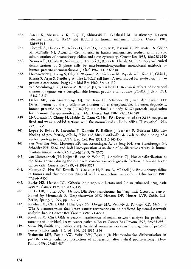
434. Sasaki K, Matsumura K, Tsuji T, Shinozaki F, Takahashi M: Relationship between labeling indices of Ki-67 and BrdUrd in human malignant tumors. Cancer 1988, 62:989-993
435. Riccardi A, Danova M, \Vilson G, Ucci G, Dormer P, Mazzini G, Bntgnatelli S, Girino M, McNally NJ. Ascari E: Cell kinetics in human malignancies studied with in vivo administration of bromodeoxyuridine and flow cytometry. Cancer Res 1988, 48:6238-6245
436. Nemoto R, Uchida K, Shimazui T, Hattori K, Koiso K, Harada M: Immunocytochemical demonstration of S phase cells by anti-bromodeoxyuridine monoclonal .mtibody in human prostate adenocarcinoma. J Urol 1989, 141:337-340
437. Horoszewicz J. Leong S, Chu T, \Vajsman Z, Friedman M, Papsidero L, Kim U, Chiu L, Kakati S, Arya S, Sandberg A: The LNCaP cell line - A new model for shldies on human prostatic carcinoma. Prog Clin Bioi Res 1980, 37:115-132
438. van Steenbntgge GJ, Groen M, Romijn JC, Schroder FH: Biological effects of hormonal treatment regimes on a transplantable human prostatic tumor line (PC-82). J Urol 1984. 131:812-817
439. Gallee MP, van Steenhrugge GJ, ten Kate FJ, Schroder FH, van der Kwast TH: Determination of the proliferative fraction of a transplantable. hormone-dependent, lnunan prostatic carcinoma {PC-82) by monoclonal antibody Ki-67: potential application for hormone therapy monitoring. J Nat! Cancer Inst 1987,79:1333-1340
440. McCormick D, Chong H. Hobbs C, Datta C, Hall PA: Detection of the Ki-67 antigen in fixed and wax-embedded sections with the monoclonal antibody MID1. Histopathol 1993, 222:355-360
441. Lopez F, Bellac F, Lacombe F, Dumain P, Reiffers J. Bernard P, Boisseau MR: The labeling of proliferating cells by Ki67 and MIB-l antibodies depends on the binding of a nuclear protein to the DNA. Exp Cell Res 1994,210:145-153
442. van \X'eerden \X'M, Maerings EP, van Kreuningen A, de Jong FH, van Steenbrugge Gj, Schroder FH: Ki-67 and BrdU incorporation as markers of proliferative activity in human prostate tumor models. Cell Pralif 1993, 26:67-75
443. van Dierendonck JH, Keijzer R, van de Velde CJ, Cornelisse CJ: Nuclear distributi0l1 of the Ki-67 antigen during the cell cycle: comparison with growth fraction in human breast cancer cells. Cancer Res 1989, 49:2999-3006
444. Morstyn G, Hsu SM, Kinsella T, Gratzner H, Russo A, Mitchell JB: Bromodeoxyuridine in tumors and chromosomes detected with a monoclonal antibody. J Clin Invest 1983, 72: 1844-1850
445. Burke HB, Hcnson DE: Criteria for prognostic factors and for an enhanced prognostic system. Cancer 1993, 72:3131-3135
446. Burke HB, Hutter RVP, Henson DE: Breast carcinoma. In: Prognostic factors in cancer. Edited by Hermanek P, Gospodarowicz MK, Henson DE, Hutter RVP, Sobin LH. Berlin, Springer, 1995, pp. 165-176
447. Ravdin PM, Clark GM, I-lilsenbeck SG, Owens MA, Vendcly P, Pandian MR, McGuire \VL: A demonstration that bre<lst C;Ulcer recurrence can bc predicted by neural network analysis. Breast Cancer Res Treatm 1992, 21:47-53
448. Ravdin PM, Clark GM: A practical application of neural network analysis for predicting outcome of individual breast cancer patients. Breast Cancer Res Treatm 1992, 22:285-293
449. Snow PB, Smith DS, Catalona \VJ: Artificial neural networks in the diagnosis af prostate cancer: a pilot study_ JUral 1994, 152:1923-1926
450. \Veinstein MH, Partin A\V, Veltri R\X', Epstein JI: Neuroendocrine differentiation in prostate cancer: enhanced prediction of progression after radical prostatectomy. Hum Pathol 1996, 27:683-687
174
references
451. Abdul M, Anezinis PE, Logothetis CI, Hoosein NM: Gro'Wth inhibition of human prostatic carcinoma cell lines by serotonin antagonists. Anticancer Res 1994, 14:1215-1220
452. Abdul M, Logothetis C], Hoosein NM: Growth inhibitory effects of serotonin uptake inhibitors on human prostate carcinoma cell lines. J Urol 1995, 154:247-250
453. Aprikian AG, Han K, Chevalier S, Bazinet M, Viallet J: Bombesin specifically induces intracellular calcium mobilization via gastrin-releasing peptide receptors in hUlllan prostate cancer cells. J Mol Endocrinol 1996, 16,297·306
454. Harper ME, Glynne-Jones E, Goddard L, Thurston VJ, Griffiths K: Vascular endothelial growth factor (VEGF) expression in prostatic tumours and its relationship to neuroendocrine cells. Br J Cancer 1996, 74:910-916
455. Bohrer M, Riehle HM, Schmoll J, Siegsmund M, Junemann P, AIken P: Relationship between neuroendocrine differentiation and neovascularisation in prostatic carcinoma (abstract #1027). J Urol 1995, 153 (Suppl):485A
456. Nilsson 0, \Vangberg B, Kolby L, Schultz GS, Ahlman H: Expression of transforming gro'Wth factor alpha and its receptor in human neuroendocrine tumors. Int J Cancer 1995, 60,45·51
457. Sheaff MT, Martin JE, Badenoch DF, Baithun SI: fihCG as a prognostic marker in adenocarcinoma of the prostate. J Clin Pathol 1996, 49:329-332
458. Shen R, Dorai T, Olsson CA, Katz A, Buttyan R: Steroid deprivation induces a neuronal cell phenotype in culhtred prostate cancer cells (abstract #1673). Proc Am Assoc Cancer Res 1996, 37,245
459. Fink G, Sumner BEH: Oestrogen and mental state. Nahlre 1996, 383:306 460. Scopsi L, Gullo M, Rilke F, Martin S, Steiner DF: Proprotein convertases (PCl/PC3 and
PC2) in normal and neoplastic human tissues: their use as markers of neuroendocrine differentiation. J Clin Endocrinol Mctab 1995, 80,294·301
461. Hook VYH, Azaryan AV, Hwang SR, Tezapsidis N: Proteases and the emerging role of protease inhibitors in prohormone processing. FASEB J 1994,8:1269-1278
462. Braks JA, Martens GJ: 7B2 Is a neuroendocrine chaperone that transiently interacts with prohormone convert.,e PC2 in the secretory pathway. Cell 1994, 78,263·273
463. Martinez A, Treston AM, Saldise L, Monhlenga LM, Linnoila Rl: Expression of peptidyl-glycine alpha-anlidating monooxygenase (PAM) enzymes in morphological abnormalities adjacent to pulmonary tumors. Am J Pathol 1996, 149:707-716
464. Samos LF, Gkonos PJ: Expression and processing of peptidylglycine alpha-amidating monooxygenase messenger RNA in rat prostate. Prostate 1996,29:101-106
175

SUMMARY
The occurrence of prostate cancer has increased dramatically over the past decade. It is at present the second leading cause of cancer death in the western male population. On average, patients are now diagnosed at an earlier stage of the disease. Most patients with tumors that afC clinically confined to the prostate are treated with curative intention (radical prostatectomy or radiotherapy). Patients with advanced disease are usually hormonally treated by medical or surgical castration which leads to a response in about 40-50% of the patients. Almost all these tumors will show progression with time, however, and the resulting hormone independently growing tumors are difficult to treat.
By the application of prognostic markers attempts are being made to predict patient outcome on the basis of tumor and patient characteristics. This should lead to a better selection of the optimal treatment and thereby to improved care. Tumor stage and) to a lesser extent, tumor grade are currently being used for clinical decision making. Nevertheless, it is very difficult to predict the clinical outcome on an individual basis. This thesis describes clinical studies on the application of additional prognostic tissue markers and experimental studies on neuroendocrine (NE) differentiation and tumor cell proliferation.
Chapter I reviews most of the prognostic tissue markers that have been studied in prostate cancer: tumor stage, tumor volume, tumor grade, tissue prostate specific antigen (PSA) levels, circulating PSA mRNA, nuclear DNA content, nuclear morphometry, p53, p21, epidermal growth factor and its receptor, C-erbB.2, E-cadherin, a-catenin, proliferation by counting mitotic figures and expression of proliferation associated proteins (peNA, Ki-67 and MIB·l), androgen receptor (AR), apoptosis, bcl-2, transforming growth-factor Bland neovascularization. Of each marker the following characteristics are described (if applicable): differences between benign and malignant prostatic tissues, heterogeneity within tumors, the prognostic value in different patient groups and the applicability to biopsy specimens. Despite a large number of published studies, no prognostic marker except for tumor stage and grade can be used clinically. This is partially due to the heterogenous nature of prostate cancer.
NE cells are present in normal, hyperplastic and most of the malignant prostatic tissues. The products secreted by these cells may influence tumor growth. The prognostic value of the extent of NE differentiation was investigated in the radical prostatectomy specimens of 90 patients of whom long-term follow-up data were available (chapter II). The results were correlated with tumor stage, grade and the likelihood of clinical progression and tumor related death. NE cells were identified in 78% of the tumors. The extent of NE differentiation did not correlate with tumor stage, grade or prognosis.
177
The expression of sub-types of CD44 has been related to the metastatic potential of several human tumors. The expression patterns and prognostic value of CD44 were studied in the radical prostatectomy specimens of 97 patients with long-term post operative follow-up (chapter Ill). The expression was also studied in !2 prostate cancer lymph node metastasis specimens. CD44 expression decreased from benign to premalignant to malignant prostatic tissues, and was finally absent in lymph node metastatic tumor cells. Loss of expression of two types of CD44 (CD44s and CD44v6) was correlated with tumor stage, grade and prognosis. If the data were adjusted for differences in tumor grade and pathological stage, CD44s was still a prognostic marker for clinical and PSA based progression. CD44v6 was of no additional prognostic value.
The function of male sex hormones is mediated through the AR. A decreased expression of AR may predict a limited response of prostate cancer to hormonal treatment. This type of treatment induces programmed cell death in prostate cancer cells. This process can be inhibited in cells expressing bcl-2, or other related gene products. Thus, expression of bcl-2 may predict a poor response to hormonal therapy. The prognostic value of bcl-2 and AR expression was studied in tumor specimens of 68 prostate Cancer patients that were obtained before the start of hormonal treatment (chapter IV). Neither AR nor bcl-2 expression was correlated with tumor stage, grade or prognosis. But, a combination of bcl-2 and AR was related to the likelihood of clinical progression, even if the data were adjusted for tumor stage and grade differences.
As described above NE cells may influence prostatic tumor growth. Besides, these cells may also playa role in hormone independent growth of prostate cancer. The concept of NE differentiation in normal, benign enlarged and malignant prostatic tissues is reviewed in chapter V with emphasis on experimental aspects. A model of how NE cells can influence prostate cancer biology is also presented.
For basic studies on NE cells in prostate cancer) experimental tumor models that contain these cells are needed. The presence of NE cells was investigated in a number of experimental human prostatic tumors growing in vivo (in immune deficient mice) or growing in vitro as cell cultures (chapter V1). NE cells were consistently found in two of the in vivo models (pC-295 and PC-3!O). Moreover, in both models the number of NE cells increased if the tumor bearing mouse was castrated. These models are now being used to further study the kinetics and mechanisms underlying NE differentiation.
Tumor growth is the net result of tumor cell divisions (proliferation) and cell death. Ki-67 is an antibody that can be used to microscopically visualize proliferating cells. Its applicability to clinical tumors was thought to be limited to fresh frozen tumor sections whereas routinely obtained tissues are almost always fixed in formalin and embedded in paraffin. The MIB-! antibody recognizes the same protein as Ki-67, but can also be applied to routinely formalin-fixed and paraffin-embedded tissues. The percentages of proliferating cells as determined by Ki·67 and MIB-! were compared in experimental prostate tumors (PC-82) that were growing at different growth rates due to hormonal manipulation of the tumor bearing mice (chapter VII). As expected a strong correlation was found between the two percentages, but, strikingly, MIB-!
!78
percentages were about 4 times as high as Ki-67 percentages. In addition, it was found that Ki-67, like MIB-l, can be applied to routinely processed tissues using microwave pre-treatment. Ki-67 percentages determined in this way doubled the frozen tissue based percentages. Since prostate cancer is in general a slowly growing malignancy, the higher scores obtained with MIB-l may reveal more reliable data.
The general discussion (chapter VIII) gives recommendations about future research on prognostic tissue markers in prostate cancer and addresses the statistical approaches that can be used in this type of research. Also an update of the clinical studies on NE differentiation in prostate cancer and possible future research directions in this field are given.
179

SAMENVATTING
Het aantal gevallen van prostaatkanker is de afgelopen 10 jaar sterk toegenomen. Het vormt op dit moment de tweede doodsoorzaak door kanker onder mannen in de westerse wereld, alhoewel de gemiddelde patient nu wordt gediagnostiseerd met een kleinere tumor dan vroeger. De meeste patienten met cen tumor die beperkt is tot de prostaat, worden behandeld met een in opzet genezende behandeling (radicale prostatectomie of bestraling). Patienten met cen grate of uitgezaaide tumor worden meestal behandeld met castratie door middel van operatie of medicijnen (hormonale therapie). Dit geeft cen verbetering in ongeveer 40-50% van de patienten, maar uiteindelijk gaan vrijwel aIle tumoren toch weer groeien. Deze hormoon onafhankelijk groeiende tumoren zijn slecht te behandelen.
Door de toepassing van prognostische markers wordt geprobeerd een goede voorspelling te doen over het klinische beloop van prostaatkanker gebaseerd op patienten tumor-factoren. Dit zou moeten leiden tot een betere selectie voor optimale therapie en daardoor tot een betere patientenzorg. De stagering (grootte) en gradering worden momenteel gebruikt bij de klinische besluitvorming. Niettemin is het zeer moeilijk om op individuele basis een goede voorspelling te geven. In dit proefschrift worden een aanta! studies beschreven waarin aanvullende prognostische weefselmarkers worden bestudeerd in klinische prostaat tumorcn. Verder bevat het experimentele studies naar neuroendocriene (NE) differentiatie en celdeling in prostaatkanker.
Hoofdstuk I geeft een overzicht van de meeste prognostische weefsel markers die zijn bestudeerd in prostaat kanker: stagering, tumor volume, gradering, weefsel prostaat specifiek antigeen (PSA), cil'culerend PSA mRNA, kern DNA inhoud, kern-vorm metingen, p53, p21, epidermale groeifactor en de receptor daarvoor, C-erbB-2, Ecadherine, ,,-catenine, celdeling zoals bekeken door het tellen van delende cellen en het aantonen van celdeling geassocieerde eiwitten (peNA, Ki-67 en MlB.1), androgeen receptor (AR), apoptotische cell en, bcl-2, transformerende groeifactor-E 1 en vaatnieuwvorming. Van elke marker zijn, voor zover van toepassing, beschreven: verschillen tussen goedaardig en kwaadaardig prostaat weefsel, verschillen binnen de tumor, de prognostische waarde in verschillende patienten-groepen en de toepasbaarheid op biopsieen. Ondanks het grote aantal gepubliceerde studies kan, buiten stagering en in mindere mate gradering niet een van deze markers klinisch toegepast worden. Dit is deels te wijten aan het heterogene karakter van prostaatkanker.
NE cellen zijn aanwezig in normale prostaat klierbuizen en in de meeste prostaat tumoren. De producten die door deze cellen worden uitgescheiden zouden de groei van de omliggende tumorcellen kunnen beYnvloeden. De prognostische waarde van de mate van NE differentiatie van de tumor werd bestudeerd in de radicale prostatectomie preparaten van 90 patienten die na de operatie langdurig werden gevolgd (hoofdstuk Il). De resultaten werden gerelateerd aan de stagering en gradering van de tumor en aan de
181
kans op tumor progressie of overlijden door de tumor. NE cellen werden gevonden in 78% van de tumoren. De mate van NE differentiatie correleerde niet met stagering of gradering van de tumor en oak niet met de prognose.
De expressie van sub·types van CD44 is gecorreleerd met het uitzaaiend vermogen van verschillende kwaadaardige tumoren. De expressie patronen en prognostische waarde van CD44 werd bestudeerd in de radicale prostatectomie preparaten van 97 patienten die langdurig gevolgd werden na de operatie (hoofdstuk IIl). De expressie werd ook bestudeerd in 12 prostaat kanker lymfeklier uitzaaiingen. CD44 expressie nam geleidelijk af gaande van goedaardig naar prostaat intra·epitheliale neoplasie (verondersteld voorstadium van kanker) naar kanker en was uiteindelijk afwezig in de lymfeklier uitzaaiingen. Verminderde expressie van 2 types CD44 (CD44s en CD44v6) was gecorreleerd met stage ring en gradering van de tumor en met de prognose. \X'anneer de gegevens gecorrigeerd werden voor verschillen in stagering en gradering van de tumorenJ bleef CD44s nog steeds van prognostische waarde voar het voorspellen van klinische progressie en PSA gebaseerde progressie.
Mannelijke geslachtshormonen oefenen hun functie uit via de AR. Een verminderde AR expressie zeu kunnen betekenen dat hormonale therapie bij die patient minder succesvol is. Deze therapie induceert geprogrammeerde celdood in prostaat kankercellen. Dit proces kan geremd worden door de expressie van) onder andere, bc1~2. Een prostaat tumor die bc1-2 tot expressie brengt, zou dus minder gevoelig voor hormonale therapie zijn. De prognostische waarde van bcl·2 en AR expressie werd bestudeerd in tumor materiaal van 68 patienten dat was verkregen voordat de hormonale therapie werd gestart. De expressie van bc1-2 noch van AR was gecorreleerd met stagering, gradering of met de prognose. Maar, een combinatie van bd·2 en AR was gerelateerd aan het optreden van klinische progressie, zelfs wanneer de gegevens gecorrigeerd werden voor de stagering en gradering van de tumoren.
Zoals boven reeds werd beschreven, kunnen NE cellen de groei van prostaat tumoren be'invloeden. Daarbuiten kunnen deze cellen ook een rol spelen in het hormoon onafhankelijk groeien van prostaatkanker. Hoofdstuk V bevat een literatuur overzicht over het concept van NE differentiatie in normaal, goedaardig vergroot en kwaadaardig prostaat weefsel, met nadruk op de experimentele aspecten. Tevens wordt in een model gepresenteerd op welke manieren NE cellen de biologie van prostaat kanker kunnen bei"nvloeden.
Voer basale studies naar NE differentiatie in prostaatkanker zijn experimentele tumormodellen die deze cellen bevatten noodzakelijk. Het voorkomen van NE cellen werd bestudeerd in een aantal experimentele prostaat tumoren die groeien in muizen met een slecht functionerend immuun-systeem (in vivo), in een weefselkweek flesje (in vitro) of beiden (hoofdstuk VI). NE cellen werden aangetroffen in 2 van de in vivo modellen (pC·295 en PC-310) en in beide modellen nam het aantal NE cellen toe na castratie van de muis waarop de tumor groeide. Deze modellen worden momenteel gebruikt om experimentele aspecten van NE cellen in prostaatkanker te bestuderen.
182
Tumor groel IS het netto resultaat van tumor celdelingen (proliferatie) en celdood. Ki-67 is een antilichaam dat gebruikt kan worden om delende cellen zichtbaar te maken. De toepassing van Ki-67 op klinische tumoren was beperkt doordat gedacht werd dat het aIleen gebruikt kan worden op vers ingevroren weefsels, terwijl in de kliniek vrijwel aIle weefsels worden gefixeerd in formaline en worden ingebed in paraffine. Het MIB-l antilichaam herkent hetzelfde eiwit als Ki-67, maar kan ook toegepast worden op routinematig formaline gefixeerd, paraffine ingebed materiaal. In experimentele prostaat tumoren (PC-82) welke door hormonale manipulatie van de tumordragende muizen verschillende groeisnelheden hadden, werden de proliferatie percentages bepaald door middel van Ki-67 en MIB-! en met elkaar gecorreleerd (hoofdstuk VIl). Zoais verwacht werd een sterke correlatie tussen de percentages gevonden, maar MIB-l percentages waren ongeveer 4 keer zo hoog ais de Ki-67 percentages. Tevens bleek dat ook Ki-67, net ais MIB-l, op routinematig verkregen materiaal kon worden toegepast na voorbehandeling in cen magnetron. De op deze marrier verkregen Ki·67 percentages waren 2 keer zo haag ais de op het ingevroren materiaal bepaalde percentages. Omdat prostaatkanker meestal langzaam groeit, zijn de hogere MIB-l scores wellicht meer betl'Ouwbaar.
In de aigemene discussie (hoofdstuk VIII) worden aanbevelingen gedaan over toekomstig onderzoek naar prognostische markers voar prostaatkanker. Tevens wordt het overzicht van de klinische studies naar NE differentiatie in prostaatkanker geactualiseerd en worden mogelijke toekomstige onderzoeksrichtingen in dit gebied aangegeven.
183

DANKWOORD
Wie gedurende het keuze co-schap urologie de prognose gesteld zau hebben dat ik de komende 4 jaar in het uro-oncologisch laboratorium door zou brengen, had ik, clinicus pur sang in spe, zeker voor gek versleten. Toch is het mijn promotor prof. Schroder gelukt om in zeer korte tijd mijn belangstelling voor het wetenschappelijk onderzoek te wekken. Ondanks het feit dat de directe betrokkenheid de laatste jaren minder werd, hebben uw inzicht en enorme vakkennis, veel bijgedragen aan het onderzoek. Mijn tweede promotor Theo van del' Kwast dank ik voor de vele urCll die we achter het microscoop doorbrachten met het stageren en graderen van aIle tumoren. Tijdens het beoordelen van de ongeveer 4000 coupes, heb je me niet aileen de kneepjes van de Gleason geheimen bijgebracht. Veel leerzamer nog waren aile discussies die deels over prostaat carcinoom of andere pathologische problematiek gingen, maar die vooral betrekking hadden op politiek, wereldproblemen en wat al niet meer. Toch is het je niet gelukt mij zo ver te krijgen de opleiding pathologic te gaan doen. Ik hoop dat de samenwerking de komende jaren kan worden voortgezet. Gert Jan, ik ben blij dat je mijn co-promotor wilt zijn. De samenwerking was altijd bijzonder prettig en zal zich ook de komende jaren middels het neuroendocriene project voortzetten. Ik hoop dat je de sams wat al te grote zelfstandigheid van deze promovendus niet als een probleem hebt ervaren.
De tijd op het lab heb ik als zeer plezierig ervaren, vaak zelfs te plezierig gezien de gemiddelde lengte van de koffie- en lunchpauzes. Zonder de anderen tekort te willen doen, wit ik cen aantal mensen met name noemen. Monique en Nic81e, die delen van het werk hebben verricht. Antoine Simons, for performing CD44 RT-PCR analysis. Boudewijn, Hans en Johan, de opeenvolgende kamergenoten die de computer het grootste deel van de tijd bezet zagen. Corrina, die ondanks moeilijke tijden steeds als een duizendpoot het proefdier gebeuren regelde. De bewoners van EE 1006, voor de vele al dan niet zinvolle discussies. Toch was ik meer tevreden met mijn plekje in EE 1000, anders was dit boekje zeker nog niet af geweest!
De samenwerking met de pathologie was behalve noodzakelijk ook heel prettig. Patrick Jansen en Cor van Vroonhoven leerden mij alles over immunohistochemie en bij Frieda van der Ham kon ik altijd antilichamen komen bietsen. Frank van der Panne verrichtte veel fotowerk. Ton de Jong deed de electronen microscopie en hielp bij het zoeken naar de neuroendocriene speld in de prostaat hooibergen.
Annet Verkerk en Marjan Hettinga van het trial bureau wi! ik bedanken voor het secuur bijhouden van de radicale prostatectomie database. Jacques Bogdanowicz en Cees van Krimpen, voor de Rotterdam-Delft connection en hun bijdrage aan hoofdstuk IV. De SUWO, voor aile financieIe support, onder andere voor de buitenlandse bezoeken. De overige sponsors, voor het bekostigen van deze publicatie. De leescommissie bestaande uit prof. dr F.J.W. ten Kate, prof. dr S.W.J. Lamberts en prof.
185
dr J.A. Schalken, voor de snelle beoordeling van het manuscript. Monique Oomen en Mieke Kalkman, die mijn paranimlen willen zijn. Christine Dijkstra, die met penseel en acrylverl de link tllssen lab en kliniek treffend kon weergeven. De stal en assistenten van de aldeling algemene heelkunde van het Academisch Ziekenhuis Maastricht, die moeten ervaren dat het verschil tllSsen AIO en AGIO slechts een letter, maar toch een wereld van verschil is, en die de promovendus nu moeten helpen een goede dokter te worden.
Lieneke, gedurende de algelopen 4 jaar hebben we beiden de ups en downs van het do en van wetenschappelijk onderzoek mogen ervaren, hoewel de ongelijkheid in de wereld ook hier gold: jij kreeg voornamelijk de downs voor je kiezen. Ik heb grote bewondering voor je inzet en inzicht in een gecompliceerd onderzoeksgebied. Alhoewel ik nu een kleine voorsprong heb opgebouwd, zal jouw boekje vast niet lang op zich laten wachten.
Tenslotte een woord van dankbaarheid aan mijn vader en moeder voar de onvoorwaardelijke steun aan hun oudste zoon die, toen hij nog nauwelijks kon praten al zei dat hij chirurg wilde worden, vervolgens conservatorium ging doen, maar uiteindelijk toch geneeskunde ging studeren en die nu, na vier jaar onbegrijpelijk werk in een ivoren toren te hebben gedaan, eindelijk op weg lijkt een yak te leren.
186
CURRICULUM VITAE
De schrijver van dit proefschrift werd geboren op 6 augustus 1965 te Rotterdam, Pernis. Hij behaalde het V\'(!O diploma aan de christelijke scholengemeenschap Maarten Luther te Rotterdam in 1985. In datzelfde jaar werd een aanvang gemaakt met de studie geneeskunde aan de Erasmus Universiteit te Rotterdam alwaar in 1990 het doctoraal examen en in 1992 het arts examen werden behaald. Van 1993 tot en met 1996 was hij werkzaam als AIO bij de afdeling urologie van de Erasmus UniversiteA te Rotterdam onder begeleiding van prof. dr F.H. Schroder, dr G.]. van Steenbrugge en prof. dr r.I-I. van del' Kwast. Gedurende deze peri ode is dit proefschrift tot stand gekomen. Momenteel werkt hij als arts-assistent algemene heelkunde in het Academisch Ziekenhuis Maastricht (opleider prof. dr G. Kootstra) in het kader van de opleiding urologie welke gedaan zal worden in het Academisch Ziekenhuis Rotterdam (opleider Prof. dr F.I-I. Schroder) en het Sint Franciscus Gasthuis Rotterdam (opleider dr J.I-l.M. Blom).
187

ADDITIONAL PUBLICATIONS
GH Mickisch, MA Noordzij, A van der Gaast, P Gebreamlack, KU Kohrmann, E Mogler-Drautz, H Kupper, FH Schroder: Dexverapamil to modulate vinblastine resistance III metastatic renal cell carCinoma. J Cancer Res Clin Oneol 1995;121supp13:Rll-6
MA Noordzij, GH Mickisch: Dexverapamil to overcome documented vinblastine resistance in metastasized renal cell cancer. In: G Staehler, SPomer and H Rubben (eds): Basic and clinical urological oncology. Springer Verlag, Berlin, Germany, in press
P Koivistol, J Kononen, C Palmberg, T Tammela, E Hyytinen, J Isola, J Trapman, MA Noordzij, K Cleutjens, T Visakorpi, O·P Kallioniemil: Androgen receptor gene amplification: a possible molecular failure of androgen deprevation therapy in prostate cancer. Cancer Res) in press
VIDEOTAPES
GH Jordan, JLHR Bosch, MA Noordzij, FH Schroder: Operative treatment of Peyronie's disease: demonstration of two different surgical procedures. EUR / AZR, 1994
G Pazzerini, JM Nijman, MA Noordzij: Pediatric Urology. EUR / AZR, 1995
J DeKernion, GH Mickisch, MA Noordzij: Surgical treatment of renal cell cancer. In preparation
189


Related Documents