The Anti-Inflammatory and Antibacterial Basis of Human Omental Defense: Selective Expression of Cytokines and Antimicrobial Peptides Abhijit Chandra 1 , Ritesh Kumar Srivastava 2,3 , Mahendra Pratap Kashyap 2,3 , Raj Kumar 4 , Rajeshwar Nath Srivastava 5 , Aditya Bhushan Pant 2 * 1 Department of Surgical Gastroenterology, Erstwhile KG Medical College, CSM Medical University, Lucknow, India, 2 Indian Institute of Toxicology Research, Lucknow, India, 3 Council of Scientific & Industrial Research, New Delhi, India, 4 Department of Basic Sciences, The Commonwealth Medical College, Scranton, Pennsylvania, United States of America, 5 Department of Orthopaedic Surgery, Erstwhile KG Medical College, CSM Medical University, Lucknow, India Abstract Background: The wound healing properties of the human omentum are well known and have extensively been exploited clinically. However, the underlying mechanisms of these effects are not well understood. We hypothesize that the omentum tissue promotes wound healing via modulation of anti-inflammatory pathways, and because the omentum is rich in adipocytes, the adipocytes may modulate the anti-inflammatory response. Factors released by human omentum may affect healing, inflammation and immune defense. Methodology: Six human omentum tissues (non obese, free from malignancy, and any other systemic disorder) were obtained during diagnostic laparoscopies having a negative outcome. Healthy oral mucosa (obtained from routine oral biopsies) was used as control. Cultured adipocytes derived from human omentum were exposed to lipopolysaccharide (LPS) (1–50 ng/mL) for 12–72 hours to identify the non-cytotoxic doses. Levels of expression (mRNA and protein) were carried out for genes associated with pro- and anti-inflammatory cytokine responses and antibacterial/antimicrobial activity using qRT- PCR, western blotting, and cell-based ELISA assays. Results: The study shows significant higher levels of expression (mRNA and protein) of several specific cytokines, and antibacterial peptides in the omentum tissues when compared to oral sub-mucosal tissues. In the validation studies, primary cultures of adipocytes, derived from human omentum were exposed to LPS (5 and 10 ng/mL) for 24 and 48 h. The altered expressions were more pronounced in cultured adipocytes cells when exposed to LPS as compared to the omentum tissue. Conclusions/Significance: Perhaps, this is the first report that provides evidence of expressional changes in pro- and anti- inflammatory cytokines and antibacterial peptides in the normal human omentum tissue as well as adipocytes cultured from this tissue. The study provides new insights on the molecular and cellular mechanisms of healing and defense by the omentum, and suggests the potential applicability of cultured adipocytes derived from the omentum for future therapeutic applications. Citation: Chandra A, Srivastava RK, Kashyap MP, Kumar R, Srivastava RN, et al. (2011) The Anti-Inflammatory and Antibacterial Basis of Human Omental Defense: Selective Expression of Cytokines and Antimicrobial Peptides. PLoS ONE 6(5): e20446. doi:10.1371/journal.pone.0020446 Editor: Markus M. Heimesaat, Charite ´, Campus Benjamin Franklin, Germany Received February 26, 2011; Accepted April 19, 2011; Published May 24, 2011 Copyright: ß 2011 Chandra et al. This is an open-access article distributed under the terms of the Creative Commons Attribution License, which permits unrestricted use, distribution, and reproduction in any medium, provided the original author and source are credited. Funding: The financial support by Council of Scientific & Industrtail Research, New Delhi, India, through Supra-Institutional Project SIP-08 is acknowledged. The funders had no role in study design, data collection and analysis, decision to publish, or preparation of the manuscript. Competing Interests: The authors have declared that no competing interests exist. * E-mail: [email protected] Introduction The human omentum has been classically regarded as the abdominal policeman [1] with instances of its reaching and plugging inflamed intra-abdominal organs [2]. Recent clinical evidence has suggested spontaneous sealing of intestinal perfora- tion in premature neonates by the omentum alone, where just aspiration was adequate [2]. Omentum has been shown to secrete many biological agents including vascular endothelial growth factor (VEGF), different other growth factors, and cytokines [3,4,5]. The omentum milky spots are conglomerates of macrophages respon- sible for its local immune response and anti-inflammatory properties [4,6]. In the omentum, the non adipose cells of the stromal vascular fraction, preadipocytes, and macrophages are thought to secrete cytokines [7,8,9]. However, recent data suggests the involvement of adipocytes in the inflammatory response [10,11]. Even though the role of omentum tissue in wound healing and injury repair is well established, the cellular and molecular mechanisms underlying these properties remain unclear. We hypothesize that the omentum tissue promotes wound healing through anti-inflammatory pathways, and because the omentum is rich in adipocytes, the adipocytes may modulate the anti-inflammatory responses [12,13]. The omentum may also possess an intrinsic direct antibacterial activity of its own [5]. Thus, the present studies were carried out to determine the levels of expression of selected pro- and anti-inflammatory cytokines PLoS ONE | www.plosone.org 1 May 2011 | Volume 6 | Issue 5 | e20446

Welcome message from author
This document is posted to help you gain knowledge. Please leave a comment to let me know what you think about it! Share it to your friends and learn new things together.
Transcript

The Anti-Inflammatory and Antibacterial Basis of HumanOmental Defense: Selective Expression of Cytokines andAntimicrobial PeptidesAbhijit Chandra1, Ritesh Kumar Srivastava2,3, Mahendra Pratap Kashyap2,3, Raj Kumar4, Rajeshwar Nath
Srivastava5, Aditya Bhushan Pant2*
1 Department of Surgical Gastroenterology, Erstwhile KG Medical College, CSM Medical University, Lucknow, India, 2 Indian Institute of Toxicology Research, Lucknow,
India, 3 Council of Scientific & Industrial Research, New Delhi, India, 4 Department of Basic Sciences, The Commonwealth Medical College, Scranton, Pennsylvania, United
States of America, 5 Department of Orthopaedic Surgery, Erstwhile KG Medical College, CSM Medical University, Lucknow, India
Abstract
Background: The wound healing properties of the human omentum are well known and have extensively been exploitedclinically. However, the underlying mechanisms of these effects are not well understood. We hypothesize that the omentumtissue promotes wound healing via modulation of anti-inflammatory pathways, and because the omentum is rich inadipocytes, the adipocytes may modulate the anti-inflammatory response. Factors released by human omentum may affecthealing, inflammation and immune defense.
Methodology: Six human omentum tissues (non obese, free from malignancy, and any other systemic disorder) wereobtained during diagnostic laparoscopies having a negative outcome. Healthy oral mucosa (obtained from routine oralbiopsies) was used as control. Cultured adipocytes derived from human omentum were exposed to lipopolysaccharide (LPS)(1–50 ng/mL) for 12–72 hours to identify the non-cytotoxic doses. Levels of expression (mRNA and protein) were carried outfor genes associated with pro- and anti-inflammatory cytokine responses and antibacterial/antimicrobial activity using qRT-PCR, western blotting, and cell-based ELISA assays.
Results: The study shows significant higher levels of expression (mRNA and protein) of several specific cytokines, andantibacterial peptides in the omentum tissues when compared to oral sub-mucosal tissues. In the validation studies, primarycultures of adipocytes, derived from human omentum were exposed to LPS (5 and 10 ng/mL) for 24 and 48 h. The alteredexpressions were more pronounced in cultured adipocytes cells when exposed to LPS as compared to the omentum tissue.
Conclusions/Significance: Perhaps, this is the first report that provides evidence of expressional changes in pro- and anti-inflammatory cytokines and antibacterial peptides in the normal human omentum tissue as well as adipocytes culturedfrom this tissue. The study provides new insights on the molecular and cellular mechanisms of healing and defense by theomentum, and suggests the potential applicability of cultured adipocytes derived from the omentum for future therapeuticapplications.
Citation: Chandra A, Srivastava RK, Kashyap MP, Kumar R, Srivastava RN, et al. (2011) The Anti-Inflammatory and Antibacterial Basis of Human Omental Defense:Selective Expression of Cytokines and Antimicrobial Peptides. PLoS ONE 6(5): e20446. doi:10.1371/journal.pone.0020446
Editor: Markus M. Heimesaat, Charite, Campus Benjamin Franklin, Germany
Received February 26, 2011; Accepted April 19, 2011; Published May 24, 2011
Copyright: � 2011 Chandra et al. This is an open-access article distributed under the terms of the Creative Commons Attribution License, which permitsunrestricted use, distribution, and reproduction in any medium, provided the original author and source are credited.
Funding: The financial support by Council of Scientific & Industrtail Research, New Delhi, India, through Supra-Institutional Project SIP-08 is acknowledged. Thefunders had no role in study design, data collection and analysis, decision to publish, or preparation of the manuscript.
Competing Interests: The authors have declared that no competing interests exist.
* E-mail: [email protected]
Introduction
The human omentum has been classically regarded as the
abdominal policeman [1] with instances of its reaching and
plugging inflamed intra-abdominal organs [2]. Recent clinical
evidence has suggested spontaneous sealing of intestinal perfora-
tion in premature neonates by the omentum alone, where just
aspiration was adequate [2]. Omentum has been shown to secrete
many biological agents including vascular endothelial growth factor
(VEGF), different other growth factors, and cytokines [3,4,5]. The
omentum milky spots are conglomerates of macrophages respon-
sible for its local immune response and anti-inflammatory
properties [4,6]. In the omentum, the non adipose cells of the
stromal vascular fraction, preadipocytes, and macrophages are
thought to secrete cytokines [7,8,9]. However, recent data suggests
the involvement of adipocytes in the inflammatory response
[10,11]. Even though the role of omentum tissue in wound healing
and injury repair is well established, the cellular and molecular
mechanisms underlying these properties remain unclear.
We hypothesize that the omentum tissue promotes wound
healing through anti-inflammatory pathways, and because the
omentum is rich in adipocytes, the adipocytes may modulate the
anti-inflammatory responses [12,13]. The omentum may also
possess an intrinsic direct antibacterial activity of its own [5].
Thus, the present studies were carried out to determine the levels
of expression of selected pro- and anti-inflammatory cytokines
PLoS ONE | www.plosone.org 1 May 2011 | Volume 6 | Issue 5 | e20446
(mRNA and protein) and antibacterial peptides genes in human
omentum tissue derived from normal, disease-free human subjects,
and cultured adipocytes obtained from these tissues. Further, the
expression and up-regulation of these genes were also assessed in
primary adipocyte cultures derived from omentum exposed to
bacterial endotoxin. Our results show that adipocytes modulate
the expression of several pro- and anti-inflammatory cytokines and
anti-microbial peptides.
Results
Effects of LPS (lipopolysaccharide from Escherichia coli)exposure on cell viability
To determine the non-cytotoxic doses of LPS exposure to
cultured adipocyte cells derived from human omentum tissues, we
carried out MTT assay. The results of cytotoxicity assays are
shown in Figure S1. It is evident from Figure S1 that there is no
significant reduction in cell viability due to treatment of LPS at
lower concentrations (#20 ng/ml) up to a time period of 72 h of
exposure. However, a concentration- and time-dependent de-
crease in percent cell viability was observed after 12 h exposure of
cells to LPS at higher concentrations (30–50 ng/ml). The most
significant reduction in cell viability was observed in cells exposed
to LPS at 50 ng/ml concentration for a period of 72 h, which was
found to be approximately 50% of controls. Together, these results
suggest that LPS exposure of #20 ng/ml fail to produce any
significant toxicity to these adipocyte cells derived from human
omentum tissues, and can be used without the concerns of LPS-
induced effects.
Transcriptional changes in human omentum tissues andadipocyte cells derived from human omentum tissues
To test our hypothesis that human omentum is capable of
producing intrinsic anti-inflammatory response, we determined
whether the major cytokines are induced in these tissues. Since
our cytotoxicity data suggest that lower doses (5 and 10 ng/ml) of
LPS do not significantly alter cell viability, we also treated
adipocyte cells derived from human omentum tissues with these
doses of LPS to determine whether cells are capable of inducing
mRNA levels of specific cytokines even at these low doses. Our
results are shown in Figure 2. We first compared the mRNA
expression levels of inflammatory cytokines (IL-1b, IL-2, IL-4, IL-
8, IL-10, TNF-a, and GM-CSF) of human omentum tissues with
those of oral sub-mucosal tissues (Figure 2 a). Real Time PCR
(qRT-PCR) was used to determine mRNA levels using specific
primers (Table 1S) for each cytokine. It is evident from Figure 2 a
that except for IL-2, IL-4 and IL-10, the levels of mRNA
expression were significantly higher (,2 fold or more; p#0.01)
for all other cytokines tested in omentum tissues when compared
to oral sub-mucosal tissues. These results suggest that omentum
tissues possess higher intrinsic level of inflammatory cytokines,
which may be responsible for its wound healing properties. We
further determined the levels of these cytokines in adipocyte cells
derived from human omentum tissues and exposed to low non-
toxic doses of LPS. Our results show that even at such low doses,
LPS-induced alterations in the mRNA expression of genes
associated with inflammatory cytokines were significantly higher.
A dose- (5 and 10 ng/ml) dependent expression of these
inflammatory cytokines in cells exposed to LPS for 48 h is shown
in Figure 2 b. The expression levels were significantly higher for
all the cytokines studied at 48 h in comparison to 24 h (data not
shown for 24 h).
Expression of anti-microbial/anti-bacterial peptide genesin human omentum tissues and adipocyte cells derivedfrom human omentum tissues
To determine whether the levels of specific anti-microbial/anti-
bacterial peptides are induced in human omentum, we carried out
Real Time PCR (qRT-PCR) assay, using specific primers (Table
S2) for LL-37, HNP 1-3, HBD-1, and HBD-2. The expression of
genes associated with these antimicrobial activities were found to
be significantly higher (2–3 fold; p#0.01) in human omentum
tissues compared to oral sub-mucosal tissues (Figure 3 a). Similar
results were also obtained in adipocyte cells derived from human
omentum tissues when exposed to low doses (5 and 10 ng/ml) of
LPS for 48 h (Figure 3 b). These results clearly demonstrate that
the expressions of anti-microbial/anti-bacterial peptides were
significantly elevated in human omentum, which became more
pronounced in the adipocyte cells derived from human omentum
tissues.
Translational changes in human omentum tissues andadipocyte cells derived from human omentum tissues
We further determined whether these alterations in the level of
mRNA expressions of these cytokines are taking place at the level
of protein expression. To determine the level of protein
expressions of each of these cytokines (IL-1b, IL-2, IL-4, IL-8,
IL-10, TNF-a, and GM-CSF), we carried out immunoreactions
using specific antibodies. Our results (Figure 4 a & 4 b) showed
that except for IL-4 and IL-10, protein expression of all other
cytokines tested in omentum tissues when compared to oral sub-
mucosal tissues were significantly higher (,2 fold or more;
p#0.01). These results are having similar trend to those observed
for mRNA levels (Figure 2 a); however, the magnitude of induction
in the expression is higher. Results of western blot analyses in
omentum-derived cultured cells exposed to LPS (5 and 10 ng/ml
for 48 h) showed significant alteration in protein expressions for all
cytokines tested including IL-2, IL-4 and IL-10 when compared to
untreated cells (Figure 5 a–g). However, the increased levels of
expressions of these cytokines were significantly higher than those
of elevated levels observed in omentum tissues. Together, our
results demonstrate that higher intrinsic levels of cytokines
observed at the mRNA levels were translated into protein
expression.
Translational changes in the expression of antimicrobial/anti-bacterial peptide gene proteins in human omentumtissues and adipocyte cells derived from humanomentum tissues
Experiments were carried out to ascertain whether the
alterations observed at transcriptional level for the expressions of
antimicrobial/ anti-bacterial peptide gene are translational to the
level of protein expression or not. To determine the level of
protein expressions of each of these antimicrobial/ anti-bacterial
peptide gene (LL-37, HNP1-3, hBD1 & hBD2), we carried out
western blot analyses using specific antibodies. Our results
(Figure 6 a) show significantly higher (,2 fold or more; p#0.01)
protein expression of all antimicrobial/ anti-bacterial peptide
tested in omentum tissues when compared to oral sub-mucosal
tissues. These results show similar trends as observed for mRNA
levels (Figure 3 a). Results of western blot analyses in omentum-
derived cultured cells exposed to LPS (5 and 10 ng/ml for 48 h)
showed significant alteration in protein expressions for all
antimicrobial/ anti-bacterial peptide genes tested when compared
to untreated cells (Figure 6 b–e). The levels of protein expressions
of these antimicrobial/ anti-bacterial peptides in omentum derived
Human Omental Defense: Expression Studies
PLoS ONE | www.plosone.org 2 May 2011 | Volume 6 | Issue 5 | e20446

cultured adipocytes were at par to the levels in observed in
omentum tissues. Together, our results demonstrate that higher
intrinsic levels of antimicrobial/ anti-bacterial peptides observed at
the mRNA levels were translated into protein expression.
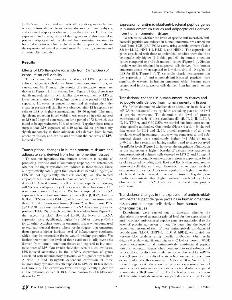
Detection of secreted levels of cytokine profiles inadipocyte cells derived from human omentum tissues
We further analyzed the secreted cytokine profiles directly in
cells. The results of cytokine expressions in the supernatants of
cultures growing with or without LPS exposure are shown in
Figure 7. Most significant induction in the level of TNF-a(4961.3, 6563.7 pg/ml) was observed following LPS (5 and
10 ng/ml) exposure for 24 h. Other cytokines showing significant
increases were IL-1b (3562.1, 5063.2 pg/ml), IL-4 (3162.6,
4563.4 pg/ml), IL-8 (3761.6, 5562.8 pg/ml), and GM-CSF
(2261.4, 2962.6 pg/ml), respectively at 24 h (Figure 7 a). The
increased levels were sustainable for up to 48 h of exposure and
levels of IL-2 (2663.2, 3763.4 pg/ml) and IL-10 (3261.5,
3662.3 pg/ml.) further increased at 48 h (Figure 7 b).
Discussion
The wound healing properties of omentum have been well
described [1,2,4]. The omentum also has a distinct role in the
sealing of intestinal perforation in premature neonates [2] and in
healing resistant sternal wounds (following infected sternotomy
incisions) [6,14]. Experimental studies have demonstrated that
omentum can potentially prevent or reduce occurrence of
infection following artificial aortic graft implantation [15,16].
However, the underlying cellular and molecular mechanisms
involved in the omentum- mediated prevention of infections and
healing of injured organs remains poorly understood. We
hypothesize that the high levels of pro- and anti-inflammatory
cytokines under normal physiological conditions in the omentum
(compared to the control), and under pathological conditions (a
Figure 1. Schematic diagram showing experimental design.doi:10.1371/journal.pone.0020446.g001
Human Omental Defense: Expression Studies
PLoS ONE | www.plosone.org 3 May 2011 | Volume 6 | Issue 5 | e20446

significant induction of cytokines and antimicrobial/antibacterial
peptides) are likely to play an important role in the omental
defense mechanism.
Our results confirm this pattern of expression of several
cytokines and antimicrobial/antibacterial peptide in naıve omen-
tal tissue to support this hypothesis. Furthermore, induction of
cytokines and anti-microbial peptides in the omentum appears
exclusive to the adipocytes, since these effects were greatly
enhanced in cultured adipocytes exposed to LPS. We identified
the non cytotoxic doses of LPS (5 and 10 ng/ml) in isolated
omentum cells. Our data of LPS cytotoxicity are in agreement
with the study conducted by Melzig and Loose [17] on bovine
aortic endothelial cells. Usually non- adipocyte cells or SV cells are
considered to be the main source of pro-inflammatory adipose
adipokine release by obese adipose tissue [7,8,9,18,19]. However,
Bassols et al. [10] have shown that obese human omental
differentiated adipocytes spontaneously release the pro-inflamma-
tory cytokines IL-6 and MIF, and the chemokines IL-8, GRO, and
MCP-1 [8,10,20]. Another study by Sopaskis et al. [21] showed
that human subcutaneous adipose tissue even from non obese
individuals release substantial amounts of IL-6, IL-8, and IL-1 RA
and the gene expression of these cytokines, like that of IL-1b and
PAI-1 is regulated by TNF-a. [21]. In the present investigations,
the expression and up regulation of IL-1b, TNFa, IL-8, IL-2, IL-4
mRNAs and protein was recorded in non obese disease free
omentum tissue as well as cultured adipocytes. We observed
higher protein expression levels for tested cytokines compared to
levels of mRNA expression in omentum tissues. It is well
documented that cytokine mRNAs are expressed transiently and
at low levels because they are tightly regulated and rapidly
processed [22,23,24]. Whereas, the proteins of cytokines are
known to express and accumulate in the cytoplasm and cell surface
till secretion required [25]. Therefore, our results may not be an
unusual phenomenon. The trends of Western blot analysis were in
accordance with the result obtained by Fain et al. [7]. Our results
show significant induction in the expression of IL-1b, TNF-a and
IL-8 in cultured adipocytes following LPS (5 and 10 ng/ml)
exposure for 24 and 48 h. Similar results have already been
Figure 2. mRNA expression of inflammatory markers in human omentum tissue and cultured omental cells. Altered expression ofmRNA of genes involved in inflammation in omentum tissue and compared with human oral tissue (Figure 2 a). Alterations expression of markergenes in omentum derived cultured adipocytes exposed to LPS for 48 h (Figure 2 b). Real Time quantitative PCR (RTq-PCR) was performed in triplicateby 26Power SYBR Green PCR master mix. b-actin was used as internal control to normalize the data and LPS induced alterations in mRNA expressionare expressed in fold change. Normal oral submucosal tissue was used to compare the changes in cytokines. *P,0.05- significant, **P,0.01- highlysignificant.doi:10.1371/journal.pone.0020446.g002
Human Omental Defense: Expression Studies
PLoS ONE | www.plosone.org 4 May 2011 | Volume 6 | Issue 5 | e20446

reported for the release of TNF a and IL-8 upon exposure of LPS
in human differentiated omentum adipocytes [10]. It is well known
that adipocytes express toll like receptor 4 (TLR-4) through which
LPS activate intracellular inflammation pathways [26]. The role of
cytokines-IL-8, TNFa and IL-1b has been suggested in the
recruitment of monocytes into adipose tissue [21].
Antimicrobial peptides are effector molecules of innate
immunity with microbicidal and pro- or anti-inflammatory
activities. There is evidence that one such multifunctional peptide,
LL-37, induces angiogenesis, a process essential for host defense,
wound healing, and tissue repair [27,28]. In normal tissue, these
peptides have a negligible expression, but this may be triggered by
injury or inflammation of the organ, and their expression or
activation is essential for the organ to resist microbial infection
[27]. Omental adipocytes could play a major role in protecting
against infection by generating defensin (DEFA1-3) [5]. We also
found that LPS exposure for 24 and 48 h induces significant
expressions of LL-37 in omentum derived cultured adipocytes. To
validate the expression of additional antimicrobial peptides in
omental tissue, we evaluated the expression of HBD-1 and HBD-
2, and found up regulation of these peptides at both m-RNA and
protein levels. Our study has shown that normal (non diseased,
non obese) human omentum has constitutive expression of several
cytokines including (IL-1b, IL-4, IL-8, TNF-a, and GM-CSF), and
antimicrobial peptides (LL-37, HNP-1, HBD-1 and 2). The
cytokines and antimicrobial peptide surge is dose and time
dependent (in response to LPS), and may be directly involved in
the fight against bacteria.
In summary, for the first time, we demonstrate a significantly
high expression (mRNA and protein) of selected pro- and anti-
inflammatory cytokines, and antimicrobial peptides in normal
human omentum tissue, when compared to control (human oral
mucosal tissue). Previous reports have predominantly focused on
cytokine expression in obese subjects [29]. Obese subjects have
altered metabolism and are prone to a number of diseases,
including cardiovascular disease and diabetes, and therefore,
hardly are models to study wound healing. Moreover, previous
studies did not evaluate a wide range of pro- and anti-
inflammatory cytokines, and anti-microbial proteins, as was
undertaken in the present study.
Figure 3. mRNA expression of antimicrobial/antibacterial peptide markers in human omentum tissue and cultured omental cells.Alerted expression of mRNA of antimicrobial peptide genes in omentum tissue and compared with human oral tissue (Figure 3 a). Alterationsexpression of marker genes in omentum derived cultured adipocytes exposed to LPS for 48 h (Figure 3 b). Real Time quantitative PCR (RTq-PCR) wasperformed in triplicate by 26Power SYBR Green PCR master mix. b-Actin (ACTB) was used as internal control to normalize the data and LPS inducedalterations in mRNA expression are expressed in fold change. Normal oral submucosal tissue was used to compare the changes in antimicrobialpeptide activity. *P,0.05- significant, **P,0.01- highly significant.doi:10.1371/journal.pone.0020446.g003
Human Omental Defense: Expression Studies
PLoS ONE | www.plosone.org 5 May 2011 | Volume 6 | Issue 5 | e20446

The expression levels of these cytokines and antimicrobial
peptides were significantly higher in cultured omentum adipocytes
than intact omentum tissue. LPS exposure (at non-toxic doses) to
primary cultured adipocytes induced significantly higher levels of
expression of cytokines, and anti-microbial peptides. This surge
was seen to be LPS dose (5 and 10 ng/ml) and time dependent (24
and 48 h). Our data supports the view that these pro-inflammatory
cytokines and antimicrobial peptides are involved in the wound
healing properties of human omentum and that their expression is
modulated by adipocytes. Together, our studies may provide a
potential cellular and molecular mechanism for the defense
mechanisms of omentum tissues in wound healing and infection,
which in turn may have significant clinical applications.
Materials and Methods
Reagents and consumablesAll the specified diagnostic kits were purchased from e-
Biosciences Chemical Company Pvt. Ltd. St. USA. Culture
medium DMEM F-12, antibiotics-antimycotic solution and fetal
bovine serum were purchased from Gibco BRL, USA. Culture
wares and other plastic consumables used in the study were
procured commercially from Nunc, Denmark. Milli Q water
(double distilled deionized water) was used in all the experiments.
All the DNA primers and Lipopolysaccharide (LPS) were
purchased from Sigma Aldrich, St. Louis, MO, USA.
Ethical clearance for collection and transportation ofhuman tissues
The protocol for human tissue collection was approved by the
‘Human Ethics Committee of Chhatrapati Shahuji Maharaj
Medical University, Lucknow, India’. Six human omentum tissues
were obtained during diagnostic laparoscopies having a negative
outcome (after obtaining informed written consent from all the
patients). All the patients were male between the age of 48.465.6
years (mean 6 SE). They were non obese, free from malignancy or
any other systemic disorder, and were not on any medication/
medical treatments at the time of sample collection. Healthy oral
mucosa (obtained from routine oral biopsies) was used as control.
One part of tissue specimen collected was persevered and
immediately transported to In Vitro Toxicology Laboratory,
Indian Institute of Toxicology Research, Lucknow, India, for
further processing. Tissues were collected in sterile Dulbecco’s
Modified Eagle’s Medium (DMEM) supplemented with 10% fetal
bovine serum (FBS) and antibiotic-antimycotic solution (Gibco
BRL, USA), and immediately processed for isolation and
cultivation of cells.
Cell culture and exposureHuman omentum tissues (10 g) were cut in small pieces and
placed in a sterile petridish containing a thin layer of minimal
essential medium with 10% fetal bovine serum. The omentum
tissues were fractioned into adipocytes and stromal vascular (SV)
cells using 0.2% collagenase and 0.125% trypsin for 30 min at
37uC as described by Maury et al., 2007 [30]. Adipocytes were
collected by centrifugation at 1006g for 5 min., the pellet of
packed cells re-suspended in poly-L-lysine pre-coated six-well
culture plates in complete minimal essential medium, and
incubated at 37uC in an atmosphere of 95% air-5% CO2 for
attachment. Growth was permitted to continue until cells attained
a confluent monolayer, at which time they were trypsinzed (trypsin
0.05%–EDTA 0.53 mM) and passaged into T-25 culture flasks to
Figure 4. Protein expression of markers associated with inflammation in human omentum tissue. (a) Alterations in the expression ofproteins involved in the induction of inflammatory cytokines in human omentum tissue. Normal oral submucosal tissue was used to compare thechanges in protein of pro and anti-inflammatory cytokines (Figure 4 a). Lane (1): Normal oral submucosal tissue; Lane (2): Human omentum tissue.Molecular weight of protein studied: IL-1b (17 kDa), IL-2 (15 kDa) IL-4 (17 kDa), IL-8 (11 kDa), IL-10 (20 kDa), TNF-a (26 kDa) GM-CSF (16 kDa) and b-Actin (42 kDa) for normalization. (b) Relative quantification of alterations in the protein expression of cytokines in human omentum tissue. Normaloral submucosal tissue was used to compare the changes in protein of pro-inflammatory cytokines. b-Actin was used as internal control to normalizethe data. Quantification (densitometry) was done in Gel Documentation System (Alpha Innotech, USA) with the help of AlphaEaseTM FC StandAloneV.4.0 software. *P,0.05- significant, **P,0.01- highly significant.doi:10.1371/journal.pone.0020446.g004
Human Omental Defense: Expression Studies
PLoS ONE | www.plosone.org 6 May 2011 | Volume 6 | Issue 5 | e20446

Figure 5. Constitutive expression and inducibility of proteins of inflammatory cytokines in primary cultures of omentum derivedadipocytes exposed to LPS. Lane (1): Unexposed control cells; Lane (2): LPS (5 ng/ml) exposed omentum cells; (3) LPS (10 ng/ml) exposedomentum cells. Molecular weight of protein studied: IL-1b (17 kDa), IL-2 (15 kDa) IL-4 (17 kDa), IL-8 (11 kDa), IL-10 (20 kDa), TNF-a (26 kDa) GM-CSF(16 kDa) and b-actin (42 kDa) for normalization. b-Actin was used as internal control to normalize the data. Quantification (densitometry) was done inGel Documentation System (Alpha Innotech, USA) with the help of AlphaEaseTM FC StandAlone V.4.0 software. *P,0.05- significant, **P,0.01- highlysignificant.doi:10.1371/journal.pone.0020446.g005
Human Omental Defense: Expression Studies
PLoS ONE | www.plosone.org 7 May 2011 | Volume 6 | Issue 5 | e20446

Figure 6. Alterations in the protein expression of antimicrobial/antibacterial peptides in omentum tissue and omentum derivedadipocytes. Normal oral submucosal tissue was used to compare the protein expression (Figure 6 a). Constitutive and inducibility in the proteinexpression of antimicrobial/antibacterial peptide genes in omentum derived adipocytes exposed to LPS (Figure 6 b–e). The expression levels ofantimicrobial/antibacterial peptides tested are: LL-37 (20 kDa), HNP1-3 (,4 kDa) hBD-1 (,4 kDa), hBD-2 ( ,5 kDa) and b-Actin (42 kDa). b-Actin wasused as internal control to normalize the data. Quantification (densitometry) was done in Gel Documentation System (Alpha Innotech, USA) with thehelp of AlphaEaseTM FC StandAlone V.4.0 software. *P,0.05- significant, **P,0.01- highly significant.doi:10.1371/journal.pone.0020446.g006
Human Omental Defense: Expression Studies
PLoS ONE | www.plosone.org 8 May 2011 | Volume 6 | Issue 5 | e20446
expand cell population (First cell passage). Cells of third and fourth
passages were trypsinzed and pooled for further experiments. The
purity of adipocytes was checked at m-RNA levels using markers
of macrophage (CD 68), endothelium (CD31) and adipocytes
(leptin). Adipocytes fractions showing more than 95% purity were
used for experiments. Cell numbers were determined using an
Electronic Coulter Counter (Model Zf, Coulter Electronics, and
Hialeah, FL, USA). Each batch of cells were assessed for viability
using trypan blue dye exclusion test prior to experiments and only
batches showing viability of more than 95% were used in the
experiments.
Experimental designCultured adipocytes derived from human omentum were
exposed to various concentrations (1–50 ng/mL) of LPS for 12–
72 h to identify the non-cytotoxic doses. Levels of expression
(mRNA and protein) were carried out for genes associated with
pro- and anti-inflammatory cytokine responses and antibacterial/
antimicrobial activity by exposing cultured cells to selected non-
cytotoxic doses (5 and 10 ng/mL for 24 and 48) of LPS. Basal
expressions of similar set of genes were also studied in whole
omentum and human oral mucosal tissues (control) before and
after exposing them to LPS. Entire analyses were done in triplicate
for each of the six omental samples and similar set of control
tissues. A schematic diagram of our experimental design is shown
in figure 1.
Cytotoxicity assessment (MTT assay)Non-cytotoxic doses of LPS were assessed using Tetrazolium
bromide salt (MTT) assay. The assay was carried out using the
protocol described earlier by Srivastava et al. [31]. In brief, cells
(16104) were allowed to adhere for 24 h under high humid
environment in 5% CO2- 95% atmospheric air at 37uC in 96 well
culture plates. The medium was aspirated and cells were exposed
Figure 7. Functional activity assay for inflammatory cytokines in human omentum derived cultured adipocytes. Alterations in theactivity of inflammatory cytokines in isolated cultured omentum cells and LPS induced omentum cells for 24 h (Figure 7 a) and 48 h (Figure 7 b)through ELISA set go kit (e-Biosciences, USA). Data were analyzed in pg/ml through preparation of stander curve as supplied by manufacture.doi:10.1371/journal.pone.0020446.g007
Human Omental Defense: Expression Studies
PLoS ONE | www.plosone.org 9 May 2011 | Volume 6 | Issue 5 | e20446

to fresh medium containing LPS (1–50 ng/ml) for 12–72 h.
Tetrazolium bromide salt (5 mg/ml of stock in PBS) was added
(10 ml/well in 100 ml of cell suspension) and plates were further
incubated for 4 h. At the end of the incubation period, the
reaction mixtures were carefully taken out and 200 ml of DMSO
added to each well. The plates were kept on rocker shaker for
10 min at room temperature and then analyzed at a wavelength of
550 nm using Multiwell Microplate Reader (Synergy HT, Bio-
Tek, USA). Parallel sets of unexposed cells were also run under
identical conditions and used as basal controls.
Real Time-PCR studiesThe study of pro-inflammatory and antimicrobial/antibacterial
peptide genes expression at transcriptional level was done by
SYBR Green quantitative Real Time PCR as earlier describe by
us [32]. Total RNA was isolated from cultured cells, omentum
tissues, and oral mucosal tissues using the TRIzol method
(Invitrogen). The total amount of RNA was determined using
Nanodrop (ND1000) Spectrophotometer and purity was assessed
by denaturing agarose gel electrophoresis. cDNA synthesis was
carried out with 1 mg of RNA using High Capacity cDNA Reverse
Transcription Kit (Applied Biosystems, USA). Quantitative Real
Time PCR was performed using ABI prism 7900HT system
(Applied Biosystems, USA). Real time assay reactions were carried
out with 26 Power SYBR Green PCR master mixes (Applied
Biosystems, USA) as per the protocol provided by manufacturer.
For PCR amplification, an initial step of 50uC for 2 min was
performed followed by denaturation step of 95uC for 15 min.
Then 45 cycles of denaturation (95uC for 15 Sec) and annealing
and extension step (60uC for 60 Sec) were performed. PCR
reactions were carried out in triplicates for each sample.
Dissociation reaction was also carried out for each primer to check
the specificity of primers. The comparative Ct method for relative
quantification (DDCt method), which describes the change in
expression of the target gene in a test sample relative to a calibrator
sample, was used to analyze the data. Data were analyzed using
7900HT Sequence Detector System (SDS) software version 2.2.1
(Applied Biosystems, USA). Results were expressed relative to the
housekeeping gene (b-actin). Details of primers used for specific
genes are given in Table S1 and Table S2).
Western Blot AnalysesLevels of protein expression were carried out using Western
immunoblotting as describe earlier by us [33]. The LPS-induced
translational changes in the level of expression of selected pro-
inflammatory cytokines (IL-1b, IL-2, IL-4, IL-8, IL-10, TNF-a,
GM-CSF) and antimicrobial/antibacterial peptide (LL-37, HNP
1-3, Hbd-1 and Hbd-2) were determined following exposure of
LPS (5 and 10 ng/ml) for 48 h. The same analyses were also
carried out in the tissue samples (with and without LPS exposure)
to measure constitutive expression of cytokines. Cells were pelleted
and lysed using CelLyticTM M Cell Lysis Reagent (Sigma, USA) in
the presence of protein inhibitor cocktail (Sigma, USA). Protein
concentrations were estimated using BCA Protein Assay Kit
(Lamda Biotech, Inc., St. Louise, MO, USA). Samples containing
50 mg/well of protein were run on 10–14% Tricine-SDS gel. After
electrophoresis, gels were transferred onto Immobilon-P mem-
brane (Millipore, USA). Nonspecific binding was blocked with 5%
nonfat dry milk powder in TBST buffer for 2 h at 37uC. After
blocking, the membranes were incubated overnight at 4uC with
anti-protein primary antibodies specific for IL-1b, IL-2, IL-4, IL-8,
IL-10, TNF-a, GM-CSF, LL-37, HNP 1-3, Hbd-1, or Hbd-2
(R&D Systems, USA) in blocking buffer. The membrane was then
incubated for 2 h at room temperature with secondary antibody
conjugated with horseradish peroxidase (Calbiochem, USA). The
blots were developed using TMB-H2O2 (Sigma, USA) and
densitometry for protein specific bands were carried out using
the Gel Documentation System (Alpha Innotech, USA) with the
help of AlphaEaseTM FC StandAlone V.4.0 software.
Cytokine determination in culture cellsCommercially available ‘‘Ready-SET-Go! ELISA Kit’’ (e
Biosciences, USA) was used to determine the levels of 7 cytokines
in the LPS-induced cultured omentum cells. These cytokines
include IL-1b, IL-2, IL-4, IL-8, IL-10, TNF-a, and GM-CSF. In
brief, cells were exposed to 5 and 10 ng/ml LPS for 24 and 48 h.
At the end of exposure period, the cell supernatant (100 ml) in
triplicate wells were used for the determination of various
cytokines as per manufacturer’s instructions. The plates were
then analyzed at 450 nm using Multiwell microplate reader
(Synergy HT, Bio-Tek, USA). Untreated sets were also run under
identical conditions and served as basal control.
Statistical analysesResults are expressed as the mean (SEM) from the values
obtained from at least three independent experiments, and
triplicate samples were used in each experiment. Statistical
analyses were performed using one-way analysis of variance
(ANOVA) followed by post hoc Dunnett’s test to compare the
findings in different groups. The values, *p,0.05, were considered
significant and **P,0.01 highly significant.
Supporting Information
Figure S1 Cytotoxicity assay. Identification of non-cytotoxic
doses of LPS in human omentum cells assessed by standard
endpoints viz., MTT Assay. Data represent as mean 6 S.E.M. of
triplicate. *P,0.05- significant, **P,0.01- highly significant.
(TIF)
Table S1 Real Time primer sequences of genes fortested cytokines.
(TIF)
Table S2 Real Time primer sequences of genes fortested antimicrobial/ antibacterial peptides.(TIF)
Author Contributions
Conceived and designed the experiments: AC ABP. Performed the
experiments: AC RKS MPK ABP. Analyzed the data: AC RKS MPK
RK RNS ABP. Contributed reagents/materials/analysis tools: AC ABP.
Wrote the paper: AC RKS MPK RK ABP.
References
1. Liebermann-Meffert D (2000) The greater omentum: anatomy, embryology,
and surgical applications. Surg Clin North Am 80: 275–293.
2. Disen DL, Skinner M (2008) Spontaneous sealing of a neonatal intestinal
perforation by the omentum. J Ped Surg 43: 2308–2310.
3. Zhang QX, Magovern CJ, Mack CA, Budenbender KT, Ko W, et al. (1997)
Vascular endothelial growth factor is the major angiogenic factor in omentum:
mechanism of the omentum-mediated angiogenesis. J Surg Res 67(2): 147–154.
4. Litbarg NO, Gudehithlu KP, Sethupathi P, Arruda JA, Dunea G, et al. (2007)
Activated omentum becomes rich in factors that promote healing and tissue
regeneration. Cell Tissue Res 328: 487–497.
5. Paslakis G, Keuneke C, Groene HJ, Schroppel B, Schmid H, et al. (2010) The
putative role of human peritoneal adipocytes in the fight against bacteria:
synthesis of the antimicrobial active peptide DEFA1-3. Nephron Exp Nephrol
115(4): e96–100.
Human Omental Defense: Expression Studies
PLoS ONE | www.plosone.org 10 May 2011 | Volume 6 | Issue 5 | e20446

6. Shimutsoma M, Takahashi T, Kawata M, Dux K (1991) Cellular subsets of the
milkynspots in human greater omentum. Cell Tissue Res 264: 599–601.7. Fain JN, Bahouth SW, Madan AK (2004) TNFa release by the nonfat cells of
human adipose tissue. Intl J Obes Relat Metab Disord 28: 616–622.
8. Fain JN, Bahouth SW, Madan AK (2005) Involvement of multiple signalingpathways in the post-bariatric induction of IL-6 and IL-8 mRNA and release in
human visceral adipose tissue. Biochem Pharmacol 69: 1315–1324.9. Fain JN, Cheema P, Tichansky DS, Madan AK (2010) The inflammatory
response seen when human omental adipose tissue explants are incubated in
primary culture is not dependent upon albumin and is primarily in the nonfatcells. J Inflamm 7: 4.
10. Bassols J, Ortega FJ, Moreno-Navarrete JM, Peral B, Ricart W, et al. (2009)Study of the proinflammatory role of human differentiated omental adipocytes.
J Cell Biochem 107: 1107–1117.11. Zhu S, Sun F, Li W, Cao Y, Wang C, et al. (2011) Apelin stimulates glucose
uptake through the PI3K/Akt pathway and improves insulin resistance in 3T3-
L1 adipocytes. Mol Cell Biochem;In press.12. Ouchi N, Parker JL, Lugus JJ, Walsh K (2011) Adipokines in inflammation and
metabolic disease. Nat Rev Immunol 11(2): 85–97.13. Maenhaut N, Van de Voorde J (2011) Regulation of vascular tone by adipocytes.
BMC Med 9: 25.
14. Puma F, Fedeli C, Ottavi P, Porcaro G, Battista Fonsi G, et al. (2003)Laparoscopic omental flap for the treatment of major sternal wound infection
after cardiac surgery. J Thorac Cardiovasc Surg 126: 1998–2002.15. Yoshida K, Ohshima H, Murakami F, Tomida Y, Matsuura A, et al. (1997)
Omental transfer as a method of preventing residual persistent subcutaneousinfection after mediastinitis. Ann Thorac Surg 63: 858–860.
16. Kuniyoshi Y, Koja K, Miyagi K, Uezu T, Yamashiro S, et al. (2005) Graft for
Mycotic thoracic aortic aneurysm; omental wrapping to prevent infection. AsianCardio Vasc Thorac Annals 13: 11–16.
17. Melzig MF, Loose R (1995) Investigations into the mechanism of toxicity oflipopolysaccharide (LPS) in bovine aortic endothelial cells. Pharmiziee 50:
558–560.
18. Mattacks CA, Pond CM (1999) Interactions of noradrenalin and tumor necrosisfactor, interleukin 4 and interleukin 6 in the control of lipolysis from adipocytes
around lymph nodes. Cytokine 11: 334–346.19. Bender TO, Riesenhuber A, Endemann M, Herkner K, Witowski J, et al. (2007)
Correlation between HSP-72 expression and IL-8 secretion in humanmesothelial cells. Int J Artif Organs 30: 199–203.
20. Kim CS, Park HS, Kawada T, Kim JH, Lim D, et al. (2006) Circulatinglevels of
MCP-1 and IL 8 are elevated in human obese subjects and associated with
obesity related parameters. Int J Obes 30: 1347–1355.
21. Sopasakis VR, Nagaev I, Smith U (2005) Cytokine release from adipose tissue of
nonobese individuals. International Journal of Obesity 29: 1144–1147.
22. Schroder K, Hertzog PJ, Ravasi T, Hume DA (2004) Interferon-c an overview
of signals, mechanisms and functions. J Leukoc Biol 75: 163–189.
23. Delbridge LM, O’Riordan MX (2007) Innate recognition of intracellular
bacteria. Curr Opin Immunol 19(1): 10–16.
24. Greenbaum D, Colangelo C, Williams K, GersteinMark (2003) Comparing
protein abundance and mRNA expression levels on a genomic scale. Genome
Biology 4: 117.
25. Stanley AC, Lacy P (2010) Pathways for Cytokine Secretion. Physiology 25:
218–229.
26. Lin Y, Lee H, Berg AH, Lisanti MP, Shapiro L, et al. (2000) The
Lipopolysaccharide-activatedtoll reseptor(TLR) 4 induces synthesisof the closely
related receptorTLR2 in adipocytes. J Biol Chem 275: 24255–24263.
27. Elsbach P (2003) What is the real role of antimicrobial polypeptides that can
mediate several other inflammatory responses? J Clin Invest 111: 1643–1645.
28. Oudhoff MJ, Blaauboer ME, Nazmi K, Scheres N, Bolscher JG, et al. (2010)
The role of salivary histatin and the human cathelicidin LL-37 in wound healing
and innate immunity. Biol Chem 391: 541–548.
29. Sewter CP, Digby JE, Blows F, Prins J, O’Rahilly S (1999) Regulation of tumor
necrosis factor- alpha release from human adipose tissue in vitro. J Endocrinol
163: 33–38.
30. Maury E, Ehala-Aleksejev K, Guiot Y, Detry R, Vandenhooft A, et al. (2007)
Adipokines oversecreted by omental adipose tissue in human obesity.
Am J Physiol Endocrinol Metab 293: E656–E665.
31. Srivastava RK, Lohani M, Pant AB, Rahman Q (2010) Cyto-Genotoxicity of
Amphibole Asbestos Fibers in Cultured Human Lung Epithelial Cell Line: Role
of surface iron. Toxicol Ind Health 26: 575–582.
32. Kashyap MP, Singh AK, Siddiqui MA, Kumar V, Tripathi VK, et al. (2010)
Caspase cascade regulated mitochondria mediated apoptosis in Monocrotophos
exposed PC12 cells. Chem Res Toxicol 23: 1663–1672.
33. Kashyap MP, Singh AK, Kumar V, Tripathi VK, Srivastava RK, et al. (2010)
Monocrotophos induced apoptotic changes in PC12 cells: involvement of
xenobiotic metabolizing cytochrome P450s. PLOs One. PLoS ONE 6(3):
e17757.
Human Omental Defense: Expression Studies
PLoS ONE | www.plosone.org 11 May 2011 | Volume 6 | Issue 5 | e20446
Related Documents


![Bolsa omental [trasncavidad de los epiplones]](https://static.cupdf.com/doc/110x72/588273b91a28ab470c8b7517/bolsa-omental-trasncavidad-de-los-epiplones.jpg)


