COLORECTAL CANCER The angiogenic switch occurs at the adenoma stage of the adenoma–carcinoma sequence in colorectal cancer Carolyn A Staton, Andrew S A Chetwood, Iain C Cameron, Simon S Cross, Nicola J Brown, Malcolm W R Reed ................................................................................................................................... See end of article for authors’ affiliations ........................ Correspondence to: Professor Malcolm Reed, Microcirculation Research Group, Academic Unit of Surgical Oncology, University of Sheffield, Floor K, Royal Hallamshire Hospital, Sheffield, South Yorkshire, S10 2JF, UK; [email protected] or [email protected] (secretary) Received 13 March 2007 Revised 10 May 2007 Accepted 15 May 2007 Published Online First 12 June 2007 ........................ Gut 2007;56:1426–1432. doi: 10.1136/gut.2007.125286 Objective: The aim of this study was to examine the relationship between tissue factor (TF), vascular endothelial growth factor (VEGF) and the onset of angiogenesis in the adenoma–carcinoma sequence (ACS), the stepwise process encompassing colorectal cancer (CRC) disease progression. Patients and methods: 210 surgical specimens comprising the ACS were immunohistochemically stained for endothelial cells (CD31), VEGF and TF. Angiogenesis quantified using Chalkley grid analysis (microvascular density; MVD), and VEGF/TF expression were semiquantitatively graded and correlated with standard prognostic indicators including 5 year follow-up. VEGF and TF were measured by ELISA in tumour specimens and normal mucosa from an additional 90 CRC patients. Results: There was a significant increase in MVD across the ACS (p , 0.0005) with significant correlations with Dukes’ stage (p = 0.01) and lymph node involvement (p = 0.02). The greatest increase in MVD was related to the onset of dysplasia, with an associated significant increase in VEGF expression (p , 0.0005). There was a significant relationship between VEGF and TF expression in the initial phase of the ACS (k = 0.44, p , 0.005), although no correlation between VEGF or TF, and MVD, tumour size, Dukes’ classification, lymph node involvement or survival was found. Conclusions: These findings are the first to suggest that the angiogenic switch occurs at the onset of dysplasia in the ACS, and provide further evidence of the close association between VEGF and TF in the early stages of CRC development. B lood clotting abnormalities are detected in up to 90% of patients with metastatic disease, and thrombosis repre- sents the second most frequent cause of cancer-related mortality. 1 Cancer coagulopathy is often linked to the upregula- tion of tissue factor (TF), the primary cellular initiator of the blood coagulation cascade. 2 TF is a 47 kDa transmembrane protein that is the cell surface receptor for factor VIIa, and normally protects the vascular integrity of tissues by initiating the coagulation cascade on vessel injury. Under normal physiological conditions, the expression of TF is restricted to extravascular cells that only interact with the blood when vascular integrity is compromised. 3 Factor VIIa then binds to TF on the cell surface leading to the activation of factor X and the generation of thrombin, with subsequent involvement of platelets and the formation of a fibrin clot. 4 In contrast to the normal vasculature, where endothelial cells do not express TF, expression has been reported in tumour- associated vascular endothelial cells in patients with invasive but not benign breast tumours. 5 Moreover, direct correlation has been found between elevated TF expression and advanced stages of malignancy in breast cancer, 6 colorectal cancer (CRC) 7 and pancreatic cancer, 8 suggesting that TF promotes tumour growth and metastasis. As a member of the class II cytokine receptor family, TF is also capable of transducing intracellular signals and regulating gene expression, 9 including the upregulation of vascular endothelial growth factor (VEGF) expression within tumours. 10 VEGF, a 34–45 kDa protein, is the most potent stimulator of angiogenesis, an essential pathway in tumour growth and metastasis. VEGF functions to stimulate endothelial chemo- taxis, survival, proliferation and permeability, all of which are important processes in angiogenesis. 11 Moreover, VEGF has been shown to correlate with blood vessel development (angiogenesis) as measured by microvascular density (MVD), a surrogate marker of angiogenesis, in a number of human cancers. 12 13 Also, VEGF expression has been shown to correlate significantly with the aberrant expression of TF by tumour and/ or endothelial cells in a number of human tumours, 14–16 confirming the suggestion that TF may modulate angiogenesis, at least in part, by upregulating the expression of VEGF. 17 18 Further data suggest that the cytoplasmic tail of TF is involved in VEGF upregulation in tumour cells. 19 CRC is a common malignancy in the western world, and is the final stage of a stepwise progression from normal tissue, via a premalignant phase to invasive carcinoma known as the adenoma–carcinoma sequence (ACS). The ACS is a series of events whereby colorectal adenomas develop, initially showing low grade dysplasia, from which some will progress to develop areas of high grade dysplasia and eventually invasive carci- noma. MVD is significantly increased in both CRC and adenoma specimens compared with normal colonic mucosa, 20 21 indicating that CRC does elicit an angiogenic response. Although there has been one suggestion that initiation of angiogenesis (the angiogenic switch) occurs simultaneous to invasion, 22 there are no previously published studies evaluating MVD in each stage of the ACS. This study therefore quantifies the expression of TF and VEGF, and measures MVD across the whole ACS, to determine when the angiogenic switch occurs. MATERIALS AND METHODS Patients Archival histological specimens were obtained from 210 patients in the Royal Hallamshire Hospital from 1995 to 1999. Abbreviations: ACS, adenoma–carcinoma sequence; CCS, cumulative Chalkley score; CRC, colorectal cancer; IPC, intra-polyp carcinoma; MVD, microvascular density; TF, tissue factor; VEGF, vascular endothelial growth factor 1426 www.gutjnl.com

Welcome message from author
This document is posted to help you gain knowledge. Please leave a comment to let me know what you think about it! Share it to your friends and learn new things together.
Transcript
COLORECTAL CANCER
The angiogenic switch occurs at the adenoma stage of theadenoma–carcinoma sequence in colorectal cancerCarolyn A Staton, Andrew S A Chetwood, Iain C Cameron, Simon S Cross, Nicola J Brown, MalcolmW R Reed. . . . . . . . . . . . . . . . . . . . . . . . . . . . . . . . . . . . . . . . . . . . . . . . . . . . . . . . . . . . . . . . . . . . . . . . . . . . . . . . . . . . . . . . . . . . . . . . . . . . . . . . . . . . . . . . . . . . . . . . . . . . . . . . . . .
See end of article forauthors’ affiliations. . . . . . . . . . . . . . . . . . . . . . . .
Correspondence to:Professor Malcolm Reed,Microcirculation ResearchGroup, Academic Unit ofSurgical Oncology,University ofSheffield, Floor K, RoyalHallamshire Hospital,Sheffield, South Yorkshire,S10 2JF, UK;[email protected] [email protected](secretary)
Received 13 March 2007Revised 10 May 2007Accepted 15 May 2007Published Online First12 June 2007. . . . . . . . . . . . . . . . . . . . . . . .
Gut 2007;56:1426–1432. doi: 10.1136/gut.2007.125286
Objective: The aim of this study was to examine the relationship between tissue factor (TF), vascularendothelial growth factor (VEGF) and the onset of angiogenesis in the adenoma–carcinoma sequence (ACS),the stepwise process encompassing colorectal cancer (CRC) disease progression.Patients and methods: 210 surgical specimens comprising the ACS were immunohistochemically stained forendothelial cells (CD31), VEGF and TF. Angiogenesis quantified using Chalkley grid analysis (microvasculardensity; MVD), and VEGF/TF expression were semiquantitatively graded and correlated with standardprognostic indicators including 5 year follow-up. VEGF and TF were measured by ELISA in tumour specimensand normal mucosa from an additional 90 CRC patients.Results: There was a significant increase in MVD across the ACS (p , 0.0005) with significant correlationswith Dukes’ stage (p = 0.01) and lymph node involvement (p = 0.02). The greatest increase in MVD wasrelated to the onset of dysplasia, with an associated significant increase in VEGF expression (p , 0.0005).There was a significant relationship between VEGF and TF expression in the initial phase of the ACS(k = 0.44, p , 0.005), although no correlation between VEGF or TF, and MVD, tumour size, Dukes’classification, lymph node involvement or survival was found.Conclusions: These findings are the first to suggest that the angiogenic switch occurs at the onset of dysplasiain the ACS, and provide further evidence of the close association between VEGF and TF in the early stages ofCRC development.
Blood clotting abnormalities are detected in up to 90% ofpatients with metastatic disease, and thrombosis repre-sents the second most frequent cause of cancer-related
mortality.1 Cancer coagulopathy is often linked to the upregula-tion of tissue factor (TF), the primary cellular initiator of theblood coagulation cascade.2 TF is a 47 kDa transmembraneprotein that is the cell surface receptor for factor VIIa, andnormally protects the vascular integrity of tissues by initiatingthe coagulation cascade on vessel injury. Under normalphysiological conditions, the expression of TF is restricted toextravascular cells that only interact with the blood whenvascular integrity is compromised.3 Factor VIIa then binds to TFon the cell surface leading to the activation of factor X and thegeneration of thrombin, with subsequent involvement ofplatelets and the formation of a fibrin clot.4
In contrast to the normal vasculature, where endothelial cellsdo not express TF, expression has been reported in tumour-associated vascular endothelial cells in patients with invasivebut not benign breast tumours.5 Moreover, direct correlationhas been found between elevated TF expression and advancedstages of malignancy in breast cancer,6 colorectal cancer (CRC)7
and pancreatic cancer,8 suggesting that TF promotes tumourgrowth and metastasis.
As a member of the class II cytokine receptor family, TF isalso capable of transducing intracellular signals and regulatinggene expression,9 including the upregulation of vascularendothelial growth factor (VEGF) expression within tumours.10
VEGF, a 34–45 kDa protein, is the most potent stimulator ofangiogenesis, an essential pathway in tumour growth andmetastasis. VEGF functions to stimulate endothelial chemo-taxis, survival, proliferation and permeability, all of which areimportant processes in angiogenesis.11 Moreover, VEGF hasbeen shown to correlate with blood vessel development(angiogenesis) as measured by microvascular density (MVD),
a surrogate marker of angiogenesis, in a number of humancancers.12 13 Also, VEGF expression has been shown to correlatesignificantly with the aberrant expression of TF by tumour and/or endothelial cells in a number of human tumours,14–16
confirming the suggestion that TF may modulate angiogenesis,at least in part, by upregulating the expression of VEGF.17 18
Further data suggest that the cytoplasmic tail of TF is involvedin VEGF upregulation in tumour cells.19
CRC is a common malignancy in the western world, and isthe final stage of a stepwise progression from normal tissue, viaa premalignant phase to invasive carcinoma known as theadenoma–carcinoma sequence (ACS). The ACS is a series ofevents whereby colorectal adenomas develop, initially showinglow grade dysplasia, from which some will progress to developareas of high grade dysplasia and eventually invasive carci-noma. MVD is significantly increased in both CRC andadenoma specimens compared with normal colonic mucosa,20 21
indicating that CRC does elicit an angiogenic response.Although there has been one suggestion that initiation ofangiogenesis (the angiogenic switch) occurs simultaneous toinvasion,22 there are no previously published studies evaluatingMVD in each stage of the ACS. This study therefore quantifiesthe expression of TF and VEGF, and measures MVD across thewhole ACS, to determine when the angiogenic switch occurs.
MATERIALS AND METHODSPatientsArchival histological specimens were obtained from 210patients in the Royal Hallamshire Hospital from 1995 to 1999.
Abbreviations: ACS, adenoma–carcinoma sequence; CCS, cumulativeChalkley score; CRC, colorectal cancer; IPC, intra-polyp carcinoma; MVD,microvascular density; TF, tissue factor; VEGF, vascular endothelial growthfactor
1426
www.gutjnl.com
All specimens were reviewed by S.S.C. (a consultant histopathol-ogist) and consisted of background mucosa (n = 45), low gradedysplastic polyps (n = 49), high grade dysplastic polyps(n = 20), intra-polyp carcinoma (IPC) (n = 30), Dukes’ A(n = 10), Dukes’ B (n = 28) and Dukes’ C (n = 28) carci-noma, respectively. The term ‘‘intra-polyp carcinoma’’ was usedfor adenomas that showed evidence of submucosal stalk invasionof less than 3 mm. CRC patients were excluded from the study ifthey had received neoadjuvant treatment. Following local ethicscommittee approval (SREC 97/78), 90 patients undergoingsurgery for the removal of primary CRC were invited to participatein the study, and tissue specimens were obtained from theseindividuals.
ImmunohistochemistryWax embedded colorectal specimens were sectioned (5 mm)and mounted on 3-aminopropyltriethoxysilane (Sigma, UK)coated slides. Immunohistochemistry was performed using apanel of antibodies to human CD31, an endothelial cell marker(M0823; Dako, UK), TF (4509; American Diagnostica, USA)and VEGF (A-20; Santa-Cruz, UK). A standard horseradishperoxidase staining procedure was followed using an appro-priate biotinylated secondary antibody (Vector Laboratories,UK), the elite ABC kit (avidin: biotinylated enzyme complex;Vector Laboratories), and diaminobenzinide as the chromo-genic substrate. The same antigen retrieval method wasperformed for each antibody as follows: sections were heatedin a microwave in TRIS/EDTA for 3 minutes on high power,followed by 7 minutes on low power. Normal sera (horse forCD31 and TF, and goat for VEGF sections) and casein, bothdiluted 1:10 in phosphate buffered saline were used forblocking all sections and for dilution of the primary andsecondary antibodies; anti-CD31 was diluted 1:50, anti-TF1:100, anti-VEGF 1:300, anti-mouse secondary antibody 1:250and anti-rabbit secondary antibody 1:300. Samples known to bepositive for each factor (placenta and wounds) were included ineach staining run, and negative controls were achieved byomission of the primary antibody and replacement of theprimary antibody with normal mouse IgG (Dako) and normalrabbit IgG (Dako) at a dilution of 1:50.
MVD assessmentMVD was assessed using the Chalkley grid method, wherebytissue sections were examined at low magnification (406) andfive areas of increased MVD (vascular hotspots) were identifiedas previously described.23 Once an area had been selected, thecluster was viewed at high power using a Chalkley grid graticule,with random dots. Under high power (2006) the dots werealigned to touch the maximum number of vessels and these werecounted. This gave a Chalkley grid score for each hotspot, and thesum of the Chalkley scores for each of the five hotspots wastermed the ‘‘cumulative Chalkley score’’ (CCS).
Quantification of VEGF and TF stainingVEGF and TF staining was assessed using a semiquantitativegrading system that reflected the intensity of staining presentwithin the specimen. Analysis was carried out by an assessorfully trained by a consultant histopathologist (S.S.C.). Positivestaining was defined as when the VEGF staining score withinthe polyp/tumour specimen was greater than the VEGF stainingscore within the corresponding background mucosa. When acorresponding background mucosa specimen was not available,the mean VEGF grading score for all the background mucosaspecimens was used as a comparison. In this way it waspossible to assess whether levels of VEGF/TF were increased intumour compared with normal colorectal tissue, and if so thetumour was deemed to ‘‘express’’ VEGF/TF.
Intra and interobserver errorTo establish the reproducibility of the Chalkley grid method forquantifying MVD, and for the quantification of VEGF and TFstaining, 10% of the slides were counted by a secondexperienced observer in a blinded manner. A selection of theslides was also counted for a second time by the first observer.Intra and interobserver error was assessed 4 weeks apart, on 20slides from each group, and analysed statistically.
ELISA for TF and VEGF expression in CRCFresh tissue specimens for both background mucosa andinvasive colorectal carcinomas were obtained from individualsundergoing surgery (n = 90). Samples were snap-frozenimmediately after removal and stored at 280 C̊ until homo-genisation. Upon thawing, samples were homogenised for10 minutes in TRIS-buffered saline pH 8.5, after which timedetergent (Triton X-100) was added to the suspension, whichwas agitated at 4 C̊ for a further 12 hours. After centrifugationat 30 000g for 60 minutes at room temperature the supernatantwas diluted 1:10 before the protein concentrations of the lysateswere determined using the BCA protein assay. Tumour lysateantigens of TF and VEGF were measured in duplicate usingcommercial two-antibody sandwich assays kits (TF: Immubind,American Diagnostica; VEGF: Quantikine, R&D Systems), andassays were performed according to the manufacturers’instructions. The detection limits of the TF and VEGF assayswere 10 pg/ml and 30 pg/ml, respectively. The measurementswere performed on an ELISA plate reader and calibrated usingthe supplied reference samples and standards. The inter andintraassay coefficients of variation were less than 10%.
Follow-upFollow-up information including the date and cause of deathwere gathered by written enquiries to the Trent Cancer Registryaccording to our ethics committee approval, and the follow-uptime was completed in October 2006. Patients who had diedduring the follow-up period from cancer-unrelated causes weretreated as censored by statistical survival analysis.
Statistical analysisData were analysed using SPSS 12.0 (SPSS Inc., Chicago,Illinois, USA) and appropriate non-parametric tests were usedto investigate factor expression and clinicopathological para-meters (Mann–Whitney U test for two independent groupings,Kruskal–Wallis test for more than two independent groupingsand Jonckheere–Terpstra test for ordinal categorical group-ings). Survival curves were plotted using the Cox regressionmodel and analysis was carried out using ‘‘death due tocolorectal cancer’’ as the endpoint for overall survival. Theinfluence of each variable on survival was assessed using theCox proportional hazards regression model. The statisticalsignificance was assessed at the 95% level and the Kappastatistic (k) was used to assess interobserver agreement.Correlations were performed using the Pearson’s productmoment correlation coefficient.
RESULTSPatient demographicsThe median age of the entire group of patients was 70 years(range 28–90), with the male to female ratio being 39 : 27 and57 : 41 (with one unknown) for the polyps and carcinomaspecimens, respectively. The majority of polyps and tumours(73% and 88%) arose from the left side of the colon and rectum.
MVD assessmentSmall vessels were commonly seen evenly spaced within thebackground mucosa, whereas tumours and polyps exhibited
Angiogenic switch in colorectal cancer 1427
www.gutjnl.com

varying degrees of MVD, with vessels with different calibresclustering together in hotspots around the dysplastic cells(fig 1A and B). Cumulative microvessel densities weresignificantly increased across the ACS (p , 0.0005), with thegreatest increase occurring at the initial dysplastic transforma-tion from normal mucosa (mean CCS ¡ SEM 21.5 ¡ 0.53) tolow grade dysplastic polyps (mean CCS ¡ SEM 38.0 ¡ 1.40;fig 1C). MVD was significantly increased in tumours withhigher Dukes’ stage (p = 0.013) and carcinomas with lymphnode involvement (p = 0.023). No relationship was observedbetween tumour site and MVD. The correlation coefficients forinter and intra-observer error scores for MVD assessment were0.87 and 0.96, respectively, showing a high level of agreement.
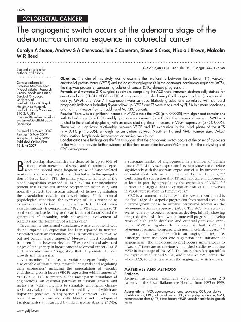
VEGF expression in the ACSVEGF was expressed by a proportion of adenoma andcarcinoma cells, with grading scores varying from 1 to 3(fig 2). In addition, expression was observed in some tumour-derived endothelial cells (fig 2) and macrophages. VEGFexpression significantly increased across the adenoma phaseof the ACS (p , 0.0005) although no further increase inexpression was observed within the carcinoma phase, despite74% of the carcinoma specimens expressing VEGF (table 1,fig 3A). No statistically significant differences were observedbetween tumour site, Dukes’ stage and lymph node involve-ment and positive tumour VEGF expression. Furthermore,there was no significant relationship between VEGF expressionand MVD counts. The correlation coefficients for inter andintra-observer error scores for VEGF assessment were 0.850 and0.867, respectively, showing a high level of agreement. TheELISA showed a significant increase in VEGF expression incarcinomas compared with respective background mucosa(1639 ¡ 113 versus 431 ¡ 35 pg/mg protein, p,0.001).
TF expression in the ACSPolyp and carcinoma specimens demonstrated TF stainingscores ranging from 0 to 3 (fig 2), with 51% of the carcinomaspecimens expressing TF (table 1), and in a similar manner toVEGF tumour-derived endothelial cells expressed high levels ofTF (fig 2). There were, however, no statistically significantdifferences in TF expression across the ACS (fig 3B).Furthermore, both the degree of TF staining and TF expressiondid not significantly correlate with MVD. No significantrelationship was observed between TF expression and tumoursite, Dukes’ stage or nodal status. The correlation coefficientsfor inter and intra-observer error scores for TF immunohisto-chemistry assessment were 0.92 and 0.89, respectively, showinga high level of agreement. ELISA studies demonstrated asignificant increase in TF expression in tumour tissue extractscompared with relative background mucosa (37.8 ¡ 1.6 versus30.9 ¡ 1.2 ng/mg protein, p , 0.01).
There was a relationship between TF and VEGF expression,with sections from the same polyp/tumour exhibiting co-localisation of staining. There was a significant relationshipbetween VEGF and TF expression in the carcinoma specimens(k = 0.24, p = 0.04), with a higher level of agreement in theadenoma specimens (k = 0.44, p , 0.0005). ELISA studiesalso demonstrated a significant correlation between theconcentrations of TF and VEGF in tumour tissue extracts(r = 0.36, p , 0.05).
Survival analysisFifty-six of the 66 patients with CRC (Dukes’ A–C) at the timeof diagnosis were followed up for a median of 69 months (range1–129). The Cox regression curve for MVD (fig 4) demonstratethat when the survival of CRC patients having a high CCS (49(the median) or more) was compared with patients with a low
Figure 1 Microvessel density across theadenoma–carcinoma sequence.Representative examples of CD31 staining ofendothelial cells in (A) normal glandularcolonic mucosa yielding a low microvasculardensity (MVD) score and (B) an area ofcolorectal carcinoma (CRC) showing a highMVD. In both cases vessel lumen are clearlyvisible (original magnification 6200). (C)Box and whisker plots representingmicrovessel density counts from the initialand the final phases of the adenoma–carcinoma sequence (ACS). Data arepresented as the median cumulative Chalkleyscore (CCS; horizontal line), with the boxrepresenting the 25th and 75th centiles, thewhiskers representing the statistical rangeand outlying data points depicted separately.An increase in MVD is clearly seen at allstages of the ACS (p , 0.0005) comparedwith background mucosa.
1428 Staton, Chetwood, Cameron, et al
www.gutjnl.com
CCS (below 49) there was an unfavourable influence of MVDon CRC-specific survival, which did not achieve significance(p = 0.120). Univariate analysis revealed that neither VEGFnor TF expression were associated with prognosis (p = 0.296and p = 0.853, respectively).
DISCUSSIONPrevious studies have suggested that the angiogenic switchoccurs simultaneously with tumour invasion;22 however, MVDanalysis of all stages of the ACS were not performed. This is thefirst study to investigate and quantify MVD across the entireACS, demonstrating that angiogenesis is significantly increasedmuch earlier in the disease spectrum than has previously beenreported, with a progressive increase in MVD throughout theACS. The greatest increase in angiogenesis occurs at the earlieststage of dysplastic transformation, suggesting that the angio-genic switch occurs at this point in the ACS. Interestingly, itmay be that the angiogenic switch actually occurs beforeadenoma formation, as studies by Shpitz et al.,24 demonstratedincreased angiogenesis in aberrant crypt foci, the earliest
morphologically identifiable preneoplastic lesions in colonicmucosa. We were unable to include this group in our studies.These data suggest that although angiogenesis is essential fortumour growth,25 the tumours do not, as previously thought,need to be 2 mm3 and undergoing hypoxic or redox stress toelicit an angiogenic response. Angiogenesis appears more likelyto be related to the factors involved in the generation ofaberrant crypt foci and polyps.
Our data clearly demonstrate a significant increase in MVD ateach stage of the ACS, in agreement with previous studies inwhich MVD correlates with the Dukes’ stage26 and lymph nodeinvolvement,27 factors usually associated with poor prognosis.Although patients with high MVD (> 49) tended to have apoorer prognosis than patients with low MVD (, 49), this didnot achieve significance. This may be a result of the relativelysmall numbers in each subgroup in the current study, andincreasing the number of cases may enhance these findings.Interestingly, these data are in agreement with many previouslypublished studies, in which MVD quantified using CD31staining failed to show a significant relationship with
Figure 2 Immunohistochemical stainingwith anti-vascular endothelial growth factorand anti-tissue factor antibodies.Representative examples from colorectaltumours are shown giving examples ofnegative cases for both vascular endothelialgrowth factor (VEGF) and tissue factor (TF)with no immunoreactivity (score 0), examplesof positive cases with weak immunoreactivityfor VEGF and TF (score 1), and positivecases with strong immunoreactivity for VEGFand TF (score 3). With both antibodies,staining is seen at the membrane and withinthe cytoplasm of tumour cells (originalmagnification 6200). Endothelial cells(arrows) also show moderateimmunoreactivity for both VEGF and TFwithout any evidence of thrombosisformation (original magnification 6400).
Angiogenic switch in colorectal cancer 1429
www.gutjnl.com

survival;20 28 29 however, a recent meta-analysis has suggestedthat MVD is inversely related to CRC survival.30 Although theMVD increased across the adenoma–carcinoma spectrum, andthe vessels quantified in the hotspots were more variable thanin background mucosa, qualitatively there did not appear to bechanges in vascular volume or surface area.
Studies investigating VEGF, the most potent pro-angiogenicfactor in the ACS, are relatively few, with the current studybeing the only one to include both low and high gradedysplastic polyps as well as IPC. Our data show that VEGFexpression is significantly increased during the early phases ofthe ACS, with no further increases in the carcinoma phase. Inaddition, the high grade dysplastic polyps and IPC specimens
demonstrate the highest degree of VEGF expression (80% and90%, respectively), suggesting that VEGF may play a role inboth carcinogenesis and angiogenesis. A number of previousstudies failed to identify VEGF expression in adenomas,31–33
whereas others are in agreement with our findings demonstrat-ing VEGF expression in adenomas.34 35 All those studies do,however, agree that VEGF expression is elevated uponprogression from adenoma to carcinoma, and also support thetheory that VEGF is involved in carcinogenesis, potentially bybinding to VEGF receptors on CRC cells and inhibiting tumourcell apoptosis.36
Although VEGF is a key angiogenic factor and is significantlyupregulated at the initial phase of the ACS alongside MVD,there was no significant relationship between the two factors inthe carcinoma specimens, although some studies have indi-cated a correlation in CRC.37 38 Despite VEGF being a crucial andwell studied angiogenic cytokine that showed significantlyincreased expression in CRC compared with backgroundmucosa (ELISA studies), many other factors may also regulateangiogenesis in CRC, and our data are in agreement with thoseof Tabara and colleagues39 demonstrating that VEGF did notcorrelate with MVD, whereas basic fibroblast growth factor,another potent pro-angiogenic cytokine, correlated with MVDin CRC. Furthermore, our data indicate that VEGF did notcorrelate with survival and was not an independent risk factor,in agreement with the majority of previous immunohistochem-ical-based studies.32 37 40
VEGF has been reported to induce TF expression inendothelial cells, and although the details of the intracellular
Figure 3 Vascular endothelial growth factor and tissue factor expressionacross the adenoma–carcinoma sequence. (A) Percentage of cases withvarying vascular endothelial growth factor (VEGF) expression in theadenoma–carcinoma sequence (ACS). A significant increase in VEGFexpression is seen in the initial phase of the ACS (*p , 0.0005). (B)Percentage of cases with varying tissure factor (TF) expression in the ACS.The sections of each bar represent the percentage of each group with aparticular score, 0–3.
Table 1 Percentage of cases showing vascular endothelial growth factor and tissue factorexpression across the adenoma–carcinoma sequence
Phase of ACS Sample sizePercentage expression ofVEGF (%)
Percentage expression ofTF (%)
Low grade dysplasia polyps 47 57 63High grade dysplasia polyps 19 80 80Intra-polyp carcinoma 30 90 67Dukes’ A carcinoma 10 70 40Dukes’ B carcinoma 26 58 52Dukes’ C carcinoma 26 65 37
ACS, Adenoma–carcinoma sequence; TF, tissue factor; VEGF, vascular endothelial growth factor. VEGF expression wasidentified in 74% of the carcinoma specimens (including intra-polyp carcinoma; IPC). In total, 51% of the carcinomas(including IPC) included in the study expressed TF.
Figure 4 Cox regression survival data for microvascular density.Comparison of overall survival using Cox regression analysis betweengroups of patients with low (less than 49) and high (49 or above)cumulative Chalkley score (CCS). The difference between the two groupsdoes not achieve significance (p = 0.12).
1430 Staton, Chetwood, Cameron, et al
www.gutjnl.com
signalling pathway responsible for this induction have not yetbeen fully elucidated, reporter gene studies imply thattranscription factors including EGR-1 and Sp1 are involved.41 42
In agreement with this, we found both TF and VEGF expressionin the endothelial cells of many tumour-associated bloodvessels.
TF is expressed by tumour cells in a number of humanmalignancies including pancreatic,43 hepatic44 and colorectal.27
In the current study, 51% of colorectal tumour specimensexpressed TF, in agreement with previous studies reporting that46–57% CRC express TF,7 27 45 46 and our ELISA data showed asignificant increase in TF expression in CRC compared withbackground mucosa. Interestingly, we demonstrated TF expres-sion in adenomas of varying histological grade, which is indirect contrast to a small study in which TF was not identifiedin six adenoma specimens.46 The finding that TF was expressedto the greatest degree in high grade dysplastic polyps (80%)suggests that, like VEGF, TF may play a role in thetransformation from dysplasia to carcinoma. The activation ofK-ras and the subsequent loss of p53 are key changes in theACS, and these events have recently been associated with TFexpression in CRC cells in culture.47 Moreover, TF, again likeVEGF, has been shown to be anti-apoptotic for tumour cells,thereby promoting cell survival.9
Furthermore, TF is thought to regulate angiogenesis, at leastin part, by upregulating VEGF expression.10 Our results agreewith earlier reports demonstrating a significant correlationbetween VEGF and TF expression in carcinomas, both usingELISA and immunohistochemistry techniques.7 27 48 A strongerrelationship was observed in the adenoma specimens, however,with both VEGF and TF most strongly expressed at the samephase (high grade dysplastic polyps) of the ACS, possiblyindicating a close relationship in disease progression, althoughthere is no association between TF and MVD at the low gradedysplasia stage. This is in direct contrast to the study byNakasaki et al.,27 which may be a result of the vessels beingstained with von Willebrand factor, counted manually and notwith a Chalkley grid, and that adenomas and IPC were notincluded in the study. An additional factor may be that theexpression of TF was similar to the background mucosal tissuein the current study.
In contrast to studies with MVD and VEGF, there is only onepublished report relating TF to prognosis. This is an immuno-histochemical study involving 67 patients with advanced CRC,who showed a significantly lower survival rate over 3 years withTF-positive tumours (29%) compared with patients with TF-negative tumours (88%).7 In contrast our study, which used thecomplete spectrum of CRC (Dukes’ A–C) failed to show anyrelationship between TF expression and survival over a 5 yearperiod, suggesting that TF may only be a prognostic factor inadvanced CRC. This may agree with the literature suggestingthat TF expression is an indicator of hepatic or lymphaticmetastasis, both factors associated with poor prognosis.27 45
In conclusion, this is the first study to assess angiogenesis,VEGF and TF in the complete ACS of CRC in a large cohort ofpatients. These data suggest that the angiogenic switch occursat the onset of dysplastic transformation in the polyp, whichhas not previously been reported. There is a trend, although notstatistically different, for patients with higher MVD to have aworse prognosis than patients with lower MVD, which has notpreviously been reported. Moreover, a significant relationshiphas been identified between VEGF and TF expression,especially in the initial phases of the ACS, suggesting theimportance of both these factors in CRC angiogenesis, diseaseprogression and tumour biology. Further work is needed toexamine the precise relationships between VEGF and TF in thedevelopment of CRC.
ACKNOWLEDGEMENTSThe authors would like to acknowledge the technical assistance ofYvonne Stephenson who sectioned the paraffin-embedded tissue forhistological examination, in addition to Yorkshire Cancer Research andthe University of Sheffield Medical School who generously funded thisproject.
Authors’ affiliations. . . . . . . . . . . . . . . . . . . . . . .
Carolyn A Staton, Andrew S A Chetwood, Iain C Cameron, N J Brown,Malcolm W R Reed, Academic Unit of Surgical Oncology, University ofSheffield Medical School, Sheffield S10 2RX, UKSimon S Cross, Academic Unit of Pathology, University of Sheffield MedicalSchool, Sheffield S10 2RX, UK
Conflict of interest: None declared.
REFERENCES1 Dvorak HF. Thrombosis and Cancer. Human Pathol 1987;18:275–84.2 Kakkar AK, DeRuvo N, Chinswangwatanakul V, et al. Extrinsic-pathway
activation in cancer with high factor VIIa and tissue factor. Lancet1995;346:1004–5.
3 Camerer E, Kolsto AB, Prydz H. Cell biology of tissue factor, the principal initiatorof blood coagulation. Thromb Res 1996;81:1–41.
4 Nagy JA, Brown LF, Senger DR, et al. Pathogenesis of tumor stroma generation:a critical role for leaky blood vessels and fibrin deposition. Biochim Biophys Acta1989;948:305–26.
5 Contrino J, Hair G, Kreutzer DL, et al. In situ detection of tissue factor in vascularendothelial cells: correlation with the malignant phenotype of human breastdisease. Nat Med 1996;2:209–15.
6 Vrana JA, Stang MT, Grande JP, et al. Expression of tissue factor in tumor stromacorrelates with progression to invasive human breast cancer: paracrineregulation by carcinoma cell-derived members of the transforming growth factorbeta family. Cancer Res 1996;7:1877–83.
7 Seto S, Onodera H, Kaido T, et al. Tissue factor expression in human colorectalcarcinoma: correlation with hepatic metastasis and impact on prognosis. Cancer2000;88:295–301.
8 Ueda C, Hirohata Y, Kihara Y, et al. Pancreatic cancer complicated bydisseminated intravascular coagulation associated with production of tissuefactor. J Gastroenterol, 2001;36:848–50.
9 Versteeg HH, Peppelenbosch MP, Spek CA. Tissue factor signal transduction inangiogenesis. Carcinogenesis 2003;24:1009–13.
10 Zhang Y, Deng Y, Luther T, et al. Tissue factor controls the balance of angiogenicand antiangiogenic properties of tumor cells in mice. J Clin Invest1994;94:1320–7.
11 Zachary I. VEGF signalling: integration and multi-tasking in endothelial cellbiology. Biochem Soc Trans 2003;31:1171–7.
12 Imura S, Miyake H, Izumi K, et al. Correlation of vascular endothelial cellproliferation with microvessel density and expression of vascular endothelialgrowth factor and basic fibroblast growth factor in hepatocellular carcinoma.J Med Invest 2004;51:202–9.
13 Raspollini MR, Castiglione F, Garbini F, et al. Correlation of epidermal growthfactor receptor expression with tumour microdensity vessels and with vascularendothelial growth factor expression in ovarian carcinoma. Int J Surg Pathol2005;13:135–42.
14 Koomagi R, Volm M. Tissue-factor expression in human non-small-cell lungcarcinoma measured by immunohistochemistry: correlation between tissue factorand angiogenesis. Int J Cancer 1998;79:19–22.
15 Shoji M, Hancock WW, Abe K, et al. Activation of coagulation and angiogenesisin cancer: immunohistochemical localization in situ of clotting proteins andvascular endothelial growth factor in human cancer. Am J Pathol1998;152:399–411.
16 Takano S, Tsuboi K, Tomono Y, et al. Tissue factor, osteopontin, alphavbeta3integrin expression in microvasculature of gliomas associated with vascularendothelial growth factor expression. Br J Cancer 2000;82:1967–73.
17 Chen J, Bierhaus A, Schiekofer S, et al. Tissue factor – a receptor involved in thecontrol of cellular properties, including angiogenesis. Thromb Haemost2001;86:334–45.
18 Rickles FR, Shoji M, Abe K. The role of the hemostatic system in tumour growth,metastasis and angiogenesis: tissue factor is a bifunctional molecule capable ofinducing both fibrin deposition and angiogenesis in cancer. Int J Hematol2001;73:145–50.
19 Abe K, Shoji M, Chen J, et al. Regulation of vascular endothelial growth factorproduction and angiogenesis by the cytoplasmic tail of tissue factor. Proc NatlAcad Sci U S A 1999;96:8663–8.
20 Bossi P, Viale G, Lee AK, et al. Angiogenesis in colorectal tumors: microvesselquantitation in adenomas and carcinomas with clinicopathological correlations.Cancer Res 1995;55:5049–53.
21 Fox SH, Whalen GF, Sanders MM, et al. Angiogenesis in normal tissue adjacentto colon cancer. J Surg Oncol 1998;69:230–4.
22 Takahashi Y, Ellis LM, Mai M. The angiogenic switch of human colon canceroccurs simultaneous to initiation of invasion. Oncol Rep 2003;10:9–13.
23 Weidner N, Semple JP, Welch WR, et al. Tumor angiogenesis and metastasiscorrelation in invasive breast carcinoma. N Engl J Med 1991;324:1–8.
Angiogenic switch in colorectal cancer 1431
www.gutjnl.com
24 Shpitz B, Gochberg S, Neurfeld D, et al. Angiogenic switch in earliest stages ofhuman colonic tumorigenesis. Anticancer Res 2003;23:5153–7.
25 Folkman J. Angiogenesis in cancer, vascular, rheumatoid and other disease. NatMed 1995;1:27–31.
26 Yonenaga Y, Mori A, Onodera H, et al. Absence of smooth muscle actin-positivepericyte coverage of tumor vessels correlates with hematogenous metastasis andprognosis of colorectal cancer patients. Oncology 2005;69:159–66.
27 Nakasaki T, Wada H, Shigemori C, et al. Expression of tissue factor andvascular endothelial growth factor is associated with angiogenesis in colorectalcancer. Am J Hematol 2002;69:247–54.
28 Leme MB, Waitzberg AF, Artigiani Neto R, et al. Assessment of angiogenesisexpression and its relationship with prognosis of colorectal cancer byconventional and computer-assisted histopathological image analysis. Acta CirBras 2006;21:392–7.
29 Cianchi F, Palomba A, Messerini L, et al. Tumor angiogenesis in lymph nodenegative rectal cancer: correlation with clinicopathological parameters andprognosis. Ann Surg Oncol 2002;9:20–6.
30 Des Guetz G, Uzzan B, Nicolas P, et al. Microvessel density and VEGFexpression are prognostic factors in colorectal cancer. Meta-analysis of theliterature. Br J Cancer 2006;94:1823–32.
31 Kondo Y, Arii S, Furutani M, et al. Implication of vascular endothelial growthfactor and p53 status for angiogenesis in noninvasive colorectal carcinoma.Cancer 2000;88:1820–7.
32 Lee JC, Chow NH, Wang S, et al. Prognostic value of vascular endothelial growthfactor expression in colorectal cancer patients. Eur J Cancer 2000;36:748–53.
33 George ML, Tutton MG, Janssen F, et al. VEGF-A, VEGF-C, and VEGF-D incolorectal cancer progression. Neoplasia 2001;3:420–7.
34 Hanrahan V, Currie MJ, Gunningham SP, et al. The angiogenic switch forvascular endothelial growth factor [VEGF]-A, VEGF-B, VEGF-C, and VEGF-D inthe adenoma–carcinoma sequence during colorectal cancer progression. J Pathol2003;200:183–94.
35 Wong MP, Cheung N, Yuen ST, et al. Vascular endothelial growth factor is upregulated in the early pre-malignant stage of colorectal tumour progression.Int J Cancer 1999;81:845–50.
36 Fan F, Wey JS, McCarty MF, et al. Expression and function of vascularendothelial growth factor receptor-1 on human colorectal cancer cells. Oncogene2005;24:2647–53.
37 Zheng S, Han MY, Xiao ZX, et al. Clinical significance of vascular endothelialgrowth factor expression and neovascularization in colorectal carcinoma.World J Gastroenterol 2003;9:1227–30.
38 Takahashi Y, Tucker SL, Kitadai Y, et al. Vessel counts and expression of vascularendothelial growth factor as prognostic factors in node-negative colon cancer.Arch Surg 1997;132:541–6.
39 Tabara H, Kohno H, Dhar DK, et al. Concurrent expression of angiogenic growthfactors and neovascularization during tumourigenesis in colorectal carcinomapatients. Acta Oncol 2001;40:622–8.
40 Doger FK, Meteoglu I, Tuncyurek P, et al. Does the EGFR and VEGF expressionpredict the prognosis in colon cancer? Eur Sur Res 2006;38:540–4.
41 Mechtcheriakova D, Wlachos A, Holzmuller H, et al. Vascular endothelial cellgrowth factor-induced tissue factor expression in endothelial cells is mediated byEGR-1. Blood 1999;93:3811–23.
42 Armesilla AL, Lorenzo E, Gomez del Arco P, et al. Vascular endothelial growthfactor activates nuclear factor of activated T cells in human endothelial cells: arole for tissue factor gene expression. Mol Cell Biol 1999;19:2032–43.
43 Kakkar AK, Lemoine NR, Scully MF, et al. Tissue factor expression correlates withhistological grade in human pancreatic cancer. Br J Surg 1995;82:1101–4.
44 Poon RT, Lau CP, Ho JW, et al. Tissue factor expression correlates with tumorangiogenesis and invasiveness in human hepatocellular carcinoma. Clin CancerRes 2003;9:5339–45.
45 Shigemori C, Wada H, Matsumoto K, et al. Tissue factor expression andmetastatic potential of colorectal cancer. Thromb Haemost 1998;80:894–8.
46 Wan YL, Yao HW, Ye JM, et al. The role of tissue factor expression in the invasiveand metastatic ability of colorectal carcinoma. Zhonghua Wai Ke Za Zhi2004;42:149–53.
47 Yu JL, May L, Lhotak V, et al. Oncogenic events regulate tissue factor expressionin colorectal cancer cells: implications for tumor progression and angiogenesis.Blood 2005;105:1734–41.
48 Altomare DF, Rotelli MT, Memeo V, et al. Expression of tissue factor and vascularendothelial growth factor in colorectal carcinoma. Tumori 2003;89:5–6.
How you can reduce C difficile infections
View the free interactive video module from BMJ Learning and the Department of Health byvisiting bmjlearning.com/cdifficile.
This module will help you identify and treat C difficile most effectively, to meet the NationalHealth Service priority of reducing the 55 634* reported cases in 2006.
See how a major UK hospital successfully managed an outbreak and introduced measures youtoo can apply to prevent infections.
On completion of the module you will learn:
N How to diagnose and treat patients with C diffcile infections
N The importance of hand hygiene in preventing infection
N The cautious use of prescribing antibiotics
N Understanding the patient experienceVisit bmjlearning.com/cdifficile to make a difference for your patients.You will need to sign in or register to access this free video module. The module will take
approximately 40 minutes to complete.* Further information is available on the Health Protection Agency website www.hpa.org.uk
1432 Staton, Chetwood, Cameron, et al
www.gutjnl.com
Related Documents


