The Anatomy of the Ear

The Anatomy of the Ear The Outer Ear 1. Auricle (Pinna) 2. External Auditory Canal (ear canal) Channels sound into the ear.
Dec 25, 2015
Welcome message from author
This document is posted to help you gain knowledge. Please leave a comment to let me know what you think about it! Share it to your friends and learn new things together.
Transcript

The Anatomy of the Ear
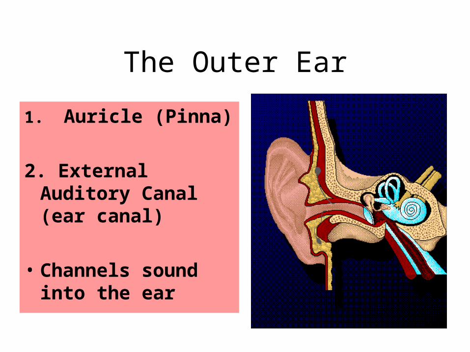
The Outer Ear
1. Auricle (Pinna)
2. External Auditory Canal (ear canal)
• Channels sound into the ear

The Middle Ear
Slide 5 of 24
Notes:

Slide 7 of 24
How We Hear
http://www.hearingcenteronline.com/ear2.shtml

CONGENITAL Disorders
A disorder that is present at birth. Can be hereditary (such as a genetic syndrome like Treacher Collin’s Syndrome). Or, can be from another source - like when fetus is affected by maternal Rubella.

Conductive Hearing Loss
Occurs when there exists a complication somewhere between the outer ear and the middle ear.

Sensorineural Hearing Loss
A loss affecting an inner ear structure or the auditory nerve. Typically a permanent loss.
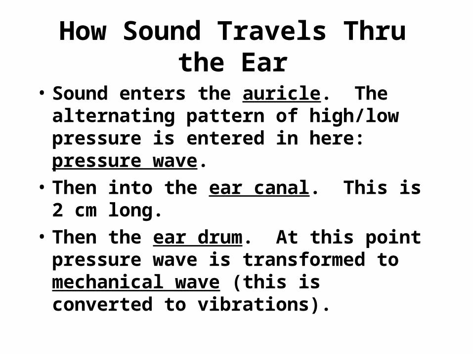
How Sound Travels Thru the Ear
• Sound enters the auricle. The alternating pattern of high/low pressure is entered in here: pressure wave.
• Then into the ear canal. This is 2 cm long.
• Then the ear drum. At this point pressure wave is transformed to mechanical wave (this is converted to vibrations).

Then into the middle ear…
• Then to the 3 tiny bones (also called the ossicles). The names of the bones are: malleus (hammer), incus (anvil), and stapes (stirrups).
• As it reaches the stapes, mechanical wave transforms to compression wave within the fluid. The stapes gives off 15 times more vibrations than that of the ear drum.
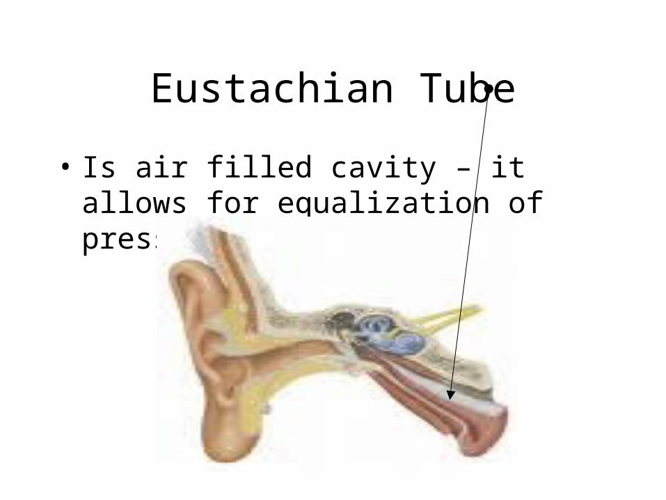
Eustachian Tube
• Is air filled cavity – it allows for equalization of pressure.
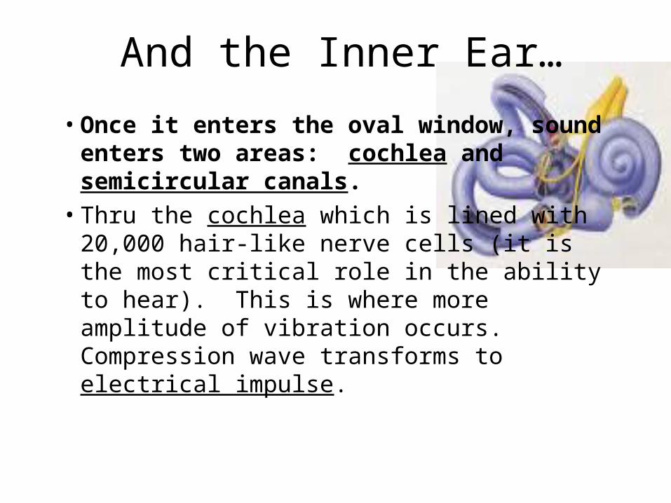
And the Inner Ear…
• Once it enters the oval window, sound enters two areas: cochlea and semicircular canals.
• Thru the cochlea which is lined with 20,000 hair-like nerve cells (it is the most critical role in the ability to hear). This is where more amplitude of vibration occurs. Compression wave transforms to electrical impulse.

And Finally…….
• Then it passes thru the auditory nerve to the brain – capable of interpreting qualities of sound upon reception of electric nerve impulses.

Meniere’s Disease
• Meniere's disease is a disorder of the inner ear which causes episodes of vertigo, ringing in the ears (tinnitus), a feeling of fullness or pressure in the ear, and fluctuating hearing loss. It is difficult to live with and can often cause victims to seek psychological help.

Symptoms of Meniere’s Disease
• Vertigo, balance problems,
• Loss of low pitches

Who has Meniere’s Disease?
• Alan Shepard
• Emily Dickinson
• Van Vough
• Martin Luthor

Meningitis
• Meningitis is a medical condition caused by inflammation of the protective membranes covering the brain and spinal cord, known collectively as the meninges.
• Diagnosed with use of a spinal tap. • Symptoms: severe pain in the ear,
irritability, and fever, headaches, and sometimes a purplish rash.

Otitis Media
• Otitis media is an infection or inflammation of the middle ear.
• This inflammation often begins when infections that cause sore throats, colds, or other respiratory or breathing problems spread to the middle ear.

• Otitis Media, continued….
• It is very isolating.
• Teens who have it are not aware that there is anyone else out there suffering from the same problems.
• Symptoms include severe pain in the ear, irritability, and fever.
• This affects children’s speech between 3 months and 3 years.
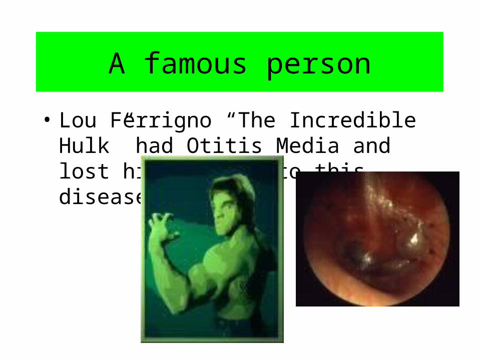
A famous person
• Lou Ferrigno “The Incredible Hulk” had Otitis Media and lost his hearing to this disease.

Ototoxicity
• A poisoning of the ear.
• Ototoxicity is damage of the ear (oto), specifically the cochlea or auditory nerve and sometimes the vestibulum, by a toxin (often medication).
• Symptoms: Severe pain in the ear, irritability, and fever

Otosclerosis
• Otosclerosis is the abnormal growth of bone of the middle ear. This bone prevents structures within the ear from working properly and causes hearing loss.
• Symptoms: Severe tinnitus, recurring auditory memories, and frequent vertigo.

Did you know?
• William Peterson of CSI: Las Vegas has Otosclerosis and a deaf mother in real life!
Rubella
• Also known as German Measles or “3-day measles”
• an infection that primarily affects the skin and lymph nodes
• usually transmitted by droplets from the nose or throat that others breathe in

German Measles
• Symptoms include a red rash covering the upper body, fever, and headaches.

Treacher Collin’s Syndrome
• Treacher Collins syndrome is a genetic, craniofacial birth defect that is characterized by a range of distinctive facial anomalies.
• The main characteristics of TCS are downward slanting eyes, small lower jaw, and malformed or missing ears.
• These anomalies can cause hearing, breathing, and eating problems.

Symptoms and Treatments
• Characterized by auricles that are small or low on the head, eyes that are sunken and droopy, and a cleft palete.
• It can be treated with hearing tests and/or plastic surgery.

Usher’s Syndrome
• Usher syndrome is the most common condition that affects both hearing and vision starting with peripheral vision.
• Usher syndrome is inherited, which means that it is passed from parents to their children through genes.

Usher’s Syndrome
• It can affect an unborn infant and cause them to be deaf and/or blind if their mother comes in contact with someone who has it.
• It is not a common disease because both parents must have the gene.
• It occurs 4 in 100,000 births. Usher syndrome is inherited, which means that it is passed from parents to their children through genes.

Things to complete:
• Parts of the Ear Diagram WS
• Parts of the Ear (fill in the blank) WS
• Study Medical Terms Signs
• QUIZ: October 30/31!!!
Related Documents