WORLD HEALTH ORGANIZATION REGIONAL OFFICE FOR EUROPE WELTGESUNDHEITSORGANISATION REGIONALBÜRO FÜR EUROPA ORGANISATION MONDIALE DE LA SANTE BUREAU REGIONAL DE L'EUROPE ВСЕМИРНАЯ ОРГАНИЗАЦИЯ ЗДРАВООХРАНЕНИЯ ЕВРОПЕЙСКОЕ РЕГИОНАЛЬНОЕ БЮРО The Analysis of Food Samples for the Presence of Genetically Modified Organisms Session 6 The Polymerase Chain Reaction (PCR) M. Somma, M. Querci

Welcome message from author
This document is posted to help you gain knowledge. Please leave a comment to let me know what you think about it! Share it to your friends and learn new things together.
Transcript
WORLD HEALTH ORGANIZATION REGIONAL OFFICE FOR EUROPE WELTGESUNDHEITSORGANISATION REGIONALBÜRO FÜR EUROPA
ORGANISATION MONDIALE DE LA SANTE BUREAU REGIONAL DE L'EUROPE
ВСЕМИРНАЯ ОРГАНИЗАЦИЯ ЗДРАВООХРАНЕНИЯ
ЕВРОПЕЙСКОЕ РЕГИОНАЛЬНОЕ БЮРО
The Analysis of Food Samples for the Presence of Genetically Modified Organisms
Session 6
The Polymerase Chain Reaction (PCR)
M. Somma, M. Querci

The Polymerase Chain Reaction (PCR) 2
The Analysis of Food Samples for the Presence of Genetically Modified Organisms Session 6
Table of Contents
Session 6
The Polymerase Chain Reaction (PCR)
Introduction 3
Components, structure and replication of DNA 3
Principles of PCR 9
Instrumentation and components for the PCR 12
Design of primers for PCR 17
Specialised PCR 21
PCR in practice 23
References 29
The Polymerase Chain Reaction (PCR) 3
The Analysis of Food Samples for the Presence of Genetically Modified Organisms Session 6
Introduction
The invention of Polymerase Chain Reaction (PCR) by K. Mullis and co-workers in
1985 has revolutionised molecular biology and molecular medicine (Saiki et al.,
1985). The Polymerase Chain Reaction is an in vitro technique used to enzymatically
amplify a specific DNA region that lies between two regions of known DNA
sequence. Whereas previously only minute amounts of a specific gene could be
obtained, now even a single gene copy can be amplified to a million copies within a
few hours using PCR.
PCR techniques have become essential for many common procedures such as
cloning specific DNA fragments, detecting and identifying genes in diagnostics and
forensics, and in the investigation of gene expression patterns. More recently, PCR
has allowed the investigation of new fields such as the control of the authenticity of
foodstuff, the presence of genetically modified DNA and microbiological
contamination. In understanding the principles of PCR and its applications, the nature
of the DNA molecule must first be considered, therefore the structure and the
replication of DNA will be described in the following section.
Components, structure and replication of DNA
Components. A molecule of DNA is constituted of two parallel complementary
twisted chains of alternating units of phosphoric acid and deoxyribose, linked by
cross-pieces of purine and pyrimidine bases, resulting in a right-handed helical
structure that carries genetic information encoded in the sequence of the bases. In
eucaryotic cells, most of the DNA is contained within the nucleus and is referred to as
chromosomal DNA. It is separated from the rest of the cell (cytoplasm) by a double
layer membrane (nuclear envelope). In addition to this, extrachromosomal DNA can
be found in the mitochondria and chloroplasts.
The building blocks of DNA, called nucleotides, are:
• dATP, deoxyadenosine triphosphate;
• dGTP, deoxyguanosine triphosphate;
• dTTP, deoxythymidine triphosphate;
• dCTP, deoxycytidine triphosphate.
For convenience, these four nucleotides are called dNTPs (deoxynucleoside
triphosphates). A nucleotide is constituted of three major parts: a purine base
(adenine, A, and/or guanine, G), or a pyrimidine base (cytosine, C, and/or thymine,

The Polymerase Chain Reaction (PCR) 4
The Analysis of Food Samples for the Presence of Genetically Modified Organisms Session 6
T), a pentose sugar molecule (deoxyribose) and a triphosphate group. As shown in
Figure 1, a purine or pyrimidine base is bound to a pentose ring by an N-glycosydic
bond and a phosphate group is bound to the 5’ carbon atom of the sugar by a
diesteric bond. In the ribonucleic acid, RNA, thymine is substituted by uracil (U) and
the deoxyribose molecule is replaced by ribose.
Figure 1. The components of nucleotides (Picture: Andy Vierstraete, 1999)
Structure. Figure 2 shows how the nucleotides form a DNA chain. DNA is formed by
coupling the nucleotides between the phosphate group from a nucleotide (which is
positioned on the fifth C-atom of the sugar molecule) with the hydroxyl on the third C-
atom on the sugar molecule of the previous nucleotide. To accomplish this, a
diphosphate group is split off (with the release of energy). This means that new
nucleotides are always added on the 3' side of the chain. As shown in Figure 3, DNA
is double-stranded (except in some viruses), and the two strands pair with one
another in a very precise way. Each base in a strand will pair with only one kind of
base across from it in the opposing strand forming a base pair (bp): A is always
paired to T by two hydrogen bonds; and C is always paired to G by three hydrogen
bonds. In this way, the two chains are complementary to each other and one chain
can serve as a template for the production of the other.

The Polymerase Chain Reaction (PCR) 5
The Analysis of Food Samples for the Presence of Genetically Modified Organisms Session 6
Figure 2. Formation of a DNA chain from individual nucleotides (Picture: Andy
Vierstraete, 1999)
The bases form a hydrophobic nucleus inside the double helix. The sugars and
phosphate groups (in their anionic form) constitute the external hydrophilic layer of
the molecule. In physiological conditions, double-stranded DNA helix is more stable
than a single-stranded DNA helix.
Replication. DNA contains the complete genetic information that defines the
structure and function of an organism. Three different processes are responsible for
the transmission of genetic information:
• replication;
• transcription;
• translation.
During replication a double-stranded nucleic acid is duplicated to give identical
copies. This process perpetuates the genetic information. During transcription, a
DNA segment that constitutes a gene is read and transcribed into a single-stranded
sequence of RNA. The RNA moves from the nucleus into the cytoplasm. Finally,

The Polymerase Chain Reaction (PCR) 6
The Analysis of Food Samples for the Presence of Genetically Modified Organisms Session 6
during translation, the RNA sequence is translated into a sequence of amino acids as
the protein is formed (Alberts et al., 1983).
Figure 3. Structure of DNA in a cell (Picture: Andy Vierstraete, 1999)

The Polymerase Chain Reaction (PCR) 7
The Analysis of Food Samples for the Presence of Genetically Modified Organisms Session 6
The replication of DNA is the process on which the PCR amplification is based, and
will be described in detail.
During replication, the DNA molecule unwinds, with each single strand becoming a
template for synthesis of a new, complementary strand. Each daughter molecule,
consisting of one old and one new DNA strand, is an exact copy of the parent
molecule.
Figure 4. The replication fork
Several enzymes are required to unwind the double helix and to synthesise a new
strand of DNA. Topoisomerase and helicase are responsible for the unwinding of the
DNA by breaking the supercoiled structure and nicking a single strand of DNA. Then,
primase (part of an aggregate of proteins called the primeosome) attaches a small
RNA primer to the single-stranded DNA, to act as a 3'OH end from which the DNA
polymerase begins synthesis. This RNA primer is eventually removed by RNase H
and the gap is filled in by DNA polymerase I. At this stage, DNA polymerase
proceeds along a single-stranded molecule of DNA, recruiting free dNTPs to
hydrogen bond with their appropriate complementary dNTP on the single strand (A
with T and G with C), forming a covalent phosphodiester bond with the previous
nucleotide of the same strand. The energy stored in the triphosphate is used to
covalently bind each new nucleotide to the growing second strand. There are
different forms of DNA polymerase but it is DNA polymerase III that is responsible for
the progressive synthesis of new DNA strands. DNA polymerase only acts from 5' to
3'. Since one strand of the double helix is 5' to 3' and the other one is 3' to 5', DNA
polymerase synthesises a second copy of the 5' to 3' strand (the lagging strand), in
spurts (Okazaki fragments) (Ogawa and Okazaki, 1980). The synthesis of the new

The Polymerase Chain Reaction (PCR) 8
The Analysis of Food Samples for the Presence of Genetically Modified Organisms Session 6
copies of the 5' to 3' strand is shown in Figure 4. The other strand, the leading strand,
can proceed with synthesis directly, from 5' to 3', as the helix unwinds. DNA
polymerase cannot start synthesising ex novo on a bare single strand but needs a
primer with a free 3'OH group onto which it can attach a dNTP.
Ligase catalyses the formation of a phosphodiester bond given an unattached but
adjacent 3'OH and 5'phosphate. This can fill in the unattached gap left when the RNA
primer is removed and filled in. It is worth noting that single-stranded binding proteins
are important to maintain the stability of the replication fork. Single-stranded DNA is
very labile, or unstable, so these proteins bind to it while it remains single-stranded,
protecting it from degradation.

The Polymerase Chain Reaction (PCR) 9
The Analysis of Food Samples for the Presence of Genetically Modified Organisms Session 6
Principles of PCR
PCR is based on the mechanism of DNA replication in vivo: dsDNA is unwound to
ssDNA, duplicated, and rewound. This technique consists of repetitive cycles of:
• denaturation of the DNA through melting at elevated temperature to convert
double-stranded DNA to single-stranded DNA
• annealing (hybridisation) of two oligonucleotides used as primers to the target
DNA
• extension of the DNA chain by nucleotide addition from the primers using DNA
polymerase as catalyst in the presence of Mg2+ ions.
The oligonucleotides typically consist of relatively short sequences, which are
different to each other and complementary to recognition sites flanking the segment
of target DNA to be amplified. The steps of template denaturation, primer annealing
and primer extension comprise a single "cycle" in the PCR amplification
methodology. Figure 5 illustrates the three major steps in a PCR amplification
process.
Figure 5. The steps of PCR amplification (Picture: Andy Vierstraete, 1999)
After each cycle, the newly synthesised DNA strands can serve as templates in the
next cycle. As shown in Figure 6, the major product of this exponential reaction is a

The Polymerase Chain Reaction (PCR) 10
The Analysis of Food Samples for the Presence of Genetically Modified Organisms Session 6
segment of dsDNA whose termini are defined by the 5' termini of the oligonucleotide
primers and whose length is defined by the distance between the primers. The
products of a successful first round of amplification are heterogeneously sized DNA
molecules, whose lengths may exceed the distance between the binding sites of the
two primers. In the second round, these molecules generate DNA strands of defined
length that will accumulate in an exponential fashion in later rounds of amplification
and will form the dominant products of the reaction. Thus, amplification, as a final
number of copies of the target sequence, is expressed by the following equation:
(2n-2n)x (1)
where n is the number of cycles, 2n is the first product obtained after the first cycle
and second products obtained after the second cycle with undefined length, x is the
number of copies of the original template. Potentially, after 20 cycles of PCR there
will be a 220–fold amplification, assuming 100% efficiency during each cycle. The
efficiency of a PCR will vary from template to template and according to the degree
of optimisation that has been carried out.
A detailed description of the three steps of PCR amplification (template denaturation,
primer annealing and extension) is given in the following paragraphs (Sambrook et
al., 1989).
Figure 6. The exponential amplification of DNA in PCR
Template denaturation
During denaturation, the double strand melts opening up to single-stranded DNA,
and all enzymatic reactions stop (i.e. the extension from a previous cycle). The two

The Polymerase Chain Reaction (PCR) 11
The Analysis of Food Samples for the Presence of Genetically Modified Organisms Session 6
complementary chains are separated by an increase in temperature. This is known
as denaturation. To obtain the denaturation of DNA, the temperature is usually
increased to ~ 93 - 96°C. In this way the strong H-bonds are broken and the number
of non-paired bases increases. The reaction is complete when all of the dsDNA
becomes ssDNA. The temperature at which half of the dsDNA is single-stranded is
known as the melting temperature, Tm. The type of solvent, the salt concentration
and the pH used, influence the denaturation process. For example, in low salt
concentrations, high pH and in the presence of organic solvents such as
formaldehyde, the melting temperature, Tm, decreases. The concentration of G/C and
T/A can also affect the value of Tm. The Tm of the DNA structure containing an
elevated quantity of G/C is higher compared to that of DNA rich in T/A. For example,
Serratia marecescens has approximately 60% G/C with a Tm of approximately 94°C,
whereas Pneumococcus has approximately 40% G/C and a Tm of approximately
85°C.
Primer annealing
The annealing or rehybridisation of the DNA strands takes place at lower
temperature (usually 55 - 65°C). Once the temperature is reduced, the two
complementary ssDNA chains will reform into a dsDNA molecule. In this phase, the
primers are flowing and hydrogen bonds are constantly formed and broken between
the single-stranded primer and the single-stranded template. The more stable bonds
last a bit longer (primers that exactly fit the template DNA) and on that small piece of
double-stranded DNA (template and primer), the polymerase can attach and begins
copying the template. Once there are a few bases built in, the ionic bond is so strong
between the template and the primer that it will not break.
Primer extension
In this step the primers are extended across the target sequence by using a heat-
stable DNA polymerase (frequently Taq DNA polymerase) in the presence of dNTPs
resulting in a duplication of the starting target material. The ideal working
temperature for the Taq DNA polymerase is 72°C. When the primers have been
extended a few bases, they possess a stronger ionic attraction to the template, which
reduces the probability of the reverse process. Primers that do not match exactly
come loose again (because of the higher temperature) and do not give an extension
of the fragment. The bases (complementary to the template) are coupled to the
primer on the 3' side (the polymerase adds dNTPs from 5' to 3', reading the template
from 3' to 5'). The length of time of the primer extension steps can be increased if the
The Polymerase Chain Reaction (PCR) 12
The Analysis of Food Samples for the Presence of Genetically Modified Organisms Session 6
region of DNA to be amplified is long, however, for the majority of PCR experiments
an extension time of 1 min is sufficient to get a complete extension.
Instrumentation and components for the PCR
Instruments
Two major advances have allowed the PCR process to be automated:
a. The use of thermostable DNA polymerases, which resist inactivation at high
temperatures. Thus, an initial aliquot of polymerase could last throughout
numerous cycles of the protocol.
b. The development of temperature baths, which could shift their temperatures up
and down rapidly and in an automated, programmed manner. These are known
as thermal cyclers or PCR machines.
Several designs of temperature cycling devices have been used. For example:
heating and cooling by fluids, heating by electrical resistance and cooling by fluids
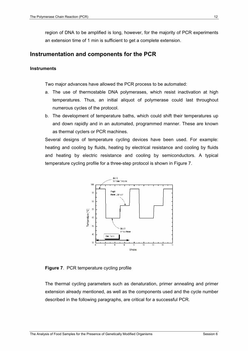
and heating by electric resistance and cooling by semiconductors. A typical
temperature cycling profile for a three-step protocol is shown in Figure 7.
Figure 7. PCR temperature cycling profile
The thermal cycling parameters such as denaturation, primer annealing and primer
extension already mentioned, as well as the components used and the cycle number
described in the following paragraphs, are critical for a successful PCR.

The Polymerase Chain Reaction (PCR) 13
The Analysis of Food Samples for the Presence of Genetically Modified Organisms Session 6
Target DNA
In principle, PCR amplification can be performed if at least one intact copy of the
target gene is present. A greater number of target copies enhance the probability of
successful DNA amplification. Any damage, such as a nick in the target DNA, will
block PCR amplification. The size of the target sequence can be anything from < 0.1
to a few kilobases. The total amount of DNA typically used for PCR is 0.05 to 1.0 µg,
this allows detection of single copies of target sequence. Even if a sample does not
need to be highly purified, some contaminants such as heparin, heme, formalin,
Mg2+-chelating agents, as well as detergents should be eliminated to avoid inhibition
of the amplification process.
Primers
Generally, primers used are 16 - 30 nucleotides in length that allows the use of a
reasonably high annealing temperature. Primers should avoid stretches of polybase
sequences (e.g. poly dG) or repeating motifs - these can hybridise inappropriately on
the template. Inverted repeat sequences should be avoided so as to prevent
formation of secondary structure in the primer, which would prevent hybridisation to
template. Sequences complementary to other primers used in the PCR should also
be avoided so to prevent hybridisation between primers, or primer dimer formation
(particularly important for the 3' end of the primer). If possible, the 3' end of the
primer should be rich in G, C bases to enhance annealing of the end that will be
extended. The distance between primers should be less than 10 Kb in length.
Typically, substantial reduction in yield is observed when the primers extend from
each other beyond ~3 Kb. Oligonucleotides are usually used at the concentration of
1µM in PCR. This is sufficient for at least 30 cycles of amplification. The presence of
higher concentration of oligonucleotides can cause amplification of undesirable non-
target sequences. Conversely, the PCR is inefficient with limiting primer
concentration.
DNA polymerase
The original method of PCR used the Klenow fragment of E. coli DNA polymerase I
(Saiki et al., 1985). This enzyme, however, denatures at temperatures lower than that
required to denature most template duplexes. Thus, in earlier experiments, fresh
enzyme had to be added to the reaction after each cycle. In addition, samples had to

The Polymerase Chain Reaction (PCR) 14
The Analysis of Food Samples for the Presence of Genetically Modified Organisms Session 6
be moved from one temperature bath to another to allow the individual steps of
denaturation, annealing and polymerisation. The use of heat-resistant DNA
polymerase has obviously facilitated the process because the addition of enzymes
after every denaturation step is no longer necessary. Typically, DNA polymerases
can only incorporate nucleotides from the 3’ end of a polynucleotide. The first
thermostable DNA polymerase used was the Taq DNA polymerase isolated from the
bacterium Thermus aquaticus (Saiki et al., 1988). Even though this enzyme is
probably the most widely used in PCR applications, several other DNA polymerases
are commercially available. Table 1 lists the properties of some thermostable DNA
polymerases currently in use for PCR (Newton and Graham, 1994).
Table 1. Characteristics of some DNA polymerases used for PCR
Taq/ AmpliTaq® Vent™ Deep-
Vent™ Pfu Tth UITma™
Source Thermus aquaticus
Thermo-coccus litoralis
Pyrococcus GB-D
Pyrococcus furiosus
Thermus thermophilus
Thermotoga maritima
Application
Taq: natural AmpliTaq: for genetic engineering
For genetic engineering
For genetic engineering Natural For genetic
engineering For genetic engineering
T½ of activity at 95 ºC (min) 40 1380 400 >120 20 >50a
5’ to 3’ Exonuclease activity
Yes
No
No
No
Yes
No
3’ to 5’ Exonuclease activity
No
Yes
Yes
Yes
No
Yes
Processivity 50-60 ? 7 ? 30-40 ? Extension rate (nt/s) 75 ? >80 60 >33 ?
Resulting DNA ends 3’A >95% blunt >95% blunt ? 3’A blunt
MW in kDa 94 ? ? 92 94 70
Taq/AmpliTaq® DNA polymerase. As already mentioned, this enzyme was isolated
from the bacterium Thermus aquaticus living in a hot spring in Yellowstone National
Park USA at temperatures close to 85°C. The optimal working temperature of this
enzyme is 70 - 80°C. At this temperature, the bacterium synthesises DNA at a rate of
35 - 100 nucleotides/sec. The average number of nucleotides, which an enzyme
incorporates into DNA before detaching itself from the template, is known as the
processivity. AmpliTaq® DNA polymerase is a genetically modified enzyme
The Polymerase Chain Reaction (PCR) 15
The Analysis of Food Samples for the Presence of Genetically Modified Organisms Session 6
expressed by E. coli. Since AmpliTaq® is recombinant, the purity and reproducibility
of this enzyme are higher than those of the wild type. However, potential
contamination might occur during the DNA amplification, with some homologous E.
coli sequences. In this case, the use of a DNA polymerase which has not been
expressed with E. coli, as host organism, is recommended. Both Taq, and
AmpliTaq® DNA polymerases possess a 5’ to 3’ exonuclease activity, which removes
nucleotides ahead of the growing chain.
Vent™ -; DeepVent™-; Pfu- and UITma™- DNA polymerases. These enzymes
have a 3’- 5’ exonuclease activity which allow the removal of mismatched residues
until a correctly base-paired terminus is generated. However, the 3’- 5’ exonuclease
activity can cause degradation of the primers. Therefore, the enzyme should only be
added after the reaction has started, or alternatively, chemically modified primers
should be used.
AmpliTaqGold™- DNA polymerase. This enzyme consists of an AmpliTaq DNA
polymerase, inactive at room temperature, and can only be activated during an
incubation period at 94°C. In this case, the program of the thermocycler should
include a pre-incubation period at a temperature of 92 - 95°C. For the time-released
PCR, the pre-incubation can be eliminated, but at least 10 cycles more than the
classic PCR must be performed.
Reaction buffers and MgCl2 in PCR reactions
In addition to the reagents directly involved in the reaction, PCR requires a suitable
buffer. Buffer composition depends on the type and characteristics of the enzyme
being used and most suppliers usually provide a 10x buffer for use with the
respective enzyme. The most common reaction buffer used with Taq/AmpliTaq®
DNA polymerase contains:
• 10 mM Tris, pH 8.3
• 50 mM KCl
• 1.5-2.5 mM MgCl2

The Polymerase Chain Reaction (PCR) 16
The Analysis of Food Samples for the Presence of Genetically Modified Organisms Session 6
The presence of divalent cations in PCR is critical. The MgCl2 concentration in the
final reaction mixture is usually between 0.5 to 5.0 mM, and the optimum
concentration is determined empirically (Innis and Gelfand, 1990).
Mg2+ ions:
• form a soluble complex with dNTPs which is essential for dNTP incorporation,
• stimulate polymerase activity,
• increase the Tm of primer/template interaction (and therefore they stabilise the
duplex interaction).
Generally, a low Mg2+ concentration leads to low yields (or no yield) whereas a high
Mg2+ concentration leads to accumulation of non-specific products (mispriming). It is
important to avoid a high concentration of chelating agents such as EDTA or
negatively charged ionic groups such as phosphate in the template DNA solution.
Current literature includes discussions on various PCR buffers and supplements,
such as DMSO, PEG 6000, formamide, glycerol, spermidine and non-ionic
detergents, used to increase the reaction specificity or efficiency (Roux, 1995).
Certain DNA polymerases will indeed reach their optimum level of activity (Rolfs et
al., 1992) only in the presence of such supplements.
Deoxyribonucleoside triphosphates
Free deoxyribonucleoside triphosphates (dNTPs) are required for DNA synthesis.
The dNTPs concentrations for PCR should be 20 to 200 µM for each dNTP and the
four dNTPs should be used at equivalent concentrations to minimize
misincorporation errors (Innis et al., 1988). High-purity dNTPs are supplied by
several manufacturers either as four individual stocks or as a mixture of all four
dNTPs. dNTPs stock solutions (usually 100 mM) should be adjusted to pH 7.0-7.5
with 1 M NaOH to ensure that the pH of the final reaction does not fall below 7.1
(Sambrook et al., 1989); however, many dNTPs stock solutions are now supplied
with already adjusted pH.
Cycle number and plateau effect
The number of amplification cycles necessary to produce a band visible on a gel
depends largely on the starting concentration of the target DNA. In order to amplify
50 target molecules, 40 - 45 cycles are recommended, whereas 25 - 30 cycles are

The Polymerase Chain Reaction (PCR) 17
The Analysis of Food Samples for the Presence of Genetically Modified Organisms Session 6
enough to amplify 3x10⁵ molecules to the same concentration (Innis and Gelfand,
1990). This non-proportionality is due to the so-called plateau effect, which is the
attenuation in the exponential rate of product accumulation in late stages of a PCR,
when the product reaches 0.3 - 1.0 nM. This may be caused by degradation of
reactants (dNTPs, enzyme), reactant depletion (primers, dNTPs – the former a
problem with short products, the latter with long products), end-product inhibition
(pyrophosphate formation), competition for reactants by non-specific products,
competition for primer binding by re-annealing of the concentrated (10 nM) product
(Innis and Gelfand, 1990). If the desired product is not obtained in 30 cycles, a small
sample (1 µl) of the amplified product should be taken, mixed and re-amplified 20 -
30 cycles in a new reaction mix, rather than extending the run to more cycles. In
some cases where the template concentration is limiting, this re-amplification can
produce a good product, whereas extension of cycling to 40 times or more does not.
Design of primers for PCR
Perhaps the most critical parameter for successful PCR is the design of primers. All
things being equal, a poorly designed primer can result in a PCR reaction that will not
work. The primer sequence determines several things such as the position and
length of the product, its melting temperature and ultimately the yield (Innis and
Gelfand, 1994). A poorly designed primer can result in little or no product due to non-
specific amplification and/or primer-dimer formation, which can become competitive
enough to suppress product formation. This application note is provided to give rules
that should be taken into account when designing primers for PCR. More
comprehensive coverage of this subject can be found elsewhere (Dieffenbach et al.,
1995).
Primer selection
Several variables must be taken into account when designing PCR primers. Among
the most critical are:
• Primer length
• Melting temperature (Tm)
• Specificity
• Complementary primer sequences

The Polymerase Chain Reaction (PCR) 18
The Analysis of Food Samples for the Presence of Genetically Modified Organisms Session 6
• G/C content and polypyrimidine (T, C) or polypurine (A, G) stretches
• 3’-end sequence
Each of these critical elements will be discussed in the following sections.
Primer length
Since specificity, temperature and time of annealing partly depend on primer length,
this parameter is critical for successful PCR. In general, oligonucleotides between 18
and 24 bases are extremely sequence-specific, provided that the annealing
temperature is optimal. Primer length is also proportional to annealing efficiency. In
general, the longer is the primer, the more inefficient the annealing. With fewer
templates primed at each step, this can result in a significant decrease in amplified
product. The primers should, however, not be too short unless the application
specifically requires it. As discussed below, the goal should be to design a primer
with an annealing temperature of at least 50°C.
The relationship between annealing temperature and melting temperature is one of
the “Black Boxes” of PCR. A general rule-of-thumb is to use an annealing
temperature that is 5°C lower than the melting temperature. Often, the annealing
temperature determined in this fashion will not be optimal and empirical experiments
will have to be performed to determine the optimal temperature. This is most easily
accomplished using a gradient thermal cycler.
Melting temperature (Tm)
It is important to keep in mind that there are two primers added to a site/target
directed PCR reaction. Both of the oligonucleotide primers should be designed so
that they have similar melting temperatures. If primers are mismatched in terms of
Tm, amplification will be less efficient or may not work at all since the primer with the
higher Tm will misprime at lower temperatures and the primer with the lower Tm may
not work at higher temperatures. The melting temperatures of oligos are most
accurately calculated using nearest neighbour thermodynamic calculations with the
formula:
Tmprimer = ∆H [∆S+ R ln (c/4)] -273.15°C + 16.6 log 10 [K+] (2)
The Polymerase Chain Reaction (PCR) 19
The Analysis of Food Samples for the Presence of Genetically Modified Organisms Session 6
where H is the enthalpy and S is the entropy for helix formation, R is the molar gas
constant and c is the concentration of primers.
This is most easily accomplished by using primer design software packages already
available on the market (Sharrocks, 1994). Fortunately, a good working
approximation of this value (generally valid for oligos in the 18 - 24 base range) can
be calculated using the formula:
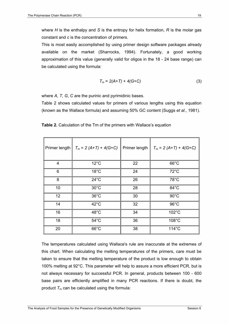
Tm = 2(A+T) + 4(G+C) (3)
where A, T, G, C are the purinic and pyrimidinic bases.
Table 2 shows calculated values for primers of various lengths using this equation
(known as the Wallace formula) and assuming 50% GC content (Suggs et al., 1981).
Table 2. Calculation of the Tm of the primers with Wallace’s equation
Primer length
Tm = 2 (A+T) + 4(G+C)
Primer length
Tm = 2 (A+T) + 4(G+C)
4 12°C 22 66°C
6 18°C 24 72°C
8 24°C 26 78°C
10 30°C 28 84°C
12 36°C 30 90°C
14 42°C 32 96°C
16 48°C 34 102°C
18 54°C 36 108°C
20 66°C 38 114°C
The temperatures calculated using Wallace's rule are inaccurate at the extremes of
this chart. When calculating the melting temperatures of the primers, care must be
taken to ensure that the melting temperature of the product is low enough to obtain
100% melting at 92°C. This parameter will help to assure a more efficient PCR, but is
not always necessary for successful PCR. In general, products between 100 - 600
base pairs are efficiently amplified in many PCR reactions. If there is doubt, the
product Tm can be calculated using the formula:

The Polymerase Chain Reaction (PCR) 20
The Analysis of Food Samples for the Presence of Genetically Modified Organisms Session 6
Tm =81.5 + 16.6 (log10[K+] + 0.41 (%G+C)-675/length (4)
Specificity
As mentioned above, primer specificity is at least partly dependent on primer length.
It is evident that there are many more unique 24 base oligos than there are 15 base
oligos. That said, primers must be chosen so that they have a unique sequence
within the template DNA that is to be amplified. A primer designed with a highly
repetitive sequence will result in a smear when amplifying genomic DNA. However,
the same primer may give a single band, if a single clone from a genomic library is
amplified. Because Taq DNA polymerase is active over a broad range of
temperatures, primer extension will occur at the lower temperatures of annealing. If
the temperature is too low, non-specific priming may occur, which can be extended
by the polymerase if there is a short homology at the 3' end. In general, a melting
temperature of 55° - 72°C gives the best results (note that this corresponds to a
primer length of 18 - 24 bases using Wallace's rule).
Complementary primer sequences
Primers need to be designed with absolutely no intra-primer homology beyond 3
base pairs. If a primer has such a region of self-homology, “snap back”, or “hair-pin”,
partially double-stranded structures can occur, which will interfere with annealing to
the template. Another related danger is inter-primer homology. Partial homology in
the middle regions of two primers can interfere with hybridisation. If the homology
occurs at the 3' end of either primer, primer dimer formation will occur, which, more
often than not, will prevent the formation of the desired product via competition.
G/C content and polypyrimidine (T, C) or polypurine (A, G) stretches
The base composition of primers should be between 45% and 55% GC. The primer
sequence must be chosen so that there is no poly-G or poly-C stretches that can
promote non-specific annealing. Poly-A and poly-T stretches are also to be avoided,
as these will “breathe” and open up stretches of the primer-template complex. This
can lower the efficiency of amplification. Polypyrimidine (T, C) and polypurine (A, G)

The Polymerase Chain Reaction (PCR) 21
The Analysis of Food Samples for the Presence of Genetically Modified Organisms Session 6
stretches should also be avoided. Ideally the primer will have a near random mix of
nucleotides, a 50% GC content and be ~20 bases long. This will put the Tm in the
range of 56° - 62°C (Dieffenbach et al., 1995).
3’-end sequence
It is well established that the 3' terminal position in PCR primers is essential for the
control of mis-priming. The problem of primer homologies occurring in these regions
has already been explored. Another variable to look at is the inclusion of a G or C
residue at the 3' end of primers. This “GC Clamp” helps to ensure correct binding at
the 3' end, due to the stronger hydrogen bonding of G/C residues. This also helps to
improve the efficiency of the reaction by minimising any “breathing” that might occur.
Specialised PCR
In addition to the amplification of a target DNA sequence by the typical PCR
procedures already described, several specialised types of PCR have been
developed for specific applications.
Nested PCR
Nested sets of primers can be used to improve PCR yield of the target DNA
sequence (Newton and Graham, 1994). PCR with nested primers is performed for 15
to 30 cycles with one primer set and then for an additional 15 to 30 cycles, with a
second primer set, for an internal region of the first amplified DNA product. Thus, the
larger fragment produced by the first round of PCR is used as the template for the
second PCR. Using the nested PCR method can dramatically increase the sensitivity
and specificity of DNA amplification. The specificity is particularly enhanced because
this technique almost always eliminates any spurious non-specific amplification
products. This is because after the first round of PCR any non-specific products are
unlikely to be sufficiently complementary to the nested primers to be able to serve as
a template for further amplification, thus the desired target sequence is preferentially
amplified. However, the increased risk of contamination is a drawback of this extreme

The Polymerase Chain Reaction (PCR) 22
The Analysis of Food Samples for the Presence of Genetically Modified Organisms Session 6
sensitivity, and great care must be taken when performing such PCRs, particularly in
a diagnostic laboratory.
Multiplex PCR
Whereas standard PCR usually uses one pair of primers to amplify a specific
sequence, Multiplex PCR uses multiple pairs of primers to amplify many sequences
simultaneously. The presence of many PCR primers in a single tube could cause
many problems, such as the increased formation of misprimed PCR products,
"primer dimers", and the amplification discrimination of longer DNA fragments (Atlas
and Bey, 1994).
For this type of PCR amplification, primers are chosen with similar annealing
temperatures. The lengths of amplified products should be similar; large differences
in the lengths of the target DNAs will favour the amplification of the shorter target
over the longer one, resulting in differential yields of amplified products. In addition,
Multiplex PCR buffers contain Taq polymerase additive, which decreases the
competition among amplicons and the discrimination of longer DNA fragments during
Multiplex PCR.
Multiplex PCR products can be further hybridised with a gene-specific probe for
verification.

The Polymerase Chain Reaction (PCR) 23
The Analysis of Food Samples for the Presence of Genetically Modified Organisms Session 6
PCR in practice
As already illustrated in the previous sections, PCR is widely used and is a powerful
analytical and preparative technique. However, because of the nature of this
procedure, trace amounts of DNA contaminants could serve as templates, resulting
in amplification of the wrong target nucleic acid (false positives). Thus, it is critical to
perform PCR amplification in a DNA-free environment. Providing physically separate
working areas with dedicated equipment reduces the risk of contamination. Strict
compliance with decontamination requirements (decontamination of nucleic acids,
prevention of aerosols etc.) is the most important prerequisite to reduce the rate of
false-positive results to a minimum. PCR contamination can be caused by several
sources such as:
• Laboratory benches, equipment and pipetting devices, which can be
contaminated by previous DNA preparations, or by purified restriction fragments
• Cross-contamination between samples
• Products from previous PCR amplifications.
This section provides some recommendations, with the aim of defining the routine
requirements for the establishment and maintenance of a clean environment for any
PCR-based assay system, regardless of the number of samples being processed
(Roth et al., 1997).
Physical prevention methods
Laboratory facilities. In order to avoid contamination, physically separate working
areas should be set up as follows:
1. Sample preparation area
This room consists of an area where all the steps prior to amplification of the
template DNA are performed (e.g. isolation and purification of DNA).
2. PCR set-up room
This “clean” room is devoted to the procedures related to the preparation of the
PCR reaction (e.g. mastermix, primers dilutions etc.).
3. Post-PCR area
The area is dedicated to the amplification of the target DNA sequence, and the
detection and analysis of the PCR products.
In addition, the following general rules should be observed:

The Polymerase Chain Reaction (PCR) 24
The Analysis of Food Samples for the Presence of Genetically Modified Organisms Session 6
• All the rooms should contain dedicated equipment (coats, gloves, reagents and
supplies).
• Reagents and other devices must be labelled with content and date of
preparation.
• Use a one-way flow system, i.e. never move material, samples or equipment from
post-PCR areas into pre-PCR locations.
• Use disposable PCR reaction tubes, which are DNase and RNase free.
• Use special aerosol-resistant pipette tips and a dedicated (used only for PCR) set
of pipettes, preferably positive displacement pipettes.
• If possible, set up PCR reactions under a fume hood that is equipped with UV
light. Under the fume hood, store a microcentrifuge and disposable gloves that
are used only for PCR.
• Periodically wash benches and shelves with 10% bleach followed by 70%
ethanol.
Sample handling
• Use sterile techniques and always wear fresh gloves when working in the areas
previously described. Change gloves frequently, especially if you suspect they
have become contaminated with solutions containing template DNA.
• Always use new and/or sterilised glassware, plasticware and pipettes to prepare
PCR reagents and template DNA.
• Autoclave all reagents and solutions that can be autoclaved without affecting their
performance. Of course, primers, dNTPs and Taq DNA Polymerase should not
be autoclaved.
• Have your own set of PCR reagents and solutions that are only used for PCR,
and store these reagents in small aliquots.
• When pipetting DNA, avoid creating aerosols that can carry contaminants.
• Always include control reactions, for example a negative (“no DNA”) control,
which contains all reaction components except the template DNA, and a positive
control that has been successfully used in previous PCRs.
The Polymerase Chain Reaction (PCR) 25
The Analysis of Food Samples for the Presence of Genetically Modified Organisms Session 6
Biochemical prevention methods
Uracil-DNA Glycosylase. The polymerase chain reaction (PCR) can amplify a single
molecule over a billionfold. Thus, even minuscule amounts of a contaminant can be
amplified and lead to a false positive result. Such contaminants are often products
from previous PCR amplifications (carry-over contamination). Therefore, methods to
avoid such contamination have been developed.
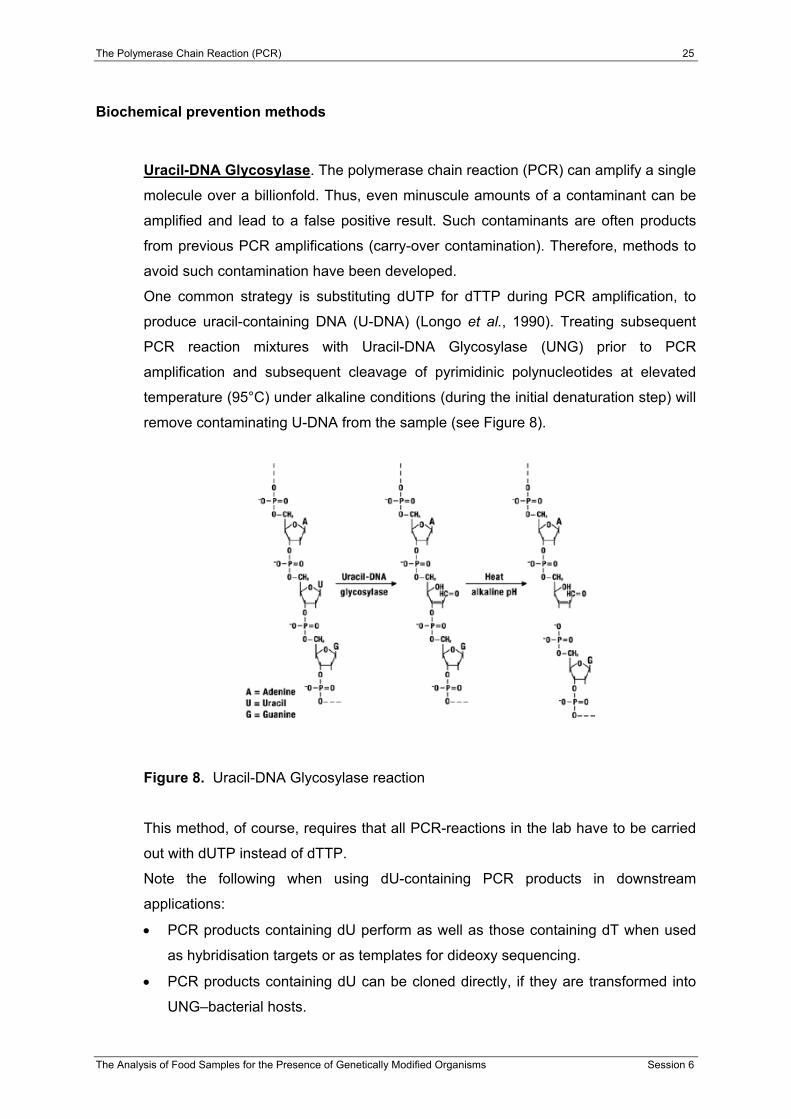
One common strategy is substituting dUTP for dTTP during PCR amplification, to
produce uracil-containing DNA (U-DNA) (Longo et al., 1990). Treating subsequent
PCR reaction mixtures with Uracil-DNA Glycosylase (UNG) prior to PCR
amplification and subsequent cleavage of pyrimidinic polynucleotides at elevated
temperature (95°C) under alkaline conditions (during the initial denaturation step) will
remove contaminating U-DNA from the sample (see Figure 8).
Figure 8. Uracil-DNA Glycosylase reaction
This method, of course, requires that all PCR-reactions in the lab have to be carried
out with dUTP instead of dTTP.
Note the following when using dU-containing PCR products in downstream
applications:
• PCR products containing dU perform as well as those containing dT when used
as hybridisation targets or as templates for dideoxy sequencing.
• PCR products containing dU can be cloned directly, if they are transformed into
UNG–bacterial hosts.

The Polymerase Chain Reaction (PCR) 26
The Analysis of Food Samples for the Presence of Genetically Modified Organisms Session 6
• A dU-containing substrate is readily digested by some common restriction
enzymes (e.g. EcoR I and BamH I), while others show reduced activity (e.g. Hpa
I, Hind II, Hind III) on these substrates.
• The use of dU-containing DNA is not recommended for protein-binding or DNA-
protein interaction studies.
DNase I, exonuclease III. Other biochemical methods are based on the treatment of
the contaminated DNA with DNase I, exonuclease III or with a restriction enzyme,
containing a recognition sequence within the target DNA. However, because of the
harsh reaction condition required, these enzymes present the disadvantage of
reducing the efficiency of the PCR amplification.
Preparation of the mixture for the PCR reaction (Mastermix)
The essential reagent components for PCR are water, the reaction buffer, a
thermostable DNA polymerase, oligonucleotide primers, deoxynucleotides (dNTPs),
template (target) DNA, and magnesium ions (Mg2+). In general, all reagents (except
the template DNA) are mixed in a single tube, in enough volume according to the
number of reactions to be performed (mastermix). The mastermix is then aliquotted
into individual tubes and the template DNA is added. The use of a mastermix solution
reduces the risk of contamination and improves the performance of the PCR reaction
for the following reasons:
• a uniformed quality of the solution is guaranteed for all the reagents for a series
of analyses,
• the risk of contamination of the parent and resulting solutions is decreased,
• larger volumes can be pipetted,
• there are fewer pipetting stages and therefore time is saved.
Successful amplification of the region of interest depends on the amount and quality
of the template DNA. The amount of template required is dependent upon the
complexity of the DNA sample. Taking into account that the size of nuclear genome
varies among organisms, the DNA concentration should be maintained constant
(usually 10 ng/µl). A comparison of genome size of plant species frequently used in
plant transformation and the corresponding number of genome copies in a defined
amount of DNA, are given in Table 3.
The Polymerase Chain Reaction (PCR) 27
The Analysis of Food Samples for the Presence of Genetically Modified Organisms Session 6
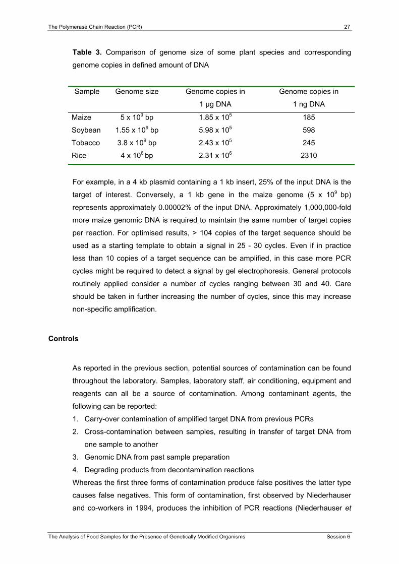
Table 3. Comparison of genome size of some plant species and corresponding
genome copies in defined amount of DNA
Sample Genome size Genome copies in
1 µg DNA
Genome copies in
1 ng DNA
Maize 5 x 10⁹ bp 1.85 x 10⁵ 185
Soybean 1.55 x 10⁹ bp 5.98 x 10⁵ 598
Tobacco 3.8 x 10⁹ bp 2.43 x 10⁵ 245
Rice 4 x 10⁸ bp 2.31 x 10⁶ 2310
For example, in a 4 kb plasmid containing a 1 kb insert, 25% of the input DNA is the
target of interest. Conversely, a 1 kb gene in the maize genome (5 x 10⁹ bp)
represents approximately 0.00002% of the input DNA. Approximately 1,000,000-fold
more maize genomic DNA is required to maintain the same number of target copies
per reaction. For optimised results, > 104 copies of the target sequence should be
used as a starting template to obtain a signal in 25 - 30 cycles. Even if in practice
less than 10 copies of a target sequence can be amplified, in this case more PCR
cycles might be required to detect a signal by gel electrophoresis. General protocols
routinely applied consider a number of cycles ranging between 30 and 40. Care
should be taken in further increasing the number of cycles, since this may increase
non-specific amplification.
Controls
As reported in the previous section, potential sources of contamination can be found
throughout the laboratory. Samples, laboratory staff, air conditioning, equipment and
reagents can all be a source of contamination. Among contaminant agents, the
following can be reported:
1. Carry-over contamination of amplified target DNA from previous PCRs
2. Cross-contamination between samples, resulting in transfer of target DNA from
one sample to another
3. Genomic DNA from past sample preparation
4. Degrading products from decontamination reactions
Whereas the first three forms of contamination produce false positives the latter type
causes false negatives. This form of contamination, first observed by Niederhauser
and co-workers in 1994, produces the inhibition of PCR reactions (Niederhauser et
The Polymerase Chain Reaction (PCR) 28
The Analysis of Food Samples for the Presence of Genetically Modified Organisms Session 6
al., 1994). In fact, decontamination using the UNG method, favours the formation of
complexes with the primers.
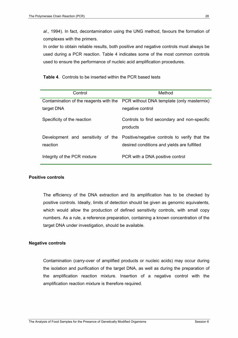
In order to obtain reliable results, both positive and negative controls must always be
used during a PCR reaction. Table 4 indicates some of the most common controls
used to ensure the performance of nucleic acid amplification procedures.
Table 4. Controls to be inserted within the PCR based tests
Control Method
Contamination of the reagents with the
target DNA
PCR without DNA template (only mastermix)
negative control
Specificity of the reaction Controls to find secondary and non-specific
products
Development and sensitivity of the
reaction
Positive/negative controls to verify that the
desired conditions and yields are fulfilled
Integrity of the PCR mixture PCR with a DNA positive control
Positive controls
The efficiency of the DNA extraction and its amplification has to be checked by
positive controls. Ideally, limits of detection should be given as genomic equivalents,
which would allow the production of defined sensitivity controls, with small copy
numbers. As a rule, a reference preparation, containing a known concentration of the
target DNA under investigation, should be available.
Negative controls
Contamination (carry-over of amplified products or nucleic acids) may occur during
the isolation and purification of the target DNA, as well as during the preparation of
the amplification reaction mixture. Insertion of a negative control with the
amplification reaction mixture is therefore required.

The Polymerase Chain Reaction (PCR) 29
The Analysis of Food Samples for the Presence of Genetically Modified Organisms Session 6
References
Alberts, B., Bray, D., Lewis, J., Raff, M., Roberts, K. and Watson, J.D. (1983).
Molecular biology of the cell. Garland Publishing, Inc., New York.
Atlas, R.M. and Bej, A.K. (1994). Polymerase Chain Reaction. In: Gerhardt, P.,
Murrey, R.G.E., Wood, W.A. and Krieg, N.R., (Eds.) Methods for general and
molecular bacteriology. Washington, D.C.: American Society for Microbiology, pp.
418–435.
Dieffenbach, C.W., Lowe, T.M.J. and Dveksler, G.S. (1995). General Concepts for
PCR Primer Design. In: Dieffenbach, C.W, and Dveksler, G.S. (Eds.) PCR
Primer: a Laboratory Manual. New York: Cold Spring Harbor Laboratory Press,
Cold Spring Harbor, NY, USA, pp. 133–155.
Innis, M.A. and Gelfand, D.H. (1990). Optimization of PCRs. In: Innis, M.A., Gelfand,
D.H., Sninsky, J.J., and White, T.J. (Eds.) PCR Protocols: a Guide to Methods
and Applications. New York: Academic Press, pp. 3–12.
Innis, M.A., and Gelfand, D.H. (1994). Optimization of PCRs. In: Innis, M.A., Gelfand,
D.H., Sninsky, J.J., and White, T.J. (Eds.) PCR Protocols: a Guide to Methods
and Applications. London: CRC Press, pp. 5–11.
Innis, M.A., Myambo, K.B., Gelfand, D.H. and Brow, M.A. (1988). DNA sequencing
with Thermus aquaticus DNA polymerase and direct sequencing of polymerase
chain reaction-amplified DNA. Proceedings of the National Academy of Science
USA 85, 9436-9440.
Longo, M.C., Berninger, M.S. and Hartley, J.L. (1990). Use of uracil DNA glycosylase
to control carry-over contamination in polymerase chain reaction. Gene 93, 125–
128.
Newton, C.R. and Graham, A. (1994). PCR. BIOS Scientific Publishers, Limited,
Oxford.
The Polymerase Chain Reaction (PCR) 30
The Analysis of Food Samples for the Presence of Genetically Modified Organisms Session 6
Niederhauser, C., Höfelein, C., Wegmüller, B., Lüthy, J. and Candrian, U. (1994).
Reliability of PCR Decontamination Systems. PCR Methods and Applications 4,
117–123.
Ogawa, T. and Okazaki, T. (1980). Discontinuous DNA replication. Annual Review of
Biochemistry 49, 421–457.
Roux, K.H. (1995). Optimization and troubleshooting in PCR. PCR Methods and
Applications 4, 185-194.
Rolfs, A., Schuller, I., Finckh, U. and Weber-Rolfs, I. (1992). Substances affecting
PCR: Inhibition and enhancement, 51-58. In: PCR: Clinical diagnostics and
research, Springer.
Roth, A., Mauch, H. and Göbel, U. (1997). Nucleic Acid Amplification Techniques –
Recommendations for Employing Molecular Methods in Diagnostic Routine
Microbiology Laboratories and Measures for Internal Quality Assurance. Gustav
Fischer Verlag, Stuttgart.
Saiki, R.K., Scharf, S.J., Faloona, F., Mullis, K.B., Horn, G.T., Erlich, H.A. and
Arnheim, N. (1985). Enzymatic amplification of ß-globin genomic sequences and
restriction site analysis for diagnosis of sickle cell anemia. Science 230, 1350.
Saiki, R.K. et al. (1988). Primer-directed enzymatic amplification of DNA with a
thermostable DNA polymerase. Science 239, 487.
Sambrook, J., Fritsch, E.F. and Maniatis, T. (1989). In vitro Amplification of DNA by
the Polymerase Chain Reaction. In: Sambrook, J., Fritsch, E.F. and Maniatis, T.
(Eds.) Molecular Cloning: a Laboratory Manual. New York: Cold Spring Harbor
Laboratory Press, Cold Spring Harbor, NY, USA, chapter 14.
Sharrocks, A.D. (1994). The design of primers for PCR. In: Griffin, H.G. and Griffin,
A.M (Eds.) PCR Technology: Current Innovations. London: CRC Press, pp. 5–11.
The Polymerase Chain Reaction (PCR) 31
The Analysis of Food Samples for the Presence of Genetically Modified Organisms Session 6
Suggs, S.V., Hirose, T., Miyake, E.H., Kawashima, M.J., Johnson, K.I., and Wallace,
R.B. (1981). Using Purified Genes. In ICN-UCLA Symp. Developmental Biology,
Vol. 23, Brown, D.D. Ed., Academic Press, New York, 1981, 683.
Additional Reading
Gayer-Herkert, G., (1992). Molekularbiologische Methoden für den Nachweis von
Mikroorganismen in Mischpopulationen – eine Literaturstudie. Bioengineering 5 + 6, 55–64.
Horton, H., Moran, L., Ochs, R., Rawn, J. and Scrimgeour, K., (1994). Principes de
Biochimie. De Boeck – Wesmael, S.A., Bruxelles.
Knippers, R. (1995). Molekulare Genetik. Georg Thieme Verlag, Stuttgart.
Kwok, S., Kellog, D.E., McKinney, N., Spasic, D., Goda, L., Levenson, C. and
Sninsky, J.J. (1990). Effects of primer-template mismatches on the polymerase
chain reaction: Human Immunodeficiency Virus 1 model studies. Nucleic Acids
Research 18, 999–1005.
Larzul, D. (1993). Le PCR: un procédé de réplication in vitro. Tec & Doc-Lavoisier,
Paris.
Stryer, L. (1991). Biochemie. Spektrum Akademischer Verlag, GmbH, Heidelberg.
Watson, D.J., Hopkins, N., Roberts, J., Steitz, J. and Weiner, A. (1988). Molecular
Biology of the Gene. The Benjamin/Cummings Publishing Company, Inc., New
York.
Related Documents