Downloaded from https://journals.lww.com/annalsofsurgery by BhDMf5ePHKav1zEoum1tQfN4a+kJLhEZgbsIHo4XMi0hCywCX1AWnYQp/IlQrHD3kFt6OSS8/9IoE8tbB4jxzVp8XKIfPXPjyVL3dYH2gTJnMB9qYo9cng== on 03/09/2020 Copyright © 2020 Wolters Kluwer Health, Inc. All rights reserved. The American Association of Endocrine Surgeons Guidelines for the Definitive Surgical Management of Thyroid Disease in Adults Kepal N. Patel, MD, Y Linwah Yip, MD, y Carrie C. Lubitz, MD, MPH, z Elizabeth G. Grubbs, MD, § Barbra S. Miller, MD, ô Wen Shen, MD, jj Peter Angelos, MD, Herbert Chen, MD, yy Gerard M. Doherty, MD, zz Thomas J. Fahey III, MD, §§ Electron Kebebew, MD, ôô Virginia A. Livolsi, MD, jjjj Nancy D. Perrier, MD, § Jennifer A. Sipos, MD, Julie A. Sosa, MD, jj David Steward, MD, yyy Ralph P. Tufano, MD, zzz Christopher R. McHenry, MD, §§§ and Sally E. Carty, MDy Objective: To develop evidence-based recommendations for safe, effective, and appropriate thyroidectomy. Background: Surgical management of thyroid disease has evolved consid- erably over several decades leading to variability in rendered care. Over 100,000 thyroid operations are performed annually in the US. Methods: The medical literature from 1/1/1985 to 11/9/2018 was reviewed by a panel of 19 experts in thyroid disorders representing multiple disciplines. The authors used the best available evidence to construct surgical manage- ment recommendations. Levels of evidence were determined using the American College of Physicians grading system, and management recom- mendations were discussed to consensus. Members of the American Associ- ation of Endocrine Surgeons reviewed and commented on preliminary drafts of the content. Results: These clinical guidelines analyze the indications for thyroidectomy as well as its definitions, technique, morbidity, and outcomes. Specific topics include Pathogenesis and Epidemiology, Initial Evaluation, Imaging, Fine Needle Aspiration Biopsy Diagnosis, Molecular Testing, Indications, Extent and Outcomes of Surgery, Preoperative Care, Initial Thyroidectomy, Periop- erative Tissue Diagnosis, Nodal Dissection, Concurrent Parathyroidectomy, Hyperthyroid Conditions, Goiter, Adjuncts and Approaches to Thyroidec- tomy, Laryngology, Familial Thyroid Cancer, Postoperative Care and Com- plications, Cancer Management, and Reoperation. Conclusions: Evidence-based guidelines were created to assist clinicians in the optimal surgical management of thyroid disease. Keywords: biopsy, cancer, carcinoma, concurrent parathyroidectomy, diagnosis, endocrine, extent of resection, goiter, hyperthyroidism, imaging, lymph nodes, management and complications, molecular markers, nodules, pathogenesis, poorly differentiated thyroid carcinoma, postoperative care, preoperative care, staging, surgery, thyroid, thyroidectomy indications, ultrasound (Ann Surg 2020;271:e21–e93) T ABLE OF CONTENTS PAGES 1. Introduction e23 2. Methods e23-e24 a. Literature Evidence b. Conflict of Interest (COI) c. Grading of Practice Recommendations d. AAES Member Input and Sister Society Endorsement e. Cautions to Implementation 3. Pathogenesis and Epidemiology e24–e26 a. Thyroid Dysfunction b. Benign Thyroid Nodules c. Lesions of Indeterminate Malignant Potential d. Thyroid Cancer e. Primary Thyroid Lymphoma 4. Initial Evaluation e26–e30 From the Divisionof Endocrine Surgery, NYU Langone Health, New York, NY; yDepartment of Surgery, University of Pittsburgh School of Medicine, Pitts- burgh, PA; zDepartment of Surgery, Massachusetts General Hospital, Boston, MA; §Department of Surgical Oncology, University of Texas MD Anderson Cancer Center, Houston, TX; ôDivision of Endocrine Surgery, University of Michigan, Ann Arbor, MI; jjDepartment of Surgery, University of California San Francisco, San Francisco, CA; Department of Surgery and MacLean Center for Clinical Medical Ethics, The University of Chicago, Chicago, IL; yyDepartment of Surgery, University of Alabama at Birmingham, Birmingham, AL; zzDepartment of Surgery, Brigham and Women’s Hospital, Boston, MA; §§Department of Surgery, The New York Presbyterian Hospital-Weill Cornell Medical Center, New York, NY; ôôDepartment of Surgery and Stanford Cancer Institute, Stanford University, Stanford, CA; jjjjDepartment of Pathology and Laboratory Medicine, Perelman School of Medicine of University of Penn- sylvania, Philadelphia, PA; Division of Endocrinology and Metabolism, The Ohio State University, Columbus, OH; yyyDepartment of Otolaryngology, University of Cincinnati College of Medicine, Cincinnati, OH; zzzDivision of Head and Neck Endocrine Surgery, Department of Otolaryngology-Head and Neck Surgery, The Johns Hopkins School of Medicine, Baltimore, MD; and §§§MetroHealth Medical Center, Case Western Reserve University School of Medicine, Cleveland, OH. Y [email protected]. The Executive Summary of these guidelines can be found here: 10.1097/ SLA.0000000000003735. S.E.C. and C.R.M. directed the project and coordinated the authors and topics. S.E.C., C.R.M., and K.N.P. developed the methodology, managed disclosures, and prepared the work for publication. C.C.L., E.G.G., B.S.M., K.N.P., W.T.S., and L.Y. carried out the systematic topic reviews and were the primary authors of text. All authors participated directly in editing the text and recommenda- tions. AAES legal counsel approved the language in the section, Cautions to Implementation. All authors received no funding for this work and declare that neither they nor their institutions received payment or support in kind for any aspect of this work (including grants, data monitoring board, study design, manuscript preparation, statistical analysis). As defined in Appendix A, relevant COI is listed for all authors: D.S. has received research support from Astra Zeneca, Veracyte and Gene- ProDx. K.N.P. has received honoraria for educational programming from Veracyte. J.A. Sipos has received research support and honoraria from UPMC/Thyroseq and Genzyme/Sanofi. P.A. has received research funding from Olympus-Gyrus. R.P.T. has served as a consultant for Medtronic and Hemostatix. V.A.L. has served as a consultant for Veracyte. The authors report no conflicts of interest. Copyright ß 2020 Wolters Kluwer Health, Inc. All rights reserved. ISSN: 0003-4932/20/27103-0e21 DOI: 10.1097/SLA.0000000000003580 Annals of Surgery Volume 271, Number 3, March 2020 www.annalsofsurgery.com | e21 ORIGINAL ARTICLE

Welcome message from author
This document is posted to help you gain knowledge. Please leave a comment to let me know what you think about it! Share it to your friends and learn new things together.
Transcript
Dow
nloadedfrom
https://journals.lww.com
/annalsofsurgeryby
BhDMf5ePH
Kav1zEoum1tQ
fN4a+kJLhEZgbsIH
o4XMi0hC
ywCX1AW
nYQp/IlQ
rHD3kFt6O
SS8/9IoE8tbB4jxzVp8XKIfPXPjyVL3dYH2gTJnM
B9qYo9cng==on
03/09/2020
Downloadedfromhttps://journals.lww.com/annalsofsurgerybyBhDMf5ePHKav1zEoum1tQfN4a+kJLhEZgbsIHo4XMi0hCywCX1AWnYQp/IlQrHD3kFt6OSS8/9IoE8tbB4jxzVp8XKIfPXPjyVL3dYH2gTJnMB9qYo9cng==on03/09/2020
Copyright © 2020 Wolters Kluwer Health, Inc. All rights reserved.
The American Association of Endocrine Surgeons Guidelines forthe Definitive Surgical Management of Thyroid Disease in Adults
Kepal N. Patel, MD,�Y Linwah Yip, MD,y Carrie C. Lubitz, MD, MPH,z Elizabeth G. Grubbs, MD,§
Barbra S. Miller, MD,� Wen Shen, MD,jj Peter Angelos, MD,�� Herbert Chen, MD,yyGerard M. Doherty, MD,zz Thomas J. Fahey III, MD,§§ Electron Kebebew, MD,�� Virginia A. Livolsi, MD,jjjj
Nancy D. Perrier, MD,§ Jennifer A. Sipos, MD,��� Julie A. Sosa, MD,jj David Steward, MD,yyyRalph P. Tufano, MD,zzz Christopher R. McHenry, MD,§§§ and Sally E. Carty, MDy
Objective: To develop evidence-based recommendations for safe, effective,
and appropriate thyroidectomy.
Background: Surgical management of thyroid disease has evolved consid-
erably over several decades leading to variability in rendered care. Over
100,000 thyroid operations are performed annually in the US.
Methods: The medical literature from 1/1/1985 to 11/9/2018 was reviewed
by a panel of 19 experts in thyroid disorders representing multiple disciplines.
The authors used the best available evidence to construct surgical manage-
ment recommendations. Levels of evidence were determined using the
American College of Physicians grading system, and management recom-
mendations were discussed to consensus. Members of the American Associ-
ation of Endocrine Surgeons reviewed and commented on preliminary drafts
of the content.
Results: These clinical guidelines analyze the indications for thyroidectomy
as well as its definitions, technique, morbidity, and outcomes. Specific topics
include Pathogenesis and Epidemiology, Initial Evaluation, Imaging, Fine
Needle Aspiration Biopsy Diagnosis, Molecular Testing, Indications, Extent
and Outcomes of Surgery, Preoperative Care, Initial Thyroidectomy, Periop-
erative Tissue Diagnosis, Nodal Dissection, Concurrent Parathyroidectomy,
Hyperthyroid Conditions, Goiter, Adjuncts and Approaches to Thyroidec-
tomy, Laryngology, Familial Thyroid Cancer, Postoperative Care and Com-
plications, Cancer Management, and Reoperation.
Conclusions: Evidence-based guidelines were created to assist clinicians in
the optimal surgical management of thyroid disease.
Keywords: biopsy, cancer, carcinoma, concurrent parathyroidectomy,
diagnosis, endocrine, extent of resection, goiter, hyperthyroidism, imaging,
lymph nodes, management and complications, molecular markers, nodules,
pathogenesis, poorly differentiated thyroid carcinoma, postoperative care,
preoperative care, staging, surgery, thyroid, thyroidectomy indications,
ultrasound
(Ann Surg 2020;271:e21–e93)
TABLE OF CONTENTS PAGES1. Introduction e232. Methods e23-e24
a. Literature Evidenceb. Conflict of Interest (COI)c. Grading of Practice Recommendationsd. AAES Member Input and Sister Society Endorsemente. Cautions to Implementation
3. Pathogenesis and Epidemiology e24–e26a. Thyroid Dysfunctionb. Benign Thyroid Nodulesc. Lesions of Indeterminate Malignant Potentiald. Thyroid Cancere. Primary Thyroid Lymphoma
4. Initial Evaluation e26–e30
From the �Division of Endocrine Surgery, NYU Langone Health, New York, NY;yDepartment of Surgery, University of Pittsburgh School of Medicine, Pitts-burgh, PA; zDepartment of Surgery, Massachusetts General Hospital, Boston,MA; §Department of Surgical Oncology, University of Texas MD AndersonCancer Center, Houston, TX; �Division of Endocrine Surgery, University ofMichigan, Ann Arbor, MI; jjDepartment of Surgery, University of CaliforniaSan Francisco, San Francisco, CA; ��Department of Surgery and MacLeanCenter for Clinical Medical Ethics, The University of Chicago, Chicago, IL;yyDepartment of Surgery, University of Alabama at Birmingham, Birmingham,AL; zzDepartment of Surgery, Brigham and Women’s Hospital, Boston, MA;§§Department of Surgery, The New York Presbyterian Hospital-Weill CornellMedical Center, New York, NY; ��Department of Surgery and Stanford CancerInstitute, Stanford University, Stanford, CA; jjjjDepartment of Pathology andLaboratory Medicine, Perelman School of Medicine of University of Penn-sylvania, Philadelphia, PA; ���Division of Endocrinology and Metabolism, TheOhio State University, Columbus, OH; yyyDepartment of Otolaryngology,University of Cincinnati College of Medicine, Cincinnati, OH; zzzDivisionof Head and Neck Endocrine Surgery, Department of Otolaryngology-Headand Neck Surgery, The Johns Hopkins School of Medicine, Baltimore, MD; and§§§MetroHealth Medical Center, Case Western Reserve University School ofMedicine, Cleveland, OH.
Y [email protected] Executive Summary of these guidelines can be found here: 10.1097/
SLA.0000000000003735.
S.E.C. and C.R.M. directed the project and coordinated the authors and topics.S.E.C., C.R.M., and K.N.P. developed the methodology, managed disclosures,and prepared the work for publication. C.C.L., E.G.G., B.S.M., K.N.P., W.T.S.,and L.Y. carried out the systematic topic reviews and were the primary authorsof text. All authors participated directly in editing the text and recommenda-tions. AAES legal counsel approved the language in the section, Cautions toImplementation.
All authors received no funding for this work and declare that neither they nor theirinstitutions received payment or support in kind for any aspect of this work(including grants, data monitoring board, study design, manuscript preparation,statistical analysis).
As defined in Appendix A, relevant COI is listed for all authors:D.S. has received research support from Astra Zeneca, Veracyte and Gene-
ProDx.K.N.P. has received honoraria for educational programming from Veracyte.J.A. Sipos has received research support and honoraria from UPMC/Thyroseq and
Genzyme/Sanofi.P.A. has received research funding from Olympus-Gyrus.R.P.T. has served as a consultant for Medtronic and Hemostatix.V.A.L. has served as a consultant for Veracyte.The authors report no conflicts of interest.Copyright � 2020 Wolters Kluwer Health, Inc. All rights reserved.ISSN: 0003-4932/20/27103-0e21DOI: 10.1097/SLA.0000000000003580
Annals of Surgery � Volume 271, Number 3, March 2020 www.annalsofsurgery.com | e21
ORIGINAL ARTICLE
Copyright © 2020 Wolters Kluwer Health, Inc. All rights reserved.
a. Historyi. Ionizing radiationii. Syndromic TCiii. Clinical Characteristics
b. Physical Examinationc. Voice Assessmentd. Laboratory Evaluation
5. Imaging e30–e32a. Preoperative
i. Ultrasonography1. Thyroid nodule US2. Parathyroid incidentaloma US3. Cervical lymph node US4. LN mapping5. Surgical planning6. Translaryngeal US7. US limitations
ii. Cross-sectional imagingiii. Elastographyiv. PET/CTv. Imaging for hyperthyroid conditions
b. Postoperativei. Functional Imaging for TC metastases
6. Fine Needle Aspiration Biopsy (FNAB) Diagnosis e32–e35a. FNAB Indicationsb. Indications for FNAB of Cervical Lymph Nodesc. Pre-FNAB Considerationsd. FNAB Techniquee. FNAB Result Categories
7. Molecular Testing e35–e38a. MT to Guide Need for Surgeryb. MT to Guide Thyroid Nodule Managementc. MT to Guide Extent of Surgeryd. Practical Aspects of MTe. Concerns with MT
8. Indications, Extent, and Outcomes of Surgery e38–e40a. Extent of Initial Thyroidectomyb. Cytologically Benign Nodulesc. Goiter and Thyroiditisd. Hyperthyroidisme. Cytologically Malignant Nodulesf. Cytologically Indeterminate Nodulesg. Other Situationsh. Outcomes of Thyroidectomy
9. Preoperative Care e40–e42a. Antibioticsb. Steroidsc. Surgical Preparation for Graves’ Disease and Hyperthy-
roidismd. Vitamin D and Calciume. Universal/Standard Consent and Counselingf. Venous Thromboembolism (VTE) Prophylaxisg. Interdisciplinary Communication
10. Initial Thyroidectomy e42–e44a. Nomenclatureb. Surgical Planningc. Positioning, Incision, and Exposured. Critical Steps of Thyroid Dissectione. Additional Considerations in Thyroidectomy for Cancer
11. Perioperative Tissue Diagnosis e44–e47a. Core Needle Biopsy of the Thyroid and Cervical
Lymph Nodesb. Incisional Biopsy of the Thyroid and Incisional/Exci-
sional Biopsy of Cervical Lymph Nodes
c. Intraoperative Pathologic Evaluation of the Thyroidd. Intraoperative Pathologic Evaluation of CLN and
Parathyroid Tissuee. Final Histopathologic Diagnosisf. Benign Lesions of the Thyroidg. Differentiated Thyroid Carcinomah. Neoplasms of Uncertain Malignant Potentiali. Poorly Differentiated Thyroid Carcinomaj. Anaplastic Thyroid Carcinomak. Medullary Thyroid Carcinomal. Thyroid Paraganglioma
12. Nodal Dissection e47–e50a. Central Compartment NDb. Lateral Compartment NDc. Prognostic Implications of LNM
13. Concurrent Parathyroidectomy e50–e51a. Epidemiology and Evaluationb. Indications for Concurrent Parathyroidectomyc. Special Situations
14. Hyperthyroid Conditions e51–e54a. Medical Treatmentb. Surgical Treatmentc. Graves’ Ophthalmopathyd. When to Start Thyroid Hormonee. Surgical Complications in Hyperthyroidismf. The Hyperthyroid Pregnant Patient
15. Goiter e54–e56a. Indications for Resectionb. Nonsurgical Treatmentc. Extent and Conduct of Resectiond. Diagnosis and Preoperative Managemente. Intraoperative Management
16. Adjuncts and Approaches e56–e58a. Energy-based Vessel-sealing Devicesb. Intraoperative Assessment of Nerve Functionc. Rapid Parathyroid Hormone Testingd. Intraoperative Tissue Analysise. Remote-Access Approachesf. Hemostatic Agents
17. Laryngology e58–e60a. Preoperative Evaluationb. Intraoperative Eventsc. Postoperative VFD Care
18. Familial Thyroid Cancer e60–e62a. Genetic Testingb. Hereditary Nonmedullary TC
i. Nonsyndromic Familial Nonmedullary TC (NFNMTC)ii. Syndromic FNMTC
1. Familial Adenomatous Polyposis2. Cowden Syndrome3. Carney Complex4. Werner Syndrome5. DICER1 Syndrome6. Papillary Renal Neoplasia7. McCune-Albright Syndrome8. CHEK2
c. Hereditary MTC19. Postoperative Care and Complications e62–e66
a. Routine Managementi. Documentation/Communicationii. Incision Careiii. Medicationsiv. Pain Management
b. Immediate Complications
Patel et al Annals of Surgery � Volume 271, Number 3, March 2020
e22 | www.annalsofsurgery.com � 2020 Wolters Kluwer Health, Inc. All rights reserved.
Copyright © 2020 Wolters Kluwer Health, Inc. All rights reserved.
i. Hematomaii. Recurrent Laryngeal Nerve Dysfunctioniii. Calcium Supplementation, Hypocalcemia,
and Hypoparathyroidismc. Rare Complications
20. Cancer Management e66–e69a. Active Surveillanceb. Postoperative Risk Assessmentc. Prognostic Variablesd. Completion Thyroidectomye. Postoperative Treatment
21. Reoperation e69–e71a. Indicationsb. Preoperative Managementc. Risks
i. Operative Approach22. Concluding Remarks/Acknowledgements e71
T hyroidectomy, a term used herein to include any type of thyroidsurgery, is a common procedure in the US, with >100,000
thyroidectomies performed every year.1,2 Recent advances in thediagnosis and management of benign and malignant thyroid diseasehave emphasized algorithmic, personalized, and evidence-basedcare. Surgical indications and treatment paradigms also have under-gone extensive change, particularly with the addition of new cyto-logic and pathologic diagnostic criteria, molecular profiling tests,operative techniques, and adjuncts. However, such transformationshave propagated differences in clinical interpretation and manage-ment, and as a result, clinical uncertainty and even controversyhave emerged.
Recognizing the importance of these changes, the AmericanAssociation of Endocrine Surgeons (AAES) determined that evi-dence-based clinical guidelines were necessary to enhance the safeand effective surgical treatment of benign and malignant thyroiddisease, and convened a multidisciplinary panel with representationfrom the disciplines of endocrinology, pathology, and surgery tosupply a broad update for all involved clinicians. The guidelinesspecifically aim to:
1. Help surgical caregivers and their team members understandrelevant aspects of the epidemiology and pathogenesis ofthyroid disease.
2. Describe the succinct diagnosis of thyroid disease using labora-tory studies, molecular profiling, and clinical findings (bothsubjective and objective).
3. Define the indications for surgical intervention and the appropri-ate extent and conduct of surgery.
4. Detail methods for safe and effective perioperative management,including complications.
5. Analyze the optimal management of thyroid cancer based on apersonalized approach.
The presentation, diagnosis, and management of thyroidnodules and thyroid cancer have been addressed by several nationaland international organizations.3–5 The guidelines presented herefocus on surgical management in adults (age>18 yrs) and include themost current information with the goal of achieving definitivesurgical treatment of thyroid disease as safely and efficientlyas possible.
METHODS
In August 2016, AAES leadership approved the developmentof clinical practice guidelines for optimal adult thyroid surgery. A
multidisciplinary writing group was assembled with broad-basedexpertise in endocrinology, pathology, and surgery, and the majortopics and questions were identified.
Using methods similar to those of the recent AAES Parathy-roidectomy Guidelines,6 the authors applied a systematic process ofassessing the quality of evidence, drafting the text and recommen-dations supported by that evidence, and amending the material indiscussion to reach consensus. In brief, a 4 to 8-person topicsubcommittee was appointed for each section based on relevantexpertise, was led by a primary author (C.C.L., E.G.G., B.J.M.,K.N.P., W.T.S., or L.Y.), and included multiple authors. The sub-committees reviewed the evidence to prepare draft text that was thendiscussed in detail by the entire group during monthly teleconfer-ences and via email from September 2016 to March 2019. Editing fororganization and to eliminate redundancy was performed by allauthors and particularly by K.N.P., C.R.M., C.C.L., and S.E.C.Commonly used abbreviations appear in Table 1.
Literature EvidenceSearch parameters for the medical literature were set from
January 1, 1985 to November 9, 2018. At the authors’ discretion, thistime frame was extended back to allow for inclusion of landmarkarticles that offered historical perspective or to illustrate time-testedprinciples. For each topic, the primary coauthor conducted a PubMedMedical Subject Heading search using Boolean logic for relevantsearch terms. Limitations were applied to select publications contain-ing an abstract (English Language) and appearing in abridged indexmedicus (AIM), otherwise known as core clinical journals in PubMed.
Conflict of Interest (COI)The cochairs (S.E.C., C.R.M.) were required to have no COI.
Working with the AAES President, the cochairs developed andinstituted a detailed COI policy that was applied before and through-out the writing process (Appendix A). Standard definitions wereutilized, and in policy development we also gratefully acknowledgethe leadership of the American Thyroid Association (ATA).7 Authorswere required to declare COI at the project’s inception and every6 months thereafter, which was reviewed for relevance by the seniorauthors. When COI was present, that author was recused fromparticipation in all writing, discussion, and editing of the topic.Topic subcommittee members were required to have zero COI.COI was otherwise present for 6/19 authors (32%) (4 for FNADiagnosis, 2 for Adjuncts and Approaches, 2 for Cancer Manage-ment.)
Grading of Practice RecommendationsThe 2010 American College of Physicians (ACP) grading
system, which employs a validated scale to critically interpret andevaluate the strength and quality of the evidence and provideguidance on how to best apply the recommendation to individualpatients,8 was utilized in manuscript preparation. The AmericanCollege of Physicians system applies the terms ‘‘Strong’’ whenbenefits clearly outweigh risks and/or the recommendation shouldbe applied to all or most patients without reservation, ‘‘Weak’’ whenbenefits are finely balanced with risks or appreciable uncertaintyexists, and ‘‘Insufficient’’ when the evidence to support a recom-mendation is conflicting, lacking, or of poor quality; in thesecircumstances, the authors provided opinion based on expert inter-pretation of the available data. The quality of the evidence wasgraded ‘‘High’’ for well-done randomized controlled trials or over-whelming evidence, ‘‘Moderate’’ for randomized controlled trialswith important limitations, well-designed cohort or case-controlstudies, or large observational studies, and ‘‘Low’’ for potentiallybiased, small observational, or case studies.8
Annals of Surgery � Volume 271, Number 3, March 2020 Definitive Surgical Management of Thyroid Disease in Adults
� 2020 Wolters Kluwer Health, Inc. All rights reserved. www.annalsofsurgery.com | e23
Copyright © 2020 Wolters Kluwer Health, Inc. All rights reserved.
AAES Member Input and Sister SocietyEndorsement
Productive feedback and suggestions from all sources werediscussed in detail, and consensus revisions were made as required,including comments received after oral presentation of progress atthe 2018 national AAES meeting, and after solicitation of feedbackfrom AAES membership in November 2018. The document wasreviewed and endorsed by the Society of Surgical Oncology, theInternational Association of Endocrine Surgeons, the AmericanThyroid Association, and the Graves’ Disease and Thyroid Founda-tion.
Cautions to ImplementationThese recommendations are meant to enhance clinician deci-
sion-making by describing and evaluating the evidence and reason-ing (eg, likely benefits and harms) behind clinical recommendations,and should not be interpreted as setting the medical standard ofcare.9. The guidelines present the authors’ view of when and howthyroid surgery is best performed based on evidence available at thetime of writing. We emphasize that it is the responsibility of treatingphysicians to maintain a current working knowledge of the manage-ment of patients who require thyroid surgery as it pertains to theirpractice, especially since current evidence is likely to change in thefuture. The guidelines are not intended for use as a basis to approve ordeny financial coverage for any therapeutic or diagnostic modality;cannot account for individual patient characteristics; may not addressall relevant comorbidities, subpopulations, effects on patient qualityof life, or other factors; cannot be considered inclusive of all propermethods of care; and may omit other treatments reasonably directedat obtaining the same results. We recommend that users confirm thatthe information presented is correct by way of independent sources.The authors accept no responsibility for inaccuracies, informationperceived as misleading, or the success of any treatment regimendetailed within. The guidelines do not represent the only approach totreating thyroid patients, are intended to be flexible, are not meant toreplace individual physician judgment, should not be relied on as asubstitute for proper patient assessment, and may require significantadaptation in practice settings. The guidelines are intended for theperioperative management of adult patients and should not be appliedto the surgical management of children (<18 yrs).
The guidelines content was subject to external peer review.The authors were completely independent from AAES in its produc-tion. Nothing in these guidelines is intended to endorse a drug orproduct; any mention of trade names is intended for identificationonly. No funding was received by the authors or AAES to supportthis work.
PATHOGENESIS AND EPIDEMIOLOGY
Thyroidectomy in the US is most commonly performed forsymptomatic benign disease, concern for malignancy, and hyperthy-roid conditions.1,2
Thyroid DysfunctionThyroid dysfunction (hyper- or hypothyroidism) is present in
approximately 6% of the US population, making assessment ofthyroid function an essential part of evaluation for all patients withthyroid disease10,11 (see ‘‘Initial Evaluation’’).
Hyperthyroidism (see ‘‘Hyperthyroid Conditions’’) is due toexcess secretion of thyroid hormone from the thyroid gland. Approx-imately 1.2% of the US population has hyperthyroidism, which canbe characterized as nodular (Plummer disease) or diffuse.12 The mostcommon etiology is Graves disease (GD), followed by toxic multi-nodular goiter (TMNG) and toxic adenoma (TA).13 Thyrotoxicosis
TABLE 1. Abbreviations
AAES American Association of Endocrine SurgeonsACP American College of PhysiciansACR American College of RadiologyAS active surveillanceATA American Thyroid AssociationBII Bethesda 2BIII Bethesda 3BIV Bethesda 4BV Bethesda 5CHRPE Congenital hypertrophy of the retinal pigment epitheliumCLN Cervical lymph nodeCLNM Cervical lymph node metastasisCNC Carney complexCND Central compartment nodal dissectionCS Cowden syndromeCXR Chest x-rayDS DICER1 syndromeDTC Differentiated thyroid cancerEBRT External beam radiation therapyEBSLN External branch of the superior laryngeal nerveETE Extrathyroidal extensionFA Follicular adenomaFAP Familial adenomatous polyposisFDA Food and Drug AdministrationFFPE Formalin fixed paraffin embeddedFNAB Fine needle aspiration biopsyFNMTC Familial non-medullary thyroid cancerFTC Follicular thyroid cancerFVPTC Follicular variant of papillary thyroid cancerGD Graves’ diseaseHCTC Hurthle cell carcinomaIOPE Intraoperative pathologic examinationIPM Intraoperative parathyroid hormone monitoringLND Lateral neck dissectionLT Laryngeal twitchMNG Multinodular goiterMTC Medullary thyroid cancerND Neck dissectionNIFTP Noninvasive follicular thyroid neoplasm with papillary-like
featuresNISQIP National surgical quality improvement programNPV Negative predictive valuePDTC/ATC Poorly differentiated/anaplastic thyroid cancerpHPT Primary hyperparathyroidismPPV Positive predictive valuePTC Papillary thyroid cancerPTH Parathyroid hormonePTL Primary thyroid lymphomaPTMC Papillary thyroid microcarcinomaPTU PropylthiouracilRAI Radioactive iodineRCT Randomized controlled trialRLN Recurrent laryngeal nerveRLNM Recurrent laryngeal nerve monitoringSLN Superior laryngeal nerveT3 TriiodothyronineTA Toxic AdenomaTC Thyroid cancerTg ThyroglobulinTI-RADS Thyroid imaging reporting and data systemTMNG Toxic multinodular goiterTSH Thyroid stimulating hormoneTSH-R TSH receptorU.S. United StatesUS UltrasoundVFD Vocal fold dysfunctionWDT-UMP Well differentiated thyroid cancer of unknown malignant potentialWHO World health organizationWS Werner syndrome
AMP indicates antimicrobial prophylaxis. BMI, body mass index; CNB, core needlebiopsy; FL, fiberoptic laryngoscopy; FSA, frozen section analysis; FVPTC, follicularvariant PTC; GO, Graves ophthalmopathy; HA, Hurthle cell adenoma; IOPE,intraoperative pathologic evaluation; MT, molecular testing; PONV, postoperativenausea and vomiting; PTC, papillary thyroid cancer; TT, total thyroidectomy; XRT,ionizing radiation.
Patel et al Annals of Surgery � Volume 271, Number 3, March 2020
e24 | www.annalsofsurgery.com � 2020 Wolters Kluwer Health, Inc. All rights reserved.
Copyright © 2020 Wolters Kluwer Health, Inc. All rights reserved.
can be autoimmune (GD, Hashimoto’s thyroiditis), drug-induced(amiodarone, lithium, cytokine, tyrosine-kinase inhibitors, immuno-therapy), or postpartum. GD is the most common form of hyperthy-roidism in the US, with an incidence of 20 to 50 cases per 100,000persons. It affects a younger population, women 5 times more oftenthan men, and is more common in Caucasians.14 TMNG prevalenceincreases with age, making this etiology more common among olderpatients.15 TA has been associated with somatic activating mutations(Table 2).16 Practice patterns for the treatment of GD vary geograph-ically, between disciplines and over time,17 with surgery as the first-line therapy in 75% of patients with TA and 50% with TMNG.12,18
Hypothyroidism is found in 4.6% of the US population, and4.3% is subclinical (elevated TSH only).18 Hypothyroidism can becaused by autoimmune disease (Hashimoto thyroiditis, Reidel thy-roiditis), thyroidectomy, radioactive iodine treatment, congenitaldisorders, and medications (eg, amiodarone and lithium). Immuno-globulin G4-related systemic disease can manifest as Reidel thyroid-itis or the fibrous variant of Hashimoto thyroiditis;19–22 such patientsfrequently present with compressive symptoms. Patients with Hashi-moto thyroiditis are at increased risk for the development of papillarythyroid carcinoma (PTC) on pooled systemic-analysis23 and rarely,for primary thyroid lymphoma.
Although most are treated medically, hypothyroid, or hyper-thyroid patients with Hashimoto thyroiditis who are poorly con-trolled with medication or have symptoms of local compression mayneed thyroidectomy (see ‘‘Indications, Extent and Outcomes ofSurgery’’ and ‘‘Hyperthyroid Conditions’’).24
Benign Thyroid NodulesNonfunctioning thyroid nodules (also referred to as nodular
goiter or nodular hyperplasia) are very common.3 They can occur inup to 19% to 68% of randomly selected individuals, with prevalenceincreasing linearly with age and higher in women.25 While the exactratio of benign to malignant thyroid nodules is unknown, as not allpatients undergo surgery, estimates are from 7 to 13:1 based oncytology and 2 to 5:1 based on surgical pathology.26 Nodule size isnot consistently associated with PTC in large cohort studies, multiplesystematic reviews, and meta-analyses.26–32 However, in 2 largeclinical studies, an association was observed between larger nodulesize and higher risk of follicular or Hurthle cell malignancy.26,33
Follicular neoplasms (either adenoma or carcinoma) make upapproximately 20% of nodules undergoing fine-needle aspirationbiopsy (FNAB).34 Follicular adenoma (FA) maintains a microfollic-ular cytoarchitecture without invasion of the capsule or microvascu-lature and does not have nuclear features of PTC. FA are typicallysolitary and may contain papillary hyperplasia (papillary cytoarch-itecture without nuclear features of PTC).35,36 Post-FNAB pseudo-
invasion can be distinguished from true capsular invasion by identi-fying evidence of inflammatory/fibrotic reaction along the needletrack. Some FA have pleomorphism, mitoses, and necrosis but notinvasion and are considered precursor lesions to poorly differentiatedor anaplastic thyroid carcinoma.37
Hurthle cell neoplasms, including Hurthle cell adenomas(HA) and carcinomas (HCTC), are now considered a distinctentity/type of well-differentiated thyroid cancer by the World HealthOrganization (WHO). The distinction between HA and HCTC isbased on evidence of capsular or vascular invasion.38
Lesions of Indeterminate Malignant PotentialAlthough thyroid histopathologic diagnostic categories were
previously limited to benign and malignant, in 2017, the WHOintroduced a category for borderline thyroid tumors which includes:1) noninvasive follicular thyroid neoplasm with papillary-like fea-tures (NIFTP); 2) well-differentiated tumor of uncertain malignantpotential (WDT-UMP), and 3) follicular tumor of uncertain malig-nant potential (FT-UMP)38 (See ‘‘Perioperative Tissue Diagnosis’’).With each of these relatively new diagnoses, future studies areneeded to elucidate the diagnostic accuracy, clinical course, andmolecular distinctions (Table 2).39
Thyroid CancerThyroid cancer (TC) incidence has been increasing over the past
few decades, predominately ascribed to the detection of smallPTC.40,41 Over 2 decades, the proportion of PTC < 1 cm (papillarythyroid microcarcinoma, PTMC) increased from 25% to 39% over 2decades.40 The increase has been observed irrespective of sex andacross race and ethnicities, with the greatest rise in white females.42
Ionizing radiation is a known risk factor for PTC (see ‘‘InitialEvaluation’’). Although the US Preventive Services Task Force con-cluded from a systematic review that screening for thyroid cancerwould likely identify indolent tumors and may increase the risk ofharm,43 recent evidence indicates that there is also an increasingincidence of larger, more aggressive PTC and, contrary to prior reports,an increase in incidence-based mortality (annual percent change of1%).44
The several different types of thyroid cancer are categorizedby cell of origin.38 Those derived from follicular cells are the well-differentiated thyroid carcinomas (DTC, broadly categorized as PTC,follicular cancer (FTC), and HCTC), poorly differentiated carcinoma(PDTC), and anaplastic thyroid carcinoma (ATC). DTC make up>95% of the TC diagnosed each year13 and the most commonsubtype (>88%) is PTC, of which 75% to 80% are the classicalvariant. Aggressive PTC variants include the tall cell, columnar cell,
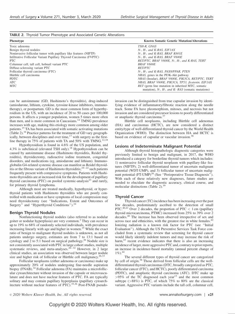
TABLE 2. Thyroid Tumor Phenotype and Associated Genetic Alterations
Phenotype Known Somatic Genetic Mutation/Alterations
Toxic adenoma TSH-R, GNASBenign thyroid nodules N-, H-, and K-RAS, EIF1AXNoninvasive follicular tumor with papillary like features (NIFTP) N-, H-, and K-RAS, BRAF K601EInfiltrative Follicular Variant Papillary Thyroid Carcinoma (FVPTC) N-, H-, and K-RAS, BRAF V600EPTC RET/PTC, BRAF V600E, N-, H-, and K-RAS, TERTColumnar cell, tall cell, hobnail variant PTC BRAF V600EDiffuse-sclerosing variant PTC RET/PTCFollicular thyroid carcinoma (FTC) N-, H-, and K-RAS, PAX8/PPAR, PTENHurthle cell carcinoma NRAS, genes in the PI3K-Akt pathwayPDTC NRAS (Insular), BRAF V600E, PIK3CA, RET/PTC, TERTATC NRAS, BRAF V600E, PIK3CA, TP53, bcatenin. EIF1AXMTC RET (germ line mutation in inherited MTC, somatic
mutation), N-, H-, and K- RAS (somatic mutations)
Annals of Surgery � Volume 271, Number 3, March 2020 Definitive Surgical Management of Thyroid Disease in Adults
� 2020 Wolters Kluwer Health, Inc. All rights reserved. www.annalsofsurgery.com | e25
Copyright © 2020 Wolters Kluwer Health, Inc. All rights reserved.
hobnail, and diffuse sclerosing types, while clinically less aggressivevariants include most FVPTC and Warthin-like tumors.
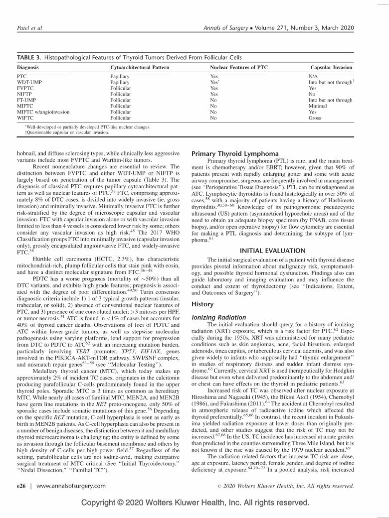
Recent nomenclature changes are essential to review. Thedistinction between FVPTC and either WDT-UMP or NIFTP islargely based on penetration of the tumor capsule (Table 3). Thediagnosis of classical PTC requires papillary cytoarchitectural pat-tern as well as nuclear features of PTC.38 FTC, comprising approxi-mately 8% of DTC cases, is divided into widely invasive (ie, grossinvasion) and minimally invasive. Minimally invasive FTC is furtherrisk-stratified by the degree of microscopic capsular and vascularinvasion. FTC with capsular invasion alone or with vascular invasionlimited to less than 4 vessels is considered lower risk by some; othersconsider any vascular invasion as high risk.45 The 2017 WHOClassification groups FTC into minimally invasive (capsular invasiononly), grossly encapsulated angioinvasive FTC, and widely-invasiveFTC.38
Hurthle cell carcinoma (HCTC, 2.3%), has characteristicmitochondrial-rich, plump follicular cells that stain pink with eosin,and have a distinct molecular signature from FTC.46–48
PDTC has a worse prognosis (mortality of �50%) than allDTC variants, and exhibits high grade features; prognosis is associ-ated with the degree of poor differentiation.49,50 Turin consensusdiagnostic criteria include 1) 1 of 3 typical growth patterns (insular,trabecular, or solid), 2) absence of conventional nuclear features ofPTC, and 3) presence of one convoluted nuclei;>3 mitoses per HPF,or tumor necrosis.51 ATC is found in <1% of cases but accounts for40% of thyroid cancer deaths. Observations of foci of PDTC andATC within lower-grade tumors, as well as stepwise molecularpathogenesis using varying platforms, lend support for progressionfrom DTC to PDTC to ATC52 with an increasing mutation burden,particularly involving TERT promoter, TP53, EIF1AX, genesinvolved in the PIK3CA-AKT-mTOR pathway, SWI/SNF complex,and mismatch repair genes53–55 (see ‘‘Molecular Testing’’).
Medullary thyroid cancer (MTC), which today makes upapproximately 2% of incident TC cases, originates in the calcitoninproducing parafollicular C-cells predominately found in the upperthyroid poles. Sporadic MTC is 3 times as common as hereditaryMTC. While nearly all cases of familial MTC, MEN2A, and MEN2Bhave germ line mutations in the RET proto-oncogene, only 50% ofsporadic cases include somatic mutations of this gene.56 Dependingon the specific RET mutation, C-cell hyperplasia is seen as early asbirth in MEN2B patients. As C-cell hyperplasia can also be present ina number of benign diseases, the distinction between it and medullarythyroid microcarcinoma is challenging; the entity is defined by someas invasion through the follicular basement membrane and others byhigh density of C-cells per high-power field.57 Regardless of thesetting, parafollicular cells are not iodine-avid, making extirpativesurgical treatment of MTC critical (See ‘‘Initial Thyroidectomy,’’‘‘Nodal Dissection,’’ ‘‘Familial TC’’).
Primary Thyroid LymphomaPrimary thyroid lymphoma (PTL) is rare, and the main treat-
ment is chemotherapy and/or EBRT; however, given that 90% ofpatients present with rapidly enlarging goiter and some with acuteairway compromise, surgeons are frequently involved in management(see ‘‘Perioperative Tissue Diagnosis’’). PTL can be misdiagnosed asATC. Lymphocytic thyroiditis is found histologically in over 50% ofcases,58 with a majority of patients having a history of Hashimotothyroiditis.30,58–60 Knowledge of its pathognomonic pseudocysticultrasound (US) pattern (asymmetrical hypoechoic areas) and of theneed to obtain an adequate biopsy specimen (by FNAB, core tissuebiopsy, and/or open operative biopsy) for flow cytometry are essentialfor making a PTL diagnosis and determining the subtype of lym-phoma.61
INITIAL EVALUATION
The initial surgical evaluation of a patient with thyroid diseaseprovides pivotal information about malignancy risk, symptomatol-ogy, and possible thyroid hormonal dysfunction. Findings also canguide laboratory and imaging evaluation and may influence theconduct and extent of thyroidectomy (see ‘‘Indications, Extent,and Outcomes of Surgery’’).
History
Ionizing RadiationThe initial evaluation should query for a history of ionizing
radiation (XRT) exposure, which is a risk factor for PTC.62 Espe-cially during the 1950s, XRT was administered for many pediatricconditions such as skin angiomas, acne, facial hirsutism, enlargedadenoids, tinea capitus, or tuberculous cervical adenitis, and was alsogiven widely to infants who supposedly had ‘‘thymic enlargement’’in studies of respiratory distress and sudden infant distress syn-drome.63 Currently, cervical XRT is used therapeutically for Hodgkindisease but even when delivered predominantly to the abdomen and/or chest can have effects on the thyroid in pediatric patients.63
Increased risk of TC was observed after nuclear exposure atHiroshima and Nagasaki (1945), the Bikini Atoll (1954), Chernobyl(1986), and Fukushima (2011).64 The accident at Chernobyl resultedin atmospheric release of radioactive iodine which affected thethyroid preferentially.65,66 In contrast, the recent incident in Fukush-ima yielded radiation exposure at lower doses than originally pre-dicted, and other studies suggest that the risk of TC may not beincreased.67,68 In the US, TC incidence has increased at a rate greaterthan predicted in the counties surrounding Three Mile Island, but it isnot known if the rise was caused by the 1979 nuclear accident.69
The radiation-related factors that increase TC risk are: dose,age at exposure, latency period, female gender, and degree of iodinedeficiency at exposure.64,70–72 In a pooled analysis, risk increased
TABLE 3. Histopathological Features of Thyroid Tumors Derived From Follicular Cells
Diagnosis Cytoarchitectural Pattern Nuclear Features of PTC Capsular Invasion
PTC Papillary Yes N/AWDT-UMP Papillary Yes� Into but not throughy
FVPTC Follicular Yes YesNIFTP Follicular Yes NoFT-UMP Follicular No Into but not throughMIFTC Follicular No MinimalMIFTC w/angioinvasion Follicular No YesWIFTC Follicular No Gross
�Well-developed or partially developed PTC-like nuclear changes.yQuestionable capsular or vascular invasion.
Patel et al Annals of Surgery � Volume 271, Number 3, March 2020
e26 | www.annalsofsurgery.com � 2020 Wolters Kluwer Health, Inc. All rights reserved.
Copyright © 2020 Wolters Kluwer Health, Inc. All rights reserved.
with doses of 0.05 to 0.1 Gy but decreased when the dose exceeded30 Gy.73 Younger age increased the risk, which appeared to peak at25 to 30 years post exposure but remained elevated more than50 years later. Exact determination of cancer latency periods islikely affected by surveillance intensity.73 In meta-analysis, theincidence of TC following XRT for breast cancer was�3-fold higherthan in the general population, although a lesser degree of associationwas also observed in women with breast cancer who did not receiveXRT, likely implicating other factors.74
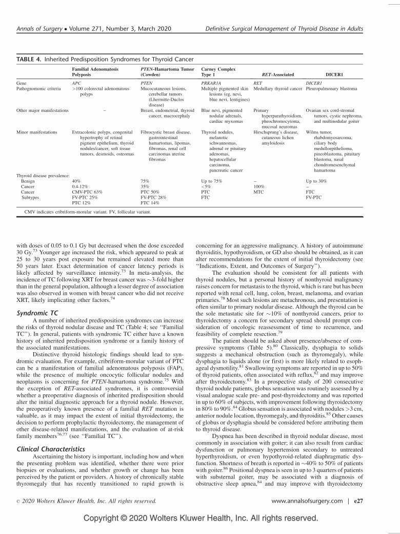
Syndromic TCA number of inherited predisposition syndromes can increase
the risks of thyroid nodular disease and TC (Table 4; see ‘‘FamilialTC’’). In general, patients with syndromic TC either have a knownhistory of inherited predisposition syndrome or a family history ofthe associated manifestations.
Distinctive thyroid histologic findings should lead to syn-dromic evaluation. For example, cribriform-morular variant of PTCcan be a manifestation of familial adenomatous polyposis (FAP),while the presence of multiple oncocytic follicular nodules andneoplasms is concerning for PTEN-hamartoma syndrome.75 Withthe exception of RET-associated syndromes, it is controversialwhether a preoperative diagnosis of inherited predisposition shouldalter the initial diagnostic approach for a thyroid nodule. However,the preoperatively known presence of a familial RET mutation isvaluable, as it may impact the extent of initial thyroidectomy, thedecision to perform prophylactic thyroidectomy, the management ofother disease-related manifestations, and the evaluation of at-riskfamily members76,77 (see ‘‘Familial TC’’).
Clinical CharacteristicsAscertaining the history is important, including how and when
the presenting problem was identified, whether there were priorbiopsies or evaluations, and whether growth or change has beenperceived by the patient or providers. A history of chronically stablethyromegaly that has recently transitioned to rapid growth is
concerning for an aggressive malignancy. A history of autoimmunethyroiditis, hypothyroidism, or GD also should be obtained, as it canalter recommendations for the extent of initial thyroidectomy (see‘‘Indications, Extent, and Outcomes of Surgery’’).
The evaluation should be consistent for all patients withthyroid nodules, but a personal history of nonthyroid malignancyraises concern for metastasis to the thyroid, which is rare but has beenreported with renal cell, lung, colon, breast, melanoma, and ovarianprimaries.78 Most such lesions are metachronous, and presentation isoften similar to primary nodular disease. Although the thyroid can bethe sole metastatic site for �10% of nonthyroid cancers, prior tothyroidectomy a concern for secondary spread should prompt con-sideration of oncologic reassessment of time to recurrence, andfeasibility of complete resection.79
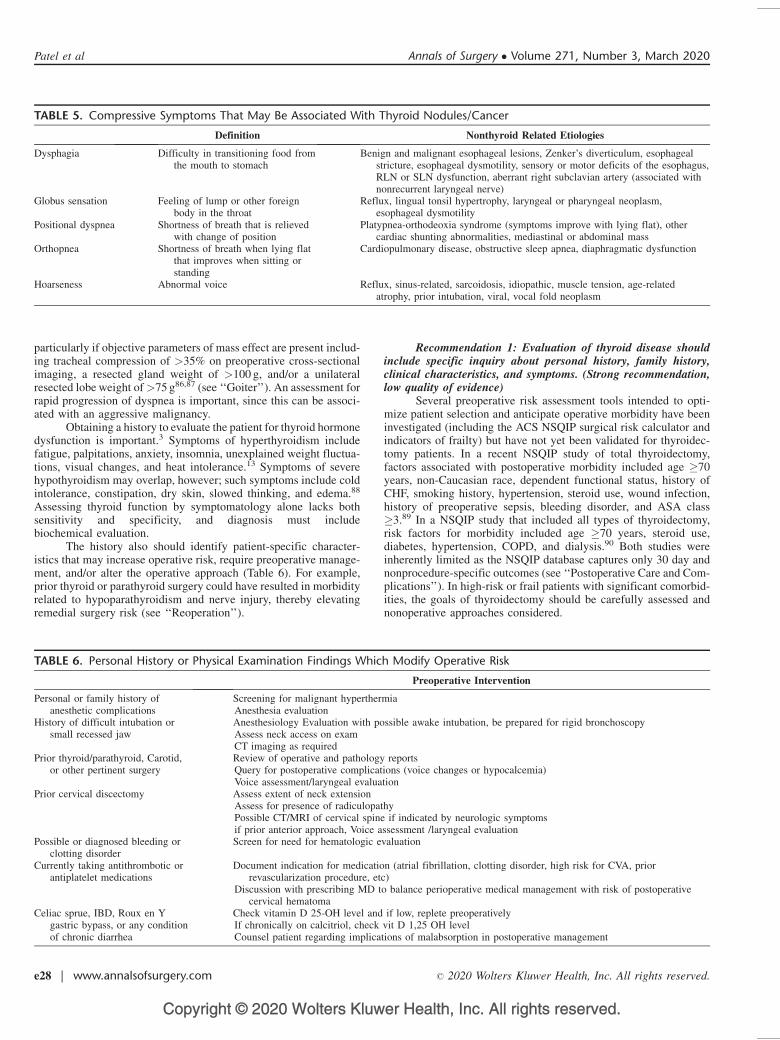
The patient should be asked about presence/absence of com-pressive symptoms (Table 5).80 Classically, dysphagia to solidssuggests a mechanical obstruction (such as thyromegaly), whiledysphagia to liquids alone (or first) is more likely related to esoph-ageal dysmotility.81 Swallowing symptoms are reported in up to 50%of thyroid patients, often associated with reflux,82 and may improveafter thyroidectomy.83 In a prospective study of 200 consecutivethyroid nodule patients, globus sensation was routinely assessed by avisual analogue scale pre- and post-thyroidectomy and was reportedin up to 60% of subjects, with improvement following thyroidectomyin 80% to 90%.84 Globus sensation is associated with nodules>3 cm,anterior nodule location, thyromegaly, and thyroiditis.85 Other causesof globus or dysphagia should be considered before attributing themto thyroid disease.
Dyspnea has been described in thyroid nodular disease, mostcommonly in association with goiter; it can also result from cardiacdysfunction or pulmonary hypertension secondary to untreatedhyperthyroidism, or even hypothyroid-related diaphragmatic dys-function. Shortness of breath is reported in �40% to 50% of patientswith goiter.86 Positional dyspnea is seen in up to 3 quarters of patientswith substernal goiter, may be associated with a diagnosis ofobstructive sleep apnea,84 and may improve with thyroidectomy
TABLE 4. Inherited Predisposition Syndromes for Thyroid Cancer
Familial AdenomatosisPolyposis
PTEN-Hamartoma Tumor(Cowden)
Carney ComplexType 1 RET-Associated DICER1
Gene APC PTEN PRKAR1A RET DICER1Pathognomonic criteria >100 colorectal adenomatous
polypsMucocutaneous lesions,
cerebellar tumors(Lhermitte-Duclosdisease)
Multiple pigmented skinlesions (eg, nevi,blue nevi, lentigines)
Medullary thyroid cancer Pleuropulmonary blastoma
Other major manifestations – Breast, endometrial, thyroidcancer, macrocephaly
Blue nevi, pigmentednodular adrenals,cardiac myxomas
Primaryhyperparathyroidism,pheochromocytoma,mucosal neuromas
Ovarian sex cord-stromaltumors, cystic nephroma,and multinodular goiter
Minor manifestations Extracolonic polyps, congenitalhypertrophy of retinalpigment epithelium, thyroidnodules/cancer, soft tissuetumors, desmoids, osteomas
Fibrocystic breast disease,gastrointestinalhamartomas, lipomas,fibromas, renal cellcarcinomas uterinefibromas
Thyroid nodules,melanoticschwannomas,adrenal or pituitaryadenomas,hepatocellularcarcinoma,pancreatic cancer
Hirschsprung’s disease,cutaneous lichenamyloidosis
Wilms tumor,rhabdomyosarcoma,ciliary bodymedulloepithelioma,pineoblastoma, pituitaryblastoma, nasalchondromesenchymalhamartoma
Thyroid disease prevalence:Benign 40% 75% Up to 75% – Up to 30%Cancer 0.4-12% 35% <5% 100% –CancerSubtypes
CMV-PTC 63% PTC 50% PTC MTC FTCFV-PTC 25% FV-PTC 28% FTC FV-PTCPTC 12% FTC 14%
CMV indicates cribriform-morular variant. FV, follicular variant.
Annals of Surgery � Volume 271, Number 3, March 2020 Definitive Surgical Management of Thyroid Disease in Adults
� 2020 Wolters Kluwer Health, Inc. All rights reserved. www.annalsofsurgery.com | e27
Copyright © 2020 Wolters Kluwer Health, Inc. All rights reserved.
particularly if objective parameters of mass effect are present includ-ing tracheal compression of >35% on preoperative cross-sectionalimaging, a resected gland weight of >100 g, and/or a unilateralresected lobe weight of>75 g86,87 (see ‘‘Goiter’’). An assessment forrapid progression of dyspnea is important, since this can be associ-ated with an aggressive malignancy.
Obtaining a history to evaluate the patient for thyroid hormonedysfunction is important.3 Symptoms of hyperthyroidism includefatigue, palpitations, anxiety, insomnia, unexplained weight fluctua-tions, visual changes, and heat intolerance.13 Symptoms of severehypothyroidism may overlap, however; such symptoms include coldintolerance, constipation, dry skin, slowed thinking, and edema.88
Assessing thyroid function by symptomatology alone lacks bothsensitivity and specificity, and diagnosis must includebiochemical evaluation.
The history also should identify patient-specific character-istics that may increase operative risk, require preoperative manage-ment, and/or alter the operative approach (Table 6). For example,prior thyroid or parathyroid surgery could have resulted in morbidityrelated to hypoparathyroidism and nerve injury, thereby elevatingremedial surgery risk (see ‘‘Reoperation’’).
Recommendation 1: Evaluation of thyroid disease shouldinclude specific inquiry about personal history, family history,clinical characteristics, and symptoms. (Strong recommendation,low quality of evidence)
Several preoperative risk assessment tools intended to opti-mize patient selection and anticipate operative morbidity have beeninvestigated (including the ACS NSQIP surgical risk calculator andindicators of frailty) but have not yet been validated for thyroidec-tomy patients. In a recent NSQIP study of total thyroidectomy,factors associated with postoperative morbidity included age �70years, non-Caucasian race, dependent functional status, history ofCHF, smoking history, hypertension, steroid use, wound infection,history of preoperative sepsis, bleeding disorder, and ASA class�3.89 In a NSQIP study that included all types of thyroidectomy,risk factors for morbidity included age �70 years, steroid use,diabetes, hypertension, COPD, and dialysis.90 Both studies wereinherently limited as the NSQIP database captures only 30 day andnonprocedure-specific outcomes (see ‘‘Postoperative Care and Com-plications’’). In high-risk or frail patients with significant comorbid-ities, the goals of thyroidectomy should be carefully assessed andnonoperative approaches considered.
TABLE 5. Compressive Symptoms That May Be Associated With Thyroid Nodules/Cancer
Definition Nonthyroid Related Etiologies
Dysphagia Difficulty in transitioning food fromthe mouth to stomach
Benign and malignant esophageal lesions, Zenker’s diverticulum, esophagealstricture, esophageal dysmotility, sensory or motor deficits of the esophagus,RLN or SLN dysfunction, aberrant right subclavian artery (associated withnonrecurrent laryngeal nerve)
Globus sensation Feeling of lump or other foreignbody in the throat
Reflux, lingual tonsil hypertrophy, laryngeal or pharyngeal neoplasm,esophageal dysmotility
Positional dyspnea Shortness of breath that is relievedwith change of position
Platypnea-orthodeoxia syndrome (symptoms improve with lying flat), othercardiac shunting abnormalities, mediastinal or abdominal mass
Orthopnea Shortness of breath when lying flatthat improves when sitting orstanding
Cardiopulmonary disease, obstructive sleep apnea, diaphragmatic dysfunction
Hoarseness Abnormal voice Reflux, sinus-related, sarcoidosis, idiopathic, muscle tension, age-relatedatrophy, prior intubation, viral, vocal fold neoplasm
TABLE 6. Personal History or Physical Examination Findings Which Modify Operative Risk
Preoperative Intervention
Personal or family history ofanesthetic complications
Screening for malignant hyperthermiaAnesthesia evaluation
History of difficult intubation orsmall recessed jaw
Anesthesiology Evaluation with possible awake intubation, be prepared for rigid bronchoscopyAssess neck access on examCT imaging as required
Prior thyroid/parathyroid, Carotid,or other pertinent surgery
Review of operative and pathology reportsQuery for postoperative complications (voice changes or hypocalcemia)Voice assessment/laryngeal evaluation
Prior cervical discectomy Assess extent of neck extensionAssess for presence of radiculopathyPossible CT/MRI of cervical spine if indicated by neurologic symptomsif prior anterior approach, Voice assessment /laryngeal evaluation
Possible or diagnosed bleeding orclotting disorder
Screen for need for hematologic evaluation
Currently taking antithrombotic orantiplatelet medications
Document indication for medication (atrial fibrillation, clotting disorder, high risk for CVA, priorrevascularization procedure, etc)
Discussion with prescribing MD to balance perioperative medical management with risk of postoperativecervical hematoma
Celiac sprue, IBD, Roux en Ygastric bypass, or any conditionof chronic diarrhea
Check vitamin D 25-OH level and if low, replete preoperativelyIf chronically on calcitriol, check vit D 1,25 OH levelCounsel patient regarding implications of malabsorption in postoperative management
Patel et al Annals of Surgery � Volume 271, Number 3, March 2020
e28 | www.annalsofsurgery.com � 2020 Wolters Kluwer Health, Inc. All rights reserved.
Copyright © 2020 Wolters Kluwer Health, Inc. All rights reserved.
Physical ExaminationExamination findings that can suggest hyperthyroidism
include elevated heart rate, hypertension, and exophthalmos. A slowheart rate and slowed Achilles heel reflex time may signal hypothy-roidism. In meta-analysis, overweight and obese patients havesignificant higher risks of PTC, FTC, and ATC, but no increasedrisk of MTC.91,92 Taller height also has been associated withincreased DTC risk.92 Demographic and anthropometric factorsare not reliable indicators of histology.
The physical assessment is also a key step in operativeplanning (Tables 6, 7). Palpable nodules are typically at least1 cm in size.93 Immobile nodules, especially with associated lymph-adenopathy, are concerning for malignancy. Thyromegaly is oftenevident preoperatively, but its extent may be apparent only withsupine positioning (see ‘‘Goiter’’). When the inferior extent of anodule or enlarged thyroid lobe is not accessible on exam (ie, goesbelow the clavicle), CT imaging should be obtained for evaluation ofthe extent of substernal projection.84–86 Pemberton sign is thepresence of vascular engorgement and facial congestion occurringwhen a patient with a large substernal goiter raises their arms overtheir head, further narrowing the thoracic inlet.94 Limited neckextension can herald difficulty in intraoperative positioning oraccess. If associated with neurologic symptoms, limited neck mobil-ity also can suggest underlying cervical disc disease requiring furtherpreoperative evaluation and even pre-emptive or simultaneous man-agement.
Voice AssessmentA careful voice assessment should be performed in the
evaluation of thyroid disease. Subjective voice impairment wasreported preoperatively in 30% to 80% of thyroidectomy patients,and the incidence varies by type of voice quality assessmentused.82,95 Voice changes and hoarseness can have numerous causesbut are frequently idiopathic. Further evaluation should be performedfor patients who have had prior surgery that put the RLN at risk,including anterior cervical discectomy, prior thyroid/parathyroidec-tomy, carotid endarterectomy, tracheostomy, cardio/thoracic proce-dures, and esophagectomy (Table 6).3 Such a history should bespecifically investigated and sites of prior incisions should be notedon examination. Documented vocal fold immobility can impact thedecision for surgery and the extent of thyroidectomy (see ‘‘Indica-tions, Extent, and Outcomes of Surgery,’’ ‘‘Laryngology’’).
Laryngeal examination can be performed using several differ-ent techniques. Both current guidelines and the cost-to-benefit ratioof routine preoperative laryngeal examination on all thyroid patientsrecommend a selective approach3,96(see ‘‘Laryngology’’).
Recommendation 2: The preoperative physical examinationshould include voice assessment. (Strong recommendation, mod-erate-quality evidence)
Laboratory EvaluationThe initial laboratory evaluation for all patients with thyroid
disease should include a serum TSH level. If the TSH is suppressed,then a free T4 and total T3 should be obtained, and management ofhyperthyroidism should be the initial clinical focus.13 In addition, athyroid uptake scan should be obtained to assess if the index nodule ishyperfunctioning; such nodules have a low risk of malignancy and donot require FNAB97,98 (see ‘‘Hyperthyroid Conditions’’). In mostcases an elevated TSH level should be normalized prior to furthermanagement.3 Thyroiditis may be further assessed if the informationwill help determine surgical management (see ‘‘Indications, Extent,and Outcomes of Surgery’’). Nodules identified in the setting ofeuthyroidism should be assessed with US and FNAB (see ‘‘Imag-ing,’’ ‘‘FNAB Diagnosis’’).
In the absence of known MEN2A, the incidence of concomi-tant parathyroid disease in patients with thyroid nodules is 3% to5%99–101 and is higher (�35%) with prior XRT102 among otherfactors.6 Because of the cost and potential morbidity of missedparathyroid disease, preoperative assessment of serum calciumshould be considered, and measurement of parathyroid hormonealso may be indicated (see ‘‘Concurrent Parathyroidectomy’’ and‘‘Preoperative Care’’).
Routine screening for MTC using preoperative calcitoninlevels is controversial. In a study of >10,000 patients with thyroidnodules, an elevated basal calcitonin level was more sensitive thanFNAB in diagnosing MTC, and such patients presented withearlier stage disease and had longer disease-specific survival.103
However, these calcitonin elevations were all confirmed by pen-tagastrin stimulation, which is no longer available in the US. False-positive rates of nonstimulated calcitonin vary widely, and anumber of benign conditions can contribute to spurious elevation,including chronic renal failure, proton pump inhibitors, chroniclymphocytic thyroiditis, DTC, and non-thyroidal malignancies.104
Current ATA and NCCN guidelines do not recommend screeningbasal serum calcitonin levels in the evaluation of thyroid nod-ules.3,105 Targeted screening for at-risk individuals should beconsidered.
When MTC is diagnosed by FNAB or is suspected onhistory, measurement of serum calcitonin and CEA levels is usefulas they can be accurate indicators of disease extent. A basalcalcitonin level <20 pg/mL is associated with a low risk of nodalmetastasis, while a preoperative level �500 pg/mL raises concern
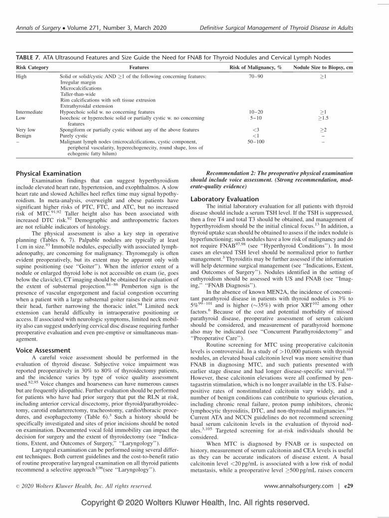
TABLE 7. ATA Ultrasound Features and Size Guide the Need for FNAB for Thyroid Nodules and Cervical Lymph Nodes
Risk Category Features Risk of Malignancy, % Nodule Size to Biopsy, cm
High Solid or solid/cystic AND �1 of the following concerning features:Irregular marginMicrocalcificationsTaller-than-wideRim calcifications with soft tissue extrusionExtrathyroidal extension
70–90 �1
Intermediate Hypoechoic solid w. no concerning features 10–20 �1Low Isoechoic or hyperechoic solid or partially cystic w. no concerning
features5–10 �1.5
Very low Spongiform or partially cystic without any of the above features <3 �2Benign Purely cystic <1 –– Malignant lymph nodes (microcalcifications, cystic component,
peripheral vascularity, hyperechogenecity, round shape, loss ofechogenic fatty hilum)
50–100 –
Annals of Surgery � Volume 271, Number 3, March 2020 Definitive Surgical Management of Thyroid Disease in Adults
� 2020 Wolters Kluwer Health, Inc. All rights reserved. www.annalsofsurgery.com | e29
Copyright © 2020 Wolters Kluwer Health, Inc. All rights reserved.
for distant metastasis.106,107 RET-gene testing should inform therisk of associated manifestations (see ‘‘Familial TC’’).
Recommendation 3: TSH should be measured in patientswith nodular thyroid disease. Additional laboratory studies mayhelp in specific circumstances. (Strong recommendation, low-quality evidence)
IMAGING
Successful thyroid surgery is contingent on thorough andaccurate imaging, which also impacts preoperative planning, extentof surgery, and postoperative management. Inadequate preoperativeimaging may be a root cause of incomplete initial surgery.108,109
Preoperative
UltrasonographyThe initial and most critical thyroid imaging study is cervical
US. Current adult guidelines recommend a thyroid US with survey ofthe cervical lymph nodes (CLN) in patients with a known orsuspected thyroid nodule, and for all patients undergoing thyroidec-tomy for malignant or suspicious cytologic or molecular findings.3
The objectives of diagnostic US are to assess the nodule or tumor(size, location, suspicious features) and to identify and characterizeabnormal lymph nodes in the central and lateral neck which may beinvolved with thyroid cancer.
Thyroid Nodule US. Multiple studies have characterized thesonographic features associated with TC, including microcalcifica-tions, hypoechogenicity, irregular margins and a shape taller thanwide measured on transverse view.110–119 Features predictive of TCare microcalcifications, irregular margins, and tall shape (Table 7).Up to 58% of benign nodules are hypoechoic compared to thyroidparenchyma, making nodule hypoechogenicity less specific.116
High, intermediate, low, very low, and benign thyroid nodule char-acteristics and malignancy rates appear in Table 7.120–125
The US features of FTC differ from those of PTC. Intranodularvascularity correlates with malignancy in follicular lesions, but notPTC. Furthermore, compared to conventional PTC, FVPTC is morelikely to have the same US appearance as FTC, and both FTC andFVPTC are more likely to be iso- or hyperechoic.115,116,126–129
However, a recent meta-analysis concluded that vascular flow oncolor Doppler sonography may not accurately predict TC.130
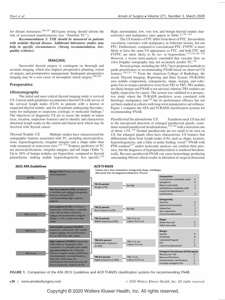
Several groups, including the ATA,3 have proposed methods toguide practitioners in recommending FNAB based on observed USfeatures.120,131–133 From the American College of Radiology, therecent Thyroid Imaging, Reporting and Data System (TI-RADS)uses nodule composition, echogenicity, shape, margin, and echo-genic foci to assign a predictive score from TR1 to TR5; TR1 nodulesare likely benign and FNAB is not advised, whereas TR5 nodules arehighly suspicious for cancer. The system was validated in a prospec-tive study where the TI-RADS predictive score correlated withhistologic malignancy rate134 but its performance efficacy has notyet been studied in cohorts with long-term nonoperative surveillance.Figure 1 compares the ATA and TI-RADS classification systems forrecommending FNAB.
Parathyroid Incidentaloma US. Fastidious neck US has ledto the unexpected detection of enlarged parathyroid glands, some-times termed parathyroid incidentalomas,135,136 with a detection rateof about 1.5%.136 Normal parathyroids are too small to be seen onUS, but enlarged glands often have characteristic US features thatdifferentiate them from lymph nodes (LN), such as shape, location,hypoechogenicity, and a hilar or polar feeding vessel.6 FNAB withPTH washout137 and/or molecular analysis can confirm their pres-ence, but the diagnosis of hyperparathyroidism is rendered biochem-ically. Because parathyroid FNAB can cause hemorrhage producingsurrounding fibrosis which results in alteration of surgical dissection
FIGURE 1. Comparison of the ATA 2015 Guidelines and ACR TI-RADS classification systems for recommending FNAB.
Patel et al Annals of Surgery � Volume 271, Number 3, March 2020
e30 | www.annalsofsurgery.com � 2020 Wolters Kluwer Health, Inc. All rights reserved.
Copyright © 2020 Wolters Kluwer Health, Inc. All rights reserved.
planes and can cause histologic changes mimicking parathyroidcarcinoma, FNAB of parathyroid glands is to be avoided whenpossible6 (see ‘‘Concurrent Parathyroidectomy’’).
Cervical Lymph Node US. US is the first-line imagingmodality for assessment of cervical lymph node metastasis(CLNM).138,139 Many experts routinely perform US evaluation ofthe central and lateral neck whenever thyroid nodules are detected.Central and lateral LN are affected by metastatic PTC in up to 70% ofcases,140 either at presentation or during surveillance. The sensitivityof US in detecting abnormal LN varies from 25% to 60% for thecentral neck and 70% to 95% for the lateral neck.141,142 One of themain factors influencing sensitivity is practitioner expertise.
Benign CLN are typically oval with a hyperechoic centralstripe and vascular flow in the center (ie, hilum).143 Loss of a visiblehilum is felt to represent interruption of lymphatic flow by tumorinvasion. In a benign LN, the hilar stripe may not always be easilyseen, thus lack of a hilum is only 29% specific for CLNM.139
Lymph node location, size, and shape are also importantfeatures of US assessment for potential metastatic involvement.Evidence generally supports a stepwise progression of PTC metas-tases, starting in ipsilateral level VI (prevalence 50–70%) andproceeding laterally to levels III and IV (prevalence 30–45%),and then to level II with some studies reporting equivalent prevalenceas for III and IV.140,144–147 Therefore, if one level is involved, acompartment-oriented selective lymph node dissection is recom-mended (see ‘‘Nodal Dissection’’). This progression is not alwaysreliable and should not preclude evaluation of any suspicious LN inthe neck. Though LN >1 cm in maximum diameter are convention-ally considered more likely to harbor malignancy, many benign orreactive nodes will exceed 1 cm while remaining fusiform in shape,especially near the submandibular glands and in patients withlymphocytic thyroiditis. In a study of DTC that carefully matchedthe US and histologic findings, size >1 cm was associated with only68% sensitivity and 75% specificity for CLNM.148 Smallest nodaldiameter >5 mm also has been proposed as a predictor of malig-nancy, with a reported specificity of 96% and sensitivity of 61%.139
Shape is more reliably associated with LNM than size. Benign lymphnodes typically are oval or fusiform even with hypertrophy orenlargement from nonmalignant causes, but malignant LN oftenwill appear rounded139,148,149 because the neoplastic infiltrationtypically occurs in the cortex, at the site of lymphatic influx.143
The Solbiati index provides an objective measure of roundness bycoding the ratio of longest to shortest nodal diameter. A ratio >2 ishighly suggestive of benign character, whereas <2 is concerning forLNM.143 Other US features predictive of malignant LN involvementinclude microcalcifications, hyperechoic or cystic character, andperipheral hypervascularity.149–153
If US identifies a suspicious LN, FNAB of the lymph nodeshould be performed for cytology, with washout for thyroglobulinmeasurement when possible.3
LN Mapping. Bilateral US evaluation of LN compartmentsII-VI (ie, mapping) should be performed routinely in the preoperativeevaluation of patients with cytologic evidence of thyroid carci-noma, and considered for levels I–VII in patients identified tohave metastatic nodal disease.2,154 LN mapping should carefullydiagram where suspicious lymph nodes are located and assess forfeatures of aggressive behavior, such as strap muscle invasion,internal jugular vein thrombus, and posterior tumor location.Ideally, this should be performed preoperatively by an experiencedsonographer to guide complete resection of the primary tumor aswell as a compartment-oriented dissection of affected LN basins, ifLNM are present.108
Surgical Planning. Prior to thyroidectomy, US is often per-formed by the surgical team to facilitate both operative planning andpatient counseling regarding surgical risks. In recent multidisci-plinary literature, surgeon-performed US is more accurate, helpfulfor preoperative planning, and associated with lower local recur-rence rates compared to radiologist-performed US.155–157 Aspatient positioning is optimized under general anesthesia, surgeonsmay elect to repeat US just prior to incision to keep an anatomic‘‘roadmap’’ fresh in their minds and ensure that US abnormalitiesare adequately addressed at operation and are present in the surgicalspecimen.108
Recommendation 4: A diagnostic US should be performed inall patients with a suspected thyroid nodule. (Strong recommenda-tion, high-quality evidence)
Recommendation 5:a. US assessment of bilateral central and lateral LN compartments
should be performed in the preoperative evaluation of patientswith cytologic evidence of thyroid carcinoma. (Strong recom-mendation, low quality of evidence).
b. US assessment of bilateral central and lateral LN compartmentsmay be performed in the preoperative evaluation of patientswith indeterminate cytologic evidence of thyroid carcinoma.(Strong recommendation, insufficient evidence).
Translaryngeal US. Vocal cord ultrasonography (VCUS) is anew modality that can be simple and accurate in diagnosing truevocal fold (TVF) paralysis preoperatively, with a sensitivity of 62–93%.158,159 In patients with abnormal mobility or inadequate vocalfold visualization, such VCUS results trigger further evaluation withfiberoptic laryngoscopy (FL, see ‘‘Laryngology’’).160–162 VCUS canbe a sensitive, noninvasive, convenient, and inexpensive method ofevaluation and can be performed during the initial surgical visit.However, when the clinical scenario suggests substantial operativerisk contingent on the optimal documentation of the degree of vocalcord dysfunction, FL is advised.
US Limitations. US is the first-line imaging modality forassessing thyroid nodules and CLN because it is widely available,inexpensive, provides detailed high-resolution anatomic data,avoids ionizing radiation, and facilitates FNAB of suspiciouslesions. However, the results are operator dependent.163,164 USutility is also limited for deep structures and those acousticallyshielded by air or bone. Patients with substernal goiter, morbidobesity, poor neck extension, remote cervical adenopathy (highlevel II, VI or VII, substernal, infraclavicular or retro/parapharyng-eal LN) or apparent locally advanced disease may benefit fromcross-sectional imaging.
Cross-sectional ImagingCross-sectional imaging, namely, computerized tomography
(CT) or magnetic resonance imaging (MRI), has a supplemental rolein the preoperative evaluation of thyroid disease, and is recom-mended with intravenous contrast as an adjunct to US for patientswith clinical suspicion of advanced disease, including invasiveprimary tumor, or clinically apparent multiple or bulky LN.108 Crosssectional imaging may also be of value with clinical findings such asvocal cord paresis/paralysis, progressive overt dysphagia or odyno-phagia, mass fixation to surrounding structures, hemoptysis, stridoror positional dyspnea, rapid enlargement, and large size or medias-tinal extension (see ‘‘Goiter’’). US features that prompt CT or MRIinclude incomplete thyroid or LN imaging, suspicion for significantextrathyroidal invasion (including irregular or indistinct marginbetween tumor and strap muscles, airway, esophagus or majorvessels), bulky, posteriorly located, or inferiorly located LN, and
Annals of Surgery � Volume 271, Number 3, March 2020 Definitive Surgical Management of Thyroid Disease in Adults
� 2020 Wolters Kluwer Health, Inc. All rights reserved. www.annalsofsurgery.com | e31
Copyright © 2020 Wolters Kluwer Health, Inc. All rights reserved.
unavailable US expertise. Both CT and MRI provide axial imagingfrom skull base to mediastinum in a standardized, reproduciblefashion that is user independent. The sensitivities of MRI andPET for detection of CLNM are relatively low (30–40%).165
The accuracy of neck CT is optimized by use of intravenousiodinated contrast. Noncontrast CT lacks definition, and its utility isthus limited to gross evaluation of mediastinal disease. MRI withgadolinium is an alternative modality that avoids iodinated contrast.Although MRI resolution can be limited in the central compartmentdue to motion artifact from swallowing and respiration (169), it isgenerally preferable to noncontrast CT in preoperative imaging forthyroid cancer.
When CT/MR imaging is required preoperatively, the benefitgained from improved anatomic resolution generally outweighs anypotential risk from postponement of subsequent radioactive iodine(RAI) imaging or therapy. Preoperative communication between thesurgeon and endocrinologist is important. When there is concern, theurinary iodine-to-creatinine ratio can be measured at an interval of atleast 1 month to allow urinary iodine levels to return to baselinelevels before moving forward with RAI.166 At present, there is noevidence to suggest delays of this minor scale adversely affectthyroid cancer outcomes.
Recommendation 6: CT or MRI with intravenous contrastshould be used preoperatively as an adjunct to US in selectedpatients with clinical suspicion for advanced locoregional thyroidcancer (Strong recommendation, low quality of evidence)
ElastographyUltrasound elastography has been investigated for its ability to
modify thyroid nodule cancer risk assessment. Elastography is ameasurement of tissue stiffness. US elastography requires an USmachine and a computational module, which often must be pur-chased separately. To allow the required direct determination oftissue strain, the index nodule must not overlap with other nodules inthe anteroposterior plane making the test unsuited for patients withmultinodular goiter, coalescent nodules, posterior/inferior nodules,or obesity. Although an initial prospective study suggested near100% PPV and NPV,167 more recent, larger studies report USelastography performance that was substantially inferior to US.168
In the largest prospective study, PPV was 36% (comparable to thatfor US detection of microcalcifications), and NPV was 97% in apopulation with a low cancer prevalence of 9%.169 Thus, while USelastography may hold promise as a noninvasive tool, its perfor-mance is variable, operator-dependent, and limited to solid nodulesof specific shape and accessibility.
PET/CTRoutine preoperative positron emission tomography (18FDG-
PET) scanning is not recommended for the initial evaluation of athyroid nodule3 but may be useful in highly selected cases whenevaluation suggests aggressive histology such as PDTC or ATC.Studies have shown that 30 to 40% of 18FDG-PET positive thyroidnodules may harbor a malignancy170–172 thus such nodules should beevaluated by US and FNAB accordingly. PET sensitivity for CLNMis even lower than that of CT/MRI, since PET also can detectinflammatory lymph nodes.
Imaging for Hyperthyroid ConditionsThyroid scintigraphy is not indicated in an euthyroid patient.3
For hyperthyroid patients, US and thyroid uptake testing or scintig-raphy are not the primary testing modalities, but their findings can becritical in the differential diagnosis and in selecting treatment afterhyperthyroidism is established with serologic test results (see‘‘Hyperthyroid Conditions’’).173,174
PostoperativeUS is an important tool for TC surveillance, and also aids in
the detection, localization, and planning of revision surgery forrecurrent/persistent disease3 (see ‘‘Reoperation’’). Prior inflamma-tion, scarring, and reactive adenopathy constitute known limitingfactors of US and other imaging modalities, thus it is generallyadvisable to allow approximately 6 months to elapse for imaging ofrecently manipulated compartments. By contrast, US LN visualiza-tion in the central compartment is often improved after total thy-roidectomy because there is now little intervening tissue between thetrachea and the common carotid arteries. CT or MRI should beconsidered in previously operated patients with rising and signifi-cantly elevated thyroglobulin or calcitonin levels plus negative US(see ‘‘Cancer Management’’).
Functional Imaging for TC MetastasesRadioiodine whole body scanning has traditionally been the
primary functional imaging modality for patients suspected to havepersistent/recurrent DTC.3 In patients who have undergone remnantablation, these scans have a high specificity but low sensitivity;moreover, their low resolution is insufficient for surgical planning.Newer technology utilizing radioiodine with SPECT/CT fusionsignificantly improves anatomic localization of radioiodine aviddisease and may be used to guide reoperation with or withoutradioguidance.108,175–177
18FDG-PET is widely accepted as a method for detectingrecurrence of DTC, particularly in patients who are thyroglobulin-positive and radioiodine whole body scan negative.178–18018FDG-PET avidity inversely correlates with iodine avidity, and the formerhas been shown to be a strong predictor of poor outcome in patientswith metastases.180A number of studies have shown that 18FDG-PET is effective in localizing resectable recurrence(s) in DTCpatients, thus facilitating revision surgery.181–184 The sensitivityof 18FDG-PET-CT appears to be enhanced with thyrotropin stimu-lation;185,186 however, some recent studies have refuted the find-ing.187
FINE NEEDLE ASPIRATION BIOPSY (FNAB)DIAGNOSIS
FNAB is used for evaluation of suspicious thyroid nodules,and the results guide management. Occasionally, core needle biopsymay be indicated (see ‘‘Perioperative Tissue Diagnosis’’). Soon afterintroduction in the 1970s, the use of routine preoperative FNABdecreased by 50% the number of patients who required thyroidec-tomy and doubled the surgical yield of TC.188,189 Enthusiastic usethen led to refinements in technique and indications.3 In a populationlevel study, >90% were performed using US guidance, and althoughthe rate of thyroid FNAB doubled from 2006 to 2011, the rate ofthyroidectomy increased more slowly.190
FNAB IndicationsThe most recent ATA Guidelines classify a nodule as having
high, intermediate, low, or very low suspicion for malignancy basedon various US features (see Fig. 1).3 In addition to these features, sizeon US can be used to select nodules for FNAB (Table 7), although itremains controversial whether nodule size as an isolated variable isassociated with malignancy. In 366 patients with PTC or FTC, distantmetastasis did not occur until the primary tumor was >2 cm insize;191 however, among 1003 thyroid nodules, those <2 cm had thehighest risk of malignancy.192 In a meta-analysis which utilized onlystudies that included histology, larger nodules (3–5.9 cm) had thehighest risk of malignancy.28 In a study in which not all nodules hadhistologic correlation, the risk of malignancy was higher in larger
Patel et al Annals of Surgery � Volume 271, Number 3, March 2020
e32 | www.annalsofsurgery.com � 2020 Wolters Kluwer Health, Inc. All rights reserved.
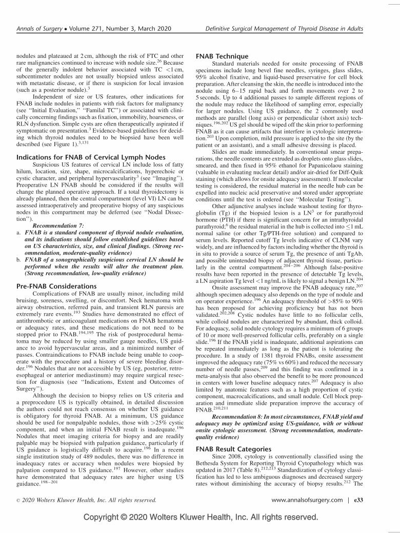
Copyright © 2020 Wolters Kluwer Health, Inc. All rights reserved.
nodules and plateaued at 2 cm, although the risk of FTC and otherrare malignancies continued to increase with nodule size.26 Becauseof the generally indolent behavior associated with TC <1 cm,subcentimeter nodules are not usually biopsied unless associatedwith metastatic disease, or if there is suspicion for local invasion(such as a posterior nodule).3
Independent of size or US features, other indications forFNAB include nodules in patients with risk factors for malignancy(see ‘‘Initial Evaluation,’’ ‘‘Familal TC’’) or associated with clini-cally concerning findings such as fixation, immobility, hoarseness, orRLN dysfunction. Simple cysts are often therapeutically aspirated ifsymptomatic on presentation.3 Evidence-based guidelines for decid-ing which thyroid nodules need to be biopsied have been welldescribed (see Figure 1).3,131
Indications for FNAB of Cervical Lymph NodesSuspicious US features of cervical LN include loss of fatty
hilum, location, size, shape, microcalcifications, hyperechoic orcystic character, and peripheral hypervascularity3 (see ‘‘Imaging’’).Preoperative LN FNAB should be considered if the results willchange the planned operative approach. If a total thyroidectomy isalready planned, then the central compartment (level VI) LN can beassessed intraoperatively and preoperative biopsy of any suspiciousnodes in this compartment may be deferred (see ‘‘Nodal Dissec-tion’’).
Recommendation 7:a. FNAB is a standard component of thyroid nodule evaluation,
and its indications should follow established guidelines basedon US characteristics, size, and clinical findings. (Strong rec-ommendation, moderate-quality evidence)
b. FNAB of a sonographically suspicious cervical LN should beperformed when the results will alter the treatment plan.(Strong recommendation, low-quality evidence)
Pre-FNAB ConsiderationsComplications of FNAB are usually minor, including mild
bruising, soreness, swelling, or discomfort. Neck hematoma withairway obstruction, referred pain, and transient RLN paresis areextremely rare events.193 Studies have demonstrated no effect ofantithrombotic or anticoagulant medications on FNAB hematomaor adequacy rates, and these medications do not need to bestopped prior to FNAB.194,195 The risk of postprocedural hema-toma may be reduced by using smaller gauge needles, US guid-ance to avoid hypervascular areas, and a minimized number ofpasses. Contraindications to FNAB include being unable to coop-erate with the procedure and a history of severe bleeding disor-der.196 Nodules that are not accessible by US (eg, posterior, retro-esophageal or anterior mediastinum) may require surgical resec-tion for diagnosis (see ‘‘Indications, Extent and Outcomes ofSurgery’’).
Although the decision to biopsy relies on US criteria anda preprocedure US is typically obtained, in detailed discussionthe authors could not reach consensus on whether US guidanceis obligatory for thyroid FNAB. At a minimum, US guidanceshould be used for nonpalpable nodules, those with >25% cysticcomponent, and when an initial FNAB result is inadequate.196
Nodules that meet imaging criteria for biopsy and are readilypalpable may be biopsied with palpation guidance, particularly ifUS guidance is logistically difficult to acquire.196 In a recentsingle institution study of 489 nodules, there was no difference ininadequacy rates or accuracy when nodules were biopsied bypalpation compared to US guidance.197 However, other studieshave demonstrated that adequacy rates are higher using USguidance.198–201
FNAB TechniqueStandard materials needed for onsite processing of FNAB
specimens include long bevel fine needles, syringes, glass slides,95% alcohol fixative, and liquid-based preservative for cell blockpreparation. After cleansing the skin, the needle is introduced into thenodule using 6–15 rapid back and forth movements over 2 to5 seconds. Up to 4 additional passes to sample different regions ofthe nodule may reduce the likelihood of sampling error, especiallyfor larger nodules. Using US guidance, the 2 commonly usedmethods are parallel (long axis) or perpendicular (short axis) tech-niques.196,202 US gel should be wiped off the skin prior to performingFNAB as it can cause artifacts that interfere in cytologic interpreta-tion.203 Upon completion, mild pressure is applied to the site (by thepatient or an assistant), and a small adhesive dressing is placed.
Slides are made immediately. In conventional smear prepa-rations, the needle contents are extruded as droplets onto glass slides,smeared, and then fixed in 95% ethanol for Papanicolaou staining(valuable in evaluating nuclear detail) and/or air-dried for Diff-Quikstaining (which allows for onsite adequacy assessment). If moleculartesting is considered, the residual material in the needle hub can beexpelled into nucleic acid preservative and stored under appropriateconditions until the test is ordered (see ‘‘Molecular Testing’’).
Other adjunctive analyses include washout testing for thyro-globulin (Tg) if the biopsied lesion is a LN3 or for parathyroidhormone (PTH) if there is significant concern for an intrathyroidalparathyroid;6 the residual material in the hub is collected into�1 mLnormal saline (or other Tg/PTH-free solution) and compared toserum levels. Reported cutoff Tg levels indicative of CLNM varywidely, and are influenced by factors including whether the thyroid isin situ to provide a source of serum Tg, the presence of anti TgAb,and possible unintended biopsy of adjacent thyroid tissue, particu-larly in the central compartment.204–206 Although false-positiveresults have been reported in the presence of detectable Tg levels,a LN aspiration Tg level<1 ng/mL is likely to signal a benign LN.204
Onsite assessment may improve the FNAB adequacy rate,207
although specimen adequacy also depends on the type of nodule andon operator experience.196 An adequacy threshold of >85% to 90%has been proposed for achieving proficiency but has not beenvalidated.202,208 Cystic nodules have little to no follicular cells,while colloid nodules are characterized by abundant, thick colloid.For adequacy, solid nodule cytology requires a minimum of 6 groupsof 10 or more well-preserved follicular cells, preferably on a singleslide.196 If the FNAB yield is inadequate, additional aspirations canbe repeated immediately as long as the patient is tolerating theprocedure. In a study of 1381 thyroid FNABs, onsite assessmentimproved the adequacy rate (75% vs 60%) and reduced the necessarynumber of needle passes,209 and this finding was confirmed in ameta-analysis that also observed the benefit to be more pronouncedin centers with lower baseline adequacy rates.207 Adequacy is alsolimited by anatomic features such as a high proportion of cysticcomponent, macrocalcifications, and small nodule. Cell block prep-aration and immediate slide preparation improve the accuracy ofFNAB.210,211
Recommendation 8: In most circumstances, FNAB yield andadequacy may be optimized using US-guidance, with or withoutonsite cytologic assessment. (Strong recommendation, moderate-quality evidence)
FNAB Result CategoriesSince 2008, cytology is conventionally classified using the
Bethesda System for Reporting Thyroid Cytopathology which wasupdated in 2017 (Table 8).212,213 Standardization of cytology classi-fication has led to less ambiguous diagnoses and decreased surgeryrates without diminishing the accuracy of biopsy results.212 The
Annals of Surgery � Volume 271, Number 3, March 2020 Definitive Surgical Management of Thyroid Disease in Adults
� 2020 Wolters Kluwer Health, Inc. All rights reserved. www.annalsofsurgery.com | e33
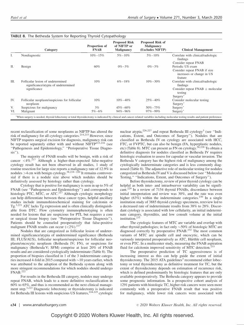
Copyright © 2020 Wolters Kluwer Health, Inc. All rights reserved.
recent reclassification of some neoplasms as NIFTP has altered therisk of malignancy for all cytology categories.214,215 However, sinceNIFTP requires surgical excision for diagnosis, malignancy risk canbe reported separately either with and without NIFTP213,216 (see‘‘Pathogenesis and Epidemiology,’’ ‘‘Perioperative Tissue Diagno-sis’’).
The majority of FNAB results will be benign, with a risk ofcancer <4%.217 Although a higher-than-expected false-negativecytology result has not been observed in all studies, 1 study ofroutine resection for large size reports a malignancy rate of 12.9% innodules >4 cm with benign cytology.38,218–220 It remains controver-sial if there is a nodule size above which nodules should bedefinitively assessed by histology rather than cytology.
Cytology that is positive for malignancy is seen in up to 5% ofFNAB (see ‘‘Pathogenesis and Epidemiology’’) and corresponds tohistologic PTC, MTC, or ATC.217 Although cytologic morphologycan help differentiate between these cancer types, helpful ancillarystudies include immunohistochemical staining for calcitonin orTg.3,221 ATC lacks Tg expression and is often clinically distinguish-able from DTC. Flow cytometry immunophenotyping is oftenneeded for lesions that are suspicious for PTL but requires a coreor surgical tissue biopsy (see ‘‘Perioperative Tissue Diagnosis’’).Patients should be counseled preoperatively that false-positivemalignant FNAB results can occur (<2%).217
Nodules that are categorized as follicular lesion of undeter-mined significance/atypia of undetermined significance (BethesdaIII; FLUS/AUS), follicular neoplasm/suspicious for follicular neo-plasm/oncocytic neoplasm (Bethesda IV; FN), or suspicious formalignancy (Bethesda V; SFM) comprise at least 20% of FNABresults and are considered cytologically indeterminate (Table 8). Theproportion of biopsies classified in 1 of the 3 indeterminate catego-ries increased 4-fold in 2015 compared with�10 years earlier, whichwas attributed to the adoption of the Bethesda system along withmore stringent recommendations for which nodules should undergoFNAB.222
For results in the Bethesda III category, nodules may undergorepeat FNAB, which leads to a more definitive reclassification in60% to 65%, and thus is recommended as the next clinical manage-ment step.3,223 Diagnostic lobectomy or thyroidectomy is indicatedfor Bethesda III lesions with suspicious US features,224,225 cytologic
nuclear atypia,226,227 and repeat Bethesda III cytology3 (see ‘‘Indi-cations, Extent, and Outcomes of Surgery’’). Nodules that areclassified as Bethesda IV on cytology are associated with HCC,FTC, or FVPTC, but can also be benign (FA, hyperplastic nodules,etc) (Table 8). MTC can present as FN on cytology.56,228 To obtain adefinitive diagnosis for nodules classified as Bethesda IV requireshistologic evaluation to assess for capsular or vascular invasion. TheBethesda V category has the highest risk of malignancy among thecytologically indeterminate categories and is less commonly diag-nosed (Table 8). The adjunctive role of molecular testing for nodulescategorized as Bethesda IVand V is discussed below (see ‘‘MolecularTesting,‘‘ ‘‘Indications, Extent, and Outcomes of Surgery’’).
Before thyroidectomy, review of prior thyroid cytology can behelpful as both inter- and intraobserver variability can be signifi-cant.229 In a review of 7154 thyroid FNABs, discordance betweeninitial interpretation and review was 30%, and the rate was evenhigher (63%) within the indeterminate categories.230 In a singleinstitution study of 3885 thyroid cytology specimens, rereview led toa decreased rate of indeterminate results from 38% to 28%. Discor-dant cytology is associated with low cellularity, an initial indetermi-nate category, thyroiditis, and low consult volume at the initialinstitution.231
The cytologic features of MTC are variable and overlap withother thyroid pathologies; in fact only �50% of histologic MTC arediagnosed correctly by preoperative FNAB.232 The most commonvariants of MTC are spindle cell and oncocytic, which can bevariously interpreted preoperatively as ATC, Hurthle cell neoplasm,or even PTC. In a multicenter study, measuring the FNAB aspirationfluid for calcitonin improved sensitivity of MTC detection.233
The preoperative prediction of TC aggressiveness is ofincreasing interest as this can help guide the extent of initialthyroidectomy. The 2015 ATA guidelines3 recommend either lobec-tomy or total thyroidectomy as definitive treatment for TC, but theextent of thyroidectomy depends on estimation of recurrence risk,which is defined predominantly by histologic features that are onlyavailable postoperatively. The Bethesda category appears to providesome prognostic information. In a prospective cohort analysis of1291 patients with histologic TC, higher risk cancers were seen morecommonly with a preoperative FNAB result that was positivefor malignancy, while lower risk cancers were associated with
TABLE 8. The Bethesda System for Reporting Thyroid Cytopathology
CategoryProportion of
FNAB
Proposed Riskof NIFTP orMalignancy
Proposed Risk ofMalignancy
(Excludes NIFTP) Clinical Management
I. Nondiagnostic 10%–15% 5%–10% 5%–10% Correlate with clinical/radiologicfindings
Consider repeat FNABII. Benign 60% 0%–3% 0%–3% Periodic US exam
Consider repeat FNAB if sizeincreases or change in USfeature
III. Follicular lesion of undeterminedsignificance/atypia of undeterminedsignificance
10% 6%–18% 10%–30% Correlate with clinical/radiologicfindings
Consider repeat FNAB � moleculartesting
Surgery�
IV. Follicular neoplasm/suspicious for follicularneoplasm
10% 10%–40% 25%–40% Consider molecular testingSurgery�
V. Suspicious for malignancy 3% 45%–60% 50%–75% Surgery�
VI. Malignant 5%–6% 94%–96% 97%–99% Surgery�
�When surgery is needed, thyroid lobectomy or total thyroidectomy is indicated by clinical and cancer-related variables including molecular testing results and patient preference.
Patel et al Annals of Surgery � Volume 271, Number 3, March 2020
e34 | www.annalsofsurgery.com � 2020 Wolters Kluwer Health, Inc. All rights reserved.
Copyright © 2020 Wolters Kluwer Health, Inc. All rights reserved.
FLUS/AUS or SFM;234 an unfavorable outcome (including localrecurrence, distant metastasis, or death) was uncommon but wasmost likely with malignant FNAB results. In 371 patients withthyroid cancer and an initial FLUS/AUS result, recurrence was rare(3%) albeit with mean follow-up of only 3 years.235 However, inanother single institution study of 360 thyroid cancers, Bethesdacategory was not predictive of worse outcome within tumor sub-types.236 Additional studies are needed to better define the potentialprognostic implications of the Bethesda system.
Recommendation 9: The Bethesda System for ReportingThyroid Cytopathology should be used to report and stratify therisk of malignancy in a thyroid nodule. (Strong recommendation,high-quality evidence)
MOLECULAR TESTING
Molecular testing (MT) to examine somatic changes hasbecome an important adjunct in the diagnosis and treatment of manycancers, and is primarily utilized in thyroid disease as a preoperativemethod to refine the cancer risk of cytologically indeterminatethyroid nodules.3,237 In development, such testing requires definingthe molecular profile of nodules that are benign, malignant, and evennonthyroidal, developing reliable analytic assays to detect theseprofiles in Clinical Laboratory Improvement Amendments-approvedlaboratories, and further levels of assessment (below). Molecularprofiling includes genomic alterations (such as point mutations,insertions, and deletions), gene fusions resulting in rearrangementsor translocations, copy number variations, RNA-based gene expres-sion, and/or micro-RNA (miRNA) expression.238
MT to Guide Need for SurgeryAvoiding surgery for a cytologically indeterminate nodule can
be considered if an informed patient wishes and it is clinicallyappropriate (see ‘‘Indications, Extent and Outcomes of Surgery’’).Negative MT results can reduce the risk of cancer to one that isapproximately equivalent to that for a benign FNAB result (�5%).239
The type, indication for, and interpretation of MT should considernodule and clinical variables, and whether the results will changeplanned recommendations.240 US and cytology results may helpclassify risk and determine if surgery is warranted. In addition, ifthyroidectomy is indicated regardless of cancer risk ie due tosymptoms, imaging features, nodule size etc, then ordering MTfor the purpose of avoiding surgery would not be indicated. In somesettings, if multidisciplinary management of cytologically indeter-minate nodules leads often to observation based on locally availableradiographic and cytologic expertise, then the clinical utility of MTmay be diminished.
Recommendation 10: If thyroidectomy is preferred for clini-cal reasons, then MT is unnecessary. (Strong recommendation,moderate-quality evidence)
MT to Guide Thyroid Nodule ManagementThere have been several versions and types of MT, but
currently 3 tests are commercially available. MTs should be evalu-ated in a systematic framework to ensure that each has analyticalvalidity (how accurately can the test detect if the variant is present orabsent), clinical validity (how well does the genetic variant beinganalyzed relate to the risk of disease), and clinical utility (does thetest provide clinically helpful information about diagnosis andtreatment).241 An ideal clinical validation study is multi-institutional,includes histologic correlation for all nodules regardless of MTresult, has histology interpreted by a pathologist who is blinded tothe MT results, and avoids post hoc exclusions. Sensitivity andspecificity are inherent test properties, but clinical use is typically
directed by negative and positive predictive values (NPV, PPV)which are dependent on both test performance and cancer prevalencewhich is institutionally and geographically variable242 (see ‘‘Patho-genesis and Epidemiology’’). The benign call rate, that is, theproportion of FNAB which are MT negative, is a predictor of thenumber of avoidable diagnostic surgeries and has implications for atest’s real-time clinical utility.
An early form of MT was the 7-gene panel (7GP) designed toevaluate point mutations in BRAF (V600E and K601E), H-, K-, andN-RAS, and the RET/PTC 1/3 and PAX8/PPARg rearrange-ments.243,244 The 7GP included genetic alterations for �70% ofall thyroid cancers, and in clinical validation studies improveddiagnostic specificity and cost savings.245–251 Under the 2009ATAG, which advised total thyroidectomy for all DTC, FNAB testingfor 7GP was associated with increased rates of correct initial extentof thyroidectomy (lobectomy vs total thyroidectomy).252 The 7GP isnow incorporated into 2 commercially available tests: ThyGenX þThyraMIR (which combines 7GP with a 10 miRNA panel) andThyroSeq, which is an expanded multigene panel.
The initial ThyGenX þ ThyraMIR clinical validation reportwas a multisite study inclusive of 109 Bethesda III and Bethesda IVnodules with histopathologic correlation.253 Cytology and histologywere interpreted at the local institution, and pathologists wereblinded to the MT results. The cancer prevalence was 32%, and61% of the cohort had benign MT. Sensitivity and specificity were89% (73–97) and 85% (76–92), respectively. Two small singleinstitution studies have provided additional clinical validation forthe ThyGenX þ ThyraMIR test, with one (n¼60) reporting a lowersensitivity of 48% (range 27–69)254 and the other (n¼10) reporting alower specificity of 71%.255 Clinical utility is not described. Thecurrent version has expanded the DNA and RNA panel, and is nowcalled ThyGeNEXT þ ThyraMIR; no performance data is availablefor this iteration.
Two iterations of ThyroSeq have been described in analyticvalidation studies and both utilize next generation sequencing toidentify a panel of molecular alterations. ThyroSeq v2 tested for56 thyroid-related genes including point mutations, gene fusions,and gene expression, while ThyroSeq v3 expanded the numberof tested genes to 112, inclusive of copy number alterations in10 genomic regions.256 The benign call rate for ThyroSeq v2was similar to that for ThyGenX þ ThyraMIR and ranges from65% to 93%.253,257–259 A multicenter prospective study of Thy-roSeq v3 including 247 Bethesda III and Bethesda IV nodules inwhich both pathologist and clinicians were blinded to MT resultsreported sensitivity of 94%, specificity of 82% with a cancerprevalence of 28% inclusive of NIFTP, and a benign call rate of61%.260
Using a different MT strategy, the Afirma Gene ExpressionClassifier (GEC) was an RNA-based panel which was specificallyselected to associate with benign nodules.261 In essence, a negativeresult ideally obviates the need for surgery. Since the initial analyticalvalidation studies, additional expression markers for MTC, Hurthlecell lesions, BRAF, and RET/PTC fusions have been added and thenewest iteration is now called the Genomic Sequencing Classifier(GSC).262–264 Clinical validation studies of GEC have been numer-ous and heterogeneous in design and outcome. The first report was amulti-institution study inclusive of 265 Bethesda III, IV, and Vnodules in analysis of GEC test performance.265 Because theNPV was 85% for Bethesda V nodules due to high cancer prevalence(62%), GEC was no longer performed for this cytologic category.265
GSC validation used this same cohort of samples, of which 190Bethesda III and IV nodules had adequate RNA for retesting.Sensitivity was 91% (79–98) and specificity was higher than forGEC at 68% (60%–76%, 95% CI). Follow-up clinical validation
Annals of Surgery � Volume 271, Number 3, March 2020 Definitive Surgical Management of Thyroid Disease in Adults
� 2020 Wolters Kluwer Health, Inc. All rights reserved. www.annalsofsurgery.com | e35

Copyright © 2020 Wolters Kluwer Health, Inc. All rights reserved.
studies of GEC reported relatively consistent sensitivities of 83% to100%266–272 but unlike the initial study,265 the majority of subse-quent analyses included only a small proportion of MT negativenodules with histologic correlation or long-term follow-up.273
Not all studies limited GEC use to only Bethesda III and IVnodules, and in others, utilization was inconsistent. Both factors mayhave led to the observed range of GEC specificity (15%–40%) andPPV (14%–58%).266–269,274–280 Clinical utilization studies of GEChave included cost analyses and reports of observed reductions innecessary diagnostic thyroidectomies.281–286 Management of posi-tive GSC results is typically diagnostic surgery with varying rates ofmalignancy expected, depending on the local pretest probabilityof cancer.
Molecular alterations characteristic of Hurthle cell carcinoma-(HCC)s are different than follicular-patterned lesions,47,287 and MTperformance in Hurthle cell neoplasms has been variable to date.288
Changes incorporated into both GSC and ThyroSeq v3 may improveMT performance in this nodule subset.260,264
Few studies have compared performance of >1 MT on eitherthe same set of nodules or within the same institution. In a study thatretrospectively obtained multiple MTs (GEC, ThyGenXþThyra-ThyraMIR, and RosettaGX which was a 24 miRNA expressionclassifier that is no longer available) on 10 cytologically indetermi-nate (Bethesda III–V) nodules with histology, all 3 MT had 100%sensitivity and NPV.255 In an analysis to determine if clinicalmanagement of all biopsied nodules (Bethesda I–VI) was alteredby the use of MT (inclusive of GEC, ThyGenX, ThyroSeq, and 7GP),<10% had a change in planned management according to the 2009ATAG.289 In a prospective direct single-institution comparison thatrandomized Bethesda III/IV nodules to GEC versus ThyroSeq v2MT,290 the benign call rate differed (ThyroSeq 77% vs GEC 43%);however, not all MT negative nodules had histology. When noduleswith histology were assessed, sensitivity for both tests was 100%(NPV 100%), but specificity was lower for GEC (16% vs ThyroSeq60%). Particularly concerning cost efficacy, clinical validation datathat evaluate use of >1 MT on the same nodule are not available.
A summary of the commercially available MTs and corre-sponding results from the index clinical validation study appears inTable 9.253,260,264 However, use of MT results to make clinicaldecisions relies on PPV and NPV which are contingent on regionalcancer prevalence for each cytology category.242 Using sensitivityand specificity from the index studies, the risk of malignancydepending on a positive or negative MT result is shown relativeto cancer prevalence in Figure 2.
Recommendation 11: When the need for thyroidectomy isunclear after consideration of clinical, imaging, and cytologicfeatures, MT may be considered as a diagnostic adjunct forcytologically indeterminate nodules. (Strong recommendation,moderate-quality evidence)
Recommendation 12: Accuracy of MT relies on institutionalmalignancy rates and should be locally examined for optimalextrapolation of results to thyroid cancer risk. (Strong recommen-dation, moderate-quality evidence)
MT to Guide Extent of SurgeryBecause it is available preoperatively, some information from
MT can be used to guide extent of surgery but clinical variablesshould also be considered, that is, as stated above, if total thyroidec-tomy is already indicated, then MT will not alter the extent ofthyroidectomy under the current ATAG although under the 2009ATAG, 7G MT was associated with increased rates of correct initialthyroidectomy.252 To date, no MT has been studied for surgicalguidance under the 2015 guidelines which substantially modifiedrecommendations on the extent of thyroidectomy, so that lobectomyor total thyroidectomy are potential options for 1 to 4 cm TCs3 (see‘‘Indications, Extent, and Outcomes of Surgery’’). To be useful fordeciding extent of surgery, a given MT needs to first provide reliableprognostic information. In a single institution study of 1510 TC,aggressive features such as CLNM, distant metastasis, and recur-rence were associated with BRAF V600E and RET/PTC-1 and -3rearrangements.291
BRAF V600E, a mutation that was focused upon early in theevolution of MT, has been extensively studied and is associated withmore aggressive PTC subtypes, such as the tall cell variant, inaddition to other histologic tumor characteristics including CLNMand ETE.292,293 BRAF V600E mutant tumors have also been associ-ated with recurrence, even in PTMC.294,295 When PTC is solitary and1 to 4 cm in size, recurrence is more likely with positive BRAFV600E (9.5% vs 3.4%).296 However, not all BRAF V600E-positivePTC are associated with aggressive disease. Overall, its prognosticvalue is limited when histologic features are available to refine riskstratification.297,298 BRAF V600E can be detected by ThyGenX,Afirma GEC, and ThyroSeq. By contrast, BRAF K601E is a clini-cally indolent mutation that should not be confused with BRAFV600E.299
ThyroSeq and ThyGenX testing can also detect TERT pro-moter mutations which are found in� 10% of TC and are associatedwith cancer-specific mortality.300–302 In a case report, aggressivePTC exhibited multiple mutations,303 and in meta-analysis aggres-sion was associated with coexistent BRAF V600E and TERT inparticular.304 Testing for BRAF V600E and TERT promoter muta-tions is not necessarily recommended for initial risk stratification;however, if MT is available and such mutations are present, they arecalled out as being associated with increased risk of recurrence in the2015 ATA Risk classification.3 Further study will determine ifgenotype provides information that has not already been obtainedclinically, by US imaging, and/or by cytologic classification, as wellas determine if altering the initial extent of surgery based on MTresults will affect outcomes.
RAS mutations have been variably associated with malig-nancy, a phenomenon that is often the cause of differing specificityvalues for the MTs that include RAS testing (Table 9). In one single-institution series, RAS-positive nodules were associated with a 75%risk of malignancy with HRAS mutations conferring the highest risk(90%).305 However, another series reported a RAS-associated malig-nancy risk of only 9%.258 RAS mutation can be seen in several thyroidhistologies, including FA, NIFTP, encapsulated and unencapsulated
TABLE 9. Commercially Available Molecular Tests for Bethesda III and IV Nodules
Test Name Type of Test False Negatives� Sensitivity (95% CI)� Specificity (95% CI)�
Afirma Gene Sequencing Classifier Multigene expression (RNA-based) 4/103 (4%) 91% (79%–98%) 68% (60%–76%)Interpace ThyGenXþThyraMir 7 gene panel þ 10 miRNA 4/67 (6%) 89% (73%–97%) 85% (75%–92%)CBLPath ThyroSeq version 3 Multigene NGS 4/150 (3%) 94% (86%–98%) 82% (75%–87%)
�Includes only results with histologic correlation and before post hoc exclusions.CI indicates confidence intervals; miRNA, micro-RNA; NGS, next-generation sequencing; RNA, ribonucleic acid.
Patel et al Annals of Surgery � Volume 271, Number 3, March 2020
e36 | www.annalsofsurgery.com � 2020 Wolters Kluwer Health, Inc. All rights reserved.
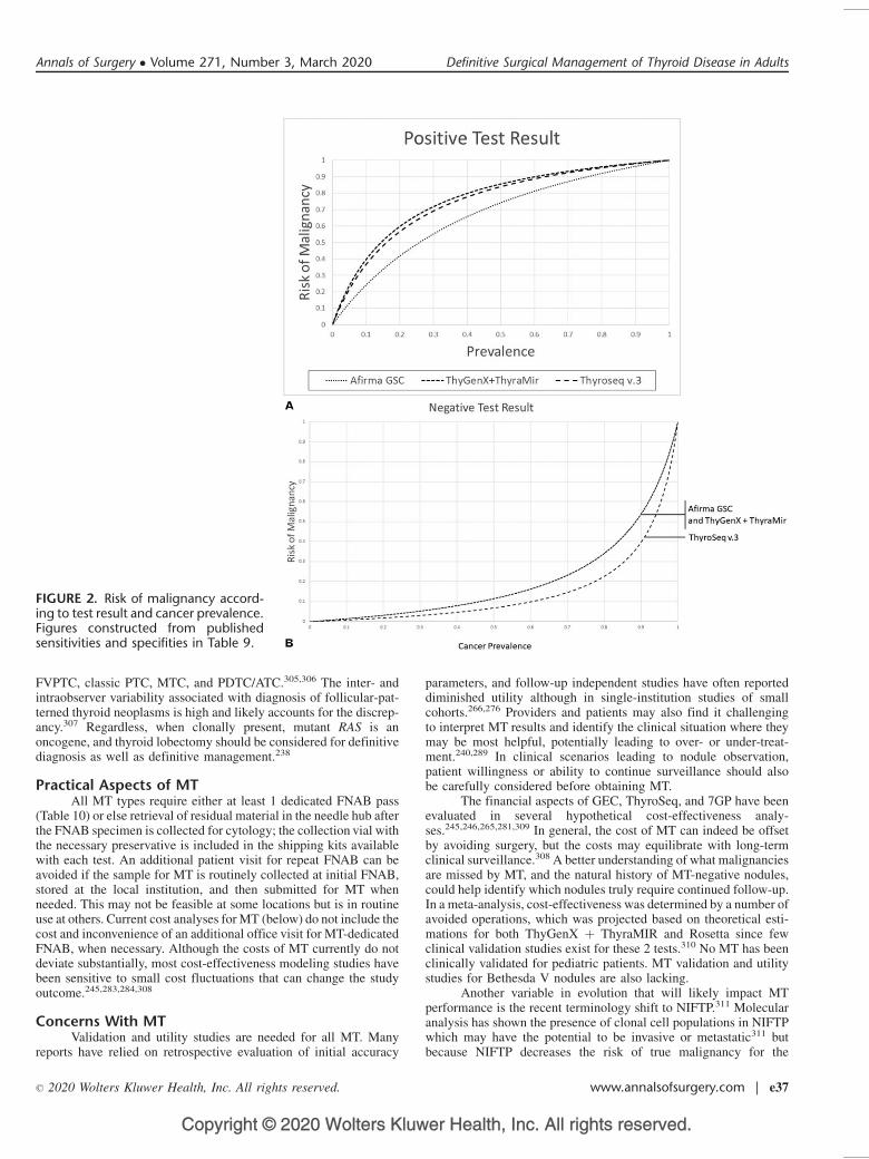
Copyright © 2020 Wolters Kluwer Health, Inc. All rights reserved.
FVPTC, classic PTC, MTC, and PDTC/ATC.305,306 The inter- andintraobserver variability associated with diagnosis of follicular-pat-terned thyroid neoplasms is high and likely accounts for the discrep-ancy.307 Regardless, when clonally present, mutant RAS is anoncogene, and thyroid lobectomy should be considered for definitivediagnosis as well as definitive management.238
Practical Aspects of MTAll MT types require either at least 1 dedicated FNAB pass
(Table 10) or else retrieval of residual material in the needle hub afterthe FNAB specimen is collected for cytology; the collection vial withthe necessary preservative is included in the shipping kits availablewith each test. An additional patient visit for repeat FNAB can beavoided if the sample for MT is routinely collected at initial FNAB,stored at the local institution, and then submitted for MT whenneeded. This may not be feasible at some locations but is in routineuse at others. Current cost analyses for MT (below) do not include thecost and inconvenience of an additional office visit for MT-dedicatedFNAB, when necessary. Although the costs of MT currently do notdeviate substantially, most cost-effectiveness modeling studies havebeen sensitive to small cost fluctuations that can change the studyoutcome.245,283,284,308
Concerns With MTValidation and utility studies are needed for all MT. Many
reports have relied on retrospective evaluation of initial accuracy
parameters, and follow-up independent studies have often reporteddiminished utility although in single-institution studies of smallcohorts.266,276 Providers and patients may also find it challengingto interpret MT results and identify the clinical situation where theymay be most helpful, potentially leading to over- or under-treat-ment.240,289 In clinical scenarios leading to nodule observation,patient willingness or ability to continue surveillance should alsobe carefully considered before obtaining MT.
The financial aspects of GEC, ThyroSeq, and 7GP have beenevaluated in several hypothetical cost-effectiveness analy-ses.245,246,265,281,309 In general, the cost of MT can indeed be offsetby avoiding surgery, but the costs may equilibrate with long-termclinical surveillance.308 A better understanding of what malignanciesare missed by MT, and the natural history of MT-negative nodules,could help identify which nodules truly require continued follow-up.In a meta-analysis, cost-effectiveness was determined by a number ofavoided operations, which was projected based on theoretical esti-mations for both ThyGenX þ ThyraMIR and Rosetta since fewclinical validation studies exist for these 2 tests.310 No MT has beenclinically validated for pediatric patients. MT validation and utilitystudies for Bethesda V nodules are also lacking.
Another variable in evolution that will likely impact MTperformance is the recent terminology shift to NIFTP.311 Molecularanalysis has shown the presence of clonal cell populations in NIFTPwhich may have the potential to be invasive or metastatic311 butbecause NIFTP decreases the risk of true malignancy for the
FIGURE 2. Risk of malignancy accord-ing to test result and cancer prevalence.Figures constructed from publishedsensitivities and specifities in Table 9.
Annals of Surgery � Volume 271, Number 3, March 2020 Definitive Surgical Management of Thyroid Disease in Adults
� 2020 Wolters Kluwer Health, Inc. All rights reserved. www.annalsofsurgery.com | e37
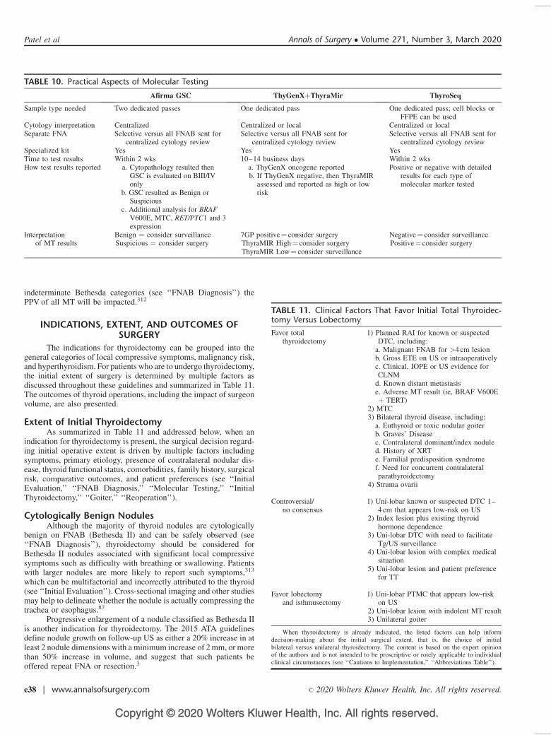
Copyright © 2020 Wolters Kluwer Health, Inc. All rights reserved.
indeterminate Bethesda categories (see ‘‘FNAB Diagnosis’’) thePPV of all MT will be impacted.312
INDICATIONS, EXTENT, AND OUTCOMES OFSURGERY
The indications for thyroidectomy can be grouped into thegeneral categories of local compressive symptoms, malignancy risk,and hyperthyroidism. For patients who are to undergo thyroidectomy,the initial extent of surgery is determined by multiple factors asdiscussed throughout these guidelines and summarized in Table 11.The outcomes of thyroid operations, including the impact of surgeonvolume, are also presented.
Extent of Initial ThyroidectomyAs summarized in Table 11 and addressed below, when an
indication for thyroidectomy is present, the surgical decision regard-ing initial operative extent is driven by multiple factors includingsymptoms, primary etiology, presence of contralateral nodular dis-ease, thyroid functional status, comorbidities, family history, surgicalrisk, comparative outcomes, and patient preferences (see ‘‘InitialEvaluation,’’ ‘‘FNAB Diagnosis,’’ ‘‘Molecular Testing,’’ ‘‘InitialThyroidectomy,’’ ‘‘Goiter,’’ ‘‘Reoperation’’).
Cytologically Benign NodulesAlthough the majority of thyroid nodules are cytologically
benign on FNAB (Bethesda II) and can be safely observed (see‘‘FNAB Diagnosis’’), thyroidectomy should be considered forBethesda II nodules associated with significant local compressivesymptoms such as difficulty with breathing or swallowing. Patientswith larger nodules are more likely to report such symptoms,313
which can be multifactorial and incorrectly attributed to the thyroid(see ‘‘Initial Evaluation’’). Cross-sectional imaging and other studiesmay help to delineate whether the nodule is actually compressing thetrachea or esophagus.87
Progressive enlargement of a nodule classified as Bethesda IIis another indication for thyroidectomy. The 2015 ATA guidelinesdefine nodule growth on follow-up US as either a 20% increase in atleast 2 nodule dimensions with a minimum increase of 2 mm, or morethan 50% increase in volume, and suggest that such patients beoffered repeat FNA or resection.3
TABLE 10. Practical Aspects of Molecular Testing
Afirma GSC ThyGenXþThyraMir ThyroSeq
Sample type needed Two dedicated passes One dedicated pass One dedicated pass; cell blocks orFFPE can be used
Cytology interpretation Centralized Centralized or local Centralized or localSeparate FNA Selective versus all FNAB sent for
centralized cytology reviewSelective versus all FNAB sent for
centralized cytology reviewSelective versus all FNAB sent for
centralized cytology reviewSpecialized kit Yes Yes YesTime to test results Within 2 wks 10–14 business days Within 2 wksHow test results reported a. Cytopathology resulted then
GSC is evaluated on BIII/IVonly
b. GSC resulted as Benign orSuspicious
c. Additional analysis for BRAFV600E, MTC, RET/PTC1 and 3expression
a. ThyGenX oncogene reportedb. If ThyGenX negative, then ThyraMIR
assessed and reported as high or lowrisk
Positive or negative with detailedresults for each type ofmolecular marker tested
Interpretationof MT results
Benign ¼ consider surveillanceSuspicious ¼ consider surgery
7GP positive¼ consider surgeryThyraMIR High¼ consider surgeryThyraMIR Low¼ consider surveillance
Negative¼ consider surveillancePositive¼ consider surgery
TABLE 11. Clinical Factors That Favor Initial Total Thyroidec-tomy Versus Lobectomy
Favor totalthyroidectomy
1) Planned RAI for known or suspectedDTC, including:
a. Malignant FNAB for >4 cm lesionb. Gross ETE on US or intraoperativelyc. Clinical, IOPE or US evidence forCLNM
d. Known distant metastasise. Adverse MT result (ie, BRAF V600Eþ TERT)
2) MTC3) Bilateral thyroid disease, including:
a. Euthyroid or toxic nodular goiterb. Graves’ Diseasec. Contralateral dominant/index noduled. History of XRTe. Familial predisposition syndromef. Need for concurrent contralateralparathyroidectomy
4) Struma ovarii
Controversial/no consensus
1) Uni-lobar known or suspected DTC 1–4 cm that appears low-risk on US
2) Index lesion plus existing thyroidhormone dependence
3) Uni-lobar DTC with need to facilitateTg/US surveillance
4) Uni-lobar lesion with complex medicalsituation
5) Uni-lobar lesion and patient preferencefor TT
Favor lobectomyand isthmusectomy
1) Uni-lobar PTMC that appears low-riskon US
2) Uni-lobar lesion with indolent MT result3) Unilateral goiter
When thyroidectomy is already indicated, the listed factors can help informdecision-making about the initial surgical extent, that is, the choice of initialbilateral versus unilateral thyroidectomy. The content is based on the expert opinionof the authors and is not intended to be proscriptive or rotely applicable to individualclinical circumstances (see ‘‘Cautions to Implementation,’’ ‘‘Abbreviations Table’’).
Patel et al Annals of Surgery � Volume 271, Number 3, March 2020
e38 | www.annalsofsurgery.com � 2020 Wolters Kluwer Health, Inc. All rights reserved.
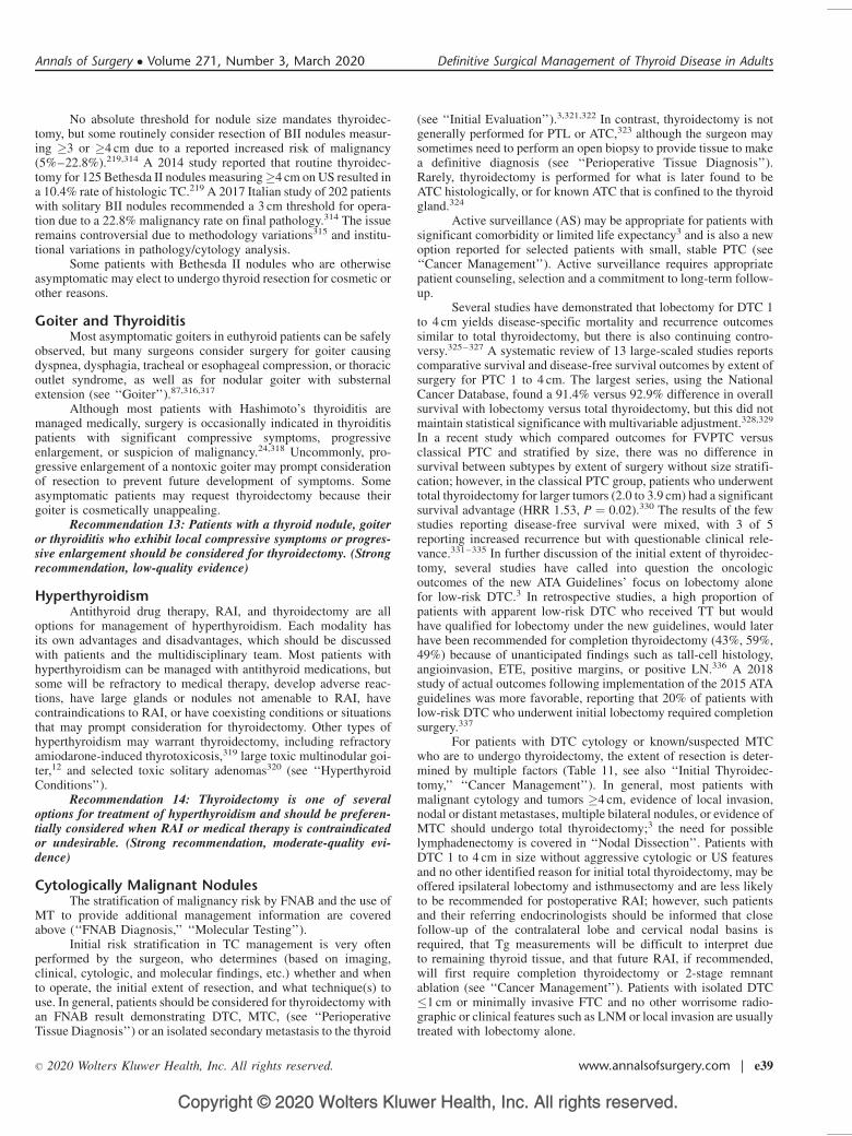
Copyright © 2020 Wolters Kluwer Health, Inc. All rights reserved.
No absolute threshold for nodule size mandates thyroidec-tomy, but some routinely consider resection of BII nodules measur-ing �3 or �4 cm due to a reported increased risk of malignancy(5%–22.8%).219,314 A 2014 study reported that routine thyroidec-tomy for 125 Bethesda II nodules measuring�4 cm on US resulted ina 10.4% rate of histologic TC.219 A 2017 Italian study of 202 patientswith solitary BII nodules recommended a 3 cm threshold for opera-tion due to a 22.8% malignancy rate on final pathology.314 The issueremains controversial due to methodology variations315 and institu-tional variations in pathology/cytology analysis.
Some patients with Bethesda II nodules who are otherwiseasymptomatic may elect to undergo thyroid resection for cosmetic orother reasons.
Goiter and ThyroiditisMost asymptomatic goiters in euthyroid patients can be safely
observed, but many surgeons consider surgery for goiter causingdyspnea, dysphagia, tracheal or esophageal compression, or thoracicoutlet syndrome, as well as for nodular goiter with substernalextension (see ‘‘Goiter’’).87,316,317
Although most patients with Hashimoto’s thyroiditis aremanaged medically, surgery is occasionally indicated in thyroiditispatients with significant compressive symptoms, progressiveenlargement, or suspicion of malignancy.24,318 Uncommonly, pro-gressive enlargement of a nontoxic goiter may prompt considerationof resection to prevent future development of symptoms. Someasymptomatic patients may request thyroidectomy because theirgoiter is cosmetically unappealing.
Recommendation 13: Patients with a thyroid nodule, goiteror thyroiditis who exhibit local compressive symptoms or progres-sive enlargement should be considered for thyroidectomy. (Strongrecommendation, low-quality evidence)
HyperthyroidismAntithyroid drug therapy, RAI, and thyroidectomy are all
options for management of hyperthyroidism. Each modality hasits own advantages and disadvantages, which should be discussedwith patients and the multidisciplinary team. Most patients withhyperthyroidism can be managed with antithyroid medications, butsome will be refractory to medical therapy, develop adverse reac-tions, have large glands or nodules not amenable to RAI, havecontraindications to RAI, or have coexisting conditions or situationsthat may prompt consideration for thyroidectomy. Other types ofhyperthyroidism may warrant thyroidectomy, including refractoryamiodarone-induced thyrotoxicosis,319 large toxic multinodular goi-ter,12 and selected toxic solitary adenomas320 (see ‘‘HyperthyroidConditions’’).
Recommendation 14: Thyroidectomy is one of severaloptions for treatment of hyperthyroidism and should be preferen-tially considered when RAI or medical therapy is contraindicatedor undesirable. (Strong recommendation, moderate-quality evi-dence)
Cytologically Malignant NodulesThe stratification of malignancy risk by FNAB and the use of
MT to provide additional management information are coveredabove (‘‘FNAB Diagnosis,’’ ‘‘Molecular Testing’’).
Initial risk stratification in TC management is very oftenperformed by the surgeon, who determines (based on imaging,clinical, cytologic, and molecular findings, etc.) whether and whento operate, the initial extent of resection, and what technique(s) touse. In general, patients should be considered for thyroidectomy withan FNAB result demonstrating DTC, MTC, (see ‘‘PerioperativeTissue Diagnosis’’) or an isolated secondary metastasis to the thyroid
(see ‘‘Initial Evaluation’’).3,321,322 In contrast, thyroidectomy is notgenerally performed for PTL or ATC,323 although the surgeon maysometimes need to perform an open biopsy to provide tissue to makea definitive diagnosis (see ‘‘Perioperative Tissue Diagnosis’’).Rarely, thyroidectomy is performed for what is later found to beATC histologically, or for known ATC that is confined to the thyroidgland.324
Active surveillance (AS) may be appropriate for patients withsignificant comorbidity or limited life expectancy3 and is also a newoption reported for selected patients with small, stable PTC (see‘‘Cancer Management’’). Active surveillance requires appropriatepatient counseling, selection and a commitment to long-term follow-up.
Several studies have demonstrated that lobectomy for DTC 1to 4 cm yields disease-specific mortality and recurrence outcomessimilar to total thyroidectomy, but there is also continuing contro-versy.325–327 A systematic review of 13 large-scaled studies reportscomparative survival and disease-free survival outcomes by extent ofsurgery for PTC 1 to 4 cm. The largest series, using the NationalCancer Database, found a 91.4% versus 92.9% difference in overallsurvival with lobectomy versus total thyroidectomy, but this did notmaintain statistical significance with multivariable adjustment.328,329
In a recent study which compared outcomes for FVPTC versusclassical PTC and stratified by size, there was no difference insurvival between subtypes by extent of surgery without size stratifi-cation; however, in the classical PTC group, patients who underwenttotal thyroidectomy for larger tumors (2.0 to 3.9 cm) had a significantsurvival advantage (HRR 1.53, P ¼ 0.02).330 The results of the fewstudies reporting disease-free survival were mixed, with 3 of 5reporting increased recurrence but with questionable clinical rele-vance.331–335 In further discussion of the initial extent of thyroidec-tomy, several studies have called into question the oncologicoutcomes of the new ATA Guidelines’ focus on lobectomy alonefor low-risk DTC.3 In retrospective studies, a high proportion ofpatients with apparent low-risk DTC who received TT but wouldhave qualified for lobectomy under the new guidelines, would laterhave been recommended for completion thyroidectomy (43%, 59%,49%) because of unanticipated findings such as tall-cell histology,angioinvasion, ETE, positive margins, or positive LN.336 A 2018study of actual outcomes following implementation of the 2015 ATAguidelines was more favorable, reporting that 20% of patients withlow-risk DTC who underwent initial lobectomy required completionsurgery.337
For patients with DTC cytology or known/suspected MTCwho are to undergo thyroidectomy, the extent of resection is deter-mined by multiple factors (Table 11, see also ‘‘Initial Thyroidec-tomy,’’ ‘‘Cancer Management’’). In general, most patients withmalignant cytology and tumors �4 cm, evidence of local invasion,nodal or distant metastases, multiple bilateral nodules, or evidence ofMTC should undergo total thyroidectomy;3 the need for possiblelymphadenectomy is covered in ‘‘Nodal Dissection’’. Patients withDTC 1 to 4 cm in size without aggressive cytologic or US featuresand no other identified reason for initial total thyroidectomy, may beoffered ipsilateral lobectomy and isthmusectomy and are less likelyto be recommended for postoperative RAI; however, such patientsand their referring endocrinologists should be informed that closefollow-up of the contralateral lobe and cervical nodal basins isrequired, that Tg measurements will be difficult to interpret dueto remaining thyroid tissue, and that future RAI, if recommended,will first require completion thyroidectomy or 2-stage remnantablation (see ‘‘Cancer Management’’). Patients with isolated DTC�1 cm or minimally invasive FTC and no other worrisome radio-graphic or clinical features such as LNM or local invasion are usuallytreated with lobectomy alone.
Annals of Surgery � Volume 271, Number 3, March 2020 Definitive Surgical Management of Thyroid Disease in Adults
� 2020 Wolters Kluwer Health, Inc. All rights reserved. www.annalsofsurgery.com | e39
Copyright © 2020 Wolters Kluwer Health, Inc. All rights reserved.
Cytologically Indeterminate NodulesThe safety and outcomes of active surveillance for manage-
ment of indeterminate thyroid nodules are unknown.Conventionally, patients with an FNAB result in the category
of Suspicious (Bethesda V; see ‘‘FNAB Diagnosis’’) should beconsidered for surgery. As with malignant cytology, the extent ofthyroid resection is determined by tumor size, radiographic features,contralateral nodular disease, MT results if performed, and otherclinical factors (Table 11).3 Although it is no longer used routinely, inpatients with Bethesda V cytology, intraoperative frozen section maybe helpful in diagnosing malignancy and guiding decisions aboutinitial surgical extent (see ‘‘Adjuncts and Approaches’’).338
Patients with a cytologic diagnosis of FN (Bethesda IV)should be considered for MT or thyroidectomy.253 Thyroidectomyis indicated in patients with larger FN tumors (�3–4 cm), serialenlargement, worrisome US features, family history of TC, or ahistory of radiation exposure, and its extent is determined by clinicalfactors (Table 11).3 In all other situations, if MT is not performed, orif it produces low yield or suspicious results, thyroidectomy isindicated, and the extent of resection is based on clinical factors.339
Patients with a cytologic diagnosis of AUS/FLUS (BethesdaIII) may be offered the options of repeat FNA (see ‘‘FNAB Diagno-sis’’), surgery, or MT to assist with further stratification of malig-nancy risk.253 If MT is performed and produces low yield orsuspicious results, thyroidectomy is indicated with initial extentdetermined by clinical factors.339 If MT is not performed, patientswith confirmed AUS/FLUS should be considered for thyroidec-tomy.339 In some multidisciplinary settings, selected Bethesda IIIpatients without worrisome radiographic or clinical features may beoffered the option of AS.340
In all cases of indeterminate cytology, patients should receivein-depth discussion regarding the utility and limitations of cytologicanalysis, MT (if utilized), the unknown role of active surveillance,and the clinical and radiographic features that also factor into thedecision-making process. In some situations, the use of MT mayrequire repeat FNAB (see ‘‘Molecular Testing’’).
Recommendation 15: For nodules that are cytologicallycategorized as Bethesda III, clinical factors, radiologic features,and patient preference should inform decision-making regardingwhether to proceed with repeat biopsy, MT, diagnostic thyroidec-tomy, or observation. (Strong recommendation, moderate-qualityevidence)
Recommendation 16: Diagnostic thyroidectomy and/or MTare accepted options for patients with nodules cytologically cate-gorized as Bethesda IV. (Strong recommendation, moderate-qual-ity evidence)
Recommendation 17: Thyroidectomy is indicated for thyroidnodules >1 cm cytologically categorized as Bethesda V or VI.(Strong recommendation, moderate-quality evidence)
Other SituationsPatients with nodules that cannot be biopsied should be
considered for thyroidectomy. In addition, patients who have under-gone �2 FNAB with inadequate results (Bethesda I) should beoffered diagnostic surgery.341–343 Some thyroid nodules are locatedin positions inaccessible for FNAB (ie, substernal, posterior, or deepto the internal jugular vein/carotid artery) and should be consideredfor surgical resection. Some thyroid cysts may require resection.344
The indications for and extent of initial thyroidectomy may be alteredwhen concurrent surgery for primary hyperparathyroidism is planned(see ‘‘Concurrent Parathyroidectomy’’). RAI treatment of strumaovarii (a rare ovarian tumor composed of thyroid tissue that maycause hyperthyroidism or present with metastasis) may first requiretotal thyroidectomy.345
Medical comorbidities can increase perioperative risks (see‘‘Initial Evaluation, ’’ ‘‘Preoperative Care, ’’ ‘‘Postoperative Care andComplications’’). Strict age or comorbidity thresholds do not existbeyond which thyroidectomy is invariably unsafe. Surgeon judgmentand patient selection remain the standards for assuring optimaloutcomes, and appropriate preoperative counseling and engagementwith other providers are particularly helpful in such instances. Thesurgeon retains the right to refuse operation if the patient is noncom-pliant or if the patient’s comorbidities render the operation unsafe.
Outcomes of ThyroidectomyNumerous recent reports have demonstrated improved out-
comes of thyroidectomy in the hands of high-volume thyroid sur-geons,346–349 including both thyroid-specific complications(hematoma, hypoparathyroidism, RLN injury) and 30-day compli-cations (infection, length of operation, length of hospital stay). A2017 analysis of the National Inpatient Sample over a 10-year periodidentified that a surgeon volume threshold of >25 thyroidectomiesper year is associated with improved outcomes (coded as reducedshort-term complication rates and shorter hospitalization).350 A 2013system-wide study of multidisciplinary surgical outcomes reportedthat surgeons who performed�30 total thyroidectomies per year hadimproved outcomes for DTC treatment, especially in completenessof thyroid resection.349 A 2017 review of a nationwide registry in theUnited Kingdom found that surgeons performing �50 thyroidecto-mies per year had lower complication rates and shorter lengths ofstay.351 A 2017 study of patients with GD using the NationalInpatient Sample demonstrated improved outcomes of thyroidec-tomy in patients treated at high-volume centers (>47 thyroid oper-ations per year).2 It should be noted that total thyroidectomy isassociated with higher complication rates than lobectomy alone, evenin the hands of experienced surgeons (see ‘‘Postoperative Care andComplications’’).352,353
Selected patients with preoperative complaints of dysphagiaand dyspnea frequently report reduction in these symptoms followingthyroidectomy. A 2012 review of 124 patients utilized a swallowingquality-of-life questionnaire to demonstrate significant improvementfollowing operation,354 and a 2012 report of >1000 patients queriedabout positional dyspnea before and after thyroidectomy for goiteridentified symptomatic improvement in 82%.87 Snoring and sleepapnea can also improve after thyroidectomy, but with less completedocumentation; a 2012 study utilizing a validated sleep apneaquestionnaire demonstrated >20% reduction in the number ofpatients with snoring and/or sleep apnea following thyroidectomyfor goiter.355 In patients with symptomatic thyroiditis, 2 single-institution reports from 2011 demonstrated >93% symptomaticbenefit in patients undergoing thyroidectomy after failing nonsurgi-cal management.24,318
A 2000 meta-analysis of 35 studies involving >7000 patientsdemonstrated a 92% success rate for subtotal or total thyroidectomyin treating hyperthyroidism.356 A 2015 review found significantlyimproved outcomes ( P ¼ 0.001), for total thyroidectomy (100%resolution of hyperthyroidism) compared with subtotal resection.357
Recommendation 18: When possible, thyroidectomy shouldbe performed by a high-volume thyroid surgeon. (Strong recom-mendation, moderate-quality evidence)
PREOPERATIVE CARE
AntibioticsSurgical site infection is a rare complication after routine
transcervical thyroidectomy, with a reported incidence of 0.09% to2%358 and in a small European study, preoperative antibiotic admin-istration did not affect its incidence.359 Antimicrobial prophylaxis
Patel et al Annals of Surgery � Volume 271, Number 3, March 2020
e40 | www.annalsofsurgery.com � 2020 Wolters Kluwer Health, Inc. All rights reserved.
Copyright © 2020 Wolters Kluwer Health, Inc. All rights reserved.
(AMP) for clean surgery of the head and neck was not recommendedin a 1999 pharmacy guideline, with moderate strength of evidence.360
A recent single-institution randomized trial confirmed the safety ofclean thyroid and parathyroid surgery without AMP.358 However,routine AMP is still commonly used for clean thyroid and parathy-roid surgeries in Japan and many other countries;360,362 according toan international survey of 275 endocrine surgeons, 26% administeredAMP ‘‘almost always’’, particularly in Asia (59%).362 Because bothpreoperative (obesity, alcohol use) and intraoperative factors (ie,operative time, airway injury) are associated with infection, high-riskpatients may benefit from selective use of AMP, in which case gram-positive coverage should be administered before or on induction.363
With rare exceptions, postoperative AMP is not indicated (see‘‘Postoperative Care and Complications’’).
Recommendation 19: Antimicrobial prophylaxis is not nec-essary in most cases of standard transcervical thyroid surgery.(Strong recommendation, high-quality evidence)
SteroidsAs demonstrated in both a meta-analysis and a randomized
controlled study, intravenous dexamethasone is associated with areduction in postoperative nausea and vomiting (PONV) as well aspain after thyroidectomy.364,365 Higher dexamethasone doses aregenerally more effective (8–10 mg vs 1.25–5 mg). The timing ofadministration is also an important factor as the effects of intravenousglucocorticoids are mediated mainly through altered protein synthe-sis, with an onset of action at 1 to 2 hours, thus dexamethasone givenbefore induction of anesthesia is more effective.366
Data are inconsistent about the impact of perioperative ste-roids on voice outcomes after thyroidectomy367 and such use is notyet widely advocated. A recent consensus clinical practice guidelinemade no recommendation regarding the impact of a single intraop-erative dose of intravenous corticosteroid,95 but a recent randomizeddouble blind placebo-controlled trial showed that preoperativeadministration of dexamethasone(8 mg) reduced rates of postopera-tive temporary RLN dysfunction and hypoparathyroidism as well asreducing pain, fatigue, and PONV.368 Similarly, a Swiss double-blind, randomized study showed that a single preoperative 8 mg doseof dexamethasone significantly improved PONV, pain, and post-operative voice function within the first 48 hours (most pronouncedwithin 16 h of thyroidectomy).369
Recommendation 20: Prior to thyroidectomy, in the absenceof contraindications, a single preoperative dose of dexamethasoneshould be considered to reduce nausea, vomiting, and pain. (Strongrecommendation, high-quality evidence)
Surgical Preparation for Graves’ Disease andHyperthyroidism
Thyroid storm may be precipitated by anesthestic agents, thestress of surgery, or thyroid manipulation and may be prevented bypretreatment with antithyroid drugs (ATDs). Whenever possible,thyrotoxic patients should be rendered clinically and if possible,medically euthyroid before undergoing surgery13 (see ‘‘Hyperthy-roid Conditions’’). TSH levels will lag behind clinical improvement.If a euthyroid state cannot be achieved prior to surgery, the mostimportant factor to control is the heart rate, using beta blockers toachieve a target rate less than 90 as this will help mitigate cardiaceffects.
In GD, use of preoperative potassium iodide (KI, given asSSKI or as Lugol solution) is frequently advised. This treatment iscontroversial but has staunch adherents, with recent strong evidencethat SSKI beneficially decreases thyroid blood flow, vascularity, andintraoperative blood loss370–372 and also beneficially blocks conver-sion of T4 to T3.373 Conversely, in a study comparing 162 patients
with GD to 102 patients with TMNG, none of whom received SSKIpreoperatively, no differences were observed in operative time, bloodloss, or complications and the authors concluded that omittingpreoperative SSKI for GD patients does not impair outcomes.374
These findings serve to mitigate concern when KI is not givenbecause of scheduling issues, shortage, allergic reactions, intoler-ance, etc. KI can be given as 5 to 7 drops (0.25–0.35 mL) of Lugolsolution (8 mg iodide/drop) or 1 to 2 drops (0.05–0.1 mL) of SSKI(50 mg iodide/drop) 3 times daily in water or juice for 7 to 10 daysbefore surgery (see ‘‘Hyperthyroid Conditions’’).
In exceptional circumstances when it is not possible to rendera patient euthyroid, when the need for thyroidectomy is urgent, orwhen the patient is allergic to ATDs, the patient should be treatedwith beta-adrenergic blockade, KI, glucocorticoids, and potentiallycholestyramine in the immediate preoperative period.13 Rarely,plasmapheresis may be required if other measures cannot controlthe thyrotoxic state.375 Plasmapheresis and plasma exchange are bestused for short-term control followed by immediate thyroidectomy. Itis best if the surgeon and anesthesiologist have experience inthis situation.
Recommendation 21: If surgery is chosen as treatment for GD:a. Ideally patients should be rendered clinically euthyroid preop-
eratively. (Strong recommendation, low-quality evidence)b. A potassium iodide containing preparation can be con-
sidered prior to surgery. (Weak recommendation, low-qualityevidence)
Vitamin D and CalciumBefore total or reoperative thyroidectomy, patients who have
had prior gastric bypass surgery should be counseled about a higherrisk of severe postoperative hypocalcemia. Roux-en-Y gastric bypasssurgery circumvents most of the stomach, duodenum, and proximaljejunum, thereby limiting the ability to absorb calcium and vitaminD,376 with calcium citrate being the type that is best absorbed. Theincidence of refractory hypocalcemia following thyroidectomy inpatients who have undergone prior gastric bypass may be under-reported377 and if uncorrected can even necessitate bypass rever-sal,378 thus vitamin D and calcium stores should be aggressivelyrepleted (see also ‘‘Postoperative Care’ and Complications’’). Inaddition, the residual equivalent of 1 normal parathyroid may not besufficient to maintain normocalcemia, which has been proposed as areason to preferentially consider lobectomy or to use a stagedapproach.379
Recommendation 22: Gastric bypass patients should becounseled about a higher risk of severe postoperative hypocalcemiaafter total or completion thyroidectomy. (Strong recommendation,low-quality evidence)
Hypercalcemia in a patient scheduled to undergo thyroidec-tomy should be further evaluated preoperatively (see ‘‘ConcurrentParathyroidectomy’’). Vitamin D deficiency is a probable risk factorfor postoperative hypocalcemia following total thyroidectomy, and inrandomized but nonstratified studies, symptomatic hypocalcemiawas less frequent in patients treated with perioperative oral calciumand vitamin D.380,381 Calcium and vitamin D administration shouldbe considered prior to thyroidectomy for patients at risk for hypo-calcemia.
Similarly, recent data for GD suggest that preoperative sup-plementation of oral calcium and/or vitamin D may reduce the post-thyroidectomy risk of hypocalcemia. A prospective study compared45 GD patients treated with oral calcium carbonate (3 g a day for2 wks) to 38 GD patients who underwent thyroidectomy withouttreatment and to 38 euthyroid controls, and found that rates ofbiochemical and symptomatic hypocalcemia were higher in non-treated GD patients.382 Again in GD, a meta-analysis identified both
Annals of Surgery � Volume 271, Number 3, March 2020 Definitive Surgical Management of Thyroid Disease in Adults
� 2020 Wolters Kluwer Health, Inc. All rights reserved. www.annalsofsurgery.com | e41
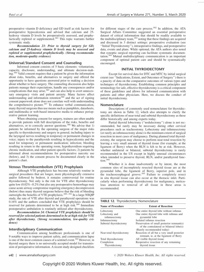
Copyright © 2020 Wolters Kluwer Health, Inc. All rights reserved.
preoperative vitamin D deficiency and GD itself as risk factors forpostoperative hypocalcemia and advised that calcium and 25-hydroxy vitamin D levels be preoperatively assessed, and prophy-lactically repleted or supplemented.383 This topic warrants furthersystematic study.
Recommendation 23: Prior to thyroid surgery for GD,calcium and 25-hydroxy vitamin D levels may be assessed andrepleted or supplemented prophylactically. (Strong recommenda-tion, moderate-quality evidence)
Universal/Standard Consent and CounselingInformed consent consists of 5 basic elements: voluntarism,
capacity, disclosure, understanding, and ultimate decision-mak-ing.384 Valid consent requires that a patient be given the informationabout risks, benefits, and alternatives to surgery and offered theopportunity to have questions answered prior to making a decisionabout whether to have surgery. The counseling discussion also helpspatients manage their expectations, handle any consequences and/orcomplications that may arise,385 and can also help to avert unneces-sary emergency visits and patient anxiety. Many factors affectunderstanding and information retention386 and the act of signingconsent paperwork alone does not correlate well with understandingthe comprehensive picture.387 To enhance verbal communication,many thyroid surgeons thus use means such as pamphlets, datasheets,and multimedia or web-based interactive media to enhance periop-erative patient learning.
When obtaining consent for surgery, trainees are often unableto provide patients with full descriptions of the risks, benefits andalternatives, or to correctly answer questions.388 We advise that 1)patients be informed by the operating surgeon of the major risksspecific to thyroidectomy and surgery in general, including injury tothe laryngeal nerves resulting in temporary or permanent changes invoice; injury to the parathyroids resulting in hypocalcemia and theneed for temporary or permanent medication; infection; bleedingresulting in return to the operating room, hypothyroidism requiringchronic medication, and other patient-specific factors; 2) patients beinformed about recommendations for postoperative narcotic use(below), and 3) the consent process be documented clearly in thepatient’s chart.
Venous Thromboembolism (VTE) ProphylaxisAlthough VTE prophylaxis has become relatively routine in
surgical procedures that are longer, more physiologically extensiveand/or followed by bedrest, it remains controversial for routinethyroidectomy. Not only is the risk for VTE after thyroidectomyquite low (0.02%–0.2%) but also any postoperative hemorrhage maycause acute airway compromise requiring emergency decompression(below) thus many thyroid surgeons believe that the risk of bleedingoutweighs the benefits of VTE prophylaxis.389–391 In a large NSQIPstudy, the rate of VTE after thyroid and parathyroid operations was0.16% and the authors concluded that VTE prophylaxis should bereserved for patients determined to be at high risk.389 Immediatepostoperative ambulation is routinely utilized after thyroidectomy.
Recommendation 24: Chemical VTE prophylaxis should bereserved for selected patients determined to be at high risk for VTEafter thyroidectomy. (Strong recommendation, low-quality evi-dence)
Interdisciplinary CommunicationCommunication among healthcare professionals is one of
9 notable ways to improve patient safety, and communication lapseis one of the most common causes of a sentinel event.392 However, inthyroid surgery there is no universally accepted model for transmis-sion of perioperative information. A recent study designed checklists
for different stages of the care process.392 In addition, the ATASurgical Affairs Committee suggested an essential perioperativedataset of critical information that should be readily available tothe multidisciplinary team,393 noting that these findings are acquiredand discussed in 3 distinct settings: preoperative evaluation (see‘‘Initial Thyroidectomy’’), intraoperative findings, and postoperativedata, events and plans. While optional, the ATA authors also notedthat synoptic surgical reporting can facilitate systematic documen-tation.393 Mutual multidisciplinary communication is an importantcomponent of optimal patient care and should be systematicallyencouraged.
INITIAL THYROIDECTOMY
Except for survival data for DTC and MTC by initial surgicalextent (see ‘‘Indications, Extent, and Outcomes of Surgery’’) there isa paucity of data on the comparative outcomes of various types andtechniques of thyroidectomy. Establishing common principles andterminology for safe, effective thyroidectomy is a critical componentof these guidelines and allows for informed communication withpatients and other physicians. Here, we discuss the conduct ofinitial thyroidectomy.
NomenclatureDescriptions of commonly used nomenclature for thyroidec-
tomy are shown in Table 12, which also attempts to clarify thespecific definitions of near-total and subtotal thyroidectomy as thesediffer historically and among experts today.
Partial thyroid lobectomy (‘‘nodulectomy’’) alone is not rec-ommended, but rarely may be necessary during other cervicalprocedures such as tracheostomy. Lobectomy and isthmusectomy(or rarely an isthmusectomy alone) is the minimum extent of surgicalresection in most cases of malignancy. During planned total thyroid-ectomy, the surgeon may choose to perform near-total resection byleaving a very small amount of thyroid tissue (for example, at theligament of Berry) when the RLN is felt to be at risk. However,whether unilateral or bilateral, subtotal thyroidectomy is rarelyrecommended in the US except in highly unusual circumstanceswhen intended to preserve thyroid, RLN, and/or parathyroid func-tion.394
Whether it is done inadvertently or by intent, the mostcommon sites of incompletely resected thyroid tissue are at thepyramidal lobe, the ligament of Berry, superior pole, and inthe tracheoesophageal groove.395 Failure to completely resectin situ thyroid tissue can also occur at the thoracic inlet. Parti-cularly when performing thyroidectomy for malignancy, meticu-lous attention to removal of all tissue in these areas isrecommended.
TABLE 12. Thyroidectomy Nomenclature
Name of Procedure Extent of Resection
Lobectomy One entire thyroid lobe without isthmusLobectomy and
isthmusectomyOne entire thyroid lobe with isthmus and
pyramidal lobeIsthmusectomy Isolated isthmus resectionSubtotal thyroidectomy Preservation of small posterior remnant(s)
of the contralateral or bilateral lobe(s)(Rarely recommended today)
Near-total thyroidectomy Resection of all but a very small posteriorremnant, ie, at the ligament of Berry
Total thyroidectomy All visible thyroid tissueCompletion
ThyroidectomyReoperative resection of any remaining
thyroid tissue
Patel et al Annals of Surgery � Volume 271, Number 3, March 2020
e42 | www.annalsofsurgery.com � 2020 Wolters Kluwer Health, Inc. All rights reserved.

Copyright © 2020 Wolters Kluwer Health, Inc. All rights reserved.
Surgical PlanningExcept for rare circumstances, thyroidectomy is an elective
procedure that occurs once the dual decisions are made that surgeryis indicated and the patient is medically optimized in all the areasdetailed above. Planning about the extent of initial thyroidectomy iscovered in ‘‘Indications, Extent, and Outcomes of Surgery’’. Imag-ing studies should be reviewed and, when possible, be available toview in the operating room (see ‘‘Imaging’’). Interdisciplinarycommunication is essential, especially when treating patients withhyperthyroidism, large substernal goiters, or airway compromise(see ‘‘Preoperative Care’’). A plan for managing a potentiallydifficult airway should be discussed with anesthesiology before-hand so that appropriate expertise and equipment are available. Asurgical timeout improves team coordination, safety, and out-comes.396,397 If a nerve monitor is being used (see ‘‘Laryngology’’)one should verify the equipment is available and functioning andthat only short acting neuromuscular blockade is used by anesthe-siology for intubation.
While vessel ligation technique, types of suture, choice ofretraction devices, and use of adjuncts (see ‘‘Adjuncts andApproaches’’) may vary between surgeons (below) certain elementsare critical to all safe and effective thyroidectomies.
Positioning, Incision, and ExposureFollowing successful intubation, the patient is generally
placed in a semi-Fowler position with a bump (ie, rolled blanket,inflatable device, padded IV bag, etc) under the shoulders to extendthe neck. Adequate neck access is important for exposure and isoften impacted by body habitus. Over-extending the neck may beassociated with increased postoperative pain.398 It is important toensure that the endotracheal tube is secured and out of the operativefield. Pressure points should be padded, and the skin preparedand draped.
A transverse centered incision is made, preferably in a naturalskin crease for optimal cosmetic results. The use of local anesthetic isat the discretion of the surgeon. Incisional positioning will varyaccording to patient habitus and surgeon preference but is generallyrostral to the sternal notch and caudal to the cricoid cartilage. Incisionlength will be dictated by body habitus and thyroid size. Subpla-tysmal skin flaps are developed by some surgeons superiorly to thethyroid cartilage and inferiorly to the sternal notch.
Next, the median raphe is opened to begin mobilization ofthe sternohyoid and sternothyroid muscles away from the under-lying thyroid. Although the strap muscles are usually retractedlaterally and preserved, improved exposure of the superior pole bytransection of the laryngeal head of the sternothyroid muscle and/or en bloc resection of cancer-invaded muscle, may be requiredon occasion.
Critical Steps of Thyroid DissectionThe order of critical steps will vary based on surgeon prefer-
ence, disease process, and patient anatomy. In general, thyroidec-tomy proceeds through tissue planes that deepen from a ventral todorsal direction, while the overlying strap muscles are dissected in amedial to lateral direction away from the underlying thyroid capsuletoward the carotid sheath.
The middle thyroid vein, which is often identifiable on theventral surface of the thyroid lobe, is ligated and divided, allowingmedial mobility of the lobe to gradually expose the tracheoesopha-geal groove. As the lobe is mobilized and rotated medially, its fine(ancillary) blood supply is isolated and divided. The inferior para-thyroid is usually seen on or near the inferior thyroid capsule andtypically resides anterior and medial to the RLN. It should bedissected away from the thyroid capsule with care being taken to
preserve the parathyroid capsule, lateral attachments and bloodsupply.
As dissection deepens, many experts routinely clear off thetrachea inferior to the isthmus and superiorly, and dissect thepyramidal lobe with care taken to preserve the cricothyroid musclefibers. The prelaryngeal ‘‘Delphian’’ lymph node(s) if present shouldbe removed in cases of suspected or known malignancy and sent forpathologic review either as a frozen section (if this will change theextent of surgery) or with the thyroid specimen.
It is important to expose and resect in continuity the entiresuperior thyroid pole. Further exposure is obtained following ligationof the superior pole vessels, which is performed by inferolateralretraction of the superior pole together with dissection to isolate thevascular pedicle, which often arborizes on the anterior superiorcapsule. Both ligation of the superior pole vessels close to the thyroidcapsule and consideration of the distance/heat of an adjunctiveenergy source will help to protect the external branch of the superiorlaryngeal nerve (EBSLN). The EBSLN may be visually identified atthe inferior edge of the inferior constrictor muscle. If a nerve monitoris being used, the fibers being considered for transection may bestimulated prior to dividing the superior pole vasculature to helpprotect the EBSLN.
Recommendation 25: The superior pole vessels should beligated close to the thyroid capsule to avoid potential EBSLNinjury. (Strong recommendation, insufficient evidence)
A critical step is identification and protection of the RLN. Itscourse ideally should be visualized through the entire operative fieldand kept in view to assess for tension during medial retraction of thethyroid lobe. It usually runs in a superior-medial path passingposterior to the inferior thyroid artery, tubercle of Zuckerkandl (ifpresent) and deep to or through the posterior suspensory ligament(ligament of Berry) before entering the larynx just deep to the lowerborder of the inferior constrictor muscle and posterior to the crico-thyroid articulation. The path of the RLN can be variable, forexample, it can pass anterior to the tubercle, and it can also branchat any point in its course. There are several ways to initially identifythe RLN including at the thoracic inlet, in the tracheoesophagealgroove, and at the laryngeal entry point. Whenever possible, RLNintegrity should be protected with delicate, careful dissection thatavoids tension and traction, taking caution in using a thermal devicealong its course. After the tubercle of Zuckerkandl (if present) ismobilized, the ligament of Berry should be divided as close to thetrachea as possible without putting the RLN at risk. The mostcommon site of RLN injury is at the ligament of Berry.399 RoutineRLN identification improves outcomes400 (see ‘‘Laryngology’’).
Recommendation 26: The RLN should be identified to helppreserve it. (Strong recommendation, low-quality evidence)
The superior parathyroid should be carefully preserved insitu. Compared with the inferior parathyroid, the superior glandlies anatomically posterior to the RLN and is often identifiedsuperior-lateral to the intersection of the inferior thyroid arteryand RLN. However, the relationships between the inferior thyroidartery, RLN, and superior parathyroid can be quite variable.Whenever possible, parathyroid glands that lie within the superfi-cial thyroid capsule should be dissected free and preserved withtheir blood supply intact. If no parathyroid glands are identified,then inspection of the thyroid specimen capsule is reasonable.Whenever feasible it is ideal not to enter the thyroid capsuleduring dissection.
If the blood supply to a parathyroid gland is compromised,parathyroid autotransplantation should be strongly considered. In theuncommon case that a parathyroid is potentially involved with cancer
Annals of Surgery � Volume 271, Number 3, March 2020 Definitive Surgical Management of Thyroid Disease in Adults
� 2020 Wolters Kluwer Health, Inc. All rights reserved. www.annalsofsurgery.com | e43

Copyright © 2020 Wolters Kluwer Health, Inc. All rights reserved.
or level VI lymph nodes, after attention is paid to other viableparathyroid tissues the involved parathyroid may be taken en bloc.One way to perform autotransplantation is to sharply mince thedevascularized parathyroid and place the tissue into a suture- orsurgical clip-marked intramuscular pocket, commonly in the ipsilat-eral sternocleidomastoid (SCM). Alternatively, it can be placed con-tralateral to a known cancer, and/or into strap muscles.6 Prior toautotransplantation it can rest briefly in cold sterile saline and it isreasonable to obtain an intraoperative pathologic evaluation (IOPE) toconfirm parathyroid tissue identity (see ‘‘Perioperative Tissue Diag-nosis’’).
Recommendation 27:a. Dissection should be performed along the thyroid capsule to
help preserve the parathyroid glands. (Strong recommendation,low-quality evidence)
b. If a parathyroid gland cannot be preserved, parathyroid auto-transplantation should be performed. (Strong recommendation,low-quality evidence)
As the RLN is traced, the inferior pole vessels are dissectedand ligated while taking care to preserve the blood supply to theinferior parathyroid gland. Usually division of the inferior vascularpedicle will complete the lobar mobilization, but care should be takento detect, deliver, and resect a substernal thyroid component ifpresent (see ‘‘Goiter’’).
The pyramidal lobe should be identified and resected duringtotal thyroidectomy, and if a thyroid lobectomy and isthmusectomy isbeing performed, the pyramidal lobe should be identified andresected en bloc with the lobe. The isthmus is likewise taken incontinuity and provides the medial margin of resection.
The specimen should be oriented for pathologic examination,typically with a suture placed to mark the superior pole to facilitatecorrelation with cytology and/or imaging. Any area of concern orquery, such as gross extrathyroidal extension, should also be identi-fied for the pathologist. Specimens are submitted for permanentsection unless frozen section analysis is being used to guide theoperative approach (see ‘‘Adjuncts and Approaches’’).
If it is indicated to proceed to the contralateral lobe for totalthyroidectomy, the surgeon can leave the ipsilateral lobe in situ ortransect it at the isthmus. The same steps are then performed tomobilize and resect the contralateral thyroid lobe.
Prior to closure, a dry, bloodless field should be verified.Some surgeons have the anesthesiologist perform positive pressureventilation to help assess for venous bleeding. Hemostatic agentshave unclear benefit (see Adjuncts and Approaches). Drains arerarely used; a meta-analysis of >2000 patients reported that drainuse after thyroidectomy was associated with no differences inreoperation rate, hematoma, or seroma formation- while also asso-ciated with increased pain and a trend for increased wound infec-tion.401 Various suture materials and techniques are used forapproximation of the strap muscles, platysma, and dermis, all withsimilar results. Postoperative antibiotics are not indicated in thyroidsurgery. The operative report should include standard recommendeddetail and features393 (see ‘‘Postoperative Care and Complica-tions’’).
Additional Considerations in Thyroidectomy forCancer
In patients with known DTC or MTC, or with a suspicion ofcancer, there are some additional technical considerations: 1) Pre-operative FNA can cause an inflammatory reaction that mimics localinvasion; when encountered, visible inflammation should be docu-mented in the operative report; 2) Ipsilateral inspection and potentialdissection of abnormal prelaryngeal (Delphian node), pretracheal,
and paratracheal lymph nodes is recommended (see ‘‘Nodal Dissec-tion’’); 3) If surgical clips are used for vessel ligation, titanium is thepreferred metal as other types can affect the quality of future cross-sectional imaging; 4) Dissection and removal of the pyramidal lobeshould be done as completely as possible; 5) Although the precept isdebated, many surgeons prefer to approach the lobe with cancer first,thus preserving the option of limiting the operation to that side in theevent of observed injury to the RLN; 6) For adequate oncologicmanagement, an en-bloc resection is necessary with removal of allgross (visible) tumor; thus if there is suspicion for extrathyroidal orextracapsular extension of tumor into the surrounding strap muscles,these tissues should be taken with the specimen. Gross thyroid cancershould not be left behind with the intent of preserving parathyroidglands or nerves with the expectation that RAI will kill residualdisease, and the risks and benefits of leaving residual tumor topreserve a functioning nerve are considered on a case by case basis;7) During lobectomy, it is important that the resection include theisthmus as well as the entire Tubercle of Zuckerkandl unless doing sojeopardizes the RLN; 8) If cancer involves the RLN, the surgeonshould consider its preoperative functional status to help decidebetween shaving tumor from a functioning nerve versus resecting anonfunctioning nerve (see ‘‘Laryngology’’); 9) In the rare event ofunexpected tracheal involvement, the surgeon may need to seekexperienced assistance i/n obtaining a grossly negative margin,which may involve tangential shaving of tumor, or consider trachealsleeve resection after interval assessment and patient consent.
PERIOPERATIVE TISSUE DIAGNOSIS
Core Needle Biopsy of the Thyroid and CervicalLymph Nodes
FNAB is the first line of investigation for suspicious thyroidnodules and cervical lymph nodes, but has nondiagnostic results in5% to 15% of cases (see ‘‘FNAB Diagnosis’’).212,384,402 Alternativemethods of diagnosis must balance high yield with ease of perfor-mance and safety, as well as cost, expertise, and patient comfort.Core needle biopsy (CNB) examines a tissue sample, rather thandissociated cells and colloid, and is associated with few complica-tions when performed by experienced operators403,404 but has asensitivity of only 68% for thyroid nodules.405 Moreover, due tothe need to examine the lesional capsule on histology, CNB cannotdistinguish FA from FTC. However, when FNAB is nondiagnostic,CNB may yield a higher rate of diagnosis than repeat FNA.405
CNB may also serve as a primary method for diagnosis forsuspected PTL and ATC. Both are aggressive malignancies for whichthyroidectomy is rarely indicated, making a noninvasive diagnosticapproach ideal. Most studies are retrospective single-institutionexperiences with sensitivities in diagnosing PTL of 93% to 95%for CNB versus 45% to 71% for FNAB, and in diagnosing ATC of77% versus 54%, respectively.407,408 Because the 2008 WHO clas-sification bases PTL diagnosis on morphologic features as well asimmunophenotypic, cytogenic, and molecular profile,409 and cellularyield for flow cytometry may be higher by FNAB than by CNB, CNBis often considered second-line for flow cytometry. In handling CNBtissue, if PTL is considered as a possible diagnosis, a portion of thefresh sterile core is teased to release cells into a transport mediumsuch as Roswell Park Memorial Institute media, to be sent for flowcytometry (if not already performed by FNAB). The remainder of thetissue core is placed in formalin. Molecular analysis of CNB speci-mens is also possible (see ‘‘FNAB Diagnosis’’).
Recommendation 28: Core needle biopsy should be rarelyutilized in the initial evaluation of a thyroid nodule. (Strongrecommendation, low–quality evidence)
Patel et al Annals of Surgery � Volume 271, Number 3, March 2020
e44 | www.annalsofsurgery.com � 2020 Wolters Kluwer Health, Inc. All rights reserved.
Copyright © 2020 Wolters Kluwer Health, Inc. All rights reserved.
Incisional Biopsy of the Thyroid and Incisional/Excisional Biopsy of Cervical Lymph Nodes
In selected, unusual circumstances an incisional biopsy ofthyroid tissue may be required to make a definitive diagnosis and isusually reserved for patients with PTL who cannot be appropriatelysubtyped or with ATC who cannot be definitively diagnosed byFNAB or CNB. Excisional or rarely incisional biopsy of a cervicalLN should be reserved for when a thyroid-related diagnosis cannot beobtained otherwise. Incisional biopsy should only be considered ifthe LN is large and additional morbidity would be incurred byresecting it entirely.
Intraoperative Pathologic Evaluation of the ThyroidIOPE of the thyroid is typically performed by frozen section
analysis (FSA) and/or cytologic touch or scrape analysis. It may beutilized in thyroidectomy to assure adequacy of tissue collection inthe setting of an unknown diagnosis (above) as well as for clarifica-tion of tissue type and margin status (below). FSA should not be usedwhen it could be deleterious, such as in the setting of a small lesionthat would necessitate being frozen in its entirety with tissue losspreventing a diagnosis on final pathology.
IOPE has been often studied for its ability to predict finalhistopathology. Unfortunately, this ability is reportedly quite low,moreover (as with CNB) FSA is not informative for follicularneoplasms410,411 since the histologic characteristics of FTC andHTC require detailed analysis of the tumor capsule and vasculaturefor invasion, which is not practical or feasible in an intraoperativesetting.412 Given that only the assessment of nuclear features isrequired to diagnose PTC, IOPE may be useful in cases where PTC issuspected and its diagnosis would change the extent of operation.410
When PTC is diagnosed preoperatively by FNAB, IOPE is notrecommended or useful.
Recently, IOPE has been suggested for use in determiningextrathyroidal extension of tumor, which may lead to a change inoperative plan,413 but this single-institution observation needs furtherinvestigation. Although negative macroscopic margins are usuallythe goal of TC resection, IOPE may also be utilized for evaluation ofmargins to assist in complete tumor extirpation, such as at interfacewith the trachea. However, data to evaluate whether IOPE accuratelypredicts margin status on final pathology or changes the extent ofoperation are lacking.
Recommendation 29: Thyroid IOPE should only be utilizedin settings in which the information it provides has a high likeli-hood of altering the operative procedure. (Strong recommendation,low–quality evidence)
Intraoperative Pathologic Evaluation of CLN andParathyroid Tissue
IOPE of a cervical LN can confirm its identity versus para-thyroid tissue or extrathyroidal thyroid tissue, can help determinewhether it harbors malignancy, and in both situations, may changethe extent of operation. In a meta-analysis examining the use of IOPEto identify metastatic adenopathy during TC surgery,414 among 242LN without malignancy on IOPE, a positive result was diagnosed onfinal histology in 12%; the majority of the false negative cases werefrom studies utilizing LN immunohistochemical analysis (a rarepractice) and/or represented metastasis. Several issues of IOPEpathologic expertise include 1) psammoma bodies without epithe-lium in an LN can represent metastatis thus should not necessarily beconsidered as negative; 2) with significant thyroiditis at the time ofLN IOPE, there is potential for a false-positive read with thespecimen sent as an LN in actuality being a sequestered, or parasitic,thyroid nodule which occurs when nodules of hyperplastic thyroid
obtain an independent blood supply and become separate from themain gland; 3) IOPE artifact can also cause intranuclear inclusionssimilar to those of PTC when a true LN should contain lymphoidfollicles, a capsule, and a subcapsular sinus.
During thyroidectomy, the distinction of parathyroid tissuefrom LNs, thyroid nodules, and fatty tissue may be challenging. In alarge retrospective review comparing 1579 cervical FSA to finalpathologic diagnosis, the overall accuracy rate of distinguishingparathyroid from nonparathyroid tissue was 99.2%.415 In situ orex vivo PTH aspiration analysis can also be used to identifyparathyroid tissue.416
Recommendation 30: IOPE has value in confirming identi-fication of parathyroid tissue. (Strong recommendation, moderate-quality evidence)
Recommendation 31: IOPE has value in identification ofCLN metastases when the information may alter extent of surgery.(Strong recommendation, moderate–quality evidence)
Final Histopathologic DiagnosisInterobserver variation in interpretation of final histopatho-
logic diagnosis is not uncommon. In evaluation of 776 thyroidnodules, expert interobserver concordance of benign versus malig-nant thyroid disease was 90.3% (95% CI, 88.0%–92.3%),26 concor-dance between the local histopathologist and final expert consensuswas 90.7% (95% CI, 88.5%–92.7%), and the most disagreementseen was in diagnosis of FTC and HTC (61%). The determination ofmalignancy type is also subject to observer variance; that is, minimalextrathyroidal extension (ETE) and extranodal extension are PTCfeatures that predict disease biology but in a recent study, theinterobserver concordance among expert pathologists was poor forETE (kappa coefficient of 0.14), and fair for extranodal extension(kappa coefficient of 0.35) suggesting the need for standardizedcriteria.417,418 In single-institution studies a second histopathologicopinion commonly alters clinical treatment. When 66 specimenswere reread at a tertiary center with a thyroid multidisciplinary focus,disagreement led to a change in the assessed prognosis for 10% ofpatients, and strong disagreement led to change of therapy for 8%.419
To summarize, second reads of thyroid histopathology should beperformed when the initial pathologist is uncertain of the diagnosisand at the discretion of the clinician or patient, and also should bestrongly considered for difficult diagnoses, such as FTC and NIFTP.
The surgeon and pathologist should establish mutual expec-tations of how tissue specimens are to be optimally received by thepathologist, the typical length of time for a diagnosis to be rendered,and the communication plan for patients in instances that requirefurther time for evaluation.
Benign Lesions of the ThyroidFollicular-patterned nodular hyperplasia can exhibit complete
or partial encapsulation and often contains a combination of macro-and microfollicles. The nodules may display a variety of degenera-tive changes including fibrosis and hemorrhage. Consistent histo-pathologic criteria distinguishing hyperplastic nodules from FA arelacking. Classically, FA is a single encapsulated lesion in an other-wise normal gland that lacks any vascular or capsular invasion andhas a distinct growth pattern (microfollular, macrofollicular, ortrabecular).420
Differentiated Thyroid CarcinomaDTC arises from the thyroid follicular epithelial cell. Classic
PTC is diagnosed by its nuclear features which include enlargement,oval shape, elongation, and overlapping.421 The nuclei usuallydisplay clearing and irregular contours, including grooves andnuclear pseudoinclusions. A papillary architecture is often present
Annals of Surgery � Volume 271, Number 3, March 2020 Definitive Surgical Management of Thyroid Disease in Adults
� 2020 Wolters Kluwer Health, Inc. All rights reserved. www.annalsofsurgery.com | e45
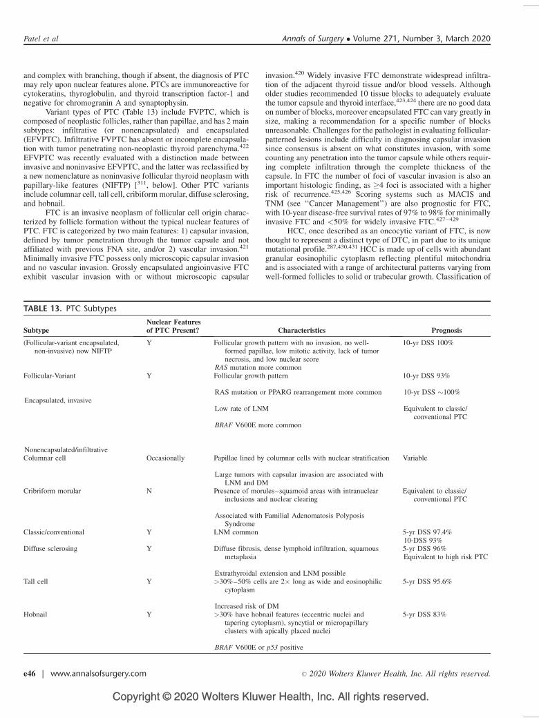
Copyright © 2020 Wolters Kluwer Health, Inc. All rights reserved.
and complex with branching, though if absent, the diagnosis of PTCmay rely upon nuclear features alone. PTCs are immunoreactive forcytokeratins, thyroglobulin, and thyroid transcription factor-1 andnegative for chromogranin A and synaptophysin.
Variant types of PTC (Table 13) include FVPTC, which iscomposed of neoplastic follicles, rather than papillae, and has 2 mainsubtypes: infiltrative (or nonencapsulated) and encapsulated(EFVPTC). Infiltrative FVPTC has absent or incomplete encapsula-tion with tumor penetrating non-neoplastic thyroid parenchyma.422
EFVPTC was recently evaluated with a distinction made betweeninvasive and noninvasive EFVPTC, and the latter was reclassified bya new nomenclature as noninvasive follicular thyroid neoplasm withpapillary-like features (NIFTP) [311, below]. Other PTC variantsinclude columnar cell, tall cell, cribiform morular, diffuse sclerosing,and hobnail.
FTC is an invasive neoplasm of follicular cell origin charac-terized by follicle formation without the typical nuclear features ofPTC. FTC is categorized by two main features: 1) capsular invasion,defined by tumor penetration through the tumor capsule and notaffiliated with previous FNA site, and/or 2) vascular invasion.421
Minimally invasive FTC possess only microscopic capsular invasionand no vascular invasion. Grossly encapsulated angioinvasive FTCexhibit vascular invasion with or without microscopic capsular
invasion.420 Widely invasive FTC demonstrate widespread infiltra-tion of the adjacent thyroid tissue and/or blood vessels. Althougholder studies recommended 10 tissue blocks to adequately evaluatethe tumor capsule and thyroid interface,423,424 there are no good dataon number of blocks, moreover encapsulated FTC can vary greatly insize, making a recommendation for a specific number of blocksunreasonable. Challenges for the pathologist in evaluating follicular-patterned lesions include difficulty in diagnosing capsular invasionsince consensus is absent on what constitutes invasion, with somecounting any penetration into the tumor capsule while others requir-ing complete infiltration through the complete thickness of thecapsule. In FTC the number of foci of vascular invasion is also animportant histologic finding, as �4 foci is associated with a higherrisk of recurrence.425,426 Scoring systems such as MACIS andTNM (see ‘‘Cancer Management’’) are also prognostic for FTC,with 10-year disease-free survival rates of 97% to 98% for minimallyinvasive FTC and <50% for widely invasive FTC.427–429
HCC, once described as an oncocytic variant of FTC, is nowthought to represent a distinct type of DTC, in part due to its uniquemutational profile.287,430,431 HCC is made up of cells with abundantgranular eosinophilic cytoplasm reflecting plentiful mitochondriaand is associated with a range of architectural patterns varying fromwell-formed follicles to solid or trabecular growth. Classification of
TABLE 13. PTC Subtypes
SubtypeNuclear Featuresof PTC Present? Characteristics Prognosis
(Follicular-variant encapsulated,non-invasive) now NIFTP
Y Follicular growth pattern with no invasion, no well-formed papillae, low mitotic activity, lack of tumornecrosis, and low nuclear score
RAS mutation more common
10-yr DSS 100%
Follicular-Variant
Encapsulated, invasive
Nonencapsulated/infiltrative
Y Follicular growth pattern
RAS mutation or PPARG rearrangement more common
Low rate of LNM
BRAF V600E more common
10-yr DSS 93%
10-yr DSS �100%
Equivalent to classic/conventional PTC
Columnar cell Occasionally Papillae lined by columnar cells with nuclear stratification
Large tumors with capsular invasion are associated withLNM and DM
Variable
Cribriform morular N Presence of morules–squamoid areas with intranuclearinclusions and nuclear clearing
Associated with Familial Adenomatosis PolyposisSyndrome
Equivalent to classic/conventional PTC
Classic/conventional Y LNM common 5-yr DSS 97.4%10-DSS 93%
Diffuse sclerosing Y Diffuse fibrosis, dense lymphoid infiltration, squamousmetaplasia
Extrathyroidal extension and LNM possible
5-yr DSS 96%Equivalent to high risk PTC
Tall cell Y >30%–50% cells are 2� long as wide and eosinophiliccytoplasm
Increased risk of DM
5-yr DSS 95.6%
Hobnail Y >30% have hobnail features (eccentric nuclei andtapering cytoplasm), syncytial or micropapillaryclusters with apically placed nuclei
BRAF V600E or p53 positive
5-yr DSS 83%
Patel et al Annals of Surgery � Volume 271, Number 3, March 2020
e46 | www.annalsofsurgery.com � 2020 Wolters Kluwer Health, Inc. All rights reserved.
Copyright © 2020 Wolters Kluwer Health, Inc. All rights reserved.
invasion for HCC is similar to that for FTC as described above.HCC may have higher risk for local recurrence and be less iodine-avid.432–435 10-year survival for HCC is lower than FTC (76% vs85%).436
The pathology report for a patient with DTC should includethe histologic diagnosis, including subtype, location, and the char-acteristics required for AJCC TNM staging. For the 8th edition AJCCguidelines, which no longer utilize microscopic ETE, requisitehistopathologic characteristics for staging of DTC and PD/ATCinclude the size of the dominant nodule as well as presence ofLNM.437 Additional histopathologic features are associated withdisease biology and should be considered for reporting, includingtumor multifocality, microscopic margin status, angioinvasion, lym-phatic invasion, perineural invasion, and microscopic ETE.438 Inreporting histopathologic features, the 8th edition AJCC guidelinessuggest inclusion in the pathology report of the number and locationof involved LN, the number of nodes sampled, the size of largestinvolved LN, the size of metastatic foci within an involvedLN, andthe presence of extranodal extension.437 The use of synoptic pathol-ogy reporting minimizes variability between institutions and ensuresinclusion of information required for patient care.439 The College ofAmerican Pathologists (CAP) provides a widely utilized checklist-format synoptic for thyroid cancer.440
Recommendation 32: A standardized synoptic pathologyreport is recommended when reporting thyroid neoplasms. (Strongrecommendation, low–quality evidence)
Neoplasms of Uncertain Malignant PotentialSome encapsulated follicular-patterned neoplasms cause diag-
nostic difficulties due to ambiguity as to whether the nuclear changesare adequate to diagnose PTC, or due to uncertainty about thepresence or absence of capsular or vascular invasion.431,441 In2017, the World Health Organization acknowledged these 2 uncer-tainties and utilized the terms ‘‘well-differentiated tumor of uncertainmalignant potential’’ (WDT-UMP) and ‘‘follicular tumor of uncer-tain malignant potential’’ (FT-UMP); FT-UMP includes cases inwhich the tumor does not have nuclear features of PTC but thecapsular or vascular invasion is equivocal, and WDT-UMP describesa well-circumscribed tumor composed of follicles with partiallydeveloped PTC-type nuclear changes and questionable capsular orvascular invasion. Evaluation of the entire tumor capsule or itsmargin circumference is required to make the diagnosis of anyborderline thyroid tumor.
NIFTP is the newly-minted term for those encapsulated orcircumscribed tumors for which capsular and vascular invasion hasbeen excluded. Other criteria for NIFTP diagnosis include: encap-sulation or clear demarcation from adjacent thyroid tissue, a follicu-lar growth pattern, a nuclear score of 2 to 3, no tumor necrosis, and nohigh mitotic activity.311 In more recent studies, any papillary growthor presence of psammoma bodies excludes a tumor from beingdescribed as NIFTP and confers a diagnosis of encapsulatedPTC.442 Because capsule invasion is one of the defining featuresdifferentiating EFVPTC and NIFTP, thorough evaluation of thetumor capsule interface is essential before making or excludingthe diagnosis of NIFTP. The presence of BRAF V600E mutationhas also been recommended as an exclusion criterion for thisdiagnosis.443
Poorly Differentiated Thyroid CarcinomaPDTC occupies a morphologically intermediate position
between DTC and ATC. Diagnostic consensus criteria for PDTC,known as the Turin criteria, were created by an international group ofthyroid pathologists and include 1) presence of a solid/trabecular/insular pattern of growth, 2) lack of conventional nuclear features of
PTC, and 3) presence of �1 of the following features: convolutednuclei; mitotic activity �3 � 10HPF; and tumor necrosis.51
Anaplastic Thyroid CarcinomaATC is a widely invasive tumor composed of a mixture of 3
main growth patterns: spindle cell, pleomorphic cell, and squamoid.Rarely (10%) ATC is confined to a thyroid lobe or gland or isrepresented by microscopic anaplastic foci.444,445 Most cases exhibitnumerous mitotic figures and atypical mitoses446 as well as extensivecoagulative necrosis.421 Immunohistochemical markers of thyroiddifferentiation, such as thyroglobulin, are usually not present. tran-scription factor-1 can be identified in up to 30% of cases. Unlikelymphoma, ATC often is positive for pankeratin (AE1/AE3).54
Medullary Thyroid CarcinomaMTC derives from C cells and is a neuroendocrine tumor.
Characteristic histologic features are sheets, nests, or trabeculae ofpolygonal, round or spindle cells that are separated by fibrovascularstroma. Tumor cells often possess round or oval regular nuclei withnonprominent nucleoi and scant mitotic figures. The most importantimmunohistochemical markers used to identify MTC are calcitoninand CEA. More general neuroendocrine markers such as chromog-ranin A and synaptophysin may also be helpful for diagnosis.
In hereditary MTC, C-cell hyperplasia (CCH) occurs prior tocancer development and most likely represents clonal proliferation ofmultiple transformed progenitor cells. The diagnostic criteria forCCH are controversial but there is general consensus on the completesurrounding of follicles by C cells and distribution of C cells beyondthe normal anatomic location.447 Also challenging is the distinctionbetween CCH and medullary microcarcinoma, which by definition isan MTC tumor of <1 cm and showing invasion. CCH may bephysiologic or neoplastic, the former requiring IHC and quantitativeanalysis for diagnosis and the later usually diagnosed by H&Estains.448 For the 8th edition AJCC guidelines, requisite histopatho-logic characteristics for MTC staging include dominant nodule sizeand presence of lymph node metastasis.437 Use of the College ofAmerican Pathologists (CAP) synoptic for thyroid cancer is recom-mended for reporting MTC and includes additional histopathologicalfeatures such as multifocality, microscopic margin status, angioin-vasion, lymphatic invasion, perineural invasion, microscopic extra-thyroidal extension, and CCH.438 Synoptic nodal information forMTC and DTC are the same (above).
Thyroid ParagangliomaParagangliomas in the region of the thyroid gland are rare
tumors arising from the inferior laryngeal (parasympathetic) para-ganglia. They can be mistaken for follicular or C-cell-derivedmalignancies, may present as intrathyroidal masses, are not func-tional in terms of catecholamine secretion, and can be distinguishedfrom other tumors by positive staining for chromogranin A, synap-tophysin, neuron-specific enolase, and/or S-100.449
NODAL DISSECTION
Lymph node metastasis (LNM) is often present at TC diag-nosis and can occur subclinically in up to 75% and 80% of MTC andPTC patients, respectively.450–452 Therefore, a preoperative diagno-sis of TC by cytology or molecular testing should prompt imaging toassess for LNM.453
Neck US that includes evaluation of central and lateral LNbasins is recommended in patients with cytologic evidence of TC(see ‘‘Imaging’’) and may be performed in patients with indetermi-nate cytology to help identify nonpalpable lymphadenopathy.3 Inselected patients, CT of the neck and upper chest can provideadditional data especially in extensive lateral neck disease and for
Annals of Surgery � Volume 271, Number 3, March 2020 Definitive Surgical Management of Thyroid Disease in Adults
� 2020 Wolters Kluwer Health, Inc. All rights reserved. www.annalsofsurgery.com | e47
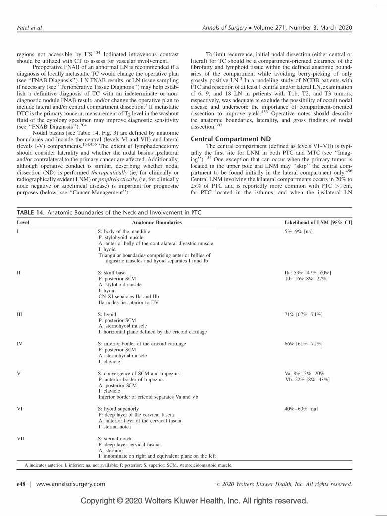
Copyright © 2020 Wolters Kluwer Health, Inc. All rights reserved.
regions not accessible by US.454 Iodinated intravenous contrastshould be utilized with CT to assess for vascular involvement.
Preoperative FNAB of an abnormal LN is recommended if adiagnosis of locally metastatic TC would change the operative plan(see ‘‘FNAB Diagnosis’’). LN FNAB results, or LN tissue samplingif necessary (see ‘‘Perioperative Tissue Diagnosis’’) may help estab-lish a definitive diagnosis of TC with an indeterminate or non-diagnostic nodule FNAB result, and/or change the operative plan toinclude lateral and/or central compartment dissection.3 If metastaticDTC is the primary concern, measurement of Tg level in the washoutfluid of the cytology specimen may improve diagnostic sensitivity(see ‘‘FNAB Diagnosis’’).204
Nodal basins (see Table 14, Fig. 3) are defined by anatomicboundaries and include the central (levels VI and VII) and lateral(levels I-V) compartments.154,455 The extent of lymphadenectomyshould consider laterality and whether the nodal basins ipsilateraland/or contralateral to the primary cancer are affected. Additionally,although operative conduct is similar, describing whether nodaldissection (ND) is performed therapeutically (ie, for clinically orradiographically evident LNM) or prophylactically, (ie, for clinicallynode negative or subclinical disease) is important for prognosticpurposes (below; see ‘‘Cancer Management’’).
To limit recurrence, initial nodal dissection (either central orlateral) for TC should be a compartment-oriented clearance of thefibrofatty and lymphoid tissue within the defined anatomic bound-aries of the compartment while avoiding berry-picking of onlygrossly positive LN.3 In a modeling study of NCDB patients withPTC and resection of at least 1 central and/or lateral LN, examinationof 6, 9, and 18 LN in patients with T1b, T2, and T3 tumors,respectively, was adequate to exclude the possibility of occult nodaldisease and underscore the importance of compartment-orienteddissection to improve yield.453 Operative notes should describethe anatomic boundaries, laterality, and gross findings of nodaldissection.393
Central Compartment NDThe central compartment (defined as levels VI–VII) is typi-
cally the first site for LNM in both PTC and MTC (see ‘‘Imag-ing’’).154 One exception that can occur when the primary tumor islocated in the upper pole and LNM may ‘‘skip’’ the central com-partment to be found initially in the lateral compartment only.456
Central LNM involving the bilateral compartments occurs in 20% to25% of PTC and is reportedly more common with PTC >1 cm,for PTC located in the isthmus, and when the ipsilateral LN
TABLE 14. Anatomic Boundaries of the Neck and Involvement in PTC
Level Anatomic Boundaries Likelihood of LNM [95% CI]
I S: body of the mandibleP: stylohyoid muscleA: anterior belly of the contralateral digastric muscleI: hyoidTriangular boundaries comprising anterior bellies of
digastric muscles and hyoid separates Ia and Ib
5%–9% [na]
II S: skull baseP: posterior SCMA: stylohoid muscleI: hyoidCN XI separates IIa and IIbIIa nodes lie anterior to IJV
IIa: 53% [47%–60%]IIb: 16%[8%–27%]
III S: hyoidP: posterior SCMA: sternohyoid muscleI: horizontal plane defined by the cricoid cartilage
71% [67%–74%]
IV S: inferior border of the cricoid cartilageP: posterior SCMA: sternohyoid muscleI: clavicle
66% [61%–71%]
V S: convergence of SCM and trapeziusP: anterior border of trapeziusA: posterior SCMI: clavicleInferior border of cricoid separates Va and Vb
Va: 8% [3%–20%]Vb: 22% [8%–48%]
VI S: hyoid superiorlyP: deep layer of the cervical fasciaA: anterior layer of the cervical fasciaI: sternal notch
40%–60% [na]
VII S: sternal notchP: deep layer cervical fasciaA: sternumI: innominate on right and equivalent plane on the left
A indicates anterior; I, inferior; na, not available; P, posterior; S, superior; SCM, sternocleidomastoid muscle.
Patel et al Annals of Surgery � Volume 271, Number 3, March 2020
e48 | www.annalsofsurgery.com � 2020 Wolters Kluwer Health, Inc. All rights reserved.
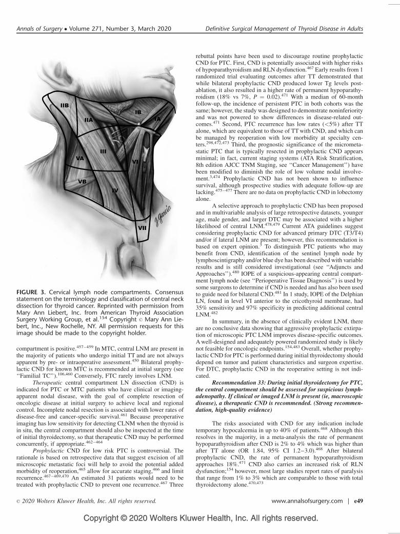
Copyright © 2020 Wolters Kluwer Health, Inc. All rights reserved.
compartment is positive.457–459 In MTC, central LNM are present inthe majority of patients who undergo initial TT and are not alwaysapparent by pre- or intraoperative assessment.450 Bilateral prophy-lactic CND for known MTC is recommended at initial surgery (see‘‘Familial TC’’).106,460 Conversely, FTC rarely involves LNM.
Therapeutic central compartment LN dissection (CND) isindicated for PTC or MTC patients who have clinical or imaging-apparent nodal disease, with the goal of complete resection ofoncologic disease at initial surgery to achieve local and regionalcontrol. Incomplete nodal resection is associated with lower rates ofdisease-free and cancer-specific survival.461 Because preoperativeimaging has low sensitivity for detecting CLNM when the thyroid isin situ, the central compartment should also be inspected at the timeof initial thyroidectomy, so that therapeutic CND may be performedconcurrently, if appropriate.462–464
Prophylactic CND for low risk PTC is controversial. Therationale is based on retrospective data that suggest excision of allmicroscopic metastatic foci will help to avoid the potential addedmorbidity of reoperation,465 allow for accurate staging,466 and limitrecurrence.467–469,470 An estimated 31 patients would need to betreated with prophylactic CND to prevent one recurrence.467 Three
rebuttal points have been used to discourage routine prophylacticCND for PTC. First, CND is potentially associated with higher risksof hypoparathyroidism and RLN dysfunction.467 Early results from 1randomized trial evaluating outcomes after TT demonstrated thatwhile bilateral prophylactic CND produced lower Tg levels post-ablation, it also resulted in a higher rate of permanent hypoparathy-roidism (18% vs 7%, P ¼ 0.02).471 With a median of 60-monthfollow-up, the incidence of persistent PTC in both cohorts was thesame; however, the study was designed to demonstrate noninferiorityand was not powered to show differences in disease-related out-comes.471 Second, PTC recurrence has low rates (<5%) after TTalone, which are equivalent to those of TT with CND, and which canbe managed by reoperation with low morbidity at specialty cen-ters.298,472,473 Third, the prognostic significance of the micrometa-static PTC that is typically resected in prophylactic CND appearsminimal; in fact, current staging systems (ATA Risk Stratification,8th edition AJCC TNM Staging, see ‘‘Cancer Management’’) havebeen modified to diminish the role of low volume nodal involve-ment.3,474 Prophylactic CND has not been shown to influencesurvival, although prospective studies with adequate follow-up arelacking.475–477 There are no data on prophylactic CND in lobectomyalone.
A selective approach to prophylactic CND has been proposedand in multivariable analysis of large retrospective datasets, youngerage, male gender, and larger DTC may be associated with a higherlikelihood of central LNM.478,479 Current ATA guidelines suggestconsidering prophylactic CND for advanced primary DTC (T3/T4)and/or if lateral LNM are present; however, this recommendation isbased on expert opinion.3 To distinguish PTC patients who maybenefit from CND, identification of the sentinel lymph node bylymphoscintigraphy and/or blue dye has been described with variableresults and is still considered investigational (see ‘‘Adjuncts andApproaches’’).480 IOPE of a suspicious-appearing central compart-ment lymph node (see ‘‘Perioperative Tissue Diagnosis’’) is used bysome surgeons to determine if CND is needed and has also been usedto guide need for bilateral CND.481 In 1 study, IOPE of the DelphianLN, found in level VI anterior to the cricothyroid membrane, had35% sensitivity and 97% specificity in predicting additional centralLNM.482
In summary, in the absence of clinically evident LNM, thereare no conclusive data showing that aggressive prophylactic extirpa-tion of microscopic PTC LNM improves disease-specific outcomes.A well-designed and adequately powered randomized study is likelynot feasible for oncologic endpoints.154,483 Overall, whether prophy-lactic CND for PTC is performed during initial thyroidectomy shoulddepend on tumor and patient characteristics and surgeon expertise.For DTC, prophylactic CND in the reoperative setting is not indi-cated.
Recommendation 33: During initial thyroidectomy for PTC,the central compartment should be assessed for suspicious lymph-adenopathy. If clinical or imaged LNM is present (ie, macroscopicdisease), a therapeutic CND is recommended. (Strong recommen-dation, high-quality evidence)
The risks associated with CND for any indication includetemporary hypocalcemia in up to 40% of patients.468 Although thisresolves in the majority, in a meta-analysis the rate of permanenthypoparathyroidism after CND is 2% to 4% which was higher thanafter TT alone (OR 1.84, 95% CI 1.2–3.0).468 After bilateralprophylactic CND, the rate of permanent hypoparathyroidismapproaches 18%.471 CND also carries an increased risk of RLNdysfunction;154 however, most large studies report rates of paralysisthat range from 1% to 3% which are comparable to those with totalthyroidectomy alone.470,473
FIGURE 3. Cervical lymph node compartments. Consensusstatement on the terminology and classification of central neckdissection for thyroid cancer. Reprinted with permission fromMary Ann Liebert, Inc. from American Thyroid AssociationSurgery Working Group, et al.154 Copyright � Mary Ann Lie-bert, Inc., New Rochelle, NY. All permission requests for thisimage should be made to the copyright holder.
Annals of Surgery � Volume 271, Number 3, March 2020 Definitive Surgical Management of Thyroid Disease in Adults
� 2020 Wolters Kluwer Health, Inc. All rights reserved. www.annalsofsurgery.com | e49
Copyright © 2020 Wolters Kluwer Health, Inc. All rights reserved.
Intraoperatively, one of the first steps of CND should beidentification of the RLN and delineation of its course. LN andCLNM are often found posterior to the RLN especially on the right,and this area should be included. During mobilization of the upperthyroid pole, particular effort should be made to preserve the superiorparathyroids on their vascular pedicles. Preservation of the inferiorparathyroid glands is a priority but because of concomitant LNdisease, may not be possible; devascularized or resected parathyroidtissue should be immediately autotransplanted (see ‘‘Initial Thyroid-ectomy’’). IOPE can help confirm parathyroid tissue prior to auto-transplantation (see ‘‘Perioperative Tissue Diagnosis’’).
Lateral Compartment NDThe lateral compartment is comprised of 5 nodal levels; the
submandibular and submental (level I), the upper (II), middle (III),and lower (IV) jugular, and the posterior triangle (V) (Table 14).455 Aradical neck dissection includes systematic dissection of all LN fromlevels I to V, together with resection of the internal jugular vein (IJV),the spinal accessory nerve (CN XI), and the SCM. The procedure ismorbid, disfiguring, and rarely performed for thyroid cancer today. Amodified radical neck dissection preserves 1 or more of the follow-ing: IJV, CN XI, SCM.
A compartment-oriented selective neck dissection (SND) isthe term used when fewer than all 5 nodal levels are removed and isthe most commonly utilized type of therapeutic LN resection forTC.455 At initial SND for PTC, levels IIa, III, IV, and Vb are typicallyincluded (Table 14)3 and their comprehensive clearance is associatedwith a lower risk of recurrence.455,484–486 To reduce the risk of injuryto CN XI and because of the low likelihood of LNM involvement,level IIb is only dissected if there is radiographic evidence of LNM orlevel IIa is positive, and similarly level Va is only dissected when ithas clinically or radiographically apparent LNM.455 TC LNM inlevel I is rare (<10%) and recurrence is also rare (<1%) if notdissected at initial SND.487–489 Prophylactic lateral ND has not beenshown to improve PTC survival or recurrence rates; thus, SND istypically performed only for clinically evident disease.490–492
At the time of MTC diagnosis with palpable primary tumor,ipsilateral lateral LNM are subclinically present in up to 75% ofpatients, and calcitonin levels can help direct the need for lateralND.450 Ipsilateral and contralateral lateral LNM are signaled by basalcalcitonin levels >20 and >200 pg/mL, respectively.106 Some there-fore advocate for prophylactic selective dissection of the ipsilateralneck for MTC although this remains controversial.56,493 The suspi-cion for lateral nodal disease in MTC can be based on US findingsand/or serum calcitonin levels (see ‘‘Familial TC’’).
Recommendation 34:a. A compartment-oriented therapeutic lateral ND is recom-
mended for lateral LNM. (Strong recommendation, high-qual-ity evidence)
b. Prophylactic lateral ND is not indicated for PTC. (Strongrecommendation, high-quality evidence)
Lateral ND can be associated with significant morbidity. CNXI palsy occurs in 6% to 20% of cases, resulting in shoulderweakness and inability to raise the arm requiring physiother-apy;455,494 the dysfunction will resolve in most patients but canbe debilitating. Other neurologic complications include injuries tothe marginal mandibular branch of the facial nerve (which can occurduring dissection of levels I and II resulting in an asymmetricalsmile), injury to the hypoglossal nerve, and lateral neck numbnessfrom injury to the cervical sensory branches in level V.495 Lesscommon (<1%) are injuries to the phrenic nerve, brachial plexus,cervical sympathetic chain (Horner syndrome) and/or hypoglossalnerve (CNXII).455 Chyle leaks occur in up to 10% of patients who
undergo lateral ND, more commonly on the left side where thethoracic duct joins the left IJV at its junction with the left subclavianvein is located but also with right-sided dissection.494,496,497 Intra-operatively, large lymphatics, which can be present especially withbulky lateral neck disease, should be ligated, and raising the intratho-racic pressure with Valsalva maneuver and head-down positioning canhelp to identify chyle leak. Drains may help diagnose and allowconservative management of chyle leak, but are not mandatory afterneck dissection.498,499 Management of low volume chyle leak canoften be done in the outpatient setting and includes adequate drainage,diet modification (a low fat, medium-chain triglyceride diet, occasion-ally TPN), and potentially octreotide.500 Persistent high output leaks(>500 mL/d) may require surgical management and if uncontrolled,even embolization of the thoracic duct with coils or tissue adhesive, orthoracoscopic ligation of the thoracic duct between the aorta andazygous vein hiatus.497 Chylothorax is a very rare occurrence (<20cases reported) but may have significant consequences.501
Prognostic Implications of LNMPatients with clinical N1 DTC (see ‘‘Cancer Management’’) or
with >5 pathologic LNM are considered ATA intermediate risk witha recurrence rate of 15% to 20%.3 In multivariable analysis, the riskof PTC recurrence is higher with age �45 years, >10 LNM, LNextranodal extension, or if the proportion of resected LNM (definedby the ratio of positive to total resected LN) exceeds 17%.502 In otherstudies of PTC, extranodal extension is associated with a higher riskof both recurrence and disease-specific mortality.503,504 The risk ofrecurrence is also higher in patients with large nodal metastasis(>3 cm).483,505 The prognostic significance of LNM in youngerpatients (age<45 yr) is controversial. A population-level study usingthe SEER database demonstrated that LNM did not alter overallsurvival in young patients.506 However, a more recent study utilizingdata from both SEER and NCDB demonstrated a modest decrease inoverall survival associated with LNM in patients<45 years old.507 Inaddition, the survival decrease was associated with a rising number ofLNM up to 6.507 In MTC, the number of LNM is also associated withdecreased overall survival.508
In DTC, reoperation for recurrent nodal disease results inbiochemical remission in only 30% to 50% of patients. In addition, ifthe compartment has been previously dissected, reoperation for LNMis associated with a higher risk of complications509,510 and although acompartment-oriented dissection is recommended at reoperation,this may be limited by scarring and the extent of initial dissection.3
In 1 study of PTC patients who had at least 1 abnormal LN on USsurveillance following initial treatment, at median follow-up of 3.5years, LN growth occurred in only 10% to 20%, suggesting thatsonographically concerning LN may not require immediate resec-tion.511
In poor operative candidates, especially those with multipleprior neck surgeries, percutaneous ethanol injection of PTC LNM hasbeen reported in small single-institution series and can lead tobiochemical remission and resolution of structural disease.512 Thereis a risk of associated RLN or other nerve (phrenic, brachial plexus)injury although the risk appears to be low (<1%).513 Recurrencesoccur in �20% to 25% as ethanol injection only treats the targetedLN.512,513 The management of recurrent or persistent nodal diseasein TC patients can be complex and should occur within a multidis-ciplinary framework.514
CONCURRENT PARATHYROIDECTOMY
Epidemiology and EvaluationConcomitant primary hyperparathyroidism (pHPT) occurs in
0.2% to 5% of patients with thyroid disease, thus various groups have
Patel et al Annals of Surgery � Volume 271, Number 3, March 2020
e50 | www.annalsofsurgery.com � 2020 Wolters Kluwer Health, Inc. All rights reserved.
Copyright © 2020 Wolters Kluwer Health, Inc. All rights reserved.
recommended routine assessment of calcium and/or PTH levels priorto planned thyroidectomy.99,100,515,516 In a retrospective study of1049 patients undergoing thyroidectomy, 5% had concomitant pHPT,and another 0.9% had normal preoperative calcium and PTH levelsbut were found to have an enlarged parathyroid at surgery.100 Aretrospective study of 13,387 patients evaluated in an endocrinologyclinic found hypercalcemia due to pHPT in 0.2% of patients with athyroid disorder; pHPT was more likely with thyroid disease [0.9%with vs 0.29% without], especially with euthyroid goiter (P < 0.05)or thyroid cancer (P ¼ 0.01).515
When pHPT is present, the cost-effectiveness of surgicaltreatment is well established.517–519 The potential benefits for pro-ceeding with initial parathyroidectomy at the same time as initialthyroidectomy are numerous.6 While published data support routinepreoperative assessment, no data exist regarding the cost-effective-ness of routine screening for pHPT. Many patients undergoingthyroid surgery will likely have had at least a recent metabolicpanel. When present, hypercalcemia should be evaluated biochemi-cally (see ‘‘Preoperative Care’’). Important cautions are that on US,an enlarged parathyroid is not necessarily hyperfunctioning; para-thyroid gland size does not perfectly predict function; and an imagedstructure may ultimately represent ectopic thyroid, fat, thymic tissue,or LN.520
Recommendation 35: Hypercalcemia should be evaluatedpreoperatively in a patient being evaluated for thyroid surgery.(Strong recommendation, low-quality evidence)
In a patient diagnosed with concurrent thyroid and parathyroiddisease, US is the preferred initial imaging modality for both diseasetypes (see ‘‘Imaging’’).6 Further specific imaging, such as 99m-technetium sestamibi with single-photon emission CT (SPECT),SPECT-CT, or 4 dimensional (4D) CT, can be helpful in guidingparathyroidectomy, especially if results allow a unilateral approachwhen thyroid lobectomy is planned, indicate a contralateral explora-tion, or diagnose an ectopic abnormal parathyroid.1 However, whentotal thyroidectomy is already planned, further parathyroid imagingmay be unnecessary given the expectation that all parathyroids willbe examined during bilateral dissection.
In the setting of concomitant thyroid disease, parathyroidimaging studies are less sensitive and less specific,521–524 for exam-ple, posterior thyroid nodules may be mistaken for enlarged para-thyroids,522,524 and 99mTc-MIBI may be retained in thyroid noduleson delayed images.523 Except in select, uncommon situations such asa suspected intrathyroidal parathyroid, preoperative parathyroidFNAB is not advised6 (see ‘‘Imaging’’). Prior to deliberate parathy-roid FNAB, the authors suggest consulting a surgeon regarding thepotential utility of results.
Incidental removal of normal parathyroid tissue occurs during4% to 22% of thyroid resections, with associated factors of femalesex, younger age, total/subtotal thyroidectomy for malignancy, andconcomitant CND.6,525–534 Unintended removal can also be due tointrathyroidal parathyroid location, which is reported in 0.7% to 4%of patients undergoing surgery for pHPT.535,536 During thyroidec-tomy, liberal autotransplantation of a questionably viable, morpho-logically normal parathyroid is recommended to minimize the risk ofpermanent hypocalcemia (see ‘‘Initial Thyroidectomy’’) after con-firming it is parathyroid tissue by IOPE.
Indications for Concurrent ParathyroidectomyTo both reduce the need for reoperative neck surgery and
appropriately address treatable parathyroid disease when present,multiple studies have advised that patients with a diagnosis of pHPTshould undergo concurrent parathyroidectomy at the time of plannedinitial thyroidectomy,100,528,530,537–539 which can be performed
safely and with equivalent rates of cure compared with para-thyroidectomy alone but may result in increased rates of hypo-calcemia.100,528,538,540 The addition of thyroidectomy toparathyroidectomy increases the risk of RLN injury.538
Occasionally, an enlarged parathyroid gland is encounteredduring thyroidectomy in a patient with normal preoperative calciumand/or PTH levels, a situation reported in 2 retrospective series tooccur in 1.0% and 1.9% of patients.100,541 We suggest that whenother viable parathyroid glands are preserved, a single incidentallydiscovered, enlarged parathyroid should be removed, although dataare lacking to support or refute this approach.
Recommendation 36: Patients undergoing initial thyroidec-tomy who are diagnosed with primary hyperparathyroidism shouldundergo concurrent parathyroidectomy. (Strong recommendation,moderate-quality evidence)
Special SituationsDuring initial surgery, the order of dissection for concurrent
thyroidectomy and parathyroidectomy depends on the indications,disease severity, risk profile, use of adjuncts, alteration in venousoutflow, and other factors. In general, the most pressing problemshould take priority along with assurance of airway preservation.6
Hypercalcemia occurs in up to 22% of patients with thyro-toxicosis, most often in older patients with GD, and in such casespreoperative biochemical evaluation to differentiate this phenome-non from pHPT should be pursued.515,537,542,543
In patients with chronic kidney disease on dialysis, at the timeof thyroidectomy the opportunity should be taken to treat secondaryHPT if present. After subtotal parathyroidectomy for multiglandularparathyroid disease of any type, careful notation of the position of theparathyroid remnant and its proximity to the RLN is advised, in theevent of need for reoperation. A clip or permanent suture may beplaced on or near the remnant to mark its location. Patients onchronic lithium therapy should undergo full evaluation for pHPTprior to planned thyroidectomy as the prevalence of lithium-associ-ated HPT is high (18%)544–546 with a high rate of multiglandparathyroid disease (32%–62%).547–549 Patients who have hadgastric bypass surgery have special considerations as well (see‘‘Preoperative Care’’).
Specific guidelines for the screening and management ofcoexisting thyroid and parathyroid disorders exist for patients withMEN I550,551 and MEN IIA.58,493,550–552 In familial conditions ofpHPT, the management of coexisting thyroid disease and HPT isfacilitated when a genetic mutation has been identified prior tosurgery (see ‘‘Familial TC’’).
Recommendation 37: Evaluation for HPT is recommendedin patients scheduled to undergo thyroid surgery who have a historyof familial pHPT. (Strong recommendation, moderate-quality evi-dence)
HYPERTHYROID CONDITIONS
Hyperthyroidism is defined as increased synthesis or secre-tion of thyroid hormone by the thyroid gland and is a principlecause of thyrotoxicosis, which is a clinical syndrome resultingfrom excessive amounts of bioavailable thyroid hormone from anysource (Table 15). The prevalence of hyperthyroidism in the US isabout 1.2%13 with variable presentation; approximately 40% ofhyperthyroid patients have subclinical disease and 60% have overtfeatures. Hyperthyroid patients with TA or TMNG should undergofurther investigation (see ‘‘Initial Evaluation,’’ ‘‘Imaging,’’‘‘FNAB Diagnosis’’ ‘‘Goiter’’). The incidence of TC in selectedcohorts of surgically-treated TMNG is variable (3%–20%), butoverall low.
Annals of Surgery � Volume 271, Number 3, March 2020 Definitive Surgical Management of Thyroid Disease in Adults
� 2020 Wolters Kluwer Health, Inc. All rights reserved. www.annalsofsurgery.com | e51
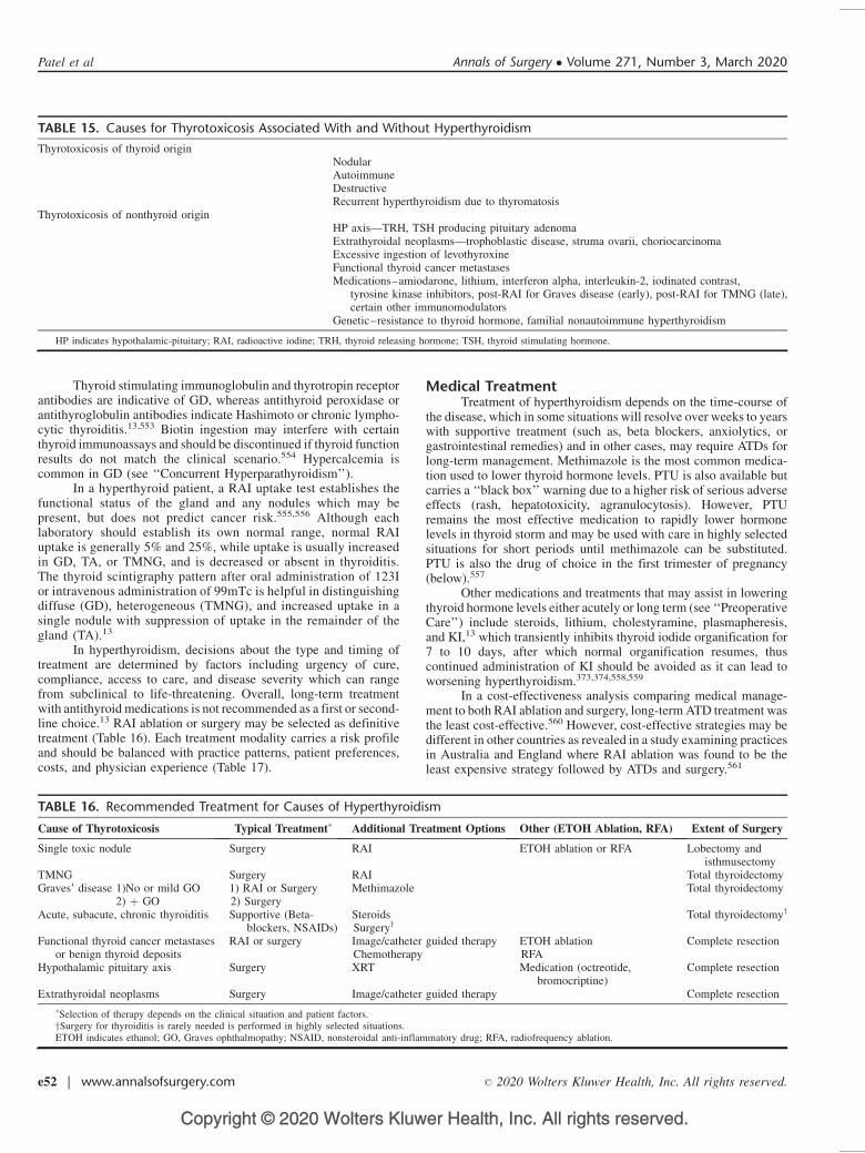
Copyright © 2020 Wolters Kluwer Health, Inc. All rights reserved.
Thyroid stimulating immunoglobulin and thyrotropin receptorantibodies are indicative of GD, whereas antithyroid peroxidase orantithyroglobulin antibodies indicate Hashimoto or chronic lympho-cytic thyroiditis.13,553 Biotin ingestion may interfere with certainthyroid immunoassays and should be discontinued if thyroid functionresults do not match the clinical scenario.554 Hypercalcemia iscommon in GD (see ‘‘Concurrent Hyperparathyroidism’’).
In a hyperthyroid patient, a RAI uptake test establishes thefunctional status of the gland and any nodules which may bepresent, but does not predict cancer risk.555,556 Although eachlaboratory should establish its own normal range, normal RAIuptake is generally 5% and 25%, while uptake is usually increasedin GD, TA, or TMNG, and is decreased or absent in thyroiditis.The thyroid scintigraphy pattern after oral administration of 123Ior intravenous administration of 99mTc is helpful in distinguishingdiffuse (GD), heterogeneous (TMNG), and increased uptake in asingle nodule with suppression of uptake in the remainder of thegland (TA).13
In hyperthyroidism, decisions about the type and timing oftreatment are determined by factors including urgency of cure,compliance, access to care, and disease severity which can rangefrom subclinical to life-threatening. Overall, long-term treatmentwith antithyroid medications is not recommended as a first or second-line choice.13 RAI ablation or surgery may be selected as definitivetreatment (Table 16). Each treatment modality carries a risk profileand should be balanced with practice patterns, patient preferences,costs, and physician experience (Table 17).
Medical TreatmentTreatment of hyperthyroidism depends on the time-course of
the disease, which in some situations will resolve over weeks to yearswith supportive treatment (such as, beta blockers, anxiolytics, orgastrointestinal remedies) and in other cases, may require ATDs forlong-term management. Methimazole is the most common medica-tion used to lower thyroid hormone levels. PTU is also available butcarries a ‘‘black box’’ warning due to a higher risk of serious adverseeffects (rash, hepatotoxicity, agranulocytosis). However, PTUremains the most effective medication to rapidly lower hormonelevels in thyroid storm and may be used with care in highly selectedsituations for short periods until methimazole can be substituted.PTU is also the drug of choice in the first trimester of pregnancy(below).557
Other medications and treatments that may assist in loweringthyroid hormone levels either acutely or long term (see ‘‘PreoperativeCare’’) include steroids, lithium, cholestyramine, plasmapheresis,and KI,13 which transiently inhibits thyroid iodide organification for7 to 10 days, after which normal organification resumes, thuscontinued administration of KI should be avoided as it can lead toworsening hyperthyroidism.373,374,558,559
In a cost-effectiveness analysis comparing medical manage-ment to both RAI ablation and surgery, long-term ATD treatment wasthe least cost-effective.560 However, cost-effective strategies may bedifferent in other countries as revealed in a study examining practicesin Australia and England where RAI ablation was found to be theleast expensive strategy followed by ATDs and surgery.561
TABLE 15. Causes for Thyrotoxicosis Associated With and Without Hyperthyroidism
Thyrotoxicosis of thyroid originNodularAutoimmuneDestructiveRecurrent hyperthyroidism due to thyromatosis
Thyrotoxicosis of nonthyroid originHP axis—TRH, TSH producing pituitary adenomaExtrathyroidal neoplasms—trophoblastic disease, struma ovarii, choriocarcinomaExcessive ingestion of levothyroxineFunctional thyroid cancer metastasesMedications–amiodarone, lithium, interferon alpha, interleukin-2, iodinated contrast,
tyrosine kinase inhibitors, post-RAI for Graves disease (early), post-RAI for TMNG (late),certain other immunomodulators
Genetic–resistance to thyroid hormone, familial nonautoimmune hyperthyroidism
HP indicates hypothalamic-pituitary; RAI, radioactive iodine; TRH, thyroid releasing hormone; TSH, thyroid stimulating hormone.
TABLE 16. Recommended Treatment for Causes of Hyperthyroidism
Cause of Thyrotoxicosis Typical Treatment� Additional Treatment Options Other (ETOH Ablation, RFA) Extent of Surgery
Single toxic nodule Surgery RAI ETOH ablation or RFA Lobectomy andisthmusectomy
TMNG Surgery RAI Total thyroidectomyGraves’ disease 1)No or mild GO
2) þ GO1) RAI or Surgery2) Surgery
Methimazole Total thyroidectomy
Acute, subacute, chronic thyroiditis Supportive (Beta-blockers, NSAIDs)
SteroidsSurgeryy
Total thyroidectomyy
Functional thyroid cancer metastasesor benign thyroid deposits
RAI or surgery Image/catheter guided therapyChemotherapy
ETOH ablationRFA
Complete resection
Hypothalamic pituitary axis Surgery XRT Medication (octreotide,bromocriptine)
Complete resection
Extrathyroidal neoplasms Surgery Image/catheter guided therapy Complete resection
�Selection of therapy depends on the clinical situation and patient factors.ySurgery for thyroiditis is rarely needed is performed in highly selected situations.ETOH indicates ethanol; GO, Graves ophthalmopathy; NSAID, nonsteroidal anti-inflammatory drug; RFA, radiofrequency ablation.
Patel et al Annals of Surgery � Volume 271, Number 3, March 2020
e52 | www.annalsofsurgery.com � 2020 Wolters Kluwer Health, Inc. All rights reserved.
Copyright © 2020 Wolters Kluwer Health, Inc. All rights reserved.
In the US, RAI ablation provides a common definitiveapproach to achieving a euthyroid state for selected patients withTMNG, isolated TA, or GD.10,13,563–566 RAI is well tolerated inmost patients but can fail to completely treat the disease, causekeratoconjunctivitis sicca and xerostomia, and exhibit differentialthyroid uptake which may require surveillance and additionallong-term treatment. Destruction of thyroid tissue by RAI ablationcan lead to a transient increase in thyroid hormone levels about1 week after treatment, which should be anticipated in patientswith cardiovascular disease, who may develop dysrhythmias orother complications. Compared with thyroidectomy, hyperthyroid-ism takes somewhat longer to resolve (4–8 wk for RAI ablation vs2–6 wk for surgery depending on preoperative levels)566 and isless likely to achieve full remission. In a retrospective study, RAIablation was successful in 92% of patients versus 100% forsurgery.567 RAI ablation is contraindicated in pregnancy. The risksof thyroidectomy specific to hyperthyroid patients are detailedbelow.
For TA, 2 other therapeutic approaches are ethanol injection(see ‘‘Nodal Dissection’’) and radiofrequency ablation,568 neither ofwhich have gained popularity in the US nor are recommended asinitial treatment,13 but which may be considered when patients arenot candidates for conventional treatment. In most cases a surgeonshould be consulted prior to proceeding, as surgery after one of theseinterventions can be more difficult if a euthyroid state does not result,hyperthyroidism recurs, or a nodule persists.
Surgical TreatmentSurgery is selected for definitive treatment of hyperthyroid-
ism after considering factors that include etiology, clinical courseand severity, regional resources and limitations, patient comorbid-ities, social constraints, and patient preference (Tables 16 and 17).Overall, while the risks of thyroidectomy in general are low, theyare demonstrably higher in hyperthyroidism (below);569–574 more-over, the likelihood of other morbidity is also increased, such as,cardiac dysfunction in amiodarone-induced thyrotoxicosis(below).575,576 For hyperthyroid patients who select thyroidec-tomy, appropriate preoperative preparation is important (see ‘‘Pre-operative Care’’).
In general (see ‘‘Indications, Extent, and Outcomes of Sur-gery’’) thyroidectomy is rarely performed for thyroiditis, but may berequired in situations of brittle, difficult-to-control thyrotoxicosis,unremitting symptoms, and/or large compressive goiter.24,318,577,578
Lobectomy may be selected for patients with isolated TA who areyoung, desire more rapid resolution than RAI ablation provides, orhave a TA that is large, enlarging, causing compressive symptoms, or
cosmetically significant. For those with TMNG, total or near-totalthyroidectomy may be preferred over RAI ablation given large goitersize or risk of persistent/recurrent disease, whereas subtotal thyroid-ectomy, once favored, is no longer recommended as definitivetreatment (see ‘‘Initial Thyroidectomy’’) 12,13.
Surgery for GD is performed commonly today in theU.S.,555,566,569–572,574,579 and total thyroidectomy is the procedureof choice. Total thyroidectomy is more effective for GD than subtotalthyroidectomy (Table 12) or RAI ablation (Table 16).359,556,580,581 Ina retrospective study of 40 patients undergoing total thyroidectomyfor GD, improvement in quality of life was reported to be marked andrapid, with resolution of hyperthyroid symptoms in an average of32 days.582
Amiodarone-induced thyrotoxicosis, which occurs as Type 1(iodine-induced) and Type 2 (thyroiditis-induced)583 may requirethyroidectomy if medical management fails. Such patients often havesignificant underlying cardiac dysfunction with high risk for com-plications from anesthesia and surgery by the time surgical consul-tation is obtained. Operative mortality as high as 9% has beenreported584), although other series reveal better outcomes.585–587
It is thus important that thyroid surgeons be involved in the thera-peutic decision making for amiodarone-induced hyperthyroidism assoon as there is evidence of nonresponse to medical therapy. Thy-roidectomy under loco-regional anesthesia can be considered inselect cases, but requires a willing and able patient and a skilledteam.588
Graves’ OphthalmopathyAdditional considerations affect treatment selection in
patients with Graves ophthalmopathy (GO) a debilitating conditionthat can jeopardize eyesight.589,590 In patients with moderate tosevere GO,591–594 surgery is preferred over RAI ablation becauseit has less potential for acute worsening of GO.13,595 A retrospectivestudy of 150 patients treated with ablation observed worseningophthalmopathy in 15% within 1 year of treatment. In a study thatcompared subtotal thyroidectomy to RAI ablation, GO progressed in6% versus 10% of patients, was stable in 78% versus 86%, andimproved in 16% versus 3%.13,596 Whether progression of GO afterRAI or surgery is simply due to the natural course of disease isunclear, since the active phase of GO may last for 18 to 36 months.590
In mild GO, RAI ablation and surgery have equivalent low ophthal-mologic risks if steroid treatment is prescribed at the time of RAI.13
In moderate to severe GO, steroids should be given with RAI ablationif surgery cannot be pursued as the first-line option, to attempt tomitigate any worsening of GO.13 As GO may not be obvious tononophthalmologists, and/or may threaten optic nerve viability, it isadvisable to refer GD patients to an ophthalmologist familiar withGO for a baseline examination, the results of which may also directselection of treatment.
Recommendation 38: In patients with moderate to severeGraves ophthalmopathy, total thyroidectomy should be consideredas first-line definitive treatment. (Strong recommendation, moder-ate-quality evidence)
When to Start Thyroid HormoneIn patients who were rendered euthyroid preoperatively based
on free T4 and T3 levels (as changes in TSH will lag), ATDs shouldbe discontinued after surgery and weight-based levothyroxine shouldbe started.13 For patients who are hyperthyroid at the time of surgery,ATDs should be discontinued postoperatively, and levothyroxinetypically started 1 week after surgery to allow time for thyroidhormone levels to decrease to normal. Beta blockers, if used, shouldbe weaned over several days as tolerated to prevent rebound tachy-cardia, headache, and other symptoms.
TABLE 17. Considerations for Selection of Surgery RatherThan Radioactive Iodine for Treatment of Thyrotoxicosis
Pregnancy or desire for pregnancy within 6 monthsConcern for thyroid malignancyGoiter >80 gModerate or severe Graves ophthalmopathySocial issues (infants, small children in the home, unable to follow RAI
safety measures)Poorly controlled hyperthyroidism requiring rapid normalization of thyroid
hormone levelsAirway compromise necessitating urgent resolutionOther central neck pathology requiring surgery (primary
hyperparathyroidism, large size (>4 cm) or multiple benign thyroidnodules)
Prior radioactive iodine treatmentLow % iodine uptake
Annals of Surgery � Volume 271, Number 3, March 2020 Definitive Surgical Management of Thyroid Disease in Adults
� 2020 Wolters Kluwer Health, Inc. All rights reserved. www.annalsofsurgery.com | e53
Copyright © 2020 Wolters Kluwer Health, Inc. All rights reserved.
Surgical Complications in HyperthyroidismA recent National Inpatient Sample database study found an
increased risk of complications (bleeding, hypocalcemia, vocal-cordparalysis, tracheostomy; see ‘‘Postoperative Care and Complica-tions’’) in patients undergoing thyroidectomy for GD compared withnontoxic goiter.2 However, when thyroidectomy is performed by ahigh-volume thyroid surgeon, permanent complications rates (RLNinjury, hypocalcemia) were no higher than for thyroidectomy per-formed for other reasons.352,597
Hematoma and intraoperative blood loss are more likely inpatients undergoing surgery for thyroiditis and especially forGD.567,571,598,699 Such glands are often larger, adherent, inflamed,and more vascular; from this perspective, KI solution given preoper-atively to help control thyroid hormone levels also may help todecrease gland vascularity371,558 (see ‘‘Preoperative Care’’).
Temporary postoperative hypocalcemia is also more commonin surgery for hyperthyroidism, especially GD;381,567,569,571,600,601
this is thought to be in part due to thyrotoxic osteodystrophy, that is,increased bone hunger in longstanding hyperthyroidism602–606
although a meta-analysis advised that calcium and 25-hydroxyvitamin D levels be preoperatively assessed and prophylacticallyrepleted or supplemented in GD patients (see ‘‘PreoperativeCare’’).383
Some groups have reported higher RLN injury rates inGD,607–609 while others report no difference in permanent dysfunc-tion.555,579,610–613 Injury is postulated to be due to stretch from moredifficult medial retraction, as well as increased intraoperative bleed-ing with diminished visualization.
In patients with GD, the feared yet rare complication ofthyroid storm13,614–619 may occur during surgery or afterward evenif the patient is preoperatively rendered euthyroid, and is postulatedto occur from intraoperative manipulation and compression withrelease of hormone into the circulation.617 Intraoperative thyroidstorm may resemble malignant hyperthermia with tachycardia,hypertension or hypotension, heart failure, fever or diaphoresis,but can be distinguished by lack of muscle rigidity/masseterspasm and rise in end-tidal CO2.615,617,620,621 Treatment includesfluids, beta-blockers and other cardiovascular medications, steroids,and anti-thyroid medications. Mortality ranges from 8% to25%.13,614–619
Recommendation 39: Due to the higher risk and greatertechnical difficulty, thyroidectomy in Graves disease is best per-formed by high volume thyroid surgeons. (Strong recommendation,low-quality evidence)
The Hyperthyroid Pregnant PatientTSH is expected to decrease in the first trimester due to a
normal hCG-mediated effect that requires no treatment.622 Truehyperthyroidism in pregnancy is treated with ATDs. Since terato-genic effects from methimazole occur in 2% to 4% of exposedchildren, PTU is the drug of choice in the first trimester. Methimazolemay be instituted after the first trimester.623–630 If beta-blockade isrequired, atenolol should be avoided due to possible fetal growthrestriction with prolonged use.13
If hyperthyroidism cannot be adequately controlled and sur-gery is deemed necessary, it is ideally performed during the secondtrimester due to teratogenic effects of anesthesia in the first trimester,and preterm labor and lower birth weight in the third trimester.631–636
Although some series report acceptable maternal and fetal outcomesduring all trimesters,637,638 there remains some risk of preterm laborwith surgery during the second trimester (4.5%–5.5%).632,639,640 USevaluation of the fetal thyroid is reasonable to gauge development inanticipation of any issues requiring treatment at birth, such as fetal
goiter causing airway compromise.641–645 In pregnant patientsundergoing thyroidectomy, consultation with a high-risk obstetricsteam is advised, including documentation of immediate perioperativefetal heart tones.646,647
GOITER
Goiter is a clinical term used to describe enlargement of thethyroid gland; it is often further defined by relationship to iodinestatus, morphology (diffuse vs nodular), functional activity (nontoxicvs toxic), hereditary relationship (familial vs sporadic), and anatomiclocation (cervical vs mediastinal). The causes of goiter are multifac-torial and depend on genetic susceptibility interacting with environ-ment.648 The definition of goiter has been debated over manydecades without consensus;87 a normal thyroid gland weighs approx-imately 10 to 20 gm. While some experts have proposed a particularweight for goiter,649–653 the ATA has utilized the more generalizedterm of ‘‘benign enlargement.’’80
Goiter is considered endemic when it is present in more than10% of a population due to chronic iodine insufficiency, a situationthat reduces thyroid hormone synthesis and secretion leading totissue overgrowth.654 In the US, dietary iodine has been consideredadequate since the 1920s when iodine fortification occurred655 andthe mean thyroid weight fell from 28 g before 1950 to 18 g after1960.656 Morphologically, iodine-deficient goiters are often diffusewith a homogenous increase in size and preservation of normalfollicle morphology, whereas multinodular goiter (MNG) representsa mixed group of etiologies and histologic features.657
Functionally, patients with goiters may be hypothyroid, euthy-roid, or endogenously hyperthyroid, and if the patient is hyperthy-roid, the cause can be due to GD, a single hyperfunctioning nodule, orTMNG (see ‘‘Hyperthyroid Conditions’’). Etiologically, in GD,autoimmune thyrotropin receptor antibodies stimulate the TSH-Rto increase hormone levels with subsequent diffuse enlargement ofthe gland,13 whereas TMNG is often a combination of variouslyfunctioning nodules with a balance of biochemical hyperthyroidismthought secondary to constitutive activation of the cAMP signalingpathway.658,659 Other etiologic factors include cigarette smoking,natural goitrogens such as cassava, Hashimoto thyroiditis and otherinfiltrative diseases, malignancy, and dyshormonogenesis.660 Famil-ial clustering and female predominance suggest a genetic predispo-sition for euthyroid goiter.657 Rarely, goiter is caused by a TSH-secreting pituitary tumor, thyroid hormone resistance,661 or (forTMNG) an autosomal-dominant activating germline mutation inthe TSHR gene.662
Goiter is denoted substernal if mediastinal extension is pres-ent, meaning that the gland extends caudally past the sternal notch onphysical examination (below), CT imaging, or at the time of surgery.A secondary mediastinal goiter involves the downward growth of anenlarged cervical thyroid and represents the majority of cases, whileprimary mediastinal goiter (2%) arises from ectopic thyroid tissuewith blood supply from intrathoracic vessels.663
The most commonly encountered goiters in current US prac-tice are nontoxic and multinodular.656 In the absence of hyperthy-roidism, an inflammatory process, or known malignancy, a goiter isdescribed as nontoxic, which is the subject of the remainder ofthis chapter.
Indications for ResectionSurgical treatment of nontoxic goiter is indicated for symp-
toms related to compression of the trachea, esophagus, RLN, orsuperior vena cava (see ‘‘‘Indications, Extent and Outcomes ofSurgery’’). The most common symptoms are nonspecific and includeshortness of breath (50%), dysphagia (30%), and voice change
Patel et al Annals of Surgery � Volume 271, Number 3, March 2020
e54 | www.annalsofsurgery.com � 2020 Wolters Kluwer Health, Inc. All rights reserved.
Copyright © 2020 Wolters Kluwer Health, Inc. All rights reserved.
(13%).667 In a systematic review of the impact of resection, trachealcompression was noted in 9% to 58% with cervical goiter and 35% to73% with a substernal component.648 Tracheal compression cancause positional dyspnea, defined as difficulty breathing thatimproves by position change,87 and generalized shortness of breathin up to half of patients.664 When findings suggest substernalextension, CT or MRI should be performed to evaluate for trachealcompression and may aid in surgical planning. After thyroidectomy,83% of goiter patients with positional dyspnea report improvementor resolution.87 The effect of thyroidectomy performed for trachealdeviation is unknown. Formally defined obstructive sleep apneasyndrome is associated with goiter;665 in evaluations assessing theinfluence of cervical and substernal goiter on tracheal airflow usingflow volume loops, 14% to 60% of patients had upper airwayobstruction, often without associated symptoms.666–670 In an analy-sis that used a validated sleep apnea questionnaire, thyroidectomysignificantly improved obstructive sleep apnea symptoms in patientswho screened positive before resection.358
The radiographic prevalence of goiter-associated esophagealcompression (8%) and deviation (14%)664 are not well studied.Patient-reported swallowing difficulties occur in 10% to 43% ofthose referred for goiter treatment,86,317,356 and goiter is also associ-ated with globus sensation.664 The efficacy of goiter resection forpatient-reported swallowing difficulties is unknown.
Goiter resection is also indicated when there is an inability tobiopsy lesions of concern, either because of inaccessibility within thethorax or because a high burden of nodularity (innumerable nodules)precludes adequate evaluation. Approximately 9% to 13% of sub-sternal goiters harbor malignancy.86,317,671–674 Additionally, thenatural history of substernal goiter can be progressive growth withdevelopment or worsening of symptoms, an observation to considerwhen deciding on potential surgical intervention.86 Patients withsubsternal goiter who have a positive Pemberton sign warrantthyroidectomy.675,676
Nonsurgical TreatmentMedical treatment options for nontoxic goiter include iodine
replacement, suppressive T4 therapy, and RAI ablation. Iodinereplacement is associated with a decrease in goiter size in areasof severe iodine deficiency,677 but it is ineffective in regions of iodinesufficiency.80 In a randomized, double-blind placebo trial of euthy-roid patients with nodular goiter in an iodine-sufficient region,suppressive T4 therapy (target TSH 0.2–0.8 mU/I) plus iodinereplacement, as well as suppressive T4 therapy alone, mildlydecreased thyroid volume at 1 year; neither long-term outcomesnor effect on symptoms were studied.678 However, suppressive T4therapy has low efficacy with a modest effect that is temporary, asnodules will regrow to baseline within 1 year of treatment cessa-tion.679,680 Suppressive T4 therapy is also associated with thyrotoxicsymptoms,681 requires lifelong use, and has been linked to both atrialfibrillation and osteoporosis.682–684
RAI ablation has been widely used to treat patients withnontoxic goiter for over 3 decades. In a randomized clinical trialof I131 versus T4 suppression, at 2 years’ time goiter size was reducedby 44% in the former versus 1% in the latter group, with a significantdecrease in bone mineral density in T4 suppressed patients as well.681
Use of recombinant human TSH (rhTSH) enhances RAI uptake,improving its long-term effect and decreasing the required dose.685–
687 Adverse effects associated with RAI use for nontoxic goiterinclude transient posttreatment thyrotoxicosis due to release of storedhormone, transient or permanent hypothyroidism, initial gland swell-ing which can pose a problem in large goiters with compromisedinspiration at baseline, and rare potential for development of sec-ondary malignancies such as salivary gland cancer and leukemia.688
RAI use is not appropriate if there is a suspicious thyroid nodule onimaging or cytology, or if there is pregnancy, impending pregnancy,or inability to tolerate the treatment or its isolation constraints (see‘‘Hyperthyroid Conditions’’). In summary, RAI ablation may beconsidered for benign goiter in selected patients and T4-suppressionshould be avoided.
Extent and Conduct of ResectionWhereas medical or RAI therapy may result in partial size
reduction of goiter, surgery offers the opportunity for definitivetreatment. In the past, subtotal thyroidectomy was a common pro-cedure but the associated high rate of recurrence has led many torecommend total thyroidectomy or initial complete lobectomy (see‘‘Initial Thyroidectomy,’’ ‘‘Reoperation’’). A systematic review689
evaluated 4 randomized trials of total or near-total thyroidectomyversus subtotal thyroidectomy for NTMG690–693 and reported thatrecurrence was lower in the total thyroidectomy group (OR 0.05(95% CI 0.01–0.21); P<0.0001; 1057 participants), the incidence oftransient hypoparathyroidism was lower in the subtotal thyroidec-tomy group (OR 2.47 (95% 1.57– 3.88); P < 0.0001; 1275 partic-ipants), and the incidences of permanent hypoparathyroidism andtransient and permanent RLN palsy did not differ. For goiter that isconfined anatomically to 1 lobe, lobectomy is a reasonable approachgiven the low risk of disease development in the contralateral loberequiring surgical intervention (2%) and the reasonable probabilityof retaining euthyroid status not requiring T4 replacement (73–78%).694,694 In short, the extent of thyroidectomy for nontoxic goitermust balance concerns for recurrence with potential morbidity.
Recommendation 40:a. When surgery is indicated, total thyroidectomy is preferred for
treatment of bilateral goiter. (Strong recommendation, low-quality evidence)
b. When the contralateral lobe is normal, lobectomy and isthmu-sectomy is recommended for treatment of unilateral goiter.(Strong recommendation, low-quality evidence)
Diagnosis and Preoperative ManagementBecause symptoms associated with nontoxic goiter (above)
can alter management, clinicians should routinely query patientsabout potential symptoms related to local mass effect (see ‘‘InitialEvaluation’’). On physical examination, a goiter may be graded byWorld Health Organization criteria: Grade 0—nonpalpable/notvisible; Grade 1a—Palpable but not visible even in full extension;Grade 1b—Palpable in neutral position/visible in extension; Grade2—visible in normal position of the neck/no palpation required tomake diagnosis; Grade 3—visible at a distance. Substernal goiteris diagnosed when the full extent of an enlarged thyroid cannot bepalpated with the patient’s neck extended, even with deglutition orsupine. A suboptimal exam may occur with obesity, a short thickneck, or kyphosis; in such cases CT or MRI imaging may be usefulfor diagnosis if US or exam fails to define the inferior thyroidborder. A preoperative voice assessment is advised (see ‘‘InitialEvaluation’’) and further laryngoscopic evaluation is reserved forthose with voice change or history of previous anterior necksurgery.696
Preoperative imaging of goiter starts with cervical US (see‘‘Imaging’’). If the inferior edge of the thyroid lobes cannot beidentified on US or there is clinical concern for a substernalcomponent (above) cross sectional imaging is recommended. CTor MR imaging is recommended to determine the smallest cross-sectional area of the trachea, and surgeon review of images is oftenhelpful as tracheal compression is not always reported radiologically.Correlation between CT and CXR is poor in this respect and CXRshould not be used for this purpose.86,664,697,698
Annals of Surgery � Volume 271, Number 3, March 2020 Definitive Surgical Management of Thyroid Disease in Adults
� 2020 Wolters Kluwer Health, Inc. All rights reserved. www.annalsofsurgery.com | e55
Copyright © 2020 Wolters Kluwer Health, Inc. All rights reserved.
Recommendation 41: Cross sectional imaging of goiter isrecommended if there is concern for a substernal component.(Strong recommendation, moderate-quality evidence)
Ninety-eight percent of substernal goiters can be removedthrough a cervical incision.663,697 Anecdotally, CT images showingcaudal extent past the top of the aortic arch, to below the arch, or tothe carina, are increasingly strong reasons to involve thoracic surgerypreoperatively, and additional factors include intrathoracic goiter thatappears larger than the thoracic inlet, involves the posterior medias-tinum, compresses the superior vena cava, or is suspicious formalignancy on imaging.663,769,700 Thoracic consultation may alsobe important to inform the patient of additional morbidity if there isconcern that substernal goiter cannot be removed cervically, and inthe rare situation of primary mediastinal goiter, which is completelyseparate from the cervical thyroid gland, precluding removal througha cervical approach.
Intraoperative ManagementSurgery for a large cervical and/or substernal goiter is multi-
faceted and best handled by experienced thyroid surgeons701 (see‘‘Indications, Extent, and Outcomes of Surgery’’). An anesthesiateam experienced in managing complex airway issues is also vital ifthere is significant tracheal compression, and video-scopic laryn-goscopy with awake fiberoptic intubation and/or rigid bronchos-copy may be useful when difficult laryngeal exposure is anticipated.In the hands of experienced anesthesiologists, intubation compli-cations (defined as>2 intubation attempts) are uncommon, reportedin up to 11% of goiters > 600 g.80,702,703 In some instances, goitersize and location may prevent rescue tracheostomy, a potentialsituation that should also be discussed preemptively by the anes-thesia and surgery teams. Other considerations may include the needfor large-bore intravenous access if concern for blood loss is high,the location of such access if the large veins within the chest could becompromised in the resection, the need for a type and screen,placement of an arterial line and/or Foley catheter, and use of anoperating room equipped to manage a potentially extensive proce-dure including median sternotomy or thoracotomy. If RLNM isbeing used (see ‘‘Adjuncts and Approaches’’) the team should keepin mind that with resection of a large goiter, manipulation of thelarynx and trachea can malposition the endotracheal tube and loss ofsignal may result.704
Recommendation 42: When performing surgery for subster-nal goiter, good communication, preparation and cooperation ofexperienced surgical and anesthesia teams is recommended.(Strong recommendation, low-quality evidence)
To lift a goiter cephalad and facilitate its cervical delivery, thepatient should be positioned with optimal extension of the neck asallowed by habitus and comorbidities. The chest should be preppedand draped if a sternal split is anticipated. The approach is typically acollar incision placed approximately 1 to 2 fingerbreadths above thesternal notch. In managing bilateral goiter, many surgeons resect thelarger lobe first while others selectively approach the smaller lobe tofacilitate later delivery of the larger lobe into the neck. Early ligationand division of the superior vascular pedicle often assists in cervicaldelivery of a thoracic component, along with sequential placement oftissue clamps and delicate use of digital mobilization. Selective earlydivision of the isthmus may allow better access to the medial inferiorvessels. Delivery of the substernal component is usually necessarybefore the RLN can be identified in the tracheoesophageal groove,but occasionally the nerve is displaced anteriorly onto the paren-chyma of the thyroid gland making it more vulnerable to injury;taking this anatomic possibility into consideration, delivery under
direct visualization is preferred to a blind sweep of the finger whichshould be done with caution.
Drain placement has not been shown to improve outcomes andmay increase length of hospitalization and postsurgical infec-tion,705,706 (see ‘‘Postoperative Care and Complications’’). In thesetting of goiter, drains have not been studied but with substernalgoiter resection are occasionally used.
In a National Inpatient Sample database study, patients under-going surgery for substernal goiter versus cervical thyroidectomywere more likely to experience iatrogenic pneumothorax (adjustedOR 5 4.40; CI 5 2.01–9.33).707 Although this complication is rare,with any concern there should be a low threshold for obtaining apostoperative CXR. Other adjusted complications specific to thesubsternal goiter population include hemorrhage or hematoma [OR1.34 (1.09–1.65)], respiratory failure [1.94 (1.48–2.55)], and post-operative hypocalcemia [1.44 (1.28–1.62)].707
An extubation consideration specific to goiter is concern fortracheomalacia, a condition traditionally thought to arise from long-standing extrinsic compression impairing the structural integrity ofthe tracheal rings and, giving rise to dynamic airway collapse >50%of diameter.708 Today, there is debate as to the existence of thisphenomenon, and it is recognized that some cases have actuallyrepresented bilateral RLN paralysis. In a systematic review, trache-omalacia related to a large goiter was rare (0.8%–10%) and mostoften handled without tracheostomy.663 Treatment options includetracheostomy, prolonged endotracheal intubation, internal stenting,and noninvasive positive pressure intubation.709
ADJUNCTS AND APPROACHES
While the basic principles of safe thyroidectomy haveremained constant since the early 20th century, numerous technicaladjuncts have been introduced, the most common and current ofwhich are discussed here.
Energy-based Vessel-sealing DevicesControl of the extensive vascular system of the thyroid has
traditionally been achieved using fine ties, titanium clips, andelectrocautery (see ‘‘Initial Thyroidectomy’’). In addition, in thepast few decades 2 types of adjunctive energy-based vessel-sealingdevices have achieved widespread use. The electrothermal bipolardevice (Ligasure) relies on bipolar cautery to seal blood vessels, andin many retrospective and prospective studies produces equivalentrates of RLN injury, hematoma, and hypoparathyroidism whencompared to conventional ligation technique.710–715 Most studiesalso demonstrate decreased operative time compared with conven-tional knot-tying, typically in the range of 5 to 10 minutes peroperation.710–715 The ultrasonic coagulator (Harmonic) uses anultrasonic shear technique to seal and divide blood vessels, and inretrospective and prospective studies has rates of RLN injury,hypoparathyroidism, and hematoma that are equivalent to conven-tional technique;716–722 a multicenter prospective randomized trial inItaly also identified decreased operative blood loss.716 All studies ofthe ultrasonic coagulator have shown decreased operative time,typically in the range of 10 to 30 minutes per operation.716–722
The 2 devices have also been compared in a number of clinical trials,predominately with no observed differences in complication rates oroperative time;723–728 although some studies found decreased oper-ative times for the ultrasonic coagulator.723,724 In 2 systematicreviews examining 35 randomized trials of >4000 thyroidectomypatients, the safety and efficacy of energy-based devices werecomparable to conventional knot-tying, and both adjuncts reducedoperative time with the ultrasonic coagulator an average of 6 minutesfaster.729,730
Patel et al Annals of Surgery � Volume 271, Number 3, March 2020
e56 | www.annalsofsurgery.com � 2020 Wolters Kluwer Health, Inc. All rights reserved.
Copyright © 2020 Wolters Kluwer Health, Inc. All rights reserved.
Intraoperative Assessment of Nerve FunctionTraditional methods for avoiding nerve injury during thyroid-
ectomy rely on both intraoperative visualization and knowledge ofthe normal and variant RLN anatomy (see ‘‘Initial Thyroidectomy’’).Reported rates of permanent RLN dysfunction after thyroidectomyare typically low (<1%–5%) although due to several factors, reportsmay underestimate the true incidence (see ‘‘Postoperative Care andComplications’’).731 In an attempt to reduce dysfunction, over thepast few decades RLNM has become an increasingly popular adjunctin thyroidectomy. At a minimum, its use (below) requires preopera-tive planning, significant expertise, and avoidance of neuromuscularblockade after anesthetic induction.
The efficacy of RLNM has been studied extensively, includingseveral large-scale multi-institutional trials, both prospective andretrospective.399,732–739 However, no study has demonstrated a sig-nificant difference in either transient or permanent RLN injury rateswith RLNM use. A 2017 review of 8 meta-analyses confirmed noobservable reduction in transient or permanent RLN injury.740
Routine use of RLNM during total thyroidectomy was not foundto be cost-effective compared with visual identification alone. A2017 study reported that RLNM was potentially cost effective inpreventing bilateral RLN dysfunction.741,742
Despite these findings, selective use of RLNM is common.Some surgeons advise that use is beneficial during challengingoperations such as reoperation, resection of large or substernal goiter,or resection of locally advanced thyroid cancer, citing a systematicreview of 10 studies that reported a trend toward decreased rates oftemporary and permanent RLN injury when compared to visualidentification alone.743 Although comprising>10,000 nerves at risk,this study was underpowered to detect a significant difference inoutcomes. Surgeons who wish to adopt the practice of selectiveRLNM should be aware that successful utilization requires a learningcurve.744 An area in which RLNM may prove useful during plannedtotal thyroidectomy is in prevention of bilateral RLN injury, and inthis regard the concept of staged thyroidectomy based on RLNMfindings has gained recent traction. During a planned total thyroid-ectomy, upon completion of the initial lobectomy, if the stimulatedsignal has decreased by certain criteria or has been lost, considerationis advised for stopping the operation rather than proceeding andplacing the contralateral nerve at risk.742,745–748 However, false-positive causes of diminished function by RLNM can occur.749
The most widely used RLNM systems utilize passive moni-toring, in which the presence and quality of the interrogation signalconfirm neural integrity, and loss of signal at any point caudal to aninjury indicates a nonfunctioning nerve or technical issues. In 2011an international group of high-volume thyroid surgeons establishedguidelines for safe use of RLNM, including an algorithm fortroubleshooting when loss of signal is encountered.704 In brief,the surgeon first palpates the posterior aspect of the cricoid cartilageduring nerve stimulation to feel for laryngeal twitch (LT), and if LT ispresent without a corresponding auditory or EMG signal, the anes-thesiologist should be asked to reposition the endotracheal tube. If LTis absent, then the RLNM system should be investigated for problemswith connections and auditory output, and the anesthesiologistshould confirm that the patient is not receiving neuromuscularblockade. The stimulator should also be tested on nearby muscleand if muscular twitch is intact, then ipsilateral RLN injury shouldbe suspected.
Recently, a technique for continuous RLN nerve stimulationvia direct placement of a flexible silicon cuff electrode whichencircles the vagus nerve has been suggested as a more accurate,real-time technique750–755 especially for early detection of potentialinjury via traction or stretch, but does require additional time as wellas dissection of the vagus within the carotid sheath. A prospective
multicenter study of>100 patients demonstrated safety.752 However,a smaller prospective study of continuous vagal nerve monitoring inthyroidectomy was aborted after 2 of 9 patients had serious adverseevents (hemodynamic instability and reversible neuropraxia attrib-uted to the monitoring apparatus).756
Intraoperative monitoring of the EBSLN has been reported toassist in its identification when compared to visualization alone, withlimited data on voice quality outcomes.757–760 The expected rate ofinjury to the EBSLN is unknown (see ‘‘Laryngology’’) and issuspected to be underreported.
Recommendation 43: While it does not prevent RLN injury,RLNM is safe and may assist the surgeon during initial orreoperative thyroidectomy. (Strong recommendation, moderate-quality evidence)
Recommendation 44: During planned total thyroidectomy,after completion of the initial lobectomy, if RLNM results suggestloss of function, the surgeon may consider stopping the operationfor possible completion at a later date. (Strong recommendation,low-quality evidence)
Rapid Parathyroid Hormone TestingSome surgeons use rapid intra- or postoperative PTH testing
during total or reoperative thyroidectomy to help determine thelikelihood of postoperative hypocalcemia due to hypoparathyroid-ism.761–770 The majority of studies are nonrandomized prospectiveanalyses demonstrating some utility. Protocols vary but the mostcommon time points for serum PTH measurements are at thebeginning of surgery and around 10 minutes following removal ofthe thyroid gland, with later postoperative (ie, recovery room)intervals also described. A PTH cutoff of <10 pmol/L is typicallyused to predict postoperative hypocalcemia.762,764,767,770 Recom-mendations on how to manage patients with single-digit PTH levelsvary between institutions and include initiation of calcitriol andplanning for prolonged hospitalization. In a meta-analysis of 14studies, intraoperative PTH testing helped predict postoperativehypocalcemia, but there was no difference in utility versus early(within 1–4 h) postoperative PTH measurement.771 The predictivevalue of this adjunct will vary with the local rate of hypoparathy-roidism after thyroidectomy. Cost efficacy has not been studied.
Recommendation 45: Rapid PTH measurement during orafter total or completion thyroidectomy may help to managepatients at risk for hypocalcemia. (Weak recommendation, moder-ate-quality evidence)
Intraoperative Tissue AnalysisPotential use of IOPE in evaluation of thyroid, LN, parathy-
roid, and other tissues is covered in ‘‘Perioperative Tissue Diagno-sis.’’ During thyroidectomy, because the intraoperative identificationof parathyroid glands may be difficult due to their small size andsimilar appearance to surrounding tissues, new technologies forassisting with parathyroid identification (such as autofluorescence)continue to be reported and evaluated.772–778
Remote-Access ApproachesWhile thyroidectomy via cervical incision remains the stan-
dard approach (see ‘‘Initial Thyroidectomy’’), numerous remote-access techniques have gained popularity internationally, and caneach be performed endoscopically or robotically, including thetransaxillary approach779,780 the bilateral axillo-breast approach(BABA),781,782 the posterior ‘‘facelift’’ approach,783 and recentlya transoral endoscopic vestibular approach (TOETVA).784 Remote-access approaches have not achieved widespread use in the US,785 afact that may relate to differences in cultural values, habitus, diseaseseverity, surgeon preference, and learning curves. In a US national
Annals of Surgery � Volume 271, Number 3, March 2020 Definitive Surgical Management of Thyroid Disease in Adults
� 2020 Wolters Kluwer Health, Inc. All rights reserved. www.annalsofsurgery.com | e57
Copyright © 2020 Wolters Kluwer Health, Inc. All rights reserved.
database study of thyroid cancer patients undergoing thyroidectomyfrom 2010 to 2011, 225 patients received a robotic approach com-pared to >68,000 who had conventional thyroidectomy.786
The primary advantage of remote-access thyroidectomy isimproved cervical cosmesis. Several Asian studies have reportedincreased patient satisfaction with cosmetic results followingremote-access thyroidectomy compared to cervical thyroidec-tomy.783 Data regarding other outcomes, including complications,are mixed, and data on long-term outcomes are lacking. A meta-analysis of 11 studies of robotic transaxillary and BABA thyroidec-tomy, identified increased operative time, hospital stay, and rate oftransient vocal fold dysfunction for robotic versus conventionalthyroidectomy, while blood loss, hematoma, and permanent vocalfold dysfunction rates were similar between groups.787 In terms ofcost, remote-access approaches require longer operating times andadditional specialized equipment.788 A 2016 ATA consensus state-ment concluded that remote-access thyroidectomy may be consid-ered in selected patients with unilateral small thyroid nodules whowish to avoid neck incision, and should only be performed in high-volume centers by surgeons with expertise in both thyroid andendoscopic or robotic surgery.783
Recommendation 46: Remote-access thyroidectomy shouldonly be performed in carefully selected patients, by surgeonsexperienced in the approach. (Strong recommendation, low-qualityevidence)
Hemostatic AgentsA wide variety of topical hemostatic agents are currently
available with the intention of facilitating surgical hemostasis, butdata are limited concerning their effectiveness in preventing bleed-ing. A 2013 randomized study of oxidized cellulose patch (Surgicel)in thyroidectomy patients reported trends toward higher rates ofhematoma, increased postoperative drain output, and longer hospitalstay when compared with untreated patients,769 and the authorsconcluded that this agent was not helpful and potentially harmful.
Although drains are not typically used in thyroidectomy in theUS (see ‘‘Initial Thyroidectomy’’), a 2009 Italian study utilizing amatrix hemostatic agent with human recombinant thrombin Flosealdemonstrated treatment reductions in drain output, operative time,and hospital stay; however, rates of hematoma were not different.790
In a multicenter prospective randomized trial, the combined use ofthis agent and the ultrasonic coagulator reduced drain output andoperative time, but again with no difference in rate of neck hematomacompared to conventional thyroidectomy.791 A 2005 Canadian pro-spective randomized study of fibrin sealant (Tisseel) in thyroidec-tomy demonstrated 44% less drain output but no differences inhematoma, hospital stay, or other outcomes.792 Additional concernshave been raised regarding the cost efficacy of topical hemostaticagents and the potential risks of infection with use of human-derived products.
LARYNGOLOGY
Responsible for vocal fold motion, the bilateral RLNs controlall intrinsic muscles of the larynx except for the cricothyroid muscle,which is innervated by the bilateral EBSLNs which lengthen andtense the vocal folds to modulate pitch and voice projection. Vocalfold dysfunction (VFD) can arise prior to thyroidectomy from priorcervical or mediastinal surgery, RLN involvement by central neck ormediastinal cancer, and other causes including idiopathic.793 Duringthyroidectomy, VFD can be caused by direct injury related to theendotracheal tube or other tubes (nasogastric, temperature probe,etc.), during intubation or extubation, and by operative impairment ofthe RLN, vagus nerve, or ESBLN, with the latter situation less well
characterized. VFD can be partial (paresis), complete (paralysis),temporary (resolves within 6 months of surgery), permanent (noresolution 12 mo after surgery), and unilateral and/or bilateral (see‘‘Postoperative Care and Complications’’).
A 2009 meta-analysis including more than 25,000 patientsundergoing thyroidectomy reported a temporary VFD rate of 9.8%(range 1.4%–38%) and a permanent rate of 2.3% (0%–18.6%).794
Factors such as age, comorbidities, complexity, and severity ofdisease (malignancy, thyroid size, inflammation, anatomic factors),extent of dissection, and surgeon experience are associated with VFDrisk after thyroid surgery.795 Less commonly, VFD may be due tointubation alone; in a large retrospective study of 31,217 patientsundergoing elective surgery, 0.077% experienced vocal fold paraly-sis believed to be due to intubation alone.796
The impact of VFD is significant, affecting quality of life andin some cases having an adverse economic impact.797 Rarely,associated swallowing dysfunction or significant preoperative pul-monary compromise may lead to intolerance of unilateral VFD andeven to a requirement for tracheotomy and/or gastrostomy. Due to thepossibility of airway compromise if bilateral VFD were to occur, thepotential need for tracheostomy should be considered. Becauselaryngeal function is so important, a number of thyroid-relatedsocieties have published statements or guidelines on perioperativeassessment of changes in voice, swallowing and respiratoryeffort.3,793,798–800 The method, timing, and indications for thisassessment have not been standardized.801
Preoperative EvaluationPreoperative knowledge of vocal fold abnormalities may alter
the conduct or extent of operation or delay thyroidectomy. Laryngealassessment may be functional or anatomic in nature. The utilizedmethods vary widely by provider, specialty, and volume and com-plexity of thyroid operation.95,802–804 Commonly used methodsinclude auditory assessment, mirror examination, transcutaneouslaryngeal ultrasound (TLUS), flexible laryngoscopy, and videolar-yngostroboscopy.161,162,696,800,804–806
When evaluating patients for thyroidectomy the surgeonshould assess voice function and quality (see ‘‘Initial Evaluation’’).Most deem simple auditory assessment by the surgeon to be anacceptable initial method. Although subjective, a recent retrospectivereview of 464 patients found that voice assessment by the surgeoncombined with knowledge of prior neck surgery was more reliablethan patient-reported voice change; however, voice quality was notassessed in this study using a validated instrument.801 Clinically, inthe absence of poor pulmonary function, a sustained maximumphonation time (MPT) <10 seconds (due to excessive air escapefrom incomplete glottic closure) may suggest vocal fold dysfunc-tion.807 Vocal quality may also be assessed by structured listeningand questioning of patients and family members; common toolsinclude the Grade, Roughness, Breathiness, Asthenia, and Strain(GRBAS) Scale, the Consensus Auditory-Perceptual Evaluation ofVoice Clinical Protocol,808 and the Voice Handicap Index (VHI).809–
812 In a meta-analysis, only 3% of patients reporting preoperativedysphonia had VFD after thyroidectomy, instead having conditionssuch as polyps, gastroesophageal reflux, allergies, and other sourcesof inflammation. Conversely, documented VFD occurs with a widerange (0%–87%) when patients report no changes in voice qual-ity.813,814
Anatomic assessment of vocal fold function can be performedin the office by indirect mirror examination, TLUS, or indirectflexible laryngoscopy. Indirect mirror examination is simple toperform and has little to no associated cost but may not provideadequate visualization in all cases and is not tolerated by somepatients.796 Introduced within the past decade, TLUS may be unable
Patel et al Annals of Surgery � Volume 271, Number 3, March 2020
e58 | www.annalsofsurgery.com � 2020 Wolters Kluwer Health, Inc. All rights reserved.
Copyright © 2020 Wolters Kluwer Health, Inc. All rights reserved.
to visualize the vocal folds due to calcified thyroid cartilage (morecommon in older patients and men) or body habitus, but with clearvisualization of equal and symmetric bilateral motion has equalsensitivity and specificity to other methods, moreover is safe,performed without discomfort, has a short learning curve (<50cases),155 and may be performed at the time of office US, therebyminimizing cost. If there is any question of inadequate visualizationof the vocal folds, indirect flexible laryngoscopy should be per-formed.153,155,808,811–815
Prior to thyroidectomy, visualization of the vocal folds isrecommended routinely by some medical societies, and selectivelyby others for patients determined to be at risk3,95,794,800,804 includingthose with notable voice changes, known VFD, prior neck, medias-tinal, cardiac or upper thoracic surgery, apparent invasive malig-nancy, large substernal goiter, or extensive LNM.3,95,800 When thevocal folds are visualized, the incidence of preoperative VFD amongpatients with TC ranges from 1.7% to 9.6% (prevalence 0.69%–4.3%).815,816 In patients without concerning findings or comorbid-ities, the incidence of preoperative VFD is lower at 0% to3.5%.664,696,802,806,813,817–819 In situations without obvious concernfor VFD, a recent retrospective study reported that routine preoper-ative flexible laryngoscopy is not cost-effective.696
Recommendation 47: In preoperative discussion of thyroid-ectomy, the surgeon should disclose to the patient the possibility,likelihood, and implications of permanent vocal fold dysfunction.(Strong recommendation, moderate-quality evidence)
Recommendation 48: Prior to thyroidectomy, laryngealexamination should be performed in patients determined to havevocal abnormalities as assessed by the surgeon, pre-existing laryn-geal disorders, prior at-risk surgery, or locally advanced thyroidcancer. (Strong recommendation, low-quality evidence)
Intraoperative EventsIntraoperative events that can cause VFD (see ‘‘Postoperative
Care and Complications’’) include: 1) intubation resulting in vocalfold laceration, hematoma, edema or rupture; arytenoid dislocation;formation of chronic granuloma or polyp; 2) thermal or traction-related nerve trauma (ie, use of energy devices too close to a nerve;unintentional clamping, ligation or transection); or 3) nerve transec-tion (intentional or rarely unintentional)(see ‘‘Postoperative Care andComplications’’). Visual intraoperative identification of the RLN andEBSLN are useful in preventing impairment (above and ‘‘InitialThyroidectomy’’). Some degree of functional assessment can beprovided by RLNM (see ‘‘Adjuncts and Approaches’’). Excessivetraction on the RLN or a thermal event are common mechanismsleading to paresis or paralysis and may not always be recognizedintraoperatively.
Postoperative VFD CareIf VFD is known or suspected to have occurred intraopera-
tively, immediate careful observation after extubation should beconsidered by the entire team (surgery, anesthesia, nursing), assess-ing in particular for altered respiratory status, stridor, and aspirationof secretions. Immediate management of suspected unilateral VFDvaries depending on the setting and severity of the condition.Immediate management of suspected bilateral VFD generallyrequires direct examination to discern vocal fold position and motion,extent of glottic closure, and presence of edema. With either unilat-eral or bilateral VFD, the symptomatology and objective findingsmay prompt additional management with options ranging fromsimple observation to moderate supportive measures (racemic epi-nephrine, humidified oxygen, elevated head of bed, steroids) to majorsupportive measures (continuous positive airway pressure) and rarelyto tracheotomy or a laryngeal procedure. Thyroid surgeons should be
well versed in the immediate management of VFD as the situationcan be life threatening. However, as long as the airway is protected,and adequate respiration is occurring, care can proceed in a carefuland calm manner. If reintubation or tracheotomy is required in therecovery area or elsewhere in the hospital, assistance from anesthe-siology and a multidisciplinary airway team should be considered,depending on the acuity and severity of the situation, equipmentavailable, and the specific expertise of the surgeon.
After 24 to 48 hours postoperatively, the reported likelihood ofaltered vocal quality varies widely (30%–90%) depending on studytype and methods.820–825 Postanesthetic changes in voice may lastfor up to 2 weeks unless structural damage to the larynx has occurredwith intubation.95,826 The AAO-HNS recommends that voice assess-ment be performed 2 to 8 weeks postoperatively since in VFD theideal time for vocal fold augmentation is <3 months after thyroid-ectomy.95,785,827,828 Early intervention in those with RLN paresis orparalysis is associated with superior functional outcomes. Anotherreason for prompt diagnosis of VFD is possible early institution ofnimodipine pharmacologic therapy. In a small 2018 prospectivestudy, nimodipine use for 2 to 3 months postoperatively was associ-ated with long-term normalization of the Vocal Handicap Index andmaximum phonation time and no atrophy of the paralyzed vocalfold.829 In a study of 30 vocal fold dysfunction patients, the motionrecovery rate was tripled compared with historical controls; nimo-dipine treatment was not tolerated by 7 excluded patients.830 In aprospective study of 53 vocal fold dysfunction patients, 67% recov-ered purposeful motion. Time to recovery was not different whennimodipine was begun within 15 days, between 15 and 30 days, andafter 30 days.831
EBSLN dysfunction reduces the highest obtainable funda-mental frequency of the projected voice,832,833 typically resulting in avoice that is low pitched, gravelly, soft, and/or cannot be raised toshout or reach high notes.793–795,823,834–849 Neuromonitoring can beused intraoperatively to assist with identification and preservation ofthe EBSLN.759 However, EBSLN dysfunction can be difficult toassess and confirm as currently there is no consensus for diagnosticfindings on laryngoscopy.832,850,851 Even laryngeal electromyogra-phy of the cricothyroid muscle, which is considered the standard fordiagnosis of EBSLN paralysis, has limitations regarding interpreta-tion of results.832,852 Because reliable diagnosis is difficult, baselinerates of EBSLN dysfunction and recovery after thyroidectomy arenot well understood. When assessed during thyroidectomy byRLNM, reported EBSLN injury rates range from 0% to 58%, withpermanent paralysis in 0% to 5%.794,795,842,844–849 Although thepotential for EBSLN dysfunction is often cited in the thyroidectomyliterature, it is unclear if treatment recommendations would signifi-cantly change outcomes. Voice therapy is most commonly employedfor EBSLN injuries but has inconsistent results.832,852
An area of continued debate among thyroid surgeons is routinepostoperative anatomic assessment of the vocal fold motion. Tworecent decision analyses found that routine laryngoscopy for allpatients was not cost-effective.98,816 In general, because patientswith VFD may be asymptomatic,847 the extent of investigationmay hinge on the outcome to be assessed, for example, voice qualityas it impacts quality of life versus accurate assessment of complica-tion rates. In the first situation, auditory assessment alone isdeemed by multiple authors and medical societies to be suffi-cient.3,95,793,798,843,844,849 In the latter circumstance, routine postop-erative anatomic testing is necessary utilizing mirror exam, TLUS,flexible laryngoscopy, or videolaryngostroboscopy. A retrospectivestudy of over 1000 RLNs at risk revealed that only 6% of patientsreporting significant voice changes after thyroidectomy had VFD,and 44% of patients with documented dysfunction were asymptom-atic.853 In a large retrospective study of over 5000 patients from the
Annals of Surgery � Volume 271, Number 3, March 2020 Definitive Surgical Management of Thyroid Disease in Adults
� 2020 Wolters Kluwer Health, Inc. All rights reserved. www.annalsofsurgery.com | e59

Copyright © 2020 Wolters Kluwer Health, Inc. All rights reserved.
Scandinavian Quality Registry, the incidence of VFD from groupsperforming routine postoperative laryngeal examination was nearlytwice that of groups utilizing subjective voice assessment.802,854
Surgeons should strive to know their own rates of temporary andpermanent VFD both to accurately inform patients and assess theneed for changes in operative technique.
Recommendation 49: Voice assessment should be performedat the postoperative visit. (Strong recommendation, low-qualityevidence)
Recommendation 50: After thyroidectomy, laryngeal exami-nation should be performed in patients with known or suspectednew RLN dysfunction or aspiration. (Strong recommendation,moderate-quality evidence)
Patients with known or suspected VFD, or those with particu-lar concern about their voice after thyroidectomy, may be referred toa laryngologist for additional evaluation and possible treatment. Aspeech pathologist will in turn often become involved. Early referral(2–8 wk postsurgery) to a laryngologist in combination with earlyintervention results in superior voice outcomes.785,827,828 Often,paresis improves over 6 to 12 months, but earlier short-term inter-vention with nimodipine (above), temporary vocal fold medializa-tion, or another procedure may benefit patients with severesymptoms until the need for definitive treatment can be deter-mined.785,827,828 Long-term treatment options for unilateral RLNparalysis may include injection of bulking agents, thyroplasty,arytenoid adduction, and reinnervation procedures. Depending onthe resting position of the paralyzed vocal fold, management optionsfor bilateral paralysis may include tracheostomy, cordotomy, aryte-noidectomy, suture lateralization, reinnervation, electrical pacing,and/or CPAP.
Recommendation 51: If vocal fold motion impairment issuspected or identified, early referral of the patient to a laryngol-ogist is recommended. (Strong recommendation, moderate-qualityevidence)
FAMILIAL THYROID CANCER
More than 90% of all TC is sporadic in nature, secondary tosomatic genetic alterations.855 Approximately 3% to 9% of TC isFNMTC,856 with PTC the most common histologic subtype (85%–91%), followed by FTC (6%–10%), ATC (1.6%), and HTC.857–859
FNMTC is nonsyndromic in 95% of cases. MTC accounts for 3% to5% of all TC with approximately 25% of cases being hereditary.
Genetic TestingThe American Society of Clinical Oncology has published
iterative policy statements on genetic and genomic testing forcancer susceptibility, stressing the importance of communicationwith the patient about the purpose of the genetic testing, thepotential outcomes and implications for the patient and familymembers, and the risks of cancers related to the genes beingtested.860 Unfortunately, direct-to-consumer testing removes pro-vider involvement and leads to inadequate counseling andinformed consent; moreover, while concurrent multigene testingis increasingly recognized as an efficient method of evaluatingmultiple high-penetrance genes, a considerable proportion of suchpanels will identify variants of uncertain significance (below)associated with over-interpretation.861–863 Additionally, manymultigene panels include moderate-penetrant genes of unclearclinical utility. For these reasons, the decision to perform genetictesting, as well as the associated counseling and interpretation ofresults, should involve an experienced professional team, whichideally includes a genetic counselor.
Recommendation 52: Germline genetic testing shouldinclude pre- and post-test counseling by a knowledgeable healthcare provider. (Strong recommendation, low-quality evidence)
Hereditary Nonmedullary TC
Nonsyndromic Familial Nonmedullary TC (NFNMTC)NFNMTC is defined by the presence of follicular cell-based
TC with 3 affected first-degree relatives864,865 in the absence ofpredisposing environmental risk factors856 or a known familialsyndrome. The genetic basis of NFNMTC is uncertain, and molecu-lar etiology may result from cumulative occurrence of several lowerpenetrance susceptibility genes, to date including SRGAP1, TITF-1/NKX2–1, FOXE1, telomere–telomerase complex, and RTFC, withfurther validation necessary.866–870 Chromosome loci such as TCO(19q13.2), NMTC1 (2q21), fPTC/PRN (1q21), MNG1 (14q32);FTEN (8p23.1-p22), have also been implicated, though the candidategenes at these locations are unknown.871–875 Currently, there is noclinically available germline testing.
NFNMTC often occurs at an earlier age than its sporadiccounterpart, with conflicting reports about aggression.876–879 Ameta-analysis of 12 studies including 12,471 patients found thatpatients with NFNMTC had a higher recurrence rate, higher risks ofmultifocality, bilaterality, and extrathyroidal extension, and lowerdisease-specific survival than those with sporadic disease.880 ‘‘Antic-ipation,’’ namely, the finding that younger generations have earlierand more severe features of malignancy than their predecessors, hasalso been observed.881,882 A prospective cohort study using annualUS surveillance found that TC prevalence in at-risk individuals fromkindreds with 2 first-degree affected relatives was the same as for thegeneral population (4.6% vs 4.5%) while the corresponding rate ofTC in families with three- or more first-degree affected relatives wassignificantly higher at 22.7%;883 the youngest age at TC diagnosis byscreening was 18 years. The age of screening initiation is unclear, butan acceptable algorithm could be to begin screening 5 years prior tothe youngest adult family member diagnosed, with the understoodcaveat of potentially finding and managing benign or indeterminatethyroid disease.884 Although no data exist to support prophylacticthyroidectomy in NFNMTC, with a clinical diagnosis the surgicaldecision about extent of thyroidectomy should take into consider-ation the potentially higher rates of observed multifocalityand bilaterality.
Recommendation 53: DTC screening should be performedin at-risk individuals from families with three or more affectedfirst-degree relatives. (Strong recommendation, low-quality evi-dence)
Syndromic FNMTCFive percent of FNMTC cases are minor components of
inherited cancer syndromes (below) that are often associated withwell-defined driver alterations; testing of known susceptibility genesis recommended when a syndromic phenotype is recognized (seeTable 4). Diagnosing a hereditary syndrome in an affected individualproperly allows both personal evaluation of other associated diseasesand screening of potentially affected family members.
Familial Adenomatous Polyposis. FAP is an autosomal-dominant disease caused by inactivating mutations of the adenoma-tous polyposis (APC) tumor suppressor gene located on chromosome5q21. FAP is typified by multiple gastrointestinal adenomatouspolyps (especially in the colon) with malignant potential. Gardnersyndrome is an FAP variant that also has extracolonic manifestationssuch as fibromas, osteomas of the mandible, epithelial cysts, con-genital hypertrophy of the retinal pigment epithelium (CHRPE), and
Patel et al Annals of Surgery � Volume 271, Number 3, March 2020
e60 | www.annalsofsurgery.com � 2020 Wolters Kluwer Health, Inc. All rights reserved.
Copyright © 2020 Wolters Kluwer Health, Inc. All rights reserved.
upper GI hamartomas. TC is associated with FAP, the most commontype being PTC with 20% to 40% possessing a cribiform-morularvariant histological pattern.885 Recent prospective registry screeningprograms suggest the prevalence of PTC in FAP is 2.6% to11.8%.886–898
Screening recommendations for TC in FAP patients varywidely, from no screening,889,890 to screening only in those withCHRPE, to screening by neck palpation891 or with US beginning atage 16887 or as an adult.892 The relative rarity of FAP and lack oftumor genetic analysis to confirm an APC-based etiology makedeterminations of prevalence challenging. Because cribiform-moru-lar variant PTC represents only 0.16% of all PTC,893 it is reasonablethat any patient presenting with this rare histology be evaluatedfor FAP.
Cowden Syndrome. Cowden syndrome (CS) is an autoso-mal-dominant disease characterized by macrocephaly, mucocutane-ous lesions, hamartomatous changes, and epithelial tumors of thethyroid, breast, kidney, colon, and endometrium. In 1995 and subse-quently, an international consortium created and refined criteria forCS diagnosis, detailing pathognomonic major and minor criteria toaid in initiation of genetic testing.894–896 Whereas early studiessuggested that 85% of CS patients harbored the PTEN tumorsuppressor gene mutation,897,898 using more relaxed CS criteria, aprospective study of 3042 probands found that 9.5% patients had aPTEN mutation;899 the authors devised a clinical scoring test toprompt PTEN testing and recommended that patients<40 years withendometrial, thyroid, or breast cancer be evaluated for other featuresof a PTEN germline mutation, particularly macrocephaly.899 Over-representation of pediatric presentation as well as FTC histology arealso associated with PTEN-mutated CS.900
Because the lifetime risk of developing TC is 35% in patientswith a known PTEN mutation,901 consideration may be given toscreening US in this population, but no published data yet exist.Prophylactic total thyroidectomy has been suggested for thosePTEN-mutated CS patients with developmental delay from severeautism, a diagnosis that may be associated impractical lifelongscreening;76 however, the burden of daily thyroid hormone replace-ment and the low likelihood of FNMTC (35%) should also beconsidered. Other germline mutations responsible for CS includealterations in SDHB-D, PIK3CA, AKT1, SEC23B, and KLLNpromoter methylation.856
Carney Complex. Carney complex (CNC) is an autosomal-dominant disease with manifestations including myxomas of the softtissues and heart, spotty skin and mucosal pigmentation, blue nevi,schwannomas, tumors of the adrenal and pituitary glands, Sertoli celltumors, thyroid nodules, and TC. CNC is diagnosed if 2 of thesefeatures are present, or 1 major manifestation and an affected firstdegree relative, or an inactivating PRKAR1A mutation.902 PRKAR1Ais thought to be a tumor suppressor gene, and a PRKAR1A mutationor deletion was present in 73% of 353 patients evaluated from theCNC consortium.903 The prevalence of thyroid cancer or nodules is5% in CNC.902,904,905 No screening programs have been describedand given the relative rarity of TC in CNC, no prominent thyroidhistology has been identified and no evidence exists to supportprophylactic thyroidectomy.
Werner Syndrome. Werner syndrome (WS) is an autosomal-recessive disease characterized by premature aging beginning in thesecond decade of life, including scleroderma-like skin changes,cataracts, atherosclerosis, osteoporosis, and premature graying andhair loss. Further manifestations include soft tissue calcification,hypogonadism, and high occurrence of neoplasms. An individual has
the clinical diagnosis if he or she possesses all of the cardinal featuresand 2 further signs (www.wernersyndrome.org). WS is associatedwith mutations of the WRN gene on chromosome 8p11–21;906
patients with a clinical diagnosis should be offered genetic testing.In a case series, TC was reported in 16% of 189 patients with WS at amedian age of 40.907 In another series of 845 patients, among the2.7% who were diagnosed with TC, FTC was the most common(48%), followed by PTC (35%) and ATC (13%).908 Patients with theclinical diagnosis of WS should be intermittently screened for TC; nodata are available on a role for prophylactic thyroidectomy.
DICER1 Syndrome. DICER1 syndrome (DS) is an autoso-mal-dominant disorder of loss of function in DICER1, a protein thatcleaves noncoding small RNA precursors to create mature miRNAs.It is characterized by pleuropulmonary blastoma (PPB), ovarianSertoli-Leydig tumors, multicystic nephroma, pineoblastoma, andmultinodular goiter along with other neoplasms. DS penetrance isestimated at <15% for any of the associated manifestations909,910
which usually present in childhood. Refined criteria for DICER1genetic testing include individuals who present with familial multi-nodular goiter with consideration in isolated cases of pediatricgoiter.911 Treatment with alkylating chemotherapy (for PPB) hasbeen suggested to be a predisposing risk for DTC912–914 although acase report for a family with DICER1 mutation and no history ofchemotherapy described multiple first-degree relatives with DTC;the youngest age of diagnosis was 7 years.915 No current guidelinesfor thyroid screening exist, but reasonable suggestions in the liter-ature include thyroid palpation from age 10 years and, if exposure tochemotherapy, thyroid US at 5 years after therapy completion or atage 10 years, whichever occurs first.911
Papillary Renal Neoplasia. Papillary renal neoplasia is atype of syndromic FNMTC in which the authors mapped a PRN1locus on chromosome 1q21 in a single family with PTC, benignthyroid nodules, and papillary renal neoplasia.873
McCune-Albright Syndrome. McCune-Albright syndromeis caused by a post-zygotic somatic activating mutation in GNASthat is associated with fibrous dysplasia and endocrinopathies,including thyroid nodules and dysfunction; because it is a somaticmutation, there is no inheritable risk.916
CHEK2. Germline mutations in the cell cycle checkpointkinase 2 (CHEK2) tumor suppressor gene may predispose to PTC.A study evaluating 4 founder mutations for CHEK2 in 936 individu-als found those harboring a mutation to be 3.3 times higher risk(P<0.0001) of harboring PTC.917
Hereditary MTCMutations of the RET proto-oncogene, located on chromo-
some 10q11.2, are responsible for almost all cases of hereditaryMTC, with the majority found in identified codons within exons10,11,13 to 16. Penetrance of MTC nears 100% in patients whoharbor a RET germline mutation. The 2015 ATA guidelines on MTCrecommend that all hereditary MTC be classified into either MultipleEndocrine Neoplasia (MEN) 2A or 2B.56
MEN2A accounts for 95% of MEN2 cases, and may besubdivided into 4 variants: 1) classical MEN2A, in which mutationsoccur in RET codons 609, 611, 618, or 620 of exon 10 or codon 634 ofexon 11, and patients uniformly develop MTC and less frequentlyhave pheochromocytoma and pHPT depending on the codoninvolved; 2) MEN2A with cutaneous lichen amyloidosis, a rarepresentation characterized by dermatological lesion(s) in the scapu-lar region of the back corresponding to dermatomes T2-T6, and
Annals of Surgery � Volume 271, Number 3, March 2020 Definitive Surgical Management of Thyroid Disease in Adults
� 2020 Wolters Kluwer Health, Inc. All rights reserved. www.annalsofsurgery.com | e61
Copyright © 2020 Wolters Kluwer Health, Inc. All rights reserved.
associated almost exclusively with RET codon 634 mutation; 3)MEN2A and Hirschsprung disease, with the latter occurring in 7% ofpatients918–920 due to a mutation in RET codon 609, 611, 618, or 620;4) familial MTC (FMTC), which is now considered a variant ofMEN2A and is defined by the presence of a RET mutation in familieswith MTC and no history of pheochromocytoma or pHPT.
Individuals with MEN2B have early onset MTC, often ininfancy, with most cases due to the codon M918T RET mutation inexon 16. Approximately 50% of patients will develop pheochromo-cytoma; other manifestations include a typical facies, thickened andeverted eyelids, the inability to form tears in infancy, skeletalmalformations (marfanoid body habitus, slipped capital femoralepiphyses, scoliosis), and ganglioneuromatosis throughout the diges-tive tract.921,922 Individuals with high risk mutations (such as C634)often develop MTC in early childhood; genetic screening, calcitoninmeasurement, and physical and US examinations should ideallybegin by age 3 with strong consideration for prophylactic totalthyroidectomy by age five. The phenotypes associated with moderaterisk mutations (below) are much more varied in presentation; geneticscreening and biochemical/radiographic evaluation can begin atapproximately age 5 and delaying surgery for years may be apossibility if the findings remain unremarkable. Discussion betweenthe parents and the experienced healthcare team is essential.
Because 1% to 7% of individuals diagnosed with presumedsporadic MTC will harbor a germline RET mutation,923,924 theliterature advises that all patients diagnosed with apparent sporadicMTC should undergo genetic testing in the setting of appropriate pre-and postgenetic counseling.51
Recommendation 54: All patients diagnosed with MTCshould undergo genetic testing for a germline RET mutation.(Strong recommendation, high-quality evidence)
At a minimum, RET testing in presumed sporadic MTCcurrently includes exons 8, 10,11,13 to16; sequencing of the entirecoding region may be performed if no RET mutation is identified andthere is high clinical suspicion. If RET testing confirms a pathogenicmutation in a patient diagnosed with MTC, further workup consistsof evaluation for pheochromocytoma and pHPT (in MEN2A) or forpheochromocytoma alone (in MEN2B). If MTC is diagnosed byFNAB prior to surgery, the patient should either undergo genetictesting before resection, or have preoperative biochemical evaluationto rule out the presence of pheochromocytoma and pHPT. A prioriknowledge of a MEN2A-associated RET mutation allows for non-cervical autografting of a parathyroid devascularized during thyroid-ectomy to facilitate management of future pHPT. When concurrentpheochromocytoma is diagnosed, its management takes clinicalpriority over thyroidectomy, whereas when pHPT is concomitantlypresent it can typically be managed during thyroidectomy (see‘‘Concurrent Parathyroidectomy’’).
As sequencing of the entire coding region has become com-monplace, more RET variants of uncertain significance are beingidentified for which the mutation pathogenicity and phenotypicexpression are unknown. In this setting, intermittent screening forpheochromocytoma and primary hyperparathyroidism is reasonable,but the approach to genetic testing of at-risk family members ischallenging. Exome sequencing databases have an increasinglyimportant role. Easily available databanking offers efficient valida-tion of newly discovered mutations implicated in the pathogenesis ofall hereditary cancers. This can help determine the pathogenicity ofthe genetic alteration and how to best counsel affected individuals.925
In 2015, the ATA revised the genotype-phenotype categoriesdenoting levels of risk associated with age of MTC development:highest risk includes patients with MEN2B with a M918T RETmutation, high risk includes patients with a C634 or A883F mutation,
and moderate risk includes patients with known pathogenic RETmutations other than M918T, C634 and A883F. With a screeningdiagnosis of RET germline mutation, these categories are helpful inguiding the timing of prophylactic total thyroidectomy, defined asremoval of the thyroid before MTC develops or while it is clinicallyunapparent and confined to the gland.56 Prophylactic thyroidectomyhas a survival advantage in MEN2B. The timing and extent ofprophylactic thyroidectomy must balance the oncologic benefit ofremoving the thyroid with the potential risks of hypoparathyroidismand recurrent laryngeal nerve compromise, particularly early in life,and typically requires considerable experience and expertise.
Familial MTC age of onset and course cannot be determinedby the kindred’s specific history, which should be used cautiously incounseling.926 In general, kindred members at risk for the M918Tmutation (highest risk, associated with MEN2B) should have genetictesting soon after birth and undergo thyroidectomy, ideally in the firstyear of life, but unfortunately this group only makes up 25% ofMEN2B patients.921,922 Again, discussion between the parents andthe experienced healthcare team is essential.
Recommendation 55: An experienced multidisciplinary careteam should manage patients diagnosed with MEN2A andMEN2B. (Strong recommendation, low-quality evidence)
The goal of total thyroidectomy in familial MTC is to removeall C cells, each of which harbors a RET mutation and therefore is atrisk for malignant transformation. Because the distribution of C cellsis in the superior third of the thyroid lobes bilaterally, care should betaken not leave remnant tissue in situ. In known MTC, whethersporadic or familial in origin, a concomitant CND should be per-formed (see ‘‘Nodal Dissection’’). However, CND is not routine inthe setting of a prophylactic thyroidectomy in a known germlinecarrier, but may be needed if abnormal LN are identified pre-or intraoperatively.
POSTOPERATIVE CARE AND COMPLICATIONS
After total thyroidectomy, testing of thyroid function shouldbe coordinated with the patient’s endocrinologist and/or primary carephysician. TSH level should be checked at 6 to 8 weeks postopera-tively and T4 dose adjusted accordingly (below).927 Patients under-going lobectomy should be aware that they are at risk for developingsubclinical hypothyroidism even with the contralateral lobeintact928–930 and should have their TSH level checked as well,931
which is especially important if potentially pregnant.635 The optimalpostoperative TSH level depends on multiple factors, includingwhether the patient has a diagnosis of TC of follicular cell originand has any ongoing features of hypo- or hyperthyroidism.
Routine Management
Documentation/CommunicationClear and detailed documentation is highly recommended.
Operative reports should include information about indication(s),informed consent, surgical findings (ie, ETE, LN status, aberrantanatomy), parathyroid gland status, RLN identification and preser-vation, attending surgeon presence during procedural steps, hemo-stasis, closure methods, and patient disposition.393 Electronic healthrecords allow for computerized and/or synoptic operative reportingwhich can improve documentation and efficiency.932,933
While thyroidectomy is generally a safe operation, compli-cations such as hematoma and bilateral VFD with airway compro-mise can be life-threatening; thus appropriate preparation isessential. The decision between inpatient and outpatient postopera-tive care also depends on local resources, access for readmission
Patel et al Annals of Surgery � Volume 271, Number 3, March 2020
e62 | www.annalsofsurgery.com � 2020 Wolters Kluwer Health, Inc. All rights reserved.
Copyright © 2020 Wolters Kluwer Health, Inc. All rights reserved.
should an emergency arise, surgeon experience, and in some casespatient preference. It is reasonable to admit patients overnight forpain control, treatment of PONV, or observation with particular riskfor hematoma (below). Thyroidectomy is increasingly being per-formed as an outpatient procedure,934,935 which in high-volumesettings has comparable outcomes and readmission rates to overnightstay after surgery.934–937
Prompt communication with referring physician(s) is courte-ous, helps ensure a cohesive postoperative plan, and provides areminder of any immediate issues to be handled in a safe and timelymanner;393 if not done preoperatively, the surgical team shouldconsider discussing with the prescribing physician the postoperativeresumption of anticoagulants/NSAIDS/aspirin. Clear and simplywritten postoperative instructions should be provided to the patientand their escort. Specific protocols vary by institution but in generalinclude contact information and ‘‘when to call’’ scenarios in additionto activity, diet, incisional care, and medication instructions. Apostoperative visit should be scheduled to evaluate the patient’srecovery, voice, and symptoms, provide potential adjustment ofmedications, review surgical pathology, and arrange further treat-ment or follow-up.
Incision CareIncision care is a frequently discussed topic. Published evi-
dence does not support the use of over-the-counter scar treatments.938
In randomized controlled trials, neither onion-extract (eg, Mederma)gels nor vitamin E-based creams improve cosmetic results and in 1study, vitamin E use resulted in 33% of patients developing contactdermatitis.938–941
MedicationsWhen required, T4 replacement dosing is initially started
based on the patient’s weight (0.8 mcg/lb or 1.6 mcg/kg), withadjustments made for age (round up for younger patients, downfor older patients), BMI, and pregnancy.942 If RAI ablation is apossibility, communication with the referring endocrinologist isprudent before starting T4. T4 suppressive dosing should beperformed per 2015 ATA guidelines, as there is evidence thatmaintaining TSH levels < 0.1 mU/L in high-risk nonmedullary TCimproves outcomes but is a risk factor for atrial fibrillation and, inpostmenopausal women, bone loss in the early postoperativeperiod.3,943,944 Arrangements should be made for a TSH levelto be checked 6 to 8 weeks following initiation or change of T4dosing. After surgery for hyperthyroidism, antithyroid medica-tions should be stopped and beta-blockade weaned in communi-cation with the prescribing physician (see ‘‘HyperthyroidConditions’’).13
Pain ManagementWhile the Centers for Disease Control and Prevention have
published guidelines for the use of opioids for chronic pain, no suchguidelines exist for acute/postoperative pain control.945 However, theCDC guidelines are helpful in underscoring the preference for non-opioid and nonpharmacologic therapy and caution that opioid depen-dence stems from prescriptions given for acute pain management.Overall, 6% of patients have persistent opioid use 90 days aftersurgery at rates equal after major and minor surgical procedures(adjusted P ¼ 0.18).946 Moreover, opioid consumption is correlatedto the amount prescribed; in a cohort of patients undergoing thyroidand parathyroid surgery, only 20% of opioid doses were used.947,948
A retrospective study of over 1700 patients undergoing parathyroid-ectomy and thyroidectomy assessed the effect of a change inprescribing practices to decrease opioid use and increased patienteducation about nonopioid adjuncts, and reported fewer requests for
additional pain medication after discharge and that many patients didnot require any opioid medication.949 Use of nonsteroidal anti-inflammatory drugs is also effective in reducing opioid use.950 Ifopioids are prescribed, the lowest effective, immediate-releaseoptions are preferred. Postoperative pain for patients undergoingthyroidectomy is general reported as minimal; 83% of patientsneeded less than 10 oral morphine equivalents (eg, 1 OMEQ ¼5 mg hydrocodone).951
Recommendation 56:a. Use of nonopioid and nonpharmacologic therapies and patient
education should be the first-line pain management afterthyroidectomy. (Strong recommendation, moderate-quality evi-dence)
b. If opioids are prescribed for postoperative pain management,the lowest effective dose of immediate release opioids (< 10 oralmorphine equivalents) should be prescribed. (Strong recom-mendation, moderate-quality evidence)
Immediate Complications
HematomaPostoperative bleeding is a well-known complication of thy-
roidectomy. The reported incidence varies by institution and patientpopulation and is 0.7% to 1.5%, although some single-institutionreviews have reported rates as high as 5%. A large US population-based study from 2000to 2009 reported an incidence rate of 1.3%.952
Hematoma risk correlates inversely with surgeon volume, and isassociated with increased mortality (1.3% vs 0.32%).935,953–954
Approximately 50% of cervical hematomas requiring reoperationoccur within 6 hours and 80% within 24 hours of the operation.598,935
In a 2016 study of 5156 patients, 89% of hematomas took placewithin 12 hours of thyroidectomy.956 Rarely, hematoma can occurdays after hospital discharge, and patients should be informed aboutwhen to call with concern.
Multiple risk factors for post-thyroidectomy hematoma havebeen reported, but with inconsistency.565,920,936,937,938,939 In a 2013case-control study involving 15 institutions from 3 countries, asso-ciated factors included male gender, age >65, smoking, continueduse of antiplatelet or anticoagulant medications, GD, total thyroid-ectomy (vs lobectomy), and drain placement.598 A single-institutionreview of 5156 patients reported that male gender, benign pathology,hypertension, and prior thyroid operation were risk factors956 A 2015population-based study of>100,000 thyroidectomies identified malegender, African-American race, history of alcohol abuse, 3 or moresignificant comorbidities, GD, substernal thyroidectomy, and reop-eration as risk factors in the southern US.952 A 2017 meta-analysisidentified only GD as an independent predictor.599 In reviewsfocusing on high BMI, postoperative hematoma rates do not appearto differ compared with normal BMI.957,958
Prevention of neck hematoma requires meticulous surgicaltechnique (see ‘‘Initial Thyroidectomy’’) and optimization of med-ical or pharmacologic factors that may contribute to bleeding (see‘‘Preoperative Care’’), that is continued anticoagulation is associ-ated with twice the odds of postoperative bleeding.598 No device ortopical agent has been demonstrated to reduce the incidence com-pared with conventional vessel ligation (see ‘‘Adjuncts andApproaches’’).
Recommendation 57: Patients at higher risk for cervicalhematoma should be considered for overnight observation follow-ing thyroidectomy. (Weak recommendation, moderate-quality evi-dence)
To detect and manage hematoma, immediate postoperativeobservation and education of recovery room staff, nurses, and
Annals of Surgery � Volume 271, Number 3, March 2020 Definitive Surgical Management of Thyroid Disease in Adults
� 2020 Wolters Kluwer Health, Inc. All rights reserved. www.annalsofsurgery.com | e63
Copyright © 2020 Wolters Kluwer Health, Inc. All rights reserved.
trainees are critical. Safe treatment relies on early detection andexpeditious evacuation to prevent or alleviate airway compromise.An emergency evacuation kit should be readily available. The classicsign is tense swelling and expansion under the incision, with anincrease in neck circumference, but early hematoma is sometimessubtle and may be difficult to recognize. Ecchymosis is not a reliableindicator as skin bruising and flap edema are common post-thyroid-ectomy. As hematoma progresses, collected blood compresses thecervical trachea and impairs venous return from the larynx, andpatients may develop rapid airway edema with sudden onset ofstridor, voice change (‘‘squeaky’’ or high-pitched), and tachypnea.Other signs can include diaphoresis, tachycardia, increasing localpain, difficulty swallowing, and anxiety or sense of ‘‘impendingdoom.’’ Decreased oxygen saturation is a late manifestation ofairway compromise; normal oxygen saturation is typical in patientswith life-threatening hematoma and such measurements should notreplace clinical examination and judgment. In all instances ofsuspected post-thyroidectomy hematoma, the patient must be evalu-ated immediately at the bedside.
The timing of intervention for post-thyroidectomy hematomadepends on the severity of airway compromise as well as surgeonjudgment and setting. If the patient is in extremis, bedside evacuationby opening the cervical incision is performed. If the patient is morestable, they may be transported to the operating room for urgentmanagement. The controlled environment with a full anesthesia teamis much preferred if the patient is maintaining an airway and can bebrought back immediately. Decompression may be necessary prior tointubation. After the airway is secured, the patient can be positioned,prepped, and draped. The surgeon’s treatment objectives are toremove clotted blood and control the bleeding source(s), whichrequires opening the incision at all levels, that is skin, platysmamand strap muscles, while exploring the deep compartments withcaution to avoid RLN or parathyroid injury. In many instances ofhematoma, a source of active bleeding is no longer identifiable due tocompression. Hematoma can cause secondary pharyngeal or laryn-geal swelling that should be discussed with anesthesia prior to safeintubation and extubation.
Nonoperative management of an acute post-thyroidectomyhematoma is not recommended; the surgeon should have a lowthreshold for cervical exploration and err on the side of caution asuntreated hematoma is life-threatening. A few case reports havedescribed successful observation for highly selected patients withminimal swelling and no signs or symptoms of airway compression,but such patients usually presented days or even weeks followingoperation.959
Recommendation 58: Patients with suspected hematomaafter thyroidectomy should be evaluated immediately with appro-priate intervention as indicated. (Strong recommendation, low-quality evidence)
Recurrent Laryngeal Nerve DysfunctionVoice changes are common after thyroidectomy. When sec-
ondary to vocal fold edema from endotracheal intubation alone, thisusually subsides in a few days. If hoarseness persists, or there iscoughing, especially while drinking fluids, or there is concern fornerve integrity, patients should be further evaluated (see ‘‘Laryngol-ogy,’’ ‘‘Initial Evaluation’’).
Injury to the RLN is a recognized risk of thyroidectomy. Mostseries report temporary VFD rates of 0.5% to 10%, and permanentrates of 0% to 5% (see ‘‘Laryngology’’). In a 2009 systematic reviewof 27 studies involving >25,000 patients, the average incidences oftemporary and permanent VFD following thyroidectomy were 9.8%and 2.3%, respectively.794 Several risk factors have been described.Reoperations have higher risk than do initial thyroidectomies (see
‘‘Reoperation’’)743,960,961 and in numerous studies, reoperations forTC are associated with higher rates of both temporary and permanentRLN injury, especially if there is tumor involvement of the nerveitself, or if concurrent LND is required.743,960,962 Goiters, eithercervical or substernal, confer higher risk, with at least 1 studycorrelating the risk of VFD directly with goiter size.963 Gravesdisease is a well-described independent risk factor forVFD,743,960,964 as is the presence of a nonrecurrent laryngealnerve.965 BMI has not been shown to affect risk957,958 nor has theuse of RLNM (see ‘‘Adjuncts and Approaches’’).
Rarely, transection of the RLN may occur or be requiredduring thyroidectomy. In such cases an experienced surgeonshould perform reanastomosis, with the purpose not to restorefunction, but rather to maintain vocalis muscular tone and post-operative voice quality by continued neural stimulation. If the 2ends of the nerve lie in close proximity, primary reanastomosis isperformed using interrupted fine monofilament nonabsorbablesutures (7–0 or finer). If the proximal end of the nerve is notidentified, or too great a distance separates them, a nerve graftutilizing ipsilateral ansa cervicalis may be performed; the ansacervicalis is mobilized from the strap muscle, transected distally,and the mobilized proximal segment is connected to the distaltransected end of the RLN. In multiple reports, RLN reanastomosisby either technique966–969 does not restore vocal fold mobility, butcan improve voice outcomes by both subjective and objectivevoice quality measurements.
Recommendation 59: If unilateral RLN transection occursduring thyroidectomy, an attempt should be made at repair. (Strongrecommendation, moderate-quality evidence)
In certain instances, the RLN is stretched during thyroidec-tomy, resulting in transient RLN dysfunction. Administration ofcalcium channel blockers can improve recovery970 (see ‘‘Laryngol-ogy’’).
Although a rare event (0.1%–0.4% of cases), bilateral vocalfold dysfunction can be a life-threatening condition in the immediatepostoperative period (see ‘‘Laryngology’’). If there is concern fortransection or resection of a nerve and/or loss of RLNM signal, it isessential to communicate this with the anesthesia team and beprepared for reintubation and/or tracheostomy. Following extubation,patients should be evaluated at the bedside for stridor ormarked dysphonia.
Calcium Supplementation, Hypocalcemia, andHypoparathyroidism
Because 2 contralateral parathyroid glands remain in situ,there is no risk of hypocalcemia with initial lobectomy and no need tomeasure postoperative calcium level in this setting.
After total or completion thyroidectomy, various institutionaland individual surgeon protocols may be used to prevent, detect, andmanage postoperative hypocalcemia; in 2018 the ATA released aconsensus statement summarizing some of these practices.971 Often,serum calcium levels (with or without PTH) are measured postoper-atively and oral calcium with or without calcitriol supplementation isadministered based on the results. Other experts routinely provideprophylactic calcium with or without calcitriol to all patients under-going total or completion thyroidectomy with low observed rates ofsymptomatic hypocalcemia.972,973As a third alternative, PTH levelsare checked intraoperatively or in the recovery room to guidesupplementation (see ‘‘Adjuncts and Approaches’’).771 In a prospec-tive cohort study of >400 patients, selective dosing of calcium andvitamin D based on PTH levels reduced both the proportion ofpatients experiencing symptoms of hypocalcemia, and critically lowcalcium levels.973,974
Patel et al Annals of Surgery � Volume 271, Number 3, March 2020
e64 | www.annalsofsurgery.com � 2020 Wolters Kluwer Health, Inc. All rights reserved.
Copyright © 2020 Wolters Kluwer Health, Inc. All rights reserved.
Recommendation 60: To prevent and/or manage postopera-tive symptoms of hypocalcemia following total or completionthyroidectomy, a strategy for calcium and/or vitamin D supple-mentation should be considered. (Strong recommendation, moder-ate-quality evidence)
Hypoparathyroidism leading to hypocalcemia is a recognizedcomplication of thyroidectomy. Reported rates vary considerably,likely as a result of variations in biochemical definition, follow-upinterval, expertise, under-reporting, and the use of rapid intraoper-ative PTH monitoring (see ‘‘Adjuncts and Approaches’’). Hypocal-cemia is defined by a calcium level below the lower limit of thenormal range for an institution’s laboratory with or without associ-ated symptoms such as acral or perioral numbness and tingling,progressing in some cases to muscle cramps, trismus, tetany, orlaryngospasm. Transient hypocalcemia occurs commonly in theimmediate postoperative period after total or reoperative thyroidec-tomy, and can also occur with diuretics, vitamin D deficiency, and/orintravenous hydration after any type of surgery.383,975 Transienthypoparathyroidism is defined by hypocalcemia with elevated phos-phorus and/or reduced PTH levels. Permanent hypoparathyroidism isdefined by a requirement for calcium and/or calcitriol 6 monthsfollowing thyroidectomy; when measured off calcitriol, the PTHlevel is usually low or undetectable. A 2014 systematic review of 115studies of thyroidectomy reported a median transient hypocalcemiarate of 27% (range 19%–38%), and median permanent hypopara-thyroidism rate of 1% (range 0%–3%).383 Others have reportedtransient hypocalcemia rates following thyroidectomy as high as50%, and permanent rates as high as 5%.383
Multiple anatomic, clinical, and technical factors can increasethe risk of post-thyroidectomy hypoparathyroidism. In numerousstudies, GD is an independent risk factor for post-thyroidectomyhypocalcemia and hypoparathyroidism383,976,977 (see ‘‘HyperthyroidConditions’’). In 1994, an increased rate of hypoparathyroidism wasnoted after thyroidectomy for large or substernal nontoxic goiter,977
but more recent analyses have not shown a higher rate in theseconditions.978,979 A systematic review of 115 studies identifiedfemale gender and GD as the only factors predicting postthyroidec-tomy hypocalcemia.383 Numerous studies have reported that concur-rent CND for thyroid cancer is a risk factor;532,978,980,981 this isbelieved to relate to both interruption of the blood supply andunintentional parathyroid removal (typically the lower glands). A2017 single-institution study of CND with thyroidectomy reported a28% rate of parathyroid removal, associated with 64% transient, and15% permanent hypoparathyroidism rates, respectively.532
The parathyroid glands are often partially subcapsular to athyroid lobe, or hidden within perithyroidal fat and/or lymph nodes,and can even be entirely intrathyroidal; thus unintentional parathy-roidectomy during thyroidectomy is a risk factor for postoperativehypoparathyroidism, occurring in 20% of patients in a 2016 single-institution study and associated to a high (6.7%) rate of permanenthypoparathyroidism compared with 1.5% without parathyroidec-tomy.534 Whenever possible, the surgeon should identify and pre-serve the parathyroid glands during thyroid operation (see ‘‘InitialThyroidectomy’’).
In general, patients with postoperative hypoparathyroidism orsymptomatic hypocalcemia, whether from bone hunger or insuffi-cient PTH, should receive calcium supplementation. The preferredinitial method of administration is oral. Calcium carbonate is widelyavailable without prescription, usually well tolerated, and a typicalpostoperative dose is 1-2 g, 2-3 times daily with prompt doseescalation as required for symptom relief (excess ingestion is readilyexcreted). For patients who have undergone prior malabsorptiveweight loss surgery, have atrophic gastritis or other conditions
leading to malabsorption, or are taking a medication for chronicacid suppression, calcium citrate is the preferred alternative becauseof its better absorption (see ‘‘Preoperative Care’’).
If significant hypocalcemia persists despite oral calciumtherapy, oral calcitriol should also be initiated, at a typical doseof 0.25 to 0.5 mg twice daily, keeping in mind that its effects oncalcium levels will not be apparent for at least a few days. Thephosphorus level can help to distinguish between bone hunger(normal PO4) and hypoparathyroidism (elevated PO4). Magnesiumand 25-OH vitamin D levels should also be checked, a 1,25-OH2
vitamin D level considered, and diuretic use should be investigated asa potential confounder.
Intravenous calcium and calcitriol should be used in patientswith life-threatening sequelae of hypocalcemia (cardiac arrhythmias,seizures, trismus, tetany, or laryngospasm) or severe refractoryhypocalcemia despite oral calcium and calcitriol therapy.974 Variousregimens for intravenous correction of hypocalcemia typically usecalcium gluconate in saline, administered over several hours.982 Theinfusion rate is adjusted based on subsequent serum calcium levels.Intravenous boluses of calcium are not recommended due to theirshort-lived effect; infusions of calcium over several hours are moreeffective and preferred.
Recombinant parathyroid hormone (teriparetide) has recentlybeen introduced as a pharmacologic method for treatment of post-thyroidectomy hypoparathyroidism refractory to oral or intravenouscalcium and calcitriol therapy.983 It is administered subcutaneously,typically twice a day. Teriparetide therapy is quite costly withminimal published data on its clinical use. In a 2016 pilot studyof 16 patients with symptomatic hypocalcemia following thyroidec-tomy, teriparetide improved symptoms of hypocalcemia within24 hours in all 8 patients who received it, allowing hospital discharge1 day earlier.984 Larger-scale studies are required and at this time,teriparetide therapy should be limited to carefully selected patientswho have exhausted all other medical options.
Recommendation 61: Patients with significant post-thyroid-ectomy hypocalcemia should receive oral calcium as first-linetherapy, calcitriol as necessary, and intravenous calcium in severeor refractory situations. (Strong recommendation, low-quality evi-dence)
Rare ComplicationsA 2014 US NISQIP study reviewed >49,000 thyroid oper-
ations over a 7-year period and identified a 0.36% surgical siteinfection rate; risk factors included longer operating time (157 vs117 min for noninfection), clean-contaminated wound, obesity,alcohol use, and dependent functional status.363 In a prospectivestudy of 6778 thyroidectomy patients operated on at a singleinstitution over 6-year period, no antibiotic prophylaxis was admin-istered and the incidence of postoperative infection was 0.49%.985
The evidence for selected, uncommon use of antibiotic prophylaxis iscovered in ‘‘Preop Care.’’
Cardiopulmonary morbidity following thyroidectomy is rare.A 2017 NSQIP review of >40,000 total thyroidectomies from 2005to 2014 reported a <1% incidence (each) for cerebrovascular acci-dent, myocardial infarction, cardiac arrest, pulmonary embolus,pneumonia, and respiratory failure,89 and an overall mortality rateof <0.1%. Appropriate preoperative medical evaluation and patientselection facilitate reduction of cardiopulmonary risk.
Aerodigestive perforation during thyroidectomy is a rare eventthat may occur with anesthetic manipulation, surgical technique, ortumor involvement of the trachea or esophagus (see ‘‘Imag-ing’’).986,987 Primary closure with muscle flap coverage is therecommended technique for repair. Pneumothorax following thy-roidectomy is rare and typically involves LND988,989 where the upper
Annals of Surgery � Volume 271, Number 3, March 2020 Definitive Surgical Management of Thyroid Disease in Adults
� 2020 Wolters Kluwer Health, Inc. All rights reserved. www.annalsofsurgery.com | e65
Copyright © 2020 Wolters Kluwer Health, Inc. All rights reserved.
aspects of the pleura are in close proximity to the lower cervicalcompartments. LND complications can also include chyle leak (see‘‘Nodal Dissection’’).
CANCER MANAGEMENT
The initial management of TC depends in part on whether thediagnosis occurs pre- or postoperatively. Only �50% of TC diagno-ses occur by FNAB234 moreover the diagnosis of FTC and HTC, inthe absence of known distant metastasis, can only be determinedhistologically (see ‘‘Perioperative Tissue Diagnosis’’). MTC canoften be diagnosed on FNAB, however may also present as cytologicFN, depending on the histologic variant.221 PD/ATC can be diag-nosed by cytologic features but patients often have clinical character-istics which raise concern.
Active SurveillanceActive surveillance (AS) is a possible management strategy
for small PTC. The initial report of AS was from Kuma Hospital inJapan on 162 patients with PTMC (size <1 cm).990 With 10-years’follow-up, the cumulative experience for 1235 patients991 reportedthat 8% had tumor size increase �3 mm, 3.8% had de novo LNM,and 16% received surgery. In a similar study from Tokyo, thecumulative data for 1465 patients with PTMC undergoing ASreported that none who required surgery for disease progressionexperienced recurrence or disease-specific mortality. Patients youn-ger than 60 years were more likely to exhibit tumor growth overtime.992,993 Generally, tumor growth is common during AS, and in astudy from South Korea by 5 years of follow-up, 36% of PTMCpatients had an increase in tumor volume.994
Using AS, long-term outcomes data are available only forPTMC. In a US series of 291 patients with median short-term follow-up of 25 months, 20% had PTC 1 to 1.5 cm in size,995 and thelikelihood of tumor growth was independent of initial size suggestingthat AS may potentially be an option even for larger tumors. Idealcandidates for observation were proposed to include older (age >60yrs) patients with a solitary nodule surrounded by �2 mm of normalthyroid that is not located posteriorly near the RLN and no evidenceof ETE or LNM on US.996
To date AS is not a widely adopted option. It requires informedsurgical discussion, patient motivation and compliance, potentiallymore cost,997 an experienced multidisciplinary management team,and high-quality neck US (see ‘‘Imaging’’). Clinical protocolspropose US every 6 months for 2 years to document nodule stabilityand then every 1 to 2 years with annual thyroid function testing.996
Known surgical indications include tumor growth (increase �3 mmin any dimension has been used, but a clinically relevant thresholdhas not been studied), as well as LNM, or inability to continueactive surveillance.
Recommendation 62: An active surveillance protocol forPTMC may be appropriate for carefully selected, informed, andcompliant patients. (Strong recommendation, moderate-qualityevidence)
Postoperative Risk AssessmentIn patients who are surgically treated for TC, postoperative
oncologic management and communication rely on accurate prog-nostication using a standardized staging system. At least 18 TCprognostic staging systems have been described, and all predomi-nantly use histologic variables to stratify risk of cancer-specificmortality. The broad utility of many systems is limited, as somewere derived from PTC only (eg, MACIS and AGES) and othersgrouped PTC and FTC survival together in analysis (eg, AMES andGAMES).998 In a comprehensive analysis of all available staging
systems applied to a single-institution cohort of PTC patients,MACIS was the most predictive of cancer-specific survival althoughat the time the available version of the AJCC TNM staging systemwas the 6th edition.998
The TNM system remains the most commonly used stagingsystem for all thyroid cancers and is periodically updated; the 8thedition was released in October, 2016 with clinical implementationin January, 2018.999 The TNM system was designed to correlate withsurvival thus is not as accurate in predicting recurrence. Changes inthe 8th edition resulted in downstaging of most TC patients withoutaltering stage-related mortality. For DTC, these changes includedincreasing the age threshold at diagnosis from 45 to 55 years beforestratifying into stage III/IV disease, removing minimal ETE from theT3 definition, and decreasing the assigned stage (from III/IV–II) fortumors �4 cm with LNM474,999 (Table 18). In new studies using theSEER and NCDB databases, the 8th edition down-staged 23% to24% of PTC patients and improved staging discrimination forsurvival.1000 For ATC, the 8th edition TNM system changed thedefinition of the T category to mirror the DTC definition, but thestaging is largely unchanged.999
The TNM staging for MTC in the 8th edition is unchangedfrom the 7th edition. However, to improve MTC risk stratification, arecent study using population-level databases to construct a newTNM grouping proposed the inclusion in MTC stage IV of onlypatients with distant metastasis.1001 Although histology is ultimatelyneeded for accurate staging, the surgeon’s findings such as degree ofintraoperative ETE and concern for LNM are important consider-ations.
Recommendation 63: A validated postoperative staging sys-tem such as the AJCC TNM classification should be used in thyroidcancer care. (Strong recommendation, moderate-quality evidence)
Prognostic VariablesDTC is generally indolent; thus, recurrence is a more acute
concern than survival. Accordingly, the ATA has a risk stratificationsystem that was optimized for predicting recurrence and was pre-dominantly based on histologic variables (Table 19).3,1002 Under the2009 ATA recommendation to perform TT for all DTC, the ATA Riskcategories were independently validated but the literature does notyet consider application to de-escalated initial treatment such aslobectomy alone.1003,1004 Although the risk of DTC recurrencevaries, particularly in the low and intermediate risk categories, themajority of patients with biochemical persistence (ie, abnormal Tglevel) will not develop structural disease.1003
TC subtype is also an important prognostic variable, particu-larly with the 2016 terminology reclassification of a subset ofencapsulated FVPTC with very low risk histologic features as NIFTP(see ‘‘Perioperative Tissue Diagnosis’’) (Table 13).311 NIFTP diag-nosis following lobectomy does not necessitate completion thyroid-ectomy; however, the diagnosis can be only made after histologicevaluation. Thus initial surgical management is stillneeded.311,1005,1006 Among PTCs, there is a wide range of biologicbehavior and aggressiveness (Table 13).1007–1012 Poorer prognosesare seen with rare variants such as diffuse sclerosing, tall-cell,hobnail, and PDTC (including insular). FTC is subclassified bythe WHO as either minimally or widely invasive (see ‘‘PerioperativeTissue Diagnosis’’).1013
Nodal metastasis in FTC and HCC is rare but PTC nodalmetastasis (including micrometastasis) is reported in up to 80% ofpatients, at rates highly reliant on detection method.483 The prog-nostic contribution of LNM also depends on patient age, volume ofnodal disease, and presence of extranodal extension (see ‘‘NodalDissection’’).3 As discussed (‘‘Nodal Dissection’’) in the 8th editionof the AJCC TNM staging system, the prognostic impact of LNM on
Patel et al Annals of Surgery � Volume 271, Number 3, March 2020
e66 | www.annalsofsurgery.com � 2020 Wolters Kluwer Health, Inc. All rights reserved.
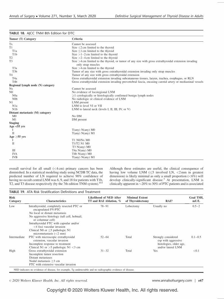
Copyright © 2020 Wolters Kluwer Health, Inc. All rights reserved.
overall survival for all small (<4 cm) primary cancers has beendiminished. In a statistical modeling study using NCDB TC data, thepredicted number of LN required to achieve 90% confidence ofhaving no occult central LNM was 6, 9, and 18 for patients with T1b,T2, and T3 disease respectively (by the 7th edition TNM system).453
Although these estimates are useful, the clinical consequence ofhaving low volume LNM (�5 involved LN, <2 mm in greatestdimension) is likely minimal as only a small proportion (<6%) willdevelop clinically-significant disease.3 At presentation, LNM isclinically apparent in�20% to 30% of PTC patients and is associated
TABLE 19. ATA Risk Stratification Definitions and Treatment
RiskCategory Characteristics
Likelihood of NED AfterTT and RAI Ablation, %
Minimal Extentof Thyroidectomy RAI?
Goal TSH,mU/L
Low Intrathyroidal, completely resected PTC orencapsulated FV-PTC
No local or distant metastasisNo aggressive histology (tall cell, hobnail,
or columnar cell)Intrathyroidal FTC with capsular and/or
<4 foci vascular invasionClinical N0 or �5 pathologic N1
micrometastasis (<2 mm)
78–91 Lobectomy Usually no 0.5–2
Intermediate PTC with microscopic extrathyroidalextension, vascular invasion
Incomplete response to treatmentClinical N1 or >5 pathologic N1 <3 cm
52–64 Total Strongly consideredesp with aggressivehistologies, older age,and/or lateral LNM
0.1–0.5
High Gross extrathyroidal extensionIncomplete tumor resectionDistant metastasesNodal metastasis �3 cmFTC with extensive vascular invasion
31–32 Total Yes <0.1
NED indicates no evidence of disease, for example, Tg undetectable and no radiographic evidence of disease.
TABLE 18. AJCC TNM 8th Edition for DTC
Tumor (T) Category Criteria
Tx Cannot be assessedT1 Size �2 cm limited to the thyroid
T1a Size �1 cm limited to the thyroidT1b Size >1–2 cm limited to the thyroid
T2 Size >2–4 cm limited to the thyroidT3 Size >4 cm limited to the thyroid, or tumor of any size with gross extrathyroidal extension invading
only strap musclesT3a Size >4 cm limited to the thyroidT3b Tumor of any size with gross extrathyroidal extension invading only strap muscles
T4 Tumor of any size with gross extrathyroidal extensionT4a Gross extrathyroidal extension invading subcutaneous tissues, larynx, trachea, esophagus, or RLNT4b Gross extrathyroidal extension invading prevertebral fascia, encasing carotid artery or mediastinal vessels
Regional lymph node (N) categoryNx Cannot be assessedN0 No evidence of locoregional LNM
N0a �1 cytologically or histologically confirmed benign lymph nodesN0b No radiologic or clinical evidence of LNM
N1 LNM presentN1a LNM to level VI or VIIN1b LNM to lateral neck (levels I, II, III, IV, or V)
Distant metastasis (M) categoryM0 No DMM1 DM present
StagingAge <55 yrs
I T(any) N(any) M0II T(any) N(any) M1
Age �55 yrsI T1 N0/Nx M0II T1/T2 N1 M0
T3 N(any) M0III T4a N(any) M0IVA T4b N(any) M0IVB T(any) N(any) M1
Annals of Surgery � Volume 271, Number 3, March 2020 Definitive Surgical Management of Thyroid Disease in Adults
� 2020 Wolters Kluwer Health, Inc. All rights reserved. www.annalsofsurgery.com | e67
Copyright © 2020 Wolters Kluwer Health, Inc. All rights reserved.
with an increased risk of local recurrence of up to 30%. Therecurrence risk is higher in patients with >5 LNM, and/or if extra-nodal extension is seen.483 Volume of LN disease is incorporated intothe 2015 ATA Risk stratification (Table 19) but not the AJCC TNM8th edition (Table 18).
Specific somatic genetic mutations also modify risk. In meta-analysis, BRAF V600E is associated with LNM (pooled OR 1.45,95% CI 1.24–1.69), recurrence (pooled OR 2.20, 95% CI 1.57–3.09), and overall survival (pooled OR 4.61, 95% CI 2.69–7.9).1014
Risk stratification improved when BRAF V600E status was consid-ered in conventional staging systems such as AMES, MACIS, TNM,and ATA Risk.1015 However, it remains unclear whether BRAFV600E independent of histologic factors is still predictive of poorprognosis,297.1016 In retrospective studies, TERT promoter mutationsare an independent predictor of poor disease-related outcomesincluding recurrence and overall survival.300,301,304,1017,1018 In arecent study that stratified DTC patients by response to therapyand TERT mutation status, prediction of mortality and recurrencewas improved.302 In another study, TERT mutation status improvedprognostication for patients already stratified by conventional stag-ing systems; although TERT in conjunction with BRAF or RAS hadthe highest risk of recurrence and disease-specific mortality, thedataset was too small to stratify further by dual mutations.1017 InFTC, a higher somatic mutational burden has been associated withpoor prognosis independent of histologic features.1019 Tumor prog-nostication using miRNA has undergone preliminary investigation,but more study is needed.1020
Completion ThyroidectomyWhen DTC is confirmed or diagnosed after initial lobectomy,
the first oncologic consideration is whether a completion thyroidec-tomy (see ‘‘Reoperation’’) is needed; this decision is primarilydependent on disease stage and risk of recurrence. In DTC, currentindications for completion thyroidectomy include consideration forpostoperative RAI therapy or concern for multifocal disease,3 butcompletion thyroidectomy is not necessarily indicated for all histo-logic DTC as lobectomy can be considered appropriate extent ofsurgery for low-risk tumors �4 cm (see ‘‘Indications, Extent, andOutcomes of Surgery’’). Histologic features are often what distin-guish low- from intermediate risk cancers, but this information isonly available postoperatively. In recent studies examining initiallobectomy for PTC �4 cm, completion thyroidectomy would poten-tially be required for high risk features diagnosed postoperatively(eg, aggressive histology, ETE, or multiple involved lymph nodes) in40% to 50% of patients.336,1021 Improved preoperative risk stratifi-cation may eventually be achieved using US findings, patient var-iables, and/or tumor molecular profile (see ‘‘Molecular Testing’’).Multidisciplinary communication to establish a risk-modified planfor surveillance is a key component in determining if completionthyroidectomy is needed.393
If MTC is diagnosed preoperatively, TT with central compart-ment ND is the minimum initial recommended procedure (see,‘‘FNAB diagnosis,’’ ‘‘Indications, Extent, and Outcomes of Sur-gery,’’ ‘‘Familial TC’’). However, if MTC is diagnosed postopera-tively, completion thyroidectomy is recommended if multifocal orresidual MTC is suspected, typically manifesting as persistent ele-vations in calcitonin and/or CEA levels or by imaging findings.56
Hereditary MTC and a detected germline RET mutation should alsobe treated with completion thyroidectomy as the incidence ofbilateral MTC is nearly 100%.56
Voice assessment should always occur prior to completionthyroidectomy, and if there is any concern for voice dysfunction apreoperative laryngeal exam should be performed (see ‘‘Laryngol-ogy’’). If RLN dysfunction is present, that is, from the initial surgery,
the options include deferring completion thyroidectomy until func-tion has returned or 2-stage ablation of the remaining lobe with RAI(below). If completion thyroidectomy is needed immediately forlocal control, the risk of permanent bilateral RLN injury should bespecifically discussed with the patient (see ‘‘Postoperative Care andComplications’’).3,95
In the absence of imaged structural disease, there is noindication for reoperative prophylactic CND for any TC histology.
Recommendation 64: Consider completion thyroidectomyfor high risk disease and/or when postoperative RAI therapy isindicated. (Strong recommendation, moderate-quality evidence)
Recommendation 65: Total thyroidectomy should be per-formed for patients undergoing prophylactic thyroidectomy formedullary thyroid cancer. (Strong recommendation, moderate-quality evidence)
Postoperative TreatmentWhen indicated, RAI ablation is administered for DTC at least
4 weeks after surgery and can be given either after T4 withdrawal orrecombinant TSH administration; both management protocols func-tion to raise TSH and increase absorption of iodine.3 RAI is given forremnant ablation to facilitate surveillance (�30 mCi), as adjuvanttherapy to treat microscopic disease (<150 mCi), and to treat distantmetastasis if not surgically resectable and for this purpose is giveneither as an empiric fixed dose or by dosimetry. Indications foradjuvant RAI ablation include: DTC considered high risk or inter-mediate risk especially with aggressive histologies, and lateral LNMat presentation (Table 19).3,332,1022 Regardless of use in staging,consideration for RAI ablation may be triggered when nonstimulatedpostoperative serum Tg levels are elevated postoperatively which isconcerning for either persistent TC or inadequate thyroid resection(see ‘‘Initial Thyroidectomy’’). Two-stage RAI administration isoccasionally necessary if large volume remnant tissue requiresablation but reoperation is not possible (ie, due to contralateralRLN paralysis). Low iodine diets are encouraged before RAI abla-tion, and a urine iodine level may help ensure clearance of the iodineload if iodinated contrast was used for imaging within 2 to 3 monthsof planned treatment.
TSH receptors are expressed in DTC and when stimulated byTSH, can cause DTC growth; thus, the use of T4 to suppress TSH is astrategy routinely used to reduce the risk of DTC recurrence.3,1022
Long-term TSH suppression even to subclinical levels can increasethe risks of atrial fibrillation, angina, and osteoporosis.13 As a result,the need for and degree of TSH suppression should follow estab-lished guidelines and be adjusted both for recurrence risk and patientcomorbidities. There is no indication for TSH suppression in MTCor ATC.
If complete surgical resection for DTC was performed, there isno indication for external beam radiation therapy (EBRT) except inhighly selected cases such as aerodigestive involvement; althoughEBRT may improve locoregional recurrence in this setting, overallsurvival is unchanged.1023,1024 Similarly, EBRT for locally advancedMTC may reduce locoregional recurrence, although not all studieshave demonstrated a benefit;1025,1026 recent guidelines recommendconsideration in selected high risk MTC patients with multiplecervical recurrences.56 EBRT does have a role in the palliativemanagement of ATC to improve local control and in some casessurvival.54 More precise radiation at potentially higher doses can bedelivered by IMRT and has been utilized for thyroid cancer patientswith access to specialized facilities.1027 Dose-limiting and latetoxicities may be lower with IMRT compared with EBRT.1028,1029
The risks associated with EBRT are potentially significant. Latetoxicities can include hoarseness, osteoradionecrosis, trismus, andbrain/central nerve injury.
Patel et al Annals of Surgery � Volume 271, Number 3, March 2020
e68 | www.annalsofsurgery.com � 2020 Wolters Kluwer Health, Inc. All rights reserved.
Copyright © 2020 Wolters Kluwer Health, Inc. All rights reserved.
There is no role for systemic adjuvant therapy for DTC orMTC. Tyrosine kinase inhibitors and small molecule inhibitors arereserved for patients with RAI nonavid and progressive metastaticdisease. Based on phase 3 trial data demonstrating superiority inprolonging progression-free survival, the FDA has approved vande-nitib and cabozantinib for advanced MTC and lenvatinib and sor-afenib for advanced DTC patients. In ATC, systemic chemotherapyand EBRT may improve outcomes.54 Typically, paclitaxel or doce-taxel (which are radiosensitizing) are combined with doxorubicinand/or cisplatin or carboplatin; data are limited to case series withsome survival up to 5-years.54 For BRAF V600E positive ATC, aphase 2 trial demonstrated that combination therapy with a BRAFinhibitor (dabrafenib) and a MEK inhibitor (trametinib) resulted in anoverall response rate of �69% which appeared to be durable andresulted in FDA approval for this treatment regimen.1030 Earlypalliative care intervention improves quality of life, optimizesresource utilization and reduces costs, and is recommended.1031
With biochemical cure, MTC 10-year survival is 97.7% andthe risk of recurrence is 5%.56 MTC patients should have a CEA andcalcitonin levels checked at least 2 to 3 months after surgery althoughlevels may take up to 6 months to reach nadir. If levels are stilldetectable, surveillance includes serial tumor markers and US every6 to 12 months. Calcitonin doubling time is independently anddirectly correlated to survival.106,552 Rapidly increasing CEA levelsmay indicate MTC dedifferentiation.56
Surveillance for DTC patients should include Tg and TgAblevels and US every 6 to 12 months.3 Patients with negative imagingand a suppressed Tg<0.2 ng/mL are considered to have an excellentresponse with a 1% to 4% risk of recurrence.3 In this cohort, cervicalrecurrences are the most common, and routine RAI whole body scansare not needed. Detectable Tg levels are common, and when imagingis negative, �30% will eventually have undetectable levels withoutintervention, and only �20% will eventually develop imageabledisease.3 The highest risk of DTC relapse occurs with imaging- orclinically-evident disease after initial treatment. Despite additionaltreatment including resection, up to 85% of such patients willcontinue to have biochemical and/or imaging evidence of persistentdisease, with low disease-specific survival requiring close intervalfollow-up.3 In patients with Tg levels >10 ng/mL, CT of the chestand/or 18FDG-PET imaging can be considered (see ‘‘Imaging’’).Serum Tg doubling time can be an important prognostic vari-able,1032,1033 and in a single-institution study, Tg doubling time<1 year was associated with 50% cause specific survival at 10years.1034
REOPERATION
Thyroid reoperation has been variously defined to meanthyroid resection after previous thyroid surgery, or as thyroidectomyin a previously dissected cervical compartment, including aftertracheostomy, parathyroidectomy, anterior cervical discectomy, orcarotid endarterectomy. This document uses the latter definition.
Although most patients undergoing thyroidectomy will require onlyan initial operation for definitive treatment, reoperation may beneeded in 3 categories: central or lateral resection of persistent/recurrent TC or LNM, resection of remnant ipsilateral thyroid tissueresection after prior partial thyroidectomy, and completion totalthyroidectomy after previous contralateral lobectomy.
IndicationsA common indication for thyroid reoperation is persistent or
recurrent TC. Approximately 30% of patients with DTC will havepersistent or recurrent anatomically structural LNM, 66% within10 years of initial surgery1035 (see ‘‘Nodal Dissection’’). A lesscommon manifestation (<5% of DTC cases in a 2017 series)1036 ispersistent or recurrent TC in remnant thyroid tissue, typically nearthe tubercle of Zuckerkandl in the previous resection bed. Whenevaluating a patient with persistent or recurrent TC, it is important todifferentiate between nodal versus remnant thyroid tissue recurrenceby careful review of imaging studies and FNAB results, as this mayimpact decision-making and planning for surgical intervention.
When persistent or recurrent TC is identified, decision-mak-ing regarding observation versus reoperation or other therapy,requires specific evaluation, risk-stratification and patient counseling(Table 20). Many subclinical, radiographically detected LNM orthyroid bed nodules will not exhibit growth or aggressive behaviorduring long-term observation. As 2011 study of 191 patients withUS-identified central compartment nodules or LN after initial thy-roidectomy reported stable disease in 91%; in the 9% with progres-sion, growth occurred slowly (mean 1.3 mm/yr) with no observedadverse outcomes such as local invasion or distant spread.1037 Asimilar 2012 study evaluated 166 patients with suspicious lateralcompartment LN after initial thyroidectomy for DTC; during a mean3.5-year follow-up period, 29% had interval progression (20% hadLNM growth of �3 mm, 9% of �5 mm), with no adverse out-comes.511 Two 2015 reports concluded that DTC patients withradiographically-detected recurrent LNM may be candidates forobservation for a central compartment LN �8 mm or lateral com-partment node�10 mm and minimal (<3–5 mm/yr) or no growth onserial imaging.3,1038 In addition, patients with a strong preference toavoid further operation, significant comorbidities, or progressivesystemic disease also may be selected for observation.
In addition to LN size, an additional criterion for determiningneed for reoperation is the trend of serial serum Tg levels. In a 2011study Tg doubling time was a strong prognostic indicator in DTC,1036
suggesting that progressive increase in Tg level is an indication forreoperation for clinically or radiologically apparent disease.
Recommendation 66: Selected patients with stable, low-volume persistent or recurrent LNM can undergo active surveil-lance. (Weak recommendation, low-quality evidence)
The management of persistent or recurrent MTC follows manyof the same principles as for DTC, although patients with MTC are
TABLE 20. Criteria for Patient Selection for Active Surveillance Versus Reoperation for Persistent or Recurrent Nodal Metastasesof DTC
Active Surveillance Reoperation
Central neck node �8 mm Central neck node >8 mmLateral neck node �10 mm Lateral neck node >10 mmNo/minimal nodal growth on serial imaging Nodal growth of >3–5 mm/yr on serial imagingThyroglobulin levels stable Thyroglobulin levels increasingPatient/endocrinologist preference for continued observation Patient/endocrinologist preference for reoperationSignificant comorbidities Able to tolerate reoperation
Annals of Surgery � Volume 271, Number 3, March 2020 Definitive Surgical Management of Thyroid Disease in Adults
� 2020 Wolters Kluwer Health, Inc. All rights reserved. www.annalsofsurgery.com | e69
Copyright © 2020 Wolters Kluwer Health, Inc. All rights reserved.
not candidates for RAI therapy. Nodal recurrence of MTC requiringreoperation occurs in 12% to 27% of patients.1039–1041 Serial cervicalimaging and measurements of calcitonin and CEA allow the clinicianto track disease progression and recurrence (see ‘‘Cancer Manage-ment’’). Calcitonin and CEA doubling times strongly correlate withMTC disease progression and clinical outcomes1042,1043 suggestingthat patients with radiographically detectable MTC LNM and pro-gressive rise in calcitonin or CEA levels should be consideredfor reoperation.
Although typically undesirable (see ‘‘Initial Thyroidec-tomy’’), partial initial thyroidectomy may occur unilaterally orbilaterally in management of TC, requiring later remnant resectionof tissue left in situ (ie, at the superior pole, isthmus, pyramidal lobe,or even substernally). Patients without a history of TC may alsorequire reoperation after partial thyroidectomy.1044 Inadequate pre-operative imaging may be a root cause of incomplete initial thyroid-ectomy. In reoperation, review of the previous operative andpathology reports is key to understanding the extent of prior dissec-tion and resection.
Contralateral thyroid reoperation may be required after initialdiagnostic lobectomy with a final histopathologic diagnosis ofclinically significant TC. Completion thyroidectomy (removal ofthe contralateral lobe) is recommended for patients with TC exhibit-ing intermediate to high-risk pathologic features, especially if RAIablation is planned (see ‘‘Cancer Management’’). If VFD is presentor the RLN was transected due to tumor involvement, the patient andsurgeon should have a detailed discussion regarding the risks andbenefits of completion thyroidectomy versus other options such as 2stage RAI ablation or observation (see ‘‘Cancer Management’’). Noprospective data exist regarding the optimal timing of completionthyroidectomy, and there is considerable variability in the recom-mended interval. We suggest that, if possible, it is best either to returnto the operating room within 1 to 2 weeks of initial surgery, or to waituntil considerably more time has passed (typically 8 to 12 wks) due tothe likelihood of encountering significant inflammation during theinterim period of healing.
Preoperative ManagementIn thyroid reoperation, one of the first priorities is to obtain all
possible documentation from the patient’s prior operation(s). Theoperative and pathology reports can detail the extent of previousdissection, the structures identified and/or removed, the gross extentof disease, and any injuries or complications that may have beenrecognized. The actual pathology specimens and slides should berequested for review, as there are often discrepancies in diagnosis orstaging on repeat evaluation by a pathologist with endocrine exper-tise.1045 Other key data include radiology reports, images, perioper-ative clinic records, and FNAB results for LN or suspicious remnanttissue.
Current imaging is another critical component of assessment,helping the surgeon to form a clear picture of the planned reoperativefield, the target(s) of resection, and adjacent vital structures. High-resolution US is the most commonly utilized study and can beperformed during office evaluation, immediately preceding reopera-tion, and/or in the operating room.1046,1047 CT with intravenouscontrast can provide detailed cross-sectional images of the relation-ships of enlarged LNs and soft tissue disease to adjacent structuresand major blood vessels.1048 MRI is an alternative to CT but it ismore costly and its accuracy varies depending on institutionalexpertise (see ‘‘Imaging’’).1049
PET-CT is not often required, but in the setting of elevated Tglevels may be of value in patients with DTC that is refractory to RAI.In addition, patients with higher-risk TC subtypes (such as tall cellvariant or PDTC) may be candidates for PET-CT prior to
reoperation,1050 which not only highlights cervical disease, butmay also identify other sites of distant metastasis, possibly alteringtreatment. RAI scanning may be useful in identifying foci ofpersistent or recurrent TC (see ‘‘Cancer Management’’), but theresolution of this modality is not high enough to allow for its use as astand-alone study for preoperative planning.
Prior to any thyroid reoperation, a TSH level should beevaluated, current medications should be reviewed, and a serumcalcium level should also be checked, as parathyroid glands mayhave been injured or removed during the previous operation. Theprior operative report and pathology report should be reviewed toconfirm if any parathyroids were removed previously.
In patients with DTC who have undergone prior total thyroid-ectomy, a Tg level with anti-Tg antibodies is also advised. Similarly,patients with MTC who are planned for reoperation should undergopreoperative calcitonin and CEA measurements, and also should bescreened for pheochromocytoma and hyperparathyroidism as appro-priate (see ‘‘Familial TC’’).
In elective thyroid reoperation, at minimum a preoperativevoice assessment should occur, and if there is concern for voicedysfunction, formal laryngeal evaluation should be performed toevaluate vocal fold mobility (see ‘‘Laryngology’’).800 This alsoapplies to any patient with prior cervical or thoracic surgery associ-ated with risk for RLN injury.
RisksDue to scarring and altered anatomy, the risks of thyroid
reoperation are higher than for initial surgery. A 2007 study of 685reoperations for both benign and malignant thyroid disease reportedsignificantly higher permanent complication rates (2.5% permanenthypocalcemia, 1.5% permanent VFD) compared with initial thyroid-ectomy.1051 Reoperation specifically for TC confers even higherrisks;1052–1056 a 2009 review reported 9.5% permanent hypopara-thyroidism and 6.4% permanent VFD, with both rates significantlyhigher than for initial thyroidectomy.1054 The parathyroid glands areat especially high risk in reoperation; a 2010 study reported a 31%rate of inadvertent partial or complete parathyroid resection onhistology.1057 Reoperation is also more technically challeng-ing.514,1058,1059
Operative ApproachWhen possible, the prior cervical incision should be utilized,
although this may not be technically feasible if the target of reopera-tion is located several cm away. In selected patients, to avoiddissection through scar (especially overlying the trachea in themidline), it may be useful in deeper layers to take a lateral approachto either the central or lateral compartments by entering the spacebetween the SCM and the lateral edge of the strap muscles, withlateral retraction of the carotid sheath structures. During reoperationin the central compartment, scarring may impede identification of theaerodigestive structures and initial placement of a nasogastric/oro-gastric tube or esophageal temperature probe can facilitate palpationof the esophagus. The extent of encountered scar tissue should benoted and when possible, described in the operative report.
The extent of reoperative LN resection is determined by thedegree of scar tissue; as with first-time operations, compartment-based node dissection is the optimal goal but may not be possible dueto altered anatomy. For lateral LNM, if the previous operation did notinvolve the lateral neck then a compartment-based dissection shouldbe performed; otherwise, a more focused resection of only theinvolved nodes may be needed to avoid injury to surroundingstructures.1060
Although uncommon, there still exist situations today where asurgeon embarks upon a planned thyroid operation but ends up
Patel et al Annals of Surgery � Volume 271, Number 3, March 2020
e70 | www.annalsofsurgery.com � 2020 Wolters Kluwer Health, Inc. All rights reserved.
Copyright © 2020 Wolters Kluwer Health, Inc. All rights reserved.
removing little or no thyroid tissue, because of inability to find thegland, inexperience, or unexpected intraoperative findings such assevere inflammation or tumor invasion.1060–1062 These outcomesshould be avoided as they do not benefit the patient, carry risk, andcreate scarring that will make future thyroid operation more difficultwith greater risk.
One of the primary challenges of reoperation is identifyingand preventing injury to the RLN and in the reoperative setting,RLNM may aid in mapping and/or confirmation of a visuallyidentified RLN.1063,1064 A 2014 study of 854 patients undergoingthyroid reoperation demonstrated a significant decrease in paresisand a trend towards a decrease in permanent VFD using RLNMversus visual identification alone.1065 A 2017 systematic review ofRLNM during high-risk thyroidectomy (including reoperation)reported a trend towards decreased rates of temporary and permanentRLN injury (see ‘‘Adjuncts and Approaches’’).743
Perioperative diagnostic evaluation of the reoperative patientoften requires specialized radiologic and endocrine pathology exper-tise. Moreover, decision-making about optimal management ofpersistent or recurrent disease often requires the input not only ofthe thyroid surgeon and endocrinologist, but also of nuclear medi-cine, radiation oncology and medical oncology team members in amultidisciplinary setting.
CONCLUDING REMARKS
‘‘In the ever-renewing society, what matures is a system orframework within which continuous innovation, renewal, and rebirthcan occur.’’1066
Evolving over more than a century, thyroidectomy today istypically a definitive and often curative procedure with a low risk ofmorbidity. Medical and technological advances have required thecreation of evidence-based guidelines to define practice, personalizecare, stratify risk, reduce health care costs, improve outcomes, andidentify rational challenges for future efforts. In the future, this workwill certainly and rightly need to be done again. The currentdocument is based on the existing published evidence and wasinformed by the clinical experience of 19 experts in thyroidology.The target audience is the practicing surgeon in a communityhospital, academic center, or training program. Throughout thewriting process, the authors’ intent was to advise surgeons aboutthe right thing to do for the patient. We also learned a great deal fromeach other. In humility and conscientious humanism, we are proud tooffer this consensus document to readers.
ACKNOWLEDGMENTSThe authors are indebted to Ms Lauren Santangelo at AMR
Management Services, and to Ms Carol Bykowski for administrativehelp. The authors also thank Dr Kia Nicholson for expert assistancein preparing Figure 2, and Dr Richard L. Simmons for advice. Theauthors are grateful to AAES leadership for commissioningthis work.
REFERENCES1. Sun GH, DeMonner S, Davis MM. Epidemiological and economic trends in
inpatient and outpatient thyroidectomy in the United States, 1996–2006.Thyroid. 2013;23:727–733.
2. Rubio GA, Koru-Sengal T, Vaghaiwalla TM, Parikh PP, Farra JC, Lew JI.Postoperative outcomes in Graves’ disease patients: results from thenationwide inpatient sample database. Thyroid. 2017;27:825–831.
3. Haugen BRM, Alexander EK, Bible KC, et al. 2015 American ThyroidAssociation Management Guidelines for adult patients with thyroid nodulesand differentiated thyroid cancer. Thyroid. 2016;26:1–133.
4. Gharib H, Papini E, Paschke R, et al. American Association of ClinicalEndocrinologists, Associazione Medici Endocrinologi, and European Thy-roid Association Medical guidelines for clinical practice for the diagnosis
and management of thyroid nodules: executive summary of recommenda-tions. J Endocrinol Invest. 2010;33(5 suppl):51–56.
5. Gharib H, Zeiger MA, Zini M, et al., AACE/AME Task Force on ThyroidNodules. American Association of Clinical Endocrinologists and Associa-zione Medici Endocrinologi medical guidelines for clinical practice for thediagnosis and management of thyroid nodules. Endocr Pract. 2006;12:63–102.
6. Wilhelm SM, Wang TS, Ruan DT, et al. The American Association ofEndocrine Surgeons Guidelines for definitive management of primaryhyperparathyroidism. JAMA Surg. 2016;151:959–968.
7. Sawka AM, Carty SE, Haugen BR, et al. American Thyroid AssociationGuidelines and Statements: past, present, and future. Thyroid. 2018;28:692–706.
8. Qaseem A, Snow V, Owens DK, et al. The development of clinical practiceguidelines and guidance statements of the American College of Physicians:summary of methods. Ann Intern Med. 2010;153:194–199.
9. Lewis MH, Gohagan JK, Merenstein DJ. The locality rule and the physi-cian’s dilemma: local medical practices vs the national standard of care.JAMA. 2007;297:2633–2637.
10. De Leo S, Lee SY, Braverman LE. Hyperthyroidism. Lancet. 2016;388:906–918.
11. Chaker L, Bianco AC, Jonklaas J, et al. Hypothyroidism. Lancet.2017;390:1550–1562.
12. Porterfield JR Jr, Thompson GB, Farley DR, et al. Evidence-basedmanagement of toxic multinodular goiter (Plummer’s Disease). World JSurg. 2008;32:1278–1284.
13. Ross DS, Burch HB, Cooper DS, et al. 2016 American Thyroid AssociationGuidelines for diagnosis and management of hyperthyroidism and othercauses of thyrotoxicosis. Thyroid. 2016;26:1343–1421.
14. Smith TJ, Hegedus L. Graves’ disease. N Engl J Med. 2017;376:185.
15. Berghout A, Wiersinga WM, Smits NJ, et al. Interrelationships betweenage, thyroid volume, thyroid nodularity, and thyroid function in patientswith sporadic nontoxic goiter. Am J Med. 1990;89:602–608.
16. Parma J, Duprez L, Van Sande J, et al. Somatic mutations in the thyrotropinreceptor gene cause hyperfunctioning thyroid adenomas. Nature. 1993;365:649–651.
17. Burch HB, Burman KD, Cooper DS. A 2011 survey of clinical practicepatterns in the management of Graves’ disease. J Clin Endocrinol Metab.2012;97:4549–4558.
18. Hollowell JG, Staehling NW, Flanders WD, et al. Serum TSH, T(4), andthyroid antibodies in the United States population (1988 to 1994): NationalHealth and Nutrition Examination Survey (NHANES III). J Clin Endo-crinol Metab. 2002;87:489–499.
19. Dahlgren M, Khosroshahi A, Nielsen GP, et al. Riedel’s thyroiditis andmultifocal fibrosclerosis are part of the IgG4-related systemic diseasespectrum. Arthritis Care Res (Hoboken). 2010;62:1312–1318.
20. Kakudo K, Li Y, Hirokawa M, et al. Diagnosis of Hashimoto’s thyroiditisand IgG4-related sclerosing disease. Pathol Int. 2011;61:175–183.
21. Li Y, Bai Y, Liu Z, et al. Immunohistochemistry of IgG4 can helpsubclassify Hashimoto’s autoimmune thyroiditis. Pathol Int. 2009;59:636–641.
22. Stan MN, Sonawane V, Sebo TJ, et al. Riedel’s thyroiditis association withIgG4-related disease. Clin Endocrinol (Oxf). 2017;86:425–430.
23. Lai X, Xia Y, Zhang B, et al. A meta-analysis of Hashimoto’s thyroiditis andpapillary thyroid carcinoma risk. Oncotarget. 2017;8:62414–62424.
24. Wormer BA, McHenry CR. Hashimoto’s thyroiditis: outcome of surgicalresection for patients with thyromegaly and compressive symptoms. Am JSurg. 2011;201:416–419.
25. Reiners C, Wegscheider K, Schicha H, et al. Prevalence of thyroid disordersin the working population of Germany: ultrasonography screening in 96,278unselected employees. Thyroid. 2004;14:926–932.
26. Kamran SC, Marqusee E, Kim MI, et al. Thyroid nodule size and predictionof cancer. J Clin Endocrinol Metab. 2013;98:564–570.
27. Cavallo A, Johnson DN, White MG, et al. Thyroid nodule size at ultrasoundas a predictor of malignancy and final pathologic size. Thyroid. 2017;27:641–650.
28. Hammad AY, Noureldine SI, Hu T, et al. A meta-analysis examining theindependent association between thyroid nodule size and malignancy.Gland Surg. 2016;5:312–317.
29. McHenry CR, Huh ES, Machekano RN. Is nodule size an independentpredictor of thyroid malignancy? Surgery. 2008;144:1062–1068.
30. Noureldine SI, Tufano RP. Association of Hashimoto’s thyroiditis andthyroid cancer. Curr Opin Oncol. 2015;27:21–25.
Annals of Surgery � Volume 271, Number 3, March 2020 Definitive Surgical Management of Thyroid Disease in Adults
� 2020 Wolters Kluwer Health, Inc. All rights reserved. www.annalsofsurgery.com | e71
Copyright © 2020 Wolters Kluwer Health, Inc. All rights reserved.
31. Shin JJ, Caragacianu D, Randolph GW. Impact of thyroid nodule size onprevalence and post-test probability of malignancy: a systematic review.Laryngoscope. 2015;125:263–272.
32. Shrestha M, Crothers BA, Burch HB. The impact of thyroid nodule size onthe risk of malignancy and accuracy of fine-needle aspiration: a 10-yearstudy from a single institution. Thyroid. 2012;22:1251–1256.
33. Sippel RS, Elaraj DM, Khanafshar E, et al. Tumor size predicts malignantpotential in Hurthle cell neoplasms of the thyroid. World J Surg.2008;32:702–707.
34. Cibas ES, Ali SZ. The bethesda system for reporting thyroid cytopathology.Thyroid. 2009;19:1159–1165.
35. Mai KT, Landry DC, Thomas J, et al. Follicular adenoma with papillaryarchitecture: a lesion mimicking papillary thyroid carcinoma. Histopathol-ogy. 2001;39:25–32.
36. Shi H, Bobanga I, McHenry CR. Are large thyroid nodules classified asbenign on fine needle aspiration more likely to harbor cancer? Am J Surg.2017;213:464–466.
37. Tzen CY, Huang YW, Fu YS. Is atypical follicular adenoma of the thyroid apreinvasive malignancy? Hum Pathol. 2003;34:666–669.
38. Lloyd RV, Osamura RY, Kloppel G, Rosai J, eds. WHO Classification ofTumours of Endocrine Organs (IARC WHO Classification of Tumours). 4thed. Lyon, France: IARC; 2017.
39. Asa SL, Giordano TJ, LiVolsi VA. Implications of the TCGA genomiccharacterization of papillary thyroid carcinoma for thyroid pathology: doesfollicular variant papillary thyroid carcinoma exist? Thyroid. 2015;25:1–2.
40. Davies L, Welch HG. Increasing incidence of thyroid cancer in the UnitedStates, 1973–2002. JAMA. 2006;295:2164–2167.
41. Davies L, Welch HG. Current thyroid cancer trends in the United States.JAMA Otolaryngol Head Neck Surg. 2014;140:317–322.
42. Chen AY, Jemal A, Ward EM. Increasing incidence of differentiated thyroidcancer in the United States, 1988–2005. Cancer. 2009;115:3801–3807.
43. Lin JS, Bowles EJA, Williams SB, et al. Screening for thyroid cancer:updated evidence report and systematic review for the US PreventiveServices Task Force. JAMA. 2017;317:1888–1903.
44. Lim H, Devesa SS, Sosa JA, et al. Trends in thyroid cancer incidenceand mortality in the United States, 1974–2013. JAMA. 2017;317:1338–1348.
45. Dionigi G, Kraimps JL, Schmid KW, et al. Minimally invasive follicularthyroid cancer (MIFTC): a consensus report of the European Society ofEndocrine Surgeons (ESES). Langenbecks Arch Surg. 2014;399:165–184.
46. Chindris AM, Casler JD, Bernet VJ, et al. Clinical and molecular features ofHurthle cell carcinoma of the thyroid. J Clin Endocrinol Metab.2015;100:55–62.
47. Ganly I, Makarov V, Deraje S, et al. Integrated genomic analysis of hurthlecell cancer reveals oncogenic drivers, recurrent mitochondrial mutations,and unique chromosomal landscapes. Cancer Cell. 2018;34:256–270.
48. Gopal RK, Kubler K, Calvo SE, et al. Widespread chromosomal losses andmitochondrial DNA alterations as genetic drivers in hurthle cell carcinoma.Cancer Cell. 2018;34:242–255.
49. Volante M, Landolfi S, Chiusa L, et al. Poorly differentiated carcinomas ofthe thyroid with trabecular, insular, and solid patterns: a clinicopathologicstudy of 183 patients. Cancer. 2004;100:950–957.
50. Volante M, Papotti M. Poorly differentiated thyroid carcinoma: 5 years afterthe 2004 WHO classification of endocrine tumours. Endocr Pathol.2010;21:1–6.
51. Volante M, Collini P, Nikiforov YE, et al. Poorly differentiated thyroidcarcinoma: the Turin proposal for the use of uniform diagnostic criteria andan algorithmic diagnostic approach. Am J Surg Pathol. 2007;31:1256–1264.
52. Hunt JL, Tometsko M, LiVolsi VA, et al. Molecular evidence of anaplastictransformation in coexisting well-differentiated and anaplastic carcinomasof the thyroid. Am J Surg Pathol. 2003;27:1559–1564.
53. Xu B, Ghossein R. Genomic landscape of poorly differentiated andanaplastic thyroid carcinoma. Endocr Pathol. 2016;27:205–212.
54. Smallridge RC, Ain KB, Asa SL, et al. American Thyroid Associationguidelines for management of patients with anaplastic thyroid cancer.Thyroid. 2012;22:1104–1139.
55. Landa I, Ibrahimpasic T, Boucai L, et al. Genomic and transcriptomichallmarks of poorly differentiated and anaplastic thyroid cancers. J ClinInvest. 2016;126:1052–1066.
56. Wells SA Jr, Asa SL, Dralle H, et al. Revised American ThyroidAssociation guidelines for the management of medullary thyroid carci-noma. Thyroid. 2015;25:567–610.
57. Baloch ZW, LiVolsi VA. C-cells and their associated lesions and conditions:a pathologists perspective. Turk Patoloji Derg. 2015;31(suppl 1):60–79.
58. Sharma A, Jasim S, Reading CC, et al. Clinical presentation and diagnosticchallenges of thyroid lymphoma: a cohort study. Thyroid. 2016;26:1061–1067.
59. Holm LE, Blomgren H, Lowhagen T. Cancer risks in patients with chroniclymphocytic thyroiditis. N Engl J Med. 1985;312:601–604.
60. Rossi D. Thyroid lymphoma: beyond antigen stimulation. Leuk Res.2009;33:607–609.
61. Matsuzuka F, Miyauchi A, Katayama S, et al. Clinical aspects of primarythyroid lymphoma: diagnosis and treatment based on our experience of 119cases. Thyroid. 1993;3:93–99.
62. Schneider AB, Ron E, Lubin J, et al. Dose-response relationships forradiation-induced thyroid cancer and thyroid nodules: evidence for theprolonged effects of radiation on the thyroid. J Clin Endocrinol Metab.1993;77:362–369.
63. Ron E. Cancer risks from medical radiation. Health Phys. 2003;85:47–59.
64. Iglesias ML, Schmidt A, Ghuzlan AA, et al. Radiation exposure and thyroidcancer: a review. Arch Endocrinol Metab. 2017;61:180–187.
65. Nikiforov YE. Radiation-induced thyroid cancer: what we have learnedfrom Chernobyl. Endocr Pathol. 2006;17:307–317.
66. Tronko MD, Howe GR, Bogdanova TI, et al. A cohort study of thyroidcancer and other thyroid diseases after the chornobyl accident: thyroidcancer in Ukraine detected during first screening. J Natl Cancer Inst.2006;98:897–903.
67. Ohira T, Takahashi H, Yasumura S, et al. Comparison of childhood thyroidcancer prevalence among 3 areas based on external radiation dose after theFukushima Daiichi nuclear power plant accident: The Fukushima healthmanagement survey. Medicine (Baltimore). 2016;95:e4472.
68. Ohira T, Takahashi H, Yasumura S, et al. Associations between childhoodthyroid cancer and external radiation dose after the Fukushima DaiichiNuclear Power Plant Accident. Epidemiology. 2018;29:e32–e34.
69. Levin RJ, De Simone NF, Slotkin JF, et al. Incidence of thyroid cancersurrounding Three Mile Island nuclear facility: the 30-year follow-up.Laryngoscope. 2013;123:2064–2071.
70. Sinnott B, Ron E, Schneider AB. Exposing the thyroid to radiation: areview of its current extent, risks, and implications. Endocr Rev.2010;31:756–773.
71. de Vathaire F, Haddy N, Allodji RS, et al. Thyroid radiation dose and otherrisk factors of thyroid carcinoma following childhood cancer. J ClinEndocrinol Metab. 2015;100:4282–4290.
72. Bhatti P, Veiga LH, Ronckers CM, et al. Risk of second primary thyroidcancer after radiotherapy for a childhood cancer in a large cohort study: anupdate from the childhood cancer survivor study. Radiat Res. 2010;174:741–752.
73. Veiga LH, Holmberg E, Anderson H, et al. Thyroid cancer after childhoodexposure to external radiation: an updated pooled analysis of 12 studies.Radiat Res. 2016;185:473–484.
74. Grantzau T, Overgaard J. Risk of second non-breast cancer among patientstreated with and without postoperative radiotherapy for primary breastcancer: a systematic review and meta-analysis of population-based studiesincluding 522,739 patients. Radiother Oncol. 2016;121:402–413.
75. Metzger R, Milas M. Inherited cancer syndromes and the thyroid: an update.Curr Opin Oncol. 2014;26:51–61.
76. Milas M, Mester J, Metzger R, et al. Should patients with Cowden syndromeundergo prophylactic thyroidectomy? Surgery. 2012;152:1201–1210.
77. Brauckhoff M, Machens A, Lorenz K, et al. Surgical curability of medullarythyroid cancer in multiple endocrine neoplasia 2B: a changing perspective.Ann Surg. 2014;259:800–806.
78. Straccia P, Mosseri C, Brunelli C, et al. Diagnosis and treatment ofmetastases to the thyroid gland: a meta-analysis. Endocr Pathol.2017;28:112–120.
79. Romero Arenas MA, Ryu H, Lee S, et al. The role of thyroidectomy inmetastatic disease to the thyroid gland. Ann Surg Oncol. 2014;21:434–439.
80. Chen AY, Bernet VJ, Carty SE, et al. American Thyroid Associationstatement on optimal surgical management of goiter. Thyroid. 2014;24:181–189.
81. Vansant MB, Kunduk M, McWhorter AJ. A review of postsurgical dyspha-gia in nonmalignant disease. Curr Opin Otolaryngol Head Neck Surg.2016;24:477–482.
82. Holler T, Anderson J. Prevalence of voice & swallowing complaints in Pre-operative thyroidectomy patients: a prospective cohort study. J OtolaryngolHead Neck Surg. 2014;43:28.
Patel et al Annals of Surgery � Volume 271, Number 3, March 2020
e72 | www.annalsofsurgery.com � 2020 Wolters Kluwer Health, Inc. All rights reserved.
Copyright © 2020 Wolters Kluwer Health, Inc. All rights reserved.
83. Greenblatt DY, Sippel R, Leverson G, et al. Thyroid resection improvesperception of swallowing function in patients with thyroid disease. World JSurg. 2009;33:255–260.
84. Burns P, Timon C. Thyroid pathology and the globus symptom: are theyrelated? A two year prospective trial. J Laryngol Otol. 2007;121:242–245.
85. Nam IC, Choi H, Kim ES, et al. Characteristics of thyroid nodules causingglobus symptoms. Eur Arch Otorhinolaryngol. 2015;272:1181–1188.
86. Shen WT, Kebebew E, Duh QY, et al. Predictors of airwaycomplications after thyroidectomy for substernal goiter. Arch Surg.2004;139:656–659.
87. Stang MT, Armstrong MJ, Ogilvie JB, et al. Positional dyspnea and trachealcompression as indications for goiter resection. Arch Surg. 2012;147:621–626.
88. Jonklaas J, Bianco AC, Bauer AJ, et al. Guidelines for the treatment ofhypothyroidism: prepared by the american thyroid association task force onthyroid hormone replacement. Thyroid. 2014;24:1670–1751.
89. Caulley L, Johnson-Obaseki S, Luo L, Javidnia H. Risk factors forpostoperative complications in total thyroidectomy: a retrospective, risk-adjusted analysis from the National Surgical Quality Improvement Pro-gram. Medicine (Baltimore). 2017;96:e5752.
90. Abraham CR, Ata A, Carsello CB, Chan TL, Stain SC, Beyer TD. A NSQIPrisk assessment for thyroid surgery based on comorbidities. J Am Coll Surg.2014;218:1231–1237.
91. Schmid D, Ricci C, Behrens G, Leitzmann MF. Adiposity and risk ofthyroid cancer: a systematic review and meta-analysis. Obes Rev.2015;16:1042–1054.
92. Kitahara CM, McCullough ML, Franceschi S, et al. Anthropometric factorsand thyroid cancer risk by histological subtype: pooled analysis of 22prospective studies. Thyroid. 2016;26:306–318.
93. Tan GH, Gharib H, Reading CC. Solitary thyroid nodule. Comparisonbetween palpation and ultrasonography. Arch Intern Med. 1995;155:2418–2423.
94. De Filippis EA, Sabet A, Sun MR, Garber JR. Pemberton’s sign: explainednearly 70 years later. J Clin Endocrinol Metab. 2014;99:1949–1954.
95. Chandrasekhar SS, Randolph GW, Seidman MD, et al. Clinical practiceguideline: improving voice outcomes after thyroid surgery. OtolaryngolHead Neck Surg. 2013;148(6 suppl):S1–S37.
96. Zanocco K, Kaltman DJ, Wu JX, et al. Cost effectiveness of routinelaryngoscopy in the surgical treatment of differentiated thyroid cancer.Ann Surg Oncol. 2018;25:949–956.
97. Choong KC, McHenry CR. Thyroid cancer in patients with toxic nodulargoiter: is the incidence increasing? Am J Surg. 2015;209:974–976.
98. Smith JJ, Chen X, Schneider DF, et al. Toxic nodular goiter and cancer: acompelling case for thyroidectomy. Ann Surg Oncol. 2013;20:1336–1340.
99. Morita SY, Somervell H, Umbricht CB, et al. Evaluation for concomitantthyroid nodules and primary hyperparathyroidism in patients undergoingparathyroidectomy or thyroidectomy. Surgery. 2008;144:862–866.
100. Murray SE, Sippel RS, Chen H. Incidence of concomitant hyperparathy-roidism in patients with thyroid disease requiring surgery. J Surg Res.2012;178:264–267.
101. Jovanovic MD, Zivaljevic VR, Diklic AD, Rovcanin BR, V Zoric G,Paunovic IR. Surgical treatment of concomitant thyroid and parathyroiddisorders: analysis of 4882 cases. Eur Arch Otorhinolaryngol. 2017;274:997–1004.
102. Prinz RA, Paloyan E, Lawrence AM, Barbato AL, Braithwaite SS, BrooksMH. Unexpected parathyroid disease discovered at thyroidectomy inirradiated patients. Am J Surg. 1981;142:355–357.
103. Elisei R, Bottici V, Luchetti F, et al. Impact of routine measurement ofserum calcitonin on the diagnosis and outcome of medullary thyroid cancer:experience in 10,864 patients with nodular thyroid disorders. J ClinEndocrinol Metab. 2004;89:163–168.
104. Costante G, Durante C, Francis Z, et al. Determination of calcitonin levelsin C-cell disease: clinical interest and potential pitfalls. Nat Clin PractEndocrinol Metab. 2009;5:35–44.
105. Network, N.C.C. Thyroid Carcinoma (Version 2.2015). http://www.nccn.org/professionals/physician_gls/pdf/thyroid.pdf. Accessed July 19,2019
106. Machens A, Dralle H. Biomarker-based risk stratification for previouslyuntreated medullary thyroid cancer. J Clin Endocrinol Metab. 2010;95:2655–2663.
107. Machens A, Schneyer U, Holzhausen HJ, et al. Prospects of remission inmedullary thyroid carcinoma according to basal calcitonin level. J ClinEndocrinol Metab. 2005;90:2029–2034.
108. Yeh MW, Bauer AJ, Bernet VA, et al. American Thyroid Associationstatement on preoperative imaging for thyroid cancer surgery. Thyroid.2015;25:3–14.
109. Kouvaraki MA, Shapiro SE, Fornage BD, et al. Role of preoperativeultrasonography in the surgical management of patients with thyroid cancer.Surgery. 2003;134:946–954.
110. Kwak JY. Thyroid ultrasonography for personalized approach at thyroidnodules. Endocrine. 2016;52:181–182.
111. Kwak JY, Han KH, Yoon JH, et al. Thyroid imaging reporting and datasystem for US features of nodules: a step in establishing better stratificationof cancer risk. Radiology. 2011;260:892–899.
112. Moon HG, Jung EJ, Park ST, et al. Role of ultrasonography in predictingmalignancy in patients with thyroid nodules. World J Surg. 2007;31:1410–1416.
113. Moon HJ, Kim EK, Kwak JY. Malignancy risk stratification in thyroidnodules with benign results on cytology: combination of thyroid imagingreporting and data system and Bethesda system. Ann Surg Oncol.2014;21:1898–1903.
114. Salmaslioglu A, Erbil Y, Dural C, et al. Predictive value of sonographicfeatures in preoperative evaluation of malignant thyroid nodules in amultinodular goiter. World J Surg. 2008;32:1948–1954.
115. Moon HJ, Kwak JY, Kim MJ, et al. Can vascularity at power Doppler UShelp predict thyroid malignancy? Radiology. 2010;255:260–269.
116. Papini E, Guglielmi R, Bianchini A, et al. Risk of malignancy in non-palpable thyroid nodules: predictive value of ultrasound and color-Dopplerfeatures. J Clin Endocrinol Metab. 2002;87:1941–1946.
117. Gul K, Ersoy R, Dirikoc A, et al. Ultrasonographic evaluation of thyroidnodules: comparison of ultrasonographic, cytological, and histopatholog-ical findings. Endocrine. 2009;36:464–472.
118. Cappelli C, Pirola I, Cumetti D, et al. Is the anteroposterior and transversediameter ratio of nonpalpable thyroid nodules a sonographic criteria forrecommending fine-needle aspiration cytology? Clin Endocrinol (Oxf).2005;63:689–693.
119. Tang AL, Falciglia M, Yang H, et al. Validation of American ThyroidAssociation ultrasound risk assessment of thyroid nodules selected forultrasound fine-needle aspiration. Thyroid. 2017;27:1077–1082.
120. Horvath E, Majlis S, Rossi R, et al. An ultrasonogram reporting system forthyroid nodules stratifying cancer risk for clinical management. J ClinEndocrinol Metab. 2009;94:1748–1751.
121. Tae HJ, Lim DJ, Baek KH, et al. Diagnostic value of ultrasonography todistinguish between benign and malignant lesions in the management ofthyroid nodules. Thyroid. 2007;17:461–466.
122. Ito Y, Amino N, Yokozawa T, et al. Ultrasonographic evaluation of thyroidnodules in 900 patients: comparison among ultrasonographic, cytological,and histological findings. Thyroid. 2007;17:1269–1276.
123. Moon WJ, Baek JH, Jung SL, et al. Ultrasonography and the ultrasound-based management of thyroid nodules: consensus statement and recom-mendations. Korean J Radiol. 2011;12:1–14.
124. Moon WJ, Jung SL, Lee JH, et al. Benign and malignant thyroid nodules:US differentiation—multicenter retrospective study. Radiology. 2008;247:762–770.
125. Brito JP, Gionfriddo MR, Al Nofal A, et al. The accuracy of thyroid noduleultrasound to predict thyroid cancer: systematic review and meta-analysis. JClin Endocrinol Metab. 2014;99:1253–1263.
126. Kim DS, Kim JH, Na DG, et al. Sonographic features of follicular variantpapillary thyroid carcinomas in comparison with conventional papillarythyroid carcinomas. J Ultrasound Med. 2009;28:1685–1692.
127. Yang GC, Fried KO. Most thyroid cancers detected by sonography lackintranodular vascularity on color doppler imaging: review of the literatureand sonographic-pathologic correlations for 698 thyroid neoplasms. JUltrasound Med. 2017;36:89–94.
128. Jeh SK, Jung SL, Kim BS, Lee YS. Evaluating the degree of conformity ofpapillary carcinoma and follicular carcinoma to the reported ultrasono-graphic findings of malignant thyroid tumor. Korean J Radiol. 2007;8:192–197.
129. Iared W, Shigueoka DC, Cristofoli JC, et al. Use of color Dopplerultrasonography for the prediction of malignancy in follicular thyroidneoplasms: systematic review and meta-analysis. J Ultrasound Med.2010;29:419–425.
130. Khadra H, Bakeer M, Hauch A, Hu T, Kandil E. Is vascular flow a predictorof malignant thyroid nodules? A meta-analysis. Gland Surg. 2016;5:576–582.
131. Grant EG, Tessler FN, Hoang JK, et al. Thyroid ultrasound reportinglexicon: white paper of the ACR Thyroid Imaging, Reporting and Data
Annals of Surgery � Volume 271, Number 3, March 2020 Definitive Surgical Management of Thyroid Disease in Adults
� 2020 Wolters Kluwer Health, Inc. All rights reserved. www.annalsofsurgery.com | e73
Copyright © 2020 Wolters Kluwer Health, Inc. All rights reserved.
System (TIRADS) Committee. J Am Coll Radiol. 2015;12(12 pt A):1272–1279.
132. Tessler FN, Middleton WD, Grant EG, et al. ACR Thyroid Imaging,Reporting and Data System (TI-RADS): White Paper of the ACR TI-RADSCommittee. J Am Coll Radiol. 2017;14:587–595.
133. Choi YJ, Baek JH, Baek SH, et al. Web-based malignancy risk estimationfor thyroid nodules using ultrasonography characteristics: development andvalidation of a predictive model. Thyroid. 2015;25:1306–1312.
134. Zhang J, Liu BJ, Xu HX, et al. Prospective validation of an ultrasound-basedthyroid imaging reporting and data system (TI-RADS) on 3980 thyroidnodules. Int J Clin Exp Med. 2015;8:5911–5917.
135. Kwak JY, Kim EK, Moon HJ, et al. Parathyroid incidentalomas detected onroutine ultrasound-directed fine-needle aspiration biopsy in patientsreferred for thyroid nodules and the role of parathyroid hormone analysisin the samples. Thyroid. 2009;19:743–748.
136. Ghervan C, Silaghi A, Nemes C. Parathyroid incidentaloma detected duringthyroid sonography—prevalence and significance beyond images. MedUltrason. 2012;14:187–191.
137. Cho M, Oweity T, Brandler TC, Fried K, Levine P. Distinguishing para-thyroid and thyroid lesions on ultrasound-guided fine-needle aspiration: acorrelation of clinical data, ancillary studies, and molecular analysis.Cancer Cytopathol. 2017;125:674–682.
138. Langer JE, Mandel SJ. Sonographic imaging of cervical lymph nodes inpatients with thyroid cancer. Neuroimaging Clin N Am. 2008;18:479–489.
139. Leboulleux S, Girard E, Rose M, et al. Ultrasound criteria of malignancy forcervical lymph nodes in patients followed up for differentiated thyroidcancer. J Clin Endocrinol Metab. 2007;92:3590–3594.
140. Ito Y, Higashiyama T, Takamura Y, et al. Risk factors for recurrence to thelymph node in papillary thyroid carcinoma patients without preoperativelydetectable lateral node metastasis: validity of prophylactic modified radicalneck dissection. World J Surg. 2007;31:2085–2091.
141. Choi JS, Kim J, Kwak JY, Kim MJ, Chang HS, Kim EK. Preoperativestaging of papillary thyroid carcinoma: comparison of ultrasound imagingand CT. AJR Am J Roentgenol. 2009;193:871–878.
142. Hwang HS, Orloff LA. Efficacy of preoperative neck ultrasound in thedetection of cervical lymph node metastasis from thyroid cancer. Laryngo-scope. 2011;121:487–491.
143. Solbiati L, Osti V, Cova L, Tonolini M. Ultrasound of thyroid, parathyroidglands and neck lymph nodes. Eur Radiol. 2001;11:2411–2424.
144. Popadich A, Levin O, Lee JC, et al. A multicenter cohort study of totalthyroidectomy and routine central lymph node dissection for cN0 papillarythyroid cancer. Surgery. 2011;150:1048–1057.
145. Gimm O, Rath FW, Dralle H. Pattern of lymph node metastases in papillarythyroid carcinoma. Br J Surg. 1998;85:252–254.
146. Wu G, Fraser S, Pai SI, Farrag TY, Ladenson PW, Tufano RP. Determiningthe extent of lateral neck dissection necessary to establish regional diseasecontrol and avoid reoperation after previous total thyroidectomy andradioactive iodine for papillary thyroid cancer. Head Neck.2012;34:1418–1421.
147. Farrag T, Lin F, Brownlee N, Kim M, Sheth S, Tufano RP. Is routinedissection of level II-B and V-A necessary in patients with papillary thyroidcancer undergoing lateral neck dissection for FNA-confirmed metastases inother levels. World J Surg. 2009;33:1680–1683.
148. Kuna SK, Bracic I, Tesic V, Kuna K, Herceg GH, Dodig D. Ultrasono-graphic differentiation of benign from malignant neck lymphadenopathy inthyroid cancer. J Ultrasound Med. 2006;25:1531–1537.
149. Gorges R, Eising EG, Fotescu D, et al. Diagnostic value of high-resolutionB-mode and power-mode sonography in the follow-up of thyroid cancer.Eur J Ultrasound. 2003;16:191–206.
150. Sipos JA. Advances in ultrasound for the diagnosis and management ofthyroid cancer. Thyroid. 2009;19:1363–1372.
151. Wunderbaldinger P, Harisinghani MG, Hahn PF, et al. Cystic lymph nodemetastases in papillary thyroid carcinoma. AJR Am J Roentgenol.2002;178:693–697.
152. Ahuja AT, King W, Metreweli C. Role of ultrasonography in thyroidmetastases. Clin Radiol. 1994;49:627–629.
153. Ahuja AT, Ying M, Ho SS, Metreweli C. Distribution of intranodal vesselsin differentiating benign from metastatic neck nodes. Clin Radiol.2001;56:197–201.
154. American Thyroid Association Surgery Working Group, American Asso-ciation of Endocrine Surgeons, American Academy of Otolaryngology-Head and Neck Surgery, et al. Consensus statement on the terminology andclassification of central neck dissection for thyroid cancer. Thyroid.2009;19:1153–1158.
155. Al-azawi D, Mann GB, Judson RT, Miller JA. Endocrine surgeon-per-formed US guided thyroid FNAC is accurate and efficient. World J Surg.2012;36:1947–1952.
156. Kumbhar SS, O’Malley RB, Robinson TJ, et al. Why thyroid surgeons arefrustrated with radiologists: lessons learned from pre- and postoperative US.Radiographics. 2016;36:2141–2153.
157. Monteiro R, Han A, Etiwy M, et al. Importance of surgeon-performedultrasound in the preoperative nodal assessment of patients with potentialthyroid malignancy. Surgery. 2018;163:112–117.
158. Wong KP, Au KP, Lam S, Lang BH. Lessons learned after 1000 cases oftranscutaneous laryngeal ultrasound (TLUSG) with laryngoscopic valida-tion: is there a role of TLUSG in patients indicated for laryngoscopicexamination before thyroidectomy? Thyroid. 2017;27:88–94.
159. Kandil E, Deniwar A, Noureldine SI, et al. Assessment of vocal foldfunction using transcutaneous laryngeal ultrasonography and flexible lar-yngoscopy. JAMA Otolaryngol Head Neck Surg. 2016;142:74–78.
160. Parangi S. Editorial: Translaryngeal vocal cord ultrasound: Ready for primetime. Surgery. 2016;159:67–69.
161. Carneiro-Pla D, Miller BS, Wilhelm SM, et al. Feasibility of surgeon-performed transcutaneous vocal cord ultrasonography in identifying vocalcord mobility: a multi-institutional experience. Surgery. 2014;156:1597–1602.
162. Carneiro-Pla D, Solorzano CC, Wilhelm SM. Impact of vocal cordultrasonography on endocrine surgery practices. Surgery. 2016;159:58–64.
163. Moon HJ, Kim EK, Yoon JH, Kwak JY. Differences in the diagnosticperformances of staging US for thyroid malignancy according to experi-ence. Ultrasound Med Biol. 2012;38:568–573.
164. Bhatki AM, Brewer B, Robinson-Smith T, Nikiforov Y, Steward DL.Adequacy of surgeon-performed ultrasound-guided thyroid fine-needleaspiration biopsy. Otolaryngol Head Neck Surg. 2008;139:27–31.
165. Jeong HS, Baek CH, Son YI, et al. Integrated 18F-FDG PET/CT for theinitial evaluation of cervical node level of patients with papillary thyroidcarcinoma: comparison with ultrasound and contrast-enhanced CT. ClinEndocrinol (Oxf). 2006;65:402–407.
166. Padovani RP, Kasamatsu TS, Nakabashi CCD, et al. One month is sufficientfor urinary iodine to return to its baseline value after the use of water-solubleiodinated contrast agents in post-thyroidectomy patients requiring radio-iodine therapy. Thyroid. 2012;22:926–930.
167. Rago T, Santini F, Scutari M, Pinchera A, Vitti P. Elastography: newdevelopments in ultrasound for predicting malignancy in thyroid nodules.J Clin Endocrinol Metab. 2007;92:2917–2922.
168. Moon HJ, Sung JM, Kim EK, Yoon JH, Youk JH, Kwak JY. Diagnosticperformance of gray-scale US and elastography in solid thyroid nodules.Radiology. 2012;262:1002–1013.
169. Azizi G, Keller J, Lewis M, Puett D, Rivenbark K, Malchoff C. Performanceof elastography for the evaluation of thyroid nodules: a prospective study.Thyroid. 2013;23:734–740.
170. Are C, Hsu JF, Schoder H, et al. FDG-PET detected thyroid incidentalomas:need for further investigation? Ann Surg Oncol. 2007;14:239–247.
171. Katz SC, Shaha A. PET-associated incidental neoplasms of the thyroid. JAm Coll Surg. 2008;207:259–264.
172. Barrio M, Czernin J, Yeh MW, et al. The incidence of thyroid cancer in focalhypermetabolic thyroid lesions: an 18F-FDG PET/CT study in more than6000 patients. Nucl Med Commun. 2016;37:1290–1296.
173. Hari Kumar KV, Pasupuleti V, Jayaraman M, Abhyuday V, Rayudu BR,Modi KD. Role of thyroid Doppler in differential diagnosis of thyrotoxico-sis. Endocr Pract. 2009;15:6–9.
174. Rosario PW, Santos JB, Nunes NS, da Silva AL, Calsolari MR. Color flowDoppler sonography for the etiologic diagnosis of thyrotoxicosis. HormMetab Res. 2014;46:505–509.
175. Avram AM. Radioiodine scintigraphy with SPECT/CT: an importantdiagnostic tool for thyroid cancer staging and risk stratification. J NuclMed Technol. 2014;42:170–180.
176. Grunwald F, Kalicke T, Feine U, Lietzenmayer R, et al. Fluorine-18fluorodeoxyglucose positron emission tomography in thyroid cancer:results of a multicentre study. Eur J Nucl Med. 1999;26:1547–1552.
177. Francis CL, Nalley C, Fan C, Bodenner D, Stack BC Jr. 18F-fluorodeox-yglucose and 131I radioguided surgical management of thyroid cancer.Otolaryngol Head Neck Surg. 2012;146:26–32.
178. Schluter B, Bohuslavizki KH, Beyer W, Plotkin M, Buchert R, Clausen M.Impact of FDG PET on patients with differentiated thyroid cancer whopresent with elevated thyroglobulin and negative 131I scan. J Nucl Med.2001;42:71–76.
Patel et al Annals of Surgery � Volume 271, Number 3, March 2020
e74 | www.annalsofsurgery.com � 2020 Wolters Kluwer Health, Inc. All rights reserved.
Copyright © 2020 Wolters Kluwer Health, Inc. All rights reserved.
179. Wang W, Macapinlac H, Larson SM, et al. [18F]-2-fluoro-2-deoxy-D-glucose positron emission tomography localizes residual thyroid cancerin patients with negative diagnostic (131)I whole body scans andelevated serum thyroglobulin levels. J Clin Endocrinol Metab.1999;84:2291–2302.
180. Robbins RJ, Wan Q, Grewal RK, et al. Real-time prognosis for metastaticthyroid carcinoma based on 2-[18F]fluoro-2-deoxy-D-glucose-positronemission tomography scanning. J Clin Endocrinol Metab. 2006;91:498–505.
181. Finkelstein SE, Grigsby PW, Siegel BA, Dehdashti F, Moley JF, HallBL. Combined [18F]Fluorodeoxyglucose positron emission tomographyand computed tomography (FDG-PET/CT) for detection of recurrent,131I-negative thyroid cancer. Ann Surg Oncol. 2008;15:286–292.
182. Kim SJ, Lee TH, Kim IJ, Kim YK. Clinical implication of F-18 FDGPET/CT for differentiated thyroid cancer in patients with negativediagnostic iodine-123 scan and elevated thyroglobulin. Eur J Radiol.2009;70:17–24.
183. Mirallie E, Guillan T, Bridji B, et al. Therapeutic impact of 18FDG-PET/CTin the management of iodine-negative recurrence of differentiated thyroidcarcinoma. Surgery. 2007;142:952–958.
184. Shammas A, Degirmenci B, Mountz JM, et al. 18F-FDG PET/CT in patientswith suspected recurrent or metastatic well-differentiated thyroid cancer. JNucl Med. 2007;48:221–226.
185. Robbins RJ, Srivastava S, Shaha A, et al. Factors influencing the basal andrecombinant human thyrotropin-stimulated serum thyroglobulin in patientswith metastatic thyroid carcinoma. J Clin Endocrinol Metab. 2004;89:6010–6016.
186. Agrawal A, Hall NC, Ringel MD, Povoski SP, Martin EW Jr. Combined useof perioperative TSH-stimulated (18)F-FDG PET/CT imaging and gammaprobe radioguided surgery to localize and verify resection of iodine scan-negative recurrent thyroid carcinoma. Laryngoscope. 2008;118:2190–2194.
187. Leboulleux S, Schroeder PR, Busaidy NL, et al. Assessment of theincremental value of recombinant thyrotropin stimulation before 2-[18F]-Fluoro-2-deoxy-D-glucose positron emission tomography/computedtomography imaging to localize residual differentiated thyroid cancer. JClin Endocrinol Metab. 2009;94:1310–1316.
188. Miller JM, Hamburger JI, Kini S. Diagnosis of thyroid nodules. Use of fine-needle aspiration and needle biopsy. JAMA. 1979;241:481–484.
189. Wetzig NR, Giddings AE. Solitary thyroid nodule: audit shows improvedcare requires cytological diagnosis. Ann R Coll Surg Engl. 1989;71:316–319.
190. Sosa JA, Hanna JW, Robinson KA, Lanman RB. Increases in thyroid nodulefine-needle aspirations, operations, and diagnoses of thyroid cancer in theUnited States. Surgery. 2013;154:1420–1426.
191. Machens A, Holzhausen HJ, Dralle H. The prognostic value of primarytumor size in papillary and follicular thyroid carcinoma. Cancer.2005;103:2269–2273.
192. Cavallo A, Johnson DN, White MG, et al. Thyroid nodule size at ultrasoundas a predictor of malignancy and final pathologic size. Thyroid.2017;27:641–650.
193. Cappelli C, Pirola I, Agosti B, et al. Complications after fine-needleaspiration cytology: a retrospective study of 7449 consecutive thyroidnodules. Br J Oral Maxillofac Surg. 2017;55:266–269.
194. Abu-Yousef MM, Larson JH, Kuehn DM, Wu AS, Laroia AT. Safety ofultrasound-guided fine needle aspiration biopsy of neck lesions in patientstaking antithrombotic/anticoagulant medications. Ultrasound Q. 2011;27:157–159.
195. Lyle MA, Dean DS. Ultrasound-guided fine-needle aspiration biopsy ofthyroid nodules in patients taking novel oral anticoagulants. Thyroid.2015;25:373–376.
196. Pitman MB, Abele J, Ali SZ, et al. Techniques for thyroid FNA: a synopsisof the National Cancer Institute Thyroid Fine-Needle Aspiration State of theScience Conference. Diagn Cytopathol. 2008;36:407–424.
197. Guo HQ, Zhang ZH, Zhao H, Niu LJ, Chang Q, Pan QJ. Factors influencingthe reliability of thyroid fine-needle aspiration: analysis of thyroid nodulesize, guidance mode for aspiration and preparation method. Acta Cytol.2015;59:169–174.
198. Izquierdo R, Arekat MR, Knudson PE, et al. Comparison of palpation-guided versus ultrasound-guided fine-needle aspiration biopsies of thyroidnodules in an outpatient endocrinology practice. Endocr Pract.2006;12:609–614.
199. Can AS, Peker K. Comparison of palpation-versus ultrasound-guided fine-needle aspiration biopsies in the evaluation of thyroid nodules. BMC ResNotes. 2008;1:12.
200. Can AS. Cost-effectiveness comparison between palpation- and ultrasound-guided thyroid fine-needle aspiration biopsies. BMC Endocr Disord.2009;9:14.
201. Cibas ES, Alexander EK, Benson CB, et al. Indications for thyroid FNA andpre-FNA requirements: a synopsis of the National Cancer Institute ThyroidFine-Needle Aspiration State of the Science Conference. Diagn Cytopathol.2008;36:390–399.
202. Lee YH, Baek JH, Jung SL, et al. Ultrasound-guided fine needle aspirationof thyroid nodules: a consensus statement by the Korean society of thyroidradiology. Korean J Radiol. 2015;16:391–401.
203. Lalzad A, Ristitsch D, Downey W, Little AF, Schneider-Kolsky ME. Effectof ultrasound transmission gel on ultrasound-guided fine needle aspirationcytological specimens of thyroid. Cytopathology. 2012;23:330–333.
204. Grani G, Fumarola A. Thyroglobulin in lymph node fine-needle aspirationwashout: a systematic review and meta-analysis of diagnostic accuracy. JClin Endocrinol Metab. 2014;99:1970–1982.
205. Boi F, Baghino G, Atzeni F, Lai ML, Faa G, Mariotti S. The diagnostic valuefor differentiated thyroid carcinoma metastases of thyroglobulin (Tg)measurement in washout fluid from fine-needle aspiration biopsy of necklymph nodes is maintained in the presence of circulating anti-Tg antibodies.J Clin Endocrinol Metab. 2006;91:1364–1369.
206. Shin JH, Baek JH, Chung J, et al. Ultrasonography diagnosis and imaging-based management of thyroid nodules: revised Korean Society of ThyroidRadiology Consensus Statement and Recommendations. Korean J Radiol.2016;17:370–395.
207. Witt BL, Schmidt RL. Rapid onsite evaluation improves the adequacy offine-needle aspiration for thyroid lesions: a systematic review and meta-analysis. Thyroid. 2013;23:428–435.
208. Baloch ZW, Cibas ES, Clark DP, et al. The National Cancer InstituteThyroid fine needle aspiration state of the science conference: a summation.Cytojournal. 2008;5:6.
209. de Koster EJ, Kist JW, Vriens MR, Borel Rinkes IH, Valk GD, de Keizer B.Thyroid ultrasound-guided fine-needle aspiration: the positive influence ofon-site adequacy assessment and number of needle passes on diagnosticcytology rate. Acta Cytol. 2016;60:39–45.
210. Cristo AP, Goldstein HF, Faccin CS, Maia AL, Graudenz MS. Increasingdiagnostic effectiveness of thyroid nodule evaluation by implementation ofcell block preparation in routine US-FNA analysis. Arch Endocrinol Metab.2016;60:367–373.
211. Grani G, Calvanese A, Carbotta G, et al. Intrinsic factors affecting adequacyof thyroid nodule fine-needle aspiration cytology. Clin Endocrinol (Oxf).2013;78:141–144.
212. Baloch ZW, LiVolsi VA, Asa SL, et al. Diagnostic terminology andmorphologic criteria for cytologic diagnosis of thyroid lesions: a synopsisof the National Cancer Institute Thyroid Fine-Needle Aspiration State of theScience Conference. Diagn Cytopathol. 2008;36:425–437.
213. Cibas ES, Ali SZ. The 2017 Bethesda System for Reporting ThyroidCytopathology. Thyroid. 2017;27:1341–1346.
214. Faquin WC, Wong LQ, Afrogheh AH, et al. Impact of reclassifyingnoninvasive follicular variant of papillary thyroid carcinoma on the riskof malignancy in The Bethesda System for Reporting Thyroid Cytopathol-ogy. Cancer Cytopathol. 2016;124:181–187.
215. Strickland KC, Howitt BE, Marqusee E, et al. The impact of noninvasivefollicular variant of papillary thyroid carcinoma on rates of malignancy forfine-needle aspiration diagnostic categories. Thyroid. 2015;25:987–992.
216. Baloch ZW, Seethala RR, Faquin WC, et al. Noninvasive follicular thyroidneoplasm with papillary-like nuclear features (NIFTP): a changing para-digm in thyroid surgical pathology and implications for thyroid cytopathol-ogy. Cancer Cytopathol. 2016;124:616–620.
217. Bongiovanni M, Spitale A, Faquin WC, Mazzucchelli L, Baloch ZW. TheBethesda System for Reporting Thyroid Cytopathology: a meta-analysis.Acta Cytol. 2012;56:333–339.
218. McCoy KL, Jabbour N, Ogilvie JB, Ohori NP, Carty SE, Yim JH. Theincidence of cancer and rate of false-negative cytology in thyroid nodulesgreater than or equal to 4 cm in size. Surgery. 2007;142:837–844.
219. Wharry LI, McCoy KL, Stang MT, et al. Thyroid nodules (>/ ¼ 4 cm): canultrasound and cytology reliably exclude cancer? World J Surg.2014;38:614–621.
220. Albuja-Cruz MB, Goldfarb M, Gondek SS, Allan BJ, Lew JI. Reliability offine-needle aspiration for thyroid nodules greater than or equal to 4 cm. JSurg Res. 2013;181:6–10.
221. Pusztaszeri MP, Bongiovanni M, Faquin WC. Update on the cytologic andmolecular features of medullary thyroid carcinoma. Adv Anat Pathol.2014;21:26–35.
Annals of Surgery � Volume 271, Number 3, March 2020 Definitive Surgical Management of Thyroid Disease in Adults
� 2020 Wolters Kluwer Health, Inc. All rights reserved. www.annalsofsurgery.com | e75
Copyright © 2020 Wolters Kluwer Health, Inc. All rights reserved.
222. Manning AM, Yang H, Falciglia M, Mark JR, Steward DL. Thyroidultrasound-guided fine-needle aspiration cytology results: observedincrease in indeterminate rate over the past decade. Otolaryngol HeadNeck Surg. 2017;156:611–615.
223. Chen JC, Pace SC, Chen BA, Khiyami A, McHenry CR. Yield of repeatfine-needle aspiration biopsy and rate of malignancy in patients with atypiaor follicular lesion of undetermined significance: the impact of the BethesdaSystem for Reporting Thyroid Cytopathology. Surgery. 2012;152:1037–1044.
224. Gweon HM, Son EJ, Youk JH, Kim JA. Thyroid nodules with Bethesdasystem III cytology: can ultrasonography guide the next step? Ann SurgOncol. 2013;20:3083–3088.
225. Mendez W, Rodgers SE, Lew JI, Montano R, Solorzano CC. Role ofsurgeon-performed ultrasound in predicting malignancy in patients withindeterminate thyroid nodules. Ann Surg Oncol. 2008;15:2487–2492.
226. Gan TR, Nga ME, Lum JH, et al. Thyroid cytology-nuclear versusarchitectural atypia within the ‘‘Atypia of undetermined significance/fol-licular lesion of undetermined significance’’ Bethesda category have sig-nificantly different rates of malignancy. Cancer. 2017;125:245–256.
227. Chen JC, Pace SC, Khiyami A, McHenry CR. Should atypia of undeter-mined significance be subclassified to better estimate risk of thyroid cancer?Am J Surg. 2014;207:331–336.
228. Zhang W, Policarpio-Nicolas ML. Aspiration cytology of primary thyroidparaganglioma. Diagn Cytopathol. 2015;43:838–843.
229. Cibas ES, Baloch ZW, Fellegara G, et al. A prospective assessment definingthe limitations of thyroid nodule pathologic evaluation. Ann Intern Med.2013;159:325–332.
230. Gerhard R, Boerner SL. The value of second opinion in thyroid cytology: areview. Cancer Cytopathol. 2014;122:611–619.
231. Olson MT, Boonyaarunnate T, Aragon Han P, Umbricht CB, Ali SZ, ZeigerMA. A tertiary center’s experience with second review of 3885 thyroidcytopathology specimens. J Clin Endocrinol Metab. 2013;98:1450–1457.
232. Trimboli P, Treglia G, Guidobaldi L, et al. Detection rate of FNA cytology inmedullary thyroid carcinoma: a meta-analysis. Clin Endocrinol (Oxf).2015;82:280–285.
233. Trimboli P, Cremonini N, Ceriani L, et al. Calcitonin measurement inaspiration needle washout fluids has higher sensitivity than cytology indetecting medullary thyroid cancer: a retrospective multicentre study. ClinEndocrinol (Oxf). 2014;80:135–140.
234. Liu X, Medici M, Kwong N, et al. Bethesda categorization of thyroid nodulecytology and prediction of thyroid cancer type and prognosis. Thyroid.2016;26:256–261.
235. Rago T, Scutari M, Latrofa F, et al. The large majority of 1520 patients withindeterminate thyroid nodule at cytology have a favorable outcome, and aclinical risk score has a high negative predictive value for a morecumbersome cancer disease. J Clin Endocrinol Metab. 2014;99:3700–3707.
236. Kleiman DA, Beninato T, Soni A, Shou Y, Zarnegar R, Fahey TJ 3rd. Doesbethesda category predict aggressive features in malignant thyroid nodules?Ann Surg Oncol. 2013;20:3484–3490.
237. Nishino M, Nikiforova M. Update on molecular testing for cytologicallyindeterminate thyroid nodules. Arch Pathol Lab Med. 2018;142:446–457.
238. Cancer Genome Atlas Research Network. Integrated genomic characteri-zation of papillary thyroid carcinoma. Cell. 2014;159:676–690.
239. National Comprehensive Cancer Network. Thyroid Carcinoma (Version2.2017). http://www.nccn.org/professionals/physician_gls/pdf/thyroid.pdf.Accessed July 19, 2019.
240. Aragon Han P, Olson MT, Fazeli R, et al. The impact of molecular testing onthe surgical management of patients with thyroid nodules. Ann Surg Oncol.2014;21:1862–1869.
241. U.S. Department of Health and Human Services. ACCE Model Process forEvaluating Genetic Tests. https://www.cdc.gov/genomics/gtesting/ACCE/.Accessed July 19, 2019.
242. Ferris RL, Baloch Z, Bernet V, et al. American thyroid associationstatement on surgical application of molecular profiling for thyroidnodules: current impact on perioperative decision making. Thyroid.2015;25:760–768.
243. Nikiforov YE, Steward DL, Robinson-Smith TM, et al. Molecular testingfor mutations in improving the fine-needle aspiration diagnosis of thyroidnodules. J Clin Endocrinol Metab. 2009;94:2092–2098.
244. Cantara S, Capezzone M, Marchisotta S, et al. Impact of proto-oncogenemutation detection in cytological specimens from thyroid nodules improvesthe diagnostic accuracy of cytology. J Clin Endocrinol Metab. 2010;95:1365–1369.
245. Yip L, Farris C, Kabaker AS, et al. Cost impact of molecular testing forindeterminate thyroid nodule fine-needle aspiration biopsies. J Clin Endo-crinol Metab. 2012;97:1905–1912.
246. Nikiforov YE, Ohori NP, Hodak SP, et al. Impact of mutational testing onthe diagnosis and management of patients with cytologically indeterminatethyroid nodules: a prospective analysis of 1056 FNA samples. J ClinEndocrinol Metab. 2011;96:3390–3397.
247. Beaudenon-Huibregtse S, Alexander EK, Guttler RB, et al. Centralizedmolecular testing for oncogenic gene mutations complements the localcytopathologic diagnosis of thyroid nodules. Thyroid. 2014;24:1479–1487.
248. Eszlinger M, Hegedus L, Paschke R. Ruling in or ruling out thyroidmalignancy by molecular diagnostics of thyroid nodules. Best Pract ResClin Endocrinol Metab. 2014;28:545–557.
249. Eszlinger M, Krogdahl A, Munz S, et al. Impact of molecular screening forpoint mutations and rearrangements in routine air-dried fine-needle aspira-tion samples of thyroid nodules. Thyroid. 2014;24:305–313.
250. Krane JF, Cibas ES, Alexander EK, Paschke R, Eszlinger M. Molecularanalysis of residual ThinPrep material from thyroid FNAs increases diag-nostic sensitivity. Cancer Cytopathol. 2015;123:356–361.
251. Eszlinger M, Bohme K, Ullmann M, et al. Evaluation of a two-year routineapplication of molecular testing of thyroid fine-needle aspirations using aseven-gene panel in a primary referral setting in Germany. Thyroid.2017;27:402–411.
252. Yip L, Wharry LI, Armstrong MJ, et al. A clinical algorithm for fine-needleaspiration molecular testing effectively guides the appropriate extent ofinitial thyroidectomy. Ann Surg. 2014;260:163–168.
253. Labourier E, Shifrin A, Busseniers AE, et al. Molecular testing for miRNA,mRNA, and DNA on fine-needle aspiration improves the preoperativediagnosis of thyroid nodules with indeterminate cytology. J Clin EndocrinolMetab. 2015;100:2743–2750.
254. Valderrabano P, Leon ME, Centeno BA, et al. Institutional prevalence ofmalignancy of indeterminate thyroid cytology is necessary but insufficientto accurately interpret molecular marker tests. Eur J Endocrinol.2016;174:621–629.
255. Partyka KL, Randolph ML, Lawrence KA, Cramer H, Wu HH. Utilizationof direct smears of thyroid fine-needle aspirates for ancillary moleculartesting: a comparison of two proprietary testing platforms. Diagn Cytopa-thol. 2018;46:320–325.
256. Nikiforova MN, Mercurio S, Wald AI, et al. Analytical performance of theThyroSeq v3 genomic classifier for cancer diagnosis in thyroid nodules.Cancer. 2018;124:1682–1690.
257. Valderrabano P, Khazai L, Leon ME, et al. Evaluation of ThyroSeq v2performance in thyroid nodules with indeterminate cytology. Endocr RelatCancer. 2017;24:127–136.
258. Taye A, Gurciullo D, Miles BA, et al. Clinical performance of a next-generation sequencing assay (ThyroSeq v2) in the evaluation of indetermi-nate thyroid nodules. Surgery. 2018;163:97–103.
259. Nikiforov YE, Carty SE, Chiosea SI, et al. Highly accurate diagnosis ofcancer in thyroid nodules with follicular neoplasm/suspicious for a follicu-lar neoplasm cytology by ThyroSeq v2 next-generation sequencing assay.Cancer. 2014;120:3627–3634.
260. Steward DL, Carty SE, Sippel RS, et al. Performance of a multigenegenomic classifier in thyroid nodules with indeterminate cytology: aprospective blinded multicenter study. JAMA Oncol. 2019;5:204–212.
261. Chudova D, Wilde JI, Wang ET, et al. Molecular classification of thyroidnodules using high-dimensionality genomic data. J Clin Endocrinol Metab.2010;95:5296–5304.
262. Kloos RT, Reynolds JD, Walsh PS, et al. Does addition of BRAF V600Emutation testing modify sensitivity or specificity of the Afirma GeneExpression Classifier in cytologically indeterminate thyroid nodules? JClin Endocrinol Metab. 2013;98:E761–E768.
263. Pankratz DG, Hu Z, Kim SY, et al. Analytical performance of a geneexpression classifier for medullary thyroid carcinoma. Thyroid. 2016;26:1573–1580.
264. Patel KN, Angell TE, Babiarz J, et al. Performance of a genomic sequencingclassifier for the preoperative diagnosis of cytologically indeterminatethyroid nodules. JAMA Surg. 2018;153:817–824.
265. Alexander EK, Kennedy GC, Baloch ZW, et al. Preoperative diagnosis ofbenign thyroid nodules with indeterminate cytology. N Engl J Med.2012;367:705–715.
266. McIver B, Castro MR, Morris JC, et al. An independent study of a geneexpression classifier (Afirma) in the evaluation of cytologically indetermi-nate thyroid nodules. J Clin Endocrinol Metab. 2014;99:4069–4077.
Patel et al Annals of Surgery � Volume 271, Number 3, March 2020
e76 | www.annalsofsurgery.com � 2020 Wolters Kluwer Health, Inc. All rights reserved.
Copyright © 2020 Wolters Kluwer Health, Inc. All rights reserved.
267. Chaudhary S, Hou Y, Shen R, Hooda S, Li Z. Impact of the Afirma geneexpression classifier result on the surgical management of thyroid noduleswith category III/IV cytology and its correlation with surgical outcome.Acta Cytol. 2016;60:205–210.
268. Wu JX, Young S, Hung ML, et al. Clinical factors influencing the perfor-mance of gene expression classifier testing in indeterminate thyroid nod-ules. Thyroid. 2016;26:916–922.
269. Samulski TD, LiVolsi VA, Wong LQ, Baloch Z. Usage trends and perfor-mance characteristics of a ‘‘gene expression classifier’’ in the managementof thyroid nodules: an institutional experience. Diagn Cytopathol.2016;44:867–873.
270. Al-Qurayshi Z, Deniwar A, Thethi T, et al. Association of malignancyprevalence with test properties and performance of the gene expressionclassifier in indeterminate thyroid nodules. JAMA Otolaryngol Head NeckSurg. 2017;143:403–408.
271. Noureldine SI, Olson MT, Agrawal N, Prescott JD, Zeiger MA, Tufano RP.Effect of gene expression classifier molecular testing on the surgicaldecision-making process for patients with thyroid nodules. JAMA Otolar-yngol Head Neck Surg. 2015;141:1082–1088.
272. Harrell RM, Bimston DN. Surgical utility of Afirma: effects of high cancerprevalence and oncocytic cell types in patients with indeterminate thyroidcytology. Endocr Pract. 2014;20:364–369.
273. Duh QY, Busaidy NL, Rahilly-Tierney C, Gharib H, Randolph G. Asystematic review of the methods of diagnostic accuracy studies of theAfirma Gene Expression Classifier. Thyroid. 2017;27:1215–1222.
274. Celik B, Whetsell CR, Nassar A. Afirma GEC and thyroid lesions: aninstitutional experience. Diagn Cytopathol. 2015;43:966–970.
275. Dhingra JK. Office-based ultrasound-guided FNA with moleculartesting for thyroid nodules. Otolaryngol Head Neck Surg. 2016;155:564–567.
276. Marti JL, Avadhani V, Donatelli LA, et al. Wide inter-institutional variationin performance of a molecular classifier for indeterminate thyroid nodules.Ann Surg Oncol. 2015;22:3996–4001.
277. Roychoudhury S, Klein M, Souza F, et al. How ‘‘suspicious’’ is that nodule?Review of ‘‘suspicious’’ Afirma gene expression classifier in high riskthyroid nodules. Diagn Cytopathol. 2017;45:308–311.
278. Baca SC, Wong KS, Strickland KC, et al. Qualifiers of atypia in thecytologic diagnosis of thyroid nodules are associated with different Afirmagene expression classifier results and clinical outcomes. Cancer Cytopathol.2017;125:313–322.
279. Harrison G, Sosa JA, Jiang X. Evaluation of the Afirma gene expressionclassifier in repeat indeterminate thyroid nodules. Arch Pathol Lab Med.2017;141:985–989.
280. Kay-Rivest E, Tibbo J, Bouhabel S, et al. The first Canadian experience withthe Afirma(R) gene expression classifier test. J Otolaryngol Head NeckSurg. 2017;46:25.
281. Li H, Robinson KA, Anton B, Saldanha IJ, Ladenson PW. Cost-effective-ness of a novel molecular test for cytologically indeterminate thyroidnodules. J Clin Endocrinol Metab. 2011;96:E1719–E1726.
282. Duick DS, Klopper JP, Diggans JC, et al. The impact of benign geneexpression classifier test results on the endocrinologist-patient decision tooperate on patients with thyroid nodules with indeterminate fine-needleaspiration cytopathology. Thyroid. 2012;22:996–1001.
283. Lee L, How J, Tabah RJ, Mitmaker EJ. Cost-effectiveness of moleculartesting for thyroid nodules with atypia of undetermined significancecytology. J Clin Endocrinol Metab. 2014;99:2674–2682.
284. Wu JX, Lam R, Levin M, Rao J, Sullivan PS, Yeh MW. Effect of malignancyrates on cost-effectiveness of routine gene expression classifier testing forindeterminate thyroid nodules. Surgery. 2016;159:118–126.
285. Dedhia PH, Rubio GA, Cohen MS, Miller BS, Gauger PG, Hughes DT.Potential effects of molecular testing of indeterminate thyroid nodule fineneedle aspiration biopsy on thyroidectomy volume. World J Surg.2014;38:634–638.
286. Sipos JA, Blevins TC, Shea HC, et al. Long-term nonoperative rate ofthyroid nodules with benign results on the afirma gene expression classifier.Endocr Pract. 2016;22:666–672.
287. Ganly I, Ricarte Filho J, Eng S, et al. Genomic dissection of Hurthle cellcarcinoma reveals a unique class of thyroid malignancy. J Clin EndocrinolMetab. 2013;98:E962–E972.
288. Brauner E, Holmes BJ, Krane JF, et al. Performance of the afirma geneexpression classifier in hurthle cell thyroid nodules differs from otherindeterminate thyroid nodules. Thyroid. 2015;25:789–796.
289. Noureldine SI, Najafian A, Aragon Han P, et al. Evaluation of the effect ofdiagnostic molecular testing on the surgical decision-making process for
patients with thyroid nodules. JAMA Otolaryngol Head Neck Surg.2016;142:676–682.
290. Livhits MJ, Kuo EJ, Leung AM, et al. Gene expression classifier versustargeted next-generation sequencing in the management of indeterminatethyroid nodules. J Clin Endocrinol Metab. 2018;103:2261–2268.
291. Yip L, Nikiforova MN, Yoo JY, et al. Tumor genotype determines pheno-type and disease-related outcomes in thyroid cancer: a study of 1510patients. Ann Surg. 2015;262:519–525.
292. Howell GM, Nikiforova MN, Carty SE, et al. BRAF V600E mutationindependently predicts central compartment lymph node metastasisin patients with papillary thyroid cancer. Ann Surg Oncol. 2013;20:47–52.
293. Li C, Lee KC, Schneider EB, Zeiger MA. BRAF V600E mutation and itsassociation with clinicopathological features of papillary thyroid cancer: ameta-analysis. J Clin Endocrinol Metab. 2012;97:4559–4570.
294. Xing M, Alzahrani AS, Carson KA, et al. Association between BRAFV600E mutation and recurrence of papillary thyroid cancer. J Clin Oncol.2015;33:42–50.
295. Chen Y, Sadow PM, Suh H, et al. BRAF(V600E) is correlated withrecurrence of papillary thyroid microcarcinoma: a systematic review,multi-institutional primary data analysis, and meta-analysis. Thyroid.2016;26:248–255.
296. Huang Y, Qu S, Zhu G, et al. BRAF V600E mutation-assisted riskstratification of solitary intrathyroidal papillary thyroid cancer for precisiontreatment. J Natl Cancer Inst. 2018;110:362–370.
297. Xing M, Alzahrani AS, Carson KA, et al. Association between BRAFV600E mutation and mortality in patients with papillary thyroid cancer.JAMA. 2013;309:1493–1501.
298. Tufano RP, Teixeira GV, Bishop J, Carson KA, Xing M. BRAF mutation inpapillary thyroid cancer and its value in tailoring initial treatment: asystematic review and meta-analysis. Medicine (Baltimore). 2012;91:274–286.
299. Afkhami M, Karunamurthy A, Chiosea S, et al. Histopathologic and clinicalcharacterization of thyroid tumors carrying the BRAF(K601E) Mutation.Thyroid. 2016;26:242–247.
300. Liu X, Qu S, Liu R, et al. TERT promoter mutations and their associationwith BRAF V600E mutation and aggressive clinicopathological character-istics of thyroid cancer. J Clin Endocrinol Metab. 2014;99:E1130–E1136.
301. Melo M, da Rocha AG, Vinagre J, et al. TERT promoter mutations are amajor indicator of poor outcome in differentiated thyroid carcinomas. J ClinEndocrinol Metab. 2014;99:E754–E765.
302. Kim TH, Ki CS, Kim HS, et al. Refining dynamic risk stratification andprognostic groups for differentiated thyroid cancer with TERT promotermutations. J Clin Endocrinol Metab. 2017;102:1757–1764.
303. Shrestha RT, Karunamurthy A, Amin K, et al. Multiple mutations detectedpreoperatively may predict aggressive behavior of papillary thyroid cancerand guide management: a case report. Thyroid. 2015;25:1375–1378.
304. Moon S, Song YS, Kim YA, et al. Effects of coexistent BRAF(V600E) andTERT promoter mutations on poor clinical outcomes in papillary thyroidcancer: a meta-analysis. Thyroid. 2017;27:651–660.
305. Patel SG, Carty SE, McCoy KL, et al. Preoperative detection ofRAS mutation may guide extent of thyroidectomy. Surgery. 2017;161:168–175.
306. Kim M, Jeon MJ, Oh HS, et al. BRAF and RAS mutational status in non-invasive follicular thyroid neoplasm with papillary-like nuclear features andinvasive subtype of encapsulated follicular variant of papillary thyroidcarcinoma in Korea. Thyroid. 2018;28:504–510.
307. Elsheikh TM, Asa SL, Chan JK, et al. Interobserver and intraobservervariation among experts in the diagnosis of thyroid follicular lesions withborderline nuclear features of papillary carcinoma. Am J Clin Pathol.2008;130:736–744.
308. Balentine CJ, Vanness DJ, Schneider DF. Cost-effectiveness of lobectomyversus genetic testing (Afirma(R)) for indeterminate thyroid nodules:considering the costs of surveillance. Surgery. 2018;163:88–96.
309. Rivas AM, Nassar A, Zhang J, et al. ThyroSeq((R))V2.0 molecular testing:a cost-effective approach for the evaluation of indeterminate thyroidnodules. Endocr Pract. 2018;24:780–788.
310. Vargas-Salas S, Martınez JR, Urra S, et al. Genetic testing for indeterminatethyroid cytology: review and meta-analysis. Endocr Relat Cancer.2018;25:R163–R177.
311. Nikiforov YE, Seethala RR, Tallini G, et al. Nomenclature revision forencapsulated follicular variant of papillary thyroid carcinoma: a paradigmshift to reduce overtreatment of indolent tumors. JAMA Oncol. 2016;2:1023–1029.
Annals of Surgery � Volume 271, Number 3, March 2020 Definitive Surgical Management of Thyroid Disease in Adults
� 2020 Wolters Kluwer Health, Inc. All rights reserved. www.annalsofsurgery.com | e77
Copyright © 2020 Wolters Kluwer Health, Inc. All rights reserved.
312. Jug RC, Datto MB, Jiang XS. Molecular testing for indeterminate thyroidnodules: performance of the Afirma gene expression classifier and Thyro-Seq panel. Cancer Cytopathol. 2018;126:471–480.
313. Eng OS, Potdevin L, Davidov T, et al. Does nodule size predict compressivesymptoms in patients with thyroid nodules? Gland Surg. 2014;3:232–236.
314. Bakkar S, Poma AM, Corsini C, et al. Underestimated risk of cancer insolitary thyroid nodules >/¼3 cm reported as benign. Langenbecks ArchSurg. 2017;402:1089–1094.
315. Tee YY, Lowe AJ, Brand CA, Judson RT. Fine-needle aspiration may miss athird of all malignancy in palpable thyroid nodules: a comprehensiveliterature review. Ann Surg. 2007;246:714–720.
316. Shaha AR, Burnett C, Alfonso A, Jaffe BM. Goiters and airway problems.Am J Surg. 1989;158:378–380.
317. Hedayati N, McHenry CR. The clinical presentation and operative man-agement of nodular and diffuse substernal thyroid disease. Am Surg.2002;68:245–251.
318. McManus C, Luo J, Sippel R, Chen H. Should patients with symptomaticHashimoto’s thyroiditis pursue surgery? J Surg Res. 2011;170:52–55.
319. Mittendorf EA, McHenry CR. Thyroidectomy for selected patients withthyrotoxicosis. Arch Otolaryngol Head Neck Surg. 2001;127:61–65.
320. O’Brien T, Gharib H, Suman VJ, van Heerden JA. Treatment of toxicsolitary thyroid nodules: surgery versus radioactive iodine. Surgery.1992;112:1166–1170.
321. Hay ID. Management of patients with low-risk papillary thyroid carcinoma.Endocr Pract. 2007;13:521–533.
322. Mazzaferri EL. Management of low-risk differentiated thyroid cancer.Endocr Pract. 2007;13:498–512.
323. Pavlidis ET, Pavlidis TE. A review of primary thyroid lymphoma: molecu-lar factors, diagnosis and management. J Invest Surg. 2017;1–6.
324. Molinaro E, Romei C, Biagini A, et al. Anaplastic thyroid carcinoma: fromclinicopathology to genetics and advanced therapies. Nat Rev Endocrinol.2017;13:644–660.
325. Matsuzu K, Sugino K, Masudo K, et al. Thyroid lobectomy for papillarythyroid cancer: long-term follow-up study of 1,088 cases. World J Surg.2014;38:68–79.
326. Mendelsohn AH, Elashoff DA, Abemayor E, St John MA. Surgery forpapillary thyroid carcinoma: is lobectomy enough? Arch Otolaryngol HeadNeck Surg. 2010;136:1055–1061.
327. Nixon IJ, Ganly I, Patel SG, et al. Thyroid lobectomy for treatment of welldifferentiated intrathyroid malignancy. Surgery. 2012;151:571–579.
328. Gartland RM, Lubitz CC. Impact of extent of surgery on tumor recurrenceand survival for papillary thyroid cancer patients. Ann Surg Oncol.2018;25:2520–2525.
329. Adam MA, Pura J, Gu L, et al. Extent of surgery for papillary thyroid canceris not associated with survival: an analysis of 61,775 patients. Ann Surg.2014;260:601–605.
330. Rajjoub SR, Yan H, Calcatera NA, et al. Thyroid lobectomy is not sufficientfor T2 papillary thyroid cancers. Surgery. 2018;163:1134–1143.
331. Bilimoria KY, Bentrem DJ, Ko CY, et al. Extent of surgery affects survivalfor papillary thyroid cancer. Ann Surg. 2007;246:375–381.
332. Carhill AA, Litofsky DR, Ross DS, et al. Long-term outcomes followingtherapy in differentiated thyroid carcinoma: NTCTCS registry analysis1987–2012. J Clin Endocrinol Metab. 2015;100:3270–3279.
333. Hay ID, Grant CS, Bergstralh EJ, et al. Unilateral total lobectomy: is itsufficient surgical treatment for patients with AMES low-risk papillarythyroid carcinoma? Surgery. 1998;124:958–964.
334. Kim MJ, Lee MC, Lee GH, et al. Extent of surgery did not affect recurrenceduring 7-years follow-up in papillary thyroid cancer sized 1–4 cm: Prelim-inary results. Clin Endocrinol (Oxf). 2017;87:80–86.
335. Guo K, Wang Z. Risk factors influencing the recurrence of papillary thyroidcarcinoma: a systematic review and meta-analysis. Int J Clin Exp Pathol.2014;7:5393–5403.
336. Kluijfhout WP, Pasternak JD, Lim J, et al. Frequency of high-risk character-istics requiring total thyroidectomy for 1–4 cm well-differentiated thyroidcancer. Thyroid. 2016;26:820–824.
337. Hirshoren N, Kaganov K, Weinberger JM, et al. Thyroidectomy practiceafter implementation of the 2015 American Thyroid Association Guidelineson Surgical options for patients with well-differentiated thyroid carcinoma.JAMA Otolaryngol Head Neck Surg. 2018;144:427–432.
338. Abu-Ghanem S, Cohen O, Raz Yarkoni T, Fliss DM, Yehuda M. Intraop-erative frozen section in ‘‘suspicious for papillary thyroid carcinoma’’ afteradoption of the bethesda system. Otolaryngol Head Neck Surg.2016;155:779–786.
339. Nagarkatti SS, Faquin WC, Lubitz CC, et al. Management of thyroidnodules with atypical cytology on fine-needle aspiration biopsy. Ann SurgOncol. 2013;20:60–65.
340. Cohen O, et al. Selection of atypia/follicular lesion of unknown significancepatients for surgery versus active surveillance, without using genetictesting: a single institute experience, prospective analysis, and recommen-dations. Thyroid. 2017;27:928–935.
341. Coorough N, Hudak K, Jaume JC, et al. Nondiagnostic fine-needle aspira-tions of the thyroid: is the risk of malignancy higher? J Surg Res.2013;184:746–750.
342. Eun NL, Yoo MR, Gweon HM, et al. Thyroid nodules with nondiagnosticresults on repeat fine-needle aspiration biopsy: which nodules should beconsidered for repeat biopsy or surgery rather than follow-up? Ultrasonog-raphy. 2016;35:234–243.
343. Woo SH, Kim KH, Kim RB. Thyroid nodules with repeat nondiagnosticcytologic results: the role of clinical and ultrasonographic findings. World JSurg. 2015;39:1721–1727.
344. McHenry CR, Slusarczyk SJ, Khiyami A. Recommendations for manage-ment of cystic thyroid disease. Surgery. 1999;126:1167–1171.
345. McGill JF, Sturgeon C, Angelos P. Metastatic struma ovarii treated withtotal thyroidectomy and radioiodine ablation. Endocr Pract. 2009;15:167–173.
346. Sosa JA, Bowman HM, Tielsch JM, Powe NR, Gordon TA, Udelsman R.The importance of surgeon experience for clinical and economic outcomesfrom thyroidectomy. Ann Surg. 1998;228:320–330.
347. Stavrakis AI, Ituarte PH, Ko CY, Yeh MW. Surgeon volume as a predictor ofoutcomes in inpatient and outpatient endocrine surgery. Surgery.2007;142:887–899.
348. Kandil E, Noureldine SI, Abbas A, Tufano RP. The impact of surgicalvolume on patient outcomes following thyroid surgery. Surgery.2013;154:1346–1352.
349. Adkisson CD, Howell GM, McCoy KL, et al. Surgeon volume and adequacyof thyroidectomy for differentiated thyroid cancer. Surgery. 2014;156:1453–1459.
350. Adam MA, Thomas S, Youngwirth L, et al. Is there a minimum number ofthyroidectomies a surgeon should perform to optimize patient outcomes?Ann Surg. 2017;265:402–407.
351. Nouraei SA, Virk JS, Middleton SE, et al. A national analysis of trends,outcomes and volume-outcome relationships in thyroid surgery. Clin Oto-laryngol. 2017;42:354–365.
352. Hauch A, Al-Qurayshi Z, Randolph G, Kandil E. The importance of surgicalvolume on outcomes in thyroid surgery revisited: old is in again: editorialresponse to ‘‘what’s old is new again’’ by Julie Ann Sosa (doi: 10.1245/s10434–014–3850-z). Ann Surg Oncol. 2014;21:3721–3722.
353. Hauch A, Al-Qurayshi Z, Randolph G, Kandil E. Total thyroidectomy isassociated with increased risk of complications for low- and high-volumesurgeons. Ann Surg Oncol. 2014;21:3844–3852.
354. Sabaretnam M, Mishra A, Chand G. Assessment of swallowing functionimpairment in patients with benign goiters and impact of thyroidectomy: acase control study. World J Surg. 2012;36:1293–1299.
355. Reiher AE, Mazeh H, Schaefer S, et al. Thyroidectomy decreases snoringand sleep apnea symptoms. Thyroid. 2012;22:1160–1164.
356. Palit TK, Miller CC 3rd, Miltenburg DM. The efficacy of thyroidec-tomy for Graves’ disease: a meta-analysis. J Surg Res. 2000;90:161–165.
357. Liu ZW, Masterson L, Fish B, Jani P, Chatterjee K. Thyroid surgery forGraves’ disease and Graves’ ophthalmopathy. Cochrane Database Syst Rev.2015;(11):CD010576.
358. Uruno T, Masaki C, Suzuki A, et al. Antimicrobial prophylaxis for theprevention of surgical site infection after thyroid and parathyroid surgery: aprospective randomized trial. World J Surg. 2015;39:1282–1287.
359. Dionigi G, Rovera F, Boni L, Castano P, Dionigi R. Surgical siteinfections after thyroidectomy. Surg Infect (Larchmt). 2006;7(suppl 2):S117–S120.
360. ASHP Therapeutic Guidelines on Antimicrobial Prophylaxis in Surgery.American Society of Health-System Pharmacists. Am J Health Syst Pharm.1999;56:1839–1888.
361. Gentile I, Rosato L, Avenia N, et al. Do Italian surgeons use antibioticprophylaxis in thyroid surgery? Results from a national study (UEC—Italian Endocrine Surgery Units Association). Ann Ital Chir. 2014;85:33–37.
362. Moalem J, Ruan DT, Farkas RL, et al. Patterns of antibiotic prophylaxis usefor thyroidectomy and parathyroidectomy: results of an international surveyof endocrine surgeons. J Am Coll Surg. 2010;210:949–956.
Patel et al Annals of Surgery � Volume 271, Number 3, March 2020
e78 | www.annalsofsurgery.com � 2020 Wolters Kluwer Health, Inc. All rights reserved.
Copyright © 2020 Wolters Kluwer Health, Inc. All rights reserved.
363. Elfenbein DM, Schneider DF, Chen H, Sippel RS. Surgical site infectionafter thyroidectomy: a rare but significant complication. J Surg Res.2014;190:170–176.
364. Cheng SP, Liu TP, Yang PS, Lee KS, Liu CL. Effect of perioperativedexamethasone on subjective voice quality after thyroidectomy: a meta-analysis and systematic review. Langenbecks Arch Surg. 2015;400:929–936.
365. Tarantino I, Warschkow R, Beutner U, et al. Efficacy of a single preopera-tive dexamethasone dose to prevent nausea and vomiting after thyroidec-tomy (the tPONV study): a randomized, double-blind, placebo-controlledclinical trial. Ann Surg. 2015;262:934–940.
366. Wang JJ, Ho ST, Tzeng JI, Tang CS. The effect of timing of dexamethasoneadministration on its efficacy as a prophylactic antiemetic for postoperativenausea and vomiting. Anesth Analg. 2000;91:136–139.
367. Lachanas VA, Exarchos S, Tsiouvaka S, et al. Does perioperative dexa-methasone affect voice-related quality of life after thyroidectomy? Eur ArchOtorhinolaryngol. 2014;271:3073–3076.
368. Schietroma M, Cecilia EM, Carlei F, et al. Dexamethasone for the preven-tion of recurrent laryngeal nerve palsy and other complications after thyroidsurgery: a randomized double-blind placebo-controlled trial. JAMA Otolar-yngol Head Neck Surg. 2013;139:471–478.
369. Worni M, Schudel HH, Seifert E, et al. Randomized controlled trial onsingle dose steroid before thyroidectomy for benign disease to improvepostoperative nausea, pain, and vocal function. Ann Surg. 2008;248:1060–1066.
370. Erbil Y, Giris M, Salmaslioglu A, et al. The effect of anti-thyroid drugtreatment duration on thyroid gland microvessel density and intraoperativeblood loss in patients with Graves’ disease. Surgery. 2008;143:216–225.
371. Erbil Y, Ozluk Y, Giris M, et al. Effect of lugol solution on thyroid glandblood flow and microvessel density in the patients with Graves’ disease. JClin Endocrinol Metab. 2007;92:2182–2189.
372. Ansaldo GL, Pretolesi F, Varaldo E, et al. Doppler evaluation of intrathyroidarterial resistances during preoperative treatment with Lugol’s iodidesolution in patients with diffuse toxic goiter. J Am Coll Surg.2000;191:607–612.
373. Yilmaz Y, Kamer KE, Ureyen O, Sari E, Acar T, Karahalli O. The effect ofpreoperative Lugol’s iodine on intraoperative bleeding in patients withhyperthyroidism. Ann Med Surg (Lond). 2016;9:53–57.
374. Shinall MC Jr, Broome JT, Baker A, Solorzano CC. Is potassium iodidesolution necessary before total thyroidectomy for Graves disease? Ann SurgOncol. 2013;20:2964–2967.
375. Simsir IY, Ozdemir M, Duman S, Erdogan M, Donmez A, Ozgen AG.Therapeutic plasmapheresis in thyrotoxic patients. Endocrine. 2018;62:144–148.
376. Schafer AL, Weaver CM, Black DM, et al. Intestinal calcium absorptiondecreases dramatically after gastric bypass surgery despite optimization ofvitamin D Status. J Bone Miner Res. 2015;30:1377–1385.
377. Goldenberg D, Ferris RL, Shindo ML, et al. Thyroidectomy in patients whohave undergone gastric bypass surgery. Head Neck. 2018;40:1237–1244.
378. Campos GM, Ziemelis M, Paparodis R, Ahmed M, Davis DB. Laparoscopicreversal of Roux-en-Y gastric bypass: technique and utility for treatment ofendocrine complications. Surg Obes Relat Dis. 2014;10:36–43.
379. Gooi Z, Ward BK, Mener DJ, Ozgursoy OB, Pai SI. A staged thyroidectomyapproach for gastric bypass patients. Laryngoscope. 2015;125:1028–1030.
380. Roh JL, Park CI. Routine oral calcium and vitamin D supplements forprevention of hypocalcemia after total thyroidectomy. Am J Surg.2006;192:675–678.
381. Tartaglia F, Giuliani A, Sgueglia M, Biancari F, Juvonen T, Campana FP.Randomized study on oral administration of calcitriol to prevent symptom-atic hypocalcemia after total thyroidectomy. Am J Surg. 2005;190:424–429.
382. Oltmann SC, Brekke AV, Schneider DF, Schaefer SC, Chen H, Sippel RS.Preventing postoperative hypocalcemia in patients with Graves disease: aprospective study. Ann Surg Oncol. 2015;22:952–958.
383. Edafe O, Antakia R, Laskar N, Uttley L, Balasubramanian SP. Systematicreview and meta-analysis of predictors of post-thyroidectomy hypocalcae-mia. Br J Surg. 2014;101:307–320.
384. Alexander EK, Heering JP, Benson CB, et al. Assessment of nondiagnosticultrasound-guided fine needle aspirations of thyroid nodules. J Clin Endo-crinol Metab. 2002;87:4924–4927.
385. Alsaffar H, Wilson L, Kamdar DP, Sultanov F, Enepekides D, Higgins KM.Informed consent: do information pamphlets improve post-operative risk-recall in patients undergoing total thyroidectomy: prospective randomizedcontrol study. J Otolaryngol Head Neck Surg. 2016;45:14.
386. Falagas ME, Korbila IP, Giannopoulou KP, Kondilis BK, Peppas G.Informed consent: how much and what do patients understand? Am J Surg.2009;198:420–435.
387. Jefford M, Moore R. Improvement of informed consent and the quality ofconsent documents. Lancet Oncol. 2008;9:485–493.
388. Angelos P, DaRosa DA, Bentram D, Sherman H. Residents seekinginformed consent: Are they adequately knowledgeable? Curr Surg.2002;59:115–118.
389. Roy M, Rajamanickam V, Chen H, Sippel R. Is DVT prophylaxis necessaryfor thyroidectomy and parathyroidectomy? Surgery. 2010;148:1163–1168.
390. Limongelli P, Tolone S, Gubitosi A. Relationship between postoperativevenous thromboembolism and hemorrhage in patients undergoing totalthyroidectomy without preoperative prophylaxis. Int J Surg. 2014;12(suppl1):S198–S201.
391. Macht R, Gardner I, Talutis S, Rosenkranz P, Doherty G, McAneny D.Evaluation of a standardized risk-based venous thromboembolism prophy-laxis protocol in the setting of thyroid and parathyroid surgery. J Am CollSurg. 2017;224:1029–1035.
392. Pardal-Refoyo JL, Cuello-Azcarate JJ, Santiago-Pena LF. Development of achecklist in risk management in thyroidectomy. Endocrinol Nutr.2014;61:445–454.
393. Carty SE, Doherty GM, Inabnet WB, 3rd. et al. American ThyroidAssociation statement on the essential elements of interdisciplinary com-munication of perioperative information for patients undergoing thyroidcancer surgery. Thyroid. 2012;22:395–399.
394. Miller BS, Doherty GM. An examination of recently revised differentiatedthyroid cancer guidelines. Curr Opin Oncol. 2011;23:1–6.
395. Zeuren R, Biagini A, Grewal RK, et al. RAI thyroid bed uptake after totalthyroidectomy: a novel SPECT-CT anatomic classification system. Laryn-goscope. 2015;125:2417–2424.
396. Molina G, Jiang W, Edmondson L, et al. Implementation of the surgicalsafety Checklist in South Carolina Hospitals is associated with improve-ment in perceived perioperative safety. J Am Coll Surg. 2016;222:725–736.
397. Haynes AB, Weiser TG, Berry WR, et al. A surgical safety checklist toreduce morbidity and mortality in a global population. N Engl J Med.2009;360:491–499.
398. Lang BH, Ng SH, Wong KP. Pain and surgical outcomes with and withoutneck extension in standard open thyroidectomy: a prospective randomizedtrial. Head Neck. 2015;37:407–412.
399. Chiang FY, Lu IC, Kuo WR, Lee KW, Chang NC, Wu CW. The mechanismof recurrent laryngeal nerve injury during thyroid surgery—the applicationof intraoperative neuromonitoring. Surgery. 2008;143:743–749.
400. Dralle H, Sekulla C, Haerting J, et al. Risk factors of paralysis and func-tional outcome after recurrent laryngeal nerve monitoring in thyroidsurgery. Surgery. 2004;136:1310–1322.
401. Portinari M, Carcoforo P. The application of drains in thyroid surgery.Gland Surg. 2017;6:563–573.
402. Tandon S, Shahab R, Benton JI, Ghosh SK, Sheard J, Jones TM. Fine-needleaspiration cytology in a regional head and neck cancer center: comparisonwith a systematic review and meta-analysis. Head Neck. 2008;30:1246–1252.
403. Ha EJ, Baek JH, Lee JH, et al. Complications following US-guided core-needle biopsy for thyroid lesions: a retrospective study of 6,169 conse-cutive patients with 6,687 thyroid nodules. Eur Radiol. 2017;27:1186–1194.
404. Na DG, Kim JH, Sung JY, et al. Core-needle biopsy is more useful thanrepeat fine-needle aspiration in thyroid nodules read as nondiagnostic oratypia of undetermined significance by the Bethesda system for reportingthyroid cytopathology. Thyroid. 2012;22:468–475.
405. Novoa E, Gurtler N, Arnoux A, Kraft M. Role of ultrasound-guided core-needle biopsy in the assessment of head and neck lesions: a meta-analysisand systematic review of the literature. Head Neck. 2012;34:1497–1503.
406. Wolinski K, Stangierski A, Ruchala M. Comparison of diagnostic yield ofcore-needle and fine-needle aspiration biopsies of thyroid lesions: system-atic review and meta-analysis. Eur Radiol. 2017;27:431–436.
407. Sharma A, et al. Presenting features of thyroid lymphoma: the mayo clinicexperience. Thyroid. 2015;25:A197.
408. Ha EJ, Baek JH, Lee JH, et al. Core needle biopsy could reduce diagnosticsurgery in patients with anaplastic thyroid cancer or thyroid lymphoma. EurRadiol. 2016;26:1031–1036.
409. Swerdlow SH, CE, Harris NL, Jaffe ES, Pileri SA, Stein H, Thiele J,Vardiman JW, WHO Classification of Tumours of Haematopoietic andLymphoid Tissues. 4 ed. Vol. 2. 2008, Lyon, France: IARC Press. 439.
Annals of Surgery � Volume 271, Number 3, March 2020 Definitive Surgical Management of Thyroid Disease in Adults
� 2020 Wolters Kluwer Health, Inc. All rights reserved. www.annalsofsurgery.com | e79
Copyright © 2020 Wolters Kluwer Health, Inc. All rights reserved.
410. Peng Y, Wang HH. A meta-analysis of comparing fine-needle aspiration andfrozen section for evaluating thyroid nodules. Diagnostic Cytopathol.2008;36:916–920.
411. Udelsman R, Westra WH, Donovan PI, et al. Randomized prospectiveevaluation of frozen-section analysis for follicular neoplasms of the thyroid.Ann Surg. 2001;233:716–722.
412. LiVolsi VA, Baloch ZW. Use and abuse of frozen section in the diagnosis offollicular thyroid lesions. Endocr Pathol. 2005;16:285–293.
413. Park YM, Wang SG, Goh JY, Shin DH, Kim IJ, Lee BJ. Intraoperativefrozen section for the evaluation of extrathyroidal extension in papillarythyroid cancer. World J Surg. 2015;39:187–193.
414. Balasubramanian SP, Harrison BJ. Systematic review and meta-analysis ofsentinel node biopsy in thyroid cancer. Br J Surg. 2011;98:334–344.
415. Westra WH, Pritchett DD, Udelsman R. Intraoperative confirmation ofparathyroid tissue during parathyroid exploration: a retrospective evaluationof the frozen section. Am J Surg Pathol. 1998;22:538–544.
416. Bian XH, Li SJ, Zhou L, et al. Applicability of rapid intraoperativeparathyroid hormone assay through fine needle aspiration to identifyparathyroid tissue in thyroid surgery. Exp Ther Med. 2016;12:4072–4076.
417. Su HK, Wenig BM, Haser GC, et al. Inter-observer variation in thepathologic identification of minimal extrathyroidal extension in papillarythyroid carcinoma. Thyroid. 2016;26:512–517.
418. Du E, Wenig BM, Su HK, et al. Inter-observer variation in the pathologicidentification of extranodal extension in nodal metastasis from papillarythyroid carcinoma. Thyroid. 2016;26:816–819.
419. Hamady ZZ, Mather N, Lansdown MR, Davidson L, Maclennan KA.Surgical pathological second opinion in thyroid malignancy: impact onpatients’ management and prognosis. Eur J Surg Oncol. 2005;31:74–77.
420. Baloch ZW, Livolsi VA. Follicular-patterned lesions of the thyroid: the baneof the pathologist. Am J Clin Pathol. 2002;117:143–150.
421. DeLellis, RA, Lloyd RV, Heitz PU, Eng C, eds. World Health OrganizationClassification of Tumours. Pathology and Genetics of Tumours of Endo-crine Organs. 2004, IARC Press: Lyon, France.
422. Liu J, Singh B, Tallini G, et al. Follicular variant of papillary thyroidcarcinoma: a clinicopathologic study of a problematic entity. Cancer.2006;107:1255–1264.
423. Lang W, Georgii A, Stauch G, Kienzle E. The differentiation of atypicaladenomas and encapsulated follicular carcinomas in the thyroid gland.Virchows Arch A Pathol Anat Histol. 1980;385:125–141.
424. Yamashina M. Follicular neoplasms of the thyroid. Total circumferen-tial evaluation of the fibrous capsule. Am J Surg Pathol. 1992;16:392–400.
425. Kim HJ, Sung JY, Oh YL, et al. Association of vascular invasion withincreased mortality in patients with minimally invasive follicular thyroidcarcinoma but not widely invasive follicular thyroid carcinoma. Head Neck.2014;36:1695–1700.
426. O’Neill CJ, Vaughan L, Learoyd DL, et al. Management of follicular thyroidcarcinoma should be individualised based on degree of capsular andvascular invasion. Eur J Surg Oncol. 2011;37:181–185.
427. D’Avanzo A, Treseler P, Ituarte PH, et al. Follicular thyroid carcinoma:histology and prognosis. Cancer. 2004;100:1123–1129.
428. Podda M, Saba A, Porru F, Reccia I, Pisanu A. Follicular thyroid carcinoma:differences in clinical relevance between minimally invasive and widelyinvasive tumors. World J Surg Oncol. 2015;13:193.
429. Ito Y, Hirokawa M, Masuoka H, et al. Prognostic factors of minimallyinvasive follicular thyroid carcinoma: extensive vascular invasion signifi-cantly affects patient prognosis. Endocr J. 2013;60:637–642.
430. Maximo V, Soares P, Lima J, Cameselle-Teijeiro J, Sobrinho-Simoes M.Mitochondrial DNA somatic mutations (point mutations and largedeletions) and mitochondrial DNA variants in human thyroid pathology:a study with emphasis on Hurthle cell tumors. Am J Pathol. 2002;160:1857–1865.
431. Lloyd RV, Osamura RY, Kloppel G, Rosai J. World Health Organiza-tion Classification of Tumours of Endocrine Organs. 4th ed. Lyon: IARC;2017.
432. Rıos A, Rodrıguez JM, Ferri B, Matınez-Barba E, Febrero B, Parrilla P. Areprognostic scoring systems of value in patients with follicular thyroidcarcinoma? Eur J Endocrinol. 2013;169:821–827.
433. D’Avanzo A, Ituarte P, Treseler P, et al. Prognostic scoring systemsin patients with follicular thyroid cancer: a comparison of differentstaging systems in predicting the patient outcome. Thyroid. 2004;14:453–458.
434. Kushchayeva Y, Duh QY, Kebebew E, Clark OH. Prognostic indications forHurthle cell cancer. World J Surg. 2004;28:1266–1270.
435. Ruegemer JJ, Hay ID, Bergstralh EJ, Ryan JJ, Offord KP, Gorman CA.Distant metastases in differentiated thyroid carcinoma: a multivariateanalysis of prognostic variables. J Clin Endocrinol Metab. 1988;67:501–508.
436. Hundahl SA, Fleming ID, Fremgen AM, Menck HR. A National CancerData Base report on 53,856 cases of thyroid carcinoma treated in the U.S.,1985–1995 [see commetns]. Cancer. 1998;83:2638–2648.
437. Amin MB and Edge SB. AJCC cancer staging manual. Eighth ed. NewYork: Springer; 2017.
438. Seethala RR, Asa SL, Carty SE, et al. Protocol for the Examination ofSpecimens From Patients With Carcinomas of the Thyroid Gland. http://www.cap.org/ShowProperty?nodePath=/UCMCon/ContributionFolders/WebContent/pdf/cp-thyroid-16protocol-3200.pdf. Accessed July 19, 2019.
439. Kahn C, Simonella L, Sywak M, Boyages S, Ung O, O’Connell D.Postsurgical pathology reporting of thyroid cancer in New South Wales,Australia. Thyroid. 2012;22:604–610.
440. Cancer Protocol Templates. Cancer Protocol Templates 2017. http://www.cap.org/ShowProperty?nodePath=/UCMCon/ContributionFolders/WebContent/pdf/cp-thyroid-17protocol-4000.pdf. Accessed July 19,2019.
441. Mainthia R, Wachtel H, Chen Y, et al. Evaluating the projected surgicalimpact of reclassifying noninvasive encapsulated follicular variant ofpapillary thyroid cancer as noninvasive follicular thyroid neoplasm withpapillary-like nuclear features. Surgery. 2018;163:60–65.
442. Alves VAF, Kakudo K, LiVolsi V, et al. Noninvasive follicular thyroidneoplasm with papillary-like nuclear features (NIFTP): achieving betteragreement by refining diagnostic criteria. Clinics (Sao Paulo).2018;73:e576.
443. Johnson DN, Furtado LV, Long BC, et al. Noninvasive follicular thyroidneoplasms with papillary-like nuclear features are genetically and biologi-cally similar to adenomatous nodules and distinct from papillary thyroidcarcinomas with extensive follicular growth. Arch Pathol Lab Med.2018;142:838–850.
444. Chen J, Tward JD, Shrieve DC, Hitchcock YJ. Surgery and radiotherapyimproves survival in patients with anaplastic thyroid carcinoma: analysis ofthe surveillance, epidemiology, and end results 1983–2002. Am J ClinOncol. 2008;31:460–464.
445. McIver B, Hay ID, Giuffrida DF, et al. Anaplastic thyroid carcinoma: a 50-year experience at a single institution. Surgery. 2001;130:1028–1034.
446. Yoshida A, Kamma H, Asaga T, et al. Proliferative activity in thyroidtumors. Cancer. 1992;69:2548–2552.
447. Mete O, Asa SL. Precursor lesions of endocrine system neoplasms. Pathol-ogy. 2013;45:316–330.
448. Perry A, Molberg K, Albores-Saavedra J. Physiologic versus neoplastic C-cell hyperplasia of the thyroid: separation of distinct histologic and biologicentities. Cancer. 1996;77:750–756.
449. Phitayakorn R, Faquin W, Wei N, Barbesino G, Stephen AE. Thyroid-associated paragangliomas. Thyroid. 2011;21:725–733.
450. Moley JF, DeBenedetti MK. Patterns of nodal metastases in palpablemedullary thyroid carcinoma: recommendations for extent of node dissec-tion. Ann Surg. 1999;229:880–887.
451. Robinson TJ, Thomas S, Dinan MA, Roman S, Sosa JA, Hyslop T. Howmany lymph nodes are enough? assessing the adequacy of lymph node yieldfor papillary thyroid cancer. J Clin Oncol. 2016;34:3434–3439.
452. Randle RW, Balentine CJ, Leverson GE, et al. Trends in the presentation,treatment, and survival of patients with medullary thyroid cancer over thepast 30 years. Surgery. 2017;161:137–146.
453. Kouvaraki MA, Lee JE, Shapiro SE, Sherman SI, Evans DB. Preventablereoperations for persistent and recurrent papillary thyroid carcinoma.Surgery. 2004;136:1183–1191.
454. Lee DW, Ji YB, Sung ES, et al. Roles of ultrasonography and computedtomography in the surgical management of cervical lymph node metas-tases in papillary thyroid carcinoma. Eur J Surg Oncol. 2013;39:191–196.
455. Stack BC Jr, Ferris RL, Goldenberg D, et al. American Thyroid Associationconsensus review and statement regarding the anatomy, terminology, andrationale for lateral neck dissection in differentiated thyroid cancer. Thy-roid. 2012;22:501–508.
456. Fraser S, Zaidi N, Norlen O, et al. Incidence and risk factors for occult level3 lymph node metastases in papillary thyroid cancer. Ann Surg Oncol.2016;23:3587–3592.
457. Moo TA, Umunna B, Kato M, et al. Ipsilateral versus bilateral central necklymph node dissection in papillary thyroid carcinoma. Ann Surg.2009;250:403–408.
Patel et al Annals of Surgery � Volume 271, Number 3, March 2020
e80 | www.annalsofsurgery.com � 2020 Wolters Kluwer Health, Inc. All rights reserved.
Copyright © 2020 Wolters Kluwer Health, Inc. All rights reserved.
458. Chae BJ, Jung CK, Lim DJ, et al. Performing contralateral central lymphnode dissection in papillary thyroid carcinoma: a decision approach.Thyroid. 2011;21:873–877.
459. Song CM, Lee DW, Ji YB, et al. Frequency and pattern of central lymphnode metastasis in papillary carcinoma of the thyroid isthmus. Head Neck.2016;38(suppl 1):E412–E416.
460. Scollo C, Baudin E, Travagli JP, et al. Rationale for central and bilaterallymph node dissection in sporadic and hereditary medullary thyroid cancer.J Clin Endocrinol Metab. 2003;88:2070–2075.
461. Lang BH, Lee GC, Ng CP, et al. Evaluating the morbidity and efficacy ofreoperative surgery in the central compartment for persistent/recurrentpapillary thyroid carcinoma. World J Surg. 2013;37:2853–2859.
462. Khokhar MT, Day KM, Sangal RB, et al. Preoperative high-resolutionultrasound for the assessment of malignant central compartment lymphnodes in papillary thyroid cancer. Thyroid. 2015;25:1351–1354.
463. Moreno MA, Edeiken-Monroe BS, Siegel ER, et al. In papillary thyroidcancer, preoperative central neck ultrasound detects only macroscopicsurgical disease, but negative findings predict excellent long-term regionalcontrol and survival. Thyroid. 2012;22:347–355.
464. Shen WT, Ogawa L, Ruan D, Suh I, Duh QY, Clark OH. Central neck lymphnode dissection for papillary thyroid cancer: the reliability of surgeonjudgment in predicting which patients will benefit. Surgery.2010;148:398–403.
465. Mazzaferri EL, Doherty GM, Steward DL. The pros and cons of prophy-lactic central compartment lymph node dissection for papillary thyroidcarcinoma. Thyroid. 2009;19:683–689.
466. Wang TS, Evans DB, Fareau GG, Carroll T, Yen TW. Effect of prophylacticcentral compartment neck dissection on serum thyroglobulin and recom-mendations for adjuvant radioactive iodine in patients with differentiatedthyroid cancer. Ann Surg Oncol. 2012;19:4217–4222.
467. Wang TS, Cheung K, Farrokhyar F, Roman SA, Sosa JA. A meta-analysis ofthe effect of prophylactic central compartment neck dissection on locore-gional recurrence rates in patients with papillary thyroid cancer. Ann SurgOncol. 2013;20:3477–3483.
468. Zhao WJ, Luo H, Zhou YM, Dai WY, Zhu JQ. Evaluating the effectivenessof prophylactic central neck dissection with total thyroidectomy for cN0papillary thyroid carcinoma: an updated meta-analysis. Eur J Surg Oncol.2017;43:1989–2000.
469. Hall CM, Snyder SK, Maldonado YM, Lairmore TC. Routine central lymphnode dissection with total thyroidectomy for papillary thyroid cancerpotentially minimizes level VI recurrence. Surgery. 2016;160:1049–1058.
470. Hughes DT, Rosen JE, Evans DB, Grubbs E, Wang TS, Solorzano CC.Prophylactic central compartment neck dissection in papillary thyroidcancer and effect on locoregional recurrence. Ann Surg Oncol.2018;25:2526–2534.
471. Viola D, Materazzi G, Valerio L, et al. Prophylactic central compartmentlymph node dissection in papillary thyroid carcinoma: clinical implicationsderived from the first prospective randomized controlled single institutionstudy. J Clin Endocrinol Metab. 2015;100:1316–1324.
472. Nixon IJ, Wang LY, Ganly I, et al. Outcomes for patients with papillarythyroid cancer who do not undergo prophylactic central neck dissection. BrJ Surg. 2016;103:218–225.
473. Kim SK, Woo JW, Lee JH, et al. Prophylactic central neck dissection mightnot be necessary in papillary thyroid carcinoma: analysis of 11,569 casesfrom a single institution. J Am Coll Surg. 2016;222:853–864.
474. Tuttle RM, Haugen B, Perrier ND. Updated American Joint Committee onCancer/Tumor-Node-Metastasis Staging System for Differentiated andAnaplastic Thyroid Cancer (Eighth Edition): what changed and why?Thyroid. 2017;27:751–756.
475. Tisell LE, Nilsson B, Molne J, et al. Improved survival of patients withpapillary thyroid cancer after surgical microdissection. World J Surg.1996;20:854–859.
476. Dobrinja C, Troian M, Cipolat Mis T, et al. Rationality in prophylacticcentral neck dissection in clinically node-negative (cN0) papillary thyroidcarcinoma: is there anything more to say? A decade experience in a single-center. Int J Surg. 2017;41(suppl 1):S40–S47.
477. Said M, Fujimoto M, Franken C, Woo S, Vuong B, Haigh PI. Preferentialuse of total thyroidectomy without prophylactic central lymph node dis-section for early-stage papillary thyroid cancer: oncologic outcomes in anintegrated health plan. Perm J. 2016;20:22–26.
478. Suman P, Wang CH, Abadin SS, Moo-Young TA, Prinz RA, Winchester DJ.Risk factors for central lymph node metastasis in papillary thyroidcarcinoma: a National Cancer Data Base (NCDB) study. Surgery.2016;159:31–39.
479. Sun W, Lan X, Zhang H, et al. Risk factors for central lymph nodemetastasis in CN0 papillary thyroid carcinoma: a systematic review andmeta-analysis. PLoS One. 2015;10:e0139021.
480. Cabrera RN, Chone CT, Zantut-Wittmann D, et al. Value of sentinel lymphnode biopsy in papillary thyroid cancer: initial results of a prospective trial.Eur Arch Otorhinolaryngol. 2015;272:971–979.
481. Iyer NG, Shaha AR, Ferlito A, et al. Delphian node metastasis in head andneck cancers—oracle or myth? J Surg Oncol. 2010;102:354–358.
482. Kim WW, Yang SI, Kim JH, Choi YS, Park YH, Kwon SK. Experience andanalysis of Delphian lymph node metastasis in patients with papillarythyroid carcinoma. World J Surg Oncol. 2012;10:226.
483. Randolph GW, Duh QY, Heller KS, et al. The prognostic significance ofnodal metastases from papillary thyroid carcinoma can be stratified basedon the size and number of metastatic lymph nodes, as well as the presence ofextranodal extension. Thyroid. 2012;22:1144–1152.
484. Javid M, Graham E, Malinowski J, et al. Dissection of levels II through V isrequired for optimal outcomes in patients with lateral neck lymph nodemetastasis from papillary thyroid carcinoma. J Am Coll Surg.2016;222:1066–1073.
485. McNamara WF, Wang LY, Palmer FL, et al. Pattern of neck recurrence afterlateral neck dissection for cervical metastases in papillary thyroid cancer.Surgery. 2016;159:1565–1571.
486. Welch K, McHenry CR. Selective lateral compartment neck dissection forthyroid cancer. J Surg Res. 2013;184:193–199.
487. Eskander A, Merdad M, Freeman JL, Witterick IJ. Pattern of spread to thelateral neck in metastatic well-differentiated thyroid cancer: a systematicreview and meta-analysis. Thyroid. 2013;23:583–592.
488. Caron NR, Tan YY, Ogilvie JB, et al. Selective modified radical neckdissection for papillary thyroid cancer-is level I, II and V dissection alwaysnecessary? World J Surg. 2006;30:833–840.
489. Merdad M, Eskander A, Kroeker T, Freeman JL. Metastatic papillarythyroid cancer with lateral neck disease: pattern of spread by level. HeadNeck. 2013;35:1439–1442.
490. Bardet S, Malville E, Rame JP, et al. Macroscopic lymph-node involvementand neck dissection predict lymph-node recurrence in papillary thyroidcarcinoma. Eur J Endocrinol. 2008;158:551–560.
491. Sugitani I, Fujimoto Y, Yamada K, Yamamoto N. Prospective outcomes ofselective lymph node dissection for papillary thyroid carcinoma based onpreoperative ultrasonography. World J Surg. 2008;32:2494–2502.
492. Ito Y, Miyauchi A, Kudo T, Kihara M, Fukushima M, Miya A. Theeffectiveness of prophylactic modified neck dissection for reducing thedevelopment of lymph node recurrence of papillary thyroid Carcinoma.World J Surg. 2017;41:2283–2289.
493. Tuttle RM, Ball DW, Byrd D, et al. Medullary carcinoma. J Natl ComprCanc Netw. 2010;8:512–530.
494. Glenn JA, Yen TW, Fareau GG, Carr AA, Evans DB, Wang TS. Institutionalexperience with lateral neck dissections for thyroid cancer. Surgery.2015;158:972–978.
495. Polistena A, Monacelli M, Lucchini R, et al. Surgical morbidity of cervicallymphadenectomy for thyroid cancer: a retrospective cohort study over 25years. Int J Surg. 2015;21:128–134.
496. McMullen C, Rocke D, Freeman J. Complications of bilateral neckdissection in thyroid cancer from a single high-volume center. JAMAOtolaryngol Head Neck Surg. 2017;143:376–381.
497. Delaney SW, Shi H, Shokrani A, Sinha UK. Management of chyle leak afterhead and neck surgery: review of current treatment strategies. Int JOtolaryngol. 2017;2017:8362874.
498. Mekel M, Stephen AE, Gaz RD, et al. Surgical drains can be safely avoidedin lateral neck dissections for papillary thyroid cancer. Am J Surg.2010;199:485–490.
499. Abboud B, Tannoury J, Sleilaty G, Daher R, Abadjian G, Ghorra C. Cervicalneck dissection without drainage in papillary thyroid carcinoma. J LaryngolOtol. 2013;127:299–302.
500. Jain A, Singh SN, Singhal P, Sharma MP, Grover M. A prospective study onthe role of octreotide in management of chyle fistula neck. Laryngoscope.2015;125:1624–1627.
501. Merki V, Pichler J, Giger R, Mantokoudis G. Chylothorax in thyroidsurgery: a very rare case and systematic review of the literature. J Otolar-yngol Head Neck Surg. 2016;45:52.
502. Wang LY, Palmer FL, Nixon IJ, et al. Lateral neck lymph node character-istics prognostic of outcome in patients with clinically evident N1b papil-lary thyroid cancer. Ann Surg Oncol. 2015;22:3530–3536.
503. Lango M, Flieder D, Arrangoiz R, et al. Extranodal extension of metastaticpapillary thyroid carcinoma: correlation with biochemical endpoints, nodal
Annals of Surgery � Volume 271, Number 3, March 2020 Definitive Surgical Management of Thyroid Disease in Adults
� 2020 Wolters Kluwer Health, Inc. All rights reserved. www.annalsofsurgery.com | e81
Copyright © 2020 Wolters Kluwer Health, Inc. All rights reserved.
persistence, and systemic disease progression. Thyroid. 2013;23:1099–1105.
504. Suh S, Pak K, Seok JW, Kim IJ. Prognostic value of extranodal extension inthyroid cancer: a meta-analysis. Yonsei Med J. 2016;57:1324–1328.
505. Sugitani I, Kasai N, Fujimoto Y, Yanagisawa A. A novel classificationsystem for patients with PTC: addition of the new variables of large (3 cm orgreater) nodal metastases and reclassification during the follow-up period.Surgery. 2004;135:139–148.
506. Zaydfudim V, Feurer ID, Griffin MR, Phay JE. The impact of lymph nodeinvolvement on survival in patients with papillary and follicular thyroidcarcinoma. Surgery. 2008;144:1070–1077.
507. Adam MA, Pura J, Goffredo P, et al. Presence and number of lymph nodemetastases are associated with compromised survival for patients youngerthan age 45 years with papillary thyroid cancer. J Clin Oncol.2015;33:2370–2375.
508. Esfandiari NH, Hughes DT, Yin H, Banerjee M, Haymart MR. The effect ofextent of surgery and number of lymph node metastases on overall survivalin patients with medullary thyroid cancer. J Clin Endocrinol Metab.2014;99:448–454.
509. Hughes DT, Laird AM, Miller BS, Gauger PG, Doherty GM. Reoperativelymph node dissection for recurrent papillary thyroid cancer and effect onserum thyroglobulin. Ann Surg Oncol. 2012;19:2951–2957.
510. Al-Saif O, Farrar WB, Bloomston M, Porter K, Ringel MD, Kloos RT.Long-term efficacy of lymph node reoperation for persistent papillarythyroid cancer. J Clin Endocrinol Metab. 2010;95:2187–2194.
511. Robenshtok E, Fish S, Bach A, Domınguez JM, Shaha A, Tuttle RM.Suspicious cervical lymph nodes detected after thyroidectomy for papillarythyroid cancer usually remain stable over years in properly selectedpatients. J Clin Endocrinol Metab. 2012;97:2706–2713.
512. Hay ID, Lee RA, Davidge-Pitts C, Reading CC, Charboneau JW. Long-termoutcome of ultrasound-guided percutaneous ethanol ablation of selected‘‘recurrent’’ neck nodal metastases in 25 patients with TNM stages III orIVA papillary thyroid carcinoma previously treated by surgery and 131Itherapy. Surgery. 2013;154:1448–1454.
513. Heilo A, Sigstad E, Fagerlid KH, et al. Efficacy of ultrasound-guidedpercutaneous ethanol injection treatment in patients with a limited numberof metastatic cervical lymph nodes from papillary thyroid carcinoma. J ClinEndocrinol Metab. 2011;96:2750–2755.
514. Tufano RP, Clayman G, Heller KS, et al. Management of recurrent/persis-tent nodal disease in patients with differentiated thyroid cancer: a criticalreview of the risks and benefits of surgical intervention versus activesurveillance. Thyroid. 2015;25:15–27.
515. Wagner B, Begic-Karup S, Raber W, Schneider B, Waldhausl W, Vierhap-per H. Prevalence of primary hyperparathyroidism in 13387 patients withthyroid diseases, newly diagnosed by screening of serum calcium. Exp ClinEndocrinol Diabetes. 1999;107:457–461.
516. Del Rio P, Arcuri MF, Bezer L, Cataldo S, Robuschi G, Sianesi M.Association between primary hyperparathyroidism and thyroid disease.Role of preoperative PTH. Ann Ital Chir. 2009;80:435–438.
517. Zanocco K, Angelos P, Sturgeon C. Cost-effectiveness analysis of parathy-roidectomy for asymptomatic primary hyperparathyroidism. Surgery.2006;140:874–881.
518. Zanocco K, Heller M, Sturgeon C. Cost-effectiveness of parathyroidectomyfor primary hyperparathyroidism. Endocr Pract. 2011;17(suppl 1):69–74.
519. Zanocco K, Sturgeon C. How should age at diagnosis impact treatmentstrategy in asymptomatic primary hyperparathyroidism? A cost-effective-ness analysis. Surgery. 2008;144:290–298.
520. Elliott DD, Monroe DP, Perrier ND. Parathyroid histopathology: is it of anyvalue today? J Am Coll Surg. 2006;203:758–765.
521. Agrawal K, Esmail AA, Gnanasegaran G, Navalkissoor S, Mittal BR,Fogelman I. Pitfalls and limitations of radionuclide imaging in endocrinol-ogy. Semin Nucl Med. 2015;45:440–457.
522. Barbaros U, Erbil Y, Salmashoglu A, et al. The characteristics of concomi-tant thyroid nodules cause false-positive ultrasonography results in primaryhyperparathyroidism. Am J Otolaryngol. 2009;30:239–243.
523. Chang MC, Tsai SC, Lin WY. Dual-phase 99mTc-MIBI parathyroidimaging reveals synchronous parathyroid adenoma and papillary thyroidcarcinoma: a case report. Kaohsiung J Med Sci. 2008;24:542–547.
524. Monroe DP, Edeiken-Monroe BS, Lee JE, Evans DB, Perrier ND. Impact ofpreoperative thyroid ultrasonography on the surgical management of pri-mary hyperparathyroidism. Br J Surg. 2008;95:957–960.
525. Sippel RS, Ozgul O, Hartig GK, Mack EA, Chen H. Risks and consequencesof incidental parathyroidectomy during thyroid resection. ANZ J Surg.2007;77:33–36.
526. Hone RW, Tikka T, Kaleva AI, et al. Analysis of the incidence and factorspredictive of inadvertent parathyroidectomy during thyroid surgery. JLaryngol Otol. 2016;130:669–673.
527. Lin YS, Hsueh C, Wu HY, Yu MC, Chao TC. Incidental parathyroidectomyduring thyroidectomy increases the risk of postoperative hypocalcemia.Laryngoscope. 2017;127:2194–2200.
528. Strichartz SD, Giuliano AE. The operative management of coexistingthyroid and parathyroid disease. Arch Surg. 1990;125:1327–1331.
529. Hellstrom J. Primary hyperparathyroidism; observations in a series of 50cases. Acta Endocrinol (Copenh). 1954;16:30–58.
530. Kissin M, Bakst H. Co-existing myxedema and hyperparathyroidism; casereport. J Clin Endocrinol Metab. 1947;7:152–158.
531. Ogburn PL, Black BM. Primary hyperparathyroidism and papillary adeno-carcinoma of the thyroid; report of four cases. Proc Staff Meet Mayo Clin.1956;31:295–298.
532. Sitges-Serra A, Gallego-Otaegui L, Suarez S, Lorente-Poch L, Munne A,Sancho JJ. Inadvertent parathyroidectomy during total thyroidectomy andcentral neck dissection for papillary thyroid carcinoma. Surgery.2017;161:712–719.
533. Applewhite MK, White MG, Xiong M, et al. Incidence, risk factors, andclinical outcomes of incidental parathyroidectomy during thyroid surgery.Ann Surg Oncol. 2016;23:4310–4315.
534. Zhou HY, He JC, McHenry CR. Inadvertent parathyroidectomy: incidence,risk factors, and outcomes. J Surg Res. 2016;205:70–75.
535. Goodman A, Politz D, Lopez J, Norman J. Intrathyroid parathyroidadenoma: incidence and location—the case against thyroid lobectomy.Otolaryngol Head Neck Surg. 2011;144:867–871.
536. Herden U, Seiler CA, Candinas D, Schmid SW. Intrathyroid adenomas inprimary hyperparathyroidism: are they frequent enough to guide surgicalstrategy? Surg Innov. 2011;18:373–378.
537. Miani C, Bracale AM, Bresadola V, Motz E. Concomitant primary hyper-parathyroidism, Graves’ disease and vitamin D deficiency. Acta Otorhino-laryngol Ital. 2003;23:199–202.
538. Riss P, Kammer M, Selberherr A, Scheuba C, Niederle B. Morbidityassociated with concomitant thyroid surgery in patients with primaryhyperparathyroidism. Ann Surg Oncol. 2015;22:2707–2713.
539. Bilezikian JP, Brandi ML, Eastell R, et al. Guidelines for the management ofasymptomatic primary hyperparathyroidism: summary statement from theFourth International Workshop. J Clin Endocrinol Metab. 2014;99:3561–3569.
540. Kiernan CM, Schlegel C, Kavalukas S, Isom C, Peters MF, Solorzano CC.Does concomitant thyroidectomy increase risks of parathyroidectomy? JSurg Res. 2016;203:34–39.
541. Abboud B, Sleilaty G, Braidy C, et al. Enlarged parathyroid glandsdiscovered in normocalcemic patients during thyroid surgery. Am J Surg.2008;195:30–33.
542. Abboud B, Sleilaty G, Mansour E, et al. Prevalence and risk factors forprimary hyperparathyroidism in hyperthyroid patients. Head Neck.2006;28:420–426.
543. Arem R, Lim-Abrahan MA, Mallette LE, et al. Graves’ disease and primaryhyperparathyroidism. Influence of hyperthyroidism on serum calcium andparathyroid hormone. Am J Med. 1986;80:693–698.
544. Hundley JC, Woodrum DT, Saunders BD, Doherty GM, Gauger PG.Revisiting lithium-associated hyperparathyroidism in the era of intra-operative parathyroid hormone monitoring. Surgery. 2005;138:1027–1031.
545. Saunders BD, Saunders EF, Gauger PG. Lithium therapy and hyperpara-thyroidism: an evidence-based assessment. World J Surg. 2009;33:2314–2323.
546. Meehan AD, Humble MB, Yazarloo P, Jarhult J, Wallin G. The prevalenceof lithium-associated hyperparathyroidism in a large Swedish populationattending psychiatric outpatient units. J Clin Psychopharmacol.2015;35:279–285.
547. Ibrahim Y, Mohamed SE, Deniwar A, Al-Qurayshi ZH, Kandil E. Lithium-associated hyperparathyroidism: a pooled analysis. ORL J Otorhinolar-yngol Relat Spec. 2015;77:273–280.
548. Marti JL, Yang CS, Carling T, et al. Surgical approach and outcomes inpatients with lithium-associated hyperparathyroidism. Ann Surg Oncol.2012;19:3465–3471.
549. Wade TJ, Yen TW, Amin AL, et al. Focused parathyroidectomy withintraoperative parathyroid hormone monitoring in patients with lithium-associated primary hyperparathyroidism. Surgery. 2013;153:718–722.
550. Akerstrom G, Stalberg P. Surgical management of MEN-1 and -2: state ofthe art. Surg Clin North Am. 2009;89:1047–1068.
Patel et al Annals of Surgery � Volume 271, Number 3, March 2020
e82 | www.annalsofsurgery.com � 2020 Wolters Kluwer Health, Inc. All rights reserved.
Copyright © 2020 Wolters Kluwer Health, Inc. All rights reserved.
551. Brandi ML, Gagel RF, Angeli A, et al. Guidelines for diagnosis andtherapy of MEN type 1 and type 2. J Clin Endocrinol Metab.2001;86:5658–5671.
552. American Thyroid Association Guidelines Task Force, Kloos RT, Eng C,et al. Medullary thyroid cancer: management guidelines of the AmericanThyroid Association. Thyroid. 2009;19:565–612.
553. Vaidya B, Pearce SH. Diagnosis and management of thyrotoxicosis. BMJ.2014;349:g5128.
554. Samarasinghe S, Meah F, Singh V, et al. Biotin interference with routineclinical immunoassays: understand the causes and mitigate the risks.Endocr Pract. 2017;23:989–998.
555. Snyder S, Govednik C, Lairmore T, et al. Total thyroidectomy as primarydefinitive treatment for Graves’ hyperthyroidism. Am Surg. 2013;79:1283–1288.
556. Genovese BM, Noureldine SI, Gleeson EM, et al. What is the best definitivetreatment for Graves’ disease? A systematic review of the existing literature.Ann Surg Oncol. 2013;20:660–667.
557. Rivkees SA. 63 years and 715 days to the ‘‘boxed warning’’: unmasking ofthe propylthiouracil problem. Int J Pediatr Endocrinol. 2010;2010:658267.
558. Huang SM, Liao WT, Lin CF, et al. Effectiveness and mechanism ofpreoperative lugol solution for reducing thyroid blood flow in patientswith euthyroid Graves’ Disease. World J Surg. 2016;40:505–509.
559. Whalen G, Sullivan M, Maranda L, Quinlan R, Larkin A. Randomized trialof a short course of preoperative potassium iodide in patients undergoingthyroidectomy for Graves’ disease. Am J Surg. 2017;213:805–809.
560. Zanocco K, Heller M, Elaraj D, Sturgeon C. Is subtotal thyroidectomy acost-effective treatment for Graves disease? A cost-effectiveness analysis ofthe medical and surgical treatment options. Surgery. 2012;152:164–172.
561. Donovan PJ, McLeod DS, Little R, Gordon L. Cost-utility analysis com-paring radioactive iodine, anti-thyroid drugs and total thyroidectomy forprimary treatment of Graves’ disease. Eur J Endocrinol. 2016;175:595–603.
562. Burch HB, Cooper DS. Management of Graves disease: a review. JAMA.2015;314:2544–2554.
563. Hookham J, Truran P, Allahabadia A, Balasubramanian SP. Patients’perceptions and views of surgery and radioiodine ablation in the definitivemanagement of Graves’ disease. Postgrad Med J. 2017;93:266–270.
564. Maia AL, Scheffel RS, Meyer EL, et al. The Brazilian consensus for thediagnosis and treatment of hyperthyroidism: recommendations by theThyroid Department of the Brazilian Society of Endocrinology and Metab-olism. Arq Bras Endocrinol Metabol. 2013;57:205–232.
565. Negro R, Attanasio R, Grimaldi F, et al. A 2015 Italian survey of clinicalpractice patterns in the management of Graves’ disease: comparison withEuropean and North American Surveys. Eur Thyroid J. 2016;5:112–119.
566. Sundaresh V, Brito JP, Wang Z, et al. Comparative effectiveness of therapiesfor Graves’ hyperthyroidism: a systematic review and network meta-analysis. J Clin Endocrinol Metab. 2013;98:3671–3677.
567. Sundaresh V, Brito JP, Thapa P, Bahn RS, Stan MN. Comparative effec-tiveness of treatment choices for Graves’ hyperthyroidism: a historicalcohort study. Thyroid. 2017;27:497–505.
568. Happel C, Korkusuz H, Kranert WT, Grunwald F. Combination of ultra-sound guided percutaneous microwave ablation and radioiodine therapy fortreatment of hyper- and hypofunctioning thyroid nodules. Nuklearmedizin.2014;53:N48–N49.
569. Bojic T, Paunovic I, Diklic A, et al. Total thyroidectomy as a method ofchoice in the treatment of Graves’ disease—analysis of 1432 patients. BMCSurg. 2015;15:39.
570. Liu J, Sun W, Dong W, et al. Risk factors for post-thyroidectomy haemor-rhage: a meta-analysis. Eur J Endocrinol. 2017;176:591–602.
571. Phitayakorn R, Morales-Garcia D, Wanderer J, et al. Surgery for Graves’disease: a 25-year perspective. Am J Surg. 2013;206:669–673.
572. Querat C, Germain N, Dumollard JM, Estour B, Peoc’h M, Prades JM.Surgical management of hyperthyroidism. Eur Ann Otorhinolaryngol HeadNeck Dis. 2015;132:63–66.
573. Yamanouchi K, Minami S, Hayashida N, Sakimura C, Kuroki T, Eguchi S.Predictive factors for intraoperative excessive bleeding in Graves’ disease.Asian J Surg. 2015;38:1–5.
574. Yamanouchi K, Minami S, Kuba S, et al. Evaluation of the operativemethods for Graves’ disease. Minerva Chir. 2015;70:77–81.
575. Pierret C, Tourtier JP, Pons Y, Merat S, Duverger V, Perrier E. Totalthyroidectomy for amiodarone-associated thyrotoxicosis: should surgeryalways be delayed for pre-operative medical preparation? J Laryngol Otol.2012;126:701–705.
576. Huang CJ, Tseng CL, Chu CH, Huang DF, Huang CC, Lin LY. Adherence toguidelines in monitoring amiodarone-induced thyroid dysfunction. J EvalClin Pract. 2017;23:108–113.
577. McManus C, Luo J, Sippel R, Chen H. Is thyroidectomy in patients withHashimoto thyroiditis more risky? J Surg Res. 2012;178:529–532.
578. Shih ML, Lee JA, Hsieh CB, et al. Thyroidectomy for Hashimoto’sthyroiditis: complications and associated cancers. Thyroid. 2008;18:729–734.
579. Liu J, Bargren A, Schaefer S, Chen H, Sippel RS. Total thyroidectomy: asafe and effective treatment for Graves’ disease. J Surg Res. 2011;168:1–4.
580. Guo Z, Yu P, Liu Z, Si Y, Jin M. Total thyroidectomy vs bilateral subtotalthyroidectomy in patients with Graves’ diseases: a meta-analysis of ran-domized clinical trials. Clin Endocrinol (Oxf). 2013;79:739–746.
581. Tezelman S, Borucu I, Senyurek Giles Y, Tunca F, Terzioglu T. The changein surgical practice from subtotal to near-total or total thyroidectomy in thetreatment of patients with benign multinodular goiter. World J Surg.2009;33:400–405.
582. Kus LH, Hopman WM, Witterick IJ, Freeman JL. Quality-of-life outcomesin Graves disease patients after total thyroidectomy. Ear Nose Throat J.2017;96:E8–E15.
583. Piga M, Serra A, Boi F, Tanda ML, Martino E, Mariotti S. Amiodarone-induced thyrotoxicosis. A review. Minerva Endocrinol. 2008;33:213–228.
584. Houghton SG, Farley DR, Brennan MD, van Heerden JA, Thompson GB,Grant CS. Surgical management of amiodarone-associated thyrotoxicosis:Mayo Clinic experience. World J Surg. 2004;28:1083–1087.
585. Kaderli RM, Fahrner R, Christ ER, et al. Total thyroidectomy for amiodar-one-induced thyrotoxicosis in the hyperthyroid state. Exp Clin EndocrinolDiabetes. 2016;124:45–48.
586. Lorberboym M, Schachter P. Drug-induced thyrotoxicosis: the surgicaloption. Isr Med Assoc J. 2007;9:79–82.
587. Franzese CB, Fan CY, Stack BC Jr. Surgical management of amiodarone-induced thyrotoxicosis. Otolaryngol Head Neck Surg. 2003;129:565–570.
588. Williams M, Lo Gerfo P. Thyroidectomy using local anesthesia in criticallyill patients with amiodarone-induced thyrotoxicosis: a review and descrip-tion of the technique. Thyroid. 2002;12:523–525.
589. Perumal B, Meyer DR. Treatment of severe thyroid eye disease: a survey ofthe American Society of Ophthalmic Plastic and Reconstructive Surgery(ASOPRS). Ophthal Plast Reconstr Surg. 2015;31:127–131.
590. Smith TJ, Hegedus L. Graves’ disease. N Engl J Med. 2016;375:1552–1565.
591. Bartalena L, Baldeschi L, Dickinson AJ, et al. Consensus statement of theEuropean group on Graves’ orbitopathy (EUGOGO) on management ofGraves’ orbitopathy. Thyroid. 2008;18:333–346.
592. Mourits MP, Koornneef L, Wiersinga WM, Prummel MF, Berghout A, vander Gaag R. Clinical criteria for the assessment of disease activity inGraves’ ophthalmopathy: a novel approach. Br J Ophthalmol.1989;73:639–644.
593. Mourits MP, Prummel MF, Wiersinga WM, Koornneef L. Clinical activityscore as a guide in the management of patients with Graves’ ophthalmop-athy. Clin Endocrinol (Oxf). 1997;47:9–14.
594. Wakelkamp IM, Baldeschi L, Saeed P, Mourits MP, Prummel MF, Wier-singa WM. Surgical or medical decompression as a first-line treatment ofoptic neuropathy in Graves’ ophthalmopathy? A randomized controlledtrial. Clin Endocrinol (Oxf). 2005;63:323–328.
595. Stein JD, Childers D, Gupta S, et al. Risk factors for developing thyroid-associated ophthalmopathy among individuals with Graves disease. JAMAOphthalmol. 2015;133:290–296.
596. Abe Y, Sato H, Noguchi M, et al. Effect of subtotal thyroidectomy on naturalhistory of ophthalmopathy in Graves’ disease. World J Surg. 1998;22:714–717.
597. Youngwirth LM, Adam MA, Scheri RP, Roman SA, Sosa JA. Patientstreated at low-volume centers have higher rates of incomplete resection andcompromised outcomes: analysis of 31,129 patients with papillary thyroidcancer. Ann Surg Oncol. 2016;23:403–409.
598. Campbell MJ, McCoy KL, Shen WT, et al. A multi-institutional interna-tional study of risk factors for hematoma after thyroidectomy. Surgery.2013;154:1283–1289.
599. Quimby AE, McCoy KL, Shen Wells ST, Hearn M, Javidnia H, Johnson-Obaseki S. Is there a group of patients at greater risk for hematomafollowing thyroidectomy? A systematic review and meta-analysis. Laryn-goscope. 2017;127:1483–1490.
600. Marcinkowska M, Sniecikowska B, Zygmunt A, Brzezinski J, Dedecjus M,Lewinski A. Postoperative hypoparathyroidism in patients after total thy-roidectomy—retrospective analysis. Neuro Endocrinol Lett. 2017;38:488–494.
Annals of Surgery � Volume 271, Number 3, March 2020 Definitive Surgical Management of Thyroid Disease in Adults
� 2020 Wolters Kluwer Health, Inc. All rights reserved. www.annalsofsurgery.com | e83
Copyright © 2020 Wolters Kluwer Health, Inc. All rights reserved.
601. Miah MS, Mahendran S, Mak C, Leese G, Smith D. Pre-operative serumalkaline phosphatase as a predictive indicator of post-operative hypocal-caemia in patients undergoing total thyroidectomy. J Laryngol Otol.2015;129:1128–1132.
602. Burnett HF, Mabry CD, Westbrook KC. Hypocalcemia after thyroidectomy:mechanisms and management. South Med J. 1977;70:1045–1048.
603. See AC, Soo KC. Hypocalcaemia following thyroidectomy for thyrotoxi-cosis. Br J Surg. 1997;84:95–97.
604. Velentzas C, Oreopoulos DG. Thyrotoxic osteodystrophy: a perplexing typeof metabolic bone disease. Clin Invest Med. 1978;1:53–55.
605. Wilkin TJ, Isles TE, Paterson CR, Crooks J, Beck J. Post-thyroidectomyhypocalcaemia: a feature of the operation or the thyroid disorder? Lancet.1977;1:621–623.
606. Bisbocci D, Gallo V, Damiano P, et al. Spontaneous release of interleukin 1beta from human blood monocytes in thyrotoxic osteodystrophy. J Endo-crinol Invest. 1996;19:511–515.
607. Vidal-Trecan GM, Stahl JE, Eckman MH. Radioiodine or surgery for toxicthyroid adenoma: dissecting an important decision. A cost-effectivenessanalysis. Thyroid. 2004;14:933–945.
608. Schabram J, Vorlander C, Wahl RA. Differentiated operative strategy inminimally invasive, video-assisted thyroid surgery results in 196 patients.World J Surg. 2004;28:1282–1286.
609. van Soestbergen MJ, van der Vijver JC, Graafland AD. Recurrence ofhyperthyroidism in multinodular goiter after long-term drug therapy: acomparison with Graves’ disease. J Endocrinol Invest. 1992;15:797–800.
610. Chiang FY, Lin JC, Wu CW, et al. Morbidity after total thyroidectomy forbenign thyroid disease: comparison of Graves’ disease and non-Graves’disease. Kaohsiung J Med Sci. 2006;22:554–559.
611. Enomoto K, Uchino S, Watanabe S, Enomoto Y, Noguchi S. Recurrentlaryngeal nerve palsy during surgery for benign thyroid diseases: riskfactors and outcome analysis. Surgery. 2014;155:522–528.
612. Joliat GR, Guarnero V, Demartines N, Schweizer V, Matter M. Recurrentlaryngeal nerve injury after thyroid and parathyroid surgery: Incidence andpostoperative evolution assessment. Medicine (Baltimore). 2017;96:e6674.
613. Welch KC, McHenry CR. Total thyroidectomy: is morbidity higher forGraves’ disease than nontoxic goiter? J Surg Res. 2011;170:96–99.
614. Akamizu T. Thyroid storm: A Japanese perspective. Thyroid. 2018;28:32–40.
615. Angell TE, Lechner MG, Nguyen CT, Salvato VL, Nicoloff JT, LoPresti JS.Clinical features and hospital outcomes in thyroid storm: a retrospectivecohort study. J Clin Endocrinol Metab. 2015;100:451–459.
616. Bacuzzi A, Dionigi G, Guzzetti L, De Martino AI, Severgnini P, Cuffari S.Predictive features associated with thyrotoxic storm and management.Gland Surg. 2017;6:546–551.
617. Chiha M, Samarasinghe S, Kabaker AS. Thyroid storm: an updated review.J Intensive Care Med. 2015;30:131–140.
618. Ono Y, Ono S, Yasunaga H, Matsui H, Fushimi K, Tanaka Y. Factorsassociated with mortality of thyroid storm: analysis using a NationalInpatient Database in Japan. Medicine (Baltimore). 2016;95:e2848.
619. Satoh T, Isozaki O, Suzuki A, et al. 2016 Guidelines for the management ofthyroid storm from The Japan Thyroid Association and Japan EndocrineSociety (First edition). Endocr J. 2016;63:1025–1064.
620. Strowd SM, Majewski MB, Asteris J. Malignant hyperthermia versusthyroid storm in a patient with symptomatic Graves Disease: a case report.A A Case Rep. 2018;10. 97–99.
621. Rosenberg H, Pollock N, Schiemann A, Bulger T, Stowell K. Malignanthyperthermia: a review. Orphanet J Rare Dis. 2015;10:93.
622. Cooper DS, Laurberg P. Hyperthyroidism in pregnancy. Lancet DiabetesEndocrinol. 2013;1:238–249.
623. Andersen SL, Lonn S, Vestergaard P, Torring O. Birth defects after use ofantithyroid drugs in early pregnancy: a Swedish nationwide study. Eur JEndocrinol. 2017;177:369–378.
624. Andersen SL, Olsen J, Wu CS, Laurberg P. Birth defects after earlypregnancy use of antithyroid drugs: a Danish nationwide study. J ClinEndocrinol Metab. 2013;98:4373–4381.
625. Laurberg P, Andersen SL. Graves’-Basedow disease in pregnancy. Newtrends in the management and guidance to reduce the risk of birth defectscaused by antithyroid drugs. Nuklearmedizin. 2015;54:106–111.
626. Laurberg P, Andersen SL. Antithyroid drug use in pregnancy and birthdefects: why some studies find clear associations, and some studies reportnone. Thyroid. 2015;25:1185–1190.
627. Milham S Jr. Scalp defects in infants of mothers treated for hyperthyroidismwith methimazole or carbimazole during pregnancy. Teratology. 1985;32:321.
628. Wolf D, Foulds N, Daya H. Antenatal carbimazole and choanal atresia: anew embryopathy. Arch Otolaryngol Head Neck Surg. 2006;132:1009–1011.
629. Yoshihara A, Noh J, Yamaguchi T, et al. Treatment of graves’ disease withantithyroid drugs in the first trimester of pregnancy and the prevalence ofcongenital malformation. J Clin Endocrinol Metab. 2012;97:2396–2403.
630. Yoshihara A, Noh JY, Watanabe N, et al. Substituting potassium iodide formethimazole as the treatment for Graves’ disease during the first trimestermay reduce the incidence of congenital anomalies: a retrospective study at asingle medical institution in Japan. Thyroid. 2015;25:1155–1161.
631. Jenkins TM, Mackey SF, Benzoni EM, Tolosa JE, Sciscione AC. Non-obstetric surgery during gestation: risk factors for lower birthweight. Aust NZ J Obstet Gynaecol. 2003;43:27–31.
632. Kuy S, Roman SA, Desai R, Sosa JA. Outcomes following thyroid andparathyroid surgery in pregnant women. Arch Surg. 2009;144:399–406.
633. ACOG Committee Opinion No. 474: nonobstetric surgery during preg-nancy. Obstet Gynecol 2011; 117(2 pt 1): 420–1.
634. Alamdari S, Azizi F, Delshad H, Sarvghadi F, Amouzegar A, Mehran L.Management of hyperthyroidism in pregnancy: comparison of recommen-dations of american thyroid association and endocrine society. J ThyroidRes. 2013;2013:878467.
635. Alexander EK, Pearce EN, Brent GA, et al. 2017 Guidelines of theAmerican Thyroid Association for the diagnosis and management ofthyroid disease during pregnancy and the postpartum. Thyroid.2017;27:315–389.
636. De Groot L, Abalovich M, Alexander EK, et al. Management of thyroiddysfunction during pregnancy and postpartum: an Endocrine Societyclinical practice guideline. J Clin Endocrinol Metab. 2012;97:2543–2565.
637. Hamilton NT, Paterson PJ, Breidahl HD. Thyroidectomy during pregnancy.Med J Aust. 1968;1:431–433.
638. Cohen-Kerem R, Railton C, Oren D, Lishner M, Koren G. Pregnancyoutcome following non-obstetric surgical intervention. Am J Surg.2005;190:467–473.
639. Weingold AB. Appendicitis in pregnancy. Clin Obstet Gynecol.1983;26:801–809.
640. Polak M, Luton D. Fetal thyroidology. Best Pract Res Clin EndocrinolMetab. 2014;28:161–173.
641. Ferianec V, Papcun P, Grochal F, Schenkova K, Bartova M. Prenataldiagnosis and successful intrauterine treatment of severe congenital hypo-thyroidism associated with fetal goiter. J Obstet Gynaecol Res.2017;43:232–237.
642. Fujii S, Nagaishi J, Mukuda N, et al. Evaluation of fetal thyroid with 3Dgradient echo T1-weighted MR imaging. Magn Reson Med Sci. 2017;16:203–208.
643. Gietka-Czernel M, Debska M, Kretowicz P, Jastrzebska H, Zgliczynski W.Increased size and vascularisation, plus decreased echogenicity, of foetalthyroid in two-dimensional ultrasonography caused by maternal Graves’disease. Endokrynol Pol. 2014;65:64–68.
644. Gietka-Czernel M, Debska M, Kretowicz P, Zgliczynski W, Ołtarzewski M.Hyperthyroidism during pregnancy—the role of measuring maternal TSHreceptor antibodies and foetal ultrasound monitoring. Endokrynol Pol.2014;65:259–268.
645. Zielinski R, Respondek-Liberska M. The role of prenatal ultrasoundassessment in management of fetal cervicofacial tumors. Arch Med Sci.2016;12:850–855.
646. Reitman E, Flood P. Anaesthetic considerations for non-obstetric surgeryduring pregnancy. Br J Anaesth. 2011;107(suppl 1):i72–i78.
647. Van De Velde M, De Buck F. Anesthesia for non-obstetric surgery in thepregnant patient. Minerva Anestesiol. 2007;73:235–240.
648. Sorensen JR, Sørensen JR, Hegedus L, Kruse-Andersen S, Godballe C,Bonnema SJ. The impact of goitre and its treatment on the trachea, airflow,oesophagus and swallowing function. A systematic review. Best Pract ResClin Endocrinol Metab. 2014;28:481–494.
649. Gerber D. [Thyroid weights and iodized salt prophylaxis: a comparativestudy from autopsy material from the Institute of Pathology, University ofZurich]. Schweiz Med Wochenschr. 1980;110:2010–2017.
650. Matovinovic J. Endemic goiter and cretinism at the dawn of the thirdmillennium. Annu Rev Nutr. 1983;3:341–412.
651. Mortensen JD, Woolner LB, Bennett WA. Gross and microscopic findingsin clinically normal thyroid glands. J Clin Endocrinol Metab.1955;15:1270–1280.
652. Pankow BG, Michalak J, McGee MK. Adult human thyroid weight. HealthPhys. 1985;49:1097–1103.
Patel et al Annals of Surgery � Volume 271, Number 3, March 2020
e84 | www.annalsofsurgery.com � 2020 Wolters Kluwer Health, Inc. All rights reserved.

Copyright © 2020 Wolters Kluwer Health, Inc. All rights reserved.
653. Langer P. Minireview: discussion about the limit between normal thyroidgoiter. Endocr Regul. 1999;33:39–45.
654. Derwahl M, Studer H. Nodular goiter and goiter nodules: where iodinedeficiency falls short of explaining the facts. Exp Clin Endocrinol Diabetes.2001;109:250–260.
655. Pearce EN, Andersson M, Zimmermann MB. Global iodine nutrition: wheredo we stand in 2013? Thyroid. 2013;23:523–528.
656. Carle A, Krejbjerg A, Laurberg P. Epidemiology of nodular goitre. Influ-ence of iodine intake. Best Pract Res Clin Endocrinol Metab. 2014;28:465–479.
657. Krohn K, Fuhrer D, Bayer Y, et al. Molecular pathogenesis of euthyroid andtoxic multinodular goiter. Endocr Rev. 2005;26:504–524.
658. Paschke R, Ludgate M. The thyrotropin receptor in thyroid diseases. N EnglJ Med. 1997;337:1675–1681.
659. Krohn K, Paschke R. Clinical review 133: progress in understanding theetiology of thyroid autonomy. J Clin Endocrinol Metab. 2001;86:3336–3345.
660. Medeiros-Neto G, Camargo RY, Tomimori EK. Approach to and treatmentof goiters. Med Clin North Am. 2012;96:351–368.
661. Hegedus L, Bonnema SJ, Bennedbaek FN. Management of simple nodulargoiter: current status and future perspectives. Endocr Rev. 2003;24:102–132.
662. Duprez L, Parma J, Van Sande J, et al. Germline mutations in the thyrotropinreceptor gene cause non-autoimmune autosomal dominant hyperthyroid-ism. Nat Genet. 1994;7:396–401.
663. White ML, Doherty GM, Gauger PG. Evidence-based surgical managementof substernal goiter. World J Surg. 2008;32:1285–1300.
664. Shin JJ, Grillo HC, Mathisen D, et al. The surgical management of goiter:Part I. Preoperative evaluation. Laryngoscope. 2011;121:60–67.
665. Gutierrez T, Leong AC, Pang L, Chevretton E, Jeannon JP, Simo R.Multinodular thyroid goitre causing obstructive sleep apnoea syndrome.J Laryngol Otol. 2012;126:190–195.
666. Menon SK, Jagtap VS, Sarathi V, et al. Prevalence of upper airwayobstruction in patients with apparently asymptomatic euthyroid multinodular goitre. Indian J Endocrinol Metab. 2011;15(suppl 2):S127–S131.
667. Thusoo TK, Gupta U, Kochhar K, Hira HS. Upper airway obstruction inpatients with goiter studies by flow volume loops and effect of thyroidec-tomy. World J Surg. 2000;24:1570–1572.
668. Miller MR, Pincock AC, Oates GD, Wilkinson R, Skene-Smith H. Upperairway obstruction due to goitre: detection, prevalence and results ofsurgical management. Q J Med. 1990;74:177–188.
669. Jauregui R, Lilker ES, Bayley A. Upper airway obstruction in euthyroidgoiter. JAMA. 1977;238:2163–2166.
670. Albareda M, Viguera J, Santiveri C, et al. Upper airway obstruction inpatients with endothoracic goiter enlargement: no relationship betweenflow-volume loops and radiological tests. Eur J Endocrinol. 2010;163:665–669.
671. Arici C, Dertsiz L, Altunbas H, Demircan A, Emek K. Operative manage-ment of substernal goiter: analysis of 52 patients. Int Surg. 2001;86:220–224.
672. Parra-Membrives P, Sanchez-Blanco JM, Gomez-Rubio D, Recio-MoyanoG, Diaz-Roldan J. Retrosternal goiters: safety of surgical treatment. IntSurg. 2003;88:205–210.
673. Erbil Y, Bozbora A, Barbaros U, Ozarmagan S, Azezli A, Molvalilar S.Surgical management of substernal goiters: clinical experience of 170cases. Surg Today. 2004;34:732–736.
674. Pieracci FM, Fahey TJ 3rd. Substernal thyroidectomy is associated withincreased morbidity and mortality as compared with conventional cervicalthyroidectomy. J Am Coll Surg. 2007;205:1–7.
675. Pemberton HS. Sign of submerged goitre. Lancet. 1946;248:509.
676. Zhou H, McHenry CR, Pemberton HS, Surgical endocrinopathies: clinicalmanagement and the founding figures, ed. Pasieka JL and L JA 2015,Switzerland: Springer International Publishing.
677. Hintze G, Emrich D, Kobberling J. Treatment of endemic goitre due toiodine deficiency with iodine, levothyroxine or both: results of a multicentretrial. Eur J Clin Invest. 1989;19:527–534.
678. Grussendorf M, Reiners C, Paschke R, Wegscheider K; LISA Investigators.Reduction of thyroid nodule volume by levothyroxine and iodine alone andin combination: a randomized, placebo-controlled trial. J Clin EndocrinolMetab. 2011;96:2786–2795.
679. Koc M, Ersoz HO, Akpinar I, Gogas-Yavuz D, Deyneli O, Akalin S. Effectof low- and high-dose levothyroxine on thyroid nodule volume: a crossoverplacebo-controlled trial. Clin Endocrinol (Oxf). 2002;57:621–628.
680. Berghout A, Wiersinga WM, Drexhage HA, Smits NJ, Touber JL. Com-parison of placebo with L-thyroxine alone or with carbimazole for treatmentof sporadic non-toxic goitre. Lancet. 1990;336:193–197.
681. Wesche MF, Tiel-V Buul MM, Lips P, Smits NJ, Wiersinga WM. Arandomized trial comparing levothyroxine with radioactive iodine in thetreatment of sporadic nontoxic goiter. J Clin Endocrinol Metab.2001;86:998–1005.
682. Fast S, Bonnema SJ, Hegedus L. The majority of Danish nontoxic goitrepatients are ineligible for Levothyroxine suppressive therapy. Clin Endo-crinol (Oxf). 2008;69:653–658.
683. Schneider R, Schneider M, Reiners C, Schneider P. Effects of levothyroxineon bone mineral density, muscle force, and bone turnover markers: a cohortstudy. J Clin Endocrinol Metab. 2012;97:3926–3934.
684. Hegedus L, Nygaard B, Hansen JM. Is routine thyroxine treatment to hinderpostoperative recurrence of nontoxic goiter justified? J Clin EndocrinolMetab. 1999;84:756–760.
685. Fast S, Nielsen VE, Grupe P, et al. Prestimulation with recombinant humanthyrotropin (rhTSH) improves the long-term outcome of radioiodine ther-apy for multinodular nontoxic goiter. J Clin Endocrinol Metab.2012;97:2653–2660.
686. Nieuwlaat WA, et al. Pretreatment with a single, low dose of recombinanthuman thyrotropin allows dose reduction of radioiodine therapy in patientswith nodular goiter. J Clin Endocrinol Metab. 2003;88:3121–3129.
687. Huysmans DA, Huysmans DA, van den Bosch HC, et al. Administration of asingle low dose of recombinant human thyrotropin significantly enhancesthyroid radioiodide uptake in nontoxic nodular goiter. J Clin EndocrinolMetab. 2000;85:3592–3596.
688. Bonnema SJ, Hegedus L. Radioiodine therapy in benign thyroid diseases:effects, side effects, and factors affecting therapeutic outcome. Endocr Rev.2012;33:920–980.
689. Cirocchi R, Trastulli S, Randolph J, et al. Total or near-total thyroidectomyversus subtotal thyroidectomy for multinodular non-toxic goitre in adults.Cochrane Database Syst Rev. 2015;(8):CD010370.
690. Giles Y, Boztepe H, Terzioglu T, Tezelman S. The advantage of totalthyroidectomy to avoid reoperation for incidental thyroid cancer in multi-nodular goiter. Arch Surg. 2004;139:179–182.
691. Pappalardo G, Guadalaxara A, Frattaroli FM, Illomei G, Falaschi P. Totalcompared with subtotal thyroidectomy in benign nodular disease: personalseries and review of published reports. Eur J Surg. 1998;164:501–506.
692. Barczynski M, Konturek A, Hubalewska-Dydejczyk A, Gołkowski F,Cichon S, Nowak W. Five-year follow-up of a randomized clinical trialof total thyroidectomy versus Dunhill operation versus bilateral subtotalthyroidectomy for multinodular nontoxic goiter. World J Surg.2010;34:1203–1213.
693. Yang W, Shao T, Ding J, et al. The feasibility of total or near-total bilateralthyroidectomy for the treatment of bilateral multinodular goiter. J InvestSurg. 2009;22:195–200.
694. Phitayakorn R, Narendra D, Bell S, McHenry CR. What constitutes adequatesurgical therapy for benign nodular goiter? J Surg Res. 2009;154:51–55.
695. Attaallah W, Erel S, Canturk NZ, et al. Is hemithyroidectomy a rationalmanagement for benign nodular goitre? A multicentre retrospective singlegroup study. Neth J Med. 2015;73:17–22.
696. Lang BH, Chu KK, Tsang RK, Wong KP, Wong BY. Evaluating theincidence, clinical significance and predictors for vocal cord palsy andincidental laryngopharyngeal conditions before elective thyroidectomy: isthere a case for routine laryngoscopic examination? World J Surg.2014;38:385–391.
697. Netterville JL, Coleman SC, Smith JC, Smith MM, Day TA, Burkey BB.Management of substernal goiter. Laryngoscope. 1998;108(1 pt 1):1611–1617.
698. Cooper JC, Nakielny R, Talbot CH. The use of computed tomography in theevaluation of large multinodular goitres. Ann R Coll Surg Engl.1991;73:32–35.
699. Tsilimigras DI, Patrini D, Antonopoulou A, et al. Retrosternal goitre: therole of the thoracic surgeon. J Thorac Dis. 2017;9:860–863.
700. Mercante G, Gabrielli E, Pedroni C, et al. CT cross-sectional imagingclassification system for substernal goiter based on risk factors for anextracervical surgical approach. Head Neck. 2011;33:792–799.
701. Pieracci FM, Fahey TJ 3rd. Effect of hospital volume of thyroidectomieson outcomes following substernal thyroidectomy. World J Surg.2008;32:740–746.
702. Agarwal A, Agarwal S, Tewari P, et al. Clinicopathological profile, airwaymanagement, and outcome in huge multinodular goiters: an institutionalexperience from an endemic goiter region. World J Surg. 2012;36:755–760.
Annals of Surgery � Volume 271, Number 3, March 2020 Definitive Surgical Management of Thyroid Disease in Adults
� 2020 Wolters Kluwer Health, Inc. All rights reserved. www.annalsofsurgery.com | e85
Copyright © 2020 Wolters Kluwer Health, Inc. All rights reserved.
703. Gilfillan N, Ball CM, Myles PS, Serpell J, Johnson WR, Paul E. A cohortand database study of airway management in patients undergoing thyroid-ectomy for retrosternal goitre. Anaesth Intensive Care. 2014;42:700–708.
704. Randolph GW, Dralle H, International Intraoperative Monitoring StudyGroup, et al. Electrophysiologic recurrent laryngeal nerve monitoringduring thyroid and parathyroid surgery: international standards guidelinestatement. Laryngoscope. 2011;121(suppl 1):S1–16.
705. Samraj K, Gurusamy KS. Wound drains following thyroid surgery.Cochrane Database Syst Rev. 2007;(4):CD006099.
706. Tian J, Li L, Liu P, Wang X. Comparison of drain versus no-drainthyroidectomy: a meta-analysis. Eur Arch Otorhinolaryngol. 2017;274:567–577.
707. Moten AS, Thibault DP, Willis AW, Willis AI. Demographics, disparities,and outcomes in substernal goiters in the United States. Am J Surg.2016;211:703–709.
708. Findlay JM, Sadler GP, Bridge H, Mihai R. Post-thyroidectomy trache-omalacia: minimal risk despite significant tracheal compression. Br JAnaesth. 2011;106:903–906.
709. Chi SY, Wu SC, Hsieh KC, Sheen-Chen SM, Chou FF. Noninvasive positivepressure ventilation in the management of post-thyroidectomy tracheoma-lacia. World J Surg. 2011;35:1977–1983.
710. Kirdak T, Korun N, Ozguc H. Use of ligasure in thyroidectomy procedures:results of a prospective comparative study. World J Surg. 2005;29:771–774.
711. Saint Marc O, Cogliandolo A, Piquard A, Fama F, Pidoto RR. LigaSure vsclamp-and-tie technique to achieve hemostasis in total thyroidectomy forbenign multinodular goiter: a prospective randomized study. Arch Surg.2007;142:150–156.
712. Yao HS, Wang Q, Wang WJ, Ruan CP. Prospective clinical trials ofthyroidectomy with LigaSure vs conventional vessel ligation: a systematicreview and meta-analysis. Arch Surg. 2009;144:1167–1174.
713. Singh P, O’Connell D, Langille M, Dziegielewski P, Allegretto M, Harris J.LigaSure versus conventional hemostasis in thyroid surgery: prospectiverandomized controlled trial. J Otolaryngol Head Neck Surg. 2010;39:378–384.
714. Schiphorst AH, Twigt BA, Elias SG, van Dalen T. Randomized clinical trialof LigaSure versus conventional suture ligation in thyroid surgery. HeadNeck Oncol. 2012;4:2.
715. Glover AR, Gundara JS, Lee JC, Sywak MS, Delbridge LW, Sidhu SB.Thermal sealing systems with and without tissue divider for total thyroid-ectomy. ANZ J Surg. 2014;84:383–385.
716. Cordon C, Cordon C, Fajardo R, Ramırez J, Herrera MF. A randomized,prospective, parallel group study comparing the Harmonic Scalpel toelectrocautery in thyroidectomy. Surgery. 2005;137:337–341.
717. Miccoli P, Berti P, Dionigi GL, et al. Randomized controlled trial ofharmonic scalpel use during thyroidectomy. Arch Otolaryngol Head NeckSurg. 2006;132:1069–1073.
718. Kang SK, Kunanandam T, Clark L. Prospective, case-control study ofsurgical efficiency of ultrasonic shear (‘harmonic scalpel’) thyroidectomycompared with conventional thyroidectomy. J Laryngol Otol. 2008;122:1194–1196.
719. Koh YW, Park JH, Lee SW, Choi EC. The harmonic scalpel techniquewithout supplementary ligation in total thyroidectomy with central neckdissection: a prospective randomized study. Ann Surg. 2008;247:945–949.
720. Lombardi CP, Raffaelli M, Cicchetti A, et al. The use of ‘‘harmonic scalpel’’versus ‘‘knot tying’’ for conventional ‘‘open’’ thyroidectomy: results of aprospective randomized study. Langenbecks Arch Surg. 2008;393:627–631.
721. Duan YF, Xue W, Zhu F, Sun DL. FOCUS harmonic scalpel compared toconventional hemostasis in open total thyroidectomy: a prospective ran-domized study. J Otolaryngol Head Neck Surg. 2013;42:62.
722. Zanghi A, Cavallaro A, Di Vita M, et al. The safety of the Harmonic(R)FOCUS in open thyroidectomy: a prospective, randomized study comparingthe Harmonic(R) FOCUS and traditional suture ligation (knot and tie)technique. Int J Surg. 2014;12(suppl 1):S132–S135.
723. Sartori PV, De Fina S, Colombo G, et al. Ligasure versus Ultracision inthyroid surgery: a prospective randomized study. Langenbecks Arch Surg.2008;393:655–658.
724. Pons Y, Gauthier J, Ukkola-Pons E, et al. Comparison of LigaSure vesselsealing system, harmonic scalpel, and conventional hemostasis in totalthyroidectomy. Otolaryngol Head Neck Surg. 2009;141:496–501.
725. Rahbari R, Mathur A, Kitano M, et al. Prospective randomized trial ofligasure versus harmonic hemostasis technique in thyroidectomy. Ann SurgOncol. 2011;18:1023–1027.
726. Dionigi G, Boni L, Rausei S, et al. The safety of energy-based devices inopen thyroidectomy: a prospective, randomised study comparing the
LigaSure (LF1212) and the Harmonic(R) FOCUS. Langenbecks Arch Surg.2012;397:817–823.
727. Kwak HY, Chae BJ, Park YG, et al. Comparison of surgical outcomesbetween papillary thyroid cancer patients treated with the Harmonic ACEscalpel and LigaSure Precise instrument during conventional thyroidec-tomy: a single-blind prospective randomized controlled trial. J Surg Res.2014;187:484–489.
728. Hwang SO, Jung JH, Park HY, Kim WW. A prospective, randomized studybetween the small Jaw(R) and the harmonic Focus(R) in open thyroidec-tomy. Otolaryngol Head Neck Surg. 2014;150:943–948.
729. Garas G, Okabayashi K, Ashrafian H, et al. Which hemostatic device inthyroid surgery? A network meta-analysis of surgical technologies. Thyroid.2013;23:1138–1150.
730. Contin P, Gooßen K, Grummich K, et al. ENERgized vessel sealing systemsversus CONventional hemostasis techniques in thyroid surgery—theENERCON systematic review and network meta-analysis. LangenbecksArch Surg. 2013;398:1039–1056.
731. Lo CY, Kwok KF, Yuen PW. A prospective evaluation of recurrent laryngealnerve paralysis during thyroidectomy. Arch Surg. 2000;135:204–207.
732. Chan WF, Lang BH, Lo CY. The role of intraoperative neuromonitoring ofrecurrent laryngeal nerve during thyroidectomy: a comparative study on1000 nerves at risk. Surgery. 2006;140:866–872.
733. Shindo M, Chheda NN. Incidence of vocal cord paralysis with and withoutrecurrent laryngeal nerve monitoring during thyroidectomy. Arch Otolar-yngol Head Neck Surg. 2007;133:481–485.
734. Dralle H, Sekulla C, Lorenz K, et al. Intraoperative monitoring of therecurrent laryngeal nerve in thyroid surgery. World J Surg. 2008;32:1358–1366.
735. Alesina PF, Rolfs T, Hommeltenberg S, et al. Intraoperative neuromonitor-ing does not reduce the incidence of recurrent laryngeal nerve palsy inthyroid reoperations: results of a retrospective comparative analysis. WorldJ Surg. 2012;36:1348–1353.
736. Cavicchi O, Caliceti U, Fernandez IJ, et al. Laryngeal neuromonitoring andneurostimulation versus neurostimulation alone in thyroid surgery: a ran-domized clinical trial. Head Neck. 2012;34:141–145.
737. Pisanu A, Porceddu G, Podda M, Cois A, Uccheddu A. Systematic reviewwith meta-analysis of studies comparing intraoperative neuromonitoring ofrecurrent laryngeal nerves versus visualization alone during thyroidectomy.J Surg Res. 2014;188:152–161.
738. Calo PG, Pisano G, Medas F, et al. Identification alone versus intraoperativeneuromonitoring of the recurrent laryngeal nerve during thyroid surgery:experience of 2034 consecutive patients. J Otolaryngol Head Neck Surg.2014;43:16.
739. Mirallie E, Caillard C, Pattou F, et al. Does intraoperative neuro-monitoring of recurrent nerves have an impact on the postoperative palsyrate? Results of a prospective multicenter study. Surgery. 2018;163:124–129.
740. Henry BM, Graves MJ, Vikse J, et al. The current state of intermittentintraoperative neural monitoring for prevention of recurrent laryngeal nerveinjury during thyroidectomy: a PRISMA-compliant systematic review ofoverlapping meta-analyses. Langenbecks Arch Surg. 2017;402:663–673.
741. Rocke DJ, Goldstein DP, de Almeida JR. A cost-utility analysis of recurrentlaryngeal nerve monitoring in the setting of total thyroidectomy. JAMAOtolaryngol Head Neck Surg. 2016;142:1199–1205.
742. Al-Qurayshi Z, Kandil E, Randolph GW. Cost-effectiveness of intraoper-ative nerve monitoring in avoidance of bilateral recurrent laryngeal nerveinjury in patients undergoing total thyroidectomy. Br J Surg.2017;104:1523–1531.
743. Wong KP, Mak KL, Wong CK, Lang BH. Systematic review and meta-analysis on intra-operative neuro-monitoring in high-risk thyroidectomy. IntJ Surg. 2017;38:21–30.
744. Wojtczak B, Kaliszewski K, Sutkowski K, Głod M, Barczynski M. Thelearning curve for intraoperative neuromonitoring of the recurrent laryngealnerve in thyroid surgery. Langenbecks Arch Surg. 2017;402:701–708.
745. Dionigi G, Frattini F. Staged thyroidectomy: time to consider intraoperativeneuromonitoring as standard of care. Thyroid. 2013;23:906–908.
746. Fontenot TE, Randolph GW, Setton TE, Alsaleh N, Kandil E. Doesintraoperative nerve monitoring reliably aid in staging of total thyroidecto-mies? Laryngoscope. 2015;125:2232–2235.
747. Schneider R, Randolph GW, Dionigi G, et al. International neural monitor-ing study group guideline 2018 part I: staging bilateral thyroid surgery withmonitoring loss of signal. Laryngoscope. 2018;128(suppl 3):S1–S17.
748. Wu CW, Dionigi G, Barczynski M, et al. International neuromonitoringstudy group guidelines 2018: Part II: optimal recurrent laryngeal nerve
Patel et al Annals of Surgery � Volume 271, Number 3, March 2020
e86 | www.annalsofsurgery.com � 2020 Wolters Kluwer Health, Inc. All rights reserved.
Copyright © 2020 Wolters Kluwer Health, Inc. All rights reserved.
management for invasive thyroid cancer-incorporation of surgical, laryn-geal, and neural electrophysiologic data. Laryngoscope. 2018;128(suppl3):S18–S27.
749. Stopa M, Barczynski M. Prognostic value of intraoperative neural moni-toring of the recurrent laryngeal nerve in thyroid surgery. Langenbecks ArchSurg. 2017;402:957–964.
750. Koulouris C, Papavramidis TS, Pliakos I, et al. Intraoperative stimulationneuromonitoring versus intraoperative continuous electromyographic neu-romonitoring in total thyroidectomy: identifying laryngeal complications.Am J Surg. 2012;204:49–53.
751. Schneider R, Randolph GW, Sekulla C, et al. Continuous intraoperativevagus nerve stimulation for identification of imminent recurrent laryngealnerve injury. Head Neck. 2013;35:1591–1598.
752. Phelan E, Schneider R, Lorenz K, et al. Continuous vagal IONM preventsrecurrent laryngeal nerve paralysis by revealing initial EMG changes ofimpending neuropraxic injury: a prospective, multicenter study. Laryngo-scope. 2014;124:1498–1505.
753. Dionigi G, Chiang FY, Hui S, et al. Continuous Intraoperative Neuro-monitoring (C-IONM) Technique with the Automatic Periodic Stimulating(APS) Accessory for Conventional and Endoscopic Thyroid Surgery. SurgTechnol Int. 2015;26:101–114.
754. Kandil E, Mohsin K, Murcy MA, Randolph GW. Continuous vagal moni-toring value in prevention of vocal cord paralysis following thyroid surgery.Laryngoscope. 2018;128:2429–2432.
755. Marin Arteaga A, Peloni G, Leuchter I, et al. Modification of the surgicalstrategy for the dissection of the recurrent laryngeal nerve using continuousintraoperative nerve monitoring. World J Surg. 2018;42:444–450.
756. Terris DJ, Chaung K, Duke WS. Continuous vagal nerve monitoring isdangerous and should not routinely be done during thyroid surgery. World JSurg. 2015;39:2471–2476.
757. Barczynski M, Barczynski M, Konturek A, Stopa M, Honowska A, NowakW. Randomized controlled trial of visualization versus neuromonitoring ofthe external branch of the superior laryngeal nerve during thyroidectomy.World J Surg. 2012;36:1340–1347.
758. Glover AR, Norlen O, Gundara JS, Morris M, Sidhu SB. Use of the nerveintegrity monitor during thyroid surgery aids identification of the externalbranch of the superior laryngeal nerve. Ann Surg Oncol. 2015;22:1768–1773.
759. Kandil E, Mohamed SE, Deniwar A, et al. Electrophysiologic identificationand monitoring of the external branch of superior laryngeal nerve duringthyroidectomy. Laryngoscope. 2015;125:1996–2000.
760. Masuoka H, Miyauchi A, Higashiyama T, et al. Prospective randomizedstudy on injury of the external branch of the superior laryngeal nerve duringthyroidectomy comparing intraoperative nerve monitoring and a conven-tional technique. Head Neck. 2015;37:1456–1460.
761. Lo CY, Luk JM, Tam SC. Applicability of intraoperative parathyroidhormone assay during thyroidectomy. Ann Surg. 2002;236:564–569.
762. Richards ML, Bingener-Casey J, Pierce D, Strodel WE, Sirinek KR.Intraoperative parathyroid hormone assay: an accurate predictor of symp-tomatic hypocalcemia following thyroidectomy. Arch Surg. 2003;138:632–635.
763. Higgins KM, Mandell DL, Govindaraj S, et al. The role of intraoperativerapid parathyroid hormone monitoring for predicting thyroidectomy-related hypocalcemia. Arch Otolaryngol Head Neck Surg. 2004;130:63–67.
764. Quiros RM, Pesce CE, Wilhelm SM, Djuricin G, Prinz RA. Intraoperativeparathyroid hormone levels in thyroid surgery are predictive of postopera-tive hypoparathyroidism and need for vitamin D supplementation. Am JSurg. 2005;189:306–309.
765. Di Fabio F, Casella C, Bugari G, Iacobello C, Salerni B. Identification ofpatients at low risk for thyroidectomy-related hypocalcemia by intraoper-ative quick PTH. World J Surg. 2006;30:1428–1433.
766. McLeod IK, Arciero C, Noordzij JP, et al. The use of rapid parathyroidhormone assay in predicting postoperative hypocalcemia after total orcompletion thyroidectomy. Thyroid. 2006;16:259–265.
767. Barczynski M, Cichon S, Konturek A. Which criterion of intraoperativeiPTH assay is the most accurate in prediction of true serum calcium levelsafter thyroid surgery? Langenbecks Arch Surg. 2007;392:693–698.
768. Lang BH, Yih PC, Ng KK. A prospective evaluation of quick intraoperativeparathyroid hormone assay at the time of skin closure in predictingclinically relevant hypocalcemia after thyroidectomy. World J Surg.2012;36:1300–1306.
769. Gupta S, Chaudhary P, Durga CK, Naskar D, et al. Validation of intra-operative parathyroid hormone and its decline as early predictors of
hypoparathyroidism after total thyroidectomy: a prospective cohort study.Int J Surg. 2015;18:150–153.
770. Reddy AC, Chand G, Sabaretnam M, et al. Prospective evaluation of intra-operative quick parathyroid hormone assay as an early predictor of postthyroidectomy hypocalcaemia. Int J Surg. 2016;34:103–108.
771. Lee DR, Hinson AM, Siegel ER, Steelman SC, Bodenner DL, Stack BC Jr.Comparison of intraoperative versus postoperative parathyroid hormonelevels to predict hypocalcemia earlier after total thyroidectomy. Otolar-yngol Head Neck Surg. 2015;153:343–349.
772. De Leeuw F, Breuskin I, Abbaci M, et al. Intraoperative near-infraredimaging for parathyroid gland identification by auto-fluorescence: a feasi-bility study. World J Surg. 2016;40:2131–2138.
773. Kim SW, Song SH, Lee HS, et al. Intraoperative real-time localization ofnormal parathyroid glands with autofluorescence imaging. J Clin Endo-crinol Metab. 2016;101:4646–4652.
774. McWade MA, Sanders ME, Broome JT, Solorzano CC, Mahadevan-JansenA. Establishing the clinical utility of autofluorescence spectroscopy forparathyroid detection. Surgery. 2016;159:193–202.
775. Vidal Fortuny J, Belfontali V, Sadowski SM, Karenovics W, Guigard S,Triponez F. Parathyroid gland angiography with indocyanine green fluo-rescence to predict parathyroid function after thyroid surgery. Br J Surg.2016;103:537–543.
776. Zaidi N, Bucak E, Yazici P, et al. The feasibility of indocyanine greenfluorescence imaging for identifying and assessing the perfusion of para-thyroid glands during total thyroidectomy. J Surg Oncol. 2016;113:775–778.
777. Lang BH, Wong CK, Hung HT, Wong KP, Mak KL, Au KB. Indocyaninegreen fluorescence angiography for quantitative evaluation of in situparathyroid gland perfusion and function after total thyroidectomy. Surgery.2017;161:87–95.
778. Kahramangil B, Dip F, Benmiloud F, et al. Detection of parathyroidautofluorescence using near-infrared imaging: a multicenter analysis ofconcordance between different surgeons. Ann Surg Oncol. 2018;25:957–962.
779. Kim MJ, Nam KH, Lee SG, et al. Yonsei experience of 5000 gaslesstransaxillary robotic thyroidectomies. World J Surg. 2018;42:393–401.
780. Lang BH, Wong CK, Tsang JS, Wong KP. A systematic review and meta-analysis comparing outcomes between robotic-assisted thyroidectomyand non-robotic endoscopic thyroidectomy. J Surg Res. 2014;191:389–398.
781. Choi JY, Lee KE, Chung KW, et al. Endoscopic thyroidectomy via bilateralaxillo-breast approach (BABA): review of 512 cases in a single institute.Surg Endosc. 2012;26:948–955.
782. Lee KE, Kim E, Koo do H, Choi JY, Kim KH, Youn YK. Roboticthyroidectomy by bilateral axillo-breast approach: review of 1,026 casesand surgical completeness. Surg Endosc. 2013;27:2955–2962.
783. Berber E, Bernet V, Fahey TJ 3rd, et al. American Thyroid Associationstatement on remote-access thyroid surgery. Thyroid. 2016;26:331–337.
784. Anuwong A. Transoral endoscopic thyroidectomy vestibular approach: aseries of the first 60 human cases. World J Surg. 2016;40:491–497.
785. Arviso LC, Johns MM 3rd, Mathison CC, Klein AM. Long-term outcomesof injection laryngoplasty in patients with potentially recoverable vocal foldparalysis. Laryngoscope. 2010;120:2237–2240.
786. Adam MA, Speicher P, Pura J, et al. Robotic thyroidectomy for cancer in theUS: patterns of use and short-term outcomes. Ann Surg Oncol.2014;21:3859–3864.
787. Lang BH, Ng SH, Lau LL, Cowling BJ, Wong KP. A systematic review andmeta-analysis comparing the efficacy and surgical outcomes of totalthyroidectomy between harmonic scalpel versus ligasure. Ann Surg Oncol.2013;20:1918–1926.
788. Landry CS, Grubbs EG, Morris GS, et al. Robot assisted transaxillarysurgery (RATS) for the removal of thyroid and parathyroid glands. Surgery.2011;149:549–555.
789. Amit M, Binenbaum Y, Cohen JT, Gil Z. Effectiveness of an oxidizedcellulose patch hemostatic agent in thyroid surgery: a prospective, random-ized, controlled study. J Am Coll Surg. 2013;217:221–225.
790. Testini M, Marzaioli R, Lissidini G, et al. The effectiveness of FloSealmatrix hemostatic agent in thyroid surgery: a prospective, randomized,control study. Langenbecks Arch Surg. 2009;394:837–842.
791. Docimo G, Tolone S, Conzo G, et al. A Gelatin-Thrombin Matrix TopicalHemostatic Agent (Floseal) in combination with harmonic scalpel iseffective in patients undergoing total thyroidectomy: a prospective, mul-ticenter, single-blind, randomized controlled trial. Surg Innov.2016;23:23–29.
Annals of Surgery � Volume 271, Number 3, March 2020 Definitive Surgical Management of Thyroid Disease in Adults
� 2020 Wolters Kluwer Health, Inc. All rights reserved. www.annalsofsurgery.com | e87
Copyright © 2020 Wolters Kluwer Health, Inc. All rights reserved.
792. Uwiera TC, Uwiera RR, Seikaly H, Harris JR. Tisseel and its effects onwound drainage post-thyroidectomy: prospective, randomized, blinded,controlled study. J Otolaryngol. 2005;34:374–378.
793. Snyder SK, Angelos P, Carty SE, et al. Injection of bulking agents forlaryngoplasty. Surgery. 2018;163:6–8.
794. Jeannon JP, Orabi AA, Bruch GA, Abdalsalam HA, Simo R. Diagnosis ofrecurrent laryngeal nerve palsy after thyroidectomy: a systematic review. IntJ Clin Pract. 2009;63:624–629.
795. Papaleontiou M, Hughes DT, Guo C, Banerjee M, Haymart MR. Popula-tion-based assessment of complications following surgery for thyroidcancer. J Clin Endocrinol Metab. 2017.
796. Kikura M, Suzuki K, Itagaki T, Takada T, Sato S. Age and comorbidity asrisk factors for vocal cord paralysis associated with tracheal intubation. Br JAnaesthesia. 2007;98:524–530.
797. Gardner GM, Smith MM, Yaremchuk KL, Peterson EL. The cost ofvocal fold paralysis after thyroidectomy. Laryngoscope. 2013;123:1455–1463.
798. Fundakowski CE, Hales NW, Agrawal N, et al. Surgical management of therecurrent laryngeal nerve in thyroidectomy: American Head and NeckSociety Consensus Statement. Head Neck. 2018;40:663–675.
799. Hodin R, Clark O, Doherty G, Grant C, Heller K, Weigel R. Voice issues andlaryngoscopy in thyroid surgery patients. Surgery. 2013;154:46–47.
800. Sinclair CF, Bumpous JM, Haugen BR, et al. Laryngeal examination inthyroid and parathyroid surgery: an American Head and Neck Societyconsensus statement: AHNS Consensus Statement. Head Neck. 2016;38:811–819.
801. Lee CY, Long KL, Eldridge RJ, Davenport DL, Sloan DA. Preoperativelaryngoscopy in thyroid surgery: Do patients’ subjective voice complaintsmatter? Surgery. 2014;156:1477–1482.
802. Bergenfelz A, Jansson S, Kristoffersson A, et al. Complications to thyroidsurgery: results as reported in a database from a multicenter audit compris-ing 3,660 patients. Langenbecks Arch Surg. 2008;393:667–673.
803. Maniakas A, Christopoulos A, Bissada E, et al. Perioperative practices inthyroid surgery: an international survey. Head Neck. 2017;39:1296–1305.
804. Scott-Coombes, D. The British Association of Endocrine and ThyroidSurgeons – Third National Audit Report.
805. Shaha AR. Editorial: laryngeal evaluation for thyroid surgery: back to thebasics. Surgery. 2016;159:65–66.
806. Xin J, Liu X, Sun H, Li J, Zhang D, Fu Y. A laryngoscopy-basedclassification system for perioperative abnormal vocal cord movement inthyroid surgery. J Int Med Res. 2014;42:1029–1037.
807. Speyer R, Bogaardt HC, Passos VL, et al. Maximum phonation time:variability and reliability. J Voice. 2010;24:281–284.
808. Kempster GB, Gerratt BR, Verdolini Abbott K, Barkmeier-Kraemer J,Hillman RE. Consensus auditory-perceptual evaluation of voice: develop-ment of a standardized clinical protocol. Am J Speech Lang Pathol.2009;18:124–132.
809. Jacobson BH, Johnson A, Grywalski C, et al. The Voice Handicap Index(VHI): Development and Validation. Am J Speech-Lang Pathol. 1997;6:66–70.
810. Hirano, M, ed. Clinical Examination of Voice. New York: Springer Verlag;1981:81–4.
811. Dejonckere, GRBAS-scaling of pathological voices: reliability, clinicalrelevance and differentiated correlation with acoustic measurements, espe-cially with cepstral measurements. Proceedings of the 22nd IALP Congress,Hanover, Germany, 1992.
812. Hirano, M. Clinical applications of voice tests, assessment of speech andvoice production: research and clinical applications. NIDCD monograph inProceedings of a conference of the National Institute of Health. 1990.Bethesda, MD.
813. Franch-Arcas G, Gonzalez-Sanchez C, Aguilera-Molina YY, et al. Is there acase for selective, rather than routine, preoperative laryngoscopy in thyroidsurgery? Gland Surg. 2015;4:8–18.
814. Lang BH, Wong CK, Tsang RK, Wong KP, Wong BY. Evaluating the cost-effectiveness of laryngeal examination after elective total thyroidectomy.Ann Surg Oncol. 2014;21:3548–3556.
815. Kay-Rivest E, Mitmaker E, Payne RJ, et al. Preoperative vocal cordparalysis and its association with malignant thyroid disease and otherpathological features. J Otolaryngol Head Neck Surg. 2015;44:35.
816. Wang CC, Wang CP, Tsai TL, et al. The basis of preoperative vocal foldparalysis in a series of patients undergoing thyroid surgery: the preponder-ance of benign thyroid disease. Thyroid. 2011;21:867–872.
817. Cheng SP, Lee JJ, Liu TP, Lee KS, Liu CL. Preoperative ultrasonographyassessment of vocal cord movement during thyroid and parathyroid surgery.World J Surg. 2012;36:2509–2515.
818. Green KM, de Carpentier JP. Are pre-operative vocal fold checks neces-sary? J Laryngol Otol. 1999;113:642–644.
819. Rowe-Jones JM, Rosswick RP, Leighton SE. Benign thyroid disease andvocal cord palsy. Ann R Coll Surg Engl. 1993;75:241–244.
820. de Pedro Netto I, Fae A, Vartanian JG, et al. Voice and vocal self-assessmentafter thyroidectomy. Head Neck. 2006;28:1106–1114.
821. Musholt TJ, Musholt PB, Garm J, et al. Changes of the speaking and singingvoice after thyroid or parathyroid surgery. Surgery. 2006;140:978–988.
822. Page C, Zaatar R, Biet A, Strunski V. Subjective voice assessment afterthyroid surgery: a prospective study of 395 patients. Indian J Med Sci.2007;61:448–454.
823. Rosato L, Carlevato MT, De Toma G, Avenia N. Recurrent laryngeal nervedamage and phonetic modifications after total thyroidectomy: surgicalmalpractice only or predictable sequence? World J Surg. 2005;29:780–784.
824. Sinagra DL, Montesinos MR, Tacchi VA, et al. Voice changes afterthyroidectomy without recurrent laryngeal nerve injury. J Am Coll Surg.2004;199:556–560.
825. Stojadinovic A, Shaha AR, Orlikoff RF, et al. Prospective functional voiceassessment in patients undergoing thyroid surgery. Ann Surg.2002;236:823–832.
826. Mendels EJ, Brunings JW, Hamaekers AE, Stokroos RJ, Kremer B, BaijensLW. Adverse laryngeal effects following short-term general anesthesia: asystematic review. Arch Otolaryngol Head Neck Surg. 2012;138:257–264.
827. Friedman AD, Burns JA, Heaton JT, Zeitels SM. Early versus late injectionmedialization for unilateral vocal cord paralysis. Laryngoscope.2010;120:2042–2046.
828. Yung KC, Likhterov I, Courey MS. Effect of temporary vocal fold injectionmedialization on the rate of permanent medialization laryngoplasty inunilateral vocal fold paralysis patients. Laryngoscope. 2011;121:2191–2194.
829. Mattsson P, Frostell A, Bjorck G, et al. Recovery of voice after reconstruc-tion of the recurrent laryngeal nerve and adjuvant nimodipine. World J Surg.2018;42:632–638.
830. Rosen CA, Smith L, Young V, Krishna P, Muldoon MF, Munin MC.Prospective investigation of nimodipine for acute vocal fold paralysis.Muscle Nerve. 2014;50:114–118.
831. Sridharan SS, Rosen CA, Smith LJ, Young VN, Munin MC. Timing ofnimodipine therapy for the treatment of vocal fold paralysis. Laryngoscope.2015;125:186–190.
832. Orestes MI, Chhetri DK. Superior laryngeal nerve injury: effects, clinicalfindings, prognosis, and management options. Curr Opin Otolaryngol HeadNeck Surg. 2014;22:439–443.
833. Fuller DR, JP, Peregoy BM. Applied Anatomy & Physiology for Speech-Language Pathology & Audiology. Baltimore, MD: Wolters Kluwer- Lip-pincott Williams & Wilkins; 2012.
834. Potenza AS, Araujo Filho VJF, Cernea CR. Injury of the external branch ofthe superior laryngeal nerve in thyroid surgery. Gland Surg. 2017;6:552–562.
835. Mcivor NP, Flint DJ, Gillibrand J, Morton RP. Thyroid surgery and voice-related outcomes. Aust N Z J Surg. 2000;70:179–183.
836. Gourin CG and Eisele DW. CHAPTER 39 - Complications of ThyroidSurgery, in Complications in Head and Neck Surgery. 2nd ed. Philadelphia,PA: Mosby; 2009:493–515.
837. Cernea CR, Ferraz AR, Furlani J, et al. Identification of the external branchof the superior laryngeal nerve during thyroidectomy. Am J Surg.1992;164:634–639.
838. Cernea CR, Nishio S, Hojaij FC. Identification of the external branch of thesuperior laryngeal nerve (EBSLN) in large goiters. Am J Otolaryngol.1995;16:307–311.
839. Lore JM Jr, Kokocharov SI, Kaufman S, Richmond A, Sundquist N. Thirty-eight-year evaluation of a surgical technique to protect the external branchof the superior laryngeal nerve during thyroidectomy. Ann Otol RhinolLaryngol. 1998;107:1015–1022.
840. Jansson S, Tisell LE, Hagne I, Sanner E, Stenborg R, Svensson P. Partialsuperior laryngeal nerve (SLN) lesions before and after thyroid surgery.World J Surg. 1988;12:522–527.
841. Barczynski M, Randolph GW, Cernea CR, et al. External branch of thesuperior laryngeal nerve monitoring during thyroid and parathyroid surgery:International Neural Monitoring Study Group standards guideline state-ment. Laryngoscope. 2013;123(suppl 4):S1–S14.
Patel et al Annals of Surgery � Volume 271, Number 3, March 2020
e88 | www.annalsofsurgery.com � 2020 Wolters Kluwer Health, Inc. All rights reserved.
Copyright © 2020 Wolters Kluwer Health, Inc. All rights reserved.
842. Genther DJ, Kandil EH, Noureldine SI, Tufano RP. Correlation of finalevoked potential amplitudes on intraoperative electromyography of therecurrent laryngeal nerve with immediate postoperative vocal fold functionafter thyroid and parathyroid surgery. JAMA Otolaryngol Head Neck Surg.2014;140:124–128.
843. Gohrbandt AE, Aschoff A, Gohrbandt B, et al. Changes of laryngealmobility and symptoms following thyroid surgery: 6-month follow-up.World J Surg. 2016;40:636–643.
844. Henry LR, Abad JD, Stojadinovic A. The voice, not the nerve, is thefunctionally relevant endpoint. J Surg Oncol. 2012;106:1005–1006.
845. Lorenz K, Abuazab M, Sekulla C, et al. Results of intraoperative neuro-monitoring in thyroid surgery and preoperative vocal cord paralysis. World JSurg. 2014;38:582–591.
846. Melin M, Schwarz K, Pearson MD, Lammers BJ, Goretzki PE. Postopera-tive vocal cord dysfunction despite normal intraoperative neuromonitoring:an unexpected complication with the risk of bilateral palsy. World J Surg.2014;38:2597–2602.
847. Shonka DC Jr, Terris DJ. The American Thyroid Association Guidelines onVoice Assessment-Have We Done Enough? JAMA Otolaryngol Head NeckSurg. 2016;142:115–116.
848. Stopa M, Barczynski M. Prognostic value of intraoperative neural moni-toring of the recurrent laryngeal nerve in thyroid surgery. Langenbecks ArchSurg. 2017;402:957–964.
849. Vicente DA, Solomon NP, Avital I, et al. Voice outcomes after totalthyroidectomy, partial thyroidectomy, or non-neck surgery using a prospec-tive multifactorial assessment. J Am Coll Surg. 2014;219:152–163.
850. Martinez ARM, Martins MP, Moreira AL, et al. Electrophysiology ofcranial nerve testing: cranial nerves IX and X. J Clin Neurophysiol.2018;35:48–58.
851. Sinagra DL, Montesinos MR, Tacchi VA, et al. Voice changes afterthyroidectomy without recurrent laryngeal nerve injury. J Am Coll Surg.2004; 199: 556–560.
852. Kaneko M, Hitomi T, Takekawa T, Tsuji T, Kishimoto Y, Hirano S. Effectsof voice therapy on laryngeal motor units during phonation in chronicsuperior laryngeal nerve paresis dysphonia. J Voice. 2018;32:729–733.
853. Steurer M, Passler C, Denk DM, Schneider B, Niederle B, Bigenzahn W.Advantages of recurrent laryngeal nerve identification in thyroidectomy andparathyroidectomy and the importance of preoperative and postoperativelaryngoscopic examination in more than 1000 nerves at risk. Laryngoscope.2002;112:124–133.
854. Bergenfelz A, Salem AF, Jacobsson H, et al. Risk of recurrent laryngealnerve palsy in patients undergoing thyroidectomy with and without intra-operative nerve monitoring. Br J Surg. 2016;103:1828–1838.
855. Xing M. Molecular pathogenesis and mechanisms of thyroid cancer. NatRev Cancer. 2013;13:184–199.
856. Peiling Yang S, Ngeow J. Familial non-medullary thyroid cancer: unravel-ing the genetic maze. Endocr Relat Cancer. 2016;23:R577–R595.
857. Loh KC. Familial nonmedullary thyroid carcinoma: a meta-review of caseseries. Thyroid. 1997;7:107–113.
858. Vriens MR, Suh I, Moses W, Kebebew E. Clinical features and geneticpredisposition to hereditary nonmedullary thyroid cancer. Thyroid.2009;19:1343–1349.
859. Moses W, Weng J, Kebebew E. Prevalence, clinicopathologic features, andsomatic genetic mutation profile in familial versus sporadic nonmedullarythyroid cancer. Thyroid. 2011;21:367–371.
860. Robson ME, Bradbury AR, Arun B, et al. American Society of ClinicalOncology Policy Statement Update: Genetic and Genomic Testing forCancer Susceptibility. J Clin Oncol. 2015;33:3660–3667.
861. Kurian AW, Hare EE, Mills MA, et al. Clinical evaluation of a multiple-genesequencing panel for hereditary cancer risk assessment. J Clin Oncol.2014;32:2001–2009.
862. Tung N, Battelli C, Allen B, et al. Frequency of mutations in individualswith breast cancer referred for BRCA1 and BRCA2 testing using next-generation sequencing with a 25-gene panel. Cancer. 2015;121:25–33.
863. Plon SE, Cooper HP, Parks B, et al. Genetic testing and cancer riskmanagement recommendations by physicians for at-risk relatives. GenetMed. 2011;13:148–154.
864. Charkes ND. On the prevalence of familial nonmedullary thyroid cancer inmultiply affected kindreds. Thyroid. 2006;16:181–186.
865. Nixon IJ, Suarez C, Simo R, et al. The impact of family history on non-medullary thyroid cancer. Eur J Surg Oncol. 2016;42:1455–1463.
866. He H, Bronisz A, Liyanarachchi S, et al. SRGAP1 is a candidate gene forpapillary thyroid carcinoma susceptibility. J Clin Endocrinol Metab.2013;98:E973–E980.
867. Ngan ES, Lang BH, Liu T, et al. A germline mutation (A339 V) in thyroidtranscription factor-1 (TITF-1/NKX2.1) in patients with multinodular goiterand papillary thyroid carcinoma. J Natl Cancer Inst. 2009;101:162–175.
868. Pereira JS, da Silva JG, Tomaz RA, et al. Identification of a novel germlineFOXE1 variant in patients with familial non-medullary thyroid carcinoma(FNMTC). Endocrine. 2015;49:204–214.
869. Capezzone M, Cantara S, Marchisotta S, et al. Short telomeres, telomerasereverse transcriptase gene amplification, and increased telomerase activityin the blood of familial papillary thyroid cancer patients. J Clin EndocrinolMetab. 2008;93:3950–3957.
870. Liu C, Yu Y, Yin G, et al. C14orf93 (RTFC) is identified as a novelsusceptibility gene for familial nonmedullary thyroid cancer. BiochemBiophys Res Commun. 2017;482:590–596.
871. Canzian F, Amati P, Harach HR, et al. A gene predisposing to familialthyroid tumors with cell oxyphilia maps to chromosome 19p13.2. Am J HumGenet. 1998;63:1743–1748.
872. McKay JD, Lesueur F, Jonard L, et al. Localization of a susceptibility genefor familial nonmedullary thyroid carcinoma to chromosome 2q21. Am JHum Genet. 2001;69:440–446.
873. Malchoff CD, Sarfarazi M, Tendler B, et al. Papillary thyroid carcinomaassociated with papillary renal neoplasia: genetic linkage analysis of adistinct heritable tumor syndrome. J Clin Endocrinol Metab.2000;85:1758–1764.
874. Bignell GR, Canzian F, Shayeghi M, et al. Familial nontoxic multinodularthyroid goiter locus maps to chromosome 14q but does not account forfamilial nonmedullary thyroid cancer. Am J Hum Genet. 1997;61:1123–1130.
875. Cavaco BM, Batista PF, Sobrinho LG, Leite V. Mapping a new familialthyroid epithelial neoplasia susceptibility locus to chromosome 8p23.1-p22by high-density single-nucleotide polymorphism genome-wide linkageanalysis. J Clin Endocrinol Metab. 2008;93:4426–4430.
876. Rosario PW, Mineiro Filho AF, Prates BS, Silva LC, Lacerda RX, CalsolariMR. Ultrasonographic screening for thyroid cancer in siblings of patientswith apparently sporadic papillary carcinoma. Thyroid. 2012;22:805–808.
877. Uchino S, Noguchi S, Yamashita H, et al. Detection of asymptomaticdifferentiated thyroid carcinoma by neck ultrasonographic screening forfamilial nonmedullary thyroid carcinoma. World J Surg. 2004;28:1099–1102.
878. Maxwell EL, Hall FT, Freeman JL. Familial non-medullary thyroid cancer:a matched-case control study. Laryngoscope. 2004;114:2182–2186.
879. Robenshtok E, Tzvetov G, Grozinsky-Glasberg S, et al. Clinical character-istics and outcome of familial nonmedullary thyroid cancer: a retrospectivecontrolled study. Thyroid. 2011;21:43–48.
880. Wang X, Cheng W, Li J, et al. Endocrine tumours: familial nonmedullarythyroid carcinoma is a more aggressive disease: a systematic review andmeta-analysis. Eur J Endocrinol. 2015;172:R253–R262.
881. Hillenbrand A, Varhaug JE, Brauckhoff M, et al. Familial nonmedullarythyroid carcinoma-clinical relevance and prognosis. A European multicen-ter study. ESES Vienna presentation. Langenbecks Arch Surg.2010;395:851–858.
882. Capezzone M, Marchisotta S, Cantara S, et al. Familial non-medullarythyroid carcinoma displays the features of clinical anticipation suggestive ofa distinct biological entity. Endocr Relat Cancer. 2008;15:1075–1081.
883. Klubo-Gwiezdzinska J, Yang L, Merkel R, et al. Results of screening infamilial non-medullary thyroid cancer. Thyroid. 2017;27:1017–1024.
884. Bauer AJ. Clinical behavior and genetics of nonsyndromic, familial non-medullary thyroid cancer. Front Horm Res. 2013;41:141–148.
885. Perrier ND, van Heerden JA, Goellner JR, et al. Thyroid cancer in patientswith familial adenomatous polyposis. World J Surg. 1998;22:738–742.
886. Cetta F, Ugolini G, Martellucci J, Gotti G. Screening for thyroid cancer inpatients with familial adenomatous polyposis. Ann Surg. 2015;261:e13–e14.
887. Jarrar AM, Milas M, Mitchell J, et al. Screening for thyroid cancer inpatients with familial adenomatous polyposis. Ann Surg. 2011;253:515–521.
888. Herraiz M, Barbesino G, Faquin W J, et al. Prevalence of thyroid cancer infamilial adenomatous polyposis syndrome and the role of screening ultra-sound examinations. Clin Gastroenterol Hepatol. 2007;5:367–373.
889. Bulow S, Holm NV, Mellemgaard A. Papillary thyroid carcinoma in Danishpatients with familial adenomatous polyposis. Int J Colorectal Dis.1988;3:29–31.
890. Bulow C, Bulow S. Is screening for thyroid carcinoma indicated in familialadenomatous polyposis? The Leeds Castle Polyposis Group. Int J Colorec-tal Dis. 1997;12:240–242.
Annals of Surgery � Volume 271, Number 3, March 2020 Definitive Surgical Management of Thyroid Disease in Adults
� 2020 Wolters Kluwer Health, Inc. All rights reserved. www.annalsofsurgery.com | e89
Copyright © 2020 Wolters Kluwer Health, Inc. All rights reserved.
891. Plail RO, Bussey HJ, Glazer G, Thomson JP. Adenomatous polyposis: anassociation with carcinoma of the thyroid. Br J Surg. 1987;74:377–380.
892. Giardiello FM, Offerhaus GJ, Lee DH, et al. Increased risk of thyroid andpancreatic carcinoma in familial adenomatous polyposis. Gut. 1993;34:1394–1396.
893. Tomoda C, Miyauchi A, Uruno T, et al. Cribriform-morular variant ofpapillary thyroid carcinoma: clue to early detection of familial adenomatouspolyposis-associated colon cancer. World J Surg. 2004;28:886–889.
894. Nelen MR, Padberg GW, Peeters EA, et al. Localization of the gene forCowden disease to chromosome 10q22-23. Nat Genet. 1996;13:114–116.
895. Pilarski R, Eng C. Will the real Cowden syndrome please stand up (again)?Expanding mutational and clinical spectra of the PTEN hamartoma tumoursyndrome. J Med Genet. 2004;41:323–326.
896. Pilarski R, Burt R, Kohlman W, Pho L, Shannon KM, Swisher E. Cowdensyndrome and the PTEN hamartoma tumor syndrome: systematic reviewand revised diagnostic criteria. J Natl Cancer Inst. 2013;105:1607–1616.
897. Marsh DJ, Coulon V, Lunetta KL, et al. Mutation spectrum and genotype-phenotype analyses in Cowden disease and Bannayan-Zonana syndrome,two hamartoma syndromes with germline PTEN mutation. Hum Mol Genet.1998;7:507–515.
898. Zhou XP, Waite KA, Pilarski R, et al. Germline PTEN promoter mutationsand deletions in Cowden/Bannayan-Riley-Ruvalcaba syndrome result inaberrant PTEN protein and dysregulation of the phosphoinositol-3-kinase/Akt pathway. Am J Hum Genet. 2003;73:404–411.
899. Tan MH, Mester J, Peterson C, et al. A clinical scoring system for selectionof patients for PTEN mutation testing is proposed on the basis of aprospective study of 3042 probands. Am J Hum Genet. 2011;88:42–56.
900. Ngeow J, Mester J, Rybicki LA, Ni Y, Milas M, Eng C. Incidence andclinical characteristics of thyroid cancer in prospective series of individualswith Cowden and Cowden-like syndrome characterized by germline PTEN,SDH, or KLLN alterations. J Clin Endocrinol Metab. 2011;96:E2063–E2071.
901. Tan MH, Mester JL, Ngeow J, Rybicki LA, Orloff MS, Eng C. Lifetimecancer risks in individuals with germline PTEN mutations. Clin Cancer Res.2012;18:400–407.
902. Stratakis CA, Kirschner LS, Carney JA. Clinical and molecular features ofthe Carney complex: diagnostic criteria and recommendations for patientevaluation. J Clin Endocrinol Metab. 2001;86:4041–4046.
903. Bertherat J, Horvath A, Groussin L, et al. Mutations in regulatory subunittype 1a of cyclic adenosine 50-monophosphate-dependent protein kinase(PRKAR1A): phenotype analysis in 353 patients and 80 different geno-types. J Clin Endocrinol Metabolism. 2009;94:2085–2091.
904. Stratakis CA, Carney JA, Lin JP, et al. Carney complex, a familial multipleneoplasia and lentiginosis syndrome. Analysis of 11 kindreds and linkage tothe short arm of chromosome 2. J Clin Invest. 1996;97:699–705.
905. Stratakis CA, Courcoutsakis NA, Abati A, et al. Thyroid gland abnormali-ties in patients with the syndrome of spotty skin pigmentation, myxomas,endocrine overactivity, and schwannomas (Carney complex). J Clin Endo-crinol Metab. 1997;82:2037–2043.
906. Yu CE, Oshima J, Fu YH, et al. Positional cloning of the Werner’s syndromegene. Science. 1996;272:258–262.
907. Lauper JM, Krause A, Vaughan TL, Monnat RJ Jr. Spectrum and risk ofneoplasia in Werner syndrome: a systematic review. PLoS One.2013;8:e59709.
908. Ishikawa Y, Sugano H, Matsumoto T, Furuichi Y, Miller RW, Goto M.Unusual features of thyroid carcinomas in Japanese patients with Wernersyndrome and possible genotype-phenotype relations to cell type and race.Cancer. 1999;85:1345–1352.
909. Foulkes WD, Priest JR, Duchaine TF. DICER1: mutations, microRNAs andmechanisms. Nat Rev Cancer. 2014;14:662–672.
910. de Kock L, Sabbaghian N, Druker H, et al. Germ-line and somatic DICER1mutations in pineoblastoma. Acta Neuropathol. 2014;128:583–595.
911. van Engelen K, Villani A, Wasserman JD, et al. DICER1 syndrome:approach to testing and management at a large pediatric tertiary care center.Pediatr Blood Cancer. 2018;65.
912. Rio Frio T, Bahubeshi A, Kanellopoulou C, et al. DICER1 mutations infamilial multinodular goiter with and without ovarian Sertoli-Leydig celltumors. JAMA. 2011;305:68–77.
913. Slade I, Bacchelli C, Davies H, et al. DICER1 syndrome: clarifying thediagnosis, clinical features and management implications of a pleiotropictumour predisposition syndrome. J Med Genet. 2011;48:273–278.
914. Rath SR, Bartley A, Charles A, et al. Multinodular goiter in children: animportant pointer to a germline DICER1 mutation. J Clin EndocrinolMetab. 2014;99:1947–1948.
915. Rutter MM, Jha P, Schultz KA, et al. DICER1 mutations and differentiatedthyroid carcinoma: evidence of a direct association. J Clin EndocrinolMetab. 2016;101:1–5.
916. Boyce AM and Collins MT. Fibrous Dysplasia/McCune-Albright Syn-drome. In: GeneReviews(R), Pagon RA, et al, eds. Seattle, WA: Universityof Washington, 1993.
917. Siolek M, Siołek M, Cybulski C, Gasior-Perczak D, et al. CHEK2 mutationsand the risk of papillary thyroid cancer. Int J Cancer. 2015;137:548–552.
918. Verdy M, Weber AM, Roy CC, Morin CL, Cadotte M, Brochu P. Hirsch-sprung’s disease in a family with multiple endocrine neoplasia type 2. JPediatr Gastroenterol Nutr. 1982;1:603–607.
919. Decker RA, Peacock ML. Occurrence of MEN 2a in familial Hirsch-sprung’s disease: a new indication for genetic testing of the RET proto-oncogene. J Pediatr Surg. 1998;33:207–214.
920. Sijmons RH, Hofstra RM, Wijburg FA, et al. Oncological implications ofRET gene mutations in Hirschsprung’s disease. Gut. 1998;43:542–547.
921. Brauckhoff M, Machens A, Hess S, et al. Premonitory symptoms precedingmetastatic medullary thyroid cancer in MEN 2B: an exploratory analysis.Surgery. 2008;144:1044–1050.
922. Wray CJ, Rich TA, Waguespack SG, Lee JE, Perrier ND, Evans DB. Failureto recognize multiple endocrine neoplasia 2B: more common than wethink? Ann Surg Oncol. 2008;15:293–301.
923. Elisei R, Romei C, Cosci B, et al. RET genetic screening in patients withmedullary thyroid cancer and their relatives: experience with 807 individu-als at one center. J Clin Endocrinol Metab. 2007;92:4725–4729.
924. Eng C, Mulligan LM, Smith DP, et al. Low frequency of germline mutationsin the RET proto-oncogene in patients with apparently sporadic medullarythyroid carcinoma. Clin Endocrinol (Oxf). 1995;43:123–127.
925. Xu JY, Grubbs EG, Waguespack SG, et al. Medullary Thyroid carcinomaassociated with germline RETK666N mutation. Thyroid. 2016;26:1744–1751.
926. Long KL, Etzel C, Rich T, et al. All in the family? Analyzing the impact offamily history in addition to genotype on medullary thyroid carcinomaaggressiveness in MEN2A patients. Fam Cancer. 2017;16:283–289.
927. Verhaert N, Vander Poorten V, Delaere P, Bex M, Debruyne F. Levothyr-oxine replacement therapy after thyroid surgery. B-ENT. 2006;2:129–133.
928. Said M, Chiu V, Haigh PI. Hypothyroidism after hemithyroidectomy. WorldJ Surg. 2013;37:2839–2844.
929. McHenry CR, Slusarczyk SJ. Hypothyroidisim following hemithyroidec-tomy: incidence, risk factors, and management. Surgery. 2000;128:994–998.
930. Park S, Jeon MJ, Song E, et al. Clinical features of early and latepostoperative hypothyroidism after lobectomy. J Clin Endocrinol Metab.2017;102:1317–1324.
931. Haymart MR, Glinberg SL, Liu J, Sippel RS, Jaume JC, Chen H. Higher serumTSH in thyroid cancer patients occurs independent of age and correlates withextrathyroidal extension. Clin Endocrinol (Oxf). 2009;71:434–439.
932. Chambers AJ, Pasieka JL, Temple WJ. Improvement in the accuracy ofreporting key prognostic and anatomic findings during thyroidectomy byusing a novel Web-based synoptic operative reporting system. Surgery.2009;146:1090–1098.
933. Iyer NG, Nixon IJ, Palmer F, Ganly I, Patel SG, Shaha AR. Electronicsynoptic operative reporting for thyroid surgery using an electronic datamanagement system: potential for prospective multicenter data collection.Ann Surg Oncol. 2011;18:762–766.
934. McLaughlin EJ, Brant JA, Bur AM, et al. Safety of outpatient thyroidec-tomy: review of the American College of Surgeons National SurgicalQuality Improvement Program. Laryngoscope. 2018;40:192–202.
935. Lee DJ, Chin CJ, Hong CJ, Perera S, Witterick IJ. Outpatient versusinpatient thyroidectomy: a systematic review and meta-analysis. HeadNeck. 2018;40:192–202.
936. Snyder SK, Hamid KS, Roberson CR, et al. Outpatient thyroidectomy issafe and reasonable: experience with more than 1,000 planned outpatientprocedures. J Am Coll Surg. 2010;210:575–582. 582–4.
937. Tuggle CT, Roman S, Udelsman R, Sosa JA. Same-day thyroidectomy: areview of practice patterns and outcomes for 1,168 procedures in New YorkState. Ann Surg Oncol. 2011;18:1035–1040.
938. Jackson BA, Shelton AJ. Pilot study evaluating topical onion extract astreatment for postsurgical scars. Dermatol Surg. 1999;25:267–269.
939. Baumann LS, Spencer J. The effects of topical vitamin E on the cosmeticappearance of scars. Dermatol Surg. 1999;25:311–315.
940. Chung VQ, Kelley L, Marra D, Jiang SB. Onion extract gel versuspetrolatum emollient on new surgical scars: prospective double-blindedstudy. Dermatol Surg. 2006;32:193–197.
Patel et al Annals of Surgery � Volume 271, Number 3, March 2020
e90 | www.annalsofsurgery.com � 2020 Wolters Kluwer Health, Inc. All rights reserved.
Copyright © 2020 Wolters Kluwer Health, Inc. All rights reserved.
941. Morganroth P, Wilmot AC, Miller C. JAAD online. Over-the-counter scarproducts for postsurgical patients: disparities between online advertisedbenefits and evidence regarding efficacy. J Am Acad Dermatol.2009;61:e31–e47.
942. Di Donna V, Santoro MG, de Waure C, et al. A new strategy to estimatelevothyroxine requirement after total thyroidectomy for benign thyroiddisease. Thyroid. 2014;24:1759–1764.
943. Sawin CT, Geller A, Wolf PA, et al. Low serum thyrotropin concentrationsas a risk factor for atrial fibrillation in older persons. N Engl J Med.1994;331:1249–1252.
944. Kim MK, Yun KJ, Kim MH, et al. The effects of thyrotropin-suppressingtherapy on bone metabolism in patients with well-differentiated thyroidcarcinoma. Bone. 2015;71:101–105.
945. Dowell D, Haegerich TM. Using the CDC guideline and tools for opioidprescribing in patients with chronic pain. Am Fam Physician. 2016;93:970–972.
946. Brummett CM, Waljee JF, Goesling J, et al. New persistent opioid use afterminor and major surgical procedures in US adults. JAMA Surg.2017;152:e170504.
947. Howard R, Fry B, Gunaseelan V, et al. Association of opioid prescribingwith opioid consumption after surgery in Michigan. JAMA Surg.2019;154:e184234.
948. Tharakan T, Jiang S, Fastenberg J, et al. Postoperative pain control andopioid usage patterns among patients undergoing thyroidectomy and para-thyroidectomy. Otolaryngol Head Neck Surg. 2019;160:394–401.
949. Shindo M, Lim J, Leon E, Moneta L, Li R, Quintanilla-Dieck L. Opioidprescribing practice and needs in thyroid and parathyroid surgery. JAMAOtolaryngol Head Neck Surg. 2018;144:1098–1103.
950. Mutlu V, Ince I. Preemptive intravenous ibuprofen application reduces painand opioid consumption following thyroid surgery. Am J Otolaryngol.2019;40:70–73.
951. Lou I, Chennell TB, Schaefer SC, et al. Optimizing outpatient painmanagement after thyroid and parathyroid surgery: a two-institution expe-rience. Ann Surg Oncol. 2017;24:1951–1957.
952. Dehal A, Abbas A, Hussain F, Johna S. Risk factors for neck hematoma afterthyroid or parathyroid surgery: ten-year analysis of the nationwide inpatientsample database. Perm J. 2015;19:22–28.
953. Dehal A, Al-temimi MH, Abbas AM, et al. Impact of surgeon volume onincidence of neck hematoma after thyroid and parathyroid surgery: tenyears’ analysis of nationwide in-patient sample database. Am Surg.2014;80:948–952.
954. Rosato L, Avenia N, Bernante P, et al. Complications of thyroid surgery:analysis of a multicentric study on 14,934 patients operated on in Italy over5 years. World J Surg. 2004;28:271–276.
955. Weiss A, Lee KC, Brumund KT, Chang DC, Bouvet M. Risk factors forhematoma after thyroidectomy: results from the nationwide inpatientsample. Surgery. 2014;156:399–404.
956. Liu J, Li Z, Liu S, Wang X, Xu Z, Tang P. Risk factors for and occurrence ofpostoperative cervical hematoma after thyroid surgery: a single-institutionstudy based on 5156 cases from the past 2 years. Head Neck. 2016;38:216–219.
957. Milone M, Musella M, Conzo G, et al. Thyroidectomy in high body massindex patients: A single center experience. Int J Surg. 2016;28(suppl1):S38–S41.
958. Finel JB, Mucci S, Branger F, et al. Thyroidectomy in patients with a highBMI: a safe surgery? Eur J Endocrinol. 2014;171:99–105.
959. Shaha AR, Jaffe BM. Practical management of post-thyroidectomy hema-toma. J Surg Oncol. 1994;57:235–238.
960. Chiang FY, Wang LF, Huang YF, Lee KW, Kuo WR. Recurrent laryngealnerve palsy after thyroidectomy with routine identification of the recurrentlaryngeal nerve. Surgery. 2005;137:342–347.
961. Muller PE, Jakoby R, Heinert G, Spelsberg F. Surgery for recurrentgoitre: its complications and their risk factors. Eur J Surg. 2001;167:816–821.
962. More Y, Shnayder Y, Girod DA, et al. Factors influencing morbidity aftersurgical management of malignant thyroid disease. Ann Otol RhinolLaryngol. 2013;122:398–403.
963. Randolph GW, Shin JJ, Grillo HC, et al. The surgical management of goiter:Part II. Surgical treatment and results. Laryngoscope. 2011;121:68–76.
964. Zambudio AR, Rodrıguez J, Riquelme J, Soria T, Canteras M, Parrilla P.Prospective study of postoperative complications after total thyroidectomyfor multinodular goiters by surgeons with experience in endocrine surgery.Ann Surg. 2004;240:18–25.
965. Qiao N, Wu LF, Gao W, et al. Anatomic characteristics, identification, andprotection of the nonrecurrent laryngeal nerve during thyroidectomy.Otolaryngol Head Neck Surg. 2017;157:210–216.
966. Dzodic R, Markovic I, Santrac N, et al. Recurrent laryngeal nerve liber-ations and reconstructions: a single institution experience. World J Surg.2016;40:644–651.
967. Lee SW, Park KN, Oh SK, Jung CH, Mok JO, Kim CH. Long-term efficacyof primary intraoperative recurrent laryngeal nerve reinnervation in themanagement of thyroidectomy-related unilateral vocal fold paralysis. ActaOtolaryngol. 2014;134:1179–1184.
968. Hong JW, Roh TS, Yoo HS, et al. Outcome with immediate directanastomosis of recurrent laryngeal nerves injured during thyroidectomy.Laryngoscope. 2014;124:1402–1408.
969. Miyauchi A, Inoue H, Tomoda C, et al. Improvement in phonation afterreconstruction of the recurrent laryngeal nerve in patients with thyroidcancer invading the nerve. Surgery. 2009;146:1056–1062.
970. Hydman J, Bjorck G, Persson JK, Zedenius J, Mattsson P. Diagnosis andprognosis of iatrogenic injury of the recurrent laryngeal nerve. Ann OtolRhinol Laryngol. 2009;118:506–511.
971. Orloff LA, Wiseman SM, Bernet VJ, et al. American Thyroid AssociationStatement on postoperative hypoparathyroidism: diagnosis, prevention, andmanagement in adults. Thyroid. 2018;28:830–841.
972. Arer IM, Kus M, Akkapulu N, et al. Prophylactic oral calcium supplemen-tation therapy to prevent early post thyroidectomy hypocalcemia andevaluation of postoperative parathyroid hormone levels to detect hypocal-cemia: A prospective randomized study. Int J Surg. 2017;38:9–14.
973. Sabour S, Manders E, Steward DL. The role of rapid PACU parathyroidhormone in reducing post-thyroidectomy hypocalcemia. Otolaryngol HeadNeck Surg. 2009;141:727–729.
974. Wiseman JE, Mossanen M, Ituarte PH, Bath JM, Yeh MW. An algorithminformed by the parathyroid hormone level reduces hypocalcemic compli-cations of thyroidectomy. World J Surg. 2010;34:532–537.
975. Demeester-Mirkine N, Hooghe L, Van Geertruyden J, De Maertelaer V.Hypocalcemia after thyroidectomy. Arch Surg. 1992;127:854–858.
976. Thomusch O, Machens A, Sekulla C, Ukkat J, Brauckhoff M, Dralle H. Theimpact of surgical technique on postoperative hypoparathyroidism inbilateral thyroid surgery: a multivariate analysis of 5846 consecutivepatients. Surgery. 2003;133:180–185.
977. McHenry CR, Speroff T, Wentworth D, Murphy T. Risk factors forpostthyroidectomy hypocalcemia. Surgery. 1994;116:641–647.
978. Cho JN, Park WS, Min SY. Predictors and risk factors of hypoparathyroid-ism after total thyroidectomy. Int J Surg. 2016;34:47–52.
979. Asari R, Passler C, Kaczirek K, Scheuba C, Niederle B. Hypoparathyroid-ism after total thyroidectomy: a prospective study. Arch Surg.2008;143:132–137.
980. Docimo G, Ruggiero R, Casalino G, Del Genio G, Docimo L, Tolone S.Risk factors for postoperative hypocalcemia. Updates Surg. 2017;69:255–260.
981. Giordano D, Valcavi R, Thompson GB, et al. Complications of central neckdissection in patients with papillary thyroid carcinoma: results of a study on1087 patients and review of the literature. Thyroid. 2012;22:911–917.
982. Stack BC Jr, Bimston DN, Bodenner DL, et al. American Association ofClinical Endocrinologists and American College of endocrinology diseasestate clinical review: postoperative hypoparathyroidism—definitions andmanagement. Endocr Pract. 2015;21:674–685.
983. Diaz-Soto G, Mora-Porta M, Nicolau J, Perea V, Halperin I, Puig-DomingoM. Efficacy and safety of long term treatment of unresponsive hypopara-thyroidism using multipulse subcutaneous infusion of teriparatide. HormMetab Res. 2012;44:708–710.
984. Shah M, Bancos I, Thompson GB, et al. Teriparatide Therapy and reducedpostoperative hospitalization for postsurgical hypoparathyroidism. JAMAOtolaryngol Head Neck Surg. 2015;141:822–827.
985. Bures C, Klatte T, Gilhofer M, et al. A prospective study on surgical-siteinfections in thyroid operation. Surgery. 2014;155:675–681.
986. Gosnell JE, Campbell P, Sidhu S, Sywak M, Reeve TS, Delbridge LW.Inadvertent tracheal perforation during thyroidectomy. Br J Surg.2006;93:55–56.
987. Ozer MT, Demirbas S, Harlak A, Ersoz N, Eryilmaz M, Cetiner S. A rarecomplication after thyroidectomy: perforation of the oesophagus: a casereport. Acta Chir Belg. 2009;109:527–530.
988. Lee SW, Cho SH, Lee JD, Lee JY, Kim SC, Koh YW. Bilateral pneumo-thorax and pneumomediastinum following total thyroidectomy with centralneck dissection. Clin Exp Otorhinolaryngol. 2008;1:49–51.
Annals of Surgery � Volume 271, Number 3, March 2020 Definitive Surgical Management of Thyroid Disease in Adults
� 2020 Wolters Kluwer Health, Inc. All rights reserved. www.annalsofsurgery.com | e91
Copyright © 2020 Wolters Kluwer Health, Inc. All rights reserved.
989. Koeppen M, Scott B, Morabito J, Fiegel M, Eckle T. Pneumomediastinumand bilateral pneumothoraces causing respiratory failure after thyroidsurgery. Case Rep Anesthesiol. 2017;2017:8206970.
990. Ito Y, Miyauchi A, Inoue H, et al. An observational trial for papillary thyroidmicrocarcinoma in Japanese patients. World J Surg. 2010;34:28–35.
991. Ito Y, Miyauchi A, Kihara M, Higashiyama T, Kobayashi K, Miya A. Patientage is significantly related to the progression of papillary microcarcinomaof the thyroid under observation. Thyroid. 2014;24:27–34.
992. Fukuoka O, Sugitani I, Ebina A, Toda K, Kawabata K, Yamada K. Naturalhistory of asymptomatic papillary thyroid microcarcinoma: time-dependentchanges in calcification and vascularity during active surveillance. World JSurg. 2016;40:529–537.
993. Ito Y, Miyauchi A, Oda H. Low-risk papillary microcarcinoma of thethyroid: a review of active surveillance trials. Eur J Surg Oncol.2018;44:307–315.
994. Oh HS, Ha J, Kim HI, et al. Active surveillance of low-risk papillary thyroidmicrocarcinoma: a multi-center cohort study in Korea. Thyroid.2018;28:1587–1594.
995. Tuttle RM, Fagin JA, Minkowitz G, et al. Natural history and tumor volumekinetics of papillary thyroid cancers during active surveillance. JAMAOtolaryngol Head Neck Surg. 2017;143:1015–1020.
996. Brito JP, Ito Y, Miyauchi A, Tuttle RM. A clinical framework to facilitaterisk stratification when considering an active surveillance alternative toimmediate biopsy and surgery in papillary microcarcinoma. Thyroid.2016;26:144–149.
997. Venkatesh S, Pasternak JD, Beninato T, et al. Cost-effectiveness of activesurveillance versus hemithyroidectomy for micropapillary thyroid cancer.Surgery. 2017;161:116–126.
998. Lang BH, Lo CY, Chan WF, Lam KY, Wan KY. Staging systems forpapillary thyroid carcinoma: a review and comparison. Ann Surg.2007;245:366–378.
999. Tuttle RM, Morris LF, Haugen B, et al, eds. AJCC cancer staging manual.New York: Springer International Publishing, 2017.
1000. Pontius LN, Oyekunle TO, Thomas SM, et al. Projecting survival inpapillary thyroid cancer: a comparison of the Seventh and Eighth Editionsof the American Joint Commission on Cancer/Union for InternationalCancer Control Staging Systems in Two Contemporary National PatientCohorts. Thyroid. 2017;27:1408–1416.
1001. Adam MA, Thomas S, Roman SA, Hyslop T, Sosa JA. Rethinking thecurrent American Joint Committee on cancer TNM staging system formedullary thyroid cancer. JAMA Surg. 2017;152:869–876.
1002. Cooper DS, Doherty GM, Haugen BR, et al. Management guidelines forpatients with thyroid nodules and differentiated thyroid cancer. Thyroid.2006;16:109–142.
1003. American Thyroid Association Guidelines Taskforce on Thyroid, N, et al.Revised American Thyroid Association management guidelines for patientswith thyroid nodules and differentiated thyroid cancer. Thyroid.2009;19:1167–1214.
1004. Lee SG, Lee WK, Lee HS, et al. Practical Performance of the 2015American Thyroid Association Guidelines for Predicting Tumor Recur-rence in Patients with Papillary Thyroid Cancer in South Korea. Thyroid.2017;27:174–181.
1005. Hodak S, Tuttle RM, Maytal G, Nikiforov YE, Randolph G. Changing thecancer diagnosis: the case of follicular variant of papillary thyroid cancer-primum non Nocere and NIFTP. Thyroid. 2016;26:869–871.
1006. Xu B, Tallini G, Scognamiglio T, et al. Outcome of large noninvasivefollicular thyroid neoplasm with papillary-like nuclear features. Thyroid.2017;27:512–517.
1007. Bongiovanni M, Mermod M, Canberk S, et al. Columnar cell variant ofpapillary thyroid carcinoma: Cytomorphological characteristics of 11 caseswith histological correlation and literature review. Cancer. 2017;125:389–397.
1008. Gupta S, Ajise O, Dultz L, et al. Follicular variant of papillary thyroidcancer: encapsulated, nonencapsulated, and diffuse: distinct biologic andclinical entities. Arch Otolaryngol Head Neck Surg. 2012;138:227–233.
1009. Shi X, Liu R, Basolo F, et al. Differential clinicopathological risk andprognosis of major papillary thyroid cancer variants. J Clin EndocrinolMetab. 2016;101:264–274.
1010. Yu XM, Schneider DF, Leverson G, Chen H, Sippel RS. Follicular variant ofpapillary thyroid carcinoma is a unique clinical entity: a population-basedstudy of 10,740 cases. Thyroid. 2013;23:1263–1268.
1011. Pezzi TA, Sandulache VC, Pezzi CM, et al. Treatment and survival ofpatients with insular thyroid carcinoma: 508 cases from the National CancerData Base. Head Neck. 2016;38:906–912.
1012. Kazaure HS, Roman SA, Sosa JA. Aggressive variants of papillary thyroidcancer: incidence, characteristics and predictors of survival among 43,738patients. Ann Surg Oncol. 2012;19:1874–1880.
1013. DeLellis R, Lloyd RV, Heitz PU, Eng C. WHO/IARC Classification ofTumours. 3rd ed. Vol. 8. Lyon: IARC Press; 2004.
1014. Liu C, Chen T, Liu Z. Associations between BRAF(V600E) and prognosticfactors and poor outcomes in papillary thyroid carcinoma: a meta-analysis.World J Surg Oncol. 2016;14:241.
1015. Prescott JD, Sadow PM, Hodin RA, et al. BRAF V600E status addsincremental value to current risk classification systems in predictingpapillary thyroid carcinoma recurrence. Surgery. 2012;152:984–990.
1016. Niederer-Wust SM, Jochum W, Forbs D, et al. Impact of clinical risk scoresand BRAF V600E mutation status on outcome in papillary thyroid cancer.Surgery. 2015;157:119–125.
1017. Song YS, Lim JA, Choi H, et al. Prognostic effects of TERT promotermutations are enhanced by coexistence with BRAF or RASmutations and strengthen the risk prediction by the ATA or TNM stagingsystem in differentiated thyroid cancer patients. Cancer. 2016;122:1370–1379.
1018. Bullock M, Ren Y, O’Neill C, et al. TERT promoter mutations are a majorindicator of recurrence and death due to papillary thyroid carcinomas. ClinEndocrinol (Oxf). 2016;85:283–290.
1019. Nicolson NG, Murtha TD, Dong W, et al. Comprehensive genetic analysisof follicular thyroid carcinoma predicts prognosis independent of histology.J Clin Endocrinol Metab. 2018;103:2640–2650.
1020. Aragon Han P, Weng CH, Khawaja HT, et al. MicroRNA expression andassociation with clinicopathologic features in papillary thyroid cancer: asystematic review. Thyroid. 2015;25:1322–1329.
1021. Lang BH, Shek TW, Wan KY. The significance of unrecognized histologicalhigh-risk features on response to therapy in papillary thyroid carcinomameasuring 1-4 cm: implications for completion thyroidectomy followinglobectomy. Clin Endocrinol (Oxf). 2017;86:236–242.
1022. Jonklaas J, Sarlis NJ, Litofsky D, et al. Outcomes of patients withdifferentiated thyroid carcinoma following initial therapy. Thyroid.2006;16:1229–1242.
1023. Yang Z, Flores J, Katz S, Nathan CA, Mehta V. Comparison of survivaloutcomes following postsurgical radioactive iodine versus external beamradiation in Stage IV differentiated thyroid carcinoma. Thyroid.2017;27:944–952.
1024. Fussey JM, Crunkhorn R, Tedla M, Weickert MO, Mehanna H. Externalbeam radiotherapy in differentiated thyroid carcinoma: a systematic review.Head Neck. 2016;38(suppl 1):E2297–E2305.
1025. Brierley J, Tsang R, Simpson WJ, Gospodarowicz M, Sutcliffe S, PanzarellaT. Medullary thyroid cancer: analyses of survival and prognostic factors andthe role of radiation therapy in local control. Thyroid. 1996;6:305–310.
1026. Martinez SR, Beal SH, Chen A, Chen SL, Schneider PD. Adjuvant externalbeam radiation for medullary thyroid carcinoma. J Surg Oncol.2010;102:175–178.
1027. Goffredo P, Robinson TJ, Youngwirth LM, Roman SA, Sosa JA. Intensity-modulated radiation therapy use for the localized treatment of thyroid cancer:Nationwide practice patterns and outcomes. Endocrine. 2016;53:761–773.
1028. Urbano TG, Clark CH, Hansen VN, et al. Intensity Modulated Radiotherapy(IMRT) in locally advanced thyroid cancer: acute toxicity results of a phaseI study. Radiother Oncol. 2007;85:58–63.
1029. Schwartz DL, Lobo MJ, Ang KK, et al. Postoperative external beamradiotherapy for differentiated thyroid cancer: outcomes and morbidity withconformal treatment. Int J Radiat Oncol Biol Phys. 2009;74:1083–1091.
1030. Subbiah V, Kreitman RJ, Wainberg ZA, et al. Dabrafenib and trametinibtreatment in patients with locally advanced or metastatic BRAF V600-Mutant Anaplastic Thyroid Cancer. J Clin Oncol. 2018;36:7–13.
1031. Ferrell BR, Temel JS, Temin S, et al. Integration of palliative care intostandard oncology care: American Society of Clinical Oncology ClinicalPractice Guideline Update. J Clin Oncol. 2017;35:96–112.
1032. Rossing RM, Jentzen W, Nagarajah J, Bockisch A, Gorges R. Serumthyroglobulin doubling time in progressive thyroid cancer. Thyroid.2016;26:1712–1718.
1033. Giovanella L, Trimboli P, Verburg FA, et al. Thyroglobulin levels andthyroglobulin doubling time independently predict a positive 18F-FDGPET/CT scan in patients with biochemical recurrence of differentiatedthyroid carcinoma. Eur J Nucl Med Mol Imaging. 2013;40:874–880.
1034. Miyauchi A, Kudo T, Miya A, et al. Prognostic impact of serum thyroglob-ulin doubling-time under thyrotropin suppression in patients with papillarythyroid carcinoma who underwent total thyroidectomy. Thyroid. 2011;21:707–716.
Patel et al Annals of Surgery � Volume 271, Number 3, March 2020
e92 | www.annalsofsurgery.com � 2020 Wolters Kluwer Health, Inc. All rights reserved.
Copyright © 2020 Wolters Kluwer Health, Inc. All rights reserved.
1035. Mazzaferri EL, Jhiang SM. Long-term impact of initial surgical and medicaltherapy on papillary and follicular thyroid cancer. Am J Med. 1994;97:418–428.
1036. Park S, Kim WG, Song E, et al. Dynamic risk stratification for predictingrecurrence in patients with differentiated thyroid cancer treated withoutradioactive iodine remnant ablation therapy. Thyroid. 2017;27:524–530.
1037. Rondeau G, Fish S, Hann LE, Fagin JA, Tuttle RM. Ultrasonographicallydetected small thyroid bed nodules identified after total thyroidectomy fordifferentiated thyroid cancer seldom show clinically significant structuralprogression. Thyroid. 2011;21:845–853.
1038. Urken ML, Milas M, Randolph GW, et al. Management of recurrent andpersistent metastatic lymph nodes in well-differentiated thyroid cancer: amultifactorial decision-making guide for the Thyroid Cancer Care Collab-orative. Head Neck. 2015;37:605–614.
1039. Kebebew E, Ituarte PH, Siperstein AE, Duh QY, Clark OH. Medullarythyroid carcinoma: clinical characteristics, treatment, prognostic factors,and a comparison of staging systems. Cancer. 2000;88:1139–1148.
1040. Rıos A, Rodrıguez JM, Acosta JM, et al. Prognostic value of histologicaland immunohistochemical characteristics for predicting the recurrence ofmedullary thyroid carcinoma. Ann Surg Oncol. 2010;17:2444–2451.
1041. Kuo EJ, Sho S, Li N, Zanocco KA, Yeh MW, Livhits MJ. Risk factorsassociated with reoperation and disease-specific mortality in patients withmedullary thyroid carcinoma. JAMA Surg. 2018;153:52–59.
1042. Laure Giraudet A, Al Ghulzan A, Auperin A, et al. Progression of medullarythyroid carcinoma: assessment with calcitonin and carcinoembryonic anti-gen doubling times. Eur J Endocrinol. 2008;158:239–246.
1043. Gawlik T, d’Amico A, Szpak-Ulczok S, et al. The prognostic value of tumormarkers doubling times in medullary thyroid carcinoma: preliminary report.Thyroid Res. 2010;3:10.
1044. Miccoli P, Frustaci G, Fosso A, Miccoli M, Materazzi G. Surgery forrecurrent goiter: complication rate and role of the thyroid-stimulatinghormone-suppressive therapy after the first operation. Langenbecks ArchSurg. 2015;400:253–258.
1045. Tan YY, Kebebew E, Reiff E, et al. Does routine consultation of thyroidfine-needle aspiration cytology change surgical management? J Am CollSurg. 2007;205:8–12.
1046. McCoy KL, Yim JH, Tublin ME, Burmeister LA, Ogilvie JB, Carty SE.Same-day ultrasound guidance in reoperation for locally recurrent papillarythyroid cancer. Surgery. 2007;142:965–972.
1047. Stulak JM, Grant CS, Farley DR, et al. Value of preoperative ultrasonogra-phy in the surgical management of initial and reoperative papillary thyroidcancer. Arch Surg. 2006;141:489–494.
1048. Lesnik D, Cunnane ME, Zurakowski D, et al. Papillary thyroid carcinomanodal surgery directed by a preoperative radiographic map utilizing CT scanand ultrasound in all primary and reoperative patients. Head Neck.2014;36:191–202.
1049. Mihailovic J, Prvulovic M, Ivkovic M, Markoski B, Martinov D. MRIversus (1)(3)(1)I whole-body scintigraphy for the detection of lymph noderecurrences in differentiated thyroid carcinoma. AJR Am J Roentgenol.2010;195:1197–1203.
1050. Nascimento C, Borget I, Al Ghuzlan A, et al. Postoperative fluorine-18-fluorodeoxyglucose positron emission tomography/computed tomography:an important imaging modality in patients with aggressive histology ofdifferentiated thyroid cancer. Thyroid. 2015;25:437–444.
1051. Lefevre JH, Tresallet C, Leenhardt L, Jublanc C, Chigot JP, Menegaux F.Reoperative surgery for thyroid disease. Langenbecks Arch Surg.2007;392:685–691.
1052. Farrag TY, Agrawal N, Sheth S, et al. Algorithm for safe and effectivereoperative thyroid bed surgery for recurrent/persistent papillary thyroidcarcinoma. Head Neck. 2007;29:1069–1074.
1053. Schuff KG, Weber SM, Givi B, Samuels MH, Andersen PE, Cohen JI.Efficacy of nodal dissection for treatment of persistent/recurrent papillarythyroid cancer. Laryngoscope. 2008;118:768–775.
1054. Ondik MP, Dezfoli S, Lipinski L, Ruggiero F, Goldenberg D. Secondarycentral compartment surgery for thyroid cancer. Laryngoscope. 2009;119:1947–1950.
1055. Roh JL, Kim JM, Park CI. Central compartment reoperation for recurrent/persistent differentiated thyroid cancer: patterns of recurrence, morbidity,and prediction of postoperative hypocalcemia. Ann Surg Oncol.2011;18:1312–1318.
1056. Shah MD, Harris LD, Nassif RG, et al. Efficacy and safety of centralcompartment neck dissection for recurrent thyroid carcinoma. Arch Oto-laryngol Head Neck Surg. 2012;138:33–37.
1057. Ondik MP, McGinn J, Ruggiero F, Goldenberg D. Unintentional parathy-roidectomy and hypoparathyroidism in secondary central compartmentsurgery for thyroid cancer. Head Neck. 2010;32:462–466.
1058. Lamartina L, Borget I, Mirghani H, et al. Surgery for neck recurrence ofdifferentiated thyroid cancer: outcomes and risk factors. J Clin EndocrinolMetab. 2017;102:1020–1031.
1059. Onkendi EO, McKenzie TJ, Richards ML, et al. Reoperative experiencewith papillary thyroid cancer. World J Surg. 2014;38:645–652.
1060. Tufano RP, Bishop J, Wu G. Reoperative central compartment dissection forpatients with recurrent/persistent papillary thyroid cancer: efficacy, safety,and the association of the BRAF mutation. Laryngoscope. 2012;122:1634–1640.
1061. Sturgeon C, Corvera C, Clark OH. The missing thyroid. J Am Coll Surg.2005;201:841–846.
1062. Bates MF, Lamas MR, Randle RW, et al. Back so soon? Is early recurrenceof papillary thyroid cancer really just persistent disease? Surgery.2018;163:118–123.
1063. Salari B, Ren Y, Kamani D, Randolph GW. Revision neural monitoredsurgery for recurrent thyroid cancer: safety and thyroglobulin response.Laryngoscope. 2016;126:1020–1025.
1064. Chuang YC, Huang SM. Protective effect of intraoperative nerve monitor-ing against recurrent laryngeal nerve injury during re-exploration of thethyroid. World J Surg Oncol. 2013;11:94.
1065. Barczynski M, Konturek A, Pragacz K, Papier A, Stopa M, Nowak W.Intraoperative nerve monitoring can reduce prevalence of recurrent laryn-geal nerve injury in thyroid reoperations: results of a retrospective cohortstudy. World J Surg. 2014;38:599–606.
1066. Gardner JW. Self-Renewal; the Individual and the Innovative Society. 1st ed.New York: Harper & Row; 1964. xvi, 141 p.
APPENDIX A
In the construction of these guidelines, all authors disclosed allpotential COI at the start of the project and every 6 months thereafter.Conflict of Interest (COI) was defined to be present when the authoror a related party had a relevant financial relationship with afinancially interested entity during any portion of the writing processor within 36 months prior. In each case, the nature of the financialrelationship (such as consultant fees, honoraria, research support,employment, salaries, and gifts) and the dollar amount was docu-mented, including but not limited to:
1. Performance of any work for the sponsor or any commercialentity with a product or class of products discussed in themanuscript (eg, Pharmaceutical Company, Medical Supply Com-pany, etc.)
2. Compensated or uncompensated service for any commercialentity related to or involved with the content and material, whichincludes gifts, salaries, honoraria, or consulting fees.
3. Owning stocks, stock options, or other forms of ownership in thesponsor or in any commercial entity that would appear to affectthe conduct or outcome of the text.
Annals of Surgery � Volume 271, Number 3, March 2020 Definitive Surgical Management of Thyroid Disease in Adults
� 2020 Wolters Kluwer Health, Inc. All rights reserved. www.annalsofsurgery.com | e93
Related Documents