St. Catherine University St. Catherine University SOPHIA SOPHIA Doctor of Physical Therapy Research Papers Physical Therapy 4-2017 The Accuracy of Wireless Sensors in Detecting the leg The Accuracy of Wireless Sensors in Detecting the leg Movements and Kicks of Young Typically Developing Infants: A Movements and Kicks of Young Typically Developing Infants: A Pilot Study Pilot Study Bri Coulter St. Catherine University Julia Johnson St. Catherine University Molly Koch St. Catherine University Christina Ramsdell St. Catherine University Follow this and additional works at: https://sophia.stkate.edu/dpt_papers Recommended Citation Recommended Citation Coulter, Bri; Johnson, Julia; Koch, Molly; and Ramsdell, Christina. (2017). The Accuracy of Wireless Sensors in Detecting the leg Movements and Kicks of Young Typically Developing Infants: A Pilot Study. Retrieved from Sophia, the St. Catherine University repository website: https://sophia.stkate.edu/ dpt_papers/57 This Research Project is brought to you for free and open access by the Physical Therapy at SOPHIA. It has been accepted for inclusion in Doctor of Physical Therapy Research Papers by an authorized administrator of SOPHIA. For more information, please contact [email protected].

Welcome message from author
This document is posted to help you gain knowledge. Please leave a comment to let me know what you think about it! Share it to your friends and learn new things together.
Transcript
St. Catherine University St. Catherine University
SOPHIA SOPHIA
Doctor of Physical Therapy Research Papers Physical Therapy
4-2017
The Accuracy of Wireless Sensors in Detecting the leg The Accuracy of Wireless Sensors in Detecting the leg
Movements and Kicks of Young Typically Developing Infants: A Movements and Kicks of Young Typically Developing Infants: A
Pilot Study Pilot Study
Bri Coulter St. Catherine University
Julia Johnson St. Catherine University
Molly Koch St. Catherine University
Christina Ramsdell St. Catherine University
Follow this and additional works at: https://sophia.stkate.edu/dpt_papers
Recommended Citation Recommended Citation Coulter, Bri; Johnson, Julia; Koch, Molly; and Ramsdell, Christina. (2017). The Accuracy of Wireless Sensors in Detecting the leg Movements and Kicks of Young Typically Developing Infants: A Pilot Study. Retrieved from Sophia, the St. Catherine University repository website: https://sophia.stkate.edu/dpt_papers/57
This Research Project is brought to you for free and open access by the Physical Therapy at SOPHIA. It has been accepted for inclusion in Doctor of Physical Therapy Research Papers by an authorized administrator of SOPHIA. For more information, please contact [email protected].
THE ACCURACY OF WIRELESS SENSORS IN DETECTING THE LEG MOVEMENTS
AND KICKS OF YOUNG TYPICALLY DEVELOPING INFANTS: A PILOT STUDY
by
Bri Coulter, SPT
Julia Johnson, SPT
Molly Koch, SPT
Christina Ramsdell, SPT
Doctor of Physical Therapy Program
St. Catherine University
April 21, 2017
Research Advisor: Associate Professor David D. Chapman, PT, PhD

ii
ABSTRACT
The Accuracy of Wireless Sensors in Detecting the Leg Movements and Kicks of Young
Typically Developing Infants: A Pilot Study
Bri Coulter, Julia Johnson, Molly Koch, Christina Ramsdell
Advisor: David Chapman, PT, PhD
BACKGROUND AND PURPOSE: Video-based behavior coding is the ‘gold-standard’ for
identifying leg movements (LMs) and kicks in pre-walking infants. 3-D motion sensors have
been successfully used to assess the frequency and quality of LMs in adults. Little research has
been conducted to determine if 3-D motion sensors can accurately detect LMs and kicks
produced by young infants. Therefore, the purpose of this pilot study was to compare the
accuracy of wireless 3-D sensors to the current gold standard of behavior coded video-taped data
to identify the LMs and kicks produced by pre-walking infants.
METHODS: The spontaneous LMs and kicks of 4 typically developing infants who entered the
study at 1 month of age were video-taped when they were supine with and without the wireless
sensors attached to their thighs and shanks. The video-taped data was behavior coded via frame
by frame analysis to identify each infant’s LMs and kicks in each condition. Custom Matlab
programs, based on the mean peak acceleration and velocity of the infants’ LMs in each cardinal
plane, were written to identify the LMs detected by the 3-D wireless sensors.
RESULTS: Wearing the 3-D wireless sensors did not result in a significant change in the
number of LMs and kicks generated by this small group of infants (p < .05). Two sets of
algorithms that relied on the peak acceleration and velocity of the infants’ LMs were written into
the custom Matlab programs. These calculations revealed that the 3-D wireless sensors detected,
on average, 89 to 93% of the LMs identified through the frame by frame behavior coding of the
video-taped data. The wireless sensors placed on the distal thigh were slightly more accurate
than the sensors placed on the distal shank.
DISCUSSION: These preliminary results are consistent with the literature regarding the use of 3-
D wireless sensors to detect infant LMs. Although promising, these initial results need to be
viewed cautiously given the small number of babies included in this pilot study. With additional
data, we hope to make a recommendation regarding the clinical use of 3-D wireless sensors to
monitor the LMs and kicks of young infants with and without disabilities in the near future.
KEY WORDS: infants, wireless sensors, leg movements, kicks, accuracy
iii
The undersigned certify that they have read, and recommended approval of the research project
entitled
THE ACCURACY OF WIRELESS SENSORS IN DETECTING THE LEG MOVEMENTS
AND KICKS OF YOUNG TYPICALLY DEVELOPING INFANTS: A PILOT STUDY
submitted by
Bri Coulter, SPT
Julia Johnson, SPT
Molly Koch, SPT
Christina Ramsdell, SPT
in partial fulfillment of the requirements for the Doctor of Physical Therapy Program

iv
Table of Contents
Abstract ii
Approval Form iii
Chapter I Introduction and Literature Review 1-8
Chapter II Methods 9-13
Chapter III Results 14-16
Chapter IV Discussion 17-21
References 22-24
1
Chapter I: Introduction and Literature Review
Infant leg movements (LMs) and kicks are important behaviors for researchers to study.
This is because of the strong relationship that exists between how often a baby moves his or her
legs and when he or she will begin to walk. This is especially true for infants who are born with a
disability, like Down syndrome (Ds) or spina bifida (SB). For example, Ulrich and Ulrich
discovered that infants with and without Ds who moved their legs and kicked more often walked
earlier in life than infants who did not move their legs and kick as often.1
In addition, Chapman
reported that infants with SB do not move their legs and kick as often as typically developing
(TD) babies over developmental time and in a variety of positions.2-4
As a result, it is important
to parents and imperative for clinicians to track how often infants, especially those with
disabilities, move their legs and kick over developmental time. To accomplish this task,
however, parents, therapists and other clinicians need efficient and accurate methods for
identifying infant LMs and kicks.
Currently, video-based behavior coding is the ‘gold-standard’ for identifying LMs and
kicks in pre-walking infants. A LM occurs when a baby moves his or her leg to either a stop or
change in direction. For instance, an infant who is supine may move his or her leg medially and
then stop and begin to move the leg laterally followed by a change in direction in the superior
direction. In this example, the baby generated 3 LMs. A kick occurs when the baby flexes and
extends his or her leg(s) at the hip and/or knee joints. There are 3 categories of kicks, i.e. single
kicks, parallel kicks and alternating kicks. Single kicks happen when the baby flexes and extends
one of his or her legs at the hip, knee, or hip and knee joints. Parallel kicks occur when the infant
flexes and extends both legs at the hip, knee, or hip and knee joints. Alternating kicks take place
2
when the infant flexes his or her legs in alternation at the hip, knee, or hip and knee joints.1-4
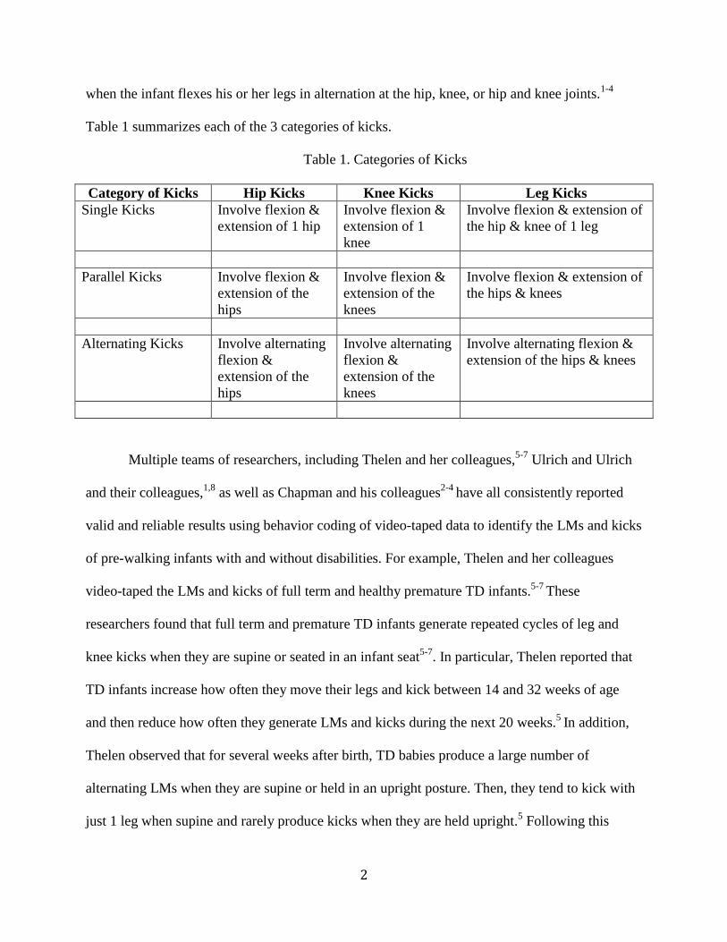
Table 1 summarizes each of the 3 categories of kicks.
Table 1. Categories of Kicks
Category of Kicks Hip Kicks Knee Kicks Leg Kicks
Single Kicks Involve flexion &
extension of 1 hip
Involve flexion &
extension of 1
knee
Involve flexion & extension of
the hip & knee of 1 leg
Parallel Kicks Involve flexion &
extension of the
hips
Involve flexion &
extension of the
knees
Involve flexion & extension of
the hips & knees
Alternating Kicks Involve alternating
flexion &
extension of the
hips
Involve alternating
flexion &
extension of the
knees
Involve alternating flexion &
extension of the hips & knees
Multiple teams of researchers, including Thelen and her colleagues,5-7
Ulrich and Ulrich
and their colleagues,1,8
as well as Chapman and his colleagues2-4
have all consistently reported
valid and reliable results using behavior coding of video-taped data to identify the LMs and kicks
of pre-walking infants with and without disabilities. For example, Thelen and her colleagues
video-taped the LMs and kicks of full term and healthy premature TD infants.5-7
These
researchers found that full term and premature TD infants generate repeated cycles of leg and
knee kicks when they are supine or seated in an infant seat5-7
. In particular, Thelen reported that
TD infants increase how often they move their legs and kick between 14 and 32 weeks of age
and then reduce how often they generate LMs and kicks during the next 20 weeks.5
In addition,
Thelen observed that for several weeks after birth, TD babies produce a large number of
alternating LMs when they are supine or held in an upright posture. Then, they tend to kick with
just 1 leg when supine and rarely produce kicks when they are held upright.5 Following this
3
period of reduced LMs and kicks, TD babies tend to produce significantly more parallel kicks
when they are lying supine.5
Ulrich and Ulrich and Chapman et al have all worked with infants with Down syndrome
(Ds) and Spina Bifida (SB) as well as infants who were TD.1, 8; 2-4
Each of these research teams
examined how infants learn to coordinate their leg movements prior to when they begin to walk
by using video-based behavior coding to identify LMs and kicks when the infants were supine
and seated in a variety of infant seats. More specifically, Ulrich and Ulrich utilized this approach
and the use of video-based behavior coding in infants with and without Ds.1 The purpose of their
study was to examine the spontaneously produced patterned and non-patterned LMs of infants
with and without Ds in a variety of contexts. Infants that participated in this study were split into
three groups. Group 1 consisted of infants with Ds while Groups 2 and 3 were comprised of 10
TD infants. Infants in Group 2 were matched with infants from the Ds group based on
chronological age plus or minus 1 week and group 3 infants were matched with infants from the
Ds group based on motor age. The researchers video-taped the infants’ LMs when they were
supine in 4 conditions: control, verbal, mobile and enriched. During the control trial the
caregiver sat next to the infant without interacting verbal or visually. In the verbal condition, the
caregiver was able to interact with the infant verbally, without touching the baby. During the
third condition, a brightly colored mobile was placed above the infant that was controlled by the
researcher to try to encourage the infant to move. In the enriched condition, the infants were able
to view the overhead mobile and interact with their parent(s) verbally and visually. Ulrich and
Ulrich did not find a significant difference between how often the 3 groups of infants generated
LMs. However, the Ds group demonstrated significantly fewer kicks than did the TD infants.
Follow-up data collected by phone with the infants’ parents to verify the age at which the infants
4
began to walk, i.e. take 3 independent steps, enabled Ulrich and Ulrich to conclude that the
frequency of kicks was significantly correlated for both infant groups with which they began to
walk.1
Chapman and his colleagues have examined how the movement context impacts
spontaneous and goal-directed LMs and kicks in babies with spina bifida (SB) between 4 and 14
months of age.4-6; 9-11
In his first studies, Chapman studied infants with lumbar or sacral SB who
were 16-20 weeks of age at entry into the study were chronologically-age matched with TD
infants.2, 3
The LMs and kicks of these infants were video-taped when they were supine and
seated in 2 infant seats. Chapman reported that the babies with SB moved their legs less
frequently than babies who were TD. Both groups of infants moved significantly less often when
they were seated in a conventional infant seat compared to when they were supine or seated in a
specially designed infant seat. These same infants altered the velocity and amplitude of their
LMs based on what position or context they were placed in, e.g. in supine they showed the
largest amplitudes and while seated in the specially designed infant seat they demonstrated LMs
with greater velocity than when they were supine.2, 3
Subsequent studies with infants with SB
who were between 8 and 10 months of age when they entered the study revealed that older
infants with SB also generate significantly more LMs and kicks when they were seated in a
specially designed infant seat compared to when they were supine or seated in a conventional
infant seat.4
Chapman and his colleagues at St. Catherine University have also reported that infants
with SB are sensitive to sensory information applied directly to their legs as well as visual and
auditory feedback provided via an overhead mobile.9-11
In particular, the LMs of infants with
lumbar or sacral SB were video-taped while they had 25%, 50%, 75%, and 100% of their calf
5
mass added to their lower leg when they were seated in a specially designed infant seat.9
These
infants were between 5 and 11 months old and generated more LMs when they had 25 and 50%
of their calf mass added to their leg compared to no weight added to their leg. Further, they
moved their legs less often when they had 75 and 100% of their calf mass added to one of their
legs.9
More recently, Chapman and his students utilized video-tape technology to verify that
when infants with lumbar or sacral SB had 1 leg tethered to an overhead mobile they generated
more LMs and kicks compared to when they were simply lying under the same mobile without 1
of their legs tethered to the mobile.10, 11
Collectively, these studies show that video-tape technology has been consistently used
over the past 40 years of developmental research and has yielded valid and reliable results for
researchers who have worked to describe and understand how pre-walking infants with and
without disabilities learn to coordinate their legs over developmental time. In spite of these
positive outcomes, this approach is time consuming and labor intensive as it takes approximately
one to two hours to behavior code one minute of video-taped data.12
In addition, it takes several
hours of training and practice for a given student to achieve an acceptable level of reliability
(percent of agreement with an expert rater > .85) before they are able to accurately identify infant
LMs and kicks. As a result, more efficient technology needs to be developed that will enable
parents and clinicians to accurately identify infant LMs and kicks.
Recently 3-D motion sensors also known as inertial measurement units (IMU) have been
used to analyze the frequency and quality of adult movement patterns as an alternative to video
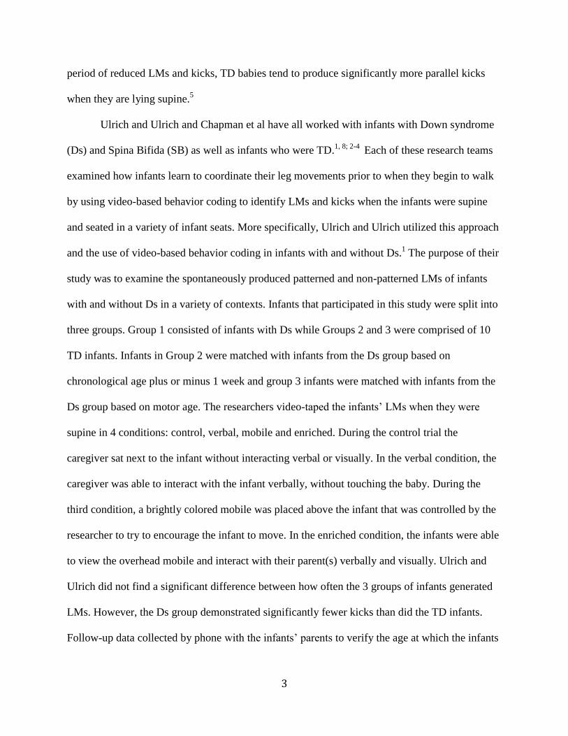
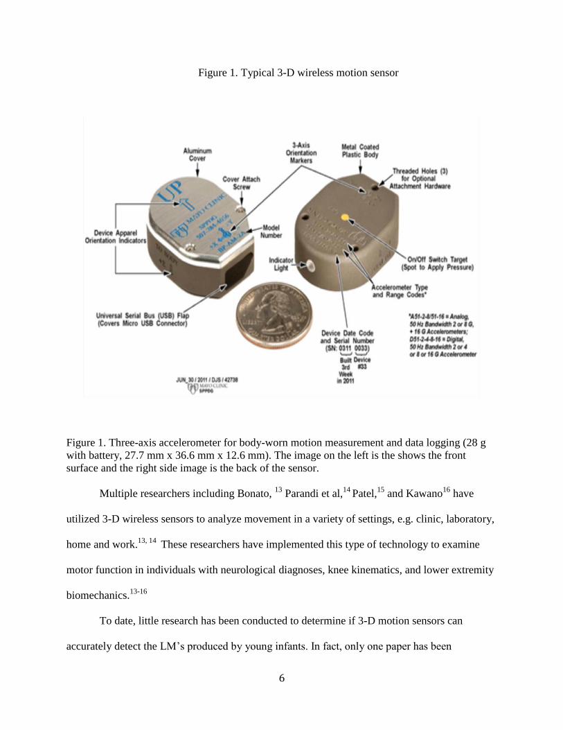
based behavior coding. Figure 1 depicts a photo of a generic 3-D wireless motion sensor.
6
Figure 1. Typical 3-D wireless motion sensor
Figure 1. Three-axis accelerometer for body-worn motion measurement and data logging (28 g
with battery, 27.7 mm x 36.6 mm x 12.6 mm). The image on the left is the shows the front
surface and the right side image is the back of the sensor.
Multiple researchers including Bonato, 13
Parandi et al,14
Patel,15
and Kawano16
have
utilized 3-D wireless sensors to analyze movement in a variety of settings, e.g. clinic, laboratory,
home and work.13, 14
These researchers have implemented this type of technology to examine
motor function in individuals with neurological diagnoses, knee kinematics, and lower extremity
biomechanics.13-16
To date, little research has been conducted to determine if 3-D motion sensors can
accurately detect the LM’s produced by young infants. In fact, only one paper has been
7
published that has relied on wireless sensors to verify the ability of sensors to detect infant
LM’s.17
Smith and associates (2015) used 3-D motion wireless sensors to document the
frequency of LMs produced by TD infants when they were supine and held upright. The babies
were tested with one sensor placed on each shank or lower leg. This research team developed
and used one algorithm to differentiate LMs from non-infant produced movement, e.g. when
they were in an infant swing. Their algorithm was based on the mean peak acceleration and
angular velocity of the baby's LMs and relied on subtracting one SD from the mean peak
acceleration and having an angular velocity greater than 0. That is, if the peak acceleration
exceeded the mean peak acceleration minus 1 SD and had a peak velocity that was greater than 0
then a LM was detected or identified by the sensors. Based on this algorithm that Smith et al
reported that their wireless sensors identified 92% of the LMs identified via behavior coding
video-tapes of the infants’ LMs.17
Taken together these studies suggest that 3-D motion wireless sensors can be used to
accurately analyze arm and leg movements in adults, but reveal the lack of data that confirms the
accuracy of 3-D wireless sensors to detect LMs and kicks produced by babies. Thus, as Fong et
al suggested, this is an area that continues to require further development in order to simplify
data processing algorithms and maximize the cost effectiveness of this approach.18
Therefore, as
a part of a larger ongoing study being conducted by Chapman and his colleagues who are
examining the frequency of LMs and kicks in babies with SB, the purpose of our pilot study was
to compare the accuracy of wireless 3-D motion sensors to the current gold standard of behavior
coding video-taped data to identify the LMs of young babies. Ultimately, our goal is to develop
lightweight portable sensors and easy to use mathematical algorithms that will enable parents

8
and health care providers to take advantage of telemedicine to communicate regarding how often
a child is moving his or her legs over developmental time.

9
Chapter II: Methods
Participants
Prior to subject recruitment, IRB approval was obtained from Mayo Clinic.
Participants were recruited via an advertisement posted on the internal website for Mayo
Clinic employees. Four TD infants, 2 males and 2 females, were recruited to participate in
this longitudinal study. They ranged in age from 29 to 34 days at entry into the study. Each
infant’s parent reviewed and provided written informed consent prior to data collection.
Each participant received a $20.00 incentive for each monthly visit. The funds were
provided from a grant provided by The Mayo Clinic Foundation. All of the babies were full
term & presented with normal vision, hearing & hip joint architecture per their newborn
screens. Each baby’s data was collected in their home or in the research lab located at Mayo
Clinic, once a month for 4 consecutive months.
Data Collection
The location for data collection was determined in light of parent preference, with
the intent to counterbalance the effects that the home or lab environment may have on how
often babies move their legs. Data was collected in 2 of the babies’ homes and in the
Restorative Technology lab at the Mayo Clinic for the other 2 infants. The babies’
spontaneous LMs were video-taped with a Sony Handy-cam when they were supine for 1.0
to 1.5 minutes at 30 frames per second with & without the 3-D sensors attached to the
anterior surface of their thighs and shanks.
The 3-D sensors sampled at 100hz per second & weighed 28 grams. Note, the video
camera and 3-D wireless sensors were time synchronized. Figure 1 presented earlier
illustrates the wireless sensors used in this pilot study. The sensors were designed and
10
manufactured in the biomedical engineering department at Mayo Clinic. The sensors were
sensitive to acceleration and velocity in the X, Y, and Z planes. The Y plane was designated
as vertical and the X as horizontal.
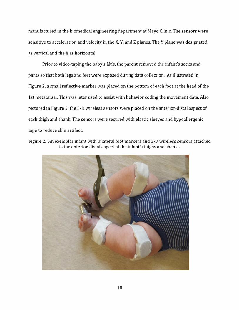
Prior to video-taping the baby’s LMs, the parent removed the infant’s socks and
pants so that both legs and feet were exposed during data collection. As illustrated in
Figure 2, a small reflective marker was placed on the bottom of each foot at the head of the
1st metatarsal. This was later used to assist with behavior coding the movement data. Also
pictured in Figure 2, the 3-D wireless sensors were placed on the anterior-distal aspect of
each thigh and shank. The sensors were secured with elastic sleeves and hypoallergenic
tape to reduce skin artifact.
Figure 2. An exemplar infant with bilateral foot markers and 3-D wireless sensors attached to the anterior-distal aspect of the infant’s thighs and shanks.
11
The infant was placed supine by his/her parent and the video camera was placed
perpendicular to the infant’s feet for data collection. The infant’s spontaneous LM’s were
then video-taped in the baseline condition, i.e. with foot markers attached to each foot, but
without sensors attached to their legs and then with the sensors attached to each leg. Data
was collected for one to one and a half minutes in each condition depending on each baby’s
tolerance. For example if an infant started to cry too much or became too fussy the trial was
terminated. The baby was given a small break after the baseline condition, during which
the sensors were placed on each leg. The infant was returned to a supine position and the
LM’s were video-taped with the sensors attached to the legs for another one to one and half
minutes. Note, we calculated our frequency data on a per minute average for each baby at
each age.
Data Reduction
The video-taped data was behavior coded through a frame by frame analysis by an
expert rater with over 20 years of experience to identify the frequency of LMs & kicks in
each condition. Custom Matlab programs were written by consultants from the Mayo Clinic
with input from researchers at Dartmouth College’s Thayer School of Engineering that
identified the acceleration and velocity of each of the infant’s LMs each month. The
calculated the mean peak resultant acceleration with the associated standard deviation for
the groups’ LMs each month and the associated velocity of each LM in each plane of
movement also calculated with the Matlab coding.
We then developed three algorithms that were used to establish when a LM was
detected by the 3-D wireless sensors. Note, that for each algorithm developed and
implemented in this study, a LM was detected if two conditions were met. For example,
12
algorithm 1, based on the values obtained in the Matlab programming described above,
detected a LM if the acceleration of a given LM was greater than the group’s mean peak
resultant acceleration minus 1standard deviation (SD) and when the velocity of a LM in one
plane was greater than the group’s mean peak velocity minus 1SD. Alternatively, a LM was
not detected if one or both of the conditions were not met in each algorithm.
Our first algorithm was based on the work of Smith et al.17 A LM was detected by
algorithm 1 when the acceleration was greater than the mean peak resultant acceleration
minus 1SD AND when the velocity was equal to or greater than the mean peak velocity
minus 1SD.
This algorithm resulted in a lower percentage of accuracy than we were willing to
accept. Thus, in light of these results and our intrinsic motivation to fully develop this
approach, we consulted with a group of researchers at the material science lab at
Dartmouth, who have extensive experience with 3D wireless sensors.19 As a result of those
conversations, we developed algorithms 2 and 3. For algorithms 2 and 3, it is important to
note that sensor data was analyzed for both the thigh and lower leg or shank.
In algorithm 2, a LM was detected when the acceleration was greater than the mean
peak resultant acceleration x 5% AND when the velocity was greater than the mean peak
velocity minus 1SD. For algorithm 3, a LM was detected when the acceleration was greater
than the mean peak resultant acceleration x 10% AND the velocity was greater than the
mean peak velocity minus 1SD.
Data Analysis
A MANOVA with repeated measures for age was used to compare the frequency of
LMs & kicks generated per minute each month in each condition (p < .05). The percent

13
agreement between the LMs detected by the 3-D sensors compared to the behavior coded
LMs was calculated at each age.
14
Chapter III: Results
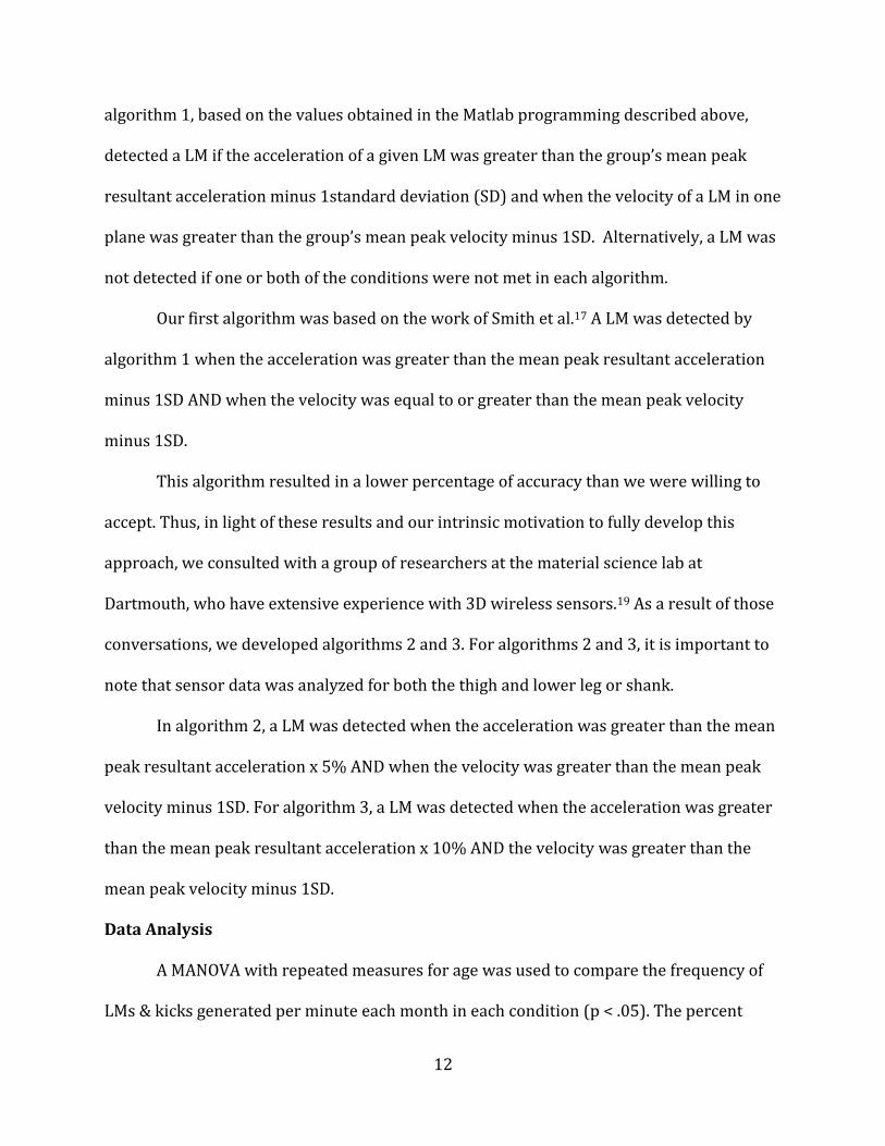
Figures 3 & 4 illustrate that wearing the 3-D sensors did not result in a significant
change in the number of LMs or kicks generated by this small group of infants over
developmental time ( p = .619, partial eta = .263, observed power = .294). On average, this
small group of babies increased how often they moved their legs from month 1 to 2 and
then decreased how often they generated LMs in each condition when they were 3 and 4
months old compared to when they were 2 months of age. They also showed more
variation as a group in how often they moved their legs when they were 2 months old
compared to when they were younger and older than 2 months of age.
Figure 3. The Mean Frequency of Leg Movements with and without Sensors per Month
15
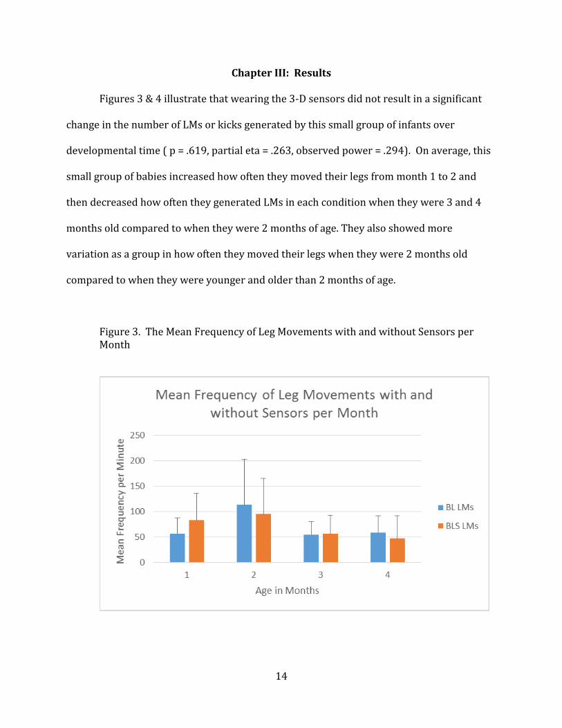
Figure 4 shows that at each age except when the infants were 3 months old they
generated more kicks, on average, when they were wearing the sensors compared to when
they were not wearing the sensors. There was a trend for these babies to generate fewer
kicks when they were 3 and 4 months old with the sensors on compared to when they were
1 and 2 months old.
Figure 4. The Mean Frequency of Kicks with and without Sensors per Month
Table 2 presents the percent agreement for each algorithm for the thigh and shank
sensors at each age. Note, that algorithm 1 is based specifically on Smith et al’s paper.17 As
a result, only the shank sensor data was used with algorithm 1.
16
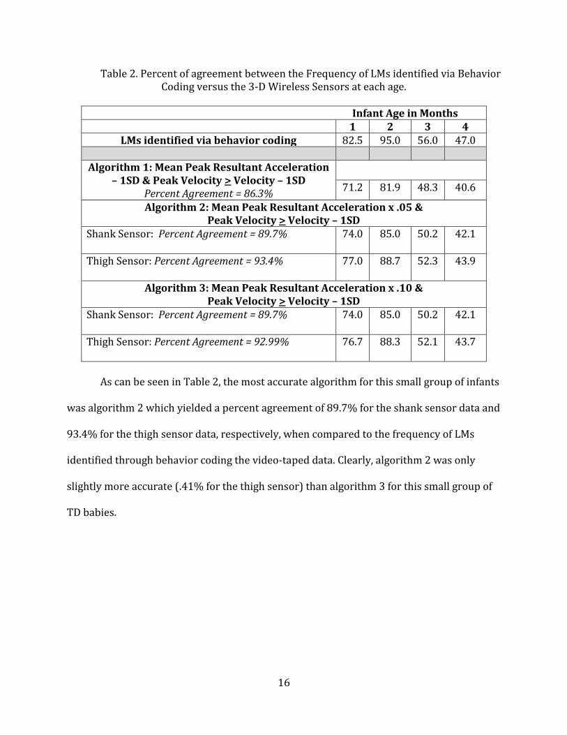
Table 2. Percent of agreement between the Frequency of LMs identified via Behavior Coding versus the 3-D Wireless Sensors at each age.
Infant Age in Months
1 2 3 4 LMs identified via behavior coding 82.5 95.0 56.0 47.0
Algorithm 1: Mean Peak Resultant Acceleration
– 1SD & Peak Velocity > Velocity – 1SD Percent Agreement = 86.3%
71.2 81.9 48.3 40.6
Algorithm 2: Mean Peak Resultant Acceleration x .05 & Peak Velocity > Velocity – 1SD
Shank Sensor: Percent Agreement = 89.7% 74.0 85.0 50.2 42.1
Thigh Sensor: Percent Agreement = 93.4% 77.0 88.7 52.3 43.9
Algorithm 3: Mean Peak Resultant Acceleration x .10 & Peak Velocity > Velocity – 1SD
Shank Sensor: Percent Agreement = 89.7% 74.0 85.0 50.2 42.1
Thigh Sensor: Percent Agreement = 92.99% 76.7 88.3 52.1 43.7
As can be seen in Table 2, the most accurate algorithm for this small group of infants
was algorithm 2 which yielded a percent agreement of 89.7% for the shank sensor data and
93.4% for the thigh sensor data, respectively, when compared to the frequency of LMs
identified through behavior coding the video-taped data. Clearly, algorithm 2 was only
slightly more accurate (.41% for the thigh sensor) than algorithm 3 for this small group of
TD babies.
17
Chapter IV: Discussion
The purpose of this pilot study was to compare the accuracy of wireless 3-D
movement sensors to identify the LMs and kicks of young babies to the current gold
standard of behavior coding the video-taped LMs and kicks of young infants. We found that
wearing the 3-D wireless sensors did not result in a significant change in the number of
LMs and kicks generated by this small group of infants. These results are consistent with
the Smith et al17 who reported that wearing 3-D sensors on the shank did not have an effect
on how often older babies moved their legs. It is important to note that wearing a shank
and thigh sensor on each leg did not significantly impact on how often the babies moved
their legs and kicked. In fact, our overall frequency data for LMs is very similar the
frequency data reported by Smith et al.17 These are important considerations given our
goal of implementing this technology with infants who have a disability, such as SB or Ds as
they usually move their legs and kick less often than TD babies.1,8; 2-4; 9-11
It is also important to recognize how limited the current literature is regarding the
use of wireless 3-D motion sensors to detect the LMs and kicks of infants. For instance,
Smith et al17 has published the only paper that has examined the utility of using wireless
sensors to detect infant LMs. Unfortunately, their work has several limitations. For
example, they reported only one algorithm that identified a LM if the acceleration of an
infant’s LM was greater than the average peak acceleration of the experimental group’s
LMs minus 1SD and if the angular velocity of an infant’s LM was greater than 0. In light of
this, we believe it is important to develop and report several algorithms for detecting LMs
and then compare the accuracy of each of those algorithms to the data obtained from
behavior coding video-taped LMs of TD infants. These beliefs are supported by Fong et
18
al’s18 observation and Chapman et al’s20, 21 perspective that on-going research needs to be
completed to refine the ability of researchers, therapists, and physicians to successfully
implement these types of algorithms in the identification of infant LMs and kicks.
As reported here, each of our algorithms detected a LM only when a LM exceeded a
specific acceleration and velocity threshold. In particular, algorithms 2 and 3, which relied
on a threshold of 5% and 10%, respectively, of the mean peak resultant thigh sensor
acceleration and a velocity value that was equal to or greater than the peak thigh velocity
minus 1SD yielded the highest percentage of agreement with the behavior coded
frequencies obtained by an expert rater. In a similar manner, algorithms 2 and 3 which also
relied on using a threshold of 5% and 10%, respectively of the mean peak resultant shank
sensor acceleration and a corresponding velocity value that was equal to or greater than
the peak shank sensor velocity minus 1SD yielded the second highest accuracy values. It is
particularly interesting to note that we obtained our lowest percent accuracy when we
used an algorithm (algorithm 1) most like the Smith et al17 algorithm, i.e. one that relied on
mean peak resultant acceleration values minus 1SD and velocity greater than 0. Further,
our obtained accuracy with algorithm 1 was lower that the results reported by Smith et
al.17 In comparison, the accuracy of using algorithms 2 and 3 from the thigh sensor data
were both higher than the percent accuracy reported by Smith et al.17 Collectively, these
results suggest that more research needs to be completed to determine if using only a thigh
sensor is ‘accurate’ enough or if we will need to continue to rely on shank and thigh sensors
and markers to identify infant LMs.
The percent of agreement noted above was obtained by behavior coding all of the
LMs of the entire trial for each baby as well as applying the algorithms to every second of
19
each trial, regardless of whether or not the infant was known to be active in a given
segment of the trial. This is in contrast to the methodology implemented by Smith et al.17
These authors only examined a small portion, i.e. 20 seconds of each trial and only selected
that 20 second window if the infant was known to be active based on their behavior coded
data. Thus, it is possible that they under or over-estimated the percent accuracy of their
algorithm. Because of the limited literature that currently exists, the observations reported
by Fong et al18, and the perspectives of Chapman et al20, 21 coupled with the results
obtained in the current study, we advocate that future studies utilize all of the data
collected as well as offer the percent accuracy results obtained when multiple algorithms
are developed and applied to the sensor data gathered from infant LMs. The seems
especially true when we begin to apply this technology to infants who have a disability as
these will be studies that are completely novel and will not have the benefit of a robust
body of knowledge from which they can be understood.
We intentionally used only the supine posture to collect our LM data. This is because
several studies have shown that when infants are placed in different postures or positions
they alter how often they move their legs and kick.2-4 In particular, Chapman’s work has
shown that when TD infants and infants with SB are placed in different positions they
produce more or less LMs depending on their position in space. Thus, it is possible that
combining the frequency, acceleration and velocity data from the LMs generated in
multiple positions may influence the relative accuracy or inaccuracy of using wireless 3-D
sensors to identify infant LMs. Again, this is in contrast to Smith et al17 who placed their
infants supine as well as held them in an upright position when they were older. It is
imperative that future studies place the infants in one position at a time and not

20
statistically collapse the results obtained from multiple positions when developing
algorithms that are written to identify infant LMs.
Although the babies moved their legs more often in month 2 we did not find any
significant developmental trends in the number of LMs and kicks generated by this small
group of infants over developmental time. These results are encouraging because they
suggest that young infants can wear light weight wireless 3-D motion sensors on their
thighs and shanks without having a negative impact on how often they move their legs and
kick. In fact, we observed a slight trend for these babies to generate more kicks when they
were wearing the sensors compared to when they did not have the sensors attached to
their legs.
Limitations and Future Research
Our results should be interpreted cautiously due to our small sample size (n=4), the
trial length of 1 to 1.5 minutes, the age of the babies tested, and our inclusion of only TD
babies as well as our reliance on the supine position.
The next step in continuing our research will be to recruit babies with SB who have their
lesion repaired postnatally and infants with SB who have their spinal lesion repaired in-
utero. This will provide 3 distinct groups from which we can compare the relative accuracy
of using 3-D wireless sensors to identify infant LMs and kicks. We plan to follow all 3
groups for 4 months of developmental time which will allow us to complete a
developmental comparison of the accuracy of the wireless 3-D sensors to detect the LMs of
TD infants, infants with SB who had their lesions repaired in-utero as well as infants with
SB who had their lesions repaired postnatally. In addition, we hope to be able to develop
algorithms that will accurately identify infant kicks as well as their LMs. This will be

21
especially important for babies who have a disability and usually learn to walk later in life
than TD infants.1-4 Being able to ‘easily’ track the infant kicks over developmental time will
allow parents, therapists, and physicians to monitor how well a given infant is learning to
coordinate his or her legs. Ultimately, with additional data, our goal is to develop
algorithms that will enable parents to take advantage of the wireless sensors and
telemedicine in order to communicate with their healthcare team regarding how often
their child is moving his or her legs and kicking over developmental time.

22
References
1. Ulrich, BD & Ulrich, DA. Spontaneous leg movements of infants with Down syndrome and
nondisabled infants. Child Dev. 1995; 66:1844-1855.
2. Chapman D. Context effects on the spontaneous leg movements of infants with spina
bifida. Ped Phys Ther. 2002;14(2):62-73.
3. Chapman, D. Context effects on the ability of young infants with myelomeningocele to
generate complex pattern leg movements. In Come to Your Senses: Creating Supportive
Environments to Nurture the Sensory Capital Within. Mukibaum Treatment Centres,
Toronto, Canada. 2009.
4. Chapman, D. The influence of position on leg movements and kicks of older infants with
spina bifida. Ped Phys Ther. 2016; 28(4): 380-385.
5. Thelen, E. Rhythmical stereotypies in normal human infants. Anim Beh. 1979; 27:699-
715.
6. Thelen, E, Fisher, DM. Newborn stepping: An explanation for a ‘disappearing’ reflex.
Develop Pysch. 1982; 18:760-775.
7. Thelen, E, Fisher, DM, Ridley-Johnson, R. The relationship between physical growth and a
newborn reflex. Inf Beh and Develop. 1984; 7:479-493.
8. Ulrich, BD, Ulrich, DA, Angulo-Kinzler, RM, Chapman, DD. Individual differences in the
intrinsic dynamics of leg movements in infants with and without Down syndrome. Res. Q.
Exer Sport. 1997; 68: 10-19.9.
9. Gulsvig, K, Hawn, C, Plummer, J, Schmitz, A. The Sensitivity of Infants with Spina Bifida to
Sensory Information. Doctor of Physical Therapy Program St. Catherine University,
Minneapolis, MN 2012. Publication: Doctor of Physical Therapy Research Papers
23
10. Chapman D, Engstrom A, Lucken S, Sis K, Wehrheim S. The influence of age, position, and
timing of surgical repair on the kicks of infants with Spina Bifida. Paper presented at the
Minnesota APTA Spring Conference; April 2014. St Paul, MN
11. DeRosier,S, Martin, J, Payne, A, Swenson, K, Wech, E. The Effect of Conjugate
Reinforcement on the Leg Movements of Infants with Spina Bifida. Doctor of Physical
Therapy Program St Catherine University, Minneapolis, MN 2015.
12. David Chapman, Personal Communication St. Catherine University Doctor of Physical
Therapy Program July, 2016.
13. Bonato, P. Wearable sensors and systems: Enabling technology to clinical applications.
Annual International Conference of the IEEE Engineering in Medicine and Biology. 2010; 25-
36.
14. Parnandi, A., Wade, E., & Matarić, M. Motor function assessment using wearable inertial
sensors. Annual International Conference of the IEEE Engineering in Medicine and Biology.
2010;86-89.
15. Patel,S., Park, H., Bonato, P, Chan, L, Rodgers, M. A review of wearable sesnors and
systems with application in rehabilitation. J of NeuroEngin and Rehab. 2012; 9(1): 21.
16. Kawano, K., Kobashi, S., Yagi, M., Kondo, K., Yoshiya, S., & Hata, Y. Analyzing 3D knee
kinematics using accelerometers, gyroscopes and magnetometers. IEEE International
Conference on System of Systems Engineering. 2007;1-6.
17. Smith, B. A., Trujillo-Priego, I. A., Lane, C. J., Finley, J. M., & Horak, F. B. Daily Quantity of
Infant Leg Movement: Wearable Sensor Algorithm and Relationship to Walking Onset.
Sensors. 2015; 15(8), 19006-19020.
24
18. Fong, D. T. P., & Chan, Y. Y. The use of wearable inertial motion sensors in human lower
limb biomechanics studies: a systematic review. Sensors. 2010;10(12), 11556-11565.
19. Ryan M Chapman, Personal Communication Dartmouth College Materials Science Lab,
Thayer School. October – November, 2016.
20. Chapman, RM, Bell, JE, & Van Citters, DW. Does our concept of Normal Shoulder
biomechanics Apply to Healthy Seniors During Activities of Daily Living? Paper No.0276.
San Diego, CA: Orthopaedic Research Society, 2017.
21. Chapman RM, Moschetti, WE & Van Citters, DW. A Novel Method for Remotely
Monitoring Knee Function using Inertial Measurement Units. Poster No. 1873. San Diego,
CA: Orthopaedic Research Society, 2017.References
Related Documents