The 1.8 A ˚ Crystal Structure of the Dimeric Peroxisomal 3-Ketoacyl-CoA Thiolase of Saccharomyces cerevisiae: Implications for Substrate Binding and Reaction Mechanism Magali Mathieu 1 , Yorgo Modis 1 , Johan Ph. Zeelen 1 , Christian K. Engel 1 Ruben A. Abagyan 2 , Anders Ahlberg 3 , Bjarne Rasmussen 3 Victor S. Lamzin 4 , Wolf H. Kunau 5 and Rik K. Wierenga 1 * 1 EMBL, Meyerhofstrasse 1 D69126, Heidelberg, Germany 2 Skirball Institute, New York 10016, NY, USA 3 EMBL Grenoble Outstation F38042, Grenoble, Cedex France 4 EMBL Hamburg Outstation D22603, Hamburg, Germany 5 Ruhr Universita ¨t Bochum D44780, Bochum, Germany The dimeric, peroxisomal 3-ketoacyl-CoA thiolase catalyses the conver- sion of 3-ketoacyl-CoA into acyl-CoA, which is shorter by two carbon atoms. This reaction is the last step of the b-oxidation pathway. The crys- tal structure of unliganded peroxisomal thiolase of the yeast Saccharo- myces cerevisiae has been refined at 1.8 A ˚ resolution. An unusual feature of this structure is the presence of two helices, completely buried in the dimer and sandwiched between two b-sheets. The analysis of the struc- ture shows that the sequences of these helices are not hydrophobic, but generate two amphipathic helices. The helix in the N-terminal domain exposes the polar side-chains to a cavity at the dimer interface, filled with structured water molecules. The central helix in the C-terminal domain exposes its polar residues to an interior polar pocket. The refined structure has also been used to predict the mode of binding of the sub- strate molecule acetoacetyl-CoA, as well as the reaction mechanism. From previous studies it is known that Cys125, His375 and Cys403 are important catalytic residues. In the proposed model the acetoacetyl group fits near the two catalytic cysteine residues, such that the oxygen atoms point towards the protein interior. The distance between SG(Cys125) and C3(acetoacetyl-CoA) is 3.7 A ˚ . The O2 atom of the docked acetoacetyl group makes a hydrogen bond to N(Gly405), which would favour the formation of the covalent bond between SG(Cys125) and C3(acetoacetyl-CoA) of the intermediate complex of the two-step reaction. The CoA moiety is proposed to bind in a groove on the surface of the protein molecule. Most of the interactions of the CoA molecule are with atoms of the loop domain. The three phosphate groups of the CoA moi- ety are predicted to interact with side-chains of lysine and arginine resi- dues, which are conserved in the dimeric thiolases. # 1997 Academic Press Limited Keywords: thiolase; CoA; crystal structure; reaction mechanism; b-oxidation *Corresponding author Introduction Thiolases form an ubiquitous family of enzymes; they are found in prokaryotes and eukaryotes (Igual et al., 1992). In eukaryotic cells thiolases are found in the cytosol, in microbodies (in particular in peroxisomes) and in mitochondria. Thiolases catalyse the reversible thiolytic cleavage of 3-ketoa- cyl-CoA into acyl-CoA and acetyl-CoA. The covalent structure and nomenclature of acetoace- tyl-CoA is shown in Figure 1(a). As is shown in Present address: M.Mathieu, LEBS Bat34, 11, Av. de la Terrasse, F91198 Gif sur Yvette, France. Abbreviations used: CoA, coenzyme A; DTT, reduced dithiothreitol; Mops, 3-(N-morpholino)-propane sulphonic acid; MPD, 2-methyl-2,4-pentanediol; NCS, non-crystallographic symmetry; PDB, Brookhaven Protein Data Bank; rms, root mean square. J. Mol. Biol. (1997) 273, 714–728 0022–2836/97/430714–15 $25.00/0/mb971331 # 1997 Academic Press Limited

Welcome message from author
This document is posted to help you gain knowledge. Please leave a comment to let me know what you think about it! Share it to your friends and learn new things together.
Transcript

The 1.8 AÊ Crystal Structure of the Dimeric Peroxisomal3-Ketoacyl-CoA Thiolase of Saccharomycescerevisiae: Implications for Substrate Binding andReaction Mechanism
Magali Mathieu1, Yorgo Modis1, Johan Ph. Zeelen1, Christian K. Engel1
Ruben A. Abagyan2, Anders Ahlberg3, Bjarne Rasmussen3
Victor S. Lamzin4, Wolf H. Kunau5 and Rik K. Wierenga1*
1EMBL, Meyerhofstrasse 1D69126, Heidelberg, Germany2Skirball Institute, New York10016, NY, USA3EMBL Grenoble OutstationF38042, Grenoble, CedexFrance4EMBL Hamburg OutstationD22603, Hamburg, Germany5Ruhr UniversitaÈt BochumD44780, Bochum, Germany
The dimeric, peroxisomal 3-ketoacyl-CoA thiolase catalyses the conver-sion of 3-ketoacyl-CoA into acyl-CoA, which is shorter by two carbonatoms. This reaction is the last step of the b-oxidation pathway. The crys-tal structure of unliganded peroxisomal thiolase of the yeast Saccharo-myces cerevisiae has been re®ned at 1.8 AÊ resolution. An unusual featureof this structure is the presence of two helices, completely buried in thedimer and sandwiched between two b-sheets. The analysis of the struc-ture shows that the sequences of these helices are not hydrophobic, butgenerate two amphipathic helices. The helix in the N-terminal domainexposes the polar side-chains to a cavity at the dimer interface, ®lledwith structured water molecules. The central helix in the C-terminaldomain exposes its polar residues to an interior polar pocket. The re®nedstructure has also been used to predict the mode of binding of the sub-strate molecule acetoacetyl-CoA, as well as the reaction mechanism.From previous studies it is known that Cys125, His375 and Cys403 areimportant catalytic residues. In the proposed model the acetoacetylgroup ®ts near the two catalytic cysteine residues, such that the oxygenatoms point towards the protein interior. The distance betweenSG(Cys125) and C3(acetoacetyl-CoA) is 3.7 AÊ . The O2 atom of the dockedacetoacetyl group makes a hydrogen bond to N(Gly405), which wouldfavour the formation of the covalent bond between SG(Cys125) andC3(acetoacetyl-CoA) of the intermediate complex of the two-step reaction.The CoA moiety is proposed to bind in a groove on the surface of theprotein molecule. Most of the interactions of the CoA molecule are withatoms of the loop domain. The three phosphate groups of the CoA moi-ety are predicted to interact with side-chains of lysine and arginine resi-dues, which are conserved in the dimeric thiolases.
# 1997 Academic Press Limited
Keywords: thiolase; CoA; crystal structure; reaction mechanism;b-oxidation*Corresponding author
Introduction
Thiolases form an ubiquitous family of enzymes;they are found in prokaryotes and eukaryotes(Igual et al., 1992). In eukaryotic cells thiolases arefound in the cytosol, in microbodies (in particularin peroxisomes) and in mitochondria. Thiolasescatalyse the reversible thiolytic cleavage of 3-ketoa-cyl-CoA into acyl-CoA and acetyl-CoA. Thecovalent structure and nomenclature of acetoace-tyl-CoA is shown in Figure 1(a). As is shown in
Present address: M.Mathieu, LEBS Bat34, 11, Av. dela Terrasse, F91198 Gif sur Yvette, France.
Abbreviations used: CoA, coenzyme A; DTT, reduceddithiothreitol; Mops, 3-(N-morpholino)-propanesulphonic acid; MPD, 2-methyl-2,4-pentanediol; NCS,non-crystallographic symmetry; PDB, BrookhavenProtein Data Bank; rms, root mean square.
J. Mol. Biol. (1997) 273, 714±728
0022±2836/97/430714±15 $25.00/0/mb971331 # 1997 Academic Press Limited
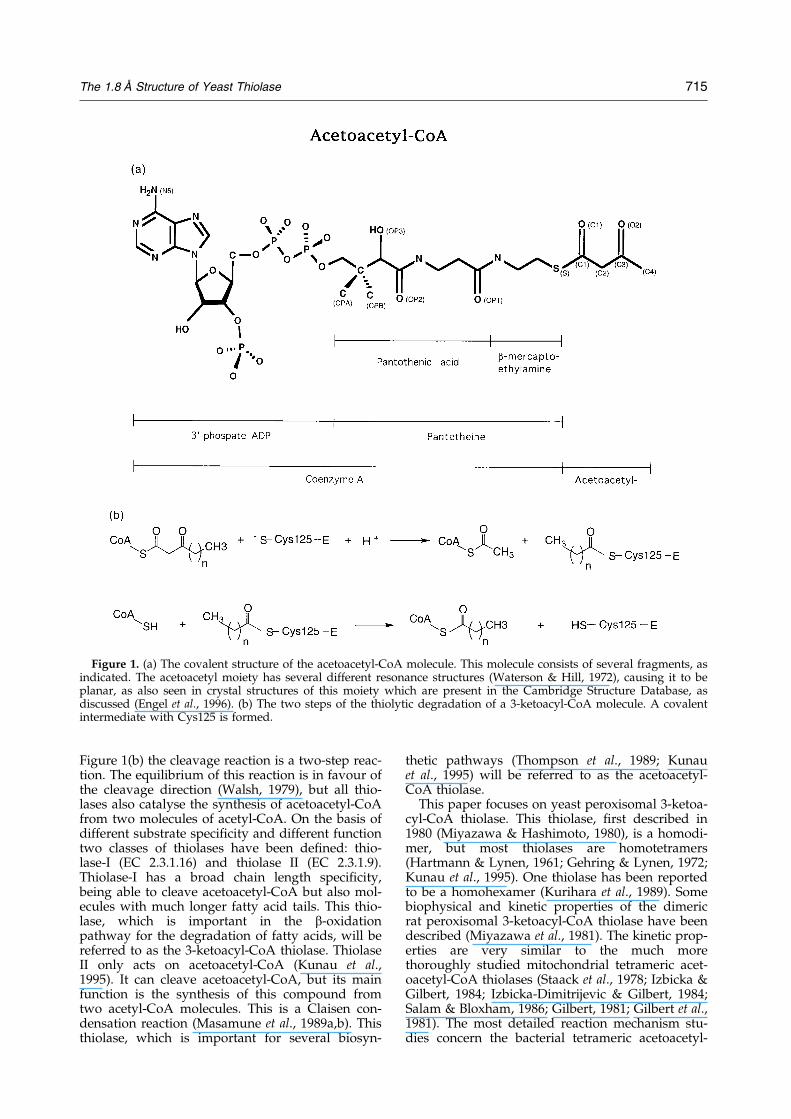
Figure 1(b) the cleavage reaction is a two-step reac-tion. The equilibrium of this reaction is in favour ofthe cleavage direction (Walsh, 1979), but all thio-lases also catalyse the synthesis of acetoacetyl-CoAfrom two molecules of acetyl-CoA. On the basis ofdifferent substrate speci®city and different functiontwo classes of thiolases have been de®ned: thio-lase-I (EC 2.3.1.16) and thiolase II (EC 2.3.1.9).Thiolase-I has a broad chain length speci®city,being able to cleave acetoacetyl-CoA but also mol-ecules with much longer fatty acid tails. This thio-lase, which is important in the b-oxidationpathway for the degradation of fatty acids, will bereferred to as the 3-ketoacyl-CoA thiolase. ThiolaseII only acts on acetoacetyl-CoA (Kunau et al.,1995). It can cleave acetoacetyl-CoA, but its mainfunction is the synthesis of this compound fromtwo acetyl-CoA molecules. This is a Claisen con-densation reaction (Masamune et al., 1989a,b). Thisthiolase, which is important for several biosyn-
thetic pathways (Thompson et al., 1989; Kunauet al., 1995) will be referred to as the acetoacetyl-CoA thiolase.
This paper focuses on yeast peroxisomal 3-ketoa-cyl-CoA thiolase. This thiolase, ®rst described in1980 (Miyazawa & Hashimoto, 1980), is a homodi-mer, but most thiolases are homotetramers(Hartmann & Lynen, 1961; Gehring & Lynen, 1972;Kunau et al., 1995). One thiolase has been reportedto be a homohexamer (Kurihara et al., 1989). Somebiophysical and kinetic properties of the dimericrat peroxisomal 3-ketoacyl-CoA thiolase have beendescribed (Miyazawa et al., 1981). The kinetic prop-erties are very similar to the much morethoroughly studied mitochondrial tetrameric acet-oacetyl-CoA thiolases (Staack et al., 1978; Izbicka &Gilbert, 1984; Izbicka-Dimitrijevic & Gilbert, 1984;Salam & Bloxham, 1986; Gilbert, 1981; Gilbert et al.,1981). The most detailed reaction mechanism stu-dies concern the bacterial tetrameric acetoacetyl-
Figure 1. (a) The covalent structure of the acetoacetyl-CoA molecule. This molecule consists of several fragments, asindicated. The acetoacetyl moiety has several different resonance structures (Waterson & Hill, 1972), causing it to beplanar, as also seen in crystal structures of this moiety which are present in the Cambridge Structure Database, asdiscussed (Engel et al., 1996). (b) The two steps of the thiolytic degradation of a 3-ketoacyl-CoA molecule. A covalentintermediate with Cys125 is formed.
The 1.8 AÊ Structure of Yeast Thiolase 715
CoA thiolase of Zoogloea ramigera (Anderson et al.,1990; Williams et al., 1992). These studies haverevealed the importance of two cysteine residuesfor the catalysis. In the yeast peroxisomal thiolasethese are Cys125 and Cys403. From the Z. ramigerathiolase studies (but also from studies with tetra-meric pig heart mitochondrial 3-ketoacyl-CoA thio-lase (Gilbert, 1981; Gilbert et al., 1981)) it is knownthat a covalent intermediate exists, in which thefatty acid moiety is covalently bound to Cys125(Figure 1). Mutagenesis of this residue into a serine(Thompson et al., 1989) reduces the catalytic rate.The importance of Cys403 is also shown by muta-genesis experiments (Palmer et al., 1991; Williamset al., 1992). Active site labelling studies indicatethe importance of His375 for catalysis (Williamset al., 1992).
The 2.8 AÊ crystal structure of the unligandeddimeric peroxisomal 3-ketoacyl-CoA thiolase of theyeast Saccharomyces cerevisiae has been reported(Mathieu et al., 1994). Here we discuss the structureof this thiolase after re®nement at 1.8 AÊ resolution.From the 1.8 AÊ structure a plausible model for themode of binding of acetoacetyl-CoA has beendeduced, including a detailed description of thepossible reaction mechanism. The mode of bindingof acetoacetyl-CoA is predicted by manually dock-ing this molecule onto the surface of thiolase fol-lowed by energy minimisation. Based on thestructure of the dimeric thiolase, using also thesequence information from the family of thiolases,a model is proposed how two dimeric thiolasemolecules can assemble to form the tetrameric thio-lase.
Results and Discussion
Quality of the model
In the 1.8 AÊ structure of thiolase there is continu-ous density for the subunit-1 main-chain from resi-dues Lys25 to Ile262 and from Gly266 to Glu417.For subunit-2 a model has been built for residuesLys25 to Glu417. Previous studies (Mathieu et al.,1994) have shown that the complete protein, resi-dues 1 to 417, has been crystallised; apparently the®rst 24 residues, which include the peroxisomaltargetting sequence (Erdmann, 1994), are disor-dered. Residues 25 to 154 and 277 to 293 are in theN-domain, residues 155 to 276 de®ne the loopdomain and residues 300 to 417 are in theC-domain (Figures 2 and 3). In the 2.8 AÊ structure87 residues (in the loop domains of subunit-1 andsubunit-2) could not be built, but in the 1.8 AÊ
model the structure of only three residues are notde®ned by the electron density map (residues 263to 265 of the loop domain of subunit-1). The 2.8 AÊ
structure (1PXT in the PDB) and the 1.8 AÊ structureagree very well: leaving out only a few residues atthe beginning or end of the gaps in the 2.8 AÊ struc-ture results in an rms difference for superimposedCa-atoms of 0.35 AÊ for subunit-1 (leaving out two
residues) and 0.44 AÊ for subunit-2 (leaving outthree residues).
There are a few peaks above 4 s in the ®naldifference Fourier map. The highest peaks (6 s) arecorrelated with structural heterogeneity of side-chains, in particular near Thr307 (subunit-1) andArg430 (subunit-2). High difference Fourier densitynear the sulphur atom of Cys353 (subunit-2)suggests that this sulphur is partially oxidised.There are no difference Fourier peaks in the activesite pockets, therefore there is no evidence for oxi-dation of any of the active site cysteine or methion-ine residues.
The model has good geometry (Table 4). TheRamachandran plot shows six outliers. It concerns,in both subunits, Phe50, Gln124, and Thr406. The(f,c) values are (73, ÿ 42), (49, ÿ 135) and (92,7),respectively, for the subunit-1 residues and (71,ÿ 40), (51, ÿ 133) and (86,11), respectively, for thesubunit-2 residues. The structures of these residuesare well de®ned by the electron density map. Theyare at or near the active site. Gln124 is just beforethe catalytic residue Cys125 and Thr406 is justafter the catalytic residue Cys403. Gln124 andThr406 are also at the dimer interface.
The two subunits are related to each other by anon-crystallographic 2-fold axis (K � 180.0, as cal-culated from the superposition of all Ca atoms).The two subunits superimpose very well with anrms difference for all Ca atoms of subunit-1 andsubunit-2 of 0.34 AÊ . Figure 4 shows that the largestdifferences between the Ca-traces of the two sub-units are near residue 75, which is in the loop afterNa1, as well as in the loop domain, residues 155 to276. Figure 5 shows the B-factor plots for subunit-1and subunit-2. The average B-factors of the loopdomain residues are higher than for the N-domainand the C-domain (Figure 5), except for residuesnear Met186. Met186 is in the active site pocket.The lowest B-factors are observed in the N-domain,consistent with the fact that the N-domain is at thedimer interface at the centre of the molecule.
The fully re®ned structure of this 3-ketoacyl-CoA thiolase provides a good model for a detailedanalysis of the active site pocket and for modellingthe acetoacetyl-CoA into its assumed bindingpocket.
Sequence alignment
Sequence alignments have been made using thesequences of seven dimeric 3-ketoacyl-CoA peroxi-somal thiolases and 14 tetrameric thiolases, asfound in sequence databanks. Two tetrameric thio-lases are mitochondrial 3-ketoacyl-CoA b-oxidationenzymes and the other 12 are acetoacetyl-CoAthiolases. These sequences show a high degree ofsequence similarity. The residues conserved in all21 thiolases are indicated in Figure 3 (11%sequence identity; Asp/Glu, Arg/Lys, and Ser/Thr are grouped together). Within the family ofdimeric thiolases there is 38% sequence identity(Figure 3). The positions of the residues conserved
716 The 1.8 AÊ Structure of Yeast Thiolase

in the 14 tetrameric thiolases (14% sequence iden-tity) cluster around the active site and the dimerinterface. The sequence similarity between thedimeric and tetrameric thiolases indicate that the
fold of the tetrameric thiolases and the dimericthiolases may be very similar, which also impliesthat the tetrameric thiolases will be an assembly oftwo yeast thiolase dimers.
Figure 2. (A) Stereo view into the active site pocket, along the dimer 2-fold axis (top view). A complete Ca-trace ofsubunit-1 is shown. The N-domain is in green, the C-domain is in red and the loop domain is in yellow. Subunit-2 isin blue. The dimer 2-fold axis is near residue B97; the predominant contacts across the dimer interface are betweenthe two N-domains. The loop domain circles around the active site; the side-chains of the catalytic residues Cys125,His375 and Cys403 are explicitly drawn. The three catalytic loops (123±128, 373±381 and 402±408) are highlighted.The catalytic loops 123±128 and 375±381 continue in the two central helices, Na3 and Ca3, respectively. (B) Side viewof the dimer (rotated 90o around the horizontal with respect to (A); both subunits are color-coded as for subunit-1 in(A). The N terminus (25) and the C terminus (417) are solvent exposed. The two central helices Na3 (125 to 140, nearthe dimer interface) and Ca3 (375 to 390) are in blue. The N termini of these two helices are near the active site,which is marked by a blue sphere, centred at SG(Cys125). The green loop (of the N-domain) contributing to the ¯atappearance of the surface (top side) consists of residues 48 to 51. This Figure was made with ICM (Abagyan, 1997).
The 1.8 AÊ Structure of Yeast Thiolase 717
Crystal contacts
Subunit-1 has relatively few crystals contacts (91contacts within 4 AÊ ), whereas subunit-2 has more(206). A very extensive crystal contact is the contactof loops 74±83 and 289±295 of subunit-2, whichpenetrate into the entrance of the active site pocketof subunit-2 of a crystallographically related mol-ecule, formed by the loop regions 50±52 and 259±262. The latter loop region is mobile in subunit-1but structured in subunit-2. The structures of theloop region 74±83 of subunit-1 and subunit-2 aredifferent (Figure 4).
Cavities, buried charged residues and thedimer interface
The cavity calculations (Connolly, 1985) weredone in the presence and absence of solvent mol-ecules. Calculations in the presence of the solventmolecules show only small cavities, with volumes
less than 25 AÊ 3. In the absence of solvent moleculesthe largest cavity (170 AÊ 3) is at the dimer interface.There are several other cavities at the dimer inter-face with volumes of approximately 40 AÊ 3. Allthese cavities are ®lled with water molecules.There is one water-®lled cavity (39 AÊ 3) near eachactive site. This cavity is ®lled with two water mol-ecules. These water molecules are part of a hydro-gen bonding network, which includes the side-chains of Asn343, Glu341 and Glu391. The buriedcarboxylate groups of Glu341 and Glu391 arehydrogen-bonded with each other, indicating thatone of them has to be protonated, as observed inother protein structures as well (Flocco &Mowbray, 1995). These side-chains in fact form acluster of buried polar groups with hydrogenbonding interactions towards the active site as wellas to the other side of the molecule. There are sev-eral other buried side-chains of glutamate (®ve permonomer), aspartate (two per monomer), arginine(four per monomer) and lysine residues (one per
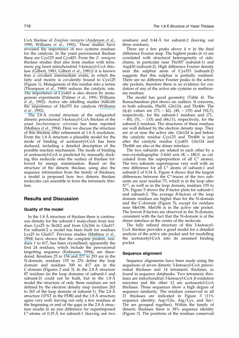
Figure 3. The sequence and secondary structure of yeast thiolase. The secondary structure assignment is according toKabsch & Sander (1983). The residues conserved (Ser/Thr, Asp/Glu and Lys/Arg have been grouped together) in allsequences are coloured green; the residues conserved only in the dimeric thiolases are coloured yellow. The layout ofthe Figure emphasises the regular ba-repeats of the N-domain and C-domain, which have the same topology. Thenomenclature of the secondary structure elements is the same as reported previously (Mathieu et al., 1994).
718 The 1.8 AÊ Structure of Yeast Thiolase
monomer). Most of these side-chains are involvedin salt bridges (Table 1). All these side-chains par-ticipate in good hydrogen bonding networks andall but one are hydrogen-bonded to at least oneburied water molecule (Table 1).
The two central helices (Na3 and Ca3) are com-pletely buried in the dimer (Figure 2). Neverthe-less, both helices have a polar and an apolar side.In the Na3 helix the polar residues are facing thelarge dimer interface cavity (®lled with water mol-ecules). In the Ca3 helix the polar residues (Arg383,Gln384 and Thr387) participate in the buried clus-ter of polar residues mentioned above. This clusterof polar residues is buried between the b-sheet ofthe C-domain and the Ca3 helix. This is a peculiarbut conserved feature of the thiolase fold, becausethe residues involved in this cluster are highly con-served in the sequences (Figure 3).
The dimer interface is extensive. The molecularsurface area buried at the dimer interface is1400 AÊ 2 per subunit. There are two salt bridges atthe dimer interface. Most of the monomer-mono-mer contacts concern residues 161 to 179 of theloop domain and the residues of helix Na2, strandNb3 and helix Na3. As can be seen in Figure 2, the
dimer interface has a rather ¯at surface; the onlyexceptions are the loop residues 161 to 179, whichprotrude out of the monomer and lay on top ofhelix Na2 of the other subunit. Although thesedimer interface regions are not directly involvedin the catalysis, they are part of the active sitearchitecture. For example, the catalytic Cys125 isin the loop between Nb3 and Na3. The extensiveinteractions seen at the dimer interface contributeto the observed rigid architecture of the twoactive sites.
The active site pocket
The catalytic residues Cys125, His375 andCys403 are at the bottom of a deep cylindricalpocket with a radius of approximately 10 AÊ .Cys125 and His375 are solvent-exposed, whereasCys403 is almost completely buried (Figure 6).Also at the bottom of this pocket is Gln124(Figure 6). Several different regions can be ident-i®ed in the walls of this pocket. There are twohydrophobic areas, which are located on oppositesides of the pocket. One region (the ``upper'' side)is near Cys403, consisting of Phe346, Met186,
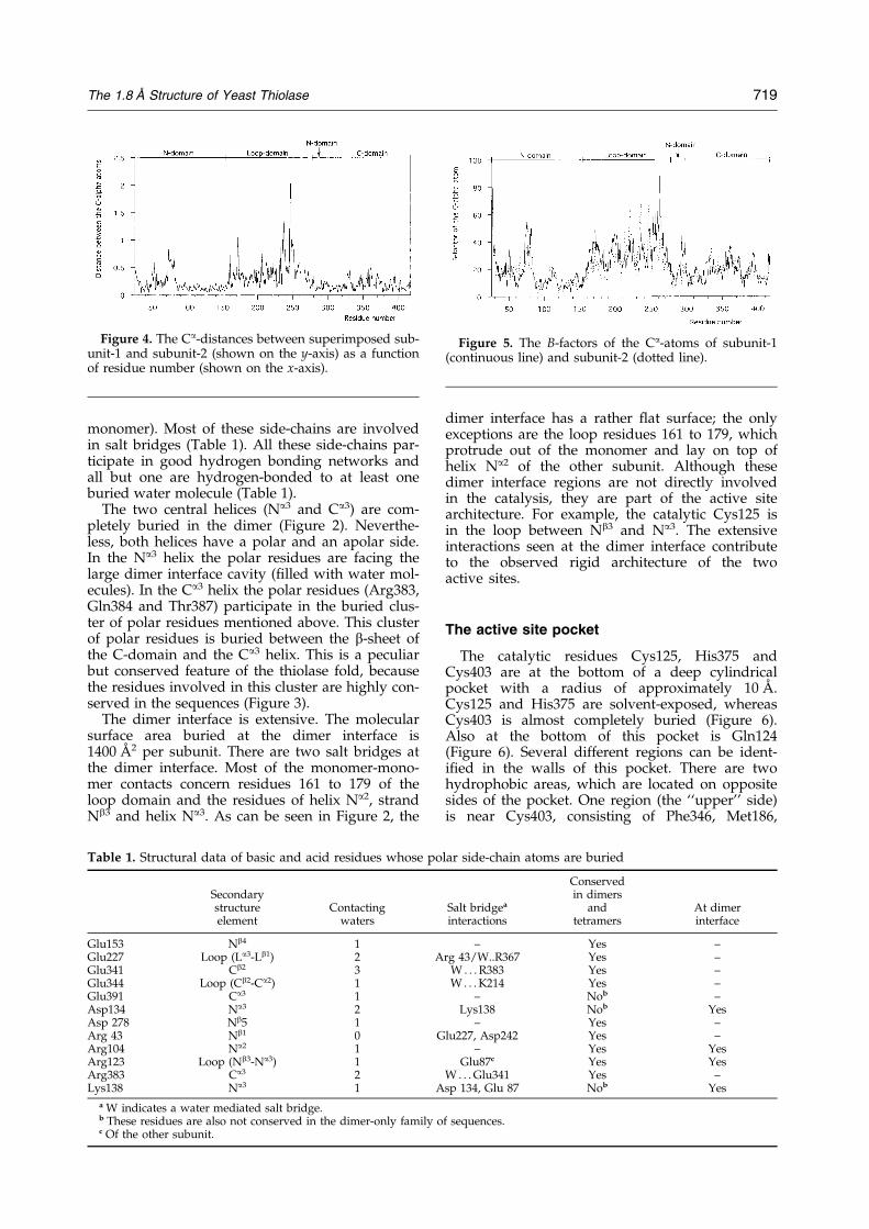
Figure 4. The Ca-distances between superimposed sub-unit-1 and subunit-2 (shown on the y-axis) as a functionof residue number (shown on the x-axis).
Figure 5. The B-factors of the Ca-atoms of subunit-1(continuous line) and subunit-2 (dotted line).
Table 1. Structural data of basic and acid residues whose polar side-chain atoms are buried
ConservedSecondary in dimersstructure Contacting Salt bridgea and At dimerelement waters interactions tetramers interface
Glu153 Nb4 1 ± Yes ±Glu227 Loop (La3-Lb1) 2 Arg 43/W..R367 Yes ±Glu341 Cb2 3 W . . . R383 Yes ±Glu344 Loop (Cb2-Ca2) 1 W . . . K214 Yes ±Glu391 Ca3 1 ± Nob ±Asp134 Na3 2 Lys138 Nob YesAsp 278 Nb5 1 ± Yes ±Arg 43 Nb1 0 Glu227, Asp242 Yes ±Arg104 Na2 1 ± Yes YesArg123 Loop (Nb3-Na3) 1 Glu87c Yes YesArg383 Ca3 2 W . . . Glu341 Yes ±Lys138 Na3 1 Asp 134, Glu 87 Nob Yes
a W indicates a water mediated salt bridge.b These residues are also not conserved in the dimer-only family of sequences.c Of the other subunit.
The 1.8 AÊ Structure of Yeast Thiolase 719

Met315, Ala345, Phe261, and Ala273. The otherregion (the ``lower'' side) is near Leu94 andincludes residues Val162, Val93, Leu377, Tyr159,Val276, Met155, and Phe50 (Figure 6). Theserather extensive hydrophobic walls are separatedfrom each other by the side-chain of Ser274,which is a conserved residue (Figure 3). The twohydrophobic walls, together with Ser274, makeup half of the cylindrical pocket. The remainingsemi-circle of the cylindrical pocket is shaped byresidues 163 to 185 of the loop domain of the
same subunit and residues 99 to 108 of helix Na2
of the other subunit, in particular Thr101(Figure 6), which side-chain is hydrogen-bondedto the side-chain of Gln124.
There are no side-chains of charged residues(Arg, Lys, Asp, Glu) within 9 AÊ of SG(Cys125) ofthe catalytic site, except for the conserved residueGlu153 (see also Table 1). The carboxylate oxygenatoms of this residue are just 9 AÊ away fromSG(Cys125). It is interesting to point out thatwithin this 9 AÊ sphere there is a high concentration
Figure 6. (A) Stereo view into the active site pocket (top view). The Ca-traces of subunit-1 and subunit-2 are in blueand green, respectively. The side-chains of the residues forming the hydrophobic walls (see the text) are shown.M501 labels the MPD molecule. X64 and X272 are the two water molecules between SG(Cys125) and SG(Cys403).Water X88 is behind SG(Cys403). Thr101, of the other subunit, is in hydrogen bonding contact with Gln124. (B) Stereoview into the active site of the modelled complex (top view). The interactions of the labelled side-chains with themodelled acetoacetyl-CoA group are discussed in the text.
720 The 1.8 AÊ Structure of Yeast Thiolase
of methionine side-chain atoms (®ve residues outof a total of six).
The three catalytic residues are in three loopswith highly conserved sequences (Figure 3). TheCys125 loop (N-domain) and the His375 loop(C-domain) continue into the buried central helices,Na3 and Ca3, respectively (Figure 2(A)). TheCys403 loop is a tight turn between the two anti-parallel strands (Cb4 and Cb5) of the C-terminaldomain. The distance between SG(Cys125) andSG(Cys403) is 5.8 AÊ . Other contact distances inthe active site are given in Table 2. The side-chain atom ND1 of His375 is hydrogen-bondedto Thr380 (Table 2). NE2(His375) interacts withSG(Cys125). The His375 hydrogen bondingsuggests that the NE2 atom is not protonated,indicating that the His375-Cys125 interaction willactivate the SG(Cys125) for nucleophilic attack(see also Figures 7 and 9).
Unexpectedly, one MPD molecule (used as acryoprotectant) is bound near the active site pocketof each subunit (Figure 6(A)). The MPD bindingsite is between residues 164 to 166 and 182 to 186of the loop domain. The mode of binding is wellde®ned by the electron density map. The MPDmolecule interacts with the protein only via vander Waals interactions. The two MPD oxygenatoms make hydrogen bonds to protein-boundwater molecules. The binding af®nity of MPD forthiolase is unknown. MPD is a very poor inhibitorof the thiolase reaction (U. Dorpmund & W.-H.Kunau, unpublished observations); this could bebecause of low af®nity or because MPD bindingdoes not effect substrate binding.
In the unliganded active site pocket there is anextensive network of water molecules. This net-work includes WAT272 and WAT64, which bindbetween SG(Cys125) and SG(Cys403) and whichare also near NE2 (His375) (Table 2 andFigure 6(A)). WAT88, which is buried behind
SG(Cys403) could be important for the reactionmechanism, as it interacts tightly with SG(Cys403)(Table 2 and Figure 6).
The predicted mode of bindingof acetoacetyl-CoA
Attempts to bind CoA to the crystalline thiolasehave failed, because crystals transferred to amother liquor with CoA deteriorate to the extentthat data collection is impossible (Mathieu, 1995).Therefore, it has been attempted to predict themode of binding of acetoacetyl-CoA into the activesite pocket. The modelling of the CoA moiety isdif®cult, because there are no known CoA bindingmotifs, as was pointed out in a recent review onstructural features of the known structures of pro-tein-CoA complexes (Engel & Wierenga, 1996). Themodelling was done in two steps. First the acetoa-cetyl-CoA molecule was manually docked onto thesurface of thiolase; subsequently the docked com-plex was regularised and energy minimised withthe ICM package (Abagyan et al., 1994). Only side-chain atoms and CoA atoms were allowed to movein the minimisation calculations. The ®nal structureof acetoacetyl-CoA has good geometry and thereare no clashes between the docked acetoacetyl-CoA molecule and the protein atoms.
The docking experiments of the acetoacetyl moi-ety are facilitated by the notion that this group isplanar (Figure 1). The knowledge about the reac-tion mechanism suggests that: (i) the C3 carbonshould be close to Cys125, because the Cys125-S-acyl complex is known to be an intermediate(Figure 1); and (ii) the C2 carbon atom has to be
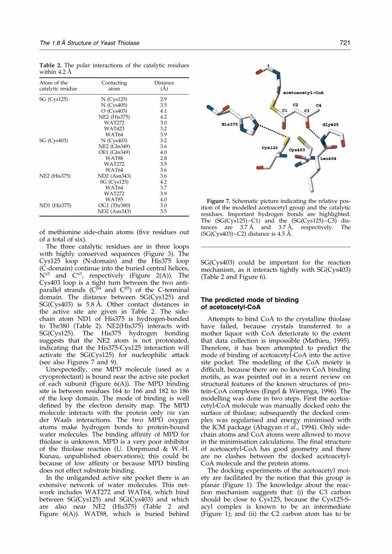
Figure 7. Schematic picture indicating the relative pos-ition of the modelled acetoacetyl group and the catalyticresidues. Important hydrogen bonds are highlighted.The (SG(Cys125)±C1) and the (SG(Cys125)±C3) dis-tances are 3.7 AÊ and 3.7 AÊ , respectively. The(SG(Cys403)±C2) distance is 4.5 AÊ .
Table 2. The polar interactions of the catalytic residueswithin 4.2 AÊ
Atom of thecatalytic residue
Contactingatom
Distance(AÊ )
SG (Cys125) N (Cys125) 2.9N (Cys405) 3.5O (Cys403) 4.1
NE2 (His375) 4.2WAT272 3.0WAT423 3.2WAT64 3.9
SG (Cys403) N (Cys403) 3.2NE2 (Gln349) 3.6OE1 (Gln349) 4.0
WAT88 2.8WAT272 3.5WAT64 3.6
NE2 (His375) ND2 (Asn343) 3.6SG (Cys125) 4.2
WAT64 3.7WAT272 3.9WAT85 4.0
ND1 (His375) OG1 (Thr380) 3.0ND2 (Asn343) 3.5
The 1.8 AÊ Structure of Yeast Thiolase 721

near to Cys403 (see also Figure 9). These geometricrequirements suggest that the acetoacetyl grouphas to be oriented as shown in Figure 6(B), withthe two oxygen atoms pointing towards the bot-tom of the active site pocket and the S-atom of theCoA moiety pointing upward. The acetoacetylgroup is locked into a position near Cys125 andCys403. The two water molecules, WAT272 andWAT64, seen in the unliganded structure (Figure 6)will be displaced on binding of the substrate. Inthis mode of binding O1 is near SG(Cys125) andNE2(His375), whereas O2 is hydrogen bonded toN(Gly405) (Figure 7). The O2-N(Gly405) distance is3 AÊ . This mode of binding is only possible whenthe Met186 side-chain adopts a conformation thatis different from what is observed in the crystalstructure.
The CoA group points out of the binding pocketas shown in Figures 6(B) and 8; the fatty acid tail(extending from the C4 atom) is pointing upwardsas well, and can therefore ®ll the remainingvolume of the binding pocket. The CoA moiety hasthree phosphate groups, which presumably inter-act with conserved positively charged side-chainson the surface of thiolase. In the proposed mode ofbinding the phosphate groups can interact withLys48, Lys51, Arg246 and Arg258 (Figures 6(B)and 8). These residues are conserved in the familyof dimeric thiolases (Figure 3). In the predictedmode of binding the CoA-molecule adopts anextended conformation, as seen in other CoA-pro-tein complexes (Engel & Wierenga, 1996). One ofthe few common features in the structures of CoA-protein complexes are the hydrogen bonds of theadenine amino group (N6) with the protein. This isalso observed in the modelled complex, in whichthe N6 atom points to the main-chain oxygenatoms of Glu243, Gly244 and Pro245.
The b-mercaptoethylamine moiety is modelledbetween the side-chains of Phe346 on the upperside and Met155 and Val276 on the lower side ofthe binding pocket (Figure 6(B)). The OP1 and OP2atoms of the pantothenic acid point inwards to theside-chain of Ser274 and N(Val276), respectively
(Figure 6(B)). The two methylgroups (CPA andCPB) are in van der Waals contact with Phe50,Val276 and Tyr159. The docked CoA molecule ®tsinto the groove formed by loops 48±51 (loopNb1±Na1 in the N-domain) and 259±262 of theloop domain (Figure 6(B)). An analysis of the vander Waals surfaces of the docked complex showsthat this groove is somewhat too wide for a perfect®t, suggesting that small loop movements mightbe induced by the CoA binding; possibly this con-cerns loop 259±262, which has very high B-factorsin this structure (Figure 5). It is interesting to pointout that the crystal packing is such that in subunit-2 the pocket between these two loops is involvedin crystal contact interactions. The presence ofthese crystal contacts would explain why the thio-lase crystals dissolve in solutions with CoA, mak-ing this crystal form unsuitable for crystallographicbinding studies.
In the proposed model CoA interacts almostexclusively with residues of the loop domain. Theexceptions are residues Lys48, Lys51 and Phe50which are in the (Nb1±Na1)-loop of the N-domain.
The mode of binding of the acyl chain of the3-ketoacyl-CoA moiety has not been modelled.Kinetic measurements indicate that the acyl chaindoes contribute to the binding energy, because theKm value of 3-ketohexadecanoyl-CoA (1.9 mM) isseven times smaller than for acetoacetyl-CoA(14.0 mM; Miyazawa et al., 1981). It seems likelythat the acyl moiety will interact with hydrophobicside-chains of the binding pocket. As describedabove there are two hydrophobic ``walls'' in thebinding pocket. The acetoacetyl moiety binds closeto the hydrophobic wall near Cys403. In this bind-ing mode there is still space left for binding of theacyl moiety to the hydrophobic wall (near Leu377)of the cyclindrically shaped active site pocket(Figure 6). In this way the acyl chain would curlaround the acetoacetyl moiety and bind at the bot-tom of the active site pocket. This mode of bindingwould also explain why acyl chains of severaldifferent chain lengths can bind.
Figure 8. The surface of thiolase, colour-coded according to the electrostatic potential by GRASP (Nicholls et al., 1991)(top view). Also shown is the predicted mode of binding of acetoacetyl-CoA (the side-chain of Met186 has beenremoved from the calculation, in order to see the atoms of the acetoacetyl moiety).
722 The 1.8 AÊ Structure of Yeast Thiolase

The reaction mechanism
The different steps of the reaction mechanismare shown in Figure 9. The ®rst step is the nucleo-philic attack of SG(Cys125) on the C3 carbon atom,which is facilitated by the predicted hydrogenbond between O2 and N(Gly405) (Figure 7). Thisresults in breaking the C2±C3 bond, producing aC2 carbanion which is subsequently protonated,possibly by a proton of the SG atom of Cys403, asdeduced from mutagenic studies with Z. ramigeraacetoacetyl-CoA thiolase (Palmer et al., 1991;Williams et al., 1992). This generates acetyl-CoA,which dissociates out of the active site pocket andthe fatty acid tail remains bound to Cys125. Afterrelease of the acetyl-CoA, a free CoA moleculebinds to the enzyme (step 2). It is assumed thatCoA binds in the same way as the CoA moiety ofthe substrate, with the S atom close to His375. Inthis way it is predicted that the CoA-S atom can beactivated by either the ND1(His375) or theNE2(His375) atom (step 3). Activation byND1(His375) would require a 180� rotation of theside-chain with respect to the position observed inthis crystal form. The crystal structure of the thio-lase-CoA complex could clarify this point. The acti-vated CoA molecule reacts with the acyl-moiety ofthe covalent intermediate which results in the for-mation of a new molecule of acyl-CoA (step 4).
It is interesting to point out that in the ®rst stepthe CoA-S atom is separated by two carbon atomsfrom the C atom which becomes bound to
SG(Cys125), while in the fourth step the CoA-Satom becomes covalently linked to a SG(Cys125)-bound C atom (Figure 9). This suggests structuralrearrangements of the S-acyl chain between step 1and step 4. Water molecules are not directlyinvolved in the reaction mechanism, but WAT88(near SG(Cys403)) could be important for the reac-tivity of SG(Cys403) (Table 2 and Figure 6(A)).
The active site of thiolase can also catalyse thesynthesis of acetoacetyl-CoA from two moleculesof acetyl-CoA. The ®rst step of this reaction is thebinding of acetyl-CoA, which is converted into aCoA molecule and a thiolase-Cys125-S-acetyl com-plex. For this reaction to occur SG(Cys125) has toreact with the C1 carbon (not the C3 carbon atomas in the degradative reaction). In our model thedistance of SG(Cys125) to the C1 atom is 3.7 AÊ
(Figure 7). Therefore, it seems that acetyl-CoA andacetoacetyl-CoA can bind in the same way, butreact differently with the SG(Cys125) atom. Thehydrogen bond between O2 and N(Gly405)(Figure 7), which exists only in the presence of theacetoacetyl moiety, will favour the reaction withthe C3 atom, as is required for the catalysis of thecleavage reaction.
The predicted structure of the thiolase tetramer
Tetrameric thiolases can be either 3-ketoacyl-CoA thiolases or acetoacetyl-CoA thiolases. Thetetrameric thiolases are more widespread than the
Figure 9. The proposed reaction mechanism for the degradative reaction of thiolase. Four steps are emphasised: 1,the formation of the covalent intermediate; 2, the replacement of acetyl-CoA by CoA; 3, the activation of CoA; and 4,the formation of the product.
The 1.8 AÊ Structure of Yeast Thiolase 723

dimeric thiolases, which occur only in the peroxi-somes (Igual et al., 1992). The high sequence simi-larity between the tetrameric and dimeric thiolasessuggests that the tetrameric thiolases are assembledas a dimer of two thiolase dimers. It is thereforevery likely that the tetrameric thiolase has tetrahe-dral symmetry, with three perpendicular, intersect-ing 2-fold axes. One of these axes is the dimer2-fold axis. This allows for only two possiblemodes of assembly: either the dimer/dimer inter-face is on the same side as the active sites(Figure 2), or it is on the opposite side, near the Nand C termini. As can be deduced from Figure 2(A)(top view) and Figure 2(B) (side view) the ®rstpossibility would require interactions of the loopdomains with each other at the dimer/dimer inter-face, whereas the latter possibility would requirethe N and C termini to interact with each other.For two reasons the latter possibility is not verylikely. (i) One would expect to see some conservedresidues in the tetrameric sequences at the dimer/dimer interface, but none of this is seen on thisside of the dimer (data not shown). (ii) The convexshape of the surface provides only a small contactarea for the potential dimer/dimer interface. How-ever the ¯at shape of the active site face provides amuch better surface for the dimer-dimer inter-actions. The ¯at appearance of this surface(Figure 2(B)) is due to four loops, which are resi-dues 48 to 70 (N-domain), residues 155 to 177(loop domain), residues 234 to 237 (loop domain)and residues 256 to 266 (loop domain). There areseveral conserved sequence features in the threeloops, which are near the 2-fold axis (48±70, 155±177, 234±237). (i) In the loop 48±70 there is astrictly conserved pattern in the dimeric sequences,being K/R(48)-G/A-G/F-K/R(51)-G-X-F-K/R(55).The equivalent tetrameric sequences are all one resi-due shorter and have a different pattern of conser-vation. (ii) At the beginning of the loop domain(residues 155 to 177) all tetrameric thiolasesequences have an insertion with a unique sequencepattern. (iii) The length of the loop region nearGly236 is very well conserved in the tetramericthiolases, but in the dimeric thiolases it is either sixresidues shorter, as in yeast, or four residues longer(as in the other dimeric thiolases). Moreover in thetetrameric thiolases a completely conserved (K/R,G) pattern is found at this site.
The loop region near residue 262, which isfurther away from the molecular centre, also con-tributes to the ¯at appearance of the surface onthis side of the molecule and therefore might alsocontribute to the postulated dimer/dimer interface.
The proposed dimer/dimer interface is on thesame side of the molecule as the active site andtherefore also near the binding site for the CoAsubstrate molecule. However, it is not incompatiblewith the proposed mode of binding of the CoAsubstrate molecule, because the CoA substrate mol-ecule is predicted to bind in a large groove belowthe outer surface (Figure 8).
The proposed mode of assembly exposes the Ntermini and the C termini in the tetramer to thesolvent (Figure 2(B)). The N terminus contains thetargetting sequence for entry into either the peroxi-some or the mitochondria (Erdmann & Kunau,1994), therefore the import apparatus would beable to recognise this targetting signal also in thefolded tetrameric protein.
Concluding Remarks
The analysis of the structure has revealed somepeculiar structural features. For example, the chan-nel of buried polar residues near the active sitepocket and the 12 buried charged residues permonomer. Most of these buried residues are com-pletely conserved (Table 1). It will be interesting toanalyse, by site-directed mutagenesis, the import-ance of these residues for the structure and func-tion of thiolase.
The active site of thiolase catalyses reactionswith 3-ketoacyl-CoA, acetyl-CoA and CoA. Thebinding of 3-ketoacyl-CoA is the ®rst step inthe degradation reaction and a covalent bondbetween SG(Cys125) and the C3 atom of thesubstrate is formed. The binding of acetyl-CoAis the ®rst step in the synthetic reaction, in thiscase a covalent bond is formed betweenSG(Cys125) and the C1 atom of the substrate.Binding of CoA to the thiolase±Cys125-S±acylcomplex is the second step of the degradativereaction and it leads to the formation of theacyl-CoA product molecule.
Here we have proposed a very plausible modeof binding of acetoacetyl-CoA as well as a reactionmechanism for thiolase. The proposed mode ofbinding of acetoacetyl-CoA and the proposed reac-tion mechanism do not infer conformationalchanges of loops at the catalytic site. However, it isrequired that the side-chain of Met186 adoptsanother conformation on binding of the acetoacetylgroup. From the modelling it is unclear if thiswould induce main-chain conformational changesin this region. Possibly loop conformation changesoccur in the 259 to 262 region, allowing for anoptimal ®t of the CoA part; this region has highB-factors in the unliganded structure.
It is proposed that CoA and acetyl-CoA bind inthe same way as acetoacetyl-CoA. The modellingexperiments with acetoacetyl-CoA suggest that theacetoacetyl group ®ts near the side-chains of thetwo catalytic cysteines. The proposed reactionmechanism, as inferred from the mode of bindingof the acetoacetyl group, is in good agreementwith previously published experimental data andit also explains the importance of the His375 side-chain. This side-chain can activate either theS(CoA) atom in the second step of the degradationreaction, or it can activate the SG(Cys125) atom fornucleophilic attack on either the C3 atom or the C1atom of the acetoacetyl group or the acetyl group,respectively. The reaction between SG(Cys125) and
724 The 1.8 AÊ Structure of Yeast Thiolase

the C3 atom of 3-ketoacyl-CoA, as required in thedegradative reaction, is favoured by the predictedhydrogen bond between O3 and N(Gly405).
Further crystallographic studies to verify theproposed mode of binding of acetoacetyl-CoA andthe reaction mechanism have been initiated.
Materials and Methods
Crystallisation and data collection
The 3-ketoacetyl CoA thiolase from yeast (S. cerevisiae)peroxisomes was expressed, puri®ed and crystallised asdescribed (Mathieu et al., 1994). The crystals are grownby a dialysis method (Zeelen & Wierenga, 1992) in a25 mM 3-(N-morpholino)-propanesulphonic acid (Mops)buffer (pH 6.5), 1 mM DTT, 1 mM NaN3, 1 mM EDTA.The space group is P212121 and the cell dimensions (atroom tempertature) are a � 71.78 AÊ , b � 93.72 AÊ andc � 120.45 AÊ . Fresh crystals diffract to 1.8 AÊ . The datawere collected with a big MAR image plate at the ERSF(Grenoble) at station BM4 from a thiolase crystal ¯ash-cooled at 100 K. MPD was used as a cryoprotectant.A freshly grown thiolase crystal was transferred from itsmother liquor into a cryosolution which was made bymixing equal volumes of MPD and the original motherliquor. The resulting MPD concentration was approxi-mately 4 M. After 20 minutes equilibration in this sol-ution the crystal was mounted using the loop technique(Teng, 1990) and ¯ash-frozen in a cold nitrogen gasstream at 100 K. The dataset was collected in two passes:a high resolution pass (20 seconds exposure time per 0.8�frame) and a low resolution pass (four second exposuretime per 1.2� frame) to achieve good completeness atlow resolution. The wavelength of the X-ray beam was0.995 AÊ . The cell dimensions of the cryo-cooled thiolasecrystal (a � 71.17 AÊ , b � 92.65 AÊ , c � 116.72 AÊ ) are smal-ler compared to a room temperature crystal. The datawere processed with DENZO (Otwinowski, 1993) andSCALEPACK (Gewirth et al., 1995). The re®ned value ofthe mosaicity for this dataset is 0.35�. The data wereinitially merged into a complete dataset with an R-factor
of 5.0% and a completeness of 98.1% between 17 AÊ and1.77 AÊ .
Structure refinement
The re®nement of the thiolase structure against this1.8 AÊ dataset was done ®rst with X-PLOR (BruÈ nger,1992). In later stages also ARP (Lamzin & Wilson, 1993)and TNT (Tronrud, 1992) were used. A test set of re¯ec-tions (5% of the data, 3790 re¯ections) was used forR-free calculations. Each aspect of the re®nement strat-egy was veri®ed by monitoring the variation of R-free.The starting model for the re®nement was the pre-viously determined structure at 2.8 AÊ resolution. Its pos-ition in the new cell was found by molecularreplacement calculations and rigid body re®nement.
Many cycles of re®nement and model building wererequired, initially at 2.6 AÊ resolution but eventually at1.8 AÊ resolution. The models were built in SIGMAA-weighted maps (Read, 1986), calculated with CCP4 pro-grams (Collaborative Computational Project, 1994). Theprogram O (Jones et al., 1991; Kleywegt & Jones, 1996)was used for the model building. NCS restraints wereused initially for the positional and B-factor re®nement.For the re®nement at 1.8 AÊ resolution no NCS-restraintswere used.
In the starting model several regions of the loopdomain were missing. Missing residues were included insubsequent models whenever the new maps clearly indi-cated their structure. Also water and two MPD mol-ecules were added to the structure. Eventually, usingTNT, a model was obtained with an R-factor of 21.1%(R-free � 28.7%). This model has 5842 protein atoms, 430water molecules and 16 MPD atoms. At this stage thedataset was reprocessed, taking particular care to have a®nal dataset as complete as possible and also using bet-ter overload criteria. Overloaded observations wereremoved during the reprocessing, which resulted in bet-ter merging statistics, apparently because of the non-line-arity properties of the detector. The data between 10 AÊ
and 17 AÊ were removed because these data were unreli-able due to a slightly misaligned beam stop. In addition,
Table 3. Statistics of the ®nal processing of the 1.8 AÊ dataset
Resolution limits: Totalinner outer R-merge Completeness number of(AÊ ) (AÊ ) (%) I/s (%) reflections
10.00 4.72 3.4 43 99.9 38224.72 3.81 3.4 54 100.0 37263.81 3.35 4.1 68 100.0 36583.35 3.05 4.7 65 100.0 36643.05 2.84 5.3 57 100.0 36252.84 2.68 5.6 51 99.8 36172.68 2.55 5.4 45 98.9 35562.55 2.44 5.5 45 99.2 35922.44 2.34 5.4 45 99.5 36082.34 2.26 5.7 41 99.6 35702.26 2.19 6.0 39 99.4 35932.19 2.13 6.2 37 99.5 35432.13 2.08 6.7 33 99.3 35912.08 2.03 7.7 30 99.6 35322.03 1.98 9.0 27 99.6 35731.98 1.94 9.9 25 99.6 35711.94 1.90 11.9 23 99.0 35631.90 1.86 12.0 21 99.2 35211.86 1.83 12.2 19 99.6 35301.83 1.80 12.2 17 86.8 3115Total 4.7 98.9 71,570
The 1.8 AÊ Structure of Yeast Thiolase 725
some bad frames were removed from the processing.This reprocessing and rescaling resulted in a consider-ably better dataset. The R-merge (on I) is 4.7% and thecompleteness between 10 AÊ and 1.8 AÊ is 98.9% (Table 3).The overall R-merge (4.7%) is entirely dominated by thelow resolution data, due to the very high redundancy ofthe data in the low resolution shells. Nevertheless, thedata at high resolution (R-merge 12.2%) are of goodquality. The reprocessing of the data improved the crys-tallographic R-factor signi®cantly.
Further re®nement, against the new dataset, with ARPand TNT resulted eventually in a model with an R-factorof 20.2% for data between 10 AÊ and 1.8 AÊ , as calculatedby TNT (now using all the data). This model includes5865 protein atoms, 472 water and two MPD molecules.The R-free value of this model was assessed by carryingout a simulated annealing X-PLOR run starting at 2000 Kwith data between 8 AÊ and 1.8 AÊ (without B-factorre®nement). The R-factor and R-free are 21.6% and26.1% for the work set and the test set, respectively. Sub-sequently the anisotropy in the native dataset was cor-rected which reduced the crystallographic R-factor by1%. Further checking of the model against the mapsuggested additional minor changes. In addition theTNT weights were optimised. The ®nal model has anR-factor of 18.7%. In this model there are 5874 proteinatoms, 448 water and two MPD molecules. Residues 263to 265 of subunit-1 are missing. The R-free value of thismodel was again veri®ed by a simulated annealingX-PLOR run starting at 2000 K for data between 8 AÊ
and 1.8 AÊ (without B-factor re®nement). The R-factorand R-free values are now 20.3% and 24.2% for the
workset and the test set, respectively. The re®nementstatistics are summarised in Table 4.
Structure analysis and sequence alignment
The structure has been analysed with O and WHATIF (Vriend, 1990). The program ICM (Abagyan et al.,1994) was used for the modelling experiments. Unlessotherwise stated the structure description refers to sub-unit-1. Sequence alignments have been made with Pile-Up of the GCG package (Devereux et al., 1984). Thecavities and molecular surfaces were calculated with theMSP package (Connolly, 1985); atomic radii and proberadius (1.2 AÊ ) were the same as used previously(Wierenga et al., 1992). The polar residues Arg, Lys, Asp,and Glu were considered to be buried when the polarside-chain atoms did not contribute to the outer surfaceof the protein.
Acknowledgements
We thank Ursula Dorpmund for the puri®cation ofthiolase, Dr Horst Schulz for stimulating discussions andDr Cusack for his interest in this project. The high resol-ution dataset was collected at station BM4 at the ESRFGrenoble. The coordinates (1AFW) and the structure fac-tors (R1AFWSF) of the re®ned structure have beendeposited at the PDB. The coordinates of the modelledacetoacetyl-CoA complex have also been deposited inthe PDB (1AFY).
Table 4. Crystallographic data
A. Refinement statisticsTotal number of reflections 71,365R-factor (%) 18.7Total number of protein atoms 5874Total number of MPD atoms 16Total number of water atoms 448
B. Geometry statisticsrms bond length (AÊ ) 0.017rms bond angle (�) 2.4rms �B (bonded atoms), all atoms (AÊ 2) 6.0rms �B (bonded atoms), main-chain atoms (AÊ 2) 5.4rms �B (bonded atoms), side-chain atoms (AÊ 2) 6.9Average B (all protein atoms) (AÊ 2) 26.3Average B (main-chain atoms) (AÊ 2) 22.8Average B (side-chain atoms) (AÊ 2) 30.2average B (water) (AÊ 2) 35.9Average B (MPD) (AÊ 2) 31.3w1w2 imperfection (�)a 33.3
C. Ramachandran plotb
Most favoured regions 597 (89.4%)Additionally allowed regions 65 (9.7%)Generally allowed (4) and disallowed regions (2) 6 (0.9%)
D. Non-crystallographic symmetryc
N L C Allrms �B (all atoms) (AÊ 2) 9.0 15.3 8.5 11.6rms �B (main-chain atoms) (AÊ 2) 7.9 12.3 7.4 9.5rms �B (side-chain atoms) (AÊ 2) 10.0 18.0 9.6 13.7rms (�Ca) (AÊ ) 0.22 0.50 0.19 0.34rms (�f) (�) 5.5 10.3 6.5 9.4rms (�c) (�) 5.4 12.4 6.4 10.7
a The w1w2 imperfection value (Noble et al., 1993) is the rms difference between observed w1w2-values and the nearest preferred clustervalue, as seen in a database of well-re®ned structures.
b As de®ned by PROCHECK (Laskowski et al., 1993).c N, N-domain (residues 25 to 154, 277 to 299); C, C-domain (residues 300 to 417); L, loop-domain (residues 155 to 276).
726 The 1.8 AÊ Structure of Yeast Thiolase
References
Abagyan, R. A. (1997). The ICM 2.6 Manual (http://molsoft.com). Molsoft LLC, New York.
Abagyan, R. A., Totrov, M. & Kuznetsov, D. (1994).ICM-A new method for protein modelling anddesign: applications to docking and structure pre-diction from the distorted native conformation.J. Comput. Chem. 15, 488±506.
Anderson, V. E., Bahnson, B. J., Wlassics, I. D. & Walsh,C. T. (1990). The reaction of acetyldithio-CoA, areadily enolized analog of acetyl-CoA with thiolasefrom Zoogloea ramigera. J. Biol. Chem. 265, 6255±6261.
BruÈ nger, A. T. (1992). X-PLOR: version 3.1, a System forX-ray Crystallography and NMR. Yale UniversityPress, New Haven and London.
Computational Project N. (1994). The CCP4 suite: pro-grams for protein crystallography. Acta Crystallog.sect. D, 50, 760±763.
Connolly, M. L. (1985). Computation of molecularvolume. J. Am. Chem. Soc. 107, 1118±1124.
Devereux, J., Haeberli, P. & Smithies, O. (1984). A com-prehensive set of sequence analysis programs forthe VAX. Nucl. Acids Res. 12, 387±395.
Engel, C. K. & Wierenga, R. (1996). The diverse worldof coenzyme A binding proteins. Curr. Opin. Struct.Biol. 6, 790±797.
Engel, C. K., Mathieu, M., Zeelen, J. Ph. , Hiltunen,J. K. & Wierenga, R. K. (1996). Crystal structure ofenoyl-coenzyme A (CoA) hydratase at 2.5 AÊ resol-ution: a spiral fold de®nes the CoA-binding pocket.EMBO J. 15, 5135±5145.
Erdmann, R. (1994). The peroxisomal targeting signal of3-oxoacy 1-CoA thiolase from Saccharomycescerevisiae. Yeast, 10, 935±944.
Erdmann, R. & Kunau, W.-H. (1994). Puri®cation andimmunolocalization of the peroxisomal 3-oxoacyl-CoA thiolase of Saccharomyces cerevisiae. Yeast, 10,1173±1182.
Flocco, M. M. & Mowbray, S. L. (1995). Strange bedfel-lows: interactions between acidic side-chains inproteins. J. Mol. Biol. 254, 96±105.
Gehring, U. & Lynen, F. (1972). Thiolase. In The Enzymes(Boyer, P. D., ed.), vol. 7, pp. 391±405, AcademicPress, New York and London.
Gewirth, D., Otwinowski, Z. & Minor, W. (1995). TheHKL Manual (4th Edit.) (Gewirth, D., ed.), YaleUniversity, New Haven.
Gilbert, H. F. (1981). Proton transfer from acetyl-coen-zyme A catalyzed by thiolase I from porcine heart.Biochemistry, 20, 5643±5649.
Gilbert, H. F., Lennox, B. J., Mossman, C. D. & Carle,W. C. (1981). The relation of acyl transfer to theoverall reaction of thiolase I from porcine heart.J. Biol. Chem. 256, 7371±7377.
Hartmann, G. & Lynen, F. (1961). Thiolase. In TheEnzymes (Boyer, P. D., ed.), vol. 5, pp. 381±386,Academic Press, New York and London.
Igual, J. C., Gonzalez-Bosch, C., Dopazo, J. & PeÂrez-Ortin, J. E. (1992). Phylogenetic analysis of the thio-lase family. Implications for the evolutionary originof peroxisomes. J. Mol. Evol. 35, 147±155.
Izbicka, E. & Gilbert, H. F. (1984). Fluorescence energytransfer measurements of spatial relationshipsbetween sulfhydryl groups of Thiolase I from por-cine heart. Biochemistry, 23, 6383±6388.
Izbicka-Dimitrijevic, E. & Gilbert, H. F. (1984). Multipleoxidation products of sulfhydryl groups near the
active site of thiolase I from porcine heart. Biochem-isty, 23, 4318±4324.
Jones, T. A., Zou, J.-Y., Cowan, S. W. & Kjeldgaard, M.(1991). Improved methods for building proteinmodels in electron density maps and the location oferrors in these models. Acta Crystallog. sect. A, 47,110±119.
Kabsch, W. & Sander, C. (1983). Dictionary of proteinsecondary structure: pattern recognition of hydro-gen-bonded and geometrical features. Biopolymers,22, 2577±2637.
Kleywegt, G. J. & Jones, T. A. (1996). Ef®cient rebuildingof protein structures. Acta Crystallog. sect. D, 52,829±832.
Kunau, W. -H., Dommes, Y. & Schulz, H. (1995). b-Oxi-dation of fatty acids in mitochondria, peroxisomesand bacteria: a centry of continued progress. Progr.Lipid Res. 34, 267±342.
Kurihara, T., Ueda, M. & Tanaka, A. (1989). Peroxisomalacetoacetyl-CoA thiolase and 3-ketoacyl-CoA thio-lase from an n-alkane-utilizing yeast, Candida tropi-calis: puri®cation and characterization. J. Biochem.106, 474±478.
Lamzin, V. S. & Wilson, K. S. (1993). Automated re®ne-ment of protein models. Acta Crystallog. sect. D, 49,129±147.
Laskowski, R. A., MacArthur, M. W., Moss, D. S. &Thornton, J. M. (1993). PROCHECK: a Program tocheck the stereochemical quality of proteinstructures. J. Appl. Crystallog. 26, 283±291.
Masamune, S., Palmer, M. A. J., Gamboni, R.,Thompson, S., Davis, J. T., Williams, S. F., Peoples,O. P., Sinskey, A. J. & Walsh, C. T. (1989a). Bio-Claisen condensation catalyzed by thiolase fromZoogloea ramigera. Active site cysteine residues.J. Am. Chem. Soc. 111, 1879±1881.
Masamune, S., Walsh, C. T., Sinskey, A. J. & Peoples,O. P. (1989b). Poly-(R)-3-hydroxybutyrate (PHB)biosynthesis: mechanistic studies on the biologicalClaisen condensation catalyzed by b-ketoacylthiolase. Pure Appl. Chem. 61, 303±312.
Mathieu, M. (1995). Structure determination of thiolase,Ecole Centrale, Paris.
Mathieu, M., Zeelen, J. P., Pauptit, R. A., Erdmann, R.,Kunau, W.-H. & Wierenga, R. K. (1994). The 2.8 AÊ
crystal structure of peroxisomal 3-ketoacyl-CoAthiolase of Saccharomyces cerevisiae: a ®ve-layeredababa structure constructed from two core domainsof identical topology. Structure, 2, 797±808.
Miyazawa, S. & Hashimoto, T. (1980). The presenceof a new 3-oxoacyl-CoA thiolase in rat liverperoxisomes. Eur. J. Biochem. 103, 589±596.
Miyazawa, S., Furuta, S., Osumi, T., Hashimoto, T. &Ui, N. (1981). Properties of peroxisomal 3-ketoacyl-CoA thiolase from rat liver. J. Biochem. 90, 511±519.
Nicholls, A., Sharp, K. & Honig, B. (1991). Protein fold-ing and association insights from the interfacial andthermodynamic properties of hydrocarbons. Pro-teins: Struct. Funct. genet. 11, 281±296.
Noble, M. E. M., Zeelen, J. P., Wierenga, R. K.,Mainfroid, V., Goraj, K., Gohimont, A.-C. &Martial, J. A. (1993). Structure of triosephosphateisomerase from Escherichia coli determined at 2.6 AÊ
resolution. Acta Crystallog. sect. D, 49, 403±417.Otwinowski, Z. (1993). Oscillation data reduction
program. In Proceedings of the CCP4 Study Weekend:``Data Collection and Processing'' (Sawyer, L., Isaacs,N. & Baily, S., eds), pp. 56±62, SERC, DaresburyLaboratory, England.
The 1.8 AÊ Structure of Yeast Thiolase 727

Palmer, M. A. J., Differding, E., Gamboni, R., Williams,S. F., Peoples, O. P., Walsh, C. T., Sinskey, A. J. &Masamune, S. (1991). Biosynthetic thiolase fromZoogloea ramigera. Evidence for a mechanism invol-ving Cys378 as the active site base. J. Biol. Chem.266, 8369±8375.
Read, R. J. (1986). Improved Fourier coef®cients formaps using phases from partial structures witherrors. Acta Crystallog. sect. A, 42, 140±149.
Salam, W. H. & Bloxham, D. P. (1986). Identi®cation ofsubsidiary catalytic groups at the active site ofb-ketoacyl-CoA thiolase by covalent modi®cation ofthe protein. Biochim. Biophys. Acta, 873, 321±330.
Staack, H., Binstock, J. F. & Schulz, H. (1978). Puri®-cation and properties of a pig heart thiolase withbroad chain length speci®city and comparison ofthiolases from pig heart and Escherichia coli. J. Biol.Chem. 253, 1827±1831.
Teng, T.-Y. (1990). Mounting of crystals for macromol-ecular crystallography in a free-standing thin ®lm.J. Appl. Crystallog. 23, 387±391.
Thompson, S., Mayerl, F., Peoples, O. P., Masamune, S.,Sinskey, A. J. & Walsh, C. T. (1989). MechanisticStudies on b-ketoacyl thiolase from Zoogloea rami-gera: identi®cation of the active-site nucleophile asCys89, its mutation to Ser89, and kinetic and ther-modynamic characterization of wild-type andmutant enzymes. Biochemistry, 28, 5735±5742.
Tronrud, D. E. (1992). Conjugate-direction minimization:an improved method for the re®nement ofmacromolecules. Acta Crystallog. sect. A, 48, 912±916.
Vriend, G. (1990). WHAT IF: a molecular modeling anddrug design program. J. Mol. Graph. 8, 52±56.
Walsh, C. T. (1979). Enzymatic Reaction Mechanisms(Bartlett, A. C. & McCombs, L. W., eds). W. H.Freeman and Company, San Francisco.
Waterson, R. M. & Hill, R. L. (1972). Enoyl coenzyme Ahydratase (Crotonase). J. Biol. Chem. 247, 5258±5265.
Wierenga, R. K., Noble, M. E. M. & Davenport, R. C.(1992). Comparison of the re®ned crystal structuresof liganded and unliganded chicken, yeast and try-panosomal triosephosphate isomerase. J. Mol. Biol.224, 1115±1126.
Williams, S. F., Palmer, M. A. J., Peoples, O. P., Walsh,C. T., Sinskey, A. J. & Masamune, S. (1992). Biosyn-thetic thiolase from Zoogloea ramigera. Mutagenesisof the putative active-site base Cys378 changes thepartitioning of the acetyl S-enzyme intermediate.J. Biol. Chem. 267, 16041±16043.
Zeelen, J. Ph. & Wierenga, R. K. (1992). The growth ofyeast thiolase crystals using a polyacrylamide gel asdialysis membrane. J. Cryst. Growth, 122, 194±198.
Edited by R. Huber
(Received 9 April 1997; received in revised form 30 July 1997; accepted 4 August 1997)
728 The 1.8 AÊ Structure of Yeast Thiolase
Related Documents