Citation: Albadr, Y.; Crowe, A.; Caccetta, R. Teucrium polium: Potential Drug Source for Type 2 Diabetes Mellitus. Biology 2022, 11, 128. https://doi.org/10.3390/ biology11010128 Academic Editors: Magdalene Montgomery and Stacey Keenan Received: 20 December 2021 Accepted: 10 January 2022 Published: 13 January 2022 Publisher’s Note: MDPI stays neutral with regard to jurisdictional claims in published maps and institutional affil- iations. Copyright: © 2022 by the authors. Licensee MDPI, Basel, Switzerland. This article is an open access article distributed under the terms and conditions of the Creative Commons Attribution (CC BY) license (https:// creativecommons.org/licenses/by/ 4.0/). biology Review Teucrium polium: Potential Drug Source for Type 2 Diabetes Mellitus Yaser Albadr , Andrew Crowe and Rima Caccetta * Curtin Medical School, Curtin Health Innovation Research Institute, Faculty of Health Sciences, Curtin University, Bentley, WA 6102, Australia; [email protected] (Y.A.); [email protected] (A.C.) * Correspondence: [email protected] Simple Summary: Teucrium polium (also known as Golden Germander) is a herb brewed and drunk as a tea by the locals of the Mediterranean region, used mostly to treat a number of illnesses including diabetes. When consumed regularly, the tea can be problematic since some of its ingredients can be toxic or interfere with other medications taken by the patient. Current anti-diabetic medications are not always suitable nor optimal for all patients living with diabetes and therefore new drugs are constantly being sought after which may be more useful and/or present less side effects. Therefore, identifying the specific constituents that give the desired anti-diabetic effect, isolating them and developing them further may provide new useful anti-diabetic drugs. This paper discusses some key compounds found in Golden Germander that might be valuable for developing a new medication for type 2 diabetics whilst outlining some issues with the research conducted thus far. Abstract: The prevalence of type 2 diabetes mellitus is rising globally and this disease is proposed to be the next pandemic after COVID-19. Although the cause of type 2 diabetes mellitus is unknown, it is believed to involve a complex array of genetic defects that affect metabolic pathways which eventually lead to hyperglycaemia. This hyperglycaemia arises from an inability of the insulin- sensitive cells to sufficiently respond to the secreted insulin, which eventually results in the inadequate secretion of insulin from pancreatic β-cells. Several treatments, utilising a variety of mechanisms, are available for type 2 diabetes mellitus. However, more medications are needed to assist with the optimal management of the different stages of the disease in patients of varying ages with the diverse combinations of other medications co-administered. Throughout modern history, some lead constituents from ancient medicinal plants have been investigated extensively and helped in developing synthetic antidiabetic drugs, such as metformin. Teucrium polium L. (Tp) is a herb that has a folk reputation for its antidiabetic potential. Previous studies indicate that Tp extracts significantly decrease blood glucose levels r and induce insulin secretion from pancreatic β-cells in vitro. Nonetheless, the constituent/s responsible for this action have not yet been elucidated. The effects appear to be, at least in part, attributable to the presence of selected flavonoids (apigenin, quercetin, and rutin). This review aims to examine the reported glucose-lowering effect of the herb, with a keen focus on insulin secretion, specifically related to type 2 diabetes mellitus. An analysis of the contribution of the key constituent flavonoids of Tp extracts will also be discussed. Keywords: glucose lowering; insulin secretion; phenolic compounds; flavonoids; anti-diabetic 1. Introduction Diabetes mellitus (DM) is defined as a chronic and multifunctional metabolic disorder characterised by hyperglycaemia, resulting from impaired insulin secretion, insulin action or both. These pathophysiological processes lead to malfunctions in carbohydrate, protein and fat metabolism. The two main types of DM are (i) type 1, which mainly results from autoimmune destruction of the pancreatic β-cells leading to a lack of insulin secretion and Biology 2022, 11, 128. https://doi.org/10.3390/biology11010128 https://www.mdpi.com/journal/biology

Welcome message from author
This document is posted to help you gain knowledge. Please leave a comment to let me know what you think about it! Share it to your friends and learn new things together.
Transcript
�����������������
Citation: Albadr, Y.; Crowe, A.;
Caccetta, R. Teucrium polium:
Potential Drug Source for Type 2
Diabetes Mellitus. Biology 2022, 11,
128. https://doi.org/10.3390/
biology11010128
Academic Editors:
Magdalene Montgomery and
Stacey Keenan
Received: 20 December 2021
Accepted: 10 January 2022
Published: 13 January 2022
Publisher’s Note: MDPI stays neutral
with regard to jurisdictional claims in
published maps and institutional affil-
iations.
Copyright: © 2022 by the authors.
Licensee MDPI, Basel, Switzerland.
This article is an open access article
distributed under the terms and
conditions of the Creative Commons
Attribution (CC BY) license (https://
creativecommons.org/licenses/by/
4.0/).
biology
Review
Teucrium polium: Potential Drug Source for Type 2Diabetes MellitusYaser Albadr , Andrew Crowe and Rima Caccetta *
Curtin Medical School, Curtin Health Innovation Research Institute, Faculty of Health Sciences, Curtin University,Bentley, WA 6102, Australia; [email protected] (Y.A.); [email protected] (A.C.)* Correspondence: [email protected]
Simple Summary: Teucrium polium (also known as Golden Germander) is a herb brewed and drunkas a tea by the locals of the Mediterranean region, used mostly to treat a number of illnesses includingdiabetes. When consumed regularly, the tea can be problematic since some of its ingredients can betoxic or interfere with other medications taken by the patient. Current anti-diabetic medications arenot always suitable nor optimal for all patients living with diabetes and therefore new drugs areconstantly being sought after which may be more useful and/or present less side effects. Therefore,identifying the specific constituents that give the desired anti-diabetic effect, isolating them anddeveloping them further may provide new useful anti-diabetic drugs. This paper discusses some keycompounds found in Golden Germander that might be valuable for developing a new medication fortype 2 diabetics whilst outlining some issues with the research conducted thus far.
Abstract: The prevalence of type 2 diabetes mellitus is rising globally and this disease is proposed tobe the next pandemic after COVID-19. Although the cause of type 2 diabetes mellitus is unknown,it is believed to involve a complex array of genetic defects that affect metabolic pathways whicheventually lead to hyperglycaemia. This hyperglycaemia arises from an inability of the insulin-sensitive cells to sufficiently respond to the secreted insulin, which eventually results in the inadequatesecretion of insulin from pancreatic β-cells. Several treatments, utilising a variety of mechanisms,are available for type 2 diabetes mellitus. However, more medications are needed to assist withthe optimal management of the different stages of the disease in patients of varying ages with thediverse combinations of other medications co-administered. Throughout modern history, somelead constituents from ancient medicinal plants have been investigated extensively and helpedin developing synthetic antidiabetic drugs, such as metformin. Teucrium polium L. (Tp) is a herbthat has a folk reputation for its antidiabetic potential. Previous studies indicate that Tp extractssignificantly decrease blood glucose levels r and induce insulin secretion from pancreatic β-cellsin vitro. Nonetheless, the constituent/s responsible for this action have not yet been elucidated. Theeffects appear to be, at least in part, attributable to the presence of selected flavonoids (apigenin,quercetin, and rutin). This review aims to examine the reported glucose-lowering effect of the herb,with a keen focus on insulin secretion, specifically related to type 2 diabetes mellitus. An analysis ofthe contribution of the key constituent flavonoids of Tp extracts will also be discussed.
Keywords: glucose lowering; insulin secretion; phenolic compounds; flavonoids; anti-diabetic
1. Introduction
Diabetes mellitus (DM) is defined as a chronic and multifunctional metabolic disordercharacterised by hyperglycaemia, resulting from impaired insulin secretion, insulin actionor both. These pathophysiological processes lead to malfunctions in carbohydrate, proteinand fat metabolism. The two main types of DM are (i) type 1, which mainly results fromautoimmune destruction of the pancreatic β-cells leading to a lack of insulin secretion and
Biology 2022, 11, 128. https://doi.org/10.3390/biology11010128 https://www.mdpi.com/journal/biology
Biology 2022, 11, 128 2 of 16
(ii) type 2, the most prevalent, that encompasses a combination of reduced responsivenessby insulin-sensitive cells and defects in insulin secretion [1,2].
Approximately 537 million people worldwide have diabetes mellitus [3]. The numberof people living with type 2 DM has dramatically increased in all countries irrespective ofsocioeconomic status. These numbers do not include those living with type 2 DM (around240 million people) but who remain undiagnosed [2,3]. Recently, it has become evidentthat diabetic patients are more susceptible to infection with Coronavirus disease 2019(COVID-19) and thus have an increased mortality rate [4–6]. COVID-19 patients couldalso develop diabetes due to sedentary lifestyle and weight gain caused by lockdown andrestricted movement [7,8]. Aside from the life-threatening risks associated with COVID-19,6.7 million deaths are directly attributed to DM or its complications in 2021, with 541 millionadults having impaired glucose tolerance [3,9]. The latest report from the World HealthOrganisation states that by 2030, this metabolic pandemic will become the 7th main causeof worldwide mortality as the prevalence is estimated to encompass more than 400 millionsufferers [10,11]. This number has since risen in post-COVID estimates to 643 million by2030 and 783 million by 2045 [3]. It is estimated that the cost for DM worldwide was USD$850 billion in 2017, and reached USD $966 billion in 2021 [3].
The exact aetiology of type 2 DM is yet to be confirmed; however, it appears to arisemainly from a complex polygenic and heterogeneous group of metabolic defects involvinginadequate insulin secretion from β-cells following an inability of the insulin-sensitivetissues, such as adipose, skeletal muscle and cardiac muscle cells to adequately respond toincreased blood glucose levels [12–14]. Several risk factors have been associated with thedevelopment of type 2 DM, which can be broken down into modifiable (hypertension, lackof physical activity and obesity) and non-modifiable (aging, being male or of particularethnicity) risk factors [15].
The onset of type 2 DM occurs gradually over several years and patients often do notnotice any symptoms [16]. In the early stages of pre-DM, the pancreatic β-cells overproduceinsulin leading to hyperinsulinemia in order to compensate for the high glucose levelsappearing in the circulating blood, thus giving normal or slightly above normal bloodglucose readings. After a few years of this constant compensation, most pancreatic β-cellsbecome damaged. Yet, during this period, some patients could restore some of their β-cellcapacity with proper treatment and management [15].
After type 2 DM sets in, the disease continues to evolve with short or long-term com-plications that can occur at any stage of the disease progression. These include metabolicdysfunction or failure of several organs (kidney, heart, eyes and blood vessels), which in-crease the risk of developing cardiovascular diseases, such as hypertension, dyslipidaemiaand obesity; this reduces the quality of life and increases the mortality rate among patientswith type 2 DM [1,17–20]. Serious imbalance in blood glucose levels could result in seriousorgan dysfunction and death [21].
Glucose is the main source of energy for most body organs and tissues [22]. Followinga meal, the body breaks food down into glucose and then transports it through the bloodcirculation. Under normal physiological conditions, increased blood glucose levels triggerthe pancreatic β-cells of the islet of Langerhans to secrete insulin which facilitates glucoseuptake, transportation and metabolism in peripheral tissues in order to maintain bloodglucose within steady state levels (a process known as glucose homeostasis) [21,23]. Unde-niably, insulin plays a major balancing role as a key hormone in glucose homeostasis and isthe only one known to actively lower high blood glucose levels.
At present, treatment objectives in type 2 DM are focused on maintaining normalblood glucose levels. Different therapeutic approaches for type 2 DM are utilised. Changesin lifestyle, such as diet and exercise as part of a weight control program, are the firstapproach, and usually accompanied by pharmacological therapy [19]. Several treatmentstrategies are available for diabetic patients either through synthetic drugs or medicinalplant products from natural sources.
Biology 2022, 11, 128 3 of 16
The currently available pharmacological treatments to help manage type 2 DM arewell-established [24–26]. These medications can be divided based on their mechanism of ac-tion into three groups. The first group improves insulin sensitivity: the thiazolidinediones,also known as glitazones, including drugs such as pioglitazone and rosiglitazone [27].The second group increases endogenous insulin: the glucagon-like peptide-1 (GLP-1) ago-nist [28], sulphonylureas, [29], dipeptidyl peptidase IV inhibitors [30] and alpha glucosidaseinhibitors [31]. Finally, the third group mainly has one representative which is metformin,and this medication decreases hepatic production of glucose and reduces glucose intestinalabsorption which directly affects insulin. Metformin is currently the first line therapy fortype 2 DM [32].
Whilst most of these conventional antidiabetic drugs are potent and effective, theyare not sufficient nor can they sustain optimal treatment for every diabetic throughout theongoing progressive stages of the disease [33]. This is further complicated by the needto switch medications when side effects arise. For instance, lactic acidosis can occur indiabetic patients taking metformin, as well as being not recommended for patients withrenal insufficiency [34–36]. Uncontrolled weight gain can occur with patients using sulpho-nylureas while thiazolidinediaones could increase the incidence of hyperlipidaemia [37].Given the high cost of insulin, which may be required in combination to offset the limitedcapabilities of the oral anti-diabetic medications, the search for new drug entities frommedicinal plants has become even more urgent.
Apart from prescribed medications, the vast majority of diabetic patients in developingcountries use complementary or medicinal natural products that have an antidiabeticreputation [38,39]. Interestingly, most people at high risk of developing type 2 DM usuallyhave the nutritional recommendation to take plant-based food, all known to be enrichedwith several phytochemicals, such as phenolic compounds [39]. Constituents of medicinalplants continue to provide some lead active compounds that can be further developedinto antidiabetic medication [40]. Thus, medicinal plants continue to provide a potentialcache of novel agents from natural products that could help in the normalisation of bloodglucose levels.
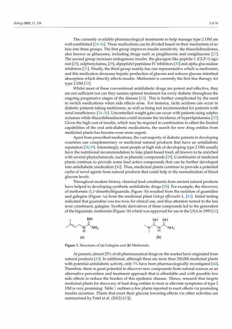
Throughout modern history, chemical lead constituents from ancient natural productshave helped in developing synthetic antidiabetic drugs [38]. For example, the discoveryof metformin (1,1-dimethylbiguanide, Figure 1b) resulted from the isolation of guanidineand galegine (Figure 1a) from the medicinal plant Galega officinalis L. [41]. Initial testingindicated that guanidine was too toxic for clinical use, and thus attention turned to the lesstoxic constituent, galegine. Synthetic derivatives of these compounds led to the generationof the biguanide, metformin (Figure 1b) which was approved for use in the USA in 1995 [42].
Biology 2022, 11, x FOR PEER REVIEW 3 of 15
The currently available pharmacological treatments to help manage type 2 DM are
well-established [24–26]. These medications can be divided based on their mechanism of
action into three groups. The first group improves insulin sensitivity: the thiazolidinedi-
ones, also known as glitazones, including drugs such as pioglitazone and rosiglitazone
[27]. The second group increases endogenous insulin: the glucagon-like peptide-1 (GLP-
1) agonist [28], sulphonylureas, [29], dipeptidyl peptidase IV inhibitors [30] and alpha
glucosidase inhibitors [31]. Finally, the third group mainly has one representative which
is metformin, and this medication decreases hepatic production of glucose and reduces
glucose intestinal absorption which directly affects insulin. Metformin is currently the first
line therapy for type 2 DM [32].
Whilst most of these conventional antidiabetic drugs are potent and effective, they
are not sufficient nor can they sustain optimal treatment for every diabetic throughout the
ongoing progressive stages of the disease [33]. This is further complicated by the need to
switch medications when side effects arise. For instance, lactic acidosis can occur in dia-
betic patients taking metformin, as well as being not recommended for patients with renal
insufficiency [34–36]. Uncontrolled weight gain can occur with patients using sulphonylu-
reas while thiazolidinediaones could increase the incidence of hyperlipidaemia [37].
Given the high cost of insulin, which may be required in combination to offset the limited
capabilities of the oral anti-diabetic medications, the search for new drug entities from
medicinal plants has become even more urgent.
Apart from prescribed medications, the vast majority of diabetic patients in develop-
ing countries use complementary or medicinal natural products that have an antidiabetic
reputation [38,39]. Interestingly, most people at high risk of developing type 2 DM usually
have the nutritional recommendation to take plant-based food, all known to be enriched
with several phytochemicals, such as phenolic compounds [39]. Constituents of medicinal
plants continue to provide some lead active compounds that can be further developed
into antidiabetic medication [40]. Thus, medicinal plants continue to provide a potential
cache of novel agents from natural products that could help in the normalisation of blood
glucose levels.
Throughout modern history, chemical lead constituents from ancient natural prod-
ucts have helped in developing synthetic antidiabetic drugs [38]. For example, the discov-
ery of metformin (1,1-dimethylbiguanide, Figure 1b) resulted from the isolation of guan-
idine and galegine (Figure 1a) from the medicinal plant Galega officinalis L. [41]. Initial
testing indicated that guanidine was too toxic for clinical use, and thus attention turned
to the less toxic constituent, galegine. Synthetic derivatives of these compounds led to the
generation of the biguanide, metformin (Figure 1b) which was approved for use in the
USA in 1995 [42].
(a) (b)
Figure 1. Structures of (a) Galegine and (b) Metformin.
At present, almost 25% of all pharmaceutical drugs on the market have originated
from natural products [43]. In additional, although there are more than 250,000 medicinal
plants with potential antidiabetic activity, only 1% have been pharmacologically investi-
gated [44]. Therefore, there is great potential to discover new compounds from natural
sources as an alternative prevention and treatment approach that is affordable and with
possible low side effects to reduce the burden of this epidemic disease. Hence, research
that targets medicinal plants for discovery of lead drug entities to treat or alleviate symp-
toms of type 2 DM is very promising: Table 1 outlines a few plants reported to exert effects
Figure 1. Structures of (a) Galegine and (b) Metformin.
At present, almost 25% of all pharmaceutical drugs on the market have originated fromnatural products [43]. In additional, although there are more than 250,000 medicinal plantswith potential antidiabetic activity, only 1% have been pharmacologically investigated [44].Therefore, there is great potential to discover new compounds from natural sources as analternative prevention and treatment approach that is affordable and with possible lowside effects to reduce the burden of this epidemic disease. Hence, research that targetsmedicinal plants for discovery of lead drug entities to treat or alleviate symptoms of type 2DM is very promising: Table 1 outlines a few plants reported to exert effects via promotinginsulin secretion. Plants that exert their glucose lowering effects via other activities aresummarised by Patel et al. (2012) [45]).
Biology 2022, 11, 128 4 of 16
Although the publications on the general anti-diabetic properties of Teucrium polium(Tp) were summarised by Asghari et al., a detailed analysis of the extract in relation to type2 DM is not presented [46]. Phenolic compounds are of rising interest. However, thesecompounds are quickly metabolised after ingestion in the gut and via the first pass effect.Therefore, our current review aims to evaluate the literature on the use of Tp for treatingtype 2 DM and to discuss the assessment strategies of studies that examined key phenoliccompounds identified in aqueous Tp extracts.
Table 1. Some antidiabetic medicinal plants with potential insulin secretagogue activity.
References Plant Name Antidiabetic Activity
[46–50] Teucrium polium Increase insulin secretion
[51] Tabernaemontana divaricata Increase blood insulin and promote pancreaticβ-cells regeneration in mice
[52] Gymnema sylvestre Increase insulin and pancreatic β-cellsregeneration in rodent
[53] Ficus deltoidea Increase insulin secretion[54] Bidens pilosa Increase insulin secretion
2. Teucrium polium
Teucrium polium L. (Lamiaceae) (Tp), commonly known as golden germander, is ashrubby plant mainly growing in the Mediterranean deserts, hills, and mountains. Thisplant forms a highly influential component of Middle Eastern and Palestinian folk medicinewhere the natives drink the plant extract as a tea to treat DM, rheumatism, gastrointestinaldisturbances, and inflammations. The folk of that region are also reported to use Tp brew forits antihyperlipidemic, analgesic, diaphoretic and antipyretic activities [39,55]. Althoughthere have been several studies that examined the total plant extract for its glucose loweringeffect [46–49,55] and others that have identified several phytochemical constituents of theextracts that include flavonoids, terpenoids and iridoids [56,57], the exact compound(s)responsible for the glucose-lowering effect of the total extract of Teucrium polium are yet to bedetermined. A few cases showing the toxicological potential of Tp, especially hepatotoxicity,have been reported; however, it is not clear which plant species was consumed, nor whetheradditional medications contributed to the adverse outcomes [58,59].
Regrettably, safety evaluation of medicinal plants is difficult due to the complexchemical nature and diversity within plants, some of which are currently considered usefulas complementary medicine in diabetes. These plants may contain thousands of unknownbioactive compounds or other contaminants and toxic materials. While broad toxicityprofiles provide a measure of safety for such complex natural components, robust clinicaland experimental data, including toxicological information, phytochemical properties andthe safe use of reputed medicinal plants and their likely active constituents with potentialantidiabetic effects, are urgently needed [60,61].
2.1. Antidiabetic Effect of Tp
Few ethnobotanical [56,62] or pharmacological [46,55,63,64] studies have focused onthe glucose lowering properties of traditional medicinal plants, especially Tp. The antihy-perglycemic activity of extracts of Tp have been studied in vivo, primarily in rodents, sincethe 1980s [50,55,63–65]. Gharaibeh et al. (1988) demonstrated that in both streptozotocin(STZ)-induced diabetic and normoglycemic rats, an aqueous extract of Tp aerial parts re-sults in a substantial reduction in blood glucose levels 4 h after intravenous administrationand 24 h after intraperitoneal administration [65]. Furthermore, chronic (30 consecutivedays) oral administration of Tp extract at 0.5 mg/kg to STZ-induced diabetic rats sig-nificantly (p < 0.05) reduced blood glucose levels compared to the STZ-induced diabeticcontrols [63,64]. Unfortunately, these studies ran for an extended period and did not includea positive control, so results may need further analysis to confirm their data. Nevertheless,two animal studies have also reported an acute antihyperglycemic effect with use of this
Biology 2022, 11, 128 5 of 16
extract [50,55]. Oral administration of Tp extract at (125 mg/kg) to normoglycemic ratsexhibited significant acute diminution of blood glucose levels 4 h after a single dose, and itwas superior to glibenclamide [50].
In one of the more recent studies, Ireng et al. (2016) reported that intravenous ad-ministration of Tp extract (100 mg/kg) to normoglycemic rats, lead to a significant acutehypoglycaemic effect in a manner that was similar in efficacy to insulin over the first30 min [55]. This outcome corroborated earlier results revealing that Tp extract could beable to ameliorate glucose homeostasis either by increasing insulin secretion or improvingglucose uptake from peripheral tissues. Some studies have attempted to assess the plantextract further ex vivo and in vitro in an attempt to elucidate the possible mechanistic roleof constituents with glucose lowering potential [48,64].
In an investigation by Mirghazanfari et al. (2010), Tp methanolic extract (1000 µg/mLin 2.8 mmol/L glucose) was able to potentiate (p < 0.05) glucose stimulated insulin secretion(GSIS) from isolated perfused rat pancreas ex vivo; however, no effect was observed withthe aqueous extract [49]. Moreover, the insulinotropic effect observed was credited to thepresence of flavonoids, including apigenin. In another study by Yazdanparast et al. (2005),ethanol/water extract of Tp at 1 to 100 µg/mL in 2 and 16 mmol/L glucose two hoursafter treatment was able to stimulate (p < 0.001) GSIS from isolated pancreatic rat islets;however, when tested at 1000 µg/mL, the effect was decreased [64]. This was attributed tothe cytotoxicity of the plant extract. Moreover, studies that also histologically examined theislets of Langerhans of STZ-treated rats reported that the pancreatic islets are regeneratedafter Tp treatment which may be attributed to the quercetin in the extract [63,64,66].
The potential mechanism explaining the molecular pathways that could contributeto the glucose-lowering activity of the extract in cell culture settings have been studied aswell. Relevant emerging in vitro studies have focused on insulin secretagogue potentialfrom Tp extract [47,48]. Stefkov et al. (2011) evaluated the insulinotropic effect of Tp extractusing INS-1E rat insulinoma cells and reported that Tp ethanolic extract at 500 µg/mL in20 mmol/L glucose exhibited a significant increase in GSIS [50]. It is worth mentioning thatin these experiments the cell viability assay was not measured and the causes of diminishedactivity at a higher dose were not clearly explained.
More recently, Mannan (2017), assessed the insulin secretagogue potential of the“aqueous” Tp extract, prepared as per methodology outlined in Ireng et al. (2016) [55],on BRIN-BD11 rat pancreatic cells. It was shown that the extract at 62, 125, 250 and500 µg/mL in 5.5 mmol/L glucose significantly (p < 0.05) increased glucose uptake andinsulin secretion in a dose-dependent manner [48]. Along with this, treatment with theTp extracts significantly (p < 0.05) increased the expression of GLUT2 and glucokinaseactivity. This was associated with an increase in ATP production and an increase in influxof intracellular calcium [48]. In keeping with this finding, a study by Kasabri et al. (2012)reported distinct results in the increasing pattern of GSIS at different concentrations (10,100, 1000, 10,000 or 25,000 µg/mL in 5.6 mmol/L glucose) of aqueous Tp extracts in mousepancreatic β-cells MIN6 without existing cytotoxicity. Moreover, the secretory mechanismwas highly dependent on calcium influx [47]. The differences in the pattern of GSIS could bedue to different reasons including the contents of culture medium, growth rate, conditionsor the type of cell line.
Since Tp extracts were able to reduce blood glucose levels by inducing insulin secre-tion in accordance with the well-known biochemical pathway for insulin secretion frompancreatic β-cells (Table 2), they are considered a promising initial target for drug discoveryresearch, especially for DM. Although Tp antidiabetic activity has been studied in differentexperimental models, all of which support the use of its extracts as an initial phase in thesearch for antihyperglycaemic agents, the bioactive constituent/s of the Tp extract are yetto be defined.
Biology 2022, 11, 128 6 of 16
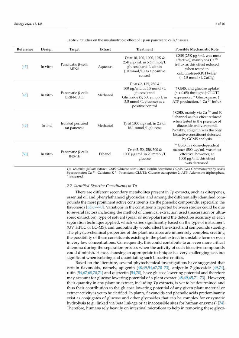
Table 2. Studies on the insulinotropic effect of Tp on pancreatic cells/tissues.
Reference Design Target Extract Treatment Possible Mechanistic Role
[47] In vitro Pancreatic β-cellsMIN6 Aqueous
Tp at 10, 100, 1000, 10K &25K µg/mL in 5.6 mmol/L
glucose) and L-alanin(10 mmol/L) as a positive
control
↑ GSIS (25K µg/mL was mosteffective), mainly via Ca 2+
influx as this effect reducedwhen tested in
calcium-free-KRH buffer(−2.5 mmol/L CaCl2)
[48] In vitro Pancreatic β-cellsBRIN-BD11 Methanol
Tp at 62, 125, 250 &500 µg/mL in 5.5 mmol/L
glucose) andGliclazide (5, 500 µmol/L in
5.5 mmol/L glucose) as apositive control
↑ GSIS, and glucose uptake(p < 0.05) through: ↑ GLUT2expression, ↑ Glucokinase, ↑
ATP production, ↑ Ca 2+ influx
[49] In situ Isolated perfusedrat pancreas Methanol Tp at 1000 µg/mL in 2.8 or
16.1 mmol/L glucose
↑ GSIS, mainly via Ca 2+ and K+ channel as this effect reducedwhen tested in the presence of
diazoxide and verapamil.Notably, apigenin was the onlybioactive constituent detected
by GCMS analysis
[50] In vitro Pancreatic β-cellsINS-1E Ethanol
Tp at 5, 50, 250, 500 &1000 µg/mL in 20 mmol/L
glucose
↑ GSIS in a dose-dependentmanner (500 µg/mL was most
effective; however, at1000 µg/mL this effect
was decreased
Tp: Teucrium polium extract; GSIS: Glucose-stimulated insulin secretion; GCMS: Gas Chromatography MassSpectrometer; Ca 2+: Calcium; K +: Potassium; GLUT2: Glucose transporter 2; ATP: Adenosine triphosphate,↑ increased.
2.2. Identified Bioactive Constituents in Tp
There are different secondary metabolites present in Tp extracts, such as diterpenes,essential oil and phenylethanoid glycosides, and among the differentially identified com-pounds the most prominent active constituents are the phenolic compounds, especially, theflavonoids [55,67–70]. Variations in the constituents reported between studies could be dueto several factors including the method of chemical extraction used (maceration or ultra-sonic extraction), type of solvent (polar or non-polar) and the detection accuracy of eachseparation technique applied, which varies significantly based on the type of instrument(UV, HPLC or LC-MS), and undoubtedly would affect the extract and compounds stability.The physico-chemical properties of the plant matrices are immensely complex, creatingthe possibility of these constituents existing in the plant extract in unstable form or evenin very low concentrations. Consequently, this could contribute to an even more criticaldilemma during the separation process when the activity of such bioactive compoundscould diminish. Hence, choosing an appropriate technique is a very challenging task butsignificant when isolating and quantitating such bioactive entities.
Based on the literature, several phytochemical investigations have suggested thatcertain flavonoids, namely, apigenin [48,49,54,67,70–73], apigenin 7-glucoside [49,74],rutin [54,67,68,70,71] and quercetin [54,70], have glucose lowering potential and thereforemay account for glucose lowering potential of a plant extract [48,49,65,71–73]. However,their quantity in any plant or extract, including Tp extracts, is yet to be determined andthus their contribution to the glucose lowering potential of any given plant material orextract activity is yet to be clarified. In plants, flavonoids and phenolic acids predominantlyexist as conjugates of glucose and other glycosides that can be complex for enzymatichydrolysis (e.g., linked via beta linkage or at inaccessible sites for human enzymes) [74].Therefore, humans rely heavily on intestinal microflora to help in removing these glyco-
Biology 2022, 11, 128 7 of 16
sides and thus enable free flavonoids/phenolic acids (aglycones) to be absorbed throughthe intestines [75–77].
Bioactivity and Bioavailability of the Key Flavonoids Found in Tp
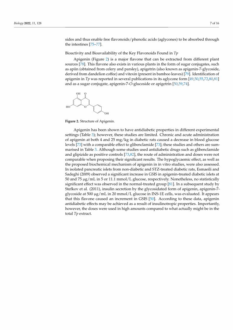
Apigenin (Figure 2) is a major flavone that can be extracted from different plantsources [78]. This flavone also exists in various plants in the form of sugar conjugates, suchas apiin (obtained from celery and parsley), apigetrin (also known as apigenin-7-glycoside,derived from dandelion coffee) and vitexin (present in bamboo leaves) [79]. Identification ofapigenin in Tp was reported in several publications in its aglycone form [49,50,55,72,80,81]and as a sugar conjugate, apigenin-7-O-glucoside or apigetrin [50,59,74].
Biology 2022, 11, x FOR PEER REVIEW 7 of 15
as conjugates of glucose and other glycosides that can be complex for enzymatic hydrol-
ysis (e.g., linked via beta linkage or at inaccessible sites for human enzymes) [74]. There-
fore, humans rely heavily on intestinal microflora to help in removing these glycosides
and thus enable free flavonoids/phenolic acids (aglycones) to be absorbed through the
intestines [75–77].
Bioactivity and Bioavailability of the Key Flavonoids Found in Tp.
Apigenin (Figure 2) is a major flavone that can be extracted from different plant
sources [78]. This flavone also exists in various plants in the form of sugar conjugates,
such as apiin (obtained from celery and parsley), apigetrin (also known as apigenin-7-
glycoside, derived from dandelion coffee) and vitexin (present in bamboo leaves) [79].
Identification of apigenin in Tp was reported in several publications in its aglycone form
[49,50,55,72,80,81] and as a sugar conjugate, apigenin-7-O-glucoside or apigetrin
[50,59,74].
Figure 2. Structure of Apigenin.
Apigenin has been shown to have antidiabetic properties in different experimental
settings (Table 3); however, these studies are limited. Chronic and acute administration of
apigenin at both 4 and 25 mg/kg in diabetic rats caused a decrease in blood glucose levels
[73] with a comparable effect to glibenclamide [73]; these studies and others are summa-
rised in Table 3. Although some studies used antidiabetic drugs such as glibenclamide
and glipizide as positive controls [73,82], the route of administration and doses were not
comparable when proposing their significant results. The hypoglycaemic effect, as well as
the proposed biochemical mechanism of apigenin in in vitro studies, were also assessed.
In isolated pancreatic islets from non-diabetic and STZ-treated diabetic rats, Esmaeili and
Sadeghi (2009) observed a significant increase in GSIS in apigenin-treated diabetic islets
at 50 and 75 μg/mL in 5 or 11.1 mmol/L glucose, respectively. Nonetheless, no statistically
significant effect was observed in the normal-treated group [81]. In a subsequent study by
Stefkov et al. (2011), insulin secretion by the glycosidated form of apigenin, apigenin-7-
glycoside at 500 μg/mL in 20 mmol/L glucose in INS-1E cells, was evaluated. It appears
that this flavone caused an increment in GSIS [50]. According to these data, apigenin an-
tidiabetic effects may be achieved as a result of insulinotropic properties. Importantly,
however, the doses were used in high amounts compared to what actually might be in the
total Tp extract.
Table 3. Apigenin potential in lowering blood glucose levels.
Refer-
ence Target Study Design Dose
Positive Con-
trol
Dura-
tion Outcome
[50]
Pancreatic β-
cells
INS-1E
Group 1: Glucose only at
(20 mmol/L)
Group 2: Apigenin-7-gly-
coside
(500 μg/mL in
20 mmol/L glu-
cose)
- 30 min
Significant (p < 0.05) in-
crease in GSIS compared
to 20 mmol/L glucose
[72]
STZ-induced (40
mg/kg i.p) dia-
betic rats (150–
250 g)
Group 1: Normal control
received saline + 5% Etha-
nol n = 6
Apigenin i.p at
(4 mg/kg/day) - 7 days
On day 7, apigenin signifi-
cantly (p < 0.01) reduced
blood glucose levels com-
Figure 2. Structure of Apigenin.
Apigenin has been shown to have antidiabetic properties in different experimentalsettings (Table 3); however, these studies are limited. Chronic and acute administrationof apigenin at both 4 and 25 mg/kg in diabetic rats caused a decrease in blood glucoselevels [73] with a comparable effect to glibenclamide [73]; these studies and others are sum-marised in Table 3. Although some studies used antidiabetic drugs such as glibenclamideand glipizide as positive controls [73,82], the route of administration and doses were notcomparable when proposing their significant results. The hypoglycaemic effect, as well asthe proposed biochemical mechanism of apigenin in in vitro studies, were also assessed.In isolated pancreatic islets from non-diabetic and STZ-treated diabetic rats, Esmaeili andSadeghi (2009) observed a significant increase in GSIS in apigenin-treated diabetic islets at50 and 75 µg/mL in 5 or 11.1 mmol/L glucose, respectively. Nonetheless, no statisticallysignificant effect was observed in the normal-treated group [81]. In a subsequent study byStefkov et al. (2011), insulin secretion by the glycosidated form of apigenin, apigenin-7-glycoside at 500 µg/mL in 20 mmol/L glucose in INS-1E cells, was evaluated. It appearsthat this flavone caused an increment in GSIS [50]. According to these data, apigeninantidiabetic effects may be achieved as a result of insulinotropic properties. Importantly,however, the doses were used in high amounts compared to what actually might be in thetotal Tp extract.
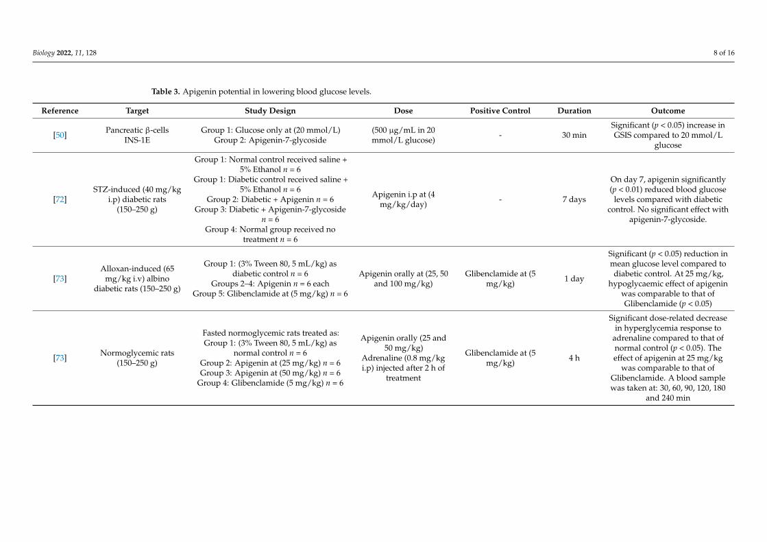
Biology 2022, 11, 128 8 of 16
Table 3. Apigenin potential in lowering blood glucose levels.
Reference Target Study Design Dose Positive Control Duration Outcome
[50] Pancreatic β-cellsINS-1E
Group 1: Glucose only at (20 mmol/L)Group 2: Apigenin-7-glycoside
(500 µg/mL in 20mmol/L glucose) - 30 min
Significant (p < 0.05) increase inGSIS compared to 20 mmol/L
glucose
[72]STZ-induced (40 mg/kg
i.p) diabetic rats(150–250 g)
Group 1: Normal control received saline +5% Ethanol n = 6
Group 1: Diabetic control received saline +5% Ethanol n = 6
Group 2: Diabetic + Apigenin n = 6Group 3: Diabetic + Apigenin-7-glycoside
n = 6Group 4: Normal group received no
treatment n = 6
Apigenin i.p at (4mg/kg/day) - 7 days
On day 7, apigenin significantly(p < 0.01) reduced blood glucose
levels compared with diabeticcontrol. No significant effect with
apigenin-7-glycoside.
[73]Alloxan-induced (65
mg/kg i.v) albinodiabetic rats (150–250 g)
Group 1: (3% Tween 80, 5 mL/kg) asdiabetic control n = 6
Groups 2–4: Apigenin n = 6 eachGroup 5: Glibenclamide at (5 mg/kg) n = 6
Apigenin orally at (25, 50and 100 mg/kg)
Glibenclamide at (5mg/kg) 1 day
Significant (p < 0.05) reduction inmean glucose level compared todiabetic control. At 25 mg/kg,
hypoglycaemic effect of apigeninwas comparable to that ofGlibenclamide (p < 0.05)
[73] Normoglycemic rats(150–250 g)
Fasted normoglycemic rats treated as:Group 1: (3% Tween 80, 5 mL/kg) as
normal control n = 6Group 2: Apigenin at (25 mg/kg) n = 6Group 3: Apigenin at (50 mg/kg) n = 6
Group 4: Glibenclamide (5 mg/kg) n = 6
Apigenin orally (25 and50 mg/kg)
Adrenaline (0.8 mg/kgi.p) injected after 2 h of
treatment
Glibenclamide at (5mg/kg) 4 h
Significant dose-related decreasein hyperglycemia response to
adrenaline compared to that ofnormal control (p < 0.05). Theeffect of apigenin at 25 mg/kg
was comparable to that ofGlibenclamide. A blood samplewas taken at: 30, 60, 90, 120, 180
and 240 min
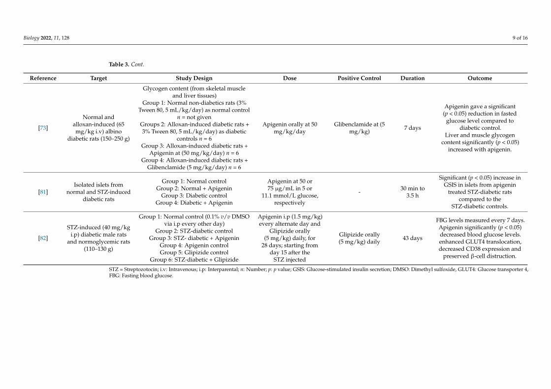
Biology 2022, 11, 128 9 of 16
Table 3. Cont.
Reference Target Study Design Dose Positive Control Duration Outcome
[73]
Normal andalloxan-induced (65mg/kg i.v) albino
diabetic rats (150–250 g)
Glycogen content (from skeletal muscleand liver tissues)
Group 1: Normal non-diabetics rats (3%Tween 80, 5 mL/kg/day) as normal control
n = not givenGroups 2: Alloxan-induced diabetic rats +3% Tween 80, 5 mL/kg/day) as diabetic
controls n = 6Group 3: Alloxan-induced diabetic rats +
Apigenin at (50 mg/kg/day) n = 6Group 4: Alloxan-induced diabetic rats +
Glibenclamide (5 mg/kg/day) n = 6
Apigenin orally at 50mg/kg/day
Glibenclamide at (5mg/kg) 7 days
Apigenin gave a significant(p < 0.05) reduction in fasted
glucose level compared todiabetic control.
Liver and muscle glycogencontent significantly (p < 0.05)
increased with apigenin.
[81]Isolated islets from
normal and STZ-induceddiabetic rats
Group 1: Normal controlGroup 2: Normal + Apigenin
Group 3: Diabetic controlGroup 4: Diabetic + Apigenin
Apigenin at 50 or75 µg/mL in 5 or
11.1 mmol/L glucose,respectively
- 30 min to3.5 h
Significant (p < 0.05) increase inGSIS in islets from apigenin
treated STZ-diabetic ratscompared to the
STZ-diabetic controls.
[82]
STZ-induced (40 mg/kgi.p) diabetic male rats
and normoglycemic rats(110–130 g)
Group 1: Normal control (0.1% v/v DMSOvia i.p every other day)
Group 2: STZ-diabetic controlGroup 3: STZ- diabetic + Apigenin
Group 4: Apigenin controlGroup 5: Glipizide control
Group 6: STZ-diabetic + Glipizide
Apigenin i.p (1.5 mg/kg)every alternate day and
Glipizide orally(5 mg/kg) daily, for
28 days; starting fromday 15 after the
STZ injected
Glipizide orally(5 mg/kg) daily 43 days
FBG levels measured every 7 days.Apigenin significantly (p < 0.05)decreased blood glucose levels.enhanced GLUT4 translocation,decreased CD38 expression and
preserved β-cell distruction.
STZ = Streptozotocin; i.v: Intravenous; i.p: Interparental; n: Number; p: p value; GSIS: Glucose-stimulated insulin secretion; DMSO: Dimethyl sulfoxide, GLUT4: Glucose transporter 4,FBG: Fasting blood glucose.
Biology 2022, 11, 128 10 of 16
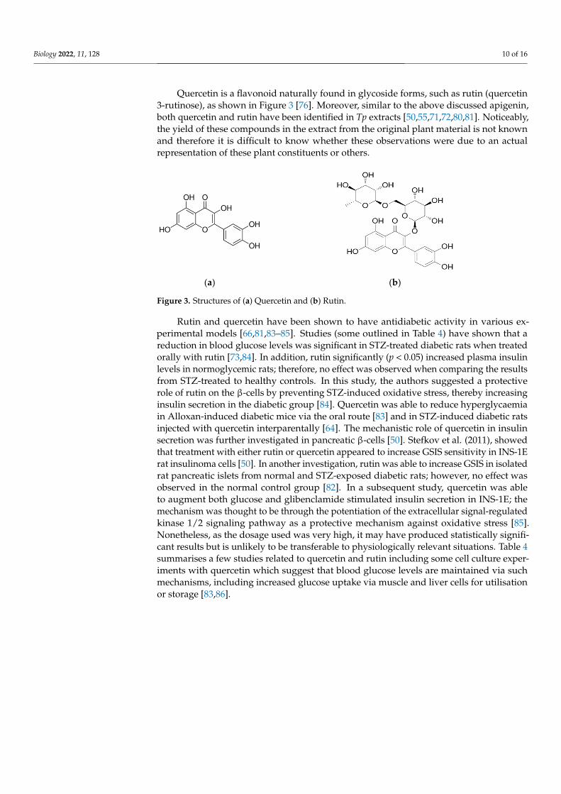
Quercetin is a flavonoid naturally found in glycoside forms, such as rutin (quercetin3-rutinose), as shown in Figure 3 [76]. Moreover, similar to the above discussed apigenin,both quercetin and rutin have been identified in Tp extracts [50,55,71,72,80,81]. Noticeably,the yield of these compounds in the extract from the original plant material is not knownand therefore it is difficult to know whether these observations were due to an actualrepresentation of these plant constituents or others.
Biology 2022, 11, x FOR PEER REVIEW 9 of 15
and STZ-in-
duced diabetic
rats
Group 3: Diabetic control
Group 4: Diabetic + Apig-
enin
mmol/L glu-
cose, respec-
tively
STZ-diabetic rats com-
pared to the STZ-diabetic
controls.
[82]
STZ-induced (40
mg/kg i.p) dia-
betic male rats
and normogly-
cemic rats (110–
130 g)
Group 1: Normal control
(0.1% v/v DMSO via i.p
every other day)
Group 2: STZ-diabetic
control
Group 3: STZ- diabetic +
Apigenin
Group 4: Apigenin con-
trol
Group 5: Glipizide con-
trol
Group 6: STZ-diabetic +
Glipizide
Apigenin i.p
(1.5 mg/kg)
every alternate
day and Gli-
pizide orally (5
mg/kg) daily,
for 28 days;
starting from
day 15 after the
STZ injected
Glipizide orally
(5 mg/kg) daily 43 days
FBG levels measured
every 7 days. Apigenin
significantly (p < 0.05) de-
creased blood glucose lev-
els. enhanced GLUT4
translocation, decreased
CD38 expression and pre-
served β-cell distruction.
STZ = Streptozotocin; i.v: Intravenous; i.p: Interparental; n: Number; p: p value; GSIS: Glucose-stimulated insulin secretion;
DMSO: Dimethyl sulfoxide, GLUT4: Glucose transporter 4, FBG: Fasting blood glucose.
Quercetin is a flavonoid naturally found in glycoside forms, such as rutin (quercetin
3-rutinose), as shown in Figure 3 [76]. Moreover, similar to the above discussed apigenin,
both quercetin and rutin have been identified in Tp extracts [50,55,71,72,80,81]. Noticeably,
the yield of these compounds in the extract from the original plant material is not known
and therefore it is difficult to know whether these observations were due to an actual rep-
resentation of these plant constituents or others.
(a) (b)
Figure 3. Structures of (a) Quercetin and (b) Rutin.
Rutin and quercetin have been shown to have antidiabetic activity in various exper-
imental models [66,81,83–85]. Studies (some outlined in Table 4) have shown that a reduc-
tion in blood glucose levels was significant in STZ-treated diabetic rats when treated
orally with rutin [73,84]. In addition, rutin significantly (p < 0.05) increased plasma insulin
levels in normoglycemic rats; therefore, no effect was observed when comparing the re-
sults from STZ-treated to healthy controls. In this study, the authors suggested a protec-
tive role of rutin on the β-cells by preventing STZ-induced oxidative stress, thereby in-
creasing insulin secretion in the diabetic group [84]. Quercetin was able to reduce hyper-
glycaemia in Alloxan-induced diabetic mice via the oral route [83] and in STZ-induced
diabetic rats injected with quercetin interparentally [64]. The mechanistic role of quercetin
in insulin secretion was further investigated in pancreatic β-cells [50]. Stefkov et al. (2011),
showed that treatment with either rutin or quercetin appeared to increase GSIS sensitivity
in INS-1E rat insulinoma cells [50]. In another investigation, rutin was able to increase
GSIS in isolated rat pancreatic islets from normal and STZ-exposed diabetic rats; however,
no effect was observed in the normal control group [82]. In a subsequent study, quercetin
Figure 3. Structures of (a) Quercetin and (b) Rutin.
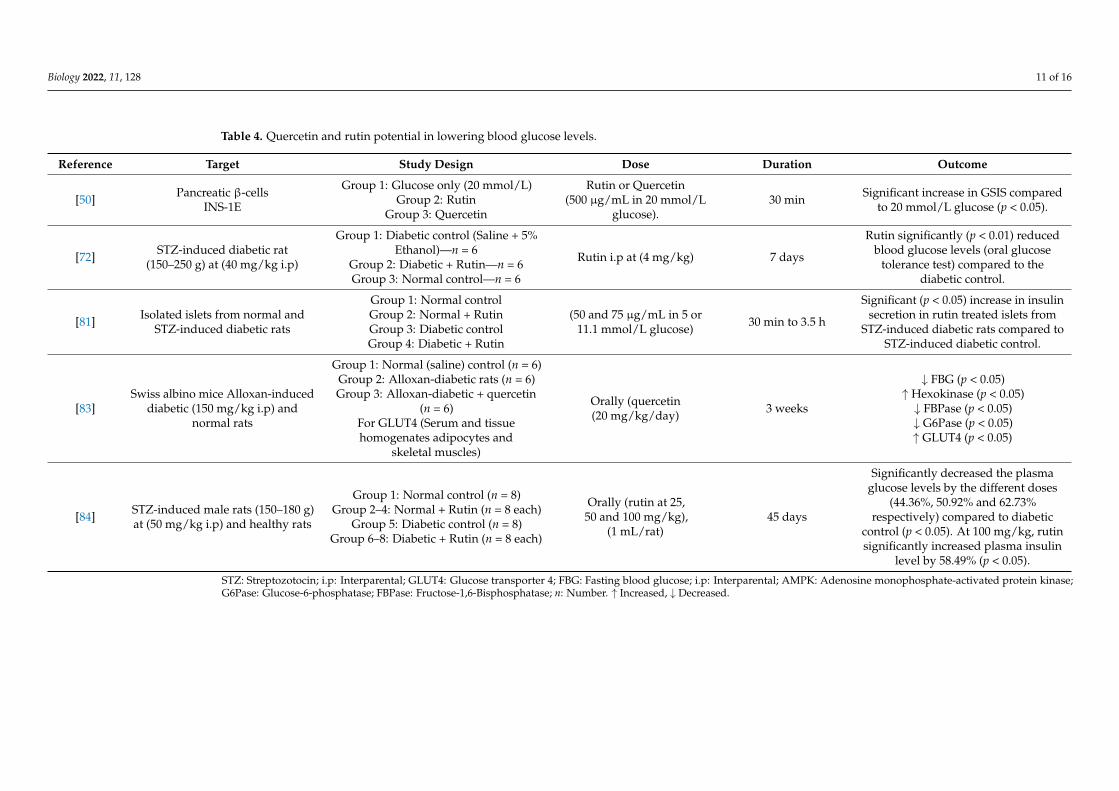
Rutin and quercetin have been shown to have antidiabetic activity in various ex-perimental models [66,81,83–85]. Studies (some outlined in Table 4) have shown that areduction in blood glucose levels was significant in STZ-treated diabetic rats when treatedorally with rutin [73,84]. In addition, rutin significantly (p < 0.05) increased plasma insulinlevels in normoglycemic rats; therefore, no effect was observed when comparing the resultsfrom STZ-treated to healthy controls. In this study, the authors suggested a protectiverole of rutin on the β-cells by preventing STZ-induced oxidative stress, thereby increasinginsulin secretion in the diabetic group [84]. Quercetin was able to reduce hyperglycaemiain Alloxan-induced diabetic mice via the oral route [83] and in STZ-induced diabetic ratsinjected with quercetin interparentally [64]. The mechanistic role of quercetin in insulinsecretion was further investigated in pancreatic β-cells [50]. Stefkov et al. (2011), showedthat treatment with either rutin or quercetin appeared to increase GSIS sensitivity in INS-1Erat insulinoma cells [50]. In another investigation, rutin was able to increase GSIS in isolatedrat pancreatic islets from normal and STZ-exposed diabetic rats; however, no effect wasobserved in the normal control group [82]. In a subsequent study, quercetin was ableto augment both glucose and glibenclamide stimulated insulin secretion in INS-1E; themechanism was thought to be through the potentiation of the extracellular signal-regulatedkinase 1/2 signaling pathway as a protective mechanism against oxidative stress [85].Nonetheless, as the dosage used was very high, it may have produced statistically signifi-cant results but is unlikely to be transferable to physiologically relevant situations. Table 4summarises a few studies related to quercetin and rutin including some cell culture exper-iments with quercetin which suggest that blood glucose levels are maintained via suchmechanisms, including increased glucose uptake via muscle and liver cells for utilisationor storage [83,86].
Biology 2022, 11, 128 11 of 16
Table 4. Quercetin and rutin potential in lowering blood glucose levels.
Reference Target Study Design Dose Duration Outcome
[50] Pancreatic β-cellsINS-1E
Group 1: Glucose only (20 mmol/L)Group 2: Rutin
Group 3: Quercetin
Rutin or Quercetin(500 µg/mL in 20 mmol/L
glucose).30 min Significant increase in GSIS compared
to 20 mmol/L glucose (p < 0.05).
[72] STZ-induced diabetic rat(150–250 g) at (40 mg/kg i.p)
Group 1: Diabetic control (Saline + 5%Ethanol)—n = 6
Group 2: Diabetic + Rutin—n = 6Group 3: Normal control—n = 6
Rutin i.p at (4 mg/kg) 7 days
Rutin significantly (p < 0.01) reducedblood glucose levels (oral glucose
tolerance test) compared to thediabetic control.
[81] Isolated islets from normal andSTZ-induced diabetic rats
Group 1: Normal controlGroup 2: Normal + RutinGroup 3: Diabetic controlGroup 4: Diabetic + Rutin
(50 and 75 µg/mL in 5 or11.1 mmol/L glucose) 30 min to 3.5 h
Significant (p < 0.05) increase in insulinsecretion in rutin treated islets from
STZ-induced diabetic rats compared toSTZ-induced diabetic control.
[83]Swiss albino mice Alloxan-induced
diabetic (150 mg/kg i.p) andnormal rats
Group 1: Normal (saline) control (n = 6)Group 2: Alloxan-diabetic rats (n = 6)Group 3: Alloxan-diabetic + quercetin
(n = 6)For GLUT4 (Serum and tissuehomogenates adipocytes and
skeletal muscles)
Orally (quercetin(20 mg/kg/day) 3 weeks
↓ FBG (p < 0.05)↑ Hexokinase (p < 0.05)↓ FBPase (p < 0.05)↓ G6Pase (p < 0.05)↑ GLUT4 (p < 0.05)
[84] STZ-induced male rats (150–180 g)at (50 mg/kg i.p) and healthy rats
Group 1: Normal control (n = 8)Group 2–4: Normal + Rutin (n = 8 each)
Group 5: Diabetic control (n = 8)Group 6–8: Diabetic + Rutin (n = 8 each)
Orally (rutin at 25,50 and 100 mg/kg),
(1 mL/rat)45 days
Significantly decreased the plasmaglucose levels by the different doses
(44.36%, 50.92% and 62.73%respectively) compared to diabetic
control (p < 0.05). At 100 mg/kg, rutinsignificantly increased plasma insulin
level by 58.49% (p < 0.05).
STZ: Streptozotocin; i.p: Interparental; GLUT4: Glucose transporter 4; FBG: Fasting blood glucose; i.p: Interparental; AMPK: Adenosine monophosphate-activated protein kinase;G6Pase: Glucose-6-phosphatase; FBPase: Fructose-1,6-Bisphosphatase; n: Number. ↑ Increased, ↓ Decreased.
Biology 2022, 11, 128 12 of 16
Since ethnopharmacological data acknowledges that Tp is being traditionally brewedand consumed like tea, it is important to further acknowledge that it undergoes severalbiotransformations in the gut and the liver. Therefore, it is important to understand thepossible different metabolites of the flavonoids that are generated upon ingestion of theextract. Bioavailability of flavonoids can be influenced by many factors including enzymatichydrolysis and metabolic conjugation [87,88]. Metabolism of flavonoids starts from themouth where some flavonoid glycosides are deglycosylated by saliva to give the flavonoidaglycone [87]. However, others undergo enzymatic hydrolysis and absorption in the smallintestine [88,89] limiting absorption to about 5–10% [77].
Further to their hydrolysis, the majority of ingested phenolic compounds andflavonoids are found in the blood conjugated with glucuronic acid in which the conjugationsite/pattern mainly depends on the molecular structure of the flavonoids [90]. Metabolitesof apigenin, namely glucuronides and sulfonates, have been reported to be present inboth rats and humans [77,91]. Metabolism of quercetin presumes formation of mixedconjugates. Primary quercetin metabolites appear to be quercetin 3-O-glucuronide,quercetin 3′-O-sulfate, isorhamnetin and quercetin glutathione conjugate [92,93].
It is noteworthy that each different class of active constituents isolated from Tp extracts(e.g., flavonoids) could exist in multiple forms. Further, these compounds are metabolizedin the gut and by the liver to give a range of biological metabolites. Therefore, studiesthus far that have assessed flavonoids in their unconjugated form or directly from extractsmust be evaluated with caution and future efforts should consider the final metabolitescirculating in the body [91–94].
3. Conclusions
Type 2 DM is a multifactorial disease that requires a comprehensive treatment ap-proach. Considering the prevalence of type 2 DM and current treatment strategies, medic-inal plants with antidiabetic activity, such as Tp, are a valuable source of research foralternative hypoglycaemic agents. Pharmacological studies have repeatedly confirmedthe hypoglycaemic effect of Tp extract in vitro and in vivo. The active constituents of theextract are not well defined from the scientific literature; however, it might be related to thepresence of flavonoids.
Few independent studies demonstrate the glucose-lowering potential of apigenin,quercetin and rutin. Some results from in vitro studies suggest that Tp extracts increaseGSIS by activating different well-known insulin signalling pathways in pancreatic β-cellsand the presence of these flavonoids may have potential to convey, at least in part, a similareffect at quantities in the plant extract. However, the observed glucose-lowering potentialof these flavonoids has currently been based on the activity of individual compounds usedat relatively high doses and, most importantly, their concentrations in the plant and theirultimate concentrations in the blood, post ingestion, are not known. The uncertainty inthese amounts within the overall plant extract could present a caveat to the validity of theglucose related observations reported. Further, these compounds are quickly metabolisedin the gut and post-absorption, generating glucuronide/sulfonate/methylated forms, i.e.,they do not appear as aglycones in the blood stream, and thus may present with differentactivities and potencies to currently studied aglycones (or glycosides from direct exposureto the extract.
Toxicological studies examining Tp extract are limited. Although Tp extract wasreported to induce liver toxicity, the reported studies are not conclusive and thus furtherassessments are needed. In any case, the antidiabetic effects of the extract are most likelynot related to the same compounds driving the reported liver toxicity; however, even ifthey were, drug development techniques and knowledge about pathological mechanismsof hepatotoxicity would ameliorate and prevent such side effects.
Insulinotropic activity is another aspect of glucose metabolism that is currently lack-ing from the literature, with a paucity of information regarding the effectiveness of theflavonoids within Tp. Thus, further investigations aiming to elucidate the mechanistic
Biology 2022, 11, 128 13 of 16
role of some of the chemical constituents conveying the insulin secretagogue character-istics of the Tp extract are essential, as we move towards a deeper understanding of themolecular mechanism cascades, ultimately assisting in the development of new effectivehypoglycaemic agents, especially for type 2 DM.
Author Contributions: Conceptualization, Y.A., A.C. and R.C.; validation, R.C. and A.C.; formalanalysis, Y.A., R.C. and A.C.; investigation, Y.A.; data curation, Y.A.; writing—original draft prepara-tion, Y.A.; writing—review and editing, A.C. and R.C.; supervision, R.C. and A.C. All authors haveread and agreed to the published version of the manuscript.
Funding: This research received no external funding; however, Y.A. is a Ph.D. candidate receiving ascholarship from Imam Abdulrahman Bin Faisal University, Saudi Arabia.
Institutional Review Board Statement: Not applicable.
Informed Consent Statement: Not applicable.
Data Availability Statement: This is a review and thus relied on peer reviewed published research.
Conflicts of Interest: The authors declare no conflict of interest.
References1. American Diabetes Association. 2. Classification and Diagnosis of Diabetes: Standards of Medical Care in Diabetes-2018. Diabetes
Care 2018, 41 (Suppl. 1), S13–S27. [CrossRef]2. World Health Organisation. Definition and Diagnosis of Diabetes Mellitus and Intermediate Hyperglycaemia: Report of a
WHO/IDF Consultation. 2006. Available online: https://www.who.int/diabetes/publications/Definition%20and%20diagnosis%20of%20diabetes_new.pdf (accessed on 19 December 2021).
3. International Diabetes Federation. Diabetes is “a pandemic of unprecedented magnitude” now affecting one in 10 adultsworldwide. Diabetes Res. Clin. Pract. 2021, 181, 109133. [CrossRef] [PubMed]
4. Parohan, M.; Yaghoubi, S.; Seraji, A.; Javanbakht, M.H.; Sarraf, P.; Djalali, M. Risk factors for mortality in patients withCoronavirus disease 2019 (COVID-19) infection: A systematic review and meta-analysis of observational studies. Aging Male2020, 23, 1416–1424. [CrossRef]
5. Apicella, M.; Campopiano, M.C.; Mantuano, M.; Mazoni, L.; Coppelli, A.; Del Prato, S. COVID-19 in people with diabetes:Understanding the reasons for worse outcomes. Lancet Diabetes Endocrinol. 2020, 8, 782–792. [CrossRef]
6. Erener, S. Diabetes, infection risk and COVID-19. Mol. Metab. 2020, 39, 101044. [CrossRef]7. Ghosal, S.; Arora, B.; Dutta, K.; Ghosh, A.; Sinha, B.; Misra, A. Increase in the risk of type 2 diabetes during lockdown for the
COVID19 pandemic in India: A cohort analysis. Diabetes Metab. Syndr. Clin. Res. Rev. 2020, 14, 949–952. [CrossRef]8. Zhang, Y.; Ma, Z.F. Impact of the COVID-19 pandemic on mental health and quality of life among local residents in Liaoning
Province, China: A cross-sectional study. Int. J. Environ. Res. Public Health 2020, 17, 2381. [CrossRef]9. Mendis, S.; Bettcher, D.; Branca, F. World Health Organization Global Status Report on Noncommunicable Diseases. 2014.
Available online: http://apps.who.int/iris/bitstream/handle/10665/148114/9789241564854_eng.pdf;jsessionid=39235CE69F7E48BF856DAFA5E81A8D0B?sequence=1 (accessed on 19 December 2021).
10. Cho, N.H.; Shaw, J.E.; Karuranga, S.; Huang, Y.; da Rocha Fernandes, J.D.; Ohlrogge, A.W.; Malanda, B. IDF Diabetes Atlas:Global estimates of diabetes prevalence for 2017 and projections for 2045. Diabetes Res. Clin. Pract. 2018, 138, 271–281. [CrossRef][PubMed]
11. World Health Organizations. Global Report on Diabetes: Global Burden of Diabetes. 2016. Available online: http://apps.who.int/iris/bitstream/handle/10665/204871/9789241565257_eng.pdf?sequence=1 (accessed on 19 December 2021).
12. Udler, M.S. Type 2 Diabetes: Multiple Genes, Multiple Diseases. Curr. Diabetes Rep. 2019, 19, 55. [CrossRef]13. Stumvoll, M.; Goldstein, B.J.; van Haeften, T.W. Type 2 diabetes: Pathgenesis and treatment. Lancet 2008, 371, 2153–2156.
[CrossRef]14. Ali, O. Genetics of type 2 diabetes. World J. Diabetes 2013, 4, 114–123. [CrossRef]15. Rudenski, A.S.; Hadden, D.R.; Atkinson, A.B.; Kennedy, L.; Matthews, D.R.; Merrett, J.D.; Pockaj, B.; Turner, R.C. Natural history
of pancreatic islet B-cell function in type 2 diabetes mellitus studied over six years by homeostasis model assessment. Diabet. Med.1988, 5, 36–41. [CrossRef]
16. Cnop, M.; Welsh, N.; Jonas, J.C.; Jorns, A.; Lenzen, S.; Eizirik, D.L. Mechanisms of pancreatic beta-cell death in type 1 and type 2diabetes: Many differences, few similarities. Diabetes 2005, 54 (Suppl. 2), S97–S107. [CrossRef] [PubMed]
17. Butler, A.E.; Janson, J.; Bonner-Weir, S.; Ritzel, R.; Rizza, R.A.; Butler, P.C. β-Cell Deficit and increased β-cell apoptosis in humanswith type 2 diabetes. Diabetes 2003, 52, 102–110. [CrossRef]
18. Armstrong, A.C.; Ambale-Venkatesh, B.; Turkbey, E.; Donekal, S.; Chamera, E.; Backlund, J.Y.; Cleary, P.; Lachin, J.; Bluemke,D.A.; Lima, J.A.C.; et al. Association of cardiovascular risk factors and myocardial fibrosis with early cardiac dysfunction in
Biology 2022, 11, 128 14 of 16
type 1 diabetes: The diabetes control and complications trial/epidemiology of diabetes interventions and complications study.Diabetes Care 2017, 40, 405–411. [CrossRef]
19. Chakrabarti, R.; Rajagopalan, R. Diabetes and insulin resistance associated disorders: Disease and the therapy. Curr. Sci. 2002, 83,1533–1538.
20. Miller, E.; Aguilar, R.B.; Herman, M.E.; Schwartz, S.S. Type 2 diabetes: Evolving concepts and treatment. Clevel. Clin. J. Med. 2019,86, 494–504. [CrossRef] [PubMed]
21. Gerich, J.E. Control of glycaemia. Baillieres Clin. Endocrinol. Metab. 1993, 7, 551–586. [CrossRef]22. Greiner, E.F.; Guppy, M.; Brand, K. Glucose is essential for proliferation and the glycolytic enzyme induction that provokes a
transition to glycolytic energy production. J. Biol. Chem. 1994, 269, 31484–31490. [CrossRef]23. Fu, Z.; Gilbert, E.R.; Liu, D. Regulation of insulin synthesis and secretion and pancreatic Beta-cell dysfunction in diabetes. Curr.
Diabetes Rev. 2013, 9, 25–53. [CrossRef]24. Doyle-Delgado, K.; Chamberlain, J.J.; Shubrook, J.H.; Skolnik, N.; Trujillo, J. Pharmacologic approaches to glycemic treatment of
type 2 diabetes: Synopsis of the 2020 American Diabetes Association’s Standards of Medical Care in Diabetes clinical guideline.Ann. Intern. Med. 2020, 173, 813–821. [CrossRef]
25. Peña, A.S.; Curran, J.A.; Fuery, M.; George, C.; Jefferies, C.A.; Lobley, K.; Ludwig, K.; Maguire, A.M.; Papadimos, E.; Peters, A.;et al. Screening, assessment and management of type 2 diabetes mellitus in children and adolescents: Australasian PaediatricEndocrine Group guidelines. Med. J. Aust. 2020, 213, 30–43. [CrossRef] [PubMed]
26. Buse, J.B.; Wexler, D.J.; Tsapas, A.; Rossing, P.; Mingrone, G.; Mathieu, C.; D’Alessio, D.A.; Davies, M.J. 2019 update to:Management of hyperglycemia in type 2 diabetes, 2018. A consensus report by the American Diabetes Association (ADA) andthe European Association for the Study of Diabetes (EASD). Diabetes Care 2020, 43, 487–493. [CrossRef]
27. Cantello, B.C.C.; Cawthorne, M.A.; Cottam, G.P.; Duff, P.T.; Haigh, D.; Hindley, R.M.; Lister, C.A.; Smith, S.A.; Thurlby, P.L.[[omega-(Heterocyclylamino)alkoxy]benzyl]-2,4-thiazolidinediones as potent antihyperglycemic agents. J. Med. Chem. 1994, 37,3977–3985. [CrossRef]
28. Drucker, D.J. Glucagon-like peptides. Diabetes 1998, 47, 159–169. [CrossRef]29. Porksen, N.K.; Munn, S.R.; Steers, J.L.; Schmitz, O.; Veldhuis, J.D.; Butler, P.C. Mechanisms of sulfonylurea’s stimulation of insulin
secretion in vivo: Selective amplification of insulin secretory burst mass. Diabetes 1996, 45, 1792–1797. [CrossRef]30. Read, P.A.; Khan, F.Z.; Heck, P.M.; Hoole, S.P.; Dutka, D.P. DPP-4 inhibition by sitagliptin improves the myocardial response to
dobutamine stress and mitigates stunning in a pilot study of patients with coronary artery disease. Circ. Cardiovasc. Imaging 2010,3, 195–201. [CrossRef] [PubMed]
31. Van de Laar, F.A.; Lucassen, P.L.; Akkermans, R.P.; Van de Lisdonk, E.H.; Rutten, G.E.; Van Weel, C. Alpha-glucosidase inhibitorsfor type 2 diabetes mellitus. Cochrane Database Syst. Rev. 2005, 2009, CD003639. [CrossRef]
32. Bailey, C.J.; Turner, R.C. Metformin. N. Engl. J. Med. 1996, 334, 574–579. [CrossRef] [PubMed]33. Grunberger, G. Should side effects influence the selection of antidiabetic therapies in type 2 diabetes? Curr. Diabetes Rep. 2017,
17, 21. [CrossRef]34. Scheen, A.J.; Paquot, N. Metformin revisited: A critical review of the benefit-risk balance in at-risk patients with type 2 diabetes.
Diabetes Metab. 2013, 39, 179–190. [CrossRef] [PubMed]35. Gheissari, A.; Hemmatzadeh, S.; Merrikhi, A.; Fadaei Tehrani, S.; Madihi, Y. Chronic kidney disease in children: A report from a
tertiary care center over 11 years. J. Nephropathol. 2012, 1, 177–182. [CrossRef]36. Davoren, P. Safe prescribing of metformin in diabetes. Aust. Prescr. 2014, 37, 2–5. [CrossRef]37. Pandey, A.; Tripathi, P.; Pandey, R.; Srivatava, R.; Goswami, S. Alternative therapies useful in the management of diabetes: A
systematic review. J. Pharm. Bioallied Sci. 2011, 3, 504–512. [PubMed]38. Alam, F.; Islam, M.A.; Kamal, M.A.; Gan, S.H. Updates on managing type 2 diabetes mellitus with natural products: Towards
antidiabetic drug development. Curr. Med. Chem. 2018, 25, 5395–5431. [CrossRef] [PubMed]39. Saad, B.; Zaid, H.; Shanak, S.; Kadan, S. Anti-Diabetes and Anti-Obesity Medicinal Plants and Phytochemicals; Springer: Cham,
Switzerland, 2017.40. Hawash, M.; Jaradat, N.; Elaraj, J.; Hamdan, A.; Abu Lebdeh, S.; Halawa, T. Evaluation of the hypoglycemic effect of seven wild
folkloric edible plants from Palestine: Antidiabetic effect of seven plants from Palestine. J. Complement. Integr. Med. 2019, 17,20190032. [CrossRef]
41. Perla, V.; Jayanty, S.S. Biguanide related compounds in traditional antidiabetic functional foods. Food Chem. 2013, 138, 1574–1580.[CrossRef] [PubMed]
42. Nyenwe, E.A.; Jerkins, T.W.; Umpierrez, G.E.; Kitabchi, A.E. Management of type 2 diabetes: Evolving strategies for the treatmentof patients with type 2 diabetes. Metabolism 2011, 60, 1–23. [CrossRef] [PubMed]
43. Saad, B.; Said, O. Greco-Arab and Islamic Herbal Medicine: Traditional System, Ethics, Safety, Efficacy, and Regulatory Issues; John Wiley& Sons: Hoboken, NJ, USA, 2011.
44. Arumugam, G.; Manjula, P.; Paari, N. A review: Anti diabetic medicinal plants used for diabetes mellitus. J. Acute Dis. 2013, 2,196–200. [CrossRef]
45. Patel, D.K.; Prasad, S.K.; Kumar, R.; Hemalatha, S. An overview on antidiabetic medicinal plants having insulin mimetic property.Asian Pac. J. Trop. Biomed. 2012, 2, 320–330. [CrossRef]
Biology 2022, 11, 128 15 of 16
46. Asghari, A.A.; Mokhtari-Zaer, A.; Niazmand, S.; Mc Entee, K.; Mahmoudabady, M. Anti-diabetic properities and bioactivecompounds of Teucrium polium L. Asian Pac. J. Trop. Biomed. 2020, 10, 433–441. [CrossRef]
47. Kasabri, V.; Abu-Dahab, R.; Afifi, F.U.; Naffa, R.; Majdalawi, L. In vitro modulation of pancreatic MIN6 insulin secretion andproliferation and extrapancreatic glucose absorption by paronechia argentea, Rheum ribes and Teucrium polium extracts. Jordan J.Pharm. Sci. 2012, 5, 203–219.
48. Mannan, A. Molecular Signaling Pathways Involved in the Glucose-Lowering Effect of Teucrium Polium; Curtin University: Perth, WA,Australia, 2017.
49. Mirghazanfari, S.M.; Keshavarz, M.; Nabavizadeh, F.; Soltani, N.; Kamalinejad, M. The Effect of Teucrium polium L. extractson insulin release from in situ isolated perfused rat pancreas in a newly modified isolation method: The role of Ca2+ and K+Channels. Iran. Biomed. J. 2010, 14, 178–185. [PubMed]
50. Stefkov, G.; Kulevanova, S.; Miova, B.; Dinevska-Kjovkarovska, S.; Mølgaard, P.; Jäger, A.K.; Josefsen, K. Effects of Teucrium poliumspp. capitatum flavonoids on the lipid and carbohydrate metabolism in rats. Pharm. Biol. 2011, 49, 885–892. [CrossRef] [PubMed]
51. Fujii, M.; Takei, I.; Umezawa, K. Antidiabetic effect of orally administered conophylline-containing plant extract on streptozotocin-treated and Goto-Kakizaki rats. Biomed. Pharmacother. 2009, 63, 710–716. [CrossRef]
52. Ramkumar, K.M.; Lee, A.S.; Krishnamurthi, K.; Devi, S.S.; Chakrabarti, T.; Kang, K.P.; Lee, S.; Kim, W.; Park, S.K.; Lee, N.H.; et al.Gymnema montanum H. protects against alloxan-induced oxidative stress and apoptosis in pancreatic β-cells. Cell. Physiol. Biochem.2009, 24, 429–440. [CrossRef]
53. Yahaya, N.; Mohd Dom, N.S.; Adam, Z.; Hamid, M. Insulinotropic activity of standardized methanolic extracts of Ficus deltoideafrom seven varieties. Evid. Based Complement. Altern. Med. 2018, 2018, 3769874. [CrossRef]
54. Chien, S.-C.; Young, P.H.; Hsu, Y.-J.; Chen, C.-H.; Tien, Y.-J.; Shiu, S.-Y.; Li, T.-H.; Yang, C.-W.; Marimuthu, P.; Tsai, L.F.-L.; et al.Anti-diabetic properties of three common Bidens pilosa variants in Taiwan. Phytochemistry 2009, 70, 1246–1254. [CrossRef]
55. Ireng, A.; Helmerhorst, E.; Parsons, R.; Caccetta, R. Teucrium polium significantly lowers blood glucose levels acutely in normo-glycemic male Wistar rats: A comparative to insulin and metformin. Adv. Med. Plant. Res. 2016, 4, 1–10.
56. Frezza, C.; Venditti, A.; Serafini, M.; Bianco, A. Chapter 4: Phytochemistry, Chemotaxonomy, Ethnopharmacology, and Nutraceu-tics of Lamiaceae. In Studies in Natural Products Chemistry; Atta-ur-Rahma, Ed.; Elsevier: Amestrdam, The Netherlands, 2019;Volume 62, pp. 125–178.
57. Venditti, A.; Frezza, C.; Zadeh, S.M.M.; Foddai, S.; Serafini, M.; Bianco, A. Secondary metabolites from Teucrium polium L. collectedin Southern Iran. AJMAP 2017, 3, 108–123.
58. Hasani-Ranjbar, S.; Nayebi, N.; Larijani, B.; Abdollahi, M. A systematic review of the efficacy and safety of Teucrium species; fromanti-oxidant to anti-diabetic effects. Int. J. Pharmacol. 2010, 6, 315–325. [CrossRef]
59. Mazokopakis, E.; Lazaridou, S.; Tzardi, M.; Mixaki, J.; Diamantis, I.; Ganotakis, E. Acute cholestatic hepatitis caused by Teucriumpolium L. Phytomedicine 2004, 11, 83–84. [CrossRef] [PubMed]
60. Gray, A.I.; Igoli, J.O.; Edrada-Ebel, R. Natural products isolation in modern drug discovery programs. Methods Mol. Biol. 2012,864, 515–534.
61. Thomford, N.E.; Senthebane, D.A.; Rowe, A.; Munro, D.; Seele, P.; Maroyi, A.; Dzobo, K. Natural products for drug discovery inthe 21st Century: Innovations for novel drug discovery. Int. J. Mol. Sci. 2018, 19, 1578. [CrossRef]
62. Yaniv, Z.; Dafni, A.; Friedman, J.; Palevitch, D. Plants used for the treatment of diabetes in Israel. J. Ethnopharmacol. 1987, 19,145–151. [CrossRef]
63. Ardestani, A.; Yazdanparast, R.; Jamshidi, S. Therapeutic effects of Teucrium polium extract on oxidative stress in pancreas ofstreptozotocin-induced diabetic rats. J. Med. Food 2008, 11, 525–532. [CrossRef]
64. Yazdanparas, R.; Esmaeili, M.A.; Ashrafi Helan, J. Teucrium polium extract effects pancreatic function of streptozotocin diabeticrats: A histopathological examination. Iran. Biomed. J. 2005, 9, 81–85.
65. Gharaibeh, M.N.; Elayan, H.H.; Salhab, A.S. Hypoglycemic effects of Teucrium polium. J. Ethnopharmacol. 1988, 24, 93–99.[CrossRef]
66. Vessal, M.; Hemmati, M.; Vasei, M. Antidiabetic effects of quercetin in streptozocin-induced diabetic rats. Comp. Biochem. Physiol.C Toxicol. Pharmacol. 2003, 135, 357–364. [CrossRef]
67. Stankovic, M.S.; Niciforovic, N.; Mihailovic, V.; Topuzovic, M.; Solujic, S. Antioxidant activity, total phenolic content and flavonoidconcentrations of different plant parts of Teucrium polium L. subsp. polium. Acta Soc. Bot. Pol. 2012, 81, 117–122. [CrossRef]
68. Sharififar, F.; Dehghn-Nudeh, G.; Mirtajaldini, M. Major flavonoids with antioxidant activity from Teucrium polium L. Food Chem.2009, 112, 885–888. [CrossRef]
69. Pacifico, S.; D’Abrosca, B.; Scognamiglio, M.; D’Angelo, G.; Gallicchio, M.; Galasso, S.; Monaco, P.; Fiorentino, A. NMR-basedmetabolic profiling and in vitro antioxidant and hepatotoxic assessment of partially purified fractions from Golden germander(Teucrium polium L.) methanolic extract. Food Chem. 2012, 135, 1957–1967. [CrossRef] [PubMed]
70. Farahmandfar, R.; Asnaashari, M.; Bakhshandeh, T. Influence of ultrasound-assist and classical extractions on total phenolic,tannin, flavonoids, tocopherol and antioxidant characteristics of Teucrium polium aerial parts. J. Food Meas. Charact. 2019, 13,1357–1363. [CrossRef]
71. Esmaeili, M.A.; Zohari, F.; Sadeghi, H. Antioxidant and protective effects of major flavonoids from Teucrium polium on β-celldestruction in a model of streptozotocin-induced diabetes. Planta Med. 2009, 75, 1418–1420. [CrossRef]
Biology 2022, 11, 128 16 of 16
72. Rauter, A.P.; Martins, A.; Borges, C.; Mota-Filipe, H.; Pinto, R.M.; Sepodes, B.; Justino, J. Antihyperglycaemic and protectiveeffects of flavonoids on streptozotocin–induced diabetic rats. Phytother. Res. 2010, 24 (Suppl. 2), S133–S138. [CrossRef]
73. Osigwe, C.C.; Akah, P.A.; Nworu, C.S.; Okoye, F.B. Apigenin: A methanol fraction component of Newbouldia laevis leaf, as apotential antidiabetic agent. J. Phytopharm. 2017, 6, 38–44. [CrossRef]
74. Bansal, P.; Paul, P.; Mudgal, J.; Nayak, P.G.; Pannakal, S.T.; Priyadarsini, K.; Unnikrishnan, M. Antidiabetic, antihyperlipidemicand antioxidant effects of the flavonoid rich fraction of Pilea microphylla (L.) in high fat diet/streptozotocin-induced diabetes inmice. Exp. Toxicol. Pathol. 2012, 64, 651–658. [CrossRef]
75. Kawabata, K.; Yoshioka, Y.; Terao, J. Role of intestinal microbiota in the bioavailability and physiological functions of dietarypolyphenols. Molecules 2019, 24, 370. [CrossRef]
76. Dias, M.C.; Pinto, D.C.G.A.; Silva, A.M.S. Plant flavonoids: Chemical characteristics and biological activity. Molecules 2021,26, 5377. [CrossRef]
77. Rodriguez-Daza, M.C.; Pulido-Mateos, E.C.; Lupien-Meilleur, J.; Guyonnet, D.; Desjardins, Y.; Roy, D. Polyphenol-mediated gutmicrobiota modulation: Toward prebiotics and further. Front. Nutr. 2021, 8, 689456. [CrossRef]
78. Ali, F.; Rahul Naz, F.; Jyoti, S.; Siddique, Y.H. Health functionality of apigenin: A review. Int. J. Food Prop. 2017, 20, 1197–1238.[CrossRef]
79. D’Abrosca, B.; Pacifico, S.; Scognamiglio, M.; D’Angelo, G.; Galasso, S.; Monaco, P.; Fiorentino, A. A new acylated flavoneglycoside with antioxidant and radical scavenging activities from Teucrium polium leaves. Nat. Prod. Res. 2013, 27, 356–363.[CrossRef]
80. Kawashty, S.; El-Din, E.G.; Saleh, N. The flavonoid chemosystematics of two Teucrium species from Southern Sinai, Egypt. Biochem.Syst. Ecol. 1999, 27, 657–660. [CrossRef]
81. Esmaeili, M.A.; Sadeghi, H. Pancreatic B-cell protective effect of rutin and apigenin isolated from Teucrium polium. Pharmacology-online 2009, 2, 341–353.
82. Hossain, C.M.; Ghosh, M.K.; Satapathy, B.S.; Dey, N.S.; Mukherjee, B. Apigenin causes biochemical modulation, GLUT4 andCD38 alterations to improve diabetes and to protect damages of some vital organs in experimental diabetes. Am. J. Pharmacol.Toxicol. 2014, 9, 39–52. [CrossRef]
83. Alam, M.M.; Meerza, D.; Naseem, I. Protective effect of quercetin on hyperglycemia, oxidative stress and DNA damage in alloxaninduced type 2 diabetic mice. Life Sci. 2014, 109, 8. [CrossRef] [PubMed]
84. Kamalakkannan, N.; Prince, P.S.M. Antihyperglycaemic and antioxidant effect of rutin, a polyphenolic flavonoid, in streptozotocin-induced diabetic wistar rats. Basic Clin. Pharmacol. Toxicol. 2006, 98, 97–103. [CrossRef]
85. Youl, E.; Bardy, G.; Magous, R.; Cros, G.; Sejalon, F.; Virsolvy, A.; Richard, J.; Quignard, J.F.; Gross, R.; Petit, P.; et al. Quercetinpotentiates insulin secretion and protects INS-1 pancreatic β-cells against oxidative damage via the ERK1/2 pathway. Br. J.Pharmacol. 2010, 161, 799–814. [CrossRef]
86. Eid, H.M.; Nachar, A.; Thong, F.; Sweeney, G.; Haddad, P.S. The molecular basis of the antidiabetic action of quercetin in culturedskeletal muscle cells and hepatocytes. Pharmacogn. Mag. 2015, 11, 74–81.
87. Walle, T.; Browning, A.M.; Steed, L.L.; Reed, S.G.; Walle, U.K. Flavonoid glucosides are hydrolyzed and thus activated in the oralcavity in humans. J. Nutr. 2005, 135, 48–52. [CrossRef]
88. Day, A.J.; DuPont, M.S.; Ridley, S.; Rhodes, M.; Rhodes, M.J.; Morgan, M.R.; Williamson, G. Deglycosylation of flavonoid andisoflavonoid glycosides by human small intestine and liver beta-glucosidase activity. FEBS Lett. 1998, 436, 71–75. [CrossRef]
89. Spencer, J.P.; Chowrimootoo, G.; Choudhury, R.; Debnam, E.S.; Srai, S.K.; Rice-Evans, C. The small intestine can both absorb andglucuronidate luminal flavonoids. FEBS Lett. 1999, 458, 224–230. [CrossRef]
90. Chen, J.; Lin, H.; Hu, M. Metabolism of flavonoids via enteric recycling: Role of intestinal disposition. J. Pharmacol. Exp. Ther.2003, 304, 1228–1235. [CrossRef]
91. Gradolatto, A.; Canivenc-Lavier, M.C.; Basly, J.P.; Siess, M.H.; Teyssier, C. Metabolism of apigenin by rat liver phase I and phase iienzymes and by isolated perfused rat liver. Drug Metab. Dispos. 2004, 32, 58–65. [CrossRef]
92. Manach, C.; Morand, C.; Crespy, V.; Demigné, C.; Texier, O.; Régérat, F.; Rémésy, C. Quercetin is recovered in human plasma asconjugated derivatives which retain antioxidant properties. FEBS Lett. 1998, 426, 331–336. [CrossRef]
93. Lee, J.; Ebeler, S.E.; Zweigenbaum, J.A.; Mitchell, A.E. UHPLC-(ESI) QTOF MS/MS profiling of quercetin metabolites in humanplasma postconsumption of apple sauce enriched with apple peel and onion. J. Agric. Food Chem. 2012, 60, 8510–8520. [CrossRef]
94. Bell, J.R.; Donovan, J.L.; Wong, R.; Waterhouse, A.L.; German, J.B.; Walzem, R.L.; Kasim-Karakas, S.E. (+)-Catechin in humanplasma after ingestion of a single serving of reconstituted red wine. Am. J. Clin. Nutr. 2000, 71, 103–108. [CrossRef]
Related Documents