Tetrairidium, a Four-Atom Cluster, Is Readily Visible as a Density Label in Three-Dimensional Cryo-EM Maps of Proteins at 10–25 Å Resolution N. Cheng,* J. F. Conway,* N. R. Watts,* J. F. Hainfeld,² V. Joshi,‡ R. D. Powell,‡ S. J. Stahl,§ P. E. Wingfield,§ andA. C. Steven* ,1 *Laboratory of Structural Biology and §Protein Expression Laboratory, National Institute of Arthritis and Musculoskeletal and Skin Diseases, National Institutes of Health, Bethesda, Maryland 20892; ²Department of Biology, Brookhaven National Laboratory, Upton, New York 11973; and ‡Nanoprobes Incorporated, Stony Brook, New York 11790 Received December 17, 1998; and in revised form April 2, 1999 Heavy metal clusters derivatized to bind to desig- nated chemical groups on proteins have great poten- tial as density labels for cryo-electron microscopy. Smaller clusters offer higher resolution and pen- etrate more easily into sterically restricted sites, but are more difficult to detect. In this context, we have explored the potential of tetrairidium (Ir 4 ) as a density label by attaching it via maleimide linkage to the C-terminus of the hepatitis B virus (HBV) capsid protein. Although the clusters are not visible in unprocessed cryo-electron micrographs, they are distinctly visible in three-dimensional density maps calculated from them, even at only partial occu- pancy. The Ir 4 label was clearly visualized in our maps at 11–14 Å resolution of both size variants of the HBV capsid, thus confirming our previous local- ization of this site with undecagold (Zlotnick, A., Cheng, N., Stahl, S. J., Conway, J. F., Steven, A. C., and Wingfield, P. T., Proc. Natl. Acad. Sci. USA 94, 9556–9561, 1997). Ir 4 penetrated to the interior of intact capsids to label this site on their inner sur- face, unlike undecagold for which labelling was achieved only with dissociated dimers that were then reassembled into capsids. The Ir 4 cluster re- mained visible as the resolution of the maps was lowered progressively to ,25 Å. r 1999 Academic Press Key Words: metal cluster; cryo-electron micros- copy; molecular marker; three-dimensional image reconstruction; hepatitis B virus INTRODUCTION Heavy metal particles, whose high electron den- sity renders them visible in electron micrographs, have been extensively used to label molecules of interest. In addition to considerations of specificity and efficiency of labelling, the resolution achieved depends primarily on the size of the metal particle and the distance between its center-of-mass (the feature detected) and its point of attachment (the targeted site). For instance, in colloidal gold-based immunolabelling (reviews, Griffiths, 1994; Ben- dayan, 1995), which is widely used to map the distributions of molecules in cells, the gold particles are typically 50–200 Å in diameter, and there may be up to 200–400 Å between their projected centers and the epitope marked, depending on whether interme- diaries such as protein A or secondary antibodies are used. For structural analysis of isolated macromol- ecules, smaller labels and more precise localizations are needed. In this context, metal clusters deriva- tized to bind directly to groups such as sulfhydryls or amines are finding a growing number of applications (reviews, Hainfeld, 1996; Hainfeld and Powell, 1997; see other papers in this issue). Nanogold (Hainfeld and Furuya, 1992) has a core of 55–75 or so gold atoms, ,14 Å in diameter; in maleimide linkage to cysteine, there is a spacing of ,20 Å between its center and the sulfur of the labelled amino acid. Undecagold (Bartlett et al., 1978; Hainfeld, 1989) has 11 atoms in a cluster ,8 Å across, and ,17 Å separate its center and the targeted residue. Nano- gold has sufficient scattering power to be directly visible in micrographs even against a background of negative stain (Wenzel and Baumeister, 1995; Grigori et al., 1997). Undecagold is smaller and only margin- ally visible in unprocessed micrographs of ice- embedded specimens. However, it has been detected in averaged two-dimensional (Crum et al., 1994) and three-dimensional (Milligan et al., 1990; Steinmetz et al., 1998) density maps at 15–30 Å resolution. Typically, the metal cluster of a labelling compound is surrounded by an organic shell that contains the 1 To whom correspondence should be addressed at Building 6, Room B2-34, MSC 2717, National Institutes of Health, Bethesda, MD 20892-2717. Fax: (301) 480-7629; E-mail: Alasdair_Steven- @nih.gov. Journal of Structural Biology 127, 169–176 (1999) Article ID jsbi.1999.4120, available online at http://www.idealibrary.com on 169 1047-8477/99 $30.00 Copyright r 1999 by Academic Press All rights of reproduction in any form reserved.

Welcome message from author
This document is posted to help you gain knowledge. Please leave a comment to let me know what you think about it! Share it to your friends and learn new things together.
Transcript
Journal of Structural Biology 127, 169–176 (1999)Article ID jsbi.1999.4120, available online at http://www.idealibrary.com on
Tetrairidium, a Four-Atom Cluster, Is Readily Visible as a Density Label inThree-Dimensional Cryo-EM Maps of Proteins at 10–25 Å Resolution
N. Cheng,* J. F. Conway,* N. R. Watts,* J. F. Hainfeld,† V. Joshi,‡ R. D. Powell,‡ S. J. Stahl,§P. E. Wingfield,§ and A. C. Steven*,1
*Laboratory of Structural Biology and §Protein Expression Laboratory, National Institute of Arthritis and Musculoskeletal and SkinDiseases, National Institutes of Health, Bethesda, Maryland 20892; †Department of Biology, Brookhaven National Laboratory,
Upton, New York 11973; and ‡Nanoprobes Incorporated, Stony Brook, New York 11790
Received December 17, 1998; and in revised form April 2, 1999
ntSebhdtcidcpmtiCa9ifatml
cr
sh
iadaftiddautdu
eata(saaccUhsgvneaeiteT
RM@
Heavy metal clusters derivatized to bind to desig-ated chemical groups on proteins have great poten-ial as density labels for cryo-electron microscopy.maller clusters offer higher resolution and pen-trate more easily into sterically restricted sites,ut are more difficult to detect. In this context, weave explored the potential of tetrairidium (Ir4) as aensity label by attaching it via maleimide linkageo the C-terminus of the hepatitis B virus (HBV)apsid protein. Although the clusters are not visiblen unprocessed cryo-electron micrographs, they areistinctly visible in three-dimensional density mapsalculated from them, even at only partial occu-ancy. The Ir4 label was clearly visualized in ouraps at 11–14 Å resolution of both size variants of
he HBV capsid, thus confirming our previous local-zation of this site with undecagold (Zlotnick, A.,heng, N., Stahl, S. J., Conway, J. F., Steven, A. C.,nd Wingfield, P. T., Proc. Natl. Acad. Sci. USA 94,556–9561, 1997). Ir4 penetrated to the interior ofntact capsids to label this site on their inner sur-ace, unlike undecagold for which labelling waschieved only with dissociated dimers that werehen reassembled into capsids. The Ir4 cluster re-ained visible as the resolution of the maps was
owered progressively to ,25 Å. r 1999 Academic Press
Key Words: metal cluster; cryo-electron micros-opy; molecular marker; three-dimensional imageeconstruction; hepatitis B virus
INTRODUCTION
Heavy metal particles, whose high electron den-ity renders them visible in electron micrographs,ave been extensively used to label molecules of
1 To whom correspondence should be addressed at Building 6,oom B2-34, MSC 2717, National Institutes of Health, Bethesda,D 20892-2717. Fax: (301) 480-7629; E-mail: Alasdair_Steven-
inih.gov.
169
nterest. In addition to considerations of specificitynd efficiency of labelling, the resolution achievedepends primarily on the size of the metal particlend the distance between its center-of-mass (theeature detected) and its point of attachment (theargeted site). For instance, in colloidal gold-basedmmunolabelling (reviews, Griffiths, 1994; Ben-ayan, 1995), which is widely used to map theistributions of molecules in cells, the gold particlesre typically 50–200 Å in diameter, and there may bep to 200–400 Å between their projected centers andhe epitope marked, depending on whether interme-iaries such as protein A or secondary antibodies aresed.For structural analysis of isolated macromol-
cules, smaller labels and more precise localizationsre needed. In this context, metal clusters deriva-ized to bind directly to groups such as sulfhydryls ormines are finding a growing number of applicationsreviews, Hainfeld, 1996; Hainfeld and Powell, 1997;ee other papers in this issue). Nanogold (Hainfeldnd Furuya, 1992) has a core of 55–75 or so goldtoms, ,14 Å in diameter; in maleimide linkage toysteine, there is a spacing of ,20 Å between itsenter and the sulfur of the labelled amino acid.ndecagold (Bartlett et al., 1978; Hainfeld, 1989)as 11 atoms in a cluster ,8 Å across, and ,17 Åeparate its center and the targeted residue. Nano-old has sufficient scattering power to be directlyisible in micrographs even against a background ofegative stain (Wenzel and Baumeister, 1995; Grigorit al., 1997). Undecagold is smaller and only margin-lly visible in unprocessed micrographs of ice-mbedded specimens. However, it has been detectedn averaged two-dimensional (Crum et al., 1994) andhree-dimensional (Milligan et al., 1990; Steinmetzt al., 1998) density maps at 15–30 Å resolution.ypically, the metal cluster of a labelling compound
s surrounded by an organic shell that contains the
1047-8477/99 $30.00Copyright r 1999 by Academic Press
All rights of reproduction in any form reserved.

fpagthlt
tctceltcta1velser1
tcui(dtdgultscotmci
C
Ipprr
5epautmscwfu
sptApmmtH(b1iir1u1bcpc(felt
tdmE(mlrrfw
Ssewtm1fTe0pi
170 CHENG ET AL.
unctional group required for specific attachment toroteins and also governs its solubility. Nanogoldnd undecagold have triphenylphosphine shells thative them full diameters of ,26 and ,20 Å, respec-ively. However, because this shell poses a stericindrance on a cluster’s ability to reach potentially
abellable sites, it is of interest to have cluster labelshat are smaller than undecagold.
Two such candidates are the four-atom clusters,etramercury (Hg4) and tetrairidium (Ir4). The mer-ury compound has been used to label the filamen-ous bacteriophage fd (Lipka et al., 1979), but it wasoncluded that the Hg was too volatile under thelectron beam to be generally suitable as an EMabel, and little subsequent use has been made ofhis reagent. Ir4 was originally developed for X-rayrystallography of large proteins whose native reflec-ions are insufficiently perturbed by single heavytom derivatives to provide adequate phases (Jahn,989; Weinstein et al., 1989). In EM, Ir4 has beenisualized in dark-field STEM micrographs (Furuyat al., 1988), but its scope as a protein label wasimited by the state of preservation of freeze-driedpecimens. Here, we report that Ir4 is a highlyffective label for ice-embedded specimens, whichetain their native conformations (Adrian et al.,984).Recently we localized the C-terminus of the hepati-
is B virus (HBV) capsid protein by appending aysteine residue at this site and labelling it withndecagold (Zlotnick et al., 1997). HBV capsids are
cosahedral shells of two sizes—280 Å in diameter90 dimers of 17-kDa subunits) and 320 Å (120imers), respectively. Unusually for viral capsids,hese shells are perforated with sizable holes whoseiameters, at ,20 Å, are similar to that of undeca-old. Our attempts to label intact capsids werensuccessful, but dissociated dimers were readily
abelled and could be reassembled into capsids. Inhe density map, undecagold was conspicuously pre-ent at a specific site on the inner surface of theapsid; on this basis, we attributed the nonlabellingf intact capsids to failure of the cluster to diffusehrough the holes in the capsid wall. In the experi-ents reported here, we were able to label intact
apsids with Ir4, demonstrating the greater penetrat-ng power of this smaller cluster.
MATERIALS AND METHODS
Preparation of the tetrairidium cluster. To prepare Ir4(CO)85P(p-6H4C(O) NHMe)363(P(p-C6H4C(O)NH Me)2 (p-C6H4C(O)NH(CH2)
3NH2)6, and its maleimido derivative. One-hundred milligrams ofr4(CO)12, 69 mg of Tris( p-N-methylcarboxamidophenyl)phos-hine, P( p-C6H4C(O)NHMe)3, and 62 mg of the amino-substitutedhosphine, P(p-C6H4C(O)NHMe)2 (p-C6H4C(O)NH(CH2)3NH2), wereefluxed for 4 h in 50 mL of anhydrous toluene under N2, cooled to
oom temperature, and then separated by gel filtration on a T00-mL column (LH-20, Pharmacia-Biotech, Piscataway, NJ)luted with methanol. Fractions with a yellow–orange color wereooled and evaporated to dryness. Fractions with similar UV-Visnd IR spectra were combined. Pure product was characterizedsing [1H], [13C], and [31P]NMR spectral data. A total of 1.4 mg ofhis cluster and 11.4 mg (150-fold excess) of N-methoxycarbonyl-aleimide were each dissolved in 0.5 mL of methanol, and the two
olutions were combined and left at 5°C for 30 min. The maleimidoluster was isolated by gel filtration (LH-20 column, 80 mL, elutedith methanol at 2-mL/min flow rate). Fractions 8–11 (5-mL
ractions) were pooled, and the solvent was quickly removednder reduced pressure.Preparation and labelling of capsids. Capsid protein con-
tructs were expressed in Escherichia coli and isolated in arocedure that involves dissociation to dimers, further purifica-ion, and reassembly (Wingfield et al., 1995; Zlotnick et al., 1997).ll labelling experiments were performed on preformed particlesrepared as follows: 2 mL of dimeric protein at 1 mg/mL in 100M NaHCO3, 20 mM DTT, pH 9.6, was mixed with 0.4 mL of 500M Hepes, 2 M NaCl, pH 5.0, and incubated for 1 h at room
emperature. For labelling, the buffer was changed to 20 mMepes, 2 mM EDTA, pH 6.9, with a PD10 gel filtration column
Pharmacia-Biotech). The protein concentration was determinedy absorbance, using e280 5 2.95 3 104 M21 · cm21 (Zlotnick et al.,997). Fifty nanomoles of tetrairidium monomaleimide dissolvedn 200 µL of DMSO was immediately added to 50 nmol of proteinn a final volume of 1200 µL. After an overnight incubation atoom temperature, MgCl2 was added to a final concentration of00 mM to alleviate capsid aggregation. Unbound label andnassembled protein were then removed by gel filtration on a2-mL, 15-cm-long column of CL-6B (Pharmacia-Biotech) equili-rated with 20 mM Hepes, 50 mM MgCl2, pH 8.0. Particles wereoncentrated to ,2 mg/mL by ultrafiltration (Ultrafree-15, Milli-ore, Bedford, MA). Labelling was quantitated as follows: theoncentration of Ir4 was calculated from the absorbance at 320 nmcorrected for the contribution by light scattering alone, estimatedrom the UV spectrum of the reduced unlabelled particles) using320 5 2.55 3 104 M21 · cm21. Protein concentrations were calcu-ated from the absorbance at 280 nm, corrected for the contribu-ion by Ir4 at this wavelength, using A280/A320 5 2.08.
Cryo-electron microscopy. Capsid samples were frozen in ahin film of vitrified ice suspended over holey carbon films, asescribed by Zlotnick et al. (1996). Micrographs were recorded at aagnification of 338 000 on a CM200-FEG microscope (Philips,indhoven, Netherlands), fitted with a model 626 cryoholder
Gatan, Pleasanton, CA) and operating at 120 keV. Pairs oficrographs at different defocus values were recorded using
ow-dose techniques. In the focal pairs analyzed in calculating theeconstructions, the first exposure had a defocus value in theange 0.8–1.1 µm, so that the first zeros of the contrast transferunction (CTF) were at spatial frequencies of (17 Å)21 2 (20 Å)21,hich was increased by 0.4 µm for the second exposure.Calculation of density maps. Micrographs were digitized on an
CAI scanner (Zeiss, Englewood, CO) at 7 µm per pixel, corre-ponding to 1.8 Å per pixel at the specimen. Particle images werextracted and processed using a semiautomated procedure (Con-ay et al., 1993). Their orientations were determined by means of
he PFT algorithm (Baker and Cheng, 1996), using as startingodels preexisting maps of Cp147 capsids: T 5 4 (Conway et al.,
997); T 5 3 (Conway et al., 1998). The CTF was corrected and theocal pairs were combined as described by Conway et al. (1997).he maps were calculated using methodology reviewed by Fullert al. (1996), including all particles with correlation coefficients of.5 or higher for each of the three statistics calculated by the PFTrogram (Baker & Cheng, 1996). The number of particles includedn each reconstruction and the resulting resolution are listed in
able I.
sddpdnmasc
dcmtmpeestiaatb
bmpctbt0
l2ctc
pd1TAtto
TTTTT
ggtPi,
cc1l0trct
171TETRAIRIDIUM AS A CRYO-EM DENSITY LABEL
RESULTS
Labelling HBV Capsids with Ir4. The capsidstudied represent variants of the HBV assemblyomain expressed in E. coli. The complete assemblyomain is 149 residues long. In the native capsidrotein, it is coupled to a 34-residue, C-terminalomain that is highly basic and binds RNA inucleocapsid assembly. The wildtype assembly do-ain has cysteines at positions 48, 61, and 107 that
re replaced by alanines in the 149-residue con-truct, Cp*149. Cp*150 has an additional residue, aysteine, appended at the C-terminus.Labelling was performed by adding Ir4 (Fig. 1)
issolved in DMSO to preassembled, freshly reducedapsids at a 1:1 molar ratio of Ir4 clusters:proteinonomers and incubating for 12 h at room tempera-
ure. At lower temperatures or at Ir4 concentrationsuch above 50 nmol/mL, the reagent began to
recipitate. In early experiments, spectroscopy gaverratic estimates of labelling levels, which werexplained by the presence of microprecipitates of Ir4een by cryo-EM (e.g., Fig. 2d). Subsequently, precipi-ation of the reagent was largely alleviated by includ-ng up to 20% DMSO or 2-propanol, additives thatre well tolerated by most (but not all) proteins. Nodverse effects of the DMSO on HBV capsid struc-ure were observed, at least to ,11 Å resolution (seeelow).After the incubation, unbound label was removed
TABLE ISummary of Three-Dimensional Reconstructions
Sample
Totalnumber ofparticlesa
Resolution(Å)b
FSC FRC
5 3: Cp p 149 1041 (2) 13.7 13.25 3: Cp p 150 1 Ir4: Expt 1 1096 (4) 14.8 14.55 4: Cp149 1296 (2) 11.3 10.95 4: Cp p 150 1 Ir4: Expt 1 1460 (4) 12.6 11.55 4: Cp p 150 1 Ir4: Expt 2 600 (4) 14.7 10.5
a The total number of particles used in each reconstruction isiven. The number of focal pairs combined to yield these data isiven in parentheses. All particles present were analyzed; ofhese, all that had all three PFT correlation coefficients (PFT,RJ, and CMP—see Baker and Cheng, 1996) of 0.5 or higher were
ncluded in the reconstruction. This fraction varied from ,40 to80% in different focal pairs.b Resolution was calculated by two criteria—the Fourier shell
orrelation (FSC, van Heel, 1987) and the Fourier ring correlationoefficient (FRC, Saxton and Baumeister, 1982; Conway et al.,993). In both cases, the criterion used to define the resolutionimit was the frequency at which the correlation dropped below.5 for the last time. The relatively large discrepancy between thewo measures for the last reconstruction (T 5 4, Expt 2) simplyeflects that the correlation hovers about the 0.5 value over aonsiderable range, i.e., between approx (14 Å)21 and (10 Å)21, in
qhis analysis.
y gel filtration and capsid labelling was assessed byeasuring absorbance by Ir4 at 320 nm and by
rotein at 280 nm. These data reproducibly indi-ated a labelling efficiency of ,11% for Cp*150 underhe conditions used. Nonspecific binding, estimatedy treating Cp*149 capsids—which have no cys-eines—in the same way, was found to be less than.5%.Cryo-EM of Ir4-labelled capsids. Micrographs of
abelled and unlabelled capsids are compared in Fig.a and Figs. 2b and 2c, respectively. There is nolear-cut difference between them, and we concludehat individual Ir4 clusters are not directly visible inryo-micrographs. The images shown were recorded
FIG. 1. Schematic diagram of the tetrairidium cluster com-ound used. The diameter of the cluster core is 5 Å and its fulliameter, including the organic shell, is ,17 Å (Weinstein et al.,989). The electron density in the metal core is 4.7 electrons/Å3.he average Ir–Ir bond distance is expected to be 2.736 Å.lthough the Ir atom at the apex of the cluster is directly bonded
o six atoms or ligands, it satisfies the 18-electron rule (as appliedo coordinate-covalent bonds in organometallic clusters), as do thether three iridium atoms (Ros et al., 1986).
uite close to focus (,0.8 µm), conditions that we

c(an,
mdtroaajltrb1cmFtavFa
eitFgrb
ebsagvdprrp
eu,T
(uw(afo500 Å.
172 CHENG ET AL.
onsidered to be conducive to visualizing the small5 Å) metal clusters while still conveying the capsids,lbeit with relatively low contrast. The clusters wereo more visible at other defocus values used, up to2 µm (data not shown).Density maps of Ir4-labelled capsids. In an experi-ent in which 11% labelling of Cp*150 was obtained,
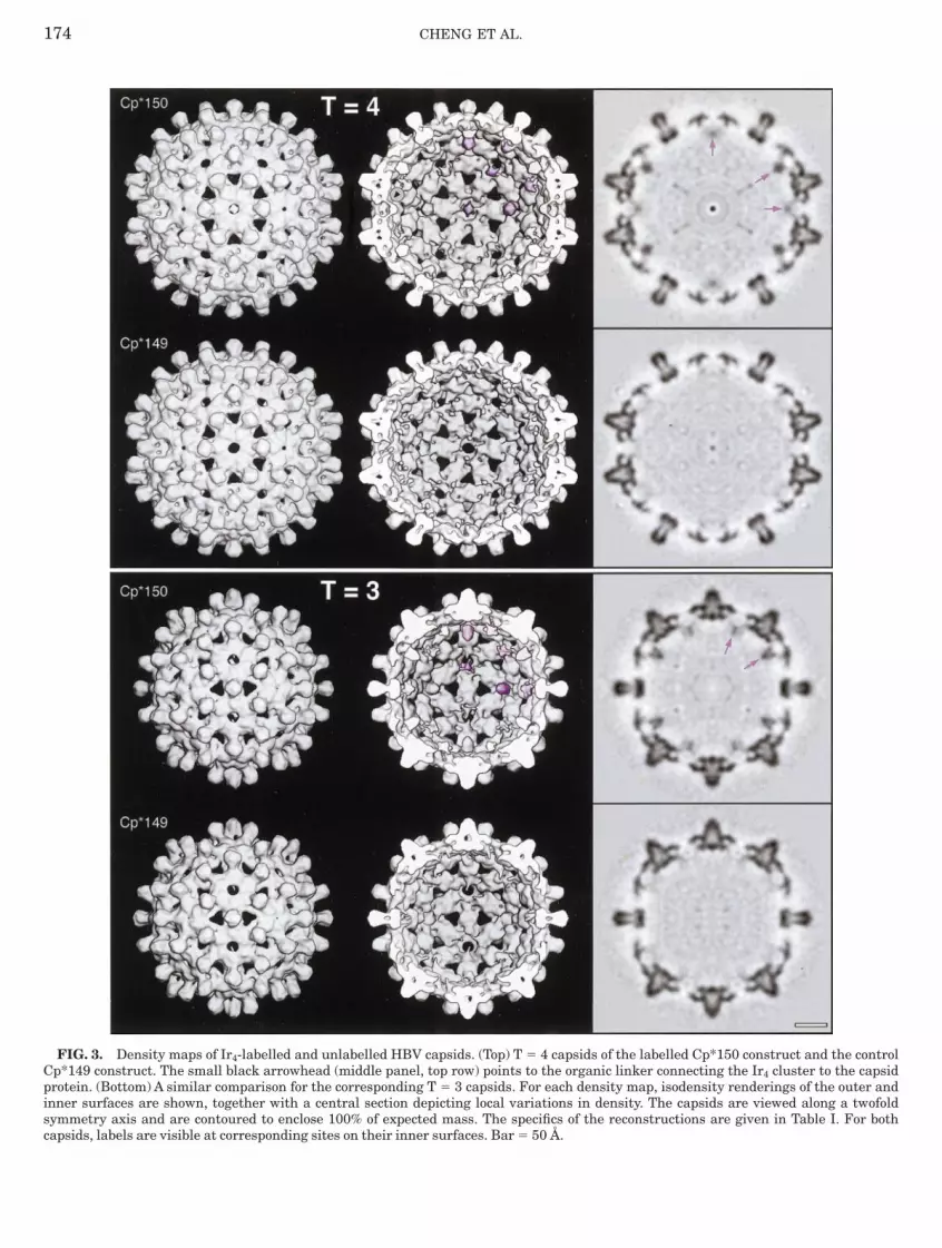
ensity maps were calculated for both the T 5 4 andhe T 5 3 capsids to 12 and 14 Å resolution,espectively (Table I, Fig. 3). In both cases, conspicu-us focal densities are present beneath the fivefoldnd quasi-sixfold lattice sites. These densities arebsent from Cp*149 control capsids that were sub-ected to the same labelling reaction (cf., Fig. 3). Thisocalization of the Ir4 clusters and, consequently, ofhe C-termini of the capsid protein confirms theesult previously obtained by visualizing undecagoldound to Cp*150 at ,20 Å resolution (Zlotnick et al.,997). Consistent results were obtained for a T 5 4apsid reconstruction in an earlier labelling experi-ent in which the DMSO was omitted (Table I, cf.,igs. 4a and 4b). Although the labelling efficiency of
he latter experiment could not be determined reli-bly by spectroscopy (see above), it must have beenery similar to that of the experiment illustrated inig. 3, to judge by the intensity of the cluster-ssociated densities in the respective maps.Dependence of cluster visibility on resolution. To
stimate the minimum resolution required to visual-ze Ir4 labels by cryo-EM, we progressively loweredhe resolution of these density maps. As shown inig. 4, the cluster-associated density becomes pro-ressively more diffuse as the resolution falls, but itemains significantly above the level of residualackground noise to at least 25 Å resolution.
DISCUSSION
Visibility of tetrairidium as a density label. Hith-rto, undecagold is the smallest metal cluster to haveeen used as a density label in cryo-EM. Ir4 isignificantly smaller (2 vs 6.5 kDa, 4 vs 11 heavytoms).Although it was essentially invisible in micro-raphs covering a considerable range of defocusalues (Fig. 2), Ir4 was clearly visualized in three-imensional density maps, even at incomplete occu-ancy and moderate resolution (Figs. 3 and 4),eflecting the large improvement in signal-to-noiseatio in such a density map compared to the originalrojection images.We reproducibly obtained a labelling stoichiom-
try of ,11% for Ir4 relative to the capsid proteinnder our conditions of incubation, corresponding to26 clusters per T 5 4 capsid and ,20 clusters per
FIG. 2. Cryo-micrographs, recorded relatively close to focusDf ,0.9 µm), of (a) Ir4-labelled Cp*150 capsids and (b, c)nlabelled capsids. The two controls are (b) Cp*149 capsids thatere subjected, with negative results, to the labelling protocol and
c) Cp147 capsids that were not so treated. Also shown (d),ttached to a carbon film, are microprecipitates of Ir4 clustersormed when the labelling reaction was attempted in the absencef DMSO, which are large enough to be directly visible. Bar 5
5 3 capsid. However—as with undecagold (Zlot-

ntbop4tm
de,uforessoe
ot
Iospt,vo(tdse
as
Rwur
173TETRAIRIDIUM AS A CRYO-EM DENSITY LABEL
ick et al., 1997)—the bound clusters are located onhe fivefold and quasi-sixfold axes, where occupancyy one cluster blocks the binding of clusters to thether four (or five) subunits surrounding the latticeoint. Thus, the maximum binding to be expected is2 (T 5 4) and 32 (T 5 3) clusters per capsid, so thathe binding obtained represents about 60% of maxi-um.As visualized (Figs. 3, 4a, and 4b), the 5 Å-
iameter clusters appear considerably larger thanxpected; in fact, their full width at half-height is20 Å. Only part of this broadening may be attrib-ted to the limited resolution (11 Å), because a stepunction 5 Å across has a full width at a half-heightf ,9 Å when band-limited to this resolution. Theemainder of the broadening must arise from otherffects, such as local disorder of the C-termini, orlight displacement of each cluster from the adjacentymmetry axis in a direction that depends on whichf the surrounding subunits it is bound to. The latter
FIG. 4. Effect of progressively lowering the resolution on theconstructions from independent labelling experiments at a resoas lowered to (d) 15 Å, (e) 20 Å, and (f ) 25 Å, respectively. (c) A 25-sed for the reconstructions in a, d, e, and f ), whose statistical pesolution. Bar 5 50 Å.
ffect would give rise to averaging-related smearing d
f the cluster density as depicted in the reconstruc-ion.
Initially, we were surprised that a label as small asr4 should be so conspicuous under these conditionsf observation (Fig. 3). However, the difference inpecific density between the cluster and the adjacentrotein (Dr ,21.5 5 22.8 vs 1.3) is so much higherhan that between protein and vitreous ice (Dr0.35 5 1.3 vs 0.95) that it remained distinctly
isible in the map, despite attenuation by partialccupancy and delocalization arising from averagingsee above). We anticipate that Hg4, which is similaro Ir4 in electron scattering power, should also beetectable under similar conditions, i.e., in 3D den-ity maps of ice-embedded specimens imaged at lowlectron doses.Resolution of metal cluster labels. The resolution
ttainable with these markers depends more on thepacing between the center of the cluster-associated
ility of the Ir4 cluster label on the T 5 4 Cp*150 capsid. (a, b)of ,11 Å (see Table I). (d, e, f ) The resolution of reconstruction aution reconstruction based on 100 particles (cf., the 1460 particlesies (residual noise level) is more typical of a density map at this
e visiblutionÅ resolropert
ensity and the labelled residue than it does on the
Cpisc faces. Bar 5 50 Å.
174 CHENG ET AL.
FIG. 3. Density maps of Ir4-labelled and unlabelled HBV capsp*149 construct. The small black arrowhead (middle panel, toprotein. (Bottom) A similar comparison for the corresponding T 5nner surfaces are shown, together with a central section depictymmetry axis and are contoured to enclose 100% of expected mapsids, labels are visible at corresponding sites on their inner sur
ids. (Top) T 5 4 capsids of the labelled Cp*150 construct and the controlrow) points to the organic linker connecting the Ir4 cluster to the capsid3 capsids. For each density map, isodensity renderings of the outer anding local variations in density. The capsids are viewed along a twofoldass. The specifics of the reconstructions are given in Table I. For both
s,HldnvrltoWdaeco
drtitisopp
tatlfir
arpqDsgcttt1maptcmiadCseatf
cAmtb1Icrapuptticob
l(laaa
175TETRAIRIDIUM AS A CRYO-EM DENSITY LABEL
ize of the cluster. With undecagold, this spacing is17 Å, and with Ir4, it is slightly less, at ,15 Å.owever, it is possible to achieve more precise
ocalizations than these figures imply because, un-er favorable circumstances, the organic linker con-ecting the cluster to the labelled protein may beisualized (arrowhead in Fig. 3, middle panel, topow; see also Fig. 4 of Zlotnick et al., 1997). Extrapo-ating an appropriate distance along this vector fromhe cluster center should give a precise localizationf the residue of interest, i.e., to within 65 Å or so.hile clusters as small as Ir4 are readily visible in
ensity maps at 25–30 Å resolution (cf., Fig. 4), onedvantage of extending their resolution to 10–15 Å isnhanced prospects of visualizing this linker and,onsequently, of effecting a more precise localizationf the targeted residue.Although shifting to progressively smaller clusters
oes not bring a major gain in resolution, theseeagents have the advantage of being less suscep-ible to steric constraints that may limit their reactiv-ty. The relatively bulky organic shell surroundinghe cluster prevents it from accessing confined spacesn much the same way that an atomic force micro-cope is limited in the vertical dimension by the sizef its tip. Thus, smaller clusters have superiorenetrating power, as illustrated by the observed
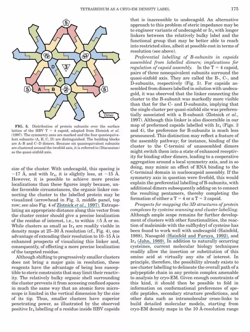
FIG. 5. Distribution of protein subunits over the surfaceattice of the HBV T 5 4 capsid, adapted from Zlotnick et al.1997). The symmetry axes are marked and the four quasiequiva-ent subunits (A, B, C, D) are distinguished. The building blocksre A–B and C–D dimers. Because six quasiequivalent subunitsre clustered around the twofold axis, it is referred to (Discussion)s the quasi-sixfold axis.
ositive Ir4 labelling of a residue inside HBV capsids c
hat is inaccessible to undecagold. An alternativepproach to this problem of steric impedance may beo engineer variants of undecagold or Ir4 with longerinkers between the relatively bulky label and theunctional group that may be better able to reachnto restricted sites, albeit at possible cost in terms ofesolution (see above).Preferential labelling of B-subunits in capsids
ssembled from labelled dimers; implications foregulation of capsid assembly. In the T 5 4 capsid,airs of three nonequivalent subunits surround theuasi-sixfold axis. They are called the B-, C-, and-subunits, respectively (Fig. 5). For capsids as-
embled from dimers labelled in solution with undeca-old, it was observed that the linker connecting theluster to the B-subunit was markedly more visiblehan that for the C- and D-subunits, implying thathe single cluster per quasi-sixfold site was preferen-ially associated with a B-subunit (Zlotnick et al.,997). Although this linker is also discernible in ourap of preformed capsids labelled with Ir4 (Figs. 3
nd 4), the preference for B-subunits is much lessronounced. This distinction may reflect a feature ofhe assembly pathway; for instance, binding of theluster to the C-termini of unassembled dimersight switch them into a state of enhanced receptiv-
ty for binding other dimers, leading to a cooperativeggregation around a local symmetry axis, and in sooing, may mimic an effect of RNA binding to the-terminal domain in nucleocapsid assembly. If theymmetry axis in question were fivefold, this wouldxplain the preferential labelling of B-subunits, withdditional dimers subsequently adding on to connecthe resulting pentamers, thereby completing theormation of either a T 5 4 or a T 5 3 capsid.
Prospects for mapping the 3D structures of proteinomplexes by cysteine scanning and cluster labelling.lthough ample scope remains for further develop-ent of clusters with other functionalities, the reac-
ion of maleimide with the sulfhydryl of cysteine haseen found to work well with undecagold (Hainfeld,988), Nanogold (Hainfeld and Furuya, 1992), andr4 (Jahn, 1989). In addition to naturally occurringysteines, current molecular biology techniqueseadily allow the insertion or substitution of thismino acid at virtually any site of interest. Inrinciple, therefore, the possibility already exists tose cluster labelling to delineate the overall path of aolypeptide chain in any protein complex amenableo analysis by cryo-EM. Given enough constraints ofhis kind, it should then be possible to fold innformation on conformational preferences of spe-ific peptides, secondary structure predictions, andther data such as intramolecular cross-links touild detailed molecular models, starting from
ryo-EM density maps in the 10 Å-resolution range
(1
d
A
B
B
B
B
C
C
C
C
F
F
G
G
H
H
H
H
H
J
L
M
R
S
S
T
V
W
W
W
Z
Z
176 CHENG ET AL.
Bottcher et al., 1997; Conway et al., 1997; Trus et al.,997).
We thank Drs. A. Zlotnick, D. Belnap, and B. Trus for helpfuliscussions and Dr. T. Baker (Purdue) for software.
REFERENCES
drian, M., Dubochet, J., Lepault, J., and McDowall, A. W. (1984)Cryo-electron microscopy of viruses, Nature 308, 32–36.
aker, T. S., and Cheng, R. H. (1996) A model-based approach fordetermining orientations of biological macromolecules imagedby cryoelectron microscopy, J. Struct. Biol. 116, 120–130.
artlett, P. A., Bauer, B., and Singer, S. J. (1978) Synthesis ofwater-soluble undecagold cluster compounds of potential impor-tance in electron microscopic and other studies of biologicalsystems, J. Am. Chem. Soc. 100, 5085–5092.
endayan, M. (1995) Colloidal gold post-embedding immunocyto-chemistry, Prog. Histochem. Cytochem. 9, 1–25.
ottcher, B., Wynne, S. A., and Crowther, R. A. (1997) Determina-tion of the fold of the core protein of hepatitis B virus by electroncryomicroscopy, Nature 386, 88–91.
onway, J. F., Trus, B. L., Booy, F. P., Newcomb, W. W., Brown,J. C., and Steven, A. C. (1993) The effects of radiation damageon the structure of frozen hydrated HSV-1 capsids, J. Struct.Biol. 111, 222–233.
onway, J. F., Cheng, N., Zlotnick, A., Wingfield, P. T., Stahl, S. J.,and Steven, A. C. (1997) Visualization of a 4-helix bundle in thehepatitis B virus capsid by cryo-electron microscopy, Nature386, 91–94.
onway, J. F., Cheng, N., Zlotnick, A., Stahl, S. J., Wingfield, P. T.,and Steven, A. C. (1998) Localization of the N-terminus ofhepatitis B virus capsid protein by peptide-based differencemapping from cryoelectron microscopy, Proc. Natl. Acad. Sci.USA 95, 14622–14627.rum, J., Gruys, K. J., and Frey, T. G. (1994) Electron microscopyof cytochrome c oxidase crystals: Labeling of subunit III with amonomaleimide undecagold cluster compound, Biochemistry33, 13719–13726.
uller, S. D., Butcher, S. J., Cheng, R. H., and Baker, T. S. (1996)Three-dimensional reconstruction of icosahedral particles: ‘‘Theuncommon line,’’ J. Struct. Biol. 116, 48–55.
uruya, F. R., Miller, L. L., Hainfeld, J. F., Christopfel, W. C., andKenny, P. W. (1988) Use of Ir4(CO)11 to measure the lengths oforganic molecules with a scanning transmission electron micro-scope, J. Am. Chem. Soc. 110, 641–643.riffiths, G. (1994) Fine Structure Immunocytochemistry,Springer-Verlag, Berlin.regori, L., Hainfeld, J. F., Simon, M. N., and Goldgaber, D. (1997)Amyloid beta-protein inhibits ubiquitin-dependent protein deg-radation in vitro, J. Biol. Chem. 272, 58–62.ainfeld, J. F. (1989) Undecagold-antibody method, in Hayat,M. A. (Ed.), Colloidal Gold: Principles, Methods, and Applica-tions., Vol. 2, pp. 413–429, Academic Press, San Diego.ainfeld, J. F. (1992) Site specific cluster labels, Ultramicroscopy46, 135–144.ainfeld, J. F., and Furuya, F. R. (1992) A 1.4-nm gold cluster
covalently attached to antibodies improves immunolabeling, J.Histochem. Cytochem. 40, 177–184.ainfeld, J. F. (1996) Labeling with Nanogold and undecagold:Techniques and results, Scanning Microsc. Suppl. (Proc. 14thPfefferkorn Conf.) 10, 309–322. [Malecki, M., and Roomans,G. M. (Eds.), Scanning Microscopy Int’l, Chicago].ainfeld, J. F., and Powell, R. D. (1997) Nanogold technology:New frontiers in gold labeling, Cell Vision 4, 408–432.
ahn, W. (1989) Synthesis of water soluble tetrairidium clusterssuitable for heavy atom labelling of proteins, Z. Naturforsch. B44, 79–82.
ipka, J. J., Lippard, S. J., and Wall, J. S. (1979) Visualization ofpolymercuri-methane-labeled fd bacteriophage in the scanningtransmission electron microscope, Science 206, 1419–1421.illigan, R. A., Whittaker, M., and Safer, D. (1990) Molecularstructure of F-actin and location of surface binding sites, Nature348, 217–221.os, R., Scrivanti, A., Albano, V. G., Braga, D., and Garlaschelli, L.(1986) Chemistry of tetrairidium carbonyl clusters. Part 1.Synthesis, chemical characterization, and nuclear magneticresonance study of mono- and di-substituted phosphine deriva-tives. X-Ray crystal structure determination of the diaxialisomer of [Ir4(CO)7(µ-CO)3(Me2PCH 2CH2PMe2)], J. Chem. Soc.Dalton Trans. 2411–2421.
axton, W. O., and Baumeister, W. (1982) The correlation averag-ing of a regularly arranged bacterial cell envelope protein, J.Microsc. (Oxford) 127, 127–138.
teinmetz, M. O., Stoffler, D., Muller, S. A., Jahn, W., Wolpens-inger, B., Goldie, K. N., Engel, A., Faulstich, H., and Aebi, U.(1998) Evaluating atomic models of F-actin with an undecagold-tagged phalloidin derivative, J. Mol. Biol. 276, 1–6.
rus, B. L., Roden, R. B., Greenstone, H. L., Vrhel, M., Schiller, J.T., and Booy, F. P. (1997) Novel structural features of bovinepapillomavirus capsid revealed by a three-dimensional recon-struction to 9 Å resolution, Nat. Struct. Biol. 4, 413–420.
an Heel, M. (1987) Similarity between images, Ultramicroscopy21, 95–100.einstein, S., Jahn, W., Hansen, H., Wittmann, H. G., andYonath, A. (1989) Novel procedures for derivatization of ribo-somes for crystallographic studies, J. Biol. Chem. 264, 19138–19142.enzel, T., and Baumeister, W. (1995) Conformational constraintsin protein degradation by the 20S proteasome, Nat. Struct. Biol.2, 199–204.ingfield, P. T., Stahl, S. J., Williams, R. W., and Steven, A. C.(1995) Hepatitis core antigen produced in Escherichia coli:Subunit composition, conformational analysis, and in vitrocapsid assembly, Biochemistry 34, 4919–4932.
lotnick, A., Cheng, N., Conway, J. F., Booy, F. P., Steven, A. C.,Stahl, S. J., and Wingfield, P. T. (1996) Dimorphism of hepatitisB virus capsids is strongly influenced by the C-terminus of thecapsid protein, Biochemistry 35, 7412–7421.
lotnick, A., Cheng, N., Stahl, S. J., Conway, J. F., Steven, A. C.,and Wingfield, P. T. (1997) Localization of the C terminus of theassembly domain of hepatitis B virus capsid protein: Implica-tions for morphogenesis and organization of encapsidated RNA,Proc. Natl. Acad. Sci. USA 94, 9556–9561.
Related Documents