Tetrahydrobiopterin in the Prevention of Hypertonia in Hypoxic Fetal Brain Jeannette Vásquez-Vivar, PhD 1,2,* , Jennifer Whitsett, BS 1 , Matthew Derrick, MD 3 , Xinhai Ji, BS 3 , Lei Yu, MD, PhD 3 , and Sidhartha Tan, MD 3 1 Department of Biophysics, Medical College of Wisconsin, Milwaukee WI 53226 2 Free Radical Research Center & Redox Biology Program, Medical College of Wisconsin, Milwaukee WI 53226 3 Department of Pediatrics, Northwestern University, Evanston IL 60201. Abstract Objective: Tetrahydrobiopterin (BH 4 ) deficiency is one of the causes of dystonia at birth. We hypothesized that BH 4 is a developmental factor determining vulnerability of the immature fetal brain to hypoxic-ischemic injury and subsequent motor deficits in newborns. Methods: Pregnant rabbits were subjected to 40-min uterine ischemia and fetal brains were investigated for global and focal changes in BH 4 . Newborn kits were assessed by neurobehavioral tests following vehicle and sepiapterin (BH 4 -analog) treatment of dams. Results: Naive fetal brains at 70% gestation (E22) were severely deficient for BH 4 compared to maternal and other fetal tissues. BH 4 concentration rapidly increased normally in the perinatal period with the highest concentrations found in the thalamus compared to basal ganglia, frontal, occipital, hippocampus and parietal cortex. Global sustained 40-min hypoxia-ischemia depleted BH 4 in E22 thalamus and to a lesser extent in basal ganglia, but not in the frontal, occipital and parietal regions. Maternal supplementation prior to hypoxia-ischemia with sepiapterin increased BH 4 in all brain regions and especially in the thalamus, but did not increase the intermediary metabolite, 7,8-BH 2 . Sepiapterin treatment also reduced incidence of severe motor deficits and perinatal death following E22 hypoxia-ischemia. Interpretation: We conclude that early developmental BH 4 deficiency plays a critical role in hypoxic-ischemic brain injury. Increasing brain BH 4 via maternal supplementation may be an effective strategy in preventing motor deficits from antenatal hypoxia-ischemia. Keywords Tetrahydrobiopterin; GTP cyclohydrolase-I; dystonia; hypertonia; muscle tonus; thalamus; dopamine; hypoxia-ischemia, brain; ischemia; uterus; infant, newborn Introduction Hypoxia-ischemia (H-I) brain injury of the immature brain causes motor, learning and mental disabilities in surviving children 1,2 . Most cases of newborn H-I encephalopathy occur during *Corresponding Author: Jeannette Vásquez-Vivar, PhD 8701 Watertown Plank Road Phone: (414) 456-8095; Fax: (414) 456-6512. Milwaukee, WI 53226 E-mail: [email protected]. Statement of conflict of interest: None NIH Public Access Author Manuscript Ann Neurol. Author manuscript; available in PMC 2009 November 30. Published in final edited form as: Ann Neurol. 2009 September ; 66(3): 323–331. doi:10.1002/ana.21738. NIH-PA Author Manuscript NIH-PA Author Manuscript NIH-PA Author Manuscript

Welcome message from author
This document is posted to help you gain knowledge. Please leave a comment to let me know what you think about it! Share it to your friends and learn new things together.
Transcript

Tetrahydrobiopterin in the Prevention of Hypertonia in HypoxicFetal Brain
Jeannette Vásquez-Vivar, PhD1,2,*, Jennifer Whitsett, BS1, Matthew Derrick, MD3, Xinhai Ji,BS3, Lei Yu, MD, PhD3, and Sidhartha Tan, MD31Department of Biophysics, Medical College of Wisconsin, Milwaukee WI 532262Free Radical Research Center & Redox Biology Program, Medical College of Wisconsin,Milwaukee WI 532263Department of Pediatrics, Northwestern University, Evanston IL 60201.
AbstractObjective: Tetrahydrobiopterin (BH4) deficiency is one of the causes of dystonia at birth. Wehypothesized that BH4 is a developmental factor determining vulnerability of the immature fetalbrain to hypoxic-ischemic injury and subsequent motor deficits in newborns.
Methods: Pregnant rabbits were subjected to 40-min uterine ischemia and fetal brains wereinvestigated for global and focal changes in BH4. Newborn kits were assessed by neurobehavioraltests following vehicle and sepiapterin (BH4-analog) treatment of dams.
Results: Naive fetal brains at 70% gestation (E22) were severely deficient for BH4 compared tomaternal and other fetal tissues. BH4 concentration rapidly increased normally in the perinatal periodwith the highest concentrations found in the thalamus compared to basal ganglia, frontal, occipital,hippocampus and parietal cortex. Global sustained 40-min hypoxia-ischemia depleted BH4 in E22thalamus and to a lesser extent in basal ganglia, but not in the frontal, occipital and parietal regions.Maternal supplementation prior to hypoxia-ischemia with sepiapterin increased BH4 in all brainregions and especially in the thalamus, but did not increase the intermediary metabolite, 7,8-BH2.Sepiapterin treatment also reduced incidence of severe motor deficits and perinatal death followingE22 hypoxia-ischemia.
Interpretation: We conclude that early developmental BH4 deficiency plays a critical role inhypoxic-ischemic brain injury. Increasing brain BH4 via maternal supplementation may be aneffective strategy in preventing motor deficits from antenatal hypoxia-ischemia.
KeywordsTetrahydrobiopterin; GTP cyclohydrolase-I; dystonia; hypertonia; muscle tonus; thalamus;dopamine; hypoxia-ischemia, brain; ischemia; uterus; infant, newborn
IntroductionHypoxia-ischemia (H-I) brain injury of the immature brain causes motor, learning and mentaldisabilities in surviving children1,2. Most cases of newborn H-I encephalopathy occur during
*Corresponding Author: Jeannette Vásquez-Vivar, PhD 8701 Watertown Plank Road Phone: (414) 456-8095; Fax: (414) 456-6512.Milwaukee, WI 53226 E-mail: [email protected] of conflict of interest: None
NIH Public AccessAuthor ManuscriptAnn Neurol. Author manuscript; available in PMC 2009 November 30.
Published in final edited form as:Ann Neurol. 2009 September ; 66(3): 323–331. doi:10.1002/ana.21738.
NIH
-PA Author Manuscript
NIH
-PA Author Manuscript
NIH
-PA Author Manuscript
the antenatal period. Currently, there is an insufficient understanding of the pathogenic H-Imechanisms in the fetal brain.
There are many similarities between dystonia and hypertonia in children resulting from H-Iand those from tetrahydrobiopterin (BH4) deficiency. Defects in the BH4 biosynthetic pathwaycause seizures, postural dystonia and developmental delays. Mutation of GTP-cyclohydrolase(GTPCH, Scheme 1) results in hereditary progressive dystonia3 Mutation of sepiapterinreductase (SR)4 causes motor and cognitive impairment. The most prevalent causes of BH4deficiency are associated with mutation of 6-pyruvoyltetrahydropterin synthase (PTPS)5 anddihydropteridine reductase (DHPR)5,6. Treatment with BH4 has been found to ameliorate somecases of dystonia7.
Brain BH4 regulates tyrosine hydroxylase activity and loss of BH4 is associated with dopaminedeficiency which causes early onset dystonia. It is noteworthy however that a genetic modelfor defective GTPCH, the hph-1 mouse8, exhibits low brain BH4, dopamine and serotoninlevels, but does not have distinctive motor phenotype. The lack of behavioral models hasdelayed a complete understanding of the mechanisms involving BH4 deficiency and braininjury9. With the recent development of an animal model mimicking cerebral palsy10, a uniqueopportunity to examine the link between BH4 and motor impairments presents itself.
We hypothesized that BH4 is a developmental factor determining vulnerability of the immaturefetal rabbit brain to H-I injury and the occurrence of subsequent motor disabilities. We showthat normally low BH4 concentrations is a critical factor increasing the vulnerability of theimmature brain to H-I injury. Maternal treatment with BH4 increased fetal levels in thalamusand basal ganglia and significantly ameliorated motor deficits and decreased stillbirths. Thisdiscovery provides fundamental information about BH4 as a target molecule in H-I fetal braindamage. Our long term goal is to establish the molecular basis of the role of BH4 in immaturebrain dysfunction.
Materials and MethodsAnimals
This study was approved by the Animal Review Committee of the NorthShore UniversityHealthSystems. Animals received humane care and were treated in compliance with the UnitedStates Public Health Service's Policy on Humane Care and Use of Laboratory Animals.
SurgeryThe surgical procedure has been previously described10. Briefly, pregnant dams at 22 daysgestation (E22) were anesthetized with intravenous fentanyl (75 μg/kg/hr) and droperidol (3.75mg/kg/hr) and bag and mask ventilation provided to maintain normal arterial blood gases.Following spinal anesthesia with 0.75% bupivacaine, the fentanyl and droperidol was reducedto enable the dam to breathe spontaneously. The control animals were delivered byhysterotomy.
Uterine ischemia was induced by inflation of 4F Fogarty arterial embolectomy catheterintroduced via the left femoral artery into the descending aorta to a level above the uterine andbelow the renal arteries. Balloon inflation resulting in complete ischemia to the uterus wasmaintained for 40 min. A subset of fetal brains was obtained after hysterotomy.
For the behavioral studies after uterine ischemia, the catheter was removed, the femoral arteryreconstructed, and the dam returned to her cage and allowed to deliver spontaneously.
Vásquez-Vivar et al. Page 2
Ann Neurol. Author manuscript; available in PMC 2009 November 30.
NIH
-PA Author Manuscript
NIH
-PA Author Manuscript
NIH
-PA Author Manuscript

Neurobehavioral assessmentNewborn kits at postnatal day 1 (E32 or P1) were subjected to previously published battery ofneurobehavioral tests 10. Muscle tone was assessed by active flexion and extension of theforelimbs and hindlimbs and scored (0-4)11. The surviving newborn kits were divided intosevere (presence of severe hypertonia and/or postural deficits) or mild (minimal or nohypertonia, no postural deficits, but presence of other neurobehavioral deficits) or normal (noobvious deficits noted).
Biochemical AnalysisTissue was obtained from rabbit fetuses between 22 and 29 days postconceptional age (E22-E29), corresponding to ~70 to 92% of fetal rabbit gestation respectively, and from newbornrabbit brains at P1 (E32).
Total biopterin, BH4, and BH2 analysisScreening of all stable biopterin metabolites BH4, BH2 and biopterin (total biopterin) wasquantified by HPLC with fluorescence detection as previously described12. For thequantification of BH4, 7,8-BH2 and ascorbate, a coulometric analysis using HPLC wasperformed13. Results were normalized to protein content.
GTP cyclohydrolase activityEnzyme activity was measured by following the conversion of GTP to dihydroneopterin 3'-triphosphate following oxidation-dephosphorylation to neopterin as previously described13.
Dopamine, serotonin and metabolitesAnalysis was performed by HPLC with a multielectrode coulometric analysis14.Concentrations were calculated using authentic standards and normalized to protein content.
Western blot analysisTissues were homogenized in PBS with 1% Triton X-100, 1 mM PMSF, 35 ng/mL pepstatinA, and 10 ng/mL leupeptin and protease inhibitor cocktail. Samples were resolved on 12%SDS-polyacrylamide gels and transferred to membranes. Blots were probed with a polyclonalcustom GTP cyclohydrolase I antibody as described15.
Gene expressioncDNA was synthesized from 3 μg of total RNA and oligo(dT) primers using the InvitrogenSuperscript First-Strand Synthesis System for RT-PCR (Invitrogen).
Statistical analysisResults were expressed as mean±S.E.M. The analysis included ANOVA with a post hocStudent-Newman-Keuls' test for multiple group comparisons or t-test for differences betweentwo group means. Alpha error was set at p<0.05.
ResultsDevelopmental regulation of BH4 in late gestation and newborn period
Total biopterin concentration was quantified in E22-E32 brain homogenates, to assess theoverall changes of BH4 and its metabolites during the course of biosynthesis and utilization ofthis cofactor in the fetal brain. There was negligible variation with gender, so the results wereanalyzed using age as the only dependent variable. The E22 brain had the lowest biopterinconcentrations and from E22 to E32 total biopterin increased ~2.5 fold (Fig.1). The brain
Vásquez-Vivar et al. Page 3
Ann Neurol. Author manuscript; available in PMC 2009 November 30.
NIH
-PA Author Manuscript
NIH
-PA Author Manuscript
NIH
-PA Author Manuscript
transition from a very low to low biopterin levels from E22 to E29 indicated that the prematurebrain is relatively biopterin deficient. Biopterin levels in fetal plasma also increased with age(Fig.2). In contrast, the biopterin in maternal plasma did not significantly change over thecourse of gestation and were higher than fetal levels at all ages. This shows a gradient inbiopterin between maternal and fetal plasma. Similar differences were shown between maternaland fetal placenta (Fig.2). We then questioned whether fetal brain biopterin could be derivedfrom maternal sources or be actively produced in the fetal brain.
BH4 biosynthesis explains fetal brain BH4 levelsThe GTPCH is the first enzyme and considered the rate limiting enzyme in the de novobiosynthesis of BH4. Fetal brain and liver expressed significant levels of both GTPCH mRNAand protein (Fig.3). Both GTPCH protein levels (Fig. 3B-3C) and enzyme activity in E22brains, however, were significantly lower than those in E29 brains (Fig. 3D). Liver GTPCHexpression was higher than the brain at all embryonic ages (Fig. 3). Thus during development,GTPCH and BH4 production in fetal brain is delayed with respect to other fetal tissues.Collectively this data shows that brain tissue is characterized by relative BH4 deficiency duringearly stages of development compared to newborn.
BH4 is developmentally regulated in specific regions of the brainWe also examined whether regional BH4 changes in normal fetal brain could indicate its roleand developmental regulation. The basal ganglia and thalamus have higher BH4 concentrationsthan other parts of the brain such as frontal, parietal and occipital regions at all postconceptionalages (Fig.4A). The BH4 in thalamus and basal ganglia also showed a significant perinatalincrease of ~54-64% between E22 and E32 (or P1). Notably, BH4 levels were highest in thethalamus at all ages (Fig.4).
BH4 as a factor in brain responses to hypoxia-ischemiaTo determine whether BH4 is involved in the brain's response to one of the important etiologiesof cerebral palsy, the effects of acute global H-I on BH4 in different parts of the fetal brainwere examined. It was found that 40 min H-I caused a marked decrease of ~25% BH4 in thethalamus (Table 1). A less prominent yet significant decrease was found in the basal gangliaand frontal regions of the brain. Other parts of the brain showed no significant change. Theuniqueness of these changes can be better appreciated when compared to the diffuse yetsignificant ascorbate depletion in all parts of brain (Table 1). Ascorbate is found in highconcentrations in all parts of the brain and its loss in H-I signifies a global response to damage.
BH4 levels can be augmented by maternal supplementationThere is evidence that BH4 in liver, kidney and to a lesser extent brain can be increased bysupplementation with exogenous BH4
16-18. The possibility of increasing fetal brain BH4 viamaternal supplementation with BH4 was examined. Administration of BH4 (1-10mg/ml) to thepregnant dam via the distal aorta was done to maximally increase fetal levels. These doseswere well tolerated and biopterin levels in maternal and fetal plasma changed with dose andtime. With a freshly prepared BH4 bolus (10mg/kg) an acute increase ~130-fold (8.59±1.61 vs1128.1±31.5nM) after 10min was found in maternal plasma, which returned to basal levels 3hours post infusion. Fetal plasma increased ~40-fold (1.46±0.23 vs 76.56±5.24nM) but fetalbrain increases were less consistent. With lower doses of BH4 changes were less pronouncedand more transient.
Regional changes in BH4 levels upon maternal supplementation with sepiapterinSince BH4 is not stable and stabilizing agents such as ascorbate may mask its effects, theBH4 precursor, sepiapterin, was preferred over tetrahydrobiopterin as a therapeutic agent
Vásquez-Vivar et al. Page 4
Ann Neurol. Author manuscript; available in PMC 2009 November 30.
NIH
-PA Author Manuscript
NIH
-PA Author Manuscript
NIH
-PA Author Manuscript
(Scheme 1). When administered peripherally, sepiapterin is converted to BH4 by a two-stepreduction catalyzed by sepiapterin reductase and dihydrofolate reductase in target tissues.Sepiapterin was delivered to E15 pregnant dams via subcutaneous osmotic mini pump thatenabled a steady dosage of 0.6-mg/kg/day. After 7 days, a consistent increase of ~20-fold wasobserved in maternal plasma. There was also a significant increase in fetal and maternalplacental BH4 (Table 2). Fetal brain levels also increased with the greatest change occurringagain in the thalamus showing a >2-fold increase over basal levels (Fig.5). The sepiapterinmetabolite, 7,8-BH2, was not detected in any supplemented animal indicating full conversionof sepiapterin to BH4.
BH4 protects immature brain from H-I injury and decreases dystonic hypertoniaThe demonstration that H-I causes a selective loss of BH4 in different regions of the fetal brainsuggested that supplementation would render the fetal brain less vulnerable to H-I. Specifically,we asked whether BH4 supplementation ameliorates motor impairments caused by H-I in thefetal rabbit brain. This possibility was assessed in newborn kits supplemented with sepiapterinor vehicle before H-I at E22, when the lowest BH4 levels were observed. After H-I, the damsrecovered and delivered spontaneously at term gestation. The newborn kits were examined forneurobehavioral deficits as described10. The sepiapterin treated group, showed a significantreduction in the severity of motor deficits (locomotion and righting reflex), hypertonia anddystonia score (Fig.6). Sepiapterin administration also increased the number of normalappearing kits (Fig.7 and video in supplemental material). To find out how BH4supplementation affects motor improvement, an exploratory study was done to investigate thelevels of levedopa (L-DOPA), 5-hydroxytryptamine (5-HT), homovallinic acid, and 5-hydroxyindolacetic acid in basal ganglia and thalamus.
Sepiapterin supplementation improves the level of biogenic aminesDopamine changes were more marked than serotonin with H-I. Increasing BH4 with sepiapterintreatment has a beneficial effect in protecting basal glanglia and thalamus from losses in L-DOPA and 5-HT (Table 3).
DiscussionThis is the first study to propose BH4 as an important pathophysiological factor in hypoxic-ischemic encephalopathy. The major findings are that typical low BH4 levels in the immaturebrain could determine its vulnerability and further decrease in BH4 levels with H-I couldcontribute to the severity of neurological outcomes from H-I.
Endogenous BH4 in the fetal brainThere is limited information about BH4 availability in the human developing brain. Earlystudies reported measurable changes in brain BH4 in human fetuses from 2-20 weeks ofgestation with increase to ~0.16-nmol/g-wet-weight at ~20-weeks19. This amount is in thesame low range observed in the E22 fetal rabbit brain (70% gestation), and lower than reportedin the hph-1 mouse (0.312±0.017 nmol/g-wet-weight)20 a model for BH4 deficiency. Thisindicates that across different species, the brain exhibits an early developmental BH4deficiency. The importance of maintaining this stability even at these low levels of BH4 tonormal development is indicated by clinical findings in patients with different forms of BH4disorders. Carriers of autosomal dominant form of GTPCH deficiency present with lowbiopterin and neopterin levels. They present with several motor signs such as poor sucking,decreased spontaneous movements, postural changes and dystonia. Those affected withsepiapterin reductase mutations also show abnormal biopterin pattern with high levels ofbiopterin and dihydrobiopterin, reflecting their inability to drive BH4 synthesis to term(Scheme I). These patients generally present with growth retardation, and motor and
Vásquez-Vivar et al. Page 5
Ann Neurol. Author manuscript; available in PMC 2009 November 30.
NIH
-PA Author Manuscript
NIH
-PA Author Manuscript
NIH
-PA Author Manuscript

developmental delays. Additional evidence is provided by the high frequency of early (~48h)postnatal death in the homozygous mouse model for PTPS5 deficiency that is severely BH4depleted. Premature death in the newborn period is also reported for children carrying defectsin PTPS and DHPR activity21. These functional relationships support the notion that BH4 is adevelopmental factor involved in gain of function.
Brain BH4 and H-I injuryMaternal levels of BH4 do not appear to influence that of the fetal brain. Therefore the geneticmake-up of the fetus plays a major role in determining brain tissue BH4 levels. In addition,BH4 production in the fetal brain is developmentally regulated transitioning from a BH4deficient to a BH4 sufficient state. There are more than 90 known mutations in the GCH1 genecausing a permanent BH4 deficient state in dopa-responsive dystonia. Many other mutationsare present in asymptomatic carriers, whose BH4 do not decrease to critical levels that causealteration in brain functions. If these carriers are challenged the subclinical deficiency may becritical in determining their responses.
We showed that at BH4 deficiency such as that found in the E22 brains is critically associatedwith poor outcome after acute H-I injury. Since maternal levels do not influence fetal brainBH4 levels, the possibility that a population with subclinical BH4 deficiency will be moreaffected. This will be addressed in future studies, especially if it can explain the variability inresponse to a fixed H-I insult. Strategies to deplete or augment the fetal BH4 synthesis wouldelucidate the mechanism further but the technological challenges are daunting at present.
How BH4 deficiency alters fetal and neonatal brain development remains unclear. Adisturbance in dopaminergic systems is one possible explanation. H-I further aggravates theavailability of BH4 in the brain and also affects normal neurotransmitter production. Mostpatients with BH4 deficiency show high responsiveness to dopamine supplementation. Thismay be related to selective loss of TH in the brain22. Since BH4 turnover in the striatum is higheven the small changes shown after H-I may be detrimental to the maintenance of normal THactivity.
We show that thalamus exhibits the most prominent changes of BH4 in normal developmentand H-I. It is possible that local production of neurotransmitters explain this pattern. A largerdopamine innervation in the primate thalamus than in rodents23,24 may indicate this role.Dopaminergic axons have been shown to target the human thalamus in abundance while inrodents this innervation is scanty. Thalamic dopamine has several sources suggesting a role incontrolling information trafficking. Injury to more than one area of the brain may be responsiblefor the motor deficits observed in the rabbit complicating our understanding of the biochemicalrole of regional variations in BH4, TH and dopamine. We continue to investigate theresponsible brain regions for motor deficits and the role of focal BH4 changes as a possiblemechanism.
Brain BH4 and sepiapterin supplementationWe show that maternal-fetal BH4 supplementation by a 7 day treatment with sepiapterinresulted in a small but persistent increase in fetal brain BH4. This is a better strategy than usingBH4 itself since BH4 produced small transient changes. This may be attributed to low BH4uptake due to maternal-placental barrier effects as previously described25. Sepiapterinincreased fetal and maternal placenta and kidney BH4 levels indicating that the drug is widelydistributed in maternal and fetal tissues. Although we cannot establish whether sepiapterinitself reaches the brain, it is likely that sepiapterin is reduced to BH4 in peripheral tissues andBH4 then reaches the brain. Most of the enzymes involved in the salvage pathway are availablein peripheral fetal tissue26. The uptake of BH4 by fetal brain has been previously shown in
Vásquez-Vivar et al. Page 6
Ann Neurol. Author manuscript; available in PMC 2009 November 30.
NIH
-PA Author Manuscript
NIH
-PA Author Manuscript
NIH
-PA Author Manuscript
pregnant mice supplemented with bolus BH416. Moreover, we showed no evidence for
accumulation of 7,8-BH2 further indicating the full conversion of sepiapterin to BH4 probablyoccurred before reaching the brain.
Neurobehavioral outcomesSustained H-I during perinatal period (E22) causes motor impairments in the newborn kitsmostly presenting as dystonic hypertonia. While the pathogenic mechanism of impaired motorfunction is likely multifactorial, some of these effects are modulated by BH4. The implicationsof this finding may prove significant for the design of therapies. Either direct supplementationor strategies aimed at increasing endogenous BH4 production should be useful. Mostimportantly, the efficacy of these therapies can now be evaluated. Further studies poweredenough to tease out effects of BH4 on individual behavioral tests will need to be done.
ConclusionsThe hph-1 mouse has been the only animal model to study the correlation between GTPCHdeficiency and neurotransmitter dysfunction. The hph-1 brain shows 90% reduction in GTPCHactivity, 50% decrease in BH4, reduced dopamine and serotonin levels. However, this seriesof biochemical changes does not result in motor dysfunction, thus complicating thetranslational significance of this model and the advance of our understanding on the role ofBH4 in motor disabilities. In contrast, the fetal rabbit presents a well defined and reproduciblemotor dysfunction that can be valuable in establishing BH4 mechanisms. Rabbit are perinatalbrain developers, are as humans, whereas rodents are postnatal and other mammals includingnonhuman primates are prenatal. The similarity of motor development makes the rabbit a goodmodel for studying developmental regulators of brain functions10,27. Thus, we have defined amodel that will not only allow us to more closely examine the link between BH4, and motorimpairments but also explore a potential new therapy in the prevention of fetal brain injury.
Supplementary MaterialRefer to Web version on PubMed Central for supplementary material.
AcknowledgmentsThis work was funded by the National Health Institutes, National Institutes of Neurological Disorders and Strokeaward NS054017 to JVV, NS43285 and NS051402 to ST.
References1. Barret RD, Bennet L, Davidson J, Dean JM, et al. Destruction and reconstruction: hypoxia and the
developing brain. Birth Defects (Part C) 2007;81:163–176.2. Robertson C, Finner N. Term infants with hypoxic-ischemic encephalopathy: outcome at 3,5 years.
Dev. Med. Child Neurol 1985;27:473–484. [PubMed: 4029517]3. Ichinose H, Ohye T, Takahashi E, Seki N, et al. Hereditary progressive dystonia with marked diurnal
fluctuation caused by mutations in the GTP cyclohydrolase I gene. Nat. Genetics 1994;8:236–242.[PubMed: 7874165]
4. Blau, N.; Thöny, B.; Cotton, RGH.; Hyland, K. Disorders of tetrahydrobiopterin and related biogenicamines. In: Scriver, CR.; Beaudet, AL.; Sly, WS.; Valle, D.; Childs, B.; Vogel-Stein, B., editors. Themetabolic and molecular basis of inherited diseases. Vol. 8th ed. McGraw-Hill; New York: 2001.
5. Blau, N.; Dhondt, JL. BIODEF international database of tetrahydrobiopterin deficiencies. Availableat: http://www.bh4.org/BH4DatabasesBiodef.asp. Accessed March 15, 2009
Vásquez-Vivar et al. Page 7
Ann Neurol. Author manuscript; available in PMC 2009 November 30.
NIH
-PA Author Manuscript
NIH
-PA Author Manuscript
NIH
-PA Author Manuscript
6. Kalkanoglu HS, Romstad A, Coşkun T, Güttler F. Evaluation of a fetus at risk for dihydropteridinereductase deficiency by direct mutation analysis using denaturing gradient gel electrophoresis. PrenatDiagn 2001;21:868–870. [PubMed: 11746132]
7. Fink JK, Ravin P, Argoff CE, Levine RA, et al. Tetrahydrobiopterin administration in biopterin-deficient progressive dystonia with diurnal variation. Neurology 1989;39:1393–1395. [PubMed:2477772]
8. Bode VC, McDonald JD, Guenet JL, Simon D. Hph-1: a mouse mutant with hereditaryhyperphenylalaninemia induced by ethylnitrosourea mutagenesis. Genetics 1988;118:299–305.[PubMed: 3360305]
9. Jinnah HA, Hess EJ, Ledoux MS, Sharma N. Rodent models for dystonia research: characteristics,evaluation, and utility. Movement Disorders 2005;20:283–292. [PubMed: 15641011]
10. Derrick M, Luo NL, Bregman JC, Jilling T, et al. Preterm fetal hypoxia-ischemia hypertonia andmotor deficits in the neonatal rabbit: A model for human cerebral palsy? J. Neurosci 2004;24:24–34.[PubMed: 14715934]
11. Damiano DL, Quinlivan JM, Owen BF, Payne P, et al. What does the Ashworth scale really measureand are instrumented measures more valid and precise? Dev. Med. Child. Neurol 2002;44:112–118.[PubMed: 11848107]
12. Fukushima T, Nixon JC. Analysis of reduced forms of biopterin in biological tissues and fluids. Anal.Biochem 1980;102:176–188. [PubMed: 7356152]
13. Whitsett J, Picklo MJ Sr, Vasquez-Vivar J. 4-Hydroxy-2-nonenal increases superoxide anion radicalin endothelial cells via stimulated GTP cyclohydrolase proteasomal degradation. Arterioscler.Thromb. Vasc. Biol 2007;27:2340–2347. [PubMed: 17872449]
14. Acworth, IN.; Cunningham, ML. The measurement of monoamine neurotransmitters in microdialysispersusates using PHLC-ECD. In: Harry, J.; Tilton, HA., editors. Methods in Molecular Medicine.Vol. 22. Humana Press; New Jersey: 1998.
15. Pieper GM, Nilakantan V, Halligan NL, Khanna AK, et al. Nitric oxide formation in acutely rejectingcardiac allografts correlates with GTP cyclohydrolase I activity. Biochem. J 2005;391:541–547.[PubMed: 16000090]
16. Hoshiga M, Hatakeyama K, Watanabe M, Shimada M, Kagamiyama H. Autoradiographic distributionof [14C]tetrahydrobiopterin and its developmental change in mice. J. Pharmacol. Exp. Ther1993;267:971–978. [PubMed: 8246172]
17. Canevari L, Land JM, Clarck JB, Heales SJR. Stimulation of the brain NO/cyclic GMP pathway byperipheral administration of tetrahydrobiopterin in the hph-1 mouse. J. Neurochem 1999;73:2563–2568. [PubMed: 10582619]
18. Hasewaga H, Sawabe K, Nakanishi N, Wakasugi OK. Delivery of exogenous tetrahydrobiopterin(BH4) to cells of target organs: Role of salvage pathway and uptake of its precursor in effectiveelevation of tissue BH4. Mol. Gen. Metab 2005;86:S2–S10.
19. Leeming RJ, Blair JA. The effects of pathological and normal physiological processes on biopterinderivative levels in man. Clin. Chim. Acta 1980;108:103–111. [PubMed: 7449131]
20. Lam AA, Hyland K, Heales SJ. Tetrahydrobiopterin availability, nitric oxide metabolism andglutathione status in the hph-1 mouse; implications for the pathogenesis and treatment oftetrahydrobiopterin deficiency states. J Inherit Metab Dis 2007;30:256–262. [PubMed: 17242981]
21. Ponzone A, Spada M, Ferreris S, et al. Dihydropteridine reductase deficiency in man: from biologyto treatment. Med. Res. Rev 2004;24:127–150. [PubMed: 14705166]
22. Sato K, Sumi-Ichinose C, Kai R, Ikemoto K, et al. Differential involvement of striosome and matrixdopamine system in a transgenic model of dopa-responsive dystonia. Proc. Natl. Acad. Sci USA2008;105:12551–12556. [PubMed: 18713855]
23. Groenewegen HJ. Organization of the afferent connections of the mediodorsal thalamic nucleus inthe rat, related to the mediodorsal-prefrontal topography. Neurosci 1988;24:379–431.
24. Sanchez-González MA, García-Cabezas MA, Rico B, et al. The primate thalamus is a key target forbrain dopamine. J. Neurosci 2005;25:6076–6083. [PubMed: 15987937]
25. Ponzone, A.; Ferraris, S.; Biasetti, S., et al. Placental barrier in mother-to-fetus transfer oftetrahydrobiopterin in humans. In: Curtius, HC.; Blau, N.; Leviev, RA., editors. Unconjugated pterinsand related biogenic amines. Walter de Gruyter; Berlin: 1987.
Vásquez-Vivar et al. Page 8
Ann Neurol. Author manuscript; available in PMC 2009 November 30.
NIH
-PA Author Manuscript
NIH
-PA Author Manuscript
NIH
-PA Author Manuscript

26. Ferrè J, Naylor EW. Sepipaterin reductase in human amniotic and skin fibroblasts, chorionic villi abdvarious blood fractions. Clin. Chim. Acta 1988;174:271–282. [PubMed: 3292089]
27. Derrick M, Drobyshevsky A, Ji X, Tan S. A model of cerebral palsy from fetal hypoxia-ischemia.Stroke 2007:731–735. [PubMed: 17261727]
28. Verbeek MM, Steenbergen-Spanjers GC, Willemsen MA, Hol FA, Smeitink J, Seeger J, Grattan-Smith P, Ryan MM, Hoffmann GF, Donati MA, Blau N, Wevers RA. Mutations in the cyclicadenosine monophosphate response element of the tyrosine hydroxylase gene. Ann Neurol2007;62:422–426. [PubMed: 17696123]
Vásquez-Vivar et al. Page 9
Ann Neurol. Author manuscript; available in PMC 2009 November 30.
NIH
-PA Author Manuscript
NIH
-PA Author Manuscript
NIH
-PA Author Manuscript

Scheme 1.Biochemical pathway of BH4 synthesis. GTP, guanosine triphosphate; GTPCH, GTPcyclohydrolase I; PTPS, 6-pyruvoyltetrahydropterin synthase; SR, sepiapterin reductase; CR,carbonyl reductase; AR, aldose reductase; DHFR, dihydrofolate reductase.Tetrahydrobiopterin-dependent enzymatic reactions: NOS, nitric oxide synthase, producingeither nitric oxide (NO) or superoxide radical anion (O2
•−) depending on BH4 levels; TH:tyrosine hydroxylase, TrH, tryptophan hydroxylase, 5-HT, 5-hydroxytryptophan.
Vásquez-Vivar et al. Page 10
Ann Neurol. Author manuscript; available in PMC 2009 November 30.
NIH
-PA Author Manuscript
NIH
-PA Author Manuscript
NIH
-PA Author Manuscript
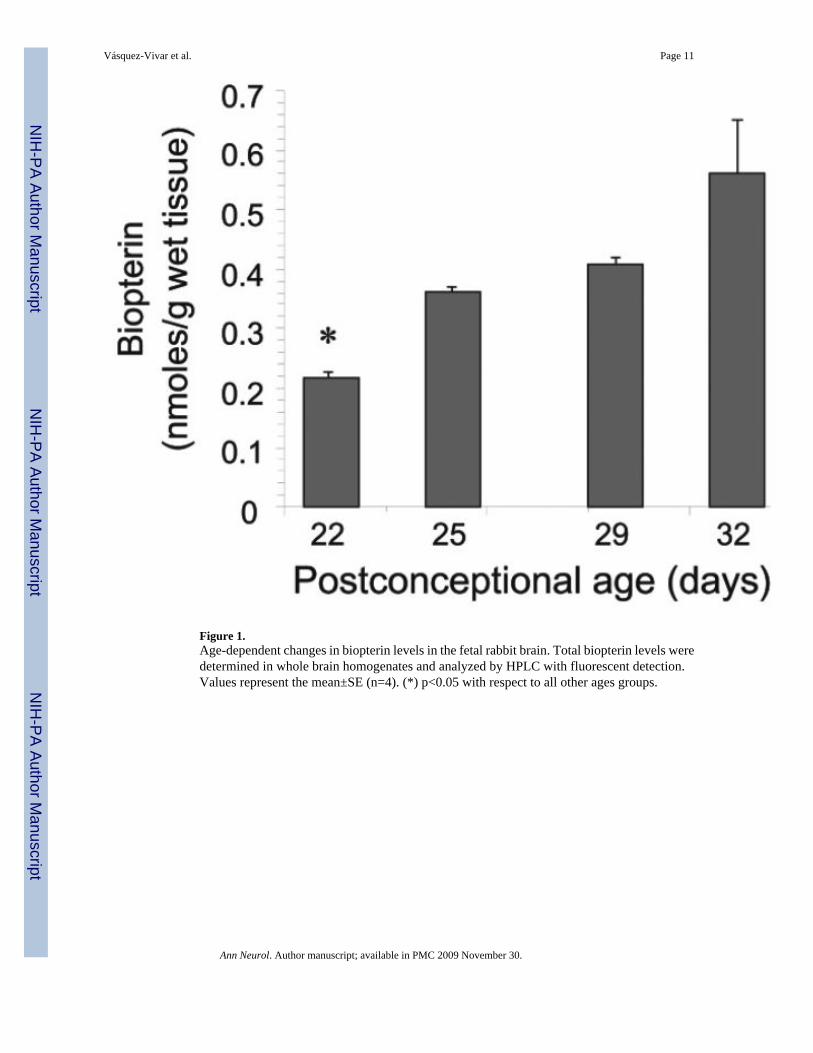
Figure 1.Age-dependent changes in biopterin levels in the fetal rabbit brain. Total biopterin levels weredetermined in whole brain homogenates and analyzed by HPLC with fluorescent detection.Values represent the mean±SE (n=4). (*) p<0.05 with respect to all other ages groups.
Vásquez-Vivar et al. Page 11
Ann Neurol. Author manuscript; available in PMC 2009 November 30.
NIH
-PA Author Manuscript
NIH
-PA Author Manuscript
NIH
-PA Author Manuscript
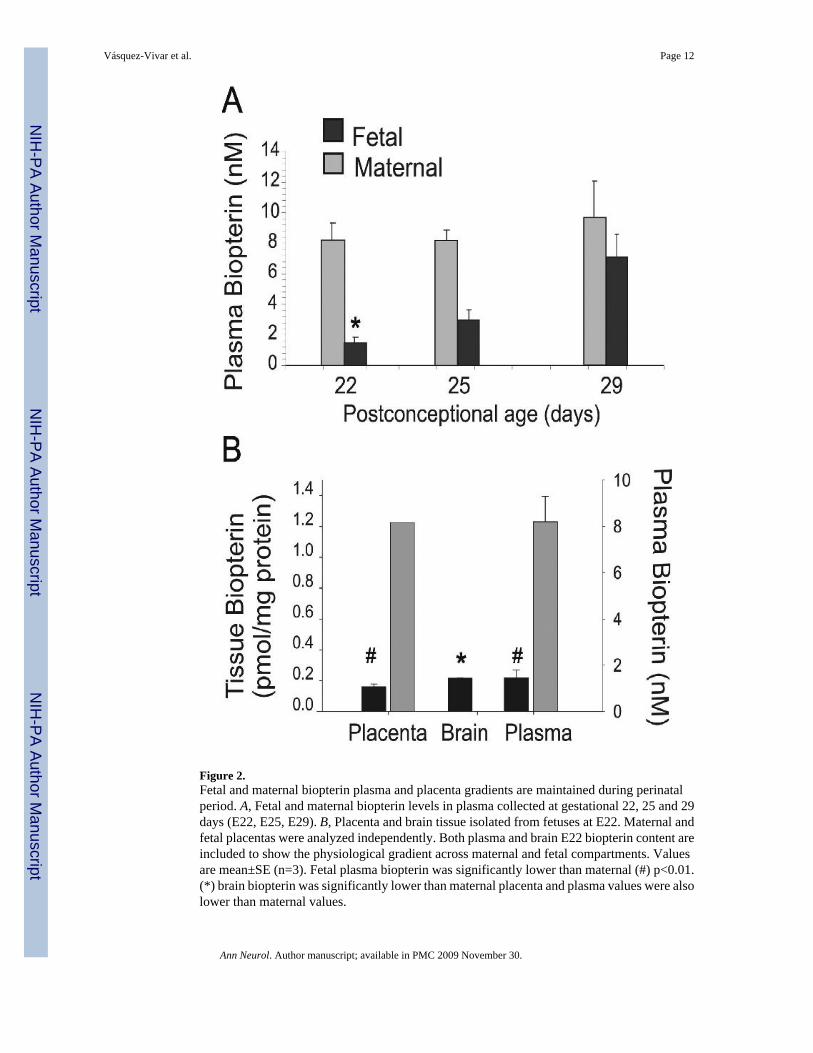
Figure 2.Fetal and maternal biopterin plasma and placenta gradients are maintained during perinatalperiod. A, Fetal and maternal biopterin levels in plasma collected at gestational 22, 25 and 29days (E22, E25, E29). B, Placenta and brain tissue isolated from fetuses at E22. Maternal andfetal placentas were analyzed independently. Both plasma and brain E22 biopterin content areincluded to show the physiological gradient across maternal and fetal compartments. Valuesare mean±SE (n=3). Fetal plasma biopterin was significantly lower than maternal (#) p<0.01.(*) brain biopterin was significantly lower than maternal placenta and plasma values were alsolower than maternal values.
Vásquez-Vivar et al. Page 12
Ann Neurol. Author manuscript; available in PMC 2009 November 30.
NIH
-PA Author Manuscript
NIH
-PA Author Manuscript
NIH
-PA Author Manuscript

Figure 3.GTP cyclohydrolase (GTPCH) expression and activity in brain increases during perinatalperiod. A, RT-PCR analysis of GTPCH in fetal brain and liver homogenates at gestational 22,25 and 29-days (E22, E25, E29). B, Western blot analysis of GTPCH protein levels in fetalbrain and liver showing increased protein expression with age; β-actin levels were used asloading control. C, GTPCH densitometry indicates that E22 brains contain the lowest level ofprotein. D, GTPCH enzyme activity in the fetal brain shows a significant difference betweenE22 and E25 with an increase of ~50% which is maintained at later times. Values representthe mean±SE (n=4). (*) p<0.05 with respect to all other ages.
Vásquez-Vivar et al. Page 13
Ann Neurol. Author manuscript; available in PMC 2009 November 30.
NIH
-PA Author Manuscript
NIH
-PA Author Manuscript
NIH
-PA Author Manuscript

Figure 4.Thalamus is the region of the brain with highest tetrahydrobiopterin levels. Tetrahydrobiopterin(BH4) was directly measured in parts of the brain between E22 and E32 (postnatal day 1). A,Low BH4 was found in all E22 with the thalamus showing significantly higher amounts withrespect to all other brain regions. Values are mean±SE (n≥5), (*) different from E22 values;p<0.05. B, Map depicting average concentration of BH4 increases with age.
Vásquez-Vivar et al. Page 14
Ann Neurol. Author manuscript; available in PMC 2009 November 30.
NIH
-PA Author Manuscript
NIH
-PA Author Manuscript
NIH
-PA Author Manuscript
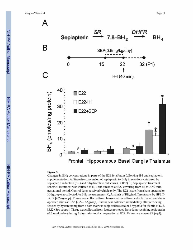
Figure 5.Changes in BH4 concentrations in parts of the E22 fetal brain following H-I and sepiapterinsupplementation. A, Stepwise conversion of sepiapterin to BH4 in reactions catalyzed bysepiapterin reductase (SR) and dihydrofolate reductase (DHFR). B, Sepiapterin treatmentscheme. Treatment was initiated at E15 and finished at E22 covering from 48 to 70% termgestational period. Control dams received vehicle only. The E22 tissue from sham operated orH-I group was collected for BH4 measurements. C, Analysis of BH4 in different parts by HPLC-ECD. [E22-group]: Tissue was collected from fetuses retrieved from vehicle treated and shamoperated dams at E22. [E22-H-I group]: Tissue was collected immediately after retrievingfetuses by hysterectomy from a dam that was subjected to sustained hypoxia for 40 min at E22.[E22+Sep group]: Tissue was collected from fetuses retrieved from dams receiving sepiapterin(0.6 mg/kg/day) during 5 days prior to sham-operation at E22. Values are mean±SE (n≥4).
Vásquez-Vivar et al. Page 15
Ann Neurol. Author manuscript; available in PMC 2009 November 30.
NIH
-PA Author Manuscript
NIH
-PA Author Manuscript
NIH
-PA Author Manuscript

(†) p<0.05 with respect to other parts of the brain; (‡) p<0.05 with respect to E22 thalamus;(#) with respect to E22 frontal; (*) p<0.01 with respect to all experimental groups.
Vásquez-Vivar et al. Page 16
Ann Neurol. Author manuscript; available in PMC 2009 November 30.
NIH
-PA Author Manuscript
NIH
-PA Author Manuscript
NIH
-PA Author Manuscript

Figure 6.Neurobehavioral summary of scores (box-whisker plots) obtained by video analysis of vehicleand sepiapterin pre-treated newborn kits (n=37 and 44 respectively). All variables are highlysignificant between groups (ttest, P<0.001). Scores are quantitative estimations for all exceptdystonia where scoring is based on 9 point scale for %time spent on dystonic activity (0-100%of time).
Vásquez-Vivar et al. Page 17
Ann Neurol. Author manuscript; available in PMC 2009 November 30.
NIH
-PA Author Manuscript
NIH
-PA Author Manuscript
NIH
-PA Author Manuscript
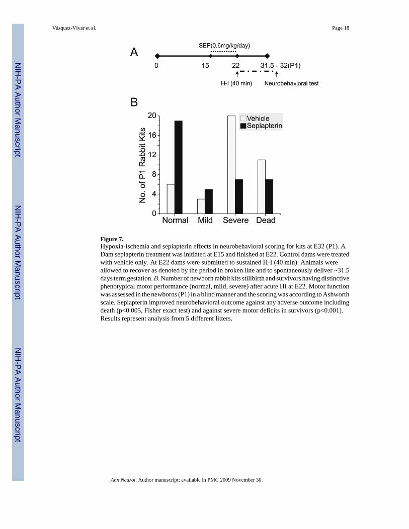
Figure 7.Hypoxia-ischemia and sepiapterin effects in neurobehavioral scoring for kits at E32 (P1). A.Dam sepiapterin treatment was initiated at E15 and finished at E22. Control dams were treatedwith vehicle only. At E22 dams were submitted to sustained H-I (40 min). Animals wereallowed to recover as denoted by the period in broken line and to spontaneously deliver ~31.5days term gestation. B. Number of newborn rabbit kits stillbirth and survivors having distinctivephenotypical motor performance (normal, mild, severe) after acute HI at E22. Motor functionwas assessed in the newborns (P1) in a blind manner and the scoring was according to Ashworthscale. Sepiapterin improved neurobehavioral outcome against any adverse outcome includingdeath (p<0.005, Fisher exact test) and against severe motor deficits in survivors (p<0.001).Results represent analysis from 5 different litters.
Vásquez-Vivar et al. Page 18
Ann Neurol. Author manuscript; available in PMC 2009 November 30.
NIH
-PA Author Manuscript
NIH
-PA Author Manuscript
NIH
-PA Author Manuscript

NIH
-PA Author Manuscript
NIH
-PA Author Manuscript
NIH
-PA Author Manuscript
Vásquez-Vivar et al. Page 19
Table 1
BH4 and ascorbate levels in parts of the fetal brain.
BH4(pmol/mg protein)
Ascorbate(nmol/mg protein)
Tissue Control Hypoxia(E22, 40 min) Control Hypoxia
(E22, 40 min)
Frontal 1.28±0.07 0.54±0.15‡ 136.4±7.2 92.2±4.5‡Parietal 1.20±0.62 1.21±0.58 102.7±30.4 NA
Thalamus 12.62±1.50 9.3±1.3‡ 131.6±8.5 94.4±5.85‡Basal Ganglia 3.60±0.60 2.7±0.1‡ 128.3±3.3 99.79±4.5‡Hippocampus 1.96±0.34 1.39±0.37 106.3±5.4 81.9±3.2‡
Values represent mean±SE (n>4 litters).
NA- data not available
‡differences were statistically significant with respect to control sham operated animals P<0.05.
Ann Neurol. Author manuscript; available in PMC 2009 November 30.

NIH
-PA Author Manuscript
NIH
-PA Author Manuscript
NIH
-PA Author Manuscript
Vásquez-Vivar et al. Page 20
Table 2BH4 distribution in sepiapterin supplemented maternal and fetal tissues
Sepiapterin was administered using an Alzet osmotic pump with 0.6 mg/Kg during seven days beginning atpostconceptional day 17 (E17) and ending at postconceptional day 22 (E22). Tissue was harvested at the end ofday E22 for BH4 analysis. Control received vehicle only for the same period of time.
Control – VehicleBH4 (pmoles/mg protein)
SepiapterinBH4 (pmoles/mg protein)
Maternal Fetal Maternal Fetal
Placenta 0.2±0.1 0.1±0.1 4.6±0.5‡ 5.2±0.1‡Kidney 4.1±0.8 1.7±0.9 5.6±0.4‡ 4.6±1.4‡Liver 8.6±1.0 1.9±0.3 10.2±1.1 1.2±0.3
Values are Mean±S.E.M (n=4).
‡p<0.05.
Ann Neurol. Author manuscript; available in PMC 2009 November 30.

NIH
-PA Author Manuscript
NIH
-PA Author Manuscript
NIH
-PA Author Manuscript
Vásquez-Vivar et al. Page 21
Table 3
Changes in dopamine, serotonin and metabolites in the brain after H-I injury with and without sepiapterinsupplementation.
Dopamine (ng/mg protein)
Control-Vehicle Hypoxia SepiapterinThalamus 0.13±0.08 0.06±0.05# 0.17±0.03¶Basal Ganglia 3.18±0.95 0.37±0.08# 1.19±0.42#,¶
Serotonin (ng/mg protein)
Control-Vehicle Hypoxia SepiapterinThalamus 3.20±0.90 2.20±0.37# 3.10±0.26¶Basal Ganglia 3.38±0.56 3.71±0.73 3.52±0.89
HVA/5HIAAa
Control-Vehicle Hypoxia SepiapterinThalamus 1.19±0.18 0.86±0.07# 0.98±0.07¶Basal Ganglia 1.15±0.13 0.52±0.14# 0.82±0.12¶
Values are Mean±S.E.M. (n=3-5).
#p<0.05 from controls
¶p<0.05 from hypoxia group.
aHVA/5HIAA reference values for 0.5 years old 1.3-3.128.
Ann Neurol. Author manuscript; available in PMC 2009 November 30.
Related Documents