Tetherin Restricts Herpes Simplex Virus 1 and Is Antagonized by Glycoprotein M Caroline Blondeau, a Annegret Pelchen-Matthews, b Petra Mlcochova, a,b Mark Marsh, b Richard S. B. Milne, a Greg J. Towers a University College London, Medical Research Council Centre for Medical Molecular Virology, Division of Infection and Immunity, University College London, London, United Kingdom a ; MRC Laboratory for Molecular Cell Biology, University College London, London, United Kingdom b Tetherin is a broadly active antiviral effector that works by tethering nascent enveloped virions to a host cell membrane, thus preventing their release. In this study, we demonstrate that herpes simplex virus 1 (HSV-1) is targeted by tetherin. We identify the viral envelope glycoprotein M (gM) as having moderate anti-tetherin activity. We show that gM but not gB or gD efficiently removes tetherin from the plasma membrane and can functionally substitute for the human immunodefi- ciency virus type 1 (HIV-1) Vpu protein, the prototypic viral tetherin antagonist, in rescuing HIV-1 release from tetherin- expressing cells. Our data emphasize that tetherin is a broadly active antiviral effector and contribute to the emerging hy- pothesis that viruses must suppress or evade an array of host cell countermeasures in order to establish a productive infection. M ammalian cells encode restriction factors that provide the host with protection against virus replication. In order to establish a productive infection, a virus must evade or suppress a repertoire of restriction factors directed against it by the host. Restriction has been studied most extensively in the context of retrovirus infections, where numerous factors and the corre- sponding viral antagonists have been well characterized (1). While some restriction factors, such as TRIM5, appear to be specific for particular classes of virus, in this case retroviruses, others, such as tetherin, are broadly active against unrelated viruses. Tetherin (also known as BST-2, CD317, or HM1.24) was identified as the cellular factor responsible for suppression of Vpu-negative hu- man immunodeficiency virus type 1 (HIV-1) (2, 3), but subse- quent work has shown that it is effective against a variety of envel- oped viruses (2, 4–8) that use distinct mechanisms to antagonize its restrictive effects (9–12). Tetherin is a type 2 integral mem- brane protein with a C-terminal GPI anchor. The antiviral activity of tetherin stems from this unusual double membrane-linked to- pology that allows the formation of a protein tether between the host membrane and the budding viral envelope, preventing re- lease of nascent virions (13). Herpesviruses, a large family of enveloped DNA viruses, are ancient pathogens thought to have coevolved with their hosts for many generations (14). As such, they might be expected to possess countermeasures to a variety of restriction factors and thus to provide a good experimental model system for studies of this aspect of the virus host interaction. To date, two mem- bers of this virus family, Kaposi’s sarcoma-associated herpes- virus (KSHV) and human cytomegalovirus (HCMV), have been shown to interact with tetherin (15–17). Surprisingly, the mode of interaction differs for these two viruses, with tetherin acting as a restriction factor for KSHV but as an entry cofactor for HCMV. In this study, we investigated the effect of tetherin on another human herpesvirus, herpes simplex virus 1 (HSV- 1). We show that tetherin restricts the HSV-1 replication cycle by suppressing virus release, and we identify the viral envelope glycoprotein M (gM) as a countermeasure contributing to an- tagonism of tetherin restriction. MATERIALS AND METHODS Cell lines, plasmids, and viruses. HT1080 cells expressing internally hemagglutinin (HA)-tagged human tetherin (at amino acid 154) or empty vector (LHCX) are nonclonal drug-selected populations and have been described (16), as has the tetherin expression vector pCR3.1/hu- Tetherin-HA (18). The HSV-1 gM (UL10) gene was PCR amplified from HSV-1 17-infected-cell DNA and inserted into pCDNA3. The HSV-1 gB (UL27) and gD (US6) plasmids (pSR175 and pSC390) were gifts from Roselyn Eisenberg and Gary Cohen (University of Pennsylvania) (19, 20). Plasmids expressing Vpu, in pCDNA3 (for HIV-1 release assay) or pIRESeGFP (for flow cytometry), were described previously (21). Wild-type (WT) HSV-1 SC16 and HSV-1 KOS K26GFP, encoding a VP26-green fluorescent protein (GFP) fusion protein (22) were gifts from Gillian Elliott (Imperial College London). HSV-1 with a deletion of UL10 (gM) and its revertant (RgM) were gifts from Helena Browne (Univer- sity of Cambridge), and their construction has been described (23). HSV-1 replication assay. HT1080 cells (3 10 5 cells/well, 6-well plates) were chilled to 4°C and then incubated with HSV-1 for 1 h. Plates were then refed and transferred to 37°C for a further hour. The medium was then removed and replaced with acid-citrate buffer (500 l, pH 3.0) to inactivate extracellular virus, followed by the addition of fresh medium. Infected-cell culture supernatants were recovered at various times postin- fection and centrifuged to remove cellular debris, and virus titers deter- mined by plaque assay on Vero cells. For cell-associated virus titers, cells were lysed by 3 freeze-thaw cycles into an equal volume of medium, cleared by centrifugation, and titrated as described above. The HSV-1 proteins ICP4 and VP5 were detected in infected-cell lysates by immuno- blotting using specific antibodies (Santa Cruz). As a loading control we detected -actin (Abcam) on stripped blots. RNA interference. We used lentiviral vectors encoding tetherin-specific hairpins (shRNA1, 5=-GGAGUUCUGGUGUUCCUGAUUAUUUCGAUG AUCAGGAGCACCAGAAUUCC-3=; shRNA2, 5=-GUGGGAAUCGUGGA Received 13 August 2013 Accepted 13 September 2013 Published ahead of print 25 September 2013 Address correspondence to Greg J. Towers, [email protected], or Richard S. B. Milne, [email protected]. Copyright © 2013 Blondeau et al. This is an open-access article distributed under the terms of the Creative Commons Attribution 3.0 Unported license. doi:10.1128/JVI.02250-13 13124 jvi.asm.org Journal of Virology p. 13124 –13133 December 2013 Volume 87 Number 24 Downloaded from https://journals.asm.org/journal/jvi on 14 February 2022 by 210.104.179.157.

Welcome message from author
This document is posted to help you gain knowledge. Please leave a comment to let me know what you think about it! Share it to your friends and learn new things together.
Transcript

Tetherin Restricts Herpes Simplex Virus 1 and Is Antagonized byGlycoprotein M
Caroline Blondeau,a Annegret Pelchen-Matthews,b Petra Mlcochova,a,b Mark Marsh,b Richard S. B. Milne,a Greg J. Towersa
University College London, Medical Research Council Centre for Medical Molecular Virology, Division of Infection and Immunity, University College London, London,United Kingdoma; MRC Laboratory for Molecular Cell Biology, University College London, London, United Kingdomb
Tetherin is a broadly active antiviral effector that works by tethering nascent enveloped virions to a host cell membrane,thus preventing their release. In this study, we demonstrate that herpes simplex virus 1 (HSV-1) is targeted by tetherin. Weidentify the viral envelope glycoprotein M (gM) as having moderate anti-tetherin activity. We show that gM but not gB orgD efficiently removes tetherin from the plasma membrane and can functionally substitute for the human immunodefi-ciency virus type 1 (HIV-1) Vpu protein, the prototypic viral tetherin antagonist, in rescuing HIV-1 release from tetherin-expressing cells. Our data emphasize that tetherin is a broadly active antiviral effector and contribute to the emerging hy-pothesis that viruses must suppress or evade an array of host cell countermeasures in order to establish a productiveinfection.
Mammalian cells encode restriction factors that provide thehost with protection against virus replication. In order to
establish a productive infection, a virus must evade or suppress arepertoire of restriction factors directed against it by the host.Restriction has been studied most extensively in the context ofretrovirus infections, where numerous factors and the corre-sponding viral antagonists have been well characterized (1). Whilesome restriction factors, such as TRIM5�, appear to be specific forparticular classes of virus, in this case retroviruses, others, such astetherin, are broadly active against unrelated viruses. Tetherin(also known as BST-2, CD317, or HM1.24) was identified as thecellular factor responsible for suppression of Vpu-negative hu-man immunodeficiency virus type 1 (HIV-1) (2, 3), but subse-quent work has shown that it is effective against a variety of envel-oped viruses (2, 4–8) that use distinct mechanisms to antagonizeits restrictive effects (9–12). Tetherin is a type 2 integral mem-brane protein with a C-terminal GPI anchor. The antiviral activityof tetherin stems from this unusual double membrane-linked to-pology that allows the formation of a protein tether between thehost membrane and the budding viral envelope, preventing re-lease of nascent virions (13).
Herpesviruses, a large family of enveloped DNA viruses, areancient pathogens thought to have coevolved with their hostsfor many generations (14). As such, they might be expected topossess countermeasures to a variety of restriction factors andthus to provide a good experimental model system for studiesof this aspect of the virus host interaction. To date, two mem-bers of this virus family, Kaposi’s sarcoma-associated herpes-virus (KSHV) and human cytomegalovirus (HCMV), havebeen shown to interact with tetherin (15–17). Surprisingly, themode of interaction differs for these two viruses, with tetherinacting as a restriction factor for KSHV but as an entry cofactorfor HCMV. In this study, we investigated the effect of tetherinon another human herpesvirus, herpes simplex virus 1 (HSV-1). We show that tetherin restricts the HSV-1 replication cycleby suppressing virus release, and we identify the viral envelopeglycoprotein M (gM) as a countermeasure contributing to an-tagonism of tetherin restriction.
MATERIALS AND METHODSCell lines, plasmids, and viruses. HT1080 cells expressing internallyhemagglutinin (HA)-tagged human tetherin (at amino acid 154) orempty vector (LHCX) are nonclonal drug-selected populations and havebeen described (16), as has the tetherin expression vector pCR3.1/hu-Tetherin-HA (18). The HSV-1 gM (UL10) gene was PCR amplified fromHSV-1 17�-infected-cell DNA and inserted into pCDNA3. The HSV-1gB (UL27) and gD (US6) plasmids (pSR175 and pSC390) were giftsfrom Roselyn Eisenberg and Gary Cohen (University of Pennsylvania)(19, 20). Plasmids expressing Vpu, in pCDNA3 (for HIV-1 release assay)or pIRESeGFP (for flow cytometry), were described previously (21).Wild-type (WT) HSV-1 SC16 and HSV-1 KOS K26GFP, encoding aVP26-green fluorescent protein (GFP) fusion protein (22) were gifts fromGillian Elliott (Imperial College London). HSV-1 with a deletion of UL10(�gM) and its revertant (RgM) were gifts from Helena Browne (Univer-sity of Cambridge), and their construction has been described (23).
HSV-1 replication assay. HT1080 cells (3 � 105 cells/well, 6-wellplates) were chilled to 4°C and then incubated with HSV-1 for 1 h. Plateswere then refed and transferred to 37°C for a further hour. The mediumwas then removed and replaced with acid-citrate buffer (500 �l, pH 3.0) toinactivate extracellular virus, followed by the addition of fresh medium.Infected-cell culture supernatants were recovered at various times postin-fection and centrifuged to remove cellular debris, and virus titers deter-mined by plaque assay on Vero cells. For cell-associated virus titers, cellswere lysed by 3 freeze-thaw cycles into an equal volume of medium,cleared by centrifugation, and titrated as described above. The HSV-1proteins ICP4 and VP5 were detected in infected-cell lysates by immuno-blotting using specific antibodies (Santa Cruz). As a loading control wedetected �-actin (Abcam) on stripped blots.
RNA interference. We used lentiviral vectors encoding tetherin-specifichairpins (shRNA1, 5=-GGAGUUCUGGUGUUCCUGAUUAUUUCGAUGAUCAGGAGCACCAGAAUUCC-3=; shRNA2, 5=-GUGGGAAUCGUGGA
Received 13 August 2013 Accepted 13 September 2013
Published ahead of print 25 September 2013
Address correspondence to Greg J. Towers, [email protected], orRichard S. B. Milne, [email protected].
Copyright © 2013 Blondeau et al. This is an open-access article distributed underthe terms of the Creative Commons Attribution 3.0 Unported license.
doi:10.1128/JVI.02250-13
13124 jvi.asm.org Journal of Virology p. 13124 –13133 December 2013 Volume 87 Number 24
Dow
nloa
ded
from
http
s://j
ourn
als.
asm
.org
/jour
nal/j
vi o
n 14
Feb
ruar
y 20
22 b
y 21
0.10
4.17
9.15
7.

UAAGAAGUAUUCGUACUUCUUGUCCGCGAUUCUCAC-3=; under-lining indicates tetherin-targeted sequence) or a GFP hairpin (24) as acontrol. Depletion was examined by immunoblotting or by quantitative PCRon cDNA (see below). Cells were infected with HSV-1 96 h post-shRNAtransduction as described above.
Quantification of tetherin and HSV-1 by TaqMan PCR. Encapsi-dated HSV-1 genomes were quantified by extracting total DNA fromDNase I-treated supernatants or infected-cell lysates as described previ-ously (16). DNA was subjected to quantitative TaqMan PCR (Q-PCR) forHSV-1 UL27 as described previously (25). Absolute copy number wasdetermined by reference to a standard curve, plotted using serial dilutionsof a cloned UL27 amplicon with a detection limit of 10 UL27 copies/15 �lof supernatant. Copy numbers were normalized to extracted DNA carrierconcentration (supernatants) or to quantities of extracted DNA (cells).Total mRNA was extracted from transduced HeLa cells or from HT1080cells expressing HA-tagged tetherin and infected with HSV-1 SC16 or notinfected, and cDNA was synthesized for use as the template in TaqManQ-PCRs for tetherin and GAPDH. Tetherin primers were as follows: for-ward, 5=-ACCTGCAACCACACTGTGATG-3=; reverse, 5=-CAAGCTCCTCCACTTTCTTTTGTC-3=; tetherin probe, 5=-FAM-CCCTAATGGCTTCCCTGGATGCAGA-TAMRA-3=. Absolute copy number was determined withreference to a standard curve derived using a tetherin-encoding plasmid. Q-PCR for GAPDH was performed as described previously (16).
Flow cytometry. For tetherin cell surface staining, HEK293T cells in6-well plates were transfected (Fugene-6; Roche) with pCR3.1/hu-Teth-erin-HA and 250 ng, 500 ng, or 1,000 ng of plasmids expressing gM, gD, orgB (21). A pIRES2eGFP plasmid coding for Vpu was used as a control. At48 h posttransfection, cell surface tetherin expression was examined onunfixed live cells with an anti-HA monoclonal antibody (Covance) andanalyzed by flow cytometry. The mean fluorescence intensity of tetherinstaining was measured as described previously (7). HSV-1 glycoproteinswere detected by immunoblotting with a rabbit anti-gM (23), a rabbitanti-gD R8 (26), or a goat anti-gB (Santa Cruz) antibody after membranestripping. As a loading control, we detected �-actin, GAPDH (Abcam), ortransferrin receptor (Invitrogen).
HIV-1 release assay. To prepare vesicular stomatitis virus G glycopro-tein (VSV-G)-pseudotyped HIV-1 particles, 106 HEK293T cells werecotransfected with the Gag-Pol expression vector p8.91 (300 ng), pMDGencoding VSV-G (300 ng), and HIV-1 vector encoding YFP (450 ng) (27).Tetherin construct (100 ng) was cotransfected along with either 250 ng,500 ng, or 1,000 ng of HSV-1 gM, gB, or gD or HIV-1 Vpu plasmid. DNAdose was equalized with the empty vector pcDNA3 (Invitrogen). After 48h, cell-associated p55 Gag and p24 capsid and p24 capsid in the superna-tant were detected by immunoblotting as described previously (27). Theintensity of p55 bands in cell lysates and p24 bands in virions was analyzedwith Image Studio 3.1.4 software (LI-COR), and ratios of p55 to p24 werecalculated with signal intensity percentages relative to values obtained inthe absence of tetherin.
Microscopy. For electron microscopy, 1 � 105 HT1080 cells express-ing HA-tagged tetherin or control cells seeded on coverslips were infectedwith 2 � 105 (experiment 1) or 1 � 105 (experiment 2) PFU (determinedon Vero cells) of HSV-1 K26GFP (HT1080 cells are an order of magnitudeless permissive than Vero cells to HSV-1, and therefore multiplicities forthese experiments can be estimated to be 0.2 and 0.1 PFU/cell, respec-tively). After 16 h, the cells were fixed for 45 min in 2% paraformaldehyde(PFA)–2% glutaraldehyde in 0.1 M sodium cacodylate buffer (pH 7.4),postfixed for 1 h on ice in 1% OsO4–1.5% K3[Fe(CN)6], treated with 1.5%tannic acid (TAAB Laboratories), dehydrated, and embedded in Epon 812(TAAB). Ultrathin sections (70 nm) were cut en face on a Leica EM UC7ultramicrotome, placed on Formvar-coated slot grids, and stained withlead citrate. Sections were examined with a Tecnai G2 Spirit transmissionEM (FEI), and digital images were recorded with a Morada 11 MegaPixeltransmission electron microscopy (TEM) camera (Olympus Soft ImagingSolutions) and ANALYSIS software. Images were adjusted for brightnessand contrast, and figures were assembled with Photoshop CS. To deter-
mine the numbers of cell surface HSV-1 particles, at least 50 consecutive/adjacent cell profiles in the section were inspected for each sample. Cir-cular profiles of HSV-1 particles, measuring between 80 and 180 nm indiameter and with the morphologies indicated in Fig. 3, were counted.
For confocal microscopy, HEK293T cells were plated on poly-L-lysine-coated coverslips in 24-well plates and transfected (Fugene-6;Roche) with plasmids encoding gM, gB, or gD and/or HA-tagged tetherin.Between 20 and 48 h later, cells were fixed (4% PFA), permeabilized (0.1%Triton X-100), and stained using anti-HA (Covance), rabbit anti-gM,rabbit anti-gD R8, goat anti-gB, or sheep anti-TGN46 (Serotec) antibod-ies and secondary antibodies linked to Alexa-488, -594, or -633 (Molecu-lar Probes) or rhodamine (Pierce). Cells were observed using a Leica TCSSPE, DM2500, confocal microscope (Leica Microsystems). Images wereadjusted for brightness and contrast with Adobe Photoshop software 10.0.
RESULTSTetherin restricts HSV-1 particle release. To seek evidence forrestriction of HSV-1, we expressed HA-tagged human tetherin inHT1080 cells. These cells were chosen because they naturally ex-press very low levels of tetherin and are highly permissive forHSV-1 (28). As a control, we used HT1080 cells transduced withempty vector. We hypothesized that tetherin overexpressionmight saturate any anti-tetherin activities mediated by the virusand reveal tetherin sensitivity. We infected both HT1080 cell lineswith HSV-1 SC16 at a low multiplicity (0.01 PFU/cell), aiming tostudy the effect of tetherin in a multicycle infection by measuringthe titer of virus released at various times by plaque assay on Verocells. Tetherin expression consistently reduced the levels of infec-tious virus released, leading to a 14-fold reduction at 48 h postin-fection (hpi), compared to controls (Fig. 1A). Quantitative PCR(Q-PCR) detection of DNase I-resistant (i.e., encapsidated)HSV-1 DNA in the infected-cell culture supernatants demon-strated a comparable decrease in signal, supporting the notionthat tetherin suppresses virion release from infected cells (Fig. 1B).Tetherin expression did not affect titers of cell-associated virus(Fig. 1C) or levels of cell-associated viral DNA (Fig. 1D) at earlytime points up to 24 hpi. At later times postinfection, titers ofcell-associated virus from tetherin-expressing cells were 7- to8-fold lower than those of controls (Fig. 1C), and consistent withthis, levels of viral proteins detected by Western blotting were alsoreduced (Fig. 1E). We assume that at these later time points, we seethe cumulative effect of tetherin’s inhibition of viral release andthe consequent reduction in number of newly infected cells insubsequent rounds of infection. Importantly, in a plaque assaythere was no difference between tetherin-expressing and controlHT1080 cells in the number, or size, of plaques obtained from agiven virus dose, suggesting that tetherin had no impact on HSV-1entry, or direct cell-to-cell spread (Fig. 1F). This is consistent withthe specificity of tetherin for virus release over cell-to-cell spread,as has been described for tetherin restriction of the lentivirusesHIV-1 and feline immunodeficiency virus (FIV) (29, 30).
We next tested whether tetherin expression could suppresshigh-multiplicity infection. Tetherin-expressing and controlHT1080 cells were infected with HSV-1 (input multiplicity, 3PFU/cell), and the virus yield was measured at various times byplaque assay as before (Fig. 1G). Again, slightly less infectiousvirus was released into the supernatant from tetherin-expressingcells at the earliest time of 14 h, although this difference was sta-tistically insignificant. Moreover, infectious titers in the superna-tants were equal by 24 h after infection and up to 48 hpi. Q-PCRdetection of DNase-protected viral genomes confirmed this effect
Restriction of HSV-1 by Tetherin
December 2013 Volume 87 Number 24 jvi.asm.org 13125
Dow
nloa
ded
from
http
s://j
ourn
als.
asm
.org
/jour
nal/j
vi o
n 14
Feb
ruar
y 20
22 b
y 21
0.10
4.17
9.15
7.

(Fig. 1H). Cell-associated virus titers at 14 hpi were very similar,and infected cell lysates showed similar levels of viral proteins,confirming that tetherin has no effect on viral DNA or proteinsynthesis (Fig. 1I and J). These data suggest that during high-multiplicity infection, tetherin is antagonized by HSV-1 infection.To test whether tetherin protein levels were impacted by HSV-1infection, we measured them by immunoblotting at various timepoints after HSV-1 infection at an input multiplicity of 2 PFU/cell(Fig. 1K). We found that tetherin levels declined as viral protein
FIG 1 Human tetherin restricts HSV-1 particle release. HT1080 cells express-ing HA-tagged tetherin (THN) or empty vector (Vector) were infected withHSV-1 SC16 at 0.01 (A to E) or 3 PFU/cell (G to J). Supernatants were har-vested at the indicated times and titrated for HSV-1 infectivity by plaque assay(A and G) or subjected to DNase I treatment followed by Q-PCR for the gB
gene UL27 (B and H). At the same time, cells were harvested, and freeze/thawed 3 times before titration for HSV-1 infectivity on Vero cells (C and I),extracted DNA was subjected to Q-PCR for the gB gene UL27 (D), or lysateswere analyzed by immunoblotting (E and J). Statistical significance, as a Pvalue, was determined by 2-way analysis of variance (ANOVA) (**, P � 0.01;***, P � 0.001). Tetherin had no effect on DNA replication up to 22 hpi (D)(2-way ANOVA; P 0.05). After high-MOI infection (3 PFU/cell), tetherinhad no effect on HSV-1 supernatant titers (G) (2-way ANOVA; P 0.05), orcell-associated virus titers (I) (t test; P 0.05). (F) HT1080 cells expressingHA-tagged tetherin or empty vector were infected with HSV-1 SC16 andwashed with acid citrate buffer 1 h later, before the addition of overlay mediumfor plaque assays. Plaques were counted 48 h later after crystal violet staining orimmune-alkaline phosphatase staining (for the plaques shown). Results aremeans and standard errors of the means (SEM) and are from 3 independentexperiments. (K, L) HT1080 cells expressing HA-tagged tetherin or emptyvector were infected with HSV-1 SC16 (2 PFU/cell), and cell lysates were pre-pared at the indicated times and analyzed by immunoblotting to detect VP5expression and HA tag (tetherin), transferrin receptor (TfR), �-actin, andGAPDH after membrane stripping (K), or tetherin and GAPDH mRNA levelswere assessed in noninfected and infected cells by Q-PCR. Under these condi-tions, tetherin mRNA was not detectable in the empty vector HT1080 cells.HSV-1 infection decreased tetherin mRNA copy number (L) (2-way ANOVA;P � 0.01). Results are means and standard deviations (SD) and are represen-tative of at least 2 separate experiments, except when specified.
FIG 2 shRNA-mediated depletion of tetherin increases HSV-1 release. HeLacells were transduced with HIV-1 vector encoding either of 2 tetherin-specificshRNAs or a shRNA targeting GFP (A to C). Cells were then infected withHSV-1 SC16 (0.1 PFU/cell). HSV-1 infectivity in the supernatants was mea-sured by plaque assay (A) and that in DNase-protected genomes by Q-PCR(B). Tetherin mRNA depletion was assessed in HeLa by Q-PCR normalized toGAPDH (C). The increase in HSV-1 titers after tetherin depletion (A) wassignificant (2-way ANOVA; P � 0.05). Tetherin reduction was also assessed byimmunoblotting HA tag (tetherin) or �-actin (after membrane stripping) inshRNA-transduced HA-tagged-tetherin-expressing HT1080 cells (D). Resultsare means and SD and are representative of 3 separate experiments.
Blondeau et al.
13126 jvi.asm.org Journal of Virology
Dow
nloa
ded
from
http
s://j
ourn
als.
asm
.org
/jour
nal/j
vi o
n 14
Feb
ruar
y 20
22 b
y 21
0.10
4.17
9.15
7.

VP5 increased. We assume that the different tetherin bands ob-served represent differently glycosylated forms (31). Importantly,all were reduced after infection. The loss of tetherin expressionwas somewhat specific, as �-actin and transferrin receptor proteinlevels were unaffected up to 16 h after infection and GAPDH levelswere only slightly reduced (Fig. 1K). We next measured tetherinand GAPDH mRNA levels by quantitative RT-PCR in the samesamples as Fig. 1K. We found that tetherin mRNA levels declinedwith a time course similar to that of protein levels, suggesting arole for the virus host shutoff (Vhs) function in which hostmRNAs are degraded by the Vhs protein, encoded by the HSV-1UL41 gene (Fig. 1L). GAPDH mRNA was also lost, although withless concomitant reduction in protein expression. We concludethat tetherin is partly antagonized through suppression of expres-sion and that this likely accounts for the loss of restriction afterhigh-multiplicity infection (Fig. 1G). These data are consistentwith the recently reported antagonism of tetherin by the HSV-1Vhs response (see the accompanying paper [32]) but do not ruleout the possibility that tetherin might be degraded through addi-tional mechanisms.
Tetherin depletion with shRNA increases HSV-1 release. Toconfirm that tetherin was responsible for the reduction in HSV-1release seen in Fig. 1A, we depleted endogenous tetherin expres-sion from HeLa cells and measured release of HSV-1 into thesupernatant at various times after low-multiplicity infection (0.1PFU/cell). HeLa cells are known to express amounts of tetherinthat restrict HIV-1 strains lacking the tetherin antagonist Vpu(27). We used two tetherin-specific shRNAs and a control shRNAtargeting GFP. Expression of either of the anti-tetherin shRNAsimproved the release of virus, as indicated by an increase in infec-tious titer in supernatants particularly after 39 hpi, compared withthe titer obtained from cells expressing shGFP (Fig. 2A). Q-PCRdetection of DNase-protected genomes confirmed that less viruswas released from cells expressing the GFP-specific shRNA (Fig.2B). Quantitative RT-PCR showed that endogenous tetherinmRNA levels were reduced in HeLa cells expressing tetherin-spe-cific shRNA compared to cells expressing the hairpin targetingGFP (Fig. 2C). The effect of shRNA expression was also confirmedby immunoblotting detecting the HA tag in extracts of HT1080cells expressing HA-tagged tetherin (Fig. 2D) and transduced withshRNA-encoding lentivectors.
Tetherin induces accumulation of HSV-1 particles at the cellsurface. Having established that tetherin can restrict HSV-1 re-lease, we sought to visualize restricted virus on the surfaces oftetherin-expressing cells. Thin-section electron microscopy re-vealed that in HSV-1-infected HT1080 cells that do not expresstetherin, there were few virions associated with the cell surface.However, in infected cells overexpressing tetherin, there were ar-eas of cell surface where many virions were associated with theplasma membrane (Fig. 3A to G). To quantify this effect, wecounted cell surface-associated virions on control and tetherin-expressing cell profiles in a blinded manner. In two experiments,tetherin-expressing cells had significantly more cell surface virionsper cell profile than control cells (Table 1; Fig. 3H). These obser-vations are consistent with tetherin suppressing HSV-1 releasefrom infected cells (Fig. 1).
HSV-1 glycoprotein M can antagonize tetherin. Viruses typ-ically encode countermeasures to the repertoire of restriction fac-tors expressed by their natural host. Numerous viral countermea-sures to tetherin have been described (2, 4–8, 10, 11) which share
one key mechanistic characteristic: they remove tetherin from thelocation of its antiviral activity. Importantly, glycoproteins fromunrelated viruses, including lentiviruses (HIV-2 and SIVtan) (26,29) and Ebola virus (33), have been shown to have anti-tetherinactivity. With this in mind, we hypothesized that the HSV-1 en-velope glycoprotein M (gM) may also act as a tetherin antagonist.This protein has been shown to relocalize membrane proteins (23,34), and a deletion mutant replicates to reduced titers in a numberof cell lines (23, 28, 35).
To investigate whether gM has a role in tetherin antagonism,we first used confocal immunofluorescence microscopy to inves-tigate the effect of gM expression on tetherin localization. Wecotransfected HEK293T cells with plasmids encoding HSV-1 gMand HA-tagged tetherin and examined their localization after 48 h(Fig. 4A). In cells that did not express gM, or expressed low levelsof gM (Fig. 4A, open arrowheads), tetherin was predominantlylocalized to the plasma membrane, as described previously (2).However, in cells staining brightly for gM, a significant proportionof the tetherin was localized in the perinuclear region of the cell,where it overlapped with gM labeling (solid arrowheads). Furtherinvestigation indicated that the gM labeling also overlapped withlabeling for the trans-Golgi network marker TGN46 (Fig. 4B),suggesting that tetherin-gM complexes are located in the TGN.Together, these data suggest that, like HIV-1 Vpu, gM can relocal-ize tetherin, consistent with its having a role in antagonizing therestriction of virus release.
In order to assess the specificity of tetherin antagonism by gM,we compared the localization of tetherin after expression of gMand HSV-1 glycoproteins gD and gB. As before, we expressed theglycoproteins transiently, together with HA-tagged tetherin, inHEK293T cells and stained tetherin and each glycoprotein usingspecific antibodies. We found that, as before, gM caused a relocal-ization of tetherin from the plasma membrane to an internal com-partment (Fig. 5A). On the other hand, gD had no such effect, andtetherin mostly localized to the cell surface, as in control cells. Onexpression of gB, we found that there was a less extensive relocal-ization of tetherin than after gM expression. We conclude that gDhas no tetherin relocalization activity and that gB may have someactivity but less than that of gM.
To examine tetherin antagonism by HSV-1 glycoproteins fur-ther, we used flow cytometry to quantify tetherin surface expres-sion when tetherin was coexpressed with glycoproteins, again inHEK293T cells. Consistent with the immunofluorescence data,fluorescence-activated cell sorting analysis showed reduced levelsof tetherin on the surfaces of gM-expressing cells compared tocells expressing empty vector (Fig. 5B). We used the HIV-1 teth-erin antagonist Vpu as a positive control in these experiments. gDexpression had no effect, whereas gB caused a small reduction oftetherin surface staining that was less than that caused by gM (Fig.5B). All glycoproteins were expressed efficiently, as measured byimmunoblotting (Fig. 5C), and there was a striking elevation inthe amount of tetherin in the gM-expressing cells, consistent withthe gM-driven accumulation in the TGN (Fig. 4B). The flow cy-tometry measurements (Fig. 5B) were therefore consistent withimmunofluorescence staining of tetherin expression (Fig. 5A).
For a functional assessment of the antagonistic effect of HSV-1glycoproteins on tetherin and to provide further mechanistic in-sight, we asked whether they could substitute for Vpu in an HIV-1release assay (27). HIV-1 particles released into the supernatantwere detected by immunoblotting for the HIV-1 Gag structural
Restriction of HSV-1 by Tetherin
December 2013 Volume 87 Number 24 jvi.asm.org 13127
Dow
nloa
ded
from
http
s://j
ourn
als.
asm
.org
/jour
nal/j
vi o
n 14
Feb
ruar
y 20
22 b
y 21
0.10
4.17
9.15
7.

Blondeau et al.
13128 jvi.asm.org Journal of Virology
Dow
nloa
ded
from
http
s://j
ourn
als.
asm
.org
/jour
nal/j
vi o
n 14
Feb
ruar
y 20
22 b
y 21
0.10
4.17
9.15
7.

protein p24 at 48 h posttransfection. As expected, Vpu expressioneffectively antagonized tetherin restriction, and HIV-1 particles(p24-CA) were detected in the culture supernatant at levels equiv-alent to those obtained in the absence of tetherin (Fig. 5D). Whenwe replaced Vpu with gM, we saw a comparable rescue of HIV-1release (intracellular-p55/supernatant-p24 ratios are shown inFig. 5D). These data demonstrate that gM can rescue HIV-1 re-lease from tetherin restriction and are consistent with gM acting asan antagonist of the antiviral function of tetherin during HSV-1infection. Importantly, neither gB nor gD was able to rescueHIV-1 release. Thus, while gB had a weak effect on tetherin local-ization, it was unable to functionally substitute for Vpu in an assaydirectly measuring functional antagonism of tetherin. These ob-servations suggest that gM has specificity in antagonizing tetherin.Clearly, this model system depends on overexpression of Vpu andgM, likely in excess of the levels achieved during an infection;nevertheless, it allows a useful functional comparison betweenunrelated molecules and provides an independent demonstrationthat gM has anti-tetherin activity.
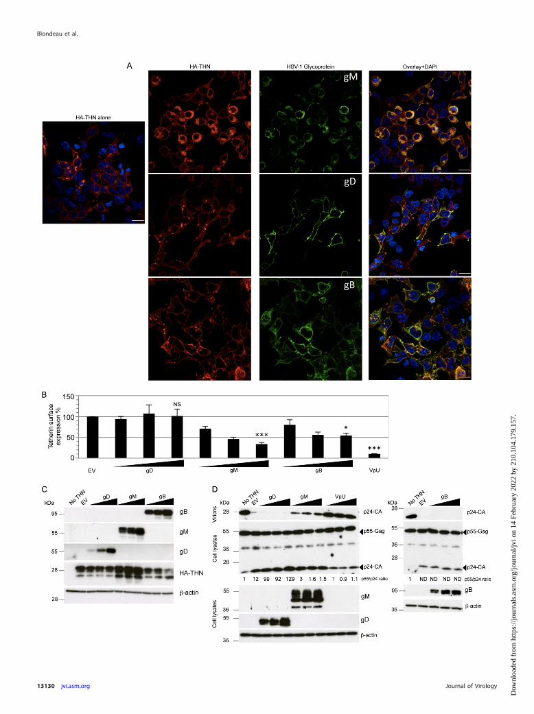
We next asked whether the tetherin antagonism shown by gMfacilitated release of HSV-1 from infected cells. We infected con-trol and tetherin-expressing HT1080 cells with HSV-1 SC16(WT), a gM deletion virus (�gM), or a revertant virus (RgM)using a low input multiplicity (0.01 PFU/cell). Titers of the threeviruses released from control cells were indistinguishable (Fig.6A). However, on cells overexpressing tetherin, while all threeviruses were inhibited, the �gM virus was inhibited by a small butstatistically significant degree over 48 h (2- to 3-fold) (Fig. 6B). Wereasoned that, as in the experiments whose results are shown inFig. 1, tetherin overexpression largely saturated HSV-1 tetherinantagonism, leading to inhibition of all three viruses. Slightlystronger restriction of the �gM virus was likely due to its reducedability to antagonize tetherin. Importantly, there was no signifi-
FIG 3 Tetherin retains HSV-1 particles at the cell surface. HT1080 cells expressing HA-tagged tetherin (A to E) or empty vector (F and G) and infected withHSV-1 K26GFP were examined by electron microscopy. The boxed areas in A, B, C, and F are shown at higher magnification in panels C, D, E, and G, as indicated.N, nucleus. The asterisks mark accumulations of viral nucleocapsids. The bracket marks a layer of viruses at the cell-cell interface in panel B. In panels D, E, andG, selected HSV-1 particles with typical morphology are indicated by the black arrows, while some of the more tangentially cut electron-dense viral profiles aremarked with black arrowheads. Bars, 10 �m (A, B, and E) and 1 �m (C, D, E, and G). (H) Counts of cell surface virus particles from both experiments (Exp) areplotted. Open symbols, particle counts from cells expressing vector; filled symbols, counts from HA-tetherin expressing cells. The number of cells analyzed isindicated at the bottom. Values are means standard deviations; P values were determined using an unpaired t test with Welch’s correction on cells having atleast one viral particle on the cell surface. (Panels A to G show data from experiment 1.)
TABLE 1 HSV-1 particle counting at the cell surface
Expt andcellsa
No. of:
Cell profilesexamined
Infectedcellsb
HSV-1 profilesat the cellsurface
Cell surface HSV-1particles perinfected-cellprofile
1Vector 60 32 419 13.09THN 51 33 1,258 38.12
2Vector 62 20 389 19.45THN 70 27 886 32.81
a THN, HT1080 cells expressing HA-tagged tetherin; Vector, HT1080 cells expressingempty vector.b Cell profiles showing at least one viral particle at the cell surface.
FIG 4 gM relocalizes tetherin to a TGN46-positive compartment. HEK293Tcells expressing HA-tetherin alone or with HSV-1 gM were stained with an-ti-HA (red), anti-gM (A) or TGN46 (B) (green), and DAPI (blue; nuclei). (A)The last panel is an enlargement of the area marked on the Overlay � DAPIpanel. Open arrowheads indicate tetherin predominantly localized to theplasma membrane in cells not expressing or expressing low levels of gM. Solidarrowheads (in cells staining brightly for gM) indicate tetherin localized toperinuclear regions and colocalized with gM. Images are representative of atleast 2 separate experiments. Bars, 20 �m.
Restriction of HSV-1 by Tetherin
December 2013 Volume 87 Number 24 jvi.asm.org 13129
Dow
nloa
ded
from
http
s://j
ourn
als.
asm
.org
/jour
nal/j
vi o
n 14
Feb
ruar
y 20
22 b
y 21
0.10
4.17
9.15
7.
Blondeau et al.
13130 jvi.asm.org Journal of Virology
Dow
nloa
ded
from
http
s://j
ourn
als.
asm
.org
/jour
nal/j
vi o
n 14
Feb
ruar
y 20
22 b
y 21
0.10
4.17
9.15
7.
cant difference between the HT1080 lines in the number ofplaques obtained for any of the viruses for a given dose (Fig. 6C).Absence of gM from �gM-infected cells was confirmed by immu-noblotting for gM protein in infected-cell extracts (Fig. 6D). Fi-nally, we infected HT1080 overexpressing HA-tagged tetherinwith RgM or �gM viruses and analyzed cell lysates by Westernblotting, detecting HA-tetherin, the HSV-1 capsid protein VP5, or�-actin as a loading control, at various time points postinfection(Fig. 6E). As early as 4 to 6 hpi, we observed a reduction of tetherin
protein in infected cells compared to noninfected cells, as previ-ously shown (Fig. 1K). Tetherin was almost completely lost by 10hpi. gM expression was not responsible for loss of tetherin, asindicated by the observation that the �gM virus infection also ledto tetherin loss. These data indicate that while HSV-1 gM can actas a tetherin antagonist, the virus has at least one other anti-teth-erin activity responsible for loss of tetherin protein.
DISCUSSION
Here we extend the repertoire of tetherin-restricted viruses toHSV-1 and the list of tetherin antagonists to include the HSV-1glycoprotein gM. It is striking that glycoproteins are common astetherin antagonists, with anti-tetherin activity being describedfor glycoproteins from the lentiviruses SIVtan and HIV-2 as wellas the filovirus Ebola virus (4, 6, 10). Our data suggest a complexrelationship between tetherin and HSV-1. Tetherin is clearly an-tagonized by HSV-1 in at least two independent ways, by gM-mediated relocalization and through a gM-independent suppres-sion of tetherin mRNA (Fig. 1), likely through the Vhs response(32).
The modest effect of tetherin on HSV-1 replication in our ex-periments may be because HSV-1 is largely insensitive to tetherinrestriction. However, we speculate that gM’s role as a tetherinantagonist may not solely be to improve HSV-1 release. Rather, itspresence within incoming virions might be important for sup-pressing tetherin innate signaling. Tetherin activates innate im-mune signaling cascades via NF-�B, inducing an innate immuneresponse on engagement with virus (36). As a pattern recognitionreceptor, tetherin may be a particularly important target for earlyantagonism before Vhs takes effect. The ability of gM to rescueHIV-1 from tetherin restriction provides good evidence for func-tional tetherin antagonism by gM. Rescue of HIV-1 from tetherinrestriction by gM appears to be more potent than rescue of HSV-1from tetherin (compare Fig. 5, HIV-1, with Fig. 6, HSV-1). Weassume that this is in part because, in the case of HIV-1, all tetherinantagonism is abrogated by deletion of Vpu, and thus tetherinrestriction is maximal and entirely rescued by gM expression.However, in the case of HSV-1, in the absence of gM, tetherin isstill antagonized by Vhs (32) and potentially other, as-yet-unchar-acterized viral functions. Indeed, gB had a minor effect on tetherinlocalization measured by immunofluorescence staining and flowcytometry, although it had no effect in an HIV-1 tethering assay.gD had no measurable effect in any of our assays.
How gM achieves ligand specificity remains unknown, but wenote that HIV-1 Vpu is also promiscuous, removing multiple pro-teins from the cell surface (37–40). It is unclear whether gM ac-
FIG 5 Ability of HSV-1 glycoproteins to remove tetherin from the cell surface and rescue HIV-1 from restriction. HEK293T cells expressing HA-tetherin aloneor with HSV-1 gM, gD, or gB were stained with anti-HA (red) and anti-gM, anti-gD, or anti-gB (green), respectively, and DAPI (blue; nuclei). Images arerepresentative of 2 separate experiments. Bars, 20 �m. (B) Flow-cytometric detection of cell surface tetherin on HEK293T cells expressing HA-tetherin alone(EV) or in combination with 3 different amounts (250, 500, and 1,000 ng) of gM, gD, or gB plasmids. Cells transfected with 1,000 ng of a Vpu plasmid were usedas a control. Mean fluorescence intensities for tetherin are plotted as a percentage of the EV value. Data shown are means of 3 or 4 experiments SEM. Statisticalsignificance was determined using a t test comparing mean fluorescence intensity from cells transfected with 1,000 ng of plasmids (*, P � 0.05; ***, P � 0.001;NS, not significant). (C) Cells transfected for the flow-cytometric assay (B) were used for immunoblotting gM, gD, gB, HA-tetherin, and �-actin, sequentially onthe same membrane after stripping. (D) HEK293T cells were cotransfected with 3 HIV-1 vector plasmids and, in addition, an empty (no THN) or tetherinexpression (EV) vector, or a tetherin expression vector plus 3 different amounts (as in panels B and C) of gM, gD, gB, or Vpu plasmids. p24 capsid (CA) insupernatants (virions) and p55 Gag expression in cell lysates, as well as glycoprotein expression, were analyzed 48 h later by immunoblotting, with �-actin usedas a loading control after membrane stripping. Intracellular-p55/supernatant-p24 ratios are indicated below the p24-stained membranes and are relative to theratio obtained with no tetherin. “ND” indicates that ratios could not be determined due to the absence of any p24 band. The image is representative of at least 3experiments.
FIG 6 gM antagonizes tetherin during HSV-1 infection. Control (A) andHA-tagged-tetherin-expressing (B) HT1080 cells were infected with HSV-1SC16 (WT), a gM deletion virus (�gM), or a revertant virus (RgM) at 0.01PFU/cell, and supernatants harvested at the indicated times postinfection weretitrated for HSV-1 infectivity by plaque assay. Two-way ANOVA of 3 separatetetherin expression experiments confirmed statistically significant differencesattributable to time and the presence of gM (P � 0.01). (C) HSV-1 SC16 WT,�gM, and RgM were plaque assayed on cell lines used for panels A and B.Results are means and SEM and are from 2 independent experiments. (D)Immunoblot detection of ICP4, gM, and �-actin in lysates of HT1080 cellsinfected with the 3 viruses or noninfected (NI). (E) Immunoblot detection ofVP5 and HA tag (tetherin), or �-actin after membrane stripping, in lysates ofHA-tagged tetherin-expressing HT1080 cells infected with �gM (�) or RgM(R) virus at 2 PFU/cell or noninfected (�). Results are representative of 3separate experiments.
Restriction of HSV-1 by Tetherin
December 2013 Volume 87 Number 24 jvi.asm.org 13131
Dow
nloa
ded
from
http
s://j
ourn
als.
asm
.org
/jour
nal/j
vi o
n 14
Feb
ruar
y 20
22 b
y 21
0.10
4.17
9.15
7.

tively removes tetherin or whether, like HIV-1 Vpu, gM preventstetherin from reaching the cell surface (41, 42). Regardless, theresult is accumulation of tetherin, detected by immunoblotting(Fig. 5C) and the presence of tetherin in a compartment positivefor TGN46 staining (Fig. 4B). Notably, HIV-2 Env also relocalizestetherin to a TGN46 positive compartment without degrading it(6). Thus, it appears that, in common with other viral tetherincountermeasures, gM acts by removing tetherin from the site ofvirus budding, in this case TGN46-negative endocytic tubules(33). We envisage the interaction between tetherin and outgoingHSV-1 virions occurring in this compartment, and the relocaliza-tion of tetherin we observed by immunofluorescence (Fig. 4B)provides a plausible basis for the anti-tetherin activity of gM. Wedid not see any chains of tethered virions similar to those formedby tetherin-restricted lentiviruses (2). This may reflect spatial con-straints imposed by budding into tubules. Indeed, we assume thatnewly budded tethered virions would not be readily apparent byelectron microscopy because they would simply be restrained atthe location in which they normally reside. However, followingrelease, virions would remain tethered to the cell surface, as weobserved (Fig. 3). This phenomenon may provide some explana-tion as to how tetherin restricts release of HSV-1 and not directcell-to-cell spread, reflected by the lack of impact on plaque size.We assume that surface-tethered virions may be able to interactwith target cell receptors to initiate infection despite being teth-ered to the infected cell membrane. Such a process has been de-scribed for the lentiviruses HIV-1 and FIV (29, 30).
Redundant herpesvirus-encoded tetherin antagonists are alsofound in KSHV (15, 16, 43). KSHV encodes the E3 ubiquitin ligaseK5, which recruits tetherin to cause its degradation yet can stillantagonize tetherin after K5 depletion with RNA interference(16). Thus, large viruses, such as herpesviruses, may encode sev-eral partially redundant tetherin antagonists that have subtly dif-ferent roles in restriction factor antagonism. The complex rela-tionship between herpesviruses and tetherin may also explain thepuzzling result that while HSV-1 and KSHV are restricted by teth-erin, this protein has been reported to act as a cofactor for HCMVreplication (15–17). Tetherin expression improves HCMV infec-tivity a few-fold, perhaps related to its ability to stabilize lipid rafts,which play a key role in HCMV entry (44, 45). We did not observean equivalent enhancement with HSV-1 (Fig. 1 and 2), and itremains unclear whether HCMV also encodes proteins that ma-nipulate tetherin to prevent it from restricting the virus.
The increasing variety of viruses that are restricted by tetherinillustrates the power of a restriction factor that targets the funda-mental processes of viral budding and release. The diversity oftetherin antagonists that viruses have evolved also emphasizes theimportance of overcoming this system of host defense. It is likelythat the study of the ongoing evolutionary conflict between vi-ruses and tetherin, which is suggested by the Red Queen hypoth-esis (46), will lead to significant enhancements to our understand-ing of the cell biology of both viruses and their hosts and therelationships between them.
ACKNOWLEDGMENTS
We are grateful to Helena Browne, Gary Cohen, Roselyn Eisenberg, Gil-lian Elliott, Roger Everett, Stuart Neil, Claire Pardieu, Sam Wilson, andBen Webb for reagents and Mahdad Noursadeghi for help with statisticalanalyses.
This work was funded by Wellcome Trust Senior Fellowship 090940 to
G.J.T., the UK Medical Research Council, and The National Institute forHealth Research UCL/UCLH Biomedical Research Centre.
REFERENCES1. Malim MH, and Bieniasz PD. 2012. HIV restriction factors and mecha-
nisms of evasion. Cold Spring Harb. Perspect. Med. 2:a006940. doi:10.1101/cshperspect.a006940.
2. Neil SJ, Zang T, Bieniasz PD. 2008. Tetherin inhibits retrovirus releaseand is antagonized by HIV-1 Vpu. Nature 451:425– 430.
3. Van Damme N, Goff D, Katsura C, Jorgenson RL, Mitchell R, JohnsonMC, Stephens EB, Guatelli J. 2008. The interferon-induced proteinBST-2 restricts HIV-1 release and is downregulated from the cell surfaceby the viral Vpu protein. Cell Host Microbe 3:245–252.
4. Gupta RK, Mlcochova P, Pelchen-Matthews A, Petit SJ, Mattiuzzo G,Pillay D, Takeuchi Y, Marsh M, Towers GJ. 2009. Simian immunode-ficiency virus envelope glycoprotein counteracts tetherin/BST-2/CD317by intracellular sequestration. Proc. Natl. Acad. Sci. U. S. A. 106:20889 –20894.
5. Jia B, Serra-Moreno R, Neidermyer W, Rahmberg A, Mackey J, FofanaIB, Johnson WE, Westmoreland S, Evans DT. 2009. Species-specificactivity of SIV Nef and HIV-1 Vpu in overcoming restriction by tetherin/BST2. PLoS Pathog. 5:e1000429. doi:10.1371/journal.ppat.1000429.
6. Le Tortorec A, Neil SJ. 2009. Antagonism to and intracellular sequestra-tion of human tetherin by the human immunodeficiency virus type 2envelope glycoprotein. J. Virol. 83:11966 –11978.
7. Sauter D, Schindler M, Specht A, Landford WN, Munch J, Kim KA,Votteler J, Schubert U, Bibollet-Ruche F, Keele BF, Takehisa J, OgandoY, Ochsenbauer C, Kappes JC, Ayouba A, Peeters M, Learn GH, ShawG, Sharp PM, Bieniasz P, Hahn BH, Hatziioannou T, Kirchhoff F. 2009.Tetherin-driven adaptation of Vpu and Nef function and the evolution ofpandemic and nonpandemic HIV-1 strains. Cell Host Microbe 6:409 –421.
8. Zhang F, Wilson SJ, Landford WC, Virgen B, Gregory D, Johnson MC,Munch J, Kirchhoff F, Bieniasz PD, Hatziioannou T. 2009. Nef proteinsfrom simian immunodeficiency viruses are tetherin antagonists. Cell HostMicrobe 6:54 – 67.
9. Bruce EA, Abbink TE, Wise HM, Rollason R, Galao RP, Banting G, NeilSJ, Digard P. 2012. Release of filamentous and spherical influenza A virusis not restricted by tetherin. J. Gen. Virol. 93:963–969.
10. Kaletsky RL, Francica JR, Agrawal-Gamse C, Bates P. 2009. Tetherin-mediated restriction of filovirus budding is antagonized by the Ebola gly-coprotein. Proc. Natl. Acad. Sci. U. S. A. 106:2886 –2891.
11. Sakuma T, Noda T, Urata S, Kawaoka Y, Yasuda J. 2009. Inhibition ofLassa and Marburg virus production by tetherin. J. Virol. 83:2382–2385.
12. Ye L, Wang X, Li J, Liu J, Ramirez SH, Wu J, Ho W. 2012. Tetherin hasnegligible activity in restricting hepatitis C virus in hepatocytes. InnateImmun. 18:398 – 405.
13. Perez-Caballero D, Zang T, Ebrahimi A, McNatt MW, Gregory DA,Johnson MC, Bieniasz PD. 2009. Tetherin inhibits HIV-1 release bydirectly tethering virions to cells. Cell 139:499 –511.
14. McGeoch DJ, Cook S, Dolan A, Jamieson FE, Telford EA. 1995. Mo-lecular phylogeny and evolutionary timescale for the family of mamma-lian herpesviruses. J. Mol. Biol. 247:443– 458.
15. Mansouri M, Viswanathan K, Douglas JL, Hines J, Gustin J, Moses AV,Fruh K. 2009. Molecular mechanism of BST2/tetherin downregulation byK5/MIR2 of Kaposi’s sarcoma-associated herpesvirus. J. Virol. 83:9672–9681.
16. Pardieu C, Vigan R, Wilson SJ, Calvi A, Zang T, Bieniasz P, Kellam P,Towers GJ, Neil SJ. 2010. The RING-CH ligase K5 antagonizes restrictionof KSHV and HIV-1 particle release by mediating ubiquitin-dependentendosomal degradation of tetherin. PLoS Pathog. 6:e1000843. doi:10.1371/journal.ppat.1000843.
17. Viswanathan K, Smith MS, Malouli D, Mansouri M, Nelson JA, Fruh K.2011. BST2/tetherin enhances entry of human cytomegalovirus. PLoS Pat-hog. 7:e1002332. doi:10.1371/journal.ppat.1002332.
18. McNatt MW, Zang T, Hatziioannou T, Bartlett M, Fofana IB, JohnsonWE, Neil SJ, Bieniasz PD. 2009. Species-specific activity of HIV-1 Vpuand positive selection of tetherin transmembrane domain variants. PLoSPathog. 5:e1000300. doi:10.1371/journal.ppat.1000300.
19. Connolly SA, Landsburg DJ, Carfi A, Wiley DC, Cohen GH, EisenbergRJ. 2003. Structure-based mutagenesis of herpes simplex virus glycopro-
Blondeau et al.
13132 jvi.asm.org Journal of Virology
Dow
nloa
ded
from
http
s://j
ourn
als.
asm
.org
/jour
nal/j
vi o
n 14
Feb
ruar
y 20
22 b
y 21
0.10
4.17
9.15
7.
tein D defines three critical regions at the gD-HveA/HVEM binding inter-face. J. Virol. 77:8127– 8140.
20. Muggeridge MI. 2000. Characterization of cell-cell fusion mediated byherpes simplex virus 2 glycoproteins gB, gD, gH and gL in transfected cells.J. Gen. Virol. 81:2017–2027.
21. Petit SJ, Blondeau C, Towers GJ. 2011. Analysis of the human immuno-deficiency virus type 1 M group Vpu domains involved in antagonizingtetherin. J. Gen. Virol. 92:2937–2948.
22. Desai P, Person S. 1998. Incorporation of the green fluorescent proteininto the herpes simplex virus type 1 capsid. J. Virol. 72:7563–7568.
23. Ren Y, Bell S, Zenner HL, Lau SY, Crump CM. 2012. Glycoprotein M isimportant for the efficient incorporation of glycoprotein H-L into herpessimplex virus type 1 particles. J. Gen. Virol. 93:319 –329.
24. Wilson SJ, Tsao EH, Webb BL, Ye H, Dalton-Griffin L, Tsantoulas C,Gale CV, Du MQ, Whitehouse A, Kellam P. 2007. X box binding proteinXBP-1s transactivates the Kaposi’s sarcoma-associated herpesvirus(KSHV) ORF50 promoter, linking plasma cell differentiation to KSHVreactivation from latency. J. Virol. 81:13578 –13586.
25. Jerome KR, Huang ML, Wald A, Selke S, Corey L. 2002. Quantitativestability of DNA after extended storage of clinical specimens as deter-mined by real-time PCR. J. Clin. Microbiol. 40:2609 –2611.
26. Isola VJ, Eisenberg RJ, Siebert GR, Heilman CJ, Wilcox WC, CohenGH. 1989. Fine mapping of antigenic site II of herpes simplex virus gly-coprotein D. J. Virol. 63:2325–2334.
27. Gupta RK, Hue S, Schaller T, Verschoor E, Pillay D, Towers GJ. 2009.Mutation of a single residue renders human tetherin resistant to HIV-1Vpu-mediated depletion. PLoS Pathog. 5:e1000443. doi:10.1371/journal.ppat.1000443.
28. Baines JD, Wills E, Jacob RJ, Pennington J, Roizman B. 2007. Glyco-protein M of herpes simplex virus 1 is incorporated into virions duringbudding at the inner nuclear membrane. J. Virol. 81:800 – 812.
29. Dietrich I, McMonagle EL, Petit S, Vijayakrishnan S, Logan N, ChanCN, Towers GJ, Hosie MJ, Willett BJ. 2011. Feline tetherin (BST-2)efficiently restricts feline immunodeficiency virus release but not spread-ing infection. J. Virol. 85:5840 –5852.
30. Jolly C, Booth NJ, Neil SJ. 2010. Cell-cell spread of human immunode-ficiency virus type 1 overcomes tetherin/BST-2-mediated restriction in Tcells. J. Virol. 84:12185–12199.
31. Andrew AJ, Miyagi E, Kao S, Strebel K. 2009. The formation of cysteine-linked dimers of BST-2/tetherin is important for inhibition of HIV-1 virusrelease but not for sensitivity to Vpu. Retrovirology 6:80.
32. Zenner HL, Mauricio R, Banting G, Crump CM. 2013. Herpes simplexvirus 1 counteracts tetherin restriction via its virion host shutoff activity. J.Virol. 87:13115–13123.
33. Hollinshead M, Johns HL, Sayers CL, Gonzalez-Lopez C, Smith GL,
Elliott G. 2012. Endocytic tubules regulated by Rab GTPases 5 and 11 areused for envelopment of herpes simplex virus. EMBO J. 31:4204 – 4220.
34. Crump CM, Bruun B, Bell S, Pomeranz LE, Minson T, Browne HM.2004. Alphaherpesvirus glycoprotein M causes the relocalization ofplasma membrane proteins. J. Gen. Virol. 85:3517–3527.
35. MacLean CA, Robertson LM, Jamieson FE. 1993. Characterization of theUL10 gene product of herpes simplex virus type 1 and investigation of itsrole in vivo. J. Gen. Virol. 74:975–983.
36. Galao RP, Le Tortorec A, Pickering S, Kueck T, Neil SJ. 2012. Innatesensing of HIV-1 assembly by tetherin induces NFkappaB-dependent pro-inflammatory responses. Cell Host Microbe 12:633– 644.
37. Bour S, Schubert U, Strebel K. 1995. The human immunodeficiencyvirus type 1 Vpu protein specifically binds to the cytoplasmic domain ofCD4: implications for the mechanism of degradation. J. Virol. 69:1510 –1520.
38. Hussain A, Wesley C, Khalid M, Chaudhry A, Jameel S. 2008. Humanimmunodeficiency virus type 1 Vpu protein interacts with CD74 andmodulates major histocompatibility complex class II presentation. J. Vi-rol. 82:893–902.
39. Moll M, Andersson SK, Smed-Sorensen A, Sandberg JK. 2010. Inhibi-tion of lipid antigen presentation in dendritic cells by HIV-1 Vpu inter-ference with CD1d recycling from endosomal compartments. Blood 116:1876 –1884.
40. Willey RL, Maldarelli F, Martin MA, Strebel K. 1992. Human immu-nodeficiency virus type 1 Vpu protein induces rapid degradation of CD4.J. Virol. 66:7193–7200.
41. Dube M, Roy BB, Guiot-Guillain P, Binette J, Mercier J, Chiasson A,Cohen EA. 2010. Antagonism of tetherin restriction of HIV-1 release byVpu involves binding and sequestration of the restriction factor in a peri-nuclear compartment. PLoS Pathog. 6:e1000856. doi:10.1371/journal.ppat.1000856.
42. Schmidt S, Fritz JV, Bitzegeio J, Fackler OT, Keppler OT. 2011. HIV-1Vpu blocks recycling and biosynthetic transport of the intrinsic immunityfactor CD317/tetherin to overcome the virion release restriction. mBio2:e00036 –11. doi:10.1128/mBio.00036-11.
43. Bartee E, McCormack A, Fruh K. 2006. Quantitative membrane pro-teomics reveals new cellular targets of viral immune modulators. PLoSPathog. 2:e107. doi:10.1371/journal.ppat.0020107.
44. Juckem LK, Boehme KW, Feire AL, Compton T. 2008. Differentialinitiation of innate immune responses induced by human cytomegalovi-rus entry into fibroblast cells. J. Immunol. 180:4965– 4977.
45. Wang X, Huang DY, Huong SM, Huang ES. 2005. Integrin alphavbeta3is a coreceptor for human cytomegalovirus. Nat. Med. 11:515–521.
46. Van Valen L. 1973. A new evolutionary law. Evol. Theory 1:1–30.
Restriction of HSV-1 by Tetherin
December 2013 Volume 87 Number 24 jvi.asm.org 13133
Dow
nloa
ded
from
http
s://j
ourn
als.
asm
.org
/jour
nal/j
vi o
n 14
Feb
ruar
y 20
22 b
y 21
0.10
4.17
9.15
7.
Related Documents