THE JOURNAL OF CELL BIOLOGY JCB: ARTICLE © The Rockefeller University Press $8.00 The Journal of Cell Biology, Vol. 174, No. 3, July 31, 2006 459–471 http://www.jcb.org/cgi/doi/10.1083/jcb.200508170 JCB 459 Introduction Endocytosis is essential for a variety of cellular functions, includ- ing the internalization of nutrients and communication among cells, or between cells and their environment. Internalized molecules must be precisely sorted to their final cellular desti- nations to fulfill their specific function. Distinct endocytic path- ways have been described to date, including clathrin-dependent endocytosis and caveolae-mediated uptake, which remain the two best-characterized mechanisms of internalization (Conner et al., 2003). Neurons have adapted their endocytic pathways to better adjust to their specific requirements. Thus, synaptic vesicle (SV) recycling is the predominant form of neuronal en- docytosis at the presynaptic terminal, whereby the fast fusion of neurotransmitter-containing vesicles is coordinated with an efficient mechanism of membrane recovery, which involves clathrin (for review see Murthy and De Camilli, 2003). In neu- rons, clathrin-independent routes have also been documented, although the physiological relevance of endocytosis via caveo- lae has been questioned in these cells because several of the caveolin isoforms found in other tissues are not detectable. A special feature of motor neurons (MNs) is that their presynaptic terminal, which forms the neuromuscular junction (NMJ), is located in the periphery, whereas the soma is located in the central nervous system. Therefore, any material that enters the MNs at the NMJ and is transported toward the cell body, such as neurotrophins, crosses the blood–brain barrier. To gain more insights into the endocytic events at the NMJ, we followed the endocytosis of tetanus neurotoxin (TeNT). TeNT is a neurospecific toxin that binds to MNs at the NMJ, where it is internalized and undergoes axonal retrograde transport to the cell body. It is then secreted and taken up by adjacent inhibi- tory interneurons, where it blocks neurotransmitter release by cleaving VAMP/synaptobrevin, which is a synaptic SNARE (Lalli et al., 2003a). Tetanus toxin is internalized by a sequential clathrin-dependent mechanism initiated within lipid microdomains and independent of epsin1 Katrin Deinhardt, 1 Otto Berninghausen, 2 Hugh J. Willison, 3 Colin R. Hopkins, 2 and Giampietro Schiavo 1 1 Molecular Neuropathobiology Laboratory, Cancer Research UK London Research Institute, London WC2A 3PX, England, UK 2 Department of Biological Sciences, Imperial College London, London SW7 2AZ, England, UK 3 Division of Clinical Neurosciences, Southern General Hospital, Glasgow G51 4TF, Scotland, UK L igand–receptor complexes are internalized by a va- riety of endocytic mechanisms. Some are initiated within clathrin-coated membranes, whereas others involve lipid microdomains of the plasma membrane. In neurons, where alternative targeting to short- or long- range trafficking routes underpins the differential pro- cessing of synaptic vesicle components and neurotrophin receptors, the mechanism giving access to the axonal retrograde pathway remains unknown. To investigate this sorting process, we examined the internalization of a tetanus neurotoxin fragment (TeNT H C ), which shares axonal carriers with neurotrophins and their receptors. Previous studies have shown that the TeNT H C receptor, which comprises polysialogangliosides, resides in lipid microdomains. We demonstrate that TeNT H C internaliza- tion also relies on a specialized clathrin-mediated path- way, which is independent of synaptic vesicle recycling. Moreover, unlike transferrin uptake, this AP-2–dependent process is independent of epsin1. These findings identify a pathway for TeNT, beginning with the binding to a lipid raft component (GD1b) and followed by dissociation from GD1b as the toxin internalizes via a clathrin-mediated mechanism using a specific subset of adaptor proteins. K. Deinhardt and O. Berninghausen contributed equally to this work. Correspondence to Giampietro Schiavo: [email protected] Abbreviations used in this paper: BoNT, botulinum neurotoxin; CCP, clathrin- coated pit; CCV, clathrin-coated vesicle; CHC, clathrin heavy chain; CLC, clath- rin light chain; CTB, cholera toxin subunit B; DRM, detergent-resistant membrane; GPI, glycosylphosphatidylinositol; MESNA, 2-mercaptoethane sulfonic acid; MN, motor neuron; NMJ, neuromuscular junction; SV, synaptic vesicles; TeNT H C , tetanus neurotoxin fragment. The online version of this article contains supplemental material. on August 1, 2006 www.jcb.org Downloaded from http://www.jcb.org/cgi/content/full/jcb.200508170/DC1 Supplemental Material can be found at:

Welcome message from author
This document is posted to help you gain knowledge. Please leave a comment to let me know what you think about it! Share it to your friends and learn new things together.
Transcript
TH
EJ
OU
RN
AL
OF
CE
LL
BIO
LO
GY
JCB: ARTICLE
© The Rockefeller University Press $8.00The Journal of Cell Biology, Vol. 174, No. 3, July 31, 2006 459–471http://www.jcb.org/cgi/doi/10.1083/jcb.200508170
JCB 459
IntroductionEndocytosis is essential for a variety of cellular functions, includ-
ing the internalization of nutrients and communication among
cells, or between cells and their environment. Internalized
molecules must be precisely sorted to their fi nal cellular desti-
nations to fulfi ll their specifi c function. Distinct endocytic path-
ways have been described to date, including clathrin- dependent
endocytosis and caveolae-mediated uptake, which remain the
two best-characterized mechanisms of internalization (Conner
et al., 2003). Neurons have adapted their endocytic pathways
to better adjust to their specifi c requirements. Thus, synaptic
vesicle (SV) recycling is the predominant form of neuronal en-
docytosis at the presynaptic terminal, whereby the fast fusion
of neurotransmitter-containing vesicles is coordinated with an
effi cient mechanism of membrane recovery, which involves
clathrin (for review see Murthy and De Camilli, 2003). In neu-
rons, clathrin-independent routes have also been documented,
although the physiological relevance of endocytosis via caveo-
lae has been questioned in these cells because several of the
caveolin isoforms found in other tissues are not detectable.
A special feature of motor neurons (MNs) is that their
presynaptic terminal, which forms the neuromuscular junction
(NMJ), is located in the periphery, whereas the soma is located
in the central nervous system. Therefore, any material that
enters the MNs at the NMJ and is transported toward the cell
body, such as neurotrophins, crosses the blood–brain barrier.
To gain more insights into the endocytic events at the NMJ, we
followed the endocytosis of tetanus neurotoxin (TeNT). TeNT
is a neurospecifi c toxin that binds to MNs at the NMJ, where it
is internalized and undergoes axonal retrograde transport to
the cell body. It is then secreted and taken up by adjacent inhibi-
tory interneurons, where it blocks neurotransmitter release by
cleaving VAMP/synaptobrevin, which is a synaptic SNARE
(Lalli et al., 2003a).
Tetanus toxin is internalized by a sequential clathrin-dependent mechanism initiated within lipid microdomains and independent of epsin1
Katrin Deinhardt,1 Otto Berninghausen,2 Hugh J. Willison,3 Colin R. Hopkins,2 and Giampietro Schiavo1
1Molecular Neuropathobiology Laboratory, Cancer Research UK London Research Institute, London WC2A 3PX, England, UK2Department of Biological Sciences, Imperial College London, London SW7 2AZ, England, UK3Division of Clinical Neurosciences, Southern General Hospital, Glasgow G51 4TF, Scotland, UK
Ligand–receptor complexes are internalized by a va-
riety of endocytic mechanisms. Some are initiated
within clathrin-coated membranes, whereas others
involve lipid microdomains of the plasma membrane. In
neurons, where alternative targeting to short- or long-
range traffi cking routes underpins the differential pro-
cessing of synaptic vesicle components and neurotrophin
receptors, the mechanism giving access to the axonal
retrograde pathway remains unknown. To investigate
this sorting process, we examined the internalization of
a tetanus neurotoxin fragment (TeNT HC), which shares
axonal carriers with neurotrophins and their receptors.
Previous studies have shown that the TeNT HC receptor,
which comprises polysialogangliosides, resides in lipid
microdomains. We demonstrate that TeNT HC internaliza-
tion also relies on a specialized clathrin-mediated path-
way, which is independent of synaptic vesicle recycling.
Moreover, unlike transferrin uptake, this AP-2–dependent
process is independent of epsin1. These fi ndings identify
a pathway for TeNT, beginning with the binding to a lipid
raft component (GD1b) and followed by dissociation from
GD1b as the toxin internalizes via a clathrin-mediated
mechanism using a specifi c subset of adaptor proteins.
K. Deinhardt and O. Berninghausen contributed equally to this work.
Correspondence to Giampietro Schiavo: [email protected]
Abbreviations used in this paper: BoNT, botulinum neurotoxin; CCP, clathrin-coated pit; CCV, clathrin-coated vesicle; CHC, clathrin heavy chain; CLC, clath-rin light chain; CTB, cholera toxin subunit B; DRM, detergent-resistant membrane; GPI, glycosylphosphatidylinositol; MESNA, 2-mercaptoethane sulfonic acid; MN, motor neuron; NMJ, neuromuscular junction; SV, synaptic vesicles; TeNT HC, tetanus neurotoxin fragment.
The online version of this article contains supplemental material.
on August 1, 2006
ww
w.jcb.org
Dow
nloaded from
http://www.jcb.org/cgi/content/full/jcb.200508170/DC1Supplemental Material can be found at:

JCB • VOLUME 174 • NUMBER 3 • 2006 460
The TeNT receptor complex has been shown to comprise
lipids and proteins (Montecucco et al., 2004). The polysialo-
gangliosides GD1b and GT1b (Habermann and Dreyer, 1986;
Lalli et al., 2003a), as well as one or more glycosylphospha-
tidylinositol (GPI)-anchored proteins (Herreros et al., 2001;
Munro et al., 2001) are required for toxin binding to the neu-
ronal surface. TeNT is associated with detergent-resistant
membranes (DRMs), which are enriched in cholesterol and
GPI-anchored proteins (Herreros et al., 2001), and its uptake
is sensitive to cholesterol depletion (Herreros et al., 2001).
Furthermore, pretreatment of neurons with phosphatidylinosi-
tol-specifi c phospholipase C to cleave GPI-anchored proteins
from their lipid anchor prevents TeNT intoxication (Munro
et al., 2001). Altogether, these fi ndings suggest that TeNT
follows a polysialoganglioside- and DRM-dependent route
for its internalization in neuronal cells. However, in previous
EM studies on spinal cord neurons, gold-labeled TeNT was
detected in surface pits resembling clathrin-coated invagina-
tions, as well as in coated and uncoated vesicles (Parton et al.,
1987; Lalli et al., 2003b). Because clathrin-mediated internal-
ization and the endocytosis of proteins associated with DRMs
have been largely viewed as mutually exclusive (Parton and
Richards, 2003), the association of TeNT with clathrin coats
was unexpected.
To resolve this apparent paradox, we have studied the in-
ternalization machinery responsible for the uptake of TeNT into
MNs using a C-terminal binding fragment of TeNT (Lalli et al.,
2003a). In this study, we show that TeNT HC endocytosis in
MNs is independent of SV recycling, the major route of inter-
nalization at the presynaptic terminal, and demonstrate that al-
though TeNT HC binds to DRMs on the MN surface, it uses
a clathrin-mediated pathway for its entry. This specialized
clathrin- and AP-2–dependent uptake mechanism does not
require the endocytic adaptor protein epsin1, further indicating
that specifi c adaptors play important functions in initial sorting
events during endocytosis.
ResultsTeNT HC internalization in MNs is independent of presynaptic activityAlthough previous studies implied that TeNT does not enter the
NMJ via SV endocytosis (Habermann and Dreyer, 1986), some
studies suggested that the toxin can take this route in brain-
derived neurons, such as hippocampal neurons (Matteoli et al.,
1996) and that it may enter SV-like vesicles in spinal cord neu-
rons in culture (Parton et al., 1987). In light of these fi ndings,
we assessed whether SV exo/endocytosis is the physiological
route of TeNT entry in MNs. Several lines of evidence indicate
that this is not the case. First, we tested the colocalization of in-
ternalized TeNT HC and the SV protein VAMP-2. MNs were
incubated with Alexa Fluor 555–TeNT HC at 37°C, fi xed, and
stained for VAMP-2. Under resting conditions, colocalization in
the cell body, neurites, or synaptic contacts was very limited
(Fig. 1, A and B). Moreover, stimulation of SV exo/endocytosis
by depolarization did not increase the extent of colocalization
(Fig. 1, A and B).
To further investigate the endocytic pathway of TeNT,
we used a biotinylated, thiol-cleavable form of TeNT HC
(b-TeNT HC). By exposing intact neurons to cell-impermeable
reducing reagents, such as 2-mercaptoethane sulfonic acid
(MESNA; Schmid and Smythe, 1991), biotin can be cleaved off
surface-bound TeNT HC, while the internalized b-TeNT HC is
protected. Staining for the remaining biotin allows the internal-
ized TeNT HC to be detected selectively over the surface-bound
TeNT HC even in thin structures such as axons. Biotinylation
does not change the binding and internalization properties of
TeNT HC because preincubation with a 100-fold excess of un-
labeled toxin completely abolished the binding of b-TeNT HC
to MNs (Fig. S1 A, available at http://www.jcb.org/cgi/content/
full/jcb.200508170/DC1). Furthermore, under internalization
Figure 1. TeNT HC internalization is independent of SV exocytosis and recycling. (A) MNs were incubated with 20 nM Alexa Fluor 555–TeNT HC for 30 min at 37°C, either under resting conditions (a–d) or after stimulation of SV exo/endocytosis by adding 60 mM KCl to the medium just before TeNT HC addition (e–h), fi xed, and stained for VAMP-2. Only very limited colocaliza-tion of TeNT HC and VAMP-2 under resting or stimulated conditions, which were quantifi ed in B, was found. Error bars represent the SEM. (C and D) MNs were incubated with 15 nM BoNT/A and 2 nM BoNT/D for 22 h at 37°C to cleave SNAP-25 and VAMP-2. Untreated cells were processed in parallel for com-parison. (C) Cells were scraped and analyzed by Western blotting using anti-bodies raised against the cleaved fragments of SNAP-25 and VAMP-2, as well as actin, as a loading control. (D) 20 nM b-TeNT HC was added to MNs for 30 min at 37°C. MNs were shifted to ice, treated with MESNA before fi xation, and stained for VAMP-2 (a and e), SNAP-25 (b and f), and biotin (c and g). Pretreatment with BoNTs did not affect TeNT HC internalization. DIC, differential interference contrast. Bars: (A) 5 μm; (D) 10 μm.
on August 1, 2006
ww
w.jcb.org
Dow
nloaded from
UPTAKE OF TETANUS TOXIN IS CLATHRIN DEPENDENT • DEINHARDT ET AL. 461
conditions, b-TeNT HC colocalized with Alexa Fluor 555–TeNT
HC (Fig. S1 B). Importantly, biotin could be cleaved off the
surface-bound b-TeNT HC by treatment with MESNA on ice.
In contrast, in cells incubated at 37°C the label remained cell-
associated, showing that b-TeNT HC had been taken up in the
soma and neurites (Fig. S1 C).
For an independent assessment of a role for SV recy-
cling in TeNT HC uptake, we then preincubated MNs with
botulinum neurotoxin (BoNT)/A and D for 22 h to block SV
exocytosis, and, thus, recycling (Humeau et al., 2000). The
samples were then incubated with b-TeNT HC for 30 min at
37°C, treated with MESNA on ice, fi xed, and stained for
VAMP-2 and SNAP-25, as well as for biotin, to visualize in-
ternalized TeNT HC. The complete cleavage of SNAP-25 and
VAMP-2 by BoNT/A and D was confi rmed by Western blot-
ting (Fig. 1 C) and by indirect immunofl uorescence (Fig. 1 D),
indicating that SV exo/endocytosis was blocked under these
conditions. However, TeNT HC internalization was not af-
fected in intoxicated MNs (Fig. 1 D, g) compared with un-
treated cells (Fig. 1 D, c).
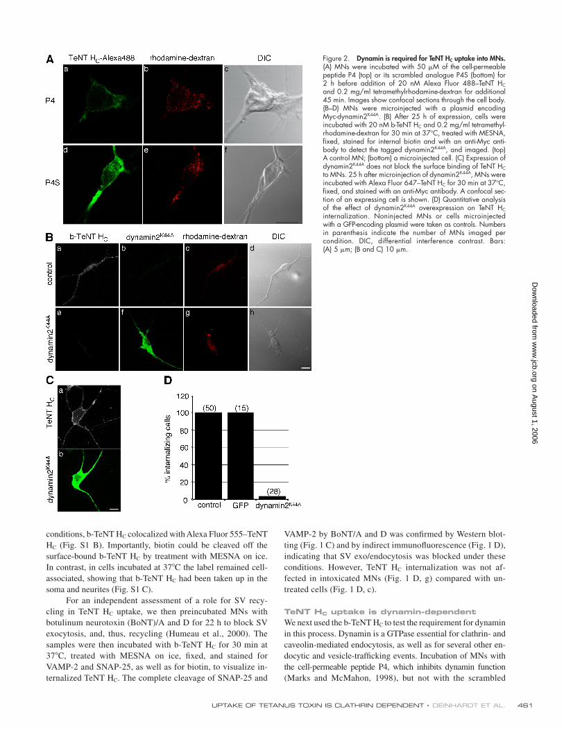
TeNT HC uptake is dynamin-dependentWe next used the b-TeNT HC to test the requirement for dynamin
in this process. Dynamin is a GTPase essential for clathrin- and
caveolin-mediated endocytosis, as well as for several other en-
docytic and vesicle-traffi cking events. Incubation of MNs with
the cell-permeable peptide P4, which inhibits dynamin function
(Marks and McMahon, 1998), but not with the scrambled
Figure 2. Dynamin is required for TeNT HC uptake into MNs. (A) MNs were incubated with 50 μM of the cell- permeable peptide P4 (top) or its scrambled analogue P4S (bottom) for 2 h before addition of 20 nM Alexa Fluor 488–TeNT HC and 0.2 mg/ml tetramethylrhodamine- dextran for additional 45 min. Images show confocal sections through the cell body. (B–D) MNs were microinjected with a plasmid encoding Myc-dynamin2K44A. (B) After 25 h of expression, cells were incubated with 20 nM b-TeNT HC and 0.2 mg/ml tetramethyl-rhodamine-dextran for 30 min at 37°C, treated with MESNA, fi xed, stained for internal biotin and with an anti-Myc anti-body to detect the tagged dynamin2K44A, and imaged. (top) A control MN; (bottom) a microinjected cell. (C) Expression of dynamin2K44A does not block the surface binding of TeNT HC to MNs. 25 h after microinjection of dynamin2K44A, MNs were incubated with Alexa Fluor 647–TeNT HC for 30 min at 37°C, fi xed, and stained with an anti-Myc antibody. A confocal sec-tion of an expressing cell is shown. (D) Quantitative analysis of the effect of dynamin2K44A overexpression on TeNT HC internalization. Noninjected MNs or cells microinjected with a GFP-encoding plasmid were taken as controls. Numbers in parenthesis indicate the number of MNs imaged per condition. DIC, differential interference contrast. Bars: (A) 5 μm; (B and C) 10 μm.
on August 1, 2006
ww
w.jcb.org
Dow
nloaded from

JCB • VOLUME 174 • NUMBER 3 • 2006 462
peptide P4S, signifi cantly reduced uptake of TeNT HC, whereas
its overall binding to the neuronal surface was not affected
(Fig. 2 A). These results were confi rmed by overexpressing the
well-characterized dynamin mutant K44A, which is defective
in GTP binding and hydrolysis and restrains invaginated pits
from pinching off (Fig. 2 B; Damke et al., 1994). We used mi-
croinjection to introduce foreign DNA into MNs because lipid-
based transfection reagents abolished axonal transport in MNs.
In contrast, microinjection of plasmids driving the overexpres-
sion of control proteins had no effect on cell viability and ret-
rograde transport (Deinhardt and Schiavo, 2005). Expression of
dynamin2K44A signifi cantly reduced TeNT HC endocytosis at the
level of both the soma and neurites (Fig. 2 B), without affecting
its binding to the MN surface (Fig. 2 C). These results indicate
that dynamin is a central player in TeNT HC internalization and
rule out differences in the mechanism of uptake of TeNT HC
between axons and the soma. This is important because, topo-
logically, only the axon is physiologically relevant for TeNT
HC uptake and retrograde transport. A total block of TeNT HC
endocytosis by the expression of dynamin2K44A was seen in
>95% of the cells (Fig. 2 D). However, dextran internalization
still took place under these conditions (Fig. 2 B, g) or upon P4
treatment (Fig. 2 A, b), confi rming that MNs were viable and
still capable of endocytosis via dynamin-independent pathways.
We chose dextran as a control marker for internalization
because we found that cholera toxin subunit B (CTB), which
is another widely used marker for clathrin-independent endo-
cytosis, is internalized in a strictly dynamin-dependent fashion
in MNs (unpublished data). This was surprising because CTB
has been shown to use a dynamin-independent entry pathway in
other cell types, such as HeLa and mouse embryonic fi broblasts
(Torgersen et al., 2001; Massol et al., 2004; Kirkham et al.,
2005; Glebov et al., 2006).
TeNT HC localizes to clathrin-coated pitsIn previous studies, TeNT was found in coated pits, as well as in
coated and uncoated vesicles in spinal cord neurons (Parton et al.,
1987; Lalli et al., 2003b). Furthermore, TeNT is taken up by a
clathrin-independent route in nonneuronal cells (Montesano et al.,
1982). Therefore, we decided to investigate whether TeNT inter-
nalization is strictly clathrin-mediated in cultured MNs. First, we
assessed the colocalization of clathrin and TeNT HC at the light
microscopy level in MNs microinjected with GFP–clathrin light
chain (CLC). Upon incubation with TeNT HC at 37°C, we could
see only a partial overlap between GFP-CLC and TeNT HC, more
readily in the cell body than in the axon (Fig. 3, A and B).
To verify this colocalization at a fi ne structural level, we
used TeNT HC coupled with HRP. Just like b-TeNT HC, binding
of this fusion protein to MNs was inhibited by preincubation
with an excess of unlabeled TeNT HC and, upon internalization,
Alexa Fluor 488–TeNT HC and HRP–TeNT HC showed exten-
sive colocalization (Fig. S2, A and B, available at http://www.
jcb.org/cgi/content/full/jcb.200508170/DC1). The relationship be-
tween internalized TeNT HC and clathrin-coated structures was
then investigated at early stages of the internalization process.
To this end, we synchronized the uptake of HRP–TeNT HC
by preincubating the MNs at 4°C and, subsequently, warming
to 12°C. This low-temperature treatment allows the plasma
membrane to invaginate but inhibits vesicle–pinching off
( unpublished data). The effectiveness of this protocol was
Figure 3. TeNT HC enters clathrin-coated structures in MNs. (A and B) MNs were micro-injected with a plasmid encoding GFP-CLC. After overnight expression, cells were incu-bated with 30 nM Alexa Fluor 555–TeNT HC, washed, and imaged. (A) GFP-CLC and TeNT HC dual-positive structures are visible in an axon (arrows). (B) Confocal section of the bottom plane of a MN soma. A partial overlap be-tween GFP-CLC–positive and TeNT HC–positive structures is observed (inset). (C) MNs were incubated with HRP–TeNT HC for 45 min on ice, and then chased for 45 min at 12 (a and b) or 18°C (c). Cells were fi xed and incubated with DAB/H2O2 to label HRP–TeNT HC. The electron-dense DAB reaction product was often associated with coated pits and budding vesicles, which were visible at higher magnifi -cation (b’ and c’). Bars: (A) 2 μm; (B) 5 μm; (C) 0.2 μm.
on August 1, 2006
ww
w.jcb.org
Dow
nloaded from

confi rmed by incubating MNs with either b-TeNT HC or HRP–
TeNT HC at 12°C and, subsequently, treating cells with MESNA
or performing the DAB reaction in the presence of ascorbic
acid, which is a membrane-impermeable inhibitor of the HRP
staining (Stoorvogel, 1998). Upon MESNA treatment, no biotin
was detectable by immunofl uorescence (Fig. S2 C). Similarly,
when the DAB reaction was performed in the presence of ascor-
bic acid on cells incubated at 12°C, we observed immunolabel-
ing of clathrin, but no DAB staining (Fig. S2 D). These fi ndings
indicated that the coated pits remained open to the external me-
dium at 12°C and confi rmed the suitability of this temperature
block for the study of the initial stages of endocytosis.
After 12°C incubations, the electron-dense DAB reaction
product generated by HRP–TeNT HC was readily observed in
coated pits on the plasma membrane of soma, dendrites, and
axons (Fig. 3 C). The nature of these coated domains was con-
fi rmed by immunogold staining with clathrin heavy chain
(CHC) antibodies, which labeled pits containing HRP–TeNT
HC (Fig. 4 A). The DAB reaction product found in both shallow
pits and in deeper invaginations was closely associated with
clathrin lattice components (Fig. 3 C, a–b’). After the 12°C
block, we allowed MNs to internalize TeNT HC at 18°C to mon-
itor its intracellular axonal transport. Fine structural analysis
suggests that progression along the endocytic pathway was
slowed down at this temperature and lead to an increase of early
endosomal carriers. At 18°C, HRP–TeNT HC was found in
coated vesicles (Fig. 3 C, c) and other vesicular and tubular
structures within the axon.
Gold immunolabeling of coated pits in axons was not
easily discerned in thin EM sections, as permeabilization of
these structures and access to the antigen appeared to be im-
paired because of the highly packed cytoskeleton in these areas.
Therefore, we decided to prepare whole-cell mounts by extract-
ing neurons with Triton X-100 before fi xation, thereby improv-
ing the antigen availability and providing a better overview over
the total population of TeNT HC–positive membranes. To stabi-
lize HRP–TeNT HC–containing pits and protect them from sol-
ubilization, DAB cross-linking was performed before detergent
extraction. At 12°C, the vast majority of DAB-positive struc-
tures located along the axons (Fig. 4 B, a, b, and d) and on the
cell body (Fig. 4 B, c) were clathrin positive.
The ganglioside GD1b does not enter coated pits in complex with TeNT HC
Polysialogangliosides of the b-series, including GD1b and its
analogues GT1b and GQ1b, have previously been described as
Figure 4. Colocalization of HRP–TeNT HC and clathrin at EM level. (A) HRP–TeNT HC – containing structures are labeled with anti-CHC. Cells were loaded with HRP–TeNT HC at 12°C for 45 min before HRP cross-linking with DAB/H2O2. MNs were permeabilized with digitonin before labeling with an anti-CHC antibody and incubated with a rabbit anti–mouse bridging antibody, and then with 10 nm immunogold. (B) Whole-mount transmission EM showing distribution of clathrin in vesicles loaded with HRP–TeNT HC. (a–d) MNs were incubated with HRP–TeNT HC at 12°C for 45 min, cross-linked with DAB/H2O2, extracted with Triton X-100, and then treated with an anti-CHC antibody followed by a bridging antibody and 10 nm immunogold. HRP–TeNT HC is found in CCPs and CCVs in axons (a, b, d) and soma (c). Control without the bridging antibody showed negligible labeling (e, arrows). Boxed areas show enlarged examples of TENT HC–positive endocytic structures. Bars, 0.2 μm.
UPTAKE OF TETANUS TOXIN IS CLATHRIN DEPENDENT • DEINHARDT ET AL. 463
on August 1, 2006
ww
w.jcb.org
Dow
nloaded from

JCB • VOLUME 174 • NUMBER 3 • 2006 464
essential components of the TeNT receptor complex (Kitamura
et al., 1999). However, these lipids, like other residents of
sphingolipid-rich microdomains, are thought not to enter clathrin-
coated pits (CCPs; Nichols, 2003). Therefore, we asked where
GD1b localizes on the neuronal surface in relation to TeNT
HC and clathrin-coated invaginations. By light microscopy, we
were able to confi rm colocalization of TeNT HC and GD1b on
the neuronal surface by using a specifi c anti- GD1b antibody
(MOG-1; Fig. 5 A). Furthermore, preincubation of MNs with
MOG-1 inhibited the binding of TeNT HC in a dose-dependent
manner (Fig. S3 A, available at http://www.jcb.org/cgi/content/
full/jcb.200508170/DC1), confi rming that GD1b is an essential
component of the TeNT receptor complex. To obtain a higher
resolution view of the association between TeNT HC and polysi-
alogangliosides, we incubated MNs with HRP–TeNT HC in the
presence of noncompeting concentrations of MOG-1 at 12°C
(Fig. S3 B) and analyzed the samples by EM. As previously de-
scribed for other components of lipid microdomains, we found
clusters of gold-labeled GD1b on the cell surface, often in close
proximity to the DAB precipitate generated by HRP–TeNT HC
(Fig. 5 B). In addition, the DAB precipitate was frequently asso-
ciated with coated structures (Fig. 5 B, arrows and arrowheads).
However, we were unable to detect GD1b in CCPs containing
the DAB cross-linking product. Instead, gold- labeled GD1b
Figure 5. GD1b and TeNT HC undergo in-dependent sorting on the plasma membrane. MNs were probed with TeNT HC and an anti-GD1b antibody and analyzed at light and EM levels. (A) Cells were incubated with the an-tibody MOG-1 (2 μg/ml) and 20 nM Alexa Fluor 555–TeNT HC for 30 min on ice, followed by 5 min at 22°C, and then were washed, fi xed, and imaged. A good overlap of the two signals could be seen both on neurites (a–d) and soma (e–h). DIC, differential interference contrast. (B) EM analysis. MNs were incubated on ice with 60 nM HRP–TeNT HC and 10 μg/ml MOG-1, followed by 10 nm immunogold. After washing, samples were shifted to 12°C for 45 min (a–h) or 37°C for 15 min (i). MNs were fi xed and incubated with DAB/H2O2 to label HRP–TeNT HC. The anti-GD1b gold- labeled antibody is excluded from endocy-tosed HRP–TeNT HC vesicles (asterisks), clathrin lattices (arrows), and invaginated coated pits (arrowheads). Bars, 0.2 μm.
on August 1, 2006
ww
w.jcb.org
Dow
nloaded from

UPTAKE OF TETANUS TOXIN IS CLATHRIN DEPENDENT • DEINHARDT ET AL. 465
was frequently found at the edge of HRP–TeNT HC–positive
pits (Fig. 5 B, a–h). Furthermore, internalized gold particles
were very rarely detected upon incubation at 37°C, suggesting
that GD1b remains surface-bound (Fig. 5 B, i). Under the same
conditions, TeNT HC was identifi able in many vesicles and tu-
bules, all of which were free of GD1b gold label (Fig. 5 B, i).
To confi rm that the DAB precipitate did not conceal any gold
particles in internalized structures, we incubated MNs with
HRP–TeNT HC together with gold-conjugated TeNT HC. Under
these conditions, gold label was clearly visible in all HRP-
positive structures (Fig. S2 E).
A subset of clathrin endocytic adaptors is required for TeNT HC endocytosisWe next examined whether CCP formation is required for TeNT
HC internalization by affecting specifi c components of the
clathrin-dependent endocytic machinery. The blockade of trans-
ferrin uptake, which strictly relies on a clathrin-mediated path-
way (Harding et al., 1983), was taken as a positive control,
whereas the CTB entry, which occurs via clathrin-independent
routes in several cell types (Torgersen et al., 2001; Nichols,
2003; Kirkham et al., 2005), allowed us to monitor the specifi c-
ity of the inhibition. In control MNs, both CTB and b-TeNT HC
readily entered neurons in soma and neurites (Fig. 6, a and c).
Expression of the phosphorylation-defi cient mutant of the AP-2
subunit μ2 (μ2T156A), which is incorporated into AP-2 com-
plexes but cannot be phosphorylated at Thr156, thus, impairing
AP-2–dependent clathrin-mediated endocytosis (Olusanya
et al., 2001), blocked the uptake of TeNT HC (Fig. 6 d), as well
as transferrin internalization (not depicted). In contrast, CTB
entry in μ2T156A-expressing MNs is barely altered (Fig. 6 f),
suggesting that, in contrast to that observed in hippocampal
neurons (Shogomori and Futerman, 2001), its mechanism of
uptake in MNs is mainly clathrin independent. As expected,
binding of TeNT HC to the cell surface was not affected by ex-
pression of μ2T156A (not depicted). Similar results were obtained
by expressing a truncation mutant of the accessory protein
AP180 (AP180-C). This mutant inhibits uptake of EGF and
transferrin in COS-7 cells (Ford et al., 2001). Expression of
AP180-C inhibited both TeNT HC (Fig. 6 g) and transferrin en-
docytosis (not depicted), whereas CTB internalization was not
visibly affected (Fig. 6 i).
In contrast to AP-2 and AP180 dominant-negative con-
structs, expression of a mutant version of the adaptor protein
epsin1 had no signifi cant effect on TeNT HC internalization
(Fig. 7 A, d and g). This epsin1R63L H73L mutant is unable to
bind phosphatidylinositol-4,5-bisphosphate (PtdIns[4,5]P2) and
blocks transferrin uptake in COS-7 cells (Ford et al., 2002).
As expected, transferrin endocytosis was completely inhibited in
epsin1R63L H73L-expressing MNs (Fig. 7 A, f). These fi ndings were
confi rmed by an independent EM analysis, where expression of
this epsin1 mutant abolished the uptake of transferrin-HRP (not
depicted), leaving the internalization of HRP–TeNT HC unaf-
fected (Fig. 7 B, arrows). In addition, epsin1R63L H73L- expressing
cells showed no obvious morphological alterations and dis-
played occasional CCPs and clathrin-coated vesicles (CCVs;
Fig. 7 B, arrowheads and insets). These fi ndings, together with
previous works reporting the existence of AP-2–independent
Figure 6. Internalization of TeNT HC is dependent on a functional clathrin machinery. 25 h after microinjec-tion of the AP-2 μ2T156A and tTA plasmids (d–f), or 26 h after microinjection of a plasmid encoding AP180-C (g–i), MNs were incubated with 20 nM b-TeNT HC and 10 ng/ml Alexa Fluor 555–CTB for 30 min, MESNA-treated, fi xed, stained for internal biotin and the epitope tags of the microinjected con-structs, and imaged. (a–c) Uninjected MNs were imaged as a control. Bar, 10 μm.
on August 1, 2006
ww
w.jcb.org
Dow
nloaded from

JCB • VOLUME 174 • NUMBER 3 • 2006 466
routes (Nesterov et al., 1999; Conner and Schmid, 2003;
Hinrichsen et al., 2003; Motley et al., 2003; Lakadamyali et al.,
2006), suggest that different subsets of adaptors proteins func-
tionally defi ne distinct clathrin-dependent pathways.
To investigate the spatial relationships of TeNT HC, trans-
ferrin, and epsin1 during the early stages of endocytosis, we
analyzed the distribution of epsin1 by immuno-EM in MNs
incubated with gold-conjugated TeNT HC and transferrin-HRP
at 12 or 20°C. Under these conditions, we could detect TeNT HC
in clathrin-coated structures either containing or devoid of
transferrin, as well as transferrin-containing pits and vesicles
devoid of TeNT HC (Fig. S4 A, available at http://www.jcb.org/
cgi/content/full/jcb.200508170/DC1). Quantitative analysis of
all TeNT HC–containing structures revealed that 54% of these
were devoid of transferrin. Interestingly, less than half of these
TeNT HC single-positive structures labeled for epsin1, whereas
two-thirds of the TeNT HC and transferrin dual-positive vesicles
and pits were also positive for epsin1 (Fig. S4 B). Therefore,
epsin1 accumulates preferentially, but not exclusively, on
transferrin-containing structures. It should be considered,
however, that permeabilization with digitonin is likely to affect
the stability of HRP-negative membranes (TeNT HC ± epsin1).
Therefore, these structures are likely to be underestimated, as
observed by independent experiments in which the occurrence
of transferrin-HRP and HRP– TeNT HC containing CCP and
CCV has been quantifi ed (unpublished data).
The effects of the disruption of different components of the
clathrin-dependent machinery on TeNT HC uptake are evident
in the quantitative analysis provided in Fig. 7 C. Although TeNT
HC endocytosis into MNs can be blocked by disruption of the
Figure 7. TeNT HC endocytosis is independent of epsin1. MNs were microinjected with a plasmid encoding epsin1R63L H73L. (A) After 26 h, cells were incubated with 20 nM b-TeNT HC and 20 μg/ml Alexa Fluor 594–transferrin or 10 ng/ml Alexa Fluor 555–CTB for 30 min at 37°C and then shifted on ice for MESNA treatment. MNs were fi xed and stained for internal biotin and with an anti-Myc antibody before imaging. Control cells readily internalized both transferrin and TeNT HC (a–c), whereas transferrin, but not TeNT HC uptake was blocked in microinjected cells (d, f, and g). CTB internalization was also not affected in epsin1R63L H73L-expressing cells (i). (B) 26 h after comicroinjection with plasmids encoding epsin1R63L H73L and HRP-KDEL, MNs were incubated with HRP–TeNT HC for 30 min at 37°C. Cells were then treated with DAB/H2O2 and analyzed by EM. Transfection was confi rmed by the char-acteristic DAB staining of the tubular ER (asterisks). The DAB reaction product generated by HRP–TeNT HC was found in various endosomes, multivesicular body–like structures (arrows), and in CCVs (arrowheads and insets). (C) Quantifi cation of the effects of the overexpression of dominant-negative mutants on TeNT HC endocytosis as determined in Fig. 5 and Fig. 6 A. Numbers in parenthesis indicate the number of MNs observed per condition. Bars: (A) 10 μm; (B) 0.2 μm.
on August 1, 2006
ww
w.jcb.org
Dow
nloaded from

UPTAKE OF TETANUS TOXIN IS CLATHRIN DEPENDENT • DEINHARDT ET AL. 467
clathrin adaptors AP-2 and AP180, it does not require the adap-
tor protein epsin1. Given that epsin1 is targeting ubiquitinated
receptors to the late endosomal/lysosomal pathway (Le Roy and
Wrana, 2005), the independence of TeNT HC internalization
from epsin1 function is in agreement with the fi nding that TeNT
HC escapes targeting to acidic compartments and degradation in
MNs (Lalli et al., 2003a; Bohnert and Schiavo, 2005).
DiscussionAn open question in the fi eld of membrane traffi cking is how
distinct extracellular ligands following internalization via the
same endocytic pathway (i.e., CCPs, caveolae), are sorted in
early endosomes to their different intracellular destinations.
In neurons, this process is crucial for the targeting of growth
factors and their receptor complexes to short- and long-range
traffi cking routes, ultimately leading to diverse and often op-
posite biological functions. This is exemplifi ed by the action
of nerve growth factor, which has been shown to alter growth
cone dynamics by local signaling, while it acts as a survival
factor following axonal retrograde transport and transcriptional
activation in the nucleus (Miller and Kaplan, 2001). The fi ne
balance between these two processes is fundamental for our un-
derstanding of differentiation, synaptogenesis, and plasticity in
the nervous system.
TeNT HC was chosen as a tool to monitor endocytosis
in MNs based on its high binding affi nity to neuronal mem-
branes (Lalli et al., 2003a) and its ability to enter the same axo-
nal transport compartment used by nerve growth factor (Lalli
and Schiavo, 2002) and the neurotrophin receptor p75NTR
(unpublished data). Previous work highlighted the association
of TeNT with both coated and uncoated structures, whereas at
later time points it was found in many endocytic organelles, includ-
ing coated and uncoated vesicles, tubules, and SV-like profi les
(Montesano et al., 1982; Parton et al., 1987; Matteoli et al.,
1996; Miana-Mena et al., 2002; Lalli et al., 2003b; Roux et al.,
2005). However, a functional analysis assessing the role of the
various internalization routes has yet to be made.
We show that clathrin-dependent internalization plays
a nonredundant role in the uptake of TeNT in MNs. At 37°C,
CCPs and CCVs that are positive for TeNT are rarely found,
especially along axons. To explore if the localization to coated
structures is representative for the entire TeNT HC pool, we low-
ered the temperature to inhibit fi ssion, thus, trapping forming
pits on the cell surface. A striking colabeling of TeNT HC and
clathrin was seen under these conditions. Importantly, whole-
mount EM analysis revealed that the HRP–TeNT HC–containing
areas along the axon were positive for clathrin, indicating that
TeNT HC does enter MNs via CCPs. In this light, the uncoated
structures containing TeNT HC observed previously (Schwab
and Thoenen, 1978; Parton et al., 1987; Lalli et al., 2003b) may
represent vesicles from which the clathrin lattice was rapidly
removed (Blanpied et al., 2002; Ehrlich et al., 2004).
Several virulence factors, such as cholera and Shiga tox-
ins, are taken up by clathrin-dependent and -independent routes
(Sandvig and van Deurs, 2002; Parton and Richards, 2003;
Saint-Pol et al., 2004), which may display different extents of
cross talk and redundancy in various cell types (Torgersen et al.,
2001). To ensure that clathrin-dependent endocytosis is the
main, nonredundant route of TeNT HC internalization and iden-
tify the endocytic machinery responsible for its uptake, we used
dominant-negative constructs interfering with distinct steps of
coat recruitment and/or pinching off from the plasma membrane.
Impairing dynamin function led to a block in TeNT HC
uptake, showing that this GTPase is necessary for TeNT HC
endocytosis. In addition, disruption of the clathrin machinery
by mutants of AP180 nearly completely abolished TeNT HC
internalization. Furthermore, the uptake of TeNT HC, as well as
of transferrin in MNs, are strictly AP-2–dependent, confi rming
previous fi ndings obtained with transferrin in other cell types
(Hinrichsen et al., 2003; Motley et al., 2003; Lakadamyali et al.,
2006). Thus, a functional clathrin machinery is strictly required
for TeNT HC endocytosis (Fig. 8).
In contrast, expression of a dominant-negative epsin1 mu-
tant did not interfere with TeNT HC internalization. Epsin1 is an
endocytic adaptor for clathrin-dependent and -independent in-
ternalization that binds to ubiquitinated receptors via its ubiqui-
tin interacting motifs (Chen and De Camilli, 2005; Le Roy and
Wrana, 2005; Sigismund et al., 2005). It has a modular structure
comprising binding sites for PtdIns(4,5)P2, CHC, AP-2, and
other accessory factors for clathrin-mediated endocytosis.
The mutant used in our study is defi cient in PtdIns(4,5)P2 binding
(Ford et al., 2002) and was reported to block endocytosis by
sequestering AP-2. However, we observed opposite effects with
the AP-2 μ2 and the epsin1 mutants on TeNT HC uptake in
MNs, suggesting that their inhibitory activity may not com-
pletely overlap. In contrast with that reported in COS-7 cells,
we did not detect aggregation of AP-2 in MNs or glial cells
expressing epsin1R63L H73L (Fig. S5, available at http://www.jcb.
org/cgi/content/full/jcb.200508170/DC1). In these conditions,
uptake of transferrin into MNs was blocked by expression of
this epsin1 mutant, indicating that epsin1R63L H73L displays a
dominant-negative effect in these cells that is likely to be inde-
pendent of AP-2 sequestration.
Epsin1 function overlaps with that of the homologues
Eps15 or Eps15R, as shown by the limited effect of single and
double knockdown on transferrin and EGF internalization in
HeLa cells (Huang et al., 2004). In contrast, transferrin uptake
was completely blocked by expression of epsin1R63L H73L, dem-
onstrating that epsin1 has a nonredundant function in a subset
of clathrin-mediated, AP-2–dependent endocytic events in MNs
(Fig. 8). Moreover, the block of transferrin internalization by
epsin1R63L H73L suggests that epsin1 acts at an early step of the
uptake process, before pit closure, implying that sorting in the
endocytic pathway initiates at the plasma membrane. In this re-
gard, we found epsin1 to be associated preferentially, but not
exclusively, with transferrin-containing CCPs and CCVs.
In agreement with this view, the sorting of endocytic cargoes
internalized via clathrin-mediated uptake, such as low- density
lipoprotein and infl uenza virus, to distinct population of
endosomes has been shown to begin at the level of CCPs
(Lakadamyali et al., 2006).
In spite of its entry into MNs via a clathrin-dependent
mechanism, TeNT HC binds to DRMs, and its uptake can be
on August 1, 2006
ww
w.jcb.org
Dow
nloaded from
JCB • VOLUME 174 • NUMBER 3 • 2006 468
blocked by cholesterol sequestration and cleavage of GPI-
anchored proteins (Herreros et al., 2001; Munro et al., 2001).
Therefore, TeNT HC may use endocytic mechanisms that have,
until recently, been viewed as mutually exclusive. Some com-
ponents of DRMs, such as GM1, are excluded from CCPs
(Nichols, 2003); others do not require a functional clathrin
machinery or dynamin for their internalization (Lamaze et al.,
2001; Sabharanjak et al., 2002; Le Roy and Wrana, 2005).
However, evidence suggesting an overlap between these two en-
docytic routes has been recently reported in the case of anthrax
toxin (Abrami et al., 2003), chemokine receptor 5 (Venkatesan
et al., 2003; Signoret et al., 2005), and prion protein (Sunyach
et al., 2003). In light of these fi ndings, it is clearly of interest to
determine if TeNT HC, on recruitment to DRMs, remains within
its lipid environment during internalization or is transferred to a
modifi ed receptor complex before sorting into CCP. To address
this issue, we examined the spatial relationship between TeNT
HC, coated pits, and GD1b. Although we readily observed TeNT
HC and GD1b clustered together at the neuronal surface, we
were unable to detect GD1b within CCP. Interestingly, GD1b-
associated immunogold was frequently found at the edge of
TeNT HC–positive pits. These observations suggest that even
though GD1b and other b-series gangliosides are essential for
TeNT binding to the neuronal surface and toxicity (Kitamura
et al., 1999), TeNT HC is no longer in complex with the bulk of
GD1b during internalization. This hypothesis is strengthened
by the lack of internalization of the anti-GD1b antibody over
the time intervals used in our experiments (unpublished data)
and the slow kinetics of retrograde transport of gangliosides
in vivo (Aquino et al., 1985). In this model, TeNT HC is ini-
tially captured by GD1b microdomains before being targeted
to CCP (Fig. 8). This lateral sorting, which could require the
integrity of lipid rafts (Herreros et al., 2001), might be medi-
ated by glycosylated proteins binding the carbohydrate-binding
pockets of TeNT HC that were previously occupied by GD1b
or other b-series gangliosides (Rummel et al., 2003).
CTB instead binds to GM1-enriched lipid rafts on the plasma
membrane, leading to its internalization via a clathrin-independent,
dynamin- dependent pathway in MNs and its late appearance in
axonal carriers distinct from those containing TeNT HC (Roux
et al., 2005). The strength and specifi city of the binding to gan-
gliosides are therefore primary determinants of the kinetics of
internalization and endocytic sorting of TeNT HC and CTB.
In conclusion, we have shown that TeNT HC internaliza-
tion occurs via a specialized clathrin-dependent pathway, which
is distinct from SV endocytosis and is preceded by a lateral sort-
ing from its lipid raft–associated ligand GD1b. As for transfer-
rin, TeNT HC uptake relies on a nonredundant function of AP-2.
However, transferrin endocytosis is dependent on epsin1,
whereas TeNT HC uptake is not, and may result in targeting of
TeNT to neutral long-range transport compartments (Lalli et al.,
2003a; Bohnert and Schiavo, 2005). These fi ndings indicate that
clathrin adaptors are assembled in a cargo-selective manner to
drive the internalization of plasma membrane proteins and their
ligands (Owen et al., 2004; Lakadamyali et al., 2006). This pro-
cess has, in turn, the power to generate different populations of
early endosomes, which have different targeting determinants
and fates within the cell.
Materials and methodsReagentsChemicals were obtained from Sigma-Aldrich, BDH Chemicals Ltd., or Invi-trogen, unless otherwise stated. Sulfo-NHS-SS-biotin and EZ-link–activated maleimide-HRP were purchased from Pierce Chemical Co. Antibodies 9E10, X22, and 12CA5 were obtained from the Cancer Research UK antibody facility, antibody 69.1 was purchased from Synaptic Systems, and the antibody against the C-terminus of SNAP-25 was a gift from O. Rossetto (University of Padova, Padova, Italy). The epsin1 antibody was a gift from L. Traub (University of Pittsburgh, Pittsburgh, PA). The IgG3 mouse mono-clonal antibody MOG1 reacts with 8 M), and GD2, but not with GT1b, GQ1b, or GD3 (Boffey et–GD1b (Kd = 10 al., 2005). Plasmids encoding dynamink44A, epsin1R63L H73L, and AP180 C-terminal mutants were a gift from H. McMahon (Laboratory of Molecular Biology, Cambridge, UK), AP-2 μ2T156A was a gift from E. Smythe, and GFP-CLC was a gift from L. Greene
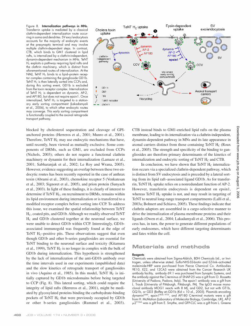
Figure 8. Internalization pathways in MNs. Transferrin uptake is mediated by a classical clathrin-dependent internalization route occur-ring in soma and dendrites. SV exo/ endocytosis accounts for the majority of endocytic events at the presynaptic terminal and may involve multiple clathrin-dependent steps. In contrast, CTB, which binds to GM1 clustered in lipid rafts, is internalized by a clathrin- independent, dynamin-dependent mechanism in MNs. TeNT HC exploits a pathway requiring lipid rafts and the clathrin machinery, which is distinct from aforementioned routes of internalization. At the NMJ, TeNT HC binds to a lipid–protein recep-tor complex containing the ganglioside GD1b. TeNT HC is then laterally sorted into CCPs and, during this sorting event, GD1b is excluded from the toxin receptor complex. Internalization of TeNT HC is dependent on dynamin, AP-2, and AP180, but does not require epsin1. Once internalized, TeNT HC is targeted to a station-ary early sorting compartment ( Lakadamyali et al., 2006), to which other endocytic routes may converge. This early sorting compartment is functionally coupled to the axonal retrograde transport pathway.
on August 1, 2006
ww
w.jcb.org
Dow
nloaded from
UPTAKE OF TETANUS TOXIN IS CLATHRIN DEPENDENT • DEINHARDT ET AL. 469
(National Institutes of Health, Bethesda, MD). TeNT HC was labeled with Alexa Fluor–maleimides (Lalli and Schiavo, 2002) or biotin, according to the manufacturers’ instructions, followed by dialysis against PBS.
Protein labelingTo couple TeNT HC to HRP, 10 nmol of cysteine-tagged TeNT HC were incu-bated with 5 mM EDTA and 6.5 mg EZ-link–activated maleimide-HRP in PBS overnight at 4°C. The conjugate was purifi ed fi rst on ConA–Sepharose (GE Healthcare) and eluted with 0.25 M α-methylmannoside in 10 mM sodium phosphate, pH 7.2. HRP–TeNT HC was bound to NiNTA-agarose (QIAGEN) and eluted in 20 mM Hepes-NaOH, pH 7.4, 150 mM NaCl, and 500 mM imidazole. Samples containing HRP–TeNT HC were pooled and dialyzed against PBS. To double label TeNT HC with an Alexa Fluor dye and HRP, fl uorophore labeling was performed fi rst, according to the manufacturer’s instructions, using half of the recommended amount of dye and without the addition of glutathione to stop the reaction. Alexa Fluor– labeled TeNT HC was dialyzed against PBS to remove the excess dye before HRP-conjugation.
Microinjection and internalization assayMN cultures were prepared and maintained in culture as previously de-scribed (Bohnert and Schiavo, 2005). Cells were injected with 0.05 mg/ml of plasmid between 4 and 7 d in vitro. In cases of microinjection of multi-ple plasmids (e.g., the pTRE-μ2 T156A plasmid that requires a transactiva-tor ptTA for expression; CLONTECH Laboratories, Inc.), 0.04 mg/ml of each construct were mixed before injection. MNs were incubated with 15–20 nM TeNT HC and then either biotinylated or Alexa Fluor–labeled for 30 min at 37°C. In selected experiments, 20 μg/ml Alexa Fluor 594–transferrin, 10 ng/ml Alexa Fluor 555–CTB, or 0.2 mg/ml tetramethylrho-damine dextran (mol wt 3,000) were mixed with TeNT HC before addition to the cells. 60 mM KCl was added to the medium just before addition of the ligands to test the effects of depolarization.
In experiments where MNs were pretreated with P4 or P4-scrambled peptide (Marks and McMahon, 1998), 50 μM of peptide was added to the medium at 37°C for 2 h before incubation with 20 nM Alexa Fluor 488–TeNT HC and 0.2 mg/ml tetramethylrhodamine dextran.
For MESNA treatment, MNs were cooled on ice and then incubated three times for 15 min with 15 mM of ice-cold MESNA in neurobasal me-dium (Invitrogen), pH 8.3. Cells were washed three times in neurobasal medium and once in PBS, and then fi xed.
To test the effect of SV exo/endocytosis on TeNT HC uptake, MNs were seeded on 13-mm coverslips. At 6 d in vitro, MNs in two wells were incubated with 15 nM BoNT/A and 2 nM BoNT/D for 22 h at 37°C, while control wells were left untreated. Coverslips from treated and untreated wells were then transferred into a new dish and incubated with 20 nM b-TeNT HC for a further 30 min at 37°C before treatment with MESNA on ice, fi xing, and processing as described in the following paragraph. The remaining cells from each well were washed in PBS, scraped, centrifuged, and then resus-pended in SDS sample buffer. Proteins were analyzed by Western blotting using standard procedures. Antibodies were used as follows: anti–VAMP-2 (69.1), 1:500; anti–SNAP-25, 1:1,000; anti-actin (AC-40), 1:1,000; and HRP-conjugated secondary antibodies (GE Healthcare), 1:1,000.
Immunofl uorescence and confocal microscopyCells were fi xed in 4% PFA and 20% sucrose in PBS for 15 min at room temperature, permeabilized with 0.1% Triton X-100 in PBS for 5 min, blocked in 2% BSA, 10% normal goat serum, and 0.25% fi sh skin gelatin in PBS for 30 min, and then incubated with the relevant antibodies (anti–VAMP-2 [69.1], 1:300; anti–SNAP-25, 1:300 [Washbourne et al., 1997]; anti-HA [12CA5], 1:1,000; anti-Myc [9E10], 1:250; secondary anti-bodies, 1:500; or streptavidin 1:500) for 30 min in blocking solution. Cells were mounted in Mowiol-488 and imaged using a LSM 510 laser scanning confocal microscope equipped with a 63×, 1.4 NA, Plan Apo-chromat oil-immersion objective (both Carl Zeiss MicroImaging, Inc.). Images were processed using LSM 510 software. Images showing GFP-CLC and TeNT HC colocalization were taken on living cells at 37°C using a la-ser scanning confocal microscope (IX70; Olympus) equipped with a 60×, 1.2 W, UPlan Apochromat oil-immersion objective and an environmental chamber. Images were captured using the Ultraview Imaging Suite Version 5.5 software (Perkin Elmer) and processed using AQM Advance 6 Kinetic Acquisition Manager software (Kinetic Imaging).
EMMNs grown on glass coverslips were incubated with 80 nM HRP–TeNT HC and/or with 10 μg/ml MOG-1 antibody in serum-free neurobasal medium for 45 min at 4°C. Cells were then washed and chased in prewarmed me-
dium at different temperatures for the indicated time. When appropriate, cells were incubated with a 10-nm gold-conjugated anti–mouse antibody (British Biocell International) on ice for additional 30 min and washed be-fore chase in medium alone. Cells were then fi xed with 2% PFA and 1.5% glutaraldehyde in 100 mM sodium cacodylate, pH 7.5, for 15 min and treated with DAB (0.75 mg/ml in 50 mM Tris-HCl, pH 7.4) and 0.02% H2O2 to cross-link HRP. Samples were postfi xed and embedded in Epon as previously described (Stinchcombe et al., 1995). MNs were then sectioned en face, and 60-nm sections stained with lead citrate were viewed in an electron microscope (CM12; Philips).
For whole mounts, MNs were treated as in the pervious paragraph, but instead of being permeabilized with digitonin, they were extracted with 1% Triton X-100 in PBS containing 1 mM MgCl2 and 0.1 mM CaCl2 for 10 min at 5°C. After gold labeling, samples were fi xed in 4% glutaral-dehyde and 1% osmium, dehydrated, and critical-point dried before being prepared for carbon replicas (Hopkins, 1985).
For EM of microinjected cells, MNs were seeded on CELLocate glass-gridded coverslips (Eppendorf). A plasmid encoding ssHRP-KDEL (Connolly et al., 1994) was used as an injection marker. 26 h later, cells were treated with HRP–TeNT HC and then with DAB, as described in the previous para-graphs. Alternatively, coverslips were incubated with 20 μg/ml human transferrin-HRP (Hopkins et al., 2000) after a 15-min preincubation at 37°C for in serum-free neurobasal medium. After fi xation and embedding in Epon, ultrathin sections were cut from the grid area containing the microinjected cells and imaged by EM.
For immunolabeling, samples were incubated with DAB in 50 mM Tris-HCl, 110 mM NaCl, pH 7.4, or with ascorbic acid buffer (20 mM Hepes-NaOH, 70 mM NaCl, and 50 mM ascorbic acid, pH 7.0) at 5°C for 30 min after treatment with HRP–TeNT HC and chased in medium. Cells were then permeabilized with 40 ng/ml digitonin in permeabilization buf-fer (25 mM Hepes-KOH, 38 mM aspartate, 38 mM glutamate, 38 mM gluconate, 2.5 mM MgCl2, and 2 mM EGTA, pH 7.2), fi xed in 2% PFA, quenched with 50 mM glycine, and blocked with 1% BSA before treatment with primary antibody in PBS containing 1% BSA for 60 min at room temperature. To enhance the signal, intermediate species-specifi c antibodies were used. MNs were washed and incubated with an appropriate 10-nm gold-labeled secondary antibody (British Biocell International) in 2% BSA and 2% FCS in PBS for 45 min at room temperature. After washing, cells were fi xed and processed for conventional EM. In double- and triple-label experiments, MNs were incubated with 80 nM HRP–TeNT HC or with 20 μg/ml transferrin-HRP together with TeNT HC directly conjugated to 10-nm gold particles (as described by Odorizzi et al., 1996) in serum-free neurobasal medium for 45 min at 4°C. Cells were washed and shifted to 12 or 20°C before incubation with DAB/H2O2.
Online supplemental materialFig. S1 shows biotinylated TeNT HC as a probe to study membrane traffi ck-ing in MNs. Fig. S2 shows characterization of HRP–TeNT HC. Fig. S3 shows that binding of TeNT HC to MNs can be competed by preincubation with a specifi c anti-GD1b antibody. Fig. S4 shows distribution of TeNT HC, epsin1, and transferrin in the endocytic pathway of MNs. Fig. S5 shows that overexpression of epsin1R63L H73L does not lead to AP-2 aggregation in spinal cord cells. Online supplemental material is available at http://www.jcb.org/cgi/content/full/jcb.200508170/DC1.
We are thankful to S. Tooze, A. Behrens, and members of the Molecular Neuropathobiology laboratory for critical reading of the manuscript.
This work was supported by Cancer Research UK (K. Deinhardt and G. Schiavo), Wellcome Trust 060349 (H.J. Willison), and the Medical Research Council (O. Berninghausen and C.R. Hopkins).
Submitted: 25 August 2005Accepted: 6 July 2006
ReferencesAbrami, L., S. Liu, P. Cosson, S.H. Leppla, and F.G. van der Goot. 2003.
Anthrax toxin triggers endocytosis of its receptor via a lipid raft-mediated clathrin-dependent process. J. Cell Biol. 160:321–328.
Aquino, D.A., M.A. Bisby, and R.W. Ledeen. 1985. Retrograde axonal transport of gangliosides and glycoproteins in the motoneurons of rat sciatic nerve. J. Neurochem. 45:1262–1267.
Blanpied, T.A., D.B. Scott, and M.D. Ehlers. 2002. Dynamics and regulation of clathrin coats at specialized endocytic zones of dendrites and spines. Neuron. 36:435–449.
on August 1, 2006
ww
w.jcb.org
Dow
nloaded from

JCB • VOLUME 174 • NUMBER 3 • 2006 470
Boffey, J., M. Odaka, D. Nicoll, E.R. Wagner, K. Townson, T. Bowes, J. Conner, K. Furukawa, and H.J. Willison. 2005. Characterisation of the immuno-globulin variable region gene usage encoding the murine anti-ganglioside antibody repertoire. J. Neuroimmunol. 165:92–103.
Bohnert, S., and G. Schiavo. 2005. Tetanus toxin is transported in a novel neu-ronal compartment characterized by a specialized pH regulation. J. Biol. Chem. 280:42336–42344.
Chen, H., and P. De Camilli. 2005. The association of epsin with ubiquitinated cargo along the endocytic pathway is negatively regulated by its interac-tion with clathrin. Proc. Natl. Acad. Sci. USA. 102:2766–2771.
Conner, S.D., and S.L. Schmid. 2003. Differential requirements for AP-2 in clathrin-mediated endocytosis. J. Cell Biol. 162:773–779.
Conner, S.D., S.L. Schmid, M. Masserini, P. Palestini, and M. Pitto. 2003. Regulated portals of entry into the cell. Nature. 422:37–44.
Connolly, C.N., C.E. Futter, A. Gibson, C.R. Hopkins, and D.F. Cutler. 1994. Transport into and out of the Golgi complex studied by transfect-ing cells with cDNAs encoding horseradish peroxidase. J. Cell Biol. 127:641–652.
Damke, H., T. Baba, D.E. Warnock, and S.L. Schmid. 1994. Induction of mutant dynamin specifi cally blocks endocytic coated vesicle formation. J. Cell Biol. 127:915–934.
Deinhardt, K., and G. Schiavo. 2005. Endocytosis and retrograde axonal traffi c in motor neurons. Biochem. Soc. Symp. 72:139–150.
Ehrlich, M., W. Boll, A. Van Oijen, R. Hariharan, K. Chandran, M.L. Nibert, and T. Kirchhausen. 2004. Endocytosis by random initiation and stabilization of clathrin-coated pits. Cell. 118:591–605.
Ford, M.G., I.G. Mills, B.J. Peter, Y. Vallis, G.J. Praefcke, P.R. Evans, and H.T. McMahon. 2002. Curvature of clathrin-coated pits driven by epsin. Nature. 419:361–366.
Ford, M.G., B.M. Pearse, M.K. Higgins, Y. Vallis, D.J. Owen, A. Gibson, C.R. Hopkins, P.R. Evans, and H.T. McMahon. 2001. Simultaneous binding of PtdIns(4,5)P2 and clathrin by AP180 in the nucleation of clathrin lattices on membranes. Science. 291:1051–1055.
Glebov, O.O., N.A. Bright, and B.J. Nichols. 2006. Flotillin-1 defi nes a clathrin-independent endocytic pathway in mammalian cells. Nat. Cell Biol. 8:46–54.
Habermann, E., and F. Dreyer. 1986. Clostridial neurotoxins: handling and action at the cellular and molecular level. Curr. Top. Microbiol. Immunol. 129:93–179.
Harding, C., J. Heuser, and P. Stahl. 1983. Receptor-mediated endocytosis of transferrin and recycling of the transferrin receptor in rat reticulocytes. J. Cell Biol. 97:329–339.
Herreros, J., T. Ng, and G. Schiavo. 2001. Lipid rafts act as specialized domains for tetanus toxin binding and internalization into neurons. Mol. Biol. Cell. 12:2947–2960.
Hinrichsen, L., J. Harborth, L. Andrees, K. Weber, and E.J. Ungewickell. 2003. Effect of clathrin heavy chain- and alpha-adaptin-specifi c small inhibi-tory RNAs on endocytic accessory proteins and receptor traffi cking in HeLa cells. J. Biol. Chem. 278:45160–45170.
Hopkins, C.R. 1985. The appearance and internalization of transferrin receptors at the margins of spreading human tumor cells. Cell. 40:199–208.
Hopkins, C., A. Gibson, J. Stinchcombe, and C. Futter. 2000. Chimeric mole-cules employing horseradish peroxidase as reporter enzyme for protein localization in the electron microscope. Methods Enzymol. 327:35–45.
Huang, F., A. Khvorova, W. Marshall, and A. Sorkin. 2004. Analysis of clathrin-mediated endocytosis of epidermal growth factor receptor by RNA interference. J. Biol. Chem. 279:16657–16661.
Humeau, Y., F. Doussau, N.J. Grant, and B. Poulain. 2000. How botulinum and tetanus neurotoxins block neurotransmitter release. Biochimie. 82:427–446.
Kirkham, M., A. Fujita, R. Chadda, S.J. Nixon, T.V. Kurzchalia, D.K. Sharma, R.E. Pagano, J.F. Hancock, S. Mayor, and R.G. Parton. 2005. Ultrastructural identifi cation of uncoated caveolin-independent early endocytic vehicles. J. Cell Biol. 168:465–476.
Kitamura, M., K. Takamiya, S. Aizawa, and K. Furukawa. 1999. Gangliosides are the binding substances in neural cells for tetanus and botulinum toxins in mice. Biochim. Biophys. Acta. 1441:1–3.
Lakadamyali, M., M.J. Rust, and X. Zhuang. 2006. Ligands for clathrin- mediated endocytosis are differentially sorted into distinct populations of early endosomes. Cell. 124:997–1009.
Lalli, G., and G. Schiavo. 2002. Analysis of retrograde transport in motor neu-rons reveals common endocytic carriers for tetanus toxin and neuro-trophin receptor p75NTR. J. Cell Biol. 156:233–239.
Lalli, G., S. Bohnert, K. Deinhardt, C. Verastegui, and G. Schiavo. 2003a. The journey of tetanus and botulinum neurotoxins in neurons. Trends Microbiol. 11:431–437.
Lalli, G., S. Gschmeissner, and G. Schiavo. 2003b. Myosin Va and microtubule-based motors are required for fast axonal retrograde transport of tetanus toxin in motor neurons. J. Cell Sci. 116:4639–4650.
Lamaze, C., A. Dujeancourt, T. Baba, C.G. Lo, A. Benmerah, and A. Dautry-Varsat. 2001. Interleukin 2 receptors and detergent-resistant membrane domains defi ne a clathrin-independent endocytic pathway. Mol. Cell. 7:661–671.
Le Roy, C., and J.L. Wrana. 2005. Clathrin- and non-clathrin-mediated endocytic regulation of cell signalling. Nat. Rev. Mol. Cell Biol. 6:112–126.
Marks, B., and H.T. McMahon. 1998. Calcium triggers calcineurin-dependent synaptic vesicle recycling in mammalian nerve terminals. Curr. Biol. 8:740–749.
Massol, R.H., J.E. Larsen, Y. Fujinaga, W.I. Lencer, and T. Kirchhausen. 2004. Cholera toxin toxicity does not require functional Arf6- and dynamin- dependent endocytic pathways. Mol. Biol. Cell. 15:3631–3641.
Matteoli, M., C. Verderio, O. Rossetto, N. Iezzi, S. Coco, G. Schiavo, and C. Montecucco. 1996. Synaptic vesicle endocytosis mediates the entry of tetanus neurotoxin into hippocampal neurons. Proc. Natl. Acad. Sci. USA. 93:13310–13315.
Miana-Mena, F.J., S. Roux, J.C. Benichou, R. Osta, and P. Brulet. 2002. Neuronal activity-dependent membrane traffi c at the neuromuscular junction. Proc. Natl. Acad. Sci. USA. 99:3234–3239.
Miller, F.D., and D.R. Kaplan. 2001. On Trk for retrograde signaling. Neuron. 32:767–770.
Montecucco, C., O. Rossetto, and G. Schiavo. 2004. Presynaptic receptor arrays for clostridial neurotoxins. Trends Microbiol. 12:442–446.
Montesano, R., J. Roth, A. Robert, and L. Orci. 1982. Non-coated membrane invaginations are involved in binding and internalization of cholera and tetanus toxin. Nature. 296:651–653.
Motley, A., N.A. Bright, M.N. Seaman, and M.S. Robinson. 2003. Clathrin- mediated endocytosis in AP-2-depleted cells. J. Cell Biol. 162:909–918.
Munro, P., H. Kojima, J.L. Dupont, J.L. Bossu, B. Poulain, and P. Boquet. 2001. High sensitivity of mouse neuronal cells to tetanus toxin requires a GPI-anchored protein. Biochem. Biophys. Res. Commun. 289:623–629.
Murthy, V.N., and P. De Camilli. 2003. Cell biology of the presynaptic terminal. Annu. Rev. Neurosci. 26:701–728.
Nesterov, A., R.E. Carter, T. Sorkina, G.N. Gill, and A. Sorkin. 1999. Inhibition of the receptor-binding function of clathrin adaptor protein AP-2 by dominant-negative mutant mu2 subunit and its effects on endocytosis. EMBO J. 18:2489–2499.
Nichols, B.J. 2003. GM1-containing lipid rafts are depleted within clathrin-coated pits. Curr. Biol. 13:686–690.
Odorizzi, G., A. Pearse, D. Domingo, I.S. Trowbridge, and C.R. Hopkins. 1996. Apical and basolateral endosomes of MDCK cells are interconnected and contain a polarized sorting mechanism. J. Cell Biol. 135:139–152.
Olusanya, O., P.D. Andrews, J.R. Swedlow, and E. Smythe. 2001. Phosphorylation of threonine 156 of the mu2 subunit of the AP2 complex is essential for endocytosis in vitro and in vivo. Curr. Biol. 11:896–900.
Owen, D.J., B.M. Collins, and P.R. Evans. 2004. Adaptors for clathrin coats: structure and function. Annu. Rev. Cell Dev. Biol. 20:153–191.
Parton, R.G., and A.A. Richards. 2003. Lipid rafts and caveolae as portals for endocytosis: new insights and common mechanisms. Traffi c. 4:724–738.
Parton, R.G., C.D. Ockleford, and D.R. Critchley. 1987. A study of the mecha-nism of internalisation of tetanus toxin by primary mouse spinal cord cultures. J. Neurochem. 49:1057–1068.
Roux, S., C. Colasante, C. Saint Cloment, J. Barbier, T. Curie, E. Girard, J. Molgo, and P. Brulet. 2005. Internalization of a GFP-tetanus toxin C- terminal fragment fusion protein at mature mouse neuromuscular junctions. Mol. Cell. Neurosci. 30:79–89.
Rummel, A., S. Bade, J. Alves, H. Bigalke, and T. Binz. 2003. Two carbohydrate binding sites in the H(CC)-domain of tetanus neurotoxin are required for toxicity. J. Mol. Biol. 326:835–847.
Sabharanjak, S., P. Sharma, R.G. Parton, and S. Mayor. 2002. GPI-anchored pro-teins are delivered to recycling endosomes via a distinct cdc42-regulated, clathrin-independent pinocytic pathway. Dev. Cell. 2:411–423.
Saint-Pol, A., B. Yelamos, M. Amessou, I.G. Mills, M. Dugast, D. Tenza, P. Schu, C. Antony, H.T. McMahon, C. Lamaze, and L. Johannes. 2004. Clathrin adaptor epsinR is required for retrograde sorting on early endo-somal membranes. Dev. Cell. 6:525–538.
Sandvig, K., and B. van Deurs. 2002. Membrane traffi c exploited by protein toxins. Annu. Rev. Cell Dev. Biol. 18:1–24.
Schmid, S.L., and E. Smythe. 1991. Stage-specifi c assays for coated pit forma-tion and coated vesicle budding in vitro. J. Cell Biol. 114:869–880.
Schwab, M.E., and H. Thoenen. 1978. Selective binding, uptake, and retrograde transport of tetanus toxin by nerve terminals in the rat iris. An electron microscope study using colloidal gold as a tracer. J. Cell Biol. 77:1–13.
on August 1, 2006
ww
w.jcb.org
Dow
nloaded from
UPTAKE OF TETANUS TOXIN IS CLATHRIN DEPENDENT • DEINHARDT ET AL. 471
Shogomori, H., and A.H. Futerman. 2001. Cholera toxin is found in detergent-insoluble rafts/domains at the cell surface of hippocampal neurons but is internalized via a raft-independent mechanism. J. Biol. Chem. 276:9182–9188.
Sigismund, S., T. Woelk, C. Puri, E. Maspero, C. Tacchetti, P. Transidico, P.P. Di Fiore, and S. Polo. 2005. Clathrin-independent endocytosis of ubiqui-tinated cargos. Proc. Natl. Acad. Sci. USA. 102:2760–2765.
Signoret, N., L. Hewlett, S. Wavre, A. Pelchen-Matthews, M. Oppermann, and M. Marsh. 2005. Agonist-induced endocytosis of CC chemokine receptor 5 is clathrin dependent. Mol. Biol. Cell. 16:902–917.
Stinchcombe, J.C., H. Nomoto, D.F. Cutler, and C.R. Hopkins. 1995. Anterograde and retrograde traffi c between the rough endoplasmic reticulum and the Golgi complex. J. Cell Biol. 131:1387–1401.
Stoorvogel, W. 1998. Analysis of the endocytic system by using horseradish peroxidase. Trends Cell Biol. 8:503–505.
Sunyach, C., A. Jen, J. Deng, K.T. Fitzgerald, Y. Frobert, J. Grassi, M.W. McCaffrey, and R. Morris. 2003. The mechanism of internalization of glycosylphosphatidylinositol-anchored prion protein. EMBO J. 22:3591–3601.
Torgersen, M.L., G. Skretting, B. van Deurs, and K. Sandvig. 2001. Internalization of cholera toxin by different endocytic mechanisms. J. Cell Sci. 114:3737–3747.
Venkatesan, S., J.J. Rose, R. Lodge, P.M. Murphy, and J.F. Foley. 2003. Distinct mechanisms of agonist-induced endocytosis for human chemokine recep-tors CCR5 and CXCR4. Mol. Biol. Cell. 14:3305–3324.
Washbourne, P., R. Pellizzari, G. Baldini, M.C. Wilson, and C. Montecucco. 1997. Botulinum neurotoxin types A and E require the SNARE motif in SNAP-25 for proteolysis. FEBS Lett. 418:1–5.
on August 1, 2006
ww
w.jcb.org
Dow
nloaded from
Related Documents