TESTICULAR OESTRADIOL-1713 PROEFSCHRIFT TER VERKRIJGING VAN DE GRAAD VAN DOCTOR IN DE GENEESKUNDE AAN DE ERASMUS UNIVERSITEIT TE ROTTERDAM, OP GEZAG VAN DE RECTOR MAGNIFICUS PROF.DR.P.W.KLEIN EN VOLGENS BESLUIT VAN HET COLLEGE VAN DEKANEN. DE OPENBARE VERDEDIGING ZAL PLAATS VINDEN OP WOENSDAG 22 MEl 1974, DES NAMIDDAGS TE 4.15 UUR DOOR FRANK HOTZE DE JONG GEBOREN TE OUDENRIJN

Welcome message from author
This document is posted to help you gain knowledge. Please leave a comment to let me know what you think about it! Share it to your friends and learn new things together.
Transcript
TESTICULAR OESTRADIOL-1713
PROEFSCHRIFT
TER VERKRIJGING VAN DE GRAAD VAN DOCTOR IN DE GENEESKUNDE
AAN DE ERASMUS UNIVERSITEIT TE ROTTERDAM,
OP GEZAG VAN DE RECTOR MAGNIFICUS
PROF.DR.P.W.KLEIN
EN VOLGENS BESLUIT VAN HET COLLEGE VAN DEKANEN.
DE OPENBARE VERDEDIGING ZAL PLAATS VINDEN OP
WOENSDAG 22 MEl 1974,
DES NAMIDDAGS TE 4.15 UUR
DOOR
FRANK HOTZE DE JONG
GEBOREN TE OUDENRIJN

Promotor
Coreferenten
Prof.Dr. H.J. van der Molen
Prof.Dr. J.c. Birkenhager
Dr. R.V. Short

Voor Maartje,
om in te kleuren


CONTENTS
CONTENTS
VOORWOORD
LIST OF TRIVIAL NAMES
LIST OF ABBREVIATIONS
CHAPTER 1. OESTROGENS IN THE MALE. QUESTIONS AND
SCOPE OF THIS THESIS
l.l.
l. 2.
l. 3.
l. 4.
l. 5.
l. 6.
l. 7.
Introduction
The origin of oestrogenic hormones in
male animals
The regulation of the testicular pro
duction and secretion of oestradiol
The intra testicular localization of the
production of oestradiol
The function of testicular oestradiol
Methods for the estimation of oestradiol
References
CHAPTER 2. METHODS USED FOR THE ESTIMATION OF
STEROIDS
2.1. Introduction
2.2. The estimation of testosterone
2.2.1. Gas-liquid chromatography
2. 2 • 1. 1. Method
2.2.1.2. Results
2.2.2. Radioimmunoassay
2. 2. 3.
2. 3.
Discussion
The estimation of oestradiol
2.3.1. Competitive protein binding
2. 3. l. l. Method
2.3.1.2. Results
2.3.2. Double competitive protein binding
2. 3. 2. 1. Method
7
11
13
15
17
18
19
19
20
21
21
23
25
25
25
28
29
30
31
31
31
31
36
36
7

2.3.2.2. Results
2.3.3. Radioimmunoassay
2.3.3.1. Method
2.3.3.2. Results
2. 3. 4.
2. 4.
Discussion
References
CHAPTER 3. EXPERIMENTS IN VIVO
3.1. Introduction
3. 2.
3. 2. l.
3. 2. 2.
3. 2. 3.
3. 2. 4.
3. 3.
3. 3. l.
3. 3. 2.
3. 3. 3.
3. 4.
3. 5.
The testis as a source of oestrogens
Oestradiol in testicular venous blood
Oestradiol in testicular tissue
Oestrogens in semen
Regulation of the testicular production
and secretion of oestrogens
Extra testicular sources of oestradiol
Production rates of oestrogens in
male manunals
Peripheral conversion of prehormones
The adrenal as a source of oestrogens
Conclusions
References
CHAPTER 4. EXPERIMENTS IN VITRO
8
4.1. Introduction
4. 2.
4. 2 .l.
4. 2. 2.
4. 2. 3.
4. 2. 4.
4. 3.
4. 3. l.
Materials and methods
Animal techniques
Steroids and steroid estimations
Incubation conditions
Techniques used for the separation and
identification of steroids
Results
Measurement of endogenous hormones
4.3.2. Conversion of radioactive precursors
4.3.2.1. Interconversion and degradation of
oestrogens
4.3.2.2. Biosynthesis of oestrogens
4.4. Discussion
37
37
37
38
39
42
45
47
47
48
49
50
55
57
63
66
67
67
73
76
76
77
78
79
81
81
84
84
85
89

4. 4. 1.
4. 4. 2.
4. 5.
4. 6.
Endogenous production of oestradiol
Production of radioactive oestradiol
Conclusions
References
CHAPTER 5. THE SIGNIFICANCE OF THE TESTICULAR
PRODUCTION OF OESTRADIOL
5. 1.
5. 2.
5. 2. 1.
5. 2. 2.
5. 2. 3.
5. 3.
5. 4.
Introduction
Influence of oestrogens on testicular
function
Effects of longterrn oestrogen treat
ment on testicular enzymes
Effects of oestrogens on spermatogenesis
Effects observed during incubations of
testicular tissue with oestrogens
Summary and conclusions
References
SUMMARY
SAMENVATTING
CURRICULUM VITAE
APPENDIX PAPERS
l. F.H. de Jong, A.H. Hey & H.J. van der Molen.
Effect of gonadotrophins on the secretion of
oestradiol-17S and testosterone by the rat
testis.
J. Endocr. 57 (1973) 277-284.
2. F.H. de Jong, A.H. Hey & H.J. van der Molen.
89
90
93
93
97
98
98
99
100
101
101
105
109
113
115
Oestradiol-17S and testosterone in rat testis
tissue: effect of gonadotrophins, localization
and production in vitro.
J. Endocr. 60 (1974) 409-419.
9


VOORWOORD
Onderzoeken is een arbeid, die moeilijk anders dan in
teamverband verricht kan worden. Daarom geloof ik dat het
juist is dit voorwoord te beginnen door alle medewerkers
van de afdeling Biochernie (Chernische Endocrinologie) en
ook hen, met wie ik buiten deze afdeling sarnengewerkt heb,
te danken voor gegevens, raadgevingen, diskussies en me
ningsverschillen. Vervolgens wil ik hen, die wel een zeer
duidelijk stempel op dit proefschrift drukten, persoonlijk
noernen.
In de eerste plaats de promotor, Prof.Dr. H.J. van der
Molen. Beste Henk, rnede dankzij jouw pragmatisme en sorns
wat agressieve redeneertrant is het onderzoek niet op
allerlei interessante zijsporen gederailleerd. Ik waardeer
je vrijgevigheid, die duidelijk wordt in je wetenschappe
lijke openheid, je bestuurlijke opstelling binnen de afde
ling en de set ups bij het volleybal, bijzonder.
Prof.Dr, J.C. Birkenhager, beste Jan, nog meer dan voor
je bernoeienissen als coreferent zou ik je willen danken
voor de wijze waarop je me de vrijheid gelaten hebt in de
Hoogbouw bezig te zijn met zaken, die niet direkt met
patienten te maken hebben.
Dr. R.V. Short, I thank you for your willingness to read
this thesis as ''coreferent". I am sure that there will be
ample time to discuss the significance of testicular
oestradiol synthesis in the future.
Dear Yolanda, you were the first one to encounter the
difficulties in the estimation of oestradiol. I admire your
"never-give-up" mentality very much.
Beste Anne, jij bent erin geslaagd de radioimmunolo
gische bepaling van oestradiol van de grand te krijgen en
van de grond te houden. Het is duidelijk dat dit boekje
zonder jouw bezigheden en administratieve vaardigheid nog
niet geschreven zou zijn.
ll

Beste Wil, Wytske, Sarah en Tilly, alleen het feit dat
jullie de klinische 11 routine 11 -kastanjes uit het vuur haal
den, maakte het voor mij mogelijk "research" te bedrijven.
Daarvoor, en voor de vele in dit boekje verwerkte testoste
rongetallen, ben ik jullie zeer dankbaar.
Beste Marja, het feit dat onze ruzies in het verleden
zelden hager opliepen dan door Van Dale beslecht kon worden,
pleit voor ons beider inkasseringsvermogen. Dat dit oak in
de periode waarin jij dit proefschrift typte het geval was,
moet waarschijnlijk alleen op jouw canto geschreven worden.
Drs. P.M. Frederik, ofwel Peter, wist wederom op ver
bluffende wijze de dingen die in mijn hoofd nag geen vorm
hadden aangenomen, terug te brengen tot een tweedimensio
naal omslag.
Ik dank Pim voor zijn niet aflatende vrolijke hulpvaar
digheid, Brian voor zijn kornmentaar op het Engels, Binding
Boven voor de gastvrijheid en het TDT voor zijn bijdrage,
vooral tijdens de grote proeven.
De medewerkers van de Audiovisuele Dienst, in het bij
zonder de heer D.M. Simons, dank ik voor het geduld dat zij
met mij en mijn figuren gehad hebben.
Beste Focko, het is triest je van zo 1 n afstand te moeten
herinneren aan wekelijkse maaltijden bij de Chinees, maan
delijkse ontmoetingen bij de brandweer en dagelijkse ver
hitte gesprekken over van alles en statistiek. We moesten
als je terug bent de blussingsmethodieken nag maar eens
doornemen.
Tenslotte wil ik mijn ouders nog eens expliciet danken
voor het feit dat zij mijn studie, en daardoor dit boekje
mogelijk maakten. En om Arjane te danken, zijn er gelukkig
nog andere mogelijkheden dan dit voorwoord.
12

LIST OF TRIVIAL NAMES
androstenedione
androsterone
cholesterol
cortisol
corticosterone
cyanoketone
dehydroepiandrosterone
dehydroepiandrosterone
sulphate
desoxycorticosterone
dexamethasone
diethylstilboestrol
epitestosterone
epitestosterone
chloroacetate
etiocholanolone
19-hydroxyandrostenedione
l7a-hydroxypregnenolone
- 4-androstene-3 1 17-dione
- 3a-hydroxy-Sa-androstan-
17-one
- S-cholesten-3S-ol
- 11S,l7,21-trihydroxy-4-
pregnene-3,20-dione
- 116,21-dihydroxy-4-pregnene-
3,20-dione
- 2a-cyano-4,4' ,17a-trimethyl-
17S-hydroxy-5-androsten-3-
one
- 36-hydroxy-5-androsten-17-
one
- 17-oxo-5-androsten-36-yl
sulphate
- 21-hydroxy-4-pregnene-3,20-
dione
- 9a-fluoro-16a-rnethyl-11S,l7,
21-trihydroxy-1,4-pregna
diene-3,20-dione
- trans-3,4-bis(4-hydroxy
phenyl)-3-hexene
- 17a-hydroxy-4-androsten-3-
one
- 3-oxo-4-androsten-17a-yl
rnonochloroacetate
- 3a-hydroxy-Ss-androstan-17-
one
- 19-hydroxy-4-androstene-
3,17-dione
- 36,17-dihydroxy-5-pregnen-
20-one
1 3

17a-hydroxyprogesterone
19-hydroxytestosterone
mevalonic acid
oestradiol(-178)
oestradiol dipropionate
oestradiol-178-
hemisuccinate
oestriol
oestrone
oestrone acetate
oestrone sulphate
19-oxoandrostenedione
19-oxotestosterone
pregnanediol
pregnenolone
progesterone
testosterone
testosterone chloroacetate
testosterone propionate
14
- 17-hydroxy-4-pregnene-3,20-
dione
- 178,19-dihydroxy-4-androsten-
3-one
- 3,5-dihydroxy-3-methyl
pen tanoic acid
- 1,3,5(10)-oestratriene-
3,178-diol
- 1,3,5(10)-oestratriene-
3,178-diol dipropionate
- 1,3,5(10)-oestratriene-
3,178-diol 17-rnonoherni
succinate
- 1,3,5(10)-oestratriene-
3,16o:,17S-triol
- 3-hydroxy-1,3,5(10)
oestratrien-17-one
- 17-oxo-1,3,5(10)
oestratrien-3-yl acetate
- 17-oxo-1,3,5(10)
oestratrien-3-yl sulphate
- 4-androstene-3,17,19-trione
- 178-hydroxy-4-androstene-
3,19-dione
- SS-pregnane-3o:,20a-diol
- 38-hydroxy-5-pregnen-20-one
- 4-pregnene-3,20-dione
- 176-hydroxy-4-androsten-
3-one
- 3-oxo-4-androsten-178-yl
rnonochloroacetate
- 3-oxo-4-androsten-178-yl
propionate

LIST OF ABBREVIATIONS
ACTH
BSA
b.w.
c.v. dpm
EDTA
EFA
FSH
g
HCG
LH
MCR
n
NADH
NADPH
p
PC
PMS
PRB
PRU R*
PRE -+ PRO p,pBB
RNA
S.D.
S .. E.M.
TLC
X
X'
- adrenocorticotrophic hormone
- bovine serum albumin
- body weight
- coefficient of variation
- disintegration per minute
- ethylenediaminetetraacetate
- essential fatty acid
- follicle-stimulating hormone
- relative centrifugal force
- human chorionic gonadotrophin
- luteinizing hormone
- metabolic clearance rate
- number of estimations
- nicotinamide-adenine dinucleotide
(reduced)
- nicotinamide-adenine dinucleotide
phosphate (reduced)
- probability
- paper chromatography
- pregnant mare serum gonadotrophin
- blood production rate
- urinary production rate
- amount of radioactivity injected in
estimation of PR8
or P~
transfer constant for the conversion in
blood of a prehormone to a product
- ribonucleic acid
- standard deviation
- standard error of the mean
- thin-layer chromatography
- concentration of hormone in blood
- concentration of labelled hormone in blood
!5


CHAPTER 1. OESTROGENS IN THE MALE.
QUESTIONS AND SCOPE OF THIS THESIS
1.1. Introduction
Oestrogenic hormones were originally isolated from ova
rian follicles and from placental tissue and were believed
to occur only in female animals. Laqueur et al. (1927}
observed, however, that extracts from human male urine
caused vaginal cornification in spayed mice. This discovery
of oestrogenic activity in urine from men was so unexpected,
that the authors thought it necessary to state that there
could be no doubt about the manliness of the subjects
studied. One of the oestrogenic substances in human male
urine was subsequently identified as oestrone (Dingemanse
et al., 1938), while later on oestradiol and oestriol were
also found to be present in urine from men (see: Diczfalusy
& Lauritzen, 1961). Since then, the occurrence of oestro
gens in the urine of male animals from several species has
been described (see: Velle, 1966). However, information on
the precise origin of these oestrogenic hormones and on the
regulation of the production of oestrogens in the male
animal is still limited. Therefore, it was decided to
investigate these points with special reference to the
testis as a possible source of oestradiol. In this first
chapter a broad outline of some questions relate'd to the
study of production and secretion of oestradiol in the male
will be given. A more detailed description of these
problems can be found in the following chapters.
17

1.2. The origin of oestrogenic hormones in male animals
The presence of oestrogenic steroids in urine from male
subjects might result from a direct secretion from endo
crine glands, such as the adrenal or the testis, or from
peripheral conversion of androgens. The latter possibility
has been shown by Steinach & Kun (1937), who found that
administration of androgens to men causes an increase in
the urinary excretion of oestrogens. The results of inves
tigations concerning the relative contribution of glandular
secretion and peripheral conversion to the total amount of
oestradiol produced are conflicting: some authors
(MacDonald et al., 1971) concluded that all circulating
oestradiol is formed peripherally, while others (Baird
et al., 1968) could not exclude the possibility of oestra
diol secretion from endocrine glands (see chapter 3). The
first purpose of the investigation described in this thesis
was to determine whether or not oestradiol is secreted by
the rat testis and if it is, to estimate the relative con
tribution of testicular oestradiol to the total amount of
oestradiol in the peripheral circulation. The results
described in chapter 3 and appendix paper 1 indicate that
the testis of the rat does secrete oestradiol. This testi
cular secretion accounts for about 20% of the circulating
oestradiol.
18

1.3. The regulation of the testicular production and secretion of
oestradiol
Maddock & Nelson (1952) observed that the urinary excre
tion of oestrogens increased more steeply than that of
17-ketosteroids after administration of HCG to men. This
finding started the discussion on the regulation of testi
cular production and secretion of oestrogens. However,
in vivo studies with intact subjects cannot provide answers
to questions on the role of the testis in the total produc
tion rate of oestradiol (see chapter 3). Therefore, the
second aim of this study was to investigate the regulation
of the testicular production and secretion of oestradiol
directly. various conditions, which are known to influence
testicular function were used. Results of these experiments,
which were carried out largely under in vivo conditions,
are described in chapter 3 and in the appendix papers 1 and
2. It is concluded that only HCG influences testicular
oestradiol secretion, when administered in short-term expe
riments. No effects could be shown after administration of
HCG during longer periods, of FSH, or of prolactin.
1.4. The intratesticular localization of the production of oestradiol
Both tissue compartments in the testis, the seminiferous
tubules and the interstitial tissue, have been considered
as a possible site of testicular oestrogen biosynthesis.
Maddock & Nelson (1952) suggested the Leydig cell as the
source of testicular oestrogens, because after administra
tion of HCG to men they observed hypertrophy of the inter
stitial tissue concomitant with an increased urinary excre
tion of oestrogens. Ashbel et al. (1951) concluded that
19

testicular oestrone was localized in the seminiferous
tubules on basis of results obtained with histochemical
techniques for the detection of phenolic 17-ketosteroids.
From studies with !!feminizing testesn or with testicular
tumours either the interstitial tissue (French et al.,
1965, Sharma et al., 1965) or the Sertoli cell (Berthrong
et al., 1949, Teilum, 1949) was suggested as the source of
testicular oestrogens.
As the third aim of the present study it was attempted,
therefore, to get more insight into the cellular localiza
tion and origin of testicular oestradiol. Endogenous
oestradiol was estimated during incubations of whole testis
tissue or of separated interstitial tissue and seminiferous
tubules (Christensen & Mason, 1965) from the rat testis.
Furthermore, incubations with radioactive oestrogen precur
sors were performed with these tissues. The results of
these experiments, as described in chapter 4 and appendix
paper 2, indicate that testicular oestradiol is mainly
localized in the interstitial tissue. Biosynthesis of
oestradiol might, however, take place in the seminiferous
tubules.
1.5. The function of testicular oestradiol
The administration of oestrogens to men or male experi
mental animals causes severe degeneration of the testis and
accessory sex organs {see: Emmens & Parkes, 1947). This
involution is thought to result from a lack of gonadotro
phins through the negative feed-back action of oestrogens
on the secretion of the hypophyseal gonadotrophins (see:
Burger et al., 1972), although direct effects of oestrogens
on the testis have also been postulated (Samuels et al.,
1967). The relevancy of these possibilities is discussed in
chapter 5.
20

1.6. Methods for the estimation of oestradiol
In order to realize the intended aims of this investi
gation, it was necessary to measure the endogenous concen
tration of oestradiol in plasma and tissues from the male
rat. Methods which were sufficiently sensitive and specific
to meet this goal had not been properly documented when
this investigation was started. Therefore, the reliability
of several methods for the estimation of oestradiol was
tested for these specific requirements. The results of
these methodological investigations are summarized in
chapter 2, together with the reliability criteria of the
methods used for the estimation of testosterone, which was
estimated as a reference in the same samples.
1.7. References
Ashbel, R., Cohen, R.B. & Seligman, A.M. (1951)
Endocrinology 49, 265-281.
Baird, D.T., Horton, R., Longcope, C. & Tait, J.F. (1968)
Recent Progr. Horm. Res. ~~ 611-664.
Berthrong, M., Goodwin, W.E. & Scott, \1.W. (1949)
J. clin. Endocr. ~~ 579-592.
Burger, H.G., Baker, H.W.G., Hudson, B. & Taft, H.P. (1972)
in Gonadotropins (Saxena, B.B., Beling, C.G. & Gandy, H.M.,
eds.), pp. 569-592, Wiley Interscience, New York.
Christensen, A.K. & Mason, N.R. (1965}
Endocrinology li• 646-656.
Diczfalusy, E. & Lauritzen, Ch. (1961)
Oestrogene beim Menschen, pp. 295-297, Springer Verlag,
Berlin.
Dingemanse, £., Laqueur, E. & Milhlbock, 0. (1938}
Nature, Land. l!l• 927.
21

22
Enunens, C.W. & Parkes, A.S. (1947)
Vitamins and Hormones 2• 233-272.
French, F.S., Baggett, B., van Wijk, J.J., Talbert, M.L.,
Hubbard, W.R., Johnston, F.R., Weaver, R.P., Forchielli, E.,
Rao, G.S. & Sarda, I.R. (1965)
J. c1in. Endocr. Metab. l2• 661-677.
Lagueur, E., Dingemanse, E., Hart, P.C. & de Jong, S.E. (1927)
Klin. wschr . .§., 1859.
liJacDonald, P.C., Grodin, J.M. & Sitteri, P.K. (1971)
in Control of Gonadal Steroid Secretion (Baird, D.T.
Strong, J.A., eds.), pp. 158-174, Edinburgh University Press,
Edinburgh.
Maddock, W.O. & Nelson, W.O. (1952)
J. clin. Endocr. Metab. ll• 985-1007.
Samuels, L.T., Uchikawa, T. & Huseby, R.A. (1967)
in Endocrinology of the Testis (\'iolstenholr:\e, G.E.W. &
O'Connor, ~-• c~s), pp. 211-232, Churchill, London.
Sharma, D.C., Dorfman, R.I. & Southren, A.L. (1965)
Endocrinology 2§._, 96Fi-'.J/S.
Steinach, E. & Kun, !! (1937)
Lancet ~. 84 5.
Teilum, G. {1949)
J. clin. Endocr. i• 301-318.
Velle, W. (1966)
J. Reprod. Fert. ~· 65-73.

CHAPTER 2. METHODS USED FOR THE ESTIMATION OF STEROIDS
2.1. Introduction
Progress in endocrinological research depends for an
important part on progress in the methodology for estima
tion of the concentrations of hormones, that are present in
tissues or body fluids under physiological or pathological
conditions. A main aspect of analytical methodology in this
respect is the sensitivity of the methods used, and in
direct connection therewith the speciLicity of detection.
For the estimation of steroids, both sensitivity and speci
ficity have been improved enormously during the last 15
years through the development of double isotope dilution
techniques (see: Bojesen et al., 1967), gas-liquid chroma
tography (see: Eik-Nes & Horning, 1968) and saturation ana
lysis (see: Diczfalusy, 1970). The older and generally less
sensitive techniques of bioassay (see: Dorfman, 1969) and
fluorimetry and colorimetry (see: Loraine & Bell, 1966)
have gradually been replaced, although many of the princi
pal observations have initially been made using these
techniques. This development is clearly reflected in the
following reviews dealing with techniques that can be used
for the estimation of a single steroid or a class of
steroids: androgens (van der Molen, 1971), oestrogens
(Preedy et al., 1967) and progesterone (van der Molen &
Aakvaag, 1967).
In order to study the endogenous production and secre
tion of oestradiol from the testis it was mandatory to use
techniques with a sensitivity which would permit measure
ment of the endogenous concentrations of this steroid in
23
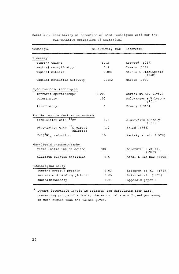
Table 2.1. Sensitivity of detection of some techniques used for the
quantitative estimation of oestradiol
Technique Sensitivity (ng)
Bioassay*
uterine weight
vaginal cornification
vaginal mitosis
vaginal metabolic activity
Spectroscopic techniques
infrared spectroscopy
colorimetry
fluorimetry
Double isotope derivative methods
bromination with Br
pipsylation with 35 s pipsyl chloride
Gas-liquid chromatography
flame ionization detection
electron capture detection
Radioligand assay
uterine cytosol protein
sex steroid binding globulin
radioimmunoassay
12.5
0. 5
Q.Q04
0.002
5,000
100
5
1.0
1.0
10
200
0.5
0.02
0.05
0. 0 1
Reference
Astwood ( 19 38)
Emmens (1941)
Hartin & Claringbold ( 1960)
Martin {1960)
Oertel et al. {1959)
Salokangas & Bulbrook { 19 61)
Preedy (1965)
Slaunwhite & Neely ( 19 6 3)
Baird (1968)
Kautsky et al. (1970)
Adlercreutz et al. (1967)
Attal & Eik-Nes (1968)
Korenman et al, ( 19 6 9)
Dufau et al. {1970)
Appendix paper 1
* Lowest detectable levels in bioassay are calculated from data,
concerning groups of animals; the amount of steroid used per assay
is much higher than the values given.
24
male experimental animals. The only data on the level of
oestradiol in male animals available at the start of this
investigation was the concentration of oestradiol in peri
pheral plasma from men (see Table 3.6.). In order to mea
sure oestradiol in the small amounts of plasma that can be
obtained from the rat, it was deemed necessary to be able
to measure at least 25 pg. The limits of detection of a
number of methods for the estimation of oestradiol are
summarized in Table 2.1.: only competitive protein binding
with the use of receptor proteins or radioimmunoassay
appeared to be sensitive enough for estimation of oestra
diol in male rats. These techniques had hardly been intro
duced at the start of this investigation and have therefore
been evaluated in detail (section 2.3.).
The sensitivity of gas-liquid chromatography with
electron capture detection is sufficient to measure concen
trations of testosterone in peripheral and testicular
venous blood from rats, which are much higher than those
of oestradiol (Bardin & Peterson, 1967, appendix paper 1).
Nevertheless, the high practicability of radioimmunoassay
made it worthwhi.le to investigate this technique as well.
The merits of these two methods for the estimation of
testosterone are discussed in section 2.2.
2.2. The estimation of testosterone
2.2.1. Gas-liquid chromatography
2.2.1.1. Method
Testosterone was estimated using the method as described
by Brownie et al. (1964) with minor modifications
(Table 2.2.). Solvents were purified as described by
van der Molen & de Jong (1974). Gas-liquid chromatography
was performed on a model 104 Pye gas-liquid chromatograph,
25

Table 2.2. Procedure for the assay of testosterone using gas-liquid
chromatography with electron capture detection
(Brownie et al., 1964)
- Add 40,000 dpm 3H-testosterone to alkaline plasma
- Extract plasma with ether
- Wash ether extracts and evaporate solvent
- Purify residue by thin-layer chromatography on silica gel using
the solvent system toluene:ethyl acetate- 2:1 (v/vl
- Extract testosterone from the appropriate silica gel fraction with
toluene/water
- Evaporate the combined toluene layers
- Chloroacetylate residue
- Extract testosterone from the chloroacetylation mixture with
ethyl acetate
- Evaporate the combined ethyl acetate layers
- Purify testosterone chloroacetate by thin-layer chromatography on
silica gel using the solvent system toluene:ethyl acetate -
9:1 (v/v)
- Extract testosterone chloroacetate from the appropriate silica gel
fraction with toluene/water
- Evaporate the combined toluene layers
- Inject suitable aliquot onto the gas-liquid chromatographic column
and estimate amount of testosterone present in total sample
- Add suitable amount of second internal standard
- Take suitable aliquot of sample for recovery estimation
- Inject suitable aliquot of sample onto the gas-liquid chromato-
graphic column for final gas-liquid chromatography
equipped with an automatic solid injection system. The 63Ni
electron capture detector was operated in the pulsed mode
(pulse interval: 150 ~sec, pulse height: 60V, pulse width:
0.75 ~sec). The 3 ft coiled column was packed with a sta
tionary phase of 1% QF-1 on gaschrom Q; nitrogen was used
as carrier and as purge gas. The internal standard for gas
liquid chromatography was 3-oxo-4-pregnen-206-yl chloro
acetate instead of 5-cholesten-36-yl chloroacetate, as used
in the original method.
26

N _,
Table 2.3. Rf values during thin-layer chromatography and relative retention times (RRT) during
gas-liquid chromatography in the systems used during the gas-liquid chromatographic
estimation of testosterone (partly from van der Molen & de Jong, 1974)
steroid
oestriol cortisol corticosterone pregnanediol desoxycorticosterone testosterone epitestosterone 20a-hydroxy-4-pregnen-3-one 206-hydroxy-4-pregnen-3-one 17a-hydroxyprogesterone etiocholanolone androsterone pregnenolone dehydroepiandrosterone androstenedione 3a-hydroxy-5S-pregnan-20-one oestradiol progesterone oestrone
Thin-layer chromatography
Rf in toluene:
ethyl acetate-2:1
0.05 0.05 0.06 0. 18 0. 2 2 0.26 0.26 0. 3 2 0.32 0.33 0.35 0.40 0.43 0.44 0.45 0.46 0.57 0.59 0.88
Rf after chloro
acetylation in
toluene: ethyl
acetate-6:1
0.08 0.08 0.91 0.35 0.42 0.42 0. 4 7 0.52 0.12 0.56 0.55 0.70 0.59 0. 18 0.71 0.82 0.25 0.56
* Retention time relative to that of 5a-cholestane = 1.
Gas-liquid chromatography
RRTX of chloroacetates on
1% QF-1
27.9
17. 1 15.0 28.9 25.0
7.9 7. 4
11.6 8. 6
9.9 25.7
10.8
2.2.1.2. Results
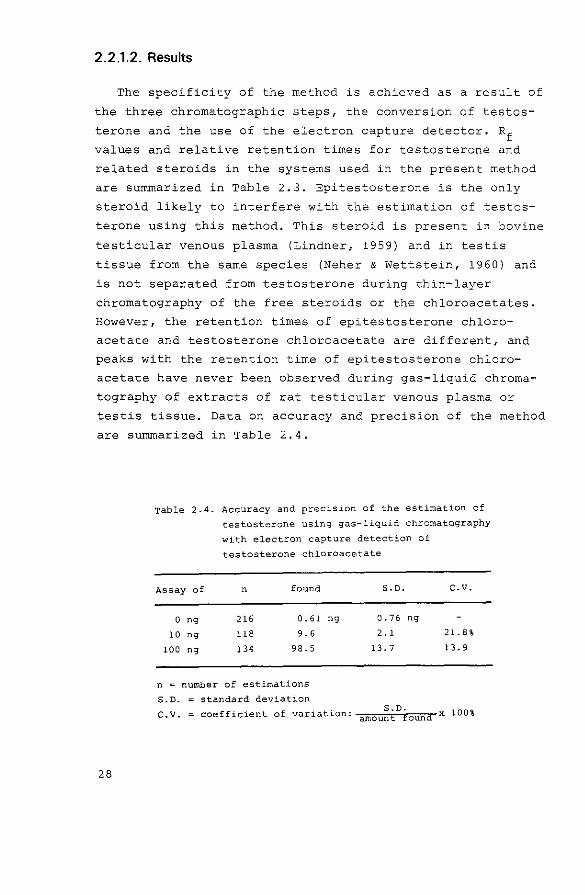
The specificity of the method is achieved as a result of
the three chromatographic steps, the conversion of testos
terone and the use of the electron capture detector. Rf
values and relative retention times for testosterone and
related steroids in the systems used in the present method
are summarized in Table 2.3. Epitestosterone is the only
steroid likely to interfere with the estimation of testos
terone using this method. This steroid is present in bovine
testicular venous plasma (Lindner, 1959) and in testis
tissue from the same species (Neher & Wettstein, 1960) and
is not separated from testosterone during thin-layer
chromatography of the free steroids or the chloroacetates.
However, the retention times of epitestosterone chloro
acetate and testosterone chloroacetate are different, and
peaks with the retention time of epitestosterone chloro
acetate have never been observed during gas-liquid chroma
tography of extracts of rat testicular venous plasma or
testis tissue. Data on accuracy and precision of the method
are summarized in Table 2.4.
28
Table 2.4. Accuracy and precision of the estimation of
testosterone using gas-liquid chromatography
with electron capture detection of
testosterone chloroacetate
Assay of n found
0 ng 216 0.61
10 ng 118 9.6
100 ng 134 98.5
n = number of estimations
S.D. standard deviation
S.D. c.v.
ng 0.76 ng
2. 1 21.8%
13.7 13.9
C.V. =coefficient of variation: amou~t0£ound x 100%

The sensitivity of the method, calculated from the
values obtained after assay of testosterone-free blanks,
was in the order of 2 ng/sample (van der Molen, 1971).
Recoveries of tritiated testosterone added to plasma
samples before extraction, were 58.9 + 12.6 (S.D.) %
(n~l85).
Results obtained using this method were compared with
those of testosterone estimations in the same samples using
competitive protein binding assay (Benraad et al., 1972)
and radioimmunoassay (Verjans et al., 1973). Correlation
coefficients for the concentrations of testosterone in
human male plasma were 0.87 and 0.87, when results of
radioimmunoassay and competitive protein binding assay
were compared with the gas-liquid chromatographic method.
Similar experiments for human female plasma gave correla
tion coefficients of 0.80 and 0.71.
2.2.2. Radioimmunoassay
In some of the experiments described in chapters 3 and
4 testosterone concentrations were measured by radioimmuno
assay, using the method as described by Verjans et al.
(1973) (see Table 2.5.). It was deemed necessary to guaran
tee the specificity of the method and to measure one well
defined compound, i.e. testosterone, for the different
physiological conditions used in this study. Therefore,
alumina column chromatography was used for plasma and
tissue samples, although no significant differences were
found between results of testosterone concentrations in
testicular tissue obtained with gas-liquid chromatography
or radioimmunoassay without chromatography (Verjans et al.,
197 3) .
Standard curves, specificity of the antibody, accuracy,
precision and sensitivity of the method have been described
in detail by Verjans et al. (1973).
29

Table 2.5. Procedure for the estimation of testosterone in plasma
or tissue using radioimmunoassay (from: Verjans et al.,
1973)
- Add 20,000 dpm 3H-testosterone
to plasma
- Extract plasma with 3 ml
hexane:ether- 8:2 (v/v}
-Add 20,000 dpm 3H-testosterone
to~
- Sonify tissue in water
- Precipitate proteins with
acetone
- Evaporate supernatant until
only water remains
- Extract water layer With 3 ml
hexane:ether - 8:2 (v/v}
- Transfer organic layer to pre-eluted alumina column
-Elute column with 1.6 ml hexane:ether- 8:2 {v/v)
6 x 1.6 ml ethanol (0.45%) in hexane
2 x 1.6 ml ethanol (0.95%) in hexane
- Divide last eluates over two tubes
- Evaporate solvents in each of the tubes
- Add 250 wl antibody solution and incubate at 4°C for 16 h
- Take 50 wl for recovery estimation
- Add 500 '"'1 dextran-coated charcoal suspension
- Incubate for 5 min at 4°C
- Centrifuge for 10 min
- Take 500 '"'l supernatant for counting of bound radioactivity
2.2.3. Discussion
The sensitivity of the radioimmunoassay of testosterone
is much higher than that of the gas-liquid chromatographic
procedure (Verjans et al., 1973). Accuracy and precision
of both methods are acceptable, but it is difficult to
compare these factors because of the different levels of
sensitivity. Finally, the specificity of the gas-liquid
chromatographic method is easily observed from the tracings
obtained for each individual sample and offers advantages
over that of the radioimmunoassay procedure. The column
chromatography used in the latter technique reduces this
30

problem, however.
It was concluded from these results {Verjans et al.,
1973) that both methods yield reliable estimations of
testosterone concentrations in tissue and plasma. When the
sensitivity of the method is not that important, as was the
case in this study, the choice between the two techniques
will depend on criteria of practicability rather than of
reliability.
2.3. The estimation of oestradiol
2.3.1. Competitive protein binding
2.3.1.1. Method
Oestradiol was initially assayed in a competitive pro
tein binding system, using the receptor from rabbit uterine
cytosol as the binding principle. Uterine cytosol, buffer
solutions and charcoal suspension were prepared as
described by Korenman {1968), with the exception that non
pregnant rabbits were used. Solvents for extraction and
chromatography of oestradiol were purified as described by
van der Molen & de Jong {1974). The chromatographic step,
which is necessary for the separation of oestradiol from
cross-reacting compounds, was performed on Sephadex LH-20
columns according to Mikhail et al. (1970). A flow sheet
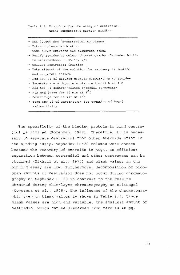
of the actual method is shown in Table 2.6.
2.3.1.2. Results
A standard curve, obtained after addition of known
amounts of oestradiol to radioactive steroid, evaporation
of the solvent and addition of the diluted rabbit uterine
cytosol is shown in panel A of Fig. 2.1.
31

w .,, £2
"' ·~· '"l 0/oE2 bou~d
'I• E2 bound
60
\ 60 50
'" A '" B <aj \ c
.. '"1 \ ' '"
I 'I
•'
'"' ' '" '"
'" '" '"
" ··+-~c-~~~--~~c-~~~------0 20 40 60 80 !00 120 1~0 P!l E2 0 20 40 50 BO 100 120 140 pg£2
o+-~------~~------~~-----
" '" '" 60 • - - - ~~
Fig. 2.1. Standard curves obtained for known amounts of oestradiol using
A. the competitive protein binding method.
B. the double competitive protein binding method,
C. radioimmunoassay.
Tatle 2.6. Procedure for the assay of oestradiol
using competitive protein binding
- Add 20,000 dpm 3H-oestradiol to plasma
- Extract plasma with ether
- Wash ether extracts and evaporate ether
- Purify residue by column chromatography (Sephadex LH-20,
toluene:methanol - 85:15, v/v)
- Collect oestradiol fraction
- Take aliquot of the solution for recovery estimation
and evaporate solvent
- Add 100 wl of diluted protein preparation to residue
- Incubate steroid-protein mixture for 17 hat 4°C
- Add 500 wl dextran-coated charcoal suspension
- Mix and leave for 10 min at 4°C
- Centrifuge for 10 min at 4°C
- Take 500 wl of supernatant for counting of bound
radioactivity
The specificity of the binding protein to bind oestra
diol is limited (Korenman, 1968). Therefore, it is neces
sary to separate oestradiol from other steroids prior to
the binding assay. Sephadex LH-20 columns were chosen
because the recovery of steroids is high, an efficient
separation between oestradiol and other oestrogens can be
obtained (Mikhail et al., 1970) and blank values in the
binding assay are low. Furthermore, decomposition of pica
gram amounts of oestradiol does not occur during chromato
graphy on Sephadex LH-20 in contrast to the results
obtained during thin-layer chromatography on silicagel
(Coyotupa et al., 1970). The influence of the chromatogra
phic step on blank values is shown in Table 2.7. Since
blank values are high and variable, the smallest amount of
oestradiol which can be discerned from zero is 40 pg.
33

w ..
Table 2.7. Blank values (means~ S.D.) obtained after extraction and/or chromatography
of oestradiol (E2 ) using different techniques
3 H-E2 added to water and
purified without chromatography
3H-E2
added to column effluent
after chromatography
3 H-E 2 added to water and
purified with chromatography
pg Ez found after correction for mass
of added 3H-E2
competitive protein
binding
24.7.:!:. 3.6 (4)
15.9 + 11.3 (116)
23.1 + 10.4 (36)
double competitive
protein binding
20.5 + 14.0 (16)
24.6 + 14.9 (10)
radioimmunoassay
33.6 ± 2.0 (4)
-2.9 + 4.4 (14)
1.2 ± 5.6 (267)
The number of estimations is given in parentheses.

Table 2.8. Accuracy and precision of the estimation of
oestradiol (E2
) by competitive protein
binding. Known amounts of oestradiol were
added to water and the solution was
processed by the method as outlined in
Table2.6.
Assay of n E2 found% S.D.
30 pg E2 3 35.3 pg 5. l pg
so pg E2 7 69. 1 pg 14.6 pg
human male plasma I 5 15.5 pg/ml 4.2 pg/ml
human male plasma II 6 21.4 pg/ml 5.8 pg/ml
%Values are corrected by subtraction of blanks.
n = number of estimations
S.D. standard deviation
C.V. = coefficient of variation: amou~t 0found x 100%
c.v.
14.4%
21.1%
27.1%
27.1%
Data on accuracy and precision of the method are summa
rized in Table 2.8. The oestradiol concentrations measured
in human male plasma using this competitive protein binding
technique agree well with values reported in the literature
(Baird, 1968). Recoveries of oestradiol after extraction
and chromatography were 86.2 + 10:7 (S.D.) % (n=44).
Estimation of oestradiol in peripheral plasma from male
rats in volumes between 1 and 5 ml resulted in percentages
binding which varied between 0.5 and 5.0, thus indicating
the presence of unlikely high concentrations (in the order
of >200 pg/ml) of "oestradiol-like" substances. This obser
vation is in agreement with data published by Labhsetwar
(1972), who estimated "oestradiol-liken substances in peri
pheral plasma from rats using radioimmunoassay and by
Weisz & Gunsalus (1973), who found similar results in
immature female rats. These observations will be discussed
in section 2.3.3.2.
35

2.3.2. Double competitive protein binding
2.3.2.1. Method
Since the specificity of the single competitive protein
binding technique was not sufficient for the estimation of
oestradiol in peripheral plasma from male rats, it was
decided to evaluate the double competitive protein binding
system as described originally by Robertson et al. (1971).
The procedure for this double competitive protein binding
system is summarized in Table 2.9.
36
Table 2.9. Procedure for the double competitiv.e protein
binding assay of oestradiol
- Add 20,000 dpm 3H-oestradiol to plasma
- Extract plasma sample with ether
- Wash ether extracts and evaporate ether
- Purify residue by column chromatography
(Sephadex LH-20, toluene:methanol- 85:15, v/v}
- Collect oestradiol fraction and evaporate solvent
- Add 200 ~1 of concentrated protein preparation to
residue
- Incubate for 30 min at 3Q°C
- Add 50 ~1 concentrated dextran-coated charcoal
suspension
- Incubate for 30 min at 30°C, centrifuge 5 min
- Take 200 ~1 supernatant and incubate for 3 min at 70°c.
Cool to 30°C
- Add 100 ~1 diluted protein preparation
- Incubate steroid protein mixture for 30 min at 30°C
- Take 100 ~1 for recovery estimation
- Add 500 ~1 diluted dextran-coated charcoal suspension
- Incubate for 30 min at 30°C
- Centrifuge for 10 min
- Take 500 ~l supernatant for counting of bound radioactivity

2.3.2.2. Results
A standard curve, obtained after addition of the concen
trated cytosol preparation to known amounts of oestradiol,
incubation, denaturation of the protein and competitive
protein binding, is shown in panel B of Fig. 2.1. Blank
values for the assay of column effluent or water, processed
through the procedure, are summarized in Table 2.7. These
values were not different from those obtained using the
single binding assay. Recoveries for the total procedure,
including extraction and purification, were 57.8 + 11.0
(S.D.) % (n=24).
Estimation of oestradiol in peripheral plasma from male
rats (5 rnl aliquots) gave a result of 2.0 ~ 0.7 (S.D.)
pg/rnl (n=3) after subtraction of the appropriate blank
values. The difference between these results and those
obtained with the single competitive protein binding assay
(>200 pg/ml, see 2.3.1.2.) suggests that additional sub
stances, different from oestradiol, are measured as oestra
diol in the single competitive protein binding assay.
2.3.3. Radioimmunoassay
2.3.3.1. Method
The method used for the estimation of oestradiol in
plasma samples using radioimmunoassay is described in
appendix paper 1. Additional procedures for the estimation
of oestradiol in testicular tissue are described in
appendix paper 2.
37

2.3.3.2. Results
A standard curve, obtained after addition of known
amounts of oestradiol to radioactive steroid, evaporation
of solvent and addition of antibody is shown in panel c of
Fig. 2.1.
The specificity of the antibody used for the estimation
of oestradiol has been described by Exley et al. (1971).
The specificity for estimation ·of oestradiol is high, but
incorporation in the radioimmunoassay method of the chroma
tographic procedure, using Sephadex LH-20 microcolurnns,
reduced blank values appreciably as shown in Table 2.7.
Results of accuracy and precision of the method are given
in Table 1 from appendix paper 1. The sensitivity of the
method, calculated from a blank value of 1.2 + 5.6 (S.D.)
pg (n=267), is 10.7 pg.
For peripheral plasma samples from male rats the low
percentages binding, as observed in the competitive protein
binding assay of oestradiol were never observed with this
radioimmunoassay using the antibody raised against the
oestradiol-6-(0-carboxymethyl)oxime-B.S.A. complex. This is
at variance with data published by Labhsetwar (1972) and
Weisz & Gunsalus (1973), who used antibodies against
oestradiol-178-hemisuccinate-B.S.A. for the estimation of
oestradiol in peripheral plasma from male or immature
female rats (see 2.3.1.2.). These authors suggested that
an "oestradiol-like" substance, probably of adrenal origin,
competes with oestradiol in their radioimmunoassay systems.
Therefore, experiments on the concentration of oestradiol
in peripheral plasma of adrenalectomized male rats might
have been performed in order to ensure that the "oestradiol
like" substance measured in the present study is different
from that described by Labhsetwar and Weisz & Gunsalus.
Since the concentration of circulating oestradiol in male
rats is very low, it was decided to perform such experi-
38

ments in immature female rats, where high oestradiol levels
are measured around the age of 15 days (Meijs-Roelofs
et al., 1973). As an additional advantage in this experi
mental procedure uterine weight can be recorded as a bio
assay estimate of circulating oestrogens in these animals.
Table 2.10. summarizes the results of experiments with
immature female rats which underwent either castration, or
adrenalectomy or both operations at the age of 13 days, and
were killed at the age of 15 or 17 days. Changes in oestra
diol levels were correlated with the changes in uterine
weight. It was concluded that the oestradiol measured was
not of adrenal origin. These data provide evidence for the
specificity of the estimation of oestradiol in the rat,
using this radioimmunological technique.
2.3.4. Discussion
The comparison of the competitive protein binding method
and the radioimmunological technique used for the estima
tion of oestradiol shows that the techniques have compa
rable accuracy and precision at the level of 30 and 50 pg
(see Table 2.8. and appendix paper 1, Table 1). Theoreti
cally, it might be expected that the sensitivity of the two
methods would also be comparable, since the association
constants for the protein-steroid complex, one of the fac
tors determining the lowest level of detection (Ekins &
Newman, 1970), are comparable for the rabbit uterine cyto
sol receptor (1-5 x 10 10 1/mole at 4°C, Mester et al.,
1971) and the antibody against oestradiol (4 x 10⁹
1/mole,
Exley et al., 1971). The similarity of the standard curves,
obtained with each of the three different methods, also
reflects that the three techniques have a comparable sensi
tivity of detection. However, the higher specificity of the
antibody causes an important increase in sensitivity when
extracts of water or biological materials are assayed. This
39

"" 0
Table 2.10. Effect of ovariectomy (OVX), adrenalectomy (ADRX) or OVX combined with ADRX
on day 13 post partum on the plasma oestradiol concentrations and uterine
weights of immature female rats on days 15 and 17 (means + S.D.)
(from: Meijs-Roelofs et al., 1973)
Treatment age at number of uterine weight oestradiol
on day l3 autopsy animals (mg/100 g b.w.) (pg/ml plasma)
15 3 81.5 + 5.9 46 (52. 0, 39. 0)
ovx 15 9 6 3. 6 .:t. 7.2 13 ( 15. 0, 11.2)
ADRX 15 14 75.2 + 10.8 54 (58. 0, 50. 0)
OVX + ADRX 15 12 39.6 ± 4. 2 0 (0' 0)
17 5 76.6 + 3.8 29 ( 31.91 25. 6)
ovx 17 8 49.9 ± 3.7 4 (0. 4, 7. 8)
ADRX 17 9 73.2 + 4 . 5 22 { 30. 6' 12.5)
OVX + ADRX 17 7 43.5 ± 4. 2 4 (0, 7. 3}

problem of specificity is even more important because of
the presence of "oestradiol-like" material in peripheral
plasma from the rat. The double competitive protein binding
method, which might be used to overcome this problem, still
lacks the specificity which is necessary to obtain a high
sensitivity. Therefore, only the radioimmunological tech
nique can be used to estimate oestradiol in plasma or
tissue from male rats, since it combines an acceptable
accuracy and precision with the required sensitivity.
For levels of oestradiol in peripheral plasma from male
rats it is still difficult, if not impossible, to compare
the specificity obtained with the radioimmunological tech
nique with the specificity obtained with other methods.
This is a consequence of the extremely low levels present:
although gas-liquid chromatographic methods (Exley & Dutton,
1969) and bioassay procedures (Martin & Claringbold, 1960)
for the estimation of picogram amounts of oestrogens have
been described, it has not been possible to use these
methods for the measurement of oestrogens in extracts from
biological samples (Wotiz et al., 1967).
Summarizing, it was concluded that radioimmunoassay is
the only technique which has the required sensitivity,
accuracy and precision to estimate oestradiol levels in
plasma and tissue of male rats. It is difficult to obtain
an independent assessment of the specificity of the radio
immunoassay method for the levels estimated, but several
observations (see section 2.3.3.2.) support the validity
of the estimation of oestradiol in male rats with this
particular radioimmunoassay system.
41

2.4. References
42
Adlercreutz, H., Salokangas, A. & Luukkainen, T. (1967)
in Memoirs of the Society for Endocrinology, No. 16,
pp. 89-113.
Astwood, E.B. (1938)
Endocrinology~~ 25-31.
Attal, J. & Eik-Nes, K.B. (1968)
Analyt. Biochem. ~. 398-411.
Baird, D.T. (1968)
J. clin. Endocr. Metab. 28, 244-258.
Bardin, C.W. & Peterson, R.E. (1967)
Endocrinology ~. 38-44.
Benraad, Th.J., de Jong, F.H., van der Molen, H.J. &
Smals, A.G.H. (1972)
J. Steroid Biochem. 1• 325-331.
Bojesen, E., Buus, o., Svendsen, R. & Thuneberg, L. (1967)
in Steroid Hormone Analysis (Carstensen, H. , ed. ) , Vol. I,
pp. l-53, Dekker, New York.
Brownie, A.C., van der Molen, H.J., Nishizawa, E.E.
& Eik-Nes, K.B. (1964)
J. clin. Endocr. Metab. £!, 1091-1102.
Coyotu!?a, ,:;,, Kinoshita, K., Ho, R.Y., Chan, C., Paul, \11.,
Foote, M. & Kushinsky, s. (1970)
Analyt. Biochern. l±• 71-73.
Diczfalusy, E. (ed.) (1970)
Acta endocr., Copnh. Supp1. lil· Dorfman, R.I. (ed.) (1969)
Methods in Hormone Research, Vol. IIA, Academic Press,
New York.
Dufau, M.L., Dulmanis, A., Catt, K.J, & Hudson, B. (1970)
J. clin. Endocr. Metab. l.Q_, 351-356.
Eik-Nes, K.B. & Horning, E.C. (eds.) (1968)
Gas Phase Chromatography of Steroids, Springer Verlag,
Berlin.
Ekins, R. & Newman, B. (1970)
Acta endocr., Copnh. Suppl. !!2• 11-3E. Enunens, c.w. (1941)
J, Endocr. ~· 444-458.

Exley, D. & Dutton, A. {1969)
Steroids li• 575-590.
Exley, D., Johnson, M.W. & Dean, P.D.G. (1971)
Steroids ~. 605-620.
Kautsky, M.P., Martin, R.P., Janda, v., Woolever, C.A. &
Hagerman, D.O. (1970)
Am. J. Obstet. Gynec. lQ£, 447-454.
Korenman, S.G. (1968)
J. clin. Endocr. Metab. 28, 127-130.
Korenman, S.G., Perrin, L.E. & McCallum, T.P. (1969)
J. clin. Endocr. Metab. ~· 879-883.
Labhsetwar, A.P. (1972)
J. Endocr. ~. 399-400.
Lindner, H.R. (1959)
Nature, Land. ~. 1605-1606.
Loraine, J .A. & Bell, E.T. (1966)
Hormone Assays and their Clinical Applications,
Livingstone, Edinburgh.
Martin, L. (1960)
J. Endocr. l..Q., 187-197.
Martin, L. & Claringbold, P.J. (1960)
J. Endocr. l.Q_, 173-186.
Mester, J., Robertson, D.M. & Kellie, A.E. (1971)
J. Steroid Biochem. ~· 1-10.
Meijs-Roelofs, H. M.A., Uilenbroek, J.Th.J., de Jong, F.H. &
Welschen, R. {1973)
J. Endocr. 21• 295-304.
Mikhail, G., Wu, C.H., Ferin, M. & VandeWiele, R.L. (1970)
Steroids !2• 333-352.
van der Molen, H.J. (1971)
in The Androgens of the Testis {Eik-Nes, K.B., ed.),
pp. 145-215, Dekker, New York.
van der Molen, H.J. & Aakvaag, A. {1967)
in Hormones in Blood (Gray, C. H. & Bacharach, A.L., eds.),
Vol. 2, pp. 221-303, Academic Press, London.
van der Molen, H.J. & de Jong, F.H. (1974)
in Methods of Hormone Analysis (Breuer, H. & KrUskemper, H.L.,
eds.), Georg Thieme Verlag, Stuttgart, in the press.
Neher, R. & Wettstein, A. {1960)
Helv. chim. Acta i}, 1628-1639.
O'Donnell, V.J. & Preedy, J.R.K. {1967}
in Hormones in Blood {Gray, C. H. & Bacharach, A. L., eds.) ,
Vol. 2, pp. 109-186, .~.cademic Press, London.
43

44
Oertel, G.W., West, C.D. & Eik-Nes, K.B. (1959)
J. clin. Endocr. Metab. !1· 1619-1625.
Preedy, J.R.K. (1962}
in Estrogen Assay in Clinical Medicine (Paulsen, C.A., ed.},
pp. 162-167, University of Washington Press, Seattle.
Robertson, D.M., Mester, J. & Kellie, A.E. (1971)
Acta endocr., Copnh. ~. 513-522.
Salokangas, R.A.A. & Bulbrook, R.D. (1961)
J. Endocr. ~. 47-58.
Slaunwhite, w.R. & Neely, L. (1963)
Analyt. Biochem. 2• 133-142.
Verjans, H.L., Cooke, B.A., de Jong, F.H., de Jong, c.M.M. &
van der Molen, H.J. (1973)
J. Steroid Biochem. !• 665-676.
Weisz, J. & Gunsalus, P. (1973}
Endocrinology 21• 1057-1065.
Wotiz, H.H., Charransol, G. & Smith, I.N. {1967)
Steroids .!.Q, 127-154.

CHAPTER 3. EXPERIMENTS IN VIVO
3 .1. Introduction
Laqueur et al. (1927) described the presence of oestro
genic activity in urine from normal men. This was the first
observation of the production of oestrogens in male mammals.
Since then, several authors have attempted to define the
source of these oestrogens, initially often by experiments
in which testicular or adrenal function was stimulated or
suppressed. The resulting changes in the concentration of
urinary oestrogens were considered to reflect production
and secretion of these steroids by gonads or adrenals.
However, the concentration of oestrogens in urine increases
after peripheral administration of androgens to men
(Steinach & Kun, 1937), or even to adrenalectomized, gona
dectomized subjects. The latter observation indicates that
androgens can be converted to oestrogens in tissues, which
are generally regarded as non-endocrine organs, provided
that precursor steroids ( "prehormones 11, Baird et al., 1968)
are present in the circulation. This phenomenon could
explain the increased urinary levels of oestrogens after
trophic stimulation of testes or adrenals, and in the same
way castration or adrenalectomy should result in a
decreased concentration of oestrogens in urine. Therefore,
no conclusion about the origin of oestrogens in men can be
drawn from these qualitative experiments.
In this chapter, it will be attempted to review the
available data on the source and production of oestrogens
in the male. The first part of the chapter concentrates on
the testis as a source of circulating oestradiol. The
45
., "'
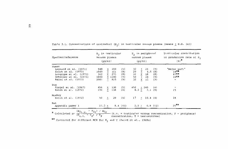
Table 3.1. Concentrations of oestradiol (E2
) in testicular venous plasma (means _:t S.D. (n))
E2 in testicular
Species/reference venous plasma
(pg/ml)
Human Leonard et al. {1971) 948 + 200 Kelch et al. ( 19 7 2) 1049 + 161 Longcope et al. (1972) 342 + 275 Scholler et al. ( 19 7 3) 1880 ~ 1240 Baird et al. (1973) 2081 + 825
Dog Siegel et al. ( 19 6 7} 456 + 130 Kelch et al. (1972) 100 + 138
Monkey Kelch et al. (1972) 50
"' 20
Rat Appendix paper 1 17.5 .:t 8. 4
E2
in peripheral
venous plasma
(pg/ml)
(5) 32 + 21 (5) (8) 20 + 4. 5 (8) (8) 32 + 18 (81 19 I 50 + 28 (9) (9) 32 + 15 (9)
( 5) 450 + 240 (4) 14 I 8. 2 + 2. 1 19 I
(4) 17
"' 10.4 14 I
{4 3) 2. 0 ± 0.9 ( 12)
Testicular contribution
to production rate of E2 (%)~
"major part" 14*)f 67** 16*:11
15
24
zs**
* Calculated (Ezt.v. - Ezpl I Ezp . .
as (T T ) 7 T (t.v. = test~cular venous concentratLon, P peripheral t.v. P P concentration, T = testosterone)
**Corrected for different MCR for E2
and T (Baird et al., 1969a)

second part of this chapter deals with the extra testicular
production of oestradiol.
3.2. The testis as a source of oestrogens
3.2.1. Oestradiol in testicular venous blood
Final proof of secretion of a hormone from an endocrine
gland can only be obtained by comparing the concentration
of the hormone in the affluent (arterial) and effluent
(venous) blood of the gland. In practice, it is generally
assumed that the hormone concentration in the arterial
blood or plasma is equal to that in the peripheral circu
lation. However, Free & Tillson (1973) showed that peri
pheral and glandular arterial concentrations of a hormone
are not necessarily equal.
French et al. (1965) showed oestradiol secretion from
the testes of a human pseudohermaphrodite by measuring a
difference between the concentrations of the steroid in
testicular venous and peripheral plasma. Results on the
secretion of oestradiol from normal testes from different
species are summarized in Table 3.1. In this table the
contribution of testicular secretion of oestradiol to the
blood production rate of the steroid is calculated from
the ratios of testicular venous and peripheral concentra
tions of oestradiol and testosterone; the peripheral con
centration of the latter steroid is assumed to result
completely from testicular secretion. According to this
calculation, the testis secretes 15-25% of the circulating
oestradiol. This may lead to the conclusion that· the oes
trogens directly secreted by the testis play a quantita
tively less important role in possible peripheral processes
than the oestrogens formed from prehormones, which may be
secreted by the adrenal or the testis also. A similar con
clusion can be drawn from the calculations of blood pro-
47

duction rates and from peripheral production rates of
oestradiol in men. These data will be discussed in
section 3. 3.
3.2.2. Oestradiol in testicular tissue
The presence of oestrogenic activity in testis tissue
was first shown by Fellner (1921), who described the
effects of extracts of bovine testes on uterus and mammary
tissue of female rabbits, and concluded that the histolo
gical results obtained with testis extracts were comparable
with those obtained after injections of ovarian or placen
tal extracts. Many authors described the detection and
measurement of oestrogenic hormones in testes from diffe
rent species (see: Diczfalusy & Lauritzen, 1961, Parkes,
1966, Table 3.2.). It is difficult to predict the concen
trations of the physiologically active, unconjugated
oestradiol in the testis from stallion or man from the
data in Table 3.2., since no correction for recovery was
performed in the estimations in these species. Furthermore,
hydrolytic procedures were used in these estimations, while
Raeside (1969) showed that at least in stallion testes the
larger part of testicular oestrogen is present as the sul
phate conjugate.
Table 3.2. Oestradiol in testis tissue from different species
Species Concentration Reference
{ng/g)
Horse 210 Beall (1940b)
Human (adult) 5. 7 Goldzieher ' Roberts ( 19 52)
8 Anliker et al. {1957)
(foetal) !.9 Reyes et al. ( 19 7 3)
Sheep (adult) 0. 035, 0.065 At tal et al. {1972)
(foetal) 20 At tal ( 1969)
Rat 0.038 Appendix paper 2
48

3.2.3. Oestrogens in semen
Apart from the production of hormones, which are secre
ted in the testicular venous blood, the testis produces
spermatozoa. These spermatozoa are ejaculated in the semen,
together with excretions from prostate and seminal vesicles.
Therefore, substances present in semen might be of testi
cular origin, and probably originate from the seminiferous
tubules. The presence of oestrogenic material in human
semen was first suggested by Green-Arrnytage (1943),
and then confirmed by Green-Armytage et al. (1947),
Riisfeldt (1948) and McCullagh & Schaffenburg (1951).
These authors used a bioassay system for the assessment of
oestrogen concentrations in semen. Diczfalusy (1952) was
the first to assay seminal oestrogens by a physico-chemical
technique, i.e. fluorimetry, using counter-current distri
bution as a means of separating and identifying oestrone,
oestradiol and oestriol.
Schaffenburg & McCullagh (1954) showed the presence of
oestrogenic material in bull spermatozoa, and suggested
the germinal epithelium as a possible source of seminal
oestrogens, whereas Raboch & Rezabek (1963) defined the
Leydig cell as the place of oestrogen production after bio
assay of seminal oestrogens from patients with different
testicular disorders. Another possible explanation for the
presence of oestrogens in semen is a transfer of oestrogens
from blood to semen via prostate or seminal vesicles. This
possibility is supported by the observations of Eriksson &
Baker (1966), who observed that oestrogens were transferred
to female rats after copulation with male rats, that were
treated by peripheral oestradiol-benzoate injections.
Furthermore, Stanek & Dostal (1972) showed the presence of
plasma proteins in seminal vesicle fluid, and Steene et al.
(1969) and Tea et al. (1972) found a close correlation
between peripheral plasma levels and seminal plasma levels
of dehydroepiandrosterone sulphate and oestrone/oestradiol
49

respectively. The observation of the latter authors, that
seminal oestrogen levels are higher than those in periphe
ral plasma, can be explained by the observation of
Varkarakis et al. (1972), that oestrogens which were taken
up from the blood are excreted in the prostatic fluid.
3.2.4. Regulation of the testicular production and secretion of
oestrogens
Maddock & Nelson (1952) observed after prolonged admi
nistration of HCG to men an increased urinary excretion of
oestrogens, concomitant with testicular Leydig cell hyper
plasia. This increase in oestrogen excretion was larger
than that of 17-ketosteroids. These observations were con
firmed by Diczfalusy (1957), Givner et al. (1960), Jayle
et al. (1962), Morse et al. (1962) and Smith (1971). Conti
et al. (1955) and Fr¢land et al. (1973) excluded adrenal
influences through administration of dexamethasone together
with the trophic hormone. The same conclusion was reached
by Fishman et al. (1967), who observed that the relative
increase of the oestradiol production rate always super
seded that of testosterone after administration of HCG to
five men. Similar studies were performed in boars by Busch
& Ittrich (1970) and Liptrap & Raeside (1971). It was con
cluded from these observations that the secretion of oes
tradiol is under the influence of HCG, and that the produc
tion of testicular oestradiol takes place in the intersti
tial compartment of the testis.
It is not possible, however, to discuss an increase of
the testicular secretion of oestradiol on basis of data
obtained by measuring urinary concentrations of the steroid,
since the metabolism of its main peripheral precursor, tes
tosterone (see section 3.3.), changes when the concentra
tion is increased above physiological levels (Southren
et al., 1968). This is certainly the case after stimulation
50

with HCG. Furthermore, the urinary method for the measure
ment of the production rate of oestradiol is not satisfac
tory (section 3.3.). Therefore, the method of choice for
the study of the regulation of production and secretion of
testicular oestradiol has to be the experiment at the level
of the testis itself, and not the study of peripheral para
meters. Such experiments have been described by Kelch et al.
(1972), who observed that the concentrations of oestradiol
in canine testicular venous blood were unchanged after
administration of HCG. Weinstein et al. (1974) showed,
however, that prolonged administration of HCG to men caused
an increased secretion of oestradiol from the testis: this
(eightfold) increase in the secretion of oestradiol was
higher than that for testosterone (fivefold). The experi
ments described in the appendix papers 1 and 2 lead to the
conclusion that the testicular secretion of oestradiol in
the rat is not influenced by prolonged administration of
HCG. The reason for this species difference is not clear.
In addition to the experiments described in the appendix
papers 1 and 2, a more detailed study was performed in
order to investigate the effects of hypophysectomy, prolac
tin administration and increased amounts of interstitial
tissue on the concentration of oestradiol and testosterone
in testis tissue and in testicular venous plasma. The expe
rimental procedures used were similar to those described in
appendix papers 1 and 2. The effect of hypophysectomy on
steroid concentrations in testicular tissue and testicular
venous plasma is shown in Fig. 3.1. and 3.2. Testosterone
levels in both testicular venous blood and in testis tissue
decrease with increasing length of hypophysectomy. The
decrease in the tissue concentration of oestradiol shows
the same trend. However, there was no significant change in
the testicular venous concentration of this steroid after
hypophysectomy, in the same way as there was no change
after prolonged HCG administration (appendix paper 1).
These results indicate that the net production of oestra
diol in the testis is not influenced by trophic stimulation.
51

52
Fig. 3.1
1.8
1.4
10
0.6
0.2
0.01
0
[ill tissue, )Jg/g
0 plasma,)Jg/ml
-12d hypox !lh... 6d hypox control Sd HCG
Testosterone concentrations (means~ S.E.M.) in testicular venous
plasma and testis tissue from hypophysectomized (HYPOX), intact
(CONTROL) and HCG-treoted (5 d HCG) intact rots.
60 l 40
[ill tissue. pg/g
0 plasma,pg/ml
20
0
Fig. 3.2.
JJ~ 12d hypox 6d hypox
.
.
·.
control Sd HCG
Oestradiol concentrations (means~ S.E.M,) in testicular venous
plasma and testis tissue from hypophysectomized (HYPOX), intact
(CONTROL) and HCG-treated (5 d HCG) intact rots.
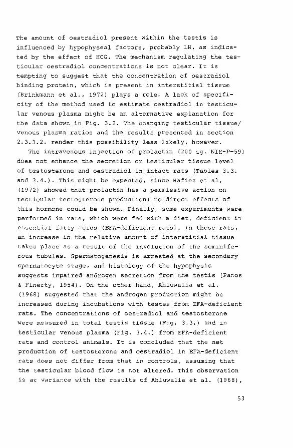
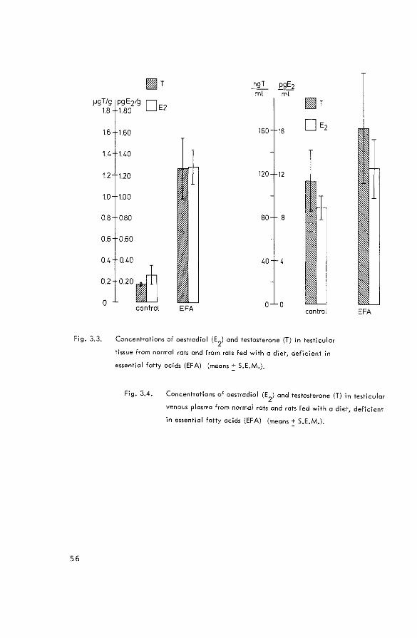
The amount of oestradiol present within the testis is
influenced by hypophyseal factors, probably LH, as indica
ted by the effect of HCG. The mechanism regulating the tes
ticular oestradiol concentrations is not clear. It is
tempting to suggest that the concentration of oestradiol
binding protein, which is present in interstitial tissue
(Brinkmann et al., 1972) plays a role. A lack of specifi
city of the method used to estimate oestradiol in testicu
lar venous plasma might be an alternative explanation for
the data shown in Fig. 3.2. The changing testicular tissue/
venous plasma ratios and the results presented in section
2.3.3.2. render this possibility less likely, however.
The intravenous injection of prolactin (200 ~g, NIH-P-59)
does not enhance the secretion or testicular tissue level
of testosterone and oestradiol in intact rats (Tables 3.3.
and 3.4.). This might be expected, since Hafiez et al.
(1972) showed that prolactin has a permissive action on
testicular testosterone production; no direct effects of
this hormone could be shown. Finally, some experiments were
performed in rats, which were fed with a dietf deficient in
essential fatty acids (EPA-deficient rats). In these rats,
an increase in the relative amount of interstitial tissue
takes place as a result of the involution of the seminife
rous tubules. Spermatogenesis is arrested at the secondary
spermatocyte stage, and histology of the hypophysis
suggests impaired androgen secretion from the testis (Panos
& Finerty, 1954}. On the other hand, Ahluwalia et al.
( 1968) suggested that the androgen production might be
increased during incubations with testes from EPA-deficient
rats. The concentrations of oestradiol and testosterone
were measured in total testis tissue (Fig. 3.3.) and in
testicular venous plasma (Fig. 3.4.) from EPA-deficient
rats and control animals. It is concluded that the net
production of testosterone and oestradiol in EPA-deficient
rats does not differ from that in controls, assuming that
the testicular blood flow is not altered. This observation
is at variance with the results of Ahluwalia et al. (1968),
53

"' "' Table 3.3. Concentrations of oestradiol (E2
J and testosterone (T) in testicular venous
plasma of intact male rats after administration of prolactin (means+ S.D.)
Treatment E2 T E2
:T
(pg/ml) (ng/ml} (pg:ng)
none 11.3 + 3.4 { 6) 49.9 + 27.5 {6) 0.28 .:t. 0.13
200 ~g prolactin i.v. • 4.9 + 2.5 { 5) 79.8 + 18.2 { 5) 0.06 + 0.03
The number of estimations is given in parentheses.
* 200 ~g prolactin (NIH-P-59) were administered immediately before collection of
testicular venous blood.
{ 6)
{ 5)
Table 3.4. Concentrations of oestradiol (E2
) and testosterone {T) in testis tissue of
intact male rats after administration of prolactin (means .:t. S.D.)
Treatment E2 T E2
:T
{pg/g) (ng/g) (pg:ng)
none 19.6 .:t. 11.0 ( 12) 186 :t. 67 { 6) 0.122:0.05
200 ~g prolactin i.v.~ 16.4 + 5. 7 ( l 0) 137 + 92 { 5) 0.16 + 0.12
The number of estimations is given in parentheses.
X 200 ~g prolactin {NIH-P-59) were administered immediately before collection of
testicular venous blood.
{6)
{5)

probably because these authors did not correct their
results for the altered ratio between interstitial tissue
and seminiferous tubules in testes from EPA-deficient
animals.
The high tissue concentrations for testosterone and
oestradiol when compared with those in control animals
might reflect that both steroids are localized in the
interstitial tissue.
3.3. Extratesticular sources of oestradiol
From the data in Table 3.1. it appears that the testis
secretes 15-25% of the total amount of oestradiol in the
peripheral circulation in different species. These calcu
lations were based on the ratios of the concentrations of
oestradiol and testosterone in testicular venous and peri
pheral plasma, and on the assumption that peripheral tes
tosterone is completely secreted by the testis. In this
section, the testicular contribution to the total blood
production will be calculated by comparison of the produc
tion rate of oestradiol with the rate of conversion of pre
hormones to this steroid. The difference between these
quantities represents the amount of oestradiol which is
secreted by endocrine glands: the testes, as discussed
previously, or the adrenals; Corey & Britton (1934) were
the first to describe oestrogenic activity in adrenal
tissue. It will be concluded that the testis is the main
glandular source of oestradiol, while about 80% of the
circulating oestradiol is formed peripherally from testos
terone or oestrone.
55
)JgT/g 1.8
16
14
12
10
0.8
0.6
0.4
0.2
0
Fig. 3.3.
56
[if{fjT ngT pgE2 rnt ml
pgE2/g 0E2 1.80
~T
160 160 16 D E2
140
120 120 12
100
0.80 BO B
0.60
040 40
020~ control EFA 0 0
control EFA
Concentrations of oestradiol (E2
) and testosterone (T) in Testicular
tissue from normal rats and from rats fed with a diet, deficient in
essential fatty acids (EFA) (means~ S.E.M.).
Fig. 3.4. Concentrations of oestradiol (E2
) and testosterone (T) in testicular
venous plasma from normal rots and rats fed with a diet, deficient
in essential fatty acids (EFA) (means! S.E.M.),
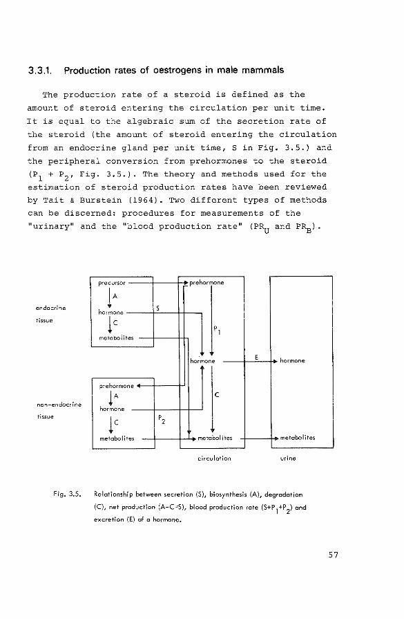
3.3.1. Production rates of oestrogens in male mammals
The production rate of a steroid is defined as the
amount of steroid entering the circulation per unit time.
It is equal to the algebraic sum of the secretion rate of
the steroid (the amount of steroid entering the circulation
from an endocrine gland per unit time, Sin Fig. 3.5.) and
the peripheral conversion from prehorrnones to the steroid
(P1
+ P2 , Fig. 3.5.). The theory and methods used for the
estimation of steroid production rates have been reviewed
by Tait & Burstein (1964). Two different types of methods
can be discerned: procedures for measurements of the
"urinary" and the 1'blood production rate" (PR0
and P~).
endocrine
tissue
non-endocrine
tissue
Fig. 3.5.
precursor prehormone
lA s hormone
lc metabolites
pl
ho;mone E hormone
pre hormone
lA c hormone
lc p2
metabolites metabolites metabolites
circulation urine
Relationship between secretion (S), biosynthesis (A), degradation
(C), net production (A-C=:S), blood production rate (S+P1+P
2) and
excretion (E) of a hormone.
57

"' "'
Table 3.5. Urinary production rates (~g/24 h) of oestradiol in normal men (means~ S.D. (n))
Radioactivity
injected as
Oestradiol
Oestradiol
Oestrone
Oestradiol
Oestradiol
Oestradiol
Oestrone/
oestradiol
Production rate calculated
from specific activity of urinary
oestrone
49, 90
67 :!:. 10 (5)
46 .:!:: 13 (12}
402:_12(7)
oestradiol
47' 89
70 :!:. 13 I 51
2 9 .:!:. 14 19 I
32 .:!:. 13 16 I
40
42 ±_ 12 I 71
oestriol
65, 169
234 :!:. 342 151
Reference
Morse et al. (1962)
Lipsett et al. (1966}
Crowell et al. (1967)
Eren et al. (1967)
Gabrilove et al, ( 1970)
MacDonald et al. (1971)
Kirschner & Taylor (1972)
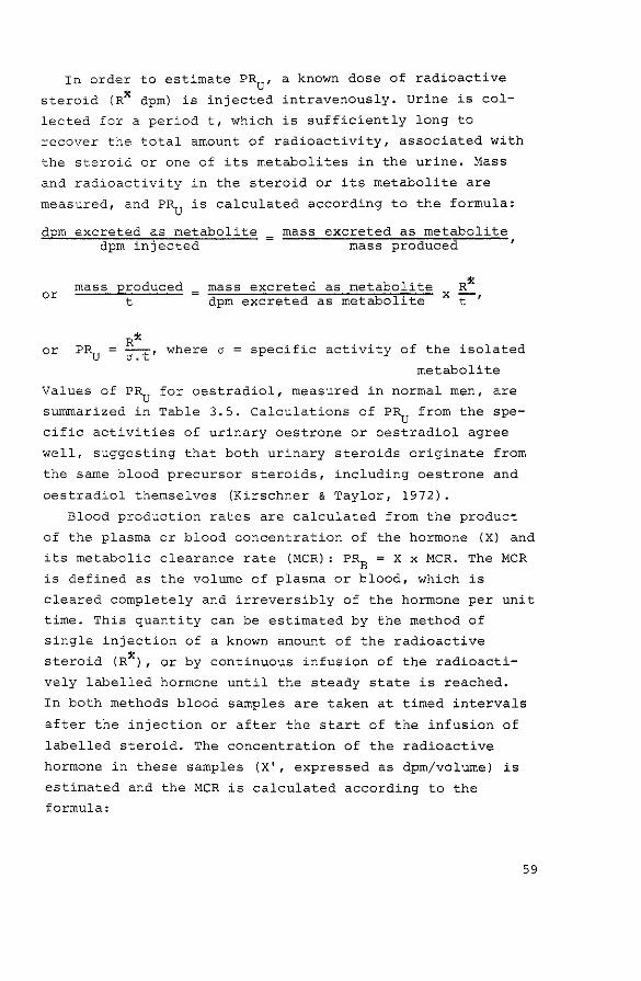
In order to estimate PR0
, a known dose of radioactive
steroid (R* dpm) is injected intravenously. Urine is col
lected for a period t, which is sufficiently long to
recover the total amount of radioactivity, associated with
the steroid or one of its metabolites in the urine. Mass
and radioactivity in the steroid or its metabolite are
measured, and PR0
is calculated according to the formula:
dpm excreted as metabolite dpm injected
mass excreted as metabolite mass produced
or mass produced t
mass excreted as metabolite R* dpm excreted as metabolite x ~,
R* or PR0 = cr.t' where a specific activity of the isolated
metabolite
Values of PR0
for oestradiol, measured in normal men, are
summarized in Table 3.5. Calculations of PR0
from the spe
cific activities of urinary oestrone or oestradiol agree
well, suggesting that both urinary steroids originate from
the same blood precursor steroids, including oestrone and
oestradiol themselves (Kirschner & Taylor, 1972).
Blood production rates are calculated from the product
of the plasma or blood concentration of the hormone (X) and
its metabolic clearance rate (MCR): PRB = X x MCR. The MCR
is defined as the volume of plasma or blood, which is
cleared completely and irreversibly of the hormone per unit
time. This quantity can be estimated by the method of
single injection of a known amount of the radioactive
steroid (R*) , or by continuous infusion of the radioacti
vely labelled hormone until the steady state is reached.
In both methods blood samples are taken at timed intervals
after the injection or after the start of the infusion of
labelled steroid. The concentration of the radioactive
hormone in these samples {X 1, expressed as dprn/volume) is
estimated and the MCR is calculated according to the
formula:
59

"' 0
Table 3.6. Plasma concentrations, plasma metabolic clearance rates (MCRP) and blood production rates
(PR8
) of oestradiol (E 2 ) in normal men (means~ S.D. (n))
Method Method for E2
for MCRP plasma E2 * (ng/1)
continuous inf.
continuous inf.
continuous inf,
single injection CPB 26 (31
DID 26 .:+:: 6 (7 I
CPB 24 + 6.5 (6 I
CPB 30 + 9 ( 15)
1:' CPB
DID
competitive protein binding method
double isotope dilution method
MCRP
(l/24 h)
1890 .:+:: 330
1640 ~. 313
1597 .:+:: 234
2430
(11)
(51
(91
( 3 I
PRB
{ug/24 h)
62 ( 3)
Reference
Longcope et al. (1968)
Hembree et al. (1969)
Longcope & Tait (1971)
Concolino et al. (1970)
Baird & Guevara (1969)
Korenman et al. (1969)
Mayes & Nugent (1970)
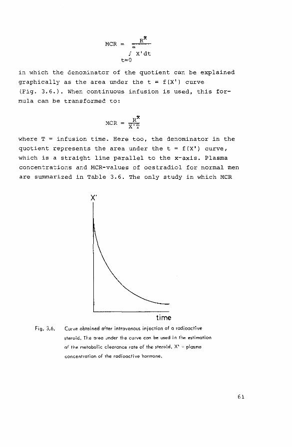
MCR
! X'dt t=O
in which the denominator of the quotient can be explained
graphically as the area under the t = f(X') curve
(Fig. 3.6.). When continuous infusion is used, this for
mula can be transformed to:
where T = infusion time. Here too, the denominator in the
quotient represents the area under the t = f(X') curve,
which is a straight line parallel to the x-axis. Plasma
concentrations and MCR-values of oestradiol for normal men
are summarized in Table 3.6. The only study in which MCR
Fig. 3.6.
X'
Curve obtained after intravenous iniection of a radioactive
steroid. The area under the curve con be used in the estimation
of the metabolic clearance rate of the steroid. X'"' plasma
concentration of the radioactive hormone.
61

and plasma concentration for oestradiol have been measured
in the same subjects is that of Concolino et al. (1970).
Calculation of the product of mean values for plasma con
centr·ation and MCR results in a PRE of 45 \Jg/24 h.
It might be concluded from the agreement between the
values for the production rates of oestradiol obtained by
urinary and blood methods, that a reliable estimate of this
production rate in men can be obtained using either of the
two types of measurement. However, both the urinary method
(Lipsett et al., 1966) and the blood method (Hembree et al.,
1969) have been criticized. The influence of the entero
hepatic circulation of oestrogens (Sandberg & Slaunwhite,
1957, Howard et al., 1969) and the existence of an
oestrone sulphate pool, which is completely formed from
circulating unconjugated oestrogens (Ruder et al., 1972,
Longcope, 1972), make it unlikely that oestradiol is irre
versibly metabolized, as is assumed in calculating the MCR.
Finally, a diurnal rhythm in the plasma concentration of
oestrogens in normal men has been described (Baird &
Guevara, 1969). This phenomenon makes it more difficult to
calculate PRE from plasma concentrations and MCR.
Production rates of oestradiol in male subjects of
other species than the human, have not been reported in the
literature. For male rats the blood production rate of
oestradiol was calculated in appendix paper 1 from the
estimated concentration of the steroid in peripheral plasma
and the MCR in normal men (Longcope et al., 1968), which
was corrected for body weight according to the formula:
MCR • (weight) 4
constant
This approximation was found to be valid in a comparison
between the MCR for oestradiol in female humans and rats
by de Hertogh et al. (1970), even though sex steroid bin
ding globulin is not present in rat plasma. The calculated
MCR of 25.6 1/24 h in male rats with a body weight of 225 g,
62

combined with a peripheral plasma concentration of 2 pg/ml
(appendix paper 1) resulted in a production rate of 51 ng/
24 h.
3.3.2. Peripheral conversion of prehormones to oestradiol
Diczfalusy & Lauritzen (1961) concluded from studies on
conversion of peripherally administered androgens to uri
nary oestrogens that this conversion could occur in the
absence of steroid-secreting glands, thus excluding the
possibility of androgen-stimulated oestrogen secretion. The
same conclusion was reached by Braun-Cantilo et al. (1962),
who administered tritiated testosterone to normal and
ovariectomized women and isolated labelled oestrogens from
urine: these authors showed the direct conversion from an
androgen to oestrogens. In a similar way, the direct con
version of testosterone to urinary oestrogens was also
shown by Ahmad & Morse (1965) and Epstein et al. (1966).
Peripheral androstenedione is also a good precursor for
urinary oestrogens (MacDonald et al., 1967) while the con
version of dehydroepiandrosterone and its sulphate to
urinary oestrogens is low in men and non-pregnant women
(Mancuso et al., 1965, Oertel & Menzel, 1970, Adams &
Brown, 1971).
Quantitative data on the contribution of peripheral con
version of prehormones to the production rate of oestradiol,
which was assumed to be 45 ~g/24 h (Tables 3.5. and 3.6.),
are summarized in Table 3.7. The theoretical aspects of
this type of calculations have been reviewed extensively by
Baird et al. (1968). Blood production rates of the prehor
mones are calculated from MCR and peripheral concentrations.
The conversion ratio for the peripheral conversion of the
prehormone to oestradiol, represents the ratio of 3H- and 14c-label in plasma oestradiol during simultaneous infusion
of prehormone and oestradiol each marked with a different
63
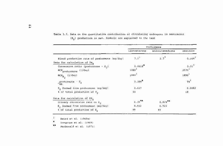
"' ... Table 3.7. Data on the quantitative contribution of circulating androgens to oestradiol
(E2
) production in men. Symbols are explained in the text
Blood production rate of prehormone {mg/day)
Data for calculation of PR8
Conversion ratio (prehormone - E2 )
MCRprehormone (!/day)
MCRE (1/day) 2
~prehormone - E2 "BB
E2 formed from prehormone (mg/day)
% of total production of E2
Data for calculation of PRU
Urinary conversion rate to E2
• ••
E2
formed from prehormone (mg/day)
% of total production of E2
Baird et al. {1969a}
Longcope et al. (1969)
MacDonald et al. ( 19 71)
testosterone
7.2§
0.0018:11.
1080~
1890"
0.38%:11.
0.027
60
0.3%!1(:11.
0.022
49
Prehorrnone
androstenedione
2. 7 §
0.82%:t:t
0.022
49
oestrone
0. 164 5
0. OS§
2570:\
1890§
5%'
0.0082
18

radioactive label until steady state conditions are
reached. Correction of the conversion ratio for the diffe
rence in MCR for prehorrnone and product (oestradiol)
results in the transfer constant p. The transfer constant
represents the fraction of circulating prehorrnone which is
converted to oestradiol. The mass of oestradiol, arising
from peripheral proCuction from prehormones, is then cal
culated as the product of p and the production rate of the
prehormone. On basis of such calculations, the total con
tribution of peripheral conversion of prehormones to the
production rate of oestradiol in men is calculated as 78%.
Using urinary conversion ratios in these calculations,
it appears from the data in Table 3.3. that the total pro
duction rate of oestradiol can be accounted for by the
peripheral production of the steroid from testosterone and
androstenedione. The urinary conversion rate is calculated
after simultaneous injection of prehormone and product,
each marked with a different radioactive label, collection
of urine, and estimation of the isotopic ratio in the iso
lated product steroid or one of its metabolites. However,
Kelly & Rizkallah (1973) showed that this type of calcula
tion may be fallacious in case of the conversion of andro
gens to oestrogens, because the isotopic ratio was not
constant in the oestrogens and their metabolites.
The site of the peripheral conversion of androgens to
oestrogens is unknown, but it has been shown during incu
bation studies that this conversion can occur in brain
tissue (Naftolin et al., 1971), in the liver (Wortmann
et al., 1973), in fat cells (Schindler et al., 1972) and
in intestinal bacteria (Goddard & Hill, 1972).
Quantitative studies on the peripheral conversion of
androgens to oestrogens in male experimental animals have
not yet been described.
65

3.3.3. The adrenal as a source of oestrogens
Baird et al. (1969b) and Saez et al. (1972) estimated
concentration gradients of oestrone over the adrenal gland
in human subjects but did not detect a secretion of oestra
diol. Levy et al. (1965) and Meyer (1955) isolated
19-hydroxyandrostenedione after perfusion of human and
bovine adrenals: this steroid is a possible intermediate
in the biosynthesis of oestrogens from androstenedione.
Beall (194Da) isolated oestrone from bovine adrenal tissue,
and in vitro biosynthesis of oestrogens during incubation
of normal adrenal tissue has been described, although the
conversions were always very small (Engel & Dimoline, 1963,
Chang et al., 1963, Vinson & Jones 1964). Feminizing
adrenal tumours are a well-known clinical disorder
(reviewed by Gabrilove et al., 1965 and Vague et al., 1970).
Radioactive aromatic steroids have been isolated after
incubation of tumour tissue with pregnenolone (Rose et al.,
1969, Axelrod et al., 1969). The urinary excretion of
oestrogens is increased after administration of ACTH and
decreased after adrenalectomy (Brown et al., 1959, Engel,
1962, Schweppe et al., 1967); peripheral oestrone levels
follow the same pattern (Saez et al., 1972). Such data,
however, do not yield information on the question of direct
secretion of oestrogens by the adrenal.
Summarizing, there is evidence that the adrenal secretes
oestrone. There are no indications for the secretion of
oestradiol from the normal adrenal gland, and information
on the regulation of the adrenal oestrogen secretion is not
available.

3.4. Conclusions
From the results presented in the appendix papers 1 and
2 and in this chapter, the following conclusions are drawn:
1. The testis of the normal adult rat secretes oestradiol.
2. In the rat and in man, the amount of oestradiol secreted
by the testis constitutes about 20% of the total blood
production rate for this steroid.
3. The testicular secretion of oestradiol is not affected
by the prolonged administration of HCG or by hypophy
sectomy. However, the testicular tissue levels of
oestradiol are clearly changed by these treatments.
4. No effect of FSH or prolactin on the testicular secre
tion or production of oestradiol or testosterone could
be shown.
5. Oestradiol is mainly localized in the interstitial
tissue of the testis.
6. The occurrence of oestrogens in seminal fluid does not
reflect that oestrogens are produced in either the
seminiferous tubules or interstitial tissue.
7. The direct adrenal contribution to the circulating con
centration of oestradiol is negligible.
3. 5. References
Adams, J.B. & Brown, J.B. (1971)
Steroidologia £, 1-6.
Ahluwalia, B., Shima, S. & Pincus, G. (1968)
J. Reprod. Fert. 11· 263-273.
Ahmad, N. & Morse, w.r. (1965)
Can. J. Biochem. il• 25-31.
Anliker, R., Perelman, M., Rohr, 0. & Ruzicka, L. (1957)
Helv. chim. Acta 40, 1517-1520.
67

68
Attal, J. (1969)
Endocrinology~, 280-289.
Attal, J., Andre, D. & Engels, J.-A. (1972}
J. Reprod. Pert. 28, 207-212.
Axelrod, L.R., Goldzieher, J.W. & Woodhead, D.M. (1969)
J. clin. Endocr. Metab. ~. 1481-1488.
Baird, D.T. & Guevara, A. (1969)
J. clin. Endocr. Metab. ~. 149-156.
Baird, D., Horton, R., Longcope, c. & Tait, J.F. (1968)
Perspect. Biol. Med. ll• 384-421.
Baird, D.T., Horton, R., Longcope, C. & Tait, J.F. {l969a)
Recent Progr. Harm. Res. ~. 611-664.
Baird, D.T., Uno, A. & Melby, J.C. {1969b)
J. Endocr. !2• 135-136.
Baird, D.T., Galbraith, A., Fraser, I.S. & Newsam, J.E. {1973)
J. Endocr. 22• 285-288.
BeaU, D. {1940a)
J. Endocr. ~. 81-87.
Beall, D. (1940b)
Biochem. J. li• 1293-1298.
Braun-Cantilo, J.A., La Roche, G., Novitsky, M. &
Lawrence, J.H. {1961)
Acta isotop. l• 351-376.
Brinkmann, A.O., Mulder, E., Lamers-Stahlhofen, G.J.M.,
Mechielsen, M.J. & van der Molen, H.J. {1972}
FEBS Lett. l£• 301-305.
Brown, J.B., Falconer, C.W.A. & Strong, J.A. (1959)
J. Endocr. 11• 52-63.
Busch, W. & Ittrich, G. {1970)
Endokrinologie 22• 125-138.
Chang, E., Mittelman, A. & Dao, T.L. (1963)
J. biol. Chern. ~. 913-917.
Concolino, G., Marocchi, A., sorcini, G. & Sciarra, F. {1970}
Folia endocr. ~. 399-411.
Conti, C., Marinosci, A. & Mazzuoli, G. F. (1955)
Folia endocr. ~. 383-402.
Corey, E.L. & Britton, S.W. (1934}
Am. J. Physiol. lQl, 207-212.
Crowell, G. C., Turner, M.E., Schmidt, F.H., Howard, C.M. &
Preedy, J .R.K. (1967)
J. clin. Endocr. Metab. ~. 807-818.
Diczfalusy, E. {1954)
Acta endocr., Copnh. !2· 317-324.
Diczfalusy, E. (1957)
Acta endocr., Copnh. Suppl. ll• 11-26.

Diczfalusy, E. & Lauritzen, Ch. (1961}
Oestrogene beim Menschen, Springer Verlag, Berlin.
Engel, L.L. (1962)
in The Human Adrenal Cortex (Currie, A.R., Symington, T. &
Grant, J.K., eds.), pp. 98-106, Livingstone, Edinburgh.
Engel, L.L. & Dimoline, A. (1963)
J. Endocr. 1.§.. 233-240.
Epstein, B.J., Raheja, M.C., Frow, E. & Morse, W.I. (1966)
Can. J. Biochem. i!• 971-977.
Eren, S., Reynolds, G.H., Turner, M.E., Schmidt, F.H.,
MacKay, J.H., Howard, C.M. & Preedy, J.R.K. (1967}
J. clin. Endocr. Metab. l}, 819-827.
Ericsson, R.J. & Baker, V.F. (1966}
J. Reprod. Fert. ll• 381-384.
Fellner, 0.0. (1921)
PflUgers Arch. ges. Physiol. ~. 199-214.
Fishman, L.M., Sarfaty, G.A., Wilson, H. & Lipsett, M.B. (1967}
in Endocrinology of the Testis {Wolstenholme, G.E.W. &
O'Connor, M., eds.), pp. 156-172, Churchill, London.
Free, M.J. & Tillson, S.A. {1973)
Endocrinology 21• 874-879.
French,, F.S., Baggett, B., Van Wijk, J.J., Talbert, L.M.,
Hubbard, W. R., Johnston, F. R., Weaver, R. P., Forchielli, E.,
Rao, G.S. & Sarda, I.R. (1965}
J. clin. Endocr. Metab. ~. 661-677.
Fr¢land, A., Frandsen, V.A. & Johnsen, S.G. (1973}
Acta endocr., Copnh. ll• 182-190.
Gabrilove, J.L., Sharma, D.C., Wotiz, H.H. & Dorfman, R.I. (1965}
Medicine, Baltimore ±±• 37-79.
Gabrilove, J.L., Nicolis, G.L. & Hausknecht, R.U. (1970)
Acta endocr., Copnh. il• 499-504.
Givner, M.L., Bauld, W.s., Hale, T.R., Vagi, K. & Nilsen, M.
( 1960)
J. clin. Endocr. Metab. ~· 665-674.
Goddard, P. & Hill, M.J. (1972)
Biochim. biophys. Acta~· 336-342.
Goldzieher, J.W. & Roberts, I.S. (1952}
J. clin. Endocr. Metab. ll• 143-150.
Green-Armytage, V.B. (1943}
Proc. R. Soc. Med. ~· 105-112.
Green-Armytage, V.B., Silberstein, F. & Wachtel, G.E. (1947}
J. Obstet. Gynaec. Br. Emp. 21• 324-339.
Hafiez, A.A., Lloyd, c.w. & Bartke, A. (1972}
J. Endocr. ~· 327-332.
Hembree, W.C., Bardin, C.W. & Lipsett, M.B. (1969}
J. clin. Invest. ±..§_, 1809-1819.
69
70
de Hertogh, R., Ekka, E., "Jander'leyden, I. & Hoet, J .J. (1970)
Endocrinology 87, 874-880.
Howard, C.M., Robinson, H., Schmidt, F.H., McCord, J.R. &
Preedy, J.R.K. (1969)
J. clin. Endocr. Metab. ~. 1618-1629.
Jayle, M.~. Scholler, R., Sfikakis, A. & Heron, M. (1962}
Clinica chim. Acta l• 212-220.
Kelch, R.P., Jenner, M.R., Weinstein, R., Kaplan, S.L. &
Grurnbach, M.M. (1972)
J. clin. Invest. ~. 824-830.
Kelly, W.G. & Rizkallah, T.H. (1973)
J. clin. Endocr. Metab. li• 196-199.
Kirschner, M.A. & Taylor, J.P. (1972)
J. clin. Endocr. Metab. ~. 513-521.
Korenman, S.G., Perrin, L.E. & McCallum, T.P. (1969)
J. clin. Endocr. Metab. li• 879-883.
Laqueur, E., Dingemanse, E., Hart, P.C. & de Jongh, S.E. (1927)
Klin. Wschr. ~. 1859.
Leonard, J.M., Flocks, R.H. & Korenman, S.G. (1971)
Program of the Endocrine Society, 53rd Meeting. Abstr. 113.
The Endocrine Society, San Francisco.
Levy, H., Cha, C.H. & Carlo, J,J. (1965)
Steroids 2• 479-493.
Lipsett, M.B., Wilson, H., Kirschner, M.A., Korenman, S.G.,
Fishman, L.M., sarfaty, G.A. & Bardin, c.w. (1966)
Recent Progr. Harm. Res. 22, 245-281.
Liptrap, R.M. & Raeside, J.I. (1971)
J. Reprod. Fert. 25, 55-60.
Longcope, c. (1972)
J. clin. Endocr. Metab. ]i, 113-122.
Longcope, C. & Tait, J.F. (1971)
J. clin. Endocr. Metab. ll• 481-490.
Longcope, c., Layne, o.s. & Tait, J.F. (1968}
J. clin. Invest. !I· 93-106.
Longcope, c., Kato, T. & Horton, R. (1969)
J. clin. Invest. 48, 2191-2201.
Longcope, C., Widrich, W. & Sawin, C.T. (1972)
Steroids lQ, 439-448.
MacDonald, P.C., Rornbaut, R.P. & Siiteri, P.K. (1967}
J. clin. Endocr. Metab. 27, 1103-1111.
MacDonald, P.C., Grodin, J.M. & Siiteri, P.K. (1971)
in control of Gonadal Steroid Secretion (Baird, D.T. &
Strong, J.A., eds.), pp. 158-174, Edinburgh University Press,
Edinburgh.
Maddock, W.O. & Nelson, W.o. (1952)
J. clin. Endocr. Metab. ll• 985-1007.
Mancuso, s., Mancuso, F.P., Tillinger, K.-G., Diczfalusy, E.
( 19 6 5)
Acta endocr., Copnh. ±2_, 248-261.
Mayes, D. & Nugent, C.A. {1970)
Steroids .1_2, 389-403.
McCullagh, E.P. & Schaffenburg, C.A. (1951)
J. clin. Endocr. Metab. !l• 1403-1406.
Meyer, A.S. (1955)
Biochim. biophys. Acta l], 441-442.
Morse, W.I., Clark, A.F., MacLeod, S.C., Ernst, W.A. &
Gosse, C.L. (1962)
J. clin. Endocr. Metab. ~. 678-682.
Naftolin, F., Ryan, K.J. & Petro, Z. (1971)
J. Endocr. 2!• 795-796.
Oertel, G.W. & Menzel, P. (1970)
Arzneimittel-Forsch. ~. 1865-1866.
Panos, T.C. & Finerty, J.C. (1954)
J. Nutr. _2i, 315-331.
Parkes, A.S. (1966)
in Marshall's Physiology of Reproduction {Parkes, A.S., ed.),
Vol. III, pp. 520-534, Longmans, London.
Raboch, J. & Rez&bek, K. {1963)
J. clin. Endocr. Metab. ll• 28-32.
Raeside, J.I. (1969)
can. J. Biochem. !2_, 811-815.
Reyes, F.I., Winter, J.S.D. & Faiman, C. (1973)
J. clin. Endocr. Metab. 12• 74-78.
Riisfeldt, o. (1948)
Acta endocr., Copnh. l• 217-221.
Rose, L.I., Williams, G. H., Emerson, K. & Villee, o.B. (1969)
J. clin. Endocr. Metab. ~. 1526-1532.
Ruder, H.J., Loriaux, L., Lipsett, M.B. (1972)
J. clin. Invest. 51, 1020-1033.
Saez, J.M., Morera, A.M., Dazord, A. & Bertrand, J. (1972)
J. Endocr. ~. 41-49.
Sandberg, A.A. & Slaunwhite, W.R. (1957)
J. clin. Invest. 1.§_, 1266-1278.
Schaffenburg, C.A. & McCullagh, E.P. (1954)
Endocrinology~. 296-302.
Schindler, A. E., Ebert, A. & Friedrich, E. (1972)
J. clin. Endocr. Metab. ~. 627-630.
Scholler, R., Grenier, J., Castanier, M., DiMaria, G.,
Niaudet, C., Millet, 0. & Netter, A. (lq73)
c. r. hebd. seanc. Acad. Sci., Paris ~. 1329-1332.
Schweppe, J.S., Jungman, R.A. & Lewin, I. (1~67)
Cancer, N.Y. lQ, 155-163.
71

Siegel, E.T., Forchielli, E., Dorfman, R.I., Brodey, R.S. &
Prier, J.E. {1967)
Endocrinology 80, 272-277.
Smith, S.R. (1971)
J. clin. Endocr. Metab. 1£, 77-82.
Southren, A.L., Gordon, G.G. & Tochimoto, S. (1968)
J. clin. Endocr. Metab. ~. 1105-1112.
Stanek, R. & Dost&l, J. (1972)
Camp. Biochem. Physiol. 438, 465-469.
Steeno, 0., Schirren, C., Heyns, W. & De t1oor, P. (1969)
Fert. Steril. ;Q, 729-734.
Steinach, E. & Kun 1 H. (1937)
Lancet ~. 845.
Tait, J.F. & Burstein, S. {1964)
in The Hormones (Pincus, G., Thimann, K.V. & Astlt.'ood, E. B.,
eds.), Vol. 5, pp. 441-557, Academic Press, New York.
72
Tea, N.T., Castanier, M., Grenier, J. & Scholler, R. (1972)
Annls. Endocr. ll• 297-298.
Vague, J., Boyer, J., Nicolina, J., Valette, A., Lebreuil, G.,
Petros, H. & Fouilloux, C. {1970)
Annls. Endocr. ll• 19-49.
Varkarakis, M.J., Kirdani, R.Y., Murphy, G.P. & Sandberg, A.A.
( 1972)
J. surg. Res. l.], 39-46.
Vinson, G.P. & Jones, I.C. (1964)
J. Endocr. ~. 185-191.
Weinstein, R.L., Kelch, R.P., Jenner, M.R., Kaplan, S.L. &
Grumbach, M.M. (1974)
J. clin. Invest. 21_, 1-6.
Wortmann, W., Blasco, L., Schnabel, C. & Touchstone, J.C. {1973)
Acta endocr., Copnh. Suppl. 122• 8.

CHAPTER 4. EXPERIMENTS IN VITRO
4.1. Introduction
A large number of reports describe the formation of
oestrogens from radioactive or non-radioactive precursors
in several tissues in vitro. The conversion of exogenous
androgens to oestrogens takes place readily in placental
tissue (see: Ryan, 1958, 1959 and Baggett et al., 1959).
The mechanism of the conversion in placental tissue has
been studied extensively, and since the observation that
19-hydroxyandrostenedione is a good precursor for oestrogen
formation (Meyer, 1955), the biosynthesis of steroids,
which contain an aromatic A-ring is thought to proceed via
19-oxygenated androstenedione or -testosterone as shown in
Fig. 4.1. (see: Dorfman & Ungar, 1965 and Schwarzel et al.,
1973).
Apart from the in vitro biosynthesis in placental tissue,
the production of oestrogens during incubation studies has
also been shown during incubation of the different compart
ments of the ovary (see: Savard, 1973), .of adrenal tissue
(section 3.3.3.) and of several non-endocrine tissues
(section 3.3.2.). Furthermore, all precursors shown in
Fig. 4.1., with the exception of 19-oxygenated androgens,
were converted to oestrone or oestradiol during in vitro
studies with testicular tissue from different species
(Table 4.1.). The literature until 1967 on oestrogen bio
synthesis in pathological human testes has been reviewed by
Grant et al. (1967). With the exception of conversions in
stallion testes, the amount of radioactivity recovered
after incubation in the oestrogenic fractions was usually
73

'
j j
j -:-0
~-c-;-c-c,0H
OH OH
2. mevalonic add
Fig. 4. 1,
74
Possible biosynthetic pathways leading to the formation of
oestrone and oestradiol.
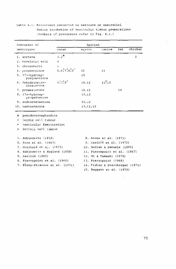
Table 4.1. Precursors converted to oestrone or oestradiol
during incubation of testicular tissue preparations
(numbers of precursors refer to Fig. 4.1.}
Precursor of
oestrogens
l. acetate
2. mevalonic acid
3. cholesterol
4. pregnenolone
5. 17a-hydroxy-pregnenolone
6. dehydroepian-drosterone
7. progesterone
8. 17a-hydroxy-progesterone
9. androstenedione
10. testosterone
human
1, 2%
4
§ + f. + 5,6,7,8;9
~ pseudohermaphrodite
Leydig cell tumour
+ testicular feminization
o Sertoli cell tumour
l. Rabinowitz (1958}
2. Rice et al. ( 19 6 7}
3. Guichard et al. ( 19 7 3)
4. Rabinowitz ' Ragland (1958}
5. Axelrod ( 19 6 5}
6. Pierrepoint et al. (1966}
7. Perez-Palacios et al. (1971}
Species
equine canine rat chicken
3
10 11
10
10, 12 11?13
1 0, 12 14
10, 12
10, 12
10,12,15
8. Ances et al. (1971)
9. cardiff et al. (1972}
10. Bedrak & samuels (1969)
11. Pierrepoint et al. (1967)
12. Oh & Tamaoki (1970)
13. Pierrepoint {1968)
14. Ficher & Steinberger (1971)
15. Baggett et al. (1959)
75

lower than 1%, while some authors (Wade et al., 1968, Rice
et al., 1966, Bardin et al., 1969) were not able to iden
tify any radioactive oestrogens after incubation of human
testicular material with androgen or 14
c-acetate.
The data obtained in the studies mentioned are all
qualitative in nature: although conversion of precursors to
oestrogens was observed, the degree of dilution of the exo
genous radioactive or non-radioactive precursor with endo
genous precursors was not taken into account in any of
these experiments. Therefore, in the experiments described
in this chapter the endogenous ·production of oestradiol and
testosterone was measured during incubation of rat testi
cular tissue. It was also attempted to measure the conver
sion of radioactive androgens to oestradiol under the same
conditions in order to assess the precursor role of the
androgens quantitatively. The results obtained during these
in vitro studies will be compared with those from the in
vivo experiments discussed in chapter 3. Finally, it was
attempted to resolve the question (see 1.4.) about the
intratesticular localization of the biosynthesis of oestro
gens. Therefore, in addition to the incubations with total
testis tissue, separated seminiferous tubules and inter
stitial tissue were also incubated.
4.2. Materials and methods
4.2.1. Animal techniques
Wistar rats with a body weight of 200-250 g were used
throughout this study, with the exception of the rats that
were fed with a diet deficient in essential fatty acids.
Testicular tissue was obtained immediately after deca
pitation of the animals and was kept in ice cold buffer
solution until it was incubated (usually not longer than
30 min, when total testis was incubated; dissection
76

generally took 60 min more). Dissection of testicular
tissue into seminiferous tubules and interstitial tissue
was performed as described by Christensen & Mason (1965) in
a dissection medium containing sucrose (0.25M) and EDTA
( l rnM) •
4.2.2. Steroids and steroid estimations
Non-radioactive steroids were obtained from Steraloids
(Pawling, u.S.A.) and used without further purification.
Cyanoketone is a product of Stirling-Winthrop, New York,
U.S.A. Radioactive androgens and oestrogens ware obtained
from New England Nuclear (Boston, U.S.A.) or from the
Radiochemical Centre (Amersham, England). [1, 2, 6, 7- 3H]
Androstenedione was prepared from [1,2,6,7- 3~-testosterone by chromium trioxide oxidation. Specific activities of
these steroids, as quoted by the manufacturer and place of
attachment of the radioactive atoms to the steroid nucleus
are shown in Table 4.2. Radioactive steroids were purified
before use by paper- and thin-layer chromatography, using
Table 4.2. Specific activities of steroids used in
incubation studies
Steroid
[4-14
c]-androstenedione
[ 4-14
c] -testosterone
[4-14
c]-dehydroepiandrosterone
[ 4-14
c] -oestradiol
[1,2,6,7-3a]-androstenedione
[1, 2, 6, 7-3a] -testosterone
[2, 4, 6, 7- 3H] -oestradiol
[ 6, 7- 3a] -oestrone
[6,7- 3H]-oestradiol
Specific activity
60 mCi/rnMol
58.8 mCi/rnMol
58.8 mCi/mMol
58 ITlCi/mMol
85 Ci/mMol
85 Ci/mMol
105 Ci/mMol
48 Ci/rnMol
40 Ci/mMol
77
purity criteria as described earlier (de Jong & van der
Molen, 1972). Endogenous oestradiol and testosterone were
estimated by radioimmunoassay, as described in chapter 2.
Protein was estimated according to the method of Lowry
et al. (1951).
4.2.3. Incubation conditions
When testes were incubated in Krebs-Ringer buffer, the
tissue was homogenized in this buffer, pH 7.4, containing
0.2% glucose (w/v), using a Potter-type homogenizer. Incu
bations were performed after addition of NADPH and NADH
(final concentration 1 mM), glucose-6-phosphate (10 rnM) and
glucose-6-phosphate dehydrogenase (0.3 Kornberg Units). The
homogenates were incubated for 3 h in a shaking water bath
at 34°C under an atmosphere of 95% o 2 and 5% co2 .
In a second series of incubations, SOOxg supernatant of
homogenized total testis tissue, seminiferous tubules or
interstitial tissue was used. Testes were homogenized in a
solution containing sucrose (0.25M) and EDTA (1 mM), using
an automatic Potter homogenizer. After centrifugation at
SOOxg for 10 min, the supernatant was collected and incu
bated in a medium and under conditions as described by
van der Vusse et al. (1973). Dissection of testis tissue
before these incubations took place under the homogeniza
tion medium. When radioactive or non-radioactive steroids
were incubated with tissue or tissue hornogenates, these
steroids were added to the incubation vessel dissolved in
ethanol. The solvent was evaporated under nitrogen at 45°C
and the residue was taken up in 50 ~1 ethanol. Then buffer
solutions and tissue or tissue hornogenates were added.
78

4.2.4. Techniques used for the separation and identification of
steroids
After incubation of testis hornogenates with radioactive
androgens non-radioactive oestrone and oestradiol (25 ~g
each) were added to the incubation vessels as carrier
steroids. Steroids were extracted from the incubation
medium with ethyl acetate and the extracts were purified
according to the scheme in Fig. 4.2. 3H-Labelled steroid
was only added after incubation with 14c-labelled precur
sors. In one experiment, radioactive oestrogens were puri
fied after incubation by chromatography on Sephadex LH-20
columns.
After incubation of 3H-labelled precursors with testi-
Fig. 4.2.
carrier steroids (25 )..lg) internal standards (105 dpm3H)
hexane
benzene
I I androgens I
I oestrone I
70% methanol
1 N NoOH
I
acetylation TLC saponification PC (8
4)
I oestrodiol-17,91
Outline of extraction and purification of radioactive oestradiol and
oestrone after incubation of labelled precursors (106
dpm 14
C- N
3H-steroid) with testicular tissue. PC= paper chromatography in the
Bush systems indicated. TLC "' thin-layer chromatography: toluene
ethyl acetate 4-1 (v/v) for oestrone and toluene-ethyl acetate 9-1
(v/v) for oestradiol.
79
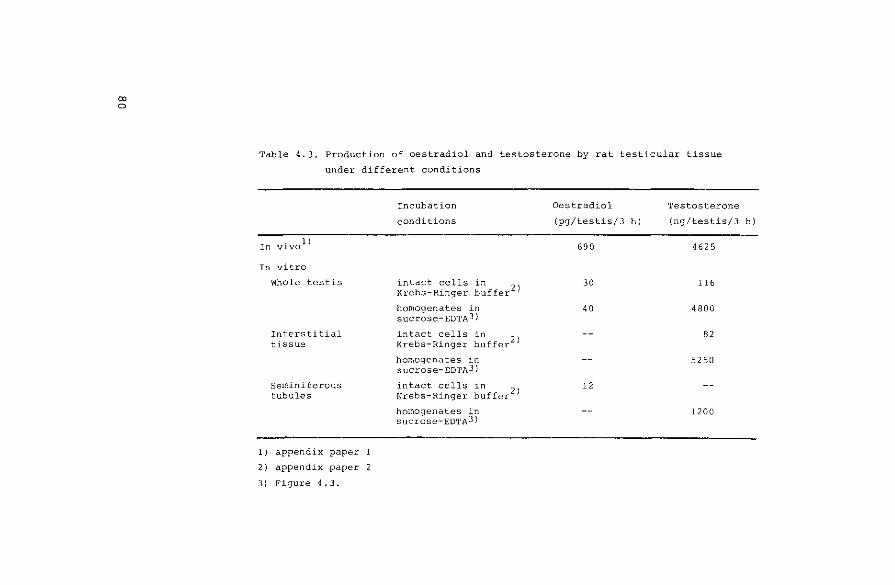
00 0
Table 4.3. Production of oestradiol and testosterone by rat testicular tissue
under different conditions
In vivo 1 )
In vitro
Whole testis
Interstitial tissue
Seminiferous tubules
1) appendix paper
2) appendix paper 2
3) Figure 4.3.
Incubation
conditions
intact cells in 2 Krebs-Ringer buffer )
homogenates in sucrose-EDTAJ}
intact cells in 2 Krebs-Ringer buffer )
homogenates in sucrose-EDTAJ}
intact cells in 2 Krebs-Ringer buffer )
homogenates in sucrose-EDTAJ)
Oestradiol Testosterone
{pg/testis/3 h) (ng/testis/3 h)
690 4625
30 116
40 4800
-- 82
-- 5250
12
-- 1200

cular 800xg supernatants, the purification scheme as dis
cussed earlier (Fig. 4.2.) was followed. After the second
paper chromatographic step, part of the sample was counted
for 3H, and 14c-oestradiol (720 dpm) was added to the
samples. After enzymatic oxidation (Rosenfield, 1971)
followed by paper chromatography (Bush A2 , 24 h) the
oestrone fraction was isolated. Part of the sample was
counted, and the remainder was acetylated and chromate
graphed (thin-layer chromatography, toluene-ethyl acetate
4-1, v/v). Subsequently, the oestrone acetate fraction was
counted.
4.3.
4.3.1.
Results
Measurement of endogenous hormones
Results of a first series of experiments in which non
homogenized testis tissues were incubated in Krebs-Ringer
buffer were reported in Table 5 of appendix paper 2. Since
the production of endogenous steroids in total testis under
these conditions was low in comparison with in vivo steroid
production (Table 4.3.) a second series of experiments
using the conditions as described by van der Vusse et al.
(1973) was performed (see: Fig. 4.3. and Table 4.3.). The
experiments on the production of endogenous steroids in
isolated seminiferous tubules and interstitial tissue, as
described in appendix paper 2, were also repeated under the
conditions of van der Vusse et al. (1973). Results of these
incubations are summarized in Table 4.4. With the exception
of the 800xg pellet of the interstitial tissue arid the dis
section medium, all fractions contained amounts of oestra
diol, which were not significantly different from the blank
value of 3.1 + 3.3 (S.D.) pg (n=12), obtained in the same
series of estimations.
Finally, the concentrations of oestradiol and testoste-
81
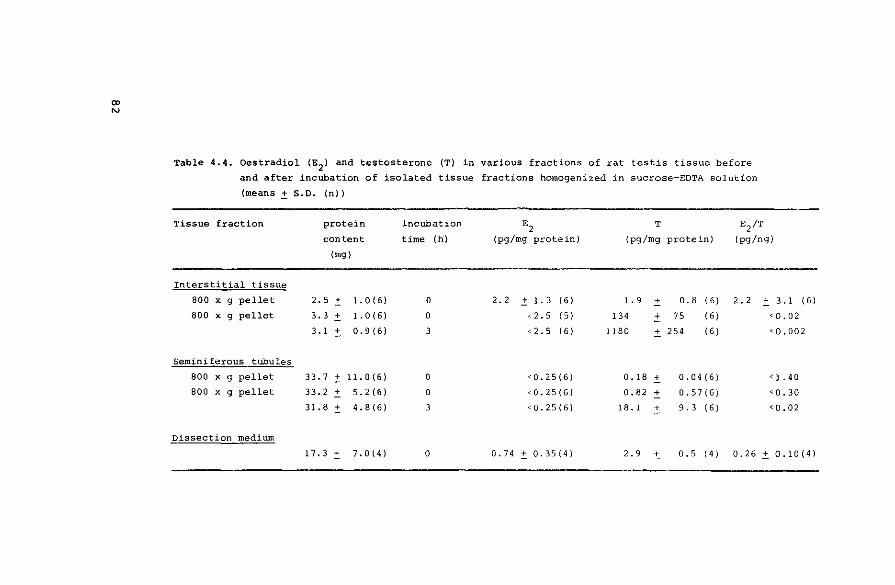
"' "'
Table 4.4. Oestradiol IE2 1 and testosterone (T) in various fractions of rat testis tissue before
and after incubation of isolated tissue fractions homogenized in sucrose-EDTA solution
(means .:!:. S.D. (n))
Tissue fraction protein incubation E2 T E2 /T
content time (h) (pg/mg protein) (pg/mg protein) (pg/ng)
(mg)
Interstitial tissue
800 x g pellet 2.5 .± l. 0 ( 6) 0 2. 2 .± 1. 3 16 I 1.9 ± 0.8 (6) 2.2 .:!:. 3. 1 I 61
800 x g pellet 3.3 .± l. 0 ( 6) 0 < 2. 5 (5) 134 ± 75 (6) <0.02
3. 1 .± 0. 9 ( 6) 3 < 2. 5 16 I 1180 + 254 (6) <0,002
Seminiferous tubules
800 x g pellet 33.7 .± 11.0(6) 0 <0,25(6) 0.18 + 0. 04 ( 6) < l. 4 0
800 x g pellet 33.2.:!:. 5. 2 (6) 0 <0,25(6) 0.82 + 0. 57 (6) <0.30
31.8 .:t 4. 8 (6) 3 <0.25(6) 18. 1 ± 9.3 (6 I <0.02
Dissection medium
17.3.:!:. 7. 0 (4) 0 0.74 ± 0.35(4} 2.9 ± 0. 5 14 I 0.26 .:t 0.10(4)

Fig. 4.3.
ngT pg E2 mg protein mg protein
60 120
" GT
o----------<J E 2
40 0.80
20 0.1.0
0 0 0 4
incubation time (h}
In vitro production (means~ S.E.M., n"'6) of oestradiol and testosterone
by 800xg supernatant of homogenized rot testes. Incubation conditions
were as described by van der Vusse et al. (1?73).
rone were estimated in isolated seminiferous tubules and
interstitial tissue from EPA-deficient rats. The results of
these estimations are summarized in Table 4.5. The concen
trations of oestradiol and testosterone in interstitial
tissue from normal rats (Table 4 in appendix paper 2) and
in EPA-deficient rats are not significantly different. How
ever, the concentrations of both steroids are significantly
higher in tubular material from EPA-deficient rats when
compared with those in normal rats (P < 0.02). This diffe
rence may be explained by a larger interstitial contamina
tion of the tubular fraction.
83

Table 4.5. Oestradiol (E2
) and testosterone (T) in testicular
tissue from essential fatty acid deficient rats
(means.:!:"_ S.D. (n))
Tissue E2 T
(pg/g) (ng/gl
Total testis 128
"' 54 ( ll) 12 6 5
"' 685 I 6}
Interstitial tissue 180
"' 115 I 6} 291
"' 123 17}
Tubules 106
"' 70 I 6) 57
"' 27 17}
4.3.2. Conversion of radioactive precursors
4.3.2.1. lnterconversion and degradation of oestrogens
After incubation of [4-14
c]-oestrone or [4-14
c]-oestra
diol (10 5 dpm) with testis tissue homogenates (equivalent
with 100 mg tissue) in Krebs-Ringer buffer, non-radioactive
carriers and tritium-labelled oestrone and oestradiol (10 5
dpm) were added. Steroids were extracted and purified using
the method outlined in Fig. 4.2. Radioactivity present in
appropriate aliquots was estimated after each purification
step: 3H; 14c ratios did not change after thin-layer chroma
tography indicating that purity of the oestrogens was
reached at this stage of purification. The percentages con
version of oestrone and oestradiol, corrected for losses
during extraction and purification, are summarized in
Table 4.6.
84
Table 4.6. Catabolism of oestrogens during incubations
of rat testis homogenates
Substrate
Oestrone (E1
)
Oestradiol (E2
)
% of substrate isolated as
36.2 6 5. l 10 l. 3
ll. 8 90.6 l Q 2. 4
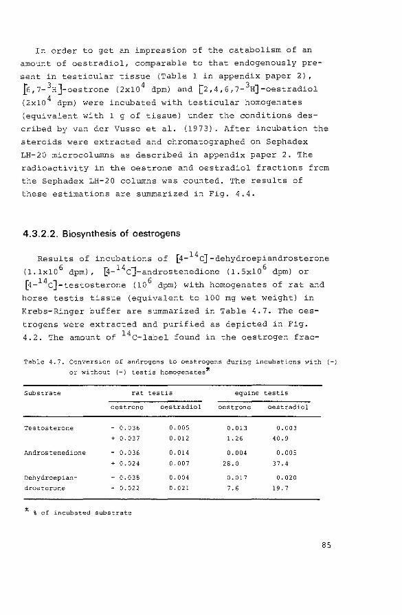
In order to get an impression of the catabolism of an
amount of oestradiol, comparable to that endogenously pre
sent in testicular tissue (Table 1 in appendix paper 2},
~,7- 3H]-oestrone (2x10 4 dpm) and [2,4,6,7- 3H]-oestradiol
(2xlo 4 dpm) were incubated with testicular homogenates
(equivalent with 1 g of tissue) under the conditions des
cribed by van der Vusse et al. (1973). After incubation the
steroids were extracted and chromatographed on Sephadex
LH-20 microcolurnns as described in appendix paper 2. The
radioactivity in the oestrone and oestradiol fractions from
the Sephadex LH-20 columns was counted. The results of
these estimations are summarized in Fig. 4.4.
4.3.2.2. Biosynthesis of oestrogens
Results of incubations of [4- 14 c] -dehydroepiandrosterone
(1.1x!0 6 dpm), @- 14c]-androstenedione (1.5x10⁶
dpm) or
8-14c]-testosterone (10 6 dpm) with hornogenates of rat and
horse testis tissue (equivalent to 100 rng wet weight) in
Krebs-Ringer buffer are summarized in Table 4.7. The oes
trogens were extracted and purified as depicted in Fig.
4.2. The amount of 14c-label found in the oestrogen frac-
Table 4.7. Conversion of androgens to oestrogens during incubations with (+)
or without {-) testis homo~enates*
Substrate
Testosterone
Androstenedione
Dehydroepian
drosterone
rat
oestrone
- 0.036
+ 0.037
- 0.036
+ 0.024
- 0.038
+ 0.022
X % of incubated substrate
testis
oestradiol
0.005
0. 012
0.014
0.007
0.004
0.021
equine testis
oestrone oestradiol
o. 013 0.003
l. 26 40.9
0.004 0.005
28.0 37.4
0. 01 7 0.020
7. 6 19. 7
85

Fig. 4.4.
86
%recovered
100
80
60
20
0 substrate
incubation oc C conditions blank
E2 E1 ace 33°C
!----tissue,----<
~recovered as E2
D recovered as E1
Radioactivity eluted in the oestrone and oestradiol fractions from
Sephadex LH-20 microcolumns after 3 hr incubation of rot
testicular tissue 800xg supernatant with 3
H-oestrone (E1, 40 pg)
or 3
H-oestradiol (E2
, 20 pg). Incubation conditions were as
described by van der Vusse et al. (1973). Results are expressed as
percentage of the incubated substrate recovered in the different
fractions. Mean values of 5-6 incubations are given.

tions isolated from rat testis incubations was not diffe
rent from that in tissue-free incubations, while all three
androgens were converted to oestrone and oestradiol during
incubation with equine testis tissue. 3H;14
c ratios for
both steroids did not change after the thin-layer chromato
graphic step.
The results of incubations of 10 6 dpm of [1,2,6,7-3H]
androstenedione or [1,2,6,7- 3H]-testosterone with 800xg
supernatants of rat testis homogenates (equivalent to 1 g
of tissue), suggested conversion rates to oestradiol of
approximately 1 °/oo (Fig. 4.5.), when corrected for the
results of blank incubations, for mean recovery (43.2 ~
4.5 (S.D.) %, n=27) after paper- and thin-layer chromato
graphy, and for 3H-loss from the 18- and 28-position
(Fishman et al., 1969, Osawa & Spaeth, 1971). No radio
activity associated with oestrone, was detected at this
stage of purification. Addition of the Bush B3
paper
chromatographic step to the purification of 3H-oestradiol,
and subsequent addition of [4- 14c]-oestradiol (720 dpm) and
further purification of the steroid by the method described
in section 4.2.4. resulted in a complete dissociation of 3
H- and 14
c-radioactivity in all oestradiol fractions. The
results from incubations where testosterone (10 vg/incuba
tion) or cyanoketone (0.5 vg/incubation) were added, showed
the same dissociation of radioactive labels. Testosterone
always accounted for more than 80% of the radioactivity in
the androgenic fractions isolated after incubation. Similar
experiments with isolated seminiferous tubules and inter
stitial tissue yielded comparable results: the 3H-radio
activity which was isolated after paper- and thin-layer
chromatography, was completely dissociated from the 14c
label after subsequent purification.
87

Fig. 4.5.
88
%conversion
0 .L._ _ __,_
§ tissue incubated § with cyanoketone
Ill tissue incubated with testosterone
Apparent percentage conversion of tritiated androstenedione (A)
or testosterone (T) to labelled oestradiol after 3 h incubat'1on
with 800xg supernatant of rat testis homogenates, Incubation
and d'1ssection procedures were as descr'1bed by van der Vusse
etal. (1973).
After incubation oestradiol was extracted and purified according
to the procedure in Fig, 4.2., with the exception of the last
paper chromatographic step.

4.4. Discussion
4.4.1. Endogenous production of oestradiol
The testicular production of endogenous oestradiol in
vitro was much lower than that observed under in vivo con
ditions (see Table 4.3.). on the other hand, by changing
the incubation conditions, it was possible to increase the
production of testosterone during incubation to a level
comparable to that observed during in vivo experiments
(Table 4.3.). This difference between the relative produc
tions of oestradiol and testosterone in vivo and in vitro
may reflect that conditions which favour the production of
testosterone are not favourable for production of oestra
diol. A similar conclusion was drawn from the vivo experi
ments described in appendix paper 1: a prolonged trophic
stimulation of the secretion of testosterone did not
increase the secretion of oestradiol. It is not likely that
the low production of oestradiol is caused by absence of
cofactors: NADPH and molecular oxygen, which are necessary
for oestrogen formation in placental homogenates (see:
Talalay, 1965), and in the microsomal fraction from equine
testes (Oh & Tamaoki, 1971), were present. A second expla
nation for the low production rate of oestradiol during
incubations of testicular homogenates is the possible inhi
bition of the arornatization reaction by testosterone cata
bolites, which may accumulate in the incubation medium.
Such an inhibition was shown to occur during incubation of
placental rnicrosornes (Schwarzel et al., 1973).
The conclusion of appendix paper 2, that under in vitro
conditions oestradiol may be synthetized in tubular tissue
could not be confirmed by the results shown in Table 4.4.,
because of the extremely low levels of oestradiol in the
samples. The localization of measurable levels of oestra
diol in the 800xg pellet of the interstitial tissue
suggests that this steroid is mainly localized in the
89

nuclei of the interstitial tissue, as might be expected on
basis of the results of Mulder et al. (1973). This obser
vation on the interstitial localization of oestradiol is
also in agreement with the results described in Table 4 of
appendix paper 2 and with the results obtained in EFA
deficient rats (Fig. 3.3. and Table 4.5.). It cannot be
decided, however, whether the high concentration of oestra
diol in isolated interstitial tissue reflects the real
situation in vivo, because redistribution of the steroid
might have occurred during the dissection procedure. The
relatively high concentration of oestradiol in the dissec
tion medium, which contrasts with the level of testosterone
(Table 4.4.), supports the latter possibility.
Finally, the apparent, albeit small production of tes
tosterone in isolated seminiferous tubules during the incu
bations of 800xg supernatants (Table 4.4.) is at variance
with the data reported by cooke et al. (1972). It is not
clear if this discrepancy is due to the difference in incu
bation conditions or to an interstitial inpurity in the
tubular fraction.
4.4.2. Production of radioactive oestradiol
The quantitative extent of oestrogen catabolism in rat
testicular tissue is small (Table 4.6. and Fig. 4.4.). The
relative amounts of oestrone and oestradiol, found after
incubation, depends largely on the cofactors added: in the
presence of a NADPH-generating system in the incubation
medium, most of the steroid will be reduced. This occurs
with large as well as with small amounts of oestrone or
oestradiol: the results of the incubation studies with 14c
or 3H-labelled steroids do not differ. The catabolism to
other steroids is quantitatively not important, since
almost 100% of the incubated radioactivity was isolated as
oestrone and oestradiol after incubation. These results
with rat testis tissue are in agreement with those of
Lucis & Lucis (1969), who found only very small conversions
90
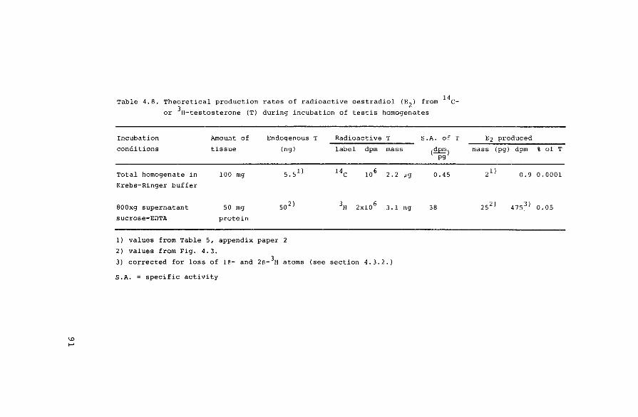
"' ~
Table 4.8. Theoretical production rates of radioactive oestradiol (E2
J from 14c
or 3H-testosterone (T} during incubation of testis homogenates
Incubation Amount of Endogenous T Radioactive T S.A. of T
conditions tissue (ng) label dpm mass (£E!!!) pg
Total homogenate in 100 mg s. sll 14c 106 2. 2 og 0.45
Krebs-Ringer buffer
SOOxg supernatant 50 mg so 2) 3H 2x10 6 3.1 ng 38
sucrose-EDTA protein
1) values from Table 5, appendix paper 2
2) values from Fig. 4.3.
3) corrected for loss of 16- and 2S- 3H atoms (see section 4.3.2.)
S.A. = specific activity
E2 produced
mass (pg) dpm % of T
2 1 I 0.9 0.0001
25 2 ) 4 75 3 ) 0.05

to 6-oxygenated products after incubation of rat and human
testes with 14c-oestrone. However, Axelrod & Goldzieher
(1962) and Wade et al. (1968) found significant amounts of
2-hydroxy- and 2-methoxy-derivatives after incubation of
human testis tissue from a normal man and from a patient
with testicular feminization.
Table 4.8. shows the radioactivity which should theo
retically be found in the oestradiol fractions after incu
bation of testis tissue homogenates with 14c- or 3H
labelled testosterone. It is assumed that production of
endogenous testosterone does not take place, and that tes
tosterone is the only precursor of oestradiol. It is evi
dently impossible to detect conversion from 14c-testoste
rone to oestradiol using the conditions shown: the calcu
lated conversion (0.0001%) is lower than the corresponding
values for blank incubations (Table 4.7.). A similar con
clusion applies to the incubations with 3H-testosterone
when a 60-fold increase in endogenous testosterone produc
tion takes place (Fig. 4.3.): this results in a comparable
decrease in specific activity of the labelled steroid.
Finally, a 0.05% conversion of testosterone to oestradiol
might be expected in incubations where the endogenous pro
duction of testosterone is blocked by the addition of
cyanoketone (van der Vusse et al., 1974). However, no pro
duction of labelled oestradiol could be detected in the
latter incubates (see 4.3.2.2.). This may be due to the
possibility that testosterone is not the obligatory pre
cursor for oestrogen biosynthesis, or that cyanoketone
influences the aromatizing system in the testis at lower
concentrations than those used to inhibit placental aroma
tization (Schwarze! et al., 1973).
The observation that no production of radioactively
labelled oestradiol could be observed during incubation of
SOO~g supernatants of homogenates of seminiferous tubules
and interstitial tissue is in agreement with the results
shown in Table 4.4.
92

4.5. Conclusions
From the results presented in appendix paper 2 and in
this chapter, the following conclusions are drawn:
1. Oestradiol and testosterone are mainly localized in the
interstitial tissue.
2. In dissected interstitial tissue oestradiol is mainly
localized in the 800xg pellet, while testosterone is
found in the 800xg supernatant.
3. Conditions favouring the in vitro production of testos
terone do not enhance the biosynthesis of oestradiol.
4. Biosynthesis of oestradiol could only be shown in whole
tissue preparations of total testis tissue and semini
ferous tubules and in total testis homogenates.
5. Biosynthesis of testosterone was observed during incuba
tions of whole tissue preparations of total testes and
interstitial tissue and in homogenates of total testes,
interstitial tissue and seminiferous tubules.
6. Radioactively labelled androgens were not converted to
oestrogens under conditions where endogenous production
of oestradiol could be shown. This is probably a result
of the dilution of the radioactive steroids with endo
genous hormones.
4.6. References
Ances, I.G., Connor, T.B., Gallagher, E.P.,} geon, C.J.,
Levin, H.S. & Ganis, F.M. (1971)
J. clin. Endocr. Metab. ll• 452-458.
Axelrod, L.R. (1965)
Biochim. biophys. Acta~. 551-556.
Axelrod, L.R. & Goldzieher, J.W. (1962}
Endocrinology 70, 943-945.
93

94
Baggett, B., Engel, L.L., Balderas, L., Lanman, G., Savard, K.
& Dorfman, 'R.I. (1959)
Endocrinology ii• 600-608.
Bardin, C.W., Rosen, S., LeMaire, W.J., Tjio, J.H., Gallup, J.,
Marshall, J. & Savard, K. (1969)
J·. clin. Endocr. Metab. Q, 1429-1437.
Bedrak, E. & Samuels, L.T. (1969)
Endocrinology ~' 1186-1195.
Cardiff, C., Uden, v. & Killinger, D.W. (1972)
Can. J. Biochem. 2Q, 849-855.
Christensen, A.K. & Mason, N.R. (1965)
Endocrinology 76, 646-656.
Cooke, B.A., de Jong, F.H., van der Molen, H.J. & Rorrunerts, F.F.G. (1972)
Nature, Land. (New Biology) ill• 255-256.
Dorfman, R.I. & Ungar, F. (1965)
Metabolism of Steroid Hormones, pp. 207-209, Academic Press,
New York.
Ficher, M. & Steinberger, E. (1971)
Acta endocr., Copnh. ~· 285-292.
Fishman, J., Guzik, H. & Dixon, D. (1969}
Biochemistry, N.Y. ~. 4304-4309.
Grant, J.K., Griffiths, K. & Pierrepoint, C.G. (1967)
in Endocrinology of the Testis (Wolstenholme, G.E.W. &
O'Connor, M., eds.), pp. 280-298, Churchill, London.
Guichard, A., Cedard, L. & Haffen, K. (1973)
Gen. camp. Endocr. 20, 16-28.
de Jong, F.H. & van der Molen, H.J. (1972)
J. Endocr. 21· 461-474.
Lowry, O.H., Rosebrough, N.J., Farr, A.L. & Randall, R.J. (1951)
J. biol. Chern. ~. 265-275.
Lucis, O.J. & Lucis, R. (1969)
Gen. camp. Endocr. 12, 63-71.
Meyer, A.S. (1955)
Biochim. biophys. Acta ll• 441-442.
Mulder, E., Brinkmann, A~O., Lamers-Stahlhofen, G.J.M. &
van der Molen, H.J. {1973)
FEBS Lett. 1!• 131-136.
Oh, R. & Tamaoki, B. (1970)
Acta endocr., Copnh. ii• 1-16.
Oh, R. & Tamaoki, B. (1971)
Acta endocr., Copnh. &l• 665-676.
Osawa, Y. & Spaeth, D.G. (1971)
Biochemistry, N.Y. !Q, 66-71.

Perez-Palacios, G., Morato, T., Perez, A. E., Castaneda, E. &
Gual, c. (1971)
Steroids !1• 471-492.
Pierrepoint, C.G. (1968)
J. Endocr. il• 99-107.
Pierrepoint, C.G., Griffiths, K., Grant, J.K. & Stewart, J.S.S.
(1966)
J. Endocr. ]2, 409-417.
Pierrepoint, C. G., Galley, J.Mci., Griffiths, K. & Grant, J.K.
( 1967}
J. Endocr. ~. 61-70.
Rabinowitz, J.L. (1958)
Atompraxis !• 85-86.
Rabinowitz, J.L. & Ragland, J.B. (1958)
Fedn Proc. ll• 293.
Rice, B.F., Johanson, C.A. & Sternberg, W.H. (1966)
Steroids 2• 79-90.
Rice, B.F., Cleveland, W.W., Sandberg, D. H., Ahmad, N.,
Politano, v.T. & Savard, K. (1967)
J. clin. Endocr. Metab. ll• 29-36.
Rosenfield, R.L. (1971)
Steroids ll• 689-696.
Ryan, K.J. (1958)
Biochim. biophys. Acta l2• 658-659.
Ryan, K.J. (1959)
J. biol. Chern. £21, 268-272.
Savard, K. (1973)
Biol. Reprod. i!_, 183-202.
Schwarzel, W.C., KruggeL W.G. & Brodie, H.J. (1973)
Endocrinology ~. 866-880.
Talalay, P. (1965}
A. Rev. Biochem. l!• 347-380.
van der Vusse, G.J., Kalkman, M.L. & van der Molen, H.J. (1973)
Biochim. biophys. Acta £22, 179-185.
van der vusse, G.J., Kalkman, M.L. & van der Molen, H.J. (1974}
Biochim. biophys. Acta, in the press.
Wade, A.P., Wilkinson, G.S., Davis, J.C. & Jeffcoate, T.N.A.
{1968)
J. Endocr. il• 391-403.
95

CHAPTER 5. THE SIGNIFICANCE OF THE TESTICULAR PRODUCTION
OF OESTRADIOL
5.1. Introduction
The effects of exogenous oestrogens on different pro
cesses in the male animal have been extensively studied:
oestrogens have been found to influence the prostate
(Harper et al., 1971, Leav et al., 1971, Bonne & Raynaud,
1973), adrenal function (Sobrinho et al., 1971), the pineal
gland (Nagle et al., 1972), the neurohypophysis (Legros &
Grau, 1973), the metabolism of testosterone (Bird et al.,
1971) and testicular and hypophyseal function.
Until recently the latter two effects of the administra
tion of oestrogens to male animals were thought to be
correlated via the negative feedback action of oestrogens
on hypothalamic-hypophyseal processes which result in the
secretion of trophic hormones; the administration of
oestrogens leads to "physiological hypophysectomy"
(Steinberger & Nelson, 1955). The role of testicular
oestradiol can be only of limited significance in this
respect, because the testis secretes only a minor part of
the amount of circulating oestradiol (chapter 3). Recent
studies (Mallampati & Johnson, 1973, Danutra et al., 1973)
showed, however, that the administration of oestrogens to
intact male rats did not influence levels of circulating
trophic hormones, while testosterone concentrations in
peripheral blood fell steeply. These results would suggest
a direct effect of oestrogens on testicular processes. This
latter possibility will be discussed in more detail in this
chapter.
97

5.2. Influence of oestrogens on testicular function
5.2.1. Effects of longterm oestrogen treatment on testicular
enzymes
As early as 1941 Huggins et al. suggested that oestro
genic steroids might influence the testicular secretion of
androgens. These authors also indicated that this action
of the oestrogenic steroids might reflect either a negative
feedback action on the secretion of gonadotrophic hormones
from the hypophysis, or a direct action on testicular pro
cesses. The inhibition of hypophyseal gonadotrophin release
has been suggested in almost all experiments on the effect
of oestrogens under simultaneous administration of trophic
hormones: Meyer (1968) showed that injection of oestradiol
dipropionate inhibits rat testicular glucose-6-phosphate
dehydrogenase, lactate dehydrogenase, NAD cytochrome-c
reductase, cytochrome oxidase and 38-hydroxysteroid
dehydrogenase, but this inhibition could be reversed
through administration of gonadotrophins. Samuels et al.
(1967) showed a similar effect of HCG on !?a-hydroxylase
and 17-20 lyase in rat testes after diethylstilboestrol
treatment. The latter authors also indicate a possible
direct effect of diethylstilboestrol on 178-hydroxysteroid
dehydrogenase in rat testes, and they showed a significant
direct effect on the testicular concentration of enzymes
involved in steroidogenesis in BALB/c mice. Effects of
diethylstilboestrol on testicular RNA synthesis and on the
growth of interstitial cell tumours in these animals have
also been reported (Samuels et al., 1968).
The only study of this type which was performed in the
human male is that of Slaunwhite et al. (1962), who found
a decreased activity of 17S-hydroxysteroid dehydrogenase
activity during in vitro studies of testis tissue, obtained
98

from men with prostatic cancer, who were treated with
oestrogens. This decrease could, however, be reversed by
the administration of HCG in vivo.
Finally, Danutra et al. (1973) described a discrepancy
between LH and testosterone levels in rats after oestrogen
administration: plasma LH levels remained normal while
plasma testosterone concentrations were decreased. This
might be the first evidence for a direct action of oestro
gens on testicular processes in the rat. However, these
observations were not confirmed by Verjans et al. (1974)
who in a comparable study observed a close correlation
between the decrease of circulating LH levels and the
decrease of testicular and plasma testosterone concentra
tions.
5.2.2. Effects of oestrogens on spermatogenesis
The influence of exogenous oestrogens on spermatogenesis
and fertility has recently been reviewed by Steinberger
(1971). This author concludes from data, which were mainly
obtained from experiments in rats, that the negative
influence of oestrogens on spermatogenesis is mediated via
the hypophysis. Steinbeck et al. (1971) postulated that the
deleterious action of oestrogens on spermatogenesis is not
only pituitary-, but primarily Leydig cell-dependent: after
oestrogen treatment the interstitial tissue degenerates
first, while after cessation of administration the Leydig
cells recover firstly, followed by the recovery of sperma
togenesis. A similar observation was recorded by
Steinberger & Nelson (1955) who reported that the adminis
tration of testosterone propionate caused reversal of the
oestrogen induced decrease in testicular hualuronidase, a
marker enzyme for advanced stages of spermatogenesis.
Elkington & Blackshaw (1970) and Lacy & Lofts (1965) res
tored spermatogenesis in oestrogen treated rats with in-
99

jections of PMS, testosterone and FSH. Similar experiments
were performed in mice by Davies et al. (1974) who did not
observe any effect of FSH alone on spermatogenesis. A
direct effect of oestrogens on spermatogenesis has never
been reported.
5.2.3. Effects observed during incubations of testicular tissue with
oestrogens
Direct effects of oestrogens on testicular metabolism in
vitro have been investigated particularly with respect to
the enzymes involved in steroidogenesis. The oestrogen con
centrations used in these studies are generally in the
range of between 10- 7 and 10- 4M, hence several orders of
magnitude higher than in the physiological situation
(appendix paper 2). It is likely, therefore, that non
physiologic effects of these high oestrogen concentrations
(Samuels et al., 1967) might have caused the observed inhi
bition of 3B-hydroxysteroid dehydrogenase-6 5-~ 4 -isomerase in the rat (Kaartinen et al., 1971) and in man (Yanaihara
& Troen, 1971}, of !?a-hydroxylase in mouse (Samuels et al.,
1967) and in man (Balatti et al., 1967), of 17-20 lyase in
mouse (Samuels et al., 1967) and of 17S-hydroxysteroid
dehydrogenase in rats (Kaartinen et al., 1971). Finally,
Vera Cruz et al. (1970) observed an increased oxygen uptake
and co2 production by testicular tissue in vitro after
addition of oestradiol. The oestrogen concentrations used
were again aphysiologic.
100
5.3. Summary and conclusions
In their 1947 review, Errunens & Parkes conclude that "in
the absence of any conclusive evidence in either direction,
one may conclude, provisionally, that the presence of a
certain amount of estrogen in the normal male is incidental
rather than functionally important, and that its entire
absence would have little or no deleterious effect on the
animal". At present, this statement may require further
specification, because there appears to be a significant
role for circulating oestrogens in the male with respect
to the stimulation of sexual behaviour (Larsson et al.,
1973, Joshi & Raeside, 1973, SOdersten, 1973, Baum &
Vreeburg, 1973). The conclusion of Emmens & Parkes may,
however, still apply to testicular oestrogens which contri
bute only a minor part of the total oestrogen production in
the male. The presence of a receptor for oestradiol in tes
ticular interstitial tissue, which in some respects seems
to behave like the uterine receptor (Mulder et al., 1973),
and the observation in the ovary that responsiveness to FSH
depends on the local presence of oestradiol (Goldenberg
et al., 1973) might indicate, however, that the validity of
this conclusion is only of passing significance, pending
further investigations.
5 .4. References
Balatti, J.J., Claret, A. & Rosner, J.M. (1967)
Fert. Steril. ~' 367-374.
Baum, M.J. & Vreeburg, J.T.M. (1973)
Science, N.Y. ~' 283-285.
10 l

Bird, C. E., Green, R.N., Calandra, R.S., Connolly, J.G. &
Clark, A.F. (1971)
Acta endocr., Copnh. il• 733-739.
Bonne, C. & Raynaud, J.-P. (1973)
Biochimie ~. 227-229.
Danutra, V., Harper, M.E., Boyns, A.R., Cole, E.N.,
Brownsey, B.G. & Griffiths, K. {1973)
J. Endocr. 22• 207-215.
Davies, A.G., Courot, M. & Gresham, P. (1974)
J. Endocr. &Q, 37-45.
Elkington, J.S.H. & Blackshaw, A.W. (1970)
J. Reprod. Fert. ll• 1-10.
Errunens, C.W. & Parkes, A.S. (1947)
Vitamins and Hormones~' 233-272.
Goldenberg, R.L., Reiter, E.O. & Ross, G.T. (1973)
Fert. Steril. l,!. 121-125.
Harper, M.E., Pierrepoint, e.G., Fahmy, A.R. & Griffiths, K.
( 19 71)
J. Endocr. 49, 213-223.
Huggins, C., Scott, W.W. & Hodges, C.V. (1941)
J. ural. .!§.. 997-1006.
Joshi, H.S. & Raeside, J.I. (1973)
J. Reprod. Fert. ll• 411-423.
Kaartinen, E., Laukkanen, M. & Saure, A. (1971)
Acta endocr., Copnh. 66, 50-64.
Lacy, D. & Lofts, B. (1965)
Proc. R. Soc. Series B. ~. 188-197.
Larsson, K., S0dersten, P. & Beyer, C. (1973)
J. Endocr. 22• 563-564.
Leav, I., Morfin, R.F., Ofner, P., Cavazos, L.F. & Leeds, E. B.
(1971)
Endocrinology 89, 465-483.
Legros, J.J. & Grau, J.D. (1973)
Nature, Land. (New Biology) lil• 247-249.
Mallampati, R.S. & Johnson, D.C. (1973)
Neuroendocr. ll• 46-56.
Meyer, W. (1968)
Acta histochem. li• 376-390.
Mulder, E., Brinkmann, A.O., Lamers-Stahlhofen, G.J.M. &
van der Molen, H.J. (1973)
FEBS Lett. ll• 131-136.
Nagle, C.A., Neuspiller, N.R., Cardinali, D.P. & Rosner, J.M.
(1972)
Life Sci. II, ll• 1109-1116.
102
Samuels, L.T., Uchikawa, T. & Huseby, R.A. (1967)
in Endocrinology of the Testis (Wolstenholme, G.E.W. &
O'Connor, M., eds.), pp. 211-232, Churchill, London.
Samuels, L.T., Uchikawa, T., Zain-ul-Abedin, M. & Huseby, R.A.
(1968)
in Advances in the Biosciences {Rasp§, G., ed.), Vol. 2,
pp. 182-193, Pergamon Press, Oxford.
Slaunwhite, W.R., Sandberg, A.A., Jackson, J.E. & Staubitz, W.J.
{ 19 62)
J. clin. Endocr. Metab. 22, 992-995.
Sobrinho, L.G., Kase, N.G. & Grunt, J.A. {1971)
J. clin. Endocr. Metab. }l, 110-114.
sBdersten, P. {1973)
Harm. Behav. !• 247-256.
Steinbeck, H., Mehring, M. & Neumann, F. {1971)
J. Reprod. Fert. ~, 65-76.
Steinberger, E. (1971)
Physiol. Rev. 2l• 1-22.
Steinberger, E. & Nelson, W.o. {1955)
Endocrinology 2i• 429-444.
Vera Cruz, N.C., Gomes, W.R. & VanDemark, N.L. (1970)
Biol. Reprod. £, 376-386.
Verjans, H.L., de Jong, F.H., Cooke, B.A., van der Molen, H.J.
& Eik-Nes, K.B. {1974)
Acta endocr., Copnh., submitted for publication•
Yanaihara, T. & Treen, P. (1972)
J. clin. Endocr. Metab. l±• 968-973.
103


SUMMARY
The presence of oestrogens in urine and blood from male
animals is well established. The origin of oestrogenic
steroids in the male is not clear, however. The study
described in this thesis concerns the role of the testis in
the production of oestrogensi the following aspects of the
testicular production and secretion of oestradiol were
investigated:
I. The regulation of testicular production and secretion
of oestradiol in vivo (chapter 3, appendix papers 1
and 2).
II. The localization of oestradiol in the two main testi
cular tissue compartments, the seminiferous tubules
and the interstitial tissue (chapter 4, appendix
paper 2).
III. The localization of the production of oestradiol in
the testicular compartments using incubation studies
(chapter 4, appendix paper 2).
In order to measure the low endogenous levels of oestra
diol in male rats, it was necessary to evaluate the sensi
tivity and specificity of methods for the estimation of
this steroid (chapter 2). In this respect three different
methods used for the estimation of oestradiol were compared.
Competitive protein binding assay, using rabbit uterine
cytosol as the source of binding protein, lacked speci
ficity. A double competitive protein binding technique
appeared more specific, but the sensitivity of the method
was not satisfactory. Adequate specificity and sensitivity
were obtained only by using radioimmunoassay for the esti
mation of oestradiol.
Testosterone, which was measured in each sample as a
reference, was estimated either by gas-liquid chromato
graphy or radioimmunoassay. Both techniques were shown to
lOS

provide reliable results (chapter 2).
r. On basis of the data from the literature (see chapter
3), and of the experiments described in the appendix
papers 1 and 2, it has been concluded that:
106
1. the testis secretes oestradiol in all species
studied thusfar.
2. in the rat, as in the human male, the contribution
of the testicular secretion of oestradiol to the
total production rate of the steroid is in the
order of 20%.
3. the intravenous administration of HCG causes an
increase in the testicular production of oestradiol
and testosterone in the rat.
4. prolonged administration of HCG to rats causes an
increased concentration of oestradiol and testoste
rone in testicular tissue. The secretion of oestra
diol remains unchanged, while the secretion of tes
tosterone increases sharply.
5. hypophysectomy does not influence the concentration
of oestradiol in testicular venous plasma, while
the testis tissue concentrations of oestradiol and
testosterone and the testicular venous plasma con
centration of testosterone are decreased.
6. the administration of FSH to intact or hypophysec
tomized rats does not influence the concentration
of either testosterone or oestradiol in testis
tissue or testicular venous plasma.
7. the intravenous administration of prolactin to
intact rats has no stimulatory effect on the con
centration of oestradiol or testosterone in testi
cular venous plasma.

II. The distribution of oestradiol and testosterone
between seminiferous tubules and interstitial tissue
was investigated by measuring the endogenous concen
trations of the steroids in the isolated tissue com
partments from normal rats and in testes from rats,
fed with a diet which was deficient in essential fatty
acids (chapter 4 and appendix paper 2). It has been
concluded that:
1. oestradiol and testosterone are mainly localized
in the interstitial tissue.
2. in dissected interstitial tissue oestradiol is
mainly localized in the 800xg pellet, while testos
terone is found in the 80Dxg supernatant.
III. The production of oestradiol and testosterone during
in vitro studies of total testis tissue or separated
interstitial tissue and seminiferous tubules has been
discussed in chapter 4 and appendix paper 2. The fol
lowing conclusions have been drawn from the experi
ments performed:
1. conditions favouring the in vitro production of
testosterone do not enhance the biosynthesis of
oestradiol.
2. biosynthesis of oestradiol could only be shown in
whole tissue preparations of total testis tissue
and seminiferous tubules and in total testis homo
genates.
3. biosynthesis of testosterone was observed during
incubations of whole tissue preparations of total
testes and interstitial tissue and in homogenates
of total testes, interstitial tissue and semini
ferous tubules.
4. radioactively labelled androgens were not converted
to oestrogens under conditions where endogenous
production of oestradiol could be shown. This is
probably a result of the dilution of the radio
active steroids with endogenous hormones.
107

Finally, the significance of the testicular production
of oestradiol was discussed (chapter 5). It is concluded
that so far no evidence for a direct effect of testicular
oestradiol on any process has been shown, although some
results in the literature might indicate an intra testi
cular function for the steroid.
108

SAMENVATTING
Oestrogene steroiden zijn aanwezig in urine en bleed van
mannelijke dieren. De herkomst van deze horrnonen is echter
niet eenduidig bepaald. Het in dit proefschrift beschreven
onderzoek heeft betrekking op de mogelijke rol van de
testis bij de produktie van oestradiol. In dit verband
werden de volgende aspekten van de testikulaire produktie
en sekretie van oestradiol onderzocht:
I. De regulatie van de produktie en sekretie van oestra
diol door de testis in vivo (hoofdstuk 3 en de artike
len 1 en 2 van de appendix) .
II. De koncentraties van oestradiol in de twee belang
rijkste kornpartimenten van de testis: de seminifere
tubuli en het interstitiele weefsel (hoofdstuk 4 en
artikel 2 van de appendix) .
III. De biosynthese van oestradiol in deze kompartirnenten
(hoofdstuk 4 en artikel 2 van de appendix).
Voor de bepaling van de lage endogene koncentraties van
oestradiol in mannelijke ratten was het noodzakelijk de ge
voeligheid en specificiteit van rnethoden voor de bepaling
van dit steroid te evalueren (hoofdstuk 2). Drie methoden
voor de bepaling van oestradiol werden vergeleken. Een kom
petitieve eiwitbindingstechniek, waarin de cytosolreceptor
van de konijne-uterus als bindend eiwit wordt gebruikt,
bleek niet voldoende specifiek te zijn. Een dubbele kompe
titieve eiwitbindingsmethode was meer specifiek, maar de
gevoeligheid van deze methode was niet bevredigend. Gevoe
ligheid en specificiteit van een radioirnrnunologische tech
niek bleken toereikend voor de bepaling van oestradiol in
plasma en weefsel van de mannelijke rat.
Testosteron, het steroid dat als referentie werd bepaald
in ieder plasma- of testisweefselmonster, werd bepaald met
behulp van een gas chromatografische of een radioirnmunolo-
109

gische methode. Met beide technieken werden betrouwbare
resultaten bereikt (hoofdstuk 2).
I. Op basis van gegevens uit de literatuur (zie hoofdstuk
3) en van de resultaten van de experimenten die in de
artikelen 1 en 2 van de appendix werden beschreven,
werd het volgende gekonkludeerd:
110
1. de testis secerneert oestradiol in alle species die
tot dusver bestudeerd zijn.
2. de testikulaire sekretie van oestradiol vormt in de
rat, evenals in de mens, ongeveer 20% van de totale
produktiesnelheid van het steroid.
3. na intraveneuze toediening van HCG wordt in de rat
een toename van de testikulaire produktie van
oestradiol en testosteron waargenomen.
4. na langdurige toediening van HCG aan ratten wordt
een verhoogde koncentratie van testosteron en
oestradiol in testisweefsel waargenomen. De sekre
tie van oestradiol verandert niet, terwijl een
sterke stijging van de testosteronsekretie optreedt.
5. hypofysektomie beinvloedt de koncentratie van
oestradiol in testikulair veneus plasma niet. De
koncentraties van oestradiol en testosteron in
testisweefsel en de koncentratie van testosteron in
testikulair veneus plasma zijn verlaagd na hypofy
sektomie.
6. na toediening van FSH aan intakte of gehypofysek
tomeerde ratten treedt geen verandering op in de
koncentraties van oestradiol en testosteron in
testisweefsel of testikulair veneus plasma.
7. na intraveneuze toediening van prolactine aan
intakte ratten worden geen verhoogde koncentraties
van oestradiol en testosteron in testisweefsel of
testikulair veneus plasma gevonden.

II. De verdeling van testosteron en oestradiol tussen
seminifere tubuli en interstitieel weefsel werd onder
zocht door de endogene steroidkoncentraties in de ge
isoleerde weefselkompartimenten te meten. Uit de re
sultaten van experimenten met testes van normale rat
ten en van ratten, die gevoed waren met een dieet,
waarin geen essentiele vetzuren aanwezig waren,werden
de volgende konklusies getrokken (hoofdstuk 4 en
artikel 2 van de appendix) :
1. oestradiol en testosteron zijn voornarnelijk in het
interstitiele weefsel gelokaliseerd.
2. in gedissekteerd interstitieel weefsel is oestra
diol voornarnelijk gelokaliseerd in het 800xg sedi
ment, terwijl testosteron zich voornamelijk in de
800xg supernatant bevindt.
III. Experimenten over de produktie van oestradiol en tes
tosteron gedurende inkubaties van totaal testisweefsel
of gescheiden interstitieel weefsel en serninifere
tubuli ~an de rat worden besproken in hoofdstuk 4 en
in artikel 2 van de appendix. De resultaten van deze
experimenten leidden tot de volgende konklusies:
1. omstandigheden die de in vitro produktie van tes
tosteron bevorderen, hebben geen stirnulerende in
vloed op de biosynthese van oestradiol.
2. biosynthese van oestradiol kan alleen worden aange
toond in inkubaties van totaal testisweefsel en
serninifere tubuli en in homogenaten van totaal
testisweefsel.
3. biosynthese van testosteron wordt waargenomen in
inkubaties van totaal testisweefsel en intersti
tieel weefsel, terwijl de hornogenaten van totaal
testisweefsel, interstitieel weefsel en serninifere
tubuli testosteron produceerden tijdens inkubaties.
4. radioaktieve androgenen worden niet orngezet naar
gemerkte oestrogenen onder ornstandigheden, waar
onder wel endogene produktie van oestradiol plaats
111
kan hebben. Deze diskrepantie wordt rnogelijk ver
oorzaakt door verdunning van de radioaktieve
steroiden met endogeen horrnoon.
Tenslotte is in hoofdstuk 5 de mogelijke betekenis van
de testikulaire oestradiol produktie besproken (hoofdstuk
5). Tot nu toe zijn geen direkte effekten van testikulaire
oestrogenen op intra- of extratestikulaire processen aange
toond, hoewel in de literatuur enkele indikaties voor een
rnogelijke intratestikulaire funktie voor oestradiol verrneld
zijn.
112

CURRICULUM VITAE
Schrijver dezes werd in 1945 te Oudenrijn geboren. Hij
behaalde in 1963 het getuigschrift gymnasium B aan het
Utrechts Stedelijk Gymnasium. In hetzelfde jaar began hij
met de scheikundestudie aan de Rijksuniversiteit te
Utrecht. Het kandidaatsexamen in de wiskunde en natuur
wetenschappen (letter g) werd afgelegd in 1967, terwijl
het doctoraalexamen (hoofdvak: analytische chernie, bijvak:
endocrinologie) in 1969 behaald werd. In 1967 was hij
enige tijd als student-assistent verbonden aan het
Analytisch-Chemisch Laboratoriurn van de Rijksuniversiteit
te Utrecht. In juni 1969 trad hij als wetenschappelijk
medewerker in dienst van de afdeling Biochemie II van de
Medische Faculteit te Rotterdam, waar het in dit proef
schrift beschreven onderzoek verricht werd. Sedert april
1971 is hij verbonden aan de afdeling Interne Geneeskunde
III. In oktober 1970 werd het diploma Brandwacht 2e klasse
(beschikking Ministerie van Binnenlandse Zaken 15-l-1970
nr. EB 70/U23) behaald.
113


APPENDIX PAPERS
115


J. Endocr. (1973), 57, 277-284
Printed in Great Britain
EFFECT OF GONADOTROPHINS ON THE SECRETION OF OESTRADIOL-17f3 AND
TESTOSTERONE BY THE RAT TESTIS
F. H. DEJONG, A. H. HEY AND H. J. VANDER MOLEN
Departments of Biochemistry (Division of Chemical Endocrinology) and Internal Medicine III (Division of Clinical Endocrinology),
Medical Faculty at Rotterdam, Rotterdam, The Netherlands
(Received 14 September 1972)
SUMMARY
Concentrations of oestradiol-17,8 and testosterone were estimated in peripheral venous plasma and testicular venous plasma of adult male rats before and after administration of human chorionic gonadotrophin (HCG) or follicle-stimulating hormone (FSH). The concentration of oestradiol-17 ,8 in peripheral plasma, as measured with a radioimmunological technique, was 2·0 ± 0·9 (S.D.) pgfml (n = 12). Peripheral testosterone concentrations were 2·4± 1·8 (S.D.) ngfml (n = 21) . Concentrations of oestradiol-17 ,8 and testosterone in testicular venous plasma were significantly higher than those in peripheral plasma. Mter intravenous administration of HCG (100 i .u.), oestradiol-17,8 and testosterone concentrations in testicular venous plasma increased significantly. Mter prolonged s.c. administration of HCG (5 days) the concentration of oestradiol-17,8 in testicular venous plasma did not change significantly, although the concentration of testosterone increased more than ten times. Intravenous administration of HCG after 5 days of pretreatment with HCG caused a significant increase in oestradiol-17,8 concentrations in testicular venous plasma. The increase in testosterone concentration was not significant under these conditions.
Intravenous administration of FSH did not change oestradiol-17,8 or testosterone concentrations in testicular venous plasma.
INTRODUCTION
277
Until very recently only indirect and conflicting evidence existed for the secretion of steroids with a phenolic A-ring by the mammalian testis. Oestrogens have been detected and their concentrations measured in testicular tissue of foetal sheep (Attal, 1969), of the horse (Beall, 1940) and of the human being (Goldzieher & Roberts, 1952; Anliker, Perelman, Rohr & Ruzicka, 1957), and synthesis of oestrogens by equine testicular tissue in vitro is well recorded (Bedrak & Samuels, 1969; Oh & Tamaoki, 1970, 1971). Eik-Nes (1967) showed conversion of radioactive androstenedione to oestrone and oestradiol during perfusion of the dog testis in vivo.
278 F. H. DEJoNG, A. H. HEY AND H. J. VANDER MoLEN
Conversions of radioactive precursors to oestrogens in vitro have been reported by Axelrod ( 1965) and Sharma & Gabrilove ( 1971) for man and by Inaba, Nakao & Kamata (1967) and Ficher & Steinberger (1971) for the rat. Pierrepoint, Galley, Griffiths & Grant (1967), however, could not show a significant conversion of pregnenolone or dehydroepiandrosterone to oestrogens in the testis of the normal dog and F . H. de Jong, A. H. Hey & H. J. van der Molen (unpublished observations) did not obtain radioactive oestrogens after incubation of rat testicular tissue with radioactively labelled testosterone, androstenedione or dehydroepiandrosterone. Peripheral conversion of androgens to oestrogens in men could account for at least part of the blood production rate of oestradiol-17/l (Baird, Horton, Longcope & Tait, 1968, 1969). However, calculations by MacDonald, Rombaut & Siiteri (1967) and MacDonald, Grodin & Siiteri (1971) suggest that all the oestradiol-17/l produced in the male human being is derived from peripheral conversion of androstenedione and testosterone.
Direct proof for the secretion of oestrogens by the testis can only be obtained by measuring concentrations of steroids in blood from the testicular vein, and comparing them with the concentrations in the testicular artery or in peripheral blood. Eik-Nes (1967) postulated that the testicular secretion of oestrogens in the dog is less than 4 ngfh. Siegel, Forchielli, Dorfman, Brodey & Prier (1967) could not show that concentrations of oestrogens in dog testicular venous plasma were higher than in peripheral plasma of the same animal. Recently, Kelch, Jenner, Weinstein, Kaplan & Grumbach (1972) and Leonard, Flocks & Korenman (1971) estimated plasma oestradiol-17/l gradients across human, monkey and dog testes. The present study was undertaken to assess the testicular secretion of oestradiol-17/l in vivo in the rat. Testicular venous and peripheral blood levels of this steroid were measured before and after administration of human chorionic gonadotrophin (HCG) or folliclestimulating hormone (FSH). Testosterone levels were measured in the same plasma samples to compare oestradiol secretion with the secretion of this testicular androgen.
MATERIALS AND l\1ETHODS Animal techniques
Adult male Wistar rats with a body weight of 200-250 g were used. Peripheral blood was usually collected in heparinized beakers after decapitation of the animals. Some rats were anaesthetized with sodium pentobarbitone (18 mgfrat, administered intraperitoneally) and then injected with heparin (500 i.u.Jrat, injected into one of the tail veins) before decapitation in order to investigate the influence of these conditions on the concentrations of oestradiol-17/l and testosterone in peripheral blood. Blood from the testicular vein was obtained under sodium pentobarbitone anaesthesia by a modification of the technique described by Bardin & Peterson (1967). Mter opening the scrotum and underlying tissue, taking care not to sever any blood vessels, heparin was injected into a tail vein. In some experiments the heparin injection was followed immediately or after 10 min by intravenous administration of either saline (0·9% NaCl in water), saline containing HCG (Pregnyl, Organon, Oss, 100 i.u.) or a solution of 0·05% human y-globulin in saline containing FSH (NIH-FSH-89, 5 p,g). Epididymal and fat tissue were separated from the testis.

Secretion of oestradiol by the rat testis 279 The rat was placed on its back on a test tube rack and each testis was placed in a small funnel. One of the veins just under the capsule was cut in both testes, and blood was collected for 30 min in small centrifuge tubes which were placed under the funnels. Blood was collected in one or two portions, depending on the time and duration of gonadotrophin injection. Blood was centrifuged immediately after collection and plasma was stored at - 15 °0 until analysed. Plasma samples from two to six rats were pooled when necessary. In some experiments rats were injected subcutaneously with 100 i.u. HOG daily for a period of 5 days.
Steroid estimations
Radioimmunoassay of oestradiol-17/1. Purification of solvents and radioactive steroids, cleaning of glassware and counting of radioactivity were carried out as described by de Jong & van der Molen (1972), with the exception of methanol and toluene used for column chromatography (BHD, Analar). These solvents were used without prior purification. Buffer solutions, antibody solution and dextran-coated charcoal suspension were as described by Hotchkiss, Atkinson & Knobil (1971). The antibody, raised against an oestradiol-17/1-6-(0-carboxymethyl)oxime-bovine serum albumin (BSA) complex was a gift from Dr D. Exley. The properties of this antibody with regard to specificity have been described by Exley, Johnson & Dean (1971). All samples were assayed at least in duplicate with different volumes of plasma. The method consisted of the following steps. After addition of [2, 4, 6, 7 -3H)oestradiol-17/1 (1 x 10' d .p.m., sp.act. 100 Cijmmol, New England Nuclear Corporation, Boston) to the plasma sample (0·5-2·0 ml for testicular venous plasma, 5·0 ml for peripheral venous plasma), the plasma was extracted three times with two volumes of ether. The combined extracts were taken to dryness under nitrogen at 45 °0 and applied to Sephadex LH-20 microchromatography columns which were packed in Pasteur pipettes as described by Wu & Lundy (1971). The columns were prewashed with 5 ml toluene:methanol (50 : 50, vjv) followed by 7 ml toluene :methanol (90: 10, vjv). After application of the plasma extract to the column, the fraction containing oestrone was eluted with 2·6 ml toluene : methanol (9 : 1, v jv). The oestradiol-17/1 fraction was eluted subsequently with 3·0 ml of the same solvent mixture and taken to dryness. The antiserum (200 ,ul of a 1:21000 dilution) was then added to the dry residue. After mixing on a vortex mixer the solution was left overnight at 4 °0 and 50 ,ul were then taken for estimation of recovery. An aliquot of 100 ,ul was transferred to another tube and 500 ,ul of a suspension of dextran-coated charcoal were added. After mixing and standing at 4 °0 for 10 min the tube was centrifuged at 4 °0 (1200g for 15 min). An aliquot (500 ,ul) of the supernatant was counted to estimate the amount of radioactivity bound to antibody. The mass of oestradiol-17 fJ was calculated from the percentage of bound radioactivity by comparison with a standard curve, prepared in the same way. The amounts of oestradiol-17 fJ in the original samples were calculated after correction for recovery and mass of added radioactive oestradiol. The accuracy and precision of the method are given in Table 1. The recovery of tritiated oestradiol-17/1 from plasma samples was 71 ·0± 5·0 (s.n.)% (n = 107); the recovery for samples in the standard curve was 73·0± 4·0% (n = 96). Method blanks were virtually zero. When only radioactive oestradiol-17/1 was added to water and assayed, no significant difference could be
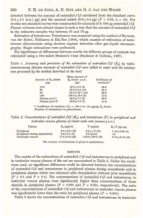
280 F. H. DEJoNG, A. H. HEY AND H. J. vAN DER MoLEN
detected between the amount of oestrad.iol-17,8 calculated from the standard curve (5·4 ± 4·1 (S.D.) pg) and the amount added (6·6 ± 0·4 pg) (P > 0·20, n = 10). For routine use standard curves were constructed for amounts of 5-100 pg oestrad.iol-17 ,8. Plasma volumes were always chosen in such a way that the amount of oestrad.iol-17,8 in the unknown samples was between 10 and 70 pg.
Estimation of testosterone. Testosterone was measured using the method of Brownie, van der Molen, Nishizawa & Eik-Nes (1964}, which consists of estimation of testosterone chloroacetate using electron capture detection after gas-liquid chromatography. Single estimations were performed.
The significance of differences between results for different groups of animals was calculated using a two-tailed Student's t-test (Snedecor & Cochran, 1967).
Table 1. Accuracy and precision of the estimation of oestradiol-17,8 (E2) by radioimmunoassay (known amounts of oestradiol-17,8 were added to water and the solution was processed by the method described in the text)
Amount of E 2 added (pg)
12·6 30 50 100 P lasma. pool I P lasma. pool II
Mean amount of E 2 found ±S.D.
(pg)
12·9 ± 6·4 (8) 33·2 ± 9·4 (10) 55·5 ± 12·7 (10)
106·0± 18·4 (8) 24·9±1·7 (8) 63·6 ± 1·5 (7)
Coefficient of variation*
49·6 28·3 22·9 11·8 30·9 11·8
* Coefficient of variation(%) = 100 x S.D. (in pg)fpg E 2 found. Number of estimations in parentheses.
Table 2. Concentrations of oestradiol-17,8 (E2) and testosterone (T) in peripheral and testicular venous plasma of intact male rats (means± S.D.)
Plasma
Peripheral Peripheral during anaesthesia Testicular venous
E 2 (pg/ml)
2·0 ± 0·9 (12) 3·4±2·4 (8)
17·5±8·4 (43)
T (ng/ml)
2·4± 1·8 (21) 2·8 (pool)
110·0±80·5 (19)
The number of estimations is given in parentheses.
RESULTS
El:T (pg:ng)
1·13±0·29 (5) 1·2 0·18±0·16 (18)
The results of the estimations of oestrad.iol-17,8 and testosterone in peripheral and in testicular venous plasma of the rat are summarized in Table 2. Under the conditions used, no significant differences could be detected between the concentrations of oestradiol-17,8 and testosterone in peripheral plasma during anaesthesia and in peripheral plasma which was obtained after decapitation without prior anaesthesia (P > 0·1 and P > 0·1}. The concentrations of oestradiol-17,8 and testosterone in testicular venous plasma were significantly higher than concentrations of these steroids in peripheral plasma (P < 0·001 and P < 0·001, respectively). The ratio of the concentrations of oestradiol-17,8 and testosterone in testicular venous plasma was significantly lower than the ratio for peripheral plasma (P < 0·001).
Table 3 shows the concentrations of oestradiol-17,8 and testosterone in testicular

Secretion of oestradiol by the rat testis 281 venous plasma after administration of HCG. The concentrations of both steroids were increased significantly after acute HOG administration (P < 0·002 and P < 0·002, respectively). The ratio of the concentrations of oestradiol-17/] and testosterone was decreased (P < 0·05). Continuous administration of HOG for 5 days did not change the concentration of oestradiol in testicular venous plasma significantly when compared with levels in untreated rats (0·1 > P > 0·05). Testosterone concentrations, however, were increased (P < 0·001) under t hese conditions. Additional i .v. administration of 100 i.u. HCG after 5 days pretreatment with HCG resulted in an increase in the testicular venous oestradiol-17/3 concentration (P < 0·002), while the increase in testosterone concentration was not significant (P > 0·1).
Table 3. Concentrations of oestradiol-17(3 (E2) and testosterone (T) in testicular venous plasma of intact male rats after administration of human chorionic gonadotrophin (H CG) (means± S.D .)
Treatment*
None 100 i.u., i.v. 5 X 100 i.u., S.C. (5 d) 5 x 100 i.u.,s.c. (5d) + 100 i.u., i.v.
E• (pgfml)
19·0 ± 8·5 ( 18) 41·3±24-1 (16) 21 ·7± 7·6 (16) 37-5± 15·1 (13)
The number of estimations is given in parentheses.
T (ngfml)
129 ± 91 (9) 666± 397 (8)
1675 ± 707 (6) 2592± 1219 (5)
E! :T (pg:ng)
0·178±0·106 (8) 0·070 ± 0·055 (8) 0·013 ± 0·007 (6) 0·021 ± 0·013 (5)
* 100 i.u., i.v. = 100 i.u. of HCG were administered intravenously immediately before collection of testicular venous blood. 5 x 100 i.u., s.c. (5 d) = 100 i.u. of HCG were administered subcutaneously each day during 5 days.
Table 4. Concentrations of oestradiol-17(3 (E2) and testosterone (T) in testicular venous plasma of intact male rats after administration of follicle-stimulating hormone (F SH) (means± S.D.)
Treatment
Non e 5p,g FSH i .v.*
E, (pgfml)
19·5± 7·0 (17) 23·1 ± 11-4 (29)
The number of estimations is given in parentheses.
T (ng/ml)
81±31 (4) 111±73 (7)
E 2 :T (pg:ng)
0·27 (2) 0·28±0·31 (5)
• 5 p,g of FSH (NIH-FSH-89) were administered intravenously immediately before collection of tes. ticular venous blood.
Finally, the effect of acute administration of FSH on testicular venous plasma concentrations of oestradiol-17/] and testosterone is shown in Table 4. The levels of both oestradiol and testosterone did not increase as a result of the FSH administration (P > 0·1 and P > 0·1, respectively).
DISCUSSION
The concentrations of testosterone in peripheral and in testicular venous plasma estimated during the present study compare well with reported values (Bardin & Peterson, 1967; Rivarola, Snipes & Migeon, 1968). The peripheral plasma concentrations of oestradiol-17 f3 that were estimated during the present study are ten times lower than those reported by Labhsetwar (1972). The difference may be caused by a greater specificity of the antibody to oestradiol-17 f3 used in our study. However, that

282 F. H. DEJoNG, A. H. HEY AND H. J. VANDER MoLEN
non-oestrogenic material, possibly originating from the adrenal, might have been bound by the antiserum in the present study cannot be disregarded. The theoretically possible presence of oestradiol-17,8 oxygenated at carbon atom 6 in the sample is not likely after column chromatography, but cannot be excluded with certainty. On the other hand, the finding of comparable levels of oestradiol-17 ,8 in peripheral plasma from male rats by using a double competitive protein-binding technique, as described by Robertson, Mester & Kellie (1971) (F. H. de Jong, A. H . Hey & H. J. van der Molen, unpublished results), makes these possibilities less probable.
A possible influence of anaesthesia or surgical stress on testosterone concentration can almost be ruled out, when blood production rates for testosterone are compared with the testicular production rate of testosterone. The latter may be calculated from testicular blood flow (Jones, 1971) and the testosterone concentration in testicular venous plasma. The blood production rate for testosterone is 80 ttgfday, assuming that 10% of the steroid is bound to erythrocyt-es and that the haematocrit value is 45 %· The testicular production rate, based on a testicular weight of 3 g per rat, is 74 ttgfday, or 92% of the blood production rate. This indicates that anaesthesia and collection of testicular venous blood did not influence the testicular testosterone secretion to a large extent. Similarly, no significant effect of anaesthesia on peripheral concentrations of oestradiol-17,8 or testosterone could be shown (Table 2).
The observation of a difference between oestradiol-17 ,8 concentrations in testicular venous plasma and in peripheral plasma reflects that the rat testis secretes oestradiol-17,8. A testicular oestradiol-17,8 secretion of 11 ngfday can be calculated from testicular blood flow and the concentration of oestradiol-17,8 in testicular venous plasma, assuming that the concentration of oestradiol-17,8 in plasma from the testicular artery is equal to the peripheral venous level. Since the metabolic clearance rate (MCR) for oestradiol-17,8 in the male rat has not yet been reported, it is not possible to calculate the blood production rate for this steroid in the male rat. However, de Hertogh, Ekka, VanderHeyden & Hoet (1970) measured the MCR for oestradiol-17,8 in the female rat and observed a close agreement with the MCR for oestradiol-17,8 estimated by Longcope, Layne & Tait (1968} for women, after correction for body weight. Applying the same correction for body weight differences to the MCR for oestradiol-17 ,8 in the male human being as estimated by Long cope et al. (1968}, the MCR for oestradiol-17,8 in the male rat is calculated to be 25·61/day. This would reflect a blood production rate of oestradiol-17,8 of 51 ngfday. Hence, the testicular secretion of 11 ngfday in the rat reflects that only 21% of the total circulating oestradiol-17 ,8 is secreted by the testis. This percentage agrees well with the observation of Kelch et al. (1972), who calculated that in men approximately 25% of circulating oestradiol is secreted by the t estis. The larger part of circulating oestradiol-17,8 may be derived from peripheral conversion of testicular and adrenal androgens. The presence of a binding principle for oestradiol-17,8 in interstitial tissue (Brinkmann, Mulder & van der Molen, 1972) might, however, be an indication for a physiological significance of oestradiol-17 ,8 in the testis itself.
Concentrations of both oestradiol-17 ,8 and testosterone in testicular venous plasma increased acutely after intravenous administration of HCG. Mter 5 days of subcutaneous HCG stimulation, testicular venous oestradiol-17,8 levels were equal to those in non-stimulated rats, although additional i.v. administration of HCG gave

Secretion of oestradiol by the rat testis 283 an increase in oestradiol-17 fJ levels (Table 3). Testosterone concentrations in the same samples were increased ten times after 5 days of HCG treatment and increased only slightly after additional i.v. HCG. This discrepancy between the effects of HCG on oestradiol-17/J and testosterone levels, which is reflected by their varying ratios, can be explained if the two steroids are synthesized in different compartments of the testis or by assuming a constant production of oestradiol-17/J by an enzyme system with a very limited capacity, whilst HCG administration stimulates release of oestradiol-17/J from some storage compartment. Follicle-stimulating hormone does not stimulate the testicular secretion of testosterone or oestradiol-17/J under the conditions used in the present investigation. If the action of this trophic hormone is mediated through cyclic AMP, this observation is in agreement with the conclusions of Dorrington, Vernon & Fritz {1972) and of Cooke, van Beurden, Rommerts & van der Molen (1972), who did not observe an increase in cyclic AMP levels during incubation of testicular tissue or seminiferous tubules from intact adult rats with FSH. The suggestion ofEik-Nes (1962) that his observation of increased testosterone secretion by the perfused dog testis after administration of FSH would be caused by LH impurities in the FSH preparation and a synergistic effect of FSH, is in agreement with the present results.
The authors gratefully acknowledge the gift of antibodies raised against the oestradiol-17/J-6-(0-carboxymethyl)oxime-BSA complex from Dr D. Exley (London}, and the gift of FSH from the NIH Endocrine Study Section.
REFERENCES
Anliker, R ., Perelman, M., Rohr, 0. & Ruzicka, L. (1957). Untersuchungen uber Organextrakte. 27 :Mitteilung. "Ober die Konzentration des Testosterons in menschlichen Testes. Helv. chim. Acta 40, 1517-1520.
Attal, J. (1969). Levels of testosterone, androstenedione, estrone and estradiol-17,8 in the testes of fetal sheep. Endocrinology 85, 280-289.
A.xelrod, L. R. (1965). Metabolic patterns of steroid biosynthesis in young and aged human testes. Biochim. biophys. Acta 97, 551- 556.
Baird, D., Horton, R., Longcope, C. & Tait, J. F. (1968). Steroid prehormones. Persp~Xt. Biol. Med. 11, 384-421.
Baird, D. T., Hor ton, R., Longcope, C. & Tait, J. F. (1969). Steroid dynamics under steady-state conditions. Recent Prog. Harm. Res. 25, 611- 664.
Bardin, C. W. & Peterson, R . E. (1967). Studies of androgen production by the rat: testosterone and androstenedione content of blood. Endocrinology 80, 38-44.
Beall, D. (1940). Isolation of a.-oestradiol and oestrone from horse testes. Biochem. J. 34, 1293-1298. Bedrak, E. & Samuels, L. T. (1969). Steroid biosynthesis by the equine testis. Endocrinology 85, 1186-
1195. Brinkmann, A. 0., Mulder, E. & van der Molen, H. J. (1972). Recepteur testiculaire de !'oestradiol chez
le rat. C. r. hebd. Seanc. Acad. Sci., Paris (Serie D) 274, 3106-3108. Brownie, A. C., van der Molen, H. J., Nishizawa, E. E. & Eik-Xes, K. B. (1964). Determination of
testosterone in human peripheral blood using gas-liquid chromatography with electron capture detection. J. clin. Endocr. Metab. 24, 1091-1102.
Cooke, B. A., van Beurden, W. M. 0., Rommerts, F. F. G. & van der Molen, H. J. (1972). Effect of troph ic hormones on 3' ,5' -cyclic AMP levels in rat testis interstitial tissue and seminiferous tubules. FEES Lett. 25, 83-86.
Darrington, J. H., Vernon, R. G. & Fritz, I. B. (1972). The effect of gonadotrophins on the 3',5'-AMP levels of seminiferous t ubules. Biochem. biophys. Res. Commun. 46, 1523-1528.
Eik-Nes, K . B. (1962). Secretion of testosterone in anesthetized dogs. Endocrinology 71, 101-106. Eik-Nes, K. B. (1967). Factors controlling the secretion of testicular steroids in the anaesthetized dog.
J. Reprod. Fert. Suppl. 2, 125-141.

284 F. H. DEJoNG, A. H. HEY AND H. J. VANDER MoLEN
Exley, D., Johnson, M. W. & Dean, P. D. G. (1971) . Antisera highly specific for 17,8-oestradiol. Steroids 18, 605-620.
Ficher, M. & Steinberger, E. (1971). I n vitro progesterone metabolism by rat testicular tissue at different stages of development. Acta endocr., Gopenh. 68, 285-292.
Goldzieher, J. W. & Roberts, I. S. (1952) . Identification of estrogen in the human testis. J. clin. Endocr. Metab. 12, 143-150.
de Hertogh, R., Ekka, E., VanderHeyden, I. & Hoet, J. J. (1970). Metabolic clearance rates and the interconversion factors of estrone and estradiol-17,8 in t he immature and adult female rat . Endocrinology 87, 874-880.
Hotchltiss, J., Atkinson, L. E. & Knobil, E. (1971). Time course of serum estrogen and luteinizing hormone (LH) concentrations during the menstrual cycle of the rhesus monkey. Endocrinology 89, 177- 183.
Inaba, M., Nakao, T. & Kamata, K. ( 1967). Possible conversion of androst-4-ene-3.8,17 ,8,19-triol to estriol in vitro in rat testis . Endocr. jap. 14, 1- 6.
Jones, T. (1971). Blood flow and volume measurements in the radiation depopulated testis of the rat. Br. J. Radiol. 44, 841- 849.
de Jong, F. H. & van der Molen, H . J. (1972). Determination of dehydroepiandrosterone and dehydroepiandrosterone sulphate in human plasma using electron capture detection of 4-androstene-3,6,17-trione after gas-liquid chromatography J. Endocr. 53, 461-474.
Kelch, R. P., Jenner, M. R., Weinstein, R., Kaplan, S. L. & Grumbach, M. M. (1972). Estradiol and testosterone secretion by human, simian and canine testes, in males with hypogonadism and in male pseudohermaphrodites with the feminizing testes syndrome. J. clin. Invest. 51, 824-830.
Labhsetwar, A. P. ( 197.2). Peripheral serum levels of immunoreactive 'oestradiol ' in rats during various reproductive states: adrenal contribution of immunoreactive material. J. Endocr. 52, 399-400.
Leonard, J. M., F locks, R. H. & Korenman, S. G. (1971). Estradiol (E2) secretion by the human testis. Program of the Endocrine Society, 53rd Meeting, Abstr. 133. San Francisco: The Endocrine Society.
Longcope, C., Layne, D. S. & Tait, J. F. (1968). Metabolic clearance rates and interconversions of estrone and 17,8-estradiol in normal males and females. J. clin. Invest. 47, 93-106.
MacDonald, P. C., Grodin, J. M. & Siiteri, P. K. (1971). Dynaiillcs of androgen and oestrogen secretion. In Control of gonadal steroid secretion, pp. 158-174. Eds D. T . Baird & J. A. Strong. Edinburgh : Edinburgh University Press.
MacDonald, P . C., Rombaut, R. P. & Siiteri, P. K. (1967). Plasma precursors of estrogen. I. Extent of conversion of plasma .6.4-androstenedione to estrone in normal males and non-pregnant normal, castrate and adrenalectomized females. J. clin. Endocr. M etab. 27, 1103- 1111.
Oh, R. & Tamaoki, B. (1970). Steroidogenesis in equine testis. Acta endocr., Gopenh. 64, 1-16. Oh, R. & Tamaoki, B. (1971). Occurrence of 19-oxo-androst-4-ene-3,17-dione in the course of oestrogen
biosynthesis by equine testicular Iillcrosomes. Acta endocr., Copenh. 67, 665-616. Pierrepoint, C. G., Galley, J . Mel., Griffiths, K. & Grant, J. K . (1961) . Steroid metabolism of a Sertoli
cell tumour of the testis of a dog with feminization and alopecia and of the normal canine testis. J. Endocr. 38, 61- 70.
Rivarola, M.A., Snipes, C. A. & Migeon, C. J. (1968). Concentration of androgens in systeiillc plasma of rats, guinea pigs, salamanders and pigeons. Endocrinology 82, 115- 121.
Robertson, D . M., Mester, J. & Kellie, A. E. (1911). A double competitive binding method of measuring oestradiol from menstrual cycle plasma with and without the prior purification of oestradiol. Acta endocr., Copenh. 68, 513-522.
Sharma, D. C. & Gabrilove, J. L. (1971) . Biosynthesis of testosterone and oestrogens in vitro by the testicular tissue from patients with Klinefelter's syndrome. Acta endocr., Copenh. 66, 737- 744.
Siegel, E. T., Forchielli, E., Dorfman, R.I., Brodey, R. S. & Prier, J. E. (1967). An estrogen study in the feminized dog with testicular neoplasia. Endocrinology 80, 272-217.
Snedecor, G. W . & Cochran, W . G. (1967) . Statistical methods, 6th edn. Ames (Iowa): Iowa State University Press.
Wu, C.-H. & Lundy, L . E. (1971). Radioimmunoassay of plasma estrogens. Steroids 18, 91-111.
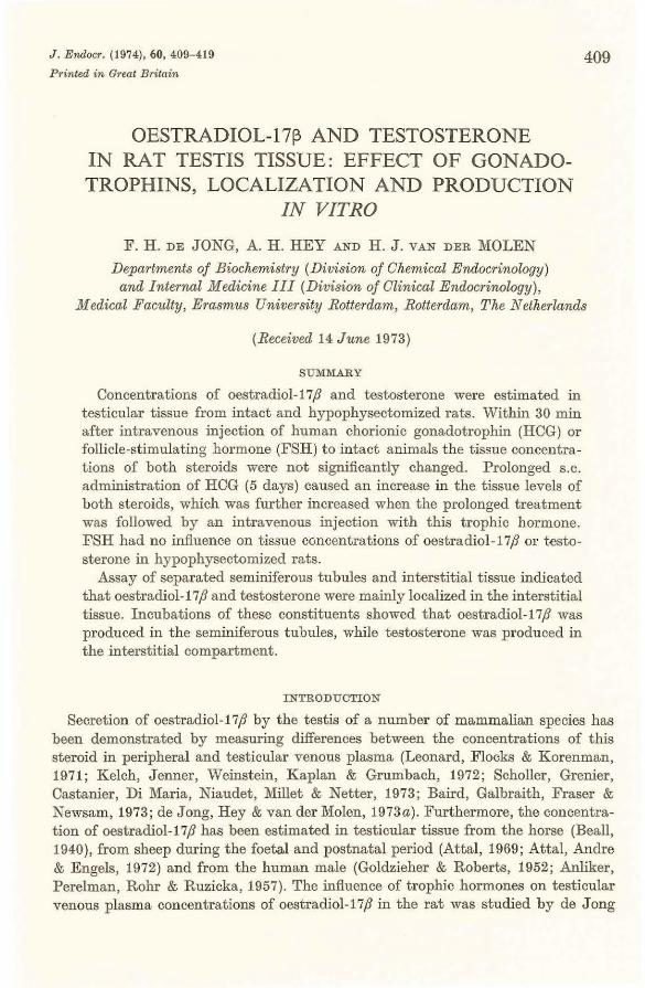
J. Endocr. (1974), 60, 409-419
Printed in Great Britain
OESTRADIOL-1713 AND TESTOSTERONE IN RAT TESTIS TISSUE: EFFECT OF GONADOTROPHINS, LOCALIZATION AND PRODUCTION
IN VITRO
F. H. DEJONG, A. H. HEY AND H. J. VANDER MOLEN
Departments of Biochemistry (Division of Chemical Endocrinology)
409
and Internal Medicine III (Division of Clinical Endocrinology), Medical Faculty, Erasmus University Rotterdam, Rotterdam, The Netherlands
(Received 14 June 1973)
SUMMARY
Concentrations of oestradiol-17,8 and testosterone were estimated in testicular tissue from intact and hypophysectomized rats. Within 30 min after intravenous injection of human chorionic gonadotrophin (HOG) or follicle-stimulating hormone (FSH) to intact animals the tissue concentrations of both steroids were not significantly changed. Prolonged s.c. administration of HCG (5 days) caused an increase in the tissue levels of both steroids, which was further increased when the prolonged treatment was followed by an intravenous injection with this trophic hormone. FSH had no influence on tissue concentrations of oestradiol-17,8 or testosterone in hypophysectomized rats.
Assay of separated seminiferous tubules and interstitial tissue indicated that oestradiol-17,8 and testosterone were mainly localized in the interstitial tissue. Incubations of these constituents showed that oestradiol-17,8 was produced in the seminiferous tubules, while testosterone was produced in the interstitial compartment.
INTRODUCTION
Secretion of oestradiol-17,8 by the testis of a number of mammalian species has been demonstrated by measuring differences between the concentrations of this steroid in peripheral and testicular venous plasma (Leonard, Flocks & Korenman, 1971; Kelch, Jenner, Weinstein, Kaplan & Grumbach, 1972; Scholler, Grenier, Castanier, Di Maria, Niaudet, Millet & Netter, 1973; Baird, Galbraith, Fraser & Newsam, 1973; de Jong, Hey & van der Molen, 1973a). Furthermore, the concentration of oestradiol-17,8 has been estimated in testicular tissue from the horse (Beall, 1940), from sheep during the foetal and postnatal period (Attal, 1969; Attal, Andre & Engels, 1972) and from the human male (Gold.zieher & Roberts, 1952; Anliker, Perelman, Rohr & Ruzicka, 1957). The influence of trophic hormones on testicular venous plasma concentrations of oestradiol-17,8 in the rat was studied by de Jong

410 F. H. DEJoNG, A. H. HEY AND H. J. VANDER MoLEN
et al. (1973a), but it was not clear whether the rise in oestradiol-17/] secretion after the intravenous administration of human chorionic gonadotrophin (HOG) reflected an increased release or an increased synthesis of steroid. Therefore, the testicular tissue concentrations of oestradiol-17/] before and after treatment with HOG were studied in the present investigation.
Follicle-stimulating hormone (FSH) did not influence testicular venous plasma concentrations of oestradiol-17/] or testosterone in the rat under the conditions used by de J ong et al. ( 1973 a). However, it has been suggested t hat testicular processes in intact animals are maximally stimulated by endogenous FSH, thus preventing a further stimulation by exogenously added FSH (Means & Hall, 1968). In t his respect it has been shown that FSH has also no influence on t he concentration of cyclic AMP in testicular t issue from intact rats during incubation, although this trophic hormone causes an increase in the cyclic AMP concentration in testis tissue from hypophysectomized rats (Dorrington, Vernon & Fritz, 1972). Therefore, the effect of FSH on the testicular tissue concentration of oestradiol-17/] and testosterone in hypophysectomized rats has been studied. Finally, it has been suggested by Scholler, Grenier & Castanier (1972) and by de Jong et al. (1973a) that oestradiol-17/] and testosterone might be synthesized in different testicular compart ments. The distribut ion of these two steroids between interstitial tissue and seminiferous tubules and their production in vitro by total and dissected testis t issues have therefore been studied.
A preliminary account of some of the present results was given at the ninth Acta Endocrinologica CongTess (de Jong, Hey & van der Molen, 1973b).
::VIATERIALS AND METHODS
Animal techniques
Adult male Wistar rats with a body weight of 200-250 g were used in the experiments with intact animals. Hypophysectomized rats were from the R-Amsterdam strain, which is an inbred Wistar substrain. These animals also weighed 200-250 g . Hypophysectomies were performed by the transauricular approach (Dr R. Welschen and Mrs lVI. Loonen-Rutte, Department of Anatomy, Erasmus University, Rotterdam). The concentration of testosterone in the testicular tissue was used as a criterion for the completeness of hypophysectomy, since a visual inspection of the sellae turcicae proved unreliable. Hypophysectomized rats were discarded if t heir testicular testosterone concentration exceeded 10 ngfg.
When the effect of injected gonadotrophins was studied, rats were anaesthetized with sodium pentobarbitone. Heparin was injected into one of the tail veins, followed by saline (0·9% NaCl in water), saline containing HOG (Pregnyl, Organon, Oss, 100 i.u. ) or a solution of 0·05% human y-globulin in saline containing FSH (~TIH-FSH
S9, 5 or 10 ;.tg). Testicular venous blood was collected during a 30 min period (de Jong et al . 1973a). The testes were t hen removed from the animal, immediately placed on ice and kept at - 15 °0 until analysed. In some experiments rats were injected subcutaneously with 100 i.u. HOG or 10 ;.tg FSH daily for a period of 5 days. FSH treatment started on the day after hypophysectomy. Total testis tissue was dissected using the technique of Christensen & Mason (1965) . The dissections were performed

Oestradiol in rat testis tissue 411
in a Krebs-Ringer buffer, pH 7·4, containing 0·2% glucose. Isolated seminiferous tubules were washed three times as described by Rommerts, van Doorn, Galjaard, Cooke & van der Molen (1973).
Incubation procedures
Testes were removed immediately after decapitation. The testes were placed in a dish on ice and decapsulated. To both testes from one animal, 1·5 ml of an icecold Krebs-Ringer buffer, pH 7·4, containing 0·2% glucose, was added. One of the testes was then kept at -15 oc and served as an unincubated control. The other testis was incubated for 180 min at 34 °C in an atmosphere of 95% 0 2 and 5% C02
and was then placed at -15 oc until analysed. The same procedure was followed for dissected interstitial tissue and seminiferous tubules.
Steroid estimations
Radioimmunoassay of oestradiol-17ft
Materials used for the radioimmunoassay of oestradiol-17ft were described by de Jong et al. (1973a). Mter addition of tritiated testosterone and oestradiol-17ft, testicular tissue was homogenized by sanification in 1·5 ml distilled water or buffer solution. Proteins were precipitated with acetone (5 ml, Union Chimique Beige, p.a. quality) . Mter centrifugation the acetone layer was removed and the precipitate washed with 2 ml of acetone. The combined acetone layers were evaporated under nitrogen at 45 °C until only water remained. This water layer was extracted twice with ether (3 ml each time) and the combined ether layers taken to dryness . The residue was partitioned between 2 ml methanol:water (70: 30, v fv) and 2 ml nhexane. Mter a second extraction with 2 ml n-hexane the methanolic layer was dried down under nitrogen at 45 °C. The residue was applied to a Sephadex LH-20 microcolumn. Testosterone was eluted from the column with 2·6 ml toluene:methanol (9: 1, v fv) and the oestradiol-17ft fraction was collected in the next 3 ml of eluate. After evaporating the solvent of this oestradiol-17ft fraction under nitrogen at 45 oc, radioimmunoassay was performed, using the antibody as described by Exley , Johnson & Dean (1971) . Details of the procedure are given by de Jong et al. (1973a). The effectiveness of the extraction procedure for oestradiol-17ft from rat testis tissue was investigated by extracting tritiated oestradiol-17ft from part of a testis, into which the steroid was infused via the spermatic artery (Frederik & van Doorn, 1973). The amount of radioactivity remaining in the precipitated protein was estimated after treatment with soluene (Packard) and amounted to 7% of the number of d. p.m. recovered (mean of duplicate determinations).
Accuracy and precision, as well as blank values for the estimation, were reported previously (de Jong et al. 1973a). The overall recovery for the estimation of oestradiol-17ft in testis tissues was 52·9 ± 6·6 (S.D.)% (n = 296). Data on the specificity of the method, with regard to oestradiol-17ft concentrations in peripheral plasma from ovariectomized and adrenalectomized prepubertal female rats, are given by MeijsRoelofs, Uilenbroek, de Jong & Welschen (1973).

412 F. H. DEJONG, A. H. HEY AND H. J. VANDER MOLEN
Estimation of testosterone
Testosterone was measured using the method of Brownie, van der Molen, Nishizawa & Eik-Nes (1964), which consists of estimation of testosterone chloroacetate using electron capture detection after gas-liquid chromatography.
The significance of differences between results for different groups was calculated using a two-tailed Student's t-test (Snedecor & Cochran, 1967).
RESULTS
The results of the estimations of oestradiol-17,8 and testosterone in testis tissue from untreated and HOG-treated rats are summarized in Table 1. The intravenous administration of HOG did not significantly increase the concentration of oestradiol-17ft (P > 0·5) or testosterone (P > 0·2) above that found in untreated rats.
Table 1. Concentrations of oestradiol-17,8 and testostemne in testicular tissue of intact male rats after administmtion of human chorionic gonadotmphin (HCG) (means± S.D.)
Oestradiol-17{1: No. of Oestradiol-17/1 Testosterone testosterone
Treatment* estimations (pg/g) (ng/g) (pg:ng)
None 6 44-6± 18-5 372±287 0-16±0·08 100 i.u., i.v. 8 48·8 ± 18-8 700±764 0-12 ±0·06 5 x 100 i.u., s.c. (5 days) 10 66·0± 14-0 1351 ± 374 0-05±0·02 5 x 100 i.u . , s.c. (5 days)
+ 100 i.u., i.v. 9 90·2 ±23·4 1491 ± 579 0·07 ±0·02
* 100 i.u ., i.v. = 100 i.u. HCG were administered intravenously immediately before collection of testicular venous blood. 5 x 100 i.u., s.c. (5 days) = 100 i.u. HCG were administered subcutaneously each day for 5 days.
Table 2. Concentrations of oest?·adiol-17,8 and testosterone in testicular tissue of intact male 1·ats after administration of follicle-stimulating hormone (FSH) (means± S.D.)
Oestradiol-17/1: No. of Oestradiol -17/1 Testosterone testosterone
Treatment* estimations (pgfg) (ng/g) (pg:ng)
None 18 30·4± 17·0 373±239 0·11 ±0·07 5 p.g, i.v. 10 38·8±26·6 258± 145 0·22 ±0·28 10 p.g, i.v. 6 43-5±24·!) 350± 288 0·16±0·08
* FSH was injected intravenously immediately before collection of testicular venous blood.
Repeated subcutaneous administration of HOG caused a significant increase in the testicular concentrations of both oestradiol-17,8 and testosterone (P < 0·05 and P < 0-001, r espectively). The relative increase in testosterone concentration was larger than the increase in oestradiol-17,8 concentration as indicated by a significant decrease of the oestradiol-17,8:testosterone ratio (P < 0-01). When the prolonged treatment with HOG was combined with an acute intravenous dose of the gonadotrophin, a further increase in testicular concentration of oestradiol-17,8 was observed (P < 0-02), while the concentration of testosterone did not increase significantly (P > 0-50) . As a consequence, t he oestradiol-17,8:testosterone ratio was significantly

Oestradiol in rat testis tissue 413 increased (P < 0·05), when compared with that of the animals receiving only subcutaneous injections of HOG.
The effect of the i.v. administration of a single dose of FSH on testicular levels of oestradiol-17ft and testosterone is shown in Table 2. Neither the administration of 5 pg FSH, nor of 10 pg FSH had a significant influence on the testicular levels of oestradiol-17ft or testosterone in intact rats (P was always > 0·10). The results in
Table 3. Concentrations of oestradiol-17ft and testosterone in testicular tissue of hypophysectomized male rats after administration of follicle-stimulating hormone (FSH) (means ±S.D.)
Treatment*
None 10 J.Lg, i.v. 5 X 10 J.Lg, S.C. (5 days) 5 X J 0 J.Lg, S.C. (5 day~)
+ 10 p.g, i.v.
Oestradiol-17/3 (pgfg)
4·5 ± 5·6 ( 18) 3·9±4·4 (16) 1·4±2·7 (10)
1·0±2·3 (12)
The number of estimations is given in parentheses.
Testosterone (ng/g)
5·1±2·7 (9) 4·5±2·7 (8) 3·7±1·3 (5)
3·0± 1·0 (6)
* 10 fl-g i.Y. = 10 I'S FSH (NIH-FSH-S9) were administered intravenously immediately before collection of testicular venous blood. 5 x 10 p.g, s.c. (5 days) = 10 #g FSH (l\TIH-FSH-S9) were administered subcutaneously each day for 5 days.
Table 4. Concentrations of oestradiol-17ft and testosterone in dissected testis tissue from intact rats (means± S.D.)
Oestradiol-17/3: Oestradiol-17/3 Testosterone testosterone
Tissue* (pgjg) (ngfg) (pg/ng)
Untreated rats: Seminiferous tubules 20·5 ± 11·7 (4) 21·4± 6·9 (6) 0·93 ± 0·41 (4) Interstitial tissue 179 ±58 (3) 226 ± 113 (4) 0·73 (2)
HCG-treated rats: Seminiferous tubules 12·2± 6·0 (4) 174 ± 53 (4) 0·07 ± 0·03 (4) Interstitial tissue 197 ±89 (7) 3428 ±410 (4) 0·06 ± 0·02 (4)
The number of estimations is given in parentheses. * Seminiferous tubules and interstitial tissue were obtained by dissection of testes from untreated
rats or from rats which were injected subcutaneously with 100 i.u. chorionic gonadotrophin (HCG) each day for 5 days.
Table 3 show that also after the administration of FSH to hypophysectomized rats no significant increase in the concentrations of oestradiol-17fJ or testosterone occurred after intravenous, repeated subcutaneous or a combination of the subcutaneous and intravenous injections ofFSH (P was always > 0·10).
Table 4 summarizes the concentrations of oestradiol-17ft and testosterone found in seminiferous tubules and interstitial tissue after dissection of testes from untreated rats and rats treated with HOG for 5 days. The concentrations of both oestradiol-17ft and testosterone were higher in interstitial tissue than in seminiferous tubules for the normal (P < 0·01 and P < 0·01) as well as for the HOG-treated rats (P < 0·001 and p < 0·001).
The results from the estimations of oestradiol-17ft and testosterone before and after incubation of total testis tissue, seminiferous tubules or interstitial tissue are

414 F. H. DE JoNG, A. H. HEY AND H. J. VANDER MoLEN
summarized in Table 5. Because the initial steroid levels in the tissue samples from individual animals showed a large variation, the ratios of the concentration after 180 min incubation over the concentration at the start of the incubation were calculated, and the significance of differences was calculated from these figures. The concentration of oestradiol-17/l increased significantly during incubation of total tissue and of seminiferous tubules from the normal rat. The testosterone concentration increased significantly during incubation of total tissue and of interstitial tissue from the normal rat. A significant decrease in testosterone levels was observed after incubation of testis from rats, which were treated 5 days with HOG.
When steroid concentrations were calculated fmg protein rather than /g tissue, essentially similar relationships were obtained.
Table 5. Concentrations of oestradiol-17fJ (E2 , pgfg) and testosterone (T, ngfg) in testicular tissue before and aftm· 180 min incubation (means± S.D.)
Incubation time (t) t= 180 min
Tissue* Steroid Omin 180 min t=Omin
Untreated rats Total testis E. 11·7± 4·0 (7) 31-6±15·1 (7) 2·42 ± 0·61 (6)
T 55 ± 32·5 (4) 132 ± 28·3 (4) 3-12± 1·75 (4)
Seminiferous t ubules E, 20·2± 11·7 (4) 39·2 ± 19·5 (4) 1·94±0·33 (4) T 21·4± 6·9 (6) 30·3 ± 19·8 (6) 1-49±0·96 (6)
Interstitial tissue E, 179 ± 58 (7) 181 ±57 (4) 0·94±0·11 (3) T 226 ±113 (4) 663 ±286 (4) 3·67 ± 1-86 (4)
HCG.treated rats : Total testis E, 64·7 ± 42·3 (14) 69·0±44·8 (16) 1·02 ± 0·26 ( 14)
T 1062 ±231 (8) 594 ±78 (8) 0·57 ± 0·09 ( 8)
The number of estimations is given in parentheses. * Testis tissue from untreated rats or from rats which were injected subcutaneously with 100 i.u.
human chorionic gonadotrophin (HCG) each day for 5 days was incubated for 180 min in a Krebs- Ringer buffer, pH 7·4, which contained 0 ·2% glucose.
Incubations were performed at 34 oc in an atmosphere of 95% 0 2 :5% C02•
DISCUSSION
The intravenous administration of HCG into intact rats causes an increase in the testicular venous plasma concentration of testosterone and oestradiol-17/l (de Jong et al. 1973a). This increase in venous plasma levels reflects increased secretion as a result of an increased biosynthesis of both steroids, since the t estis tissue concentrations of testosterone and oestradiol-17 fJ were not decreased after the 30 min period of testicular venous blood collection (Table 1). The influence of intravenous administration of HOG on the biosynthesis of oestradiol-17/l was even more pronounced after pretreatment with HOG for 5 days : a significant increase in the secretion rate of oestradiol-17/l was accompanied by a significant rise of the testicular tissue concentration of the steroid. The rate of testosterone biosynthesis seems to be maximally stimulated after prolonged pretreatment with HOG: additional intravenous administration of HOG did not further increase the plasma or tissue concentrations.
After 5 days of HOG treatment the relative increase of the testicular venous testosterone concentration was larger than the increase of the testis tissue level

:!' .::;:,
'" E ~ n. -g " -;JJ ciJ
.5 -~ ;;:: 3
·-~
? ;::; ;;:
~ u
f-
Oestradiol in rat testis tissue 415 (Fig. 1 ). This indicates that the secretion of testosterone is faster in the rat treated for 5 days with HOG than in the untreated animal. On the other hand, the repeated administration of trophic hormone raised the tissue content of oestradiol-17,8, although the testicular venous plasma level was not increased (Fig. 2). It is not clear if this apparent retention of oestradiol-17,8 by testicular tissue is caused byintratesticular
!·8
1·6
1-4
1·2
1·0
0·8
0 ·6
0·4
0· 2
0 Co ntrol
Fig. 1
Ra ts trcatcJ " ·ith HCG
~
E Oil 5 "' ~ c.. -o c: " ';jj eo c..
-~ v. ~
·-~ ..... ' :§
2 '" "' 0
60
50
40
30
20
10
0 Comrols
Fig. 2
Ra ts treated with HCG
Fig. 1. Concentrations of testosterone in testis t issue (stippled bars) and testicular venous plasma (open bars) from normal intact rats and rats which were injected subcutaneously with 100 i.u. of human chorion.lc gonadotrophin (HCG) per day for 5 days (means± s.E.llr. ). (Data partly from de Jong et al. 1973a .)
Fig. 2. Concentrat ions of oestracliol-17P in testis tissue (stippled bars) and test icular venous plasma (open bars) from normal intact rats and rats which were injected subcutaneously with 100 i.u. human chorion.lc gonadotrophin (HCG) per day for 5 days (means ±s.E.M.). (Data partly from de Jong eta/. 1973a.)
metabolism of t he steroid, occurring also under conditions in vitro (Table 5), which should take place before oestradiol-17,8 reaches the blood stream. This possibility suggests a different localization of the enzymes which synthesize or metabolize testosterone and oestradiol-17,8, since the secretion of testosterone is increased, although intratesticular metabolism of testosterone is greater than that of oestradiol-

416 F. H. DEJoNG, A. H. HEY .AND H. J. VANDER :MoLEN
17fl under conditions in vitro (Table 5). On the other hand, the retention of oestradiol-17(3 in the tissue may be caused by changes in the concentration of testicular receptor proteins, which bind the steroid with a high affinity (Brinkmann, Mulder, LamersStahlhofen, Mechielsen & van der Molen, 1972).
Since no data are available on the distribution of oestrogen precursors and the aromatizing enzyme system in the different intracellular or intratesticular compartments, it cannot be concluded if HCG influences the biosynthesis of oestradiol-17(3 by increasing the tissue concentrations of androgens, the most likely precursors for aromatic steroids, or by changing the activity of the aromatizing enzyme system.
Intravenous administration of FSH to intact or hypophysectomized rats had no detectable effects on the tissue concentrations of testosterone or oestradiol-17(3 (Tables 2 and 3) in agreement with earlier data on the concentration of these steroids in testicular venous plasma after FSH administration to intact rats (de Jong et al. 1973a).
Mter dissection of total testis tissue, oestradiol-17(3, like testosterone, was mainly present in interstitial tissue (Table 4). However, the possibility of redistribution of oestradiol-17(3 during the dissection period cannot be excluded, since the steroid is quickly secreted and the interstitial t issue contains a receptor protein for oestradiol-17(3 (Brinkmann et al. 1972). The increased oestradiol-17fJ:testosterone ratios in both t ubules and interstitial tissue when compared with total testis in the normal rat indicate that the dissection procedure results in an apparent loss of testosterone from the tissue.
The intratesticular localization of oestrogen production has been amply discussed and several investigators suggest that oestrogens may be produced in Sertoli cells. Huggins & Moulder (1945) isolated oestrogens from a Sertoli cell tumour, while Lacy, Lofts, Ryan & Hopkin (1966) isolated oestradiol-17(3 from the lipid droplets from Sertoli cells of heat-treated rat testes . Teilum (1949) described feminizing Sertoli cell tumours in men. Incubation studies with human Sertoli cell tumours showed conversion of androgens (Neher, Kahnt, Roversi & Bompiani, 1965; Sharma, Dorfman & Southren, 1965) of progesterone (French, Baggett, van Wijk, Talbert, Hubbard, Johnston, Weaver, Forchielli, Rao & Sarda, 1965) to oestrogens. Cameron, Markwald & Worthington (1972) observed ultrastructural changes in Sertoli cells after FSH administration to immature rats. These changes were interpreted as an augmentation of the steroidogenic potential ofthese cells. However, Griffiths, Grant & Whyte (1963) and Pierrepoint, Griffiths, Grant & Steward (1966) did not observe an increased production of oestrogens after incubation of human and canine Sertoli cell tumours with radioactive precursors.
Other observations indicate that oestrogen production may also occur in interstit ial t issue. Maddock & Nelson (1952) and Jayle, Scholler, Sfikakis & Heron (1962) observed that the amount of urinary oestrogens excreted after prolonged HCG treatment of normal men increased relatively more than the 17 -oxosteroid excretion did. These authors concluded from the hyperplasia of the Leydig cells that these cells secreted oestrogens. Moreover, R ice, Cleveland, Sandberg, Ahmad, Politano & Savard (1967) observed oestrogen formation in a Leydig cell preparation, while Pierrepoint et al. (1966) and F. H . de Jong, B. A. Cooke & H. J. van der Molen (unpublished results) observed oestrogen production by an interstitial cell tumour.

Oestradiol in rat testis tissue 417
This evidence is contradictory, and many of the conclusions are based on indirect evidence (urinary steroids) or unphysiological situations (tumours). The present investigation shows that oestradiol-17/J can be synthesized in vitro in the tubular compartment of the testis, while no significant increase in the testosterone content of the same samples could be demonstrated. The testosterone production takes place in the interstitial compartment, as observed earlier by Cooke, de Jong, van der Molen & Rommerts (1972). Testosterone concentrations decreased during incubations with testicular tissue from rats that were pretreated with HOG for 5 days. This may reflect steroid metabolism during incubation and is in agreement with the observations of Shikita & Hall (1967) in immature rats. The metabolism of oestrogens during incubations is not quantitatively important, except for the interconversion between oestradiol-17/J and oestrone (Lucis & Lucis, 1969).
A possible physiological role of the testicular production of oestradiol-17/J remains to be elucidated. The quantitative contribution of testicular oestradiol-17/J to the amounts of oestrogen in the peripheral circulation is probably of minor importance (de Jong et al. 1973a). However, local effects of oestradiol-17/J in the testis might be considered. Studies which report direct effects of oestrogens on testicular processes in vivo or in vitro have been carried out with amounts of oestrogen which are far in excess of the endogenous concentrations reported in the present investigation. The presence of a receptor protein for oestradiol-17/J in interstitial tissue (Brinkmann et al. 1972), which is found also in the nuclei of the interstitial cells (Mulder, Brinkmann, Lamers-Stahlhofen & van der Molen, 1973), might suggest a role of the oestradiol-17/J in the regulation of interstitial tissue function, thus providing a means of communication between tubular and interstitial cells.
We thank Dr Exley for the gift of antibodies raised against the oestradiol-17/J-6-(0-carboxymethyl)oxime-BSA complex and the NIH Endocrine Study Section for the gift ofFSH. We thank Dr. R. Welschen and J\frs M. Loonen-Rutte for performing the hypophysectomies and Mrs H. B. W. Clotscher-Rooseboom, Mrs M. J. L. LamersWilbers and Mrs S. M. van Woerkom-van Bree for expert technical assistance.
REFERENCES
Anliker, R., Perelman, M., Rohr, 0. & Ruzicka, L. (1957). Untersuchungen iiber Organextrakte. 27. lVIitteiling. "'Ober die Konzentration des Testosterons in menschlichen Testes. H elv. chim. Acta 40, 1517-1520.
Attal, J. (1969). Levels of testosterone, androstenedione, estrone and estradiol-17/3 in the testes of fetal sheep. Endccrino/Qgy 85, 280-289.
Attal, J., Andre, J. & Engels, J.-A. (1972). Testicular androgen a.nd oestrogen levels during the postnatal period in rams. J. Reprod. Fert. 28, 207-212.
Baird, D. T., Galbraith, A., Fraser, I. S. & Newsa.m, J. E. (1973). The concentration of oestrone a.nd oestra.cliol-17/3 in spermatic venous blood in man. J. Endocr. 57, 285-288.
Beall, D. (1940). Isolation of a:-oestradiol a.nd oestrone from horse testes. Biochem. J. 34, 1293-1298. Brinkmann, A. 0. , Mulder, E. , La.mers-Sta.hlhofen, G. J. M., Meohielsen, M. J. & va.n der Molen, H. J.
(1972). An oestradiol receptor in rat testis interstitia.! tissne. FEBS Lett. 26, 301-305. Brownie, A. C., van der Molen, H. J., Nishizawa, E. E. & Eik-Nes, K. B. (1964). Determination of testo
sterone in human peripheral blood using gas-liqnid chromatography with electron capture detection. J. clin. Endocr. Metab. 24, 1091-1102.
Cameron, D., Markwald, R . & Worthington, W. C. (1972). Ultrastructural a.nd histochemical effects of FSH on Bertoli cells of prepuberal rats. A. nat. Rec. 172, 284.
END 60
418 F. H. DEJoNG, A. H. HEY AND H. J. VANDER MoLEN
Christensen, A. K. & l\fason, N. R. (1965). Comparative ability of seminiferous tubules and interstitial tissue of rat testes to synthesize androgens from progesterone-4-"C in vitro. Endocrinology 76, 646-656.
Cool,e, B. A., de Jong, F. H., van der Molen, H. J. & Rommerts, F. F. G. (1972). Endogenous testosterone concentrations in rat testis interstitial tissue and seminiferous tubules during in vitro incubation. Nature New Biol. 237, 255-256.
Dorrington, J. H., Vernon, R. G. & Fritz, I. B. (1972). The effect of gonadotrophins on the 3', 5'-Al\l:P levels of seminiferous tubules. Biochem. biophys. Res. Oommun. 46, 1523-1528.
Exley, D., Johnson, M. W. & Dean, P. D. G. (1971). Antisera highly specific for 17,8-oestradiol. Steroids 18, 605-620.
Frederik, P.M. & van Doorn, L . G. (1973). A technique for perfusion of rat testes in situ through the internal spermatic arteries. J. Reprod. Fert. 35, 117-121.
French, F. S., Baggett, B., van Wijk, J . J ., Talbert, L. M., Hubbard, W. R., Johnston, F. R., Weaver, R. P., Forchielli, E., Rao, G. S. & Sarda, I. R. (1965) . Testicular feminization: clinical, morphological and biochemical studies. J . clin. Endocr. 111etab. 25, 661-677.
Goldzieher, J. W. & Roberts, I. S. (1952). Identification of estrogen in the human testis. J. clin. Endocr. ll!letab. 12, 143-150.
Griffiths, K ., Grant, J. K. & Whyte, W. G. (1963). Steroid biosynthesis in vitro by cryptorchid testes from a case of testicular feminization. J . clin. Endocr. Metab. 23, 1044-1055.
Huggins, C. & Moulder, P. V. (1945). Estrogen production by Sertoli cell tumors of the testis. Cancer Res. 5, 510-514.
Jayle, M. F., Scholler, R., Sf~, A. & Heron, M. (1962) . Excretion des phenolsteroides et des 17-cotosteroides apres administration de gonadotropines chorioniques a des hommes. Olin. chim. Acta 7, 212-220.
de Jong, F . H ., Hey, A. H . & van der Molen, H. J. (1973a). Effect of gonadotrophins on the secretion of oestradiol-17/3 and testosterone by the rat testis. J. Endocr. 57, 277-284.
de Jong, F. H., Hey, A. H. & van der Molen, H. J. (1973b). Oestradiol in rat testis tissue: stimulation and localization. Acta endocr., Oopenh. Suppl. 177, 345 (Abstr.).
Kelch, R. P., J enner, M. R., ' Veinstein, R., Kaplan, S. L. & Grumbach, M. M. (1972). Estradiol and testosterone secretion by human, simian, and canine testes, in males with hypogonadism and in male pseudohermophrodites with the feminizing testes syndrome. J. clin. I nvest. 51, 824-830.
Lacy, D., Lofts, B., Ryan, D. & Hopkin, D. (1966). Occurrence of oestrogens in fractions rich in Sertoli lipid: e~:periment to test for contamination from supernatant and residue. In Abstr. papers 2nd Int . Oongr. HormonalSte·roids, Milan, May 1966, Int. Oongr. Series, no. 111 , p. 266. Eds E. B. R omanoff & L. Martini. Amsterdam: Excerpta Medica Foundation.
Leonard, J. M., Flocks, R. H . & Korenman, S. G. (1971). Estradiol (E2) secretion by the human testis. P1·ogram of the Endocrine Society, 53rd Meeting, Abstr. 133, San Francisco: The Endocrine Society.
Lucis, 0. J. & Lucis, R . (1969). Metabolism of C18 and C19 steroids by testis tissue. Gen. comp. Endocr. 12, 63-71.
Maddock, W. 0. & :Nelson, W. 0. (1952) . The effects of chorionic gonadotropin in adult men: increased estrogen and 17-ketosteroid excretion, gynecomastia, Leydig cell stimulat.ion and seminiferous tubule damage. J. clin. Endocr. Metab. 12, 985-1007.
Means, A. R. & Hall, P. F. (1968). Protein biosynthesis in the testis: I. Comparison between stimulation by FSH and glucose. Endocrinology 82, 597-602.
1\Jieijs-Roelofs, H. M. A., Uilenbroek, J. Th. J., de Jong, F . H. & Welschen, R. (1973). Plasma oestradiol-17/3 and its relationship to serum follicle-stimulating hormone in immature female rats. J. Endocr. 59, 295-304.
Mulder, E., Brinkmann, A. 0., Lamers-Stahlhofen, G. J. M. & van der Molen, H . J. (1973) . Binding of oestradiol by the nuclear fraction of rat testis interstitial tissue. FEES Lett. 31, 131-136.
Neher, R., Kahnt, F. W., Roversi, G. D. & Bompiani, A. (1965). Steroid transformations in vitro by testicular t issue from two cases of testicular feminisation. Acta endocr., Oopenh. 49, 177-192.
Pierrepoint, C. G., Griffiths, K., Grant, J. K. & Steward, J . S. S. (1966). Neutral steroid sulphation and oestrogen biosynthesis by a feminizing Leydig cell tumour of the testis. J. Endocr. 35, 409-417.
Rice, B. F., Cleveland, W. W., Sandberg, D. H., Ahmad, N., Politano, V. T. & Savard, K. (1967). Steroid hormone formation by testes of a male pseudohermaphrodite. J . clin. Endocr. Metab. 27, 29-36.
Rommerts, F . F. G., van Doorn, L. G., Galjaard, H., Cooke, B. A. & van der Molen, H. J. (1973). Dissection of wet tissue and of freeze-dried sections in the investigation of seminiferous tubules and interstitial tissue from rat testis. J. Oytochem. Histochem. 21, 572-579.
Scholler, R., Grenier, J. & CaRtanier, M. {1972). Reponse du testicule normale ala stimulation par la gonadotropine chorionique. Taux des steroides dans !'urine, le plasma et le sperme. Annls. Endocr. 33, 299-300.
Scholler, R., Grenier, J., Castanier, M., DiMaria, G., Niaudet, C., Millet, D. & Netter, A. (1973). Concentrations de l'estradiol-17,8, de !'estrone et de Ia testosterone dans la veine spermatique de l'homme. 0. r. hebd. Stano. Acad. Sci., Paris (Serie D) 276, 1329-1332.

Oestradiol in rat testis tissue 419 Sharma, D. C., Dorfman, R. I. & Southren, A. L. (1965). Steroid biosynthesis in vitro by feminizing
testes. Endocrinology 76, 966-978. Shikita, 1\IL & Hall, P. F. (1967). The action of human chorionic gonadotrophin in vivo upon microsomal
enzymes of immature rat testis. Biochim. biophys. Acta 136, 484-497. Snedecor, G. W. & Cochran, W. G. (1 967). Stcttistical methods, 6th edn. Ames: Iowa State University Press. Teilum, G. (1949). Estrogen-producing Sertoli cell tumors (anclroblastoma tubulare lipoides) of the
human testis and ovary. Homologous ovarian and testicular tumors. ill. J. clin. Endoc1-. 111.etab. 9, 301-318.
28-2

Related Documents