Temporal Coincidence Between Synaptic Vesicle Quantai Secretion of Acetylcholine Fusion and F. TORRI-TARELLI, F. GROHOVAZ, R. FESCE, and B. CECCARELLI Department of Medical Pharmacology, Consiglio Nazionale delle Ricerche Center of Cytopharmacology and Center for the Study of Peripheral Neuropathies and Neuromuscular Diseases, University of Milan, 20129 Milan, Italy ABSTRACT We applied the quick-freezing technique to investigate the precise temporal coincidence between the onset of quantal secretion and the appearance of fusions of synaptic vesicles with the prejunctional membrane. Frog cutaneous pectoris nerve-muscle preparations were soaked in modified Ringer's solution with 1 mM 4-aminopyridine, 10 mM Ca2+, and 10-4 M d-Tubocurarine and quick-frozen 1-10 ms after a single supramaximal shock. The frozen muscles were then either freeze-fractured or cryosubstituted in acetone with 13% OsO4 and processed for thin section electron microscopy. Temporal resolution of <1 ms can be achieved using a quick-freeze device that increases the rate of freezing of the muscle after it strikes the chilled copper block (15°K) and that minimizes the precooling of the muscle during its descent toward the block. We minimized variations in transmission time by examining thin sections taken only from the medial edge of the muscle, which was at a fixed distance from the point of stimulation of the nerve. The ultrastructure of the cryosubstituted preparations was well preserved to a depth of 5-10/~m, and within this narrow band vesicles were found fused with the axolemma after a minimum delay of 2.5 ms after stimulation of the nerve. Since the total transmission time to this edge of the muscle was ~3 ms, these results indicate that the vesicles fuse with the axolemma precisely at the same time the quanta are released. Freeze-fracture does not seem to be an adequate experimental technique for this work because in the well-preserved band of the muscle the fracture plane crosses, but does not cleave, the inner hydrophobic domain of the plasmalemma. Fracture faces may form in deeper regions of the muscle where tissue preservation is unsatisfactory and freezing is delayed. Neurotransmitters are released from nerve terminals in two ways: by the continuous leakage of individual molecules across the nerve terminal membrane (23, 33, 41, 50) and by synchronous secretion of few thousands molecules in pack- ages, or quanta (14, 15, 20). Quantal secretion causes the changes in membrane potential that excite the postsynaptic cell, and transmission at a synapse is critically dependent upon this mechanism of release. The vesicle hypothesis holds that each quantum is confined within one of the synaptic vesicles that populate the nerve terminal and is released by exocytosis when the membrane of that vesicle fuses with the axolemma at one of the many specialized regions called "active zones" (8, 13, 51). A number of workers have shown that at frog neuromus- cular junctions vesicles fuse with the axolemma of stimulated 1386 terminals and some of the remaining vesicles become labeled with extracellular tracers (9-11, 25). Although these observa- tions indicate that vesicles fuse with and are recovered from the axolemma when quanta are actively secreted, the slowness of chemical fixation precludes demonstrating that vesicle fusion and transmitter release are coincident. The images of fusion seen after chemical fixation represent various states of association of vesicles with the axolemma, trapped during the several seconds required for complete fixation, and depict an integration of the events that occur over the fixation time (7). The rapid-freezing technique (18, 47-49), has been used in the attempt to correlate more precisely in time the appearance of images of fusion with the enhanced release of quanta of acetylcholine evoked by a single action potential in frog nerve-muscle preparations treated with 4-aminopyridine (4- THE IOURNAL OF CELL BIOLOGY . VOLUME 101 OCTOBER 1985 1386-1399 © The Rockefeller University Press . 0021-952518511011386/14 $1.00 on June 12, 2015 jcb.rupress.org Downloaded from Published October 1, 1985

Welcome message from author
This document is posted to help you gain knowledge. Please leave a comment to let me know what you think about it! Share it to your friends and learn new things together.
Transcript
Temporal Coincidence Between Synaptic Vesicle Quantai Secretion of Acetylcholine
Fusion and
F. TORRI-TARELLI, F. GROHOVAZ, R. FESCE, and B. CECCARELLI Department of Medical Pharmacology, Consiglio Nazionale delle Ricerche Center of Cytopharmacology and Center for the Study of Peripheral Neuropathies and Neuromuscular Diseases, University of Milan, 20129 Milan, Italy
ABSTRACT We applied the quick-freezing technique to investigate the precise temporal coincidence between the onset of quantal secretion and the appearance of fusions of synaptic vesicles with the prejunctional membrane. Frog cutaneous pectoris nerve-muscle preparations were soaked in modified Ringer's solution with 1 mM 4-aminopyridine, 10 mM Ca 2+, and 10 -4 M d-Tubocurarine and quick-frozen 1-10 ms after a single supramaximal shock. The frozen muscles were then either freeze-fractured or cryosubstituted in acetone with 13% OsO4 and processed for thin section electron microscopy. Temporal resolution of <1 ms can be achieved using a quick-freeze device that increases the rate of freezing of the muscle after it strikes the chilled copper block (15°K) and that minimizes the precooling of the muscle during its descent toward the block. We minimized variations in transmission time by examining thin sections taken only from the medial edge of the muscle, which was at a fixed distance from the point of stimulation of the nerve. The ultrastructure of the cryosubstituted preparations was well preserved to a depth of 5-10/~m, and within this narrow band vesicles were found fused with the axolemma after a minimum delay of 2.5 ms after stimulation of the nerve. Since the total transmission time to this edge of the muscle was ~3 ms, these results indicate that the vesicles fuse with the axolemma precisely at the same time the quanta are released.
Freeze-fracture does not seem to be an adequate experimental technique for this work because in the well-preserved band of the muscle the fracture plane crosses, but does not cleave, the inner hydrophobic domain of the plasmalemma. Fracture faces may form in deeper regions of the muscle where tissue preservation is unsatisfactory and freezing is delayed.
Neurotransmitters are released from nerve terminals in two ways: by the continuous leakage of individual molecules across the nerve terminal membrane (23, 33, 41, 50) and by synchronous secretion of few thousands molecules in pack- ages, or quanta (14, 15, 20). Quantal secretion causes the changes in membrane potential that excite the postsynaptic cell, and transmission at a synapse is critically dependent upon this mechanism of release. The vesicle hypothesis holds that each quantum is confined within one of the synaptic vesicles that populate the nerve terminal and is released by exocytosis when the membrane of that vesicle fuses with the axolemma at one of the many specialized regions called "active zones" (8, 13, 51).
A number of workers have shown that at frog neuromus- cular junctions vesicles fuse with the axolemma of stimulated
1386
terminals and some of the remaining vesicles become labeled with extracellular tracers (9-11, 25). Although these observa- tions indicate that vesicles fuse with and are recovered from the axolemma when quanta are actively secreted, the slowness of chemical fixation precludes demonstrating that vesicle fusion and transmitter release are coincident. The images of fusion seen after chemical fixation represent various states of association of vesicles with the axolemma, trapped during the several seconds required for complete fixation, and depict an integration of the events that occur over the fixation time (7). The rapid-freezing technique (18, 47-49), has been used in the attempt to correlate more precisely in time the appearance of images of fusion with the enhanced release of quanta of acetylcholine evoked by a single action potential in frog nerve-muscle preparations treated with 4-aminopyridine (4-
THE IOURNAL OF CELL BIOLOGY . VOLUME 101 OCTOBER 1985 1386-1399 © The Rockefeller University Press . 0021-952518511011386/14 $1.00
on June 12, 2015jcb.rupress.org
Dow
nloaded from
Published October 1, 1985

AP) ~ (26, 28). A high density of vesicle openings was observed on freeze-fracture replicas of the protoplasmic faces (P faces) of presynaptic membrane quick-frozen 4-6 ms after the single stimulus, but not earlier. If quanta were released by exocytosis from synaptic vesicles, then under the experimental condi- tions reported (references 26 and 28; -1 cm of nerve at 20"C) one would expect to observe vesicle openings before 2 ms. Although many factors may have contributed to the observed delay (8, 28), this discrepancy has been used by some workers as an argument to disprove the validity of the vesicle hypoth- esis (30, 45), and they have suggested that vesicles fuse with the axolemma after the quanta have been secreted and there- fore serve functions other than storing and secreting quanta of neurotransmitter (45).
The present paper describes an attempt to obtain a better time correlation between the appearance of vesicle fusions on the axolemma and the release of quanta of acetylcholine evoked by a single action potential in frog nerve-muscle preparations treated with 4-AP. We quick-froze nerve termi- nals using an improved quick-freezing apparatus (19), and we studied their morphology primarily on thin sections of cry- osubstituted specimens, excised always from the medial edge of the muscle in order to keep constant the length of nerve so that their axons had similar conduction times. The ultrastruc- ture was well preserved only in the outermost layer of tissue, 5-10 #m below the surface, that struck the copper block, and only terminals from this narrow region were examined. Freeze-fracture proved to be an inadequate technique for these experiments for in the well-preserved superficial 5- l0 ~m the fracture plane crossed the plasmalemma but did not cleave its inner hydrophobic domain.
MATERIALS AND METHODS
Cutaneous pectoris nerve-muscle preparations were dissected out from pithed frogs Rana pipiens of 2-in body length (Connecticut Valley Biological Supply Co., Southampton, MA) and pinned through a flap of skin at one end and a bit of rectus at the other on the floor of small (5 ml) Petri dishes covered with a thin layer of Sylgard 184 (Dow Coming Corp., Midland, MI). The composi- tion of the standard Ringer's solution was (millimolar): Na ÷, 116; K ÷, 2.1; Ca 2÷, 1.8; Cl-, 116; Tris-HCl, 6; pH 7.0 (reagents from Merck, Bracco S.p.A., Milan).
In each preparation the length of the nerve between the stimulating electrodes and the medial edge of the muscle from which samples were to be excised was kept constant (20 mm) (Fig. IA). The preparation was gently draped over a foam cube stuck to the surfac~ of a specially designed holder (Fig. IA) and pinned into place. In the experiments in which the muscles were to be freeze- substituted for thin sectioning a silicone lens was interposed between the foam and the rear surface of the muscle to ensure good contact between the copper block and the central regions of the muscle that are richest in endplates. When the muscles were to be freeze-fractured a fiat, rectangular copper support was interposed between the foam and the muscle, whereas a nickel support was used when muscles were to be freeze-fractured and then cryosubstituted (see below). The proximal end of the nerve stump was laid over two platinum electrodes embedded inside a groove cut in the holder and secured in place by a tiny sliding cover. The contraction ofthe muscle in response to supramaximal stimuli was visually checked and then the head of the holder with the mounted preparation was immersed in a modified Ringer's solution containing l mM 4-AP (Sigma Chemical Co., St. Louis, MO), l0 mM Ca 2÷, and 10 4 M d- Tubocurarine (d-Tc, Schwarz/Mann, Orangeburg, NY). High concentrations of Ca 2÷ are required to prevent repetitive axonal firing caused by the 4-AP (21) and d-Tc was added to prevent muscle twitches. After the 20-30-rain incubation the muscle was blotted with filter paper, great care being taken to avoid mechanical stimulation to the nerve during this potentially dangerous step. Then the holder with the preparation attached was quickly plugged into the plunger of the freezing device and quick-frozen (Fig. I A).
Abbreviations used in this paper." 4-AP, 4-aminopyridine; d-Tc, d- Tubocurarine; epp, endplate potential; LDCV, large dense core vesi- cle.
The quick-freezing device we used (Cryoblock mounted on a Cryofract 190, Reichert Jung S. A., Paris), previously described in detail, provides one of the quickest rates for freezing thick biological specimens with minimum precooling (19). With this device, slices of tissue 40-t~m thick can be frozen in 1.5 ms, and damage to the ultrastructure by ice crystals is not evident within the depth of 6-30 #m from the surface, depending on the specimen used. We summarize here its major advantages. The copper block is cooled by liquid helium to the required temperature (in our case ~ 15°K) in a microchamber under clean high vacuum so that contaminations of the polished copper surface are negligible. This maximizes heat transfer between the block and the muscle. When the operator activates a switch, a valve opens and the microchamber is flooded with helium gas. The shutter of the chamber is held in place by the pressure difference, so when atmospheric pressure is reached within the chamber it is pulled open by a spring and activates the electromagnetic plunger, which propels the specimen toward the copper block, with a final speed of 3 m/s. As the specimen falls a beam of light passes through a window in the shaft of the plunger and activates an optical switch that triggers the sweep of a dual beam oscilloscope and the ramp start of the stimulator (Tektronix S.p.A., Milan). The transmission of light through the window was recorded on one beam of the oscilloscope, the stimulus pulse on the other. The upper edge of this window was adjusted so that it interrupted the light beam when the specimen hit the copper block (Fig. l B). By means of this device specimens were quick-frozen at intervals ranging from 1 to l0 ms after a single supramaximal stimulus and reproducibility in these intervals was -0.2 ms. Some muscles were quick-frozen without stimulation. The frozen specimens were stored in liquid nitrogen until they were further processed.
Some muscles with the same length of nerve between the stimulating electrode and the medial edge of the muscle as those used in quick-freezing experiments were mounted in a Lucite chamber (29) at room temperature (20°C) in standard Ringer's solution. Standard electrophysiological techniques were used for intraceUular recordings of endplate potentials (epp's) and minia- ture endplate potentials from end plate regions of fibers lying in the extreme lateral (proximal) or medial (distal) edges of the muscles. When an active endplate was found (miniature endplate potentials) the modified Ringer's solution containing 1 mM 4-AP, l0 mM Ca 2+, and l0 -4 M d-Tc was flushed in. After 10-30 min a single supramaximal stimulus was delivered and the epp was recorded. The average time between the nerve stimulus and the beginning of the rising phase of the epp was 2 ms at the lateral edge of the muscle and 3 ms at the medial edge (Fig. l C). To reduce the variability introduced by the spread in transmission times we examined only thin sectioned terminals taken from the medial edge.
Cryosubstitution and Sectioning Procedures: Thesubstitution medium was prepared by dissolving I g anhydrous OsO4 crystals (Polysciences Inc., Warrington, PA) in 7.5 ml anhydrous acetone at -70*(? to prevent oxidation of the fixative. 3 g of molecular sieves (type 3A I / 16, Union Carbide S.p.A., Milan) were added to absorb the water released to the medium from the frozen tissue during cryosubstitution (24, 44, 48). The tubes containing the cooled medium were immersed in absolute ethanol in a Dewar flask and cooled to -90"C with a Cryocool CCI00 (Neslab Instruments, Inc., Portsmouth, NH). The frozen muscles were rapidly transferred from liquid nitrogen to the tubes and kept in this refrigerated solution for 5 d. Afterward, the temperature was slowly raised to -250C over two days, and the samples were rinsed in cold anhydrous acetone and finally brought to room temperature. Small bits of tissue, containing suspected endplate regions, were dissected out only from the extreme medial edge of the muscles (Fig. lA) and flat-embedded in Epon 812 resin. Specimen blocks were sectioned in a Reichert Jung Ultracut (Reichert Jung, Vienna).
Semithin sections were cut with a glass knife, stained with a solution containing 0. 1% thionine and 0.1% methylene blue B (Merck, Bracco S.p.A., Milan) in 0.1 M Na2HPO4, and examined with a Zeiss Photomicroscope IlI (Carl Zeiss, S.p.l., Milan). Silver-gray ultrathin sections were cut with a diamond knife (Diatome, Ltd., Bienne, Switzerland), double stained with uranyl acetate and lead citrate, and examined in a Hitachi H 600 electron microscope (Hitachi, Ltd., Tokyo). When not specified, reagents for electron microscopy were purchased from Fluka AG (Buchs, Switzerland).
Freeze-Fracture Procedures: Quick-frozen specimens attached to copper holders were freeze-fractured in a Balzers BAF 301 apparatus (Balzers, S.p.A., Milan). Large variations in the sample temperature during freeze- fracture procedure were previously reported (42). These variations may be particularly troublesome in quick-frozen uncryoprotected specimens. Therefore we checked the reliability of the temperature reading in our apparatus under vacuum by means of another thermocouple directly placed on the table surface. When the temperature of the specimen table reached -150"C (the temperature at which the specimen is usually introduced), as read by the Balzers instrument, the second thermocouple read only -75"C. The discrepancy between these two values is due to thermal inertia of the specimen table, and it disappears in ~30
TORRI-TARELLI IT AL, Quantal Release Coincident with Vesicle Openings ] 387
on June 12, 2015jcb.rupress.org
Dow
nloaded from
Published October 1, 1985

lateral edge
medial edge
FIGURE 1 Schematic diagram of the freezing head (A), the recording of the time interval between the stimulus and the impact of the tissue on the copper block (B), and intracellular recordings of epp's in 4-AP (C). (A) The nerve-muscle preparation is pinned to a foam cube stuck on top of the freezing head. A silicone lens is interposed between the muscle and the foam. The nerve lies on two platinum electrodes and is secured by a sliding cover. The distance between the stimulating anode and the medial edge of the muscle (square) where specimens for thin sectioning are collected was 20 mm. (B) The upper trace is the record of the light that passes through the window in the shaft of the plunger. The stimulator is triggered on its rising phase; its falling signals the moment the tissue hits the cooled copper surface. The upward deflection in the lower record indicates the time the single stimulus is applied to the platinum electrodes. The supramaximal stimulus, 200 ps long, is recorded before the isolation unit (20 V). The interval chosen for this illustration corresponds to the one for which vesicle openings are first observed. (C) Examples of epp's recorded from the two edges of a muscle bathed in 1 mM 4-AP, 10 mM Ca 2÷, and 10 -4 M d-Tc. The downward deflections preceding the stimulus artifacts are calibration signals of 5 mV amplitude and 0.6 ms duration.
min. Thus, we waited an additional 30 min after the Balzers reading reached -150"(? before introducing the specimen to avoid the risk of undue recrystal- lization. The lissues were fractured at -130"C in a vacuum o f - 5 × l 0 -7 torr, and the muscle surface was gently grazed with the blade so that only very superficial fractures would be obtained. Several different commercially available blades and some specially prepared glass knives were used in different runs. The fractured surface was coated with C and PI deposited by electron beam guns and a quartz crystal thin-film monitor was used to prepare replicas of constant thickness. The specimens were removed from the apparatus and carefully infiltrated with 8% glutaraldehyde at <0*C to prevent contractions of the muscles after thawing. The Pt-C replicas were cleaned in sodium hypoclorite (concentrated laundry bleach) for 2 h, taken up on uncoated grids, and examined in a Hitachi H 600 electron microscope.
Some of the fractured muscles were not shadowed and were stored in liquid N2 after they had been removed from the freeze-fracture apparatus. They were later cryosubstituted with OsO4 in acetone and processed for light and electron microscopic observations as above described.
M o r p h o m e t r i c Analys i s : The number of vesicles clearly apposed or fused to the active zone and the number of those vesicles clearly opened to the extracellular space were counted on micrographs of cross-sectioned terminals. Most of these sections fall in between active zones and are not expected to show any fusions. On the other hand, the number of fusions or openings at the active zones is expected to follow a Poisson distribution, i.e., Po(n) = e-m(m"/n!), where Po(n) is the proportion of observations showing n events, and m is the population mean number of events.
If one considers only sections showing at least one event, the number of
events per section is expected to follow the distribution:
P(n)=O for n = 0 .
P(n) = Po(n)/[l - Po(0)] = e-m(m"/n!)/(l - e -m) for n ~ 0.
This equation allows m to be estimated from the observed distribution of nonzero values while overcoming the bias that would be introduced by includ- ing sections that are not on the active zone. The mean number of events per section on the active zone is taken as the value of m that minimizes the squares of the deviations from the observed distribution. The range of values of m that gave values of x 2 corresponding to P > 0.01 (goodness-of-fit test) are given in Results as 99% confidence limits for m.
RESULTS
Thin Sectioning of Quick-Frozen and Cryosubstituted Specimens
T h e e x a m i n a t i o n o f t h i n s e c t i o n s o f q u i c k f r o z e n a n d
c r y o s u b s t i t u t e d s p e c i m e n s i s a t e c h n i q u e s u p e r i o r t o f r e e z e -
f r a c t u r e s i n c e i t p r o v i d e s d i r e c t i n f o r m a t i o n a b o u t t h e d e g r e e
o f u l t r a s t r u c t u r a l p r e s e r v a t i o n a c h i e v e d i n e a c h s p e c i m e n , I t
a l l o w s o n e t o s e l e c t o n l y t h o s e t e r m i n a l b r a n c h e s t h a t l i e i n
t h e w e l l - p r e s e r v e d p o r t i o n s o f t h e o u t e r l a y e r o f m u s c l e f ibe r s .
1388 -['HE JOURNAL OF CELL BIOLOGY • VOLUME 101, 1985
on June 12, 2015jcb.rupress.org
Dow
nloaded from
Published October 1, 1985

In addition, the degree of ultrastructural preservation is a powerful, though indirect, indication of the freezing rates achieved.
Control Muscles Figs. 2-4 are micrographs from control muscles that illus-
trate the typical degree of preservation achieved in our exper- iments. The uppermost superficial layer of the muscle fibers (muscle--copper block interface), where the maximum heat transfer occurs, is well preserved (1, 16, 19, 40). The depth of this layer, marked by the dotted line in Fig. 2, is highly variable, not only in different preparations but also in different areas of the same preparation, and lies in the range of 5-10 #m. Within this thin layer, freezing should be fast enough to resolve physiological events o f - 1 ms (16, 19, see also Discus- sion). In deeper regions of the superficial muscle fibers the freezing process is slower and the ordered arrangement of myofilaments appears to be distorted (see Fig. 2) because ice crystal size exceeds the resolution obtainable in thin-sectioned material (> 10 nm). As one moves deeper into the sarcoplasm, freezing is more delayed and the ice crystals become larger and cause increasing damage to the ultrastructure. This dam- age ultimately becomes so severe that most of the details in the muscle fine structure are lost.
In longitudinal sections of junctions from resting prepara- tions bathed in 4-AP and then quick-frozen, all the primary structures of the nerve ending are clearly seen (5) including a large number of synaptic vesicles and a few large dense core vesicles, LDCVs (Fig. 3). The regions of presynaptic mem- brane opposite the troughs of the postjunctional folds show active zones. In these regions the inner surface of the axo-
lemma is covered by a feltwork of fibrillar material. Synaptic vesicles tend to cluster around these areas, and a few vesicles closely approach the axolemma at the lateral edges of the zones. Because of this geometry, cross-sections ensure the highest chances of obtaining active zones (presynaptic dense projections) of considerable length. Fig. 4 is another resting preparation cross-sectioned through the full length of an active zone. No images of fusion are seen although some vesicles are congregated in a row against the inside of the prejunctional membrane.
Stimulated Muscles
In each experiment it was important to record the interval between the moment of nerve stimulation and the moment the tissue hit the cooled copper block. Nerve-muscle prepa- rations were quick frozen at time intervals ranging from 1 to 10 ms after the nerve had been stimulated once.
No images of vesicles fused with the axolemma were seen before 2.0 ms. However, images of vesicle fusion were ob- served in virtually all of the preparations quick-frozen between 2.5 and 10 ms after a single stimulus (see below).
Figs. 5-8 are electron micrographs from neuromuscular junctions quick-frozen at 2.5 ms. In longitudinally sectioned terminals synaptic vesicles are always seen in close apposition to the lateral edges of the active zones (Fig. 5), and some are clearly opened to the extracellular space. When terminals are observed in cross-section at the level of active zones, several different degrees of association between the vesicles and the prejunctional membrane are evident (Figs. 7 and 8), and some are suggestive of the intermediate steps in the fusion process
FIGURE 2 (over) Low-power micrograph from a control preparation quick-frozen without stimulation; cross-section of a muscle fiber with terminal branches. The arrowheads indicate the orientation of the muscle fiber surface with respect to the surface of the copper block. The depth of excellent preservation from the first clearly recognizable ultrastructural elements (collagen fibrils) to the dotted line is ~6 #m. If one excludes the extracellular space between the arrowhead and the sarcolemma the figure is further reduced to ~4 #m. The depth of the good preservation seen in this field lies in the upper part of the range of our routine observations. × 30,000.
FIGURES 3 and 4 (p. 1391) Fig. 3: Micrograph from a control preparation quick-frozen without stimulation; longitudinal equatorial section. Note the good preservation of all the primary structures in the nerve endings. Some synaptic vesicles are seen apposed to the lateral edges of the active zones. Two LDCVs are indicated by the arrows. The asterisk marks a large vesicular structure, x 52,000. Fig. 4: Micrograph of a cross-section of a control preparation quick-frozen without stimulation. In this section, throughout an active zone (presynaptic dense projection), many synaptic vesicles are seen aligned in a row along the inside of the prejunctional membrane. No images of openings or clear fusions are seen. x 51,000.
FIGURES 5 and 6 (p. 1392) Fig. 5: Micrograph from a preparation quick-frozen 2.5 ms after a single stimulus in I mM 4-AP; longitudinal equatorial section. Even at this relatively low magnification some vesicle openings can be clearly seen at the active zones, mvb, multivesicular body. Inset: Higher magnification of the central portion of the prejunctional membrane. The arrowheads indicate vesicles opened to the extracellular space. × 29,000. Inset, × 55,000. Fig. 6: Micrograph from a preparation quick- frozen 2.5 ms after a single stimulus in 1 mM 4-AP. In this case the muscle fiber is cross-sectioned whereas the terminal branch is tangentially sectioned; probably here the nerve branch is forming a loop on the sarcolemmal surface. Some vesicles opened to the extracellular space are seen. Note the small ice crystals that are already visible a few micrometers below the muscle fiber surface (lower left corner). Inset: Higher magnification of a portion of the prejunctional membrane. The arrowheads mark two clear vesicle openings. × 30,000. Inset, x 63,000.
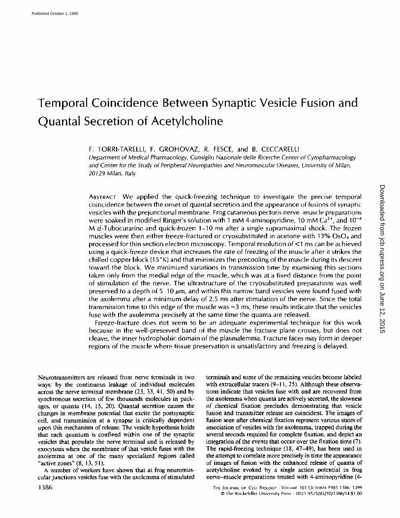
FIGURES 7 and 8 (p. 1393) Micrographs from two different preparations quick-frozen 2.5 ms after a single stimulus in 1 mM 4- AP. In these cross-sections at the level of active zones different degrees of association between vesicles and the prejunctional membrane are evident. Arrowheads indicate clear openings whereas arrows indicate images that suggest intermediate steps between fusion and fission. 2 Note that some ice crystals are evident in the postjunctional axoplasm in Fig. 8 despite the good preservation of all of the prejunctional structures. This suggests that some lengthening in freezing time may have occurred in this terminal. More fusions and vesicle openings are seen in this micrograph, which suggests that an integration over time may have occurred (see Discussion). Asterisk, large vesicular structure; P, Schwann cell process. Fig. 7, x 53,000. Fig. 8, × 70,000.
TORRI-TARELU ET AL. Quantal Release Coincident with Vesicle Openings 1389
on June 12, 2015jcb.rupress.org
Dow
nloaded from
Published October 1, 1985

1390 THE JOURNAL OF CELL BIOLOGY - VOLUME 101, 1985
on June 12, 2015jcb.rupress.org
Dow
nloaded from
Published October 1, 1985

TOAAI-T^RELLI E~" AL. Quantal Release Coincident with Vesicle Openings 1 3 91
on June 12, 2015jcb.rupress.org
Dow
nloaded from
Published October 1, 1985

1392 THE JOURNAL OF CELL BIOLOGY - VOLUME 101, 1985
on June 12, 2015jcb.rupress.org
Dow
nloaded from
Published October 1, 1985
TORRI-TAR~LLI ET AL. Quantal Release Coincident with Vesicle Openings 1393
on June 12, 2015jcb.rupress.org
Dow
nloaded from
Published October 1, 1985

hypothesized by Palade, 2 i.e., destabilization and mixing of lipid molecules between the two membranes at domain boundaries. However, the lack of a clear continuity between the vesicle compartment and the extracellular space could be equally well explained if the plane of section passed tangen- tially along the smaller tubular neck of a vesicle that was open to the extracellular space.
The overall picture that emerges when terminals are quick- frozen 5-10 ms after a single stimulus in l mM 4-AP is quite similar to the one observed after 2.5 ms. Figs. 9 and 10 are from cross-sectioned terminals quick-frozen 5 and 10 ms, respectively, after a single stimulus. Vesicle openings are still evident at these longer intervals. These findings are consistent with previous results and with the idea that 4-AP increases both the quantity of transmitter released and the duration of the release period (28, 34, 46).
2.5-10 ms after single stimulus in 4-AP, we observed some LDCVs closely apposed to the presynaptic membrane (Fig. 10), but we never observed any clear fusions or openings. However, LDCVs are so rare in neuromuscular junctions that this negative finding cannot be taken as an argument for the attractive suggestion that secretion via these organeUes might be regulated by stimuli other than the depolarization of the nerve terminal (4, 38).
Morphometric Analysis We counted the numbers of vesicles that were closely
apposed to the axolemma or clearly fused and opened to the extracellular space on cross-sectioned terminals of control and stimulated preparations.
The number of closely apposed vesicles in control terminals was 1.74 (0.61-2.86) per section (least squares mean and 99% confidence limits for the mean); in stimulated ones it was 1.87 (1.56-2.8) at 2.5 ms, 2.34 (1.9-2.8) at 5 ms, and 2.26 (1.84-2.82) at l0 ms. Although the confidence limits for the means so overlap that high levels of statistical significance cannot be obtained in comparing the different conditions, the number of vesicles apposed to the presynaptic membrane does not appear to decline during the first l0 ms after a single shock in 4-AP, but rather to increase.
Neither fused nor omega-shaped vesicles were seen in con- trol preparations. In stimulated preparations the number of clear fusions and openings was 1.34 per section (0.97-2.1 l) at 2.5 ms; 0.79 (0.55-1.65) at 5 ms, and 1.0 (0.6-1.92) at l0 ms. Although no highly statistically significant comparison is allowed by the overlapping of the confidence limits, it appears that the number of fusions and openings after 5 and l0 ms is smaller, rather than greater, than the number at 2.5 ms.
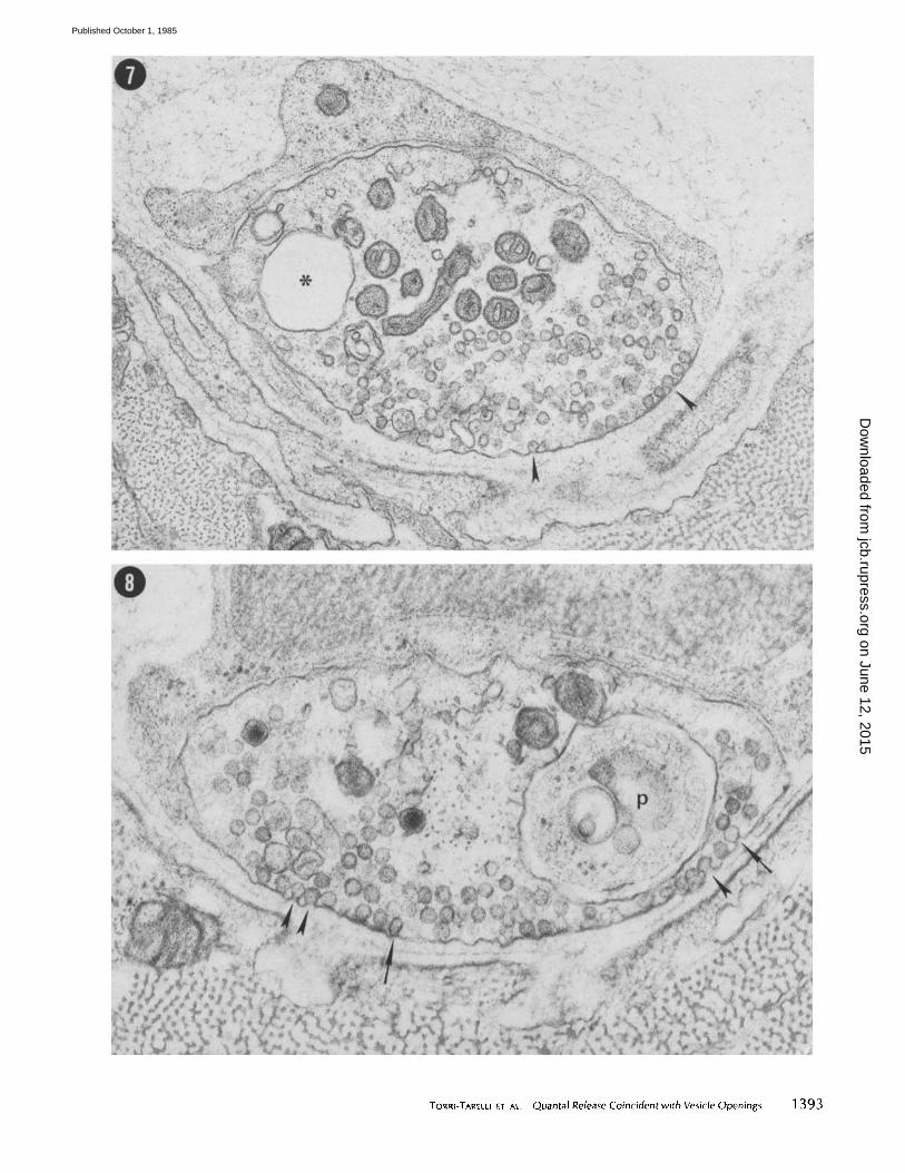
The histograms of the distributions of the number of fusion images in sections from preparations fixed 2.5, 5, or l0 ms after the stimulus are shown in Fig. 1 I. The values of ×2 from the goodness of fit test correspond to probabilities in the range 0.2-0.8, which does not contradict the hypothesis of a Poisson distribution (see Materials and Methods).
2 The nomenclature used here is that proposed by Palade (39). He distinguishes in the membrane events that lead to exocytosis three successive steps and refers to them as apposition, fusion, and fission, respectively. Fission of the fused membranes creates continuity be- tween the other portions of the membranes and continuity between their delimited compartments. Fission is envisioned as proceeding by the sequential elimination of membrane layers and leading to the real vesicle opening.
1394 TNE JOURNAL Of: CELL B,OLOGV • VOLUME 101, 1985
Freeze-Fracture of Quick-Frozen Specimens Fig. 12A is a schematic drawing of frog cutaneous pectoris
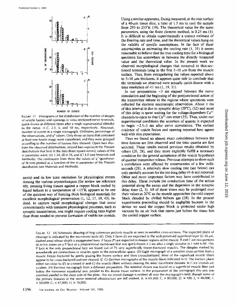
muscle as seen in a semithin cross-section. The dashed area marks the regions of the superficial layer of the muscle where high freezing rates and adequate tissue preservation is attained with our quick-freezing technique. Indeed, the true extent of the well-preserved region is exaggerated in the figure. Figs. 2- l0 are examples of terminals that lie on the outer semicircle of the sarcolemmas of the most superficial layer of fibers in the muscle. If the presynaptic membrane of these terminals were freeze-cleaved, one would find only extracellular fracture faces (E faces) of their prejunctional membrane since their complementary P faces would be scooped out by the blade. The prejunctional membranes of terminals located in regions immediately below the transverse axis of the first layer of muscle fibers (Fig. 12A), such as the one in Fig. 12H, have low probabilities of being cleaved because of the unfavorable angle of incidence with the advancing fracture front. However, if the prejunctional membranes of these terminals were cleaved their P faces could be revealed. As the depth of the terminals along the surface of the same muscle fiber increases, the probability that they will be cleaved along the prejunc- tional membrane increases, and the P face is more likely to be revealed. Thus, when P faces are found they must belong to terminals that lie well below the 5-10 #m limit for properly and rapidly cryofixed tissue.
We freeze-fractured very superficially 75 muscles (including control and experimental muscles) and found no E faces and only two portions of active zones on a P face ofa prejunctional membrane from a preparation that had been quick-frozen 2.5 ms after a single stimulus (Fig. 12, B and C). All of the replicas were characterized by an abundance of cross-fractures through the muscle cells and by an extreme paucity of any cleaved membrane faces. Thus, in our quick-frozen and fractured muscles the fracture plane has little tendency to cleave the inner domain of the plasmalemma. This observation is fully substantiated by experiments in which some of the fractured muscles were cryosubstituted and processed for light and electron microscopy. Fig. 12 is an example of the results obtained in all these experiments. When semithin sections were observed under the light microscope most of the super- ficial fibers appeared clearly cross-fractured (Fig. 12D). In some regions where the location of the fracture plane was uncertain, the electron microscopic examination of the same specimens revealed without doubt that either the sarcolemma was cross-fractured or the fracture plane ran close to the muscle fiber, through the superficial extraceUular layer of ice (Fig. 12, E-G). Fig. 12H illustrates the poor preservation of a terminal found below the equatorial axis of a superficial muscle fiber. It is possible that the prejunctional membranes of such terminals are cleaved so that their P faces are revealed (Fig. 12 B and C).
DISCUSSION
The purpose of the present work was to obtain a more accurate correlation between the time quanta of acetylcholine are first released by a single stimulus and the time vesicle openings are first observed on the prejunctional membrane. The phys- ical procedure of cryofixation provides a path to circumvent the low spatial resolution of chemical fixation for morpholog- ical events (i.e., maintain in situ diffusible chemical compo-
on June 12, 2015jcb.rupress.org
Dow
nloaded from
Published October 1, 1985

FIGURES 9 and 10 Micrographs of cross-sections from preparations quick-frozen 5 and 10 ms after single stimuli in 4-AP, respectively. Vesicle openings are indicated by the arrowheads, and an LDCV closely apposed to the presynaptic membrane is shown by the arrow. Fig. 9, x 67,000. Fig. 10, x 49,000.
1395
on June 12, 2015jcb.rupress.org
Dow
nloaded from
Published October 1, 1985
1 2 3 4 5 6 7
ca
o=
1 2 3 t~ 5 '~6 7
1 2 3 & S 6 "/
NUMBER OF EVENTS
FIGURE 1 1 Histograms of the distr ibut ion o f the number of images
of vesicle fusions and openings in cross-sectioned nerve terminals, quick-frozen at different times after a single supramaximal stimulus to the nerve. A-C, 2.5, 5, and 10 ms, respectively. Abscissae, number of events in a single micrograph. Ordinates, percentage of the observations, and x 2 values. Only those sections that contained at least one fusion image were considered, and they were grouped according to the number of fusions they showed. Open bars illus- trate the observed distributions, striped bars represent the Poisson distributions that best fit the data {least square errors); values of the m parameter were: (A) 1.34, (B) 0.78, and (C) 1.0 (see Materials and Methods). The continuous lines show the values of x 2 (goodness- of-fit test) plotted as a function of the m parameter of the Poisson distribution (see Materials and Methods).
nents) and its low time resolution for physiological events. Among the various cryotechniques (for review see reference 40), pressing living tissues against a copper block cooled by liquid helium to a temperature of ~ 15*K appears to be one of the quickest way of freezing thick biological materials for excellent morphological-preservation (1, 12, 17, 19, 43). In- deed, to capture rapid morphological changes that occur concomitantly with transient physiological processes, such as synaptic transmission, one might require cooling rates higher than those needed to prevent formation of visible ice crystals.
Using a similar apparatus, Escaig measured, at the rear surface of a 40-#m tissue slice, a time of 1.5 ms to cool the sample from 293 to 253"K (19). The theoretical value for the same parameters, using the finite element method, is 0.25 ms (1). It is difficult to obtain experimentally a correct estimate of the freezing rate and time, and the theoretical values hang on the validity of specific assumptions. In the face of these uncertainties in estimating the cooling rate (l, 31) it seems reasonable to believe that the true cooling time for a biological specimen lies somewhere in between the directly measured value and the theoretical value. In the present work we observed morphological changes that occurred in thin-sec- tioned terminals lying in the first 5-10 ~m from the muscle surface. Thus, from extrapolating the values reported above to 5-10 #m thickness, it appears quite safe to conclude that the terminals we observed were actually quick-frozen with a time resolution o f < l ms (1, 19, 31).
In our preparations ~3 ms elapsed between the nerve stimulation and the beginning of the postjunctional action of the transmitter release in the regions where specimens were collected for electron microscopic observation. About 1 ms of this interval is due to synaptic delay (20"C), (32) and most of this delay is spent waiting for the voltage-dependent Ca 2+ channels to open so that C a 2+ c a n enter (35). Thus, under our experimental conditions the secretion of quanta is expected to begin ~2.5-3 ms after nerve stimulation. The earliest evidence of vesicle fusion and opening reported here agrees well with this expectation.
Thus we found an almost exact coincidence between the time fusions are first observed and the time quanta are first secreted. These results extend previous results obtained by others (26, 28), and they more explicitly fulfill a necessary condition for the general acceptance of the vesicle hypothesis of quantal transmitter release. Previous attempts to show such a correlation were affected by uncertainties of a few milli- seconds (28). A relatively slow cooling rate (see below) can only partially account for the too long delay (4-6 ms) reported. Other and more important factors may have contributed to this delay. These include the conduction time of the action potential along the axons and the dispersion in the synaptic delay time (2, 3). All of these times may be prolonged over their values at 20"C as the muscle approaches the cold copper block clouded by chilled helium gas (28). In the present experiments precooling should be negligible because in the device we used the copper block is protected under high vacuum by an air lock that opens just before the tissue hits the cooled copper surface.
FIGURE 12 (A) Schematic drawing of frog cutaneous pectoris muscle as seen in semithin cross-sections. The expected plane of cleavage is indicated by the microtome knife (K). Only E faces (E) are expected in the well-preserved superficial layer (5-10 #m, dashed area) whose depth is exaggerated here. P faces (P) are expected in deeper regions of the muscle. B and C are two portions of active zones on a P face of a prejunctional membrane that was quick-frozen 2.5 ms after a single stimulus in I mM 4-AP. This P face is the only prejunctional face we found out of 75 very superficially freeze-fractured muscles. The dimples marked by arrowheads are connections to vesicles open to the extracellular space. (D) Light micrograph of a semithin cross-section from a muscle freeze-fractured by gently grazing the frozen surface and then cryosubstituted. Most of the superficial muscle fibers appear to be cross-fractured and not cleaved. (E-G) Electron micrographs of the muscle fibers indicated in D. The fracture plane either ran close to (E) or crossed (F and G) the muscle fibers without cleaving the inner membrane domain. In G ice crystals are evident. (H) Electron micrograph from a different preparation. The terminal shown was found on a superficial muscle fiber just below the transverse equatorial axis, parallel to the frozen tissue surface. In the preparation of the micrograph this axis was oriented parallel to the short side of the plate. The ice crystal damage is evident all over the micrograph's field, though some of the primary features of the nerve terminal ultrastructure are still evident. B, x 69,500; C, x 80,000; D, x 400; E, x 48,000; F, x 50,000; G, × 47,000; H, x 19,000.
1 3 9 6 THE JOURNAL OF CELL BIOLOGY • VOLUME 101, 1985
on June 12, 2015jcb.rupress.org
Dow
nloaded from
Published October 1, 1985

1397
on June 12, 2015jcb.rupress.org
Dow
nloaded from
Published October 1, 1985

Another major difference between our results and those previously reported by others is that they found many P faces of prejunctional membrane (26, 28, 37), whereas we found no fractured faces in the thin outermost layer of the muscle, since the tissue was mostly cross-fractured. Whatever the cause of the difference between the two sets of results, it appears that in those regions of the muscles where an adequate degree of tissue preservation is achieved the plane of fracture is quite different from the one normally obtained in chemi- cally fixed and cryoprotected specimens (6, 7, 27). This is not surprising since there are no convincing a priori reasons to believe that in a quasi-vitrified portion of a cell the inner membrane domain should be more prone to be freeze-cleaved (22). In any event, had presynaptic membranes been cleaved in the well preserved uppermost superficial layers of the muscle, only E faces would have been obtained (see Results and Fig. 12).
Our findings do not exclude the possibility that the inner plasmalemmal domain might be cleaved in deeper regions of the specimens. In these regions, below the equatorial trans- verse axis of the superficial muscle fibers, P faces of the axolemma might be obtained, but here tissue preservation is inadequate, as shown in Fig. 12H. Even though the ice crystal damage is evident all over the field of this micrograph, indi- cating a slow cooling rate, the dehydrated inner domain of the axolemma is probably not so distorted to preclude obtain- ing adequate presynaptic face images such as the ones pre- sented in Fig. 12, B and C. Thus, it seems that all of the information about the freezing rate that, in thin sections, comes with the degree o f ultrastructural preservation and with the possibility of localizing precisely the terminal with respect to the impact surface, is lost in freeze-fracture replicas, where the depth of the fracture plane cannot be controlled. In particular, data derived from P faces of terminals suffer from the problems associated with slow and variable cooling rates, among them, the possible integration of many instantaneous events over the cooling time. This may account for the difference between our finding that the number of synaptic vesicles caught in the act of exocytosis does not increase with time and the finding of others that it does (26).
Expressing our morphometric data in terms of number of images per active zone, 3 we observed 5-24 fusions per active zone at 2.5 ms, 2-18 at 5 ms, and 2-24 at 10 ms; the values reported by others are in the upper part of this range (28). These data would indicate a total number of fusions per terminal ( -500 active zones) in the range t,000-10,000, which is compatible with the estimated quantal content of the epp in 4-AP and high Ca 2÷ (28). On the other hand we did not observe increasing densities of fusions at later times, although quantal secretion supposedly continues for >5 ms (28, 34). These results argue against the proposal that after exocytosis vesicle membrane collapses into the axolemma, and they support the alternative interpretation that exocytosis of a quantum is followed by a quick direct removal of the vesicle membrane without its flattening into the axolemma, at least at this early stage in the secretion process. In the short
3 The data reported in Results are expressed as number of images per section. Our sections were -40 nm thick, and an active zone with vesicles at its edges 200 nm wide and 1 zm long; therefore, considering a possible angle between the plane of the section and the direction of the active zone between 0 and 30", a complete active zone should be comprised within 5-12 sections.
1398 THE IOURNAL OF CELL BIOLOGY • VOLUME 101, 1985
term this mechanism might be convenient for at least two reasons. First, it would allow quanta of transmitter to escape while rapidly recovering vesicles for subsequent release, with minimal intermixing of membrane components (36). Second, it would save the work required to invaginate the flattened patches of membrane (8).
We are indebted to DEs. W. P. Hurlbut and J. Meldolesi for their suggestions and critical reading of the manuscript. We thank A. Giuni of the Atelier Pantografica, Milan, who patiently realized the different versions of the freezing head reported in Fig. 1 A, and who provided the special cover with a stirring device for the Dewar used for the cryosubstitution. We gratefully acknowledge the assistance of N. Iezzi who helped with the electron microscopy, P. Tinelli and F. Crippa who prepared the illustrations, and S. Avogadro who typed the manuscript.
This work was partially supported by a grant from the Muscular Dystrophy Association of America, Inc. to B. Ceccarelli.
Received for publication 8 May 1985, and in revised form 24 May 1985.
REFERENCES
1. Bald, W. B. 1983. Optimizing the cooling block for the quick freeze method. J. Microsc. 131:11-23.
2. Barrett, E. F., and F. Stevens. 1972. Quantal independence and uniformity ofpresynaptic release kinetics of the frog neuromuscular junction. J. Physiol. (Land.). 227:665-689.
3. Barrett, E. F., and F. Stevens. 1972. The kinetics of transmitter release at the frog neuromuscular junction. J. Physiol. (Land.). 227:691-708.
4. Basbaum, C. B., and J. E. Heuser. 1979. Morphological studies of stimulated adrenergic axon varicosities in the mouse vas deferens. J. CellBioL 80:310-325.
5. Birk, R., H. E. Huxley, and B. Kalz. 1960. The fine structure of the neuromuscular junction of the frog. J. Physiol. (Land.). 150". 134-- 144.
6. Ceccarelli, B., F. Grohovaz, and W. P. Hudbut. 1979. Freeze-fracture studies of frog neuromuscular junctions during intense release of neurotransmitter. I. Effects of black widow spider venom and Ca2+-free solutions on the structure of the active zone. J. Cell BioL 81:163-177.
7. Ceecarelli, B., F. Grohova2, and W. P. Hudbut. 1979. Freeze-fracture studies of frog neuromuscular junctions during intense release of neurotransmittec. I1. Effects of electrical stimulation and high potassium. J. Cell BioL 81:178-192.
8. Ceccarelli, B., and W. P. Hurlbut. 1980. Vesicle hypothesis of the release of quanta of acetylcholine. Physiol. Rev. 60:396-441.
9. Ceccarelli, B., and W. P. Hurlbut. 1980. CaZ÷-dependent recycling of synaptic vesides at the frog neuromuscular junction. J. Cell Biol. 87:297-303.
10. Ceccar¢lli, B., W. P. HuE/but, and A. Munro. 1972. Depletion of vesicles from frog neuromuscular junctions by prolonged tetanic stimulation, J. Cell Biol. 54:30-38.
11. CeccareUi, B., W. P. Hurlbut, and A. Munro. 1973. Turnover of transmitter and synaptic vesicles at the frog neuromurc.,ular junction. J. Cell BioL 57:499-524.
12. Co~tello, M. J., R. Fetter, and M. Mrchli. 1982. Simple procedures for evaluating the cryofixation of biological samples. J. Microsc. 125:125-136.
13. Couteaux, R., and M. P~ot-Deehavassine. 1970. Vesicules synaptiques et poches au nivean des "zones actives" de la jonction neuromusculaire. CR Acad ScL (Paris). 271:2346-2349.
14, del Castillo, J., and B. Katz. 1954. Quanta] components of the endplate potential. Z Physiol. (Land.). 124:560-573.
15, del Castillo, J., and B. Katz. 1956. Biophysical aspects of neuro-muscular transmission. Prog. Biophys. Biophys, Chem. 6:121 - 170.
16, Dempsey, G. P., and S. Bullivant. t976. A copper block method tbr freezing non- cryoprotected tissue to produce ice-crystal-free regions foe elecron microscopy. J. Mi- erase. 106:251-260.
17. Elder, H. Y., C. C. Gray, A. G. Jardine, J. N. Chapman, and W. H. Biddlecombe. 1982. Optimum conditions for cryoquenching the small tissue blocks in liquid coolants..L Microsc. 126:45-51.
18. E r~kr , O. 1954. Quenching of tissues for freeze-drying. Acta Anat. 22:331-335. 19. Escaig, J. 1981. New instruments which facilitate rapid freezing at 83 K and 6 K. J.
Microsc. (Paris.) 126:221-229. 20. Fatt~ P., and B. Katz. 1951. An analysis of the endplate potential recorded with an
intracellular electrode..L Physiol. (Land). 115:320-370. 21. Frankenhaeuser, B. 195 7. The effect of caldum on the myeliuated nerve fiber. J. Physiol
(Land). 137:245-260. 22. Franks, F. 1977, Biological freezing and cryofixation. J. Microsc. I 11:3-16. 23. Gorio, A., W. P. Hudbut, and B, Ceccsrelli. 1978. Acetylcholine compartments in
mouse diaphragm. Z Cell Biol. 78:716-733. 24. Harvey, D. M. R, 1982, Freeze-substitution. J. Microsc. 127:209-221. 25. Heuser, J. E., and T. S. Reese. 1973. Evidence for recycling ofsynaptic vesicle membrane
during transmitter release at the frog neuromuscular junction. Z Cell Biol. 57:315-344. 26. Heuser, J. E., and T. Reese. 1981. Structural changes following transmitter release at
the frog neuromuscular junction..L Cell Biol. 88:564-58@ 27, Heuser, J. E., T. S. Reese, and D. M. D. Landis. 1974, Functional changes in frog
neuromuscular junctions studied with freeze-fracture. Z Neurocytol. 3:109-131. 28. Heuser, J. E,, T. Reese, M. J. Dennis, Y. Jan, and L. Evans. 1979. Synaptic vesicle
exocytosis captured by quick-freezing and correlated with quantal transmitter release. J. Cell Biol. 81:275-300.
29. Hudbut, W. P., and B. Ceccarelli. 1974_ Transmitter release and recycling of synaptic vesicle membrane at the neuromuscular junction. Adv Cylopharmacol. 2:14 I - 154.
on June 12, 2015jcb.rupress.org
Dow
nloaded from
Published October 1, 1985

30. Israel, M., Y. Dunant, and R. Manaranche. 1979. The present status of the vesicular hypothesis. Prog, Neurobiol. 13:237-275.
31. Jones, G. J. 1984. On estimating freezing times during tissue rapid freezing. Z Microsc. 136:349-360.
32. Katz, B., and R. Miledi. 1965. The measurement of synaptic delay, and the time course of ACh release at the neuromuscular junction. Proc. R. Soc. Lond. BBiol. Sci. 161:483- 495.
33. Katz, B., and R. Miledi. 1977. Transmitter leakage from motor nerve endings. Proc. R. Sac. Lond. B Biol. Sci. 196:59-72.
34. Katz, B., and R. Miledi. 1979. Estimates ofquantal content during "chemical POtentia- tion ~ of transmitter release. Prac. R. Sac. Lond. B Biol. Sci. 205:369-378.
35. Llinas, 1., Z. Steinberg, and K. Walton. 1981. Relationship between calcium current and postsynaptic potential in squid giant synapse. Biophys. J. 33:323-352.
36. Meldolesi, J., and B. Ceccarelli. 1981. Exocytosis and membrane recycling. Philos. Trans. R Sac. Lond B Biol. Sci. 296:55-4/5.
37. Miller, T. M., and J. E. Heuser. 1984. Endocytosis ofsynaptic vesicle membrane at the frog neuromuscular junction. J. Celt Biol. 98:685-698.
38. Navone, F., P. Grecngard, and P. De Camilli. 1984. Synapsin I in nerve terminals: selective association with small synaptic vesicles. Science (Wash. DC). 226:1209-1211.
39. Palade, G. E. 1975. Intracellular aspects of the process of protein synthesis. Science (Wash. DC). 189:347-358.
40. Platlner, H., and L. Bachmann. 1982. Cryofixation: a tool in biological ultrastructural research. Int. Rev. Cytol 79:237-304.
41. Polack, R. L., U C. Sellin, and S. Thesleff. 1981. Acetylchnline content and release in denervated or betulinum poisoned rat skeletal muscle. J. Physiol. (Lond.). 319:253-259.
42. Rosenkranz, J. 1975. The course of temperature variation in an object during the freeze- etching procedure. Arzneim. Forsch. 25:454--455.
43. Schwabe, K. G., and U Terracio. 1980. Ultrastructural and thermocouple evaluation of rapid freezing techniques. Cryobiology. 17:571-584.
44. Steinbrecht, R. A. 1982. Experiments on freezing damage with freeze substitution using moth antennae as test objects. J. Microsc 125:187-192.
45. Tauc, L. 1982. Non-vesicular release of neurotransmitter. Physiol. Rev. 62:857-893. 46. Thesleff, S. 1980. Aminopyridines and syuaptic transmission. Neuroscience. 5:1413-
1419. 47. Van Harreveld, A., and J. Crowell. 1964. Electron microscopy alter rapid freezing on a
metal surface and substitution fixation. Anal. Rec. 149:381-385. 48. Van Harreveld, A., J. Crowell, and S. K. Malhotra. 1965. A study of extracellular space
in central nervous tissue by freeze-substitution..L Cell BioL 25:117-137. 49. Van Harreveld, A., J. Trubatch, and J. Steiner. 1974. Rapid freezing and electron
microscopy for the arrest of physiological processes. Z Microsc. 100:189-198. 50. Vizi, E., and F. Vyskocil. 1979. Changes in total and quantal release of acetylcholine in
the mouse diaphragm during activation and inhibition of membrane ATPase. J. Physiol. (Lond.). 286:1 - 14.
51, Zimmermann, M. 1979. Vesicle recycling and transmitter release. Neuroscience. 4:1773- 180,1.
TORRI-TARELLI ET AL. Quantal Release Coincident with Vesicle Openings 1399
on June 12, 2015jcb.rupress.org
Dow
nloaded from
Published October 1, 1985
Related Documents