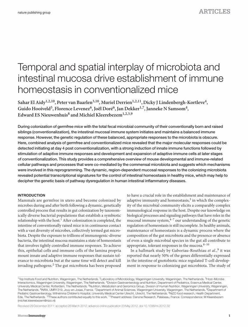
Mucosal Immunology 1 nature publishing group ARTICLES INTRODUCTION Mammals are germfree in utero and become colonized by microbes during and after birth following a dynamic, genetically controlled process that results in gut colonization by taxonom- ically diverse bacterial populations that establish a symbiotic relationship with the host. 1 After colonization is completed, the intestine of conventionally raised mice is in continuous contact with a vast diversity of microbes, collectively termed gut micro- biota. Despite the exposure to trillions of immunogenic-diverse bacteria, the intestinal mucosa maintains a state of homeostasis that involves tightly controlled immune responses. To achieve this, epithelial cells and immune cells of the lamina propria mount innate and adaptive immune responses that sustain tol- erance to microbiota but at the same time will detect and kill invading pathogens. 2 The gut microbiota has been proposed to have a crucial role in the establishment and maintenance of adaptive immunity and homeostasis, 3 in which the complex- ity of the microbial community elicits a comparably complex immunological response in the host. Despite our knowledge on biological processes and signaling pathways that have roles in the mucosal immune system, 4–7 our understanding of the genetic regulation of homeostasis is still incomplete. In healthy animals, maintenance of homeostasis is a dynamic process where the composition of the gut microbiota and the presence or absence of even a single microbial species in the gut all contribute to appropriate, tolerant responses in the mucosa. 8–10 In a hallmark study by Gaboriau-Routhiau et al., 9 it was reported that nearly 50% of the genes differentially expressed in the intestine of gnotobiotic mice regulated T-cell develop- ment in response to colonizing gut microbiota. The study of Temporal and spatial interplay of microbiota and intestinal mucosa drive establishment of immune homeostasis in conventionalized mice Sahar El Aidy 1,2,10 , Peter van Baarlen 3,10 , Muriel Derrien 1,2,11 , Dicky J Lindenbergh-Kortleve 4 , Guido Hooiveld 5 , Florence Levenez 6 , Joël Doré 6 , Jan Dekker 1,7 , Janneke N Samsom 4 , Edward ES Nieuwenhuis 8 and Michiel Kleerebezem 1,2,3,9 During colonization of germfree mice with the total fecal microbial community of their conventionally born and raised siblings (conventionalization), the intestinal mucosal immune system initiates and maintains a balanced immune response. However, the genetic regulation of these balanced, appropriate responses to the microbiota is obscure. Here, combined analysis of germfree and conventionalized mice revealed that the major molecular responses could be detected initiating at day 4 post conventionalization, with a strong induction of innate immune functions followed by stimulation of adaptive immune responses and development and expansion of adaptive immune cells at later stages of conventionalization. This study provides a comprehensive overview of mouse developmental and immune-related cellular pathways and processes that were co-mediated by the commensal microbiota and suggests which mechanisms were involved in this reprogramming. The dynamic, region-dependent mucosal responses to the colonizing microbiota revealed potential transcriptional signatures for the control of intestinal homeostasis in healthy mice, which may help to decipher the genetic basis of pathway dysregulation in human intestinal inflammatory diseases. 1 Top Institute Food and Nutrition, Wageningen, The Netherlands. 2 Laboratory of Microbiology, Wageningen University , Wageningen, The Netherlands. 3 Host–Microbe Interactomics, Wageningen University , Wageningen, The Netherlands. 4 Division Gastroenterology and Nutrition, Department of Pediatrics, Erasmus Medical Center, University Medical Center , Rotterdam, The Netherlands. 5 Nutrition, Metabolism and Genomics Group, Division of Human Nutrition, Wageningen University , Wageningen, The Netherlands. 6 INRA, UMR1319, Jouy-en-Josas, France. 7 Department of Animal Sciences, Wageningen University , Wageningen, The Netherlands. 8 Department of Pediatric Gastroenterology, Wilhelmina Children’s Hospital, University Medical Center Utrecht, Utrecht, The Netherlands. 9 NIZO food research, Health Department, Ede, The Netherlands. 10 These authors contributed equally to this work. 11 Present address: Danone Research, Palaiseau, France. Correspondence: M Kleerebezem ([email protected]) Received 29 December 2011; accepted 26 March 2012; advance online publication 23 May 2012. doi:10.1038/mi.2012.32

Welcome message from author
This document is posted to help you gain knowledge. Please leave a comment to let me know what you think about it! Share it to your friends and learn new things together.
Transcript
MucosalImmunology | VOLUME XX NUMBER X | MONTH 2012 1
nature publishing group ARTICLES
See COMMENTARY page XX
INTRODUCTION Mammals are germfree in utero and become colonized by
microbes during and after birth following a dynamic, genetically
controlled process that results in gut colonization by taxonom-
ically diverse bacterial populations that establish a symbiotic
relationship with the host. 1 After colonization is completed, the
intestine of conventionally raised mice is in continuous contact
with a vast diversity of microbes, collectively termed gut micro-
biota. Despite the exposure to trillions of immunogenic-diverse
bacteria, the intestinal mucosa maintains a state of homeostasis
that involves tightly controlled immune responses. To achieve
this, epithelial cells and immune cells of the lamina propria
mount innate and adaptive immune responses that sustain tol-
erance to microbiota but at the same time will detect and kill
invading pathogens. 2 The gut microbiota has been proposed
to have a crucial role in the establishment and maintenance of
adaptive immunity and homeostasis, 3 in which the complex-
ity of the microbial community elicits a comparably complex
immunological response in the host. Despite our knowledge on
biological processes and signaling pathways that have roles in the
mucosal immune system, 4 – 7 our understanding of the genetic
regulation of homeostasis is still incomplete. In healthy animals,
maintenance of homeostasis is a dynamic process where the
composition of the gut microbiota and the presence or absence
of even a single microbial species in the gut all contribute to
appropriate, tolerant responses in the mucosa. 8 – 10
In a hallmark study by Gaboriau-Routhiau et al. , 9 it was
reported that nearly 50 % of the genes differentially expressed
in the intestine of gnotobiotic mice regulated T-cell develop-
ment in response to colonizing gut microbiota. The study of
Temporal and spatial interplay of microbiota and intestinal mucosa drive establishment of immune homeostasis in conventionalized mice Sahar El Aidy 1 , 2 , 10 , Peter van Baarlen 3 , 10 , Muriel Derrien 1 , 2 , 11 , Dicky J Lindenbergh-Kortleve 4 ,
Guido Hooiveld 5 , Florence Levenez 6 , Jo ë l Dor é 6 , Jan Dekker 1 , 7 , Janneke N Samsom 4 ,
Edward ES Nieuwenhuis 8 and Michiel Kleerebezem 1 , 2 , 3 , 9
During colonization of germfree mice with the total fecal microbial community of their conventionally born and raised siblings (conventionalization), the intestinal mucosal immune system initiates and maintains a balanced immune response. However, the genetic regulation of these balanced, appropriate responses to the microbiota is obscure. Here, combined analysis of germfree and conventionalized mice revealed that the major molecular responses could be detected initiating at day 4 post conventionalization, with a strong induction of innate immune functions followed by stimulation of adaptive immune responses and development and expansion of adaptive immune cells at later stages of conventionalization. This study provides a comprehensive overview of mouse developmental and immune-related cellular pathways and processes that were co-mediated by the commensal microbiota and suggests which mechanisms were involved in this reprogramming. The dynamic, region-dependent mucosal responses to the colonizing microbiota revealed potential transcriptional signatures for the control of intestinal homeostasis in healthy mice, which may help to decipher the genetic basis of pathway dysregulation in human intestinal inflammatory diseases.
1 Top Institute Food and Nutrition , Wageningen , The Netherlands . 2 Laboratory of Microbiology, Wageningen University , Wageningen , The Netherlands . 3 Host – Microbe Interactomics, Wageningen University , Wageningen , The Netherlands . 4 Division Gastroenterology and Nutrition, Department of Pediatrics, Erasmus Medical Center, University Medical Center , Rotterdam , The Netherlands . 5 Nutrition, Metabolism and Genomics Group, Division of Human Nutrition, Wageningen University , Wageningen , The Netherlands . 6 INRA, UMR1319 , Jouy-en-Josas , France . 7 Department of Animal Sciences, Wageningen University , Wageningen , The Netherlands . 8 Department of Pediatric Gastroenterology, Wilhelmina Children ’ s Hospital, University Medical Center Utrecht , Utrecht , The Netherlands . 9 NIZO food research, Health Department , Ede , The Netherlands . 10 These authors contributed equally to this work . 11 Present address: Danone Research , Palaiseau , France . Correspondence: M Kleerebezem ( [email protected] )
Received 29 December 2011; accepted 26 March 2012; advance online publication 23 May 2012. doi: 10.1038/mi.2012.32
2 VOLUME XX NUMBER X | MONTH 2012 | www.nature.com/mi
ARTICLES
Gaboriau-Routhiau et al. 9 aimed to unravel the mechanisms
by which segmented filamentous bacteria induced mucosal
adaptive immune responses, with the main focus on the ter-
minal ileum. More recently, Larsson et al. 11 provided a detailed
description of the tissue-specific host transcriptional responses
to the normal gut microbiota, with the main focus to identify
the interaction between the host innate immune responses and
microbial composition throughout the gut, by comparing ger-
mfree and conventional mice. In the current study, we present
the time-resolved, genome-wide immune-related gene expres-
sion programs that are elicited in the mucosa of jejunum, ileum,
and colon in germfree mice upon their conventionalization, with
special attention to immune-related gene expression programs
and to the validation of these programs by immunohistochem-
istry. Our findings show that conventionalization of germfree
mice induced multigenic defense- and immune- related tran-
scriptional responses that reflect the sequential activation of
innate and adaptive immune responses, most pronounced
processes associated with T-cell development and maturation.
Moreover, this study enabled the identification of time-resolved
transcriptional signatures of genes that are proposed to be
involved in the regulation of the dynamic intestinal response
to the microbiota and have a key role in the maintenance of
mucosal homeostasis.
RESULTS Dynamic changes in intestinal physiology and morphometry during conventionalization This study was aimed at identifying the temporal and spatial intes-
tinal mucosal changes in germfree and conventionalized mice
as measured in three independent experiments (for an experi-
mental set up, see Supplementary Figure S1 in Supplementary
Information online). As a typical hallmark of conventionali-
zation, the cecal weight was 80 % reduced upon convention-
alization when compared with germfree mice ( Figure 1a );
this difference was detected from day 4 post conventionaliza-
tion onward. Intestinal morphometric analysis revealed an ini-
tial significant increase ( P < 0.05) in the intestinal crypt depth
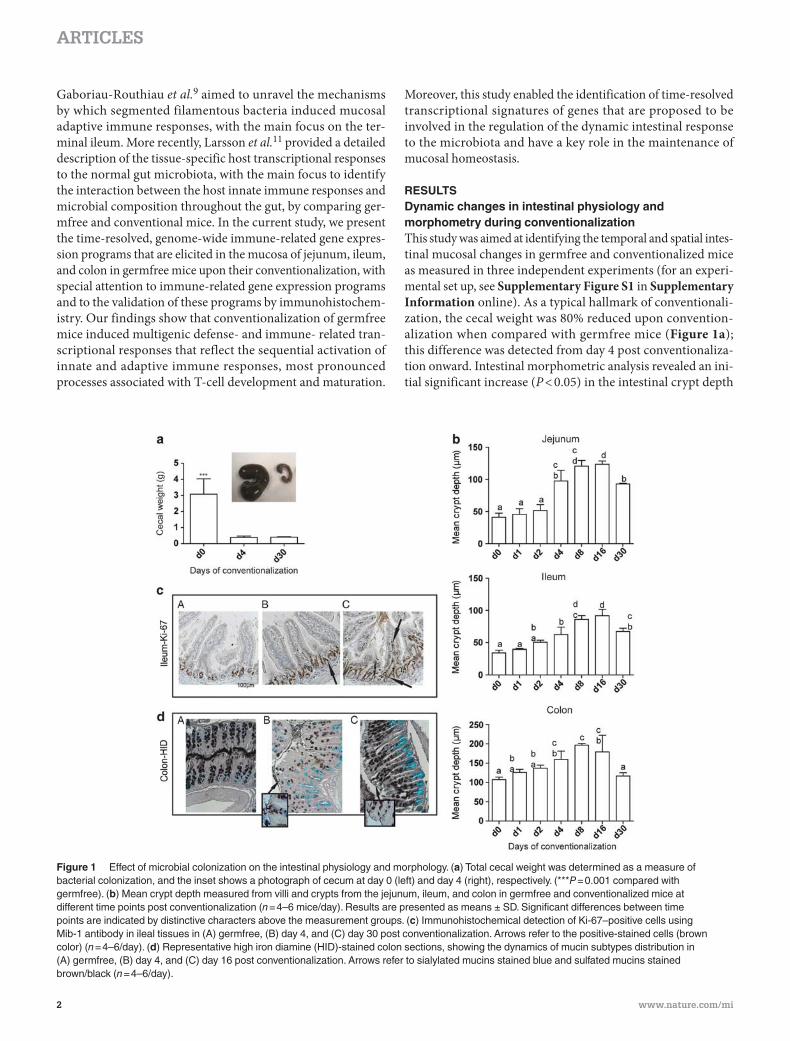
Figure 1 Effect of microbial colonization on the intestinal physiology and morphology. ( a ) Total cecal weight was determined as a measure of bacterial colonization, and the inset shows a photograph of cecum at day 0 (left) and day 4 (right), respectively. ( * * * P = 0.001 compared with germfree). ( b ) Mean crypt depth measured from villi and crypts from the jejunum, ileum, and colon in germfree and conventionalized mice at different time points post conventionalization ( n = 4 – 6 mice / day). Results are presented as means ± SD. Significant differences between time points are indicated by distinctive characters above the measurement groups. ( c ) Immunohistochemical detection of Ki-67 – positive cells using Mib-1 antibody in ileal tissues in (A) germfree, (B) day 4, and (C) day 30 post conventionalization. Arrows refer to the positive-stained cells (brown color) ( n = 4 – 6 / day). ( d ) Representative high iron diamine (HID)-stained colon sections, showing the dynamics of mucin subtypes distribution in (A) germfree, (B) day 4, and (C) day 16 post conventionalization. Arrows refer to sialylated mucins stained blue and sulfated mucins stained brown / black ( n = 4 – 6 / day).
MucosalImmunology | VOLUME XX NUMBER X | MONTH 2012 3
ARTICLES
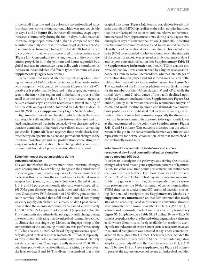
in the small intestine and the colon of conventionalized mice
four days post conventionalization, which was not yet visible
on days 1 and 2 ( Figure 1b ). In the small intestine, crypt depth
increased continuously during the first 16 days. At day 30, small
intestinal crypt depth remained higher as compared with the
germfree mice. By contrast, the colon crypt depth reached a
maximum level from day 8 to day 16 but at day 30, had returned
to crypt depths that were also measured in the germfree mice
( Figure 1b ). Concomitant to the lengthening of the crypts, the
lamina propria in both the jejunum and ileum expanded by a
global increase in connective tissue cells, with a simultaneous
increase in the abundance of different types of immune cells (see
Supplementary Figure S2A online).
Conventionalized mice at later time points (days 4 – 30) had
higher number of Ki-67 (cellular marker for proliferation) – positive
cells compared with germfree animals ( Figure 1c ). Ki-67 –
positive cells predominantly localized in the crypts, but were also
seen in the inter-villus region and in the lamina propria of the
small intestine starting at day 8. Ki-67 – positive and – negative
cells in colonic crypt epithelia revealed a maximal staining of
positive cells on days 4 and 8, followed by a decline at days 16
and 30 ( P < 0.05; see Supplementary Figure S2B – C online).
High iron diamine-alcian blue stain, which detects the mucin
load of goblet cells and discriminates between sialylated and sul-
fated mucins, showed that in the colon, day 4 was characterized by a
transient domination of sialylated over sulfated mucin-containing
goblet cells ( Figure 1d ). Taken together, these results clearly illus-
trate the region-specific transient and permanent changes in the
intestinal morphology and cell proliferation, correlating with
longer microbial colonization. These changes did become most
pronounced from day 4 post conventionalization onward.
Establishment of the gut microbiota during conventionalization To evaluate whether the above mentioned intestinal changes
were accompanied by correlating changes in the abundance of
microbial groups or just a consequence of increased numbers of
bacteria without changing the ratios of specific bacterial groups,
samples from jejunum, ileum, and colon were collected at days 1,
2, 4, 8, and 16 post conventionalization and were compared for
16S RNA gene diversity among each other and with the inocu-
lum. Quantitative PCR detection of 16S rRNA gene copies in
colon samples indicated that a full-sized microbial community
was very rapidly established, i.e., already on day 1 post conven-
tionalization the microbial community contained approximately
11.6 ± 0.5 16S rRNA copies / g colon content (expressed in log10).
This community size estimate did not significantly change during
the experiment, indicating that the microbial community reached
its climax size in a single day. Molecular fingerprinting of the
composition of the colonizing microbiota was performed using
MITChip analysis, a 16S rRNA-based phylogenetic array specifi-
cally designed to classify murine microbiota. 12,13 MITChip analy-
sis revealed that the colon microbial diversity remained relatively
low during days 1 and 2 and significantly increased ( P = 0.001) at
later time points of conventionalization, reaching a stable diver-
sity level on days 8 and 16. This diversity resembled that of the
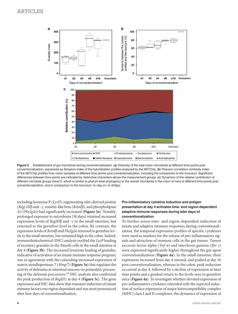
original inoculum ( Figure 2a ). Pearson correlation-based simi-
larity analysis of MITChip profiles of the colon samples indicated
that the similarity of the colon microbiota relative to the inocu-
lum increased from approximately 60 % during early days to 80 %
during later days of conventionalization ( Figure 2b ), indicating
that the climax community at days 8 and 16 was indeed compara-
ble with that of conventional mice (inoculum). This level of simi-
larity (80 % ) corresponded to what was found when the similarity
of the colon microbiota was assessed in individual mice at days 8
and 16 post conventionalization (see Supplementary Table S1
in Supplementary Information online). MITChip analysis also
revealed that day 1 was characterized by a higher relative abun-
dance of Gram-negative Bacteroidetes, whereas later stages of
conventionalization (days 8 and 16) showed an expansion of the
relative abundance of the Gram-positive Firmicutes ( Figure 2c ).
The expansion of the Firmicutes phylum was particularly large
for the members of Clostridium clusters IV and XIVa, while the
initial (days 1 and 2) abundance of the bacilli declined upon
prolonged conventionalization (see Supplementary Figure S3A
online). Finally, multi-variate analysis by redundancy analysis of
colon- and small intestine (jejunum and ileum)-derived micro-
biota profiles clearly established that each intestinal region did
harbor different microbial consortia; especially the diversity of
the small intestine community appeared to be significantly lower
as that encountered in the colon (see Supplementary Figure
S3B – C and S4 online). This outcome indicates that the coloni-
zation of the gut in the conventionalized mice was efficient and
representative for normal colonization levels that are reached in
conventionally raised mice.
Induction of local antimicrobial defense and surface receptors at day 4 post conventionalization along the gastrointestinal (GI) tract In order to investigate the pathways underlying the mucosal
changes observed, tissue gene expression patterns of jejunum,
ileum, and colon at all time points post conventionalization were
compared with each other. The Short Time series Expression
Miner (STEM) and GO-enriched bayesian clustering were used
to identify genes with similar, time-dependent gene expres-
sion patterns over the 30-day timespan of conventionalization.
STEM time-series analysis and GO-enriched bayesian cluster-
ing (for detailed description, see Supplementary Methods in
Supplementary Information online) demonstrated that nearly
40 % of the genes regulated in response to conventionalization
were annotated with immune-related GO terms ( P < 0.001), in
a time- and region-dependent manner (see Supplementary
Figure S5 , Supplementary Table S2, S3 online. To view Table S3
content properly, readers are directed to http://genomica.weizmann.
ac.il/ where Genomica is freely available for academic use.).
Significant induction of expression of surface receptors involved
in microbial recognition was detected at day 4 post convention-
alization throughout the GI tract. These receptors included the
lipopolysaccharide receptor Cd14 , the intracellular signaling
adaptor protein, Myd88 and the Toll-like receptors Tlr1 , 2 , 8 , 9 ,
and 12 but not Tlr4 or 5 (see Supplementary Figure S6 online).
In parallel, the expression levels of several antimicrobial peptides,
4 VOLUME XX NUMBER X | MONTH 2012 | www.nature.com/mi
ARTICLES
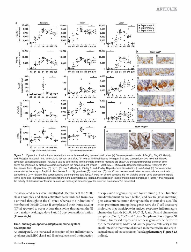
including lysozyme P ( LyzP ), regenerating islet-derived protein
( Reg ) III � and - � , resistin-like beta ( Retnl � ), and phospholipase
A2 ( Pla2g2a ) had significantly increased ( Figure 3a ). Notably,
prolonged exposure to microbiota (30 days) retained increased
expression levels of RegIII � and - � in the small intestine, but
returned to the germfree level in the colon. By contrast, the
expression levels of Retnl � and Pla2g2a returned to germfree lev-
els in the small intestine, but remained high in the colon. Indeed,
immunohistochemical (IHC) analysis verified the LyzP loading
of secretory granules in the Paneth cells in the small intestine at
day 4 ( Figure 3b ). The increased lysozyme loading of granules,
indicative of activation of an innate immune response program,
was in agreement with the coinciding increased expression of
matrix metalloprotease 7 ( Mmp7 ) ( Figure 3a ) that regulates the
activity of defensins in intestinal mucosa via proteolytic process-
ing of the defensin precursors. 14 IHC analysis also confirmed
the peak production of RegIII � at day 4 ( Figure 3c ). The gene
expression and IHC data show that transient induction of innate
immune factors was region dependent and was most pronounced
after four days of conventionalization.
Pro-inflammatory cytokine induction and antigen presentation at day 4 activates time- and region-dependent adaptive immune responses during later days of conventionalization To further assess time- and region-dependent induction of
innate and adaptive immune responses during conventionali-
zation, the temporal expression profiles of specific cytokines
were used as markers for the release of pro-inflammatory sig-
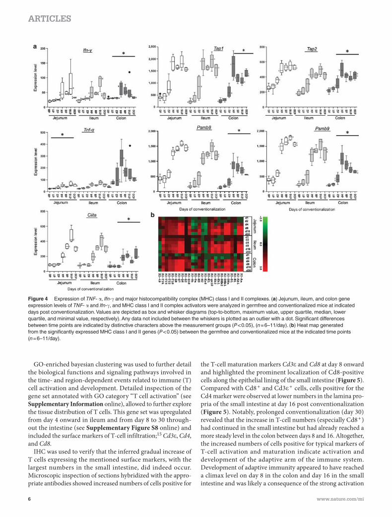
nals and attraction of immune cells in the gut tissues. Tumor
necrosis factor alpha ( Tnf- � ) and interferon gamma ( Ifn- � )
were expressed significantly higher throughout the gut upon
conventionalization ( Figure 4a ). In the small intestine, their
expression increased from day 4 onward, and peaked at day 16
post conventionalization, whereas in the colon, peak induction
occurred at day 4, followed by a decline of expression at later
time points and a gradual return to the levels seen in germfree
mice ( Figure 4a ). To investigate whether elevated expression of
pro-inflammatory cytokines coincided with the expected induc-
tion of surface expression of major histocompatibility complex
(MHC) class I and II complexes, the dynamics of expression of
Figure 2 Establishment of gut microbiota during conventionalization. ( a ) Diversity of the total colon microbiota at different time points post conventionalization, expressed as Simpson index of the hybridization profiles analyzed by the MITChip. ( b ) Pearson correlation similarity index of the MITChip profiles from colon samples at different time points post conventionalization, including the comparison to the inoculum. Significant differences between time points are indicated by distinctive characters above the measurement groups. ( c ) Dynamics of the relative contribution of different microbial groups (level 0, which is similar to phylum level phylogeny) to the overall microbiota in the colon of mice at different time points post conventionalization, and in comparison to the inoculum. d = day ( n = 5 – 6 / day).
MucosalImmunology | VOLUME XX NUMBER X | MONTH 2012 5
ARTICLES
the associated genes were investigated. Members of the MHC
class I complex and their activators were induced from day
4 onward throughout the GI tract, whereas the induction of
members of the MHC class II complex and their transactivator
( Ciita ) appeared to occur at later time points throughout the GI
tract, mainly peaking at days 8 and 16 post conventionalization
( Figure 4a,b ).
Time- and region-specific adaptive immune system development As anticipated, the increased expression of pro-inflammatory
cytokines and MHC class I and II molecules elicited the induction
of expression of genes required for immune (T) cell function
and development on day 8 (colon) and day 16 (small intestine)
post conventionalization throughout the intestinal tissues. The
most prominent among these genes were the T-cell accessory
molecules that participate in antigen response, inflammatory
chemokine ligands ( Cxcl9 , 10 , Ccl2 , 3 , and 5 ), and chemokine
receptors ( Cxcr3 , Ccr2 , and 5 ) (see Supplementary Figure S7
online). Increased expression of these genes coincided with
the increased villus width and lamina propria cellularity in the
small intestine that were observed in hematoxylin and eosin-
stained mucosal tissue sections (see Supplementary Figure S2A
online).
Figure 3 Dynamics of induction of innate immune molecules during conventionalization. ( a ) Gene expression levels of RegIII � , RegIII � , Retnl � , and Pla2g2a, in jejunal, ileal, and colonic tissues, and Mmp7 in jejunal and ileal tissues from germfree and conventionalized mice at indicated days post conventionalization. Individual values determined in the animals and their medians are shown. Significant differences between time points are indicated by distinctive characters above the measurement groups ( P < 0.05; n = 6 – 11 / day) ( b ) Representative IHC of lysozyme-P in ileal tissues from (A) germfree, (B) day 1, (C) day 2, (D) day 4, (E) day 8, and (F) day 16 post conventionalization ( n = 4 – 6 / day). ( c ) Representative immunohistochemistry of RegIII � in ileal tissues from (A) germfree, (B) day 4, and (C) day 30 post conventionalization. Arrows indicate positively stained cells ( n = 4 – 6 / day). The corresponding transcriptome data for lyzP were not shown because it is not trivial to assign gene expression signals to this gene due to ambiguous gene identifiers in the array datasets. Instead, the expression level of matrix metalloprotease 7 ( Mmp7 ) that regulates the activity of defensins in intestinal mucosa via proteolytic processing of the defensin precursors 14 is presented.
6 VOLUME XX NUMBER X | MONTH 2012 | www.nature.com/mi
ARTICLES
GO-enriched bayesian clustering was used to further detail
the biological functions and signaling pathways involved in
the time- and region-dependent events related to immune (T)
cell activation and development. Detailed inspection of the
gene set annotated with GO category “ T cell activation ” (see
Supplementary Information online), allowed to further explore
the tissue distribution of T cells. This gene set was upregulated
from day 4 onward in ileum and from day 8 to 30 through-
out the intestine (see Supplementary Figure S8 online) and
included the surface markers of T-cell infiltration; 15 Cd3 � , Cd4 ,
and Cd8 .
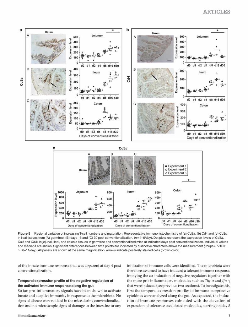
IHC was used to verify that the inferred gradual increase of
T cells expressing the mentioned surface markers, with the
largest numbers in the small intestine, did indeed occur.
Microscopic inspection of sections hybridized with the appro-
priate antibodies showed increased numbers of cells positive for
the T-cell maturation markers Cd3 � and Cd8 at day 8 onward
and highlighted the prominent localization of Cd8-positive
cells along the epithelial lining of the small intestine ( Figure 5 ).
Compared with Cd8 + and Cd3 � + cells, cells positive for the
Cd4 marker were observed at lower numbers in the lamina pro-
pria of the small intestine at day 16 post conventionalization
( Figure 5 ). Notably, prolonged conventionalization (day 30)
revealed that the increase in T-cell numbers (especially Cd8 + )
had continued in the small intestine but had already reached a
more steady level in the colon between days 8 and 16. Altogether,
the increased numbers of cells positive for typical markers of
T-cell activation and maturation indicate activation and
development of the adaptive arm of the immune system.
Development of adaptive immunity appeared to have reached
a climax level on day 8 in the colon and day 16 in the small
intestine and was likely a consequence of the strong activation
Figure 4 Expression of TNF- � , Ifn- � and major histocompatibility complex (MHC) class I and II complexes. ( a ) Jejunum, ileum, and colon gene expression levels of TNF- � and Ifn- � , and MHC class I and II complex activators were analyzed in germfree and conventionalized mice at indicated days post conventionalization. Values are depicted as box and whisker diagrams (top-to-bottom, maximum value, upper quartile, median, lower quartile, and minimal value, respectively). Any data not included between the whiskers is plotted as an outlier with a dot. Significant differences between time points are indicated by distinctive characters above the measurement groups ( P < 0.05), ( n = 6 – 11 / day). ( b ) Heat map generated from the significantly expressed MHC class I and II genes ( P < 0.05) between the germfree and conventionalized mice at the indicated time points ( n = 6 – 11 / day).
MucosalImmunology | VOLUME XX NUMBER X | MONTH 2012 7
ARTICLES
of the innate immune response that was apparent at day 4 post
conventionalization.
Temporal expression profile of the negative regulation of the activated immune response along the gut So far, pro-inflammatory signals have been shown to activate
innate and adaptive immunity in response to the microbiota. No
signs of disease were noticed in the mice during conventionaliza-
tion and no microscopic signs of damage to the intestine or any
infiltration of immune cells were identified. The microbiota were
therefore assumed to have induced a tolerant immune response,
implying the co-induction of negative regulators together with
the more pro-inflammatory molecules such as Tnf- � and Ifn- �
that were induced (see previous two sections). To investigate this,
first the temporal expression profiles of immune-suppressive
cytokines were analyzed along the gut. As expected, the induc-
tion of immune responses coincided with the elevation of
expression of tolerance-associated molecules, starting on day 8
Figure 5 Regional variation of increasing T-cell numbers and maturation. Representative immunohistochemistry of ( a ) Cd8a, ( b ) Cd4 and ( c ) Cd3 � in ileal tissues from (A) germfree, (B) days 16 and (C) 30 post conventionalization, ( n = 4 – 6 / day). Dot plots represent the expression levels of Cd8a , Cd4 and Cd3 � in jejunal, ileal, and colonic tissues in germfree and conventionalized mice at indicated days post conventionalization. Individual values and medians are shown. Significant differences between time points are indicated by distinctive characters above the measurement groups ( P < 0.05; n = 6 – 11 / day). All panels are shown at the same magnification; arrows indicate positively stained cells (brown color).
8 VOLUME XX NUMBER X | MONTH 2012 | www.nature.com/mi
ARTICLES
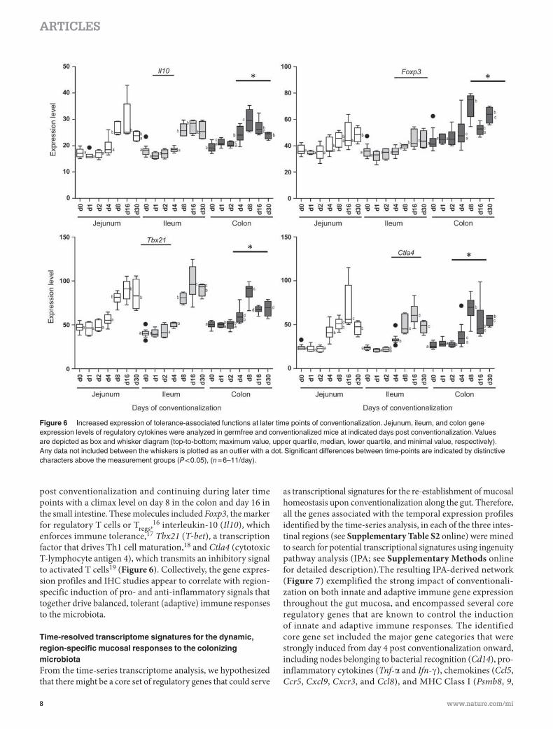
post conventionalization and continuing during later time
points with a climax level on day 8 in the colon and day 16 in
the small intestine. These molecules included Foxp3 , the marker
for regulatory T cells or T regs , 16 interleukin-10 ( Il10 ), which
enforces immune tolerance, 17 Tbx21 ( T-bet ), a transcription
factor that drives Th1 cell maturation, 18 and Ctla4 (cytotoxic
T-lymphocyte antigen 4), which transmits an inhibitory signal
to activated T cells 19 ( Figure 6 ). Collectively, the gene expres-
sion profiles and IHC studies appear to correlate with region-
specific induction of pro- and anti-inflammatory signals that
together drive balanced, tolerant (adaptive) immune responses
to the microbiota.
Time-resolved transcriptome signatures for the dynamic, region-specific mucosal responses to the colonizing microbiota From the time-series transcriptome analysis, we hypothesized
that there might be a core set of regulatory genes that could serve
as transcriptional signatures for the re-establishment of mucosal
homeostasis upon conventionalization along the gut. Therefore,
all the genes associated with the temporal expression profiles
identified by the time-series analysis, in each of the three intes-
tinal regions (see Supplementary Table S2 online) were mined
to search for potential transcriptional signatures using ingenuity
pathway analysis (IPA; see Supplementary Methods online
for detailed description).The resulting IPA-derived network
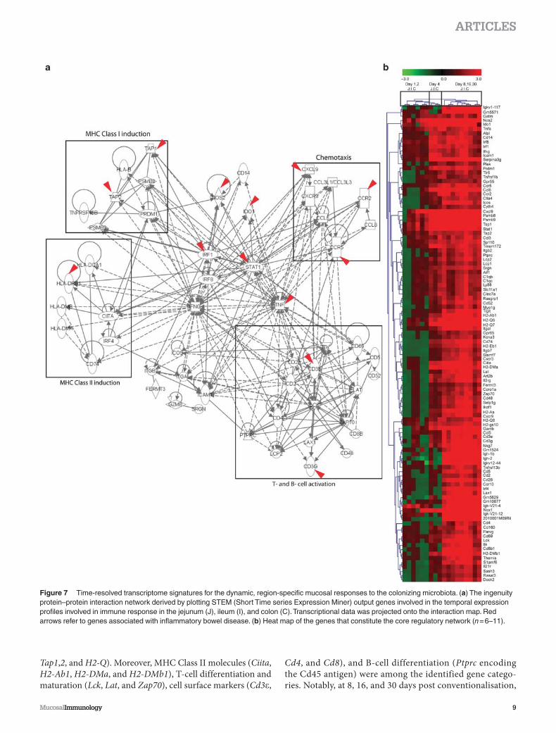
( Figure 7 ) exemplified the strong impact of conventionali-
zation on both innate and adaptive immune gene expression
throughout the gut mucosa, and encompassed several core
regulatory genes that are known to control the induction
of innate and adaptive immune responses . The identified
core gene set included the major gene categories that were
strongly induced from day 4 post conventionalization onward,
including nodes belonging to bacterial recognition ( Cd14 ), pro-
inflammatory cytokines ( Tnf- � and Ifn- � ), chemokines ( Ccl5 ,
Ccr5 , Cxcl9 , Cxcr3 , and Ccl8 ), and MHC Class I ( Psmb8 , 9 ,
Figure 6 Increased expression of tolerance-associated functions at later time points of conventionalization. Jejunum, ileum, and colon gene expression levels of regulatory cytokines were analyzed in germfree and conventionalized mice at indicated days post conventionalization. Values are depicted as box and whisker diagram (top-to-bottom; maximum value, upper quartile, median, lower quartile, and minimal value, respectively). Any data not included between the whiskers is plotted as an outlier with a dot. Significant differences between time-points are indicated by distinctive characters above the measurement groups ( P < 0.05), ( n = 6 – 11 / day).
MucosalImmunology | VOLUME XX NUMBER X | MONTH 2012 9
ARTICLES
Tap1 , 2 , and H2-Q ). Moreover, MHC Class II molecules ( Ciita ,
H2-Ab1 , H2-DMa , and H2-DMb1 ), T-cell differentiation and
maturation ( Lck , Lat , and Zap70 ), cell surface markers ( Cd3 � ,
Cd4 , and Cd8 ), and B-cell differentiation ( Ptprc encoding
the Cd45 antigen) were among the identified gene catego-
ries. Notably, at 8, 16, and 30 days post conventionalisation,
Figure 7 Time-resolved transcriptome signatures for the dynamic, region-specific mucosal responses to the colonizing microbiota. ( a ) The ingenuity protein – protein interaction network derived by plotting STEM (Short Time series Expression Miner) output genes involved in the temporal expression profiles involved in immune response in the jejunum (J), ileum (I), and colon (C). Transcriptional data was projected onto the interaction map. Red arrows refer to genes associated with inflammatory bowel disease. ( b ) Heat map of the genes that constitute the core regulatory network ( n = 6 – 11).
10 VOLUME XX NUMBER X | MONTH 2012 | www.nature.com/mi
ARTICLES
all the identified genes were induced ( Figure 7b ) in a region-
specific manner, i.e., day 8 in the colon and day 16 in the small
intestine.
In parallel, a protein – protein interaction map was gener-
ated from cluster-driven time-series analysis of GO catego-
ries using bayesian statistics (see Supplementary Figure S9
online). This network representing tissue responses to micro-
bial colonization combined genes that belong to T-cell dif-
ferentiation and maturation, again showing that, as in the
IPA output above, T-cell selection, — induction and — differ-
entiation pathways are among the most important induced
mucosal pathways during mouse conventionalization. Also
this core regulatory network, constructed using different
approaches and statistical methods, contained identical major
regulatory nodes as found in the IPA output, supporting the
prominent roles of genes involved in T-cell differentiation
and maturation in the tissue response to commensal microbial
colonization.
Among the identified tissue transcriptome signatures, the
human counterparts of 13 genes have known roles in inflam-
matory bowel disease (indicated with red arrows in Figure 7a ).
These findings support the biological relevance of the identified
transcriptional signatures for mucosal control of homeostasis
along the gut.
DISCUSSION It has been widely recognized that the interplay between gut
microbiota and the host is crucial for the proper development
of the (adaptive) immune system 3 and that dysregulation of this
interaction contributes to the development of inflammatory
bowel disease symptoms in human. 20 There is a clear require-
ment for tightly controlled genetic regulation of appropriate,
tolerant responses to the microbiota. 21,22 This is also supported
by the finding that mutated forms of genes involved in the
regulation of basal immunological processes such as microbial
uptake are strongly associated with the inflammatory bowel
disease phenotypes. 23
The present study implemented the genome-wide expres-
sion profiling of genes during microbial colonization of ger-
mfree mice, using a time-series design with six time points. We
found that throughout the intestine, the largest proportion of
differentially expressed genes was involved in the development
of mucosal immune system. Our study corroborated several
important findings from the studies by Gaboriau-Routhiau
et al. 9 and Larsson et al. 11 These three studies consistently identify
“ immune response ” as the largest category of genes that is regu-
lated in response to microbial colonization. Our transcriptome
analysis showed that there was a time- and region-dependent
enrichment of genes involved in balanced innate and adaptive
immune responses. These tolerant responses ensured that a novel
state of homeostasis was reached within 30 days of conventionali-
zation. Unlike days 1 and 2, which did not show any remarkable
changes in the mucosal transcriptome or histological staining,
day 4 post conventionalization consistently stood out in the
transcriptome analyses and was characterized by drastic changes
in gene transcription. For instance, gene expression could
switch from induction to repression and vice versa; and some
genes were no longer expressed, whereas others were expressed
for the first time. At this time point, the activation of cas-
cades of genes involved in innate immunity and initiation of
adaptive immune (T) cell activation and maturation was most
pronounced.
Strikingly, both transcriptome and IHC analysis for cytokines,
chemokines, T-cell surface markers, immune cell transcription
factors, and histological stainings of innate immune parameters
showed that a novel homeostasis had been reached in the colon
within 8 to 16 days, whereas establishment of homeostasis in
the small intestine required 16 to 30 days of conventionaliza-
tion, roughly double the amount of time. Remarkably, the largest
shift in the microbiota composition coincided with the most
comprehensive shift in the expression of mucosal genes that
regulate the host immune response. Microbial profiling of the
colon microbiota during conventionalization indicated that the
microbial colonization proceeds via the rapid (one day) appear-
ance of early colonizers, followed by the establishment of a stable
community that resembles the microbiota of the conventional
donor animals. As recently reported, 22 our data support the
notion that inflammatory tissue conditions were avoided by
T regs , inferred from increased expression of T-bet , Foxp3 , and
Il10 , markers for tolerance-promoting T regs that were induced
especially from day 8 onward. Interestingly, these T regs markers
and other cytokine markers showed a tendency towards increas-
ing expression in jejunum and ileum throughout the experi-
ment. However, in the colon, expression of the same tolerance
markers clearly peaked at day 8 and subsequently declined at
days 16 and 30 post conventionalization, always remaining
higher than the levels observed in the germfree state and dur-
ing the first two days of conventionalization. Notably, the climax
expression level of T regs at day 8 post-conventionalization in the
colon coincides with the colonization by Clostridium groups,
which have recently been reported to stimulate the expression
of colonic regulatory T cells. 10
Similar differential expression patterns in small intestine
vs. colon were also observed for six inflammatory chemokine
ligands and the corresponding three receptors. We propose
that the expression of these chemokines contributed to T-cell
chemotaxis. IHC showed that at day 8, Cd8 + T cells were pre-
dominantly localized near the mucosal epithelia, which likely
resulted from epithelial chemotactic chemokine secretion and
expression of MHC class I molecules. This timing of Cd8 +
T-cell accumulation in response to accumulation of Th1 chem-
okines in “ danger zones ” is in line with the results reported by
Valbuena et al. 24 during bacterial infection of mice. Moreover,
the faster accumulation of Cd8 + T in the lamina propria of the
colon at day 8, but at day 16 in the small intestine, illustrates an
important location difference that is relevant in the context of
establishing homeostasis.
No changes in the expression level of Il-17 were noted through-
out the GI tract during the process of conventionalization (not
shown), suggesting that the colonization of the C57BL / 6 J mice
with their normal fecal microbiota did not induce Th17 dif-
ferentiation. This finding corroborates the results of Ivanov
MucosalImmunology | VOLUME XX NUMBER X | MONTH 2012 11
ARTICLES
et al. 25 who reported that colonization of germfree C57BL / 6
mice purchased from the Jackson laboratories did not lead to
Th17 differentiation in their lamina propria.
Epithelia contain, in addition to the common enterocytes that
are mainly involved in metabolic functions, specialized Paneth
cells that secrete high amounts of a broad range of antimicrobial
peptides and goblet cells that secrete mucins. One of the broad
spectrum antimicrobials secreted by Paneth cells, RegIII � , was
induced in this study in agreement with Gaboriau-Routhiau
et al. 9 and Larsson et al. , 11 together with the related RegIII � .
The expression of these two peptides appeared to peak at day 4
post conventionalization, in particular in the ileum as supported
by IHC analysis. This could reflect a pronounced induction of
innate immune responses at day 4 post conventionalization,
corroborated by peak expression levels of the genes encoding
the innate immune molecules Retnl � and Pla2g2a , and the
coinciding increased lysozyme P load of secretory granules in
Paneth cells. Although innate immunity was clearly induced in
all the sampled regions of the gut, its dynamics over time was
distinct per region. For example, RegIII � and RegIII � expression
peaked at day 4 post conventionalization in both small intestine
and colon, and high level expression was retained in the small
intestine but declined to germfree levels in the colon at later
time points. Conversely, expression of Retnl � and Pla2g2a
peaked also at day 4 but returned to germfree levels in the
small intestine, whereas expression remained high in colon.
These data suggest that RegIII � and RegIII � are important to
keep microbes at bay in the small intestine, 26 whereas this anti-
microbial function is predominantly exerted by Retnl � and
Pla2g2a in the colon. This is in agreement with the presence
of RegIII peptide-producing Paneth cells in the small intestine
and their absence in the colon. By contrast, Pla2g2a and Retnl �
are secreted by goblet cells, 27,28 a cell type that is common in
the colon. These results show that, in healthy germfree mice
and during bacterial colonization, innate immune responses
are the first line of defence against microbiota, and that this
response displays regional (small intestine vs. colon) differences
in terms of molecules and expression levels. Other responses of
epithelia to increasing bacterial colonization were the increased
proliferation of crypt epithelial cells and villus connective tis-
sue cells, measured as Ki-67 expression starting at day 4 post
conventionalization, and the transient lengthening of the crypts,
measured as crypt depth, also starting at day 4. Our data cor-
roborate results obtained by Cherbuy et al. , 29 demonstrating the
role of microbial colonization in maturation of epithelial cells
in gnotobiotic animals.
The change in biochemistry of colon mucins at day 4 post con-
ventionalization, characterized by a reduction in the amounts
of sulfated, thus stronger antimicrobial mucins, compared with
sialylated, less antimicrobial mucins, 30,31 could have led to
a more intense contact between microbiota and epithelia.
This could indeed be shown using the bacterial FISH EUB338
probe (see Supplementary Figure S10 online). It seems that the
biochemical changes of the mucin barrier at day 4 post conven-
tionalization may have allowed a more intense contact between
the microbiota and the mucosa, which then primed innate
immune responses that were followed by adaptive immune
responses four days later.
The temporal and spatial analysis presented in this study
provides a solid catalogue of genes, pathways, and histology of
intestinal adaptations of germfree mice to microbial coloniza-
tion, thereby providing an important resource that complements
various other studies of mouse intestinal colonization by micro-
biota. Taken together, the data presented here show that a novel
state of homeostasis was reached within 30 days following the
conventionalization of germfree mice. Homeostasis appeared
to be established earlier in the colon (days 8 and 16) as com-
pared with the jejunum and ileum (days 16 and 30). We show
that activation of the adaptive immune system mainly involved
T cells, not B cells, both in the small intestine and in the colon.
The extensive transcriptome datasets for jejunum, ileum, and
colon identified a time-resolved transcriptional signature of
genes that appear to regulate the major tissue transcriptome
changes throughout the intestine during the 30-day convention-
alization. The identified signatures included several genes of
which the human orthologues are inflammatory bowel disease-
associated genes that have also been discovered in genome-wide
association studies, suggesting their relevance for the mucosal
control of homeostasis, and supporting their importance in the
dysregulation of immune-associated pathways in inflammatory
bowel disease patients.
METHODS Animals, experimental design, and sampling . All procedures were carried out according to the European guidelines for the care and use of laboratory animals and with permission 78 – 122 of the French Veterinary Services. Germfree and conventionalized mice (male, C57 BL / 6 J) were maintained in sterile conditions, on a commercial laboratory chow diet. Three independent biological experiments were performed using mice of different age. After 2 weeks of acclimatization and diet adaptation, a first set of germfree mice ( n = 3) were randomly assigned to sacrifice by oral anesthesia using isoflurane. The remaining germfree mice were conventionalized by oral gavage with 0.5 ml of mixed fecal suspension obtained from 0.2 g of freshly obtained fecal material of conventionally raised mice (C57 BL / 6 J) diluted 100-folds in brain heart infusion broth. In the first two experiments; conventionalized mice were killed at days 1, 2, 4, 8, and 16 post conventionalization ( n = 3 per group per experiment). In the third experiment; conventionalized mice were killed at days 4 and 30 post conventionalization ( n = 4 – 5 per group). Small intestine (jeju-num, and ileum), and colon from each mouse were removed. The two segments of the small intestine and the entire colon were then divided into 2 cm segments that were immediately stored in RNAlater at room temperature for 1 h before subsequent storage at − 80 ° C for RNA isola-tion, fixed overnight in 4 % (wt / vol) paraformaldehyde or snap frozen and stored at − 80 ° C for IHC procedures. Luminal content from intesti-nal segments was removed by gentle squeezing, snap frozen, and stored at − 80 ° C for microbiota analysis (see Supplementary Figure S1 and Supplementary Methods in Supplementary Information online).
Histology and immunohistochemistry . In all, 4 � m-thick cross sections of the 2 cm intestinal segments fixed in 4 % (wt / vol) parafor-maldehyde and paraffin-embedded were stained with haematoxylin (Vector Laboratories, Burlingame, CA) and eosin (Sigma-Aldrich, Zwijndrecht, the Netherlands). To detect morphometric differences, 12 – 15 well-oriented villi and crypts were chosen per intestinal segment and measured. Mucin histochemistry was performed using high iron diamine-alcian blue as described. 32 For Lysozyme-P detection, sections
12 VOLUME XX NUMBER X | MONTH 2012 | www.nature.com/mi
ARTICLES
were incubated with anti-Lysozyme P (1:50 in PBS, DakoCytomation, Denmark). For Cd3 � and Ki-67 detection, sections were incubated with anti-Cd3 � (DAKO, Heverlee, Belgium) or anti-Ki-67 (NovoCastra Laboratories, Newcastle upon Tyne, UK), respectively. Expression of RegIII � was detected using a custom-made antibody (for detailed descriptions, see Supplementary Information online). For Cd4 – 8 detection, cryostat sections were incubated with anti-Cd4 and anti-Cd8 (DakoCytomation). Primary antibodies were detected using VECTASTAIN ABC Elite kit (Vector Laboratories), including bioti-nylated Donkey anti-rat serum (Sigma-Aldrich) using the manufac-turer ’ s instructions. For all stainings, nuclei were counterstained with haematoxylin (Vector Laboratories). Stained tissues were examined using a Nikon Microphot FXA microscope (for detailed descriptions, see Supplementary Methods online). All data were presented as means ± s.d. for the number of animals indicated above. Comparisons of data were per-formed at each time point using one-way analysis of variance (ANOVA) followed by Tukey ’ s Studentized range test (GLM, SPSS program, Chicago, IL). For all parameters P < 0.05 was considered the level of significance.
Microbial profiling of intestinal luminal contents . Luminal contents from jejunum, ileum, colon, as well as inoculum were analyzed by Mouse Intestinal Tract Chip (MITChip), a diagnostic 16S rRNA arrays that con-sists of 3,580 unique probes especially designed to profile murine gut microbiota. 12,13
Quantification of total bacteria was performed using qPCR detection of 16 S rRNA-gene copies, while fluorescent in situ hybridization was used to detect bacteria from tissue samples (for detailed descriptions, see Supplementary Methods online).
Transcriptome analysis . High-quality total RNA was obtained from a 2 cm segment of jejunum, ileum, and colon by extraction with TRIzol reagent, followed by DNAse treatment and column purification. Samples were hybridized on Affymetrix GeneChip Mouse Gene 1.1 ST arrays. Quality control and statistical analysis were performed using Bioconductor packages integrated in an on-line pipeline 33 (for detailed descriptions, see Supplementary Methods online). Complementary methods were used for the biological interpretation for the transcriptome data; gene clustering using Multi-experiment Viewer (MeV), 34 overrep-resentation analysis of GO terms using temporal and location compara-tive analysis using STEM, 35 Bayesian clustering using Genomica, and construction of biological interaction networks using IPA (for detailed descriptions see Supplementary Methods online).
Accession numbers . The mouse microarray dataset is deposited in the Gene Expression Omnibus (GEO) with accession number (GSE32513).
SUPPLEMENTARY MATERIAL is linked to the online version of the paper at http://www.nature.com/mi
ACKNOWLEDGMENTS We thank the technical staff in the animal facilities in the lab of J Dor é ; (INRA, Jouy en Jossas) for assistance with animal sacrifice and sampling. R Raatgeep and CL Menckeberg (Department of Pediatrics, Erasmus Medical Center), A Taverne-Thiele and H Schipper (Cell biology and immunology, Wageningen University), S Brugman (Pediatric Gastroenterology, University Medical Center Utrecht) are acknowledged for their excellent assistance with immunohistochemical staining and data analyses. J Jansen, M Grootte-Bromhaar, M Boekschoten and P de Groot (Division for Human Nutrition, Wageningen University) for their technical support in microarray hybridization and microarray data-quality control and processing. L Loonen and J Wells (Host-Microbe Interactomics, Wageningen University) are thanked for providing the RegIII � antibody.
DISCLOSURE The authors declared no conflict of interest.
© 2012 Society for Mucosal Immunology
REFERENCES 1 . Falk , P . G . , Hooper , L . V . , Midtvedt , T . & Gordon , J . I . Creating and
maintaining the gastrointestinal ecosystem: what we know and need to know from gnotobiology . Microbiol. Mol. Biol. Rev. 62 , 1157 – 1170 ( 1998 ).
2 . Sansonetti , P . J . & Di Santo , J . P . Debugging how bacteria manipulate the immune response . Immunity 26 , 149 – 161 ( 2007 ).
3 . Lee , Y . K . & Mazmanian , S . K . Has the microbiota played a critical role in the evolution of the adaptive immune system? Science 330 , 1768 – 1773 ( 2010 ).
4 . Zhu , Y . , Yao , S . & Chen , L . Cell surface signaling molecules in the control of immune responses: a tide model . Immunity 34 , 466 – 478 ( 2011 ).
5 . Chen , G . , Shaw , M . H . , Kim , Y . G . & Nunez , G . NOD-like receptors: role in innate immunity and infl ammatory disease . Annu. Rev. Pathol. 4 , 365 – 98 ( 2009 ).
6 . Hayden , M . S . & Ghosh , S . Shared principles in NF-kappa B signaling . Cell 132 , 344 – 362 ( 2008 ).
7 . West , A . P . , Koblansky , A . A . & Ghosh , S . Recognition and signaling by toll-like receptors . Annu. Rev. Cell Dev. Biol. 22 , 409 – 37 ( 2006 ).
8 . Kuchroo , V . K . , Ohashi , P . S . , Sartor , R . B . & Vinuesa , C . G . Dysregulation of immune homeostasis in autoimmune diseases . Nat. Med. 18 , 42 – 47 ( 2012 ).
9 . Gaboriau-Routhiau , V . et al. The Key role of segmented fi lamentous bacteria in the coordinated maturation of gut helper T cell responses . Immunity 31 , 677 – 689 ( 2009 ).
10 . Atarashi , K . et al. Induction of colonic regulatory T cells by indigenous clostridium species . Science 331 , 337 – 341 ( 2011 ).
11 . Larsson , E . et al. Analysis of gut microbial regulation of host gene expression along the length of the gut and regulation of gut microbial ecology through MyD88 . Gut ( 2011 ) doi:10.1136/gutjnl-2011 – 301104 .
12 . Rajilic-Stojanovic , M . et al. Development and application of the human intestinal tract chip, a phylogenetic microarray: analysis of universally conserved phylotypes in the abundant microbiota of young and elderly adults . Environ. Microbiol. 11 , 1736 – 1751 ( 2009 ).
13 . Geurts , L . et al. Altered gut microbiota and endocannabinoid system tone in obese and diabetic leptin-resistant mice: impact on apelin regulation in adipose tissue . Front Microbiol. 2 , 149 ( 2011 ).
14 . Wilson , C . L . et al. Regulation of intestinal alpha-defensin activation by the metalloproteinase matrilysin in innate host defense . Science 286 , 113 – 117 ( 1999 ).
15 . DeJarnette , J . B . et al. Specifi c requirement for CD3 epsilon in T cell development . Proc. Natl. Acad. Sci USA 95 , 14909 – 14914 ( 1998 ).
16 . Hori , S . , Nomura , T . & Sakaguchi , S . Control of regulatory T cell development by the transcription factor Foxp3 . Science 299 , 1057 – 1061 ( 2003 ).
17 . Fujio , K . , Okamura , T . & Yamamoto , K . The Family of IL-10-secreting CD4(+) T cells . Adv. Immunol. 105 , 99 – 130 ( 2010 ).
18 . Miller , S . A . & Weinmann , A . S . Molecular mechanisms by which T-bet regulates T-helper cell commitment . Immunol Rev 238 , 233 – 246 ( 2010 ).
19 . Magistrelli , G . et al. A soluble form of CTLA-4 generated by alternative splicing is expressed by nonstimulated human T cells . Eur. J. Immunol. 29 , 3596 – 3602 ( 1999 ).
20 . Abraham , C . & Cho , J . H . Mechanisms of disease. Infl ammatory bowel disease . N. Engl. J. Med. 2066 – 2078 ( 2009 ).
21 . Bouma , G . & Strober , W . The immunological and genetic basis of infl ammatory bowel disease . Nat. Rev. Immunol. 3 , 521 – 533 ( 2003 ).
22 . Geuking , M . B . et al. Intestinal bacterial colonization induces mutualistic regulatory T cell responses . Immunity 34 , 794 – 806 ( 2011 ).
23 . Deretic , V . & Levine , B . Autophagy, immunity, and microbial adaptations . Cell Host Microbe. 5 , 527 – 549 ( 2009 ).
24 . Valbuena , G . , Bradford , W . & Walker , D . H . Expression analysis of the T-cell-targeting chemokines CXCL9 and CXCL10 in mice and humans with endothelial infections caused by rickettsiae of the spotted fever group . Am. J. Pathol. 163 , 1357 – 1369 ( 2003 ).
25 . Ivanov , I . I . et al. Induction of intestinal Th17 cells by segmented fi lamentous bacteria . Cell 139 , 485 – 498 ( 2009 ).
26 . Vaishnava , S . et al. The antibacterial lectin RegIII gamma promotes the spatial segregation of microbiota and host in the intestine . Science 334 , 255 – 258 ( 2011 ).
MucosalImmunology | VOLUME XX NUMBER X | MONTH 2012 13
ARTICLES
27 . Fijneman , R . J . A . et al. Expression of Pla2g2a prevents carcinogenesis in Muc2-defi cient mice . Cancer Sci. 99 , 2113 – 2119 ( 2008 ).
28 . Krimi , R . B . et al. Resistin-like molecule beta regulates intestinal mucous secretion and curtails TNBS-induced colitis in mice . Infl amm. Bowel Dis. 14 , 931 – 941 ( 2008 ).
29 . Cherbuy , C . et al. Microbiota matures colonic epithelium through a coordinated induction of cell cycle-related proteins in gnotobiotic rat . Am. J. Physiol. Gastrointest. Liver Physiol. 299 , G348 – G357 ( 2010 ).
30 . Deplancke , B . & Gaskins , H . R . Microbial modulation of innate defense: goblet cells and the intestinal mucus layer . Am. J. Clin. Nutr. 73 , 1131S – 1141S ( 2001 ).
31 . Linden , SK . SP . , Karlsson , N . G . , Korolik , V . & McGuckin , M . A . Mucins in the mucosal barrier to infection . Mucosal Immunol. 1 , 183 – 197 ( 2008 ).
32 . Bogomoletz , W . V . , Williams , G . T . & Potet , F . High iron diamine-alcian blue and histochemistry of mucins in colic diseases-20 years later . Gastroenterol. Clin. Biol. 11 , 865 – 868 ( 1987 ).
33 . Lin , K . et al. MADMAX – Management and analysis database for multiple ~ omics experiments . J. Integr. Bioinform. 8 , 160 ( 2011 ).
34 . Saeed , A . I . et al. TM4 microarray software suite . Methods Enzymol. 411 , 134 – 193 ( 2006 ).
35 . Ernst , J . & Bar-Joseph , Z . STEM: a tool for the analysis of short time series gene expression data . BMC Bioinformatics 7 , 191 ( 2006 ).
Related Documents