Water-mediated nanostructures for enhanced MRI: impact of water dynamics on relaxometric properties of Gd-DTPA § Franca De Sarno 1,2 , § Alfonso Maria Ponsiglione 1,2* , Maria Russo 1,2 , Anna Maria Grimaldi 3 , Ernesto Forte 3 , Paolo Antonio Netti 1,2,4 , Enza Torino 1,2,4 * 1 Department of Chemical, Materials Engineering & Industrial Production, University of Naples Federico II, Piazzale Tecchio 80, 80125 Naples, Italy. 2 Center for Advanced Biomaterials for Health Care, CABHC, Istituto Italiano di Tecnologia, IIT@CRIB, Largo Barsanti e Matteucci 53, 80125 Naples, Italy. 3 IRCCS SDN, Via E. Gianturco 113, 80143 Naples, Italy. 4 Interdisciplinary Research Center on Biomaterials, CRIB, Piazzale Tecchio 80, 80125 Naples, Italy. Corresponding author: Enza Torino, PhD. Department of Chemical, Materials Engineering & Industrial Production, University of Naples Federico II, Piazzale Tecchio 80, 80125 Naples, Italy. Tel. +390817685990; email: [email protected] 1 1 2 3 4 5 6 7 8 9 10 11 12 13 14 15 16 17 18 1

Welcome message from author
This document is posted to help you gain knowledge. Please leave a comment to let me know what you think about it! Share it to your friends and learn new things together.
Transcript

Water-mediated nanostructures for enhanced MRI:
impact of water dynamics on relaxometric properties
of Gd-DTPA
§Franca De Sarno1,2, §Alfonso Maria Ponsiglione1,2*, Maria Russo1,2, Anna Maria Grimaldi3,
Ernesto Forte3, Paolo Antonio Netti1,2,4, Enza Torino1,2,4*
1Department of Chemical, Materials Engineering & Industrial Production, University of Naples Federico II, Piazzale Tecchio 80, 80125 Naples, Italy.
2Center for Advanced Biomaterials for Health Care, CABHC, Istituto Italiano di Tecnologia, IIT@CRIB, Largo Barsanti e Matteucci 53, 80125 Naples, Italy.
3IRCCS SDN, Via E. Gianturco 113, 80143 Naples, Italy.
4Interdisciplinary Research Center on Biomaterials, CRIB, Piazzale Tecchio 80, 80125 Naples, Italy.
Corresponding author: Enza Torino, PhD. Department of Chemical, Materials Engineering & Industrial Production, University of Naples Federico II, Piazzale Tecchio 80, 80125 Naples, Italy. Tel. +390817685990; email: [email protected]
KEYWORDS: Nanoparticles, MRI, Hydrogels, Hydrodenticity, Contrast Agents
ABSTRACT. Recently, rational design of a new class of contrast agents (CAs), based on biopolymers (hydrogels), have received considerable attention in Magnetic Resonance Imaging (MRI) diagnostic field. Several strategies have been adopted to improve relaxivity without chemical modification of the commercial CAs, however, understanding the MRI enhancement mechanism remains a challenge. Methods: A multidisciplinary approach is used to highlight the basic principles ruling biopolymer-CA interactions in the perspective of their influence on the relaxometric properties of the CA. Changes in polymer conformation and thermodynamic interactions of CAs and polymers in aqueous solutions are detected by isothermal titration calorimetric (ITC) measurements and later, these interactions are investigated at the molecular level using NMR to better understand the involved phenomena. Water molecular dynamics of these systems is also studied using Differential Scanning Calorimetry (DSC). To observe relaxometric properties
1
1
2
3
4
5
67
89
10
11
121314
15
161718192021222324252627
1

variations, we have monitored the MRI enhancement of the examined structures over all the experiments. The study of polymer-CA solutions reveals that thermodynamic interactions between biopolymers and CAs could be used to improve MRI Gd-based CA efficiency. High-Pressure Homogenization is used to obtain nanoparticles. Results: The effect of the hydration of the hydrogel structure on the relaxometric properties, called Hydrodenticity and its application to the nanomedicine field, is exploited. The explanation of this concept takes place through several key aspects underlying biopolymer-CA’s interactions mediated by the water. In addition, Hydrodenticity is applied to develop Gadolinium-based polymer nanovectors with size around 200 nm with improved MRI relaxation time (10-times). Conclusions: The experimental results indicate that the entrapment of metal chelates in hydrogel nanostructures offers a versatile platform for developing different high performing CAs for disease diagnosis.
Hydrogel Nanoparticles based on Hydrodenticity able to boost relaxometric properties of clinically relevant Contrast Agents for an earlier and accurate diagnosis
INTRODUCTION
Magnetic Resonance Imaging (MRI) is a promising technology in biomedical research and
clinical diagnosis and provides high spatial resolution without the use of ionizing radiation [1].
Contrast Agents (CAs) are metal ions injected prior to MRI scanning in the human body to
enhance the signal intensity and improve the contrast between healthy and pathological tissues
[2]. Among them, the most extensively used CAs in the clinical practice are paramagnetic
gadolinium (Gd) chelates [3, 4]. Despite their widespread use, these Gd-based CAs are limited
by low sensitivity [5, 6]; therefore, a large amount of these paramagnetic agents needs to be used
2
28293031323334353637383940
41
424344
45
46
47
48
49
50
51
52
2
Chen, Shawn (NIH/NIBIB) [E], 01/18/19,
References should be inserted before punctuation with a space between the preceding word and the citation (e.g., "… as was observed previously [4-7]."). Do not include personal communications, unpublished observations, conference abstracts or conference papers in references. Please do not format references as footnotes.
to obtain an appropriate diagnostic contrast [7]. In many cases, the exposure to Gd-based MRI
CAs in patients with compromised renal function is associated with Nephrogenic Systemic
Fibrosis (NSF) [8], a systemic disease that can lead to death [9]. Moreover, recent studies in
humans conducted by McDonald, Kanda and coworkers [10–12] have revealed that these
compounds are retained in some tissues (i.e. kidneys, bone, skin and brain) also in healthy
subjects.
In this framework, the opportunity to develop a safer and more effective probe for MRI [13]
starting by clinical approved CAs is a significant and valuable challenge [14–16].
It is well known that MRI CAs are classified based on their relaxivity, which represents the
rate of change in longitudinal (r1) or transverse (r2) relaxation times of the water protons per mM
concentration of metal ions. The relaxivity determines the enhancement of the image contrast
and it can be influenced by many factors, such as molecular motion, size, rigidity and possible
binding between Gd-chelates and other macromolecules [2, 4, 13].
The Solomon-Bloembergen-Morgan (SBM) [17] theory explains the principles of the
relaxation enhancement and describes some variables, called characteristic times, which can be
manipulated to produce changes in the relaxivity of a Gd-chelate. Among these parameters, the
water exchange rates, the hydration number and the rotational correlation time of the metal
chelate play a key role in the design of high relaxivity CAs [18].
As reported by Port et al. [19], rigidification of Gd-based CAs would be favorable to an
increase in the relaxivity of the metal chelate since the presence of the ligand around the Gd ion
induces shortening of the residence lifetime of the inner-sphere water molecules (τM) [20]. Also,
they hypothesized that the presence of a rigid coordination cage of a chelate should limit its
intramolecular conformational motions, which distorts the ligand field at the metal centre due to
3
53
54
55
56
57
58
59
60
61
62
63
64
65
66
67
68
69
70
71
72
73
74
75
3
solvent molecules collisions, thus influencing the electronic relaxation times (τS1 and τS2) [19]. To
assess the rigidification strategy, Port synthetized a constrained derivative of Gd-PCTA12, Gd-
cyclo-PCTA12, in which one ethylene bridge connecting two nitrogen atoms of the triamine
block is replaced by a cyclohexylene bridge, and the impact of rigidification was studied by
comparing the physicochemical and relaxometric properties of both gadolinium MRI contrast
agents, Gd-PCTA12 and Gd-cyclo-PCTA12.
Other experimental approaches studied by Decuzzi et al. [21, 22] proved that geometrical
confinement could limit the mobility of water molecules and thereby enhance the relaxation
response of Gd-based CAs without their chemical modification. In particular, they observed that
nanometric pores of silica microparticles increase the rotational correlation time (τR) of Gd-
DTPA (inner-sphere effect), which cannot tumble freely being adsorbed on the walls of the 100
nm pores. At the same time, it also increases the diffusion correlation time (τD) for water
molecules (outer-sphere effect), which are geometrically confined and forced to interact longer
with Gd-DTPA adsorbed to the inner pore surface [22]. Through the confinement strategy, a
poor increment of the relaxivity can be obtained without modifying the chemical structure of the
CA.
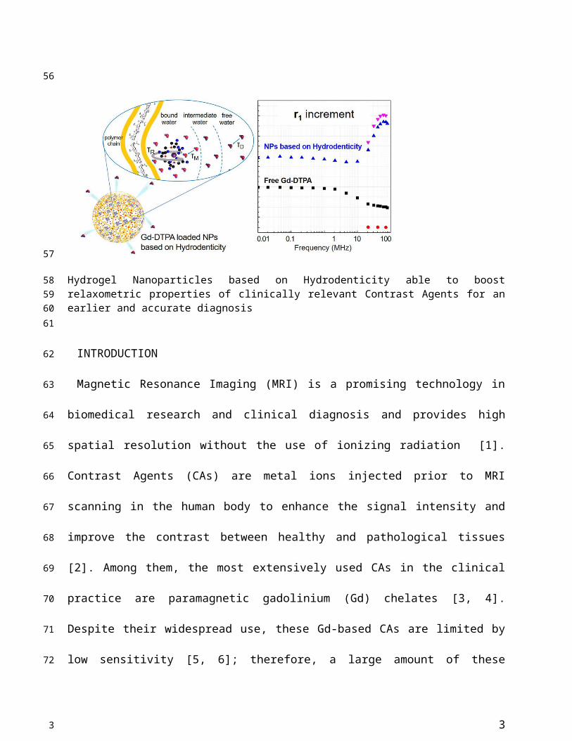
As advancement of the geometrical confinement [23], Courant et al. [24] and Callewaert et al.
[25], showed that biocompatible hydrophilic hydrogels can be exploited to produce high water
content nanoparticles (NPs) encapsulating the metal chelate. Inside the hydrogel, which creates a
favorable aqueous environment for Gd-based CAs [26, 27], the rotational motion of the
encapsulated CA (Gd-DOTP, Gd-DOTA and Gd-DTPA) is restricted and its magnetic properties
are amplified.
4
76
77
78
79
80
81
82
83
84
85
86
87
88
89
90
91
92
93
94
95
96
97
4
In recent studies [14, 15, 28–30], the use of polymers to develop safer, more efficient and
smart MRI CAs has significantly increased. Biopolymers, and particularly polysaccharides, have
received considerable attention because the most of them are non-toxic and have various
derivable groups (e.g. hydroxyl, carboxyl and amino groups) allowing easy chemical
modifications or reaction with functional molecules [31–33]. Natural-occurring polysaccharides,
like chitosan, alginate, heparin and hyaluronic acid, offer a suitable platform to produce
nanoparticles due to their biodegradability, biocompatibility and ease in molecular modification
and formation processes, such as by ionic or covalent crosslinking, ion-complex or self-assembly
[33]. These properties not only make polysaccharides very promising materials for drug delivery
but also proved to be useful in designing nanostructures for enhanced MRI [24, 34–37].
In our recently published works [5, 38], we have initially analysed the impact that hydrophilic
biopolymer networks have on the relaxivity of Gd-based CAs and explained the role of water in
the interaction between polymers and metal chelates. This concept, called “Hydrodenticity”, has
been the subject of further investigations as reported by Russo et al. [39]. In a former work
published by Russo and co-worker [6], crosslinked Hyaluronic Acid NanoParticles (cHANPs)
containing a Gd-chelate (Gd-DTPA), are synthesized through a microfluidic platform that allows
a high degree of control over particle synthesis, enabling the production of monodisperse
particles as small as 35 nm for MRI applications. The relaxivity (r1) achieved with the cHANPs
is 12-times higher than Gd-DTPA. Within cHANPs, the properties of Hydrodenticity can be
modulated to obtain the desired mesh size, crosslink density, hydrophilicity and loading
capability, as reported by Russo et al. [39, 40]. Moreover, they proved that an increase of the
crosslinking degree of biopolymer can induce the enhancement of relaxivity by restricting
molecular tumbling while maintaining the switching property [41] and allowing easy access of
5
98
99
100
101
102
103
104
105
106
107
108
109
110
111
112
113
114
115
116
117
118
119
120
5

water throughout the structure, which is a key feature in MRI CAs. The possibility to adopt a
unique platform to tune the hydrogel structural parameters and, consequently, increase the
relaxivity of a metal chelate without any chemical modification, could have a great impact on the
clinical outcome. In fact, thanks to their improved relaxometric properties, cHANPs could ensure
a brighter contrast with a lower amount of metal chelate, thus enabling the potential reduction of
the administration dosage as approved for clinical use.
In a further work [42], we reported an efficient way to produce Hybrid Core-Shell (HyCoS)
NPs composed of a Chitosan core and a shell of Hyaluronic Acid (HA) with improved
relaxometric properties (up to 5-times than the commercial CA). Subsequently, the same
nanosystem is used to develop a new nanoprobe for simultaneous Positron Emission
Tomography (PET)/MRI acquisitions as reported in our more recent publication [43].
Based on the above-reported works, it has been finally demonstrated that the polymer
architecture affects some characteristic parameters of the metal chelate and tunes its relaxometric
properties [24, 39, 44]. Moreover, it is clear that crosslinked biopolymers can have a significant
role in overcoming the limitations of clinically relevant CAs without their chemical modification
and as a compound in the design of advanced nanostructures with improved safety profile and
switchable relaxometric properties. Indeed, it is known that the functional features as well as the
swelling behaviour of hydrogels are influenced by the hydration degree, which can be likely
modulated by changing the chemical composition of the system [45–47].
Here, we aim to highlight the basic principles ruling biopolymer-CA interactions in the
perspective of their influence on the relaxometric properties of the CA by adopting a
multidisciplinary experimental approach. HA [26] is used as a model polymer because of its
biocompatibility and high hydrophilicity. We characterize, physically and chemically, the
6
121
122
123
124
125
126
127
128
129
130
131
132
133
134
135
136
137
138
139
140
141
142
143
6
interactions between hydrophilic biopolymers and Gd-based CAs. In this theoretical framework,
the peculiar effect of Hydrodenticity on the polymer conformation and the formation of the
stable water compartments responsible for the enhancement of the MRI signal is introduced and
discussed. Finally, we use the acquired knowledge about polymer-CA systems to apply the
concept of Hydrodenticity to the design of Gd-based polymer NPs with enhanced relaxometric
properties.
RESULTS
Changes in polymer conformation induced by a Gd-based contrast agent
The polymer conformation can be modified by the affinity with the solvent solution [48, 49].
Furthermore, the addition of a solute can still induce a change in the polymer conformation. In
our previous work, we proved that the relaxivity of CAs can be modulated combining them with
macromolecules or polymers [5]. Therefore, the understanding of the interaction between
polymers and CAs in aqueous solution could be critical to tune the relaxometric properties of
CAs. We aim to show how the presence of the Gd-DTPA in the aqueous solution can influence
the behaviour of the polymer matrix and, on the other side, how these adjustments of the polymer
conformation can govern the characteristic correlation times of the Gd-DTPA [5, 6, 39].
To investigate thermodynamic interactions between polymer and contrast agent, HA and Gd-
DTPA respectively, are selected to be tested by Isothermal Titration Calorimetry (ITC). We aim
to take advantage of the molecular interactions that are accompanied by some level of heat
exchange between the interacting system and its surrounding medium; indeed, these interactions
can be evaluated, at constant temperature, through the ITC. Basic principles of this technique
have been widely discussed elsewhere [50, 51].
7
144
145
146
147
148
149
150
151
152
153
154
155
156
157
158
159
160
161
162
163
164
165
166
7
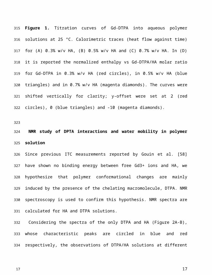
Titration experiments are conducted injecting a solution of Gd-DTPA in the ITC cell
containing the polymer solution. Different HA concentrations, ranging from 0.3 to 0.7% w/v, are
tested and more representative results are reported in Figure 1 (peaks above the baseline
represent exothermic phenomena while peaks below the baseline represent endothermic
phenomena). It is clear that significant enthalpy variations are obtained in the titration
experiments (Figure 1A-C), which induces changes in the polymer chains’ conformation as
explained below. Since Figure 1A-C show ITC thermograms varying the HA concentration in
the sample cell, a wide range of Gd-DTPA/HA molar ratios is examined and the relative
energetic contribution and enthalpy values are calculated by integrating peaks of the
experimental curves and are reported in Figure 1D. Simple dilution of Gd-DTPA in water
(Figure S1) exhibits only small constant exothermic peaks over the whole experiment.
In Figure 1, it is worth noting that the energetic contribution decreases as the Gd-DTPA/HA
molar ratio increases; thus, the higher is the concentration of HA in the sample cell, the higher is
the Gd-DTPA concentration needed to observe endothermic peaks. It can also be noted that the
endothermic contribution exceeds the exothermic one at the recurrent Gd-DTPA/HA ratio
approximatively equal to 0.5 through all the experiments at different HA concentrations in the
sample cell. It means that a specific energetic contribution is needed to induce the adjustment of
the polymer conformation. Then, when the Gd-DTPA/HA molar ratio equals 0.5, the
endothermic peaks start slightly increasing until reaching a plateau, which corresponds to the
thermodynamic equilibrium established within the ternary system (polymer-CA-water). The
measured energetic variation reflects the conformational changes of polymer chains due to the
presence of the CA in solution and leads to the formation of stable sub-domains in which a
balanced exchange of water molecules occurs between the polymer, the CA and the bulk.
8
167
168
169
170
171
172
173
174
175
176
177
178
179
180
181
182
183
184
185
186
187
188
189
8

This is confirmed by the relation between Gd-DTPA/HA molar ratio and enthalpy values
showed in Figure 1D. Indeed, at low Gd-DTPA/HA ratio, the enthalpy term is negative, as
expected for polyelectrolytes in water such as HA, which generally dissolve easily in aqueous
media and behaves like a long, more or less randomly mobile chain and its conformation is
governed by electrostatic forces [52]. Indeed, it is well-known that, at low polymer
concentrations (dilute solution regime), the intrachain electrostatic interactions and interactions
with the surrounding polyelectrolyte chain ions determine the chain conformations since the
polyelectrolyte chains are separated from each other by average distances larger than their size
[52, 53]. As Gd-DTPA/HA ratio increases the enthalpy increases too, meaning that a change in
conformation is occurring due to the presence of a ionic compound (Gd-DTPA). Indeed, apart
from the solvent type (e.g., water), the polymer conformation can be modified by the
concentration of added co-solutes. In particular, in the presence of ionic compounds, a screening
of the electrostatic repulsion between the charged monomers occurs at low salt concentration
form a cloud surrounding the chain and the polymer collapses from an extended coil to a more
compact conformation as the salt is added [52, 54, 55].
At Gd-DTPA/HA = 0.5, the enthalpy becomes constant, meaning that an equilibrium is
reached. The attainment of this thermodynamic equilibrium derives from a water-mediated
interaction occurring between HA and Gd-DTPA. As both hydrophilic components, HA and Gd-
DTPA interact with the water by forming hydrogen bonds and by coordinating water molecules.
This competitive behaviour generates a measurable heat that reflects the change in polymer
chains conformation and the exchange of bound water molecules with the bulk, thereby, bringing
the system to a more stable configuration. As also observed in other metal-polymer systems [56,
57], in the presence of small amounts of metal-chelate compounds, a change of the polymer
9
190
191
192
193
194
195
196
197
198
199
200
201
202
203
204
205
206
207
208
209
210
211
212
9

structure occurs due to the weak macromolecule-metal interactions, which favors the formation
of a new hierarchy in the structural organization of the polymer as compared with metal-free
system.
In our previous paper [39], we preliminary showed how this new structural organization is able
to affect the relaxometric properties of the system, as an effect of the new concept of
Hydrodenticity, which will be further explained in the following paragraphs.
10
213
214
215
216
217
218
219
10
Figure 1. Titration curves of Gd-DTPA into aqueous polymer solutions at 25 °C. Calorimetric
traces (heat flow against time) for (A) 0.3% w/v HA, (B) 0.5% w/v HA and (C) 0.7% w/v HA. In
(D) it is reported the normalized enthalpy vs Gd-DTPA/HA molar ratio for Gd-DTPA in 0.3%
w/v HA (red circles), in 0.5% w/v HA (blue triangles) and in 0.7% w/v HA (magenta diamonds).
The curves were shifted vertically for clarity; y-offset were set at 2 (red circles), 0 (blue
triangles) and -10 (magenta diamonds).
NMR study of DPTA interactions and water mobility in polymer solution
Since previous ITC measurements reported by Gouin et al. [58] have shown no binding energy
between free Gd3+ ions and HA, we hypothesize that polymer conformational changes are
mainly induced by the presence of the chelating macromolecule, DTPA. NMR spectroscopy is
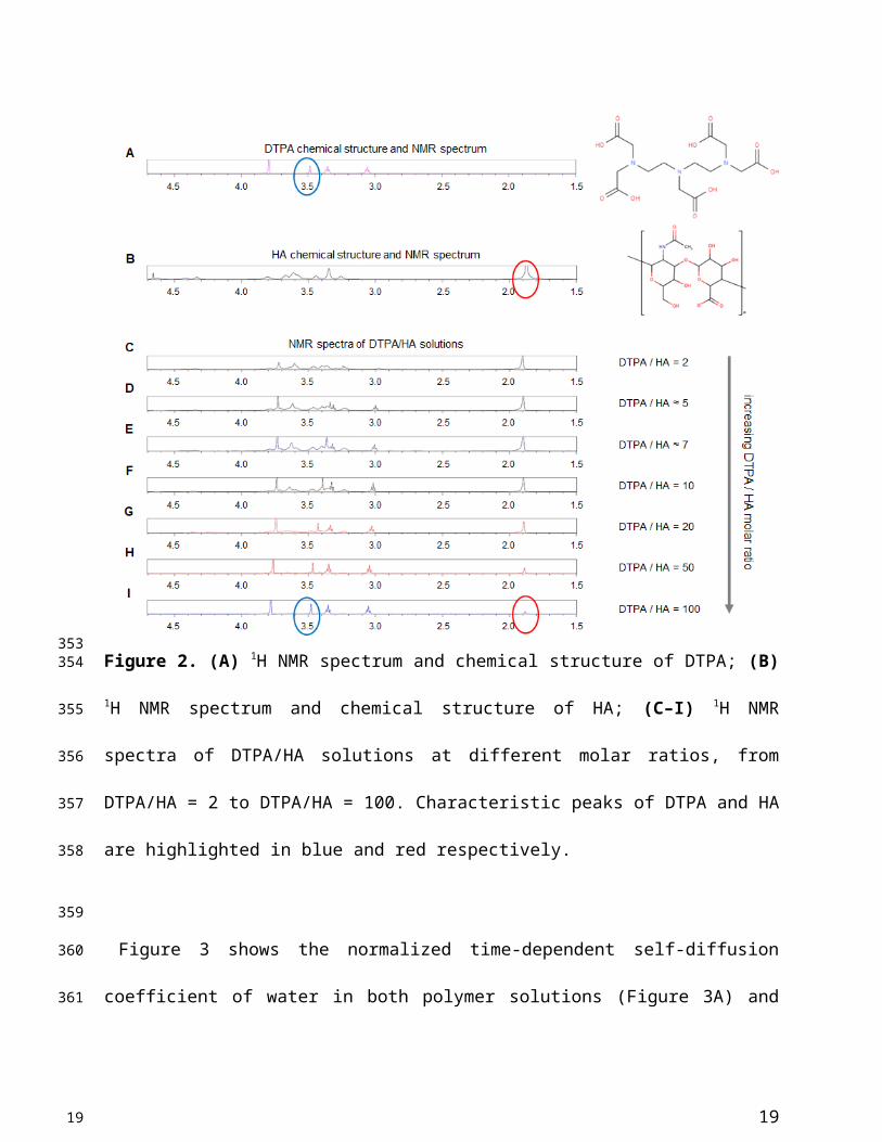
used to confirm this hypothesis. NMR spectra are calculated for HA and DTPA solutions.
Considering the spectra of the only DTPA and HA (Figure 2A-B), whose characteristic peaks
are circled in blue and red respectively, the observations of DTPA/HA solutions at different
molar ratios are reported (Figure 2C-I). The molar ratio ranges from 2 to 100 and is obtained by
decreasing the HA concentration from 150 to 10 µM.
In Figure 2, it can be observed that the characteristic DTPA peak at 3.50 ppm (s, 2 H, CH2–
COOH) is influenced by the presence of HA in solution. In fact, it seems to shift and reduce its
intensity far more than the other peaks by increasing the HA concentration. As an example, the
shift is evident by comparing Figure 2I, where the DTPA peak is highlighted in blue, with Figure
2C, where the signal is dramatically reduced. It results that an interaction between the two
components of the system exists and generates changes in the NMR spectrum of the solution.
11
220
221
222
223
224
225
226
227
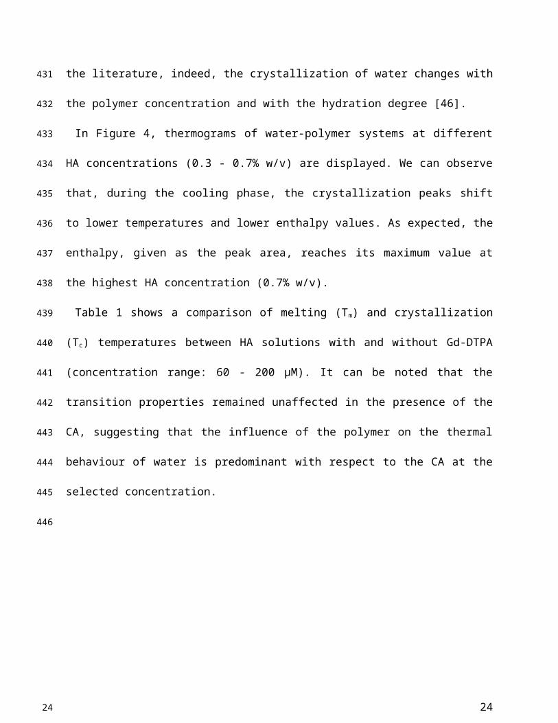
228
229
230
231
232
233
234
235
236
237
238
239
240
241
11
Chen, Shawn (NIH/NIBIB) [E], 01/18/19,
The labels of axes appear to be distorted. Use Arial font only. Figure labels (A/B…) should be 12 points Arial bold.

This can be due to electrostatic repulsion between the two anionic macromolecules, HA and
DTPA.
Through NMR-DOSY, instead, we investigate how the presence of both HA and Gd-DTPA
can affect the mobility of water molecules.
Figure 2. (A) 1H NMR spectrum and chemical structure of DTPA; (B) 1H NMR spectrum and
chemical structure of HA; (C–I) 1H NMR spectra of DTPA/HA solutions at different molar
ratios, from DTPA/HA = 2 to DTPA/HA = 100. Characteristic peaks of DTPA and HA are
highlighted in blue and red respectively.
12
242
243
244
245
246
247
248249
250
251
252
12
Chen, Shawn (NIH/NIBIB) [E], 01/18/19,
Chemical structure should follow ACS style. Specifically ACS Document1996 style. Again figure panel labels should be 12 points. Figure text should be 8 points or above.
Figure 3 shows the normalized time-dependent self-diffusion coefficient of water in both
polymer solutions (Figure 3A) and polymer-CA solutions (Figure 3B). For short diffusion
delays, the measured self-diffusion coefficient D is nearly equal to the free self-diffusion
coefficient D0 of water at 25°C (2.5·10-9 m2/s), since the molecules travel over a short distance
and only a few of them feel the surrounding macromolecules. As the diffusion time increases,
more water molecules go through these restrictions and the self-diffusion coefficient reaches a
plateau value.
We can hypothesize that the presence of Gd-DTPA competes with those HA-molecular sites
beared by water molecules and that are responsible for polymer hydration and hydrogel
formation. As highlighted with ITC results, the polymer conformation can be modified by the
presence of Gd-DTPA, which could interplay with the water molecules and with the formation of
hydrogen bonding. NMR-DOSY measurements are carried out to assess these hypothesized
changes in water mobility. It can be observed that, in the case of the ternary system, the
diffusivity of water beyond decreases, as expected for solvent molecules within polymer
matrices or in confined environments [59, 60], suggesting that the polymer-CA combination
affects the water mobility more than the polymer itself.
Figure 3A clearly shows that water diffusion behaviour is affected by the polymer
concentration. In particular, the diffusion coefficient decreases at increasing polymer
concentration. Besides, Figure 3B shows the additional contribution of the CA to the water
mobility. In fact, the presence of Gd-DTPA, even at relatively low concentrations (5 - 30 µM),
can further reduce the value of the water self-diffusion coefficient for both short and long
diffusion times.
13
253
254
255
256
257
258
259
260
261
262
263
264
265
266
267
268
269
270
271
272
273
274
275
276
13
Figure 3. (A) Normalized time dependent water self-diffusion coefficient in 0.1% w/v HA
(squares), 1% w/v HA (triangles), 2% w/v HA (flipped triangles), 3% w/v HA (diamonds). (B)
Normalized time dependent water self-diffusion coefficient in 1% w/v HA (triangles), 5 μM Gd-
DTPA in 1% w/v HA (flipped triangles) and 30 μM Gd-DTPA in 1% w/v HA (stars).
It is worth noting that low Gd-DTPA concentrations are chosen (Figure 3B) because Gd-
DTPA is highly paramagnetic and it can interfere with NMR measurements [61, 62], while the
HA concentrations (0.1 - 3% w/v) are slightly higher than those used in the ITC experiments to
highlight and make more evident the differences in diffusion behaviour between samples. In
particular, as illustrated in Figure 3B, a fixed polymer concentration of 1% w/v is selected to
show the effect of CA on the diffusion of water molecules.
A data comparison between ITC and NMR spectra confirms the hypothesized fundamental
properties behind the concept of Hydrodenticity: the ability of Gd-DTPA to induce changes in
polymer conformation and in water mobility.
14
277278279
280
281
282
283
284
285
286
287
288
289
290
291
292
14
Chen, Shawn (NIH/NIBIB) [E], 01/18/19,
Increase panel label size to 12 points

Water dynamics within hydrated polymer matrix containing Gd-DTPA
To analyse further the role of water mobility in the Hydrodenticity, a study of the dynamics
and behaviour of water molecules is needed. Within hydrated polymer matrices (hydrogels),
containing metal chelates, water molecules mediate polymer-CA interactions and, therefore, play
a dual role: on the one hand, the amount of absorbed water [63, 64] and its interaction with the
hydrogel structure affects the chain motion of the hydrophilic polymer; on the other, the mobility
of water molecules in the polymer matrix is responsible for the relaxometric properties of the
CA.
We investigate the water dynamics in water-HA systems, with and without Gd-DTPA, using
the Differential Scanning Calorimetry (DSC). We focus on the thermal effects that the polymer
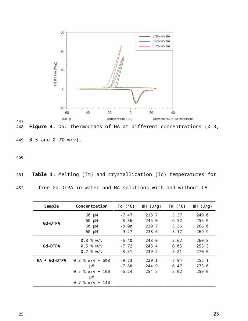
(Figure 4) and the CA (Table 1) have on the water dynamics. According to the literature, indeed,
the crystallization of water changes with the polymer concentration and with the hydration
degree [46].
In Figure 4, thermograms of water-polymer systems at different HA concentrations (0.3 - 0.7%
w/v) are displayed. We can observe that, during the cooling phase, the crystallization peaks shift
to lower temperatures and lower enthalpy values. As expected, the enthalpy, given as the peak
area, reaches its maximum value at the highest HA concentration (0.7% w/v).
Table 1 shows a comparison of melting (Tm) and crystallization (Tc) temperatures between HA
solutions with and without Gd-DTPA (concentration range: 60 - 200 µM). It can be noted that
the transition properties remained unaffected in the presence of the CA, suggesting that the
influence of the polymer on the thermal behaviour of water is predominant with respect to the
CA at the selected concentration.
15
293
294
295
296
297
298
299
300
301
302
303
304
305
306
307
308
309
310
311
312
313
314
315
15

Figure 4. DSC thermograms of HA at different concentrations (0.3, 0.5 and 0.7% w/v).
Table 1. Melting (Tm) and crystallization (Tc) temperatures for free Gd-DTPA in water and HA
solutions with and without CA.
Sample Concentration Tc (°C) ΔH (J/g) Tm (°C) ΔH (J/g)
Gd-DTPA
60 µM60 µM60 µM60 µM
-7.47-8.36-8.00-9.27
218.7245.0239.7238.6
5.376.525.365.17
249.0255.0266.0269.9
Gd-DTPA0.3 % w/v0.5 % w/v0.7 % w/v
-6.40-7.72-8.31
243.0248.4239.2
5.626.055.21
260.8253.3270.0
HA + Gd-DTPA0.3 % w/v + 600 µM0.5 % w/v + 100 µM0.7 % w/v + 140 µM
-9.73-7.88-6.24
229.1244.9254.5
7.946.475.02
255.1273.0259.0
Relaxation times, rates and relaxivity of the polymer matrix
16
316
317318
319
320
321
322
323
16
The existence of a water-mediated interaction between Gd-DTPA and HA, observed through
ITC, NMR and DSC, and the effect of the polymer conformation on the characteristic correlation
time of the metal chelate could explain the boosting of the relaxivity in the studied systems.
Relaxometric properties are investigated using time-domain relaxometry on two different
systems: non-crosslinked and crosslinked polymer matrix (0.5% w/v HA) containing Gd-DTPA.
In the latter case, rheological and chemical-physical properties of the polysaccharide can be
modulated by changing the crosslinker (DVS) concentration, as known as crosslinking density.
In fact, thanks to the presence of hydrophilic groups in the skeleton of HA, the hydrogel is able
to uptake a large amount of water. Under these conditions, water is in an abnormal aggregate
state that influences the relaxivity of hydrated Gd-DTPA.
Figure 5 shows the results of relaxometric measurements for the hydrogel system (0.5% w/v
HA) studied by loading different concentration of Gd-DTPA. The hydrogel system is analysed
and compared to the free Gd-DTPA solution. In particular, we display the increment in the
percentage of the paramagnetic relaxation as a function of Gd-DTPA/HA ratio.
17
324
325
326
327
328
329
330
331
332
333
334
335
336
337
17

Figure 5. Increment in longitudinal relaxation rate, R1, at different Gd-DTPA/HA ratio (from 0
to 9) for: free Gd-DTPA in water (black squares); Gd-DTPA in 0.5% w/v HA solution (blue
filled triangles); Gd-DTPA in 0.5% w/v HA crosslinked with DVS (blue empty triangles). The
R1 increment is calculated in percentage with respect to the corresponding R1 of Gd-DTPA in
water. A fast increment in R1 is observed until a 2.5 Gd-DTPA/HA ratio (Gd-DTPA
concentration equal to 300 µM). For higher ratios (i.e. higher Gd-DTPA concentrations), the R1
increment reaches a plateau.
The increment in percentage of the longitudinal relaxation rate (R1) has been calculated as
follows (Equation 1):
18
338
339
340
341
342
343
344
345
346
347
18
% R1increment=
1T1|GdDTPA∈HA
− 1T 1|GdDTPA
1T1|GdDTPA
∗100 (1)
Details on relaxation times and rates values are reported in Table S1 of the Supporting
Information.
We find that the stronger is the interaction between Gd-DTPA and HA, the better is the MRI
enhancement. Moreover, the crosslinked system is much more efficient than the non-crosslinked
one. Indeed, in the crosslinked system, since the enhancement reaches a plateau at Gd-DTPA/HA
molar ratio equal to 2.5 (i.e. at 300 µM of Gd-DTPA in 0.5% w/v HA water solution), it is not
necessary to overload the system with Gd-DTPA in order to achieve higher relaxation. It is worth
noting that, for both studies, with and without crosslinker, the Gd-DTPA concentration of 200
μM seems to represent a threshold for the maximum effect of the hydrogel on the Gd-DTPA
relaxation mechanism.
Furthermore, transversal relaxation times (T2) and rates (R2) have also been measured and
reported in Table S1 of the Supporting Information.
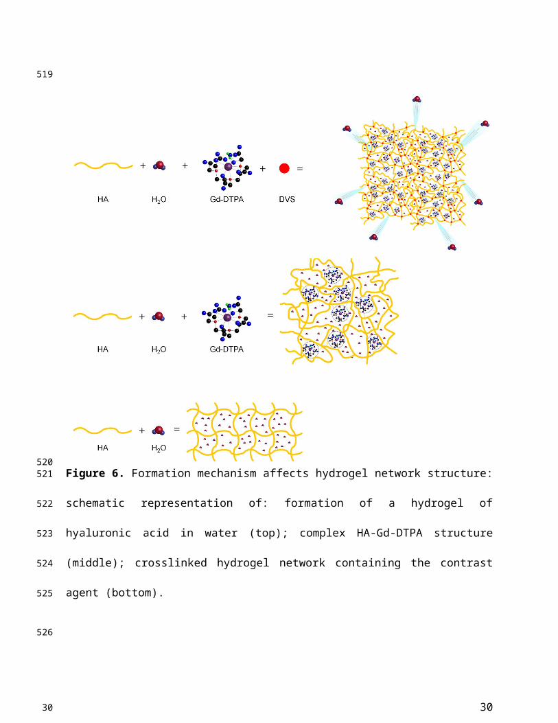
Figure 6 displays a schematic representation of the hydrogel network formation, even in the
presence of the crosslinking agent, and its influence on the polymer conformation.
We hypothesize that the boosting of Gd-DTPA relaxivity in a hydrogel matrix is due to a
proper complexation between the polymer and the CA in solution, mediated by the water and
further amplified by the addition of a crosslinker. It is confirmed that the reached equilibrium
among osmotic pressure and elastodynamic forces of the polymer meshes and hydration degree
of the CA in the matrix are able to tune finely the relaxometric properties of the metal chelates in
the ternary system. The overall ensemble of these phenomena is defined as Hydrodenticity [39].
19
348
349
350
351
352
353
354
355
356
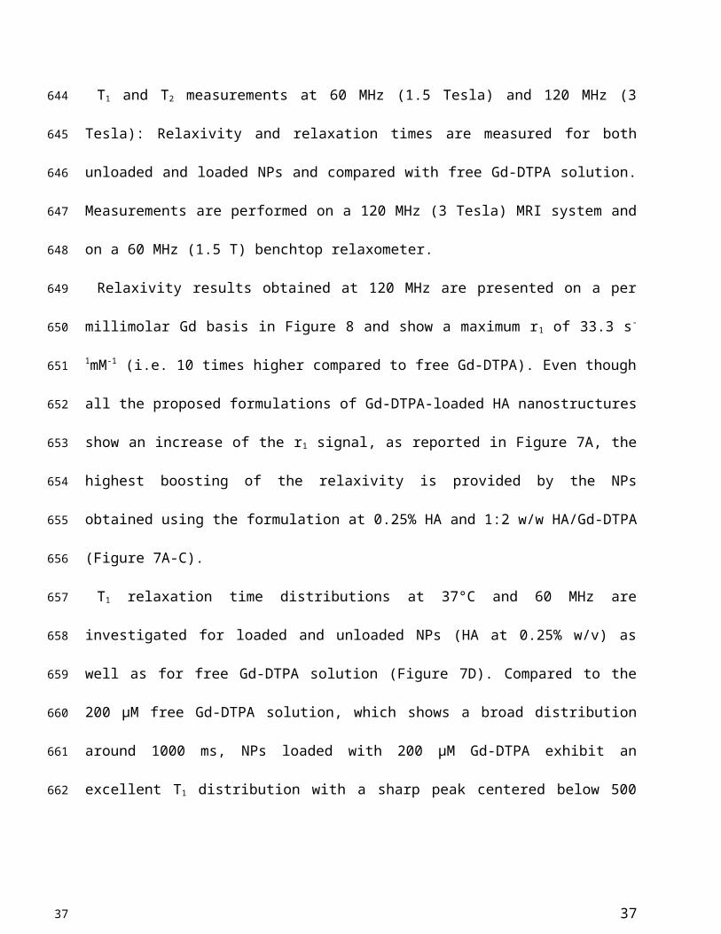
357
358
359
360
361
362
363
364
365
366
367
368
19

Figure 6. Formation mechanism affects hydrogel network structure: schematic representation of:
formation of a hydrogel of hyaluronic acid in water (top); complex HA-Gd-DTPA structure
(middle); crosslinked hydrogel network containing the contrast agent (bottom).
Case study: production of polymer particles based on Hydrodenticity
Recent recommendations from Food and Drug Administration (FDA) and European Medicine
Agency (EMA) about the Gd deposition in the brain and other tissues have highlighted the
20
369
370371
372
373
374
375
376
377
20
importance to design polymer biocompatible NPs with enhanced relaxivity without chemical
modification of the clinical relevant CAs [11, 65]. Thus, crosslinked NPs formed by HA, a
biodegradable, biocompatible, non-toxic, non-immunogenic and non-inflammatory linear
polysaccharide [66], could represent a successful candidate among nanovectors for MRI
applications [67]. Indeed, in the last decades, it is undisputed the growing research interest
toward the therapeutic action of HA and in developing new diagnostic tools based on this
polymer [67]. In this work, starting from the above-presented results, we aim to apply the
Hydrodencity in the design of biocompatible hydrogel nanostructures to obtain improved
relaxometric properties. We propose a concrete example of the concept of Hydrodenticity
applied to the production of crosslinked HA NPs for MRI, loaded with Gd-DTPA. An emulsion-
based method is used to obtain stable W/O nanoemulsions as templates.
Study of emulsion stability
Stable W/O emulsions are prepared by stirring appropriate amounts of the oil phase (Mineral
oil) and an aqueous phase containing different concentrations of Span-80 (S80) or S80 with
Tween-85 (S80/T85). The pH, ranging from 12 to 14 is adjusted by adding appropriate amounts
of NaOH from a stock solution (0.2 M). Further details are reported in the Materials and Method
Section. As expected, in the absence of any surfactant, W/O emulsions prepared in the same
conditions split very rapidly in two phases due to their unfavourable thermo-dynamic state.
Visual comparison, turbidimetry and backscattering are successfully used to study emulsion
stability (Figure S2 and S3) [68]. Comparing emulsions obtained at different W/O ratio, 10:90
and 20:80, but at the same concentration of surfactant, the stability is more extended for
emulsions with lower water content. In particular, a formulation 10/90 W/O volume ratio
21
378
379
380
381
382
383
384
385
386
387
388
389
390
391
392
393
394
395
396
397
398
399
400
21

containing S80 (1% w/v) and T85 (0.5 % w/v) resulted the more stable. However, even though
the stability of the emulsion is crucial to reduce polydispersity, an alkaline environment (addition
of NaOH) is necessary for the crosslinking reaction to take place. Indeed, Balazs and Leshchiner
[69] showed that the crosslinking reaction starts shortly after addition of DVS (5 - 10 min) and,
that, 1 hour is sufficient for the completion of the reaction [70, 71]. By these requirements, to
conduct the experimental campaign, we select the formulation with S80 (1% w/v) and NaOH
(0.2 M) as the optimal trade-off to obtain an emulsion stable for at least 3 hours (Figure S2 and
S3), enough for the DVS to react.
Preparation of DVS-crosslinked nanoparticles with and without CA
The exploitation of the best process conditions to design biocompatible nanostructures based
on Hydrodenticity and control their relaxation parameters for MRI application is reported. In
particular, the effect of the homogenization, HA concentration and the role of the crosslinking
reaction is analysed. Different experimental parameters and conditions are tested and details are
reported in the Materials and Methods section. A preliminary mixing is performed at 5000 or
7000 rpm for 10 min, by keeping constant the temperature at 25 °C. A 5000 rpm speed is
preferred to avoid and uncontrolled increase of the temperature.
After the homogenization, a crosslinking reaction is performed at high pH values (12 - 14) and
creates sulfonyl bis-ethyl linkages between the hydroxyl groups of HA [72]. This crosslinking
method has the advantage of occurring at room temperature, which limits the degradation of HA
in alkaline solutions. Even though the starting material DVS is highly reactive and toxic, the
biocompatibility of the HA-DVS hydrogels is confirmed by histological analysis [73].
22
401
402
403
404
405
406
407
408
409
410
411
412
413
414
415
416
417
418
419
420
421
422
22

In our protocol, a study of the modalities of injection of the crosslinking agent at different
steps of the homogenization process has shown that only when DVS is added after the
homogenization step spherical NPs are obtained. On the contrary, when the addition of the
crosslinker is performed at any time point during the homogenization phase, a shear stress
behavior of the polymer phase, interfering with the formation of those particles, is observed
(Figure S4).
The best experimental condition for production of crosslinked NPs is reached at 0.045% v/v
DVS (Table S2).
Based on these results and using the same process conditions, loaded NPs are obtained by
adding the CA in the water phase of the emulsion. Among several FDA approved CAs, we have
chosen to encapsulate a Gd chelated, Gd-DTPA (9.13 mM).
Purification and characterization of HA-NPs
Ultracentrifugation (UC) and dialysis are performed to purify HA NPs. Dynamic Light
Scattering (DLS) measurements are made on aqueous dilute NP suspension (1:10). The smaller
NPs’ size without CA (217.57 ± 34.65 nm) is obtained at 0.25% w/v of HA solution. At higher
polymer concentration (0.5% w/v) particle size is higher (401.67± 77,65 nm), while the
formulation with 0.1% w/v HA shows a reverse phenomenon with larger particles (760.15 ± 86
nm), probably due to less stability of the nuclei that tend to coalesce. When Gd-DTPA is added
to the process, the particle size at HA 0.25% w/v is slightly increased (258.77 ± 15.65 nm) for
the same process conditions. After purification, NPs are investigated by electron microscopy
techniques (SEM and TEM). The morphology of the NPs observed revealed that the particles are
spherical in shape and monodisperse (Figure S5).
23
423
424
425
426
427
428
429
430
431
432
433
434
435
436
437
438
439
440
441
442
443
444
445
23
Loading Capability (LC) and Encapsulation Efficiency (EE) is determined through ICP-MS by
comparing the theoretical amount initially used to prepare the particles and the Gd encapsulated
in the system after ultracentrifugation. The higher encapsulation results for 0.25 % w/v HA (1:2
w/w HA/Gd-DTPA ratio). Results show that probably ionic nature of Gd-DTPA impacts on its
encapsulation. In addition, HA NPs result stable up to 6 months or more, at 4°C and the release
of Gd-DTPA from HA NPs is tested at physiological condition (up to 48 hr), because it is
expecting that the clearance is shorter than 48 hr.
The zeta potential value of the 0.25% HA-NPs, with and without CA, indicate that they had a
negatively charged surface (-37.4 ± 1.34 mV and -31.8 ± 0.88 mV, respectively), due to the
carboxylic group of HA. IR measurements were also performed (Figure S6)
Relaxivity studies
T1 and T2 measurements at 60 MHz (1.5 Tesla) and 120 MHz (3 Tesla): Relaxivity and
relaxation times are measured for both unloaded and loaded NPs and compared with free Gd-
DTPA solution. Measurements are performed on a 120 MHz (3 Tesla) MRI system and on a 60
MHz (1.5 T) benchtop relaxometer.
Relaxivity results obtained at 120 MHz are presented on a per millimolar Gd basis in Figure 8
and show a maximum r1 of 33.3 s-1mM-1 (i.e. 10 times higher compared to free Gd-DTPA). Even
though all the proposed formulations of Gd-DTPA-loaded HA nanostructures show an increase
of the r1 signal, as reported in Figure 7A, the highest boosting of the relaxivity is provided by the
NPs obtained using the formulation at 0.25% HA and 1:2 w/w HA/Gd-DTPA (Figure 7A-C).
24
446
447
448
449
450
451
452
453
454
455
456
457
458
459
460
461
462
463
464
465
466
467
24
T1 relaxation time distributions at 37°C and 60 MHz are investigated for loaded and unloaded
NPs (HA at 0.25% w/v) as well as for free Gd-DTPA solution (Figure 7D). Compared to the 200
µM free Gd-DTPA solution, which shows a broad distribution around 1000 ms, NPs loaded with
200 µM Gd-DTPA exhibit an excellent T1 distribution with a sharp peak centered below 500 ms.
Gd concentration within loaded NPs was determined through Inductively Coupled Plasma Mass
Spectrometry (ICP-MS).
Loaded NPs perform far better even compared to the unloaded ones, whose distribution
appears to be broad and centered around 2800 ms.
It is worth mentioning that, compared to T1 distribution for bulk water (3600 ms), unloaded
NPs’ distribution shows that a slight contribution to the longitudinal relaxivity is ascribable to
the crosslinked polymer nanostructure, which is able by itself to tune the water mobility for a
non-nanostructured material. The contribution, therefore, to the overall relaxivity is further
enhanced in the ternary system, thanks to the water-mediated interaction between the polymer
and metal chelate.
25
468
469
470
471
472
473
474
475
476
477
478
479
480
481
25
Figure 7. (A) Relaxivity values r1 determined at magnetic field strengths of 3T for different set
of HA-NPs with respect to free Gd-DTPA in water. (B) Longitudinal relaxation rate (1/T1)
versus Gd-DTPA concentration for free Gd-DTPA in water and for HA-NPs at different polymer
concentrations loaded with Gd-DTPA. (C) T1-weighted images of Gd-DTPA, unloaded (used as
control) and HA-NPs at different polymer concentrations loaded with Gd-DTPA. All samples are
imaged at 3T, 25°C, using standard spin echo (SE) sequence. (D) Distribution of longitudinal
relaxation times of (T1) of 200 μM Gd-DTPA in water (squares), unloaded 0.25% HA-NPs
(circles) and 0.25% HA-NPs loaded with 200 μM Gd-DTPA (triangles).
26
482
483
484
485
486
487
488
489
490
26
Chen, Shawn (NIH/NIBIB) [E], 01/18/19,
Panel label size needs to be increased to 12 points.

Modeling of NMR dispersion: NMRD profile
The NMRD profiles as a function of the static magnetic field of the aqueous solutions of Gd-
DTPA and loaded and unloaded NPs (Figure 8) are set up to establish the effects caused by
Hydrodenticity functionalities on the parameters that determine the observed relaxivities. The
longitudinal relaxation rates are recorded at 37°C as a function of resonance frequency and
according to NP Gd-loading obtained by ICP-MS. The NMRD experimental curve for free Gd-
DTPA shows a plateau in longitudinal relaxivity at low fields and significantly decreases as the
applied magnetic field increases starting from 1 MHz. Conversely, longitudinal relaxivity (r1) for
loaded NPs (HA at 0.25% w/v) is characterized by the presence of a low-field plateau and a
gradual increase starting from 10 MHz, reaching a “dispersion peak” between 60 and 70 MHz.
The same peak and trend in the high field region (20 – 70 MHz) is observed for both at 25°C and
37°C. As a control, unloaded NPs do not exhibit increase in relaxivity in this field region,
confirming that the nanohydrogel structure containing Gd-DTPA contributes to slowing the
chelator's tumbling motion and allows water exchange thanks to its hydrophilic properties, as
hypothesized in the concept of Hydrodenticity.
27
491
492
493
494
495
496
497
498
499
500
501
502
503
504
505
506
507
27

Figure 8. NMRD profiles showing relaxivity of Gd-DTPA in water (squares), unloaded 0.25%
HA-NPs (circles), Gd-loaded 0.25% HA-NPs at 25°C (triangles) and 37°C (flipped triangles).
DISCUSSION
Theory of the Gado-Mesh formation
This work reports the use of polymer hydrogels for boosting the relaxivity of clinical relevant
CAs.
Results show that a spontaneous complexation exists between HA and Gd-DTPA due to
thermodynamic interactions as demonstrated by both calorimetric and NMR measurements. It is
already known that, in an aqueous polymer solution, solvent affinity can alter polymer
conformation by inducing local solvation of the structure that influences the number of available
conformations of the polymer chains. Moreover, the introduction of another soluble ionic
28
508509
510
511
512
513
514
515
516
517
518
519
520
28

component, such as Gd-DTPA, in the system can induce further changes in polymer
conformation, detectable via diverse thermodynamic approaches [74–77]. Based on our findings,
we prove for the first time that these conformational changes, induced by the metal chelate,
contribute to an increase in CA’s relaxivity.
To tune the MRI enhancement, we exploit the versatile structural characteristics of HA. In fact,
the high porosity of the hydrogel, which shows a mesh-like structure, is controlled by varying the
density of covalent crosslinking [78, 79]. Moreover, the presence of negatively charged groups
and the degree of crosslinking influence water adsorption capacity and, thus, the relaxometric
properties of the ternary system [45].
Therefore, the presence of the hydrogel matrix can significantly amplify the magnetic
properties of the encapsulated Gd-DTPA, suggesting a strong outer-sphere and second-sphere
contribution to the relaxivity.
Based on these observations, we hypothesize that increased relaxivity is mainly related to the
creation of water domains or clusters (water compartments) around the CA within the polymer
matrix. In fact, biopolymer systems contain intermolecular cavities that can be considered as
molecular nano-domains in which various self-assembly processes can be implemented in
principle [80]. The formation of peculiar structures within these cavities can be associated with
thermodynamic transitions and it is a characteristic of many metallopolymer systems [81, 82].
The sub-nanostructures, here defined “Gado-Meshes”, are generated from a three-way
interaction between HA, Gd-DTPA and water. The entrapment of the CA inside the hydrophilic
matrix of HA results in a reduction of the rotational tumbling rate, due to an increase of the
effective viscosity of the aqueous solution into the hydrogel matrix. At the same time, multiple
CA-water interaction pathways occur between the exchangeable protons of the water molecules
29
521
522
523
524
525
526
527
528
529
530
531
532
533
534
535
536
537
538
539
540
541
542
543
29
coordinated by the CA and the other water molecules freely moving within the hydrogel mesh or
bonded to the polymer chains [22, 44, 83].
Our “Gado-mesh” consists of highly relaxing Gd-water compartments spontaneously
generated within the hydrogel matrix by the combination of these multiple physico-chemical
interactions. The so created nanostructure is composed of different water layers departing from
the polymer chains that surround the Gd-molecules. It is known, in fact, that hydrated
polysaccharides, such as HA, are characterized by the presence of multiple water layers,
contiguous regions of variable water density within a polyelectrolyte solution [84], differing in
their physical properties depending on the distance from the polysaccharide chain [85]. The
hydration process of HA generates: the “bound water layer”, which is the water fractions closely
associated with the polymer matrix; the “unbound water layer”, made by the water molecules
which are not directly interacting with the polymer; and the “free water layer”, which resembles
dynamics of pure water. In addition, the developed polymer network can be dependent not only
on the water layers organization but also on other solute species, such as Gd-DTPA, altering the
bound water layer with non-negligible effect on the HA conformation and dynamic [86]. In the
“Gado-mesh”, Gd-DTPA has a competitive behaviour with the respect of HA, similar to the
cation shielding of the HA due to the presence of salts [87], and interposes itself between the
water molecules around the HA, altering the bound water layer and generating water
compartments with high MRI enhancing properties. The “Gado-mesh” influences the τR, τD and
τM times through the action of the Hydrodenticity, whose effect is magnified by the
crosslinking. Hydrodenticity, hence, refers to the status of the hydrated Gd-DTPA with the
coordination water subjected to osmotic pressure deriving from elastodynamics equilibrium of
swollen gels [39, 88–93].
30
544
545
546
547
548
549
550
551
552
553
554
555
556
557
558
559
560
561
562
563
564
565
566
30

We hypothesize that the attainment of this equilibrium is reached when the standard energetic
stability of the meshes is compromised by the presence of the Gd-DTPA and evolves to a new
spontaneous equilibrium involving the formation of nanocompartments, so-called “Gado-
Meshes”, in which water is in an abnormal aggregate state that influences the relaxivity. Water
molecules in the hydrogel matrix that are subjected to the effect of Hydrodenticity are able to
change their water dynamics and can mediate the hydrogel conformation and the physical and
relaxometric properties of the metal chelate.
CONCLUSION
From a biomedical point of view, the possibility to tune relaxometric properties of CAs by
controlling hydrogel structural parameters can pave the way to new advancements in the design
of nanovectors for diagnosis and therapy. This work proves that a new generation of more
efficient nanoparticle-based CAs can be developed by exploiting the affinity between CAs and
biopolymers. It can be done using biocompatible and clinical relevant CAs without their
chemical modification as approved in the clinical practice. Furthermore, the size of the resulting
NPs is in a range that makes them suitable for delivery to cells and tissues and a further increase
in relaxivity can be potentially achieved by tuning the system to the most efficient structure by
choosing the correct biopolymer-CA combination and optimizing concentration and crosslinking
degree of the structure.
EXPERIMENTAL SECTION
MATERIALS. All chemicals used are of analytical reagent grade quality and are employed as
received. Sorbitan monooleate (Span® 80) (S80), Polyoxyethylenesorbitan trioleate (Tween®
31
567
568
569
570
571
572
573
574
575
576
577
578
579
580
581
582
583
584
585
586
587
588
589
31

85) (T85), Mineral oil (light oil, 0.8 gr/cm at 25°C), Divinyl sulfone (DVS, 118.15 Da),
Diethylenetriaminepentaacetic acid gadolinium(III) dihydrogen salt hydrate (Gd-DTPA, 547.57
Da), Sodium hydroxide pellets (NaOH), Acetone and Ethanol are purchased from Sigma Aldrich
Chemical (Italy). Sodium Hyaluronate, with an average molecular weight of 850 kDa (purity
99%; Hyasis® 850P) and 42 kDa, is respectively supplied by Novozymes Biopharma and Bohus
Biotech (Sweden) as dry powder and used without purification.
Magnevist® (Bracco Imaging, Italy), a contrast agent commercially available, is used in this
study. The water is purified by distillation, deionization, and reserve osmosis (Milli-Q Plus) and
systematically used for sample preparation, purification and analysis. All experiments are
repeated in triplicate and conducted at room temperature, 25°C.
ISOTHERMAL TITRATION CALORIMETRY. Titration experiments are performed by
using a Nano ITC Low Volume calorimeter (TA Instruments). CA and polymer are prepared in
double-distilled filtered water without any additives. The sample cell (700 µL) and the syringe
(50 µL) are filled with aqueous solutions of HA and Gd-DTPA respectively. Syringe Gd-DTPA
concentration is fixed at 1.5 mM, while different HA concentrations in the sample cell are tested,
ranging from 0.3 to 0.7% w/v. The measurements are performed at 25 °C and at fixed stirring
rate of 200 rpm. Fifty Injections, each of 1 μL of Gd-DTPA, are delivered in intervals of 500 s.
The concentration of polymer is expressed as the mass of the repeat unit (unit mol/L).
Data analysis and processing to provide ITC and enthalpy change (ΔH) profiles is carried out
using the NanoAnalyze (TA instruments) and the OriginPro software.
32
590
591
592
593
594
595
596
597
598
599
600
601
602
603
604
605
606
607
608
609
610
611
32

NMR SPECTROSCOPY. 1H NMR spectra are recorded at 25 °C with Varian Agilent NMR
spectrometer operating at 600 MHz to observe chemical interactions between polymer and
chelating agent (DTPA). The NMR samples consisted of water solution of HA-DTPA at
different molar ratios (HA/DTPA ranging from 0 to 0.5), with 10% v/v D2O.
Diffusion-ordered NMR Spectroscopy (DOSY) is also performed and the z-gradient strengths
(Gz) is varied in 20 steps from 500 to 32500 G/cm (maximum strength). The gradient pulse
duration (δ) and the diffusion delay (Δ) are kept constant, 2 ms for δ and ranging from 7 to 1000
ms for Δ. After Fourier transformation and baseline correction, DOSY spectra are processed and
analysed using Varian software VNMRJ (Varian by Agilent Technologies, Italy) in order to
obtain the values of water self-diffusion coefficient.
DIFFERENTIAL SCANNING CALORIMETRY. For all measurements the HA/water
solution (Mw = 42 kDa) is used. The aqueous solutions are prepared in a concentration range of
polymer 0.3–0.7% w/v. Next, Gd-DTPA is added as CA at different molar ratio HA/Gd-DTPA
(from 1:0.25 to 1:3) and stirred for 12 h. The hydrated polymer samples, with and without CA,
are sealed at room temperature in a Tzero hermetic pans prior to analysis. DSC measurements
are performed in a TA Instruments’ Q20TM calorimeter on samples between 5 and 10 mg. The
samples are cooled down from 25°C to -50°C followed by heating scan up to 25°C. The same
heating and cooling rate are 10°C/min for all runs. Samples are tested in triplicate to ensure
reproducibility. For DSC and ITC measurements, we used low molecular weight HA (42 kDa) to
highlight better the energetic contributions of different components without exceeding the
maximum scale of the instruments.
33
612
613
614
615
616
617
618
619
620
621
622
623
624
625
626
627
628
629
630
631
632
633
634
33

TIME-DOMAIN RELAXOMETRY. The spin-lattice relaxation times (T1) are measured in a
Bruker Minispec (mq 60) bench-top relaxometer operating at 60 MHz for protons (magnetic field
strength: 1.41 T). Measurements are taken at 37°C, and before NMR measurements, the tube is
placed into the NMR probe for about 15 min for thermal equilibration. Experiments are made
using water solutions of Gd-DTPA (from 0 to 0.1 mM) and HA (0.3, 0.5 and 0.7% w/v)
crosslinked with DVS (DVS/HA weight percentage ratio equal to 1:8). T1 values are determined
by both saturation (SR) and inversion recovery (IR) pulse sequences. The relaxation recovery
curves are fitted using a multi-exponential model. Relaxivities, r1, are calculated from the slope
of the regression line of 1/T1 [s-1] versus concentration [mM] with a least-squares method.
EMULSION PREPARATION. The emulsions are prepared at different water to oil (W/O)
ratio (10/90 and 20/80 v/v). Mineral oil is used as oil phase (or continuous phase, PC) and W/O
emulsions are made by varying the concentration of surfactants for the PC and water phase (or
dispersed phase, PD) and the concentration of NaOH (from 0 to 0.2 M) for the PD in order to
obtain emulsion systems. In particular, non-ionic surfactants, Span-80 (S80) and Tween-85
(T85), are used to prepare mixtures with a range from 4.3 to 7.65 of HLB values. Depending on
the initial HLB to be used, mixtures of S80 and T85 are pre-dissolved in the appropriate S80/T85
mass ratios (from 50/50 to 75/25) in PC and PD, respectively. PD containing T85 and NaOH, is
added dropwise to PC and W/O emulsions are prepared using a high-shear homogenizer
(Silverson L5M-A, Silverson Machines Ltd, Waterside, UK). Homogenization of the Emulsion
is performed from 5000 to 7000 rpm for 10 min at room temperature (25°C).
34
635
636
637
638
639
640
641
642
643
644
645
646
647
648
649
650
651
652
653
654
655
656
34

TEMPORAL EMULSION STABILITY DETERMINATION. The stability of emulsions is
evaluated, at regular time intervals, by visual observation, measuring the height of the phase
separated by creaming in centimeters as a function of the time. In addition, optical
characterization of emulsion stability made is using a Turbiscan (Turbiscan LabThermo) by
static multiple light scattering (MLS), sending a light beam from an electroluminescent diode
(λ=880 nm) through a cylindrical glass cell containing the sample. The emulsion sample without
dilution is placed in a cylindrical glass cell and two synchronous optical sensors receive the light
transmitted through the sample (180° from the incident light) and the light backscattered by the
droplets in the sample (45° from the incident light). The optical reading head scans the height of
the sample in the cell (about 40 mm), by acquiring transmission and backscattering data every 40
μm. Transmitted and backscattered light are monitored as a function of time and cell height for
24 hours at an interval of 30 min at 25°C.
PREPARATION OF DVS-CROSSLINKED NANOPARTICLES. Based on these preliminary
results, PD/PC ratio in all samples is set at 10/90 v/v. In particular, for the preparation of cross-
linked NPs, HA powder (Mw = 850 kDa) is dissolved at different concentrations (from 0.1 to
0.5% w/v) under alkaline condition (NaOH ranging from 0 to 0.2 M) by vigorous stirring at
room temperature for 4 hours until a homogenous solution is obtained. Mineral oil and S80 (from
0.5 to 2% w/v) are separately mixed by stirring. PD is added drop-wise in the PC without stirring
and all the components are completely mixed by homogenization at various times (5-15 minutes)
and speeds (5000 - 7000 rpm). Then, the cross-linking agent (DVS) is added to the final
emulsion (40 ml), which is kept in agitation on a laboratory tube rotator for 24 hours to obtain a
homogeneous DVS distribution in the PD. To test DVS activity, various conditions of
35
657
658
659
660
661
662
663
664
665
666
667
668
669
670
671
672
673
674
675
676
677
678
679
35
crosslinking reaction are explored: (1) at different DVS concentrations (from 0.01 to 0.5% v/v);
(2) at three starting times of reaction (beginning, during and post homogenization) and (3) at
different temperatures (4 and 25°C).
The best experimental conditions for production of crosslinked HA-NPs are reported in Table
S2 of the Supporting Information.
LOADING OF HA NPS WITH CONTRAST AGENTS. After identifying the protocol to
obtain NPs, Gd-DTPA is chosen as CA and mixed in the PD before homogenization. Gd-loaded
HA NPs (HA-Gd NPs) are prepared using different HA/CA mass ratios (1:1, 1:2 and 1:5). DVS
is added post homogenization to the batch at room temperature using the same procedure
reported above.
COLLECTION OF THE NANOPARTICLES. Recovery of the NPs and their separation from
W/O emulsion system is made using dialysis and/or ultracentrifugation. For dialysis method, the
obtained emulsion is placed in a pre-washed cellulose membrane tubing (Spectra/Por® Dialysis
Tubing, cut-off of 25 kDa). Organic impurities (Mineral oil and S80) are removed dialyzing first
against solvents as acetone and/or ethanol, and gradually against water. Dialyzing solutions are
changed at regular time intervals. In the case of ultracentrifugation, 1 ml of the emulsion is added
to 5 ml of ethanol and mixed for 2 hours. Then, this mix is centrifuged with an ultracentrifuge
(Beckman-Coulter OPTIMA MAX-XP) at 55000 rpm for 20 min at 15°C. The resulting pellet is
washed twice and resuspended in MilliQ water. The second step of ultracentrifugation (70000
rpm, 10 min, 15°C) is applied to the pellet in order to obtain purified NPs.
36
680
681
682
683
684
685
686
687
688
689
690
691
692
693
694
695
696
697
698
699
700
701
702
36
CHARACTERIZATION OF THE NANOPARTICLES. To determine the size distribution of
NPs, dynamic light scattering (DLS) is performed using a Zetasizer S-90 1000 HS (Malvern
Instruments, UK).
All samples are diluted (1:10) with deionized water to prevent the effects of multiple
scattering. The measurement temperature is set at 25°C. The morphology and size of NPs are
investigated using ULTRA PLUS field emission Scanning Electron microscope (FE-SEM Carl
Zeiss, Oberkochen, Germany) and a Transmission Electron microscopy (TEM, TECNAI). In the
first case, the samples are coated with gold (7 nm).
DETERMINATION OF GADOLINIUM LOADING BY ICP-MS. The quantitative
determination of loaded Gd in HA NPs is assessed by ICP-MS (NexION 350, Perkin Elmer)
without any previous digestion processes. For all examinations, purified NP suspensions are
used. The non-encapsulated Gd-complexes are separated from the NPs by high-speed
centrifugation (55000 rpm, 20 min, 15 °C).
MRI TESTING. To explore the potential of Gd-loaded HA NPs as MRI contrast agent, MRI in
vitro test is performed at two different magnetic fields, 1.5 T and 3 T MR (Philips Achieva)
using Sense Head 8 coil. The T1-weighted MR images of HA NPs, unloaded and loaded with
Gd-DTPA at different concentrations using an inversion recovery sequence are measured with
the following parameters: TR = 2500 ms; TE = 12 ms; TI = 50, 100, 200, 400, 800, 1100, 1800
ms; FOV= 180x146 mm; slice thickness = 4 mm, acquisition matrix = 360x292.
The signal intensity of the samples is measured on the obtained T1-weighted MR images and
compared to Gd-DTPA.
37
703
704
705
706
707
708
709
710
711
712
713
714
715
716
717
718
719
720
721
722
723
724
725
37
NMR DISPERSION MEASUREMENTS. The proton 1/T1 NMRD profiles are measured using
a fast-field-cycling Stelar SmarTracer relaxometer over a continuum of magnetic field strengths
from 0.00024 to 0.25 T (which correspond to 0.01 - 10 MHz proton Larmor frequencies). The
uncertainty of these measurements is less than 1%. Additional data points in the range 15 - 70
MHz are obtained using a Stelar Relaxometer and a Bruker WP80 NMR electromagnet adapted
to variable-field measurements (15 - 80 MHz proton Larmor frequency).
FOURIER TRANSFORM INFRARED SPECTROSCOPY. Fourier transform infrared spectra
(FT-IR) were collected from Nicolett 6700 FT-IR spectrometer (Thermo Scientific). All the IR
spectra of the specimen were collected at 0.09 cm-1 resolution with 2 min interval. Empty HA
NPs and Gd-DTPA- Lodaed HA NPs were analyzed to observe the interaction. Spectra were
recorded and analyzed for signal assignation.
ASSOCIATED CONTENT
Supporting Information. The following files are available free of charge. Titration curve of the
contrast agent (Gd-DTPA) solution. Photographic image of the effect of increasing concentration
of surfactants and NaOH on the stability of W/O emulsion. Transmission and backscattering
spectra of W/O emulsion with and without NaOH. SEM images of crosslinked NPs under
various experimental conditions (i.e. polymer’s concentration, with and without contrast agent,
etc…).
AUTHOR INFORMATION
38
726
727
728
729
730
731
732
733
734
735
736
737
738
739740
741
742
743
744
745
746
747
748
38

Corresponding Author
Dr. E. Torino
Department of Chemical, Materials & Industrial Production Engineering, University of Naples
Federico II, Piazzale Tecchio 80, 80125 Naples, Italy.
Center for Advanced Biomaterials for Health Care, CABHC, Istituto Italiano di Tecnologia,
IIT@CRIB, Largo Barsanti e Matteucci 53, 80125 Naples, Italy.
Interdisciplinary Research Center on Biomaterials, CRIB, Piazzale Tecchio 80, 80125 Naples,
Italy.
E-mail: [email protected]
Author Contributions
Franca De Sarno and Alfonso Maria Ponsiglione contributed equally to this work. The
manuscript was written through contributions of all authors. All authors have given approval to
the final version of the manuscript.
Notes
The authors have declared that no competing interest exists.
ABBREVIATIONS
39
749
750
751
752
753
754
755
756
757
758
759
760
761
762
763
764
765
766
767
768
39

CA: contrast agent; cHANPs: crosslinked hyaluronic acid nanoparticles; DOSY: diffusion-
ordered nmr spectroscopy; DSC: differential scanning calorimetry; DVS: divinyl sulphone; EE:
encapsulation efficacy; EMA: european medicines agency; FDA: food and drug administration;
Gd: gadolinium; Gd-DTPA: diethylenetriaminepentaacetic acid gadolinium(iii) dihydrogen salt
hydrate; HA: hyaluronic acid; HLB: hydrophilic-lipophilic balance; HyCoS: hybrid core-shell;
ICP-MS: inductively coupled plasma mass spectrometry; IR: inversion recovery; ITC: isothermal
titration calorimetry; LC: loading capability; MLS: multiple light scattering; MRI: magnetic
resonance imaging; NaOH: sodium hydroxyde pellets; NMR: nuclear magnetic resonance;
NMRD: nuclear magnetic relaxation dispersion; NPs: nanoparticles; NSF: nephrogenic systemic
fibrosis; PC: continuous phase; PD: dispersed phase; PET: positron emission tomography; S80:
sorbitan monooleate (span® 80); SBM: solomon-bloembergen-morgan; SEM: scanning electron
microscopy; SR: saturation recovery; T85: polyoxyethylenesorbitan trioleate (tween® 85); TEM:
transmission electron microscopy; W/O: water in oil.
REFERENCES
1. Mansfield P. Snapshot magnetic resonance imaging (Nobel lecture). Angew. Chem. Int. Ed Engl. 2004;43:5456–64.
2. Caravan P. Strategies for increasing the sensitivity of gadolinium based MRI contrast agents. Chem. Soc. Rev. 2006;35:512–23.
3. Shokrollahi H. Contrast agents for MRI. Mater. Sci. Eng. C Mater. Biol. Appl. 2013;33:4485–97.
4. Zhang W, Liu L, Chen H, Hu K, Delahunty I, Gao S et al. Surface impact on nanoparticle-based magnetic resonance imaging contrast agents. Theranostics. 2018;8:2521–48.
5. Ponsiglione AM, Russo M, Netti PA, Torino E. Impact of biopolymer matrices on relaxometric properties of contrast agents. Interface Focus. 2016. doi:10.1098/rsfs.2016.0061.
40
769
770
771
772
773
774
775
776
777
778
779
780
781
782
783
784785
786787
788789
790791
792793794
40
Chen, Shawn (NIH/NIBIB) [E], 01/18/19,
Some references are incomplete. Please update.
Chen, Shawn (NIH/NIBIB) [E], 01/18/19,
Reformat references: [Author surname] [Author initials], [Other author surnames & initials]. [Article title]. [Journal name abbreviation]. [Year]; [Volume]: [First page number]- [Last page number].Please use the NLM Catalog (https://www.ncbi.nlm.nih.gov/nlmcatalog/journals) to find the abbreviations of journals. Omit any "." in the journal abbreviations. It is not necessary to italicize or bold the title, journal name, or any other part of the references. For journals that do not use pagination, use the article number in place of the page range. Use sentence format for article titles (only capitalize the first word). Include names of the first six authors for multi-author articles. For articles with six or more authors, use "et al."Example:1. Eknoyan G, Beck GJ, Cheung AK, et al. Effect of dialysis dose and membrane flux in maintenance hemodialysis. N Engl J Med. 2002; 347: 2010-9.

6. Russo M, Bevilacqua P, Netti PA, Torino E. A Microfluidic Platform to design crosslinked Hyaluronic Acid Nanoparticles (cHANPs) for enhanced MRI. Sci. Rep. 2016;6:37906.
7. Caravan P. Protein-targeted gadolinium-based magnetic resonance imaging (MRI) contrast agents: design and mechanism of action. Acc. Chem. Res. 2009;42:851–62.
8. Klemm PJ, Floyd WC, Smiles DE, Fréchet JMJ, Raymond KN. Improving T1 and T2 magnetic resonance imaging (MRI) contrast agents through the conjugation of an esteramide dendrimer to high water coordination Gd(III) hydroxypyridinone (HOPO) complexes. Contrast Media Mol. Imaging. 2012;7:95–9.
9. Thomsen HS, Morcos SK, Almén T, Bellin M-F, Bertolotto M, Bongartz G et al. Nephrogenic systemic fibrosis and gadolinium-based contrast media: updated ESUR Contrast Medium Safety Committee guidelines. Eur. Radiol. 2013;23:307–18.
10. Kanda T, Ishii K, Kawaguchi H, Kitajima K, Takenaka D. High signal intensity in the dentate nucleus and globus pallidus on unenhanced T1-weighted MR images: relationship with increasing cumulative dose of a gadolinium-based contrast material. Radiology. 2014;270:834–41.
11. McDonald RJ, McDonald JS, Kallmes DF, Jentoft ME, Murray DL, Thielen KR et al. Intracranial Gadolinium Deposition after Contrast-enhanced MR Imaging. Radiology. 2015;275:772–82.
12. Rogosnitzky M, Branch S. Gadolinium-based contrast agent toxicity: a review of known and proposed mechanisms. Biometals. 2016;29:365–76.
13. Huang J, Zhong X, Wang L, Yang L, Mao H. Improving the Magnetic Resonance Imaging Contrast and Detection Methods with Engineered Magnetic Nanoparticles. Theranostics. 2012;2:86–102.
14. Guo C, Sun L, Cai H, Duan Z, Zhang S, Gong Q et al. Gadolinium-Labeled Biodegradable Dendron–Hyaluronic Acid Hybrid and Its Subsequent Application as a Safe and Efficient Magnetic Resonance Imaging Contrast Agent. ACS Appl. Mater. Interfaces. 2017;9:23508–19.
15. Luo Q, Xiao X, Dai X, Duan Z, Pan D, Zhu H et al. Cross-Linked and Biodegradable Polymeric System as a Safe Magnetic Resonance Imaging Contrast Agent. ACS Appl. Mater. Interfaces. 2018;10:1575–88.
16. Corti M, Lascialfari A, Marinone M, Masotti A, Micotti E, Orsini F et al. Magnetic and relaxometric properties of polyethylenimine-coated superparamagnetic MRI contrast agents. J. Magn. Magn. Mater. 2008. doi:10.1016/j.jmmm.2008.02.115.
17. Bloembergen N, Morgan LO. Proton Relaxation Times in Paramagnetic Solutions. Effects of Electron Spin Relaxation. J. Chem. Phys. 1961;34:842–50.
18. Li Y, Beija M, Laurent S, Elst L vander, Muller RN, Duong HTT et al. Macromolecular Ligands for Gadolinium MRI Contrast Agents. Macromolecules. 2012;45:4196–204.
41
795796
797798
799800801802
803804805
806807808809
810811812
813814
815816817
818819820821
822823824
825826827
828829
830831
41
19. Port M, Raynal I, Vander Elst L, Muller RN, Dioury F, Ferroud C et al. Impact of rigidification on relaxometric properties of a tricyclic tetraazatriacetic gadolinium chelate. Contrast Media Mol. Imaging. 2006;1:121–7.
20. Jászberényi Z, Sour A, Tóth É, Benmelouka M, Merbach AE. Fine-tuning water exchange on GdIII poly(amino carboxylates) by modulation of steric crowding. Dalton Trans. 2005;0:2713–9.
21. Gizzatov A, Stigliano C, Ananta JS, Sethi R, Xu R, Guven A et al. Geometrical confinement of Gd(DOTA) molecules within mesoporous silicon nanoconstructs for MR imaging of cancer. Cancer Lett. 2014;352:97–101.
22. Sethi R, Ananta JS, Karmonik C, Zhong M, Fung SH, Liu X et al. Enhanced relaxivity of Gd3+-based contrast agents geometrically confined within porous nanoconstructs. Contrast Media Mol. Imaging. 2012. doi:10.1002/cmmi.1480.
23. Kuang Y, Cao Y, Liu M, Zu G, Zhang Y, Zhang Y et al. Geometrical Confinement of Gadolinium Oxide Nanoparticles in Poly(ethylene glycol)/Arginylglycylaspartic Acid-Modified Mesoporous Carbon Nanospheres as an Enhanced T1 Magnetic Resonance Imaging Contrast Agent. ACS Appl. Mater. Interfaces. 2018;10:26099–107.
24. Courant T, Roullin VG, Cadiou C, Callewaert M, Andry MC, Portefaix C et al. Hydrogels Incorporating GdDOTA: Towards Highly Efficient Dual T1/T2 MRI Contrast Agents. Angew. Chem. Int. Ed. 2012;51:9119–22.
25. Callewaert M, Roullin VG, Cadiou C, Millart E, Gulik LV, Andry MC et al. Tuning the composition of biocompatible Gd nanohydrogels to achieve hypersensitive dual T1/T2 MRI contrast agents. J. Mater. Chem. B. 2014;2:6397–405.
26. Necas J, Bartosikova L, Brauner P, Kolar J. Hyaluronic acid (hyaluronan): a review. Vet. Med, 53, 2008 (8): 397–411
27. Zhu W, Artemov D. Biocompatible blood pool MRI contrast agents based on hyaluronan. Contrast Media Mol. Imaging. 2011;6:61–8.
28. Zhu L, Yang Y, Farquhar K, Wang J, Tian C, Ranville J et al. Surface Modification of Gd Nanoparticles with pH-Responsive Block Copolymers for Use As Smart MRI Contrast Agents. ACS Appl. Mater. Interfaces. 2016;8:5040–50.
29. Sun L, Li X, Wei X, Luo Q, Guan P, Wu M et al. Stimuli-Responsive Biodegradable Hyperbranched Polymer–Gadolinium Conjugates as Efficient and Biocompatible Nanoscale Magnetic Resonance Imaging Contrast Agents. ACS Appl. Mater. Interfaces. 2016;8:10499–512.
30. Wang S, Zhou Z, Yu G, Lu N, Liu Y, Dai Y et al. Gadolinium Metallofullerene-Polypyrrole Nanoparticles for Activatable Dual-Modal Imaging-Guided Photothermal Therapy. ACS Appl. Mater. Interfaces. 2018;10:28382–9.
42
832833834
835836837
838839840
841842843
844845846847
848849850
851852853
854855
856857
858859860
861862863864
865866867
42

31. Huang Y, Hu H, Li R-Q, Yu B, Xu F-J. Versatile Types of MRI-Visible Cationic Nanoparticles Involving Pullulan Polysaccharides for Multifunctional Gene Carriers. ACS Appl. Mater. Interfaces. 2016;8:3919–27.
32. Swierczewska M, Han HS, Kim K, Park JH, Lee S. Polysaccharide-based Nanoparticles for Theranostic Nanomedicine. Adv. Drug Deliv. Rev. 2016;99:70–84.
33. Nitta SK, Numata K. Biopolymer-based nanoparticles for drug/gene delivery and tissue engineering. Int. J. Mol. Sci. 2013;14:1629–54.
34. Almalik A, Karimi S, Ouasti S, Donno R, Wandrey C, Day PJ et al. Hyaluronic acid (HA) presentation as a tool to modulate and control the receptor-mediated uptake of HA-coated nanoparticles. Biomaterials. 2013;34:5369–80.
35. Almalik A, Benabdelkamel H, Masood A, Alanazi IO, Alradwan I, Majrashi MA et al. Hyaluronic Acid Coated Chitosan Nanoparticles Reduced the Immunogenicity of the Formed Protein Corona. Sci. Rep. 2017;7:10542.
36. Almalik A, Alradwan I, A. Majrashi M, A. Alsaffar B, T. Algarni A, S. Alsuabeyl M et al. Cellular responses of hyaluronic acid-coated chitosan nanoparticles. Toxicol. Res. 2018;7:942–50.
37. Lallana E, Donno R, Magrì D, Barker K, Nazir Z, Treacher K et al. Microfluidic-assisted nanoprecipitation of (PEGylated) poly (d,l-lactic acid-co-caprolactone): Effect of macromolecular and microfluidic parameters on particle size and paclitaxel encapsulation. Int. J. Pharm. 2018;548:530–9.
38. Russo M, Bevilacqua P, Netti PA, Torino E. Commentary on “A Microfluidic Platform to Design Crosslinked Hyaluronic Acid Nanoparticles (cHANPs) for Enhanced MRI”. Mol. Imaging. 2017. doi:10.1177/1536012117706237.
39. Russo M, Ponsiglione AM, Forte E, Netti PA, Torino E. Hydrodenticity to enhance relaxivity of gadolinium-DTPA within crosslinked hyaluronic acid nanoparticles. Nanomed. 2017;12:2199–210.
40. Russo M, Grimaldi AM, Bevilacqua P, Tammaro O, Netti PA, Torino E. PEGylated crosslinked hyaluronic acid nanoparticles designed through a microfluidic platform for nanomedicine. Nanomed. 2017;12:2211–22.
41. Okada S, Mizukami S, Kikuchi K. Switchable MRI contrast agents based on morphological changes of pH-responsive polymers. Bioorg. Med. Chem. 2012;20:769–74.
42. Vecchione D, Grimaldi AM, Forte E, Bevilacqua P, Netti PA, Torino E. Hybrid Core-Shell (HyCoS) Nanoparticles produced by Complex Coacervation for Multimodal Applications. Sci. Rep. 2017;7:45121.
43. Vecchione D, Aiello M, Cavaliere C, Nicolai E, Netti PA, Torino E. Hybrid core shell nanoparticles entrapping Gd-DTPA and 18F-FDG for simultaneous PET/MRI acquisitions. Nanomed. 2017;12:2223–31.
43
868869870
871872
873874
875876877
878879880
881882883
884885886887
888889890
891892893
894895896
897898
899900901
902903904
43
44. Courant T, Roullin GV, Cadiou C, Callewaert M, Andry MC, Portefaix C et al. Biocompatible nanoparticles and gadolinium complexes for MRI applications. Comptes Rendus Chim. 2013;16:531–9.
45. Pasqui D, De Cagna M, Barbucci R, Pasqui D, De Cagna M, Barbucci R. Polysaccharide-Based Hydrogels: The Key Role of Water in Affecting Mechanical Properties. Polymers. 2012;4:1517–34.
46. Panagopoulou A, Molina JV, Kyritsis A, Pradas MM, Lluch AV, Ferrer GG et al. Glass Transition and Water Dynamics in Hyaluronic Acid Hydrogels. Food Biophys. 2013;8:192–202.
47. Davies G-L, Kramberger I, Davis JJ. Environmentally responsive MRI contrast agents. Chem. Commun. 2013;49:9704–21.
48. Budkov YA, Kolesnikov AL, Georgi N, Kiselev MG. A flexible polymer chain in a critical solvent: Coil or globule? EPL Europhys. Lett. 2015;109:36005.
49. Flory PJ. Thermodynamics of High Polymer Solutions. J. Chem. Phys. 1942;10:51–61.
50. Martinez JC, Murciano-Calles J, Cobos ES, Iglesias-Bexiga M, Ruiz-Sanz IL and J. Isothermal Titration Calorimetry: Thermodynamic Analysis of the Binding Thermograms of Molecular Recognition Events by Using Equilibrium Models. Appl. Calorim. Wide Context - Differ. Scanning Calorim. Isothermal Titration Calorim. Microcalorim. 2013. doi:10.5772/53311.
51. Kabiri M, Unsworth LD. Application of isothermal titration calorimetry for characterizing thermodynamic parameters of biomolecular interactions: peptide self-assembly and protein adsorption case studies. Biomacromolecules. 2014;15:3463–73.
52. Carrillo J-MY, Dobrynin AV. Polyelectrolytes in Salt Solutions: Molecular Dynamics Simulations. Macromolecules. 2011;44:5798–816.
53. Cesaro A, Paoletti S, Rizzo R, Benegas JC. Thermodynamics of polysaccharide polyelectrolytes: Enthalpy changes in the conformational transition of κ-carrageenan. Pure Appl. Chem. 1994;66:461–467.
54. Chudoba R, Heyda J, Dzubiella J. Tuning the Collapse Transition of Weakly Charged Polymers by Ion-Specific Screening and Adsorption. 2018. doi:10.26434/chemrxiv.6954860.v1.
55. Horkay F, Hecht AM, Rochas C, Basser PJ, Geissler E. Anomalous small angle x-ray scattering determination of ion distribution around a polyelectrolyte biopolymer in salt solution. J. Chem. Phys. 2006;125:234904.
56. Kozak N, Lobko E. Bottom-Up Nanostructured Segmented Polyurethanes with Immobilized in situ Transition and Rare-Earth Metal Chelate Compounds - Polymer Topology - Structure and Properties Relationship. Polyurethane. 2012. doi:10.5772/48002.
44
905906907
908909910
911912913
914915
916917
918
919920921922923
924925926
927928
929930931
932933934
935936937
938939940
44
57. Kozak N, Nizelskii Y, Mnikh N, Shtompel V, Grischuk O. Formation of Nanostructures in Multi Component Systems Based on Organic Polymer and Coordination Metal Compound. Macromol. Symp. 2006;243:247–60.
58. Gouin S, Winnik FM. Quantitative assays of the amount of diethylenetriaminepentaacetic acid conjugated to water-soluble polymers using isothermal titration calorimetry and colorimetry. Bioconjug. Chem. 2001;12:372–7.
59. Ori G, Massobrio C, Pradel A, Ribes M, Coasne B. Structure and Dynamics of Ionic Liquids Confined in Amorphous Porous Chalcogenides. Langmuir. 2015;31:6742–51.
60. Ori G, Villemot F, Viau L, Vioux A, Coasne B. Ionic liquid confined in silica nanopores: molecular dynamics in the isobaric–isothermal ensemble. Mol. Phys. 2014;112:1350–61.
61. Strain SM, Fesik SW, Armitage IM. Structure and metal-binding properties of lipopolysaccharides from heptoseless mutants of Escherichia coli studied by 13C and 31P nuclear magnetic resonance. J. Biol. Chem. 1983;258:13466–77.
62. Prudêncio M, Rohovec J, Peters JA, Tocheva E, Boulanger MJ, Murphy MEP et al. A caged lanthanide complex as a paramagnetic shift agent for protein NMR. Chem. Weinh. Bergstr. Ger. 2004;10:3252–60.
63. Yoshida H, Hatakeyama T, Hatakeyama H. Characterization of water in polysaccharide hydrogels by DSC. J. Therm. Anal. 1993;40:483–9.
64. Yoshida H, Hatakeyama T, Hatakeyama H. Effect of Water on the Main Chain Motion of Polysaccharide Hydrogels. Viscoelasticity Biomater. American Chemical Society. 1992;489:217–30.
65. Boyken J, Frenzel T, Lohrke J, Jost G, Pietsch H. Gadolinium Accumulation in the Deep Cerebellar Nuclei and Globus Pallidus After Exposure to Linear but Not Macrocyclic Gadolinium-Based Contrast Agents in a Retrospective Pig Study With High Similarity to Clinical Conditions. Invest. Radiol. 2018;53:278–85.
66. Jin Y-J, Ubonvan T, Kim D-D. Hyaluronic Acid in Drug Delivery Systems. J. Pharm. Investig. 2010;40:33–43.
67. Tripodo G, Trapani A, Torre ML, Giammona G, Trapani G, Mandracchia D. Hyaluronic acid and its derivatives in drug delivery and imaging: Recent advances and challenges. Eur. J. Pharm. Biopharm. Off. J. Arbeitsgemeinschaft Pharm. Verfahrenstechnik EV. 2015;97:400–16.
68. Mengual O, Meunier G, Cayré I, Puech K, Snabre P. TURBISCAN MA 2000: multiple light scattering measurement for concentrated emulsion and suspension instability analysis. Talanta. 1999;50:445–56.
69. Balazs, E. A. and Leshchiner, A., Crosslinked gels of hyaluronic acid and products containing such gels, U.S. Patent 4,582,865 (1986)
45
941942943
944945946
947948
949950
951952953
954955956
957958
959960961
962963964965
966967
968969970971
972973974
975976
45
70. Sahiner N, Jia X. One-Step Synthesis of Hyaluronic Acid-Based (Sub)micron Hydrogel Particles: Process Optimization and Preliminary Characterization. Turk. J. Chem. 2008;32:397–410.
71. Collins MN, Birkinshaw C. Investigation of the swelling behavior of crosslinked hyaluronic acid films and hydrogels produced using homogeneous reactions. J. Appl. Polym. Sci. 2008;109:923–31.
72. Quignard F, Di Renzo F, Guibal E. From natural polysaccharides to materials for catalysis, adsorption, and remediation. Top. Curr. Chem. 2010;294:165–97.
73. Oh EJ, Kang S-W, Kim B-S, Jiang G, Cho IH, Hahn SK. Control of the molecular degradation of hyaluronic acid hydrogels for tissue augmentation. J. Biomed. Mater. Res. A. 2008;86:685–93.
74. Diao Y, Whaley KE, Helgeson ME, Woldeyes MA, Doyle PS, Myerson AS et al. Gel-induced selective crystallization of polymorphs. J. Am. Chem. Soc. 2012;134:673–84.
75. Shogbon CB, Brousseau J-L, Zhang H, Benicewicz BC, Akpalu YA. Determination of the Molecular Parameters and Studies of the Chain Conformation of Polybenzimidazole in DMAc/LiCl. Macromolecules. 2006;39:9409–18.
76. Tao, Z. and P.T. Cummings, Molecular dynamics simulation of inorganic ions in PEO aqueous solution. Mol. Simul. 2007:33(15):1255-1260.
77. Teraoka I. Polymer solutions: An introduction to physical properties. 2002: ISBN: 978-0-471-38929-3
78. Shetye SP, Godbole DA, Shilpa D, Gajare P. Hydrogels: Introduction, Preparation, Characterization and Applications. 2015:Vol. 1 (1): 47-71.
79. Tondera C, Wieduwild R, Röder E, Werner C, Zhang Y, Pietzsch J. In Vivo Examination of an Injectable Hydrogel System Crosslinked by Peptide–Oligosaccharide Interaction in Immunocompetent Nude Mice. Adv. Funct. Mater. 2017;27:1605189.
80. Mikhailov OV. Molecular nanotechnologies of gelatin-immobilization using macrocyclic metal chelates. Nano Rev. 2014. doi:10.3402/nano.v5.21485.
81. Riess G. Micellization of block copolymers. Prog. Polym. Sci. 2003;28:1107–70.
82. Pomogailo PAD. Polvmer-Immobilised Clusters of (he Platinum Group Metals. Platinum Metals Rev. 1994:38(2):60.
83. Aime S, Frullano L, Crich SG. Compartmentalization of a Gadolinium Complex in the Apoferritin Cavity: A Route To Obtain High Relaxivity Contrast Agents for Magnetic Resonance Imaging. Angew. Chem. 2002;114:1059–61.
84. Vogler, E. A, Structure and Reactivity of Water at Biomaterial Surfaces. Adv. Colloid and Interface Sci.1998: 74:69-117.
46
977978979
980981982
983984
985986987
988989
990991992
993994
995996
997998
99910001001
10021003
1004
10051006
100710081009
10101011
46
85. Průšová A, Šmejkalová D, Chytil M, Velebný V, Kučerík J. An alternative DSC approach to study hydration of hyaluronan. Carbohydr. Polym. 2010;82:498–503.
86. Ivanov D, Neamtu A., Molecular dynamics evaluation of hyaluronan interactions with dimethylsilanediol in aqueous solution. Revue Roumaine de Chimie. 2013:58(2):229-238.
87. Guillaumie F, Furrer P, Felt-Baeyens O, Fuhlendorff BL, Nymand S, Westh P et al. Comparative studies of various hyaluronic acids produced by microbial fermentation for potential topical ophthalmic applications. J. Biomed. Mater. Res. A. 2010;92:1421–30.
88. Velasco D, Tumarkin E, Kumacheva E. Microfluidic encapsulation of cells in polymer microgels. Small Weinh. Bergstr. Ger. 2012;8:1633–42.
89. Peppas NA, Huang Y, Torres-Lugo M, Ward JH, Zhang J. Physicochemical foundations and structural design of hydrogels in medicine and biology. Annu. Rev. Biomed. Eng. 2000;2:9–29.
90. Johnson DL. Elastodynamics of gels. J. Chem. Phys. 1982;77:1531–9.
91. Ström A, Larsson A, Okay O. Preparation and physical properties of hyaluronic acid-based cryogels. J. Appl. Polym. Sci. 2015. doi:10.1002/app.42194.
92. Utech S, Boccaccini AR. A review of hydrogel-based composites for biomedical applications: enhancement of hydrogel properties by addition of rigid inorganic fillers. J. Mater. Sci. 2016;51:271–310.
93. Phinikaridou A, Andia ME, Protti A, Indermuehle A, Shah A, Smith A et al. Noninvasive magnetic resonance imaging evaluation of endothelial permeability in murine atherosclerosis using an albumin-binding contrast agent. Circulation. 2012;126:707–19.
47
10121013
10141015
101610171018
10191020
102110221023
1024
10251026
102710281029
103010311032
1033
1034
47
Related Documents


