Reference: Biol. Bull. 182: 324-332. (June. 1992) Temperature Stress Causes Host Cell Detachment in Symbiotic Cnidarians: Implications for Coral Bleaching RUTH D. GATES, GAREN BAGHDASARIAN, AND LEONARD MUSCATINE Department of Biology, University of California, Los Angeles, California 90024 Abstract. During the past decade, acute and chronic bleaching of tropical reef corals has occurred with in- creasing frequency and scale. Bleaching, i.e., the loss of pigment and the decrease in population density of sym- biotic dinoflagellates (zooxanthellae), is often correlated with an increase or decrease in sea surface temperature. Because little is known of the cellular events concomitant with thermal bleaching, we have investigated the mech- anism of release of zooxanthellae by the tropical sea ane- mone Aiptasia pulchella and the reef coral Pocillopora damicornis in response to cold and heat stress. Both spe- cies released intact host endoderm cells containing zoox- anthellae. The majority of the released host cells were viable, but they soon disintegrated in the seawater leaving behind isolated zooxanthellae. The detachment and re- lease of intact host cells suggests that thermal stress causes host cell adhesion dysfunction in these cnidarians. Knowledge of the cellular entity released by the host dur- ing bleaching provides insight into both the underlying release mechanism and the way in which natural envi- ronmental stresses evoke a bleaching response. Introduction Most tropical corals and sea anemones (Phylum Cni- daria) contain large populations of symbiotic dinoflagel- lates (zooxanthellae). The zooxanthellae are located in vacuoles within the host endoderm cells (Glider et al., 1980; Trench, 1987) where they mediate the flux of carbon and nutrients between the host and the environment (Muscatine, 1990). Zooxanthellae-cnidarian symbioses are normally stable; that is, they have a relatively constant ratio of zooxan- Received 17 January 1992; accepted 17 March 1992. thellae to host biomass (Drew, 1972). During the past decade, however, ecologists have observed that relatively small changes in the physical parameters of the marine environment can dramatically influence the stability of these symbioses (Glynn, 1990). Cnidarian bleaching and mortality have often been correlated with unusually high or low sea surface temperatures in tropical oceans world- wide (Brown and Suharsono, 1990; Coles and Fadlallah, 1990; Glynn, 1990; Williams and Bunkley-Williams, 1990). Bleaching has been attributed to a reduction in the amount of chlorophyll a (Coles and Jokiel, 1977; Kleppel et al., 1989; Porter et al., 1989; Szmant and Gassman, 1990) and accessory pigments (Kleppel et al., 1989) per zooxanthella cell, a decline in the population density of the zooxanthellae (Fisk and Done, 1985; Hoegh-Guldberg and Smith, 1989), or both (Glynn and D’Croz, 1990; Lesser et al., 1990). Loss of zooxanthellae per se has been described extensively at the organismic level (Jaap, 1979; Gates, 1990; Glynn and D’Croz, 1990; Goreau and Mac- farlane, 1990; Hayes and Bush, 1990; Jokiel and Coles, 1990; Lesser et al., 1990; Szmant and Gassman, 1990), yet few investigators have addressed the underlying cel- lular mechanism (see O’Brien and Wyttenbach, 1980; Sandeman, 1988; Lesser et al., 1990) or the morphology of the cellular entity released. Insight into these features is essential for an understanding of how sea surface tem- perature anomalies or other environmental stresses de- stabilize zooxanthellae-cnidarian symbioses. Zooxanthellae could be released by any of five mech- anisms (Fig. l), four of them resulting in the release of morphologically characteristic cellular entities. The five mechanisms are: (a) exocytosis of zooxanthellae from the host cell, resulting in the release of isolated algae (Steen and Muscatine, 1987); (b) apoptosis (programmed cell death) and (c) necrosis, both resulting in the release of 324

Welcome message from author
This document is posted to help you gain knowledge. Please leave a comment to let me know what you think about it! Share it to your friends and learn new things together.
Transcript
Reference: Biol. Bull. 182: 324-332. (June. 1992)
Temperature Stress Causes Host Cell Detachment in Symbiotic Cnidarians: Implications
for Coral Bleaching
RUTH D. GATES, GAREN BAGHDASARIAN, AND LEONARD MUSCATINE
Department of Biology, University of California, Los Angeles, California 90024
Abstract. During the past decade, acute and chronic bleaching of tropical reef corals has occurred with in- creasing frequency and scale. Bleaching, i.e., the loss of pigment and the decrease in population density of sym- biotic dinoflagellates (zooxanthellae), is often correlated with an increase or decrease in sea surface temperature. Because little is known of the cellular events concomitant with thermal bleaching, we have investigated the mech- anism of release of zooxanthellae by the tropical sea ane- mone Aiptasia pulchella and the reef coral Pocillopora damicornis in response to cold and heat stress. Both spe- cies released intact host endoderm cells containing zoox- anthellae. The majority of the released host cells were viable, but they soon disintegrated in the seawater leaving behind isolated zooxanthellae. The detachment and re- lease of intact host cells suggests that thermal stress causes host cell adhesion dysfunction in these cnidarians. Knowledge of the cellular entity released by the host dur- ing bleaching provides insight into both the underlying release mechanism and the way in which natural envi- ronmental stresses evoke a bleaching response.
Introduction
Most tropical corals and sea anemones (Phylum Cni- daria) contain large populations of symbiotic dinoflagel- lates (zooxanthellae). The zooxanthellae are located in vacuoles within the host endoderm cells (Glider et al., 1980; Trench, 1987) where they mediate the flux of carbon and nutrients between the host and the environment (Muscatine, 1990).
Zooxanthellae-cnidarian symbioses are normally stable; that is, they have a relatively constant ratio of zooxan-
Received 17 January 1992; accepted 17 March 1992.
thellae to host biomass (Drew, 1972). During the past decade, however, ecologists have observed that relatively small changes in the physical parameters of the marine environment can dramatically influence the stability of these symbioses (Glynn, 1990). Cnidarian bleaching and mortality have often been correlated with unusually high or low sea surface temperatures in tropical oceans world- wide (Brown and Suharsono, 1990; Coles and Fadlallah, 1990; Glynn, 1990; Williams and Bunkley-Williams, 1990). Bleaching has been attributed to a reduction in the amount of chlorophyll a (Coles and Jokiel, 1977; Kleppel et al., 1989; Porter et al., 1989; Szmant and Gassman, 1990) and accessory pigments (Kleppel et al., 1989) per zooxanthella cell, a decline in the population density of the zooxanthellae (Fisk and Done, 1985; Hoegh-Guldberg and Smith, 1989), or both (Glynn and D’Croz, 1990; Lesser et al., 1990). Loss of zooxanthellae per se has been described extensively at the organismic level (Jaap, 1979; Gates, 1990; Glynn and D’Croz, 1990; Goreau and Mac- farlane, 1990; Hayes and Bush, 1990; Jokiel and Coles, 1990; Lesser et al., 1990; Szmant and Gassman, 1990), yet few investigators have addressed the underlying cel- lular mechanism (see O’Brien and Wyttenbach, 1980; Sandeman, 1988; Lesser et al., 1990) or the morphology of the cellular entity released. Insight into these features is essential for an understanding of how sea surface tem- perature anomalies or other environmental stresses de- stabilize zooxanthellae-cnidarian symbioses.
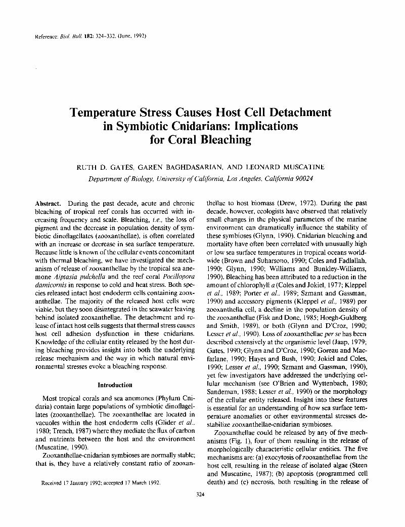
Zooxanthellae could be released by any of five mech- anisms (Fig. l), four of them resulting in the release of morphologically characteristic cellular entities. The five mechanisms are: (a) exocytosis of zooxanthellae from the host cell, resulting in the release of isolated algae (Steen and Muscatine, 1987); (b) apoptosis (programmed cell death) and (c) necrosis, both resulting in the release of
324
CELL DETACHMENT IN SYMBIOTIC CNIDARIANS 325
Host Cell Endoderm Cellular Product Mechanism
EXOCYTOSIS
APOPTOSIS
NECROSIS
PINCHING OFF
Figure 1. A schematic representation of five potential mechanisms by which zooxanthellae could be released from the endoderm of cni- darians, and the cellular entities associated with each mechanism. m, mesoglea; vm, host vacuolar membrane; hn, host cell nucleus; zx, zoox- anthella (shaded for clarity of presentation).
zooxanthellae associated with remnants of the host cell (Searle et al., 1982); (d) pinching off of the distal portion of the host cell, resulting in the release of zooxanthellae surrounded by the vacuolar and pinched off plasma membrane (Glider, 1983); and (e) detachment of endo- derm cells from the host and release of these intact cells containing their complement of zooxanthellae.
Because cnidarians can be readily bleached in the lab- oratory by brief exposure to low (Steen and Muscatine, 1987; Muscatine et al., 1991) or high (Hoegh-Guldberg and Smith, 1989; Glynn and D’Croz, 1990) seawater temperature, the mechanism of bleaching and the mor- phology of the cellular entities released can be investigated experimentally. This approach, together with scanning electron microscopy of endoderm of the Hawaiian sea anemone A. pulchellu after experimental cold shock, re- vealed profiles that were interpreted as evidence of exo- cytosis of zooxanthellae (Steen and Muscatine, i 987). In- deed, examination of the cellular entities released 24 h after cold stress revealed abundant isolated zooxanthellae.
In this paper, we describe the cellular entity released by A. pulchella and the Hawaiian coral Pocillopora dam- icornis immediately after a brief exposure to low or high temperature. P. dumicornis is one of several coral genera that have undergone extensive bleaching in the tropical eastern Pacific during the elevated temperature of the El Niiio-southern oscillation event (Glynn, 1990), and during upwelling and seasonal low temperatures (see Discussion in Glynn and D’Croz, 1990; see also Walker et al., 1982). Both species can be bleached in the laboratory, and bleaching is due to a reduction in zooxanthellae popu- lation density (Glynn and D’Croz, 1990; Muscatine et al., 1991). Observations of the cellular entities released at hourly intervals during, and shortly after, both cold and heat stress, showed clearly that temperature stress causes detachment and release of intact endoderm cells contain- ing zooxanthellae. Soon after release, the host cells dis- integrate in the environment, leaving isolated zooxan- thellae.
Materials and Methods
Animal collection and maintenance
A. pulchella and P. damicornis were collected at 1 meter depth on Checker Reef adjacent to the Hawaii Institute of Marine Biology (HIMB), Coconut Island, Oahu, Ha- waii. Habitat temperatures range annually from 2 1-22°C to 26-27°C (Jokiel and Coles, 1977). P. damicornis col- onies were placed in running seawater and used for ex- periments at HIMB within three days of collection. A. pulchellu was transported to the University of California at Los Angeles, maintained in an aquarium at 25°C on a 12 h light/dark regime, and fed twice a week on Artemia nauplii. Prior to experiments, the anemones were starved for 24 h in an incubator (Precision Scientific Model 8) at 25°C on a 12 h light/dark cycle at 40 pmole quanta rn-‘. s-‘.
Temperature treatments
All experiments were carried out in darkness following the protocol of Muscatine et al. ( 199 1).
Cold stress. Individuals of A. pulchella were incubated in Petri dishes (35 X 10 mm) containing 4 ml of 0.45 pm Millipore filtered seawater (MFSW) chilled to 12°C. After 2.5 h, the chilled seawater was removed and replaced with seawater at 25°C. Anemones were maintained at 25°C in an incubator in darkness for 14 h. The cellular entities released to the seawater were then collected and processed as described below.
Small branches of P. dumicornis (2-3 cm length) were removed from each coral colony and placed in beakers containing 25 ml of MFSW chilled to 12°C (for the protein assay, corals were cold stressed at 14°C). After 4 h, the
326
-
7r rp
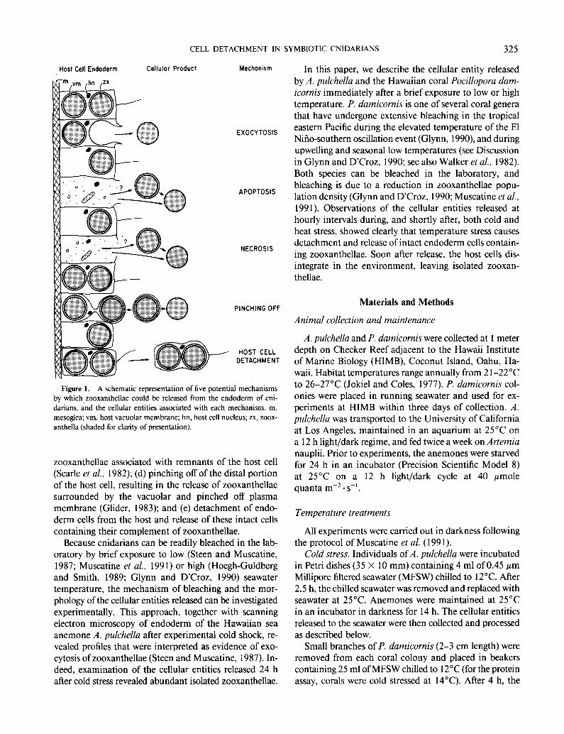
Figure 2. I.eft panel: photomicrographs of the host cells released to the seawater b) .,l;pravn ~~~ic~hella m response to cold stress. stained for wablhty wnh lluoreurcm diacetate (X4000). Right panel: photomlcro- graphs of the host cells released to the seawater b) P~~rlloporu rfumrcorr~rt in response to cold stress. stained with the DNA spwlic fluorochrome Hoechst 33258 (X4000)
branches were immediately transferred to beakers con- taining 25 ml of MFSW at ambient temperature (23- 24°C). The beakers were placed in the seawater tables for ambient temperature control and the coral tissue and sea- water in the beakers was sampled after 12 h. Controls for both species were treated identically to experimental an- imals but were maintained at ambient seawater temper- ature (25’C for .,l. p~lchellu and 23-24°C for P. dutni- cot%) for 16-16.5 h.
Heat sircw. Individuals of .-I. puichella were placed in Petri dishes (35 X 10 mm) containing 4 ml of MFSW warmed to 32°C. Small branches of P. dutnicorni.s (2-3 cm length) were placed in beakers containing 25 ml MFSW pm-heated to 32°C. The animals were maintained at this temperature for up to 16 h. The water surrounding experimental specimens was examined microscopically at hourly intervals and the cellular entity released to the seawater removed and treated as described below. Control animals of both species were maintained at ambient sea- water temperature (25’C for .1. prrl~~lrc~lla and 23-24°C for P. dutnicortzis) over the experimental time period.
Stntnitlg and t~~ti17uore~c~ence tnictmcop~~
The cellular entities released during and after temper- ature stress were collected with a fine bore mouth suction
pipette and deposited onto coverslips coated with poly- L-lysine (0. l’? in distilled water). The entities were stained for viability with the fluorogenic dye fluorcscein diacctate (Sigma Chemical Co.. stock solution 15 mg/ml in acetone; working solution 0.04 ml in 9.96 ml 0. I hit sodium phos- phate, 3% sodium chloride, 0.004? calcium chloride, pH 7.4). The coverslips were rinsed twice in phosphate buffer. mounted and viewed under epifluorescence with an Olympus BH-2 microscope. Non-specific esterases in vi- able cells hydrolyze non-polar fluorescein diacetate to po- lar molecular fluorescein (Schupp and Erlandsen, 1987). Additional coverslips were treated for 30 min with the DNA specific fluorochrome Hoechst 33258 (Reynolds et ~11.. 1986: Sigma Chemical Co., stock solution 5 mg/ml in distilled water: working solution 0.04 ml stock in 9.96 ml 0.1 &l sodium phosphate. 3% sodium chloride, 0.004% calcium chloride). The coverslips were dipped in phos- phate butfer, mounted, and viewed with epifluorescence microscopy.
The cellular entities released after cold stress were com- pared to isolated endoderm cells obtained by maceration of control anemones and corals. .+I. ptdcklla tissue was macerated using 0.05% collagenase (Type 1. Sigma Chemical Co.) and P. datnicotxis tissue was dissociated using calcium-free artificial seawater (Gates and Musca- tine, 1992). Endoderm cells released by maceration and the cellular entities released to the seawater as a result of temperature stress were collected with a mouth pipette and transferred onto poly-L-lysinc coated covcrslips. The coverslips were immersed in 3% glutaraldehyde in 0.1 ;2[ sodium cacodylate buffer (pH 7.4) for I h, rinsed twice in 0.1 ;\I sodium cacodylate buffer and post-fixed in I % osmium tetroxide in 0.1 J/ sodium cacodylate for 30 min. After dehydration in 30, 50, 70. 90. 95. and 100% (X3) ethanol. the coverslips and attached cells were im- mersed in hexamethyldisilazane (Applied Sciences. Inc.) for 5 min (Nation. 1983) dried in air. and then mounted on aluminum stubs. The stubs were coated with gold and viewed on a Cambridge 360 scanning electron microscope. with an accelerating voltage of 10 kV.
For transmission electron microscopy, the endoderm cells released by maceration of P. dutnicortzis tissue and the cellular entities released as a result of temperature stress were collected and centrifuged (Eppendorf model 5414, full speed for 30 s) in microfuge tubes (Gilson. 1.5 ml). The pellets were fixed as described for scanning elec- tron microscopy. After partial dehydration by sequential 30 min treatments in 30. 50. and 70%) ethanol. the 709 ethanol was drained from the tube and immediately re- placed with 2% agar. After the agar solidified, the tube was cut away from the agar plug containing either cold-
CELL DETACHMENT IN SYMBIOTIC CNIDARIANS 327
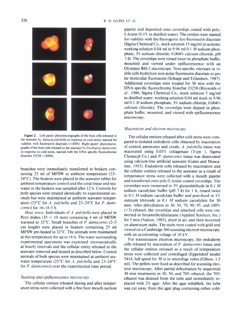
Figure 3. Scanning electron micrographs of individual host cells released by Aiptasiu puichellu (A) and Pocillopora damicornis (B) in response to cold stress, and those obtained from A. p&he/la (C) and P. damicornis (D) by tissue maceration. Bar = 1 pm.
stressed or macerated cells and dehydration completed through 90,95, and 100% (X3) ethanol. The preparations were embedded via propylene oxide into epoxy resin (Spurr). Thin sections were cut using a Sorvall 6000 ul- tramicrotome, stained with lead acetate, and viewed on a JEOL transmission electron microscope.
Protein determination
To investigate the loss of animal protein to the seawater as a result of temperature stress, the seawater was removed from the Petri dishes of cold stressed and control A. pul- chella and homogenized in a teflon-glass tissue grinder. Sodium dodecyl sulphate (SDS, 1% in seawater) was added to each homogenate to a final concentration of 0.05%
(modified from McAuley, 1986). For P. damicornis a 4 ml sub-sample was removed from 25 ml seawater samples, homogenized, and treated with SDS as described above. Each sample was incubated at room temperature for 45 min to solubilize protein in the seawater and host cell membranes associated with released algae. The algae were pelleted by centrifugation (Damon/IEC model HN-S for 4 min at 3000 rpm) and the supernatant put aside for protein analysis as described below. Each algal pellet was resuspended in a known volume of MFSW and the total number of algae assessed using a hemacytometer.
Two 1 ml samples were removed from each supernatant and the amount of protein assessed spectrophotometri- tally using the method of Hartree (1972). To ensure that protein in the seawater samples was animal in origin and
328 R. D. GATES ET AL
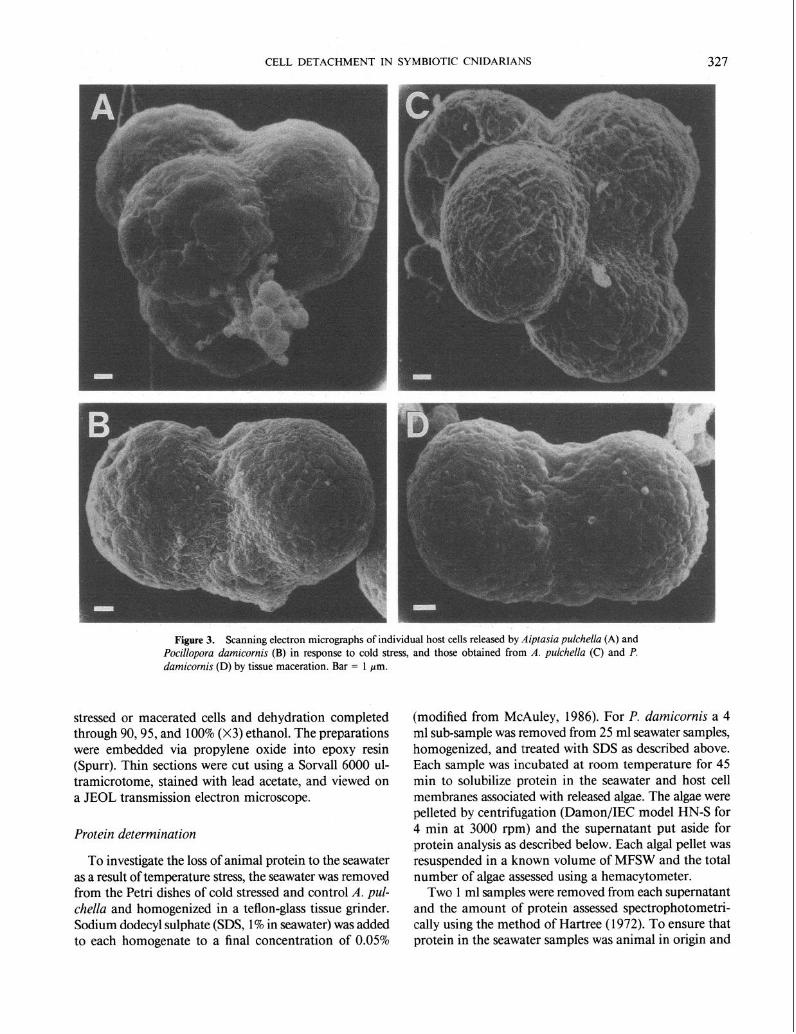
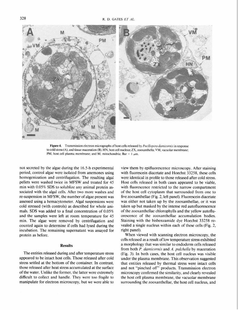
Figure 4. Transmission electron micrographs of host cells released by Pocillopora damicornis in response to cold stress (A), and tissue maceration (B). HN, host cell nucleus; ZX, zooxanthella; VM, vacuolar membrane; PM, host cell plasma membrane; and M, mitochondria; Bar = 1 pm.
not secreted by the algae during the 16.5-h experimental view them by epifluorescence microscopy. After staining with fluorescein diacetate and Hoechst 33258, these cells were identical in profile to those released after cold stress. Host cells released in both cases appeared to be viable, with fluorescence restricted to the narrow compartment of the host cell cytoplasm that surrounded from one to five zooxanthellae (Fig. 2, left panel). Fluorescein diacetate was either not taken up by the zooxanthellae, or it was taken up but masked by the intense red autofluorescence of the zooxanthellae chlorophylls and the yellow autoflu- orescence of the zooxanthellae accumulation bodies. Staining with the bisbenzamide dye Hoechst 33258 re- vealed a single nucleus within each of these cells (Fig. 2, right panel).
period, control algae were isolated from anemones using homogenization and centrifugation. The resulting algal pellets were washed twice in MFSW and treated for 45 min with 0.05% SDS to solublize any animal protein as- sociated with the algal cells. After two more washes and re-suspension in MFSW, the number of algae present was assessed using a hemacytometer. Algal suspensions were cold stressed (with controls) as described for whole ani- mals. SDS was added to a final concentration of 0.05% and the samples were left at room temperature for 45 min. The algae were removed by centrifugation and counted again to determine if cells had lysed during the incubation. The remaining supernatant was assayed for protein as before.
Results
The entities released during and after temperature stress appeared to be intact host cells. Those released after cold stress settled at the bottom of the container. In contrast, those released after heat stress accumulated at the surface of the water. Unlike the former, the latter were extremely difficult to collect and handle. They were too fragile to manipulate for electron microscopy, but we were able to
When viewed with scanning electron microscopy, the cells released as a result of low temperature stress exhibited a morphology that was similar to endoderm cells released from both P. damicornis and A. pulchella by maceration (Fig. 3). In both cases, the host cell nucleus was visible under the plasma membrane.. This observation suggested that entities released by thermal stress were intact cells and not “pinched off” products. Transmission electron microscopy confirmed the similarity, and clearly revealed the host cell plasma membrane, the vacuolar membrane surrounding the zooxanthellae, the host cell nucleus, and
CELL DETACHMENT IN SYMBIOTIC CNIDARIANS 329
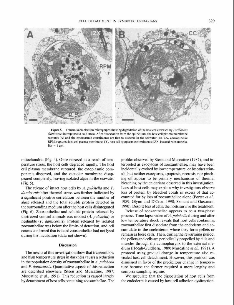
Figure 5. Transmission electron micrographs showing degradation of the host cells released by Pocilloporu damicornis in response to cold stress. After dissociation from the epithelium, the host cell plasma membrane ruptures (A) and the cytoplasmic constituents are free to disperse in the seawater (B). ZX, zooxanthella; RPM, ruptured host cell plasma membrane; CC, host cell cytoplasmic constituents; IZX, isolated zooxanthella. Bar = 1 pm.
mitochondria (Fig. 4). Once released as a result of tem- perature stress, the host cells degraded rapidly. The host cell plasma membrane ruptured, the cytoplasmic com- ponents dispersed, and the vacuolar membrane disap- peared completely, leaving isolated algae in the seawater (Fig. 5).
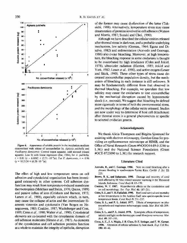
The release of intact host cells by A. pulchellu and P. dumicornis after thermal stress was further indicated by a significant positive correlation between the number of algae released and the total soluble protein detected in the surrounding medium after the host cells disintegrated (Fig. 6). Zooxanthellae and soluble protein released by unstressed control animals was modest (A. pulchellu) or negligible (P. dumicornis). Protein released by isolated zooxanthellae was below the limits of detection, and cell counts confirmed that isolated zooxanthellae had not lysed during the incubation (data not shown).
Discussion
The results of this investigation show that transient low and high temperature stress in darkness causes a reduction in the population density of zooxanthellae in A. pulchellu and P. dumicornis. Quantitative aspects of this reduction are described elsewhere (Steen and Muscatine, 1987; Muscatine et al., 1991). This reduction is caused largely by detachment of host cells containing zooxanthellae. The
profiles observed by Steen and Muscatine (1987), and in- terpreted as exocytosis of zooxanthellae, may have been incidentally evoked by low temperature, or by other stim- uli, but neither exocytosis, apoptosis, necrosis, nor pinch- ing off appear to be primary mechanisms of thermal bleaching by the cnidarians observed in this investigation. Loss of host cells may explain why investigators observe loss of protein by bleached corals in excess of that ac- counted for by loss of zooxanthellae alone (Porter et al., 1989; Glynn and D’Croz, 1990; Szmant and Gassman, 1990). Despite loss of cells, the hosts survive the treatment.
Release of zooxanthellae appears to be a two-phase process. Time-lapse video of A. pulchellu during and after low temperature shock reveals that host cells containing zooxanthellae first dissociate from the endoderm and ac- cumulate in the coelenteron where they form pellets or remain as loose cells. Then, during the rewarming period, the pellets and cells are periodically propelled by cilia and muscles through the actinopharynx to the external me- dium (Hoegh-Guldberg, 1989; Muscatine et al., 199 1). A protocol using gradual change in temperature also re- vealed host cell detachment. However, this protocol was dismissed in favor of the precipitous change in tempera- ture because the former required a more lengthy and complex sampling regime.
We speculate that the dissociation of host cells from the endoderm is caused by host cell adhesion dysfunction.
330 R. D. GATES ET AL.
I I ”
,oo Aiptasia pulchella
0 1 2 3 4
No. zooxanthellae released (x 16 )
4M) Pooillopora damicornis
I s 350 2
No. of zooxanthellae released (x 106)
Figure 6. Appearance of soluble protein in the incubation medium concomitant with release of zooxanthellae by Aiptasiu pulchellu and Pocillopora damicornis. Control (open squares), cold stressed (closed squares). Line fit with linear regression (Zar, 1984) for A. pulchella, r = 0.81 (y = 6.6012 + (2.71. 10~‘)~). For P. dumicornis, r = 0.96 (y = 10.2124 + (6.28. W5)x).
The effect of high and low temperature stress on cell adhesion and cytoskeletal organization has been investi- gated extensively in other systems. Cell adhesion dys- function may result from temperature-induced membrane thermotropism (Melchior and Steim, 1976; Quinn, 1989) and passive influx of ions (Grisham and Barnett, 1973; Larsen et al., 1988), especially calcium which, in turn, may cause the collapse of actin and the intermediate fil- aments vimentin and cytokeratin (Van Bergen en He- negouwen, 1985; Coakley, 1987; Wachsberger and Goss, 1989; Cress et al., 1990; Walter et al., 1990). Cytoskeletal elements are co-located with the cytoplasmic domain of cell adhesion molecules (Hirano et al., 1987). As elements of the cytoskeleton and cell adhesion proteins function as a whole to maintain the integrity of epithelia, disruption
of the former may cause dysfunction of the latter (Tak- eichi, 1988). Alternatively, temperature stress may cause denaturation of proteins involved in cell adhesion (Watson and Morris, 1987; Suzuki and Choi, 1990).
Although we have described the cellular entities released after thermal stress in darkness, and a probable underlying mechanism, low salinity (Goreau, 1964; Egana and Di- salvo, 1982) and sedimentation (Acevedo and Goenaga, 1986) also evoke bleaching. Moreover, at high tempera- ture, the bleaching response in some cnidarians is thought to be exacerbated by high u-radiance (Coles and Jokiel, 1978) ultraviolet radiation (Harriot, 1985; Jokiel and York, 1982; Lesser er al., 1990) and active oxygen (Lesser and Shick, 1990). These other types of stress cause de- creased zooxanthellae population density, but the mech- anism of bleaching in each instance is still unknown. It may be fundamentally different from that observed in thermal bleaching. For example, we speculate that low salinity may cause the cnidarians to lose zooxanthellae by the mechanical disruption caused by hypoosmotic shock (i.e., necrosis). We suggest that bleaching be defined more rigorously in terms of both the environmental stress, and the morphology of the cellular entity released. Studies are now under way to determine if host cell detachment alter thermal stress is a general phenomenon or specific to selected cnidarian genera.
Acknowledgments
We thank Alicia Thompson and Birgitta Sjostrand for assisting with electron microscopy, Gordon Grau for pro- viding an epifluorescence microscope at HIMB, and the Office of Naval Research (Grant #NO00 14-89-J-3246 to L.M.) and the National Science Foundation (Grant #OCE-8723090 to L.M.) for research support.
Literature Cited
Acevedo, R., and C. Goenaga. 1986. Note on coral bleaching after a chronic flooding in southwestern Puerto Rico. Curibb. J. Sci. 22: 225.
Brown, B. E., and Suharsono. 1990. Damage and recovery of coral reefs affected by El Nino related seawater warming in the Thousand Islands, Indonesia. Coral Reefs 8: 63-170.
Coakley, W. T. 1987. Hyperthermia effects on the cytoskeleton and on cell morphology. Sot. Exp. Biol. 41: 187-2 I 1.
Coles, S. J., and Y. H. Fadtallah. 1990. Reefcoral survival and mortality at low temperatures in the Arabian Gulf: new species-specific lower temperature limits. Coral Reefs 9: 231-237.
Coles, S. L., and P. L. Jokiel. 1977. Effects of temperature on pho- tosynthesis and respiration in hermatypic corals. Mar. Biol. 43: 209- 216.
Coles, S. L., and P. L. Jokiel. 1978. Synergistic effects of temperature, salinity and light on the hermatypic coral Monfiporu verrucosa. Mar. Biol. 49: 187-195.
Cress, A. E., J. A. Majda, J. R. Glass, D. E. Stringer, and E. W. Gerner. 1990. Alteration of cellular adhesion by heat shock. Exp. Cell Res. 190: 40-46.
CELL DETACHMENT IN SYMBIOTIC CNIDARIANS 331
Drew, E. A. 1972. The biology and physiology of alga-invertebrate symbioses. II. The density of symbiotic algal cells in a number of herrnatypic corals and alcyonarians from various depths. J. Exp. Mar. Biol. Ecol. 9: 11-75.
Egana, A. C., and L. H. DiSalvo. 1982. Mass expulsion of zooxanthellae by Easter Island corals. Pac. Sci. 36: 6 l-63.
Fisk, T. A., and T. J. Done. 1985. Taxonomic and bathymetric patterns of bleaching in corals, Myrmidon Reef (Queensland). Proc. Ftfth Int. Coral Reef Cong. 6: I49- 154.
Gates, R. D. 1990. Seawater temperature and sublethal coral bleaching in Jamaica. Coral Reefs 8: 193-198.
Gates, R. D., and L. Muscatine. 1992. Three methods for isolating viable anthozoan endoderm cells with their intracellular symbiotic dinoflagellates. Coral Reefs (in press).
Glider, W. V. 1983. The biology of the association of Symbiodinium microadriaticum with Aiptasia pallida: an anemone-alga symbiosis. Ph.D. Thesis, University of Nebraska. 102 pp.
Glider, W. V., D. Phipps, and R. L. Pardy. 1980. Localization of sym- biotic dinotlagellate cells within tentacle tissue of Aiptasia pallida (Coelenterata, Anthozoa). Trans. Am. Microsc. Sot. 99: 426-438.
Glynn, P. 1990. Coral mortality and disturbances to coral reefs in the tropical eastern Pacific. F’p. 55- 126 in Global Ecological Consequences of the 1982-83 El-Nino Southern Oscillation, P. W. Glynn, ed. El- sevier, Amsterdam.
Glynn, P., and L. D’Croz. 1990. Experimental evidence for high tem- perature stress as the cause of El Nino-coincident coral mortality. Coral Reef 8: 181-192.
Goreau, T. F. 1964. Mass expulsion of zooxanthellae from Jamaican reef communities after Hurricane Flora. Science 145: 383-386.
Goreau, T. J., and A. H. Macfarlane. 1990. Reduced growth rate of Montastrea annuluris following the 1987-1988 coral-bleaching event. Coral Reefs 8: 211-216.
Grisham, C. M., and R. E. Barnett. 1973. The role of lipid-phase tran- sitions in the regulation of the (sodium + potassium) adenosine tri- phosphatase. Biochemistry 12: 2635-2637.
Harriot, V. J. 1985. Mortality rates of scleractinian corals before and during a mass bleaching event. Mar. Ecol. Prog. Ser. 21: 81-88.
Hartree, E. F. 1972. Determination of protein: a modification of the Lowry method that gives a linear photometric response. Anal. Biochem. 48: 422-427.
Hayes, R. L., and P. G. Bush. 1990. Microscopic observations of recovery in the reef-building scleractinian coral, Montastrea annu- luris, after bleaching on a Cayman reef. Coral Reefs 8: 203- 209.
Hirano, S., A. Nose, K. Hat@ A. Kawakami, and M. Takeichi. 1987. Calcium-dependent cell-cell adhesion molecules (Cadherins): subclass specificities and possible involvement of actin bundles. J. Cell Biol. 105: 2501-2510.
Hoegh-Guldberg, 0.1989. The regulatory biology of plant-animal en- dosymbiosis. Ph.D. Thesis, University of California at Los Angeles. 309 pp.
Hoegh-Guldberg, O., and G. J. Smith. 1989. The effect of sudden changes in temperature, light, and salinity on population density and export of zooxanthellae from the reef corals Seriatopora hystrix and Stylophora pistillata. J. Exp. Mar. Biol. Ecol. 129: 279- 303.
Jaap, W. 1979. Observations on zooxanthellae expulsion at Middle Sambo Reef, Florida Keys. Bull. Mar. Sci. 29: 4 14-422.
Jokiel, P. L., and S. L. Coles. 1977. Effects of temperature on the mortality and growth of Hawaiian reef corals. Mar. Biol. 43: 201- 208.
Jokiel, P. L., and S. L. Coles. 1990. Response of Hawaiian and other Indo-Pacific reef corals to elevated temperature. Coral Reefs 8: 155- 162.
Jokiel, P. L., and R. H. York. 1982. Solar ultraviolet photobiology of the reef coral Pocillopora dumicornis and symbiotic zooxanthellae. Bull. Mar. Sci. 32: 301-315.
Kleppel, G. S., R. E. Dodge, and C. J. Reese. 1989. Changes in pig- mentation associated with the bleaching of stony corals. Limnol. Oceanogr. 34: 1331-1335.
Larsen, T., S. Solberg, R. Johansen, and L. Jorgensen. 1988. Effect of cooling in the intracellular concentrations of Na+, K+ and Cl- in cultured human endothelial cells. Scund. J. Lab. Invest. 48: 565- 571.
Lesser, M. P., and J. M. Shick. 1990. Photoadaptation and defenses against oxygen toxicity in zooxanthellae from natural populations of symbiotic cnidarians. J. Exp. Mar. Biol. Ecol. 134: 129- 141.
Lesser, M. P., W. R. Stochaj, D. W. Tapley, and J. M. Shick. 1990. Bleaching in coral reef anthozoans: effects of irradiance, ul- traviolet radiation, and temperature on the activities of protective enzymes against active oxygen. Coral Reefs 8: 225-232.
McAuley, P. J. 1986. Isolation of viable uncontaminated Chlorella from green hydra. Limnol. Oceanogr. 31: 222-224.
Melchior, D., and J. M. Steim. 1976. Thermotropic transitions in bio- membranes. Ann. Rev. Biophys. Bioeng. 5: 205-238.
Muscatine, L. 1990. The role of symbiotic algae in carbon and energy flux in reef corals. Pp. 75-87 in Coral Reef, Z. Dubinsky, ed. Elsevier, Amsterdam.
Muscatine, L., D. Grossman, and J. Doino. 1991. Release of symbiotic algae by tropical sea anemones and corals after cold shock. Mar. Ecol. Prog. Ser. 77: 233-243.
Nation, J. L. 1983. A new method using hexamethyldisilazane for preparation of soft insect tissue for scanning electron microscopy. Stain. Technol. 56: 341-35 I.
O’Brien, T. L., and C. R. Wyttenbach. 1980. Some effects of temper- ature on the symbiotic association between zoochlorellae (Chloro- phyceae) and the sea anemone Anthopleura xanthogrammica. Trans. Am. Microsc. Sot. 99: 22 l-225.
Porter, J. W., W. K. Fitt, H. J. Spero, C. S. Rogers, and M. W. White. 1989. Bleaching in reef corals: physiological and stable isotope re- sponses. Proc. Natl. Acad. Sci. USA 86: 9342-9346.
Quinn, P. J. 1989. Principles of membrane stability and phase behavior under extreme conditions. J. Bioenergetics Biomembranes 21: 3-19.
Reynolds, C. P., A. T. Black, and J. N. Woody. 1986. Sensitive method for detecting viable cells seeded into bone marrow. Cancer Res. 46: 5878-588 I.
Sandeman, I. M. 1988. Coral bleaching at Discovery Bay, Jamaica: a possible mechanism for temperature-related bleaching. Pp. 46-48 in Mass Bleaching of Corals in the Caribbean: A Research Strategy. Research report 88-2, J. Ogden and R. Wicklund, eds. NOAA Un- dersea Research Program, Rockville, MD.
Scbupp, D. G., and S. L. Erlandsen. 1987. A new method to determine Giardiu cyst viability: correlation of fluorescein diacetate and pro- pidium iodide staining and animal infectivity. Appt. Environ. Micro- biol. 53: 704-707.
Searle, J., J. F. R. Kerr, and C. J. Bishop. 1982. Necrosis and apoptosis: distinct modes of cell death with fundamentally different significance. Pathol. Annu. 17: 229-259.
Steen, R. G., and L. Muscatine. 1987. Low temperature evokes rapid exocytosis of symbiotic algae by a sea anemone. Biol. Bull. 172: 246- 263.
Suzuki, M., and B. H. Cboi. 1990. The behavior of the extracellular matrix and the basal lamina during the repair of cryogenic injury in the adult rat cerebral cortex. Acta. Neuropathol. 80: 355- 361.
332 R. D. GATES ET AL.
Szmant, A., and N. J. Gassman. 1990. The effects of prolonged “bleaching” on the tissue biomass and reproduction of the reef coral Montastrea annularis. Coral Reefs 8: 2 I l-224.
Trench, R. K. 1987. Dinoflagellates in non-parasitic symbiosis. Pp. 530- 570 in Biology of Dinoflagellates, F. J. R. Taylor, ed. Blackwell, Ox- ford.
Takeichi, M. 1988. The cadherins: cell-cell adhesion molecules con- trolling animal morphogenesis. Development 102: 639-655.
Van Bergen en Henegouwen, P. M. P, W. J. R. M. Jordii G. Van Dongen, F. C. S. Ramaekers, H. Amesz, and W. A. M. Linnemans. 1985. Studies on a possible relationship between alterations in the cytoskeleton and induction of heat shock protein synthesis in mam- malian cell. Int. .I. Hyperthermia 11: 69-83.
Wachsberger, P. R., and R. A. Coss. 1989. Acrylamide sensitization of the heat response of the cytoskeleton and cytotoxicity in attaching
and well-spread synchronous Chinese hamster ovary cells. Cell Mo- tility Cytoskel. 13: 61-82.
Walker, N. D., H. H. Roberts, L. J. Rouse, Jr., and 0. K. Huh. 1982. Thermal history of reef-associated environments during a record cold-air outbreak event. Coral Reefs 1: 83-87.
Walter, M. F., N. S. Petersen, and H. Biessman. 1990. Heat shock causes the collapse of the intermediate filament cytoskeleton in Dro- sophila embryos. Dev. Genet. 1 I: 270-219.
Watson, P. F., and G. J. Morris. 1987. Cold shock injury in animal cells. Symp. Sot. Exp. Biol. 41: 31 I-340.
Williams, E. H., Jr., and L. Bunkley-Williams. 1990. The world-wide coral reef bleaching cycle and related sources of coral mortality. Atoll Res. Bull. 335: I-7 1.
Zar, J. H. 1984. Biostatistical Analysis, 2nd ed. Prentice Hall, New Jersey.
Related Documents


