Temperature dependence of the lipid packing in thylakoid membranes studied by time- and spectrally resolved fluorescence of Merocyanine 540 Sashka B. Krumova a , Rob B.M. Koehorst a,b , Attila Bóta c , Tibor Páli d , Arie van Hoek a,b , Győző Garab e , Herbert van Amerongen a,b, ⁎ a Wageningen University, Laboratory of Biophysics, P.O. Box 8128, 6700 ET Wageningen, The Netherlands b MicroSpectroscopy Centre, Wageningen University, P.O. Box 8128, 6700 ET Wageningen, The Netherlands c Department of Physical Chemistry, Budapest University of Technology and Economics H-1521, Budapest, Hungary d Institute of Biophysics, Biological Research Center, Hungarian Academy of Sciences P.O. Box 521, Szeged H-6701, Hungary e Institute of Plant Biology, Biological Research Center, Hungarian Academy of Sciences P.O. Box 521, Szeged H-6701, Hungary abstract article info Article history: Received 28 July 2008 Received in revised form 18 September 2008 Accepted 18 September 2008 Available online 26 September 2008 Keywords: Merocyanine 540 Lipid packing Thylakoid membrane The lipid packing of thylakoid membranes is an important factor for photosynthetic performance. However, surprisingly little is known about it and it is generally accepted that the bulk thylakoid lipids adopt the liquid-crystalline phase above − 30 °C and that a phase transition occurs only above 45 °C. In order to obtain information on the nature of the lipid microenvironment and its temperature dependence, steady-state and time-resolved fluorescence measurements were performed on the fluorescence probe Merocyanine 540 (MC540) incorporated in isolated spinach thylakoids and in model lipid systems (dipalmitoyl phosphati- dylcholine and dioleoyl phosphatidylethanolamine) adopting different phases. It is demonstrated that the degree and way of incorporation differs for most lipid phases – upon selective excitation at 570 nm, the amplitude of the fluorescence component that corresponds to membrane-incorporated MC540 is about 20% in gel-, 60% in rippled gel-, and 90% in liquid-crystalline and inverted hexagonal phase, respectively. For thylakoids, the data reveal hindered incorporation of MC540 (amplitude about 30% at 7 °C) and marked spectral heterogeneity at all temperatures. The incorporation of MC540 in thylakoids strongly depends on temperature. Remarkably, above 25 °C MC540 becomes almost completely extruded from the lipid environment, indicating major rearrangements in the membrane. © 2008 Elsevier B.V. All rights reserved. 1. Introduction Many physiological processes that take place in the photosynthetic thylakoid membrane as well as a variety of its properties have been demonstrated to depend on temperature. These include oxygen evolution [1], CO 2 assimilation, non-photochemical quenching, aggre- gation of the major light-harvesting complex LHCII [2], photophos- phorylation [2,3], expression of proteins [4], lipid-protein ratio [5] and lipid–protein interactions ([6] and references therein), membrane permeability [7–9], electron transport activity [10–14], proton efflux [15] and the rate of NADP-reduction [16]. The temperature depen- dences of some of these processes and properties were correlated with (or related to) the mobility of the chloroplast-membrane lipids as measured by EPR spin-label spectroscopy [17–19]. Abrupt changes in the membrane fluidity were also reported at 10–12 °C [20]. It is known that the lipid packing and phase behavior are essential for the operation of violaxanthin de-epoxidase [21–23] and that they are modulated by the xanthophyll cycle pigments solubilized in the bilayer [24]. They might also play a significant role in providing the optimal conditions for diffusion of hydrophobic molecules such as plastoquinol [25,26] or integral protein complexes (e.g. during state transitions [27,28]). It is generally accepted that the bulk lipid mixture in the thylakoid lamellae exists in the liquid crystalline phase (Lα) above − 30 °C [29] and no phase transition is expected to occur up to about 45 °C; above this temperature the thylakoid lipids segregate from the membrane and form non-bilayer structures [30,31]. This, however, does not exclude the possibility of changes in the lipid packing and/or formation of membrane domains with distinct physical properties at lower temperatures. Since the thylakoid membranes contain about 70–80% proteins, and only 20–30% lipids, the lipid packing might also be influenced by changes in the macro-organization of the protein complexes, as indeed indicated by temperature- and light-induced reorganizations in the LHCII-containing chiral macrodomains (see e.g. [32]). The packing of biological membranes in general, as well as the Biochimica et Biophysica Acta 1778 (2008) 2823–2833 ⁎ Corresponding author. Wageningen University, Laboratory of Biophysics, P.O. Box 8128, 6700 ET Wageningen, The Netherlands. Tel.: +31 317 482634. E-mail address: [email protected] (H. van Amerongen). 0005-2736/$ – see front matter © 2008 Elsevier B.V. All rights reserved. doi:10.1016/j.bbamem.2008.09.007 Contents lists available at ScienceDirect Biochimica et Biophysica Acta journal homepage: www.elsevier.com/locate/bbamem

Welcome message from author
This document is posted to help you gain knowledge. Please leave a comment to let me know what you think about it! Share it to your friends and learn new things together.
Transcript
Biochimica et Biophysica Acta 1778 (2008) 2823–2833
Contents lists available at ScienceDirect
Biochimica et Biophysica Acta
j ourna l homepage: www.e lsev ie r.com/ locate /bbamem
Temperature dependence of the lipid packing in thylakoid membranes studied bytime- and spectrally resolved fluorescence of Merocyanine 540
Sashka B. Krumova a, Rob B.M. Koehorst a,b, Attila Bóta c, Tibor Páli d, Arie van Hoek a,b,Győző Garab e, Herbert van Amerongen a,b,⁎a Wageningen University, Laboratory of Biophysics, P.O. Box 8128, 6700 ET Wageningen, The Netherlandsb MicroSpectroscopy Centre, Wageningen University, P.O. Box 8128, 6700 ET Wageningen, The Netherlandsc Department of Physical Chemistry, Budapest University of Technology and Economics H-1521, Budapest, Hungaryd Institute of Biophysics, Biological Research Center, Hungarian Academy of Sciences P.O. Box 521, Szeged H-6701, Hungarye Institute of Plant Biology, Biological Research Center, Hungarian Academy of Sciences P.O. Box 521, Szeged H-6701, Hungary
⁎ Corresponding author. Wageningen University, Lab8128, 6700 ET Wageningen, The Netherlands. Tel.: +31 3
E-mail address: [email protected] (H.
0005-2736/$ – see front matter © 2008 Elsevier B.V. Aldoi:10.1016/j.bbamem.2008.09.007
a b s t r a c t
a r t i c l e i n f oArticle history:
The lipid packing of thylako Received 28 July 2008Received in revised form 18 September 2008Accepted 18 September 2008Available online 26 September 2008Keywords:Merocyanine 540Lipid packingThylakoid membrane
id membranes is an important factor for photosynthetic performance. However,surprisingly little is known about it and it is generally accepted that the bulk thylakoid lipids adopt theliquid-crystalline phase above −30 °C and that a phase transition occurs only above 45 °C. In order to obtaininformation on the nature of the lipid microenvironment and its temperature dependence, steady-state andtime-resolved fluorescence measurements were performed on the fluorescence probe Merocyanine 540(MC540) incorporated in isolated spinach thylakoids and in model lipid systems (dipalmitoyl phosphati-dylcholine and dioleoyl phosphatidylethanolamine) adopting different phases. It is demonstrated that thedegree and way of incorporation differs for most lipid phases – upon selective excitation at 570 nm, theamplitude of the fluorescence component that corresponds to membrane-incorporated MC540 is about 20%in gel-, 60% in rippled gel-, and 90% in liquid-crystalline and inverted hexagonal phase, respectively. Forthylakoids, the data reveal hindered incorporation of MC540 (amplitude about 30% at 7 °C) and markedspectral heterogeneity at all temperatures. The incorporation of MC540 in thylakoids strongly depends ontemperature. Remarkably, above 25 °C MC540 becomes almost completely extruded from the lipidenvironment, indicating major rearrangements in the membrane.
© 2008 Elsevier B.V. All rights reserved.
1. Introduction
Many physiological processes that take place in the photosyntheticthylakoid membrane as well as a variety of its properties have beendemonstrated to depend on temperature. These include oxygenevolution [1], CO2 assimilation, non-photochemical quenching, aggre-gation of the major light-harvesting complex LHCII [2], photophos-phorylation [2,3], expression of proteins [4], lipid-protein ratio [5] andlipid–protein interactions ([6] and references therein), membranepermeability [7–9], electron transport activity [10–14], proton efflux[15] and the rate of NADP-reduction [16]. The temperature depen-dences of some of these processes and properties were correlatedwith (or related to) themobility of the chloroplast-membrane lipids asmeasured by EPR spin-label spectroscopy [17–19]. Abrupt changes inthe membrane fluidity were also reported at 10–12 °C [20].
oratory of Biophysics, P.O. Box17 482634.van Amerongen).
l rights reserved.
It is known that the lipid packing and phase behavior are essentialfor the operation of violaxanthin de-epoxidase [21–23] and that theyare modulated by the xanthophyll cycle pigments solubilized in thebilayer [24]. They might also play a significant role in providing theoptimal conditions for diffusion of hydrophobic molecules such asplastoquinol [25,26] or integral protein complexes (e.g. during statetransitions [27,28]).
It is generally accepted that the bulk lipid mixture in the thylakoidlamellae exists in the liquid crystalline phase (Lα) above −30 °C [29]and no phase transition is expected to occur up to about 45 °C; abovethis temperature the thylakoid lipids segregate from the membraneand form non-bilayer structures [30,31]. This, however, does notexclude the possibility of changes in the lipid packing and/orformation of membrane domains with distinct physical properties atlower temperatures. Since the thylakoid membranes contain about70–80% proteins, and only 20–30% lipids, the lipid packing might alsobe influenced by changes in the macro-organization of the proteincomplexes, as indeed indicated by temperature- and light-inducedreorganizations in the LHCII-containing chiral macrodomains (see e.g.[32]). The packing of biological membranes in general, as well as the
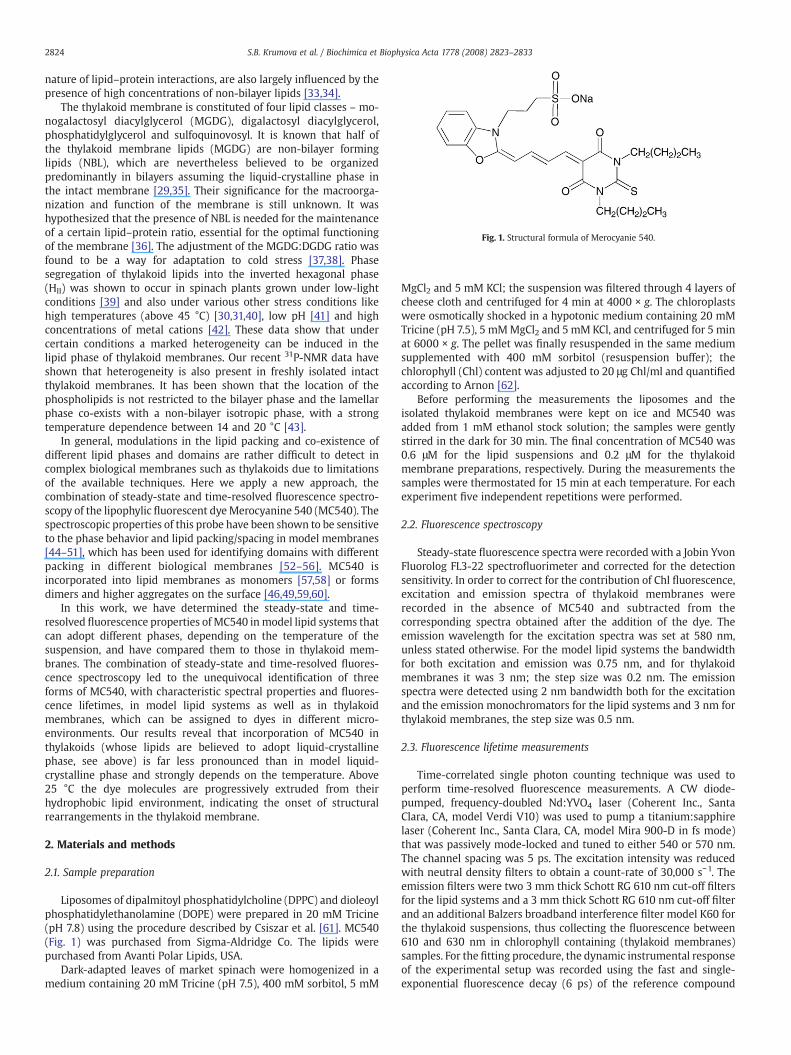
Fig. 1. Structural formula of Merocyanie 540.
2824 S.B. Krumova et al. / Biochimica et Biophysica Acta 1778 (2008) 2823–2833
nature of lipid–protein interactions, are also largely influenced by thepresence of high concentrations of non-bilayer lipids [33,34].
The thylakoid membrane is constituted of four lipid classes – mo-nogalactosyl diacylglycerol (MGDG), digalactosyl diacylglycerol,phosphatidylglycerol and sulfoquinovosyl. It is known that half ofthe thylakoid membrane lipids (MGDG) are non-bilayer forminglipids (NBL), which are nevertheless believed to be organizedpredominantly in bilayers assuming the liquid-crystalline phase inthe intact membrane [29,35]. Their significance for the macroorga-nization and function of the membrane is still unknown. It washypothesized that the presence of NBL is needed for the maintenanceof a certain lipid–protein ratio, essential for the optimal functioningof the membrane [36]. The adjustment of the MGDG:DGDG ratio wasfound to be a way for adaptation to cold stress [37,38]. Phasesegregation of thylakoid lipids into the inverted hexagonal phase(HII) was shown to occur in spinach plants grown under low-lightconditions [39] and also under various other stress conditions likehigh temperatures (above 45 °C) [30,31,40], low pH [41] and highconcentrations of metal cations [42]. These data show that undercertain conditions a marked heterogeneity can be induced in thelipid phase of thylakoid membranes. Our recent 31P-NMR data haveshown that heterogeneity is also present in freshly isolated intactthylakoid membranes. It has been shown that the location of thephospholipids is not restricted to the bilayer phase and the lamellarphase co-exists with a non-bilayer isotropic phase, with a strongtemperature dependence between 14 and 20 °C [43].
In general, modulations in the lipid packing and co-existence ofdifferent lipid phases and domains are rather difficult to detect incomplex biological membranes such as thylakoids due to limitationsof the available techniques. Here we apply a new approach, thecombination of steady-state and time-resolved fluorescence spectro-scopy of the lipophylic fluorescent dyeMerocyanine 540 (MC540). Thespectroscopic properties of this probe have been shown to be sensitiveto the phase behavior and lipid packing/spacing in model membranes[44–51], which has been used for identifying domains with differentpacking in different biological membranes [52–56]. MC540 isincorporated into lipid membranes as monomers [57,58] or formsdimers and higher aggregates on the surface [46,49,59,60].
In this work, we have determined the steady-state and time-resolved fluorescence properties of MC540 inmodel lipid systems thatcan adopt different phases, depending on the temperature of thesuspension, and have compared them to those in thylakoid mem-branes. The combination of steady-state and time-resolved fluores-cence spectroscopy led to the unequivocal identification of threeforms of MC540, with characteristic spectral properties and fluores-cence lifetimes, in model lipid systems as well as in thylakoidmembranes, which can be assigned to dyes in different micro-environments. Our results reveal that incorporation of MC540 inthylakoids (whose lipids are believed to adopt liquid-crystallinephase, see above) is far less pronounced than in model liquid-crystalline phase and strongly depends on the temperature. Above25 °C the dye molecules are progressively extruded from theirhydrophobic lipid environment, indicating the onset of structuralrearrangements in the thylakoid membrane.
2. Materials and methods
2.1. Sample preparation
Liposomes of dipalmitoyl phosphatidylcholine (DPPC) and dioleoylphosphatidylethanolamine (DOPE) were prepared in 20 mM Tricine(pH 7.8) using the procedure described by Csiszar et al. [61]. MC540(Fig. 1) was purchased from Sigma-Aldridge Co. The lipids werepurchased from Avanti Polar Lipids, USA.
Dark-adapted leaves of market spinach were homogenized in amedium containing 20 mM Tricine (pH 7.5), 400 mM sorbitol, 5 mM
MgCl2 and 5 mM KCl; the suspension was filtered through 4 layers ofcheese cloth and centrifuged for 4 min at 4000 × g. The chloroplastswere osmotically shocked in a hypotonic medium containing 20 mMTricine (pH 7.5), 5 mMMgCl2 and 5 mM KCl, and centrifuged for 5 minat 6000 × g. The pellet was finally resuspended in the same mediumsupplemented with 400 mM sorbitol (resuspension buffer); thechlorophyll (Chl) content was adjusted to 20 μg Chl/ml and quantifiedaccording to Arnon [62].
Before performing the measurements the liposomes and theisolated thylakoid membranes were kept on ice and MC540 wasadded from 1 mM ethanol stock solution; the samples were gentlystirred in the dark for 30 min. The final concentration of MC540 was0.6 μM for the lipid suspensions and 0.2 μM for the thylakoidmembrane preparations, respectively. During the measurements thesamples were thermostated for 15 min at each temperature. For eachexperiment five independent repetitions were performed.
2.2. Fluorescence spectroscopy
Steady-state fluorescence spectra were recorded with a Jobin YvonFluorolog FL3-22 spectrofluorimeter and corrected for the detectionsensitivity. In order to correct for the contribution of Chl fluorescence,excitation and emission spectra of thylakoid membranes wererecorded in the absence of MC540 and subtracted from thecorresponding spectra obtained after the addition of the dye. Theemission wavelength for the excitation spectra was set at 580 nm,unless stated otherwise. For the model lipid systems the bandwidthfor both excitation and emission was 0.75 nm, and for thylakoidmembranes it was 3 nm; the step size was 0.2 nm. The emissionspectra were detected using 2 nm bandwidth both for the excitationand the emission monochromators for the lipid systems and 3 nm forthylakoid membranes, the step size was 0.5 nm.
2.3. Fluorescence lifetime measurements
Time-correlated single photon counting technique was used toperform time-resolved fluorescence measurements. A CW diode-pumped, frequency-doubled Nd:YVO4 laser (Coherent Inc., SantaClara, CA, model Verdi V10) was used to pump a titanium:sapphirelaser (Coherent Inc., Santa Clara, CA, model Mira 900-D in fs mode)that was passively mode-locked and tuned to either 540 or 570 nm.The channel spacing was 5 ps. The excitation intensity was reducedwith neutral density filters to obtain a count-rate of 30,000 s−1. Theemission filters were two 3 mm thick Schott RG 610 nm cut-off filtersfor the lipid systems and a 3 mm thick Schott RG 610 nm cut-off filterand an additional Balzers broadband interference filter model K60 forthe thylakoid suspensions, thus collecting the fluorescence between610 and 630 nm in chlorophyll containing (thylakoid membranes)samples. For the fitting procedure, the dynamic instrumental responseof the experimental setup was recorded using the fast and single-exponential fluorescence decay (6 ps) of the reference compound
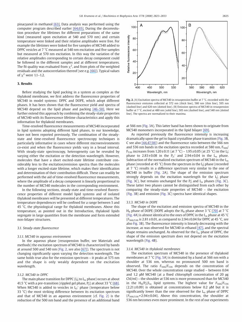
Fig. 2. (A) Excitation spectra of MC540 in resuspension buffer at 7 °C, recorded with thefluorescence emission collected at 572 nm (thick line), 580 nm (thin line), 595 nm(dashed line) and 620 nm (dotted line). (B) Emission spectra of MC540 in resuspensionbuffer at 7 °C, excited at 480 nm (solid line), 505 nm (dashed line), and 540 nm (dottedline). The spectra are normalized to their maxima.
2825S.B. Krumova et al. / Biochimica et Biophysica Acta 1778 (2008) 2823–2833
pinacyanol in methanol [63]. Data analysis was performed using thecomputer program described earlier [64,65]. During the deconvolu-tion procedure the lifetimes for different preparations of the samekind (measured upon excitation at 540 and 570 nm) and certaintemperature were linked and their relative amplitudes were free. Forexample the lifetimes were linked for five samples of MC540 added toDPPC vesicles at 7 °C measured at 540 nm excitation and five samplesbut measured at 570 nm excitation. In this way the variation of therelative amplitudes corresponding to certain decay component couldbe followed in the different samples and at different temperatures.The fit quality was evaluated from χ2, and from plots of the weightedresiduals and the autocorrelation thereof (see e.g. [66]). Typical valuesof χ2 were 1.1–1.2.
3. Results
Before studying the lipid packing in a system as complex as thethylakoid membrane, we first address the fluorescence properties ofMC540 in model systems: DPPC and DOPE, which adopt differentphases. It has been shown that the fluorescence yield and spectra ofMC540 depend on the lipid phase and packing [46–50]. Here wefurther extend this approach by combining the steady-state propertiesof MC540 with its fluorescence lifetime characteristics and apply thisinformation for thylakoid membranes.
Time-resolved fluorescence measurements of MC540 incorporatedin lipid systems adopting different lipid phases, to our knowledge,have not been reported previously. The combination of the steady-state and time-resolved fluorescence spectroscopy techniques isparticularly informative in cases where different microenvironmentsco-exist and when the fluorescence yields vary in a broad interval.With steady-state spectroscopy, heterogeneity can be revealed byvarying either the excitation or the detection wavelength. However,molecules that have a short excited-state lifetime contribute con-siderably less to the excitation/emission spectra than the moleculeswith a longer excited-state lifetime, which makes their identificationand determination of their contribution difficult. These can readily beperformed with the aid of time-resolved fluorescence measurements,where the amplitude of a decay component is linearly proportional tothe number of MC540 molecules in the corresponding environment.
In the following sections, steady-state and time-resolved fluores-cence properties of different model lipid systems and of isolatedthylakoid membranes will be presented at different temperatures. Thetemperature dependences will be confined for a range between 5 and45 °C, the physiological range for thylakoid membranes. Above thistemperature, as pointed out in the Introduction, thylakoid lipidssegregate in large quantities from the membrane and form extendednon-bilayer structures.
3.1. Steady-state fluorescence
3.1.1. MC540 in aqueous environmentIn the aqueous phase (resuspension buffer, see Materials and
methods) the excitation spectrum of MC540 is characterized by bandsat around 500 and 540 nm (Fig. 2, see also [67]). The spectrum is notchanging significantly upon varying the detection wavelength. Thesame holds true also for the emission spectrum – it peaks at 575 nmand the shape is only weakly dependent on the excitationwavelength.
3.1.2. MC540 in DPPCThemain phase transition for DPPC (Lβ′ to Lα phase) occurs at about
41.5 °C with a pre-transition (rippled gel phase, Pβ′) at about 33 °C [68].When MC540 is added to vesicles in Lβ′ phase (temperature below33 °C) the most striking difference between its excitation spectrumand that of MC540 in an aqueous environment (cf. Fig. 2) is thereduction of the 500 nm band and the presence of an additional band
at 566 nm (Fig. 3A). This latter band has been shown to originate fromMC540 monomers incorporated in the lipid bilayer [69].
As reported previously the fluorescence intensity is increasingdramatically upon the gel to liquid crystalline phase transition (Fig. 3B,C see also [44,47,58]) and the fluorescence ratio between the 566 nmand 536 nm bands in the excitation spectra recorded at 580 nm, F566/F536 increases from 1.20±0.11 (at 7 °C) – 1.95±0.05 (at 25 °C) in the Lβ′phase to 2.83±0.08 in the Pβ′ and 2.94±0.04 in the Lα phase.Subtraction of the normalized excitation spectrum of MC540 in the Lαphase (recorded at 45 °C) from the spectrum in the Lβ′ phase (recordedat 7 °C) results in a difference spectrum very similar to the one ofMC540 in buffer (Fig. 2A). The shape of the emission spectrumstrongly depends on the excitation wavelength for the Lβ′ phase(Fig. 3C), but remains unchanged for the Pβ′ and Lα phases (Fig. 3C).These latter two phases cannot be distinguished from each other bycomparing the steady-state properties of MC540 – the excitation(Fig. 3B) and emission (Fig. 3C) spectra have identical shapes.
3.1.3. MC540 in DOPEThe shape of the excitation and emission spectra of MC540 in the
HII phase of DOPE (DOPE adopts the HII phase above 3 °C [70] at 7 °C(Fig. 4A) is almost identical to the ones of DPPC in the Lα phase at 45 °C(F566/536 is 2.81±0.01, as compared to 2.94±0.04 for DPPC at 45 °C, seealso Fig. 3B). The fluorescence intensity is linearly decreasing with theincrease, as was observed for MC540 in ethanol [47], and the spectralshape remains unchanged. As observed for the Lα phase of DPPC, theshape of the emission spectrum does not depend on the excitationwavelength (Fig. 4B).
3.1.4. MC540 in thylakoid membranesThe excitation spectrum of MC540 in the presence of thylakoid
membranes at 7 °C (Fig. 5A) is dominated by a band at 566 nmwith ashoulder at 536 nm, whereas no pronounced 500 nm band isobserved. The ratio F566/F536 depends on the concentration ofMC540. Over the whole concentration range studied – between 0.04and 1.2 μM MC540 (at a fixed chlorophyll concentration of 20 μgChl/ml) – the shoulder at 536 nm is more pronounced than for MC540in the HII/Pβ′/Lα lipid systems. The highest value for F566/F536(2.21±0.09) is obtained at concentrations below 0.2 μM but it issignificantly lower than the one observed for the Lα phase of DPPC(F566/536=2.94±0.04). Above this concentration, the shoulder at536 nm becomes evenmore prominent. In the rest of our experiments
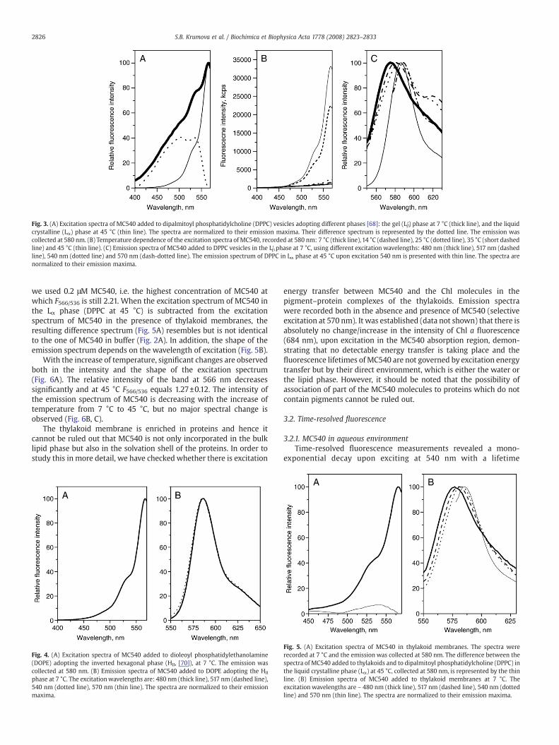
Fig. 3. (A) Excitation spectra of MC540 added to dipalmitoyl phosphatidylcholine (DPPC) vesicles adopting different phases [68]: the gel (Lβ′) phase at 7 °C (thick line), and the liquidcrystalline (Lα) phase at 45 °C (thin line). The spectra are normalized to their emission maxima. Their difference spectrum is represented by the dotted line. The emission wascollected at 580 nm. (B) Temperature dependence of the excitation spectra of MC540, recorded at 580 nm: 7 °C (thick line), 14 °C (dashed line), 25 °C (dotted line), 35 °C (short dashedline) and 45 °C (thin line). (C) Emission spectra of MC540 added to DPPC vesicles in the Lβ′ phase at 7 °C, using different excitation wavelengths: 480 nm (thick line), 517 nm (dashedline), 540 nm (dotted line) and 570 nm (dash-dotted line). The emission spectrum of DPPC in Lα phase at 45 °C upon excitation 540 nm is presented with thin line. The spectra arenormalized to their emission maxima.
2826 S.B. Krumova et al. / Biochimica et Biophysica Acta 1778 (2008) 2823–2833
we used 0.2 μM MC540, i.e. the highest concentration of MC540 atwhich F566/536 is still 2.21. When the excitation spectrum of MC540 inthe Lα phase (DPPC at 45 °C) is subtracted from the excitationspectrum of MC540 in the presence of thylakoid membranes, theresulting difference spectrum (Fig. 5A) resembles but is not identicalto the one of MC540 in buffer (Fig. 2A). In addition, the shape of theemission spectrum depends on the wavelength of excitation (Fig. 5B).
With the increase of temperature, significant changes are observedboth in the intensity and the shape of the excitation spectrum(Fig. 6A). The relative intensity of the band at 566 nm decreasessignificantly and at 45 °C F566/536 equals 1.27±0.12. The intensity ofthe emission spectrum of MC540 is decreasing with the increase oftemperature from 7 °C to 45 °C, but no major spectral change isobserved (Fig. 6B, C).
The thylakoid membrane is enriched in proteins and hence itcannot be ruled out that MC540 is not only incorporated in the bulklipid phase but also in the solvation shell of the proteins. In order tostudy this in more detail, we have checked whether there is excitation
Fig. 4. (A) Excitation spectra of MC540 added to dioleoyl phosphatidylethanolamine(DOPE) adopting the inverted hexagonal phase (HII, [70]), at 7 °C. The emission wascollected at 580 nm. (B) Emission spectra of MC540 added to DOPE adopting the HII
phase at 7 °C. The excitationwavelengths are: 480 nm (thick line), 517 nm (dashed line),540 nm (dotted line), 570 nm (thin line). The spectra are normalized to their emissionmaxima.
energy transfer between MC540 and the Chl molecules in thepigment–protein complexes of the thylakoids. Emission spectrawere recorded both in the absence and presence of MC540 (selectiveexcitation at 570 nm). It was established (data not shown) that there isabsolutely no change/increase in the intensity of Chl a fluorescence(684 nm), upon excitation in the MC540 absorption region, demon-strating that no detectable energy transfer is taking place and thefluorescence lifetimes of MC540 are not governed by excitation energytransfer but by their direct environment, which is either the water orthe lipid phase. However, it should be noted that the possibility ofassociation of part of the MC540 molecules to proteins which do notcontain pigments cannot be ruled out.
3.2. Time-resolved fluorescence
3.2.1. MC540 in aqueous environmentTime-resolved fluorescence measurements revealed a mono-
exponential decay upon exciting at 540 nm with a lifetime
Fig. 5. (A) Excitation spectra of MC540 in thylakoid membranes. The spectra wererecorded at 7 °C and the emission was collected at 580 nm. The difference between thespectra of MC540 added to thylakoids and to dipalmitoyl phosphatidylcholine (DPPC) inthe liquid crystalline phase (Lα) at 45 °C, collected at 580 nm, is represented by the thinline. (B) Emission spectra of MC540 added to thylakoid membranes at 7 °C. Theexcitation wavelengths are – 480 nm (thick line), 517 nm (dashed line), 540 nm (dottedline) and 570 nm (thin line). The spectra are normalized to their emission maxima.
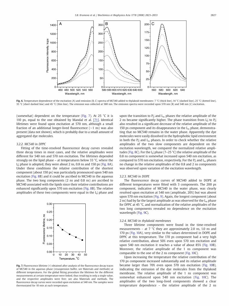
Fig. 6. Temperature dependence of the excitation (A) and emission (B, C) spectra of MC540 added to thylakoid membranes: 7 °C (thick line), 14 °C (dashed line), 25 °C (dotted line),35 °C (short dashed line) and 45 °C (thin line). The emission was collected at 580 nm. The emission spectra were recorded upon 570 nm (B) and 540 nm (C) excitation.
2827S.B. Krumova et al. / Biochimica et Biophysica Acta 1778 (2008) 2823–2833
(somewhat) dependent on the temperature (Fig. 7). At 25 °C it is110 ps, equal to the one obtained by Mandal et al. [71]. Identicallifetimes were found upon excitation at 570 nm, although a smallfraction of an additional longer-lived fluorescence (∼1 ns) was alsopresent (data not shown), which is probably due to a small amount ofaggregated dye molecules.
3.2.2. MC540 in DPPCFitting of the time-resolved fluorescence decay curves revealed
three decay times in most cases, and the relative amplitudes weredifferent for 540 nm and 570 nm excitation. The lifetimes dependedstrongly on the lipid phase – at temperatures below 33 °C, where theLβ′ phase is adopted, they were about 2 ns, 0.8 ns and 150 ps (Fig. 8A).Under these conditions the relative contribution of the shortestcomponent (about 150 ps) was particularly pronounced upon 540 nmexcitation (Fig. 8B) and it could be ascribed to MC540 in the aqueousphase. The two long components (2 ns and 0.8 ns) are ascribed toMC540 associated with the lipids since their relative contributions areenhanced significantly upon 570 nm excitation (Fig. 8B). The relativeamplitudes of these two components were equal in the Lβ′ phase and
Fig. 7. Fluorescence lifetime (τ) obtained after analysis of the fluorescence decay tracesof MC540 in the aqueous phase (resuspension buffer, see Materials and methods) atdifferent temperatures. For the global fitting procedure the lifetimes for the differentmeasurements at certain temperature were linked, thus resulting in only a single value,and the respective amplitudes were free; see also Materials and methods. Thefluorescence decay curves were recorded upon excitation at 540 nm. The samples werethermostated for 10 min at each temperature.
upon the transition to Pβ′ and Lα phases the relative amplitude of the2 ns became significantly higher. The phase transition from Lβ′ to Pβ′also resulted in a significant decrease of the relative amplitude of the150 ps component and its disappearance in the Lα phase, demonstra-ting that no MC540 remains in the water phase. Apparently the dyemolecules were easily dissolved in the hydrophobic lipid environmentin both the Pβ′ and Lα phases. In order to check whether the relativeamplitudes of the two slow components are dependent on theexcitation wavelength, we compared the normalized relative ampli-tudes (Fig. 8C). For the Lβ′ phase (7–25 °C) the relative amplitude of the0.8 ns component is somewhat increased upon 540 nm excitation, ascompared to 570 nm excitation, respectively. For the Pβ′ and Lα phasesno change in the relative amplitudes of the 0.8 and 2 ns componentswas observed upon variation of the excitation wavelength.
3.2.3. MC540 in DOPEThe fluorescence decay curves of MC540 added to DOPE at
different temperatures were fitted with 3 components. The 200 pscomponent, indicative of MC540 in the water phase, was clearlyresolved upon excitation at 540 nm (amplitude, 20%) but was absentupon 570 nm excitation (Fig. 9). Again, the longest component (about2 ns) had by far the largest amplitude as was observed for the Lα phasefor DPPC at 45 °C, and normalization of the relative amplitudes of thetwo long components revealed no dependence on the excitationwavelength (Fig. 9C).
3.2.4. MC540 in thylakoid membranesThree lifetime components were found in the time-resolved
measurements – at 7 °C they are approximately 2.0 ns, 1.0 ns and170 ps (Fig. 10A), very similar to the values determined in DOPE andDPPC at this temperature. The 170 ps component had a very highrelative contribution, about 50% even upon 570 nm excitation andupon 540 nm excitation it reaches a value of about 85% (Fig. 10B).Moreover, the relative amplitude of the 1 ns component wascomparable to the one of the 2 ns component (Fig. 10C).
Upon increasing the temperature the relative contribution of the170 ps component increased substantially and its relative amplitudebecame larger than 70% even upon 570 nm excitation (Fig. 10B),indicating the extrusion of the dye molecules from the thylakoidmembrane. The relative amplitude of the 1 ns component wassomewhat enhanced upon 540 nm excitation (Fig. 10C). Theamplitudes of the two long-lived components showed a cleartemperature dependence – the relative amplitude of the 2 ns
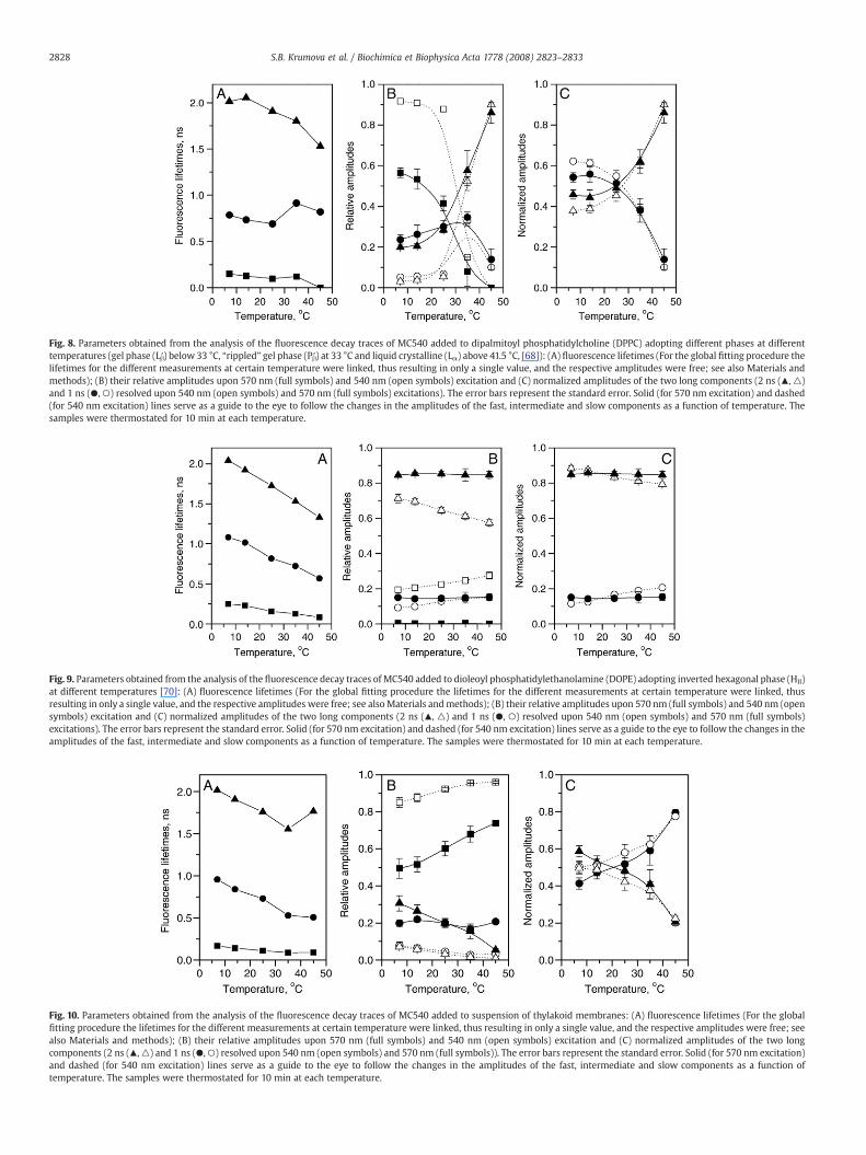
Fig. 8. Parameters obtained from the analysis of the fluorescence decay traces of MC540 added to dipalmitoyl phosphatidylcholine (DPPC) adopting different phases at differenttemperatures (gel phase (Lβ′) below 33 °C, “rippled” gel phase (Pβ′) at 33 °C and liquid crystalline (Lα) above 41.5 °C, [68]): (A) fluorescence lifetimes (For the global fitting procedure thelifetimes for the different measurements at certain temperature were linked, thus resulting in only a single value, and the respective amplitudes were free; see also Materials andmethods); (B) their relative amplitudes upon 570 nm (full symbols) and 540 nm (open symbols) excitation and (C) normalized amplitudes of the two long components (2 ns (▴, △)and 1 ns (●, ○) resolved upon 540 nm (open symbols) and 570 nm (full symbols) excitations). The error bars represent the standard error. Solid (for 570 nm excitation) and dashed(for 540 nm excitation) lines serve as a guide to the eye to follow the changes in the amplitudes of the fast, intermediate and slow components as a function of temperature. Thesamples were thermostated for 10 min at each temperature.
Fig. 9. Parameters obtained from the analysis of the fluorescence decay traces of MC540 added to dioleoyl phosphatidylethanolamine (DOPE) adopting inverted hexagonal phase (HII)at different temperatures [70]: (A) fluorescence lifetimes (For the global fitting procedure the lifetimes for the different measurements at certain temperature were linked, thusresulting in only a single value, and the respective amplitudes were free; see also Materials andmethods); (B) their relative amplitudes upon 570 nm (full symbols) and 540 nm (opensymbols) excitation and (C) normalized amplitudes of the two long components (2 ns (▴, △) and 1 ns (●, ○) resolved upon 540 nm (open symbols) and 570 nm (full symbols)excitations). The error bars represent the standard error. Solid (for 570 nm excitation) and dashed (for 540 nm excitation) lines serve as a guide to the eye to follow the changes in theamplitudes of the fast, intermediate and slow components as a function of temperature. The samples were thermostated for 10 min at each temperature.
Fig. 10. Parameters obtained from the analysis of the fluorescence decay traces of MC540 added to suspension of thylakoid membranes: (A) fluorescence lifetimes (For the globalfitting procedure the lifetimes for the different measurements at certain temperature were linked, thus resulting in only a single value, and the respective amplitudes were free; seealso Materials and methods); (B) their relative amplitudes upon 570 nm (full symbols) and 540 nm (open symbols) excitation and (C) normalized amplitudes of the two longcomponents (2 ns (▴,△) and 1 ns (●,○) resolved upon 540 nm (open symbols) and 570 nm (full symbols)). The error bars represent the standard error. Solid (for 570 nm excitation)and dashed (for 540 nm excitation) lines serve as a guide to the eye to follow the changes in the amplitudes of the fast, intermediate and slow components as a function oftemperature. The samples were thermostated for 10 min at each temperature.
2828 S.B. Krumova et al. / Biochimica et Biophysica Acta 1778 (2008) 2823–2833
2829S.B. Krumova et al. / Biochimica et Biophysica Acta 1778 (2008) 2823–2833
component decreased with the increase of temperature, most promi-nently above 25 °C.
4. Discussion
In this work, in order to obtain information on the lipid packing inthylakoid membranes, we have performed time-resolved and steady-state spectroscopy measurements on thylakoids and model mem-branes. This combination of fluorescence measurements, which to ourknowledge is applied for the first time in this systematic manner, hasrevealed the existence of different microenvironments in differentmodel systems. These data, in turn, can help us to assess the lipidicmicroenvironments in the thylakoid membranes. These types ofmeasurements may thus complement the information for the lipidphase of the thylakoid membranes at physiological temperatures, i.e.before the break-down of the membrane impermeability and thesegregation of the lipids from the membrane, and the denaturation ofproteins. It is believed that the bulk lipid mixture in thylakoidmembranes exists in liquid crystalline phase [29] and, althoughmarked temperature dependences were reported (see Introduction),phase transitions were detected only above 45 °C [30,31]. Also labelingof thylakoid membranes with the fluorescent probe DPH [72] and theEPR probe TEMPO [40] showed a monotonous increase in the fluidityof themembrane upon increasing the temperature from 10 °C to 45 °C.In contrast, our previous work pointed towards a more complexbehavior and organization/packing of the lipid molecules; 31P-NMRmeasurements showed a change in the bulk phospholipid behaviorbetween 7 °C and 20 °C and revealed the presence of small non-bilayerstructures in thylakoids co-existing with the lipid bilayer at alltemperatures [43]. Here we show that steady-state and time-resolvedMC540 measurements also reveal different microenvironments forthe dye in model lipid systems and in intact thylakoids in thephysiological range of temperatures; these will be discussed below.
4.1. MC540 in different lipid systems
The fluorescence spectra of MC540 have already been shown to besensitive to the lipid packing and phase behavior of different modellipid systems [46,47,49,50]. In short, it was established that thefluorescence yield is substantially increased upon relocation of MC540from aqueous to lipidic environment and there is a red-shift in theabsorption and fluorescence spectra [44,58,73]; moreover the transi-tion from gel to liquid crystalline and from liquid crystalline toinverted hexagonal phases were also correlated with higher fluores-cence yields [47,50]. Here we further extend this knowledge byexploring the excitation wavelength dependence of the spectralshapes of MC540 in different environments.
In buffer solution the excitation spectrum is very similar to theabsorption spectrum of MC540 in water [49,58,73–75] and does notchange substantially upon detection at different wavelengths (Fig. 2A).Correspondingly the emission spectrum also changes only slightlywith the variation of the excitation wavelength (Fig. 2B), reflecting arather homogeneous dye solution. It was reported that the spectralproperties of MC540 depend on the presence of salts [76]. Under ourexperimental conditions no change in the spectral properties ofMC540 was observed when solved in the buffer used for thepreparation of the model lipid systems (20 mM Tricine, pH 7.5) orthe one for the thylakoid membranes (20 mM Tricine, pH 7.5, 400 mMsorbitol, 5 mM MgCl2, 5 mM KCl).
In contrast to MC540 free in the water phase, for MC540 in the Lβ′phase a strong wavelength-dependence was observed – the emissionmaximum varies between 575 nm and 589 nm (Fig. 3C), revealing alarge heterogeneity in the MC540 population, i.e. the existence ofdifferent microenvironments for the dye. Moreover, in accordancewith previous studies [46,49] we also observeMC540 dimer formationin the Lβ′ phase of DPPC, reflected by a distinct fluorescent band at
620 nm (Fig. 3C). The transition to the Pβ′ and Lα phases leads to apronounced change in the shape of both the excitation and emissionspectra – the 566 nm band becomes the dominating one in theexcitation spectra (Fig. 3A, B) and no 620 nm band is observed in theemission spectra (Fig. 3C). The same holds true for the HII phase(Fig. 4). For the Pβ′, Lα and HII phases no heterogeneity can be detectedupon varying the excitation and detectionwavelength, indicating thatthe spectra are largely dominated by one spectral form of MC540,which is attributed to MC540 monomers incorporated into thehydrophobic lipid environment [57,58]. The excitation spectra for Pβ′,Lα and HII phases showed no evidence for the presence of MC540molecules free in the buffer, as shown by the absence of a welldiscernible band at 500 nm; however those could be detected by time-resolved measurements (see below).
Furthermore, our work correlates the steady-state characteristicsof MC540 with its time-resolved fluorescence properties and assignsthe different fluorescence lifetime components to distinct micro-environments of the dye.
In the lipid systems studied here, MC540 exhibits three differentfluorescence lifetime components, which in most cases decrease withthe increase of the temperature, similarly to MC540 in buffer solution(Fig. 7) due to enhanced photoisomerization [67]. However, theirrelative amplitudes depend exclusively on the local environments ofMC540 and on the physical state of the lipid system (Figs. 8B, 9B and10B). The different lifetime components will briefly be discussed.
4.1.1. The short-lifetime component – MC540 in the aqueous phaseThe short-lifetime component (113 ps–214 ps) is found with
different relative amplitudes in the various lipid-containing systems(Figs. 8, 9 and 10) but it is by far the dominating component for MC540in buffer (Fig. 7). In the aqueous buffer phase at 25 °C a value of 110 psis observed, identical to the one determined by Mandal et al. forMC540 in water [71]. In the studied lipid systems its relativecontribution was always higher upon 540 nm than upon 570 nmexcitation (Figs. 8B, 9B and 10B). This is in accordance with theexpectations (as judged by the absorption spectrum of MC540 inaqueous environment, where the absorption at 570 nm is far less thanat 540 nm [49,58,73–75], see also Fig. 2), and the short lifetimecomponent thus represents MC540 molecules which remain free inthe aqueous phase. Therefore, it can also be used as an indicator of theaccessibility of the lipid membranes for MC540. We have establishedthat the Lβ′ phase is particularly inaccessible for the dye molecules(Fig. 8), presumably due to the tight packing of the lipids. In all othermodel lipid phases studied, the short component was not present orhad small amplitude (Figs. 8 and 9).
4.1.2. The long-lifetime components – MC540 interacting with lipidsThe 1 ns and 2 ns components are originating from MC540 in
hydrophobic environments (in the lipid phase) and hence provideinformation about the lipid packing and phase behavior. Theselifetimes might be assigned to two discrete populations of themolecules, reflecting two different microenvironments. It is alsopossible that there is a broad distribution of lifetimes due toincorporation of MC540 in a variety of environments with onlysmall differences in their physical properties (dielectric constant, lipidpacking).
4.1.3. Discrete vs. broad lifetime distributionsAccording to the discrete lifetime distribution model, the two
components should be assigned to MC540 residing in differentenvironments. The 1 and 2 ns lifetimes are somewhat similar to theones obtained by Aramendia et al. [77] for dimyristoyl phospha-tidylcholine vesicles in the Lα phase at 20 °C – 1.87 ns and 410 ps. Itshould be pointed out that these authors did not resolve the 200 pscomponent. Therefore, the 410 ps might be a weighted average of thevalues of 200 ps and 1 ns, which were resolved in the present study.
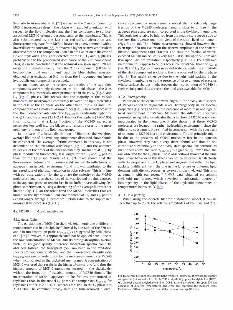
Fig. 11. Average lifetimes obtained from the weighted lifetimes of the two longest decaycomponents (∼2 ns and ∼1 ns) for MC540 in dipalmitoyl phosphatidylcholine (DPPC,●), dioleoyl phosphatidylethanolamine (DOPE, ▴) and thylakoids (■) upon 570 nmexcitation at different temperatures. The error bars represent the standard error.Excitation at 540 nm resulted in essentially the same average lifetimes.
2830 S.B. Krumova et al. / Biochimica et Biophysica Acta 1778 (2008) 2823–2833
Similarly to Aramendia et al. [77] we assign the 2 ns component toMC540 incorporated deep in the bilayer with parallel orientationwithrespect to the lipid molecules and the 1 ns component to surface-associated MC540 oriented perpendicular to the membrane. This isalso substantiated by the fact that red-shifted absorption andfluorescence originate from MC540 located in an environment with alower dielectric constant [58]. Moreover, a higher relative amplitude isobserved for the 1 ns component upon 540 nmexcitation in the case ofLβ′ and thylakoids. This is not detected for the Pβ′, Lα and HII phases,probably due to the pronounced dominance of the 2 ns component.Thus, it can be concluded that the red-most emission upon 570 nmexcitation originates mainly from the 2 ns component (MC540 inhydrophobic lipid environment) and the blue shifted emissionobtained after excitation at 540 nm from the 1 ns component (morehydrophilic environment), respectively.
As mentioned above the relative amplitudes of the two slowcomponents are strongly dependent on the lipid phase – the 2 nscomponent is substantially more pronounced in the Pβ′, Lα (Fig. 8) andHII (Fig. 9) phases. This reveals that the majority of the MC540molecules are incorporated completely between the lipid molecules.In the case of the Lβ′ phase on the other hand, the 2 ns and 1 nscomponents have almost equal amplitudes (Fig. 8). In accordancewiththis, the fluorescence intensity ratio F566/536 is significantly higher forthe Pβ′, Lα and HII phases (2.81–2.94) than for the Lβ′ phase (1.20–1.95),thus indicating that a large fraction of the MC540 moleculespenetrates less well into the bilayer and remains closer to the morepolar environment of the lipid headgroups.
In the case of a broad distribution of lifetimes, the weightedaverage lifetime of the two long components discussed above shouldbe considered. The average lifetime estimated in this way is notdependent on the excitation wavelength (Fig. 11) and the obtainedvalues are of the order of the ones obtained by Onganer et al. [67] byphase modulation fluorometry. It is longer for the HII and Lα phasesthan for the Lβ′ phase. Mandal et al. [71] have shown that thefluorescence lifetime and quantum yield are significantly lower inaqueous than in polar environment and this was attributed to theincreased rate of photoizomerization in polar solvents. This is in linewith our observations – for the Lβ′ phase the majority of the MC540molecules remains on the surface of the vesicles and are thus exposedto the aqueous phase or remain free in the buffer phase, allowing fastphotoisomerization, causing a shortening of the average fluorescencelifetime (Fig. 11). On the other hand the MC540 molecules that areburied in the hydrophobic lipid environment of HII and Lα phasesexhibit longer average fluorescence lifetimes due to the suppressednon-radiative processes (Fig. 11).
4.2. MC540 in thylakoid membranes
4.2.1. AccessibilityThe partitioning of MC540 in the thylakoid membrane at different
temperatures can in principle be followed by the ratio of the 570 nmand 530 nm absorption peaks (A570/540), as suggested by Bakalchevaet al. [78]. However, this approach could not be applied here – due tothe low concentration of MC540 and its strong absorption overlapwith Chl no good quality difference absorption spectra could beobtained. Instead, the fingerprint (566 nm band in the excitationspectra) for monomeric MC540, and the fluorescence intensity ratioF566/536 was used in order to probe the microenvironments of MC540when incorporated in the thylakoid membranes. A concentration ofMC540 was used that results in the highest F566/536 ratio (and thus thehighest amount of MC540 monomers located in the thylakoids)without the formation of sizeable amounts of MC540 dimers. Theincorporation of MC540 appeared to be far less pronounced inthylakoids than in the model Lα phase. For comparison F566/536 forthylakoids at 7 °C is 2.21±0.09, whereas for DPPC in the Lα phase it is2.94±0.04. The combined steady-state and time-resolved fluores-
cence spectroscopy measurements reveal that a relatively largefraction of the MC540 molecules remains close to or free in theaqueous phase and are not incorporated in the thylakoid membrane.This could not reliably be inferred from the steady-state spectra due tothe low fluorescence quantum yield of the short-lived component(Figs. 5 and 10). The lifetime measurements, showed however thateven upon 570 nm excitation the relative amplitude of the shortestlifetime component (100–200 ps), and thus the fraction of water-exposed MC540 molecules is very high – it is 50% upon 570 nm and85% upon 540 nm excitation, respectively (Fig. 10B). The thylakoidmembrane thus appear to be less accessible for MC540 than the Lα, Pβ′(Fig. 8) and HII (Fig. 9) phases in model systems, while the amplitudeof the short component is close to the one observed for the Lβ′ phase(Fig. 8). This might either be due to the tight lipid packing in thethylakoid membrane or to the presence of large amount of proteinswhose surface charges might prevent the incorporation of MC540 intheir vicinity and thus decrease the lipid area available for MC540.
4.2.2. HeterogeneityVariation of the excitation wavelength in the steady-state spectra
of MC540 added to thylakoids reveal heterogeneity in its spectralproperties (Fig. 5C) and thus the presence of more than one type oflocal environment for MC540. Moreover, the difference spectrumpresented in Fig. 5A also indicates that a fraction of MC540 is not wellincorporated in the membrane. It also shows that these MC540molecules are located in a rather hydrophilic environment since thedifference spectrum is blue-shifted in comparison with the spectrumof monomeric MC540 in a lipid environment. This, in principle, mightbe due to the presence of MC540 molecules free in the aqueousphase. However, they have a very short lifetime and thus do notcontribute substantially to the steady-state spectra. Furthermore, asmentioned above the ratio F566/F536 is significantly lower than theone observed for the Lα phase. These observations show that the bulklipid phase behavior in thylakoids can not be described satisfactorilywith the properties of the Lα phase and suggests that either the lipidpacking is different from the one in the Lα phase or different lipiddomains with distinct properties co-exist in the thylakoid. This is inagreement with our recent 31P-NMR data, obtained on spinachthylakoid membranes, which revealed a substantial degree ofheterogeneity in the lipid phases of the thylakoid membranes attemperatures below 45 °C [43].
4.2.3. Lipid packingWhen using the discrete lifetime distribution model, it can be
seen that up to 25 °C the relative amplitudes of the 1 ns and 2 ns
2831S.B. Krumova et al. / Biochimica et Biophysica Acta 1778 (2008) 2823–2833
components are almost equal (Fig. 10C), resembling very much thesituation for the Lβ′ phase (Fig. 8C). Above 25 °C, the 1 ns componentbecomes the dominant one (Fig. 10C). This was not observed for anyof the model lipid phases studied, and it indicates the onset of arearrangement of the bulk lipid matrix of the thylakoids – theMC540 which was buried inside the membrane becomes extrudedfrom this hydrophobic environment and hence localized in anenvironment with higher water content. This conclusion is corrobo-rated with the changes in the steady-state spectral propertiesbetween 25 °C and 45 °C – a strong reduction of the intensity ofthe 566 nm peak (hydrophobic environment), as compared to the536 nm band (Fig. 6).
The temperature dependence of the average lifetime of the twolong components (Fig.11) does not correspond to the ones determinedfor model lipid systems in different phases. The nature of this effect isstill unclear; the high protein content might influence the MC540fluorescence lifetime to some extent. It is also worth mentioning thatthe temperature dependence of the average lifetime for thylakoidmembranes is linear and thus does not reveal any major phasetransition. However, it does reveal rather complex behavior of the bulklipid matrix in the thylakoid membrane, since it differs from themono-phasic model lipid systems. It has been shown that the decreasein A570/540 is proportional to the increase of the dielectric constant[78]. In the present experiments the significant decrease in F566/536 forthylakoids between 7 and 45 °C should be regarded as a decrease inthe A570/540, which thus indicates relocation of MC540 to anenvironment with higher dielectric constant. Taking into accountalso the strong reduction of the fluorescence intensity upon anincrease in temperature (Fig. 6), it can be concluded that MC540 isextruded from the hydrophobic lipidmembrane at high temperatures.The observed changes in the excitation spectra of MC540 in thesuspension of thylakoid membranes with the increase of temperaturestrongly resemble the absorption changes observed by Mateasik et al.induced by increasing the negative surface charge of model lipidmembranes [79]. Their data show that electrostatic repulsion preventsthe penetration of MC540 into the lipidmembrane. The surface chargeof thylakoids is also negative and it has been shown to arise mainlyfrom the exposed charged amino acids of the membrane proteins[80,81]. Thus a rearrangement of the protein complexes and/or thelipid molecules upon the increase of temperature might lead to moresurface-exposed charges and consequently lead to extrusion ofMC540from the membrane. This hypothesis is further supported by the dataof Dobrikova et al. [82] showing that in the temperature range20–30 °C changes in the permanent dipole moment (transversalcharge asymmetry) and the electric polarizability (motion of chargesin the diffuse electric double layer) of pea thylakoid membrane occur,which have also been shown to strongly depend on the supramole-cular organization of LHCII [83].
5. Conclusion
The steady-state and time-resolved spectroscopic data shown heredemonstrate that in a system as complex as the thylakoid membranethe lipophylic fluorescent probe MC540 is exposed to differentmicroenvironments and thus the thylakoid membrane can not bedescribed satisfactorily with the properties of simple, mono-phasicmodel lipid systems. This conclusion is in line with our earlier results,based on 31P-NMR studies, indicating heterogeneity in the bulk lipidphases of thylakoid membranes [43]. The results presented here alsoreveal a strong temperature dependence of the spectral properties ofMC540 in the thylakoid membranes. At temperatures between 25 and45 °C rearrangements in the bulk lipid phase occur leading torelocation of MC540 into more hydrophilic environment and itsextrusion from the membrane. The formation of inverted hexagonallipid phase (HII), i.e., the segregation of lipids from the membrane, andthe degradation of the pigment–protein complexes occur at higher
temperatures, at about 45 °C [30,31,40] and 65–70 °C [84], respectively,thus the changes detected by MC540 can not be correlated with theabove mentioned events. Most probably they are due to internalprotein rearrangements or changes in the lipid–protein interactions inthe otherwise intact and functional membrane. For example theymight be associated with the destacking of the membranes at about30 °C, observed by negative staining electron microscopy (data notshown) and consequent changes in the macroorganization of thethylakoid membranes.
Acknowledgements
This work has been supported in part by grants by the HungarianFund for Basic Research (OTKA K 63252) and by EU FP6 MC RTN(INTRO2) to G.G.; S.K. received Sandwich-Programme fellowship fromWageningen University, The Netherlands. The authors wish to thankto Sergey Laptenok for the help with the analysis of the time-resolvedfluorescence measurements.
References
[1] D. Nash, M. Miyao, N. Murata, Heat inactivation of oxygen evolution inPhotosystem II particles and its acceleration by chloride depletion and exogenousmanganese, Biochim. Biophys. Acta 807 (1985) 127–133.
[2] Y. Tang, X. Wen, Q. Lu, Z. Yang, Z. Cheng, C. Lu, Heat stress induces an aggregationof the light-harvesting complex of Photosystem II in spinach plants, Plant Physiol.143 (2007) 629–638.
[3] P. Mohanty, J.S.S. Prakash, Elevated temperature treatment induced alteration inthylakoid membrane organization and energy distribution between the twophotosystems in Pisum sativum, Z. Naturforsch. 57c (2002) 836–842.
[4] S. Caffarri, S. Frigerio, P.G. Righetti, Differential accumulation of Lhcb geneproducts inthylakoid membranes of Zea mays plants grown under contrastinglight and temperature conditions, Proteomics 5 (2005) 758–768.
[5] D.J. Chapman, J. De-Felice, J. Barber, Growth temperature effects on thylakoidmembrane lipid and protein content of pea chloroplasts, Plant Physiol. 72 (1983)225–228.
[6] T. Páli, G. Garab, L.I. Horváth, Z. Kóta, Functional significance of the lipid–protein interface in photosynthetic membranes, Cell. Mol. Life Sci. 60 (2003)1591–1606.
[7] P. Gräber, H.T. Witt, M. Avron, The effect of temperature on flash inducedtransmembrane currents in choroplasts of spinach, in: M. Avron (Ed.), Proceedingsof the Third International Congress on Photosynthesis, Amsterdam, Rehovot,Elsevier, 1974, pp. 951–956.
[8] Y. Yamamoto, M. Nishimura, Characteristics of light-induced 515-nm absorbancechange in spinach chloroplasts at lower temperatures I. Participation of structuralchanges of thylakoid membranes in light-induced 515-nm absorbance change,Plant Cell Physiol. 18 (1977) 55–66.
[9] M. Havaux, F. Tardy, J. Ravenel, D. Chanu, P. Parot, Thylakoid membrane stability toheat stress studied by flash spectroscopic measurements of the electrochromicshift in intact potato leaves: influence of the xanthophyll content, Plant Cell Env.19 (1996) 1359–1368.
[10] F.A. McEvoy, W.S. Lynn, The effects of temperature on photophosphorylation andon the two forms of cytochrome 559 in subchloroplast particles, Arch. Biochem.Biophys. 150 (1972) 632–635.
[11] W.G. Nolan, R.M. Smillie, Temperature-induced changes in Hill activity ofchloroplasts isolated from chilling-sensitive and chilling-resistant plants, PlantPhysiol. 59 (1977) 1141–1145.
[12] H. Inoue, Break points in Arrhenius plots of Hill reaction of spinach chloroplastfragments in the temperature range from −25 to 25 °C, Plant Cell Physiol.19 (1978)355–363.
[13] W.G. Nolan, The effect of temperature on electron transport activities of isolatedchloroplasts, Plant Physiol. 66 (1980) 234–237.
[14] W.G. Nolan, Effect of temperature on proton efflux from isolated chloroplastthylakoids, Plant Physiol. 67 (1981) 1259–1263.
[15] Y. Yamamoto, Y.M. Nishimura, Characteristics of light-induced H+ transport inspinach chloroplast at lower temperatures. I. Relationship between H+ transportand physical changes of the microenvironment in chloroplast membranes, PlantCell Physiol. 17 (1976) 11–16.
[16] A. Shneyour, J.K. Raison, R.M. Smillie, The effect of temperature of the rate ofphotosynthetic electron transfer in chloroplasts of chilling-sensitive and chilling-resistant plants, Biochim. Biophys. Acta 292 (1973) 152–161.
[17] J.K. Raison, The influence of temperature-induced phase changes on the kinetics ofrespiratory and other membrane-associated enzyme systems, J. Bioenerg. 4 (1973)285–309.
[18] J. Torres-Pereira, R. Mehlhorn, A.D. Keith, L. Packer, Changes in membrane lipidstructure of illuminated chloroplasts: studies with spin-labeled and freeze-fractured membranes, Arch. Biochem. Biophys. 160 (1974) 90–99.
[19] P. Jursinic, Govindjee, Temperature dependence of delayed light emission in the 6to 340 microsecond range after a single flash in chloroplasts, Photochem.Photobiol. 26 (1977) 617–628.
2832 S.B. Krumova et al. / Biochimica et Biophysica Acta 1778 (2008) 2823–2833
[20] I. Carlberg, S. Bingsmark, F. Vennigerholz, U.K. Larsson, B. Andersson, Lowtemperature effects on thylakoid protein phosphorylation and membranedynamics, Biochim. Biophys. Acta 1099 (1992) 111–117.
[21] R. Goss, M. Lohr, D. Latowski, J. Grzyb, A. Vieler, C. Wilhelm, K. Strzałka, Role ofhexagonal structure-forming lipids in diadinoxanthin and violaxanthin solubili-zation and de-epoxidation, Biochemistry 44 (2005) 4028–4036.
[22] A. Szilágyi, M. Sommarin, H.-E. Åkerlund, Membrane curvature stress controls themaximal conversion of violaxanthin to zeaxanthin in the violaxanthin cycle –
influence of α-tocopherol, cetylethers, linolenic acid, and temperature, Biochim.Biophys. Acta 1768 (2007) 2310–2318.
[23] A. Vieler, H.A. Scheidt, P. Schmidt, C. Montag, J.F. Nowoisky, M. Lohr, C. Wilhelm, D.Huster, R. Goss, The influence of phase transitions in phosphatidylethanolaminemodels on the activity of violaxanthin de-epoxidase, Biochim. Biophys. Acta 1778(2008) 1027–1034.
[24] A. Szilágyi, E. Selstam, H.-E. Åkerlund, Laurdan fluorescence spectroscopy in thethylakoid bilayer: The effect of violaxanthin to zeaxanthin conversion on thegalactolipid dominated lipid environment, Biochim. Biophys. Acta 1778 (2008)348–355.
[25] H. Kirchhoff, S. Horstmann, E. Weis, Control of the photosynthetic electrontransport by PQ diffusion microdomains in thylakoids of higher plants, Biochim.Biophys. Acta 1459 (2000) 148–168.
[26] H. Kirchhoff, U. Mukherjee, H.-J. Galla, Molecular architecture of the thylakoidmembrane: Lipid diffusion space for plastoquinone, Biochemistry 41 (2002)4872–4882.
[27] J.F. Allen, J. Forsberg, Molecular recognition in thylakoid structure and function,Trends Plant Sci. 6 (2001) 317–326.
[28] M. Tikkanen, M. Nurmi, M. Suorsa, R. Danielsson, F. Mamedov, S. Styring, E.M. Aro,Phosphorylation-dependent regulation of excitation energy distribution betweenthe two photosystems in higher plants, Biochim. Biophys. Acta 1777 (2008)425–432.
[29] W.P. Williams, P.J. Quinn, The phase behavior of lipids in photosyntheticmembranes, J. Bioenerg. Biomembr. 19 (1987) 605–624.
[30] K. Gounaris, A.P.R. Brain, P.J. Quinn, W.P. Williams, Structural and functionalchanges associated with heat-induced phase-separations of non-bilayer lipids inchloroplast thylakoid membranes, FEBS Lett. 153 (1983) 47–52.
[31] Z. Kóta, B. Szalontai, M. Droppa, G. Horváth, T. Páli, Fourier transform infrared andelectron paramagnetic resonance spectroscopic studies of thylakoid membranes,J. Mol. Str. 480–481 (1999) 395–400.
[32] Z. Cseh, S. Rajagopal, T. Tsonev, M. Busheva, E. Papp, G. Garab, Thermooptic effectin chloroplast thylakoid membranes. Thermal and light stability of pigment arrayswith different levels of structural complexity, Biochemistry 39 (2000)15250–15257.
[33] R.M. Epand, Functional roles of non-lamellar forming lipids, Chem. Phys. Lipids 81(1996) 101–104.
[34] B. de Kruijff, Lipids beyond the bilayer, Nature 386 (1997) 129–130.[35] W.P. Williams, The physical properties of thylakoid membrane lipids and their
relation to photosynthesis, in: P.-A. Siegenthaler, N. Murata (Eds.), Advances inPhotosynthesis. Lipids in Photosynthesis, Kluwer Academic Publishers, 1998, pp.103–118.
[36] G. Garab, K. Lohner, P. Laggner, T. Farkas, Self-regulation of the lipid content ofmembranes by non-bilayer lipids: a hypothesis, Trends Plant Sci. 5 (2000)489–494.
[37] L. Vigh, I. Horváth, P.R. van Hasselt, P.J.C. Kuiper, Effect of frost hardening on lipidand fatty-acid composition of chloroplast thylakoid membranes in two wheat-varieties of contrasting hardiness, Plant Physiol. 79 (1985) 756–759.
[38] J.L. Harwood, Involvement of chloroplast lipids in the reaction of plantssubmitted to stress, in: P.-A. Siegenthaler, N. Murata (Eds.), Lipids inPhotosynthesis: Structure, Function and Genetics, Kluwer Academic Publisher,1998, pp. 287–302.
[39] K. Kirchhoff, W. Haase, S. Wegner, R. Danielsson, R. Ackermann, P.-A. Albertsson,Low-light-induced formation of semicrystalline Photosystem II arrays in higher-plant chloroplasts, Biochemistry 46 (2007) 11169–11176.
[40] Z. Kóta, B. Szalontai, M. Droppa, G. Horváth, T. Páli, The formation of an invertedhexagonal phase from thylakoid membranes upon heating, Cell. Mol. Biol. Lett. 7(2002) 126–128.
[41] G.A. Semenova, The relationship between the transformation of thylakoid acyllipids and the formation of tubular lipid aggregates visible on fracture faces,J. Plant Physiol. 155 (1999) 669–677.
[42] K. Gounaris, A. Sen, A.P.R. Brain, P.J. Quinn, W.P. Williams, The formation of non-bilayer structures in total polar lipid extracts of chloroplast membranes, Biochim.Biophys. Acta 728 (1983) 129–139.
[43] S.B. Krumova, C. Dijkema, P. de Waard, H. Van As, G. Garab, H. van Amerongen,Phase behavior of phosphatidylglycerol in spinach thylakoid membranes asrevealed by 31P-NMR, Biochim. Biophys. Acta 1778 (2008) 997–1003.
[44] P. Williamson, K. Mattocks, R. Schlegel, Merocyanine 540, a fluorescent probesensitive to lipid packing, Biochim. Biophys. Acta 732 (1983) 387–393.
[45] H. Yu, S.-W. Hui, Merocyanine 540 as a probe to monitor the molecular packing ofphosphatidylcholine: a monolayer epifluorescence microscopy and spectroscopystudy, Biochim. Biophys. Acta 1107 (1992) 154–245.
[46] D.L. Bernik, E.A. Disalvo, Gel state surface-properties of phosphatidylcholineliposomes as measured with merocyanine 540, Biochim. Biophys. Acta 1146(1993) 169–177.
[47] M. Langner, S.W. Hui, Merocyanine interaction with phosphatidylcholine bilayers,Biochim. Biophys. Acta 1149 (1993) 175–179.
[48] W. Stillwell, S.R. Wassail, A.C. Dumaual, W.D. Ehringer, C.W. Browning, L.J. Jenski,Use of merocyanine (MC540) in quantifying lipid domains and packing in
phospholipid vesicles and tumor cells, Biochim. Biophys. Acta 1146 (1993)136–144.
[49] D. Bernik, E. Tymczyszyn, M.E. Daraio, R.M. Negri, Fluorescent dimers ofmerocyanine 540 (MC540) in the gel phase of phosphatidylcholine liposomes,Photochem. Photobiol. 70 (1999) 40–48.
[50] M. Langner, S.W. Hui, Merocyanine 540 as a fluorescence indicator for molecularpacking stress at the onset of lamellar-hexagonal transition of phosphatidyletha-nolamine bilayers, Biochim. Biophys. Acta 1415 (1999) 323–330.
[51] H.A. Wilson-Ashworth, Q. Bahm, J. Erickson, A. Shinkle, M.P. Vu, D. Woodbury, J.D.Bell, Differential detection of phospholipid fluidity, order, and spacing byfluorescence spectroscopy of bis-pyrene, prodan, nystatin, and merocyanine540, Biophys. J. 91 (2006) 4091–4101.
[52] J.E. Valinsky, T.G. Easton, E. Reich, Merocyanine 540 as a fluorescent probe ofmembranes: selective staining of leukemic and immature hemopoietic cells, Cell13 (1978) 487–499.
[53] R.A. Schlegel, B.M. Phelps, Binding of Merocyanine 540 to normal and leukemicerythroid cells, Cell 20 (1980) 321–328.
[54] P.L. Williamson, W.A. Massey, B.M. Phelps, R.A. Schlegel, Membrane phase stateand the rearrangement of hematopoietic cell surface receptors, Mol. Cell Biol. 1(1981) 128–135.
[55] B.M. Phelps, P. Williamson, R.A. Schlegel, Lecitin induced rearrangement of animmature hematopoietic cell surface marker, J. Cell Physiol. 110 (1982)245–248.
[56] R.A. Schlegel, B.M. Phelps, G.P. Cofer, P. Williamson, Enucleation eliminates adifferentiation-specific marker from normal and leukemic murine erythroid cells,Exp. Cell Res. 139 (1982) 321–328.
[57] A.S. Waggoner, A. Grinvald, Mechanisms of rapid optical changes of potentialsensitive dyes, Ann. N. Y. Acad. Sci. 303 (1978) 217–241.
[58] P.I. Lelkes, I.R. Miller, Perturbations of membrane structure by optical probes: I.Location and structural sensitivity of merocyanine 540 bound to phospholipidmembranes, J. Membr. Biol. 52 (1980) 1–15.
[59] A.S. Verkman, M. Frosch, Temperature-jump studies of merocyanine-540 relaxa-tion kinetics in lipid bilayer membranes, Biochemistry 24 (1985) 7117–7122.
[60] L. Sikurova, R. Frankova, Temperature induced changes in monomer–dimerdistribution of merocyanine 540 in dimyristoyl lecithin liposomes, Studia Biophys.140 (1991) 21–28.
[61] A. Csiszar, E. Klumpp, A. Bota, K. Szegedi, Effect of 2,4-dichlorophenol on DPPC/water liposomes studied by X-ray and freeze-fracture electron microscopy, Chem.Phys. Lipids 126 (2003) 155–166.
[62] D. Arnon, Copper enzymes in isolated chloroplasts. Polyphenoloxidase in Betavulgaris, Plant Physiol. 24 (1949) 1–15.
[63] B. van Oort, A. Amunts, J.W. Borst, A. van Hoek, N. Nelson, H. van Amerongen, R.Croce, Picosecond fluorescence of intact and dissolved PSI-LHCI crystals, Biophys.J. (in press).
[64] E.G. Novikov, A. van Hoek, A.J.W.G. Visser, J.W. Hofstraat, Linear algorithms forstretched exponential decay analysis, Opt. Commun. 166 (1999) 189–198.
[65] A.V. Digris, V.V. Skakun, E.G. Novikov, A. van Hoek, A. Claiborne, A.J.W.G. Visser,Thermal stability of a flavoprotein assessed from associative analysis of polarizedtime-resolved fluorescence spectroscopy, Eur. Biophys. J. 28 (1999) 526–531.
[66] N.V. Visser, A.H. Westphal, A. van Hoek, C.P.M. van Mierlo, A.J.W.G. Visser, H. vanAmerongen, Tryptophan-tryptophan energy migration as a tool to follow apofla-vodoxin folding, Biophys. J. 95 (2008) 2462–2469.
[67] Y. Onganer, E.L. Quitevis, Dynamics ofMerocyanine-540 inmodel biomembranes –photoisomerization studies in small unilamellar vesicles, Biochim. Biophys. Acta1192 (1994) 27–34.
[68] H. Bursing, S. Kundu, P. Vohringer, Solvation dynamics at aqueous lipid membraneinterfaces explored by temperature-dependent 3-pulse-echo peak shifts: in-fluence of the lipid polymorphism, J. Phys. Chem. B 107 (2003) 2404–2414.
[69] D.A. Garcia, M.A. Perillo, Effects of flunitrazepam on the L-HII phase transition ofphosphatidylethanolamine using merocyanine 540 as a fluorescent indicator, Coll.Surf. B 37 (2004) 61–69.
[70] G.E.S. Toombes, A.C. Finnefrock, M.W. Tate, S.M. Gruner, Determination of L–HII
phase transition temperature for 1,2-dioleoyl-sn-glycero-3-phosphatidylethano-lamine, Biophys. J. 82 (2002) 2504–2510.
[71] D. Mandal, S.K. Pal, D. Sukul, K. Bhattacharyya, Photophysical processes ofmerocyanine 540 in solutions and in organized media, J. Phys. Chem. A 103 (1999)8156–8159.
[72] J. Barber, R.C. Ford, R.A.C. Mitchell, P.A. Millner, Chloroplast thylakoid membranefluidity and its sensitivity to temperature, Planta 161 (1984) 375–380.
[73] C. Sato, J. Nakamura, Y. Nakamaru, A chemometric approach to the estimation ofthe absorption spectra of dye probe merocyanine 540 in aqueous and phospho-lipid environments, J. Biochem. 127 (2000) 603–610.
[74] P. Kaschny, F.M. Goñi, The components of Merocyanine-540 absorption spectra inaqueous, micellar and bilayer environments, Eur. J. Biochem. 207 (1992)1085–1091.
[75] B. Ehrenberg, E. Pevzner, Spectroscopic properties of the potentiometric probemerocyanine-540 in solutions and in liposomes, Photochem. Photobiol. 57 (1993)228–234.
[76] L. Sikurova, B. Cunderlikova, J. Turisova, I. Waczulikova, Interaction of merocyanine-540 with cations of physiological solutions, Anal. Chim. Acta 303 (1995) 79–83.
[77] P.F. Aramendia, M. Krieg, C. Nitsch, E. Bittersmann, S.E. Braslavsky, The photo-physics of merocyanine 540. A comparative study in ethanol and in liposomes,Photochem. Photobiol. 48 (1988) 187–194.
[78] I. Bakaltcheva, W.P. Williams, J.M. Schmitt, D.K. Hincha, The solute permeability ofthylakoidmembranes is reduced by low concentrations of trehalose as a co-solute,Biochim. Biophys. Acta 1189 (1994) 38–44.

2833S.B. Krumova et al. / Biochimica et Biophysica Acta 1778 (2008) 2823–2833
[79] A. Mateasik, L. Sikurova, D. Chorvat, Interaction of merocyanine 540 with chargedmembranes, Bioelectrochemistry 55 (2002) 173–175.
[80] J. Barber, Membrane surface charges and potentials in relation to photosynthesis,Biochim. Biophys. Acta 594 (1980) 253–308.
[81] H.Y. Nakatani, J. Barber, Further studies of the thylakoid membrane surfacecharges by particle electrophoresis, Biochim. Biophys. Acta 591 (1980) 82–91.
[82] A. Dobrikova, I. Petkanchin, S.G. Taneva, Temperature-induced changes in thesurface electric properties of thylakoids and photosystem II membrane fragments,Coll. Surf. A 209 (2002) 185–192.
[83] A. Dobrikova, R.M. Morgan, A.G. Ivanov, E. Apostolova, I. Petkanchin, N.P.A. Huner,S.G. Taneva, Electric properties of thylakoid membranes from pea mutants withmodified carotenoid and chlorophyll–protein complex composition, Photosynth.Res. 65 (2000) 165–174.
[84] A.G. Dobrikova, Z. Várkonyi, S.B. Krumova, L. Kovács, G.K. Kostov, S.J. Todinova,M.C. Busheva, S.G. Taneva, G. Garab, Structural rearrangements in chloroplastthylakoid membranes revealed by differential scanning calorimetry and circulardichroism spectroscopy. Thermo-optic effect, Biochemistry 42 (2003)11272–11280.
Related Documents